Premature immunosenescence in HIV-infected patients on highly active antiretroviral therapy with low-level CD4 T cell repopulation Sonia Molina-Pinelo 1,2 †, Alejandro Vallejo 2 †, Laura Dı ´az 3 , Natalia Soriano-Sarabia 1 , Sara Ferrando- Martı ´nez 1 , Salvador Resino 4 ,Marı´a A ´ ngeles Mun ˜ oz-Ferna ´ ndez 3 and Manuel Leal 1 * 1 Laboratory of Inmunovirology, Service of Infectious Diseases, University Hospital Virgen del Rocı ´o, Instituto de Biomedicina de Sevilla (IBIS), Sevilla, Spain; 2 Laboratory of Molecular Virology, Service of Infectious Diseases, University Hospital Virgen del Rocı ´o, Sevilla, Spain; 3 Laboratory of Molecular Immunobiology, Gregorio Maran ˜on University General Hospital, Madrid, Spain; 4 Unit of Investigation, Instituto de Salud Carlos III, Madrid, Spain Received 8 January 2009; returned 5 April 2009; revised 2 June 2009; accepted 21 June 2009 Objectives: To analyse the role of thymic function and its association with cellular immunosenescence markers in patients with low-level CD4 T cell repopulation, despite complete HIV RNA replication control on highly active antiretroviral therapy (HAART). Methods: Cellular immunosenescence markers comparing patients with CD4 T cell counts 250 cells/mm 3 for 48 weeks (n 5 11) and patients with a CD4 T cell count 500 cells/mm 3 (n 5 11) were investigated. Both groups were also compared with 11 healthy volunteers of similar age. Naive CD4 T cell counts, b- and d-T cell rearrangement excision circles, recent thymic emigrants, replicative senescence marker, cell activation, and rate of apoptosis were analysed. The Mann– Whitney U-test was used to compare parameters between both low-level and high-level CD4 T cell repopulation groups, and healthy volunteers. Results: Our results showed a lower thymic activity in patients with low-level CD4 T cell repopula- tion, leading to a decline in CD4 T cell production. On the other hand, a higher activation along with a higher replicative senescence of CD4 T cells contributed to a higher rate of apoptotic CD4 T cells in this group of patients. Conclusions: We propose a model with several different related mechanisms involved in premature immune senescence in HIV-infected patients with low-level CD4 repopulation on HAART. The under- standing of such different mechanisms could help find effective strategies to prevent immune decay. Keywords: CD4 T cells, HIV, activation, apoptosis, thymic function, senescence, HAART Introduction CD4 T cell increases have been shown to be associated with the level of virus suppression in HIV-infected patients on highly active antiretroviral therapy (HAART), although with a substan- tial variability among them. 1–3 However, some patients show only a low CD4 T cell level, despite HIV RNA replication control on HAART. 4–6 Moreover, HIV-infected patients with an altered T cell homeostasis are of special interest because they may be more susceptible to opportunistic infections. 7 The low CD4 T cell repopulation could be due either to low cell reconstitution or high cell destruction. Several mechanisms have been reported to be involved, such as apoptosis of unin- fected cells, 8,9 lack of cell redistribution from secondary lym- phoid organs, 10,11 and poor proliferative capacity of senescent CD4 T cells. 6 Another mechanism to explain a higher impairment in the immune system is the presence of thymotropic virus. 5 However, the importance of thymic dysfunction in HAART-associated T cell restoration remains controversial. 12 Although Delobel et al. 5 reported a similar frequency of recent ..................................................................................................................................................................................................................................................................................................................................................................................................................................... *Corresponding author. Laboratorio de Inmunovirologı ´a, Servicio de Enfermedades Infecciosas, Hospital Universitario Virgen del Rocı ´o, Manuel Siurot s/n, 41013 Seville, Spain. Tel: þ34-955-012-391; Fax: þ34-955-013-292; E-mail: [email protected] †These authors contributed equally to this work. Journal of Antimicrobial Chemotherapy (2009) 64, 579–588 doi:10.1093/jac/dkp248 Advance Access publication 16 July 2009 ..................................................................................................................................................................................................................................................................................................................................................................................................................................... 579 # The Author 2009. Published by Oxford University Press on behalf of the British Society for Antimicrobial Chemotherapy. All rights reserved. For Permissions, please e-mail: [email protected]

Welcome message from author
This document is posted to help you gain knowledge. Please leave a comment to let me know what you think about it! Share it to your friends and learn new things together.
Transcript
Premature immunosenescence in HIV-infected patients on highlyactive antiretroviral therapy with low-level CD4 T cell repopulation
Sonia Molina-Pinelo1,2†, Alejandro Vallejo2†, Laura Dıaz3, Natalia Soriano-Sarabia1, Sara Ferrando-
Martınez1, Salvador Resino4, Marıa Angeles Munoz-Fernandez3 and Manuel Leal1*
1Laboratory of Inmunovirology, Service of Infectious Diseases, University Hospital Virgen del Rocıo, Instituto de
Biomedicina de Sevilla (IBIS), Sevilla, Spain; 2Laboratory of Molecular Virology, Service of Infectious Diseases,
University Hospital Virgen del Rocıo, Sevilla, Spain; 3Laboratory of Molecular Immunobiology, Gregorio
Maranon University General Hospital, Madrid, Spain; 4Unit of Investigation, Instituto de Salud Carlos III,
Madrid, Spain
Received 8 January 2009; returned 5 April 2009; revised 2 June 2009; accepted 21 June 2009
Objectives: To analyse the role of thymic function and its association with cellular immunosenescencemarkers in patients with low-level CD4 T cell repopulation, despite complete HIV RNA replicationcontrol on highly active antiretroviral therapy (HAART).
Methods: Cellular immunosenescence markers comparing patients with CD4 T cell counts�250 cells/mm3 for �48 weeks (n511) and patients with a CD4 T cell count �500 cells/mm3 (n511)were investigated. Both groups were also compared with 11 healthy volunteers of similar age.Naive CD4 T cell counts, b- and d-T cell rearrangement excision circles, recent thymic emigrants,replicative senescence marker, cell activation, and rate of apoptosis were analysed. The Mann–Whitney U-test was used to compare parameters between both low-level and high-level CD4 T cellrepopulation groups, and healthy volunteers.
Results: Our results showed a lower thymic activity in patients with low-level CD4 T cell repopula-tion, leading to a decline in CD4 T cell production. On the other hand, a higher activation alongwith a higher replicative senescence of CD4 T cells contributed to a higher rate of apoptotic CD4 Tcells in this group of patients.
Conclusions: We propose a model with several different related mechanisms involved in prematureimmune senescence in HIV-infected patients with low-level CD4 repopulation on HAART. The under-standing of such different mechanisms could help find effective strategies to prevent immune decay.
Keywords: CD4 T cells, HIV, activation, apoptosis, thymic function, senescence, HAART
Introduction
CD4 T cell increases have been shown to be associated with thelevel of virus suppression in HIV-infected patients on highlyactive antiretroviral therapy (HAART), although with a substan-tial variability among them.1 – 3 However, some patients showonly a low CD4 T cell level, despite HIV RNA replicationcontrol on HAART.4 – 6 Moreover, HIV-infected patients with analtered T cell homeostasis are of special interest because theymay be more susceptible to opportunistic infections.7
The low CD4 T cell repopulation could be due either to lowcell reconstitution or high cell destruction. Several mechanismshave been reported to be involved, such as apoptosis of unin-fected cells,8,9 lack of cell redistribution from secondary lym-phoid organs,10,11 and poor proliferative capacity of senescentCD4 T cells.6 Another mechanism to explain a higher impairmentin the immune system is the presence of thymotropicvirus.5 However, the importance of thymic dysfunction inHAART-associated T cell restoration remains controversial.12
Although Delobel et al.5 reported a similar frequency of recent
. . . . . . . . . . . . . . . . . . . . . . . . . . . . . . . . . . . . . . . . . . . . . . . . . . . . . . . . . . . . . . . . . . . . . . . . . . . . . . . . . . . . . . . . . . . . . . . . . . . . . . . . . . . . . . . . . . . . . . . . . . . . . . . . . . . . . . . . . . . . . . . . . . . . . . . . . . . . . . . . . . . . . . . . . . . . . . . . . . . . . . . . . . . . . . . . . . . . . . . . . . . . . . . . . . . . . . . . . . . . . . . . . . . . . . . . . . . . . . . . . . . . . . . . . . . . . . . . . . . . . . . . . . . . . . . . . . . . . . . . . . . . . . . . . . . . . . . . . . . . . . . . . . . . . . . . . . . . . . . . . . . . . . . . . . . . . . . . . . . . . . . . . . . . . . . . . . . . . . . . . . . . . . . . . . . . . . . . . . . . . . . . . . . . . . . . . . . . . . . . . . . . . . . . . . . . . . . . . . . . .
*Corresponding author. Laboratorio de Inmunovirologıa, Servicio de Enfermedades Infecciosas, Hospital Universitario Virgen del Rocıo,Manuel Siurot s/n, 41013 Seville, Spain. Tel: þ34-955-012-391; Fax: þ34-955-013-292; E-mail: [email protected]
†These authors contributed equally to this work.
Journal of Antimicrobial Chemotherapy (2009) 64, 579–588
doi:10.1093/jac/dkp248
Advance Access publication 16 July 2009
. . . . . . . . . . . . . . . . . . . . . . . . . . . . . . . . . . . . . . . . . . . . . . . . . . . . . . . . . . . . . . . . . . . . . . . . . . . . . . . . . . . . . . . . . . . . . . . . . . . . . . . . . . . . . . . . . . . . . . . . . . . . . . . . . . . . . . . . . . . . . . . . . . . . . . . . . . . . . . . . . . . . . . . . . . . . . . . . . . . . . . . . . . . . . . . . . . . . . . . . . . . . . . . . . . . . . . . . . . . . . . . . . . . . . . . . . . . . . . . . . . . . . . . . . . . . . . . . . . . . . . . . . . . . . . . . . . . . . . . . . . . . . . . . . . . . . . . . . . . . . . . . . . . . . . . . . . . . . . . . . . . . . . . . . . . . . . . . . . . . . . . . . . . . . . . . . . . . . . . . . . . . . . . . . . . . . . . . . . . . . . . . . . . . . . . . . . . . . . . . . . . . . . . . . . . . . . . . . . . . .
579
# The Author 2009. Published by Oxford University Press on behalf of the British Society for Antimicrobial Chemotherapy. All rights reserved.
For Permissions, please e-mail: [email protected]
thymic emigrants in patients with low and high CD4 T cell repo-pulation, we have previously demonstrated that thymic volume isthe best predictor of the speed and degree of CD4 T cell recoveryin patients receiving HAART.13 – 15 Moreover, it has been recentlyobserved that patients with low CD4 T cell repopulation had aquantitative and functional hematopoietic progenitor celldefect.16 In addition, a slower CD4 T cell repopulation correlatedwith lower thymic volume in patients with an increase in CD4 Tcell count of ,100 cells/mm3 after 48 weeks on HAART.17
Some characteristics found in HIV-infected patients with lowCD4 T cell repopulation could show similarities with factorsassociated with immunosenescence in the elderly. This ageing isinvolved in many processes affecting the functionality of theimmune system, i.e. thymic involution, alteration of the T cellrepertoire, cellular activation, apoptosis, and also decline of thefrequency of CD4 T cells that is a consequence of interleukin(IL)-2 and IL-2 receptor down-regulation.18 – 20 Therefore, ouraim was to analyse the role of thymic function and its associ-ation with cellular immunosenescence markers in patients withlow-level CD4 T cell repopulation, despite complete HIV RNAreplication control on HAART.
Materials and methods
Patients
A cross-sectional study was performed in a cohort of HIV-1-infected
patients from the Infectious Diseases Service at Virgen del RocıoUniversity Hospital, Spain. Eleven consecutive patients who hadundetectable viral load since they started HAART (�48 weeks) andhad �250 cells/mm3 at the time of study were included as the low-
level CD4 T cell repopulation group. In addition, another 11 con-secutive patients who had undetectable viral load since they startedHAART (�48 weeks) and had �500 cells/mm3 at the time of studywere included as the high-level CD4 T cell repopulation group.Both groups were compared with 11 healthy volunteers of similar
age. A written informed consent was obtained from all the patientsand the ethics committee of the hospital approved the study.
HIV RNA quantification and hepatitis C virus (HCV)
co-infection
Plasma HIV-1 RNA was measured by quantitative PCR (HIVMonitorTM Test kit version 1.5, Roche Molecular System,Hoffman-La Roche, Basel, Switzerland) according to the manufac-turer’s instructions. This assay has a detection limit of 50 HIV-1
RNA copies/mL. HCV RNA was quantified using plasma samplesby a commercially available PCR procedure (COBAS Amplicor,Roche Diagnostics, Barcelona, Spain).
Immunophenotypic analysis
Fresh samples were used to determine absolute numbers of CD4 Tcells and percentages of naive CD4 T cells (CD4-PerCP,CD45RA-FITC, CD27-PE) by flow cytometry, as previouslydescribed.21,22 Cells were analysed using a BD FACSCalibur cyt-
ometer and CellQuest Software. Using multiparametric flow cytome-try, percentages of naive CD4 T cells that also expressed CD57 wereanalysed in frozen samples. CD57 was used to determine the immunesystem exhaustion. The monoclonal antibodies used for the analysisof lymphocyte subsets were CD57-FITC, CD45RA-ECD, CD27-PC5
and CD4-PC7 (Immunotech, Marseille, France). Cells were analysedusing the Cytomics FC 500 Flow Cytometer (Beckman Coulter,Fullerton, CA, USA) and CXP software (Beckman Coulter) acqui-sition program immediately after cell staining.
d-/b-T cell receptor excision circle (TREC) ratio level
quantification in isolated CD4 T cells
TRECs are generated by the rearrangement of T cell receptor genes
during intrathymic maturation.23 DNA from isolated CD4 T cellswas used for the quantification of b- and d-TRECs. CD4 T cellswere isolated from peripheral blood mononuclear cells (PBMCs) byimmunomagnetic-positive separation (Dynabeads, Dynal) usingmonoclonal anti-CD4 antibodies according to the manufacturer’s
instructions.The determination of b- and d-TREC contents was carried out
using the following protocol: (i) two first-round amplifications wereperformed in parallel, one amplifying the six fragments of b-TRECsusing 80 nM of each of the six sense primers (T3A, T3B, T3C, T3D,
T3E, T3F) and 280 nM of antisense AS primer, and the other amplify-ing the d-TRECs using 100 nM of primers DTF6 and DTR61; and (ii)one second-round amplification (real-time PCR) of both b- andd-TRECs together using 200 nM of primers T3R, T2, DTF7 andDTR66, and probes TCRFL/TCRLC and P1/P2 (Table 1).
Both first rounds were performed in triplicate with an initialdenaturation at 958C for 10 min, followed by 20 cycles of 20 s at958C, 45 s at 578C and 30 s at 728C, with a final elongation at 728Cfor 5 min. The second-round amplification was run using the
LightCyclerw FastStart DNA MasterPLUS (Roche MolecularBiochemicals) at 958C for 10 min, followed by 45 cycles of 958Cfor 10 s, 578C for 20 s and 728C for 15 s. Fluorescence was acquiredat the end of the annealing phase using hybridization probescoupled to the acceptor fluorophores LC640 or Red705.
To perform the standard curve, amplification products of boththe b- and d-TREC first-round PCRs were cloned into the pGEMT-EASY vector according to the manufacturer’s instructions(Promega, Madison, WI, USA). The concentrations of the differentplasmids were measured by quantitative real-time PCR with SP6/T7
primers using the LightCyclerw FastStart DNA MasterPLUS SYBRGreen I (Roche Molecular Biochemicals, Mannheim, Germany).PCR conditions were 10 s at 958C followed by 40 cycles of 958Cfor 10 s, 508C for 15 s and 728C for 20 s. The standard curve for theratio determination was performed by mixing the d-TREC plasmid
with an equimolecular mix of all four cloned b-TRECs at a ratio of25:1. Serial dilutions of the standard curve were used to refer theamplification products from unknown samples of both the b- andd-TRECs. Thus, known concentrations of the cloned fragments, the
crossing point of b- and d-TREC molecules, and known CD4 T cellDNA concentration were used to calculate b- and d-TRECs per 106
CD4 T cells.
Thymic volume
Six patients with low-level CD4 T cell repopulation and sevenpatients with high-level CD4 T cell repopulation had previously par-ticipated in other studies and their thymic volume measurementshad been recorded before starting HAART. Mediastinic computedtomography was performed with a modified method previously
described.24 Briefly, 5 mm thick contiguous mediastinic sections at5 mm intervals were always measured by the same radiologist usinga 3000 GE Sytec Scanner in coded samples. Thymic tissue wascarefully delimited in all the slices between the first and the last toexclude mediastinic fat infiltration (high density for soft tissue and
Molina-Pinelo et al.
580
low density for surrounding fat). CT Sytec software (version 4.0,
General Electrics Medical Systems, Milwaukee, WI, USA) inte-grated all the defined thymic areas along the slices (the first and lastslice volumes being automatically halved to account for partialvolume averaging) to calculate thymic volume.25
Analysis of cell activation
Activated CD4 T cells from fresh samples were analysedusing CD4-PerCP in combination with CD38-FITC and HLA-
DR-PE (Becton Dickinson monoclonal antibodies) by flow cytome-try. The level of b2-microglobulin in serum samples was deter-mined using a turbidimetric microparticle enzyme immunoassay(Tina-quant, b2-microglobulin, Roche Diagnostics, USA), accord-ing to the manufacturer’s recommendations.
Apoptosis level
Apoptotic rate was analysed in fresh CD4 T cells with vital dye 7AADand annexin-V PE (Becton Dickinson), according to the manufac-turer’s instructions. Four types of cells can be identified: (i) viablecells (annexin-V2 7AAD2); (ii) cells in the early stage of apoptosis(annexin-Vþ 7AAD2); (iii) apoptotic dead cells (annexin-Vþ
7AADþ); and (iv) necrotic dead cells (annexin-V2 7AADþ). Inaddition, intracellular activated caspase-3 was analysed in permeabi-lized CD4 T cells using anti-cleaved caspase-3 polyclonal antibody(Becton Dickinson).
For the detection of Bid protein, equal amounts of total extracted
protein (50 mg) from PBMCs, as determined by using a BCAprotein assay kit (Pierce, Rockford, IL, USA), were separated bySDS–PAGE on 12% polyacrylamide gels (Invitrogen, Carlsbad,CA, USA) and electroblotted onto nitrocellulose membranes(Amersham, Little Chalfont, UK). Membranes were then blocked
with 1% blocking reagent (Roche, Mannheim, Germany) in 0.05%
Tween 20–PBS and incubated with anti-Bid polyclonal antibody(dilution 1:1000; Cell Signaling Technology, Danvers, MA, USA)in blocking buffer overnight. Immunological complexes wererevealed with horseradish peroxidase (HRP)-conjugated anti-rabbitantibody (1:1000 dilution; Cell Signaling Technology) in blocking
buffer for 1 h at room temperature. Bid protein was detected by che-moluminescence following film exposure. Scanning densitometrywas performed with the Scan Analysis software (Biosoft,Cambridge, UK). Arbitrary densitometric units of the protein ofinterest were corrected for those of b-actin.
Statistical analysis
All continuous variables were expressed as a median for each vari-able [interquartile range (IQR)], and categorical variables as number
of cases and percentage. The Mann–Whitney U-test was used tocompare parameters between both low-level and high-level CD4 Tcell repopulation groups, and healthy volunteers. Statistical analysiswas performed using the Statistical Package for the Social Sciencessoftware (SPSS 15, Chicago, IL, USA). A P value ,0.05 was con-
sidered statistically significant.
Results
Characteristics of the patients
Comparisons between the low-level and high-level CD4 T cellrepopulation groups are shown in Table 2. Only two variableswere statistically different between the two groups: total CD4 Tcell count (P,0.001), since this was a selection criterion; andnadir CD4 T cell count (P,0.001). No other statistical differ-ences were found.
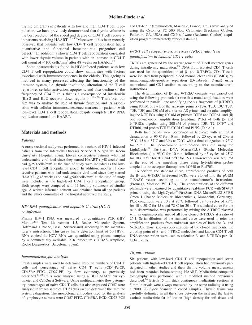
Table 1. Primers and probes used for the quantification of d- and b-TRECs
Name Sequence (50!30)
First round of amplification A
T3A CGGCCGTAGCGACGTACCCTTTCGATGGACCCTCACAG
T3B CGGCCGTAGCGACGTACCGACAAGGCACCAGACTCACAG
T3C CGGCCGTAGCGACGTACCAAGCTCTGGAAGGGAACACAG
T3D CGGCCGTAGCGACGTACCCCGTTTCTCTCCCTCACACAG
T3E CGGCCGTAGCGACGTACCGGGCAGAAACTGAGAACACAG
T3F CGGCCGTAGCGACGTACCCTTGCGCCTTATGCTGCACAG
AS TGAACCAAATTGCATTAAGACC
First round of amplification B
DTR61 TCTGACATTTGCTCCGTG
DTF6 AGAAGGCTCTGTCTAGTGTG
Second round of amplification
DTF7 AGGCTCTGTCTAGTGTGATAAC
T2 CCCAGGAGGAAAGAAGAGGAC
T3R CGGCCGTAGCGACGTACC
DTR66 TGACATGGAGGGCTGAAC
Probes
P1 CTGGGAGTTGGGACCGCCAGAGAGGT-FL
P2 Red705-TTTGTAAAGGTTTCCCGTAGAGTTGAATCATTGTG-PH
TCRFL AGGGATGTGGCATCACCTTTGTTGACA-FL
TCRLC LC640-GGCACCCCTCTGTTCCCCACAGGA-PH
Immunosenescence in HIV-infected patients
581
Thymic function
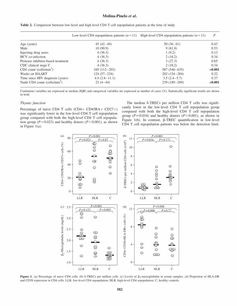
Percentage of naive CD4 T cells (CD4þ CD45RAþ CD27þ)was significantly lower in the low-level CD4 T cell repopulationgroup compared with both the high-level CD4 T cell repopula-tion group (P¼0.023) and healthy donors (P,0.001), as shownin Figure 1(a).
The median d-TRECs per million CD4 T cells was signifi-cantly lower in the low-level CD4 T cell repopulation groupcompared with both the high-level CD4 T cell repopulationgroup (P¼0.034) and healthy donors (P,0.001), as shown inFigure 1(b). In contrast, b-TREC quantification in low-levelCD4 T cell repopulation patients was below the detection limit.
P < 0.001P < 0.001P = 0.034P = 0.023 P = 0.87
P < 0.001P = 0.123 P = 0.003
P = 0.008P = 0.006 P = 0.77
P = 0.17112
10
8
6
4
2
0
12
8
4
0
80
60
40
20
0
LLR HLR C
LLR HLR C LLR HLR C
LLR HLR C
3.5
3.0
2.5
2.0
1.5
1.0
CD
4+ C
D45
RA
+ C
D27
+ c
ells
(%
)
δ-T
RE
Cs
per
mill
ion
CD
4 ce
lls (
×10
4 )C
D4+
CD
38+
HL
A-D
R+
cel
ls (
%)
β 2-M
icro
glob
ulin
leve
ls (
mg/
dL)
(a) (b)
(c) (d)
Figure 1. (a) Percentage of naive CD4 cells. (b) d-TRECs per million cells. (c) Levels of b2-microglobulin in serum samples. (d) Proportion of HLA-DR
and CD38 expression in CD4 cells. LLR, low-level CD4 repopulation; HLR, high-level CD4 repopulation; C, healthy controls.
Table 2. Comparison between low-level and high-level CD4 T cell repopulation patients at the time of study
Low-level CD4 repopulation patients (n¼11) High-level CD4 repopulation patients (n¼11) P
Age (years) 45 (42–49) 50 (36–61) 0.43
Male 10 (90.9) 9 (81.8) 0.53
Injecting drug users 4 (36.3) 1 (9.2) 0.13
HCV co-infection 4 (36.3) 2 (18.2) 0.34
Protease inhibitor-based treatment 4 (36.3) 3 (27.3) 0.65
CDC clinical stage C 4 (36.3) 2 (18.2) 0.34
CD4 count (cells/mm3) 169 (112–203) 587 (546–635) <0.001
Weeks on HAART 124 (57–224) 202 (154–284) 0.22
Time since HIV diagnosis (years) 4.4 (2.8–11.1) 3.5 (2.4–5.7) 0.37
Nadir CD4 count (cells/mm3) 23 (4–44) 239 (189–289) <0.001
Continuous variables are expressed as median (IQR) and categorical variables are expressed as number of cases (%). Statistically significant results are shownin bold.
Molina-Pinelo et al.
582
The quantification of b-TRECs in high-level CD4 T cell repopu-lation patients was only possible in three patients (27.2%), butin nine healthy donors (81.8%). Hence, the d-/b-TREC ratiocould not be measured in the low-level and high-level CD4 Tcell repopulation groups (Table 3).
Before HAART, thymic volume was higher in patients withhigh-level CD4 T cell repopulation [median 3.07 cm3 (interquartilerange 2.01–6.26), n¼7] than in low-level CD4 T cell repopulationpatients [median 2.13 cm3 (interquartile range 0.28–4.8), n¼6],although with no statistical significance (P¼0.199).
Activation analysis
As shown in Figure 1(c), the median level of b2-microglobulinwas higher in patients with low-level CD4 T cell repopulation
compared with patients with high-level CD4 T cell repopulation(P¼0.123), although with no statistical significance. Both levelswere statistically higher than the levels found in healthy controls(P,0.001 and P¼0.003, respectively). In addition, HLA-DRand CD38 were highly expressed by CD4 T cells in the low-level CD4 T cell repopulation group compared with the high-level CD4 T cell repopulation group (P¼0.006) (Figure 1d).
Analysis of CD4 T cell senescence
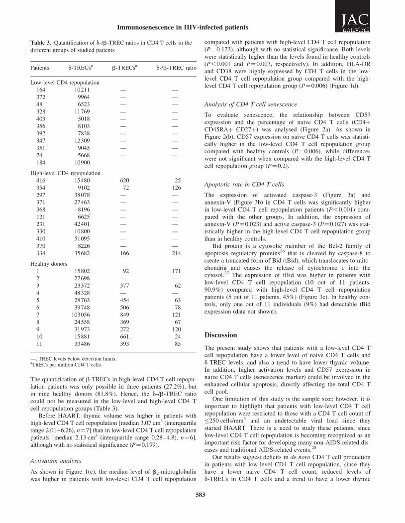
To evaluate senescence, the relationship between CD57expression and the percentage of naive CD4 T cells (CD4þCD45RAþ CD27þ) was analysed (Figure 2a). As shown inFigure 2(b), CD57 expression on naive CD4 T cells was statisti-cally higher in the low-level CD4 T cell repopulation groupcompared with healthy controls (P¼0.006), while differenceswere not significant when compared with the high-level CD4 Tcell repopulation group (P¼0.2).
Apoptotic rate in CD4 T cells
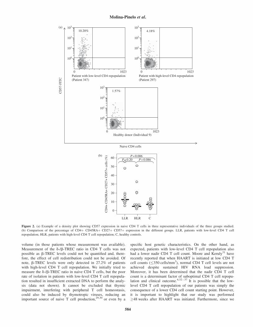
The expression of activated caspase-3 (Figure 3a) andannexin-V (Figure 3b) in CD4 T cells was significantly higherin low-level CD4 T cell repopulation patients (P,0.001) com-pared with the other groups. In addition, the expression ofannexin-V (P¼0.023) and active caspase-3 (P¼0.027) was stat-istically higher in the high-level CD4 T cell repopulation groupthan in healthy controls.
Bid protein is a cytosolic member of the Bcl-2 family ofapoptosis regulatory proteins26 that is cleaved by caspase-8 tocreate a truncated form of Bid (tBid), which translocates to mito-chondria and causes the release of cytochrome c into thecytosol.27 The expression of tBid was higher in patients withlow-level CD4 T cell repopulation (10 out of 11 patients,90.9%) compared with high-level CD4 T cell repopulationpatients (5 out of 11 patients, 45%) (Figure 3c). In healthy con-trols, only one out of 11 individuals (9%) had detectable tBidexpression (data not shown).
Discussion
The present study shows that patients with a low-level CD4 Tcell repopulation have a lower level of naive CD4 T cells andd-TREC levels, and also a trend to have lower thymic volume.In addition, higher activation levels and CD57 expression innaive CD4 T cells (senescence marker) could be involved in theenhanced cellular apoptosis, directly affecting the total CD4 Tcell pool.
One limitation of this study is the sample size; however, it isimportant to highlight that patients with low-level CD4 T cellrepopulation were restricted to those with a CD4 T cell count of�250 cells/mm3 and an undetectable viral load since theystarted HAART. There is a need to study these patients, sincelow-level CD4 T cell repopulation is becoming recognized as animportant risk factor for developing many non-AIDS-related dis-eases and traditional AIDS-related events.28
Our results suggest deficits in de novo CD4 T cell productionin patients with low-level CD4 T cell repopulation, since theyhave a lower naive CD4 T cell count, reduced levels ofd-TRECs in CD4 T cells and a trend to have a lower thymic
Table 3. Quantification of d-/b-TREC ratios in CD4 T cells in the
different groups of studied patients
Patients d-TRECsa b-TRECsa d-/b-TREC ratio
Low-level CD4 repopulation
164 10211 — —
372 9964 — —
48 6523 — —
328 11769 — —
403 5018 — —
356 8103 — —
392 7838 — —
347 12309 — —
351 9045 — —
74 5668 — —
184 10900 — —
High-level CD4 repopulation
416 15480 620 25
354 9102 72 126
297 38078 — —
371 27463 — —
368 8196 — —
121 6625 — —
231 42401 — —
330 10800 — —
410 51095 — —
370 8226 — —
334 35682 166 214
Healthy donors
1 15802 92 171
2 27698 — —
3 23372 377 62
4 48328 — —
5 28763 454 63
6 39748 506 78
7 103056 849 121
8 24558 369 67
9 31973 272 120
10 15881 661 24
11 33486 393 85
—, TREC levels below detection limits.aTRECs per million CD4 T cells.
Immunosenescence in HIV-infected patients
583
volume (in those patients whose measurement was available).Measurement of the d-/b-TREC ratio in CD4 T cells was notpossible as b-TREC levels could not be quantified and, there-fore, the effect of cell redistribution could not be avoided. Ofnote, b-TREC levels were only detected in 27.2% of patientswith high-level CD4 T cell repopulation. We initially tried tomeasure the d-/b-TREC ratio in naive CD4 T cells, but the poorrate of isolation in patients with low-level CD4 T cell repopula-tion resulted in insufficient extracted DNA to perform the analy-sis (data not shown). It cannot be excluded that thymicimpairment, interfering with peripheral T cell homeostasis,could also be induced by thymotropic viruses, reducing animportant source of naive T cell production,29,30 or even by a
specific host genetic characteristics. On the other hand, asexpected, patients with low-level CD4 T cell repopulation alsohad a lower nadir CD4 T cell count. Moore and Keruly31 haverecently reported that when HAART is initiated at low CD4 Tcell counts (�350 cells/mm3), normal CD4 T cell levels are notachieved despite sustained HIV RNA load suppression.Moreover, it has been determined that the nadir CD4 T cellcount is a determinant factor of suboptimal CD4 T cell repopu-lation and clinical outcome.4,32 – 37 It is possible that the low-level CD4 T cell repopulation of our patients was simply theconsequence of a lower CD4 cell count starting point. However,it is important to highlight that our study was performed�48 weeks after HAART was initiated. Furthermore, since we
103(a)10.20% 4.18%
102
101
100
0Patient with low-level CD4 repopulation(Patient 347)
1023
103
102
101
100
0Patient with high-level CD4 repopulation(Patient 297)
1023
1.57%103
102
101
100
0Healthy donor (Individual 9)
1023
Naive CD4 cells
CD
57-F
ITC
P = 0.006P = 0.20 P = 0.086
60
50
40
30
20
10
0
LLR HLR C
CD
4+ C
D45
RA
+ C
D27
+ C
D57
+ c
ells
(%
)(b)
Figure 2. (a) Example of a density plot showing CD57 expression in naive CD4 T cells in three representative individuals of the three groups studied.
(b) Comparison of the percentage of CD4þ CD45RAþ CD27þ CD57þ expression in the different groups. LLR, patients with low-level CD4 T cell
repopulation; HLR, patients with high-level CD4 T cell repopulation; C, healthy controls.
Molina-Pinelo et al.
584
also had a greater follow-up of all patients, we confirmed thatthese patients did not reach a CD4 T cell level of .250 cells/mm3, even after the additional 2 years on HAART since thisstudy was performed (data not shown). Therefore, it is possiblethat the lower nadir CD4 T cell count and also the persistentimmune dysfunction could be the consequence of impairment inthymic function. The thymus is crucial for the reconstitution of
the T cell compartment following lymphodepletion;13,14 thus,lower nadir CD4 T cell count is not inconsistent with a thymicimpairment. There is evidence that thymic activity may increaseafter HAART and correlates with cell reconstitution.38
On the other hand, immunological activation may be anadditional factor that contributes to immunological and clinicaldeterioration of HIV-infected patients,6,39 despite sustained viral
Patients with low-level CD4 repopulation
Patients with high-level CD4 repopulation
P < 0.001 P < 0.001P < 0.001 P < 0.001P = 0.027 P = 0.0236 70
60
50
40
30
20
10
0
Act
ivat
ed c
aspa
se-3
+ C
D4+
cel
ls (
%)
Ann
exin
-V+
CD
4+ c
ells
(%
)
5
4
3
2
1
0
LLR HLR C LLR HLR C
Code
Bid (22 kDa)
tBid (15 kDa)
tBid-Bid expression levels
tBid-Bid expression levels
Arb
itrar
y un
its
β-Actin (45 kDa)
Code
Bid (22 kDa)
tBid (15 kDa)
β-Actin (45 kDa)
330
410
334
297
368
231
121
354
371
416
370
74 351
372
347
392
184
403
48 164
328
356
1
0.5
0
–0.5
–1
Arb
itrar
y un
its
1
0.5
0
–0.5
–1
74 351
372
347
392
184
403 48 164
328
356
330
410
334
297
368
231
121
354
371
416
370
(a)
(c)
(b)
Figure 3. Apoptotic rate of CD4 T cells. (a) Expression of activated caspase-3 in CD4 T cells in the different groups of patients. (b) Expression of
annexin-V in CD4 T cells in the different groups of patients. LLR, patients with low-level CD4 T cell repopulation; HLR, patients with high-level CD4 T cell
repopulation; and C, healthy controls. (c) Expression of Bid and tBid forms in patients with low-level and high-level CD4 T cell repopulation; densitometric
analysis of the difference between tBid expression levels and Bid expression levels normalized by b-actin expression levels is shown next to each western
blot.
Immunosenescence in HIV-infected patients
585
control under HAART. HIV infection has been associated with ahigh expression of activation markers on CD4 T cells, but thesemarkers decrease after initiating HAART.39 – 41 In this study, thehigh activation level of CD4 T cells found in patients with low-level CD4 T cell repopulation might be due to undetectable HIVreplication or viral replication in inaccessible compartments.42
It has been previously reported that an increased number ofCD4 T cells expressing CD57 is associated with poor CD4 Tcell repopulation.6 Specifically, our data showed a higher pro-portion of naive CD4 T cells expressing CD57 in patients withlow-level CD4 T cell repopulation, despite a controlled virologi-cal response. The expression of the senescence marker CD57 onCD4 cells with a naive phenotype may be surprising. Probably,peripheral compensatory mechanisms would be necessary toreverse peripheral T cell lymphopenia in HIV-infected patientswith poor immunological response.43 Thus, an increased entryinto cell cycle of peripheral T cells might induce a higher levelof immunosenescence in patients with low-level CD4 T cellrepopulation. In patients with high-level CD4 T cell repopula-tion, the CD57 expression on naive CD4 T cells was statisticallylower due to the ability to generate sufficient numbers of CD4 Tcells. However, more studies are necessary to establish this cellsubset.
The higher rate of peripheral CD4 T cell apoptosis in patientswith low-level CD4 T cell repopulation could also affect cellrepopulation in patients under HAART. There are many reporteddata indicating that apoptosis plays a major role in this CD4 Tcell depletion.4,44 Moreover, we also analysed active caspase-3since it has been recognized as a central key in mediating apop-tosis and precedes phosphatidyl serine exposure on the externalleaflet of the lipid bilayer, as indicated by annexin-V
binding.45,46 A lower survival rate of CD4 T cells along with ahigher expression of proapoptotic proteins was found in patientswith low-level CD4 T cell repopulation.
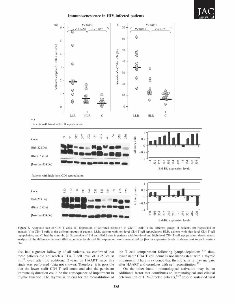
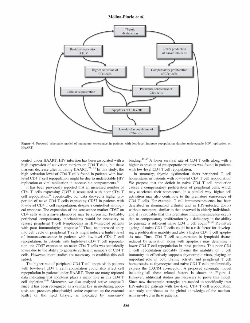
In summary, thymic dysfunction alters peripheral T cellhomeostasis in patients with low-level CD4 T cell repopulation.We propose that the deficit in naive CD4 T cell productioncauses a compensatory proliferation of peripheral cells, whichmay accelerate their senescence. In a parallel way, higher cellactivation may also contribute to the premature senescence ofCD4 T cells. For example, T cell immunosenescence has beendescribed in rheumatoid arthritis and in HIV-infected donorswithout treatment, similar to that observed in elderly individuals,and it is probable that this premature immunosenescence occursdue to compensatory proliferation by a deficiency in the abilityto generate a sufficient naive CD4 T cell count.47,48 Prematureageing of naive CD4 T cells could be a risk factor for develop-ing a proliferative inability and also a higher CD4 T cell apopto-sis rate. Thus, CD4 T cell sequestration in lymphoid tissuesinduced by activation along with apoptosis may determine alower CD4 T cell repopulation in these patients. This poor CD4T cell repopulation probably favours the inability of T cellimmunity to effectively suppress thymotropic virus, playing animportant role in both thymic activity and peripheral T cellhomeostasis, as thymocytes and naive CD4 T cells preferentiallyexpress the CXCR4 co-receptor. A proposed schematic modelincluding all these related factors is shown in Figure 4.However, additional studies are necessary to prove this model.Since new therapeutic strategies are needed to specifically treatHIV-infected patients with low-level CD4 T cell repopulation,our study contributes to the global knowledge of the mechan-isms involved in these patients.
Thymicdysfunction
Residual replicationof HIV
Higher activation ofCD4 cells
Cellular sequestrationPremature senescence of
CD4 cells
Apoptosis of CD4 cells
Low-level repopulation ofCD4 cells
Lower productionof naive CD4 cells
Compensatory proliferationof CD4 cells
Figure 4. Proposed schematic model of premature senescence in patients with low-level immune repopulation despite undetectable HIV replication on
HAART.
Molina-Pinelo et al.
586
Acknowledgements
We thank all the participants of this study for their cooperation.We also thank M. M. Rodrıguez for continuous technicalsupport.
Funding
This work was partially supported by Redes Tematicas deInvestigacion en SIDA (ISCIII-RETIC RD 06/0006/0021), byProyectos de Excelencia de la Consejerıa de Innovacion, Cienciay Empresa, Junta de Andalucıa (P06-CTS-01579), by Consejerıade Salud, Servicio Andaluz de Salud (156/2006), by Fundacionpara la Investigacion y la Prevencion del SIDA (FIPSE 12481/05, 36624/06 and 36688/07), by Fundacion Caja Navarra(no. 11553), by Comunidad Autonoma Madrid (SAL-0159-2006) and by Fondo de Investigacion Sanitaria (FIS PI040993).
Transparency declarations
None to declare.
References
1. Collier AC, Coombs RW, Schoenfeld DA et al. Treatment of
human immunodeficiency virus infection with saquinavir, zidovudine,
and zalcitabine. AIDS Clinical Trials Group. N Engl J Med 1996; 334:
1011–7.
2. Hammer SM, Squires KE, Hughes MD et al. A controlled trial of
two nucleoside analogues plus indinavir in persons with human immu-
nodeficiency virus infection and CD4 cell counts of 200 per cubic milli-
meter or less. AIDS Clinical Trials Group 320 Study Team. N Engl J
Med 1997; 337: 725–33.
3. Connick E, Lederman MM, Kotzin BL et al. Immune reconstitu-
tion in the first year of potent antiretroviral therapy and its relationship
to virologic response. J Infect Dis 2000; 181: 358–63.
4. Benveniste O, Flahault A, Rollot F et al. Mechanisms involved in
the low-level regeneration of CD4þ cells in HIV-1-infected patients
receiving highly active antiretroviral therapy who have prolonged
undetectable plasma viral loads. J Infect Dis 2005; 191: 1670–9.
5. Delobel P, Nugeyre MT, Cazabat M et al. Naive T-cell depletion
related to infection by X4 human immunodeficiency virus type 1 in poor
immunological responders to highly active antiretroviral therapy. J Virol
2006; 80: 10229–36.
6. Fernandez S, Price P, McKinnon EJ et al. Low CD4þ T-cell
counts in HIV patients receiving effective antiretroviral therapy are
associated with CD4þ T-cell activation and senescence but not with
lower effector memory T-cell function. Clin Immunol 2006; 120:
163–70.
7. Dronda F, Moreno S, Moreno A et al. Long-term outcomes
among antiretroviral-naive human immunodeficiency virus-infected
patients with small increases in CD4þ cell counts after successful
virologic suppression. Clin Infect Dis 2002; 35: 1005–9.
8. Kaplan D, Sieg S. Role of the Fas/Fas ligand apoptotic pathway
in human immunodeficiency virus type 1 disease. J Virol 1998; 72:
6279–82.
9. Pitrak D, Bolanos J, Hershow R et al. Discordant CD4 T lympho-
cyte responses to antiretroviral therapy for HIV infection are associated
with ex-vivo rates of apoptosis. AIDS 2001; 15: 1317–9.
10. Hazenberg MD, Otto SA, Wit FW et al. Discordant responses
during antiretroviral therapy: role of immune activation and T cell redis-
tribution rather than true CD4 T cell loss. AIDS 2002; 16: 1287–9.
11. Pakker NG, Notermans DW, de Boer RJ et al. Biphasic kinetics
of peripheral blood T cells after triple combination therapy in HIV-1
infection: a composite of redistribution and proliferation. Nat Med 1998;
4: 208–14.
12. Hazenberg MD, Hamann D, Schuitemaker H et al. T cell
depletion in HIV-1 infection: how CD4þ T cells go out of stock. Nat
Immunol 2000; 1: 285–9.
13. de la Rosa R, Leal M, Rubio A et al. Baseline thymic volume is
a predictor for CD4 T cell repopulation in adult HIV-infected patients
under highly active antiretroviral therapy. Antivir Ther 2002; 7: 159–63.
14. Ruiz-Mateos E, de la Rosa R, Soriano N et al. Comparison of
thymic function-related markers to predict early CD4 T-cell repopulation
in adult HIV-infected patients on HAART. Antivir Ther 2003; 8:
289–94.
15. Ruiz-Mateos E, Rubio A, Vallejo A et al. Thymic volume is
associated independently with the magnitude of short- and long-term
repopulation of CD4þ T cells in HIV-infected adults after highly active
antiretroviral therapy (HAART). Clin Exp Immunol 2004; 136: 501–6.
16. Isgro A, Leti W, Santis W et al. Altered clonogenic capability and
stromal cell function characterize bone marrow of HIV-infected subjects
with low CD4 T cell counts despite viral suppression during HAART.
Clin Infect Dis 2008; 46: 1902–10.
17. Molina-Pinelo S, Leal M, Soriano-Sarabia N et al. Prevalence
and factors involved in discordant responses to highly active antiretro-
viral treatment in a closely followed cohort of treatment-naive
HIV-infected patients. J Clin Virol 2005; 33: 110–5.
18. Ginaldi L, De Martinis M, D’Ostilio A et al. The immune system
in the elderly: II. Specific cellular immunity. Immunol Res 1999; 20:
109–15.
19. Ginaldi L, De Martinis M, Monti D et al. The immune system in
the elderly: activation-induced and damage-induced apoptosis.
Immunol Res 2004; 30: 81–94.
20. Gruver AL, Hudson LL, Sempowski GD. Immunosenescence of
ageing. J Pathol 2007; 211: 144–56.
21. Cohen Stuart J, Hamann D, Borleffs J et al. Reconstitution of
naive T cells during antiretroviral treatment of HIV-infected adults is
dependent on age. AIDS 2002; 16: 2263–6.
22. Cummings JS, Cairo C, Armstrong C et al. Impacts of HIV infec-
tion on Vg2Vd2 T cell phenotype and function: a mechanism for
reduced tumor immunity in AIDS. J Leukoc Biol 2008; 84: 371–9.
23. Douek DR, McFarland P, Keiser E et al. Changes in thymic
function with age and during treatment of HIV infection. Nature 1998;
396: 690–5.
24. Choyke PL, Zeman RK, Gootenberg JE et al. Thymic atrophy
and regrowth in response to chemotherapy: CT evaluation. AJR Am J
Roentgenol 1987; 149: 269–72.
25. Franco JM, Rubio A, Martınez-Moya M et al. T cell repopulation
and thymic volume in HIV-1-infected patients after highly active antire-
troviral therapy. Blood 2002; 99: 3702–6.
26. Wei MC, Lindsten T, Mootha VK et al. tBID, a membrane-
targeted death ligand, oligomerizes BAK to release cytochrome c.
Genes Dev 2000; 14: 2060–71.
27. Li H, Zhu H, Xu CJ et al. Cleavage of BID by caspase 8 med-
iates the mitochondrial damage in the Fas pathway of apoptosis. Cell
1998; 94: 491–501.
28. Baker JV, Peng G, Rapking J et al. Poor initial CD4þ recovery
with antiretroviral therapy prolongs immune depletion and increases
risk for AIDS and non-AIDS diseases. J Acquir Immune Defic Syndr
2008; 48: 541–6.
29. Hellerstein MK, McCune JM. T cell turnover in HIV-1 disease.
Immunity 1997; 7: 583–9.
Immunosenescence in HIV-infected patients
587
30. Correa R, Munoz-Fernandez MA. Viral phenotype affects the
thymical production of new T-cells in HIV-1 infected children. AIDS
2001; 15: 1959–63.
31. Moore RD, Keruly JC. CD4þ cell count 6 years after commence-
ment of highly active antiretroviral therapy in persons with sustained
virologic suppression. Clin Infect Dis 2007; 44: 441–6.
32. Grabar S, Le Moing V, Goujard C et al. Clinical outcome of
patients with HIV-1 infection according to immunologic and virologic
response after 6 months of highly active antiretroviral therapy. Ann
Intern Med 2000; 133: 401–10.
33. Egger M, May M, Chene G et al. Prognosis of HIV-1-infected
patients starting highly active antiretroviral therapy: a collaborative
analysis of prospective studies. Lancet 2002; 360: 119–29.
34. Florence E, Lundgren J, Dreezen C et al. Factors associated
with a reduced CD4 lymphocyte count response to HAART despite full
viral suppression in the EuroSIDA study. HIV Med 2003; 4: 255–62.
35. Anthony KB, Yoder C, Metcalf JA et al. Incomplete CD4 T cell
recovery in HIV-1 infection after 12 months of highly active antiretroviral
therapy is associated with ongoing increased CD4 T cell activation and
turnover. J Acquir Immune Defic Syndr 2003; 33: 125–33.
36. Kaufmann GR, Furrer H, Ledergerber B et al. Characteristics,
determinants, and clinical relevance of CD4 T cell recovery to ,500
cells/microL in HIV type 1-infected individuals receiving potent antire-
troviral therapy. Clin Infect Dis 2005; 41: 361–72.
37. Marchetti G, Gori A, Casabianca A et al. Comparative analysis
of T-cell turnover and homeostatic parameters in HIV-infected patients
with discordant immune-virological responses to HAART. AIDS 2006;
20: 1727–36.
38. Hardy G, Worrell S, Hayes P et al. Evidence of thymic reconsti-
tution after highly active antiretroviral therapy in HIV-1 infection. HIV
Med 2004; 5: 67–73.
39. Hazenberg MD, Stuart JW, Otto SA et al. T-cell division in
human immunodeficiency virus (HIV)-1 infection is mainly due to
immune activation: a longitudinal analysis in patients before and during
highly active antiretroviral therapy (HAART). Blood 2000; 95: 249–55.
40. Hunt PW, Martin JN, Sinclair E et al. T cell activation is associ-
ated with lower CD4þ T cell gains in human immunodeficiency
virus-infected patients with sustained viral suppression during antiretro-
viral therapy. J Infect Dis 2003; 187: 1534–43.
41. Deeks SG, Kitchen CM, Liu L et al. Immune activation set point
during early HIV infection predicts subsequent CD4þ T-cell changes
independent of viral load. Blood 2004; 104: 942–7.
42. McCune JM. The dynamics of CD4þ T-cell depletion in HIV
disease. Nature 2001; 410: 974–9.
43. Dion ML, Bordi R, Zeidan J et al. Slow disease progression and
robust therapy-mediated CD4þ T-cell recovery are associated with effi-
cient thymopoiesis during HIV-1 infection. Blood 2007; 109: 2912–20.
44. Hansjee N, Kaufmann GR, Strub C et al. Persistent apoptosis in
HIV-1-infected individuals receiving potent antiretroviral therapy is
associated with poor recovery of CD4 T lymphocytes. J Acquir Immune
Defic Syndr 2004; 36: 671–7.
45. Verhoven B, Krahling S, Schlegel RA et al. Regulation of phos-
phatidylserine exposure and phagocytosis of apoptotic T lymphocytes.
Cell Death Differ 1999; 6: 262–70.
46. Diaz C, Lee AT, McConkey DJ et al. Phosphatidylserine externa-
lization during differentiation-triggered apoptosis of erythroleukemic.
Cell Death Differ 1999; 6: 218–26.
47. Weyand CM, Fulbright JW, Goronzy JJ. Immunosenescence,
autoimmunity, and rheumatoid arthritis. Exp Gerontol 2003; 38: 833–41.
48. Appay V, Almeida JR, Sauce D et al. Accelerated immune
senescence and HIV-1 infection. Exp Gerontol 2007; 42: 432–7.
Molina-Pinelo et al.
588
Related Documents