PREDICTING IMPROVEMENT OF POST-ORTHODONTIC WHITE SPOT LESIONS SUSAN KIM A THESIS SUBMITTED IN PARTIAL FULFILLMENT OF THE REQUIREMENTS FOR THE DEGREE OF MASTER OF SCIENCE IN DENTISTRY UNIVERSITY OF WASHINGTON 2015 COMMITTEE: GREG HUANG BURCU BAYIRLI MEHMET SARIKAYA PROGRAM AUTHORIZED TO OFFER DEGREE: DEPARTMENT OF ORTHODONTICS

Welcome message from author
This document is posted to help you gain knowledge. Please leave a comment to let me know what you think about it! Share it to your friends and learn new things together.
Transcript
PREDICTING IMPROVEMENT OF POST-ORTHODONTIC
WHITE SPOT LESIONS
SUSAN KIM
A THESIS SUBMITTED IN PARTIAL FULFILLMENT OF THE
REQUIREMENTS FOR THE DEGREE OF
MASTER OF SCIENCE IN DENTISTRY
UNIVERSITY OF WASHINGTON
2015
COMMITTEE: GREG HUANG
BURCU BAYIRLI MEHMET SARIKAYA
PROGRAM AUTHORIZED TO OFFER DEGREE:
DEPARTMENT OF ORTHODONTICS

Copyright 2015
Susan Kim
i
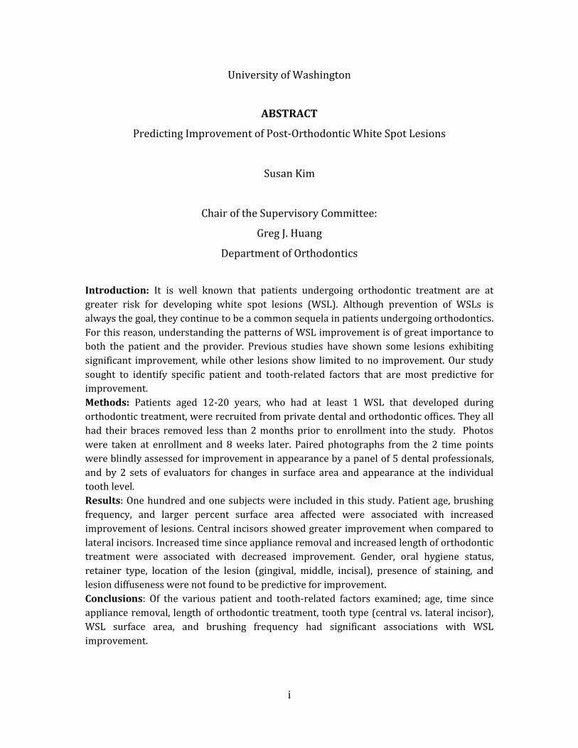
University of Washington
ABSTRACT
Predicting Improvement of Post-Orthodontic White Spot Lesions
Susan Kim
Chair of the Supervisory Committee:
Greg J. Huang
Department of Orthodontics Introduction: It is well known that patients undergoing orthodontic treatment are at
greater risk for developing white spot lesions (WSL). Although prevention of WSLs is
always the goal, they continue to be a common sequela in patients undergoing orthodontics.
For this reason, understanding the patterns of WSL improvement is of great importance to
both the patient and the provider. Previous studies have shown some lesions exhibiting
significant improvement, while other lesions show limited to no improvement. Our study
sought to identify specific patient and tooth-related factors that are most predictive for
improvement.
Methods: Patients aged 12-20 years, who had at least 1 WSL that developed during
orthodontic treatment, were recruited from private dental and orthodontic offices. They all
had their braces removed less than 2 months prior to enrollment into the study. Photos
were taken at enrollment and 8 weeks later. Paired photographs from the 2 time points
were blindly assessed for improvement in appearance by a panel of 5 dental professionals,
and by 2 sets of evaluators for changes in surface area and appearance at the individual
tooth level.
Results: One hundred and one subjects were included in this study. Patient age, brushing
frequency, and larger percent surface area affected were associated with increased
improvement of lesions. Central incisors showed greater improvement when compared to
lateral incisors. Increased time since appliance removal and increased length of orthodontic
treatment were associated with decreased improvement. Gender, oral hygiene status,
retainer type, location of the lesion (gingival, middle, incisal), presence of staining, and
lesion diffuseness were not found to be predictive for improvement.
Conclusions: Of the various patient and tooth-related factors examined; age, time since
appliance removal, length of orthodontic treatment, tooth type (central vs. lateral incisor),
WSL surface area, and brushing frequency had significant associations with WSL
improvement.

ii
ACKNOWLEDGEMENTS The author wishes to thank her research committee for their guidance in conducting this project. She is especially grateful to her committee chair, Greg Huang, who was always been so helpful in providing the necessary mentorship and feedback in order to successfully complete this project.
iii
TABLE OF CONTENTS
Abstract ....................................................................................................................................................... i
Acknowledgements ................................................................................................................................. ii
Table of Contents .................................................................................................................................... iii
List of Figures ........................................................................................................................................... iv
List of Tables ............................................................................................................................................. v
Introduction ............................................................................................................................................... 1
Material and Methods .............................................................................................................................. 3
Results ......................................................................................................................................................... 8
Discussion ................................................................................................................................................ 11
Conclusion .............................................................................................................................................. 20
References ............................................................................................................................................... 21
Figures ...................................................................................................................................................... 23
Tables ........................................................................................................................................................ 25
iv
LIST OF FIGURES
Figure Number
1. Image of single teeth and tooth thirds ................................................................................. 23
2. Appearance of a stained lesion .............................................................................................. 24
3. Appearance of a diffuse, discrete, mixed lesion ................................................................ 24
v
LIST OF TABLES
Table Number
1. Description of evaluations ...................................................................................................... 25
2. Summary of single teeth ......................................................................................................... 25
3. Summary of tooth thirds ........................................................................................................ 26
4. Summary of descriptive data ................................................................................................ 26
5. Subjective improvement of incisors (univariate analysis) ............................................ 27
6. Objective improvement of incisors (univariate analysis) .............................................. 27
7. Subjective improvement of incisors (multivariate analysis) ........................................ 28
8. Objective improvement of incisors (multivariate analysis) ......................................... 28
9. Improvement scores of single teeth .................................................................................... 28
10. Improvement scores of tooth thirds ................................................................................... 29
1
INTRODUCTION
Orthodontic treatment has long served as a means for providing patients with improved
esthetic, functional, and psychological benefits. Unfortunately, white spot lesions (WSLs) are
one of the most common and undesirable side effects that can diminish the satisfaction and
joy that a patient experiences following orthodontic treatment. Some studies have shown the
prevalence of WSLs to be as high as 97% among orthodontic populations1.
WSLs are characterized by their increased opacity compared to healthy enamel. They
possess a whiter appearance as a result of mineral loss in the surface layers, which alters the
refractory index2 and increases the scattering of light in the affected area. The appearance of
the lesion can vary from an initial loss of mineral to total tooth destruction3. In some
instances, stains can be incorporated into a lesion and lead to the formation of brown spots
during the remineralization process, worsening the esthetic problem4.
Prevention, diagnosis, and treatment of WSLs are crucial to minimizing tooth decay and
discoloration that can compromise the esthetics of a smile. It is a significant concern for both
orthodontists and their patients because in addition to the risk for progressing to active
disease, WSLs often affect maxillary incisors, which are generally the most visible teeth.
Several options have been proposed to address these lesions, depending upon their nature
and severity. The recommended treatment can be as simple as improved home care with
fluoride toothpaste for minor lesions, to more invasive options involving composite
restorations. However, there is still a lack of strong evidence in the literature regarding the
most effective treatment protocol5 and the ideal timing for maximizing improvement.
In addition to the abundance of available treatment options, the unpredictable patterns
and degree of improvement adds to the complexity of WSL management. Often times, we see
a wide range of improvement in lesions from one patient to the next. Lesions can vary in size,
2
shape, and location, and are as unique as the oral environment of the patients in which they
are found.
The objective of our study was to attempt to identify patient and tooth-related factors that
might be predictive for WSL improvement. Knowledge of these factors might help
orthodontists develop management strategies at the time braces are removed.
3
MATERIAL AND METHODS
This study is a further investigation of data from a prior project that investigated WSLs.
The photographs that formed the sample were originally collected from a randomized
(1:1:1), single-blind, 3-arm, active controlled, parallel-group trial evaluating the
improvement of WSLs for 3 different treatment arms6. The treatment arms compared
effectiveness of MI Paste Plus (900 ppm fluoride; GC America), PreviDent fluoride varnish
(22,600 ppm fluoride; Colgate Oral Pharmaceuticals, New York, NY), and usual home-care
oral hygiene instruction using over-the-counter toothpaste (1100 ppm fluoride; Colgate Oral
Pharmaceuticals) (control group). In the original study, photographs of WSLs were taken at
two different time points: at the start of the study (T1) and 8 weeks later (T2). Data were
collected from private orthodontic and general dentistry offices belonging to the Practice-
based Research Collaborative in Evidence-based Dentistry network in the Northwestern
United States (NW PRECEDENT). The network was co-operated by the University of
Washington and the Oregon Health and Science University, and was comprised of the
following states—Washington, Oregon, Montana, Idaho, and Utah.
Eligibility criteria for this study included the fulfillment of the following conditions:
completion of fixed appliance orthodontic therapy within the past 2 months, the presence of
at least 1 WSL on the facial surface of a maxillary incisor that was not present prior to
starting orthodontic treatment, and being between the ages of 12 and 20 years. Subjects
excluded from this study were those who were unwilling to be randomly assigned to 1 of the
3 treatment groups; had any abnormal oral, medical, or mental conditions; received therapy
for WSLs after orthodontic treatment; displayed frank cavitations associated with the
maxillary incisors; or were unable to speak or read the English language. Patients (and
parents, for those under 18 years of age) consented to participate prior to the start of the
study.

4
All subjects completed a questionnaire, which provided us with information regarding the
patient’s average daily brushing frequency. Patient information such as age, gender, length of
orthodontic treatment, retainer type, and oral hygiene throughout treatment was provided
by their clinicians.
Two different types of evaluations (subjective and objective improvement) were
performed for the 4 maxillary incisors, for each pair of photographs (initial and 8-weeks).
For subjective improvement, a blinded panel of 5 dental professionals performed their
assessment of improvement using a visual analog scale from 0 to 100mm (0mm = no
improvement or worsened, to 100mm = complete resolution of WSL). These evaluations
were performed as part of the original study, and the ratings of the panel were used for
overall improvement of the 4 maxillary incisors.
For objective improvement, 2 examiners (a dental student and a general dentist)
performed the assessment for improvement by measuring changes in WSL surface area from
each time point. WSL surface area was divided by total tooth surface area to calculate the
pretreatment and posttreatment percentages of affected surface area. The change in
percentage of affected surface area was obtained by subtracting posttreatment surface area
from pretreatment surface area. These assessments were also performed as part of the
original study for all 4 incisors.
Results from the randomized control trial found no significant difference in subjective or
objective improvement in the appearance of the WSL between those who received MI Paste
Plus, PreviDent fluoride varnish, and normal home care, during the 8-week period. Although
some WSLs exhibited little to no improvement, many lesions did in fact show a large degree
of improvement. Since the treatment arm did not appear to have a large role in the
improvement of the WSL, we decided to investigate other possible factors that might be
associated with the degree of WSL improvement that occurred.
5
For this current study, we considered improvement of a lesion to be a visible decrease in
affected surface area, minimized contrast between the WSL and surrounding healthy tooth
structure, or any combination of changes resulting in an overall improved esthetic
appearance. The previous study evaluated all 4 incisors as a unit, but did not evaluate each
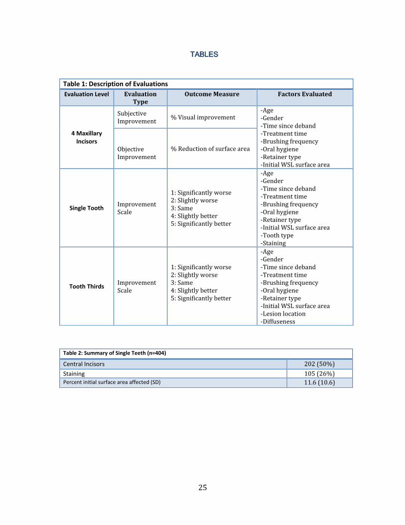
tooth individually (Table 1). In order to perform evaluations at the single tooth level, we
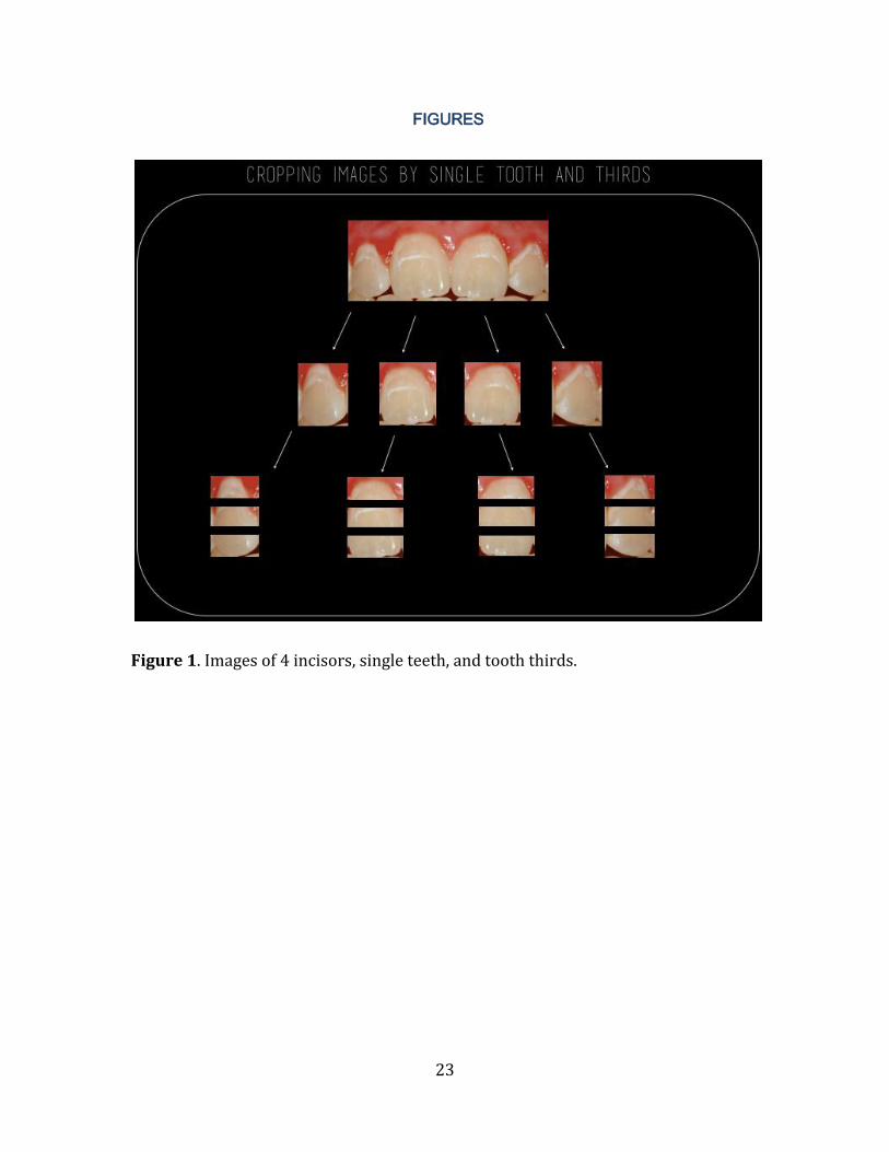
cropped the images of the 4 maxillary incisors into individual teeth (n=404) and then further
cropped them into horizontal thirds, but only evaluating those portions affected (n=728)
(figure1). In order to maintain a uniform size and a similar level of magnification, a grid of a
fixed dimension was used to reveal only the tooth or portion of the tooth being evaluated.
This method allowed us to mask all other teeth and parts of the teeth, minimizing any
undesired influence from the surrounding teeth on evaluation scores. Once all images were
cropped to proper dimensions, lesions were then categorized according to the different
characteristics we were interested in studying.
At the single tooth level, each lesion was categorized by the presence or absence of
staining (Table 2). Most WSLs are uniformly white throughout (unstained), but occasionally,
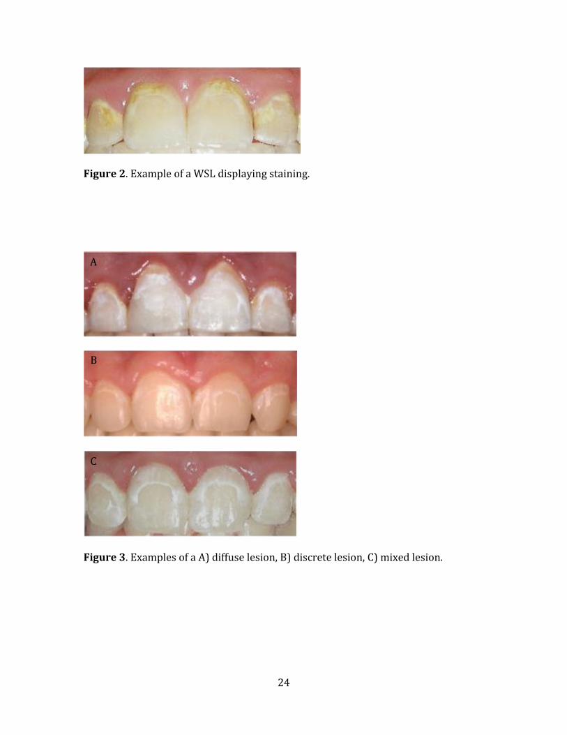
some WSLs display a yellowish or brownish area of discoloration (figure 2). A primary and
secondary evaluator (an orthodontic resident and a general dentist) categorized staining
prior to blinding for time point in order to ensure that the staining was present at the start of
the study. Following categorization, time points were obscured for all images in order to
reduce any expectation bias for improvement, as one may naturally expect improvement to
occur over time even when no such improvement has occurred.
The images of the horizontal thirds were labeled as gingival (G), middle (M), and incisal (I)
thirds (Table 3). Only portions of the tooth affected by a WSL were evaluated. Portions of the
tooth not containing a lesion were not included. The same 2 evaluators independently
examined each third and categorized each lesion by their diffuseness (figure 3). Any lesion
6
with a discrete linear shape with areas of healthy, unaffected tooth structure adjacent to both
sides of the lesion was considered to be a discrete lesion. Any lesion with a non-linear,
amorphous, or ill-defined appearance was categorized as being diffuse. Any lesion containing
both types of these lesions on the same tooth third was classified as being a mixed lesion.
Time points were also obscured for each third, prior to evaluation.
For the evaluation of single teeth and tooth thirds, the evaluators rated each image on a
WSL improvement scale of 1 to 5 as being: (1) significantly worse, (2) slightly worse, (3) the
same, (4) slightly better, or (5) significantly better than its corresponding image taken at the
alternative time point (Table 1). Twenty images were evaluated a second time, at least 1
month apart, in order to calculate intra-rater and inter-rater reliability.
In addition to our evaluations of images taken during the 8-week period between T1–T2,
we also had images for 31 subjects taken on the day of appliance removal (T0), prior to
starting the study. Following appliance removal, most subjects began the study within 1
week’s time (n=73), but no subjects were started on the same day of appliance removal. This
was to allow for natural rehydration of the enamel and to prevent excessive gingival swelling
from obscuring any portion of the lesion. Of the 31 subjects who had T0 data, 4 subjects
dropped out prior to completing the study. Analyzing the 27 subjects with both T0 and T1
data allowed us to evaluate the WSL improvement that occurred naturally in the first days
following appliance removal, before any intervention was initiated.
Statistical analysis
Analyses were conducted using SAS software (version 9.2; SAS Institute, Cary, NC) on a
Windows operating system (Microsoft).
Descriptive data were summarized with frequency tables (Table 4). Regression models
were run using Generalized Estimating Equations (GEE), which allows us to account for
7
clustering by site and for tooth-within-subject. ICCs for both factors were found to be
negligible.
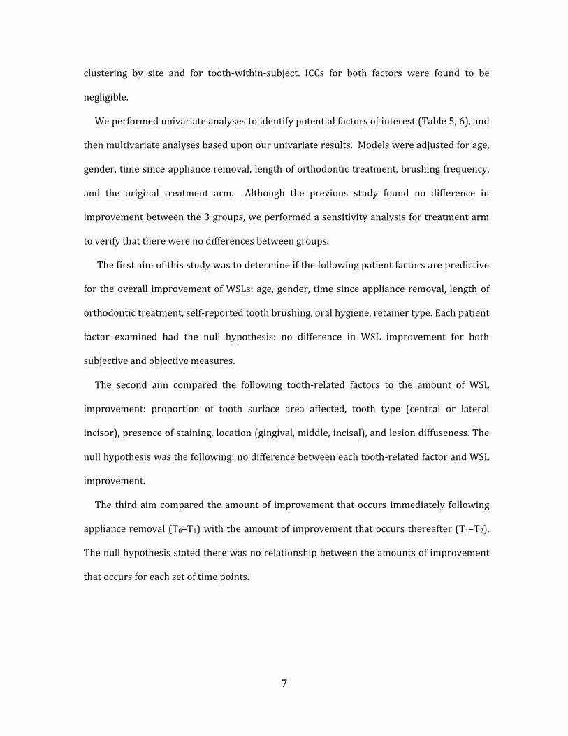
We performed univariate analyses to identify potential factors of interest (Table 5, 6), and
then multivariate analyses based upon our univariate results. Models were adjusted for age,
gender, time since appliance removal, length of orthodontic treatment, brushing frequency,
and the original treatment arm. Although the previous study found no difference in
improvement between the 3 groups, we performed a sensitivity analysis for treatment arm
to verify that there were no differences between groups.
The first aim of this study was to determine if the following patient factors are predictive
for the overall improvement of WSLs: age, gender, time since appliance removal, length of
orthodontic treatment, self-reported tooth brushing, oral hygiene, retainer type. Each patient
factor examined had the null hypothesis: no difference in WSL improvement for both
subjective and objective measures.
The second aim compared the following tooth-related factors to the amount of WSL
improvement: proportion of tooth surface area affected, tooth type (central or lateral
incisor), presence of staining, location (gingival, middle, incisal), and lesion diffuseness. The
null hypothesis was the following: no difference between each tooth-related factor and WSL
improvement.
The third aim compared the amount of improvement that occurs immediately following
appliance removal (T0–T1) with the amount of improvement that occurs thereafter (T1–T2).
The null hypothesis stated there was no relationship between the amounts of improvement
that occurs for each set of time points.
8
RESULTS
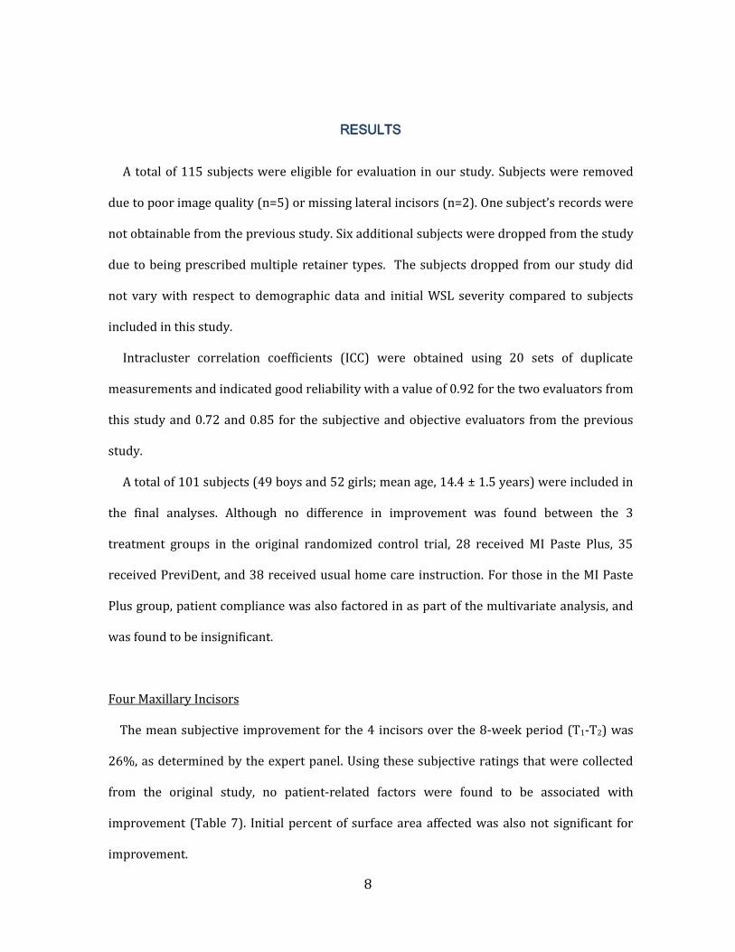
A total of 115 subjects were eligible for evaluation in our study. Subjects were removed
due to poor image quality (n=5) or missing lateral incisors (n=2). One subject’s records were
not obtainable from the previous study. Six additional subjects were dropped from the study
due to being prescribed multiple retainer types. The subjects dropped from our study did
not vary with respect to demographic data and initial WSL severity compared to subjects
included in this study.
Intracluster correlation coefficients (ICC) were obtained using 20 sets of duplicate
measurements and indicated good reliability with a value of 0.92 for the two evaluators from
this study and 0.72 and 0.85 for the subjective and objective evaluators from the previous
study.
A total of 101 subjects (49 boys and 52 girls; mean age, 14.4 ± 1.5 years) were included in
the final analyses. Although no difference in improvement was found between the 3
treatment groups in the original randomized control trial, 28 received MI Paste Plus, 35
received PreviDent, and 38 received usual home care instruction. For those in the MI Paste
Plus group, patient compliance was also factored in as part of the multivariate analysis, and
was found to be insignificant.
Four Maxillary Incisors
The mean subjective improvement for the 4 incisors over the 8-week period (T1-T2) was
26%, as determined by the expert panel. Using these subjective ratings that were collected
from the original study, no patient-related factors were found to be associated with
improvement (Table 7). Initial percent of surface area affected was also not significant for
improvement.
9
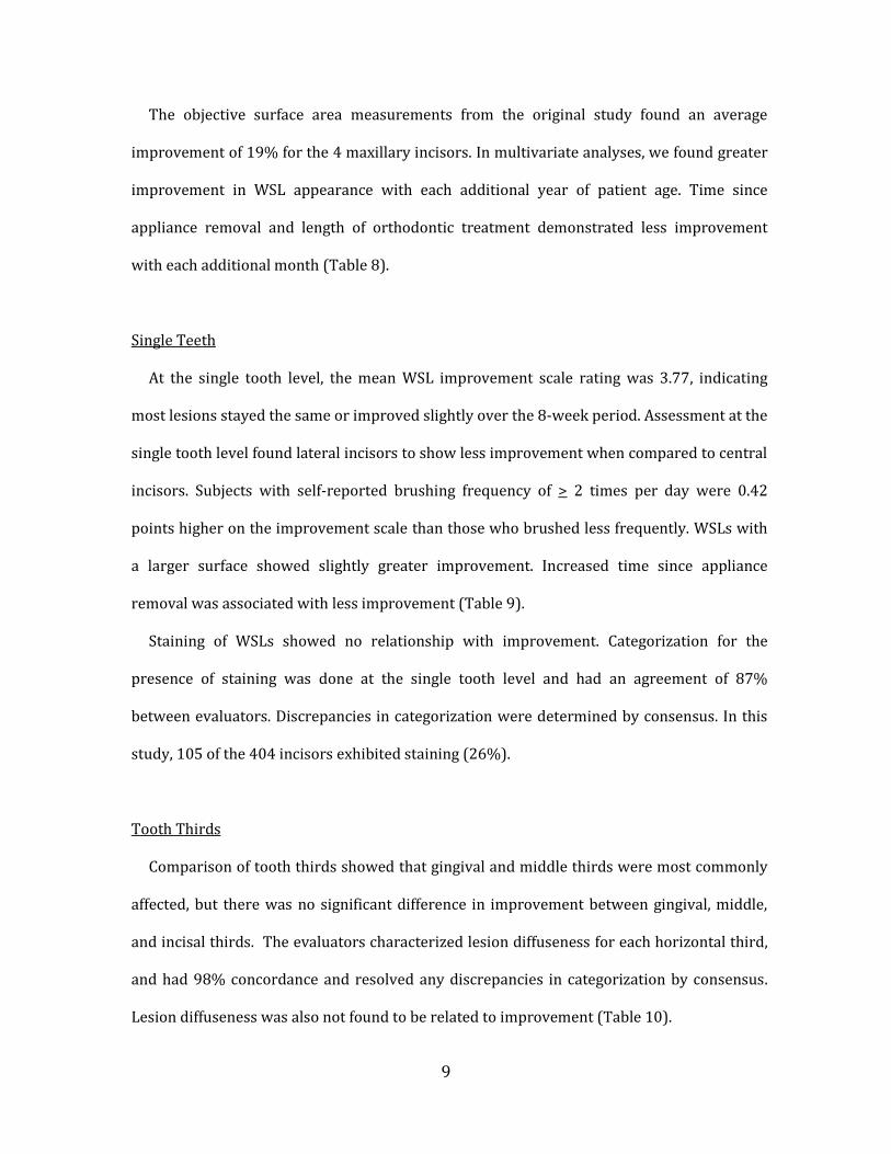
The objective surface area measurements from the original study found an average
improvement of 19% for the 4 maxillary incisors. In multivariate analyses, we found greater
improvement in WSL appearance with each additional year of patient age. Time since
appliance removal and length of orthodontic treatment demonstrated less improvement
with each additional month (Table 8).
Single Teeth
At the single tooth level, the mean WSL improvement scale rating was 3.77, indicating
most lesions stayed the same or improved slightly over the 8-week period. Assessment at the
single tooth level found lateral incisors to show less improvement when compared to central
incisors. Subjects with self-reported brushing frequency of > 2 times per day were 0.42
points higher on the improvement scale than those who brushed less frequently. WSLs with
a larger surface showed slightly greater improvement. Increased time since appliance
removal was associated with less improvement (Table 9).
Staining of WSLs showed no relationship with improvement. Categorization for the
presence of staining was done at the single tooth level and had an agreement of 87%
between evaluators. Discrepancies in categorization were determined by consensus. In this
study, 105 of the 404 incisors exhibited staining (26%).
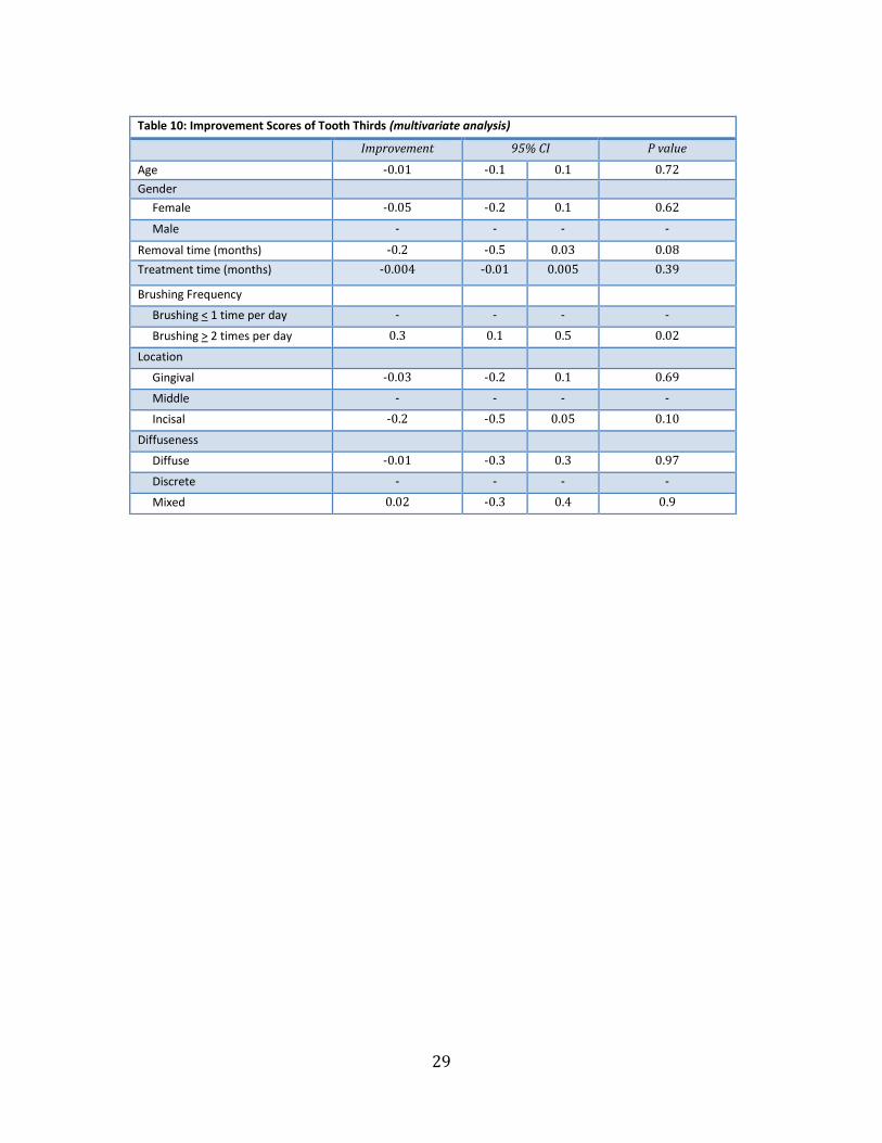
Tooth Thirds
Comparison of tooth thirds showed that gingival and middle thirds were most commonly
affected, but there was no significant difference in improvement between gingival, middle,
and incisal thirds. The evaluators characterized lesion diffuseness for each horizontal third,
and had 98% concordance and resolved any discrepancies in categorization by consensus.
Lesion diffuseness was also not found to be related to improvement (Table 10).
10
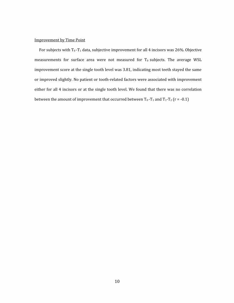
Improvement by Time Point
For subjects with T0 -T1 data, subjective improvement for all 4 incisors was 26%. Objective
measurements for surface area were not measured for T0 subjects. The average WSL
improvement score at the single tooth level was 3.81, indicating most teeth stayed the same
or improved slightly. No patient or tooth-related factors were associated with improvement
either for all 4 incisors or at the single tooth level. We found that there was no correlation
between the amount of improvement that occurred between T0 -T1 and T1-T2 (r = -0.1)
11
DISCUSSION
We identified several factors that were associated with a significant effect on
improvement in the appearance of WSLs. Overall, we saw a tendency for WSLs to improve
during the 8-week period, according to all 3 evaluation methods. However, different patient
and tooth-related factors were significant for improvement depending on the type of
assessment.
Age
With each additional year of age, there was a 3.1% greater reduction in WSL surface area
over the 8-week period. Although the range in patient age was not very large for our study
(12-20 years) we can expect that with greater age, there may be some improved dexterity
and ability to remove plaque and food debris with brushing. Even if patients are brushing
with the same frequency and duration each day, the older patients may be more effective. It
is also possible that patients who are older have a greater appreciation for the esthetics of
their smile, and will make a greater effort to be diligent with oral hygiene once they become
aware of their WSLs and the etiology.
When considering the effect of age on the oral environment, one study comparing the salivary
content of children (6-12 years) and adults (19-44 years) found that calcium concentrations were
lower in children7. Concentrations of calcium and phosphate in saliva have significant influence
on the protective mechanisms of dental hard tissues within the oral environment. Since a lower
salivary calcium concentration creates a higher critical pH and a lower driving force for
remineralization, those who are younger in age may also have a saliva composition less
conducive to WSL improvement.
12
Gender
Studies have shown conflicting findings regarding the effect of gender on the prevalence of
WSL development. There is a common belief that males tend to have decreased compliance and
are also less concerned with their dental appearance8. Although some may argue that this could
also affect motivation for improvement, our study found no difference in improvement between
males and females.
Time Since Appliance Removal
With each additional month that elapses since appliance removal, there was 14.9% less
improvement in surface area. This would suggest that the majority of improvement occurs
immediately following appliance removal, so that even by the second month, improvement
diminishes significantly. Some studies regarding remineralization patterns of WSLs support these
findings and have described an exponential pattern of remineralization, with all improvement
occurring within the first month9. This pattern of improvement is believed to be more
characteristic of lesions that were experimentally induced within a few short weeks and are likely
to be caused by softening of the outermost enamel layer. Other studies involving WSLs that were
developed over a longer period of time, involving subsurface layers of enamel, showed the
greatest amount of remineralization occurring during the first few months and then continuing at
a slower rate thereafter10
. As the average treatment time for our subjects was 25.7 months, the
lesions were likely to have formed over a period of months rather than weeks. With that in mind,
we would have expected more of a gradual tapering of improvement, but instead our findings
were more consistent with that of an exponential pattern.
The average amount of subjective improvement that occurred between the day of appliance
removal to start of the study (T0 -T1) was 26%. The average time elapsed between T0 and T1 was
only 4 days, so within this short period of time, without any intervention whatsoever, patients
showed significant improvement. This is an important issue to consider, as baseline improvement
13
must be factored into the amount of improvement that occurs after a particular form of
intervention is instituted. This is especially relevant if a WSL therapy calls for application at the
time of appliance removal, since the spontaneous improvement that will occur naturally should be
subtracted from overall improvement.
When we looked for a correlation in subjective improvement scores between T0 -T1 and T1-T2,
we found no relationship between the amount of improvement that occurs early on (T0 -T1) and
the amount of improvement that occurs subsequently (T1-T2). We were curious to determine
whether lesions that initially show a greater degree of change would show less improvement in
the later weeks, since the majority of improvement may have already occurred. However, there
was no particular pattern of improvement since some WSLs that improved drastically in T0 -T1
went on to show minimal change during T1-T2, while others improved greatly.
Treatment Time
According to objective measurements, length of orthodontic treatment showed 0.29%
decreased improvement of WSLs with each additional month in treatment. Although
statistically significant, a difference of 0.29% per month is not clinically significant. Even
with an additional year of treatment, this only amounts to a 3.5% decrease in improvement.
One study found that teeth bonded for a relatively short treatment time (12-16 months) had
the same incidence of WSLs as those with longer treatment time (up to 36 months)11. A
second study found that the greatest formation of WSLs occurs in the first 6 months of
treatment, with a gradual slowing occurring by 12 months12. Therefore, if lesion severity and
formation are relatively established by the first year of orthodontic treatment, it is not
surprising to see very minimal differences in improvement between our subjects, who were
in treatment for greater than 12 months.
14
Tooth Brushing Frequency
Brushing frequency of > 2 times per day was associated with an increase of 0.42 on the
WSL improvement scale, when compared to those who brushed less frequently. This is not
surprising, as more frequent clearing of food and plaque away from the tooth surface would
decrease exposure of the enamel to the acidogenic bacteria and their products. Interestingly,
in reviewing the patient survey results, we found that more subjects reported brushing > 2
times per day after appliances were removed, compared to when they were undergoing
orthodontic treatment. Perhaps this increase in brushing frequency can be attributed to a
heightened commitment to improving oral hygiene, since the WSLs are usually more
apparent after appliances are removed.
Retainer Type
There was no significant difference in improvement attributed to the type of retainer
prescribed. In terms of distribution, 55% received Hawley retainers and 45% received
vacuum-formed (Essix) retainers. Since it is believed that the free flow of saliva may be a
major factor in developing WSLs11, it would be reasonable to believe that the full coverage
design of an Essix retainer might inhibit some of the remineralization process from occurring.
Although we do not know the compliance level of retainer wear among these subjects during
the 8-week period, we can assume compliance for the Essix retainer was generally as good, if
not better13 than they Hawley retainer group, due to its more esthetic design. However,
improvement of the WSLs was found to be comparable between groups. Therefore, when
selecting a retainer type for patients, WSL improvement need not be a primary concern for
our retention protocol.
15
Oral Hygiene
Surprisingly, oral hygiene did not have a significant relationship with improvement. Most
patients had poor (47%) or fair (43%) oral hygiene throughout their orthodontic treatment.
Despite having good oral hygiene, the 10% of subjects in the good hygiene group were still
susceptible to WSL formation during treatment and did not exhibit greater improvement in
the weeks following appliance removal. This may be due to patient factors other than oral
hygiene that were not accounted for such as composition of biofilm, diet, and variations in
salivary flow rate, pH and buffer capacity14.
Tooth Type
Lateral incisors showed less improvement compared to central incisors, which was 0.18
points lower on the improvement scale. Previous studies have shown that lateral incisors are
more frequently affected with WSLs compared to central incisors15, 16. Lateral incisors were
also found to be the most severely affected teeth15. In comparison to the central incisor,
lateral incisors are smaller in size and also have a smaller distance from bracket to free
gingival margin, which is more conducive to accumulating plaque and debris11. The
diminished improvement of the lateral incisors may be due to a relatively larger proportion
of affected surface area and lesions that are generally more severe in nature.
Location
The gingival and middle thirds made up the greatest proportion of affected sites, at 44%
and 45%, respectively. In comparing all potential sites, the gingival and middle thirds were
affected 26% and 27% of the time, respectively. This is expected, as the areas with the
greatest difficulty to cleanse are directly adjacent to and gingival to the bracket. The incisal
third of the tooth comprised only 11% of the affected sites, and was affected 6% of the time.
16
Although there were differences in WSL formation rates for each region of the tooth, there
were no differences in their improvement.
Staining
It is believed that more severe WSLs can change color from their characteristic shade of
white to a brown-black color14. We examined the presence of staining to determine whether
these potentially more severe WSLs would exhibit a different pattern of improvement. Of the
404 single teeth examined, 105 incisors (26%) had staining associated with their WSLs. Our
study found that the presence of staining was not related to WSL improvement, either at the
single tooth level or for all 4 incisors. Those subjects with a greater number of stained teeth
showed no difference in improvement compared to those with lesser or no staining present.
Diffuseness
Lesion diffuseness was not a predictive factor for improvement. Eighty-eight percent of
lesions were categorized as diffuse, and only 7% were discrete. The remaining proportion
had some combination of both a diffuse and discrete appearance. Our objective for qualifying
lesions by diffuseness and discreteness was to try and identify a lesion characteristic that can
simply be evaluated by visual inspection. As lesions are so unique in appearance, it was
difficult to identify a meaningful and consistent characteristic for all lesions. Although our
study did not find a relationship between lesion diffuseness and improvement, future studies
should attempt to identify a reliable trait that can be used.
WSL Surface Area
Although we found a positive relationship between the affected surface area and the
amount of improvement, it was not a very significant amount. In order to see a 0.5 difference
17
on the 5 point WSL improvement scale, it would require a 34% increase in initial WSL
surface area. We would have expected to find that WSLs with less affected surface area
would should greater improvement, but we did not find that to be the case. This could be
related to the fact that even though a large part of the tooth’s surface has developed a WSL, it
does not necessarily mean that the lesion has penetrated deep into the enamel.14 Therefore, a
very superficial lesion with a larger surface area could be more likely to improve compared
to a deeper lesion that is confined to a smaller area. Further studies relating depth and
volume of a lesion related to improvement should be performed.
One of the strengths of our study is the methods of evaluation that were utilized, as we
used a panel and multiple measures of improvement looking at all 4 incisors, each single
tooth, as well as tooth thirds. The visual assessment of improvement, performed by an expert
panel and 2 evaluators, was an important part of evaluation, since it demonstrates changes
that a clinician perceives.
Our objective measure of improvement assessed the change in percent of tooth surface
affected (proportional change), which has been used in many previous studies15, 17-20, rather
than absolute measurements of luminance or size21, 22. The comparison of proportional
change also allows for us to account for any differences in magnification and angulations of
teeth, when using photograph images.
In evaluating improvement, we noted differences between the amount of subjective and
objective improvement that occurred. Subjective scores were generally higher when
compared to objective changes in surface area, suggesting that improvement as we perceive
is tied to other factors in addition to surface area reduction. We also speculate that a lesion
must exhibit reduction beyond a certain threshold to be detected as clinical improvement.
This may explain why we were able to find more predictors for improvement using
18
objectives measures, compared to subjective measures. However, one might argue that the
subjective measures might align better with a patient’s own assessment of improvement.
A limitation of our study design is that we have combined patients who underwent three
different treatment regimens for WSLs. However, the patients in each of the arms showed, on
average, the same amount of improvement, and thus, the treatment arm was thought not to
be the primary factor related to the amount of improvement. We also did adjust for the
treatment arms in our multivariate analyses. Additionally, one common limitation to this
study is that improvement may be related to lesion depth, and lesion depth is difficult to
measure accurately and consistently in vivo. This is also a clinically relevant challenge since
the orthodontist cannot predict the volume (depth of demineralization) of the WSL using
visual cues, but they can only see the surface area. With limited information, it is difficult to
correctly inform a concerned patient about the future outcome of his or her WSLs14.
Methods to accurately report lesion depth and volume, in addition to photographic images,
would be ideal for future studies on improvement. Some studies have used laser
fluorescence, such as DIAGNOdent (KaVo, Biberach, Germany) or quantitative light-induced
fluorescence (QLF) to evaluate WSLs. Although these newer technologies are able to
quantitatively assess WSLs, each method has its limitations.
With laser fluorescence technology, because these readings measure the bacterial
metabolites that have penetrated into the surface deposits and porosities, they do not
directly detect changes within the enamel structure23 and are not necessarily a measure of
lesion size or depth. QLF is known to have a close correlation with changes in enamel
structure and mineral content24 and has been validated against a number of other methods
for quantification of demineralization25 in in vitro studies. Although QLF has been shown to
be highly sensitive, it may not be practical for diagnosis of WSL2. A recent systematic review
19
on the various detection methods concluded that for both cost and practicality
considerations, visual methods should still be the standard for clinical assessment26.
Additional considerations for future studies would involve collection of saliva samples to
test for differences in oral biofilm composition and activity, stimulated and unstimulated
salivary flow rate, pH, calcium, phosphate, and bicarbonate concentration. Lastly, details of
patient diet, including snacking frequency and consumption of sugary or acidic drinks could
also be evaluated.

20
CONCLUSION
Of the various patient and tooth-related factors examined; age, time since appliance
removal, length of orthodontic treatment, tooth type (central vs. lateral incisor), WSL surface
area, and brushing frequency had significant associations with WSL improvement. Gender,
oral hygiene status, retainer type, location of the lesion (gingival, middle, incisal), presence of
staining, and lesion diffuseness were not found to be predictive for improvement.
21
REFERENCES
1. Boersma JG, van der Veen MH, Lagerweij MD, Bokhout B, Prahl-Andersen B. Caries prevalence measured with QLF after treatment with fixed orthodontic appliances: influencing factors. Caries Res 2005;39(1):41-7.
2. Heymann GC, Grauer D. A contemporary review of white spot lesions in orthodontics. J Esthet Restor Dent 2013;25(2):85-95.
3. Kidd EA, Fejerskov O. What constitutes dental caries? Histopathology of carious enamel and dentin related to the action of cariogenic biofilms. J Dent Res 2004;83 Spec No C:C35-8.
4. Gugnani N, Pandit IK, Gupta M, Josan R. Caries infiltration of noncavitated white spot lesions: A novel approach for immediate esthetic improvement. Contemp Clin Dent 2012;3(Suppl 2):S199-202.
5. Chen H, Liu X, Dai J, et al. Effect of remineralizing agents on white spot lesions after orthodontic treatment: a systematic review. Am J Orthod Dentofacial Orthop 2013;143(3):376-82 e3.
6. Huang GJ, Roloff-Chiang B, Mills BE, et al. Effectiveness of MI Paste Plus and PreviDent fluoride varnish for treatment of white spot lesions: a randomized controlled trial. Am J Orthod Dentofacial Orthop 2013;143(1):31-41.
7. Anderson P, Hector MP, Rampersad MA. Critical pH in resting and stimulated whole saliva in groups of children and adults. Int J Paediatr Dent 2001;11(4):266-73.
8. Vallittu PK, Vallittu AS, Lassila VP. Dental aesthetics--a survey of attitudes in different groups of patients. J Dent 1996;24(5):335-8.
9. Ogaard B, Ten Bosch JJ. Regression of white spot enamel lesions. A new optical method for quantitative longitudinal evaluation in vivo. Am J Orthod Dentofacial Orthop 1994;106(3):238-42.
10. Al-Khateeb S, Forsberg CM, de Josselin de Jong E, Angmar-Mansson B. A longitudinal laser fluorescence study of white spot lesions in orthodontic patients. Am J Orthod Dentofacial Orthop 1998;113(6):595-602.
11. Gorelick L, Geiger AM, Gwinnett AJ. Incidence of white spot formation after bonding and banding. Am J Orthod 1982;81(2):93-8.
12. Tufekci E, Dixon JS, Gunsolley JC, Lindauer SJ. Prevalence of white spot lesions during orthodontic treatment with fixed appliances. Angle Orthod 2011;81(2):206-10.
13. Pratt MC, Kluemper GT, Lindstrom AF. Patient compliance with orthodontic retainers in the postretention phase. Am J Orthod Dentofacial Orthop 2011;140(2):196-201.
14. Torlakovic L, Olsen I, Petzold C, Tiainen H, Ogaard B. Clinical color intensity of white spot lesions might be a better predictor of enamel demineralization depth than traditional clinical grading. Am J Orthod Dentofacial Orthop 2012;142(2):191-8.
15. Chapman JA, Roberts WE, Eckert GJ, Kula KS, Gonzalez-Cabezas C. Risk factors for incidence and severity of white spot lesions during treatment with fixed orthodontic appliances. Am J Orthod Dentofacial Orthop 2010;138(2):188-94.
16. Sonesson M, Twetman S, Bondemark L. Effectiveness of high-fluoride toothpaste on enamel demineralization during orthodontic treatment--a multicenter randomized controlled trial. Eur J Orthod 2013.
17. Willmot DR. White lesions after orthodontic treatment: does low fluoride make a difference? J Orthod 2004;31(3):235-42; discussion 02.
22
18. Wu G, Liu X, Hou Y. Analysis of the effect of CPP-ACP tooth mousse on enamel remineralization by circularly polarized images. Angle Orthod 2010;80(5):933-8.
19. Benson PE, Shah AA, Willmot DR. Measurement of white lesions surrounding orthodontic brackets: captured slides vs digital camera images. Angle Orthod 2005;75(2):226-30.
20. Kanthathas K, Willmot DR, Benson PE. Differentiation of developmental and post-orthodontic white lesions using image analysis. Eur J Orthod 2005;27(2):167-72.
21. Livas C, Kuijpers-Jagtman AM, Bronkhorst E, Derks A, Katsaros C. Quantification of white spot lesions around orthodontic brackets with image analysis. Angle Orthod 2008;78(4):585-90.
22. Benson PE, Ali Shah A, Robert Willmot D. Polarized versus nonpolarized digital images for the measurement of demineralization surrounding orthodontic brackets. Angle Orthod 2008;78(2):288-93.
23. Pretty IA. Caries detection and diagnosis: novel technologies. J Dent 2006;34(10):727-39.
24. Aljehani A, Tranaeus S, Forsberg CM, Angmar-Mansson B, Shi XQ. In vitro quantification of white spot enamel lesions adjacent to fixed orthodontic appliances using quantitative light-induced fluorescence and DIAGNOdent. Acta Odontol Scand 2004;62(6):313-8.
25. Pretty IA, Pender N, Edgar WM, Higham SM. The in vitro detection of early enamel de- and re-mineralization adjacent to bonded orthodontic cleats using quantitative light-induced fluorescence. Eur J Orthod 2003;25(3):217-23.
26. Gomez J, Tellez M, Pretty IA, Ellwood RP, Ismail AI. Non-cavitated carious lesions detection methods: a systematic review. Community Dent Oral Epidemiol 2013;41(1):54-66.
23
FIGURES
Figure 1. Images of 4 incisors, single teeth, and tooth thirds.
24
Figure 2. Example of a WSL displaying staining.
Figure 3. Examples of a A) diffuse lesion, B) discrete lesion, C) mixed lesion.
C
A
B
25
TABLES
Table 1: Description of Evaluations
Evaluation Level Evaluation Type
Outcome Measure Factors Evaluated
4 Maxillary Incisors
Subjective Improvement
% Visual improvement -Age -Gender -Time since deband -Treatment time -Brushing frequency -Oral hygiene -Retainer type -Initial WSL surface area
Objective Improvement
% Reduction of surface area
Single Tooth Improvement Scale
1: Significantly worse 2: Slightly worse 3: Same 4: Slightly better 5: Significantly better
-Age -Gender -Time since deband -Treatment time -Brushing frequency -Oral hygiene -Retainer type -Initial WSL surface area -Tooth type -Staining
Tooth Thirds Improvement Scale
1: Significantly worse 2: Slightly worse 3: Same 4: Slightly better 5: Significantly better
-Age -Gender -Time since deband -Treatment time -Brushing frequency -Oral hygiene -Retainer type -Initial WSL surface area -Lesion location -Diffuseness
Table 2: Summary of Single Teeth (n=404)
Central Incisors 202 (50%)
Staining 105 (26%) Percent initial surface area affected (SD) 11.6 (10.6)
26
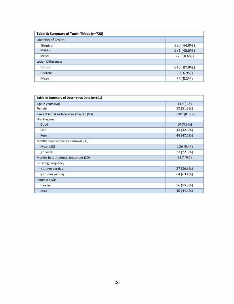
Table 3: Summary of Tooth Thirds (n=728)
Location of Lesion
Gingival 320 (44.0%) Middle 331 (45.5%)
Incisal 77 (10.6%)
Lesion Diffuseness
Diffuse 640 (87.9%)
Discrete 50 (6.9%)
Mixed 38 (5.2%)
Table 4: Summary of Descriptive Data (n=101)
Age in years (SD) 14.4 (1.5)
Female 52 (51.5%)
Percent initial surface area affected (SD) 0.107 (0.077)
Oral Hygiene
Good 10 (9.9%)
Fair 43 (42.6%)
Poor 48 (47.5%)
Months since appliance removal (SD)
Mean (SD) 0.26 (0.44)
< 1 week 73 (72.3%)
Months in orthodontic treatment (SD) 25.7 (9.7)
Brushing Frequency
< 1 time per day 37 (36.6%)
> 2 times per day 64 (63.4%)
Retainer style
Hawley 56 (55.5%)
Essix 45 (44.6%)
27
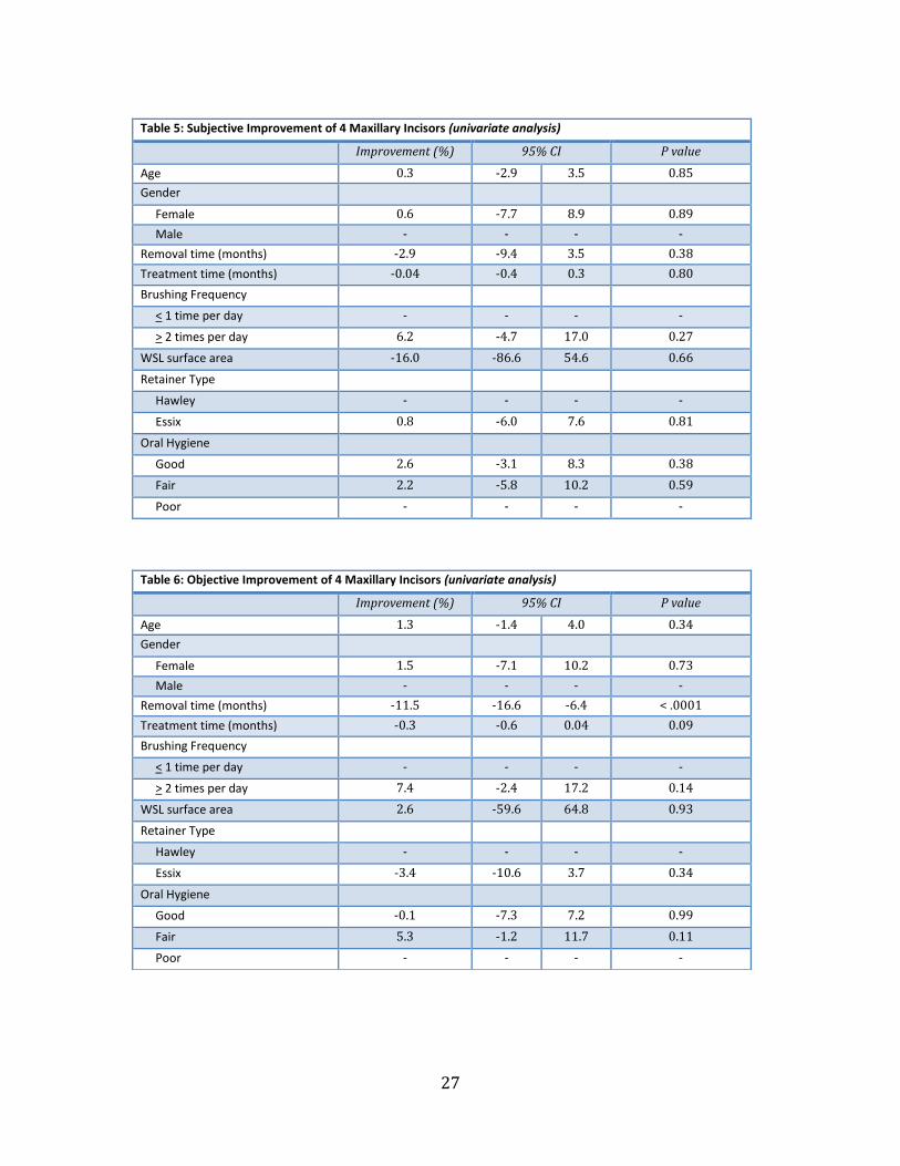
Table 5: Subjective Improvement of 4 Maxillary Incisors (univariate analysis)
Improvement (%) 95% CI P value
Age 0.3 -2.9 3.5 0.85
Gender
Female 0.6 -7.7 8.9 0.89
Male - - - -
Removal time (months) -2.9 -9.4 3.5 0.38
Treatment time (months) -0.04 -0.4 0.3 0.80
Brushing Frequency
< 1 time per day - - - -
> 2 times per day 6.2 -4.7 17.0 0.27
WSL surface area -16.0 -86.6 54.6 0.66
Retainer Type
Hawley - - - -
Essix 0.8 -6.0 7.6 0.81
Oral Hygiene
Good 2.6 -3.1 8.3 0.38
Fair 2.2 -5.8 10.2 0.59
Poor - - - -
Table 6: Objective Improvement of 4 Maxillary Incisors (univariate analysis)
Improvement (%) 95% CI P value
Age 1.3 -1.4 4.0 0.34
Gender
Female 1.5 -7.1 10.2 0.73
Male - - - -
Removal time (months) -11.5 -16.6 -6.4 < .0001
Treatment time (months) -0.3 -0.6 0.04 0.09
Brushing Frequency
< 1 time per day - - - -
> 2 times per day 7.4 -2.4 17.2 0.14
WSL surface area 2.6 -59.6 64.8 0.93
Retainer Type
Hawley - - - -
Essix -3.4 -10.6 3.7 0.34
Oral Hygiene
Good -0.1 -7.3 7.2 0.99
Fair 5.3 -1.2 11.7 0.11
Poor - - - -

28
Table 7: Subjective Improvement of 4 Maxillary Incisors (multivariate analysis)
Improvement (%) 95% CI P value
Age 0.5 -2.5 3.6 0.72
Gender
Female -0.6 -9.3 8.1 0.90
Male - - - -
Removal time (months) -4.3 -10.9 2.3 0.20
Treatment time (months) -0.01 -0.3 0.3 0.97
Brushing Frequency
Brushing < 1 time per day - - - -
Brushing > 2 times per day 6.6 -5.6 18.8 0.29
Table 8: Objective Improvement of 4 Maxillary Incisors (multivariate analysis)
Improvement (%) 95% CI P value
Age 3.1 0.5 5.8 0.02
Gender
Female 0.8 -6.6 8.3 0.83
Male - - - -
Removal time (months) -14.9 -20.6 -9.2 <0.0001
Treatment time (months) -0.3 -0.6 -0.02 0.04
Brushing Frequency
Brushing < 1 time per day - - - -
Brushing > 2 times per day 7.5 -2.8 17.8 0.15
Table 9: Improvement Scores of Single Teeth (multivariate analysis)
Improvement 95% CI P value
Age 0.01 -0.08 0.1 0.84
Gender
Female -0.09 -0.3 0.1 0.44
Male - - - -
Removal time (months) -0.64 -0.9 -0.4 <0.0001
Treatment time (months) -0.002 -0.02 0.01 0.81
Brushing Frequency
Brushing < 1 time per day - - - -
Brushing > 2 times per day 0.42 0.03 0.81 0.03
Tooth Type
Central Incisor - - - -
Lateral Incisors -0.18 -0.3 -0.02 0.03
WSL surface area 0.01 0.003 0.03 0.02
Staining 0.2 -0.1 0.4 0.17
29
Table 10: Improvement Scores of Tooth Thirds (multivariate analysis)
Improvement 95% CI P value
Age -0.01 -0.1 0.1 0.72
Gender
Female -0.05 -0.2 0.1 0.62
Male - - - -
Removal time (months) -0.2 -0.5 0.03 0.08
Treatment time (months) -0.004 -0.01 0.005 0.39
Brushing Frequency
Brushing < 1 time per day - - - -
Brushing > 2 times per day 0.3 0.1 0.5 0.02
Location
Gingival -0.03 -0.2 0.1 0.69
Middle - - - -
Incisal -0.2 -0.5 0.05 0.10
Diffuseness
Diffuse -0.01 -0.3 0.3 0.97
Discrete - - - -
Mixed 0.02 -0.3 0.4 0.9
Related Documents