Killer Bee Molecules: Antimicrobial Peptides as Effector Molecules to Target Sporogonic Stages of Plasmodium Victoria Carter 1 , Ann Underhill 1 , Ibrahima Baber 2 , Lakamy Sylla 2 , Mounirou Baby 3 , Isabelle Larget- Thiery 4 , Agne ` s Zettor 4 , Catherine Bourgouin 4 ,U ¨ lo Langel 5 , Ingrid Faye 6 , Laszlo Otvos 7 , John D. Wade 8 , Mamadou B. Coulibaly 2 , Sekou F. Traore 2 , Frederic Tripet 1 , Paul Eggleston 1 *, Hilary Hurd 1 1 Centre for Applied Entomology and Parasitology, School of Life Sciences, Keele University, Keele, Staffordshire, United Kingdom, 2 Malaria Research and Training Centre (MRTC), Universite ´ des Sciences, des Techniques et des Technologies de Bamako, Bamako, Mali, 3 Centre National de Transfusion Sanguine, Bamako, Mali, 4 Institut Pasteur, Centre for Production and Infection of Anopheles (CEPIA), Parasitology and Mycology Department, Paris, France, 5 Department of Neurochemistry Svante Arrhenius v. 21A, Stockholm University, Stockholm, Sweden, 6 Department of Molecular Bioscience, the Wenner-Gren Institute, Svante Arrhenius v. 20C, Stockholm University, Stockholm, Sweden, 7 Temple University Department of Biology, Philadelphia, Pennsylvania, United States of America, 8 Howard Florey Research Laboratories, Florey Institute for Neuroscience and Mental Health, University of Melbourne, Melbourne, Victoria, Australia Abstract A new generation of strategies is evolving that aim to block malaria transmission by employing genetically modified vectors or mosquito pathogens or symbionts that express anti-parasite molecules. Whilst transgenic technologies have advanced rapidly, there is still a paucity of effector molecules with potent anti-malaria activity whose expression does not cause detrimental effects on mosquito fitness. Our objective was to examine a wide range of antimicrobial peptides (AMPs) for their toxic effects on Plasmodium and anopheline mosquitoes. Specifically targeting early sporogonic stages, we initially screened AMPs for toxicity against a mosquito cell line and P. berghei ookinetes. Promising candidate AMPs were fed to mosquitoes to monitor adverse fitness effects, and their efficacy in blocking rodent malaria infection in Anopheles stephensi was assessed. This was followed by tests to determine their activity against P. falciparum in An. gambiae, initially using laboratory cultures to infect mosquitoes, then culminating in preliminary assays in the field using gametocytes and mosquitoes collected from the same area in Mali, West Africa. From a range of 33 molecules, six AMPs able to block Plasmodium development were identified: Anoplin, Duramycin, Mastoparan X, Melittin, TP10 and Vida3. With the exception of Anoplin and Mastoparan X, these AMPs were also toxic to an An. gambiae cell line at a concentration of 25 mM. However, when tested in mosquito blood feeds, they did not reduce mosquito longevity or egg production at concentrations of 50 mM. Peptides effective against cultured ookinetes were less effective when tested in vivo and differences in efficacy against P. berghei and P. falciparum were seen. From the range of molecules tested, the majority of effective AMPs were derived from bee/wasp venoms. Citation: Carter V, Underhill A, Baber I, Sylla L, Baby M, et al. (2013) Killer Bee Molecules: Antimicrobial Peptides as Effector Molecules to Target Sporogonic Stages of Plasmodium. PLoS Pathog 9(11): e1003790. doi:10.1371/journal.ppat.1003790 Editor: David S. Schneider, Stanford University, United States of America Received May 21, 2013; Accepted September 27, 2013; Published November 21, 2013 Copyright: ß 2013 Carter et al. This is an open-access article distributed under the terms of the Creative Commons Attribution License, which permits unrestricted use, distribution, and reproduction in any medium, provided the original author and source are credited. Funding: This work was supported by Wellcome Trust Programme Grant 084582, awarded to PE, HH, and FT. The funders had no role in study design, data collection and analysis, decision to publish, or preparation of the manuscript. Competing Interests: The authors have declared that no competing interests exist. * E-mail: [email protected] Introduction In the pursuit of malaria eradication, novel tools are in constant demand due to the lack of an effective vaccine and the emergence of pesticide-resistant insects and drug-resistant parasites [1]. Targeting the weak link in the life cycle, namely transmission between the vector and human host, is an historically valid approach to providing reliable and sustainable control [2]. There have been several advances in the development of strategies to block parasite transmission in the vector, aimed at larvae or adult mosquitoes or the sporogonic stages of the malaria parasite [3]. These are being pursued through the use of natural or genetically modified microbes (reviewed in [4]), or through genetic modifi- cation of the mosquito vector itself (e.g. [5,6]). Whilst pathogenic organisms or modified symbionts may become part of an integrated control strategy, it has been suggested that sustained application is challenging in developing countries [7]. An attractive tool for use in control programs would therefore be the production of a genetically modified vector incapable of transmitting the disease, which propagates itself through wild populations without further intervention [8,9]. To achieve this, transgenic techniques to generate mosquitoes compromised in their ability to transmit malaria and other pathogens are being developed [10]. Genetic modification of the major Asian vector, An. stephensi, has been achieved on many occasions (e.g. [6,11]), whilst transgenesis of the African malaria vector, An. gambiae, remains more of a technical challenge. Nonetheless, progress is being made with transgenesis of An. gambiae [12,13,14], including the development of mechanisms for driving the desired gene through target populations [15,16]. However, to date, screening for an array of anti-malarial effector molecules that could be incorporated as transgenes into the genomes of relevant mosquitoes or microbes has attracted less attention, and the choice is currently very limited. PLOS Pathogens | www.plospathogens.org 1 November 2013 | Volume 9 | Issue 11 | e1003790

Welcome message from author
This document is posted to help you gain knowledge. Please leave a comment to let me know what you think about it! Share it to your friends and learn new things together.
Transcript
Killer Bee Molecules: Antimicrobial Peptides as EffectorMolecules to Target Sporogonic Stages of PlasmodiumVictoria Carter1, Ann Underhill1, Ibrahima Baber2, Lakamy Sylla2, Mounirou Baby3, Isabelle Larget-
Thiery4, Agnes Zettor4, Catherine Bourgouin4, Ulo Langel5, Ingrid Faye6, Laszlo Otvos7, John D. Wade8,
Mamadou B. Coulibaly2, Sekou F. Traore2, Frederic Tripet1, Paul Eggleston1*, Hilary Hurd1
1 Centre for Applied Entomology and Parasitology, School of Life Sciences, Keele University, Keele, Staffordshire, United Kingdom, 2 Malaria Research and Training Centre
(MRTC), Universite des Sciences, des Techniques et des Technologies de Bamako, Bamako, Mali, 3 Centre National de Transfusion Sanguine, Bamako, Mali, 4 Institut
Pasteur, Centre for Production and Infection of Anopheles (CEPIA), Parasitology and Mycology Department, Paris, France, 5 Department of Neurochemistry Svante
Arrhenius v. 21A, Stockholm University, Stockholm, Sweden, 6 Department of Molecular Bioscience, the Wenner-Gren Institute, Svante Arrhenius v. 20C, Stockholm
University, Stockholm, Sweden, 7 Temple University Department of Biology, Philadelphia, Pennsylvania, United States of America, 8 Howard Florey Research Laboratories,
Florey Institute for Neuroscience and Mental Health, University of Melbourne, Melbourne, Victoria, Australia
Abstract
A new generation of strategies is evolving that aim to block malaria transmission by employing genetically modified vectorsor mosquito pathogens or symbionts that express anti-parasite molecules. Whilst transgenic technologies have advancedrapidly, there is still a paucity of effector molecules with potent anti-malaria activity whose expression does not causedetrimental effects on mosquito fitness. Our objective was to examine a wide range of antimicrobial peptides (AMPs) fortheir toxic effects on Plasmodium and anopheline mosquitoes. Specifically targeting early sporogonic stages, we initiallyscreened AMPs for toxicity against a mosquito cell line and P. berghei ookinetes. Promising candidate AMPs were fed tomosquitoes to monitor adverse fitness effects, and their efficacy in blocking rodent malaria infection in Anopheles stephensiwas assessed. This was followed by tests to determine their activity against P. falciparum in An. gambiae, initially usinglaboratory cultures to infect mosquitoes, then culminating in preliminary assays in the field using gametocytes andmosquitoes collected from the same area in Mali, West Africa. From a range of 33 molecules, six AMPs able to blockPlasmodium development were identified: Anoplin, Duramycin, Mastoparan X, Melittin, TP10 and Vida3. With the exceptionof Anoplin and Mastoparan X, these AMPs were also toxic to an An. gambiae cell line at a concentration of 25 mM. However,when tested in mosquito blood feeds, they did not reduce mosquito longevity or egg production at concentrations of50 mM. Peptides effective against cultured ookinetes were less effective when tested in vivo and differences in efficacyagainst P. berghei and P. falciparum were seen. From the range of molecules tested, the majority of effective AMPs werederived from bee/wasp venoms.
Citation: Carter V, Underhill A, Baber I, Sylla L, Baby M, et al. (2013) Killer Bee Molecules: Antimicrobial Peptides as Effector Molecules to Target Sporogonic Stagesof Plasmodium. PLoS Pathog 9(11): e1003790. doi:10.1371/journal.ppat.1003790
Editor: David S. Schneider, Stanford University, United States of America
Received May 21, 2013; Accepted September 27, 2013; Published November 21, 2013
Copyright: � 2013 Carter et al. This is an open-access article distributed under the terms of the Creative Commons Attribution License, which permitsunrestricted use, distribution, and reproduction in any medium, provided the original author and source are credited.
Funding: This work was supported by Wellcome Trust Programme Grant 084582, awarded to PE, HH, and FT. The funders had no role in study design, datacollection and analysis, decision to publish, or preparation of the manuscript.
Competing Interests: The authors have declared that no competing interests exist.
* E-mail: [email protected]
Introduction
In the pursuit of malaria eradication, novel tools are in constant
demand due to the lack of an effective vaccine and the emergence
of pesticide-resistant insects and drug-resistant parasites [1].
Targeting the weak link in the life cycle, namely transmission
between the vector and human host, is an historically valid
approach to providing reliable and sustainable control [2]. There
have been several advances in the development of strategies to
block parasite transmission in the vector, aimed at larvae or adult
mosquitoes or the sporogonic stages of the malaria parasite [3].
These are being pursued through the use of natural or genetically
modified microbes (reviewed in [4]), or through genetic modifi-
cation of the mosquito vector itself (e.g. [5,6]). Whilst pathogenic
organisms or modified symbionts may become part of an
integrated control strategy, it has been suggested that sustained
application is challenging in developing countries [7]. An
attractive tool for use in control programs would therefore be
the production of a genetically modified vector incapable of
transmitting the disease, which propagates itself through wild
populations without further intervention [8,9].
To achieve this, transgenic techniques to generate mosquitoes
compromised in their ability to transmit malaria and other
pathogens are being developed [10]. Genetic modification of the
major Asian vector, An. stephensi, has been achieved on many
occasions (e.g. [6,11]), whilst transgenesis of the African malaria
vector, An. gambiae, remains more of a technical challenge.
Nonetheless, progress is being made with transgenesis of An.
gambiae [12,13,14], including the development of mechanisms for
driving the desired gene through target populations [15,16].
However, to date, screening for an array of anti-malarial effector
molecules that could be incorporated as transgenes into the
genomes of relevant mosquitoes or microbes has attracted less
attention, and the choice is currently very limited.
PLOS Pathogens | www.plospathogens.org 1 November 2013 | Volume 9 | Issue 11 | e1003790

We and others have previously proposed that the ideal time to
block malaria development in the mosquito occurs within the first
24 hours post-blood meal [17,18,19,20]. To eliminate parasites in
the hostile environment of the mosquito midgut, any effector
molecules chosen to be expressed in this location must be highly
effective, small, soluble, fast acting and resistant to proteolytic
digestion [17]. One source of such molecules is present in the
evolutionarily conserved innate immune system, namely antimi-
crobial peptides (AMPs) [21]. AMPs are diverse molecules; many
are cationic and amphiphilic; properties that facilitate interactions
with negatively charged parasite surfaces rather than neutrally
charged mammalian membranes, such as blood cells or macro-
phages ingested with the parasites [22].
AMPs are broadly divided into membrane active (pore forming
through barrel/stave, carpet or torroidal pore mechanisms) and
non-membrane active (having intracellular targets such as DNA or
RNA, protein synthesis, folding or enzyme activity; e.g. cell
penetrating peptides), as reviewed in [21]). They can be further
classified according to toxicity, secondary structure, distinct amino
acid predominance, presence of cysteine residues, or families of
conserved sequences [23]. However, despite extensive studies on
structural and physiochemical AMP parameters (reviewed in [24]),
it is still impossible to make predictions from the available data as
to what will be effective; making anti-Plasmodium peptide selection
almost arbitrary.
Many candidate AMP molecules have been tested for their
activity against various stages of laboratory cultured Plasmodium
spp. in vitro and several have been tested in vivo via infections of
laboratory-reared anopheline mosquitoes (e.g. [17,25]). What we
are severely lacking, however, is data on field-collected parasites
and field-caught mosquitoes from malaria endemic areas.
Here we report the screening of a range of antimicrobial peptides
from a variety of sources, including molecules from different
antimicrobial structural classes as well as cell-penetrating peptides.
As our previous work had uncovered an inverse relationship between
upregulation of the mosquito immune system and fecundity [26], we
avoided endogenous mosquito immune peptides, as discussed in
[17]. Exogenous sources included toxins from bacteria, invertebrate
stings and venoms, amphibian skin secretions, fish mucus and
vertebrate AMPs. From designer-molecule sources, we included
short arbitrary sequences, peptides based on sequence alignments of
other AMPs and modified variants of existing effective peptides. Our
objective was to identify peptides with efficient anti-Plasmodium
activity that did not cause detectable fitness costs to the mosquito
vector. From initial laboratory screens, we were able to take
candidate peptides through initial tests in Mali, mixing gametocy-
taemic blood with AMPs and feeding through a membrane to
recently colonised An. gambiae.
Results
Initial in vitro screens of antimicrobial peptides fortoxicity to a mosquito cell line and anti-Plasmodiumactivity
Our initial screen assessed the ability of each of 33 peptides to
cause any detrimental effects to an An. gambiae cell line and to
kill ookinetes of the rodent malaria P. berghei (see Table 1 for list
Table 1. Antimicrobial peptide used in the current study:origin and size.
Peptide name Origin Size Reference
Alytesin Amphibian 14 aa [70]
Anoplin Wasp 10 aa [43]
Apamin Bee 18 aa [71]
Chex1-Arg20 metabolite Synthetic 19 aa [36]
Duramycin Bacteria 19 aa [72]
Flagellin 22 Bacteria 22 aa [73]
Granuliberin R Amphibian 12 aa [74]
ILF Synthetic 13 aa [45]
Indolicidin Bovine 13 aa [75]
KLK Synthetic 11 aa [76]
Lactoferricin B Bovine 11 aa [77,78,79]
Levitide Amphibian 14 aa [80]
Magainin II Amphibian 23 aa [81]
Mastoparan X Wasp 14 aa [82]
Melittin Bee 26 aa [38]
P2WN Synthetic 14 aa [45]
Parasin I Catfish 19 aa [83]
Ranatensin Amphibian 17 aa [84]
Scorpine Scorpion 75 aa [27]
TAT HIV-1 11 aa [85]
Temporin A Amphibian 13 aa [86]
Temporin B Amphibian 13 aa [86]
TP10 Wasp 21 aa [35]
TP10 (dimer) Wasp 44 aa [35]
Ubiquitin Unspecified 34 aa [87]
Uperolein Amphibian 11 aa [88]
Val-APO Synthetic 21 aa [36,89]
Vida 1 Synthetic 14 aa [45]
Vida 2 Synthetic 14 aa [45]
Vida 3 Synthetic 14 aa [45]
Vida 3 dimer Synthetic 32 aa [45]
Vida 4 Synthetic 14 aa [45]
WKY Synthetic 5 aa [90]
A description of all antimicrobial peptides tested in the current study. AMPswere sourced from a variety of organisms displaying antibacterial, antifungaland/or anti-parasitic properties. Custom peptides were also included.doi:10.1371/journal.ppat.1003790.t001
Author Summary
Breaking the complex life cycle of malaria by blocking itsdevelopment in the mosquito is one area of researchbeing pursued for malaria control. Currently, the mosquitoitself, or microbes that live within it, are being geneticallymodified to provide toxic or lethal outcomes to theparasite. However, this usually comes with a cost to thelifespan and reproductive capabilities of the mosquito,resulting in a strong disadvantage if these modifiedorganisms were to be released in the wild. This workaimed to identify a group of molecules suitable forinclusion in genetic modification strategies, which aretoxic to malaria parasites, but have no costly side-effects tothe mosquito. Within this group of molecules, toxins frombee and wasp venoms were prominent in their effects onmouse and human parasites. These particular moleculesmay prove effective in novel malaria control strategies andsuch venoms may also be a promising source of additionalanti-malaria toxins.
Effects of Antimicrobial Peptides on Plasmodium
PLOS Pathogens | www.plospathogens.org 2 November 2013 | Volume 9 | Issue 11 | e1003790

and Table S1 for individual peptide information and source).
This facilitated rapid screening of peptides for anti-malaria
activity using minimal quantities of parasite and peptide
material, whilst alerting us to potential toxicity to insects.
The effect of AMPs on An. gambiae cells in vitroOur first screen focussed on toxicity to the An. gambiae cell
line, Sua 4.0, using a concentration of 25 mM of each AMP.
Only five peptides demonstrated significant impairment of
mosquito cell growth over a 48 h period compared with
controls: Duramycin, Melittin, TP10 monomer and dimer and
Vida3 dimer (Table 2). Of these, Melittin was the most toxic,
causing a considerable reduction in cell numbers after 3 h of
incubation, with no cells remaining by 24 h. Duramycin caused
a similar, though not so rapid, effect with a decrease in cell
numbers of 88% by 48 h whereas the decline in cell numbers in
response to Vida3 dimer was 62% by 24 h and this remained
relatively stable through to 48 h. TP10 reduced cell numbers
initially; however, not all cells were affected at this dose, and the
population had resumed growth by 24 h. A different pattern was
observed when cells were incubated with TP10 dimer; after an
initial reduction in cell numbers, no further loss occurred, but
growth was impaired for the entire 48 hr period of the
experiment.
The viability of mosquito cells remaining after 48 h was also
assessed for all 33 peptides. The five peptides that caused a
significant reduction in cell numbers also caused reduced viability
of the remaining cells (Table 3). With the addition of Melittin, no
intact cells remained after 48 h. With the addition of Duramycin
or TP10 dimer, although cells were still present, Duramycin
reduced viability to 0% and TP10 dimer to 1%. Cell viability
remained above 50% after 48 h for Vida3 dimer and TP10
monomer. Although Indolicidin did not reduce cell numbers, cell
viability was impaired compared with controls. However, it should
be noted that the cell population did not expand as rapidly in these
control experiments compared with others.
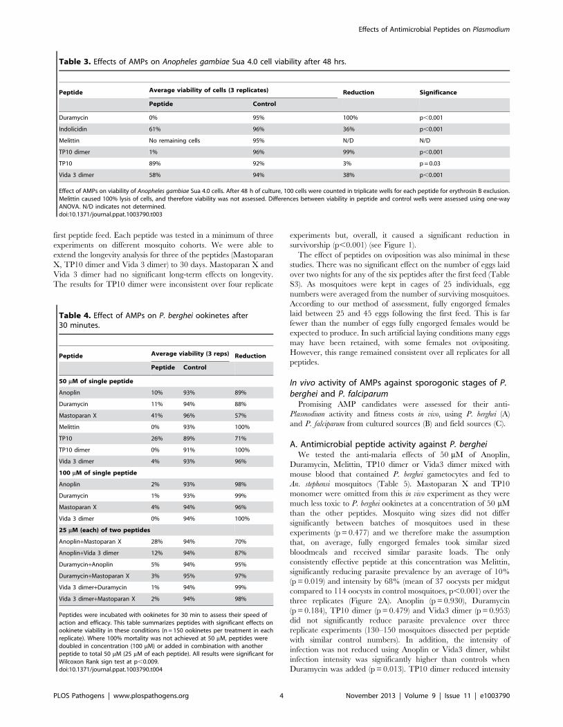
The effect of antimicrobial peptides on P. bergheiookinetes in vitro
We also screened all 33 peptides for toxicity against P. berghei
ookinetes. Ookinete viability ranged from 89–96% after 30 min in
control cultures. Only seven peptides demonstrated significant
toxicity to ookinetes at a concentration of 50 mM (Table 4). These
were, in order of efficacy, Melittin, TP10 dimer, Vida3 dimer,
Anoplin, Duramycin, TP10 monomer and Mastoparan X. TP10
dimer and Melittin were toxic to 100% of the ookinetes at 50 mM,
and this could also be achieved by increasing the peptide
concentration to 100 mM in the case of Vida3 dimer. At this
higher concentration, the peptides Duramycin, Anoplin and
Mastoparan X reduced viability to 1%, 2% and 4% respectively.
To assess whether some of the less effective peptides could act
synergistically to increase toxicity, we combined 25 mM concen-
trations of certain peptides in our P. berghei assay. Synergistic effects
were recorded in all cases, producing higher ookinete mortality
than 50 mM concentrations of a single peptide (Table 4).
The effect of antimicrobial peptides on mosquito fitnessIf incorporated into a transmission blocking strategy, an AMP
would be expressed even if the mosquito had not fed on an
infective blood meal. In order to assess any effects of ingesting
antimicrobial peptides on mosquito fitness, independently from
effects caused by malaria infection, we focussed on six of the most
promising peptides and screened these candidates by adding them
to a bloodmeal to determine any impacts on mosquito longevity
and fecundity. At a concentration of 50 mM, none of the peptides
had a significant impact on longevity over a 10 day period (Table
S2). This period included 2 blood feeds containing AMP, with the
analysis weighted to focus on early deaths immediately after the
Table 2. Effects of AMPs on Anopheles gambiae Sua 4.0 cell growth.
Peptide Time in culture
Average number of cells (6104/ml) over 3replicates
Reduction (comparedto control) Significance
Peptide Control
Duramycin 3 hrs 18 31 42% p,0.001
24 hrs 13 48 73% p,0.001
48 hrs 9 78 88% p,0.001
Melittin 3 hrs 5 31 84% p,0.001
24 hrs 0 48 100% p,0.001
48 hrs 0 79 100% p,0.001
TP10 dimer 3 hrs 24 30 20% p,0.001
24 hrs 25 43 42% p,0.001
48 hrs 26 62 58% p,0.001
TP10 3 hrs 22 32 31% p,0.001
24 hrs 40 52 23% p = 0.001
48 hrs 78 80 3% p,0.001
Vida 3 dimer 3 hrs 28 30 7% p = 0.001
24 hrs 17 45 62% p,0.001
48 hrs 19 69 72% p = 0.001
Effect of AMPs on growth of Anopheles gambiae Sua 4.0 cells. Cells were seeded at a density of 306104/ml with the addition of 25 mM AMP. Cells in different duplicatewells were counted after 3, 24 and 48 h to assess cell growth. Experiments were repeated three times. Differences between cell numbers in peptide and control wellswere assessed using one-way ANOVA.doi:10.1371/journal.ppat.1003790.t002
Effects of Antimicrobial Peptides on Plasmodium
PLOS Pathogens | www.plospathogens.org 3 November 2013 | Volume 9 | Issue 11 | e1003790
first peptide feed. Each peptide was tested in a minimum of three
experiments on different mosquito cohorts. We were able to
extend the longevity analysis for three of the peptides (Mastoparan
X, TP10 dimer and Vida 3 dimer) to 30 days. Mastoparan X and
Vida 3 dimer had no significant long-term effects on longevity.
The results for TP10 dimer were inconsistent over four replicate
experiments but, overall, it caused a significant reduction in
survivorship (p,0.001) (see Figure 1).
The effect of peptides on oviposition was also minimal in these
studies. There was no significant effect on the number of eggs laid
over two nights for any of the six peptides after the first feed (Table
S3). As mosquitoes were kept in cages of 25 individuals, egg
numbers were averaged from the number of surviving mosquitoes.
According to our method of assessment, fully engorged females
laid between 25 and 45 eggs following the first feed. This is far
fewer than the number of eggs fully engorged females would be
expected to produce. In such artificial laying conditions many eggs
may have been retained, with some females not ovipositing.
However, this range remained consistent over all replicates for all
peptides.
In vivo activity of AMPs against sporogonic stages of P.berghei and P. falciparum
Promising AMP candidates were assessed for their anti-
Plasmodium activity and fitness costs in vivo, using P. berghei (A)
and P. falciparum from cultured sources (B) and field sources (C).
A. Antimicrobial peptide activity against P. bergheiWe tested the anti-malaria effects of 50 mM of Anoplin,
Duramycin, Melittin, TP10 dimer or Vida3 dimer mixed with
mouse blood that contained P. berghei gametocytes and fed to
An. stephensi mosquitoes (Table 5). Mastoparan X and TP10
monomer were omitted from this in vivo experiment as they were
much less toxic to P. berghei ookinetes at a concentration of 50 mM
than the other peptides. Mosquito wing sizes did not differ
significantly between batches of mosquitoes used in these
experiments (p = 0.477) and we therefore make the assumption
that, on average, fully engorged females took similar sized
bloodmeals and received similar parasite loads. The only
consistently effective peptide at this concentration was Melittin,
significantly reducing parasite prevalence by an average of 10%
(p = 0.019) and intensity by 68% (mean of 37 oocysts per midgut
compared to 114 oocysts in control mosquitoes, p,0.001) over the
three replicates (Figure 2A). Anoplin (p = 0.930), Duramycin
(p = 0.184), TP10 dimer (p = 0.479) and Vida3 dimer (p = 0.953)
did not significantly reduce parasite prevalence over three
replicate experiments (130–150 mosquitoes dissected per peptide
with similar control numbers). In addition, the intensity of
infection was not reduced using Anoplin or Vida3 dimer, whilst
infection intensity was significantly higher than controls when
Duramycin was added (p = 0.013). TP10 dimer reduced intensity
Table 3. Effects of AMPs on Anopheles gambiae Sua 4.0 cell viability after 48 hrs.
Peptide Average viability of cells (3 replicates) Reduction Significance
Peptide Control
Duramycin 0% 95% 100% p,0.001
Indolicidin 61% 96% 36% p,0.001
Melittin No remaining cells 95% N/D N/D
TP10 dimer 1% 96% 99% p,0.001
TP10 89% 92% 3% p = 0.03
Vida 3 dimer 58% 94% 38% p,0.001
Effect of AMPs on viability of Anopheles gambiae Sua 4.0 cells. After 48 h of culture, 100 cells were counted in triplicate wells for each peptide for erythrosin B exclusion.Melittin caused 100% lysis of cells, and therefore viability was not assessed. Differences between viability in peptide and control wells were assessed using one-wayANOVA. N/D indicates not determined.doi:10.1371/journal.ppat.1003790.t003
Table 4. Effect of AMPs on P. berghei ookinetes after30 minutes.
Peptide Average viability (3 reps) Reduction
Peptide Control
50 mM of single peptide
Anoplin 10% 93% 89%
Duramycin 11% 94% 88%
Mastoparan X 41% 96% 57%
Melittin 0% 93% 100%
TP10 26% 89% 71%
TP10 dimer 0% 91% 100%
Vida 3 dimer 4% 93% 96%
100 mM of single peptide
Anoplin 2% 93% 98%
Duramycin 1% 93% 99%
Mastoparan X 4% 94% 96%
Vida 3 dimer 0% 94% 100%
25 mM (each) of two peptides
Anoplin+Mastoparan X 28% 94% 70%
Anoplin+Vida 3 dimer 12% 94% 87%
Duramycin+Anoplin 5% 94% 95%
Duramycin+Mastoparan X 3% 95% 97%
Vida 3 dimer+Duramycin 1% 94% 99%
Vida 3 dimer+Mastoparan X 2% 94% 98%
Peptides were incubated with ookinetes for 30 min to assess their speed ofaction and efficacy. This table summarizes peptides with significant effects onookinete viability in these conditions (n = 150 ookinetes per treatment in eachreplicate). Where 100% mortality was not achieved at 50 mM, peptides weredoubled in concentration (100 mM) or added in combination with anotherpeptide to total 50 mM (25 mM of each peptide). All results were significant forWilcoxon Rank sign test at p,0.009.doi:10.1371/journal.ppat.1003790.t004
Effects of Antimicrobial Peptides on Plasmodium
PLOS Pathogens | www.plospathogens.org 4 November 2013 | Volume 9 | Issue 11 | e1003790

Figure 1. Survival plot for mosquitoes fed with TP10 dimer. Mosquitoes were fed blood containing 50 mM of TP10 dimer, or control on days 0and 7 (arrows). Fully engorged females from each treatment group were separated into cages of 25 individuals to facilitate counting. Mosquitodeaths, from a starting total of 100, were recorded daily. This figure shows the survival plot for one replicate with 50 mM of TP10 dimer, using theKaplan-Meier method. The black line indicates mosquitoes fed with TP10 dimer, the red line, control mosquitoes.doi:10.1371/journal.ppat.1003790.g001
Table 5. Effect of antimicrobial peptides against P. berghei infections in Anopheles stephensi.
Replicate 1 Replicate 2 Replicate 3
Peptide Control Peptide Control Peptide Control Significance
Anoplin
Prevalence 66% (44) 73% (45) 98% (50) 96% (50) 100% (50) 97% (35) N/S
Intensity 21 (0–147) 30 (0–182) 74 (0–580) 179 (0–587) 177 (1–666) 88 (0–344) N/S
Duramycin
Prevalence 80% (50) 72% (50) 88% (50) 84% (50) 98% (50) 94% (50) N/S
Intensity 17 (0–114) 11 (0–93) 59 (0–294) 57 (0–285) 135 (0–319) 76 (0–309) p = 0.013 (m)
Melittin
Prevalence 98% (50) 100% (50) 82% (49) 90% (49) 72% (50) 88% (50) p = 0.019 (.)
Intensity 50 (0–192) 182 (1–371) 28 (0–160) 96 (0–372) 31 (0–412) 66 (0–374) p,0.001(.)
TP10 dimer
Prevalence 56% (50) 62% (50) 66% (50) 66% (50) 56% (50) 62% (50) N/S
Intensity 2 (0–84) 2 (0–87) 4 (0–67) 8 (0–54) 2 (0–13) 2 (0–13) p = 0.039 (.)
Vida 3 dimer
Prevalence 33% (40) 34% (50) 80% (50) 92% (50) 76% (50) 68% (50) N/S
Intensity 1 (0–6) 1 (0–12) 37 (0–271) 47 (0–253) 4 (0–67) 1 (0–7) N/S
Mosquitoes were provided with a gametocytaemic blood meal mixed with 50 mM of peptide, performed in triplicate. Prevalence (the proportion of infected mosquitoeswith total numbers in parentheses) and intensity (mean number of oocysts with the range in parentheses) of infections with paired controls are shown. N/S indicatesnon-significance. Significant differences are indicated by probability values with (m) representing oocyst numbers significantly higher than control and (.) representingoocyst numbers lower than control.doi:10.1371/journal.ppat.1003790.t005
Effects of Antimicrobial Peptides on Plasmodium
PLOS Pathogens | www.plospathogens.org 5 November 2013 | Volume 9 | Issue 11 | e1003790

Effects of Antimicrobial Peptides on Plasmodium
PLOS Pathogens | www.plospathogens.org 6 November 2013 | Volume 9 | Issue 11 | e1003790

(p = 0.039), but not prevalence. Egg production (measured by the
number of retained eggs) was not significantly affected by any of
the peptides at this concentration. Thus, neither longevity or
reproductive fitness are compromised by ingesting these peptides.
B. Antimicrobial peptide activity against cultured P.falciparum
Melittin, Mastoparan X or TP10 dimer were tested in vivo for
their effect on the sporogonic stages of P. falciparum. Cultured
gametocytes were mixed with 50 mM of AMP immediately before
feeding to mosquitoes. The anti-P. falciparum activity of Vida3,
when expressed in tetrameric form in transgenic An. gambiae
mosquitoes has been reported elsewhere [12] and therefore was
not tested here. Mosquito size was again consistent throughout the
experiments (p = 0.638). Melittin (Figure 2B) and TP10 dimer
were able to reduce prevalence by an average of 60% over three
replicates (p,0.001) (Table 6). Both peptides completely blocked
infection in one of the replicates. Intensity of infection was reduced
by an average of 57% by Melittin (p = 0.001) and 82% by TP10
dimer (p,0.001), although variability between replicate experi-
ments was high. Mastoparan X, which had low anti-P. berghei
activity, had no effect on P. falciparum oocyst prevalence (p = 0.649)
or intensity (p = 0.651).
C. Antimicrobial peptide activity against circulating P.falciparum from a malaria endemic area
We carried out a preliminary investigation using vectors and
parasites from the same malaria endemic district to validate the
efficacy of specific peptides in semi-natural conditions. Infecting
recently colonised mosquitoes with parasites from gametocyte
carriers was extremely challenging, and often resulted in no, or
very low, infections. We tested 20 gametocyte carriers, fed to over
8500 mosquitoes; 75% of which took a full blood meal. Wing
lengths were consistent throughout the experiments (p = 0.635)
and egg numbers were not significantly different in mosquitoes fed
with any peptide compared to controls (p = 0.397). Feeds from
only nine gametocyte carriers resulted in mosquito infections, with
gametocyte numbers ranging from 5–90 gametocytes/ml, produc-
ing infections with a prevalence range of 0–46% and intensity of
0–23 oocysts per midgut. Despite this low success rate, we were
able to test two of our top candidate AMPs; TP10 dimer (n = 3
replicates) and Vida3 dimer (n = 2 replicates). Over the replicates,
TP10 dimer did not provide significant anti-parasitic effects on
either prevalence (p = 0.699) or intensity (p = 0.493, Table 7). For
Vida3, parasite prevalence was lower than the controls in both
replicates. In addition, we were able to carry out single
experiments using Mastoparan X, lactoferricin B, levitide and
parasin (Table 7). In these preliminary experiments, the first three
of these peptides did not reduce parasite prevalence, whilst parasin
reduced prevalence by 80%, but did not affect parasite intensity.
Discussion
In this study, we concentrated on discovering effector molecules
that target the first 24 hours of malaria sporogonic stage
development within the mosquito, without affecting mosquito
fitness. We tested a large number of effector molecules, the
majority of which were chosen due to reported antimicrobial
activity (Table S1). Seven of these molecules displayed significant
killing effects against malaria parasites, namely Melittin, TP10
monomer and dimer, Vida3 dimer, Anoplin, Mastoparan X and
Duramycin. Melittin and TP10 dimer were the most effective anti-
ookinete molecules, reducing viability to zero within 30 minutes in
vitro. For the remaining AMPs, doubling the concentration
increased their activity and a synergistic effect was observed when
two peptides were combined. In our hands, scorpine was not
effective against rodent or human malaria [27] but, perhaps due to
its large size, the actual peptide concentration, based purely on the
weight of the lyophilized material, may have been lower than
expected. The effective molecules were mainly derived from
components of bee and wasp venom (except Duramycin and
Vida3 dimer). Importantly, none of the peptides had negative
impacts on fecundity and only TP10 dimer negatively impacted
longevity.
Our seven candidate peptides showed greater efficacy against
enriched ookinete cultures than against parasites in the mosquito. As
in vivo studies encompassed all parasite stages developing within
24 hours (macro- and micro-gametes, zygotes, retorts and ookinetes)
these differences may reflect either stage-specific activity or a
difference in the experimental environment. If the former, the choice
of transgene promoters that direct AMP expression to maturing
ookinetes would be critical to maximising the impact of any chosen
effector molecule [12]. Promoters also need to be capable of
expressing the peptide in sufficient quantities and with the correct
temporal and spatial profile. Our findings also support the rationale
for including more than one effector molecule in a given transgenic
strain, as has been illustrated by other studies. For example, using the
Magainin family, a combination of PGLa and Magainin 1 or 2
resulted in a 20 to 50-fold increase in parasite lysis [28,29].
In parallel, we tested all AMPs for toxicity to an An. gambiae cell
line, to assess potential impacts on mosquito fitness and help
determine speed and mode of action. Of the seven peptides that
displayed anti-malarial effects, five also caused a reduction in
viability of insect cells. Despite this, feeding AMPs to mosquitoes
did not have any significant negative impact on mosquito longevity
or fecundity over a 10 day period that included two separate blood
meals. These contrasting results may relate to physiological
differences between the midgut epithelial cells and cultured cells.
The latter lack a glycocalyx, which may act as an initial protective
layer in midgut cells [30], enhanced later by the development of
the peritrophic matrix [31]. We conclude that the mosquito cell
lines currently available are not good models for the midgut
epithelium.
One peptide (TP10 dimer) did reduce mosquito longevity in 2 of
4 replicate experiments during the latter stages of a 30 day
assessment. However, since no effects were seen prior to 10 days
(encompassing at least 2 egg batches) any fitness cost when
expressed as a transgene would likely be minimal. This is because
the majority of reproductive potential is likely to be realized before
this time. To date, investigations of transgenic mosquito fitness
indicate variable outcomes. For example, SM1 transgenics showed
Figure 2. Effect of Melittin on Plasmodium development in mosquitoes. Mosquitoes were fed blood containing gametocytes of rodentmalaria (fed to An. stephensi) or human malaria (fed to An. gambiae) supplemented with the AMP Melittin. Fully engorged females were maintained instandardized conditions for 7–8 days prior to dissection for oocyst burdens. Each experiment was performed in triplicate with control feedscontaining no AMP. Individual value plots for each dissected midgut are shown. Black diamonds represent the median oocyst burden for each group.Approximately 40–50 individuals were dissected for P. berghei infections and 30 individuals for P. falciparum infections (see Tables 5 and 6 for fulldetails). A. 50 mM of Melittin added to blood containing P. berghei gametocytes. B. 50 mM of Melittin added to blood containing P. falciparumgametocytes.doi:10.1371/journal.ppat.1003790.g002
Effects of Antimicrobial Peptides on Plasmodium
PLOS Pathogens | www.plospathogens.org 7 November 2013 | Volume 9 | Issue 11 | e1003790

no fitness cost when fed on uninfected blood [32] and even a
fitness advantage when fed on blood infected with P. berghei [33].
In contrast, PLA2 transgenics had significantly reduced fitness
[32]. We know of no direct evidence that effector molecules used
so far have detrimental impacts on fitness per se. However, it is
possible that perceived fitness costs in transgenic strains may be
associated with specific insertion sites, plasmid constructs or loss of
genetic heterogeneity as a result of inbreeding [34]. It is therefore
important that these potential problems are not compounded and
exacerbated by using costly effector molecules.
Some candidate AMPs were additionally investigated using
P. falciparum and An. gambiae. TP10 dimer and Melittin lowered
both prevalence and intensity of P. falciparum infections produced
from laboratory cultures. It has already been reported that the
monomer of TP10, added to blood meals at 30 mM, reduced
parasite prevalence by 35–45% [35]. Here, in the only direct
Table 6. Effect of antimicrobial peptides against P. falciparum cultured gametocytes.
Replicate 1 Replicate 2 Replicate 3
Peptide Control Peptide Control Peptide Control Significance
Mastoparan X
Prevalence 47% (30) 54% (28) 40% (30) 43% (30) 60% (30) 40% (30) N/S
Intensity 2 (0–11) 2 (0–21) 2 (0–29) 2 (0–8) 11 (0–72) 9 (0–61) N/S
Melittin
Prevalence 0% (30) 54% (28) 17% (30) 43% (30) 37% (30) 40% (30) p,0.001
Intensity 0 2 (0–21) ,1 (0–1) 2 (0–8) 6 (0–74) 9 (0–61) P = 0.001
TP10 dimer
Prevalence 17% (30) 54% (28) 0% (30) 43% (30) 37% (30) 40% (30) p,0.001
Intensity ,1 (0–2) 2 (0–21) 0 2 (0–8) 2 (0–14) 9 (0–61) p,0.001
Peptides (final concentration of 50 mM) were mixed with cultured P. falciparum gametocytes and membrane-fed to An. gambiae mosquitoes (performed at the PasteurInstitute, Paris). For each of three replicates, oocyst prevalence (number of oocyst positive mosquitoes, total number in parentheses) and oocyst intensity (mean numberof oocysts present per gut, range in parentheses) were recorded. An equivalent volume of water without peptide was used for the control. N/S indicates non-significance.doi:10.1371/journal.ppat.1003790.t006
Table 7. Effect of antimicrobial peptides on field parasites and mosquitoes.
Replicate 1 Replicate 2 Replicate 3
Peptide Control Peptide Control Peptide Control Significance
TP10 dimer
Prevalence 9% (58) 14% (42) 3% (38) 6% (35) 20% (49) 15% (59) N/S
Intensity ,1 (0–4) ,1 (0–6) ,1 (0–1) ,1 (0–3) ,1 (0–2) ,1 (0–4) N/S
Vida 3 dimer
Prevalence 3% (63) 14% (42) 0% (45) 6% (35) N/D N/D p = 0.011
Intensity ,1 (0–1) ,1 (0–6) 0 ,1 (0–3) N/S
Mastoparan X
Prevalence 45% (40) 46% (24) N/D N/D N/D N/D N/S
Intensity 3 (0–21) 3 (0–23) N/S
Lactoferricin B
Prevalence 46% (37) 46% (24) N/D N/D N/D N/D N/S
Intensity 2 (0–17) 3 (0–23) N/S
Levitide
Prevalence 8% (60) 15% (59) N/D N/D N/D N/D N/S
Intensity ,1 (0–1) ,1 (0–4) N/S
Parasin
Prevalence 3% (64) 15% (59) N/D N/D N/D N/D p = 0.02
Intensity ,1 (0–2) ,1 (0–4) N/S
Peptides (final concentration of 50 mM) were mixed with human blood containing P. falciparum parasites from gametocyte carriers in the village of Nyaganabougou.This was membrane-fed to the progeny of An. gambiae mosquitoes collected from neighbouring areas. For each replicate, oocyst prevalence (number of oocyst positivemosquitoes, total number in parentheses) and oocyst intensity (mean number of oocysts present per gut, range in parentheses) were recorded. An equivalent volumeof water without peptide was used for the control. N/D indicates not determined. N/S indicates non-significance.doi:10.1371/journal.ppat.1003790.t007
Effects of Antimicrobial Peptides on Plasmodium
PLOS Pathogens | www.plospathogens.org 8 November 2013 | Volume 9 | Issue 11 | e1003790

comparison available with other published data, we show that
TP10 dimer at 50 mM concentration reduced prevalence by 35–
100%. This again suggests that the ratio of AMP molecules to
parasites may be crucial to their effectiveness.
One possible reason for differences in vivo and in vitro is the
midgut environment. Although we have no information on the
stability of our AMPs when added to blood, it has been reported
that very few peptides survive proteolytic degradation longer than
a few minutes [36]. Rapid degradation may not be an issue if small
AMPs are delivered over a period of time via transgenes under the
control of a midgut promoter, or if expressed outside of the midgut
to target later stages of the parasite.
Studying AMPs in field conditions in Mali proved challenging,
as very few membrane feeds produced infections. Whilst every
blood sample drawn was gametocyte-positive, only one sample was
able to provide an infection where AMPs were effectively tested.
For practical reasons, we conclude that the use of in vitro-produced
P. falciparum gametocytes is the most feasible method for large-scale
screening of novel AMPs. What may be best practice, however, is
to focus on testing recently isolated parasites from malaria
endemic areas, combined with vectors collected from the same
area [37].
Whilst we are largely unaware of how these AMPs work in
isolation or in synergy, the data presented here provide specific
information on our candidate AMPs under given conditions. The
Sua 4.0 cell line data confirm the lytic mode of action of Melittin
[38]. We can also infer that Duramycin and Vida3 dimer work in
a lytic capacity, but at much slower rates at the same
concentration and therefore may not be suitable for targeting
parasites before they escape into the midgut lumen. TP10 is also
toxic to mosquito cells, but without causing cell lysis, supporting
the hypothesis that it functions through internal targets rather than
membrane lysis [35]. GOR V, a server for predicting the
secondary structure of proteins [39], predicts a coil structure for
Duramycin and Vida3 dimer whilst confirming that Melittin,
TP10 dimer, Mastoparan X and Anoplin are dominated by a-
helices, which may be important to their mode of action
[40,41,42,43]. It has been possible to distinguish between
structural motifs crucial for haemolytic, rather than antibacterial,
activity [38] and we may be able to greatly enhance performance
of existing AMPs by amino acid substitutions [44,45,46]. For
instance, D-analogue substitutions for some peptides (including
Melittin) have been shown to reduce cytotoxic effects whilst
retaining antimicrobial activity [47]. AMPs may also act to
enhance the entry of other compounds to the inside of pathogens
through transient pores, or hitchhiking with other cell-penetrating
peptides.
One benefit of the non-specific nature of AMP activity may be
the slow emergence of resistance. Parasites would require
profound changes in membrane structure to develop resistance
to this class of molecules. Yet, resistance must always be
considered as a possibility, and Peschel and Sahl present several
mechanisms of how this could evolve [47]. However, it is
heartening to know that AMPs have been effective against
bacterial infections for at least 100 million years [21].
We have concentrated here specifically on the effects of AMPs
on malaria parasites. In the natural environment, AMPs may
come into contact with other microbes in both an advantageous
or negative manner. On the positive side, AMPs active against
malaria may also have activity against other human pathogens
such as Trypanosoma and Leishmania spp. [48,49]. For example,
TP10 was previously shown to have activity against both
P. falciparum and Trypanosoma brucei brucei [35]. This suggests the
possibility that transgenic vectors may be engineered for
resistance to more than one pathogen. On the negative side,
AMPs may be toxic to natural gut microbes that themselves have
adverse effects on Plasmodium [50]. It must therefore be
emphasised that, as an integral part of the mosquito midgut,
bacterial flora must be analysed in response to any anti-parasitic
molecule deployment.
There is clearly a need to consider how chosen effector
molecules might best be deployed through transgenic technologies.
Both transgenic and paratransgenic strategies are considered by
Wang and Jacobs-Lorena in a recent review [18]. Their
deployment in transgenic mosquitoes allow for regulation of time,
quantity and localization of effector molecules to best target
malaria parasites at specific bottlenecks of development in the
mosquito. However, An. gambiae is still a challenging species for
transgenesis and logistical difficulties in transgene dispersal and
reproductive barriers must be overcome [51]. An alternative
strategy may be paratransgenesis [52]. Recent advances with
engineered natural symbionts from anopheline midguts have
shown rapid proliferation of bacteria and subsequent secretion of
the desired effector molecule after a blood meal [53] and progress
is also being made using the endosymbiont Wolbachia [54].
However, paratransgenic approaches also face significant difficul-
ties, including the diversity and stability of infections [55] and
fitness costs [56]. Whatever strategy is adopted, the choice of
effector molecules remains critical. From the data presented here,
AMPs isolated from wasp/bee venoms may provide a class of
peptides with potent anti-Plasmodium activity that deserves further
exploration. If used in synergy and in multiple mosquito
compartments, these should be able to eliminate parasites within
the vector or be used as blueprints to rationally develop new
synthetic AMPs. [57].
In conclusion, we studied a broad range of AMPs and identified
Melittin, TP10, Vida3, Mastoparan X and Anoplin as promising
candidates to limit malaria transmission in An. gambiae. We further
suggest that the concentration of the effector molecule, in relation
to parasite load, is an important determinant of success. Thus,
multiple peptides, acting in synergy, could perhaps achieve a
complete transmission blockade within the mosquito. We highlight
AMPs from the venom of bees and wasps as a future source of
novel anti-Plasmodium effector molecules. Over the last few years,
bee, wasp and hornet venoms have attracted attention as potential
bioactive substances, with new AMPs regularly being described
(e.g. [58,59]).
Materials and Methods
Ethics statementFor human participants in Mali: The project was approved by
the IRB of the Faculty of Medicine, Pharmacy and Dentistry at the
University of Bamako in Mali (Ethical Review Nu03/FMPOS). At
the study site in Nyaganabougou, ethical approval from commu-
nity leaders and written informed consent from parents of minors
was obtained for all blood samples. Every effort was made to
minimise distress and all parasite-positive individuals identified by
the study were subsequently treated by trained medical staff.
For animal work at Keele: Animals were housed in the Central
Animal Facility at Keele University, which is designated by the
Home Office. All work was carried out in accordance with the UK
Animals (Scientific Procedures) Act 1986 as amended by EU
Directive (2010/63/EU). Protocols were approved by veterinary
staff and conducted by trained personnel under Project Licence
PPL 40/2997. Every effort was made to minimize suffering.
For animal work in Paris: Animals were housed in the Pasteur
Institute Animal Facility, which is accredited by the French
Effects of Antimicrobial Peptides on Plasmodium
PLOS Pathogens | www.plospathogens.org 9 November 2013 | Volume 9 | Issue 11 | e1003790

Ministry of Agriculture (Accreditation A75-15-31). All work was
conducted in accordance with French and European regulations
on care and protection of the Laboratory Animals (EC Directive
86/609, French Law 2001-486). Protocols were approved by
veterinary staff and performed in compliance with the NIH
Animal Welfare Insurance #A5476-01 issued on 31/07/2012.
Every effort was made to minimise suffering.
All reagents were purchased from Sigma UK unless otherwise
stated. Experiments were conducted at the University of Keele,
UK, the Malaria Research and Training Centre (MRTC) at the
University of Bamako, Mali and CEPIA at the Pasteur Institute,
Paris, France.
MosquitoesMosquitoes were maintained in standardized conditions [60]. In
Keele and Bamako, larval stages were initially reared on Liquifry,
followed by TetraMin fish flakes (Tetra, UK). Adults were fed ad
libitum on 10% glucose supplemented with 0.05% PABA (para-
amino-benzoic acid) and females provided with defibrinated horse
blood (TCS, UK) for egg production in the UK, O+ human blood
in Bamako. In Paris, larvae were fed dry cat food pellets, adults
were fed 10% sucrose without PABA and the colony maintained
by feeding on anaesthetized rabbits. For P. falciparum infections,
gametocytes were mixed with AB+ human blood/serum and
infected mosquitoes maintained on 10% sucrose supplemented
with 0.05% PABA. Wing lengths of experimental mosquitoes were
recorded throughout to check for any differences in mosquito size,
as differences would affect bloodmeal volume, egg production and
numbers of parasites ingested.
The progeny of field-caught mosquitoes from the environs of
Kenieroba, Mali (12.6458uN, 7.99222uW) were established as
laboratory colonies in Bamako and Keele and were verified as An.
gambiae, M form, according to the method of Fanello [61]. These
mosquitoes were named the Mali strain and were colonized for
approximately 15 generations before experiments began to ensure
effective membrane feeding rates. The mosquitoes were then used
for longevity and fecundity studies at Keele and for Plasmodium
falciparum infection in Mali for a further 20–30 generations. An.
stephensi mosquitoes (SDA 500 [62]), the best experimental vector
for Plasmodium berghei infections, were reared at Keele using the
same protocols [63]. An. gambiae (Ngousso strain [64,65]) were used
for cultured P. falciparum infections in Paris.
PeptidesA total of 33 peptides were rehydrated in sterile distilled water
to a stock concentration of 1 mM and stored at 220uC in aliquots
suitable for individual experiments. All peptides were used in
assays at a final concentration of 50 mM, unless otherwise stated.
This choice of concentration was based on reported stimulated
endogenous insect AMP concentrations ranging from 1–100 mM,
[66]).
The effect of antimicrobial peptides on a mosquito cellline
To establish potential effects of AMPs on mosquito cells, the An.
gambiae Sua 4.0 cell line was used [67]. This facilitated an initial
high-throughput screening and provided insights into AMP
toxicity, mode of action (lytic or other) and speed of action. Sua
4.0 cells were maintained at 27uC in Schneider’s insect medium
supplemented with 10% FBS (foetal bovine serum) in T25 culture
flasks (Fisher, UK).
Each AMP was tested separately to establish if it affected cell
viability and growth. Sua 4.0 cells were seeded in 48 well
microtitre plates (BD Biosciences, UK) at a density of 306104/
ml in 195 ml of supplemented Schneider’s insect medium, with
the addition of 5 ml (final concentration 50 mM) of the test
peptide. Cells were maintained for 3, 6, 24 or 48 h, after which
they were resuspended and a sub-sample from duplicate wells
counted using a haemocytometer. To establish cell viability after
48 h (whether simply arrested in development or non-viable),
resuspended cells were added to an equal volume of 5%
erythrosin B vital dye and viable cells excluding the stain were
counted. The viability of 100 cells in triplicate wells was
recorded. Each AMP assay was performed in triplicate and
included a negative control minus the peptide (5 ml of water
alone added to the medium) and a further control of 5 ml of
10% SDS (sodium dodecyl sulphate) substituted for the AMP as
this causes 100% cell lysis.
The effect of antimicrobial peptides on mosquitolongevity and fecundity
Female An. gambiae (Mali strain, 3–5 days old) were used to
establish the effects of AMPs on mosquito longevity and fecundity
in the absence of confounding factors introduced with parasite
infection [26]. Two hundred 3–6 day old females were fed on
human O+ whole blood supplemented with 50 mM AMP (or water
control, 1 ml total volume) via feeding chambers of a Hemotek
membrane feeding system. Fully engorged mosquitoes were
randomly moved into four separate cages, each containing 25
mosquitoes, to facilitate monitoring. Mortality rates were recorded
daily for a 10-day period, and oviposition sites were provided on
the second and third night post-feeding. A second blood feed
containing the same AMP was administered 7 days later to mimic
the likelihood that mosquitoes would feed at least twice in the field
during the development of sporogonic stages of the malaria
parasite. Oviposition sites were again provided to assess fecundity
through the number of eggs laid as an average per surviving
mosquito. Assays for each AMP were repeated in triplicate on
separate generations of mosquitoes. Due to egg retention under
experimental conditions, egg production is best measured by
dissecting mosquitoes that have not been provided with an
oviposition site. However, this is not a feasible method of
determining fecundity when longevity is also being measured.
The effect of antimicrobial peptides on Plasmodiumberghei ookinetes in vitro
Our primary aim was to target parasites in mosquitoes before
they cross the midgut wall, therefore AMPs were first assessed for
their ability to kill ookinetes in vitro. As protocols for the conversion
of P. falciparum gametocytes to ookinetes do not provide high yields,
the rodent malaria parasite, P. berghei ANKA, was used to assess
the effects of AMPs on early sporogonic stages. Ookinete cultures
of P. berghei were established from gametocytaemic blood as
previously described [68]. Parasites were enriched using cold
0.17M ammonium chloride after 18 h of culture and washed
thoroughly with PBS (phosphate buffered saline). Parasites were
seeded in triplicate wells of a 96-well microtitre plate at a density of
16105/ml in a volume of 47.5 ml Schneider’s insect medium
(Invitrogen) with 2.5 ml of the test AMP (final concentration of
50 mM). Ookinetes were incubated for 30 min in conjunction with
triplicate control wells containing medium with 2.5 ml of water
instead of AMP. A sub-sample of parasites was then added to an
equal volume of 5% erythrosin B to assess viability (150 ookinetes
in total, taken from 3 wells). Each of the 33 AMPs was screened in
this way and assays were performed 3 times. AMPs displaying a
high degree of toxicity (.90% of ookinetes displaying erythrosin B
Effects of Antimicrobial Peptides on Plasmodium
PLOS Pathogens | www.plospathogens.org 10 November 2013 | Volume 9 | Issue 11 | e1003790

staining) underwent further ookinete assays using a concentration
of 100 mM AMP. To assess synergistic effects, combinations of two
peptides were also tested at a concentration of 25 mM per AMP, in
the assay described above.
The effect of antimicrobial peptides on Plasmodiumberghei sporogonic development in vivo
Five of the seven most effective peptides against P. berghei
ookinetes in vitro were subsequently tested on P. berghei parasites in
vivo (the effects of TP10 monomer and Vida3 have been reported
elsewhere [35,45]). To assess the effects of AMPs on parasites in
vivo, mouse blood containing P. berghei gametocytes was obtained
by cardiac puncture and 475 ml was rapidly mixed with 25 ml
(50 mM final concentration) of AMP. This mixture was fed to 100
nulliparous 3–5 day old female An. stephensi. A further 475 ml of
blood from the same infection was mixed with 25 ml of distilled
water to act as a control. Mosquitoes were maintained at 19uC for
optimum parasite development. Between 30 and 50 mosquitoes
were dissected after 10 days. Mosquito wing length, numbers of
retained eggs and oocysts were recorded in triplicate experiments
carried out on different mosquito cohorts.
The effect of selected antimicrobial peptides onPlasmodium falciparum in vitro
P. falciparum NF54 gametocytes were produced through large-
scale automated culture at the Pasteur Institute, Paris [64,69].
Gametocytes were grown in 10 ml RPMI 1640 medium
(Invitrogen), supplemented with 25 mM HEPES and L-glutamine,
10% heat-inactivated human serum and hypoxanthine (20 mg/L)
under a constant gas supply (5% CO2, 1% O2, 94% N2). Fresh red
blood cells (RBCs) were added to obtain a 7% haematocrit.
Fourteen days after initiating the culture, gametocyte maturity was
assessed on thin Giemsa-stained blood smears, and male
gametocyte maturity verified by exflagellation tests. Gametocyte
cultures were centrifuged for 5 min at 1500 rpm and the ,500–
600 ml pellet resuspended in fresh RBCs and AB human serum to
give a final haematocrit of 40%.
Blood containing mature gametocytes (475 ml) was added to
25 ml of the test AMP (50 mM final concentration). Peptides were
replaced by 25 ml of sterile deionized water for each control. The
blood/parasite/peptide mixture was placed in a Hemotek
membrane feeder, previously warmed to 37uC. For each feed,
70 to 100 nulliparous female An. gambiae mosquitoes were left to
feed in the dark for 15 min and only fully engorged females were
transferred to small cages and provided with 10% sucrose
containing 0.05% PABA. After 8 days, 30 midguts were dissected
and stained with bromo fluorescein for the detection of oocysts.
The effect of selected antimicrobial peptides onPlasmodium falciparum in vivo
Study site. Nyaganabougou, a village approximately 60 km
south-west of the Malian capital, Bamako, was used as a site for
the source of donors of malaria-infected blood. Due to its
proximity to the river Niger, which provides perennial mosquito
breeding sites, Nyaganabougou has high numbers of infected
individuals and an extended transmission season. Mosquitoes used
to establish the An. gambiae Mali colony used for this study were
originally collected in the village of Kenieroba (a distance of 15 km
from Nyaganabougou). Potential male and female gametocyte
carriers between the ages of 6–10 were screened by thick smear
and parasite species and density determined by local staff, led by
Saibou Doumbia.
P. falciparum parasites. Gametocyte carriers were taken to
the Malaria Research and Training Centre at the University of
Bamako for blood donation. Approximately 4 ml of blood was
extracted from carriers and immediately divided into 3 Hemotek
membrane feeders for testing 2 separate AMPs and one control for
each carrier. Peptides were added to 950 ml of gametocytaemic
blood for a final concentration of 50 mM. All parasite-positive
individuals were subsequently treated appropriately for parasites
and associated symptoms, as determined by, and carried out by,
local health workers in Nyaganabougou.Infections. Approximately 120 adult female An. gambiae
(Mali, 4–7 days old) were starved overnight, and allowed to feed
on the gametocytaemic blood/AMP mix for 30 min. Engorged
females were maintained on 10% glucose with 0.05% PABA for 8
days before dissection. No oviposition sites were provided so that
fecundity could be measured by determining retained eggs.
Mosquito wing length, numbers of retained eggs and oocysts were
recorded, and triplicate peptide experiments were carried out on
different mosquito cohorts, using blood from different donors.
Where possible, a minimum of 50 mosquitoes were dissected for
each condition.
Statistical analysesAnalyses were performed using Minitab 16 statistical software.
Data were checked for normality (Anderson-Darling) and repli-
cates were checked for between-experiment variation using one-
way ANOVA. Sua 4.0 cell growth and viability assays were also
analyzed using one-way ANOVA. The effects of AMPs on P.
berghei ookinetes were assessed by the Wilcoxon signed rank test
and oocyst burdens of both rodent and human malaria were
compared via the Mann-Whitney U test. Differences in infection
prevalence and mosquito sizes (wing length) used in all in vivo
studies were assessed using a 2-sample t-test. Longevity studies
were analyzed by Kaplan-Meier tests, where survival curves were
compared using the Wilcoxon test to weight early failures
(immediately after the first feed) more heavily. The number of
eggs produced per mosquito after the first feed was analyzed by a
one-way ANOVA.
Supporting Information
Table S1 Peptide sequence, activity and source.(DOC)
Table S2 Effect of AMPs on mosquito longevity over 10days.(DOC)
Table S3 Effect of AMPs on mosquito oviposition over10 days.(DOC)
Acknowledgments
We would like to thank the Malian village members of Keneiroba and
Nyaganabouogou and staff at Bancoumana field station, led by Saibou
Doumbia for assistance with gametocyte carrier screening. We thank
Andrew Blagborough (Imperial College, London) and Lisa Ranford-
Cartwright (University of Glasgow) for expert technical advice.
Author Contributions
Conceived and designed the experiments: HH VC MBC SFT FT PE.
Performed the experiments: VC AU HH ILT CB AZ IB LS. Analyzed the
data: VC. Contributed reagents/materials/analysis tools: CB UL IF LO
JDW MB. Wrote the paper: VC HH. Reviewed/commented on the
manuscript: VC HH PE LO CB.
Effects of Antimicrobial Peptides on Plasmodium
PLOS Pathogens | www.plospathogens.org 11 November 2013 | Volume 9 | Issue 11 | e1003790

References
1. Trape JF, Tall A, Diagne N, Ndiath O, Ly AB, et al. (2011) Malaria morbidity
and pyrethroid resistance after the introduction of insecticide-treated bednetsand artemisinin-based combination therapies: a longitudinal study. Lancet Infect
Dis 11: 925–932.
2. Takken W, Knols BG (2009) Malaria vector control: current and futurestrategies. Trends Parasitol 25: 101–104.
3. Ramirez JL, Garver LS, Dimopoulos G (2009) Challenges and approaches for
mosquito targeted malaria control. Curr Mol Med 9: 116–130.
4. Abdul-Ghani R, Al-Mekhlafi AM, Alabsi MS (2012) Microbial control ofmalaria: biological warfare against the parasite and its vector. Acta Trop 121:
71–84.
5. Ito J, Ghosh A, Moreira LA, Wimmer EA, Jacobs-Lorena M (2002) Transgenicanopheline mosquitoes impaired in transmission of a malaria parasite. Nature
417: 452–455.
6. Isaacs AT, Jasinskiene N, Tretiakov M, Thiery I, Zettor A, et al. (2012)Transgenic Anopheles stephensi coexpressing single-chain antibodies resist Plasmo-
dium falciparum development. Proc Natl Acad Sci U S A 109: E1922–1930.
7. Riehle MA, Jacobs-Lorena M (2005) Using bacteria to express and display anti-parasite molecules in mosquitoes: current and future strategies. Insect Biochem
Mol Biol 35: 699–707.
8. James AA (2005) Gene drive systems in mosquitoes: rules of the road. TrendsParasitol 21: 64–67.
9. Sinkins SP, Gould F (2006) Gene drive systems for insect disease vectors. Nat
Rev Genet 7: 427–435.
10. Riehle MA, Srinivasan P, Moreira CK, Jacobs-Lorena M (2003) Towardsgenetic manipulation of wild mosquito populations to combat malaria: advances
and challenges. J Exp Biol 206: 3809–3816.
11. Jacobs-Lorena M (2003) Interrupting malaria transmission by genetic manip-ulation of anopheline mosquitoes. J Vector Borne Dis 40: 73–77.
12. Meredith JM, Basu S, Nimmo DD, Larget-Thiery I, Warr EL, et al. (2011) Site-
specific integration and expression of an anti-malarial gene in transgenicAnopheles gambiae significantly reduces Plasmodium infections. PLoS One 6:
e14587.
13. Fuchs S, Nolan T, Crisanti A (2013) Mosquito transgenic technologies to reducePlasmodium transmission. Methods Mol Biol 923: 601–622.
14. Meredith JM, Underhill A, McArthur CC, Eggleston P (2013) Next-generation
site-directed transgenesis in the malaria vector mosquito Anopheles gambiae: self-
docking strains expressing germline-specific phiC31 integrase. PLoS One 8:e59264.
15. Marshall JM, Hay BA (2011) Inverse Medea as a novel gene drive system for
local population replacement: a theoretical analysis. J Hered 102: 336–341.16. Marshall JM, Pittman GW, Buchman AB, Hay BA (2011) Semele: a killer-male,
rescue-female system for suppression and replacement of insect disease vector
populations. Genetics 187: 535–551.17. Carter V, Hurd H (2010) Choosing anti-Plasmodium molecules for genetically
modifying mosquitoes: focus on peptides. Trends Parasitol 26: 582–590.
18. Wang S, Jacobs-Lorena M (2013) Genetic approaches to interfere with malariatransmission by vector mosquitoes. Trends Biotechnol 31: 185–193.
19. Sinden RE, Dawes EJ, Alavi Y, Waldock J, Finney O, et al. (2007) Progression of
Plasmodium berghei through Anopheles stephensi is density-dependent. PLoS Pathog 3:
e195.20. Drexler AL, Vodovotz Y, Luckhart S (2008) Plasmodium development in the
mosquito: biology bottlenecks and opportunities for mathematical modeling.
Trends Parasitol 24: 333–336.21. Giuliani A, Pirri G, Nicoletto SF (2007) Antimicrobial peptides: an overview of a
promising class of therapeutics. Central European Journal of Biology 2: 1–33.
22. Martin E, Ganz T, Lehrer RI (1995) Defensins and other endogenous peptideantibiotics of vertebrates. J Leukoc Biol 58: 128–136.
23. Splith K, Neundorf I (2011) Antimicrobial peptides with cell-penetrating peptide
properties and vice versa. Eur Biophys J 40: 387–397.
24. Tossi A, Sandri L, Giangaspero A (2000) Amphipathic, alpha-helicalantimicrobial peptides. Biopolymers 55: 4–30.
25. Bell A (2011) Antimalarial Peptides: The Long and the Short of it. Curr Pharm
Des 17: 2719–2731.
26. Ahmed AM, Hurd H (2006) Immune stimulation and malaria infection imposereproductive costs in Anopheles gambiae via follicular apoptosis. Microbes Infect 8:
308–315.
27. Conde R, Zamudio FZ, Rodriguez MH, Possani LD (2000) Scorpine, an anti-malaria and anti-bacterial agent purified from scorpion venom. FEBS Lett 471:
165–168.
28. Bevins CL, Zasloff M (1990) Peptides from frog skin. Annu Rev Biochem 59:395–414.
29. Gwadz RW, Kaslow D, Lee JY, Maloy WL, Zasloff M, et al. (1989) Effects of
magainins and cecropins on the sporogonic development of malaria parasites inmosquitoes. Infect Immun 57: 2628–2633.
30. Parish LA, Colquhoun DR, Ubaida Mohien C, Lyashkov AE, Graham DR,
et al. (2011) Ookinete-interacting proteins on the microvillar surface arepartitioned into detergent resistant membranes of Anopheles gambiae midguts.
J Proteome Res 10: 5150–5162.
31. Lehane MJ (1997) Peritrophic matrix structure and function. Annu RevEntomol 42: 525–550.
32. Moreira LA, Wang J, Collins FH, Jacobs-Lorena M (2004) Fitness of anopheline
mosquitoes expressing transgenes that inhibit Plasmodium development. Genetics
166: 1337–1341.
33. Marrelli MT, Li C, Rasgon JL, Jacobs-Lorena M (2007) Transgenic malaria-
resistant mosquitoes have a fitness advantage when feeding on Plasmodium-
infected blood. Proc Natl Acad Sci USA 104: 5580–5583.
34. Marrelli MT, Moreira CK, Kelly D, Alphey L, Jacobs-Lorena M (2006)
Mosquito transgenesis: what is the fitness cost? Trends Parasitol 22: 197–202.
35. Arrighi RB, Ebikeme C, Jiang Y, Ranford-Cartwright L, Barrett MP, et al.
(2008) Cell-penetrating peptide TP10 shows broad-spectrum activity against
both Plasmodium falciparum and Trypanosoma brucei brucei. Antimicrob Agents
Chemother 52: 3414–3417.
36. Noto PB, Abbadessa G, Cassone M, Mateo GD, Agelan A, et al. (2008)
Alternative stabilities of a proline-rich antibacterial peptide in vitro and in vivo.
Protein Sci 17: 1249–1255.
37. Tripet F, Aboagye-Antwi F, Hurd H (2008) Ecological immunology of
mosquito-malaria interactions. Trends Parasitol 24: 219–227.
38. Asthana N, Yadav SP, Ghosh JK (2004) Dissection of antibacterial and toxic
activity of melittin: a leucine zipper motif plays a crucial role in determining its
hemolytic activity but not antibacterial activity. J Biol Chem 279: 55042–55050.
39. Sen TZ, Jernigan RL, Garnier J, Kloczkowski A (2005) GOR V server for
protein secondary structure prediction. Bioinformatics 21: 2787–2788.
40. Vogel H, Jahnig F (1986) The structure of melittin in membranes. Biophys J 50:
573–582.
41. Eiriksdottir E, Konate K, Langel U, Divita G, Deshayes S (2010) Secondary
structure of cell-penetrating peptides controls membrane interaction and
insertion. Biochim Biophys Acta 1798: 1119–1128.
42. Wakamatsu K, Okada A, Miyazawa T, Ohya M, Higashijima T (1992)
Membrane-bound conformation of mastoparan-X, a G-protein-activating
peptide. Biochemistry 31: 5654–5660.
43. Konno K, Hisada M, Fontana R, Lorenzi CC, Naoki H, et al. (2001) Anoplin, a
novel antimicrobial peptide from the venom of the solitary wasp Anoplius
samariensis. Biochim Biophys Acta 1550: 70–80.
44. Maciel C, de Oliveira Junior VX, Fazio MA, Nacif-Pimenta R, Miranda A, et al.
(2008) Anti-Plasmodium activity of angiotensin II and related synthetic peptides.
PLoS One 3: e3296.
45. Arrighi RB, Nakamura C, Miyake J, Hurd H, Burgess JG (2002) Design and
activity of antimicrobial peptides against sporogonic-stage parasites causing
murine malarias. Antimicrob Agents Chemother 46: 2104–2110.
46. Dagan A, Efron L, Gaidukov L, Mor A, Ginsburg H (2002) In vitro anti-
Plasmodium effects of dermaseptin S4 derivatives. Antimicrob Agents Chemother
46: 1059–1066.
47. Peschel A, Sahl HG (2006) The co-evolution of host cationic antimicrobial
peptides and microbial resistance. Nat Rev Microbiol 4: 529–536.
48. Harrington JM (2011) Antimicrobial peptide killing of African trypanosomes.
Parasite Immunol 33: 461–469.
49. McGwire BS, Kulkarni MM (2010) Interactions of antimicrobial peptides with
Leishmania and trypanosomes and their functional role in host parasitism. Exp
Parasitol 126: 397–405.
50. Cirimotich CM, Dong Y, Clayton AM, Sandiford SL, Souza-Neto JA, et al.
(2011) Natural microbe-mediated refractoriness to Plasmodium infection in Anopheles
gambiae. Science 332: 855–858.
51. Bass C, Williamson MS, Wilding CS, Donnelly MJ, Field LM (2007)
Identification of the main malaria vectors in the Anopheles gambiae species
complex using a TaqMan real-time PCR assay. Malar J 6: 155.
52. Coutinho-Abreu IV, Zhu KY, Ramalho-Ortigao M (2010) Transgenesis and
paratransgenesis to control insect-borne diseases: current status and future
challenges. Parasitol Int 59: 1–8.
53. Wang S, Ghosh AK, Bongio N, Stebbings KA, Lampe DJ, et al. (2012) Fighting
malaria with engineered symbiotic bacteria from vector mosquitoes. Proc Natl
Acad Sci U S A 109: 12734–12739.
54. Kambris Z, Blagborough AM, Pinto SB, Blagrove MS, Godfray HC, et al.
(2010) Wolbachia stimulates immune gene expression and inhibits Plasmodium
development in Anopheles gambiae. PLoS Pathog 6: e1001143.
55. Boissiere A, Tchioffo MT, Bachar D, Abate L, Marie A, et al. (2012) Midgut
microbiota of the malaria mosquito vector Anopheles gambiae and interactions with
Plasmodium falciparum infection. PLoS Pathog 8: e1002742.
56. Hoffmann AA, Turelli M (2013) Facilitating Wolbachia introductions into
mosquito populations through insecticide-resistance selection. Proc Biol Sci 280:
20130371.
57. Wiradharma N, Khoe U, Hauser CA, Seow SV, Zhang S, et al. (2011) Synthetic
cationic amphiphilic alpha-helical peptides as antimicrobial agents. Biomaterials
32: 2204–2212.
58. Chen L, Chen W, Yang H, Lai R (2010) A novel bioactive peptide from wasp
venom. J Venom Res 1: 43–47.
59. Van Vaerenbergh M, Cardoen D, Formesyn EM, Brunain M, Van Driessche G,
et al. (2013) Extending the honey bee venome with the antimicrobial peptide
apidaecin and a protein resembling wasp antigen 5. Insect Mol Biol 22: 199–
210.
Effects of Antimicrobial Peptides on Plasmodium
PLOS Pathogens | www.plospathogens.org 12 November 2013 | Volume 9 | Issue 11 | e1003790

60. Jahan N, Hurd H (1997) The effects of infection with Plasmodium yoelii nigeriensis
on the reproductive fitness of Anopheles stephensi. Ann Trop Med Parasitol 91:365–369.
61. Fanello C, Santolamazza F, della Torre A (2002) Simultaneous identification of
species and molecular forms of the Anopheles gambiae complex by PCR-RFLP.Med Vet Entomol 16: 461–464.
62. Feldmann AM, Billingsley PF, Savelkoul E (1990) Bloodmeal digestion by strainsof Anopheles stephensi liston (Diptera: Culicidae) of differing susceptibility to
Plasmodium falciparum. Parasitology 101 Pt 2: 193–200.
63. Alavi Y, Arai M, Mendoza J, Tufet-Bayona M, Sinha R, et al. (2003) Thedynamics of interactions between Plasmodium and the mosquito: a study of the
infectivity of Plasmodium berghei and Plasmodium gallinaceum, and their transmissionby Anopheles stephensi, Anopheles gambiae and Aedes aegypti. Int J Parasitol 33: 933–
943.64. Mitri C, Jacques JC, Thiery I, Riehle MM, Xu J, et al. (2009) Fine pathogen
discrimination within the APL1 gene family protects Anopheles gambiae against
human and rodent malaria species. PLoS Pathog 5: e1000576.65. Hurd H, Taylor PJ, Adams D, Underhill A, Eggleston P (2005) Evaluating the
costs of mosquito resistance to malaria parasites. Evolution 59: 2560–2572.66. Hetru C, Hoffmann D, Bulet P (1998) Antimicrobial peptides from insects. In:
Brey PT, Hultmark D, editors. Molecular Mechanisms of Immune Responses in
Insects. London: Chapman & Hall. pp. 40–66.67. Catteruccia F, Nolan T, Blass C, Muller HM, Crisanti A, et al. (2000) Toward
Anopheles transformation: Minos element activity in anopheline cells andembryos. Proc Natl Acad Sci U S A 97: 2157–2162.
68. Carter V, Nacer AM, Underhill A, Sinden RE, Hurd H (2007) Minimumrequirements for ookinete to oocyst transformation in Plasmodium. Int J Parasitol
37: 1221–1232.
69. Ponnudurai T, Lensen AH, Leeuwenberg AD, Meuwissen JH (1982) Cultivationof fertile Plasmodium falciparum gametocytes in semi-automated systems. 1. Static
cultures. Trans R Soc Trop Med Hyg 76: 812–818.70. Erspamer V, Erspamer GF, Inselvini M, Negri L (1972) Occurrence of
bombesin and alytesin in extracts of the skin of three European discoglossid frogs
and pharmacological actions of bombesin on extravascular smooth muscle.Br J Pharmacol 45: 333–348.
71. Rosario LM (1985) Differential effects of the K+ channel blockers apamin andquinine on glucose-induced electrical activity in pancreatic beta-cells from a
strain of ob/ob (obese) mice. FEBS Lett 188: 302–306.72. Fredenhagen A, Fendrich G, Marki F, Marki W, Gruner J, et al. (1990)
Duramycins B and C, two new lanthionine containing antibiotics as inhibitors of
phospholipase A2. Structural revision of duramycin and cinnamycin. J Antibiot(Tokyo) 43: 1403–1412.
73. Bargieri DY, Leite JA, Lopes SC, Sbrogio-Almeida ME, Braga CJ, et al. (2010)Immunogenic properties of a recombinant fusion protein containing the C-
terminal 19 kDa of Plasmodium falciparum merozoite surface protein-1 and the
innate immunity agonist FliC flagellin of Salmonella typhimurium. Vaccine 28:2818–2826.
74. Nakajima T, Yasuhara T, Uzu S, Wakamatsu K, Miyazawa T, et al. (1985)Wasp venom peptides; wasp kinins, new cytotrophic peptide families and their
physico-chemical properties. Peptides 6 Suppl 3: 425–430.
75. Subbalakshmi C, Krishnakumari V, Nagaraj R, Sitaram N (1996) Requirements
for antibacterial and hemolytic activities in the bovine neutrophil derived 13-
residue peptide indolicidin. FEBS Lett 395: 48–52.
76. Alvarez-Bravo J, Kurata S, Natori S (1995) Mode of action of an antibacterial
peptide, KLKLLLLLKLK-NH2. J Biochem 117: 1312–1316.
77. Hwang PM, Zhou N, Shan X, Arrowsmith CH, Vogel HJ (1998) Three-
dimensional solution structure of lactoferricin B, an antimicrobial peptide
derived from bovine lactoferrin. Biochemistry 37: 4288–4298.
78. Fritsch G, Sawatzki G, Treumer J, Jung A, Spira DT (1987) Plasmodium
falciparum: inhibition in vitro with lactoferrin, desferriferrithiocin, and desferri-
crocin. Exp Parasitol 63: 1–9.
79. Tanaka T, Omata Y, Saito A, Shimazaki K, Yamauchi K, et al. (1995)
Toxoplasma gondii: parasiticidal effects of bovine lactoferricin against parasites.
Exp Parasitol 81: 614–617.
80. Poulter L, Terry AS, Williams DH, Giovannini MG, Moore CH, et al. (1988)
Levitide, a neurohormone-like peptide from the skin of Xenopus laevis. Peptide
and peptide precursor cDNA sequences. J Biol Chem 263: 3279–3283.
81. Imura Y, Choda N, Matsuzaki K (2008) Magainin 2 in action: distinct modes of
membrane permeabilization in living bacterial and mammalian cells. Biophys J
95: 5757–5765.
82. Longland CL, Mezna M, Michelangeli F (1999) The mechanism of inhibition of
the Ca2+-ATPase by mastoparan. Mastoparan abolishes cooperative ca2+binding. J Biol Chem 274: 14799–14805.
83. Koo YS, Kim JM, Park IY, Yu BJ, Jang SA, et al. (2008) Structure-activity
relations of parasin I, a histone H2A-derived antimicrobial peptide. Peptides 29:
1102–1108.
84. Yasuhara T, Ishikawa O, Nakajima T (1979) The studies on the active peptide in
the skin of Rana rugosa. II. The structure of ranatensin-R, the new ranatensin
analogue, and granuliberin-R, the new mast cell degranulating peptide. Chem
Pharm Bull (Tokyo) 27: 492–498.
85. Takeshima K, Chikushi A, Lee KK, Yonehara S, Matsuzaki K (2003)
Translocation of analogues of the antimicrobial peptides magainin and buforin
across human cell membranes. J Biol Chem 278: 1310–1315.
86. Mangoni ML, Saugar JM, Dellisanti M, Barra D, Simmaco M, et al. (2005)
Temporins, small antimicrobial peptides with leishmanicidal activity. J Biol
Chem 280: 984–990.
87. Kieffer AE, Goumon Y, Ruh O, Chasserot-Golaz S, Nullans G, et al. (2003) The
N- and C-terminal fragments of ubiquitin are important for the antimicrobial
activities. Faseb J 17: 776–778.
88. Erspamer V, Negri L, Erspamer GF, Endean R (1975) Uperolein and other
active peptides in the skin of the Australian leptodactylid frogs Uperoleia and
Taudactylus. Naunyn Schmiedebergs Arch Pharmacol 289: 41–54.
89. Otvos L, Jr., Wade JD, Lin F, Condie BA, Hanrieder J, et al. (2005) Designer
antibacterial peptides kill fluoroquinolone-resistant clinical isolates. J Med Chem
48: 5349–5359.
90. Bae YS, Kim Y, Park JC, Suh PG, Ryu SH (2002) The synthetic
chemoattractant peptide, Trp-Lys-Tyr-Met-Val-D-Met, enhances monocyte
survival via PKC-dependent Akt activation. J Leukoc Biol 71: 329–338.
Effects of Antimicrobial Peptides on Plasmodium
PLOS Pathogens | www.plospathogens.org 13 November 2013 | Volume 9 | Issue 11 | e1003790
Related Documents