MOLECULAR AND CELLULAR BIOLOGY, June 2004, p. 5521–5533 Vol. 24, No. 12 0270-7306/04/$08.000 DOI: 10.1128/MCB.24.12.5521–5533.2004 Copyright © 2004, American Society for Microbiology. All Rights Reserved. Positive and Negative Regulation of Poly(A) Nuclease David A. Mangus, Matthew C. Evans, Nathan S. Agrin, Mandy Smith, Preetam Gongidi†, and Allan Jacobson* Department of Molecular Genetics and Microbiology, University of Massachusetts Medical School, Worcester, Massachusetts 01655 Received 26 September 2003/Returned for modification 4 November 2003/Accepted 17 March 2004 PAN, a yeast poly(A) nuclease, plays an important nuclear role in the posttranscriptional maturation of mRNA poly(A) tails. The activity of this enzyme is dependent on its Pan2p and Pan3p subunits, as well as the presence of poly(A)-binding protein (Pab1p). We have identified and characterized the associated network of factors controlling the maturation of mRNA poly(A) tails in yeast and defined its relevant protein-protein interactions. Pan3p, a positive regulator of PAN activity, interacts with Pab1p, thus providing substrate specificity for this nuclease. Pab1p also regulates poly(A) tail trimming by interacting with Pbp1p, a factor that appears to negatively regulate PAN. Pan3p and Pbp1p both interact with themselves and with the C terminus of Pab1p. However, the domains required for Pan3p and Pbp1p binding on Pab1p are distinct. Single amino acid changes that disrupt Pan3p interaction with Pab1p have been identified and define a binding pocket in helices 2 and 3 of Pab1p’s carboxy terminus. The importance of these amino acids for Pab1p-Pan3p interaction, and poly(A) tail regulation, is underscored by experiments demonstrating that strains harboring substitutions in these residues accumulate mRNAs with long poly(A) tails in vivo. With rare exceptions, mRNAs whose synthesis originates within nuclei contain a 3 poly(A) tail. Poly(A) tracts are not encoded within genes but are added to nascent pre-mRNAs in a processing reaction that involves site-specific cleavage and subsequent polyadenylation (11, 16, 40, 42). In Saccharomyces cerevisiae, newly synthesized poly(A) tails of different tran- scripts are relatively homogeneous, with their final lengths determined by the combined actions of poly(A) polymerase holoenzyme (Pap1p and Fip1p), poly(A)-binding protein (Pab1p), poly(A) nuclease (PAN), and the Pab1p-associated factor, Pbp1p (10, 24, 43). Poly(A) tracts are generally bound by Pab1p, a highly con- served protein with four RNA recognition motifs (RRMs) connected to a carboxy-terminal helical domain via a proline- and methionine-rich segment (25, 31). Association of Pab1p with poly(A) requires a minimal binding site of 12 adenosines, and multiple molecules can bind via RRMs 1 and 2 to the same poly(A) tract, spaced approximately 25 nucleotides apart (1, 2, 31, 33). In yeast, the relatively abundant 70-kDa poly(A)-bind- ing protein is encoded by the PAB1 gene. Mutations in PAB1 cause a significant increase in the average steady-state poly(A) tail length of total cellular mRNA (32), and these effects have been attributed to two apparently nuclear functions of Pab1p: the regulation of a switch between processive and distributive activities in poly(A) polymerase (43) and the stimulation of PAN activity (7, 9, 21). Yeast poly(A) tails are initially synthesized to default lengths of 70 to 90 A’s and then trimmed to mRNA-specific lengths by PAN. Analyses of three different mRNAs indicate that such trimmed tails have lengths ranging from 55 to 71 A’s (8). PAN, a Pab1p-dependent 3 to 5 poly(A) exoribonuclease, requires magnesium, releases AMP as a product, and is regulated by cis-acting mRNA sequences (21). Purified PAN contains two proteins which are essential for nuclease activity: Pan2p is a 127-kDa protein with homology to the RNase T family of 335 exoribonucleases, while Pan3p is a 76-kDa protein which apparently acts as a positive activator of PAN activity (8). Deletion of PAN2 and/or PAN3 eliminates poly(A) nucle- ase activity but does not hinder cell growth. Physical interac- tion between Pan2p and Pan3p has been inferred from coim- munoprecipitation and two-hybrid analyses of the full-length proteins (9). Pbp1p (Pab1p-binding protein 1) specifically interacts with a 74-amino-acid segment encompassing the proline- and methi- onine-rich domain of the Pab1p C terminus. PBP1 is not es- sential for viability, but its disruption can suppress the lethality associated with a PAB1 deletion (23). In the absence of Pbp1p, 3 termini of pre-mRNAs are properly cleaved but receive poly(A) tails that are, on average, 15 to 30 nucleotides shorter than normal (23, 25). In vitro polyadenylation reactions using extracts from wild-type and pbp1 strains demonstrated that the mutant extracts initially produced full-length tails equiva- lent to their wild-type counterparts but subsequently trimmed the poly(A) at rates that were faster than normal. This accel- erated poly(A) trimming did not occur in pbp1/pan2 or pbp1/pan3 extracts, indicating that Pbp1p is a negative reg- ulator of PAN activity (25). Once in the cytoplasm, yeast poly(A) tails are further short- ened to 10 to 15 A’s by the Ccr4p and/or Caf1p exoribonucle- ases (38), after which decay of the rest of the mRNA is trig- gered; the mRNA is cleaved at its 5 end by the Dcp1p/Dcp2 decapping enzyme, and the uncapped and deadenylated mRNA is subsequently degraded exonucleolytically (5, 10, 15, 18, 24, 27). Poly(A) tail length is thus a negative regulator of * Corresponding author. Mailing address: Department of Molecular Genetics and Microbiology, University of Massachusetts Medical School, Worcester, MA 01655. Phone: (508) 856-2442. Fax: (508) 856- 5920. E-mail: [email protected]. † Present address: Program in Molecular Biology and Biotechnol- ogy, University of North Carolina at Chapel Hill, Chapel Hill, NC 27599. 5521 on August 19, 2015 by guest http://mcb.asm.org/ Downloaded from

Welcome message from author
This document is posted to help you gain knowledge. Please leave a comment to let me know what you think about it! Share it to your friends and learn new things together.
Transcript

MOLECULAR AND CELLULAR BIOLOGY, June 2004, p. 5521–5533 Vol. 24, No. 120270-7306/04/$08.00�0 DOI: 10.1128/MCB.24.12.5521–5533.2004Copyright © 2004, American Society for Microbiology. All Rights Reserved.
Positive and Negative Regulation of Poly(A) NucleaseDavid A. Mangus, Matthew C. Evans, Nathan S. Agrin, Mandy Smith,
Preetam Gongidi†, and Allan Jacobson*Department of Molecular Genetics and Microbiology, University of Massachusetts
Medical School, Worcester, Massachusetts 01655
Received 26 September 2003/Returned for modification 4 November 2003/Accepted 17 March 2004
PAN, a yeast poly(A) nuclease, plays an important nuclear role in the posttranscriptional maturation ofmRNA poly(A) tails. The activity of this enzyme is dependent on its Pan2p and Pan3p subunits, as well as thepresence of poly(A)-binding protein (Pab1p). We have identified and characterized the associated network offactors controlling the maturation of mRNA poly(A) tails in yeast and defined its relevant protein-proteininteractions. Pan3p, a positive regulator of PAN activity, interacts with Pab1p, thus providing substratespecificity for this nuclease. Pab1p also regulates poly(A) tail trimming by interacting with Pbp1p, a factor thatappears to negatively regulate PAN. Pan3p and Pbp1p both interact with themselves and with the C terminusof Pab1p. However, the domains required for Pan3p and Pbp1p binding on Pab1p are distinct. Single aminoacid changes that disrupt Pan3p interaction with Pab1p have been identified and define a binding pocket inhelices 2 and 3 of Pab1p’s carboxy terminus. The importance of these amino acids for Pab1p-Pan3p interaction,and poly(A) tail regulation, is underscored by experiments demonstrating that strains harboring substitutionsin these residues accumulate mRNAs with long poly(A) tails in vivo.
With rare exceptions, mRNAs whose synthesis originateswithin nuclei contain a 3� poly(A) tail. Poly(A) tracts are notencoded within genes but are added to nascent pre-mRNAs ina processing reaction that involves site-specific cleavage andsubsequent polyadenylation (11, 16, 40, 42). In Saccharomycescerevisiae, newly synthesized poly(A) tails of different tran-scripts are relatively homogeneous, with their final lengthsdetermined by the combined actions of poly(A) polymeraseholoenzyme (Pap1p and Fip1p), poly(A)-binding protein(Pab1p), poly(A) nuclease (PAN), and the Pab1p-associatedfactor, Pbp1p (10, 24, 43).
Poly(A) tracts are generally bound by Pab1p, a highly con-served protein with four RNA recognition motifs (RRMs)connected to a carboxy-terminal helical domain via a proline-and methionine-rich segment (25, 31). Association of Pab1pwith poly(A) requires a minimal binding site of 12 adenosines,and multiple molecules can bind via RRMs 1 and 2 to the samepoly(A) tract, spaced approximately 25 nucleotides apart (1, 2,31, 33). In yeast, the relatively abundant 70-kDa poly(A)-bind-ing protein is encoded by the PAB1 gene. Mutations in PAB1cause a significant increase in the average steady-state poly(A)tail length of total cellular mRNA (32), and these effects havebeen attributed to two apparently nuclear functions of Pab1p:the regulation of a switch between processive and distributiveactivities in poly(A) polymerase (43) and the stimulation ofPAN activity (7, 9, 21).
Yeast poly(A) tails are initially synthesized to default lengthsof 70 to 90 A’s and then trimmed to mRNA-specific lengths by
PAN. Analyses of three different mRNAs indicate that suchtrimmed tails have lengths ranging from 55 to 71 A’s (8). PAN,a Pab1p-dependent 3� to 5� poly(A) exoribonuclease, requiresmagnesium, releases AMP as a product, and is regulated bycis-acting mRNA sequences (21). Purified PAN contains twoproteins which are essential for nuclease activity: Pan2p is a127-kDa protein with homology to the RNase T family of3�35� exoribonucleases, while Pan3p is a 76-kDa proteinwhich apparently acts as a positive activator of PAN activity(8). Deletion of PAN2 and/or PAN3 eliminates poly(A) nucle-ase activity but does not hinder cell growth. Physical interac-tion between Pan2p and Pan3p has been inferred from coim-munoprecipitation and two-hybrid analyses of the full-lengthproteins (9).
Pbp1p (Pab1p-binding protein 1) specifically interacts with a74-amino-acid segment encompassing the proline- and methi-onine-rich domain of the Pab1p C terminus. PBP1 is not es-sential for viability, but its disruption can suppress the lethalityassociated with a PAB1 deletion (23). In the absence of Pbp1p,3� termini of pre-mRNAs are properly cleaved but receivepoly(A) tails that are, on average, 15 to 30 nucleotides shorterthan normal (23, 25). In vitro polyadenylation reactions usingextracts from wild-type and pbp1� strains demonstrated thatthe mutant extracts initially produced full-length tails equiva-lent to their wild-type counterparts but subsequently trimmedthe poly(A) at rates that were faster than normal. This accel-erated poly(A) trimming did not occur in pbp1�/pan2� orpbp1�/pan3� extracts, indicating that Pbp1p is a negative reg-ulator of PAN activity (25).
Once in the cytoplasm, yeast poly(A) tails are further short-ened to 10 to 15 A’s by the Ccr4p and/or Caf1p exoribonucle-ases (38), after which decay of the rest of the mRNA is trig-gered; the mRNA is cleaved at its 5� end by the Dcp1p/Dcp2decapping enzyme, and the uncapped and deadenylatedmRNA is subsequently degraded exonucleolytically (5, 10, 15,18, 24, 27). Poly(A) tail length is thus a negative regulator of
* Corresponding author. Mailing address: Department of MolecularGenetics and Microbiology, University of Massachusetts MedicalSchool, Worcester, MA 01655. Phone: (508) 856-2442. Fax: (508) 856-5920. E-mail: [email protected].
† Present address: Program in Molecular Biology and Biotechnol-ogy, University of North Carolina at Chapel Hill, Chapel Hill, NC27599.
5521
on August 19, 2015 by guest
http://mcb.asm
.org/D
ownloaded from
decapping and, as might be inferred from this conclusion, somemRNAs whose poly(A) tails are lengthened as a consequenceof pan mutations are also stabilized (8). The presence of apoly(A) tail also influences mRNA translation activity. Pab1passociated with the poly(A) tail interacts with the initiationfactor eIF4G, and the network of ensuing interactions pro-motes early events in ribosome binding to the mRNA (24).These and other activities indicate that the Pab1p/poly(A)mRNP has several important functions in both the nuclear andcytoplasmic posttranscriptional control of gene expression(24).
In this study, we sought to elucidate the mechanism ofpoly(A) tail maturation by defining the interactions of thefactors involved in this process. We utilized the two-hybridsystem to map the domains required for interactions betweenPan3p and Pan2p, Pab1p and Pan3p, and Pbp1p and Pan2pand for multimerization interactions of Pan3p and Pbp1p. Mu-tagenesis of Pab1p was also performed to further map theinteraction domain of Pan3p onto Pab1p. Collectively, theseanalyses have led to the identification of the key components ofa regulatory network controlling the maturation of mRNApoly(A) tails.
MATERIALS AND METHODS
General methods. Preparation of standard yeast media and methods of cellculture were as described previously (30). Transformation of yeast utilized therapid method (36). DNA manipulations were performed according to standardtechniques (34). PCR amplifications were performed with Taq DNA polymerase(41) and confirmed, where appropriate, with DNA sequencing according to themethod of Sanger et al. (35) or by PCR sequencing by the Nucleic Acid Facilityat the University of Massachusetts Medical School. Plasmid DNAs were prop-agated in Escherichia coli strain DH5�.
Yeast strains. Yeast strain L40 (yDM61) was used for all two-hybrid analyses(23). Yeast strain yAS1255 (MATa ade2-1 his3-11,15 leu2-3,112 trp1-1 ura3-1can1-100 pab1�::HIS3), a pab1� strain bearing a plasmid expressing a C-terminaltruncation of PAB1 (lacking the proline- and methionine-rich domains), wasprovided by Alan Sachs. Strain yAS320 (yDM197) (23), which bears a chromo-somal deletion of PAB1 covered by a wild-type PAB1-URA3-CEN plasmid, wasused to introduce mutant PAB1 alleles by the plasmid shuffle technique (14).Strain yME43, the pan3� strain, was created by PCR-based gene deletion asdescribed previously (4). PCR using oligonucleotide pair PAN3-D5-L–PAN3-D3-L amplified the LEU2 gene from plasmid pJJ252 (19). The PCR product waselectrophoresed on an agarose gel and extracted, and the full-length product wasrecovered using a QIAquick gel extraction kit (QIAGEN Inc.). The recoveredproduct was transformed into yeast strain yAS306 (yDM117) (23), and transfor-mants were selected on appropriate media. Genomic DNA was isolated fromcultures started from individual colonies, and the presence of the disruption wasconfirmed by PCR using the gene-specific primers, PAN3-5–PAN3-3.
Oligonucleotides. The oligonucleotides used in this study were prepared byOperon, Inc., and are listed in Table 1.
Plasmid constructions. The numbers following the gene names indicate aminoacid residues encompassed by the respective constructs. To create lexA(DB)constructs, fragments of PAB1 were amplified by PCR from plasmid YPA3 (23)with oligonucleotide pairs PAB1.4a-PAB1.6 (PAB1 406 to 577), PAB1.15-PAB1.6 (PAB1 422 to 577), PAB1.5b-PAB1.6 (PAB1 488 to 577), PAB1.5a-PAB1.6 (PAB1 494 to 577), PAB1.4a-PAB1.10 (PAB1 406 to 492), PAB1.4a-PAB1.13 (PAB1 406 to 479), PAB1.7a-PAB1.10 (PAB1 422 to 492), PAB1.7a-PAB1.13 (PAB1 422 to 479), PAB1.4a-PAB1.14 (PAB1 406 to 457), PAB1.11-PAB1.10 (PAB1 429 to 492), PAB1.11-PAB1.13 (PAB1 429 to 479), PAB1.11-PAB1.14 (PAB1 429 to 457), PAB1.12-PAB1.10 (PAB1 435 to 492), PAB1.12-PAB1.13 (PAB1 435 to 479), and PAB1.12-PAB1.14 (PAB1 435 to 457). All otherPAB1 constructs were as described previously (23). The products of the reactionswere digested with EcoRI and SalI and subcloned into pBTM116.
PAN2 lexA(DB) constructs were created by PCR amplification of PAN2 frag-ments from plasmid pAS468 (9) by using the following oligonucleotide pairs:PAN2.1F-PAN2.1115R (PAN2 1 to 1115), PAN2.1F-PAN2.748R (PAN2 1 to748), PAN2.333F-PAN2.1115R (PAN2 333 to 1115), PAN2.498F-PAN2.1115R
(PAN2 498 to 1115), PAN2.619F-PAN2.1115R (PAN2 619 to 1115), PAN2.671F-PAN2.1115R (PAN2 671 to 1115), and PAN2.814F-PAN2.1115R (PAN2 814 to1115). The products of the reactions were digested with EcoRI and PstI andsubcloned into pBTM116.
To create lexA(DB) and GAL4(AD)-PAN3 full-length (FL) constructs (PAN31 to 679), PAN3 was amplified by PCR from genomic DNA with oligonucleotidepairs PAN3-ATG and PAN3-UAA. The products of each reaction were digestedwith BamHI and XhoI and subcloned into both pBTM116 and pGAD424. Tocreate lexA(DB) and GAL4(AD)-PAN3 truncations, fragments of PAN3 wereamplified by PCR from genomic DNA with oligonucleotide pairs PAN3.001-PAN3.548 (PAN3 1 to 548), PAN3.001-PAN3.437 (PAN3 1 to 437), PAN3.001-PAN3.249 (PAN3 1 to 249), PAN3.125-PAN3.679 (PAN3 125 to 679), PAN3.250-PAN3.679 (PAN3 250 to 679), PAN3.337-PAN3.679 (PAN3 337 to 679),PAN3.420-PAN3.679 (PAN3 420 to 679), PAN3.250-PAN3.548 (PAN3 250 to548), PAN3.549-PAN3.679 (PAN3 549 to 679), PAN3.560-PAN3.679 (PAN3 560to 679), PAN3.337-PAN3.548 (PAN3 337 to 548), PAN3.593-PAN3.679 (PAN3593 to 679), PAN3.549-PAN3.662TAA (PAN3 549 to 662), PAN3.250-PAN3.437(PAN3 250 to 437), and PAN3.337-PAN3.548 (PAN3 337 to 548). The productsof the reactions were digested with EcoRI and SalI and subcloned into bothpBTM116 and pGAD424.
Full-length and amino-terminal GAL4(AD) PBP1 constructs were amplifiedby PCR from genomic DNA with oligonucleotide pairs PBP1-ATG–PBP1.722R(PBP1 1 to 722) and PBP1-ATG–PBP1.443 (PBP1 1 to 443). Carboxy-terminalGAL4(AD) PBP1 constructs were amplified by PCR from plasmid GAL4(AD)PBP1 (357 to 722) (24) with oligonucleotide pairs PBP1.357F-PBP1.695R (PBP1357 to 695), PBP1.357F-PBP1.578R (PBP1 357 to 578), PBP1.421F-PBP1.722R(PBP1 421 to 722), PBP1.421F-PBP1.695R (PBP1 421 to 695), PBP1.421F-PBP1.578R (PBP1 421 to 578), PBP1.421F-PBP1.506R (PBP1 421 to 506),PBP1.454F-PBP1.722R (PBP1 454 to 695), PBP1.454F-PBP1.695R (PBP1 454 to695), PBP1.454F-PBP1.578R (PBP1 454 to 578), and PBP1.454F-PBP1.506R(PBP1 454 to 506). The products of the reactions were digested with EcoRI andSalI and subcloned into pGAD424. All other GAL4(AD) PBP1 constructs wereoriginally isolated from a library in the two-hybrid screen with lexA(DB) PAB1P-H (24). The lexA(DB) PBP1 constructs were made by subcloning EcoRI/SalIrestriction fragments bearing the various PBP1 alleles from the GAL4(AD)vector (pGAD424) to the lexA(DB) vector (pBTM116).
Two-hybrid analysis. Yeast strain L40 was transformed with the appropriatelexA(DB) and GAL4(AD) constructs described above. In each experiment, ap-propriate empty vector controls were also included. Transformants were initiallyselected on plates with synthetic complete medium lacking Leu and Trp (SC�Leu, �Trp plates) and subsequently screened for lacZ activity using 5-bromo-4-chloro-3-indolyl-�-D-galactopyranoside (X-Gal) filter lift assays. To determinethe extent of HIS3 reporter activity, colonies from each transformation weregrown overnight in SC �Leu, �Trp broth and serially diluted on SC �Leu,�Trp and SC �Leu, �Trp, �His plates containing 0, 5, 10, 20, 40, 60, 80, or 100mM 3-aminotriazole (3-AT).
Construction and screening of the lexA(DB)-PAB1 (406 to 577) mutant li-brary. PCRs were performed under suboptimal conditions (28) using oligonu-cleotides PAB1.4 and PAB1.6 to amplify the carboxy terminus of PAB1 asdescribed previously (23). Reactions were purified using QIAquick PCR purifi-cation kits (QIAGEN Inc.) and subcloned into the pBTM116 vector using SalIand BamHI restriction enzymes. E. coli transformants were pooled and amplifiedand plasmids were isolated using QIAfilter maxi-prep kits (QIAGEN Inc.). Theresulting library was cotransformed with GAL4(AD)-PAN3-FL and plated onminimal medium plates containing X-Gal. Colonies that displayed reduced lacZactivity (white or light blue) were retained for further analysis. Total nucleic acidwas isolated from each strain (17) and electroporated into E. coli strain JF1754(14). The DNA-binding domain plasmids were selected by their inability tocomplement the E. coli leuB mutation when cells were replica plated to E �Leuplates with ampicillin (14, 39). Isolated plasmids were further characterized byrestriction mapping with EcoRI and PstI to confirm the presence of the PAB1insert. DNA-binding domain plasmids representing each allele identified werethen retransformed with empty vector, GAL4(AD)-PBP1 (377 to 722), orGAL4(AD)-PAN3-FL plasmids, and the loss of interaction was reconfirmed byassaying both �-galactosidase activity and 3-AT resistance.
Site-directed mutagenesis of lexA(DB)-PAB1 (406 to 577). Site-directed mu-tagenesis was carried out on the wild-type lexA(DB)-PAB1 (406 to 577) plasmidusing Stratagene Quik Change kits as directed by the manufacturer. The oligo-nucleotide pairs in these reactions were PAB1.444.G-D.For–PAB1.444.G-D.Rev(PAB1 G444D), PAB1.451.V-A.For–PAB1.451.V-A.Rev (PAB1 V451A),PAB1.506.R-G.For–PAB1.506.R-G.Rev (PAB1 R506G), PAB1.514.Y-C.For–PAB1.514.Y-C.Rev (PAB1 Y514C), PAB1.519.A-V.For–PAB1.519.A-V.Rev(PAB1 A519V), PAB1.528.G-D.For–PAB1.528.G-D.Rev (PAB1 G528D), and
5522 MANGUS ET AL. MOL. CELL. BIOL.
on August 19, 2015 by guest
http://mcb.asm
.org/D
ownloaded from

PAB1.533.M-T.For–PAB1.533.M-T.Rev (PAB1 M533T). The PAB1 G510R al-lele was created by reverting the A519V allele in the original 510/519 doublemutant clone to wild type by using the primer pair PAB1.519.Rvt.F–PAB1.519.Rvt.R. The resulting lexA(DB)-PAB1 (406 to 577) constructs weretransformed into yeast cells and assayed for interaction with empty vector,
GAL4(AD)-PBP1 (357 to 722), or GAL4(AD)-PAN3-FL by using two-hybridanalysis. PAB1-FL single mutant alleles were created using the same primer pairsas above. To generate the PAB1 G510R mutant in full-length PAB1, the muta-tion was amplified from the lexA(DB)-PAB1 (406 to 577) G510R allele by PCRusing Pfu Turbo polymerase (Stratagene) and primers PAB1.4x and PAB1.6x.
TABLE 1. Oligonucleotides used in this study
Name Sequence (5�–3�)
PAB1.4a CCGGAATTCATGCAATTGGCTCAACAAATCCAAGCCPAB1.4x CAATTGGCTCAACAAATCCAAGCCPAB1.5a CCGGAATTCATGGCCAACGATAACAACCAATTTTATCPAB1.5b CCGGAATTCGGTGGTGGTGGTTTCCCAAGAAATGCCAACGATAACPAB1.6 TTACGCGTCGACTTAAGCTTGCTCAGTTTGTTGTTCPAB1.6x TTAAGCTTGCTCAGTTTGTTGTTCPAB1.7a CCGGAATTCATGCGTGACTCTCAATTGGAAGAGACTAAGGCPAB1.10 ACGCGTCGACCTATCTTGGGAAACCACCTTGTGGGGGGACGCCGPAB1.11 CCGGAATTCGCCGCTGCCGGTATGCCAGGTCAATTCATGCCPAB1.12 CCGGAATTCGGTCAATTCATGCCTCCAATGTTCTATGGTGPAB1.13 ACGCGTCGACCTAACCATTTCTAAATTGAGGTCGCAAGCCGTTCTTTGGCPAB1.14 ACGCGTCGACCTAGTTTGGACCGTTGAATGGAACACCTCTTGGTGGCPAB1.15 CCGGAATTCGCTACTGCTGCCGCTGCCGCCGCCGCTGCC
PAB1.444.G-D.For GCCTCCAATGTTCTATGATGTTATGCCACCAAGAGGTGTTCCPAB1.444.G-D.Rev GGAACACCTCTTGGTGGCATAACATCATAGAACATTGGAGGCPAB1.451.V-A.For GCCACCAAGAGGTGCTCCATTCAACGGTCCAAACCCPAB1.451.V-A.Rev GGGTTTGGACCGTTGAATGGAGCACCTCTTGGTGGCPAB1.506.R-G.For CAACCAATTTTATCAACAAAAGCAAGGACAAGCTTTGGGTGAACPAB1.506.R-G.Rev GTTCACCCAAAGCTTGTCCTTGCTTTTGTTGATAAAATTGGTTGPAB1.514.Y-C.For GCTTTGGGTGAACAATTATGCAAGAAGGTTTCTGCTAAGACPAB1.514.Y-C.Rev GTCTTAGCAGAAACCTTCTTGCATAATTGTTCACCCAAAGCPAB1.519.A-V.For CAAGAAGGTTTCTGTTAAGACTTCAAATGAAGAAGCAGCTGGPAB1.519.A-V.Rev CCAGCTGCTTCTTCATTTGAAGTCTTAACAGAAACCTTCTTGPAB1.528.G-D.For GCTAAGACTTCAAATGAAGAAGCAGCTGATAAAATTACTGGTATGATTTTGGPAB1.528.G-D.Rev CCAAAATCATACCAGTAATTTTATCAGCTGCTTCTTCATTTGAAGTCTTAGCPAB1.533.M-T.For AGCTGGTAAAATTACTGGTACGATTTTGGATTTGCCACCTCPAB1.533.M-T.Rev GAGGTGGCAAATCCAAAATCGTACCAGTAATTTTACCAGCTPAB1.519.Rvt.F CAAGAAGGTTTCTGCTAAGACTTCAAATGAAGAAGCAGCTGGPAB1.519.Rvt.R CCAGCTGCTTCTTCATTTGAAGTCTTAGCAGAAACCTTCTTG
PAN2.1F CGGAATTCATGAATAATTGGCAACATTTCTTCAACPAN2.333F CGGAATTCATGTTTACCAATACCCCTGAAATGCTAGCPAN2.498F CGGAATTCCCCTTTAATAACACTGAGTATTCAGGAPAN2.619F CGGAATTCCCTCAAAAATTTAACAGATTCCTGCTCTCGPAN2.671F CGGAATTCCCCTCCTTATCAATATCAGGAATCAACAAAACCPAN2.784R AACTGCAGTTATATGTGGCTTGTGCCCTTCAATTCGGPAN2.814F CGGAATTCCCAAAAGAGAATTGCTTCAAGTGGCTCPAN2.1115R AACTGCAGTTATCCCTTTGAAGTTTCTGGAACTTTG
PAN3.1 CCGGAATTCATGGACAAAATCAATPAN3.125 CCGGAATTCTTAGTGAATAACAATAACAATAATAGTAATATAAGCPAN3.250 CCGGAATTCGAAAGGTCTGCAGATCAGCTTTTCATTPAN3.337 CCGGAATTCAACAAAATATCCAAAATATATCAGATATGGPAN3.560 CCGGAATTCACCTATACAGAATACATGGAGTCTGTCPAN3.593 CCGGAATTCGAATCAAGAATAGACATAAATTGGTCCPAN3.437TAA ACGCGTCGACTTAGTCCCCAGTAATCAAAACTTTTCTCCAGPAN3.548TAA ACGCGTCGACTTAATAAAAGTGACTAGTAAGATCATGAATGCPAN3.662TAA ACGCGTCGACTTAGGATATAATAATACAGTTTAACTCATGGGGPAN3.679TAA ACGCGTCGACTTATTGGGTGATGGATCGAAAGGTGGPAN3-5 TACATATCAGACCTTTACAGGGTAACCPAN3-3 GATGTGGCGAACAAGGGATGAATCGCPAN3-D5-L CGATGATAGGTAAGCCTAAAAGACTAGTCAGACGTATCTTACGCCCGCACAGAATCAAATTCGATGACTGGPAN3-ATG CGGGATCCTCATGGACAAAATCAATCCTGATTGGGCGPAN3-UAA CCCGCTCGAGTTATTGGGTGATGGATCGAAAGGTGGPAN3-D3-L GTATAATTAATATATATGTGTGTATCTACATTATTTGGTGTTGTTTAACTTTTTGTGTGGTGCCCTCCTCC
PBP1-ATG CCCGCTCGAGGAATTCATGAAGGGAAACTTTAGGAAAAGAGATAGCPBP1.357F CCGGAATTCGCAGCAGCAGGTGCGCCAGAGGGAAAACCCCCPBP1.421F CCGGAATTCTTGCCTCCAAAACCGATCAGCAAAACCCCPBP1.454F CCGGAATTCAACATATCTCAAGGCCAATCATCTACCGGCCPBP1.506R ACGCGTCGACCTAGGATTTGATGAACATATTAAAACTTTTTCCPBP1.578R ACGCGTCGACCTAACCAGGCACAGCATTGCCCATGGAGTTCPBP1.695R ACGCGTCGACCTATGCCGACCCGTTCGCTGGCATGCCAGATGGPBP1.722R ACGCGTCGACCTATTTATGGCCACTGGATCATCTATTATTG
VOL. 24, 2004 REGULATION OF POLY(A) NUCLEASE 5523
on August 19, 2015 by guest
http://mcb.asm
.org/D
ownloaded from
The resulting product was then used as the forward and reverse template in asite-directed mutagenesis reaction.
Analysis of mRNA poly(A) tail lengths. Total yeast RNA was isolated byvortexing cells in the presence of glass beads and phenol (12). Poly(A)� RNAwas isolated by binding to oligo(dT)-cellulose, as described previously (17),except that the RNA was bound, washed twice with binding buffer and twice withwash buffer, and then eluted in batch. Poly(A) tails were analyzed by end labelingwith 32pCp (Amersham Corp.) and RNA ligase, followed by digestion of theRNA with RNase A and subsequent fractionation on denaturing polyacrylamidegels (32, 37). Autoradiographs of poly(A) tail lengths were scanned with aMolecular Dynamics personal densitometer SI and displayed graphically.
RESULTS
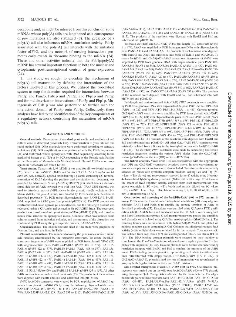
Mapping of Pan2p-Pan3p interaction domains. An earlierstudy demonstrated that Pan2p and Pan3p, the subunits ofPAN, were interacting proteins (9). We utilized the two-hybridgenetic system (3) to delineate the respective interaction do-mains on these two proteins. To map the Pan3p-binding site on
Pan2p, fragments of PAN2 were tested for interaction withfull-length PAN3 (Fig. 1A). For these constructs, we wereunable to create functional fusions of PAN2 with the GAL4activation domain, and all PAN2-PAN3 interaction studieswere thus performed with lexA(DB)-PAN2 fusions andGAL4(AD)-PAN3 alleles. These experiments showed that full-length Pan3p interacted strongly with full-length Pan2p, pro-ducing resistance up to or greater than 100 mM 3-AT (Fig.1A). A comparable, strong interaction was also observed be-tween PAN2 (333 to 1115) and full-length PAN3. Further trun-cations of the Pan2p N terminus led to a loss of interactionwith Pan3p-FL, as did a C-terminal truncation (Fig. 1A). Testsof PAN3 fragments against full-length PAN2 demonstratedthat only a small portion of the C-terminal region of Pan3p isnecessary for interaction with Pan2p (Fig. 1B). The smallestC-terminal PAN3 segment capable of producing 3-AT resis-tance equal to that of full-length PAN3 was a fragment encom-
FIG. 1. Two-hybrid mapping of Pan2p-Pan3p interactions. Protein fragments used in two-hybrid analyses are denoted as bars with numbers toindicate the span of specific amino acid residues included in each. Proteins that interacted activated transcription of the HIS3 gene, producingresistance to the competitive inhibitor 3-AT. Results are expressed as the highest millimolar concentration of 3-AT (on SC �His, �Leu, �Trpplates) that still allowed substantial cellular growth; no growth indicates cells that could grow in the presence of histidine but were unable to growon medium lacking histidine. Bars for those proteins that produced 50 to 100% resistance to 3-AT are filled in black. Appropriate empty vectorcontrols were performed for each analysis and are indicated only when they significantly contributed to 3-AT resistance levels. (A) Pan3p-FL wastested against Pan2p truncations lacking either the C terminus or N terminus in order to determine the binding domain of Pan3p on Pan2p.(B) Fragments of Pan3p were used to identify the binding domain of Pan2p on Pan3p.
5524 MANGUS ET AL. MOL. CELL. BIOL.
on August 19, 2015 by guest
http://mcb.asm
.org/D
ownloaded from
passing amino acids 560 to 679. Interestingly, a PAN3 N-ter-minal truncation that retained amino acids 250 to 679 alsoproduced high-level 3-AT resistance, while the PAN3 (337 to679) and PAN3 (420 to 679) segments were unable to activatetranscription and supported growth only on plates lacking his-tidine (Fig. 1B). The inactivity of the last two fragments isprobably attributable to protein misfolding, sequestration ofthe interaction domain, and/or protein instability, but we havenot tested these hypotheses.
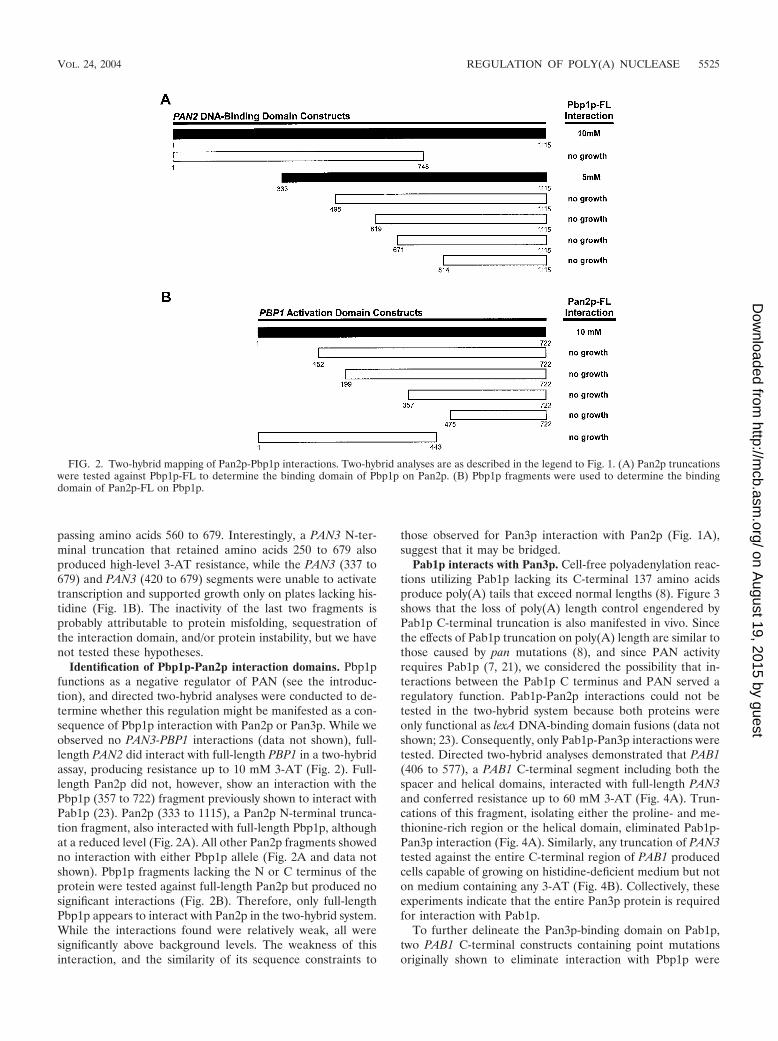
Identification of Pbp1p-Pan2p interaction domains. Pbp1pfunctions as a negative regulator of PAN (see the introduc-tion), and directed two-hybrid analyses were conducted to de-termine whether this regulation might be manifested as a con-sequence of Pbp1p interaction with Pan2p or Pan3p. While weobserved no PAN3-PBP1 interactions (data not shown), full-length PAN2 did interact with full-length PBP1 in a two-hybridassay, producing resistance up to 10 mM 3-AT (Fig. 2). Full-length Pan2p did not, however, show an interaction with thePbp1p (357 to 722) fragment previously shown to interact withPab1p (23). Pan2p (333 to 1115), a Pan2p N-terminal trunca-tion fragment, also interacted with full-length Pbp1p, althoughat a reduced level (Fig. 2A). All other Pan2p fragments showedno interaction with either Pbp1p allele (Fig. 2A and data notshown). Pbp1p fragments lacking the N or C terminus of theprotein were tested against full-length Pan2p but produced nosignificant interactions (Fig. 2B). Therefore, only full-lengthPbp1p appears to interact with Pan2p in the two-hybrid system.While the interactions found were relatively weak, all weresignificantly above background levels. The weakness of thisinteraction, and the similarity of its sequence constraints to
those observed for Pan3p interaction with Pan2p (Fig. 1A),suggest that it may be bridged.
Pab1p interacts with Pan3p. Cell-free polyadenylation reac-tions utilizing Pab1p lacking its C-terminal 137 amino acidsproduce poly(A) tails that exceed normal lengths (8). Figure 3shows that the loss of poly(A) length control engendered byPab1p C-terminal truncation is also manifested in vivo. Sincethe effects of Pab1p truncation on poly(A) length are similar tothose caused by pan mutations (8), and since PAN activityrequires Pab1p (7, 21), we considered the possibility that in-teractions between the Pab1p C terminus and PAN served aregulatory function. Pab1p-Pan2p interactions could not betested in the two-hybrid system because both proteins wereonly functional as lexA DNA-binding domain fusions (data notshown; 23). Consequently, only Pab1p-Pan3p interactions weretested. Directed two-hybrid analyses demonstrated that PAB1(406 to 577), a PAB1 C-terminal segment including both thespacer and helical domains, interacted with full-length PAN3and conferred resistance up to 60 mM 3-AT (Fig. 4A). Trun-cations of this fragment, isolating either the proline- and me-thionine-rich region or the helical domain, eliminated Pab1p-Pan3p interaction (Fig. 4A). Similarly, any truncation of PAN3tested against the entire C-terminal region of PAB1 producedcells capable of growing on histidine-deficient medium but noton medium containing any 3-AT (Fig. 4B). Collectively, theseexperiments indicate that the entire Pan3p protein is requiredfor interaction with Pab1p.
To further delineate the Pan3p-binding domain on Pab1p,two PAB1 C-terminal constructs containing point mutationsoriginally shown to eliminate interaction with Pbp1p were
FIG. 2. Two-hybrid mapping of Pan2p-Pbp1p interactions. Two-hybrid analyses are as described in the legend to Fig. 1. (A) Pan2p truncationswere tested against Pbp1p-FL to determine the binding domain of Pbp1p on Pan2p. (B) Pbp1p fragments were used to determine the bindingdomain of Pan2p-FL on Pbp1p.
VOL. 24, 2004 REGULATION OF POLY(A) NUCLEASE 5525
on August 19, 2015 by guest
http://mcb.asm
.org/D
ownloaded from

tested for interaction with full-length Pan3p (23). The firstconstruct contained a single mutation at codon 444, changing aG to a D. This mutation, located within the Pab1p proline- andmethionine-rich segment, did not affect interaction with Pan3psince strains harboring this construct still produced resistanceup to 60 mM 3-AT (Fig. 4A). The second construct containedtwo point mutations, one at codon 451, changing a V to an A,and a second at codon 514, changing a Y to a C. Two-hybridreporter strains containing this construct were unable to growon media lacking histidine, indicating that one or both of thetwo altered amino acid residues are critical for binding ofPab1p to Pan3p (Fig. 4A). To determine their individual ef-fects, each mutation was independently created in PAB1 (406to 577). While the pab1 451 (V3A) substitution had no effect,the pab1 514 (Y3C) mutant exhibited reduced interactionwith Pan3p (Table 2 and data not shown).
Identification of Pan3p interaction mutations within thePAB1 (406 to 577) segment. To better understand the interac-tion between Pab1p and Pan3p, additional point mutants weregenerated in PAB1 (406 to 577). The mutants were screenedfor a loss of interaction with full-length Pan3p, while maintain-ing interaction with Pbp1p. A total of four alleles were isolatedin this screen: pab1 528 (G3D), pab1 481 (V3A)/506(R3G), pab1 497 (N3D)/533 (M3T), and pab1 510 (G3R)/519 (A3V). These alleles were further manipulated to isolatethe pertinent individual point mutants by using site-directedmutagenesis of the wild-type lexA(DB)-PAB1 (406 to 577) con-struct to make each mutation. The selection of alleles to beisolated was dictated by the apparent requirement of the fourPab1p carboxy-terminal helices for interaction with Pan3p.This hypothesis stems from several observations, includingthose demonstrating that (i) the initially isolated mutant that
FIG. 3. Truncation of the Pab1p C terminus affects poly(A) length control in vivo. mRNA poly(A) tail lengths were determined in wild-typeor pab1 strains bearing a carboxy-terminal truncation (C-term�). The resulting autoradiograph (A) was analyzed by densitometric tracing (B).Numbers of adenylate residues were determined by comparison with a DNA sequence ladder.
5526 MANGUS ET AL. MOL. CELL. BIOL.
on August 19, 2015 by guest
http://mcb.asm
.org/D
ownloaded from

decreases interaction with Pan3p [pab1 514 (Y3C)] bears anamino acid change in helix 2, (ii) all of the new pab1 alleleswhich fail to interact with Pan3p bear mutations in helix 2 or 3,and (iii) the sole single point mutant isolated, pab1 528(G3D), is located within helix 3.
The set of individual mutants was then cotransformed andtested for interaction with empty vector, PBP1 (357 to 722), orPAN3-FL. The pab1 506 (R3G), pab1 510 (G3R), pab1 528(G3D), and pab1 533 (M3T) alleles all maintained wild-typeinteraction with PBP1 (357 to 722) and eliminated interactionwith PAN3-FL (Table 2). These results support the hypothesisthat the binding site for Pan3p is located within the C-terminalhelices of Pab1p. Interestingly, the pab1 519 (A3V) mutant,
which is located between helices 2 and 3, interacted with Pan3pat wild-type levels (data not shown).
This collection of mutants provides strong support for amodel in which Pan3p and Pbp1p have separate binding do-mains on Pab1p. The mutations affecting Pan3p binding werelocated in the four helices, while the one mutation affectingPbp1p binding (24) was located in the N-terminal portion ofthe proline- and methionine-rich region (Fig. 5A). Examina-tion of the point mutations in the context of the nuclear mag-netic resonance (NMR) structure of Pab1p’s carboxy-terminalhelices showed that three of the five mutations (506, 510, and514) were clustered on the same side of helix 2 (Fig. 5B). Theidentification of mutations at residues 528 and 533 of helix 3
FIG. 4. Two-hybrid mapping of Pab1p-Pan3p interactions. Two-hybrid analyses are as described in the legend to Fig. 1. (A) Pab1p C-terminalfragments were tested with Pan3p-FL to determine the binding site of Pan3p on Pab1p. (B) Pan3p fragments were used to determine the locationof Pab1p binding on Pan3p.
VOL. 24, 2004 REGULATION OF POLY(A) NUCLEASE 5527
on August 19, 2015 by guest
http://mcb.asm
.org/D
ownloaded from
further defines a pocket into which Pan3p can bind Pab1p (Fig.5B).
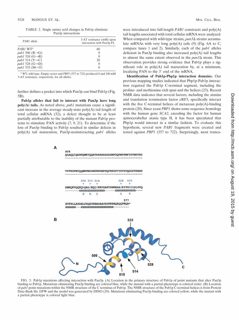
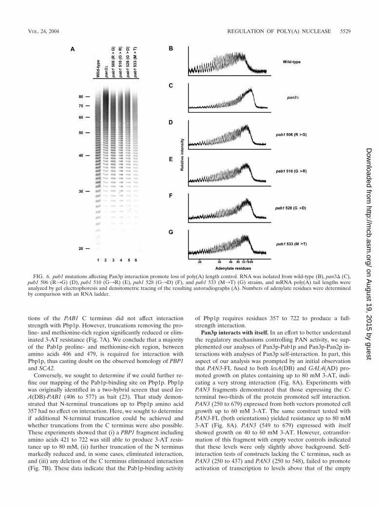
Pab1p alleles that fail to interact with Pan3p have longpoly(A) tails. As noted above, pab1 mutations cause a signifi-cant increase in the average steady-state poly(A) tail length oftotal cellular mRNA (32), a defect thought to be at leastpartially attributable to the inability of the mutant Pab1p pro-teins to stimulate PAN activity (7, 9, 21). To determine if theloss of Pan3p binding to Pab1p resulted in similar defects inpoly(A) tail maturation, Pan3p-noninteracting pab1 alleles
were introduced into full-length PAB1 constructs and poly(A)tail lengths associated with total cellular mRNA were analyzed.When compared with wild-type strains, pan3� strains accumu-late mRNAs with very long poly(A) tails (9) (Fig. 6A to C,compare lanes 1 and 2). Similarly, each of the pab1 allelesdeficient in Pan3p binding also increased poly(A) tail lengthsto almost the same extent observed in the pan3� strain. Thisobservation provides strong evidence that Pab1p plays a sig-nificant role in poly(A) tail maturation by, at a minimum,localizing PAN to the 3� end of the mRNA.
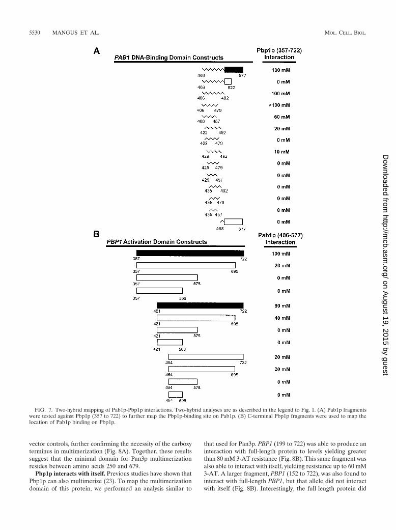
Identification of Pab1p-Pbp1p interaction domains. Ourprevious mapping studies indicated that Pbp1p-Pab1p interac-tion required the Pab1p C-terminal segment, including theproline- and methionine-rich span and the helices (23). RecentNMR data indicates that several factors, including the ataxinsand translation termination factor eRF3, specifically interactwith the five C-terminal helices of metazoan poly(A)-bindingprotein (20). Since yeast PBP1 shows some sequence homologywith the human gene SCA2, encoding the factor for humanspinocerabellar ataxia type II, it has been speculated thatPbp1p would interact in a similar fashion. To evaluate thishypothesis, several new PAB1 fragments were created andtested against PBP1 (357 to 722). Surprisingly, most trunca-
FIG. 5. Pab1p mutations affecting interaction with Pan3p. (A) Location in the primary structure of Pab1p of point mutants that alter Pan3pbinding to Pab1p. Mutations eliminating Pan3p binding are colored blue, while the mutant with a partial phenotype is colored violet. (B) Locationof pab1 point mutations within the NMR structure of the C terminus of Pab1p. The NMR structure of the Pab1p C-terminal helices is from ProteinData Bank file 1IFW and the model was generated by DINO (29). Mutations eliminating Pan3p binding are colored yellow, while the mutant witha partial phenotype is colored light blue.
TABLE 2. Single amino acid changes in Pab1p eliminatePan3p interactions
PAB1 allele 3-AT resistance (mM) uponinteraction with Pan3p-FL
PAB1 WTa 60pab1 506 (R3G) 0pab1 510 (G3R) 0pab1 514 (Y3C) 20pab1 528 (G3D) 0pab1 533 (M3T) 0
a WT, wild type. Empty vector and PBP1 (357 to 722) produced 0 and 100 mM3-AT resistance, respectively, for all alleles.
5528 MANGUS ET AL. MOL. CELL. BIOL.
on August 19, 2015 by guest
http://mcb.asm
.org/D
ownloaded from
tions of the PAB1 C terminus did not affect interactionstrength with Pbp1p. However, truncations removing the pro-line- and methionine-rich region significantly reduced or elim-inated 3-AT resistance (Fig. 7A). We conclude that a majorityof the Pab1p proline- and methionine-rich region, betweenamino acids 406 and 479, is required for interaction withPbp1p, thus casting doubt on the observed homology of PBP1and SCA2.
Conversely, we sought to determine if we could further re-fine our mapping of the Pab1p-binding site on Pbp1p. Pbp1pwas originally identified in a two-hybrid screen that used lex-A(DB)-PAB1 (406 to 577) as bait (23). That study demon-strated that N-terminal truncations up to Pbp1p amino acid357 had no effect on interaction. Here, we sought to determineif additional N-terminal truncation could be achieved andwhether truncations from the C terminus were also possible.These experiments showed that (i) a PBP1 fragment includingamino acids 421 to 722 was still able to produce 3-AT resis-tance up to 80 mM, (ii) further truncation of the N terminusmarkedly reduced and, in some cases, eliminated interaction,and (iii) any deletion of the C terminus eliminated interaction(Fig. 7B). These data indicate that the Pab1p-binding activity
of Pbp1p requires residues 357 to 722 to produce a full-strength interaction.
Pan3p interacts with itself. In an effort to better understandthe regulatory mechanisms controlling PAN activity, we sup-plemented our analyses of Pan3p-Pab1p and Pan3p-Pan2p in-teractions with analyses of Pan3p self-interaction. In part, thisaspect of our analysis was prompted by an initial observationthat PAN3-FL fused to both lexA(DB) and GAL4(AD) pro-moted growth on plates containing up to 80 mM 3-AT, indi-cating a very strong interaction (Fig. 8A). Experiments withPAN3 fragments demonstrated that those expressing the C-terminal two-thirds of the protein promoted self interaction.PAN3 (250 to 679) expressed from both vectors promoted cellgrowth up to 60 mM 3-AT. The same construct tested withPAN3-FL (both orientations) yielded resistance up to 80 mM3-AT (Fig. 8A). PAN3 (549 to 679) expressed with itselfshowed growth on 40 to 60 mM 3-AT. However, cotransfor-mation of this fragment with empty vector controls indicatedthat these levels were only slightly above background. Self-interaction tests of constructs lacking the C terminus, such asPAN3 (250 to 437) and PAN3 (250 to 548), failed to promoteactivation of transcription to levels above that of the empty
FIG. 6. pab1 mutations affecting Pan3p interaction promote loss of poly(A) length control. RNA was isolated from wild-type (B), pan3� (C),pab1 506 (R3G) (D), pab1 510 (G3R) (E), pab1 528 (G3D) (F), and pab1 533 (M3T) (G) strains, and mRNA poly(A) tail lengths wereanalyzed by gel electrophoresis and densitometric tracing of the resulting autoradiographs (A). Numbers of adenylate residues were determinedby comparison with an RNA ladder.
VOL. 24, 2004 REGULATION OF POLY(A) NUCLEASE 5529
on August 19, 2015 by guest
http://mcb.asm
.org/D
ownloaded from
vector controls, further confirming the necessity of the carboxyterminus in multimerization (Fig. 8A). Together, these resultssuggest that the minimal domain for Pan3p multimerizationresides between amino acids 250 and 679.
Pbp1p interacts with itself. Previous studies have shown thatPbp1p can also multimerize (23). To map the multimerizationdomain of this protein, we performed an analysis similar to
that used for Pan3p. PBP1 (199 to 722) was able to produce aninteraction with full-length protein to levels yielding greaterthan 80 mM 3-AT resistance (Fig. 8B). This same fragment wasalso able to interact with itself, yielding resistance up to 60 mM3-AT. A larger fragment, PBP1 (152 to 722), was also found tointeract with full-length PBP1, but that allele did not interactwith itself (Fig. 8B). Interestingly, the full-length protein did
FIG. 7. Two-hybrid mapping of Pab1p-Pbp1p interactions. Two-hybrid analyses are as described in the legend to Fig. 1. (A) Pab1p fragmentswere tested against Pbp1p (357 to 722) to further map the Pbp1p-binding site on Pab1p. (B) C-terminal Pbp1p fragments were used to map thelocation of Pab1p binding on Pbp1p.
5530 MANGUS ET AL. MOL. CELL. BIOL.
on August 19, 2015 by guest
http://mcb.asm
.org/D
ownloaded from

not demonstrate an ability to interact with itself, suggestingthat its N terminus may fold in such a way as to regulate andprevent multimerization.
DISCUSSION
Eukaryotic mRNAs are subject to considerable posttranscrip-tional modification, including capping, splicing, and polyade-nylation. The process of polyadenylation adds a 3�-poly(A) tailand provides mRNAs with a binding site for a major class ofregulatory factors, the poly(A)-binding proteins. These highly con-served polypeptides bind poly(A) by using one or more RRMsand then act as cis-acting effectors of specific steps in the polyad-enylation, export, translation, and turnover of the transcripts towhich they are bound. They provide a scaffold for the bindingof factors that mediate these steps and also act as apparent an-tagonists to the binding of factors that enable the terminal stepsof mRNA degradation. Collectively, these sequential nuclearand cytoplasmic contributions control mRNA function (24).
Previous experiments have suggested two possible roles forthe major yeast poly(A)-binding protein, Pab1p, in the nuclearmaturation of mRNA. Pab1p appears to have an indirect effecton the transition of poly(A) polymerase (Pap1p) from proces-sive to distributive activity and a direct effect on the poly(A)
tail-trimming activity of PAN (9, 43). The latter role of Pab1p,and the protein-protein interactions which underlie that role,have been addressed in this study. By using two-hybrid analysis,we have demonstrated that Pab1p interacts with Pan3p andPbp1p, two proteins that respectively appear to act as positiveand negative regulators of PAN activity (9, 23). Not surpris-ingly, our experiments have also shown that these two proteinsalso interact with the Pan2p subunit of PAN, i.e., the subunitwhose homologies to previously characterized exonucleases (8,26) suggest that it harbors the catalytic activity relevant to PANfunction. Figure 9 summarizes this set of interactions andraises interesting possibilities for the precise role of Pab1p inpoly(A) trimming. At a minimum, Pab1p provides a sitethrough which PAN and its negative regulator, Pbp1p, gainaccess to the newly formed poly(A) tract. Additionally, as hasbeen proposed for Pab1p interactions with translation initia-tion factors (6, 22), Pab1p may alter the abilities of Pan3pand/or Pbp1p to bind to additional factors, e.g., binding toPab1p may enhance the ability of Pan3p to bind to, and/oractivate, Pan2p. Additional analyses have shown that Pab1p-Pan3p and Pab1p-Pbp1p two-hybrid interactions still occur inpan2� or pan3� strains and are thus not dependent on theintegrity of PAN (data not shown). It should be noted, how-
FIG. 8. Mapping Pan3p and Pbp1p self-interactions. Two-hybrid analyses are as described in the legend to Fig. 1. (A) Pan3p fragments weretested against full-length Pan3p and themselves to map the domain(s) necessary for self-interaction. (B) Pbp1p fragments were tested againstfull-length Pbp1p and themselves to map the domain(s) necessary for self-interaction.
VOL. 24, 2004 REGULATION OF POLY(A) NUCLEASE 5531
on August 19, 2015 by guest
http://mcb.asm
.org/D
ownloaded from
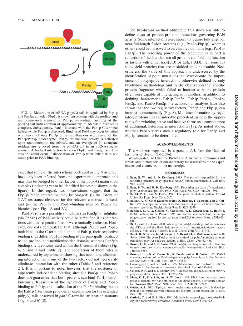
ever, that some of the interactions portrayed in Fig. 9 as directhave only been inferred from one experimental approach andmay thus be bridged by other factors in the poly(A) maturationcomplex (including yet to be identified factors not shown in thefigure). In this regard, two observations suggest that thePbp1p-Pan2p interaction may be bridged: (i) the extent of3-AT resistance observed for the relevant constructs is weakand (ii) the Pan3p- and Pbp1p-binding sites on Pan2p areidentical (see Fig. 1A and 2A).
Pab1p’s role as a possible stimulator (via Pan3p) or inhibitor(via Pbp1p) of PAN activity could be simplified if its interac-tions with the respective factors were mutually exclusive. How-ever, our data demonstrate that, although Pan3p and Pbp1pboth bind to the C-terminal domain of Pab1p, their respectivebinding sites differ. Pbp1p’s binding site is principally localizedto the proline- and methionine-rich domain, whereas Pan3p’sbinding site is concentrated within the C-terminal helices (Fig.4, 5, and 7 and Table 2). The separation of these sites isunderscored by experiments showing that mutations eliminat-ing interaction with one of the two factors do not necessarilyeliminate interaction with the other (Table 2 and reference24). It is important to note, however, that the existence ofapparently independent binding sites for Pan3p and Pbp1pdoes not guarantee that both proteins can bind Pab1p simul-taneously. Regardless of the dynamics of Pan3p and Pbp1pbinding to Pab1p, the localization of the Pan3p-binding site tothe Pab1p C terminus provides an explanation for the extendedpoly(A) tails observed in pab1 C-terminal truncation mutants(Fig. 3 and 6) (8).
The two-hybrid method utilized in this study was able todefine a set of protein-protein interactions governing PANactivity. Some interactions were shown to require full-length ornear-full-length fusion proteins (e.g., Pan2p-Pbp1p), whereasothers could be narrowed to very limited domains (e.g., Pab1p-Pan3p). The resolving power of the technique is in part areflection of the fact that not all proteins can fold and functionas fusions with either lexA(DB) or GAL4(AD), i.e., some fu-sions yield proteins that are misfolded and/or unstable. Nev-ertheless, the value of this approach is underscored by theidentification of point mutations that corroborate the impor-tance of polypeptide interactions otherwise defined by onlytwo-hybrid methodology and by the observation that specificprotein fragments which failed to interact with one proteinoften were capable of interacting with another. In addition todefining heteromeric Pab1p-Pan3p, Pab1p-Pbp1p, Pbp1p-Pan2p, and Pan3p-Pan2p interactions, our analyses have alsoshown that the two regulatory factors, Pan3p and Pbp1p, caninteract homomerically (Fig. 8). Multimer formation by regu-latory proteins has considerable precedent, as does the oppor-tunity for switching active and inactive forms as a consequenceof additional, heteromeric interactions (13). As noted above,whether Pab1p serves such a regulatory role for Pan3p andPbp1p remains to be determined.
ACKNOWLEDGMENTS
This work was supported by a grant to A.J. from the NationalInstitutes of Health (GM61096).
We are grateful to Christine Brown and Alan Sachs for plasmids andstrains and to members of our laboratory for discussions of the exper-iments and comments on the manuscript.
REFERENCES
1. Baer, B. W., and R. D. Kornberg. 1983. The protein responsible for therepeating structure of cytoplasmic poly(A)-ribonucleoprotein. J. Cell Biol.96:717–721.
2. Baer, B. W., and R. D. Kornberg. 1980. Repeating structure of cytoplasmicpoly(A)-ribonucleoprotein. Proc. Natl. Acad. Sci. USA 77:1890–1892.
3. Bartel, P. L., and S. Fields. 1997. The yeast two-hybrid system. OxfordUniversity Press, New York, N.Y.
4. Baudin, A., O. Ozier-Kalogeropoulos, A. Denouel, F. Lacroute, and C. Cul-lin. 1993. A simple and efficient method for direct gene deletion in Saccha-romyces cerevisiae. Nucleic Acids Res. 21:3329–3330.
5. Beelman, C. A., A. Stevens, G. Caponigro, T. E. LaGrandeur, L. Hatfield,D. M. Fortner, and R. Parker. 1996. An essential component of the decap-ping enzyme required for normal rates of mRNA turnover. Nature 382:642–646.
6. Bi, X., and D. J. Goss. 2000. Wheat germ poly(A)-binding protein increasesthe ATPase and the RNA helicase activity of translation initiation factorseIF4A, eIF4B, and eIF-iso4F. J. Biol. Chem. 275:17740–17746.
7. Boeck, R., S. Tarun, Jr., M. Rieger, J. A. Deardorff, S. Muller-Auer, and A. B.Sachs. 1996. The yeast Pan2 protein is required for poly(A)-binding protein-stimulated poly(A)-nuclease activity. J. Biol. Chem. 271:432–438.
8. Brown, C. E., and A. B. Sachs. 1998. Poly(A) tail length control in Saccha-romyces cerevisiae occurs by message-specific deadenylation. Mol. Cell. Biol.18:6548–6559.
9. Brown, C. E., S. Z. Tarun, Jr., R. Boeck, and A. B. Sachs. 1996. PAN3encodes a subunit of the Pab1p-dependent poly(A) nuclease in Saccharomy-ces cerevisiae. Mol. Cell. Biol. 16:5744–5753.
10. Caponigro, G., and R. Parker. 1996. Mechanisms and control of mRNAturnover in Saccharomyces cerevisiae. Microbiol. Rev. 60:233–249.
11. Colgan, D. F., and J. L. Manley. 1997. Mechanism and regulation of mRNApolyadenylation. Genes Dev. 11:2755–2766.
12. Elder, R. T., E. Y. Loh, and R. W. Davis. 1983. RNA from the yeast trans-posable element Ty1 has both ends in the direct repeats, a structure similarto retrovirus RNA. Proc. Natl. Acad. Sci. USA 80:2432–2436.
13. Gotter, A. L. 2003. Tipin, a novel timeless-interacting protein, is develop-mentally co-expressed with timeless and disrupts its self-association. J. Mol.Biol. 331:167–176.
14. Guthrie, C., and G. R. Fink. 1991. Methods in enzymology: molecular biol-ogy of Saccharomyces cerevisiae. Academic Press, New York, N.Y.
FIG. 9. Maturation of mRNA poly(A) tails is regulated by Pbp1pand Pan3p: a model. Pbp1p is shown associating with the proline- andmethionine-rich segment of Pab1p, preventing trimming of thepoly(A) tail until addition of approximately 90 adenylate residues iscomplete. Subsequently, Pan3p interacts with the Pab1p C-terminalhelices, while Pbp1p is displaced. Binding of PAN may occur by initialrecruitment of only Pan3p or by simultaneous recruitment of thePan2p/Pan3p holoenzyme. Pan2p exonuclease activity is activatedupon recruitment to the mRNA, and an average of 30 adenylateresidues are removed from the poly(A) tail in an mRNA-specificmanner. A bridged interaction between Pbp1p and Pan2p (see Dis-cussion) could occur if dissociation of Pbp1p from Pab1p does notoccur prior to PAN binding.
5532 MANGUS ET AL. MOL. CELL. BIOL.
on August 19, 2015 by guest
http://mcb.asm
.org/D
ownloaded from
15. Hatfield, L., C. A. Beelman, A. Stevens, and R. Parker. 1996. Mutations intrans-acting factors affecting mRNA decapping in Saccharomyces cerevisiae.Mol. Cell. Biol. 16:5830–5838.
16. Jacobson, A. 1996. Poly(A) metabolism and translation: the closed loopmodel, p. 451–480. In N. Sonenberg (ed.), Translational control, 1st ed. ColdSpring Harbor Laboratory Press, Cold Spring Harbor, N.Y.
17. Jacobson, A. 1987. Purification and fractionation of poly(A)� RNA. Meth-ods Enzymol. 152:254–261.
18. Jacobson, A., and S. W. Peltz. 1996. Interrelationships of the pathways ofmRNA decay and translation in eukaryotic cells. Annu. Rev. Biochem. 65:693–739.
19. Jones, J. S., and L. Prakash. 1990. Yeast Saccharomyces cerevisiae selectablemarkers in pUC18 polylinkers. Yeast 6:363–366.
20. Kozlov, G., J. F. Trempe, K. Khaleghpour, A. Kahvejian, I. Ekiel, and K.Gehring. 2001. Structure and function of the C-terminal PABC domain ofhuman poly(A)-binding protein. Proc. Natl. Acad. Sci. USA 98:4409–4413.
21. Lowell, J. E., D. Z. Rudner, and A. B. Sachs. 1992. 3�-UTR-dependentdeadenylation by the yeast poly(A) nuclease. Genes Dev. 6:2088–2099.
22. Luo, Y., and D. J. Goss. 2001. Homeostasis in mRNA initiation: wheat germpoly(A)-binding protein lowers the activation energy barrier to initiationcomplex formation. J. Biol. Chem. 276:43083–43086.
23. Mangus, D. A., N. Amrani, and A. Jacobson. 1998. Pbp1p, a factor interact-ing with Saccharomyces cerevisiae poly(A)-binding protein, regulates polyad-enylation. Mol. Cell. Biol. 18:7383–7396.
24. Mangus, D. A., M. C. Evans, and A. Jacobson. 2003. Poly(A)-binding pro-teins: multifunctional scaffolds for the post-transcriptional control of geneexpression. Genome Biol. 4:223.
25. Mangus, D. A., M. M. Smith, J. M. McSweeney, and A. Jacobson. 2004.Identification of factors regulating poly(A) tail synthesis and maturation.Mol. Cell. Biol. 24:4196–4206.
26. Moser, M. J., W. R. Holley, A. Chatterjee, and I. S. Mian. 1997. Theproofreading domain of Escherichia coli DNA polymerase I and other DNAand/or RNA exonuclease domains. Nucleic Acids Res. 25:5110–5118.
27. Muhlrad, D., C. J. Decker, and R. Parker. 1994. Deadenylation of theunstable mRNA encoded by the yeast MFA2 gene leads to decapping fol-lowed by 5�33� digestion of the transcript. Genes Dev. 8:855–866.
28. Muhlrad, D., R. Hunter, and R. Parker. 1992. A rapid method for localizedmutagenesis of yeast genes. Yeast 8:79–82.
29. Philippsen, A. 2003. DINO: visualizing structural biology. [Online.] http://www.dino3d.org.
30. Rose, M. D., F. Winston, and P. Hieter. 1990. Methods in yeast genetics: alaboratory course manual. Cold Spring Harbor Laboratory Press, ColdSpring Harbor, N.Y.
31. Sachs, A. B., M. W. Bond, and R. D. Kornberg. 1986. A single gene fromyeast for both nuclear and cytoplasmic polyadenylate-binding proteins: do-main structure and expression. Cell 45:827–835.
32. Sachs, A. B., and R. W. Davis. 1989. The poly(A) binding protein is requiredfor poly(A) shortening and 60S ribosomal subunit-dependent translationinitiation. Cell 58:857–867.
33. Sachs, A. B., R. W. Davis, and R. D. Kornberg. 1987. A single domain ofyeast poly(A)-binding protein is necessary and sufficient for RNA bindingand cell viability. Mol. Cell. Biol. 7:3268–3276.
34. Sambrook, J., E. Fritsch, and T. Maniatis. 1989. Molecular cloning: a lab-oratory manual, 2nd ed. Cold Spring Harbor Laboratory Press, Cold SpringHarbor, N.Y.
35. Sanger, F., S. Nicklen, and A. R. Coulson. 1977. DNA sequencing withchain-terminating inhibitors. Proc. Natl. Acad. Sci. USA 74:5463–5467.
36. Soni, R., J. P. Carmichael, and J. A. Murray. 1993. Parameters affectinglithium acetate-mediated transformation of Saccharomyces cerevisiae anddevelopment of a rapid and simplified procedure. Curr. Genet. 24:455–459.
37. Steel, L. F., and A. Jacobson. 1991. Sequence elements that affect mRNAtranslational activity in developing Dictyostelium cells. Dev. Genet. 12:98–103.
38. Tucker, M., M. A. Valencia-Sanchez, R. R. Staples, J. Chen, C. L. Denis, andR. Parker. 2001. The transcription factor associated Ccr4 and Caf1 proteinsare components of the major cytoplasmic mRNA deadenylase in Saccharo-myces cerevisiae. Cell 104:377–386.
39. Vogel, H. J., and D. M. Bonner. 1956. Acetylornithinase of Escherichia coli:partial purification and some properties. J. Biol. Chem. 218:97–106.
40. Wahle, E., and U. Kuhn. 1997. The mechanism of 3� cleavage and poly-adenylation of eukaryotic pre-mRNA. Prog. Nucleic Acid Res. Mol. Biol.57:41–71.
41. White, T. J., N. Arnheim, and H. A. Erlich. 1989. The polymerase chainreaction. Trends Genet. 5:185–189.
42. Zhao, J., L. Hyman, and C. Moore. 1999. Formation of mRNA 3� ends ineukaryotes: mechanism, regulation, and interrelationships with other steps inmRNA synthesis. Microbiol. Mol. Biol. Rev. 63:405–445.
43. Zhelkovsky, A., S. Helmling, and C. Moore. 1998. Processivity of the Sac-charomyces cerevisiae poly(A) polymerase requires interactions at the car-boxyl-terminal RNA binding domain. Mol. Cell. Biol. 18:5942–5951.
VOL. 24, 2004 REGULATION OF POLY(A) NUCLEASE 5533
on August 19, 2015 by guest
http://mcb.asm
.org/D
ownloaded from
Related Documents


![The conserved Fanconi anemia nuclease Fan1 and the SUMO E3 … · 2017. 2. 23. · FAN1 (Fanconi anemia-associated nuclease 1, or FANCD2/FANCI-associated nuclease 1) [13–18]. Human](https://static.cupdf.com/doc/110x72/60c9d965c710eb0d72008d0e/the-conserved-fanconi-anemia-nuclease-fan1-and-the-sumo-e3-2017-2-23-fan1-fanconi.jpg)


