509 Reprod. Nutr. Dev. 44 (2004) 509–538 © INRA, EDP Sciences, 2005 DOI: 10.1051/rnd:2004063 Review Polyunsaturated fatty acids in the central nervous system: evolution of concepts and nutritional implications throughout life Jean-Marc ALESSANDRI a *, Philippe GUESNET a , Sylvie VANCASSEL a , Pierre ASTORG b , Isabelle DENIS a , Bénédicte LANGELIER a , Sabah AÏD a , Carine POUMÈS-BALLIHAUT a , Gaëlle CHAMPEIL-POTOKAR a , Monique LAVIALLE a a Neurobiologie des Lipides, Laboratoire de Nutrition et Sécurité Alimentaire, INRA, 78352 Jouy-en-Josas, France b UMR 557 INSERM/INRA/CNAM Épidémiologie Nutritionnelle, Institut Scientifique et Technique de la Nutrition et de l’Alimentation, CNAM, 5 rue du Vertbois, 75003 Paris, France (Received 18 June 2004; accepted 10 September 2004) Abstract – Docosahexaenoic acid (DHA, 22:6n-3) and arachidonic acid (AA, 20:4n-6) are the major polyunsaturated fatty acids in the membranes of brain and retinal cells. Animals specifically deficient in dietary n-3 fatty acids have low DHA content in their membranes, reduced visual acuity and impaired learning ability. Studies on bottle-fed human infants have shown that adding DHA and AA to milk replacer-formulas can bring their concentrations in the infant blood lipids to values as high as those produced by breast-feeding and significantly improves mental development and maturation of visual function. In older subjects, diverse neuropsychiatric and neurodegenerative diseases have been associated to decreased blood levels of n-3 PUFA. Low intakes of fish or of n-3 PUFA in pop- ulations have been associated with increased risks of depression and Alzheimer disease, and n-3 PUFA, especially eicosapentaenoic acid (EPA, 20:5n-3), have shown efficacy as adjunctive treat- ment – and in some cases as the only treatment – in several psychiatric disorders. The mechanisms by which polyunsaturated fatty acids have an impact on neuronal functions will be reviewed: the modulation of membrane biophysical properties, regulation of neurotransmitter release, synthesis of biologically active oxygenated derivatives, and nuclear receptor-mediated transcription of genes responsive to fatty acids or to their derivatives. docosahexaenoic acid / arachidonic acid / retina / brain / milk feeding / eicosanoids / synaptic terminals / phototransduction / neuroprotection / nuclear receptors / neuropsychiatric diseases / neurodegenerative diseases Abbreviations AA: arachidonic acid (20:4n-6), ABC: ATP- binding cassette transporters, ACS2: acyl- CoAsynthase 2, ADHD: attention-deficit hyper- activity disorders, AFSSA: Agence Française de Sécurité Sanitaire des Aliments, AOX: peroxi- somal acylCoA-oxidase, aP2: adipocyte lipid- binding protein 2, ApoE: apolipoprotein E, B- FABP: brain fatty acid-binding protein, CARβ constitutive androstane receptor β, CD36: type B scavenger receptor, CNS: central nervous sys- tem, COUP-TF1: chicken ovalbumin upstream promoter-transcription factor 1, COX1 and COX2: * Corresponding author: [email protected]

Welcome message from author
This document is posted to help you gain knowledge. Please leave a comment to let me know what you think about it! Share it to your friends and learn new things together.
Transcript

509Reprod. Nutr. Dev. 44 (2004) 509–538© INRA, EDP Sciences, 2005DOI: 10.1051/rnd:2004063
Review
Polyunsaturated fatty acids in the central nervous system: evolution of concepts and nutritional
implications throughout life
Jean-Marc ALESSANDRIa*, Philippe GUESNETa, Sylvie VANCASSELa, Pierre ASTORGb, Isabelle DENISa, Bénédicte LANGELIERa, Sabah AÏDa,
Carine POUMÈS-BALLIHAUTa, Gaëlle CHAMPEIL-POTOKARa, Monique LAVIALLEa
a Neurobiologie des Lipides, Laboratoire de Nutrition et Sécurité Alimentaire, INRA, 78352 Jouy-en-Josas, France
b UMR 557 INSERM/INRA/CNAM Épidémiologie Nutritionnelle, Institut Scientifique et Technique de la Nutrition et de l’Alimentation, CNAM, 5 rue du Vertbois, 75003 Paris, France
(Received 18 June 2004; accepted 10 September 2004)
Abstract – Docosahexaenoic acid (DHA, 22:6n-3) and arachidonic acid (AA, 20:4n-6) are the majorpolyunsaturated fatty acids in the membranes of brain and retinal cells. Animals specifically deficientin dietary n-3 fatty acids have low DHA content in their membranes, reduced visual acuity andimpaired learning ability. Studies on bottle-fed human infants have shown that adding DHA and AAto milk replacer-formulas can bring their concentrations in the infant blood lipids to values as highas those produced by breast-feeding and significantly improves mental development and maturationof visual function. In older subjects, diverse neuropsychiatric and neurodegenerative diseases havebeen associated to decreased blood levels of n-3 PUFA. Low intakes of fish or of n-3 PUFA in pop-ulations have been associated with increased risks of depression and Alzheimer disease, and n-3PUFA, especially eicosapentaenoic acid (EPA, 20:5n-3), have shown efficacy as adjunctive treat-ment – and in some cases as the only treatment – in several psychiatric disorders. The mechanismsby which polyunsaturated fatty acids have an impact on neuronal functions will be reviewed: themodulation of membrane biophysical properties, regulation of neurotransmitter release, synthesisof biologically active oxygenated derivatives, and nuclear receptor-mediated transcription of genesresponsive to fatty acids or to their derivatives.
docosahexaenoic acid / arachidonic acid / retina / brain / milk feeding / eicosanoids / synapticterminals / phototransduction / neuroprotection / nuclear receptors / neuropsychiatricdiseases / neurodegenerative diseases
Abbreviations
AA: arachidonic acid (20:4n-6), ABC: ATP-binding cassette transporters, ACS2: acyl-CoAsynthase 2, ADHD: attention-deficit hyper-activity disorders, AFSSA: Agence Française deSécurité Sanitaire des Aliments, AOX: peroxi-
somal acylCoA-oxidase, aP2: adipocyte lipid-binding protein 2, ApoE: apolipoprotein E, B-FABP: brain fatty acid-binding protein, CARβconstitutive androstane receptor β, CD36: typeB scavenger receptor, CNS: central nervous sys-tem, COUP-TF1: chicken ovalbumin upstreampromoter-transcription factor 1, COX1 and COX2:
* Corresponding author: [email protected]

510 J.M. Alessandri et al.
cyclo-oxygenases 1 and 2, CYP2B: cytochromeP450-2B, DHA: docosahexaenoic acid (22:6n-3),DGLA: dihomo-γ-linolenic acid (20:3n-6),DHAmax: plateau value of DHA content inmembrane phospholipids, DHA50: dietary doseof DHA required to reach half the DHAmax,DPA: docosapentaenoic (22:5n-6 or 22:5n-3),EPA: eicosapentaenoic acid (20:5n-3), ERG:electroretinogram, 8s-HETE: 8s-hydroxyeico-satetraenoic acid, HMG-CoAS: mitochondrialhydroxy-methyl-glutarylCoA synthase, HNF-4:hepatocyte nuclear factor-4, 3-KAT2: 3-ketoacylCoA thiolase 2, LA: linoleic acid (18:2n-6),α-LNA: alpha-linolenic acid (18:3n-3), 5- and15-LOX: 5- and 15-lipoxygenases, L-PBE: L-peroxisomal bifunctional enzyme, LTB4: leuko-triene B4, LXR: liver X receptor, NFκB: nuclearfactor κB, NPD1: “neuroprotectin D1”, PBREM:phenobarbital responsive enhancer module, PE:phosphatidylethanolamines, PEPCK: phosphoe-nolpyruvate carboxykinase, 15d-PGJ2: 15-deoxyprostaglandin J2, PLA2: phospholipase A2, PP:peroxisome proliferators, PPAR: peroxisomeproliferators-activated receptors, PPRE: perox-isome proliferator-responsive element, PTE-1b:peroxisomal thioesterase 1b, PUFA: polyunsat-urated fatty acids, 9c-RA: 9cis retinoic acid,RAR: retinoic acid receptor, RXR: 9cis-retinoidX receptor, SREBP-1c: sterol regulatory ele-ment binding protein 1c, SCD: stearoyl-CoAdesaturase, UCP: uncoupling proteins.
1. ESSENTIAL POLYUNSA-TURATED FATTY ACIDS (PUFA) IN THE FOOD CHAIN: ANIMALS NEED 20 AND 22 CARBON ATOMS, PLANTS SUPPLY ONLY 18
The incorporation of polyunsaturatedfatty acids (PUFA) in the nerve cell mem-branes of the brain and retina is one of theprocesses of perinatal development thatcontributes to the functional maturation ofthe central nervous system (CNS). In ver-tebrates, two major PUFA contribute to theframework of the nerve cell membranes,arachidonic acid (AA, 20:4n-6) and docosa-hexaenoic acid (DHA, 22:6n-3). The pref-erential incorporation of DHA in the brainand retina, as compared to other tissues, isa remarkable constant throughout the evo-lution of species. AA is the main long-chainderivative of the precursor of the n-6 series
of essential fatty acids, linoleic acid (LA,18:2n-6), whereas DHA is formed from theprecursor of the n-3 series, alpha-linolenicacid (α-LNA, 18:3n-3) (see review in [1])(Fig. 1). Both precursors, which are them-selves very minor constituents of the nervecell membranes, are synthesised in variableproportions by plants, algae, bacteria andfungi, but not by vertebrate animals. In mostanimals, and some algae, bacteria and fungi,but not in plants, LA and α-LNA are trans-formed into long chain PUFA, includingwhich AA and DHA, respectively (Fig. 1).Thus, the concentration of AA and DHA inanimal tissues and animal fats depends, inpart, on the intake of precursors containedin foods. Man and omnivorous and carniv-orous animals can directly ingest AA andDHA from animal preys or foods, whereasherbivorous animals exclusively depend onthe supply of precursors, LA and α-LNA.
Starting in the 1970s, the development ofanimal models specifically deficient in n-3fatty acids, using vegetable oils very rich inLA and very poor in α-LNA [2], hasallowed the discovery of the essential roleof DHA in the maturation of visual [3] andcerebral functions [4]. In n-3 PUFA defi-cient animals, alterations of retinal andbrain functions are accompanied by adecrease in retinal and cerebral concentra-tions of DHA and by its replacement in tis-sue lipids by an n-6 long-chain PUFA,docosapentaenoic acid (n-6 DPA, 22:5n-6)(Fig. 1). This structural replacement is pos-sibly related to the functional alterationsobserved in n-3 deficient animals, with oneof the most thoroughly investigated beingthat which occurs during the developmentof visual acuity.
2. DHA, AN ESSENTIAL FACTOR IN THE MATURATION OF VISUAL ACUITY
In rodents and in the rhesus monkey, achronic dietary deficiency in n-3 fatty acids,i.e. starting from conception and continuingafter birth and weaning, induces a sharp

Impact of dietary PU
FA on neuronal functions
511
Figure 1. Fatty acid metabolism of essential fatty acids of the n-6 and n-3 series via the elongation-desaturation pathway (based on data reviewed in [1]).

512 J.M. Alessandri et al.
decrease (from 50 to 80%) of the DHA con-centration in nerve cells and results inimpairments of cerebral and visual func-tions [4–6]. Cerebral performances havebeen evaluated in rodents by their capacityto elaborate and memorise a stress-escapeprocedure (electrical shock [4]), or to per-form in an elevated plus-maze or in a watermaze [7]. Their visual performances havebeen determined by electroretinography(ERG) [3, 4]. The analysis of componentsof ERG allow to characterise the responseof photoreceptor cells (hyperpolarised a-wave) and those of bipolar cells and Müllercells (depolarised b-wave). Visual acuity inprimates can also be evaluated by consid-ering the visual reflex (preferential lookingacuity) [5, 6] or by measuring the visual-evoked potential at the surface of the occip-ital cortex. These studies have shown thata diet containing vegetable oils with a veryhigh LA/α-LNA ratio (greater than 200),such as sunflower, groundnut or safflowerseed oils, induces a significant decrease inthe electrophysiological responses, as com-pared to a diet with a LA/α-LNA ratiobetween 5 and 10, obtained by adding rape-seed or soybean oil. In n-3 PUFA deficientmonkeys, these alterations consisted in anincrease in the delay of the ERG peak ofcone and rod cells, a decrease in the a-waveamplitude [5] and an increase in the recov-ery time in darkness [6, 8]. These n-3 PUFAdeficiency-induced alterations have long-lasting effects on the visual acuity of youngprimates, even after a repletion by supple-mentation of the diet with fish oil for severalmonths [9]. These studies suggest that theDHA status reached during the period ofperinatal development is critical for thematuration of visual function [9]. Morerecently, the ERG measured in newbornbaboons, not deficient at birth in n-3 PUFAand fed with a formula supplemented or notin DHA for 4 weeks, has shown that theparameters of the a-wave (initial slopechange, amplitude and implicit time) are allpositively correlated with the retinal con-centration in DHA [10].
The data obtained in deficient animalshave raised the question of the neurosenso-rial consequences of n-3 fatty acid intakesin newborn humans fed with milk replacerformulas, containing corn or sunflower oilspoor in – or almost devoid of – α-LNA.
3. BREAST VS. BOTTLE FEEDING: EFFECTS ON PUFA STATUS AND NEURAL DEVELOPMENT OF INFANTS
3.1. Breast milk and milk replacers have a different impact on the development of mental and visual functions
At the beginning of the 1990s, it wasshown that feeding low birth-weight pre-mature infants with milk replacer formulascontaining corn oil as the only source of lipidsdecreases the sensitivity of the photorecep-tor cells measured by ERG [11, 12], reducesthe visual-evoked potential and impairs theforced-choice preferential-looking [13]. Theseauthors showed for the first time that feedingsuch a milk replacer also induces a decreasein visual acuity of 4 month old children bornat term, as compared to breast-fed infants[13]. Since then, numerous studies havebeen conducted in infants born at term orprematurely, breast-fed or bottle-fed withformulas supplemented or not with n-3long-chain fatty acids (reviewed in [14]).One can conclude that supplementationwith DHA (or with DHA and EPA) eitherimproves the visual acuity of the youngchild in a persistent or a transient way, orhas no effect. It is noteworthy that no studyhas detected any deleterious effect of long-chain PUFA supplementation on the visualacuity of the child. The differences associ-ated with the type of feeding are lessmarked in the child than those shown in ani-mal models, and in some cases are eveninexistent. In normal conditions of devel-opment, newborn humans are not born withn-3 deficiency at birth, and the amplitudes

Impact of dietary PUFA on neuronal functions 513
of the structural modifications generated bybottle-feeding as compared to breast-feed-ing are in no way comparable to the collapseof the DHA status caused by chronic defi-ciencies in n-3 PUFA.
Post mortem analyses of the cortex ofinfants has shown that the DHA cerebralstatus is, at the age of 12 months, decreasedby 20% with bottle feeding as compared tobreast feeding [15], which represents a 3 to4 times less important decrease than incases of chronic deficiencies. In addition, itis impossible to standardise the intake inDHA of maternal milk since this dependson the dietary habits of the mothers; thismakes breast-feeding as a nutritional andfunctional reference very aleatory, although itis generally considered as being the “goldenstandard”. The determination of the func-tional capacities of children should alsoconsider confounding factors, including thepost-conception age, which are even moredifficult to control since the distribution ofchildren into the different feeding groupscannot always be done in a random way. Asatisfying approach consists in comparing,in infants born at term, the effects of bottlefeeding with formulas containing LA andα-LNA in equilibrated proportions, and ran-domly supplemented or not with AA andDHA. Recent studies realised in these condi-tions show that the score of mental devel-opment determined at the age of 18 monthsis better in children receiving supplementsin AA and DHA, at concentrations of 0.72%and 0.36% of total milk fatty acids, and thattheir visual acuity at the age of 1 year is 5/20ths higher as compared to children neverreceiving a supplement (but receiving thetwo precursors in an equilibrated propor-tion) [16, 17]. It is to be noted that beforebeing placed in one of the two groups (witha supplement or not), all the infants werebreast-fed during their first 4 to 6 months oflife, and they all had thus received duringthis pre-experimental period a DHA intakerepresenting on average 0.4% of the totalfatty acids in maternal milk. The authors ofthis study established a linear relationshipbetween the concentration in DHA in the
erythrocytes, which is itself a reflection ofthe quantities ingested by the child, and thevisual acuity determined by the visualevoked potential method.
3.2. PUFA supplementation of milk replacers: long-chain or precursors?
Since the DHA status of infants fed withcorn oil formulas is markedly lower thanthat of breast-fed infants, the readjustmentof the balance between LA and α-LNA inmilk replacer formulas has appeared neces-sary [18]. However, raising α-LNA in milkformula is not sufficient to reproduce theDHA blood levels of breast-fed infants. Thehuman newborn possesses the enzymaticcapacity to synthesise DHA from α-LNA,but to a limited extent [19], and it was foundto decrease with gestational age [20]. Thefraction of dietary α-LNA which is actuallyconverted to DHA in infants is still to bedetermined, but it is suspected to be low. Inadults, it has been estimated at less than0.1% [21]. Finally, only milk replacer for-mulas supplemented with preformed DHAcan ensure the same DHA status as that ofbreast-fed infants [22, 23].
The equilibrium between the n-6 and n-3series in formulas can be obtained by co-supplementating with AA, so that the AA/DHA ratio mimics that of human milk (gen-erally comprised between 1.3 and 2.0) [24,25]. It is well known that the PUFA levelsin peripheral tissues such as the heart, intes-tine and liver, and particularly the levels ofAA and DHA, are much more sensitive todietary influences than the PUFA levels ofnerve tissue [26, 27]. Thus, in formula-fednewborn pigs, we have shown that supple-menting the formula with DHA and EPA,but not with AA, decreased the concentra-tion of AA by 65% in cardiac phosphati-dylethanolamines (PE) as compared to sowmilk-fed piglets, but only by 10% in braincortex PE [27]. This lowering effect on theAA content was avoided by using a formulacontaining egg phospholipids, with 0.6%long chain n-6 PUFA and 0.5% long chainn-3 PUFA, allowing to reach the maximal

514 J.M. Alessandri et al.
incorporation of DHA in the brain and ret-ina without altering the AA status of theother tissues [27, 28]. Most of the milkreplacer formulas commercialised in occi-dental countries, and almost all in France,are not supplemented in long-chain PUFA(AA or DHA) but only in precursors (LAand α-LNA), which ironically makes thebottle-fed human infant the only mammalthat does not receive 20 and 22 carbonPUFA during the first months of his existence.
4. USING AN ANIMAL MODEL OF DHA DEFICIENCY AND DIETARY REPLENISHMENT TO ESTIMATE THE INFANT’S BRAIN REQUIREMENTS IN DHA
The estimation of DHA needs in infantsraises the question of the correspondencebetween the PUFA levels in circulating lipids
(the only that are accessible to analysis) andthose in nerve tissues. We have proposed amethod based on the dose-response curvelinking brain DHA status and dietary DHAin rats [29], and on the dose-response curvelinking erythrocyte DHA level and DHAconcentration of maternal milk in breast-fed infants [24].
In the deficient rat model, the level ofDHA in nerve tissue phospholipids fromyoung rats born to mothers chronicallydeprived of n-3 fatty acids defines the low-est physiological level of DHA incorpora-tion. When graded, increasing amounts ofdietary DHA are fed throughout pre- andpostnatal life to rats born to n-3 PUFA defi-cient mothers, the brain and retina DHAlevels measured after weaning are related toDHA concentrations in the diet accordingto a hyperbolic dose-response curve, reach-ing a plateau (Fig. 2, left). The plateau values(DHAmax) were extrapolated from linear
Figure 2. The plateau-value of DHA incorporation (expressed in % of total fatty acids) in the ratbrain and retina phosphatidylethanolamine (PE) fractions was computed from reciprocal plotting:the reciprocal of the DHA content (1/DHA) in PE was plotted relative to the reciprocal of the dose(1/ dose), the dose being the concentration of dietary DHA expressed in mg per 100 g of diet (from[29]). The double reciprocal plot results in the value of 1/dose tending toward zero as the externalDHA tends toward infinite amounts, which defines the theoretical status of DHAmax. The ordinateat the origin is thus the reciprocal of the DHAmax, and the dose giving rise to twice the value ofthe ordinate at the origin, i.e., one half the DHAmax, gives the DHA50. The straight line drawnthrough the double reciprocal data is thus described by the general equation: (1/DHA) = (1/DHAmax)+ a (1/dose), where a = slope. The reciprocal of the slope (1/a = DHAmax/DHA50) is a reflectionof the specific avidity of the tissue phospholipid for DHA (the lower the slope, the higher the avidityfor DHA).

Impact of dietary PUFA on neuronal functions 515
regression after double-reciprocal plotting(Fig. 2, right). Each phospholipid class ofbrain areas and retina can be characterisedby its DHAmax, and by the dietary dose(DHA50) required to reach half the DHAmax.The tissue avidity for DHA is indicated bythe ratio of DHAmax to DHA50: the higherthe ratio, the higher the efficiency for takingup DHA from the blood to the tissue andmatching its DHAmax. We found that, amongrat nerve tissues, the retina had the highestDHAmax (46% of total fatty acids in PE)but the lowest DHA50 (4 mg DHA·100 g–1
diet), and thus the highest DHAmax/DHA50ratio (11.5). In the PE fractions from differ-ent brain areas, the DHAmax/DHA50 ratioswere comprised between 1.6 and 1.9, indi-cating that tissue avidity for dietary DHA is6 to 7 times lower in the brain than in theretina [29]. The method we propose to esti-mate the DHA requirement of the humaninfant is based on the assumptions that (1) therequirement is met when the DHA levels innerve tissue reach 90% of the DHAmax,and that (2) the dose-response curves andthe DHAmax/DHA50 ratios are roughlysimilar in young rats and in infants. Accord-ing to the rat model, about 10 times theDHA50 calculated for brain PE, i.e. 180 mgDHA·100 g–1 diet, is needed to reach 90%of the DHAmax in the brain and 97% in theretina. This dietary supply corresponds to aDHA concentration in milk of 0.8% of totalfatty acids. The validity of this estimationbased on animal model values is supportedby a dose-response study by Gibson et al.in infants, who found that the DHA contentin erythrocyte phospholipids from infantsfed maternal milk containing 0.8% DHAreaches 91% of its plateau-value (calculatedfrom [24]). Therefore, we can estimate thatthe DHA status in infants fed a milk con-taining 0.8% DHA (corresponding to 0.4%of energy and to around 200 mg·day–1) willmatch 90 to 97% of its maximum value inerythrocytes, brain and retina. In France,the mean DHA content in breast milk rangesfrom 0.3 to 0.4% of total fatty acids [25],leading theoretically to 85% of the infanterythrocyte DHAmax (calculated from [24]),
and by extrapolation from the rat model, to80 and 95% of the brain and retina DHAmaxvalues [29].
5. RECOMMENDED DIETARY INTAKES FOR N-3 FATTY ACIDS AND THEIR CONSUMPTION IN WESTERN COUNTRIES
5.1. Recommendations
Numerous official committees of nutri-tion such as the Agence Française de Sécu-rité Sanitaire des Aliments AFSSA [30, 31]and scientific societies such as the Interna-tional Society for the Study of Fatty Acidsand Lipids ISSFAL [32, 33] have proposedrecommendations for n-3 PUFA to coverhuman requirements throughout life (Fig. 3).The recommended values are generallyexpressed both as g·day–1 and as % of totalenergy intake. The following daily intakesare recommended for α-LNA: 0.8–1% ofenergy (corresponding for adult femalesand males to 1.6 and 2.2 g·day–1), and forDHA: 0.05–0.1% of energy, i.e. 100–200and 120–240 mg·day–1 for females andmales, respectively. The recommended dailyintakes reach 2.2 g α-LNA and 300 mgDHA for lactating women. For the 6-month-old infant, the α-LNA recommended intakesrange from 0.45–0.6% of energy [31, 33]to 1.5% (corresponding to an intake of1 g·day–1 for a 650 Kcal diet) [31]. In breastmilk and infant formulas, α-LNA amountsto 1 to 3% of total fatty acids. The DHA rec-ommended intakes for the newborn aremainly based on the breast milk contents,which are submitted to great variationsamong populations [34, 35]. The ISSFALestimates as adequate a DHA intake of0.15% of energy, i.e. around 80 mg·day–1
(equivalent to 80 mg·L–1 of milk and to0.35% of total fatty acids). As stated above,we estimated as optimal for DHA incorpo-ration in the developing brain and retina, aDHA intake of 0.8% of total fatty acids[29], corresponding to around 180 mg ofDHA ingested daily (Fig. 3). The value of

516 J.M. Alessandri et al.
0.8% of total fatty acids is in a range similarto the DHA concentrations found in thebreast milk of women having fish and sea-food consumption habits [34, 36].
5.2. Intakes
The estimated daily intake of n-3 PUFAin Western countries varies largely, but isoften under the recommended intakes. Themean intake of α-LNA rarely reaches therecommended intakes [37–42]. The meanDHA intakes mainly depend on fish con-sumption, which can differ greatly betweencountries [43]. In some countries, it can bebelow the recommended intake (90–125 vs.200–300 mg·day–1) [39], but it may be wellover in countries with fish-eating habits[41]. In France, the mean intake of α-LNAis one of the lowest in Europe (0.5–0.7 g·day–1 in women and 0.6–0.9 g·day–1
in men, or 0.3–0.4% of energy) [37, 42, 44],less than half of the recommended intake.These low α-LNA intakes, while those inLA are much higher, have to be related tothe high mean value (close to 20) of the LAto α-LNA ratio that we have found in themilk of French mothers [25, 35]. However,the mean intakes of long-chain n-3 PUFA(400 mg·day–1 in women and 500 mg·day–1
in men), and especially that of DHA
(225 mg·day–1 in women and 270 mg·day–1
in men), appear to meet the recommenda-tions, although with very large individualvariations [42]. Therefore, it would be use-ful to increase the intake of α-LNA by con-suming α-LNA-rich vegetable oils, such asrapeseed or soybean oils, and to encouragethe consumption of fatty fish in personswho never eat fish, or who eat fish onlyrarely.
6. N-3 PUFA AND NEUROPSYCHIATRIC DISEASES
6.1. Clinical data
Essential PUFA can affect brain func-tions beyond the critical period of perinataldevelopment. Alterations of n-6 and n-3status have been associated with psychiat-ric pathologies in children and adults.Decreases in the blood levels of n-3 and/orn-6 PUFA have been observed in patientswith depression [45–50], bipolar disorder[51, 52], schizophrenia [53–57], child hyper-activity [58] and autism [59]. Whether thesealterations of the essential fatty acid con-tents of blood lipids are implicated in theetiology of the diseases, and whether theyare causes or only side consequences of the
Figure 3. Recommended dietary intakes for n-3 fatty acids (g·day–1) throughout life.
Impact of dietary PUFA on neuronal functions 517
pathologies are still matters of debate. In thecase of schizophrenia, decreased levels ofboth n-3 PUFA (EPA, n-3 DPA, DHA) andAA in blood lipids have been repeatedlyobserved [53–57]. These alterations arecorrelated to the intensity of psychotic symp-toms of nontreated patients [57] and tend tobe normalised by a neuroleptic treatment[56, 57], which suggests that the decreaseof blood PUFA levels is tightly involved inthe pathophysiology of the disease. There isto-date little argument, however, that alower PUFA status due to dietary deficien-cies could be by itself an independent causeor even a risk factor for this disease with astrong genetic background. At variance,patients with depression showed decreasedlevels of one or several n-3 PUFA (α-LNA,EPA, n-3 DPA, DHA) in blood lipids (ascompared to non-depressed controls), butnot of n-6 PUFA [45–49]. Some studieshave shown a negative correlation betweenthe n-3 PUFA levels or the EPA/AA ratioin erythrocytes and the depression score ofthe patients [45, 48]. In one study [48], thelevels of α-LNA, EPA, DPA and DHA inerythrocytes were correlated positivelywith their respective dietary intakes, whichsuggests that their lower levels found indepressed patients could be a consequenceof a dietary deficiency. A study on post-par-tum depression found that women whohave developed postpartum depression hadless DHA in plasma lipids at delivery thanwomen who did not, which suggests acausal relation between a lower n-3 PUFAsstatus at delivery and the ulterior onset ofdepression [50].
6.2. Epidemiological studies
The possible relations between fish or n-3PUFA intakes or PUFA status and depres-sion or other mood disorders have been furtherinvestigated in epidemiological populationstudies. Several international comparisonstudies have searched the existence of anassociation between mean fish consump-tion and the prevalence of affective disor-
ders in different countries around the world.A strong decreasing relation was foundbetween the prevalence of major depression[60], postpartum depression [61] and bipo-lar spectrum disorders [62] and the apparentmean fish and seafood consumption calcu-lated from FAO statistics. In the case ofpostpartum depression, both a greater fishand seafood consumption and a higherDHA content of mother’s milk predicted alower postpartum depression prevalence[61]. No such relation was observed withthe prevalence of schizophrenia, suggest-ing that it would be specific to affective dis-orders [62]. In these ecological studies,however, the relations found can be in partdue to many possible confounding factors(genetic, cultural, environmental), espe-cially since the countries with the lowestrates of mood disorders are mainly Asiancountries (Taiwan, Korea, Japan, Singa-pore, Hong-Kong, Malaysia), but there arealso other countries such as Iceland or Chile.
Other types of epidemiological studies(cross-sectional, case-control, nested case-control and cohorts) dealing with fish orn-3 PUFA intake and depression or depres-sion-related endpoints are summarised inTable I. One large cohort study in Japanfound a negative association between fishconsumption at the baseline and suicide rateduring follow-up [63]. A case-control studyin China found a strong association betweensuicide attempt and low EPA and DHA lev-els in red blood cells [64]. Five cross-sec-tional studies in Finland, New Zealand andJapan found a significant negative associa-tion of depression or depressive symptomswith fish consumption [65–68], or with theintake of α-LNA [69]. When men and womenwere analysed separately, this associationwas only seen in women [65, 68]. Two smallcross-sectional studies in population sam-ples from Crete and a nested case-controlstudy in the Netherlands found lower levelsof α-LNA or of DHA in the blood or adi-pose tissue of depressed subjects than of non-depressed subjects [70–72]. Finally, onlyone large cohort of men smokers in Finlanddid not report any inverse association of fish

518J.M
. Alessandri et al.
Table I. Fish and polyunsaturated fatty acid intake and depression: epidemiologic observational studies.
Country, period Study type Sample size: cases/controls or cases (cohort) and age (yr)
Fish or n-3 PUFA intake (contrast for OR or RR), or blood or tissue biomarker
Method/endpoints Odds ratio (OR) or relative risk (RR) (95% confidence interval) or main result,
and P for trend or difference
Reference
Japan, 1965–1982(follow-up period)
Cohort 870 (265 118)men and women≥ 40 at inclusion
daily vs. non-daily fish intake
death register/suicide 0.81 (0.72–0.91) P not determined
[63]
China, 2002 Case-control 100/100men and women
(all ages)
EPA and DHA % in red cell PL
4th vs. 1st quartile
hospital admitted patients: suicide attempt cases vs. accident
trauma controls
EPA: 012 (0.04–0.36) P = 0.0001DHA: 0.21 (0.07–0.60) P = 0.002
[64]
Finland, 1992 Cross-sectional 896 (3 403)men and women
25–64
≥ 1 fish meal·week–1 vs. less often
self-reported depressive symptoms (BDI)
0.76 (0.64–0.91);0.71 (0.56–0.90) in women (P < 0.01),
not significant in men
[65]
Finland, 1999 Cross-sectional not reported (1 767)men and women,
25–64
≥ 2 fish meals·week–1 vs. less often
self-reported depressive symptoms (BDI)
0.63 (0.43–0.94) P = 0.02
[66]
New Zealand, 1996–1997
Cross-sectional (4 644)men and women, ≥ 15
any fish intakevs. no fish intake
self-reported mental health (SF–36) higher mental health score in fish consumers P < 0.01
[67]
Crete,1999
Cross-sectional 22 (143)men and women, mean 39
% fatty acids in adipose tissue
self-reported depressive symptoms (Zung)
less DHA in mildly depressed subjects, P < 0.01
[70]
Crete, 2000 Cross-sectional 24 (67)men, mean 85
% fatty acids in adipose tissue
depressive symptoms (GDS–15)
less α-LNA in depressed subjects P < 0.02
[71]
Netherlands1997–1999
Nested case-control
264/461 among 3884
men and women, ≥ 60
% fatty acids inplasma phospholipids
psychiatric interview less DHA and more AA in depressed subjects
P = 0.05
[72]
Japan Cross-sectional 436 (771)men and women lung
cancer patients, mean 64
α-LNA, EPA and DHA intakes
(4th vs. 1st quartile)
depression(HADS-D)
α-LNA: 0.50 (0.31–0.71);P = 0.004
no association with EPA or DHA
[69]
Finland, 1985–1994(follow-up period)
Cohort(ATBC study)
8,612 (27 111)male smokers
50–69
fish, fish and vegetable n-3 PUFA intakes (3rd vs. 1st tertile)
self-reported depressed mood during follow-up
fish intake: 1.06 (1.00–1.12); P = 0.04; no association with
α-LNA, EPA or DHA
[73]
Finland, 1997 Cross-sectional (in a cohort)
107 to 483 (8 463)men and women,
mean 31
≥ 1 fish meal/week vs. less often
self-reported depression (HSCL–25) and doctor-diagnosed
lifetime depression
in women: lifetime depression 0.83 (0.62–1.11); current depression 0.71 (0.55–0.91); both 0.42 (0.24–0.71),
no association in men
[68]

Impact of dietary PUFA on neuronal functions 519
or n-3 PUFA intakes with depression [73].On the whole, most studies have found anegative association of depression or depres-sive symptoms with fish or n-3 PUFA intake,or with n-3 PUFA levels in blood or adiposetissue. The relation was seen among women,not (or not significantly) among men. Thus,there is a strong suspicion from epidemiolog-ical studies that a low fish or n-3 PUFA intakecan increase depression or suicide risk, espe-cially in women, but studies with a prospec-tive design are still needed to prove it.
6.3. Intervention studies: specific effects for EPA and DHA?
Randomised, double-blind, placebo-controlled intervention studies have beenundertaken to test the effect of n-3 PUFA(fish oil, EPA or DHA) in psychiatricpatients with diverse diagnoses. Those ofthese studies which deal with mood disor-ders are summarised in Table II. EPA at thedose of 1 or 2 g·day–1 has shown efficacyin reducing symptoms of treated depressivepatients [74, 75], but not, curiously, at ahigher dose [75]. A high dose of EPA(6 g·day–1) did not improve bipolar patients[76]. However fish oil providing 4–6 g EPAand 2–3 g DHA·day–1 appeared to improveboth depressive and bipolar patients [77,78], including bipolar patients receiving noother treatment [77]. In the latter trial onbipolar patients, the improvement due tofish oil treatment consisted in a lower rateof depressive relapses [77]. EPA at 1 g·day–1
was efficient in reducing both aggressionand depression symptoms of women withuntreated borderline personality disorder[79]. In contrast, DHA showed no effect ondepression in the two studies where it wastested as the only treatment [80, 81]. In par-ticular, DHA at a dose sufficient to preventits depletion during pregnancy and lacta-tion (200 mg·day–1) has no effect on post-partum depression rate when given tobreast-feeding women after delivery [81],which suggests that either higher doses arerequired, or that supplementing during preg-
nancy is necessary, or that DHA by itself is notactive in preventing postpartum depression.
EPA or DHA as the only or adjunctivetreatment have been tested in psychiatricdisorders other than mood disorders. EPAas adjunctive treatment helps to improveschizophrenic patients when given at thedose of 2–3 g·day–1 [82–84], but not, again,at higher doses [83], whereas DHA showedno effect [82]. One trial on children withlearning problems and attention-deficithyperactivity disorder (ADHD) symptomshas shown a significant improvement insubjects receiving a mixture of n-6 and n-3PUFA as the only treatment [85]. In a seriesof open cases, young girls with anorexia ner-vosa improved or recovered when treatedwith EPA (1 g·day–1) in addition to a stand-ard treatment [86].
On the whole, EPA at doses varyingfrom 1 to 3 g·day–1, or fish oil at higher dosesappear to improve psychiatric patients withastonishingly diverse diagnoses. DHA showedno effect in the few trials where it wastested. In schizophrenic patients, improve-ment due to EPA treatment was surpris-ingly correlated with the rise of red bloodcell AA level [83, 87]. The fact that highdoses (≥ 4 g·day–1) of pure EPA are not effi-cient (or less than lower doses) in schizo-phrenic, depressive and bipolar patients,might be related to a decrease (or a lack ofrestoration) of the membrane levels of AA[84]. Other trials are warranted to determinethe efficacy of n-3 PUFA in psychiatricpatients. Owing to the high prevalence ofdepression in Western countries, an increas-ing interest is borne to the potential preven-tive and therapeutic role of n-3 PUFA indepression, in particular: (1) in cases wherea standard antidepressant treatment is notindicated or not desired (depression of lowor moderate intensity, depression duringpregnancy); (2) as a maintenance or preven-tive treatment in at-risk subjects (patientswith previous depressive episodes, preg-nant women, dysthymic subjects, etc).

520J.M
. Alessandri et al.
Table II. n-3 polyunsaturated fatty acids and mood or mood-related disorders: randomised, placebo-controlled interventional studies. Borderline per-sonality disorder (BPD) is generally not classified in mood disorders; we include here one trial on BPD, since it involves the evaluation of depressivesymptoms.
Country, samplesize and duration
Diagnosis Intervention Endpoints Results Reference
Israel3 men and 17 women4 weeks
Past episodes of depression and current major depression,
treated but resistant
Adjunctive ethyl-EPA 2 g·day–1, or placebo
Depression score (Hamilton Depression Rating Scale) at baseline and after 1–4 weeks
Depression score lower in EPA group than in placebo after 2 weeks; 50% decrease after 4 weeks
on EPA, whereas no decrease in placebo group
[74]
UK11 men and 59 women12 weeks
Persistent major depression despite ongoing treatment
Adjunctive ethyl-EPA 1, 2 or 4 g·day–1, or placebo
Depression scores (HDRS, MADRS, BDI) at baseline and after 4, 8 and 12 weeks
Depression scores lower in the 1 g EPA·day–1 group than in the placebo group at all time points; no or marginal effect of EPA at the doses of 2·day–1 and
4g·day–1
[75]
Taiwan4 men and 18 women8 weeks
Major depression, ongoing treatment
Adjunctive fish oil, 4.4 g EPA + 2.2 g
DHA·day–1, or placebo
Depression score (HDRS) at baseline and after 8 weeks
Depression score lower in the group receiving fish oil than in the placebo group after 8 weeks
[78]
USA35 men and women6 weeks
Major depression, no ongoing treatment
DHA 2g·day–1,or placebo
Depression scores (HDRS, MADRS)
No effect of DHA on depression scores [80]
USA89 women at delivery4 months
Women at delivery, planning to breast-feed
for at least 4 months
DHA (as algae oil) 200 mg·day–1,
or placebo
Self-rated depression score (BDI) at baseline, 3 weeks,
2 and 4 months ; postpartum depression score (EPDS)
at 18 months
No effect of DHA on depression scores or depression rate at any time point
[81]
USA10 men and 20 women4 months
Bipolar disorder I or II, ongoing mood-stabilizing treatment (22)
or no treatment (8)
Fish oil, 6.2 g EPA + 3.4 g DHA·day–1, or placebo, adjunctive
or alone
Duration of time to exit the trial because of a depressive
relapse requiring a new treatment
Greater mean duration of time remaining in the study and less depressive relapses in the fish oil-treated group, including among the patients receiving no
other treatment
[77]
USA121 men and women4 months
Bipolar disorder: current acute depression (59) or rapid cycling
(62)
Adjunctive EPA 6 g·day–1, or placebo
Depression score (?) No clinical response of EPA [76]
USA20 women8 weeks
Borderline personality disorder, not treated
Ethyl-EPA 1 g·day–1, or placebo
Aggression score (MOAS) and depression score
(MADRS) at baseline and after 2–8 weeks
Past episodes of depression and current major depression, treated but resistant
Better improvement of patients receivingEPA than those receiving placebo on both agression
and depression scores
[79]
Impact of dietary PUFA on neuronal functions 521
7. PUFA STATUS AND NEURO-DEGENERATIVE DISEASES
7.1. PUFA status in Alzheimer disease
Modifications of the PUFA status havealso been associated with neurodegenerativediseases, in particular Alzheimer disease.Alzheimer disease is the most frequent neu-rodegenerative dementia occurring in eld-erly people. Contrary to vascular demen-tias, which are secondary to vascular events,the etiology of Alzheimer disease appearsto be idiopathic, and only concerns braincells. Case-control studies have shown thatpatients affected with Alzheimer diseasepresent 30 to 50% lower concentrations ofEPA and DHA in their erythrocyte or serumlipids than normal subjects of the same age[88, 89]. In both a cross-sectional study anda prospective study in population samplesof mean-old age (45–75 years), a higherintake of marine n-3 PUFA or a higher per-centage of EPA and DHA in erythrocyteswere associated with a lower risk of cogni-tive decline [90, 91]. Several prospectivestudies have examined if a relationshipexists between baseline consumption of seaproducts rich in n-3 PUFA and the inci-dence of Alzheimer disease. People eatingfish or seafood once a week or more have arisk of developing Alzheimer disease in thefollowing years 30 to 70% lower than peo-ple eating seafood only rarely or never [92–94]. In one of these studies, the detailedanalysis of the n-3 PUFA intakes showedthat a decreased risk was strongly associ-ated with the intake of DHA, but not withthe intakes of EPA or α-LNA [93]. A 70%decreased risk was found in persons with adaily intake of 100 mg DHA (highest quin-tile), as compared to those with a dailyintake of 30 mg DHA (lowest quintile) [93].This result could suggest that, on the con-trary to depression, dietary DHA, ratherthan EPA, is involved in neuroprotectiveprocesses which prevent or delay the devel-opment of Alzheimer disease, but more evi-dence, in particular from intervention studies,is needed to support this hypothesis. In fact,
in observational studies, it is generally diffi-cult to separately analyse the effects of dietaryEPA and DHA, since both of them arealways strongly correlated with fish andseafood intake. In addition to the associationobserved with Alzheimer disease, isolated,small uncontrolled open clinical trials suggesta possible efficacy of n-3 PUFA in otherneurological diseases: multiple sclerosis [95],epilepsy [96], and Huntington disease [97].
7.2. Trial of DHA treatment in rat studies
Since the protecting effect of DHAseems to be associated with well-installedfood habits, it is possible that the mecha-nisms implicated concern the concentrationof this fatty acid in brain tissue membranes.This hypothesis seems to be supported bythe results of a Japanese study using an ani-mal model for the infusion of the amyloidAβ peptide by intracerebral microdialysis,reproducing the neurofibrillar degeneres-cence of Alzheimer disease [98]. Infusedrats presented lipoperoxidation products andneuronal apoptosis signs, and their learningcapacities strongly decreased. These effectswere cancelled if the rats had ingested 100 mgDHA daily during 12 weeks before the infu-sion of the Aβ peptide, which increased theconcentration of DHA in the brain by 30%as compared to rats not receiving any sup-plement. In the third session of the testimplicating the memory function, the per-formances of the rats pretreated with DHAand infused with the amyloid peptide werehigher than those of placebo rats and closerto those of rats pretreated with DHA and notbeing infused. The authors suggested thatDHA has an antioxidant neuroprotectingeffect on the apoptotic process induced byfree radicals and lipoperoxidation products.
8. MECHANISMS OF ACTION OF N-3 PUFA IN THE BRAIN AND RETINA: FOUR HYPOTHESES
The different hypotheses emitted toexplain the mechanisms of action of PUFA
522 J.M. Alessandri et al.
illustrates the wide diversity of the regulatedfunctions by these components. Although thedifferent pathways may be strongly inter-connected at all the molecular, cellular andphysiological levels, four hypotheses willbe successively and separately presentedwithin the framework of this paper.
8.1. PUFA are precursors of active mediators
8.1.1. Synthesis of eicosanoids
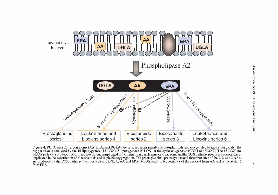
This pathway of PUFA action on nervecells starts with their release from the inter-nal sn-2 position of membrane phospholip-ids under the action of a hormone- or a cal-cium-dependent phospholipase A2 (PLA2).PUFA with 20 carbon atoms (AA, EPA, anddihomo-gamma-linolenic acid or DGLA,20:3n-6) are then converted in oxygenatedproducts, eicosanoids (Fig. 4). The reactionsare catalysed by 15-lipoxygenase (15-LOX),5-lipoxygenase (5-LOX) or cyclo-oxygen-ases (COX1 and COX2). The 15-LOX and5-LOX pathways lead to the formation oflipoxins and leukotrienes involved in immuneand inflammatory reactions, whereas COXpathways produce endoperoxides, whichare notably active in the constriction ofblood vessels and in platelet aggregation.Prostaglandins, prostacyclins and throm-boxanes of the 1, 2, and 3 series are pro-duced by the COX pathway from respec-tively DGLA, AA and EPA. The 5-LOXpathway leads to leucotrienes of the series 4from AA and of the series 5 from EPA.Since the fatty acid substrates are competingfor the same enzymes, the biosynthesis ofendoperoxides of the series 1 and 3 counter-acts that of the series 2, and the biosynthesisof leucotrienes of the series 5 counteractsthat of the series 4. The dietary equilibriumbetween the n-6 and n-3 fatty acids thusdetermines the balance in the synthesis ofactive mediators whose properties are gen-erally opposed. AA generates proagregantand vasoconstrictor (series 2 endoperox-ides) and proinflammatory (series 4 leucot-rienes) mediators, whereas the mediators
issued from EPA have antiagregating andvasodilating properties (endoperoxides of theseries 3), and non-inflammatory properties(series 5 leucotrienes).
8.1.2. Eicosanoids and the regulator role of astrocytes
The AA-derived eicosanoids probablyhave an action in the brain. By binding onastrocyte membrane receptors, the series 2prostaglandins, produced in response to aninflux of calcium ions, may be involved inthe regulation of the glutamate release in thesynaptic cleft (for a review see [99]). Thereleased glutamate modulates neuronal excit-ability and synaptic transmission at the pre-synaptic level. On the contrary, glutamateuptake by astrocytes (Fig. 5), avoids itspotentially neurotoxic accumulation in thesynaptic cleft. A defect in the astrocyticuptake of glutamate may be the cause ofneuronal damages acquired during trauma-tisms (ischemia, lesions) and could be associ-ated to several neurodegenerative pathologies.Moreover, the excessive release of AAcould alter glutamate transporters and con-tribute to the extracellular accumulation ofglutamate. The effects of oxidative stress onglutamate transport is possibly mediated bya product of PUFA peroxidation, 4-hydroxy-nonenal, which may interact with the GLT-1astrocyte transporter (EAAT2) and induceits inactivation [100]. Under the action ofdifferent stimuli, especially in response toproinflammatory cytokines [101], astrocytesin the synaptic cleft also release a sPLA2which releases AA from the sn-2 positionof membrane phospholipids. Compared toAA, EPA and DHA are less good substratesfor LOX and COX, and they modulate thenature and amounts of the oxygenated deriv-atives [102–105]. Increasing the dietaryintake of n-3 PUFA could thus slow downthe inflammatory cycle maintained by theaction of cytokines on astrocytes, i.e. thesecretion of sPLA2, a large release of AAand production of PGE2 in the extracellularspace, inducing in return the synthesis ofinflammatory interleukins by astrocytes [99].
Impact of dietary PU
FA on neuronal functions
523
Figure 4. PUFA with 20 carbon atoms (AA, EPA, and DGLA) are released from membrane phospholipids and oxygenated to give eicosanoids. Theoxygenation is catalysed by the 15-lipoxygenase (15-LOX), 5-lipoxygenase (5-LOX) or the cyclo-oxygenases (COX1 and COX2). The 15-LOX and5-LOX pathways produce lipoxins and leucotrienes implicated in the immune and inflammatory reactions, and the COX pathway produces endoperoxidesimplicated in the constriction of blood vessels and in platelet aggregation. The prostaglandins, prostacyclins and thromboxanes of the 1, 2, and 3 seriesare produced by the COX pathway from respectively DGLA, AA and EPA. 5-LOX leads to leucotrienes of the series 4 from AA and of the series 5from EPA.

524 J.M. Alessandri et al.
We recently showed that the concentra-tions in DHA and AA in the phospholipidmembranes of cultured astrocytes vary as afunction of the concentration of the fatty
acids in this medium, and notably that theconcentration of AA is inversely related tothe concentration of DHA [106]. In addi-tion, we observed that supplemental DHA
Figure 5. Implication of PUFA in the synaptic regulation by astrocytes: the release of glutamate(Glu) during neuronal activation induces a calcium influx in the postsynaptic neurons and an acti-vation of neuronal and astrocytic glutamate receptors. The activation of metabotropic receptors(mGluR) results in an increase of intracellular calcium concentration through the IP3 pathway [phos-pholipase C (PLC) activation, IP3 increase, calcium release of intracellular pools]. Calcium-media-ted activation of the cytoplasmic phospholipase A2 (cPLA2) produces the release of AA and DHAfrom membrane phospholipids. The cPLA2 may be directly activated through G-protein-linkedmGluR. A secretory PLA2 (sPLA2), may be released from the presynaptic terminal. Lipid signallingcan modulate neuronal function and survival in several ways : (1) Free AA regulates the extracellularconcentration of glutamate by inhibiting the glutamate uptake by the astrocyte transporter (EAAT),and by activating its release in a Ca2+-dependent mechanism via the stimulation of the astrocytereceptors for PGE2 (prostaglandins E2). (2) Free AA and DHA may enter the enzymatic oxygenationpathways, producing the respective eicosanoids and docosanoids which can also regulate the synap-tic transmission. (3) AA and PGE2 can regulate through the IP3–mediated propagation of Ca2+ wavesthroughout the glial cells through inhibition of gap junctions.

Impact of dietary PUFA on neuronal functions 525
has an effect on the morphology of astro-cytes from primary culture, which exhibit astellar shape different from that induced byAA (Denis et al., unpublished results). In thebrain, the reversible process of astrocyticstellation allows the emission and the retrac-tion of the astrocytic processes around thesynapses, thus regulating the neuronalenergy flux and the ion and neurotransmit-ter homeostasis in the synaptic cleft (reviewin [107]). The hypothesis can be made thatthe respective proportions of DHA and AAin membranes may have an impact on themorphological plasticity and on the differ-ent astrocyte functions involved in the reg-ulation of synaptic transmission.
8.1.3. New mediators, the docosanoids
The nerve membranes contain muchmore AA than EPA, which makes the localsynthesis of eicosanoids from cerebral EPAquite questionable. However, these mem-branes contain almost as much DHA as AA,or even more in the case of the retina, andbioactive docosanoids could be producedfrom DHA via the same enzymatic path-ways as those of PUFA made of 20 carbons.The biosynthesis in the brain of a new medi-ator formed from enzymatic oxygenation ofDHA was recently evidenced in a model ofcerebral ischemia and reperfusion in themouse [108]. The ischemic stroke results inthe release of lipoperoxides and cytokines,the increase of COX2 expression and theinfiltration of leukocytes. Among the liber-ated mediators, the authors isolated a newdi-hydroxylated compound issued from theoxygenation of DHA by 15-LOX, the10,17S-docosatriene, which once isolatedand perfused in another animal can stronglyinhibit the inflammatory responses inducedby the ischemic stroke. According to theauthors, this pathway of enzymatic oxygen-ation of endogenous DHA may contributeto protect the brain against injury producedby oxidising stress. Very recently, theyhave shown that the 10,17S-docosatrienemolecule, so called “neuroprotectin D1”(NPD1), is synthesised by human retinalpigment epithelial cells treated with a cal-
cium ionophore or with DHA [109]. NPD1added to retinal pigment epithelial cellspotently counteracted the apoptotic DNAdamage triggered by treatments with oxida-tive agents. NPD1 also up-regulated theexpression of antiapoptotic proteins (Bcl-2and Bcl-xL), while it decreased those ofproapoptotic proteins (Bax, Bad) and COX2[109]. Overall, NPD1 protected cells fromoxidative stress-induced apoptosis. The NPD1pathway will very probably lead to new per-spectives in research to understand the pro-tective effect of DHA-derived mediators inhypoxic-ischemic situations.
8.2. PUFA are components of membrane domains
8.2.1. Membrane lipids do not mix uniformly
Since the fluid mosaic model of Singerand Nicholson, the membrane concept hasevolved. The protein-protein interactions,within and around the membrane clearlydepend on the quality of their lipid micro-environment, and membrane domains (“lipidrafts”) have received increasing attention aspotential platforms for proteins in signal-ling and trafficking. The theory of lipid rafts(reviews in [110, 111]) conceptualises thatseparation of discrete liquid-ordered andliquid-disordered phase domains occurs inmembranes containing sufficient amountsof sphingolipids, sterols and saturated phos-phatidylcholines. These clusters of orderedlipids are characterised by their insolubility,at low temperature, in nonionic detergents.Then, hydrophobic proteins can be locallypackaged with these lipid clusters and beorganised as membrane rafts. The organisa-tion, and thus the possibilities for interactionsand activations of these lipid raft-interact-ing proteins, are very different from those ofproteins preferentially embedded in the fluidphase, the latter being composed of PUFA-rich phospholipids having a low degree oforder of their acyl chains and allowing amuch larger freedom of movement. Studieson artificial membranes have focussed on
526 J.M. Alessandri et al.
the possible link between DHA, the moreunsaturated among structural fatty acids(characterised by a low degree of order withinthe membrane phospholipids) and the capac-ity of rhodopsin to receive photons and totransduce them into a biochemical signal.
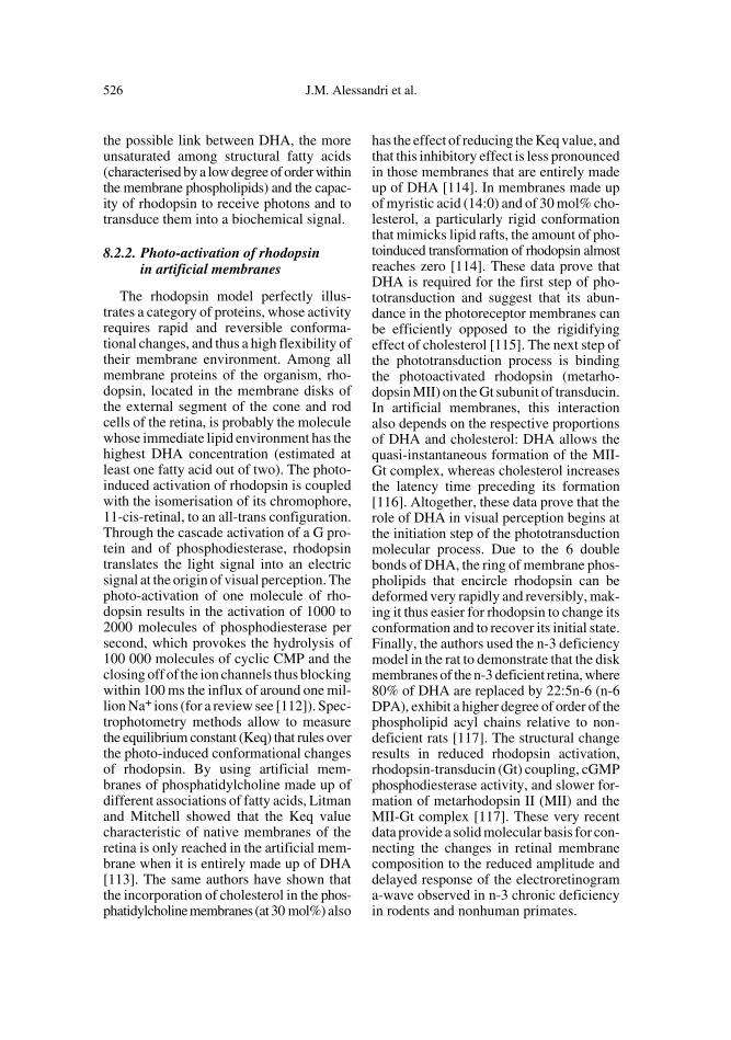
8.2.2. Photo-activation of rhodopsin in artificial membranes
The rhodopsin model perfectly illus-trates a category of proteins, whose activityrequires rapid and reversible conforma-tional changes, and thus a high flexibility oftheir membrane environment. Among allmembrane proteins of the organism, rho-dopsin, located in the membrane disks ofthe external segment of the cone and rodcells of the retina, is probably the moleculewhose immediate lipid environment has thehighest DHA concentration (estimated atleast one fatty acid out of two). The photo-induced activation of rhodopsin is coupledwith the isomerisation of its chromophore,11-cis-retinal, to an all-trans configuration.Through the cascade activation of a G pro-tein and of phosphodiesterase, rhodopsintranslates the light signal into an electricsignal at the origin of visual perception. Thephoto-activation of one molecule of rho-dopsin results in the activation of 1000 to2000 molecules of phosphodiesterase persecond, which provokes the hydrolysis of100 000 molecules of cyclic CMP and theclosing off of the ion channels thus blockingwithin 100 ms the influx of around one mil-lion Na+ ions (for a review see [112]). Spec-trophotometry methods allow to measurethe equilibrium constant (Keq) that rules overthe photo-induced conformational changesof rhodopsin. By using artificial mem-branes of phosphatidylcholine made up ofdifferent associations of fatty acids, Litmanand Mitchell showed that the Keq valuecharacteristic of native membranes of theretina is only reached in the artificial mem-brane when it is entirely made up of DHA[113]. The same authors have shown thatthe incorporation of cholesterol in the phos-phatidylcholine membranes (at 30 mol%) also
has the effect of reducing the Keq value, andthat this inhibitory effect is less pronouncedin those membranes that are entirely madeup of DHA [114]. In membranes made upof myristic acid (14:0) and of 30 mol% cho-lesterol, a particularly rigid conformationthat mimicks lipid rafts, the amount of pho-toinduced transformation of rhodopsin almostreaches zero [114]. These data prove thatDHA is required for the first step of pho-totransduction and suggest that its abun-dance in the photoreceptor membranes canbe efficiently opposed to the rigidifyingeffect of cholesterol [115]. The next step ofthe phototransduction process is bindingthe photoactivated rhodopsin (metarho-dopsin MII) on the Gt subunit of transducin.In artificial membranes, this interactionalso depends on the respective proportionsof DHA and cholesterol: DHA allows thequasi-instantaneous formation of the MII-Gt complex, whereas cholesterol increasesthe latency time preceding its formation[116]. Altogether, these data prove that therole of DHA in visual perception begins atthe initiation step of the phototransductionmolecular process. Due to the 6 doublebonds of DHA, the ring of membrane phos-pholipids that encircle rhodopsin can bedeformed very rapidly and reversibly, mak-ing it thus easier for rhodopsin to change itsconformation and to recover its initial state.Finally, the authors used the n-3 deficiencymodel in the rat to demonstrate that the diskmembranes of the n-3 deficient retina, where80% of DHA are replaced by 22:5n-6 (n-6DPA), exhibit a higher degree of order of thephospholipid acyl chains relative to non-deficient rats [117]. The structural changeresults in reduced rhodopsin activation,rhodopsin-transducin (Gt) coupling, cGMPphosphodiesterase activity, and slower for-mation of metarhodopsin II (MII) and theMII-Gt complex [117]. These very recentdata provide a solid molecular basis for con-necting the changes in retinal membranecomposition to the reduced amplitude anddelayed response of the electroretinograma-wave observed in n-3 chronic deficiencyin rodents and nonhuman primates.

Impact of dietary PUFA on neuronal functions 527
8.3. Impact of n-3 PUFA deficiency on vesicular neurotransmission
The synaptic release of neurotransmit-ters constitutes another membrane processdirectly implicated in the functioning of theCNS. The process initially concerns thematuration of vesicles carrying neurotrans-mitters and their storage in the active zoneof the presynaptic terminals. The arrival ofthe action potential causes an influx of cal-cium that induces the fusion of vesicularand plasma membranes, releasing via exo-cytosis the neurotransmitters in the synap-tic cleft. An opposite process allows torecruit membrane proteins and to regener-ate by endocytosis small empty vesicles fora new cycle of maturation and presynapticstorage. The neurotransmitters activate thepost-synaptic receptors that translate theentry of calcium ions, permitting the prop-agation of the action potential within theneuronal network. All functions of the nerv-ous system, from sensorial perception tolearning and memory, are governed by thesynaptic coupling between exocytosis andendocytosis. The understanding of the com-plex protein machinery implicated in thevesicular processes has considerably evolvedover the last 10 years (review in [118]). Thespecific role of membrane PUFA in thevesicular processes has been closely stud-ied over the last few years. The use of achronically n-3 fatty acid deficient ratmodel has showed the role of n-3 PUFA inthe storage and presynaptic releasing proc-esses of several neurotransmitters. The mono-aminergic and cholinergic systems havebeen more specifically studied, due to theirimplication both in the regulation of impor-tant physiological functions and in the controlof cognitive processes including attention,motivation and memory. These studies haveshown in n-3 deficient rats, an increase inspontaneous release of dopamine in thenucleus accumbens [119, 120], acetylcholine[121] or serotonin [122] in the hippocam-pus (Fig. 6). In contrast, the drug-inducedrelease of these neurotransmitters is signif-icantly reduced [120, 122, 123]. This phe-
nomenon could result from an increasedbasal leakage of the neurotransmitter in thesynaptic cleft that would reduce its storagein the vesicles (Fig. 7). During a nervousstimulation, generated for example in alearning situation, the amplitude of neuro-transmitter release would then be reducedleading to a lower efficiency of the nervousinflux. Concerning the study of the dopamin-ergic system, it was shown that the n-3 defi-ciency decreases the number of dopaminergicvesicles in the frontal cortex. These resultssuggest that n-3 deficiency leads to a hypo-functioning of the mesocortical dopaminergicsystem, and a model for the general dys-functioning of the dopaminergic mesocortico-limbic loop has been proposed to explainthe behavioural perturbations observed indeficient animals [124]. The membrane defi-cit in DHA can be one possible explanationof the effects of n-3 deficiency on neuro-transmitter storage and release. Besides,modifications caused by n-3 PUFA defi-ciency in the neurotransmission processcould also partly result from alterations inbrain energy metabolism, such as glucoseutilisation and oxidative phosphorylation[125]. It is therefore assumed that multipleinteractions exist among membrane PUFAand vesicular neurotransmission processes,with significant incidences on learning capac-ity and behaviour.
8.4. PUFA and their metabolites regulate gene transcription
8.4.1. Fatty acids are natural ligands of nuclear receptors
In 1990, Issemann and Green discoveredthat molecules used for the treatment ofhyperlipidemia act by binding on transcrip-tion factors (PPARs, peroxisome prolifera-tors-activated receptors) that belong to thesuperfamily of steroid hormone receptors[126]. Since it has been shown that fattyacids are natural endogenous activators ofPPARs [127], the concept of regulation ofbiological functions by PUFA and their

528 J.M. Alessandri et al.
oxygenated derivatives has progressed con-siderably. Binding to PPARs allows fattyacids to partly control their metabolic fateby directly inducing the transcription ofgenes coding for proteins involved in mito-chondrial and peroxisomal β-oxidation [128].PPARs interact with responsive elementsof the genome made up of two AGGTCAhexamers separated by one base (DR1repeat) [129]. The genes that possess thistype of sequence upstream of their pro-moter, or a similar sequence, are suscepti-ble to be activated by the transcriptionalheterodimer formed by the association of aPPARs with RXR (retinoid receptor) [130].The PPARs are themselves coded by 3 dif-ferent genes, differently expressed accordingto cell type, and translated into 3 isoforms:PPARα, PPARγ and PPARβ/δ. RXR alsodecomposes into 3 isotypes (α, β and γ).PPAR-RXR heterodimers can also repressthe transcription of genes normally activatedby other transcription factors by occupyingin an inoperative way the target sequencecorresponding to their promoters.
A large number of combinations is pos-sible between the different isotypes of PPARsand RXR (including their respective lig-ands) that may induce varying effects on thetarget gene transcription level, whether they
are directly or indirectly involved in lipidmetabolism. The preferential distribution ofthe PPARs and RXR isotypes in the differ-ent tissues and cell types contributes to func-tional specificities: catabolism of lipids asso-ciated with a predominance of PPARα inthe liver, brown adipose tissue and skeletalmuscles; differentiation and lipogenesis inthe adipose tissue, and uptake of oxidisedLDL in macrophages, both preferentiallyexpressing PPARγ. The tissue distributionof PPARδ is ubiquitous but this isotype isgenerally more abundant in most of thedifferent cerebral regions [131], the diges-tive tract, kidneys, heart, diaphragm andoesophagus [132]. In the mouse skeletalmuscle, PPARδ has been shown to controlfatty acid oxidation by regulating genesinvolved in fatty acid transport, β-oxidation,and mitochondrial respiration [133, 134].
8.4.2. PUFA as possible regulators of gene transcription in the CNS
It is known that transcription of thegenes encoding the first two enzymes of theperoxisomal-oxidation pathway, acylCoA-oxidase (AOX) and L-peroxisomal bifunc-tional enzyme (L-PBE), is mediated throughthe RXR-PPAR heterodimer which binds
Figure 6. A schematic representation of functional interactions between dopaminergic, serotoni-nergic and cholinergic neurotransmission systems in the control of Ach release in the hippocampus.In n-3 deficiency, the increase of dopamine synthesis in the ventral tegmental area and the alterationsof serotonin release in the hippocampus could participate in the higher spontaneous Ach release (from[120–122]).

Impact of dietary PU
FA on neuronal functions
529
Figure 7. A schematic representation of acetylcholine (Ach) release in the synaptic cleft in resting (spontaneous release) and neuronal activation (sti-mulated release) conditions, in control (A – B) and n-3 PUFA deficient rats (C – D). In the resting state, a small amount of Ach is released in theextracellular space in control rats (A), whereas this spontaneous release is enhanced in n-3 PUFA deficient rats (C). In stimulated conditions (KClinfusion), cytoplasmic Ach is stored in vesicles through the vesicular Ach transporter (VAVhT). Full vesicles can be recruited for exocytosis and highlevels of Ach are released (B). In deficient rats (D), the Ach leakage that occurs in resting conditions leads to storage depletion and then inducesreduced Ach release under stimulation (D).

530 J.M. Alessandri et al.
to the corresponding peroxisome prolifera-tor-responsive elements (PPRE) [130] (AOXinitiates the peroxysomal oxidation of thevery long-chain fatty acids, allowing theshortening of neurotoxic compounds andthe synthesis of DHA from its upstream pre-cursors). However, the specific role ofPPARs in the different regions of the CNS,in a possible relation with the regulation ofthe fatty acid metabolism, is an unresolvedquestion. We have recently made the hypoth-esis that in retinoblastoma cells, the expres-sion of AOX and the terminal step of DHAsynthesis could be regulated by the upstreamprecursors of DHA through the modulationof PPARδ-mRNA abundance (PPARδ isthe major isotype in these cells) [135]. Incultured oligodendrocytes, the chemically-induced activation of PPARδ favoured thecell morphological differentiation and inducedthe synthesis of the myelin basic protein,suggesting that PPARδ could play a role innervous conduction by regulating the for-mation and the maintenance of myelin [136].Although PUFAs, or some of their metab-olites, are likely to exert the role of PPARsligands in the CNS, the endogenous ligandsof the brain PPARs, especially of the δ-iso-type, have not been clearly identified.
8.4.3. The pivotal role of RXR
Actually, it appears that RXR, the obliga-tory transcriptional partner of PPARs [130],constitutes a very plausible potential targetfor DHA. This ubiquitous factor regulatesthe transcription of a large number of genesimplicated in the pathways of proliferation,differentiation, and apoptosis in severaltypes of neural cells [137]. RXRα also reg-ulates the transcription of most of the genesimplicated in lipid metabolism through itsinteraction with other transcription factors,such as the constitutive androstane recep-tor, CARβ [138], the retinoic acid receptor,RARα [139] and the liver X receptors, LXR(also expressed in the rodent brain) [140].The multiple roles of RXRα, a transcriptionfactor positioned at the cross-road of genesencoding for miscellaneous receptors, trans-
porters and enzymes of the lipid metabolism,are schematically illustrated in Figure 8.
Finally, the binding of DHA on the lig-and binding domain of RXRα has been evi-denced in the mouse brain [141], and tworecent studies using transfected cells dem-onstrated that DHA potently activates thetranscription of a reporter gene mediated byRXRα [142, 143]. These novel data supportthe concept that DHA, and possibly otherPUFA, could activate one or several RXRsignalling pathways in the CNS. Therefore,the putative effects of DHA on gene tran-scription in the brain, possibly recruitingdifferent RXR heterodimers (Fig. 8), andtheir possible influence on neural function,opens up a large field of investigations.
8.4.4. PUFA and transcriptional regulations in the CNS: the contribution of microarrays
Berger et al. studied the effect of dietarylong-chain PUFA on the expression of 329genes in the liver and 356 genes in the hip-pocampus of mice [144]. One-month-oldmice received during 57 days, diets rich inlong-chain PUFA (supplementation withfish oil rich in EPA and DHA or a fungaloil rich in AA, or a combination of both).The gene expression profile of these tissueswas compared to that of a control diet con-taining only linoleic and α-LNA as PUFA.In the hippocampus, the long-chain n-6 andn-3 PUFA supplementation affected a cat-egory of genes controlling the transthyretinsignalling pathway (a transporter of thy-roxin in the cerebrospinal liquid), the liber-ation of serotonin, the functions relative toimmunity (immunoglobulins) and the acti-vation of transcriptional factors implicatedin inflammation (NFκB). By releasing theirfatty acids, certain molecular species of eth-anolamine-phosphoglycerides could be atthe origin of transcriptional effects specifi-cally induced by n-3 PUFA at the brainlevel [145–147]. These studies showed thatamong a panel of 3 200 genes screened inthe rat brain, the supplementation in n-3

Impact of dietary PUFA on neuronal functions 531
long chain PUFA increased the expressionof 55 genes and decreased that of 47 genes.The genes affected are involved in synapticplasticity, signal transduction, ion channelassembly, energetic metabolism and pro-tein regulation. Applying microarrays to2 400 genes expressed in human retinalexplants in culture, Rojas et al recently
found that the transcription of 14% of themwas significantly increased when the explantswere treated with 27 µM DHA, whereasonly 0.4% increased upon treatment witholeic acid [148]. Among the genes whosetranscription of which was induced by DHAare those playing roles in neurogenesis, neu-rotransmission and intercellular connections.
Figure 8. A schematic representation for the multiple gene transcriptions which are potentially acti-vated by the dimer formed by the ligand-induced associations of RXR with one nuclear receptor.The liganded homo- or hetero-dimer binds to a specific DNA sequence, known as a responsive ele-ment (RE), i.e. RARE, LXRE, RXRE, PPRE and PBREM, located in the promoter region of the targetgene. For simplification, tissue specificity is not considered, corepressors and coactivators of trans-cription are not represented, and only some examples of the target genes involved in the lipid meta-bolism are mentioned. By disrupting the interactions between RXR and other nuclear receptors, theligand-activated PPARs can potentially interfere with the RXR-dependent transcription of multiplegenes, notably those coding for lipogenic transcription factors. Fatty acids and some eicosanoidshave been identified as natural ligands of PPARs in muscle, hepatic and adipose tissues. Accordingto recent studies, DHA could be an endogenous ligand for RXR in different tissues, including thebrain.

532 J.M. Alessandri et al.
To-date, the pathways of transcriptional acti-vation induced by DHA have not yet beenidentified.
9. CONCLUSION
DHA, which is massively incorporatedinto the nerve cell membranes, exerts struc-tural and neuroprotective roles that arefavourable to the development and mainte-nance of cerebral and visual performances.EPA, although not stored in the brain cellmembranes, can ameliorate diverse psychi-atric disorders and probably has roles in brainfunction, possibly by counteracting the AA-mediated signalling. The mechanisms impli-cated are complex and multiple, reflectingthe extraordinary diversity of the functionsexercised by the PUFA, going from themodulation of dynamic properties of themembranes to the production of active medi-ators and the regulation of the expression ofgenes. The lipid nutrition of the brain, sus-ceptible to influence each of the pathwaysat each step of life, is thus an essential elementof its functioning.
ACKNOWLEDGEMENTS
We would like to thank Wendy Brand-Williams, INRA (France), for the translation ofthis paper.
REFERENCES
[1] Sprecher H. An update on the pathways ofpolyunsaturated fatty acid metabolism. CurrOpin Clin Nutr Metab Care 1999, 2: 135–138.
[2] Galli D, Agradi E, Paoletti R. The (n-6) pen-taene:(n-3) hexaene fatty acid ratio as anindex of linolenic acid deficiency. BiochimBiophys Acta 1974, 369: 142–145.
[3] Wheeler TG, Benolken RM, Anderson RE.Visual membranes: specificity of fatty acidprecursors for the electrical response to illu-mination. Science 1975, 188: 1312–1314.
[4] Bourre JM, François M, Youyou A, DumontO, Piciotti M, Pascal G, Durand G. Theeffects of dietary α-linolenic acid on the com-position of nerve membranes, enzymaticactivity, amplitude of electrophysiologicalparameters, resistance to poisons and per-
formance of learning tasks in rat. J Nutr 1989,119: 1880–1892.
[5] Neuringer M, Connor WE, Van Petten C,Barstad L. Dietary ω-3 fatty acid deficiencyand visual loss in infant Rhesus monkeys. JClin Invest 1984, 73: 272–276.
[6] Neuringer M, Connor W, Lin DS, Barstad L,Luck S. Biochemical and functional effectsof prenatal and postnatal n-3 fatty acid defi-ciency on retina and brain in Rhesus mon-keys. Proc Natl Acad Sci USA 1986, 83:4021–4025.
[7] Moriguchi T, Sheaff-Greiner R, Salem N Jr.Behavioral deficits associated with dietaryinduction of decreased brain docosahexae-noic acid concentration. J Neurochem 2000,75: 2563–2573.
[8] Jeffrey BG, Mitchell DC, Gibson RA, NeuringerM. n-3 Fatty acid deficiency alters recoveryof the rod photoresponse in Rhesus monkeys.Invest Ophthalmol Vis Sci 2002, 43: 2806–2814.
[9] Connor WE, Neuringer M. The effects of n-3 fatty acid deficiency and repletion upon thefatty acid composition and function of thebrain and retina. Prog Clin Biol Res 1988,282: 275–294.
[10] Diau GY, Loew ER, Wijendran V, Sarkadi-Nagy E, Nathanielsz PW, Brenna JT.Docosahexaenoic and arachidonic acid influ-ence on preterm baboon retinal compositionand function. Invest Ophthalmol Vis Sci 2003,44: 4559–4566.
[11] Uauy RD, Birch DG, Birch EE, Tyson JE,Hoffman DR. Effect of dietary ω-3 fatty acidson retinal function of very-low-birth-weightneonates. Pediatr Res 1990, 28: 485–492.
[12] Birch D, Birch E, Hoffman D, Uauy R. Ret-inal development in very-low-birth-weightinfants fed diets differing in ω-3 fatty acids.Invest Ophthalmol Vis Sci 1992, 33: 2365–2376.
[13] Birch E, Birch D, Hoffman D, Hale L, EverettM, Uauy R. Breast-feeding and optimal vis-ual development. J Pediatr Ophthalmol Stra-bismus 1993, 30: 33–38.
[14] Lauritzen L, Hansen HS, Jùrgensen MH,Michaelsen KF. The essentiality of longchain n-3 fatty acids in relation to develop-ment and function of the brain and retina.Prog Lipid Res 2001, 4: 1–94.
[15] Makrides M, Neumann MA, Byard RW,Simmer K, Gibson RA. Fatty acid composi-tion of brain, retina, and erythrocytes inbreast- and formula-fed infants. Am J ClinNutr 1994, 60: 189–194.
[16] Birch EE, Garfield S, Hoffman DR, Uauy R,Birch DG. A randomized controlled trial ofearly dietary supply of long-chain polyunsat-urated fatty acids and mental development in

Impact of dietary PUFA on neuronal functions 533
term infants. Dev Med Child Neurol 2000,42: 174–181.
[17] Hoffman DR, Birch EE, Castaneda YS,Fawcett SL, Wheaton DH, Birch DG, UauyR. Visual function in breast-fed term infantsweaned to formula with or without long-chain polyunsaturates at 4 to 6 months: a ran-domized clinical trial. J Pediatr 2003, 142:669–677.
[18] Connor W, Neuringer M, Reisbick S. Essen-tial fatty acids: the importance of n-3 fattyacids in the retina and brain. Nutr Rev 1992,50: 21–29.
[19] Salem N Jr, Wegher B, Mena P, Uauy R. Ara-chidonic and docosahexaenoic acids are bio-synthesized from their 18-carbon precursorsin human infants. Proc Natl Acad Sci USA,1996, 93: 49–54.
[20] Uauy R, Mena P, Wegher B, Nieto S, SalemN Jr. Long chain polyunsaturated fatty acidformation in neonates: effect of gestationalage and intrauterine growth. Pediatr Res2000, 47: 127–135.
[21] Pawlosky RJ, Hibbeln JR, Novotny JA,Salem N Jr. Physiological compartmentalanalysis of α-linolenic acid metabolism inadult humans. J Lipid Res 2001, 42: 1257–1265.
[22] Maurage C, Guesnet P, Pinault M, Rochettede Lempdes J, Durand G, Antoine J, Couet C.Effect of two types of fish oil supplementa-tion on plasma and erythrocyte phospholip-ids in formula-fed term infants. Biol Neonate1998, 74: 416–429.
[23] Cunnane SC, Francescutti V, Brenna JT,Crawford MA. Breast-fed infants achieve ahigher rate of brain and whole body docosa-hexaenoate accumulation than formula-fedinfants not consuming dietary docosahex-aenoate. Lipids 2000, 35: 105–111.
[24] Gibson RA, Neumann MA, Makrides M.Effect of increasing breast milk docosahex-aenoic acid on plasma and erythrocyte phos-pholipid fatty acids and neural indices ofexclusively breast fed infants. Eur J Clin Nutr1997, 51: 578–584.
[25] Guesnet P, Antoine J-M, Rochette de LempdesJ-B, Galent A, Durand G. Polyunsaturatedfatty acid composition of human milk inFrance: changes during the course of lactationand regional differences. Eur J Clin Nutr1993, 47: 700–710.
[26] Alessandri JM, Goustard B, Guesnet P,Durand G. Polyunsaturated fatty acids statusin blood, heart, liver, intestine, retina andbrain of newborn piglets fed either sow milkor a milk replacer diet. Reprod Nutr Dev1996, 36: 95–109.
[27] Goustard-Langelier B, Guesnet P, Durand G,Antoine JM, Alessandri JM. n-3 and n-6 fattyacid enrichment by dietary fish oil and phos-pholipid sources in brain cortical areas andnonneural tissues of formula-fed piglets. Lip-ids 1999, 34: 5–16.
[28] Alessandri JM, Goustard B, Guesnet P, DurandG. Docosahexaenoic acid concentrations inretinal phospholipids of piglets fed an infantformula enriched with long-chain polyunsat-urated fatty acids: effects of egg phospholip-ids and fish oils with different ratios of eicos-apentaenoic acid to docosahexaenoic acid.Am J Clin Nutr 1998, 67: 377–385.
[29] Alessandri JM, Poumès-Ballihaut C, LangelierB, Perruchot MH, Raguénez G, Lavialle M,Guesnet P. Incorporation of docosahexaenoicacid into nerve membrane phospholipids:bridging the gap between animals and culturedcells. Am J Clin Nutr 2003, 78: 702–710.
[30] Legrand P, Bourre JM, Descomps B, DurandG, Renaud S. Lipides. In: Martin A (Ed),Apports nutritionnels conseillés pour la popu-lation française, AFSSA 3e ed, Tec & Doc,2001, p 63–82.
[31] Beaufrère B, Briend A, Ghisolfi J, Goulet O,Putet G, Rieu D, Turck D, Vidailhet M. Nour-rissons, enfants et adolescents. In: Martin A(Ed), Apports nutritionnels conseillés pour lapopulation française, AFSSA 3e ed, Tec &Doc, 2001, p 255–291.
[32] Galli C. Commentary on the workshop state-ment. Essentiality of and recommended dietaryintake of Omega-6 and Omega-3 fatty acids.Prostaglandins Leukot Essent Fat Acids2000, 63: 127–129.
[33] Simopoulos AP, Leaf A, Salem N Jr. Work-shop statement on the essentiality of and rec-ommended dietary intakes for Omega-6 andOmega-3 fatty acids. Prostaglandins LeukotEssent Fat Acids 2000, 63: 119–121.
[34] Innis SM. Human milk and formula fattyacids. J Pediatr 1992, 120: S56-S61.
[35] Guesnet P, Couet C, Alessandri JM, AntoineJM, Durand G. Variabilité de la teneur enacide linoléique (18:2n-6) et du rapport18:2n-6/18:3n-3 des lipides dans le lait defemme en France. Ann Pédiatr (Paris) 1995,42: 282–288.
[36] Chulei R, Xiaofang L, Hongsheng M, XiulanM, Guizheng L, Gianhong D, DeFrancescoCA, Connor WE. Milk composition inwomen from five different regions of China:the great diversity of milk fatty acids. J Nutr1995, 125: 2993–299.
[37] Combes N, Boué C. Apports alimentaires enacides linoléique et α-linolénique d’une pop-ulation aquitaine. OCL 2001, 8: 118–121.
534 J.M. Alessandri et al.
[38] Djoussé L, Hunt SC, Arnett DK, ProvinceMA, Eckfeldt JH, Ellison RC. Dietary lino-lenic acid is inversely associated with plasmatriacylglycerol: the National Heart, Lung,and Blood Institute Family Heart Study. AmJ Clin Nutr 2003, 78: 1098–1102.
[39] Meyer BJ, Mann NJ, Lewis JL, Milligan GC,Sinclair AJ, Howe PR. Dietary intakes andfood sources of omega-6 and omega-3 poly-unsaturated fatty acids. Lipids 2003, 38: 391–398.
[40] Voskuil DW, Feskens EJM, Katan MB,Kromhout D. Intake and sources of α-lino-lenic acid in Dutch elderly men. Eur J ClinNutr 1996, 50: 784–787.
[41] Johansson LRK, Solvoll K, Bjornboe GEA,Drevon CA. Intake of very long-chain n-3fatty acids related to social status and life-style. Eur J Clin Nutr 1998, 52: 716–721.
[42] Astorg P, Arnault N, Czernichow S, NoisetteN, Galan P, Hercberg S. Dietary intakes andfood sources of n-6 and n-3 polyunsaturatedfatty acids in French adult men and women.Lipids (in press).
[43] Welch AA, Lund E, Amiano P, DorronsoroM, Brustad M, Kumle M et al. Variability offish consumption within the 10 Europeancountries participating in the EuropeanInvestigation into Cancer and Nutrition(EPIC) study. Public Health Nutr 2002, 5:1273–1285.
[44] Hulshof KFAM, van Erp-Baart MA,Anttolainen M, Becker W, Church SM, CouetC, Hermann-Kunz E, Kesteloot H, Leth T,Martins I. Intake of fatty acids in WesternEurope with emphasis on trans fatty acids:the TRANSFAIR study. Eur J Clin Nutr1999, 53: 143–157.
[45] Adams PB, Lawson S, Sanigorski A, SinclairAJ. Arachidonic acid to eicosapentaenoicacid ratio in blood correlates positively withclinical symptoms of depression. Lipids1996, 31 (Suppl): S157–S161.
[46] Maes M, Smith R, Christophe A, Cosyns P,Desnyder R, Meltzer H. Fatty acid composi-tion in major depression: decreased ω3 frac-tions in cholesteryl esters and increasedC20:4ω6/C20:5ω3 ratio in cholesteryl estersand phospholipids. J Affect Disord 1996, 38:35–46.
[47] Peet M, Murphy B, Shay J, Horrobin D.Depletion of omega-3 fatty acid levels in redblood cell membranes of depressive patients.Biol Psychiatry 1998, 43: 315–319.
[48] Edwards R, Peet M, Shay J, Horrobin D.Omega-3 polyunsaturated fatty acid levels inthe diet and in red blood cell membranes ofdepressed patients. J Affect Disord 1998, 48:149–155.
[49] Maes M, Christophe A, Delanghe J, AltamuraC, Neels H, Meltzer HY. Lowered ω3 poly-unsaturated fatty acids in serum phospholipidsand cholesteryl esters of depressed patients.Psychiatry Res 1999, 85: 275–291.
[50] De Vriese SR, Christophe AB, Maes M. Low-ered serum n-3 polyunsaturated fatty acid(PUFA) levels predict the occurrence of post-partum depression: further evidence that low-ered n-3PUFAs are related to major depression.Life Sci 2003, 73: 3181–3187.
[51] Ranjekar PK, Hinge A, Hegde MV, Ghate M,Kale A, Sitasawad S, Wagh UV, DebsikdarVB, Mahadik SP. Decreased antioxidantenzymes and membrane essential polyunsat-urated fatty acids in schizophrenic and bipo-lar mood disorder patients. Psychiatry Res2003, 121: 109–122.
[52] Chiu CC, Huang SY, Su KP, Lu ML, HuangMC, Chen CC, Shen WW. Polyunsaturatedfatty acid deficit in patients with bipolarmania. Eur Neuropsychopharmacol 2003,13: 99–103.
[53] Glen AIM, Glen EMT, Horrobin DF, VaddadiKS, Spellman M, Morsefisher N, Ellis K,Shinner FS. A red cell membrane abnormal-ity in a subgroup of schizophrenic patients:evidence for two diseases. Schizophr Res1994, 12: 53–61.
[54] Peet M, Laugharne J, Rangarajan N, HorrobinD, Reynolds G. Depleted red cell membraneessential fatty acids in drug-treated schizo-phrenic patients. J Psychiatr Res 1995, 29:227–232.
[55] Fenton WS, Hibbeln J, Knable M. Essentialfatty acids, lipid membrane abnormalities,and the diagnosis and treatment of schizo-phrenia. Biol Psychiatry 2000, 47: 8–21.
[56] Khan MM, Evans DR, Visayasudha G, SchefferRE, Parikh VV, Mahadik SP. Reduced eryth-rocyte membrane essential fatty acids andincreased lipid peroxides in schizophrenia atthe never-medicated first episode of psycho-sis and after years of treatment with antipsy-chotics. Schizophr Res 2002, 58: 1–10.
[57] Arvindakshan M, Sitasawads S, DebsikdarV, Ghate M, Evans D, Horrobin DF, BennettC, Ranjekar PK, Mahadik SP. Essential poly-unsaturated fatty acid and lipid peroxide lev-els in never-medicated and medicated schiz-oprenia patients. Biol Psychiatry 2003, 53:56–64.
[58] Burgess JR, Stevens L, Zhang W, Peck L.Long-chain polyunsaturated fatty acids inchildren with attention-deficit hyperactivitydisorder. Am J Clin Nutr 2000, 71: 327S–330S.
[59] Vancassel S, Durand G, Bartélémy C, LejeuneB, Martineau J, Guilloteau D, Andrès C,Chalon S. Plasma fatty acid levels in autistic
Impact of dietary PUFA on neuronal functions 535
children. Prostaglandins Leukot Essent FatAcids 2001, 65: 1–7.
[60] Hibbeln JR. Fish consumption and majordepression. Lancet 1998, 351: 1213.
[61] Hibbeln JR. Seafood consumption, the DHAcontent of mothers’ milk and prevalence ratesof postpartum depression: a cross-national,ecological analysis. J Affect Disord 2002, 69:15–29.
[62] Noaghiul S, Hibbeln JR. Cross-national com-parisons of seafood consumption and rates ofbipolar disorders. Am J Psychiatry 2003,160: 2222–2227.
[63] Hirayama T. Life-style and mortality: a largecensus-based cohort study in Japan. EdKarger, Basel, 1990.
[64] Huan M, Hamazaki K, Sun Y, Itomura M, LiuH, Kang W, Watanabe S, Terasawa K,Hamazaki T. Suicide attempt and n-3 fatty acidlevels in red blood cells: a case-control studyin China. Biol Psychiatry 2004, 56: 490–496.
[65] Tanskanen A, Hibbeln JR, Hintikka J,Haatainen K, Honkalampi K, Viinamaki H.Fish consumption, depression, and suicidal-ity in a general population. Arch Gen Psychi-atry 2001, 58: 512–513.
[66] Tanskanen A, Hibbeln JR, Tuomilehto J,Uutela A, Haukkala A, Viinamaki H, LehtonenJ, Vartiainen E. Fish consumption and depres-sive symptoms in the general population inFinland. Psychiatr Serv 2001, 52: 529–531.
[67] Silvers KM, Scott KM. Fish consumptionand self-reported physical and mental healthstatus. Public Health Nutr 2002, 5: 427–431.
[68] Timonen M. Horrobin D, Jokelainen J, LaitinenJ, Herva A, Räsänen P. Fish consumption anddepression: the Northern Finland 1966 birthcohort study. J Affect Disord 2004, 82: 447–452.
[69] Suzuki S, Akechi T, Kobayashi M, TaniguchiK, Goto K, Sasaki S, Tsugane S, NishiwakiY, Miyaoka H, Uchitomi Y. Daily omega-3fatty acid intake and depression in Japanesepatients with newly diagnosed lung cancer.Br J Cancer 2004, 90: 787–793.
[70] Mamalakis G, Tornaritis M, Kafatos A.Depression and adipose essential polyunsat-urated fatty acids. Prostaglandins LeukotEssent Fat Acids 2002, 6: 311–318.
[71] Mamalakis G, Kiriakakis M, Tsibinos G,Kafatos A. Depression and adipose polyun-saturated fatty acids in the survivors of theseven countries study population of Crete.Prostaglandins Leukot Essent Fat Acids2004, 70: 495–501.
[72] Tiemeyer H, van Thuil HR, Hofman A, KiliaanAJ, Breteler MMB. Plasma fatty acid com-position and depression are associated in theelderly: the Rotterdam Study. Am J Clin Nutr2003, 78: 40–46.
[73] Hakkarainen R, Partonen T, Haukka J, VirtamoJ, Albanes D, Lonnqvist J. Is low dietaryintake of omega-3 fatty acids associated withdepression? Am J Psychiatry 2004, 161:567–569.
[74] Nemets B, Stahl Z, Belmaker RH. Additionof omega-3 fatty acid to maintenance medi-cation treatment for recurrent unipolardepressive disorder. Am J Psychiatry 2002,159: 477–479.
[75] Peet M, Horrobin DF. A dose-ranging studyof the effects of ethyl-eicosapentaenoatein patients with ongoing depression despiteapparently adequate treatment with standarddrugs. Arch Gen Psychiatry 2002, 59: 913–919.
[76] Post RM, Leverich GS, Altshuler LL, FryeMA, Suppes TM, Keck PE Jr, McElroy SL,Kupka R, Nolen WA, Grunze H, Walden J.An overview of recent findings of the StanleyFoundation Bipolar Network (Part I). BipolarDisord 2003, 5: 310–319.
[77] Stoll AL, Severus WE, Freeman MP, RueterS, Zboyan HA, Diamond E, Cress KK,Marangell LB. Omega 3 fatty acids in bipolardisorder: a preliminary double-blind, pla-cebo-controlled trial, Arch Gen Psychiatry1999, 56: 407–412.
[78] Su KP, Huang SY, Chiu CC, Shen WW.Omega-3 fatty acids in major depressive dis-order: a preliminary double-blind, placebo-controlled trial. Eur Neuropsychopharmacol2003, 13: 267–271.
[79] Zanarini MC, Frankenburg MD. Omega-3fatty acid treatment of women with border-line personality disorder: a double-blind, pla-cebo-controlled pilot study. Am J Psychiatry2003, 160: 167–169.
[80] Marangell LB, Martinez JM, Zboyan HA,Kertz B, Kim HF, Puryear LJ. A double-blind,placebo-controlled study of the omega-3fatty acid docosahexaenoic acid in the treat-ment of major depression. Am J Psychiatry2003, 160: 996–998.
[81] Llorente AM, Jensen CL, Voigt RG, FraleyJK, Berretta MC, Heird WC. Effect of mater-nal docosahexaenoic acid supplementationon postpartum depression and informationprocessing. Am. J Obstet Gynecol 2003, 188:1348–1353.
[82] Peet M, Brind J, Ramchand CN, Shah S,Vankar GK. Two double-blind placebo-con-trolled pilot studies of eicosapentaenoic acidin the treatment of schizophrenia. SchizophrRes 2001, 49: 243–251.
[83] Peet M, Horrobin DF, in association with theE-E Multicentre Study Group. A dose-rang-ing exploratory study of the effects of ethyl-eicosapentaenoate in patients with persistentschizophrenic symptoms. J Psychiatr Res2002, 36: 7–18.
536 J.M. Alessandri et al.
[84] Emsley RA, Myburgh CC, Oosthuizen PP,Van Rensburgh SJ. Randomised placebo-controlled study of ethyl-eicosapentaenoicacid as supplemental treatment in schizo-phrenia. Am J Psychiatry 2002, 159: 1596–1598.
[85] Richardson AJ, Puri BK. A randomized dou-ble-blind, placebo-controlled study of theeffects of supplementation with highly unsatu-rated fatty acids on ADHD-related symptomsin children with specific learning difficulties.Prog Neuropsychopharmacol Biol Psychiatr2002, 26: 233–239.
[86] Ayton AK, Azaz A, Horrobin DF. A pilotopen case series of ethyl-EPA supplementa-tion in the treatment of anorexia nervosa.Prostaglandins Leukot Essent Fat Acids 2004,71: 205–209.
[87] Horrobin DF, Jenkins K, Bennett CN, ChristieWW. Eicosapentaenoic acid and arachidonicacid: collaboration and not antagonism is thekey to biological understanding. ProstaglandinsLeukot Essent Fat Acids 2002, 66: 83–89.
[88] Conquer JA, Tierney MC, Zecevic J, BettgerWJ, Fisher RH. Fatty acid analysis of bloodplasma of patients with Alzheimer's disease,other types of dementia, and cognitiveimpairment. Lipids 2000, 35: 1305–1312.
[89] Tully AM, Roche HM, Fallon C, Bruce I,Lawlor B, Coakley D, Gibney MJ. Lowserum cholesteryl ester-docosahexaenoicacid levels in Alzheimer’s disease: a case-control study. Br J Nutr 2003, 89: 483–489.
[90] Heude B, Ducimetière P, Berr C. Cognitivedecline and fatty acid composition of eryth-rocyte membranes – The EVA Study. Am JClin Nutr 2003, 77: 803–808.
[91] Kalmijn S, van Boxtel MPJ, Ocke M,Verschuren WMM, Kromhout D, Launer LJ.Dietary intake of fatty acids and fish in rela-tion to cognitive performance at middle age.Neurology 2004, 62: 275–280.
[92] Kalmijn S, Launer LJ, Ott A, Witteman JCM,Hofman A, Breteler MB. Dietary fat intakeand the risk of incident dementia in the Rot-terdam study. Ann Neurol 1997, 62: 275–280.
[93] Morris MC, Evans DA, Bienias JL, TangneyCC, Bennett DA, Wilson RS, Aggarwal N,Schneider J. Consumption of fish and n-3fatty acids and risk of incident Alzheimer dis-ease. Arch Neurol 2003, 60: 940–946.
[94] Barberger-Gateau P, Letenneur L, DeschampsV, Pérès K, Dartigues JF, Renaud S. Fish,meat, and risk of dementia: cohort study. BrMed J 2002, 325: 932–933.
[95] Nordvik I, Myhr KM, Nyland H, Bjerve KS.Effect of dietary advice and n-3 supplemen-
tation in newly diagnosed MS patients. ActaNeurol Scand 2000, 102: 143–149.
[96] Schlanger S, Shinitsky M, Yam D. Dietenriched with omega-3 fatty acids alleviatesconvulsion symptoms in epilepsy patients.Epilepsia 2002, 43: 103–104.
[97] Puri BK, Bydder GM, Counsell SJ, CorridanBJ, Richardson AJ, Hajnal JV, Appel C,Mckee HM, Vaddadi KS, Horrobin DF. MRIand neuropsychological improvement inHuntington disease following ethyl-EPAtreatment. Neuroreport 2002, 13: 123–126.
[98] Hashimoto M, Hossain S, Shimada T, SugiokaK, Yamasaki H, Fujii Y, Ishibashi Y, Oka JI,Shido O. Docosahexaenoic acid provides pro-tection from impairment of learning ability inAlzheimer’s disease model rats. J Neurochem2002, 81: 1084–1091.
[99] Bazan NG. Synaptic lipid signaling: signifi-cance of polyunsaturated fatty acids andplatelet-activating factor. J Lipid Res 2003,44: 2221–2233.
[100] Blanc EM, Keller JN, Fernandez S, MattsonMP. 4-Hydroxynonenal, a lipid peroxidationproduct, impairs glutamate transport in cor-tical astrocytes. Glia 1998, 22: 149–160.
[101] Tong W, Shah D, Xu J, Diehl JA, Hans A,Hannink M, Sun GY. Involvement of lipidmediators on cytokine signaling and induc-tion of secretory phospholipase A2 in immor-talized astrocytes (DITNC). J Mol Neurosci1999, 12: 89–99.
[102] Jump DB. The biochemistry of n-3 polyun-saturated fatty acids. J Biol Chem 2002, 277:8755–8758.
[103] Petroni A, Salami M, Blasevich M, Papini N,Galli C. Inhibition by n-3 fatty acids of ara-chidonic acid metabolism in a primary cul-ture of astroglial cells. Neurochem Res 1994,19: 1187–1193.
[104] Gordon-Bell J, Tocher DR, Sargent JR. Effectof supplementation with 20:3(n-6), 20:4(n-6)and 20:5(n-3) on the production of prostag-landins E and F of the 1-, 2- and 3-series inturbot (Scophthalmus maximus) brain stro-glial cells in primary culture. Biochem Bio-phys Acta 1994, 1211: 335–342.
[105] Galli C, Marangoni F, Petroni A. Modulationof arachidonic acid metabolism in culturedrat astroglial cells by long-chain n-3 fattyacids. Adv Exp Med Biol 1992, 318: 115–120.
[106] Champeil-Potokar G, Denis I, Goustard-Langelier B, Alessandri JM, Guesnet P, LavialleM. Astrocytes in culture require docosahex-aenoic acid to restore the n-3/n-6 polyunsat-urated fatty acid balance in their membranephospholipids. J Neurosci Res 2004, 75: 96–106.

Impact of dietary PUFA on neuronal functions 537
[107] Newman EA. New roles for astrocytes: reg-ulation of synaptic transmission. Trends Neu-rosci 2003, 26: 536–542.
[108] Marcheselli VL, Hong S, Lukiw WJ, TianXH, Gronert K, Musto A, Hardy M, GimenezJM, Chiang N, Serhan CN, Bazan NG. Noveldocosanoids inhibit brain ischemia-reper-fusion-mediated leukocyte infiltration andpro-inflammatory gene expression. J Biol Chem2003, 278: 43807–43817.
[109] Mukherjee PK, Marcheselli VL, Serhan CN,Bazan NG. Neuroprotectin D1: a docosahex-aenoic acid-derived docosatriene protectshuman retinal pigment epithelial cells fromoxidative stress. Proc Natl Acad Sci USA2004, 101: 8491–8496.
[110] Lee A. Membrane structure. Curr Biol 2001,11: R811–R814.
[111] Mayor S, Rao M. Rafts: scale-dependent,active lipid organization at the cell surface.Traffic 2004, 5: 231–240.
[112] DeMar JC Jr, Rundle DR, Wensel TG,Anderson RE. Heterogeneous N-terminalacylation of retinal proteins. Prog Lipid Res1999, 38: 49–90.
[113] Litman BJ, Mitchel DC. A role for phosphol-ipid polyunsaturation in modulating mem-brane protein function. Lipids 1996, 31S:S193–S197.
[114] Litman BJ, Niu SL, Polozova A, MitchellDC. The role of docosahexaenoic acid con-taining phospholipids in modulating G pro-tein-coupled signaling pathways: visualtransduction. J Mol Neurosci 2001, 16: 237–242.
[115] Polozova A, Litman BJ. Cholesterol depend-ent recruitment of di22:6-PC by a G protein-coupled receptor into lateral domains. Bio-phys J 2000, 79: 2632–2643.
[116] Mitchell DC, Niu SL, Litman BJ. Optimiza-tion of receptor-G protein coupling by bilayerlipid composition I. Kinetics of rhodopsin-transducin binding. J Biol Chem 2001, 276:42801–42806.
[117] Niu SL, Mitchell DC, Lim SY, Wen ZM, KimHY, Salem N Jr, Litman BJ. Reduced G pro-tein-coupled signaling efficiency in retinalrod outer segments in response to n-3 fattyacid deficiency. J Biol Chem 2004, 279:31098–31104.
[118] Yao PJ. Synaptic frailty and clathrin-medi-ated synaptic vesicle trafficking in Alzhe-imer’s disease. Trends Neurosci 2004, 27:24–29.
[119] Zimmer L, Delion-Vancassel S, Durand G,Guilloteau D, Bodard S, Besnard JC, ChalonS. Modification of dopamine neurotransmis-sion in the nucleus accumbens of rats defi-
cient in n–3 polyunsaturated fatty acids. JLipid Res 2000, 41: 32–40.
[120] Zimmer L, Vancassel S, Cantagrel S, BretonP, Delamanche S, Guilloteau D, Durand G,Chalon S. The dopamine mesocorticolimbicpathway is affected by deficiency in n-3 poly-unsaturated fatty acids. Am J Clin Nutr 2002,75: 662–667.
[121] Aïd S, Vancassel S, Poumès-Ballihaut C, ChalonS, Guesnet P, Lavialle M. Effect of a diet-induced (n-3) polyunsaturated fatty acid deple-tion on cholinergic parameters in the rat hip-pocampus. J Lipid Res 2003, 44: 1545–1551.
[122] Kodas E, Galineau L, Bodard S, Vancassel S,Guilloteau D, Besnard JC, Chalon S. Serot-oninergic neurotransmission is affected byn-3 polyunsaturated fatty acids in the rat. JNeurochem 2004, 89: 695–702.
[123] Delion S, Chalon S, Herault J, Guilloteau D,Besnard JC, Durand G. Chronic dietary α-linolenic acid deficiency alters dopaminergicand serotoninergic neurotransmission in rats.J Nutr 1994, 124: 2466–2476.
[124] Chalon S, Vancassel S, Zimmer L, GuilloteauD, Durand G. Polyunsaturated fatty acids andcerebral function: focus on monoaminergicneurotransmission. Lipids 2001, 36: 937–944.
[125] Ximenes da Silva A, Lavialle F, Gendrot G,Guesnet P, Alessandri JM, Lavialle M. Glu-cose transport and utilization are altered inthe brain of rats deficient in n-3 polyunsatu-rated fatty acids. J Neurochem 2002, 81:1328–1337.
[126] Issemann I, Green S. Activation of a memberof the steroid hormone receptor superfamilyby peroxisome proliferators. Nature 1990,347: 645–650.
[127] Kliewer SA, Sundseth SS, Jones SA, BrownPJ, Wisely GB, Koble CS, Devchand P,Wahli W, Willson TM, Lenhard JM, LehmannJM. Fatty acids and eicosanoids regulategene expression through direct interactionswith peroxisome proliferator-activated recep-tors alpha and gamma. Proc Natl Acad SciUSA 1997, 94: 4318–4323.
[128] Zhang B, Marcus SL, Sajjadi FG, Alvares K,Reddy JK, Subramani S, Rachubinski RA,Capone JP. Identification of a peroxisomeproliferator-responsive element upstream ofthe gene encoding rat peroxisomal enoyl-CoA hydratase/3-hydroxyacyl-CoA dehy-drogenase. Proc Natl Acad Sci USA 1992,89: 7541–7545.
[129] Desvergne B, Ijpenberg A, Devchand PR,Wahli W. The peroxisome proliferator-acti-vated receptors at the cross-road of diet andhormonal signalling. J Steroid Biochem MolBiol 1998, 65: 65–74.

538 J.M. Alessandri et al.
[130] Marcus SL, Miyata KS, Zhang B, SubramaniS, Rachubinski RA, Capone JP. Diverse per-oxisome proliferator-activated receptors bindto the peroxisome proliferator-responsiveelements of the rat hydratase/dehydrogenaseand fatty acyl-CoA oxidase genes but differ-entially induce expression. Proc Natl AcadSci USA 1993, 90: 5723–5727.
[131] Moreno S, Farioli-Vecchioli S, Cerù MP.Immunolocalization of peroxisome prolifer-ator-activated receptors and retinoid X recep-tors in the adult rat CNS. Neuroscience 2004,123: 131–145.
[132] Escher P, Braissant O, Basu-Modak S,Michalik L, Wahli W, Desvergne B. Rat PPARs:quantitative analysis in adult rat tissues andregulation in fasting and refeeding. Endo-crinology 2001, 142: 4195–4202.
[133] Holst D, Luquet S, Nogueira V, KristiansenK, Leverve X, Grimaldi PA. Nutritional reg-ulation and role of peroxisome proliferator-activated receptor δ in fatty acid catabolismin skeletal muscle. Biochim Biophys Acta –Mol Cell Biol Lipids 2003, 1633: 43–50.
[134] Tanaka T, Yamamoto J, Iwasaki S, Asaba H,Hamura H, Ikeda Y, Watanabe M, Magoori K,Ioka RX, Tachibana K, Watanabe Y, UchiyamaY, Sumi K, Iguchi H, Ito S, Doi T, HamakuboT, Naito M, Auwerx J, Yanagisawa M,Kodama T, Sakai J. Activation of peroxisomeproliferator-activated receptor δ inducesfatty acid β-oxidation in skeletal muscle andattenuates metabolic syndrome. Proc NatlAcad Sci USA 2003, 100: 15924–15929.
[135] Langelier B, Furet JP, Perruchot MH, AlessandriJM. Docosahexaenoic acid membrane con-tent and mRNA expression of acyl-CoA oxi-dase and of peroxisome proliferator-activatedreceptor-σ are modulated in Y79 retinoblas-toma cells differently by low and high dosesof α-linolenic acid. J Neurosci Res 2003, 74:134–141.
[136] Saluja I, Granneman JG, Skoff RP. PPARdelta agonists stimulate oligodendrocyte dif-ferentiation in tissue culture. Glia 2001, 33:191–204.
[137] McCaffery PJ, Adams J, Maden M, Rosa-Molinar E. Too much of a good thing: retin-oic acid as an endogenous regulator of neuraldifferentiation and exogenous teratogen. EurJ Neurosci 2003, 18: 457–472.
[138] Kassam A, Winrow CJ, Fernandez-RachubinskiF, Capone JP, Rachubinski RA. The peroxi-some proliferator response element of thegene encoding the peroxisomal-oxidationenzyme enoyl-CoA hydratase/3-hydroxya-cyl-CoA dehydrogenase is a target for con-stitutive androstane receptor/9-cis-retinoicacid receptor-mediated transactivation. J BiolChem 2000, 275: 4345–4350.
[139] Samuel W, Kutty RK, Nagineni S, GordonJS, Prouty SM, Chandraratna RAS, WiggertB. Regulation of stearoyl coenzyme A desat-urase expression in human retinal pigmentepithelial cells by retinoic acid. J Biol Chem2001, 276: 28744–28750.
[140] Whitney KD, Watson MA, Collins JL, BensonWG, Stone TM, Numerick MJ, Tippin TK,Wilson JG, Winegar DA, Kliewer SA. Reg-ulation of cholesterol homeostasis by theliver X receptors in the central nervous sys-tem. Mol Endocrinol 2002, 16: 1378–1385.
[141] Mata de Urquiza M, Liu S, Sjoberg M,Zetterstrom RH, Griffiths W, Sjovall J,Perlmann T. Docosahexaenoic acid, a ligandfor the retinoid X receptor in mouse brain.Science 2000, 290: 2140–2144.
[142] Fan YY, Spencer TE, Wang N, Moyer MP,Chapkin RS. Chemopreventive n-3 fattyacids activate RXRα in colonocytes. Car-cinogenesis 2003, 24: 1541–1548.
[143] Lengqvist J, De Urquiza AM, Bergman AC,Wilson TM, Sjovall J, Perlmann T, GriffithsWJ. Polyunsaturated fatty acids includingdocosahexaenoic and arachidonic acid bindto the retinoid X receptor alpha ligand bind-ing domain. Mol Cell Proteomics 2004, 3:692–703.
[144] Berger A, Mutch DM, German JB, RobertsMA. Dietary effects of arachidonate-richfungal oil and fish oil on murine hepatic andhippocampal gene expression. Lipids HealthDis 2002, 1: 1–23.
[145] Kitajka K, Puskas LG, Zvara A, Hackler L,Barcelo-Coblijn G, Yeo YK, Farkas T. Therole of n-3 polyunsaturated fatty acids inbrain: modulation of rat brain gene expres-sion by dietary n-3 fatty acids. Proc NatlAcad Sci USA 2002, 99: 2619–2624.
[146] Barcelo-Coblijn G, Kitajka K, Puskas LG,Hogyes E, Zvara A, Hackler L, Farkas T.Gene expression and molecular compositionof phospholipids in rat brain in relation to die-tary n-6 to n-3 fatty acid ratio. Biochim Bio-phys Acta 2003, 1632: 72–79.
[147] Bracelo-Coblijn G, Hogyes E, Kitajka K,Puskas LG, Zvara A, Hacker L, Nyakas C,Penke Z, Farkas T. Modification by docosa-hexaenoic acid of age-induced alterations ingene expression and molecular compositionof rat brain phospholipids. Proc Natl AcadSci USA 2003, 100: 11321–11326.
[148] Rojas CV, Martinez JI, Flores I, HoffmanDR, Uauy R. Gene expression analysis inhuman fetal retinal explants treated withdocosahexaenoic acid. Invest OphthalmolVis Sci 2003, 44: 3170–3177.
Related Documents