Review TheScientificWorldJOURNAL (2009) 9, 373–389 ISSN 1537-744X; DOI 10.1100/tsw.2009.48 *Corresponding author. ©2009 with author. Published by TheScientificWorld; www.thescientificworld.com 373 Polyunsaturated Fatty Acid and S-Adenosylmethionine Supplementation in Predementia Syndromes and Alzheimer’s Disease: A Review Francesco Panza 1, *, Vincenza Frisardi 1 , Cristiano Capurso 2 , Alessia D’Introno 1 , Anna M. Colacicco 1 , Alessandra Di Palo 1 , Bruno P. Imbimbo 3 , Gianluigi Vendemiale 2,4 , Antonio Capurso 1 , and Vincenzo Solfrizzi 1 1 Department of Geriatrics, Center for Aging Brain, Memory Unit, University of Bari, Bari, Italy; 2 Department of Geriatrics, University of Foggia, Foggia, Italy; 3 Research and Development Department, Chiesi Farmaceutici, Parma, Italy; 4 Internal Medicine Unit, IRCSS Casa Sollievo dalla Sofferenza, Italy E-mail: [email protected] ; [email protected] ; [email protected] ; [email protected] ; [email protected] ; [email protected]; [email protected] ; [email protected] ; [email protected] ; [email protected] Received April 7, 2009; Revised May 15, 2009; Accepted May 18, 2009; Published May 22, 2009 A growing body of evidence indicates that nutritional supplements can improve cognition; however, which supplements are effective remains controversial. In this review article, we focus on dietary supplementation suggested for predementia syndromes and Alzheimer’s disease (AD), with particular emphasis on S- adenosylmethionine (SAM) and polyunsaturated fatty acids (PUFA). Very recent findings confirmed that SAM can exert a direct effect on glutathione S-transferase (GST) activity. AD is accompanied by reduced GST activity, diminished SAM, and increased S- adenosylhomocysteine (SAH), the downstream metabolic product resulting from SAM- mediated transmethylation reactions, when deprived of folate. Therefore, these findings underscored the critical role of SAM in maintenance of neuronal health, suggesting a possible role of SAM as a neuroprotective dietary supplement for AD patients. In fact, very recent studies on early-stage AD patients and moderate- to late-stage AD patients were conducted with a nutriceutical supplementation that included SAM, with promising results. Given recent findings from randomized clinical trials (RCTs) in which n-3 PUFA supplementation was effective only in very mild AD subgroups or mild cognitive impairment (MCI), we suggest future intervention trials using measures of dietary supplementation (dietary n-3 PUFA and SAM plus B vitamin supplementation) to determine if such supplements will reduce the risk for cognitive decline in very mild AD and MCI. Therefore, key supplements are not necessarily working in isolation and the most profound impact, or in some cases the only impact, is noted very early in the course of AD, suggesting that nutriceutical supplements may bolster pharmacological approaches well past the window where supplements can work on their own. Recommendations regarding future research on the effects of SAM or n-3 PUFA supplementation on predementia syndromes and very mild AD include properly designed

Welcome message from author
This document is posted to help you gain knowledge. Please leave a comment to let me know what you think about it! Share it to your friends and learn new things together.
Transcript
Review TheScientificWorldJOURNAL (2009) 9, 373–389 ISSN 1537-744X; DOI 10.1100/tsw.2009.48
*Corresponding author. ©2009 with author. Published by TheScientificWorld; www.thescientificworld.com
373
Polyunsaturated Fatty Acid and S-Adenosylmethionine Supplementation in Predementia Syndromes and Alzheimer’s Disease: A Review
Francesco Panza1,*, Vincenza Frisardi1, Cristiano Capurso2, Alessia D’Introno1, Anna M. Colacicco1, Alessandra Di Palo1, Bruno P. Imbimbo3, Gianluigi Vendemiale2,4, Antonio Capurso1, and Vincenzo Solfrizzi1 1Department of Geriatrics, Center for Aging Brain, Memory Unit, University of Bari,
Bari, Italy; 2Department of Geriatrics, University of Foggia, Foggia, Italy;
3Research
and Development Department, Chiesi Farmaceutici, Parma, Italy; 4Internal Medicine
Unit, IRCSS Casa Sollievo dalla Sofferenza, Italy
E-mail: [email protected]; [email protected]; [email protected]; [email protected]; [email protected]; [email protected]; [email protected]; [email protected]; [email protected]; [email protected]
Received April 7, 2009; Revised May 15, 2009; Accepted May 18, 2009; Published May 22, 2009
A growing body of evidence indicates that nutritional supplements can improve cognition; however, which supplements are effective remains controversial. In this review article, we focus on dietary supplementation suggested for predementia syndromes and Alzheimer’s disease (AD), with particular emphasis on S-adenosylmethionine (SAM) and polyunsaturated fatty acids (PUFA). Very recent findings confirmed that SAM can exert a direct effect on glutathione S-transferase (GST) activity. AD is accompanied by reduced GST activity, diminished SAM, and increased S-adenosylhomocysteine (SAH), the downstream metabolic product resulting from SAM-mediated transmethylation reactions, when deprived of folate. Therefore, these findings underscored the critical role of SAM in maintenance of neuronal health, suggesting a possible role of SAM as a neuroprotective dietary supplement for AD patients. In fact, very recent studies on early-stage AD patients and moderate- to late-stage AD patients were conducted with a nutriceutical supplementation that included SAM, with promising results. Given recent findings from randomized clinical trials (RCTs) in which n-3 PUFA supplementation was effective only in very mild AD subgroups or mild cognitive impairment (MCI), we suggest future intervention trials using measures of dietary supplementation (dietary n-3 PUFA and SAM plus B vitamin supplementation) to determine if such supplements will reduce the risk for cognitive decline in very mild AD and MCI. Therefore, key supplements are not necessarily working in isolation and the most profound impact, or in some cases the only impact, is noted very early in the course of AD, suggesting that nutriceutical supplements may bolster pharmacological approaches well past the window where supplements can work on their own. Recommendations regarding future research on the effects of SAM or n-3 PUFA supplementation on predementia syndromes and very mild AD include properly designed

Panza et al.: PUFA and SAM in Predementia and AD TheScientificWorldJOURNAL (2009) 9, 373–389
374
RCTs that are sufficiently powered and with an adequate length (e.g., 3–5 years of follow-up).
KEYWORDS: Alzheimer’s disease, mild cognitive impairment, S-adenosylmethionine, S-adenosylhomocysteine, n-3 polyunsaturated fatty acids
INTRODUCTION
The burden of the age-related neurodegenerative diseases, particularly dementia, is expected to increase
dramatically in both developed and developing nations[1]. Dementia is estimated to affect approximately
6% of the population aged 65 and older, with the prevalence increasing exponentially with age, being 40–
70% at the age of 95 years and above[2]. In occidental countries, the most common forms of dementia are
Alzheimer’s disease (AD) and vascular dementia (VaD), with respective frequencies of 70 and 15% of all
dementias[3]. The transitional phase between mild nondisabling cognitive decline and disabling dementia
is an ambiguous diagnostic period during which it is unclear whether mild cognitive deficits predict
incipient dementia or not. In the present article, we will use the term “predementia syndrome” to identify
all conditions with age-related deficits in cognitive function reported in the literature, including a mild
stage of cognitive impairment based on normal or pathological conditions considered predictive or early
stages of dementia[4,5]. Such predementia syndromes have been defined for AD, but have not yet been
operationalized for VaD and other specific forms of dementia.
In the last decade, advances in understanding the neurobiology of AD have translated into an increase
in clinical trials that assess various potential AD treatments[6]. AD involves aberrant protein processing
and is characterized by the presence of both intraneuronal protein clusters composed of paired helical
filaments of hyperphosphorylated tau protein (neurofibrillary tangles [NFTs]) and extracellular protein
aggregates (senile plaques [SPs]). Therefore, Alzheimer’s classic pathological description of AD as a
“two hallmarks disorder“[7] was confirmed by subsequent observations[8]. These neuropathological
hallmarks of AD strongly influenced recent therapeutic approaches[9]. The SPs are the result of
misprocessing of the amyloid precursor protein (APP), a type-1 transmembrane protein, by β- and γ-
secretases to form a toxic β-amyloid (Aβ) peptide of 40–42 amino acids that aggregates and initiates a
pathogenic self-perpetuating cascade, ultimately leading to neuronal loss and dementia[10]. Extracellular,
and perhaps also intracellular, Aβ exert neurotoxic effects[11]. Extracellular Aβ peptides cluster in a β-
sheet structure to form SPs. According to the “amyloid cascade hypothesis”[12], the development of SPs
is thought to precede and precipitate the formation of NFTs as a result of the cellular changes invoked,
and the oligomeric forms of Aβ peptide are the main cause of neuronal death in AD. APP may be
metabolically processed according to two pathways. In the so-called nonamyloidogenic pathway, the α-
secretase enzyme cleaves APP within the Aβ sequence and releases its transmembrane fragment sAPPα
that appears to exert neuroprotective activity. In the amyloidogenic pathway, the β-secretase enzyme
releases APP plus a 12-kDa protein fragment (C99), which in turn is cleaved by the γ-secretase enzyme
giving way to Aβ. Accumulation of toxic aggregated forms of Aβ seems crucial in the pathogenesis of
familial forms of AD[13], while many studies showed a weak correlation between Aβ deposits and
cognitive status[14], and some showed that cognitively healthy elderly people could have substantial
amyloid burden[15,16].
Indeed, the hypothesis that Aβ is the key pathologic factor affecting the disease process is strongly
questioned by a recently published paper showing that although immunization with preaggregated Aβ1-42
(AN1792) resulted in almost complete clearance of SPs from the brain of patients with AD, this plaque
removal did not prevent progressive neurodegeneration[17]. Aβ may have a physiological role in
modulating synaptic plasticity[18] and hippocampal neurogenesis[19]. Aβ deposition could simply
represent a host response to an upstream pathophysiologic process[14] or serve a protective function[20],
Panza et al.: PUFA and SAM in Predementia and AD TheScientificWorldJOURNAL (2009) 9, 373–389
375
likely as an antioxidant/metal chelator[21,22]. Nevertheless, the fight against Aβ is continuing with more
effective compounds.
A growing body of evidence indicates that nutritional supplements can improve cognition[23];
however, which supplements are effective remains controversial. At present, epidemiological studies have
not suggested that intake of antioxidant vitamins is beneficial against dementia or AD[24,25]. Studies of
supplementation with folic acid and B vitamins have yielded conflicting results[23,26,27]. In particular,
in a multicenter, randomized, double-blind controlled clinical trial of high-dose folate, vitamin B6, and
vitamin B12 supplementation (5 mg/day of folate, 25 mg/day of vitamin B6, 1 mg/day of vitamin B12) in
409 individuals with mild to moderate AD, although the vitamin supplement regimen was effective in
reducing homocysteine levels, it had no beneficial effect on the primary cognitive measure, rate of change
in Alzheimer’s Disease Assessment Scale (ADAS)-cog score during 18 months, or on any secondary
measures[28].
Some improvement in cognition has been achieved with other vitamins, such as vitamin E[29] and
dietary fatty acids and fish oil, but also has been subject to controversy[30,31]. In fact, despite the wide
use of vitamin E in the treatment of AD, this antioxidant compound was not effective in a prevention trial
for mild cognitive impairment (MCI) to reduce progression to AD[32] nor clearly effective in patients
with AD[33,34].
A very recent study, directly analyzing the levels of lipid peroxidation products from a large series of
patients, found that although there are increases in the levels of oxidation products from patients with AD
and other disease conditions, there was not the expected decrease in response to vitamin/antioxidant
supplementation[35]. Sonnen and colleagues suggested that vitamin E intake in these patients was either
taken at insufficient levels to alter oxidant balance or that other broader spectrum antioxidants may be
required[35]. An alternative view is expressed in our questioning of the concept of oxidative stress as a
simple equilibrium between oxidants and antioxidants. Instead, this concept may be interpreted as a finely
tuned and robust system that regulates the oxidant balance that primarily depends on metabolic reducing
power, and the complex interplay between endogenous (glutathione) and exogenous (vitamins)
reductants[36]. Beyond a deficiency in antioxidant vitamins, their excess is also detrimental[36], possibly
because the pro-oxidant/antioxidant balance is a complex self-correcting system that regulates the system
to a set point equilibria necessary to maintain physiology. This concept has been recently confirmed by a
study in which 55 AD patients were recruited and divided into two groups: placebo or treated with 800 IU
of vitamin E per day for 6 months. In the first group of patients, “respondents” to vitamin E, blood
oxidized glutathione (GSSG) levels were lower after the treatment and scores on the cognitive tests were
maintained. The second group, “nonrespondents”, consisted of patients in which vitamin E was not
effective in preventing oxidative stress. In these patients, cognition decreased sharply to levels even lower
than those of patients taking placebo. Based on these findings, it appears that vitamin E lowers oxidative
stress in some AD patients and maintains cognitive status; however, in those in which vitamin E does not
prevent oxidative stress, it is detrimental in terms of cognition. Therefore, supplementation of AD patients
with vitamin E cannot be recommended without determination of its antioxidant effect in each
patient[37].
Since 2002, the PREADVISE (Prevention of AD by Vitamin E and Selenium) trial has recruited
patients, with an expected final enrollment of 10,400 people (estimated study completion date: December
2012), and only participants who are taking part in the SELECT study (a study that looks at the use of
vitamin E and selenium for preventing prostate cancer) may apply to participate in the PREADVISE
study[38]. In addition to vitamin E, several antioxidants have been suggested for the modulation of brain
metal metabolism and attenuation of oxidative stress for the prevention and treatment of AD[39]. Finally,
the National Institute on Aging (NIA) has very recently ended enrollment for a Phase IB clinical trial to
measure the effects of vitamin E, ascorbic acid (vitamin C), thioctic acid (α-lipoic acid), and coenzyme-Q
on markers of oxidative stress in AD[40]. Furthermore, the epidemiological evidence of an association
between a reduced risk of AD and a diet high in polyunsaturated fatty acids (PUFA)[41] is further
supported by recent findings that certain diets have been associated with a lower incidence of predementia
syndromes[30,42,43,44], i.e., MCI or age-related cognitive decline (ARCD).
Panza et al.: PUFA and SAM in Predementia and AD TheScientificWorldJOURNAL (2009) 9, 373–389
376
Correct assessment of the efficacy of supplementation is further compromised by any underlying
dietary deficiency[45]. A larger question is whether supplements can boost cognition under healthy
conditions or offset cognitive loss. These are not the same result and supplements that are effective in one
instance may not be effective in the other. In fact, Ginkgo biloba, which has long been considered to
improve cognitive performance, was recently shown not to delay the onset of AD[46,47]. Not all
supplements that maintain health should also be expected to prevent disease-related, or perhaps even age-
related, decline[48,49]. Studies in animal and cell culture models indicate that antioxidant protection
derived from nutritional supplementation with key foods or individual agents is associated with improved
cognition[50,51,52,53,54]. In this review article, we focus on dietary supplementation suggested for
predementia syndromes and AD, with particular emphasis on S-adenosylmethionine (SAM) and PUFA.
S-ADENOSYLMETHIONINE IN ALZHEIMER’S DISEASE
As seen above, antioxidants such as vitamin E provide some, but not complete, neuroprotection in
AD[55,56]. Limitations of vitamin E are likely to be due, at least in part, to its lipophilic nature and
resultant inability to quench cytosolic oxidative species, including those resulting from antecedent
membrane oxidation[57]. An additional approach may be to stimulate the production of endogenous
antioxidants. The endogenous antioxidant glutathione (GSH) and activity of the associated glutathione-S-
transferase (GST; EC 2.5.1.18) enzymes are reduced in AD[58,59]. Thus, the role of GST isoenzymes as
risk factors for AD could be important; in particular, GSTs detoxify commonly encountered products
generated by oxidative damage, and reduced GST activity has been reported in multiple brain regions and
in ventricular cerebrospinal fluid in short postmortem-interval AD patients[59]. Oxidative stress activates
the GSTs, with the M1, T1, and P1 variants acting to detoxify numerous products of oxidizing reactions
that cause damage to nucleic acids, lipids, and proteins[60]. An association between late-onset AD and
genetic variants of GSTP1 (V allele) and GSTT1 (0/0 genotype) was also recently found[60], confirming
other findings also suggesting that polymorphisms of this enzyme with diminished activity potentiate the
impact of apolipoprotein E (APOE) deficiency[61]. Strategies to maintain appropriate GSH production
may be useful as part of a therapeutic approach to delay the onset or progression of AD. GSH itself
cannot be taken up; however, the GSH precursor N-acetyl cysteine (NAC) increases GSH production and
demonstrated some efficacy in clinical trials[62]. Folate deficiency contributes to many neurological and
psychiatric disorders including AD[63]. Folate- and B12-dependent reactions regenerate methionine from
the neurotoxin homocysteine, which is related to the severity and progression of AD. The deleterious
effects of folate deprivation are potentiated by deficiency in APOE, which itself increases oxidative stress
and is associated with AD[64]. Functional folate deficiency can also arise from polymorphisms in 50,100
methylene tetrahydrofolate reductase (MTHFR, the enzyme that uses folate), which represent synergistic
AD risk factors along with APOE deficiency[65]. Supplementation with folate and/or B12 in AD has
generated conflicting results[66]. Folate deficiency decreases SAM, the major methyl donor, which
declines in normal aging and AD, and may underlie the gradual hypomethylation of DNA that
accompanies aging[67]. APOE deficiency also fosters a critical reduction in SAM and because SAM is an
essential cofactor for GST, restricts the ability of GSH to quench cytosolic oxidative species[68].
Diminished SAM in AD may foster increased expression of presenilin (PS), leading to an increase in Aβ,
the pathological hallmark of AD, and β- and γ-secretase activity, the enzymes responsible for the
abnormal cleavage of the APP[69,70]. Although SAM provided limited efficacy in clinical trials for
depression[71], its effect in AD remains unknown.
A very recent study by Tchantchou and colleagues demonstrated that SAM increased GST activity in
murine brain homogenates. In particular, this study observed that SAM increased GST activity in
homogenates of APOE−/− mice (transgenic mice lacking in APOE, a model for age-related oxidative
damage) in a dose-response manner[53], which were devoid of SAM and demonstrated reduced GST
activity, but not normal mice, which had normal SAM and GST activity in situ[65]. These results were
consistent with the ability of dietary supplementation with SAM to restore normal levels of GST activity

Panza et al.: PUFA and SAM in Predementia and AD TheScientificWorldJOURNAL (2009) 9, 373–389
377
in APOE−/− mice maintained on the deficient diet, but not to increase GST activity in normal mice
maintained on the complete diet. As seen above, decreased GSH represents an early event in
neurodegeneration[72]. GSH peroxide (GPX) and GSH reductase (GR) display increased activity in AD,
yet GST undergoes a paradoxical decrease in activity, despite increased oxidative damage[59,73]; the
reason for this imbalance in GSH enzymes remains unclear. Prior studies demonstrated that SAM
supplementation improves working memory and can compensate for cognitive decline, depression, and
increased aggression, including that accompanying dietary folate deficiency and deficiency in
APOE[50,74]. Similar to the situation in AD[59,73], brain tissue of APOE−/− mice displayed reduced
levels of SAM, and underwent an even further reduction in SAM and an increase in S-
adenosylhomocysteine (SAH), the downstream metabolic product resulting from SAM-mediated
transmethylation reactions when deprived of folate[65].
From this research area, a very recent case-control study determined the plasma concentrations of
SAH, SAM, and homocysteine, and the erythrocyte composition of phosphatidylcholine (PC),
phosphatidylethanolamine (PE), and their respective PUFA concentrations in 26 patients with AD and 29
healthy control subjects[75,76]. In this report, there was a significant increase in the plasma
concentrations of SAH and homocysteine, and a significant increase in the plasma concentrations of SAM
in the AD patients. There was a significant decrease in the erythrocyte content of PC and an increase in
the erythrocyte content of PE in the AD patients. The erythrocyte PC from AD patients had a significant
depletion of docosahexaenoic acid (DHA), an n-3 PUFA, and arachidonic acid (ARA), an n-6
PUFA[75,76]. There was a significant negative correlation between plasma SAH and the DHA
composition of erythrocyte PC, reflecting the inhibition of hepatic phosphatidylethanolamine N-
methyltransferase (PEMT) activity by SAH in AD. The PEMT pathway also plays an important role in
the mobilization of ARA into plasma[77]. The inhibition of hepatic PEMT by SAH may influence the
production of ARA-derived regulatory lipids, such as prostaglandins. Several investigators have found
inverse associations between objective measures of cognitive function and plasma or serum homocysteine
concentrations in patients with AD, suggesting that homocysteine can serve as a predictor of cognitive
performance[63,78]. The most common cause of hyperhomocysteinemia is considered to be a deficiency
of folate or vitamin B12[79]. In fact, although the catabolic rate of homocysteine results from the
interaction between genetic makeup and B vitamin status, it is generally accepted that elevated plasma
homocysteine concentrations are a sensitive marker for folate and vitamin B12 tissue deficiency[80,81]. It
has been shown that plasma homocysteine is a better correlate of cognitive function than the serum folate
or vitamin B12 concentrations themselves[81], thus indicating a model for the relationship between
subclinical vitamin deficiency and cognitive function[81].
However, very recently, an open-label pilot study on early-stage AD patients and a placebo-controlled
study on moderate- to late-stage AD patients were conducted on a nutriceutical supplementation that
included SAM[82,83] (see Table). In fact, in these two studies, the nutriceutical formulation investigated
consisted of folic acid (400 mg), vitamin B12 (6 mg), vitamin E (30 IU), SAM (400 mg), NAC (600 mg),
and acetyl-L-carnitine (ALCAR) (500 mg). Nutriceutical formulation was prepared at United States
Pharmacopeia (USP) grade under Food and Drug Administration (FDA)–approved, cyclic guanosine
monophosphate (cGMP) conditions in tablet form, with two tablets constituting a daily dose, by Nutricap
Labs (Farmingdale, NY). In the first study, at baseline and at 3-month intervals for a total of 12 months,
14 mild to moderate AD patients completed the protocol. Participants were offered the opportunity to
continue with the nutriceutical formulation beyond the 12-month trial with a requirement for caregiver
reports only. A subset (N = 7) of these caregivers supplied information for their family members at 18
months[82]. Participants improved in the Dementia Rating Scale 2 (DRS-2) and the Clock Drawing Tests
(Clox 1 and 2) (CDT) (neuropsychological performance). Family caregivers reported improvement in
multiple domains of the 12-item Neuropsychiatric Inventory (NPI) (abnormal behavior affecting the
patient’s well-being) and maintenance of performance in the Alzheimer’s Disease Cooperative Study–
Activities of Daily Living (ADCS-ADL) (ability to engage in day-to-day activities).
Sustained performance was reported by caregivers for those participants who continued in an 11-month
Panza et al.: PUFA and SAM in Predementia and AD TheScientificWorldJOURNAL (2009) 9, 373–389
378
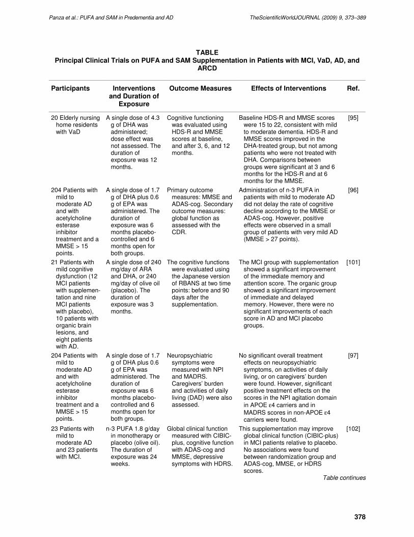
TABLE Principal Clinical Trials on PUFA and SAM Supplementation in Patients with MCI, VaD, AD, and
ARCD
Participants Interventions and Duration of
Exposure
Outcome Measures Effects of Interventions Ref.
20 Elderly nursing home residents with VaD
A single dose of 4.3 g of DHA was administered; dose effect was not assessed. The duration of exposure was 12 months.
Cognitive functioning was evaluated using HDS-R and MMSE scores at baseline, and after 3, 6, and 12 months.
Baseline HDS-R and MMSE scores were 15 to 22, consistent with mild to moderate dementia. HDS-R and MMSE scores improved in the DHA-treated group, but not among patients who were not treated with DHA. Comparisons between groups were significant at 3 and 6 months for the HDS-R and at 6 months for the MMSE.
[95]
204 Patients with mild to moderate AD and with acetylcholine esterase inhibitor treatment and a MMSE > 15 points.
A single dose of 1.7 g of DHA plus 0.6 g of EPA was administered. The duration of exposure was 6 months placebo-controlled and 6 months open for both groups.
Primary outcome measures: MMSE and ADAS-cog. Secondary outcome measures: global function as assessed with the CDR.
Administration of n-3 PUFA in patients with mild to moderate AD did not delay the rate of cognitive decline according to the MMSE or ADAS-cog. However, positive effects were observed in a small group of patients with very mild AD (MMSE > 27 points).
[96]
21 Patients with mild cognitive dysfunction (12 MCI patients with supplemen-tation and nine MCI patients with placebo), 10 patients with organic brain lesions, and eight patients with AD.
A single dose of 240 mg/day of ARA and DHA, or 240 mg/day of olive oil (placebo). The duration of exposure was 3 months.
The cognitive functions were evaluated using the Japanese version of RBANS at two time points: before and 90 days after the supplementation.
The MCI group with supplementation showed a significant improvement of the immediate memory and attention score. The organic group showed a significant improvement of immediate and delayed memory. However, there were no significant improvements of each score in AD and MCI placebo groups.
[101]
204 Patients with mild to moderate AD and with acetylcholine esterase inhibitor treatment and a MMSE > 15 points.
A single dose of 1.7 g of DHA plus 0.6 g of EPA was administered. The duration of exposure was 6 months placebo-controlled and 6 months open for both groups.
Neuropsychiatric symptoms were measured with NPI and MADRS. Caregivers’ burden and activities of daily living (DAD) were also assessed.
No significant overall treatment effects on neuropsychiatric symptoms, on activities of daily living, or on caregivers’ burden were found. However, significant positive treatment effects on the scores in the NPI agitation domain
in APOE ε4 carriers and in
MADRS scores in non-APOE ε4 carriers were found.
[97]
23 Patients with mild to moderate AD and 23 patients with MCI.
n-3 PUFA 1.8 g/day in monotherapy or placebo (olive oil). The duration of exposure was 24 weeks.
Global clinical function measured with CIBIC-plus, cognitive function with ADAS-cog and MMSE, depressive symptoms with HDRS.
This supplementation may improve global clinical function (CIBIC-plus) in MCI patients relative to placebo. No associations were found between randomization group and ADAS-cog, MMSE, or HDRS scores.
[102]
Table continues

Panza et al.: PUFA and SAM in Predementia and AD TheScientificWorldJOURNAL (2009) 9, 373–389
379
TABLE (continued)
Participants Interventions and Duration of
Exposure
Outcome Measures Effects of Interventions Ref.
14 Community-dwelling individuals with early-stage AD.
Vitamin/nutriceutical formulation (folate, vitamin B6, vitamin E, SAM, NAC, and ALCAR) in an open-label trial. The duration of exposure was 12 months.
At baseline and at 3-month intervals for a total of 12 months, participants completed the DRS-2 and the CDT ; caregivers completed the ADCS-ADL, and the 12-item NPI.
Participants improved in the DRS-2 and CDT. Caregivers reported improvement in multiple domains of the NPI and maintenance of performance in the ADCS-ADL. Sustained performance was reported by caregivers for those participants who continued in an 11-month extension.
[82]
Independently living individuals (n = 302) aged ≥ 65 years; CES-D score < 16, MMSE score > 21.
A single dose of 1,800 mg/day EPA+DHA (n = 96), 400 mg/day EPA+DHA (n = 100), or placebo capsules (n = 106). The duration of exposure was 26 weeks.
Changes in mental well-being were assessed as the primary outcome with the CES-D, MADRS, GDS-15, and HADS-A.
Treatment with neither 1800 nor 400 mg EPA+DHA differentially affected any of the measures of mental well-being after 13 or 26 weeks of intervention compared with placebo.
[31]
Independently living individuals (n = 302) aged ≥ 65 years; CES-D score < 16, MMSE score > 21.
A single dose of 1,800 mg/day EPA+DHA (n = 96), 400 mg/day EPA+DHA (n = 100), or placebo capsules (n = 106). The duration of exposure was 26 weeks.
Cognitive performance was assessed using an extensive neuropsychological test battery that included the cognitive domains of attention (SC-WT; fWDST), sensorimotor speed (TMT-A), memory (WLT; bWDST), and executive function (TMT-B; VFT).
There were no significant differential changes in any of the cognitive domains for either low- or high-dose fish oil supplementation compared with placebo; an effect of EPA-DHA supplementation in
subjects who carried the APOE ε4 allele was also found, but only on the cognitive domain of attention.
[108]
12 Institutionalized patients diagnosed with moderate- to later-stage AD.
Vitamin/nutriceutical formulation (folate, vitamin B6, vitamin E, SAM, NAC, and ALCAR) randomly assigned to treatment group. The duration of exposure was 9 months.
At baseline and at 3-month intervals for a total of 9 months, participants completed the DRS-2 and the CDT; caregivers completed the ADCS-ADL and the 12-item NPI.
Participants receiving the formulation demonstrated a clinically significant delay in decline in the DRS-2 and the CDT as compared to those receiving placebo. Institutional caregivers reported approximately 30% improvement in the NPI and maintenance of performance in the ADCS-ADL or more than 9 months.
[83]
bWDST = Backward Test of the Wechsler Digit Span Task; CDR = Clinical Dementia Rating Scale; CES-D = Center for Epidemiologic Studies Depression Scale; CIBIC-plus = Clinician’s Interview-Based Impression of Change Scale; DAD = Disability Assessment for Dementia; EPA = Eicosapentaenoic Acid; fWDST = Forward Test of the Wechsler Digit Span Task; GDS-15 = 15-Item Geriatric Depression Scale; HADS-A = Hospital Anxiety and Depression Scale; HDRS = Hamilton Depression Rating Scale; HDS-R = Hasegawa’s Dementia Rating Scale; MADRS = Montgomery Asberg Depression Scale; MMSE: Mini-Mental State Examination; RBANS = Repeatable Battery for Assessment of Neuropsychological Status; SC-WT = Stroop Color–Word Test; TMT-A = Trail Making Test Version A; TMT-B = Trail Making Test Version B; VFT = Verbal Fluency Test; WLT = Word Learning Test.

Panza et al.: PUFA and SAM in Predementia and AD TheScientificWorldJOURNAL (2009) 9, 373–389
380
extension[82] (Table). Although the present study suffers from the lack of a placebo, nutriceutical
formulation efficacy over the course of 12 months exceeded that of historical placebos from three studies
on mild to moderate AD[84,85,86]. Furthermore, in a small cohort of 12 institutionalized patients
diagnosed with moderate- to later-stage AD, participants were randomly separated into treatment or
placebo groups. Participants receiving the nutriceutical formulation demonstrated a clinically significant
delay in decline in the DRS-2 and Clox 1 and 2 as compared to those receiving placebo[83] (Table).
Institutional caregivers reported approximately 30% improvement in the NPI and maintenance of
performance in the ADCS-ADL for more than 9 months[83].
In particular, the beneficial effect on irritability and agitation/aggression in the NPI is likely to be
derived principally from SAM, which reduced aggression in transgenic mice harboring the APOE ε4[82];
the underlying biochemical mechanism is not clear, but may relate to restoration/improved
neurotransmitter balance, as SAM restored acetylcholine levels in mice, including that resulting from
folate deficiency[50]. These promising findings provided direct support that nutritional intervention can
have a positive effect on progression of early-stage AD and even at advanced stages. They further
suggested that a combinatorial approach can provide superior neuroprotection more than individual
supplements and suggest that this nutriceutical formulation may potentiate pharmacological approaches.
The small participant populations in both of these studies suggested that the efficacy of nutriceutical
formulation be interpreted with caution and that larger placebo-controlled trials are warranted.
POLYUNSATURATED FATTY ACIDS IN PREDEMENTIA SYNDROMES AND ALZHEIMER’S DISEASE
Recently, a number of dietary elements and foods have been reported to be either risk or protective factors
for the development of dementia and AD. These include fat, fatty acids, antioxidants, fish,
homocysteine/methionine, vitamins, and alcohol[87], suggesting that the connection among diet,
dementia, and aging may be oxidative stress with a possible central role for dietary antioxidants[88]. The
epidemiological evidence of an association between a reduced risk of AD and a diet high in PUFA,
particularly DHA[38,41], is further supported by recent findings that certain diets have been associated
with a lower incidence of predementia syndromes[43,89]. In fact, findings from the Italian Longitudinal
Study on Aging (ILSA) demonstrated that high monounsaturated fatty acids (MUFA), PUFA, and total
energy intake were significantly associated with a better cognitive performance in ARCD subjects in a
8.5-year follow-up[43]. Furthermore, findings from the same population-based study demonstrated that
while dietary fatty acid intakes were not associated with incident MCI, high PUFA intake appeared to
have a borderline nonsignificant trend for a protective effect against the development of MCI[83,89].
Moreover, plasma levels of n-3 PUFA appear to be directly associated with cognitive function and are
found to be lower in AD patients[90,91]. Also, phospholipid fatty acid profiles in the brains of AD
patients are altered[86,92]. In contrast, high consumption of fish, an important source of n-3 PUFA, has
been associated with a reduced risk of AD development[44,93].
One randomized clinical trial (RCT), using an n-3/n-6 fatty acid compound for 4 weeks for 100 AD
patients (60 received the fatty acid compound and 40 a placebo control), found improvements in mood,
cooperation, appetite, sleep, ability to navigate in the home, and short-term memory[94]. Furthermore,
another RCT assessed the effect of supplementation with DHA on cognitive function among 20 elderly
nursing home residents with VaD. Cognitive functioning was evaluated using the Hasegawa’s Dementia
Rating Scale (HDS-R) and Mini-Mental State Examination (MMSE) scores at baseline and after 3, 6, and
12 months. Baseline HDS-R and MMSE scores were 15 to 22, consistent with mild to moderate dementia.
HDS-R and MMSE scores improved in the DHA-treated group, but not among patients who were not
treated with DHA. Comparisons between groups were significant at 3 and 6 months for the HDS-R and at
6 months for the MMSE[95] (Table).
Recently, Freund-Levi and colleagues examined the effects of dietary n-3 PUFA supplementation,
randomizing 204 patients with moderate AD to receive DHA and eicosapentaenoic acid (EPA) (for a total

Panza et al.: PUFA and SAM in Predementia and AD TheScientificWorldJOURNAL (2009) 9, 373–389
381
dose of 1,720 mg DHA/600 mg EPA) or placebo for 6 months (OmegAD Study). After the treatment
period, all of the subjects received open-label n-3 PUFA for another 6 months. The authors found that the
supplementation did not delay the rate of cognitive decline but, in the group of 32 patients with the most
mild AD (MMSE > 27, Clinical Dementia Rating Score 0.5–1), n-3 PUFA supplementation slowed the
decline in MMSE scores[96]. In addition, the subjects in the placebo group of these very mild AD patients
also showed a statistically significant slowing of decline when they were switched to treatment between 6
and 12 months, suggesting that n-3 PUFA might be of benefit to slow the progression of the disease in
MCI or very mild AD[96] (Table). Furthermore, this supplementation did not result in marked effects on
neuropsychiatric symptoms in mild to moderate AD patients except for possible positive effects on
depressive symptoms and agitation symptoms in subgroups[97]. In fact, there were positive effects on
depressive symptoms in non-APOE ε4 carriers and in APOE ε4 carriers on agitation symptoms[97]
(Table). Furthermore, very recent findings from the OmegAD Study suggested that a DHA-enriched n-3
PUFA supplement may positively affect weight and appetite in patients with mild to moderate AD. Not
carrying the APOE ε4 allele and high DHA were independently associated with weight gain[98]. Finally,
in the OmegAD project, the 6-month treatment with a DHA-rich n-3 PUFA preparation was associated
with clear effects on released cytokines from peripheral blood mononuclear cells stimulated ex vivo with
lipopolysaccharide. A significant decline of released interleukin (IL)-1β, IL-6, and granulocyte colony-
stimulating factor (G-CSF) was found, whereas tumor necrosis factor-α (ΤΝF−α), IL-8, -10, and
granulocyte-macrophage CSF secretions were not significantly lower at 6 months. However, the G-CSF
response was not significantly different statistically compared with the response of the placebo group[99].
The clinical significance of the cytokines and growth factors analyzed in the OmegAD project is further
emphasized by the recent report by Ray and colleagues that showed that plasma levels of IL-1, TNF, and
G-CSF are strong predictors of development of AD[100].
Furthermore, the effect of ARA and DHA after a 90-day supplementation on MCI, organic brain
lesions, or AD showed a significant improvement of the immediate memory and attention score for MCI
patients, and a significant improvement of immediate and delayed memories for patients with organic
brain damage[101] (Table). The AD group showed no improvement after the supplementation of ARA
and DHA, and the placebo group showed no significant improvement of cognitive functions by the
supplementation of 240 mg/day of olive oil (high MUFA content)[101] (Table). Finally, the preliminary
results from a 24-week, randomized, double-blind placebo-controlled study on 23 participants with mild
or moderate AD and 23 with MCI randomized to receive n-3 PUFA 1.8 g/day or placebo (olive oil),
suggested that n-3 PUFA monotherapy was well tolerable for most of the participants with AD or
MCI[102] (Table). This supplementation may improve global clinical function, as measured by the
Clinician’s Interview-Based Impression of Change (CIBIC) Scale, which included caregiver-supplied
information (CIBIC-plus), relative to placebo. No associations were found between randomization group
and ADAS-cog, MMSE or Hamilton Depression Rating Scale (HDRS) scores. Levels of EPA on
erythrocyte membrane were associated with cognitive function, measured by ADAS-cog, in these
patients[102]. However, in a secondary analysis, participants with MCI showed more improvement on the
ADAS-cog than those with AD associated with n-3 PUFA administration[102] (Table), which supports
recent reports that indicate that PUFA supplementation could be more effective on cognition in people
with very mild AD[96] or MCI[101].
There is a correlation between the fatty acid composition of the brain and that of circulating
erythrocytes[103]. The decreased concentrations of DHA in n-3 PUFA deficiency can be reversed by the
administration of a DHA diet[101], but the inhibition of PEMT by SAH could still inhibit the uptake of
DHA into lipoproteins and impair its transport to peripheral tissues[77]. The most effective means for
lowering plasma homocysteine is B vitamin supplementation (a combination of folate, vitamin B12, and
vitamin B6)[63]. Some authors suggested that the possible metabolic link between the increased
production of SAH and phospholipid metabolism, with a decreased mobilization of DHA from the liver
into plasma and peripheral tissues, may increase the risk of atherosclerosis and stroke leading to
cerebrovascular and neurodegenerative changes in AD[75]. The use of a combination of n-3 PUFA, folic

Panza et al.: PUFA and SAM in Predementia and AD TheScientificWorldJOURNAL (2009) 9, 373–389
382
acid, and vitamin B12 may be a more effective means of increasing the uptake of DHA into the brain than
a diet high in n-3 PUFA alone. In fact, elevations in plasma levels of homocysteine is an emerging risk
factor for AD, but it remains an open question whether interventions designed to lower plasma
homocysteine concentrations will improve cognitive function or retard the rate of cognitive decline in
older adults with or without AD[63]. Homocysteine is a sulfur-containing amino acid that can induce
apoptosis and cause increased neuronal vulnerability to excitotoxicity by mechanisms mainly involving
DNA damage[104]. Formed through methionine metabolic conversion, homocysteine is metabolized by
remethylation and trans-sulfuration pathways. The remethylation pathway is controlled by vitamin B12–
dependent methionine synthase and methylenetetrahydrofolate reductase, whereas vitamin B6–dependent
cystathionine β synthase takes part in the trans-sulfuration pathway. Reduced activity of any of these
three enzymes would result in increased homocysteine plasma levels[105]. Furthermore, the accumulation
of alleles of single-nucleotide polymorphisms that decrease the activity of the two main enzymes involved
in homocysteine degradation (i.e., methionine synthase and cystathionine β synthase) was found to
augment the risk of developing AD[106], supporting the hypothesis that homocysteine metabolism
genetics is directly involved in AD pathogenesis. It would be interesting to consider whether an
increasing uptake of PUFA into the brain will decrease the risk for AD and cognitive decline as well. The
very recent findings of the study by Tchantchou and colleagues confirmed that SAM can exert a direct
effect on GST activity[53]. Since AD is accompanied by reduced GST activity, diminished SAM, and
increased SAH, these findings underscored the critical role of SAM in maintenance of neuronal health,
suggesting a possible role of SAM as a neuroprotective dietary supplement in AD.
A few years ago, a Cochrane review concluded that there was a growing body of evidence from
biological, observational, and epidemiological studies that suggested a protective effect of n-3 PUFA
against dementia. However, the Cochrane review team was unable to locate a single published RCT on
which to base recommendations for the use of dietary or supplemental n-3 PUFA for the prevention of
cognitive impairment or dementia[107]. However, very recently, in a randomized, double-blind, placebo-
controlled trial of 302 cognitively healthy (MMSE score > 21) individuals aged 65 years or older, the
possible impact of n-3 PUFA on the mental well-being and cognitive performance of nondepressed (CES-
D score < 16) older individuals was investigated[31,108] (Table). In this RCT, participants were
randomly assigned to 1,800 mg/day EPA-DHA, 400 mg/day EPA-DHA, or placebo capsules for 26
weeks[31,108]. In older Dutch subjects, no effect of daily supplementation with low or high doses of
EPA-DHA on mental well-being as assessed by depression and anxiety questionnaires was found[31]
(Table). Furthermore, there were no significant differential changes in any of the cognitive domains
(attention, sensorimotor speed, memory, and executive function) for either low- or high-dose fish oil
supplementation compared with placebo[99,108]. However, an effect of EPA-DHA supplementation in
subjects who carried the APOE-ε4 allele was found, but only on the cognitive domain of
attention[99,108] (Table). An improvement in men after 26 weeks of intervention for the low-dose fish oil
group compared with placebo was also found, although these subgroup analyses did not include
adjustment for multiple comparisons. Fish oil may be beneficial in these subjects who are most sensitive
to developing dementia. These two substantially negative studies on ARCD may be explained by the
samples investigated (nondepressed and noncognitively impaired older subjects). Further trials in
depressed patients or APOE ε4 carriers with MCI are needed. Finally, there is another ongoing RCT with
cognitive endpoints of n-3 PUFA supplementation in healthy cognitively intact older persons. The Older
People And n-3 Long-chain polyunsaturated fatty acid (OPAL) study is a double-blind, randomized,
placebo-controlled trial examining the effect of daily supplementation with 700 mg n-3 PUFA (500 mg
DHA and 200 mg EPA) for 24 months on cognitive performance in healthy older persons aged 70–79
with good cognitive function (MMSE ≥ 24 out of 30 points at baseline) who are recruited from 20
primary care practices[109]. The OPAL study was completed at the end of 2007 and findings will be
published shortly. At present, only baseline data are available, suggesting once again that higher fish
consumption is associated with better cognitive function in later life[110].

Panza et al.: PUFA and SAM in Predementia and AD TheScientificWorldJOURNAL (2009) 9, 373–389
383
The mechanisms by which high unsaturated fatty acid (UFA: MUFA or PUFA) intake could be
protective against cognitive decline and dementia in healthy older people are, at present, unknown. In the
older subjects of the ILSA, which fulfilled a Mediterranean dietary pattern, total fat is 29% of energy,
with a high consumption of olive oil (46 g/day), a MUFA energy intake of 17.6% of total energy, 85% of
which derived from olive oil, and a saturated fatty acid (SFA) intake of only 6%[111]. In this population,
the prolonged protection of MUFA intake against age-related changes in cognitive functions may be
linked to the relevant quota of antioxidant compounds in olive oil, including low molecular weight
phenols[112]. In fact, animal studies suggested that diets high in antioxidant-rich foods, such as spinach,
strawberries, and blueberries, rich in anthocyanins and other flavonoids may be beneficial in slowing age-
related cognitive decline[113]. The possible role of antioxidant compounds from olive oil do not diminish
or otherwise alter the argument concerning the fatty acids because this is only a possible explanation of
the role of MUFA on age-related cognitive changes in our population, in which MUFA intake derived for
a large part from olive oil.
The protective effect of dietary UFA could be related to the role of fatty acids in maintaining the
structural integrity of neuronal membranes, determining the fluidity of synaptosomal membranes and
thereby regulating neuronal transmission. Furthermore, essential fatty acids can modify the activity of
certain membrane-bound enzymes (phospholipase A2, protein kinase C, and acetyltranferase), and the
function of the neurotransmitters’ receptors. Finally, free fatty acids, lipid metabolites, and phospholipids
modify the function of membrane proteins, including ion channels[114]. Moreover, fatty acid
composition of neuronal membranes in advancing age demonstrated an increase in MUFA content and a
decrease in PUFA content[115]. There is also evidence associating a dietary deficiency of n-3 PUFA with
changes in cortical dopaminergic function[116]. The n-3 PUFA from fish may be inversely associated
with dementia because it lowers the risk of thrombosis[117], stroke[118], cardiovascular disease[119],
and cardiac arrhythmia, reducing the risk of thromboembolism in the brain and consequently of lacunar
and large infarcts that can lead to VaD and AD. Furthermore, the n-3 PUFA may be important as lipids in
the brain, particularly for the possible influence of DHA on the physical properties of the brain that are
essential for its function[120]. Furthermore, fish oil was a better source than α-linoleic acid for the
incorporation of n-3 PUFA into rat brain phospholipid subclasses[30]. On the contrary, high linoleic acid
intake (n-6 PUFA) may increase the susceptibility of LDL cholesterol to oxidation, which makes it more
atherogenic[121], even if the association between linoleic acid and atherosclerosis is controversial[122].
Therefore the ratio of dietary n-3/n-6 PUFA intake may influence the potential role of PUFA on cognitive
decline and dementia, the optimal ratio of n-6:n-3 for an healthier diet should be <5:1[123]. Finally, a
high dietary intake of SFA and cholesterol increases the risk for cardiovascular disease, and therefore for
cognitive decline, VaD, and AD[124]. On the contrary, treatment for 4 weeks with a Mediterranean-
inspired diet rich in n-3 PUFA decreased blood lipids in healthy individuals with a low-risk profile for
cardiovascular disease, with a beneficial effect also on vascular function and oxidative stress[125].
CONCLUSIONS
Recent findings suggested that SAM can exert a direct effect on GST activity, and AD is accompanied by
reduced GST activity, diminished SAM, and increased SAH, the downstream metabolic product resulting
from SAM-mediated transmethylation reactions when deprived of folate. Therefore, several experimental
findings underscored the critical role of SAM in maintenance of neuronal health, suggesting a possible
role of SAM as a neuroprotective dietary supplement in AD[126]. Very recently, a nutriceutical
formulation consisting of six vitamins and nutriceuticals (folic acid, vitamin B12, vitamin E, ALCAR,
NAC, and SAM) showed improved cognitive performance in AD. However, to be effective, this
supplementation should be started prior to extensive cognitive decline; this nutriceutical formulation
improved cognitive performance for more than 2 years when treatment was initiated during the early
stages of AD, but was only capable of delaying the decline vs. placebo when initiated later in the course
of the disease[82,83]. In subjects who already have severe cognitive impairment or dementia, it might be
Panza et al.: PUFA and SAM in Predementia and AD TheScientificWorldJOURNAL (2009) 9, 373–389
384
too late for dietary supplementation to counteract the process of cognitive decline. Furthermore,
epidemiological evidence suggested a possible association between PUFA (particularly, n-3 PUFA) and
reduced risk of dementia. However, due to the small number of studies that discuss this topic, further
research is necessary before a strong conclusion can be drawn. Some recent RCTs assessed the cognitive
or functional effect of n-3 PUFA supplementation on patients with VaD, AD, MCI, or ARCD in
cognitively unimpaired older subjects. These RCTs suggested a positive effect of this intervention only in
very mild AD or MCI patients, or in subgroups (e.g., APOE ε4 carriers), for cognitive performance in
nondemented subjects, or for neuropsychiatric symptoms in mild to moderate AD patients.
In conclusion, for mild AD and predementia syndromes, we suggest a high-risk condition for
progression to dementia of vascular and degenerative origin: intervention trials using measures of dietary
supplementation (dietary n-3 PUFA and SAM plus B vitamin supplementation) to determine if such
supplements will reduce the risk for cognitive decline. Therefore, key supplements are not necessarily
working in isolation, or in solely the "predicted" or anticipated manner, and the most profound impact, or
in some cases the only impact, is noted very early in the course of AD. This is to be expected in that
serious damage to neurons may exert a protracted loss, but one that cannot be reversed by nutrition alone.
This does bring up the thought, however, that nutriceutical supplements may bolster pharmacological
approaches well past the window where supplements can work on their own. Recommendations regarding
future research on the effects of SAM or n-3 PUFA supplementation on predementia syndromes and very
mild AD include properly designed RCTs that are sufficiently powered and with an adequate length (e.g.,
3–5 years of follow-up). In particular, the future RCTs on PUFA supplementation should address the
effects of different types of n-3 PUFA (i.e., DHA, EPA, ARA, and total n-3 PUFA) as well as the ratio of
n-6 to n-3 PUFA. Finally, these RCTs should be designed to include a baseline assessment of dietary n-3
and n-6 PUFA intake, and to evaluate the effect of dose, treatment duration, and the sustainment of effect
after discontinuation of n-3 PUFA consumption.
REFERENCES
1. Fratiglioni, L. and Qiu, C. (2008) Prevention of common neurodegenerative disorders in the elderly. Exp. Gerontol.
44(1–2), 46–50.
2. Qiu, C., De Ronchi, D., and Fratiglioni, L. (2007) The epidemiology of the dementias: an update. Curr. Opin.
Psychiatry 20, 380–385.
3. Whitehouse, P.J., Sciulli, C.G., and Mason, R.M. (1997) Dementia drug development: use of information systems to
harmonize global drug development. Psychopharmacol. Bull. 33, 129–133.
4. Panza, F., D'Introno, A., Colacicco, A.M., Capurso, C., Del Parigi, A., Caselli, R.J., Pilotto, A., Argentieri, G.,
Scapicchio, P.L., Scafato, E., Capurso, A., and Solfrizzi, V. (2005) Current epidemiology of mild cognitive
impairment and other predementia syndromes. Am. J. Geriatr. Psychiatry 13, 633–644.
5. Panza, F., D'Introno, A., Colacicco, A.M., Capurso, C., Del Parigi, A., Capurso, S.A., Caselli, R.J., Pilotto, A.,
Scafato, E., Capurso, A., and Solfrizzi, V. (2006) Cognitive frailty: predementia syndrome and vascular risk factors.
Neurobiol. Aging 27, 933–940.
6. Panza, F., Solfrizzi, V., Frisardi, V., Capurso, C., D’Introno, A,, Colacicco, A.M., Vendemiale, G., Capurso, A., and
Imbimbo, B.P. (2009) Disease-modifying approach for the treatment of Alzheimer’s disease: from α-secretase
activators to γ-secretase inhibitors and modulators. Drugs Aging, in press.
7. Alzheimer, A. (1907) Uber eine eigenartige Erkrankung der Hirnrinde. Allg. Z. Psychiatr. 64, 146–148.
8. Terry, R.D. (2006) Alzheimer’s disease at mid century (1927–1977). In Alzheimer: 100 Years and Beyond. Jucker,
M., Beyreuther, K., Haas, C., and Nitsch, R., Eds. Springer-Verlag, Berlin. pp. 58–61.
9. Giacobini, E. and Becker, R.E. (2007) One hundred years after the discovery of Alzheimer's disease. A turning point
for therapy? J. Alzheimers Dis. 12, 37–52.
10. Walter, J., Kaether, C., Steiner, H., and Haass, C. (2001) The cell biology of Alzheimer's disease: uncovering the
secrets of secretases. Curr. Opin. Neurobiol. 11, 585–590.
11. Ohyagi, Y., Asahara, H., Chui, D.H., Tsuruta, Y., Sakae, N., Miyoshi, K., Yamada, T., Kikuchi, H., Taniwaki, T.,
Murai, H., Ikezoe, K., Furuya, H., Kawarabayashi, T., Shoji, M., Checler, F., Iwaki, T., Makifuchi, T., Takeda, K.,
Kira, J., and Tabira, T. (2005) Intracellular Abeta42 activates p53 promoter: a pathway to neurodegeneration in
Alzheimer's disease. FASEB J. 19, 255–257.
12. Hardy, J. and Allsop, D. (1991) Amyloid deposition as the central event in the aetiology of Alzheimer’s disease.
Trends Pharmacol. Sci. 12, 383–388.

Panza et al.: PUFA and SAM in Predementia and AD TheScientificWorldJOURNAL (2009) 9, 373–389
385
13. Hardy, J. and Selkoe, D.J. (2002) The amyloid hypothesis of Alzheimer’s disease: progress and problems on the road
to therapeutics. Science 297, 353–356.
14. Castellani, R.J., Lee, H.G., Zhu, X., Perry, G., and Smith, M.A. (2008) Alzheimer disease pathology as a host
response. J. Neuropathol. Exp. Neurol. 67, 523–531.
15. Crystal, H.A., Dickson, D.W., Sliwinski, M.J., Lipton, R.B., Grober, E., Marks-Nelson, H., and Antis, P. (1993)
Pathological markers associated with normal aging and dementia in the elderly. Ann. Neurol. 34, 566–573.
16. Knopman, D.S., Parisi, J.E., Salviati, A., Floriach-Robert, M., Boeve, B.F., Ivnik, R.J., Smith, G.E., Dickson, D.W.,
Johnson, K.A., Petersen, L.E., McDonald, W.C., Braak, H., and Petersen, R.C. (2003) Neuropathology of cognitively
normal elderly. J. Neuropathol. Exp. Neurol. 62, 1087–1095.
17. Holmes, C., Boche, D., Wilkinson, D., Yadegarfar, G., Hopkins, V., Bayer, A., Jones, R.W., Bullock, R., Love, S.,
Neal, J.W., Zotova, E., and Nicoll, J.A. (2008) Long-term effects of Aβ42 immunisation in Alzheimer's disease:
follow-up of a randomised, placebo-controlled phase I trial. Lancet 372, 216–223.
18. Puzzo, D., Privitera, L., Leznik, E., Fà, M., Staniszewski, A., Palmeri, A., and Arancio, O. (2008) Picomolar amyloid-
β positively modulates synaptic plasticity and memory in hippocampus. J. Neurosci. 28, 14537–14545.
19. Chen, Y. and Dong, C. (2009) Aβ40 promotes neuronal cell fate in neural progenitor cells. Cell Death Differ. 16,
386–394.
20. Lee, H.G., Zhu, X., Nunomura, A., Perry, G., and Smith, M.A. (2006) Amyloid beta: the alternate hypothesis. Curr.
Alzheimer Res. 3, 75–80.
21. Hayashi, T., Shishido, N., Nakayama, K., Nunomura, A., Smith, M.A, Perry, G., and Nakamura, M. (2007) Lipid
peroxidation and 4-hydroxy-2-nonenal formation by copper ion bound to amyloid-beta peptide. Free Radic. Biol.
Med. 43, 1552–1559.
22. Nakamura, M., Shishido, N., Nunomura, A., Smith, M.A., Perry, G., Hayashi, Y., Nakayama, K., and Hayashi, T.
(2007) Three histidine residues of amyloid-beta peptide control the redox activity of copper and iron. Biochemistry
46, 12737–12743.
23. Bryan, J., Calvaresi, E., and Hughes, D. (2002) Short-term folate, vitamin B-12 or vitamin B-6 supplementation
slightly affects memory performance but not mood in women of various ages. J. Nutr. 132, 1345–1356.
24. Luchsinger, J.A., Tang, M.X., Shea, S., and Mayeux, R. (2003) Antioxidant vitamin and risk of Alzheimer disease.
Arch. Neurol. 60, 203–208.
25. Salerno-Kennedy, R. and Cashman, K.D. (2005) Relationship between dementia and nutrition-related factors and
disorders: an overview. Int. J. Vitam. Nutr. Res. 75, 83–95.
26. Balk, E.M., Raman, G., Tatsioni, A., Chung, M., Lau, J., and Rosenberg, I.H. (2007) Vitamin B6, B12, and folic acid
supplementation and cognitive function: a systematic review of randomized trials. Arch. Intern. Med. 167, 21–30.
27. Malouf, R. and Grimley Evans, J. (2008) Folic acid with or without vitamin B12 for the prevention and treatment of
healthy elderly and demented people. Cochrane Database Syst. Rev. CD004514.
28. Aisen, P.S., Schneider, L.S., Sano, M., Diaz-Arrastia, R., van Dyck, C.H., Weiner, M.F., Bottiglieri, T., Jin, S.,
Stokes, K.T., Thomas, R.G., Thal, L.J.; Alzheimer Disease Cooperative Study (2008) High-dose B vitamin
supplementation and cognitive decline in Alzheimer disease: a randomized controlled trial. JAMA 300, 1774–1783.
29. Masaki, K.H., Losonczy, K.G., Izmirlian, G., Foley, D.J., Ross, G.W., Petrovitch, H., Havlik, R., and White, L.R.
(2000) Association of vitamin E and C supplement use with cognitive function and dementia in elderly men.
Neurology 54, 1265–1272.
30. Solfrizzi, V., Capurso, C., D'Introno, A., Colacicco, A.M., Frisardi, V., Santamato, A., Ranieri, M., Fiore, P.,
Vendemiale, G., Seripa, D., Pilotto, A., Capurso, A., and Panza, F. (2008) Dietary fatty acids, age-related cognitive
decline, and mild cognitive impairment. J. Nutr. Health Aging 12, 382–386.
31. van de Rest, O., Geleijnse, J.M., Kok, F.J., van Staveren, W.A., Hoefnagels, W.H., Beekman, A.T., and de Groot,
L.C. (2008) Effect of fish-oil supplementation on mental well-being in older subjects: a randomized, double-blind,
placebo-controlled trial. Am. J. Clin. Nutr. 88, 706–713.
32. Petersen, R.C., Thomas, R.G., Grundman, M., Bennett, D., Doody, R., Ferris, S., Galasko, D., Jin, S., Kaye, J., Levey,
A., Pfeiffer, E., Sano, M., van Dyck, C.H., Thal, L.J.; Alzheimer's Disease Cooperative Study Group (2005) Vitamin
E and donepezil for the treatment of mild cognitive impairment. N. Engl. J. Med. 352, 2379–2388.
33. Tabet, N., Birks, J., and Grimley, E.J. (2000) Vitamin E for Alzheimer’s disease. Cochrane Database Syst. Rev.,
CD002854.
34. Sano, M., Ernesto, C., Thomas, R.G., Klauber, M.R., Schafer, K., Grundman, M., Woodbury, P., Growdon, J.,
Cotman, C.W., Pfeiffer, E., Schneider, L.S., and Thal, L.J. (1997) A controlled trial of selegiline, alpha-tocopherol, or
both as treatment for Alzheimer’s disease. N. Engl. J. Med. 336, 1216–1222.
35. Sonnen, J.A., Larson, E.B., Gray, S.L., Wilson, A., Kohama, S.G., Crane, P.K., Breitner, J.C., and Montine, T.J.
(2009) Free radical damage to cerebral cortex in Alzheimer's disease, microvascular brain injury, and smoking. Ann.
Neurol. 65, 226–229.
36. Perry, G., Zhu, X., Moreira, P.I., and Smith, M.A. (2009) Altered redox balance in disease: can we change the new
equilibria? Ann. Neurol. 65, 121–123.
37. Lloret, A., Badía, M.-C., Mora, N.J., Pallardó, F.V., Alonso, M.-D., and Viña J. (2009) Vitamin E paradox in
Alzheimer’s disease: it does not prevent loss of cognition and may even be detrimental. J. Alzheimers Dis., in press.
38. National Institute on Aging. Prevention of Alzheimer’s Disease by VItamin E and SElenium (PREADVISE)[online].
Panza et al.: PUFA and SAM in Predementia and AD TheScientificWorldJOURNAL (2009) 9, 373–389
386
Available from URL: http://www.clinicaltrials.gov [Accessed 30 November 2008].
39. Hajieva, P. and Behl, C. (2006) Antioxidants as a potential therapy against age-related neurodegenerative diseases:
amyloid beta toxicity and Alzheimer's disease. Curr. Pharm. Des. 12, 699–704.
40. National Institute on Aging. Anti-oxidant treatment of Alzheimer’s disease [online]. Available from URL:
http://www.clinicaltrials.gov [Accessed 30 November 2008].
41. Panza, F., Solfrizzi, V., Colacicco, A.M., D'Introno, A., Capurso, C., Torres, F., Del Parigi, A., Capurso, S., and
Capurso, A. (2004) Mediterranean diet and cognitive decline. Public Health Nutr. 7, 959–963.
42. Panza, F., Capurso, C., D'Introno, A., Colacicco, A.M., Chirico, M., Capurso, A., and Solfrizzi, V. (2007) Dietary
polyunsaturated fatty acid supplementation, pre-dementia syndromes, and Alzheimer's disease. J. Am. Geriatr. Soc.
55, 469–470.
43. Solfrizzi, V., Colacicco, A.M., D'Introno, A., Capurso, C., Torres, F., Rizzo, C., Capurso, A., and Panza, F. (2006)
Dietary intake of unsaturated fatty acids and age-related cognitive decline: a 8.5-year follow-up of the Italian
Longitudinal Study on Aging. Neurobiol. Aging 27, 1694–1704.
44. Solfrizzi, V., Colacicco, A.M., D'Introno, A., Capurso, C., Del Parigi, A., Capurso, S.A., Argentieri, G., Capurso, A., and
Panza, F. (2005) Dietary fatty acids intake: possible role in cognitive decline and dementia. Exp. Gerontol. 40, 257–270.
45. Woodside, J.V., McCall, D., McGartland, C., and Young, I.S. (2005) Micronutrients: dietary intake v. supplement
use. Proc. Nutr. Soc. 64, 543–553.
46. Birks, J. and Grimley Evans, J. (2009) Ginkgo biloba for cognitive impairment and dementia. Cochrane Database
Syst. Rev. CD003120.
47. Kleijnen, J. and Knipschild, P. (1992) Ginkgo biloba for cerebral insufficiency. Br. J. Clin. Pharmacol. 34, 352–358.
48. Huang, H.Y., Caballero, B., Chang, S., Alberg, A., Semba, R., Schneyer, C., Wilson, R.F., Cheng, T.Y.,
Prokopowicz, G., Barnes, G.J., 2nd, Vassy, J., and Bass E.B. (2006) Multivitamin/mineral supplements and
prevention of chronic disease. Evid. Rep. Technol. Assess. (Full Rep), 1–117.
49. McDaniel, M.A., Maier, S.F., and Einstein, G.O. (2003) "Brain-specific" nutrients: a memory cure? Nutrition 19,
957–975.
50. Chan, A., Tchantchou, F., Graves, V., Rozen, R., and Shea, T.B. (2008) Dietary and genetic compromise in folate
availability reduces acetylcholine, cognitive performance and increases aggression: critical role of S-adenosyl
methionine. J. Nutr. Health Aging 12, 252–261.
51. Choi, J., Conrad, C.C., Dai, R., Malakowsky, C.A., Talent, J.M., Carroll, C.A., Weintraub, S.T., and Gracy, R.W.
(2003) Vitamin E prevents oxidation of antiapoptotic proteins in neuronal cells. Proteomics 3, 73–77.
52. Rogers, E.J., Milhalik, S., Orthiz, D., and Shea, T.B. (2004) Apple juice prevents oxidative stress and impaired
cognitive performance caused by genetic and dietary deficiencies in mice. J. Nutr. Health Aging 8, 92–97.
53. Tchantchou, F., Graves, M., Falcone, D., and Shea, T.B. (2008) S-adenosylmethionine mediates glutathione efficacy
by increasing glutathione S-transferase activity: implications for S-adenosyl methionine as a neuroprotective dietary
supplement. J. Alzheimers Dis. 14, 323–328.
54. Willis, L.M., Shukitt-Hale, B., and Joseph, J.A. (2009) Recent advances in berry supplementation and age-related
cognitive decline. Curr. Opin. Clin. Nutr. Metab. Care 12, 91–94.
55. Berman, K. and Brodaty, H. (2004) Tocopherol (vitamin E) in Alzheimer’s disease and other neurodegenerative
disorders. CNS Drugs 18, 807–825.
56. Morris, M.C., Evans, D.A., Tangey, C.C., Bienas, J.L., Wilson, R.S., and Aggarwal, N.T. (2005) Relation of the
tocopherol forms to incident Alzheimer’s disease and to cognitive change. Am. J. Clin. Nutr. 82, 508–514.
57. Dhitavat, S., Rivera, E., and Shea, T.B. (2001) Differential efficacy of lipophilic and cytosolic antioxidants on
generation of reactive oxygen species by amyloid-beta. J. Alzeimers Dis. 3, 525–529.
58. Liu, H., Wang, H., Shenvi, S., Hagen, T.M., and Liu, R.M. (2004) Glutathione metabolism during aging and in
Alzheimer disease. Ann. N. Y. Acad. Sci. 1019, 346–349.
59. Lovell, M.A., Xie, C., and Markesbery, W.R. (1998) Decreased glutathione transferase activity in brain and
ventricular fluid in Alzheimer’s disease. Neurology 51, 1562–1566.
60. Pinhel, M.A., Nakazone, M.A., Cação, J.C., Piteri, R.C., Dantas, R.T., Godoy, M.F., Godoy, M.R., Tognola, W.A.,
Conforti-Froes, N.D., and Souza, D. (2008) Glutathione S-transferase variants increase susceptibility for late-onset
Alzheimer's disease: association study and relationship with apolipoprotein E epsilon4 allele. Clin. Chem. Lab. Med.
46, 439–445.
61. Bernardini, S., Bellincampi, L., Ballerini, S., Federici, G., Iori, R., Trequattrini, A., Ciappi, F., Baldinetti, F., Bossù,
P., Caltagirone, C., and Spalletta, G. (2005) Glutathione S-transferase P1 *C allelic variant increases susceptibility for
late-onset Alzheimer disease: association study and relationship with apolipoprotein E epsilon4 allele. Clin. Chem. 51,
944–951.
62. Adair, J.C., Knoefel, J.E., and Morgan, N. (2001) Controlled trial of N-acetylcysteine for patients with probable
Alzheimer’s disease. Neurology 57, 1515–1517.
63. Del Parigi, A., Panza, F., Capurso, C., and Solfrizzi, V. (2006) Nutritional factors, cognitive decline, and dementia.
Brain Res. Bull. 69, 1–19.
64. St George-Hyslop, P.H. (2000) Genetic factors in the genesis of Alzheimer’s disease. Ann. N. Y. Acad. Sci. 924, 1–7.
65. Tchantchou, F., Graves, M., Ortiz, D., and Shea, T.B. (2006) S-adenosyl methionine: a connection between
nutritional and genetic risk factors in Alzheimer’s disease. J. Nutr. Health Aging 10, 541–544.
Panza et al.: PUFA and SAM in Predementia and AD TheScientificWorldJOURNAL (2009) 9, 373–389
387
66. Connelly, P.J., Prentice, N.P., Cousland, G., and Bonham, J. (2008) A randomised double-blind placebo-controlled
trial of folic acid supplementation of cholinesterase inhibitors in Alzheimer’s disease. Int. J. Geriatr. Psychiatry 23,
155–160.
67. Kennedy, B.P., Bottiglieri, T., Arning, E., Ziegler, M.G., Hansen, L.A., and Masliah, E. (2004) Elevated S-
adenosylhomocysteine in Alzheimer brain: influence on methyltransferases and cognitive function. J. Neural Transm.
111, 547–567.
68. Tchantchou, F., Graves, M., Ashline, D., Morin, A., Pimenta, A., Ortiz, D., Rogers, E., and Shea, T.B. (2004)
Increased transcription and activity of glutathione synthase in response to deficiencies in folate, vitamin E and
apolipoprotein E. J. Neurosci. Res. 75, 508–515.
69. Fuso, A., Seminara, L., Cavallaro, R.A., D’Anselmi, F., and Scarpa, S. (2005) S-adenosylmethionine/homocysteine
cycle alterations modify DNA methylation status with consequent deregulation of PS1 and BACE and beta-amyloid
production. Mol. Cell. Neurosci. 28, 195–204.
70. Scarpa, S., Fuso, A., D’Anselmi, F., and Cavallaio, R.A. (2003) Presenilin 1 gene silencing by S-adenosylmethionine:
a treatment for Alzheimer disease? FEBS Lett. 541, 145–148.
71. Mischoulon, D. and Fava, M. (2002) Role of S-adenosyl-L-methionine in the treatment of depression: a review of the
evidence. Am. J. Clin. Nutr. 76, 1158S–1161S.
72. Morozova, N., Khrapko, K., Panee, J., Liu, W., Harney, J.W., and Berry, M.J. (2007) Glutathione depletion in
hippocampal cells increases levels of H and L ferritin and glutathione S-transferase mRNAs. Genes Cells 12, 561–
567.
73. Lovell, M.A., Ehmann, W.D., Butler, S.M., and Markesbery, W.R. (1995) Elevated thiobarbituric acid-reactive
substances and antioxidant enzyme activity in the brain in Alzheimer’s disease. Neurology 45, 1594–1601.
74. Chan, A. and Shea, T.B. (2007) Effects of dietary supplementation with N-acetyl cysteine, acetyl-L-carnitine and S-
adenosyl methionine on cognitive performance and aggression in normal mice and mice expressing human ApoE4.
Neuromolecular Med. 9, 264–269.
75. Selley, M.L. (2007) A metabolic link between S-adenosylhomocysteine and polyunsaturated fatty acid metabolism in
Alzheimer's disease. Neurobiol. Aging 28, 1834–1839.
76. Panza, F., Capurso, C., D'Introno, A., Colacicco, A.M., Capurso, A., and Solfrizzi, V. (2008) S-
adenosylhomocysteine and polyunsaturated fatty acid metabolism in predementia syndromes and Alzheimer's disease.
Neurobiol. Aging 29, 478–480.
77. Watkins, S.M., Zhu, X., and Zeisel, S.H. (2003) Phosphatidylethanolamine-N-methyltransferase activity and dietary
choline regulate liver-plasma lipid flux and essential fatty acid metabolism in mice. J. Nutr. 133, 3386–3391.
78. Solfrizzi, V., D'Introno, A., Colacicco, A.M., Capurso, C., Todarello, O., Pellicani, V., Capurso, S.A., Pietrarossa, G.,
Santamato, V., Capurso, A., and Panza, F. (2006) Circulating biomarkers of cognitive decline and dementia. Clin.
Chim. Acta 364, 91–112.
79. Selhub, J., Bagley, L.C., Miller, J., and Rosenberg, I.H. (2000) B vitamins, homocysteine, and neurocognitive
function in the elderly. Am. J. Clin. Nutr. 71, 614S–620S.
80. Fassbender, K., Mielke, O., Bertsch, T., Nafe, B., Froschen, S., and Hennerici, M. (1999) Homocysteine in cerebral
macroangiography and microangiopathy. Lancet 353, 1586–1587.
81. Lokk, J. (2003) News and views on folate and elderly persons. J. Gerontol. A Biol. Sci. Med. Sci. 58, 354–361.
82. Chan, A., Paskavitz, J., Remington, R., Rasmussen, S., and Shea, T.B. (2008-2009) Efficacy of a vitamin/nutriceutical
formulation for early-stage Alzheimer's disease: a 1-year, open-label pilot study with an 16-month caregiver
extension. Am. J. Alzheimers Dis. Other Demen. 23, 571–585.
83. Remington, R., Chan, A., Paskavitz, J., and Shea, T.B. (2009) Efficacy of a vitamin/nutriceutical formulation for
moderate-stage to later-stage Alzheimer's disease: a placebo-controlled pilot study. Am. J. Alzheimers Dis. Other
Demen. 24, 27–33.
84. Erkinjuntti, T., Kurz, A., Gauthier, S., Bullock, R., Lilienfeld, S., and Damaraju, C.V. (2002) Efficacy of galantamine
in probable vascular dementia and Alzheimer’s disease combined with cerebrovascular disease: a randomised trial.
Lancet 359, 1283–1290.
85. Feldman, H., Gauthier, S., Hecker, J., Vellas, B., Subbiah, P., and Whalen, E. (2001) A 24-week, randomized, double-
blind study of donepezil in moderate to severe Alzheimer’s disease. Neurology 57, 613–620.
86. Aisen, P.S., Schafer, K.A., Grundman, M., Pfeiffer, E., Sano, M., Davis, K.L., Farlow, M.R., Jin, S., Thomas, R.G.,
Thal, L.J.; Alzheimer's Disease Cooperative Study (2003) Effects of rofecoxib or naproxen vs placebo on Alzheimer
disease progression: a randomized controlled trial. JAMA 289, 2819–2826.
87. Petot, G.J. and Friedland, R.P. (2004) Lipids, diet and Alzheimer disease: an extended summary. J. Neurol. Sci. 226,
31–33.
88. Smith, M.A., Petot, G.J., and Perry, G. (1999) Diet and oxidative stress: a novel synthesis of epidemiological data on
Alzheimer's disease. J. Alzheimers Dis. 1, 203–206.
89. Solfrizzi, V., Colacicco, A.M., D'Introno, A., Capurso, C., Del Parigi, A., Capurso, S.A., Argentieri, G., Capurso, A.,
and Panza, F. (2006) Dietary fatty acids intakes and rate of mild cognitive impairment. The Italian Longitudinal Study
on Aging. Exp. Gerontol. 41, 619–627.
90. Conquer, J.A., Tierney, M.C., Zecevic, J., Bettger, W.J., and Fisher, R.H., (2000) Fatty acid analysis of blood plasma
of patients with Alzheimer's disease, other types of dementia, and cognitive impairment. Lipids 35, 1305–1312.
Panza et al.: PUFA and SAM in Predementia and AD TheScientificWorldJOURNAL (2009) 9, 373–389
388
91. Tully, A.M., Roche, H.M., Doyle, R., Fallon, C., Bruce, I., Lawlor, B., Coakley, D., and Gibney, M.J. (2003) Low
serum cholesteryl ester-docosahexaenoic acid levels in Alzheimer's disease: a case-control study. Br. J. Nutr. 89, 483–
489.
92. Soderberg, M., Edlund, C., Kristensson, K., and Dallner, G. (1991) Fatty acid composition of brain phospholipids in
aging and in Alzheimer's disease. Lipids 26, 421–425.
93. Kalmijn, S., Launer, L.J., Ott, A., Witteman, J.C., Hofman, A., and Breteler, M.M. (1997) Dietary fat intake and the
risk of incident dementia in the Rotterdam Study. Ann. Neurol. 42, 776–782.
94. Yehuda, S., Rabinovtz, S., Carasso, R.L., and Mostofsky, D.I. (1996) Essential fatty acids preparation (SR-3)
improves Alzheimer’s patients quality of life. Int. J. Neurosci. 87, 141–149.
95. Terano, T., Fujishiro, S., Ban, T., Yamamoto, K., Tanaka, T., Noguchi, Y., Tamura, Y., Yazawa, K., and Hirayama,
T. (1999) Docosahexaenoic acid supplementation improves the moderately severe dementia from thrombotic
cerebrovascular diseases. Lipids 34(Suppl.), S345–S346.
96. Freund-Levi, Y., Eriksdotter-Jönhagen, M., Cederholm, T., Basun, H., Faxén-Irving, G., Garlind, A., Vedin, I.,
Vessby, B., Wahlund, L.O., and Palmblad, J. (2006) ω-3 Fatty acid treatment in 174 patients with mild to moderate
Alzheimer disease: OmegAD Study: a randomized double-blind trial. Arch. Neurol. 63, 1402–1408.
97. Freund-Levi, Y., Basun, H., Cederholm, T., Faxén-Irving, G., Garlind, A., Grut, M., Vedin, I., Palmblad, J., Wahlund,
L.O., and Eriksdotter-Jönhagen, M. (2008) Omega-3 supplementation in mild to moderate Alzheimer's disease:
effects on neuropsychiatric symptoms. Int. J. Geriatr. Psychiatry 23, 161–169.
98. Faxén Irving, G., Freund-Levi, Y., Eriksdotter-Jönhagen, M., Basun, H., Brismar, K., Hjorth, E., Palmblad, J.,
Vessby, B., Vedin, I., Wahlund, L.O., and Cederholm, T. (2009) Omega-3 fatty acid supplementation effects on
weight and appetite in patients with Alzheimer's disease: The Omega-3 Alzheimer's Disease Study. J. Am. Geriatr.
Soc. 57, 11–17.
99. Vedin, I., Cederholm, T., Freund Levi, Y., Basun, H., Garlind, A., Faxén Irving, G., Jönhagen, M.E., Vessby, B.,
Wahlund, L.O., and Palmblad, J. (2008) Effects of docosahexaenoic acid-rich n-3 fatty acid supplementation on
cytokine release from blood mononuclear leukocytes: the OmegAD study. Am. J. Clin. Nutr. 87, 1616–1622.
100. Ray, S., Britschgi, M., Herbert, C., Takeda-Uchimura, Y., Boxer, A., Blennow, K., Friedman, L.F., Galasko, D.R.,
Jutel, M., Karydas, A., Kaye, J.A., Leszek, J., Miller, B.L., Minthon, L., Quinn, J.F., Rabinovici, G.D., Robinson,
W.H., Sabbagh, M.N., So, Y.T., Sparks, D.L., Tabaton, M., Tinklenberg, J., Yesavage, J.A., Tibshirani, R., and Wyss-
Coray, T. (2007) Classification and prediction of clinical Alzheimer’s diagnosis based on plasma signaling proteins.
Nat. Med. 13, 1359–1362.
101. Kotani, S., Sakaguchi, E., Warashina, S., Matsukawa, N., Ishikura, Y., Kiso, Y., Sakakibara, M., Yoshimoto, T., Guo,
J., and Yamashima, T. (2006) Dietary supplementation of arachidonic and docosahexaenoic acids improves cognitive
dysfunction. Neurosci. Res. 56, 159–164.
102. Chiu, C.C., Su, K.P., Cheng, T.C., Liu, H.C., Chang, C.J., Dewey, M.E., Stewart, R., and Huang, S.Y. (2008) The
effects of omega-3 fatty acids monotherapy in Alzheimer's disease and mild cognitive impairment: a preliminary
randomized double-blind placebo-controlled study. Prog. Neuropsychopharmacol. Biol. Psychiatry 32, 1538–1544.
103. Richardson, A.J., Allen, S.J., Hajnal, J.V., Cox, I.J., Easton, T., and Puri, B.K. (2001) Associations between central
and peripheral measures of phospholipids breakdown revealed by cerebral 31-phosophorus magnetic resonance
spectroscopy and fatty acid composition of erythrocyte membranes. Prog. Neuropsychopharmacol. Biol. Psychiatry
25, 1513–1521.
104. Kruman, I.I., Culmsee, C., Chan, S.L., Kruman, Y., Guo, Z., Penix, L., and Mattson, M.P. (2000) Homocysteine
elicits a DNA damage response in neurons that promotes apoptosis and hypersensitivity to excitotoxicity. J.
Neurosci.. 20, 6920–6926.
105. Tsai, M.Y., Welge, B.G., Hanson, N.Q., Bignell, M.K., Vessey, J., Schwichtenberg, K., Yang, F., Bullemer, F.E.,
Rasmussen, R., and Graham, K.J. (1999) Genetic causes of mild hyperhomocysteinemia in patients with premature
occlusive coronary artery diseases. Atherosclerosis 143, 163–170.
106. Beyer, K., Lao, J.I., Latorre, P., and Ariza, A. (2005) Age at onset: an essential variable for the definition of genetic
risk factors for sporadic Alzheimer's disease. Ann. N. Y. Acad. Sci. 1057, 260–278.
107. Lim, W.S., Gammack, J.K., Van Niekerk, J.K., and Dangour, A. (2006) Omega 3 fatty acid for the prevention of
dementia. Cochrane Database Syst. Rev. CD005379.
108. van de Rest, O., Geleijnse, J.M., Kok, F.J., van Staveren, W.A., Dullemeijer, C., Olderikkert, M.G., Beekman, A.T.,
and de Groot, C.P. (2008) Effect of fish oil on cognitive performance in older subjects: a randomized, controlled trial.
Neurology 71, 430–438.
109. Dangour, A.D., Clemens, F., Elbourne, D., Fasey, N., Fletcher, A.E., Hardy, P., Holder, G.E., Huppert, F.A., Knight,
R., Letley, L., Richards, M., Truesdale, A., Vickers, M., and Uauy, R. (2006) A randomised controlled trial
investigating the effect of n-3 long-chain polyunsaturated fatty acid supplementation on cognitive and retinal function
in cognitively healthy older people: the Older People And n-3 Long-chain polyunsaturated fatty acids (OPAL) study
protocol [ISRCTN72331636]. Nutr. J. 5, 20.
110. Dangour, A.D., Allen, E., Elbourne, D., Fletcher, A., Richards, M., and Uauy, R. (2009) Fish consumption and cognitive
function among older people in the UK: baseline data from the Opal Study. J. Nutr. Health Aging 13, 198–202.
111. Solfrizzi, V., Panza, F., Torres, F., Mastroianni, F., Del Parigi, A., Venezia, A., and Capurso, A. (1999) High
monounsaturated fatty acids intake protects against age-related cognitive decline. Neurology 52, 1563–1569.
Panza et al.: PUFA and SAM in Predementia and AD TheScientificWorldJOURNAL (2009) 9, 373–389
389
112. Briante, R., Febbraio, F., and Nucci, R. (2003) Antioxidant properties of low molecular weight phenols present in the
Mediterranean diet. J. Agric. Food Chem. 51, 6975–6981.
113. Joseph, J.A., Shukitt-Hale, B., Denisova, N.A., Bielinski, D., Martin, A., McEwen, J.J., and Bickford, P.C. (1999)
Reversals of age-related declines in neuronal signal transduction; cognitive and motor behavioral deficits with
blueberry; spinach or strawberry dietary supplementation. J. Neurosci. 19, 8114–8121.
114. Solfrizzi, V., Panza, F., and Capurso, A. (2003) The role of diet in cognitive decline. J. Neural Transm. 110, 95–110.
115. Favrelere, S., Stadelmann-Ingrand, S., Huguet, F., De Javel, D., Piriou, A., Tallineau, C., and Durand, G. (2000) Age-
related changes in ethanolamine glycerophospholipid fatty acid levels in rat frontal cortex and hippocampus.
Neurobiol. Aging 21, 653–660.
116. Delion, S., Chalon, S., Hearault, J., Guilloteau, D., Besnard, J.-C., and Durand, G. (1994) Chronic dietary-linolenic
acid deficiency alters dopaminergic and serotoninergic neurotransmission in rats. J. Nutr. 124, 2466–2476.
117. Horrocks, L.A. and Yeo, Y.K. (1999) Health benefits of docosahexaenoic acid [DHA]. Pharmacol. Res. 40, 211–225.
118. Keli, S.O., Feskens, E.J., and Kromhout, D. (1994) Fish consumption and risk of stroke. The Zutphen Study. Stroke
25, 328–332.
119. Marchiali, R., Barzi, F., Bomba, E., Chieffo, C., Di Gregorio, D., Di Mascio, R., Franzosi, M.G., Geraci, E.,
Levantesi, G., Maggioni, A.P., Mantini, L., Marfisi, R.M., Mastrogiuseppe, G., Mininni, N., Nicolosi, G.L., Santini,
M., Schweiger, C., Tavazzi, L., Tognoni, G., Tucci, C., Valagussa, F., GISSI-Prevenzione Investigators (2002) Early
protection against sudden death by n-3 polyunsaturated fatty acids after myocardial infarction: time-course analysis of
the results of the Gruppo Italiano per lo Studio della Sopravvivenza nell'Infarto Miocardio [GISSI]-Prevenzione.
Circulation 105, 1897–1903.
120. Crawford, M.A., Bloom, M., Broadhurst, C.L., Schmidt, W.F., Cunnane, S.C., Galli, C., Gehbremeskel, K., Linseisen,
F., Lloyd-Smith, J., and Parkington, J. (1999) Evidence for the unique function of docosahexaenoic acid during the
evolution of the modern hominid brain. Lipids 34(Suppl.), S39–S47.
121. Reaven, P.D., Grasse, B.J., and Tribble, D.L. (1994) Effects of linoleate-enriched and oleate-enriched diets in
combination with alpha-tocopherol on the susceptibility of LDL and LDL subfractions to oxidative modification in
humans. Arterioscler. Thromb. 14, 557–566.
122. Blankenhorn, D.H., Johnson, R.L., Mack, W.J., el Zein, H.A., and Vailas, L.I. (1990) The influence of diet on the
appearance of new lesions in human coronary arteries. JAMA 263, 1646–1652.
123. de Lorgeril, M., Salen, P., Martin, J.L., Monjaud, I., Boucher, P., and Mamelle, N. (1998) Mediterranean dietary
pattern in a randomized trial: prolonged survival and possible reduced cancer rate. Arch. Intern. Med. 158, 1181–
1187.
124. Solfrizzi, V., Capurso, C., D'Introno, A., Colacicco, A.M., Santamato, A., Ranieri, M., Fiore, P., Capurso, A., and
Panza, F. (2008) Lifestyle-related factors in predementia and dementia syndromes. Export. Rev. Neurother. 8, 133–
158.
125. Ambring, A., Friberg, P., Axelsen, M., Laffrenzen, M., Taskinen, M.R., Basu, S., and Johansson, M. (2004) Effects of
a Mediterranean-inspired diet on blood lipids; vascular function and oxidative stress in healthy subjects. Clin. Sci.106,
519–525.
126. Panza, F., Frisardi, V., Capurso, C., D'Introno, A., Colacicco, A.M., Vendemiale, G., Capurso, A., and Solfrizzi, V.
(2009) Possible role of s-adenosylmethionine, s-adenosylhomocysteine, and polyunsaturated fatty acids in
predementia syndromes and Alzheimer's disease. J. Alzheimers Dis. 16, 467–470.
This article should be cited as follows:
Panza, F., Frisardi, V., Capurso, C., D’Introno, A., Colacicco, A.M., Di Palo, A., Imbimbo, B.P., Vendemiale, G., Capurso, A.,
and Solfrizzi, V. (2009) Polyunsaturated fatty acid and S-adenosylmethionine supplementation in predementia syndromes and
Alzheimer’s disease: a review. TheScientificWorldJOURNAL 9, 373–389. DOI 10.1100/tsw.2009.48.
Related Documents