REVIEW Open Access Diagnosis and biomarkers of predementia in Alzheimer’s disease Orestes V Forlenza, Breno S Diniz, Wagner F Gattaz * Abstract In view of the growing prevalence of Alzheimer’s disease (AD) worldwide, there is an urgent need for the development of better diagnostic tools and more effective therapeutic interventions. At the earliest stages of AD, no significant cognitive or functional impairment is detected by conventional clinical methods. However, new technologies based on structural and functional neuroimaging, and on the biochemical analysis of cerebrospinal fluid (CSF) may reveal correlates of intracerebral pathology in individuals with mild, predementia symptoms. These putative correlates are commonly referred to as AD-related biomarkers. The relevance of the early diagnosis of AD relies on the hypothesis that pharmacological interventions with disease-modifying compounds are likely to produce clinically relevant benefits if started early enough in the continuum towards dementia. Here we review the clinical characteristics of the prodromal and transitional states from normal cognitive ageing to dementia in AD. We further address recent developments in biomarker research to support the early diagnosis and prediction of dementia, and point out the challenges and perspectives for the translation of research data into clinical practice. Introduction Alzheimer’s disease (AD) is the most common dement- ing disorder in older people. As a consequence of popu- lation aging worldwide, a fourfold increase in the prevalence of AD is expected to occur over the next decades. Recent estimates foresee that more than 80 mil- lion individuals will be affected by the disease by 2040, which is a natural consequence of the age-dependent increase in the number of incident cases of AD [1-3]. An important contemporaneous challenge in the man- agement of AD is to establish its early diagnosis, or, ide- ally, to identify the cases of AD prior to the actual onset of dementia. This requires the development of new diag- nostic tools to predict the dementia outcome among older people with very mild symptoms of cognitive dys- function, or even in asymptomatic individuals. Although a few promising methods have been experimentally vali- dated, the translation of the current knowledge into clinical practice still requires methodological pruning and guidance by operational criteria. The National Institute on Aging and the Alzheimer’s Association have recently convened working groups to re-edit the diagnostic criteria for AD dementia, taking into account the vast expansion of the knowledge of the neurobiology of the disease, most of which was obviously unavailable by the time the original National Institute of Neurological and Communicative Disorders and Stroke-Alzheimer’ s Disease and Related Disorders Association (NINCDS-ADRDA) criteria were launched 26 years ago [4,5]. Another important accomplishment of these workgroups was to revise the clinical and biolo- gical correlates of AD in the symptomatic predementia phase, yielding the proposition of the diagnostic criteria for ‘ mild cognitive impairment (MCI) due to AD’ [6]. The authors incorporated the use of biomarkers to define three levels of certainty of the clinical diagnosis, given the characterization of mild cognitive deficits in non-demented older people: (i) ‘MCI of a neurodegen- erative etiology’ , in the presence of the typical clinical presentation of individuals who are at an increased risk of progression to AD dementia, but have negative or ambiguous biomarker evidence of the underlying AD pathology; (ii) ‘ MCI of the Alzheimer type’ , when the subject meets the MCI criteria above and, in addition, has one or more topographic biomarkers associated with the downstream effects of the AD pathology (for example, MRI evidence of medial temporal lobe atrophy, or fluorodeoxyglucose positron emission tomography (FDG-PET) evidence of decreased temporomedial * Correspondence: [email protected] Laboratory of Neuroscience (LIM 27), Department and Institute of Psychiatry, Faculty of Medicine, University of São Paulo, São Paulo, Brazil Forlenza et al. BMC Medicine 2010, 8:89 http://www.biomedcentral.com/1741-7015/8/89 © 2010 Forlenza et al; licensee BioMed Central Ltd. This is an Open Access article distributed under the terms of the Creative Commons Attribution License (http://creativecommons.org/licenses/by/2.0), which permits unrestricted use, distribution, and reproduction in any medium, provided the original work is properly cited.

Welcome message from author
This document is posted to help you gain knowledge. Please leave a comment to let me know what you think about it! Share it to your friends and learn new things together.
Transcript
REVIEW Open Access
Diagnosis and biomarkers of predementia inAlzheimer’s diseaseOrestes V Forlenza, Breno S Diniz, Wagner F Gattaz*
Abstract
In view of the growing prevalence of Alzheimer’s disease (AD) worldwide, there is an urgent need for the developmentof better diagnostic tools and more effective therapeutic interventions. At the earliest stages of AD, no significantcognitive or functional impairment is detected by conventional clinical methods. However, new technologies based onstructural and functional neuroimaging, and on the biochemical analysis of cerebrospinal fluid (CSF) may revealcorrelates of intracerebral pathology in individuals with mild, predementia symptoms. These putative correlates arecommonly referred to as AD-related biomarkers. The relevance of the early diagnosis of AD relies on the hypothesisthat pharmacological interventions with disease-modifying compounds are likely to produce clinically relevant benefitsif started early enough in the continuum towards dementia. Here we review the clinical characteristics of the prodromaland transitional states from normal cognitive ageing to dementia in AD. We further address recent developments inbiomarker research to support the early diagnosis and prediction of dementia, and point out the challenges andperspectives for the translation of research data into clinical practice.
IntroductionAlzheimer’s disease (AD) is the most common dement-ing disorder in older people. As a consequence of popu-lation aging worldwide, a fourfold increase in theprevalence of AD is expected to occur over the nextdecades. Recent estimates foresee that more than 80 mil-lion individuals will be affected by the disease by 2040,which is a natural consequence of the age-dependentincrease in the number of incident cases of AD [1-3].An important contemporaneous challenge in the man-agement of AD is to establish its early diagnosis, or, ide-ally, to identify the cases of AD prior to the actual onsetof dementia. This requires the development of new diag-nostic tools to predict the dementia outcome amongolder people with very mild symptoms of cognitive dys-function, or even in asymptomatic individuals. Althougha few promising methods have been experimentally vali-dated, the translation of the current knowledge intoclinical practice still requires methodological pruningand guidance by operational criteria.The National Institute on Aging and the Alzheimer’s
Association have recently convened working groups tore-edit the diagnostic criteria for AD dementia, taking
into account the vast expansion of the knowledge of theneurobiology of the disease, most of which wasobviously unavailable by the time the original NationalInstitute of Neurological and Communicative Disordersand Stroke-Alzheimer’s Disease and Related DisordersAssociation (NINCDS-ADRDA) criteria were launched26 years ago [4,5]. Another important accomplishmentof these workgroups was to revise the clinical and biolo-gical correlates of AD in the symptomatic predementiaphase, yielding the proposition of the diagnostic criteriafor ‘mild cognitive impairment (MCI) due to AD’ [6].The authors incorporated the use of biomarkers todefine three levels of certainty of the clinical diagnosis,given the characterization of mild cognitive deficits innon-demented older people: (i) ‘MCI of a neurodegen-erative etiology’, in the presence of the typical clinicalpresentation of individuals who are at an increased riskof progression to AD dementia, but have negative orambiguous biomarker evidence of the underlying ADpathology; (ii) ‘MCI of the Alzheimer type’, when thesubject meets the MCI criteria above and, in addition,has one or more topographic biomarkers associatedwith the downstream effects of the AD pathology (forexample, MRI evidence of medial temporal lobe atrophy,or fluorodeoxyglucose positron emission tomography(FDG-PET) evidence of decreased temporomedial
* Correspondence: [email protected] of Neuroscience (LIM 27), Department and Institute of Psychiatry,Faculty of Medicine, University of São Paulo, São Paulo, Brazil
Forlenza et al. BMC Medicine 2010, 8:89http://www.biomedcentral.com/1741-7015/8/89
© 2010 Forlenza et al; licensee BioMed Central Ltd. This is an Open Access article distributed under the terms of the CreativeCommons Attribution License (http://creativecommons.org/licenses/by/2.0), which permits unrestricted use, distribution, andreproduction in any medium, provided the original work is properly cited.
metabolism); and (iii) ‘prodromal Alzheimer’s dementia’,when the subject meets the MCI criteria above and, inaddition, has a positive biomarker for the molecularneuropathology of AD (such as molecular imaging ofintracerebral amyloid with PET, or the typical pattern ofthe AD-related cerebrospinal fluid (CSF) biomarkers, aswill be discussed below. The latter proposition does notrequire, but is reinforced by the topographic (down-stream) evidence of the AD pathological process, as pro-vided by structural or functional neuroimaging [6].In this review article, we address the clinical charac-
teristics of the prodromal stages of AD and the transi-tional states from normal cognitive ageing anddementia. We further present recent developments inbiomarker research, and the perspectives of using thesetechniques to reinforce the clinical diagnosis of AD atpredementia stages.
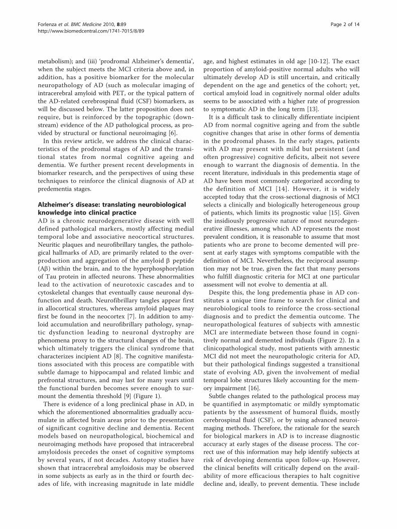
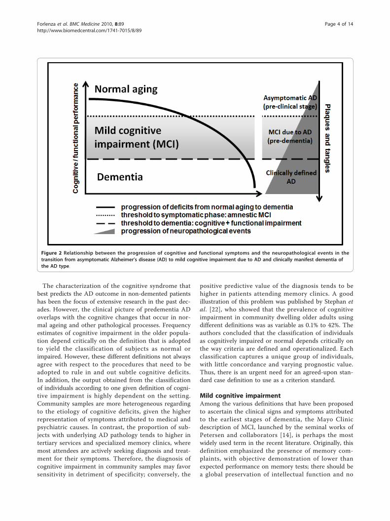
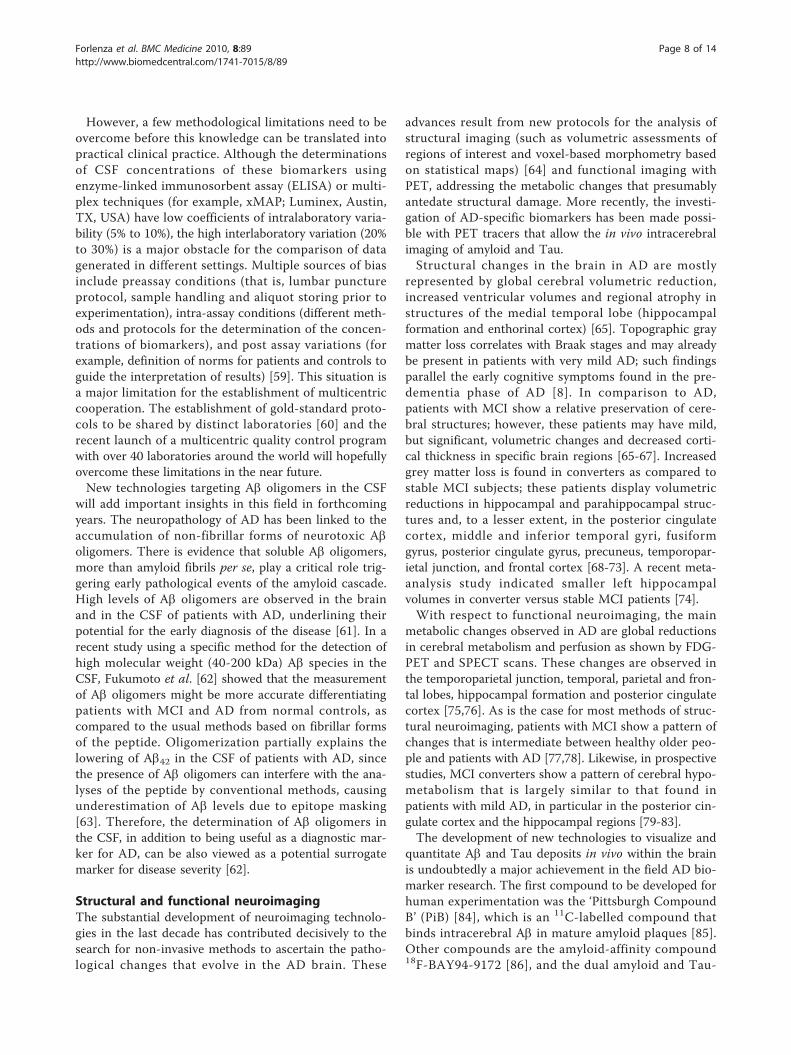
Alzheimer’s disease: translating neurobiologicalknowledge into clinical practiceAD is a chronic neurodegenerative disease with welldefined pathological markers, mostly affecting medialtemporal lobe and associative neocortical structures.Neuritic plaques and neurofibrillary tangles, the patholo-gical hallmarks of AD, are primarily related to the over-production and aggregation of the amyloid b peptide(Ab) within the brain, and to the hyperphosphorylationof Tau protein in affected neurons. These abnormalitieslead to the activation of neurotoxic cascades and tocytoskeletal changes that eventually cause neuronal dys-function and death. Neurofibrillary tangles appear firstin allocortical structures, whereas amyloid plaques mayfirst be found in the neocortex [7]. In addition to amy-loid accumulation and neurofibrillary pathology, synap-tic dysfunction leading to neuronal dystrophy arephenomena proxy to the structural changes of the brain,which ultimately triggers the clinical syndrome thatcharacterizes incipient AD [8]. The cognitive manifesta-tions associated with this process are compatible withsubtle damage to hippocampal and related limbic andprefrontal structures, and may last for many years untilthe functional burden becomes severe enough to sur-mount the dementia threshold [9] (Figure 1).There is evidence of a long preclinical phase in AD, in
which the aforementioned abnormalities gradually accu-mulate in affected brain areas prior to the presentationof significant cognitive decline and dementia. Recentmodels based on neuropathological, biochemical andneuroimaging methods have proposed that intracerebralamyloidosis precedes the onset of cognitive symptomsby several years, if not decades. Autopsy studies haveshown that intracerebral amyloidosis may be observedin some subjects as early as in the third or fourth dec-ades of life, with increasing magnitude in late middle
age, and highest estimates in old age [10-12]. The exactproportion of amyloid-positive normal adults who willultimately develop AD is still uncertain, and criticallydependent on the age and genetics of the cohort; yet,cortical amyloid load in cognitively normal older adultsseems to be associated with a higher rate of progressionto symptomatic AD in the long term [13].It is a difficult task to clinically differentiate incipient
AD from normal cognitive ageing and from the subtlecognitive changes that arise in other forms of dementiain the prodromal phases. In the early stages, patientswith AD may present with mild but persistent (andoften progressive) cognitive deficits, albeit not severeenough to warrant the diagnosis of dementia. In therecent literature, individuals in this predementia stage ofAD have been most commonly categorized according tothe definition of MCI [14]. However, it is widelyaccepted today that the cross-sectional diagnosis of MCIselects a clinically and biologically heterogeneous groupof patients, which limits its prognostic value [15]. Giventhe insidiously progressive nature of most neurodegen-erative illnesses, among which AD represents the mostprevalent condition, it is reasonable to assume that mostpatients who are prone to become demented will pre-sent at early stages with symptoms compatible with thedefinition of MCI. Nevertheless, the reciprocal assump-tion may not be true, given the fact that many personswho fulfill diagnostic criteria for MCI at one particularassessment will not evolve to dementia at all.Despite this, the long predementia phase in AD con-
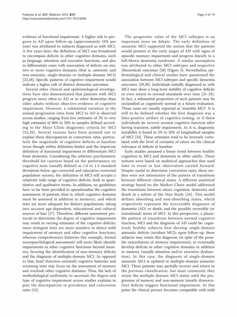
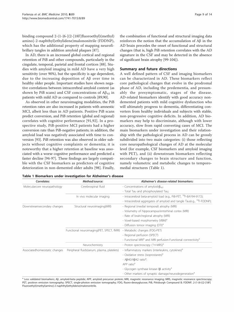
stitutes a unique time frame to search for clinical andneurobiological tools to reinforce the cross-sectionaldiagnosis and to predict the dementia outcome. Theneuropathological features of subjects with amnesticMCI are intermediate between those found in cogni-tively normal and demented individuals (Figure 2). In aclinicopathological study, most patients with amnesticMCI did not meet the neuropathologic criteria for AD,but their pathological findings suggested a transitionalstate of evolving AD, given the involvement of medialtemporal lobe structures likely accounting for the mem-ory impairment [16].Subtle changes related to the pathological process may
be quantified in asymptomatic or mildly symptomaticpatients by the assessment of humoral fluids, mostlycerebrospinal fluid (CSF), or by using advanced neuroi-maging methods. Therefore, the rationale for the searchfor biological markers in AD is to increase diagnosticaccuracy at early stages of the disease process. The cor-rect use of this information may help identify subjects atrisk of developing dementia upon follow-up. However,the clinical benefits will critically depend on the avail-ability of more efficacious therapies to halt cognitivedecline and, ideally, to prevent dementia. These include
Forlenza et al. BMC Medicine 2010, 8:89http://www.biomedcentral.com/1741-7015/8/89
Page 2 of 14
the development of more specific pharmacological inter-ventions targeting the pathological cascades of AD.
Predementia in Alzheimer’s diseaseThe cognitive syndrome of early stage AD is primarilyrepresented by encoding and retrieval deficits, resultingin episodic memory impairment and diminished capacityto learn new information [17-19]. Through clinicalassessment, such deficits may be revealed by memorytests such as story/picture recall and word list learning.However, the specificity of these findings may be ques-tionable, since similar deficits can be found in other pre-valent conditions in older people, namely depression andcerebrovascular disease. Studies suggest that AD-pronepatients fail to benefit from associative learning strategiesand from mnemonic cues (for example, cued delayedrecall paradigms). In the recent literature, short-term andlong-term memory binding deficits have been suggested
to be strong predictors of AD in older adults [19]. Thesesubfunctions of the memory process refer to the abilityto hold multiple sources of information in memory, andto bind together different aspects of one given stimulusin order to form integrated memories. These abilities arerequired for associative learning, and binding deficitsmay be early signs of hippocampal dysfunction. In fact,AD patients have difficulties in learning associationsbetween two or more characteristics of the same object(for example, shape and color), distinct verbal contents ofone given idea, the association between faces and names,and also to integrate spatial locations to other mnemoniccontents. The clinical progression towards dementiaincludes the additional impairment of at least one morecognitive domain, which is normally represented byexecutive dysfunction [20]. Recent data indicate a posi-tive strong correlation between the magnitude of execu-tive and functional impairment [21].
Figure 1 Hypothetical model of the pathological processes in Alzheimer’s disease (AD), focusing on the amyloid b peptide (Ab)cascade. (Other relevant mechanisms have been omitted or presented in a secondary perspective for didactic purposes.) Dotted arrows indicatepossible or secondary mechanisms affecting core pathological processes within the amyloid cascade. Background shades of gray separated bydotted lines are a schematic representation to integrate the progression of pathological events along with the development of the cognitivesyndrome of AD (these thresholds are arbitrary and not experimentally validated, and represent the authors’ point of view of the diseaseprocess). Three clinical phases of the disease are defined: presymptomatic (or preclinical) AD may last for several years or decades until theoverproduction and accumulation of Ab in the brain reaches a critical level that triggers the amyloid cascade; in the predementia phase,compatible with the definition of mild cognitive impairment secondary to AD, early stage pathology is present in varying degrees, from mildneuronal dystrophy to early stage Braak pathology, according to individual resilience and brain reserve. Finally, in the clinically defined dementiaphase, there is a progressive accumulation of the classical pathological hallmarks of AD (that is, neuritic plaques and neurofibrillary tangles),bearing relationship with the progression of cognitive deficits and the magnitude of functional impairment. APP = amyloid precursor protein;PS1/2 = presenilin 1/2; TAU = microtubule-associated protein Tau.
Forlenza et al. BMC Medicine 2010, 8:89http://www.biomedcentral.com/1741-7015/8/89
Page 3 of 14
The characterization of the cognitive syndrome thatbest predicts the AD outcome in non-demented patientshas been the focus of extensive research in the past dec-ades. However, the clinical picture of predementia ADoverlaps with the cognitive changes that occur in nor-mal ageing and other pathological processes. Frequencyestimates of cognitive impairment in the older popula-tion depend critically on the definition that is adoptedto yield the classification of subjects as normal orimpaired. However, these different definitions not alwaysagree with respect to the procedures that need to beadopted to rule in and out subtle cognitive deficits.In addition, the output obtained from the classificationof individuals according to one given definition of cogni-tive impairment is highly dependent on the setting.Community samples are more heterogeneous regardingto the etiology of cognitive deficits, given the higherrepresentation of symptoms attributed to medical andpsychiatric causes. In contrast, the proportion of sub-jects with underlying AD pathology tends to higher intertiary services and specialized memory clinics, wheremost attendees are actively seeking diagnosis and treat-ment for their symptoms. Therefore, the diagnosis ofcognitive impairment in community samples may favorsensitivity in detriment of specificity; conversely, the
positive predictive value of the diagnosis tends to behigher in patients attending memory clinics. A goodillustration of this problem was published by Stephan etal. [22], who showed that the prevalence of cognitiveimpairment in community dwelling older adults usingdifferent definitions was as variable as 0.1% to 42%. Theauthors concluded that the classification of individualsas cognitively impaired or normal depends critically onthe way criteria are defined and operationalized. Eachclassification captures a unique group of individuals,with little concordance and varying prognostic value.Thus, there is an urgent need for an agreed-upon stan-dard case definition to use as a criterion standard.
Mild cognitive impairmentAmong the various definitions that have been proposedto ascertain the clinical signs and symptoms attributedto the earliest stages of dementia, the Mayo Clinicdescription of MCI, launched by the seminal works ofPetersen and collaborators [14], is perhaps the mostwidely used term in the recent literature. Originally, thisdefinition emphasized the presence of memory com-plaints, with objective demonstration of lower thanexpected performance on memory tests; there should bea global preservation of intellectual function and no
Figure 2 Relationship between the progression of cognitive and functional symptoms and the neuropathological events in thetransition from asymptomatic Alzheimer’s disease (AD) to mild cognitive impairment due to AD and clinically manifest dementia ofthe AD type.
Forlenza et al. BMC Medicine 2010, 8:89http://www.biomedcentral.com/1741-7015/8/89
Page 4 of 14
evidence of functional impairment. A higher risk to pro-gress to AD upon follow-up (approximately 10% peryear) was attributed to subjects diagnosed as with MCI.A few years later, the definition of MCI was broadenedto encompass deficits in other cognitive domains, suchas language, attention and executive functions, and alsoto differentiate cases with association of deficits on one,two or more cognitive domains (that is, amnestic andnon-amnestic, single-domain or multiple-domain MCI)[23,24]. Specific patterns of cognitive impairment wouldindicate a higher risk of distinct dementia outcomes.Several other clinical and epidemiological investiga-
tions have also demonstrated that patients with MCIprogress more often to AD or to other dementias thanolder adults without objective evidence of cognitiveimpairment. However, a substantial variation in theannual progression rates from MCI to AD is observedacross studies, ranging from low estimates of 3% to veryhigh estimates of 40% to 50% in samples defined accord-ing to the Mayo Clinic diagnostic criteria for MCI[25,26]. Several reasons have been pointed out toexplain these discrepancies in conversion rates, particu-larly the magnitude of cognitive deficits at baseline(even though within definition limits) and the imprecisedefinition of functional impairment to differentiate MCIfrom dementia. Considering the arbitrary psychometricthreshold for caseness based on the performance oncognitive tests (usually defined as 1.0 to 1.5 standarddeviations below age-corrected and education-correctedpopulation norms), the definition of MCI still accepts arelatively wide range of cognitive deficits, both in quan-titative and qualitative terms. In addition, no guidelineshave so far been provided to operationalize the cognitiveassessment of patients (that is, which cognitive domainsmust be assessed in addition to memory), and whichtests are more adequate for distinct populations, takinginto account age-dependent, educational and culturalsources of bias [27]. Therefore, different assessment pro-tocols to determine the degree of cognitive impairmentmay result in varying estimates of the cognitive deficits:more stringent tests are more sensitive to detect mildimpairment of memory and other cognitive functions,whereas comprehensive batteries (for example, formalneuropsychological assessment) will more likely identifyimpairments in other cognitive functions beyond mem-ory, favoring the identification of non-memory deficitsand the diagnosis of multiple-domain MCI. As opposedto that, brief (function-oriented) cognitive batteries andscreening tests may focus on the assessment of memoryand overlook other cognitive domains. Thus, the lack ofmethodological uniformity to ascertain the degree andtype of cognitive impairment across studies explains inpart the discrepancies in prevalence and conversionrates [15].
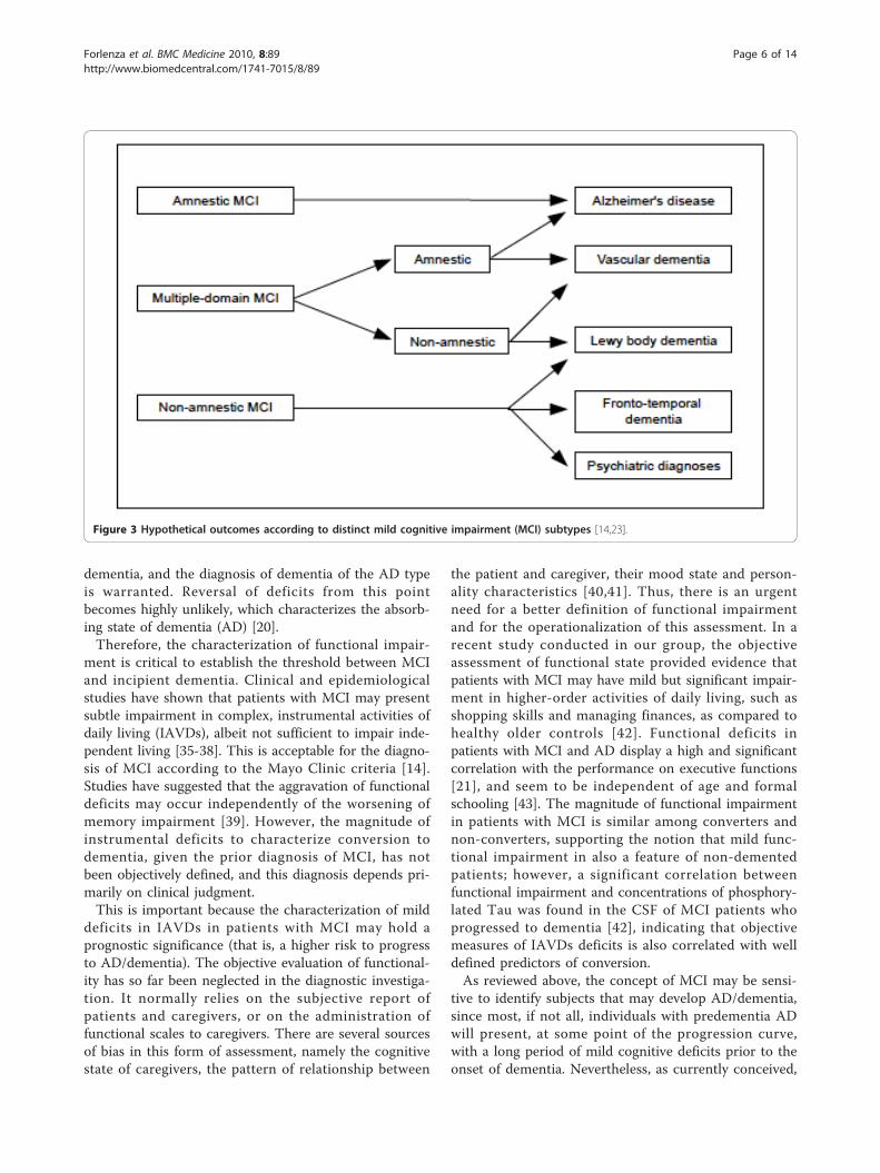
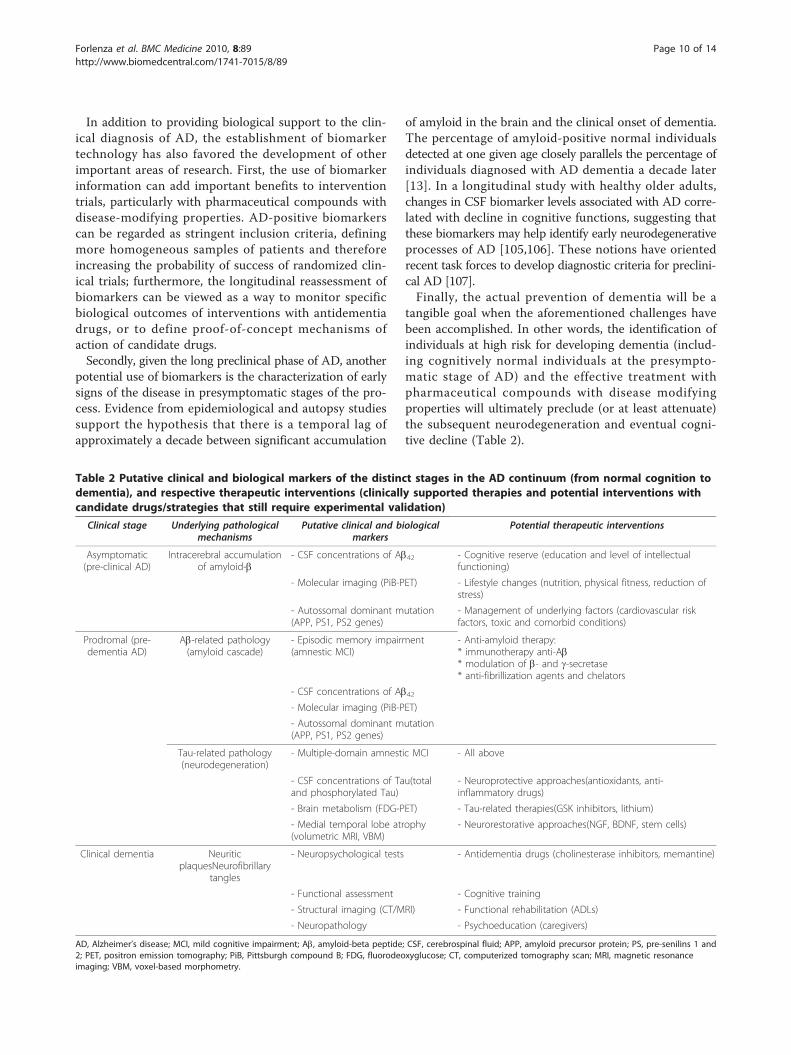
The prognostic value of the MCI subtypes is animportant issue on debate. The early definition ofamnestic MCI supported the notion that the patientswould present at the early stages of AD with signs ofepisodic memory impairment and progress linearly to afull-blown dementia syndrome. A similar assumptionwas attributed to other MCI subtypes and respective(theoretical) outcomes [28] (Figure 3). Nevertheless, epi-demiological and clinical studies have questioned theassociation between MCI subtypes and specific dementiaoutcomes [29,30]. Individuals initially diagnosed as withMCI may show a long-term stability of cognitive deficitsor even return to normal standards over time [31-33].In fact, a substantial proportion of such patients may bereclassified as cognitively normal in a future evaluation.These cases are usually reported as ‘unstable MCI’. It isstill to be defined whether the first diagnosis was afalse-positive artifact of cognitive testing, or if theseindividuals do recover normal cognitive function afterhaving transient, subtle impairment. As it is, diagnosticinstability is found in 5% to 20% of longitudinal samplesof MCI [33]. These estimates tend to be inversely corre-lated with the level of certainty of raters on the clinicalrelevance of deficits at baseline.Early studies assumed a linear trend between healthy
cognition to MCI and dementia in older adults. Thesenotions were based on analytical approaches that usedtime to event or last observation carried forward.Despite useful to determine conversion rates, these stu-dies were not informative of the pattern of transitionsbetween different clinical states. A different analyticalstrategy based on the Markov Chain model addressedthe transitions between intact cognition, dementia anddeath in a subset of the Nun Study [34]. This modeldefines absorbing and non-absorbing states, whichrespectively represent the irreversible diagnoses ofdementia (AD) or death, and the possibly reversible (ortransitional) states of MCI. In this perspective, a plausi-ble pattern of transitions between normal cognitivefunction, MCI and the diagnosis of AD could be: cogni-tively healthy subjects first develop single-domainamnestic deficits (incident MCI); upon follow-up, thesesubjects may retain this diagnosis (in spite of the possi-ble exacerbation of memory impairment), or eventuallydevelop deficits in other cognitive domains in additionto memory (usually attention and/or executive dysfunc-tion). In this case, the diagnosis of single-domainamnestic MCI is updated to multiple-domain amnesticMCI. These patients may partially recover and return tothe previous classification, but most commonly theyretain the multiple-domain MCI status until the pro-gression of memory and non-memory (mostly dysexecu-tive) deficits triggers functional impairment. At thispoint the clinical picture becomes compatible with mild
Forlenza et al. BMC Medicine 2010, 8:89http://www.biomedcentral.com/1741-7015/8/89
Page 5 of 14
dementia, and the diagnosis of dementia of the AD typeis warranted. Reversal of deficits from this pointbecomes highly unlikely, which characterizes the absorb-ing state of dementia (AD) [20].Therefore, the characterization of functional impair-
ment is critical to establish the threshold between MCIand incipient dementia. Clinical and epidemiologicalstudies have shown that patients with MCI may presentsubtle impairment in complex, instrumental activities ofdaily living (IAVDs), albeit not sufficient to impair inde-pendent living [35-38]. This is acceptable for the diagno-sis of MCI according to the Mayo Clinic criteria [14].Studies have suggested that the aggravation of functionaldeficits may occur independently of the worsening ofmemory impairment [39]. However, the magnitude ofinstrumental deficits to characterize conversion todementia, given the prior diagnosis of MCI, has notbeen objectively defined, and this diagnosis depends pri-marily on clinical judgment.This is important because the characterization of mild
deficits in IAVDs in patients with MCI may hold aprognostic significance (that is, a higher risk to progressto AD/dementia). The objective evaluation of functional-ity has so far been neglected in the diagnostic investiga-tion. It normally relies on the subjective report ofpatients and caregivers, or on the administration offunctional scales to caregivers. There are several sourcesof bias in this form of assessment, namely the cognitivestate of caregivers, the pattern of relationship between
the patient and caregiver, their mood state and person-ality characteristics [40,41]. Thus, there is an urgentneed for a better definition of functional impairmentand for the operationalization of this assessment. In arecent study conducted in our group, the objectiveassessment of functional state provided evidence thatpatients with MCI may have mild but significant impair-ment in higher-order activities of daily living, such asshopping skills and managing finances, as compared tohealthy older controls [42]. Functional deficits inpatients with MCI and AD display a high and significantcorrelation with the performance on executive functions[21], and seem to be independent of age and formalschooling [43]. The magnitude of functional impairmentin patients with MCI is similar among converters andnon-converters, supporting the notion that mild func-tional impairment in also a feature of non-dementedpatients; however, a significant correlation betweenfunctional impairment and concentrations of phosphory-lated Tau was found in the CSF of MCI patients whoprogressed to dementia [42], indicating that objectivemeasures of IAVDs deficits is also correlated with welldefined predictors of conversion.As reviewed above, the concept of MCI may be sensi-
tive to identify subjects that may develop AD/dementia,since most, if not all, individuals with predementia ADwill present, at some point of the progression curve,with a long period of mild cognitive deficits prior to theonset of dementia. Nevertheless, as currently conceived,
Figure 3 Hypothetical outcomes according to distinct mild cognitive impairment (MCI) subtypes [14,23].
Forlenza et al. BMC Medicine 2010, 8:89http://www.biomedcentral.com/1741-7015/8/89
Page 6 of 14
the clinically oriented diagnostic criteria for MCI yield aheterogeneous group of patients with distinct short-term and long-term outcomes. In other words, the spe-cificity and the predictive value of the MCI diagnosisare low, and the cross-sectional identification of cases ofprodromal AD may not reach adequate diagnostic accu-racy if based solely on clinical tools [15]. Rather, it bene-fits substantially from the combination of clinical andbiological information. In the next sections we willrevise the recent developments on biochemical and neu-roimaging biomarkers for the early diagnosis of AD. Ofcourse, if such tests are unavailable, which may be thecase in most healthcare settings, particularly in lessfavored countries, the expert interpretation of testresults, the criterious observation of longitudinal mea-sures, including the careful judgment of all availablevariables, is certainly the best alternative to drive clinicaldecisions.
The search for biological biomarkers ofAlzheimer’s diseaseThe development of biomarker research in AD is a goodexample of the successful effort to translate the knowl-edge of key pathophysiological mechanisms of the diseaseinto clinical applications. A biomarker is a characteristicthat can be measured and evaluated as an indicator ofthe pathogenetic processes, or to ascertain the effect ofpharmacological interventions on predefined biologicalcascades [44]. The ideal diagnostic marker for AD shouldmeet at least three basic requirements: (i) reflect coreneurobiological changes that characterize the diseaseprocess; (ii) be validated by post mortem studies, assum-ing that the neuropathological findings are gold stan-dards of abnormalities affecting the same cascade; and(iii) be measurable as early as possible in the disease con-tinuum, ideally at presymptomatic stages. Additionalrequirements include being non-invasive and simple toperform, precise and reliable, and adequate for large-scale screenings. Among many candidate markers, nonehas so far achieved universal acceptance, nor fully metthe abovementioned criteria. Nonetheless, there has beensignificant progress toward this goal in the areas of CSFand neuroimaging biomarker identification, with atten-tion focusing on the prediction of AD in the prodromalstages of disease and in high-risk groups.
CSF biomarkersThe CSF may be considered an ideal source for viable bio-markers in AD. It is in intimate contact with the cerebraltissue, and pathological changes in the brain are oftenreflected in the CSF [45]. Among several potential diag-nostic biomarkers, the most consistent findings have beenobtained with the measurement of CSF concentrations ofAb peptide (Ab42), total Tau (T-Tau) and phosphorylated
Tau (P-Tau). AD patients characteristically display lowconcentrations of Ab42 and high concentrations of T-Tauand P-Tau. This pattern of CSF biomarkers is commonlyreferred to as the ‘AD signature’ in the CSF. The afore-mentioned biomarkers reflect core pathophysiological fea-tures of the disease [46], and have been validated in postmortem studies [47-49]. Increased concentration of T-Taumay be a less specific marker of axonal damage, as it canbe found in vascular and other neurodegenerative demen-tias in addition to AD (for example, prion diseases). How-ever, it bears a positive correlation with the speed and themagnitude of the neurodegenerative process. DecreasedAb42 and increased P-Tau are more specific to AD. Ab42is a byproduct of the abnormal processing of the amyloidprecursor protein (APP) leading to amyloidogenesis andformation of neuritic plaques. In addition, decreased con-centrations of Ab42 likely reflect its deposition in plaques,preventing its clearance through the CSF P-Tau illustratesthe cytoskeletal changes that arise from the deregulationof microtubule homeostasis and ultimately cause axonaldysfunction and neuronal death. This marker is more spe-cifically associated with AD, given the central role of Tauhyperphosphorylation in the formation of paired helicalfilaments (PHFs) and neurofibrillary tangles [50].To date, over 100 studies have been published to sup-
port the notion that this AD-positive CSF pattern hasgood diagnostic accuracy to distinguish between normalageing and AD (> 85%) and a positive predictive value (>90%) to determine the dementia outcome in patients withMCI [51]. However, in the differential diagnosis of estab-lished dementia syndromes, the sensitivity/specificity pro-file to differentiate AD from other dementias issignificantly lower [52]. Large-scale longitudinal studies ofMCI cohorts consistently demonstrated that the presenceof the ‘AD signature’ in the CSF has a good diagnosticaccuracy (that is, >80%) discriminating patients with MCIwho progress to AD (’MCI converters’) from those whoremain cognitively stable (’MCI-stable’ patients) andhealthy controls [51], as well as those MCI patients whoprogress to non-AD dementias [53]. These sets of datahave been extensively replicated by different researchgroups worldwide [54-57]. Findings are largely confirma-tory, as reinforced by a recent meta-analysis [58]. Takentogether, these studies provide compelling evidence thatthe ‘AD signature’ in the CSF is a strong predictor ofdementia outcome. MCI patients who convert to AD havea CSF biomarker pattern indistinguishable to that found inpatients with dementia of the AD type; and MCI patientswith progressive deficits (albeit not severe enough to char-acterize conversion) have a similar pattern to the formerpatients. Conversely, MCI patients with unstable (transi-ent) MCI and those who display non-progressive deficitsover time have a CSF biomarker pattern very similar tothat found in healthy older adults.
Forlenza et al. BMC Medicine 2010, 8:89http://www.biomedcentral.com/1741-7015/8/89
Page 7 of 14
However, a few methodological limitations need to beovercome before this knowledge can be translated intopractical clinical practice. Although the determinationsof CSF concentrations of these biomarkers usingenzyme-linked immunosorbent assay (ELISA) or multi-plex techniques (for example, xMAP; Luminex, Austin,TX, USA) have low coefficients of intralaboratory varia-bility (5% to 10%), the high interlaboratory variation (20%to 30%) is a major obstacle for the comparison of datagenerated in different settings. Multiple sources of biasinclude preassay conditions (that is, lumbar punctureprotocol, sample handling and aliquot storing prior toexperimentation), intra-assay conditions (different meth-ods and protocols for the determination of the concen-trations of biomarkers), and post assay variations (forexample, definition of norms for patients and controls toguide the interpretation of results) [59]. This situation isa major limitation for the establishment of multicentriccooperation. The establishment of gold-standard proto-cols to be shared by distinct laboratories [60] and therecent launch of a multicentric quality control programwith over 40 laboratories around the world will hopefullyovercome these limitations in the near future.New technologies targeting Ab oligomers in the CSF
will add important insights in this field in forthcomingyears. The neuropathology of AD has been linked to theaccumulation of non-fibrillar forms of neurotoxic Aboligomers. There is evidence that soluble Ab oligomers,more than amyloid fibrils per se, play a critical role trig-gering early pathological events of the amyloid cascade.High levels of Ab oligomers are observed in the brainand in the CSF of patients with AD, underlining theirpotential for the early diagnosis of the disease [61]. In arecent study using a specific method for the detection ofhigh molecular weight (40-200 kDa) Ab species in theCSF, Fukumoto et al. [62] showed that the measurementof Ab oligomers might be more accurate differentiatingpatients with MCI and AD from normal controls, ascompared to the usual methods based on fibrillar formsof the peptide. Oligomerization partially explains thelowering of Ab42 in the CSF of patients with AD, sincethe presence of Ab oligomers can interfere with the ana-lyses of the peptide by conventional methods, causingunderestimation of Ab levels due to epitope masking[63]. Therefore, the determination of Ab oligomers inthe CSF, in addition to being useful as a diagnostic mar-ker for AD, can be also viewed as a potential surrogatemarker for disease severity [62].
Structural and functional neuroimagingThe substantial development of neuroimaging technolo-gies in the last decade has contributed decisively to thesearch for non-invasive methods to ascertain the patho-logical changes that evolve in the AD brain. These
advances result from new protocols for the analysis ofstructural imaging (such as volumetric assessments ofregions of interest and voxel-based morphometry basedon statistical maps) [64] and functional imaging withPET, addressing the metabolic changes that presumablyantedate structural damage. More recently, the investi-gation of AD-specific biomarkers has been made possi-ble with PET tracers that allow the in vivo intracerebralimaging of amyloid and Tau.Structural changes in the brain in AD are mostly
represented by global cerebral volumetric reduction,increased ventricular volumes and regional atrophy instructures of the medial temporal lobe (hippocampalformation and enthorinal cortex) [65]. Topographic graymatter loss correlates with Braak stages and may alreadybe present in patients with very mild AD; such findingsparallel the early cognitive symptoms found in the pre-dementia phase of AD [8]. In comparison to AD,patients with MCI show a relative preservation of cere-bral structures; however, these patients may have mild,but significant, volumetric changes and decreased corti-cal thickness in specific brain regions [65-67]. Increasedgrey matter loss is found in converters as compared tostable MCI subjects; these patients display volumetricreductions in hippocampal and parahippocampal struc-tures and, to a lesser extent, in the posterior cingulatecortex, middle and inferior temporal gyri, fusiformgyrus, posterior cingulate gyrus, precuneus, temporopar-ietal junction, and frontal cortex [68-73]. A recent meta-analysis study indicated smaller left hippocampalvolumes in converter versus stable MCI patients [74].With respect to functional neuroimaging, the main
metabolic changes observed in AD are global reductionsin cerebral metabolism and perfusion as shown by FDG-PET and SPECT scans. These changes are observed inthe temporoparietal junction, temporal, parietal and fron-tal lobes, hippocampal formation and posterior cingulatecortex [75,76]. As is the case for most methods of struc-tural neuroimaging, patients with MCI show a pattern ofchanges that is intermediate between healthy older peo-ple and patients with AD [77,78]. Likewise, in prospectivestudies, MCI converters show a pattern of cerebral hypo-metabolism that is largely similar to that found inpatients with mild AD, in particular in the posterior cin-gulate cortex and the hippocampal regions [79-83].The development of new technologies to visualize and
quantitate Ab and Tau deposits in vivo within the brainis undoubtedly a major achievement in the field AD bio-marker research. The first compound to be developed forhuman experimentation was the ‘Pittsburgh CompoundB’ (PiB) [84], which is an 11C-labelled compound thatbinds intracerebral Ab in mature amyloid plaques [85].Other compounds are the amyloid-affinity compound18F-BAY94-9172 [86], and the dual amyloid and Tau-
Forlenza et al. BMC Medicine 2010, 8:89http://www.biomedcentral.com/1741-7015/8/89
Page 8 of 14
binding compound 2-(1-{6-[(2-[18F]fluoroethyl)(methyl)amino]-2-naphthyl}ethylidene)malononitrile (FDDNP),which has the additional property of mapping neurofi-brillary tangles in addition amyloid plaques [87].In AD, there is an increased global cortical and regional
retention of PiB and other compounds, particularly in thecingulate, temporal, parietal and frontal cortices [88]. Stu-dies with amyloid imaging in mild AD have a very highsensitivity (over 90%), but the specificity is age dependent,due to the increasing deposition of Ab over time inhealthy older people. Important studies have shown nega-tive correlations between intracerebral amyloid content (asshown by PiB scans) and CSF concentrations of Ab42 inpatients with mild AD as compared to controls [89,90].As observed in other neuroimaging modalities, the PiB
retention rates are also increased in patients with amnesticMCI, albeit less than in AD patients. Positive PiB scanspredict conversion, and PiB retention (global and regional)correlates with cognitive performance [91,92]. In a pro-spective study, PiB-positive MCI patients had a higherconversion rate than PiB-negative patients; in addition, theamyloid load was negatively associated with time to con-version [93]. PiB retention was also observed in older sub-jects without cognitive complaints or dementia; it isnoteworthy that a higher retention at baseline was asso-ciated with a worse cognitive performance and predicted afaster decline [94-97]. These findings are largely compati-ble with the CSF biomarkers as predictors of cognitivedeterioration in non-demented older adults [98]. Finally,
the combination of functional and structural imaging datareinforces the notion that the accumulation of Ab in theAD brain precedes the onset of functional and structuralchanges (that is, high PiB retention correlates with the ADsignature in the CSF and may be detected in the absenceof significant brain atrophy [99-104]).
Summary and future directionsA well defined pattern of CSF and imaging biomarkerscan be characterized in AD. These biomarkers reflectcore pathological changes that evolve in the prodromalphase of AD, including the predementia, and presum-ably the presymptomatic, stages of the disease.AD-related biomarkers identify with good accuracy non-demented patients with mild cognitive dysfunction whowill ultimately progress to dementia, differentiating con-verters from healthy individuals and subjects with stable,non-progressive cognitive deficits. In addition, AD bio-markers may help to discriminate, although with loweraccuracy, slow from rapid converting cases of MCI. Themain biomarkers under investigation and their relation-ship with the pathological process in AD can be grosslysubdivided into two main categories: (i) those reflectingcore neuropathological changes of AD at the molecularlevel (for example, CSF biomarkers and amyloid imagingwith PET), and (ii) downstream biomarkers reflectingsecondary changes to brain structure and function,namely volumetric and metabolic changes to temporo-medial structures (Table 1).
Table 1 Biomarkers under investigation for Alzheimer’s disease
Correlates: Method/source: Alzheimer’s disease-related biomarkers:
Molecularcore neuropathology Cerebrospinal fluid - Concentrations of amyloid-b42;- Total Tau and phosphorylated Tau;
In vivo molecular imaging - Intracerebral beta-amyloid load (e.g., PiB-PET, 18F-BAY94-9172);
- Intracerebral aggregates of amyloid and tangle Tau(e.g., 18F-FDDNP);
Downstreamsecondary changes Structural neuroimaging(MRI) - Regional (medial temporal) atrophy (MRI)
- Volumetry of hippocampus/entorhinal cortex (MRI)
- Rate of brain/regional atrophy (MRI)
- Voxel-based morphometry (VBM)a
- Diffusion tensor imaging (DTI)a
Functional neuroimaging(PET, SPECT, fMRI) - Metabolic changes (FDG-PET)
- Regional perfusion (SPECT)
- Functional MRIa and MRI perfusion-Functional connectivitya
Neurochemistry - Proton spectroscopy (+H-MRS)a
Associatedhomeostatic changes Peripheral fluids(serum, plasma, platelets) - Inflammatory markers (interleukins, cytokines)a
- Oxidative stress (isoprostanes)a
- Ab40/Ab42 ratio*;
APP ratioa
- Glycogen synthase kinase-3b activitya
- Other markers of synaptic damage/neurodegenerationa
a Less validated biomarkers; Ab, amyloid-beta peptide; APP, amyloid precursor protein; MRI, magnetic resonance imaging; MRS, magnetic resonance spectroscopy;PET, positron emission tomography; SPECT, single-photon emission tomography; FDG, fluoro-deoxyglucose; PiB, Pittsburgh Compound B; FDDNP, 2-(1-{6-[(2-[18F]Fluoroethyl)(methyl)amino]-2-naphthyl}ethylidene)malononitrile.
Forlenza et al. BMC Medicine 2010, 8:89http://www.biomedcentral.com/1741-7015/8/89
Page 9 of 14
In addition to providing biological support to the clin-ical diagnosis of AD, the establishment of biomarkertechnology has also favored the development of otherimportant areas of research. First, the use of biomarkerinformation can add important benefits to interventiontrials, particularly with pharmaceutical compounds withdisease-modifying properties. AD-positive biomarkerscan be regarded as stringent inclusion criteria, definingmore homogeneous samples of patients and thereforeincreasing the probability of success of randomized clin-ical trials; furthermore, the longitudinal reassessment ofbiomarkers can be viewed as a way to monitor specificbiological outcomes of interventions with antidementiadrugs, or to define proof-of-concept mechanisms ofaction of candidate drugs.Secondly, given the long preclinical phase of AD, another
potential use of biomarkers is the characterization of earlysigns of the disease in presymptomatic stages of the pro-cess. Evidence from epidemiological and autopsy studiessupport the hypothesis that there is a temporal lag ofapproximately a decade between significant accumulation
of amyloid in the brain and the clinical onset of dementia.The percentage of amyloid-positive normal individualsdetected at one given age closely parallels the percentage ofindividuals diagnosed with AD dementia a decade later[13]. In a longitudinal study with healthy older adults,changes in CSF biomarker levels associated with AD corre-lated with decline in cognitive functions, suggesting thatthese biomarkers may help identify early neurodegenerativeprocesses of AD [105,106]. These notions have orientedrecent task forces to develop diagnostic criteria for preclini-cal AD [107].Finally, the actual prevention of dementia will be a
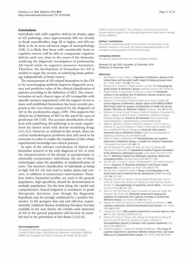
tangible goal when the aforementioned challenges havebeen accomplished. In other words, the identification ofindividuals at high risk for developing dementia (includ-ing cognitively normal individuals at the presympto-matic stage of AD) and the effective treatment withpharmaceutical compounds with disease modifyingproperties will ultimately preclude (or at least attenuate)the subsequent neurodegeneration and eventual cogni-tive decline (Table 2).
Table 2 Putative clinical and biological markers of the distinct stages in the AD continuum (from normal cognition todementia), and respective therapeutic interventions (clinically supported therapies and potential interventions withcandidate drugs/strategies that still require experimental validation)
Clinical stage Underlying pathologicalmechanisms
Putative clinical and biologicalmarkers
Potential therapeutic interventions
Asymptomatic(pre-clinical AD)
Intracerebral accumulationof amyloid-b
- CSF concentrations of Ab42 - Cognitive reserve (education and level of intellectualfunctioning)
- Molecular imaging (PiB-PET) - Lifestyle changes (nutrition, physical fitness, reduction ofstress)
- Autossomal dominant mutation(APP, PS1, PS2 genes)
- Management of underlying factors (cardiovascular riskfactors, toxic and comorbid conditions)
Prodromal (pre-dementia AD)
Ab-related pathology(amyloid cascade)
- Episodic memory impairment(amnestic MCI)
- Anti-amyloid therapy:* immunotherapy anti-Ab* modulation of b- and g-secretase* anti-fibrillization agents and chelators
- CSF concentrations of Ab42- Molecular imaging (PiB-PET)
- Autossomal dominant mutation(APP, PS1, PS2 genes)
Tau-related pathology(neurodegeneration)
- Multiple-domain amnestic MCI - All above
- CSF concentrations of Tau(totaland phosphorylated Tau)
- Neuroprotective approaches(antioxidants, anti-inflammatory drugs)
- Brain metabolism (FDG-PET) - Tau-related therapies(GSK inhibitors, lithium)
- Medial temporal lobe atrophy(volumetric MRI, VBM)
- Neurorestorative approaches(NGF, BDNF, stem cells)
Clinical dementia NeuriticplaquesNeurofibrillary
tangles
- Neuropsychological tests - Antidementia drugs (cholinesterase inhibitors, memantine)
- Functional assessment - Cognitive training
- Structural imaging (CT/MRI) - Functional rehabilitation (ADLs)
- Neuropathology - Psychoeducation (caregivers)
AD, Alzheimer’s disease; MCI, mild cognitive impairment; Ab, amyloid-beta peptide; CSF, cerebrospinal fluid; APP, amyloid precursor protein; PS, pre-senilins 1 and2; PET, positron emission tomography; PiB, Pittsburgh compound B; FDG, fluorodeoxyglucose; CT, computerized tomography scan; MRI, magnetic resonanceimaging; VBM, voxel-based morphometry.
Forlenza et al. BMC Medicine 2010, 8:89http://www.biomedcentral.com/1741-7015/8/89
Page 10 of 14
ConclusionsIndividuals with mild cognitive deficits do display signsof AD pathology, since approximately 50% are alreadyin Braak neurofibrillary stage III or higher, and 20% arelikely to be in more advanced stages of neuropathology[108]. It is likely that those with considerable brain orcognitive reserve will be able to compensate cognitivedeficits until very close to the onset of the dementia,rendering the diagnostic investigation of predementiaAD based solely on cognitive measures insensitive.Therefore, the development of biomarkers for AD isneeded to target the severity of underlying brain pathol-ogy independently of brain reserve.The measurement of AD-related biomarkers in the CSF
or by neuroimaging methods improves diagnostic accu-racy and predictive value of the clinical classification ofpatients according to the definition of MCI. The charac-terization of early clinical signs of AD (compatible withepisodic memory impairment) with the support of one ormore well established biomarkers has been recently pro-posed as the core feature required for the diagnosis ofAD in the predementia stages [109]. This supports theclinical use of definition of MCI in the search for cases ofprodromal AD [110]. The accurate identification of sub-jects with underlying AD pathology is an acute require-ment for future trials with disease-modifying drugs[111,112]. However, as outlined in this review, there arecritical methodological problems that still need to beovercome in order to enable the translation of this robustexperimental knowledge into clinical practice.In spite of the relevant contribution of clinical and
biomarker research in the early diagnosis of AD, or eventhe characterization of the disease in asymptomatic orminimally symptomatic individuals, the use of thesetechnologies raises the possibility of misidentification ofcases. The incorrect classification of individuals as beingat high risk for AD may lead to undue alarm and con-cern, in addition to unnecessary interventions. There-fore, before biomarker profiles are used in the generalpopulation, high specificity should be demonstrated inmultiple populations. For the time being, the careful andcomprehensive clinical judgment is mandatory to guidetherapeutic decisions, even though the diagnostichypothesis may be strongly reinforced by a positive bio-marker. In the prospect that safe and effective, experi-mentally validated disease-modifying therapies becomeavailable in the near future, the reliable early detectionof AD in the general population will become an essen-tial tool in the prevention of this illness [113,114].
AcknowledgementsThe present work was supported by Conselho Nacional de PesquisaCientífica (CNPq, Project 554535/2005-0), Alzheimer’s Association (NIRG-08-90688), and Fundação de Amparo à Pesquisa do Estado de São Paulo
(FAPESP, Project 02/13633-7). The Laboratory of Neuroscience (LIM-27)receives financial support from Associação Beneficente Alzira Denise Hertzogda Silva (ABADHS).
Authors’ contributionsThe authors contributed equally to the selection and discussion of theliterature reviewed in this work. The authors participated equally in theconception and preparation of the final manuscript.
Competing interestsThe authors declare that they have no competing interests.
Received: 30 July 2010 Accepted: 22 December 2010Published: 22 December 2010
References1. Brookmeyer R, Gray S, Kawas C: Projections of Alzheimer’s disease in the
United States and the public health impact of delaying disease onset.Am J Public Health 1998, 88:1337-1342.
2. Brookmeyer R, Johnson E, Ziegler-Graham K, Arrighi HM: Forecasting theglobal burden of Alzheimer’s disease. Alzheimers Dement 2007, 3:186-191.
3. Ziegler-Graham K, Brookmeyer R, Johnson E, Arrighi HM: Worldwidevariation in the doubling time of Alzheimer’s disease incidence rates.Alzheimers Dement 2008, 4:316-323.
4. McKhann G, Drachman D, Folstein M, Katzman R, Price D, Stadlan EM:Clinical diagnosis of Alzheimer’s disease: report of the NINCDS-ADRDAWork Group under the auspices of Department of Health and HumanServices Task Force on Alzheimer’s Disease. Neurology 1984, 34:939-944.
5. McKhann G, Hyman B, Jack C, Kawas C, Klunk W, Knopman D, Koroshetz W,Manly J, Mayeux R, Mohs R, Morris J, Weintraub S, Alzheimer’s DiseaseDementia Workgroup: Criteria for AD dementia. [http://www.alz.org/research/diagnostic_criteria/dementia_recommendations.pdf].
6. Albert M, DeKosky S, Dickson D, Dubois B, Feldman H, Fox N, Gamst A,Holtzman D, Jagust W, Petersen R, Snyder P, Mild Cognitive Impairmentdue to Alzheimer’s Disease Workgroup: Criteria for mild cognitiveimpairment due to Alzheimer’s disease. [http://www.alz.org/research/diagnostic_criteria/mci_reccomendations.pdf].
7. Nelson PT, Braak H, Markesbery WR: Neuropathology and cognitiveimpairment in Alzheimer disease: a complex but coherent relationship.J Neuropathol Exp Neurol 2009, 68:1-14.
8. Jack CR, Knopman DS, Jagust WJ, Shaw LM, Aisen PS, Weiner MW,Petersen RC, Trojanowski JQ: Hypothetical model of dynamic biomarkersof the Alzheimer’s pathological cascade. Lancet Neurol 2010, 9:119-128.
9. Blass JP: Alzheimer’s disease and Alzheimer’s dementia: distinct butoverlapping entities. Neurobiol Aging 2002, 23:1077-1084.
10. Thal DR, Rüb U, Schultz C, Sassin I, Ghebremedhin E, Del Tredici K, Braak E,Braak H: Sequence of Aβ-protein deposition in the human medialtemporal lobe. J Neuropathol Exp Neurol 2000, 59:733-748.
11. Thal DR, Rüb U, Orantes M, Braak H: Phases of A β-deposition in thehuman brain and its relevance for the development of AD. Neurology2002, 58:1791-1800.
12. Knopman DS, Parisi JE, Salviati A, Floriach-Robert M, Boeve BF, Ivnik RJ,Smith GE, Dickson DW, Johnson KA, Petersen LE, McDonald WC, Braak H,Petersen RC: Neuropathology of cognitively normal elderly. J NeuropatholExp Neurol 2003, 62:1087-1095.
13. Morris JC, Roe CM, Grant EA, Head D, Storandt M, Goate AM, Fagan AM,Holtzman DM, Mintun MA: Pittsburgh compound B imaging andprediction of progression from cognitive normality to symptomaticAlzheimer disease. Arch Neurol 2009, 66:1469-1475.
14. Petersen RC, Smith GE, Waring SC, Ivnik RJ, Tangalos EG, Kokmen E: Mildcognitive impairment: clinical characterization and outcome. Arch Neurol1999, 56:303-308.
15. Forlenza OV, Chiu E: Mild cognitive impairment: a concept ready to moveon? Cur Opin Psychiatry 2008, 21:529-532.
16. Petersen RC, Parisi JE, Dickson DW, Johnson KA, Knopman DS, Boeve BF,Jicha GA, Ivnik RJ, Smith GE, Tangalos EG, Braak H, Kokmen E:Neuropathologic features of amnestic mild cognitive impairment. ArchNeurol 2006, 63:665-672.
17. Small BJ, Fratiglioni L, Viitanen M, Winblad B, Bäckman L: The course ofcognitive impairment in preclinical Alzheimer disease: three- and 6-yearfollow-up of a population-based sample. Arch Neurol 2000, 57:839-844.
Forlenza et al. BMC Medicine 2010, 8:89http://www.biomedcentral.com/1741-7015/8/89
Page 11 of 14
18. Blacker D, Lee H, Muzikansky A, Martin EC, Tanzi R, McArdle JJ, Moss M,Albert M: Neuropsychological measures in normal individuals thatpredict subsequent cognitive decline. Arch Neurol 2007, 64:862-871.
19. Parra MA, Abrahams S, Fabi K, Logie R, Luzzi S, Della Sala S: Short termmemory binding deficits in Alzheimer’s disease. Brain 2009,132:1057-1066.
20. Forlenza OV, Diniz BS, Nunes PV, Memoria CM, Yassuda MS, Gattaz WF:Diagnostic transitions in mild cognitive impairment subtypes. IntPsychogeriatr 2009, 21:1088-1095.
21. Pereira FS, Yassuda MS, Oliveira AM, Forlenza OV: Executive dysfunctioncorrelates with impaired functional status in older adults with varyingdegrees of cognitive impairment. Int Psychogeriatr 2008, 20:1104-1115.
22. Stephan BC, Matthews FE, McKeith IG, Bond J, Brayne C, Medical ResearchCouncil Cognitive Function and Aging Study: Early cognitive change inthe general population: how do different definitions work? J Am GeriatrSoc 2007, 55:1534-1540.
23. Petersen RC, Stevens JC, Ganguli M, Tangalos EG, Cummings JL,DeKosky ST: Practice parameter: early detection of dementia: mildcognitive impairment (an evidence-based review). Report of the QualityStandards Subcommittee of the American Academy of Neurology.Neurology 2001, 56:1133-1142.
24. Winblad B, Palmer K, Kivipelto M, Jelic V, Fratiglioni L, Wahlund LO,Nordberg A, Bäckman L, Albert M, Almkvist O, Arai H, Basun H, Blennow K,de Leon M, DeCarli C, Erkinjuntti T, Giacobini E, Graff C, Hardy J, Jack C,Jorm A, Ritchie K, van Duijn C, Visser P, Petersen RC: Mild cognitiveimpairment–beyond controversies, towards a consensus: report of theInternational Working Group on Mild Cognitive Impairment. J Intern Med2004, 256:240-246.
25. Bruscoli M, Lovestone S: Is MCI really just early dementia? A systematicreview of conversion studies. Int Psychogeriatr 2004, 16:129-140.
26. Mitchell A, Shiri-Feshki M: Rate of progression of mild cognitiveimpairment to dementia - meta-analysis of 41 robust inception cohortstudies. Acta Psychiatr Scand 2009, 119:252-265.
27. Visser P, Scheltens P, Verhey FR: Do MCI criteria in drug trials accuratelyidentify subjects with predementia Alzheimer’s disease? J NeurolNeurosurg Psychiatry 2005, 76:1348-1354.
28. Petersen RC: Mild cognitive impairment as a diagnostic entity. J InternMed 2004, 256:183-194.
29. Busse A, Hansel A, Gühne U, Angermeyer MC, Riedel-Heller SG: Mildcognitive impairment: long-term course of four clinical subtypes.Neurology 2006, 67:2176-2185.
30. Fischer P, Jungwirth S, Zehetmayer S, Weiss-gram S, Hoenigschnabl S,Gelpi E, Krampla W, Tragl KH: Conversion of subtypes of mild cognitiveimpairment to Alzheimer dementia. Neurology 2007, 68:288-291.
31. Palmer K, Wang HX, Bäckman L, Winblad B, Fratiglioni L: Differentialevolution of cognitive impairment in nondemented older persons:results from the Kungsholmen project. Am J Psychiatry 2002, 159:436-442.
32. Loewenstein DA, Acevedo A, Agron J, Duara R: Stability of neurocognitiveimpairment in different subtypes of mild cognitive impairment. DementGeriatr Cogn Disord 2007, 23:82-86.
33. Diniz BS, Nunes PV, Yassuda MS, Forlenza O: Diagnosis of mild cognitiveimpairment revisited after one year. Preliminary results of a prospectivestudy. Dement Geriatr Cogn Disord 2009, 27:224-231.
34. Tyas SL, Salazar JC, Snowdon DA, Desrosiers MF, Riley KP, Mendiondo MS,Kryscio RJ: Transitions to mild cognitive impairments, dementia, anddeath: findings from the Nun Study. Am J Epidemiol 2007, 165:1231-1238.
35. Burton CL, Strauss E, Bunce D, Hunter MA, Hultsch DF: Functional abilitiesin older adults with mild cognitive impairment. Gerontology 2009,55:570-581.
36. Kim KR, Lee KS, Cheong HK, Eom JS, Oh BH, Hong CH: Characteristicprofiles of instrumental activities of daily living in different subtypes ofmild cognitive impairment. Dement Geriatr Cogn Disord 2009, 27:278-285.
37. Wadley VG, Okonkwo O, Crowe M, Ross-Meadows LA: Mild cognitiveimpairment and everyday function: evidence of reduced speed inperforming instrumental activities of daily living. Am J Geriatr Psychiatry2008, 16:416-424.
38. Jefferson AL, Byerly LK, Vanderhill S, Lambe S, Wong S, Ozonoff A,Karlawish JH: Characterization of activities of daily living in individualswith mild cognitive impairment. Am J Geriatr Psychiatry 2008, 16:375-383.
39. Rozzini L, Chilovi BV, Conti M, Bertoletti E, Delrio I, Trabucchi M, Padovani A:Conversion of amnestic mild cognitive impairment to dementia of
Alzheimer type is independent to memory deterioration. Int J GeriatrPsychiatry 2007, 22:1217-1222.
40. Jorm AF: Assessment of cognitive impairment and dementia usinginformant report. Clin Psychol Rev 1996, 16:51-73.
41. Abreu ID, Nunes PV, Diniz BS, Forlenza OV: Combining functional scalesand cognitive tests in screening for mild cognitive impairment at auniversity-based memory clinic in Brazil. Rev Bras Psiquiatr 2008,30:346-349.
42. Pereira FS, Yassuda MS, Oliveira AM, Diniz BS, Radanovic M, Talib LL,Gattaz WF, Forlenza OV: Profiles of functional deficits in mild cognitiveimpairment and dementia: benefits from objective measurement. J IntNeuropsychol Soc 2010, 16:297-305.
43. Pereira FS, Oliveira AM, Diniz BS, Forlenza OV, Yassuda MS: Cross-culturaladaptation, reliability and validity of the DAFS-R in a sample of Brazilianolder adults. Arch Clin Neuropsychol 2010, 25:335-343.
44. Wagner JA: Biomarkers: principles, policies, and practice. Clin PharmacolTher 2009, 86:3-7.
45. Reiber H: Dynamics of brain-derived proteins in cerebrospinal fluid. ClinChim Acta 2001, 310:173-186.
46. Wiltfang J, Lewczuk P, Riederer P, Grünblatt E, Hock C, Scheltens P,Hampel H, Vanderstichele H, Iqbal K, Galasko D, Lannfelt L, Otto M,Esselmann H, Henkel AW, Kornhuber J, Blennow K: Consensus paper of theWFSBP Task Force on Biological Markers of Dementia: the role of CSFand blood analysis in the early and differential diagnosis of dementia.World J Biol Psychiatry 2005, 6:69-84.
47. Buerger K, Ewers M, Pirttilä T, Zinkowski R, Alafuzoff I, Teipel SJ,DeBernardis J, Kerkman D, McCulloch C, Soininen H, Hampel H: CSFphosphorylated tau protein correlates with neocortical neurofibrillarypathology in Alzheimer’s disease. Brain 2006, 129:3035-3041.
48. Clark CM, Xie S, Chittams J, Ewbank D, Peskind E, Galasko D, Morris JC,McKeel DW Jr, Farlow M, Weitlauf SL, Quinn J, Kaye J, Knopman D, Arai H,Doody RS, DeCarli C, Leight S, Lee VM, Trojanowski JQ: Cerebrospinal fluidtau and beta-amyloid: how well do these biomarkers reflect autopsy-confirmed dementia diagnoses? Arch Neurol 2003, 60:1696-1702.
49. Tapiola T, Alafuzoff I, Herukka SK, Parkkinen L, Hartikainen P, Soininen H,Pirttilä T: Cerebrospinal fluid β-amyloid 42 and tau proteins asbiomarkers of Alzheimer-type pathologic changes in the brain. ArchNeurol 2009, 66:382-389.
50. Blennow K, Hampel H: CSF markers for incipient Alzheimer’s disease.Lancet Neurol 2003, 2:605-613.
51. Hansson O, Zetterberg H, Buchhave P, Londos E, Blennow K, Minthon L:Association between CSF biomarkers and incipient Alzheimer’s diseasein patients with mild cognitive impairment: a follow-up study. LancetNeurol 2006, 5:228-34.
52. Andreasen N, Minthon L, Davidsson P, Vanmechelen E, Vanderstichele H,Winblad B, Blennow K: Evaluation of CSF-tau and CSF-Aβ42 as diagnosticmarkers for Alzheimer disease in clinical practice. Arch Neurol 2001,58:373-379.
53. Riemenschneider M, Lautenschlager N, Wagenpfeil S, Diehl J, Drzezga A,Kurz A: Cerebrospinal fluid tau and β-amyloid 42 proteins identifyAlzheimer disease in subjects with mild cognitive impairment. ArchNeurol 2002, 59:1729-1734.
54. Arai H, Ishiguro K, Ohno H, Moriyama M, Itoh N, Okamura N, Matsui T,Morikawa Y, Horikawa E, Kohno H, Sasaki H, Imahori K: CSF phosphorylatedtau protein and mild cognitive impairment: a prospective study. ExpNeurol 2000, 166:201-203.
55. Hampel H, Teipel SJ, Fuchsberger T, Andreasen N, Wiltfang J, Otto M,Shen Y, Dodel R, Du Y, Farlow M, Möller HJ, Blennow K, Buerger K: Value ofCSF beta-amyloid1-42 and tau as predictors of Alzheimer’s disease inpatients with mild cognitive impairment. Mol Psychiatry 2004, 9:705-710.
56. Shaw LM, Vanderstichele H, Knapik-Czajka M, Clark CM, Aisen PS,Petersen RC, Blennow K, Soares H, Simon A, Lewczuk P, Dean R, Siemers E,Potter W, Lee VM, Trojanowski JQ, Alzheimer’s Disease NeuroimagingInitiative: Cerebrospinal fluid biomarker signature in Alzheimer’s diseaseneuroimaging initiative subjects. Ann Neurol 2009, 65:403-413.
57. Forlenza OV, Diniz BS, Talib LL, Radanovic M, Yassuda MS, Ojopi EB,Gattaz WF: Clinical and biological predictors of Alzheimer’s disease inpatients with amnestic mild cognitive impairment. Rev Bras Psiquiatr2010, 32:216-222.
58. Diniz B, Pinto J, Forlenza OV: Do CSF total tau, phosphorylated tau, andβ-amyloid 42 help to predict progression of mild cognitive impairment
Forlenza et al. BMC Medicine 2010, 8:89http://www.biomedcentral.com/1741-7015/8/89
Page 12 of 14

to Alzheimer’s disease? A systematic review and meta-analysis of theliterature. World J Biol Psychiatry 2008, 9:172-182.
59. Mattsson N, Blennow K, Zetterberg H: Inter-laboratory variation incerebrospinal fluid biomarkers for Alzheimer’s disease: united we stand,divided we fall. Clin Chem lab Med 2010, 48:603-607.
60. Blennow K, Hampel H, Weiner M, Zetterberg H: Cerebrospinal fluid andplasma biomarkers in Alzheimer disease. Nat Rev Neurol 2010, 6:131-144.
61. Santos AN, Torkler S, Nowak D, Schlittig C, Goerdes M, Lauber T,Trischmann L, Schaupp M, Penz M, Tiller FW, Böhm G: Detection ofamyloid-β oligomers in human cerebrospinal fluid by flow cytometryand fluorescence resonance energy transfer. J Alzheimers Dis 2007,11:117-125.
62. Fukumoto H, Tokuda T, Kasai T, Ishigami N, Hidaka H, Kondo M, Allsop D,Nakagawa M: High-molecular-weight β-amyloid oligomers are elevatedin cerebrospinal fluid of Alzheimer patients. FASEB J 2010, 24:2716-2726.
63. Englund H, Degerman Gunnarsson M, Brundin RM, Hedlund M, Kilander L,Lannfelt L, Pettersson FE: Oligomerization partially explains the loweringof Aβ42 in Alzheimer’s disease cerebrospinal fluid. Neurodegener Dis 2009,6:139-147.
64. Busatto GF, Diniz BS, Zanetti MV: Voxel-based morphometry inAlzheimer’s disease. Expert Rev Neurother 2008, 8:1691-1702.
65. Apostolova LG, Dinov ID, Dutton RA, Hayashi KM, Toga AW, Cummings JL,Thompson PM: 3D comparison of hippocampal atrophy in amnesticmild cognitive impairment and Alzheimer’s disease. Brain 2006,129:2867-2873.
66. Seo SW, Im K, Lee JM, Kim YH, Kim ST, Kim SY, Yang DW, Kim SI, Cho YS,Na DL: Cortical thickness in single-versus multiple-domain amnestic mildcognitive impairment. NeuroImage 2007, 36:289-297.
67. Singh V, Chertkow H, Lerch JP, Evans AC, Dorr AE, Kabani NJ: Spatialpatterns of cortical thinning in mild cognitive impairment andAlzheimer’s disease. Brain 2006, 129:2885-2893.
68. Davatzikos C, Xu F, An Y, Fan Y, Resnick SM: Longitudinal progression ofAlzheimer’s-like patterns of atrophy in normal older adults: the SPARE-AD index. Brain 2009, 132:2026-2035.
69. Chetelat G, Desgranges B, de la Sayette V, Viader F, Eustache F, Baron JC,Mild cognitive impairment: Can FDG-PET predict who is to rapidlyconvert to Alzheimer’s disease? Neurology 2003, 60:1374-1377.
70. Chetelat G, Eustache F, Viader F, De La Sayette V, Pölerin A, Mäzenge F,Hannequin D, Dupuy B, Baron JC, Desgranges B: FDG-PET measurement ismore accurate than neuropsychological assessments to predict globalcognitive deterioration in patients with mild cognitive impairment.Neurocase 2005, 11:14-25.
71. Tapiola T, Pennanen C, Tapiola M, Tervo S, Kivipelto M, Hänninen T,Pihlajamäki M, Laakso MP, Hallikainen M, Hämäläinen A, Vanhanen M,Helkala EL, Vanninen R, Nissinen A, Rossi R, Frisoni GB, Soininen H: MRI ofhippocampus and entorhinal cortex in mild cognitive impairment: afollow-up study. Neurobiol Aging 2008, 29:31-38.
72. Hämäläinen A, Tervo S, Grau-Olivares M, Niskanen E, Pennanen C,Huuskonen J, Kivipelto M, Hänninen T, Tapiola M, Vanhanen M,Hallikainen M, Helkala EL, Nissinen A, Vanninen R, Soininen H: Voxel-basedmorphometry to detect brain atrophy in progressive mild cognitiveimpairment. NeuroImage 2007, 37:1122-1131.
73. Ridha BH, Barnes J, Bartlett JW, Godbolt A, Pepple T, Rossor MN, Fox NC:Tracking atrophy progression in familial Alzheimer’s disease: a serial MRIstudy. Lancet Neurol 2006, 5:828-834.
74. Ferreira LK, Diniz BS, Forlenza OV, Busatto GF, Zanetti MV: Neurostructuralpredictors of Alzheimer’s disease: a meta-analysis of VBM studies.Neurobiol Aging .
75. Mielke R, Pietrzyk U, Jacobs A, Fink GR, Ichimiya A, Kessler J, Herholz K,Heiss WD: HMPAO SPET and FDG PET in Alzheimer’s disease andvascular dementia: comparison of perfusion and metabolic pattern. Eur JNucl Med 1994, 21:1052-1060.
76. Herholz K, Salmon E, Perani D, Baron JC, Holthoff V, Frölich L,Schönknecht P, Ito K, Mielke R, Kalbe E, Zändorf G, Delbeuck X, Pelati O,Anchisi D, Fazio F, Kerrouche N, Desgranges B, Eustache F, Beuthien-Baumann B, Menzel C, Schröder J, Kato T, Arahata Y, Henze M, Heiss WD:Discrimination between Alzheimer dementia and controls by automatedanalysis of multicenter FDG PET. Neuroimage 2002, 17:302-316.
77. Nobili F, Salmaso D, Morbelli S, Girtler N, Piccardo A, Brugnolo A, Dessi B,Larsson SA, Rodriguez G, Pagani M: Principal component analysis of FDGPET in amnestic MCI. Eur J Nucl Med Mol Imaging 2008, 35:2191-2202.
78. Mosconi L, Tsui WH, Herholz K, Pupi A, Drzezga A, Lucignani G, Reiman EM,Holthoff V, Kalbe E, Sorbi S, Diehl-Schmid J, Perneczky R, Clerici F, Caselli R,Beuthien-Baumann B, Kurz A, Minoshima S, de Leon MJ: Multicenterstandardized 18F-FDG PET diagnosis of mild cognitive impairment,Alzheimer’s disease, and other dementias. J Nucl Med 2008, 49:390-398.
79. Chetelat G, Desgranges B, de la Sayette V, Viader F, Eustache F, Baron JC:Mild cognitive impairment: Can FDG-PET predict who is to rapidlyconvert to Alzheimer’s disease? Neurology 2003, 60:1374-1377.
80. Chetelat G, Landeau B, Eustache F, Mezenge F, Viader F, de la Sayette V,Desgranges B, Baron JC: Using voxel-based morphometry to map thestructural changes associated with rapid conversion in MCI: alongitudinal MRI study. NeuroImage 2005, 27:934-946.
81. Mosconi L, Perani D, Sorbi S, Herholz K, Nacmias B, Holthoff V, Salmon E,Baron JC, De Cristofaro MT, Padovani A, Borroni B, Franceschi M, Bracco L,Pupi A: MCI conversion to dementia and the APOE genotype: aprediction study with FDG-PET. Neurology 2004, 63:2332-2340.
82. DeLeon MJ, Convit A, Wolf OT, Tarshish CY, DeSanti S, Rusinek H, Tsui W,Kandil E, Scherer AJ, Roche A, Imossi A, Thorn E, Bobinski M, Caraos C,Lesbre P, Schlyer D, Poirier J, Reisberg B, Fowler J: Prediction of cognitivedecline in normal elderly subjects with 2-[(18)F]fluoro-2-deoxy-d-glucose/poitron-emission tomography (FDG/PET). Proc Natl Acad Sci USA2001, 98:10966-10971.
83. Drzezga A, Grimmer T, Riemenschneider M, Lautenschlager N, Siebner H,Alexopoulus P, Minoshima S, Schwaiger M, Kurz A: Prediction of individualclinical outcome in MCI by means of genetic assessment and (18)F-FDGPET. J Nucl Med 2005, 46:1625-1632.
84. Mathis C, Wang Y, Holt DP, Huang GF, Debnath ML, Klunk W: Synthesisand evaluation of 11C-labeled 6-substituted 2-arylbenzothiazoles asamyloid imaging agents. J Med Chem 2003, 46:2740-2754.
85. Klunk WE, Engler H, Nordberg A, Wang Y, Blomqvist G, Holt DP,Bergström M, Savitcheva I, Huang GF, Estrada S, Ausén B, Debnath ML,Barletta J, Price JC, Sandell J, Lopresti BJ, Wall A, Koivisto P, Antoni G,Mathis CA, Långström B: Imaging brain amyloid in Alzheimer’s diseasewith Pittsburgh Compound-B. Ann Neurol 2004, 55:306-319.
86. Rowe CC, Ackerman U, Browne W, Mulligan R, Pike KL, O’Keefe G, Tochon-Danguy H, Chan G, Berlangieri SU, Jones G, Dickinson-Rowe KL, Kung HP,Zhang W, Kung MP, Skovronsky D, Dyrks T, Holl G, Krause S, Friebe M,Lehman L, Lindemann S, Dinkelborg LM, Masters CL, Villemagne VL:Imaging of amyloid β in Alzheimer’s disease with 18F-BAY94-9172, anovel PET tracer: proof of mechanism. Lancet Neurol 2008, 7:129-135.
87. Small GW, Kepe V, Ercoli LM, Siddarth P, Bookheimer SY, Miller KJ,Lavretsky H, Burggren AC, Cole GM, Vinters HV, Thompson PM, Huang SC,Satyamurthy N, Phelps ME, Barrio JR: PET of brain amyloid and tau in mildcognitive impairment. New Engl J Med 2006, 355:2652-2663.
88. Edison P, Archer HA, Hinz R, Hammers A, Pavese N, Tai YF, Hotton G,Cutler D, Fox N, Kennedy A, Rossor M, Brooks DJ: Amyloid,hypometabolism, and cognition in Alzheimer disease: an [11C]PIB and[18F]FDG PET study. Neurology 2007, 68:501-508.
89. Fagan AM, Mintun MA, Mach RH, Lee SY, Dence CS, Shah AR, LaRossa GN,Spinner ML, Klunk WE, Mathis CA, DeKosky ST, Morris JC, Holtzman DM:Inverse relation between in vivo amyloid imaging load andcerebrospinal fluid Aβ42 in humans. Ann Neurol 2006, 59:512-519.
90. Fagan AM, Mintun MA, Shah AR, Aldea P, Roe CM, Mach RH, Marcus D,Morris JC, Holtzman DM: Cerebrospinal fluid tau and ptau(181) increasewith cortical amyloid deposition in cognitively normal individuals:implications for future clinical trials of Alzheimer’s disease. EMBO MolMed 2009, 1:371-380.
91. Kemppainen NM, Aalto S, Wilson IA, Någren K, Helin S, Brück A, Oikonen V,Kailajärvi M, Scheinin M, Viitanen M, Parkkola R, Rinne JO: PET amyloidligand [11C]PIB uptake is increased in mild cognitive impairment.Neurology 2007, 68:1603-1606.
92. Forsberg A, Engler H, Almkvist O, Blomquist G, Hagman G, Wall A,Ringheim A, Langström B, Nordberg A: PET imaging of amyloiddeposition in patients with mild cognitive impairment. Neurobiol Aging2008, 29:1456-1465.
93. Okello A, Koivunen J, Edison P, Archer HA, Turkheimer FE, Nagren K,Bullock R, Walker Z, Kennedy A, Fox NC, Rossor MN, Rinne JO, Brooks DJ:Conversion of amyloid positive and negative MCI to AD over 3 years: an11C-PIB PET study. Neurology 2009, 73:754-760.
94. Villemagne V, Pike KE, Darby D, Maruff P, Savage G, Ng S, Ackermann U,Cowie TF, Currie J, Chan SG, Jones G, Tochon-Danguy H, O’Keefe G,
Forlenza et al. BMC Medicine 2010, 8:89http://www.biomedcentral.com/1741-7015/8/89
Page 13 of 14
Masters CL, Rowe CC: Aβ deposits in older non-demented individualswith cognitive decline are indicative of preclinical Alzheimer’s disease.Neuropsychologia 2008, 46:1688-1697.
95. Aizenstein HJ, Nebes RD, Saxton JA, Price JC, Mathis CA, Tsopelas ND,Ziolko SK, James JA, Snitz BE, Houck PR, Bi W, Cohen AD, Lopresti BJ,DeKosky ST, Halligan EM, Klunk WE: Frequent amyloid deposition withoutsignificant cognitive impairment among the elderly. Arch Neurol 2008,65:1509-1517.
96. Resnick SM, Sojkova J, Zhou Y, An Y, Ye W, Holt DP, Dannals RF, Mathis CA,Klunk WE, Ferrucci L, Kraut MA, Wong DF: Longitudinal cognitive declineis associated with fibrillar amyloid-β measured by [11C]PiB. Neurology2010, 74:807-815.
97. Reiman EM, Chen K, Liu X, Bandy D, Yu M, Lee W, Ayutyanont N, Keppler J,Reeder SA, Langbaum JB, Alexander GE, Klunk WE, Mathis CA, Price JC,Aizenstein HJ, DeKosky ST, Caselli RJ: Fibrillar amyloid-β burden incognitively normal people at 3 levels of genetic risk for Alzheimer’sdisease. Proc Natl Acad Sci USA 2009, 106:6820-6825.
98. Fagan A, Roe CM, Xiong C, Mintun MA, Morris J, Holtzman D:Cerebrospinal fluid tau/β-amyloid(42) ratio as a prediction of cognitivedecline in nondemented older adults. Arch Neurol 2007, 64:343-349.
99. Fellgiebel A, Siessmeier T, Scheurich A, Winterer G, Bartenstein P,Schmidt LG, Möller MJ: Association of elevated phospho-tau levels withAlzheimer-typical 18F-fluoro-2-deoxy-D-glucose positron emissiontomography findings in patients with mild cognitive impairment. BiolPsychiatry 2004, 56:279-283.
100. Bouwman FH, Schoonenboom SN, van der Flier WM, van Elk EJ, Kok A,Barkhof F, Blankenstein MA, Scheltens P: CSF biomarkers and medialtemporal lobe atrophy predict dementia in mild cognitive impairment.Neurobiol Aging 2007, 28:1070-1074.
101. Josephs KA, Whitwell JL, Ahmed Z, Shiung MM, Weigand SD, Knopman DS,Boeve BF, Parisi JE, Petersen RC, Dickson DW, Jack CR Jr: Beta-amyloidburden is not associated with rates of brain atrophy. Ann Neurol 2008,63:204-212.
102. Hansson O, Buchhave P, Zetterberg H, Blennow K, Minthon L, Warkentin S:Combined rCBF and CSF biomarkers predict progression from mildcognitive impairment to Alzheimer’s disease. Neurobiol Aging 2009,30:165-173.
103. J Jack CR Jr, Lowe VJ, Weigand SD, Wiste HJ, Senjem ML, Knopman DS,Shiung MM, Gunter JL, Boeve BF, Kemp BJ, Weiner M, Petersen RC, theAlzheimer’s Disease Neuroimaging Initiative: Serial PIB and MRI in normal,mild cognitive impairment and Alzheimer’s disease: implications forsequence of pathological events in Alzheimer’s disease. Brain 2009,132:1355-1365.
104. Walhovd KB, Fjell AM, Brewer J, McEvoy LK, Fennema-Notestine C,Hagler DJ, Jennings RG, Karow D, Dale AM, the Alzheimer’s DiseaseNeuroimaging Initiative: Combining MR imaging, positron-emissiontomography, and CSF biomarkers in the diagnosis and prognosis ofAlzheimer disease. Am J Neuroradiol 2010, 31:347-354.
105. Stomrud E, Hansson O, Blennow K, Minthon L, Londos E: Cerebrospinalfluid biomarkers predict decline in subjective cognitive function over 3years in healthy elderly. Dement Geriatr Cogn Disord 2007, 24:118-124.
106. Stomrud E, Hansson O, Zetterberg H, Blennow K, Minthon L, Londos E:Correlation of longitudinal cerebrospinal fluid biomarkers with cognitivedecline in healthy older adults. Arch Neurol 2010, 67:217-223.
107. Sperling R, Beckett L, Bennett D, Craft S, Fagan A, Kaye J, Montine T, Park D,Reiman E, Siemers E, Stern Y, Yaffe K, Preclinical Alzheimer’s DiseaseWorkgroup: Criteria for preclinical Alzheimer’s disease.[http://www.alz.org/research/diagnostic_criteria/preclinical_recommendations.pdf].
108. Riley KP, Snowdon DA, Markesbery WR: Alzheimer’s neurofibrillarypathology and the spectrum of cognitive function: findings from theNun Study. Ann Neurol 2002, 51:567-577.
109. Dubois B, Feldman HH, Jacova C, Dekosky ST, Barberger-Gateau P,Cummings J, Delacourte A, Galasko D, Gauthier S, Jicha G, Meguro K,O’Brien J, Pasquier F, Robert P, Rossor M, Salloway S, Stern Y, Visser PJ,Scheltens P: Research criteria for the diagnosis of Alzheimer’s disease:revising the NINCDS-ADRDA criteria. Lancet Neurol 2007, 6:734-746.
110. Petersen RC, Knopman DS: MCI is a clinically useful concept. IntPsychogeriatr 2006, 18:394-314.
111. Citron M: Alzheimer’s disease: strategies for disease modification. Nat RevDrug Discov 2010, 9:387-398.
112. Frank RA, Galasko D, Hampel H, Hardy J, de Leon MJ, Mehta PD, Rogers J,Siemers E, Trojanowski JQ, National Institute on Aging Biological MarkersWorking G: Biological markers for therapeutic trials in Alzheimer’sdisease. Proceedings of the biological markers working group; NIAinitiative on neuroimaging in Alzheimer’s disease. Neurobiol Aging 2003,24:521-536.
113. Mortimer JA, Petersen RC: Detection of prodromal Alzheimer’s disease.Ann Neurol 2008, 64:479-480.
114. Sloane PD, Zimmerman S, Suchindran C, Reed P, Wang L, Boustani M,Sudha S: The public health impact of Alzheimer’s disease, 2000-2050:potential implication of treatment advances. Annu Rev Public Health 2002,23:213-231.
Pre-publication historyThe pre-publication history for this paper can be accessed here:http://www.biomedcentral.com/1741-7015/8/89/prepub
doi:10.1186/1741-7015-8-89Cite this article as: Forlenza et al.: Diagnosis and biomarkers ofpredementia in Alzheimer’s disease. BMC Medicine 2010 8:89.
Submit your next manuscript to BioMed Centraland take full advantage of:
• Convenient online submission
• Thorough peer review
• No space constraints or color figure charges
• Immediate publication on acceptance
• Inclusion in PubMed, CAS, Scopus and Google Scholar
• Research which is freely available for redistribution
Submit your manuscript at www.biomedcentral.com/submit
Forlenza et al. BMC Medicine 2010, 8:89http://www.biomedcentral.com/1741-7015/8/89
Page 14 of 14
Related Documents