Point mutations in the a2 domain of HLA-A2.1 define a functionally relevant interaction with TAP Jonathan W. Lewis*, Anne Neisig † , Jacques Neefjes † and Tim Elliott* Background: Glycoproteins encoded by the major histocompatibility complex class I region (MHC class I) present peptide antigens to cytotoxic T cells (CTLs). Peptides are delivered to the site of MHC class I assembly by the transporter associated with antigen processing (TAP), and cell lines that lack this transporter are unable to present endogenous antigens to CTLs. Although it has been shown that a fraction of newly synthesized class I molecules are in physical association with TAP, it is not known whether this interaction is functionally relevant, or where on the class I molecule the TAP binding site might be. Results: C1R cells transfected with a mutant HLA-A2.1 heavy chain (HC), where threonine at position 134 in the a2 domain is changed to lysine (T134K), are unable to present endogenous antigens to CTLs. We have studied the biochemistry of this mutant in C1R cells, and found that a large pool of unstable empty class I HC–b 2 m (b-2 microglobulin) heterodimers exist that are rapidly transported to the cell surface. The T134K mutant seemed to bind peptide antigens and assemble with b 2 m as efficiently as wild-type HLA-A2.1. However, we show here that the inefficiency with which T134K presents intracellular antigen is associated with its inability to interact with the TAP heterodimer. Conclusions: These experiments establish that the class I–TAP interaction is obligatory for the presentation of peptide epitopes delivered to the endoplasmic reticulum (ER) by TAP. Wild-type HLA-A2.1 molecules in TAP-deficient cells are retained in the ER, whereas T134K is rapidly released to the cell surface, but is unstable, suggesting a role for the TAP complex as an intracellular checkpoint that only affects the release of class I molecules with stably bound peptide ligands. Background Intracellular antigens are presented to cytotoxic T lymph- ocytes (CTLs) by MHC class I molecules. Class I molec- ules consist of heavy chain (HC), b-2 microglobulin ( b 2 m), and antigenic peptides, forming a trimolecular complex. This is transported from the endoplasmic reticulum (ER) to the cell surface where it can be recognized by the T-cell receptor. Correct assembly of the class I–peptide complex in the lumen of the ER is required for stable expression at the plasma membrane. Peptide epitopes seem to come predominantly from proteins degraded in the cytosol (see, for example, [1,2]; reviewed in [3]). These peptides are subsequently translocated across the ER membrane by the heterodimeric transporter associated with antigen pro- cessing (TAP; [4–6]). Longer peptides may be trimmed subsequently in the ER to yield epitopes of an optimal size for binding to class I molecules [7–9], and peptides that fail to associate with class I molecules are removed from the ER in an ATP-dependent way [7]. In vitro experiments have shown that, as the binding of b 2 m and peptide to HC are co-operative, peptide binding and class I assembly are linked phenomena [10]. It is still unclear how the trimolecular complex is assembled in vivo, but recent data suggest that assembly of the peptide-receptive HC– b 2 m heterodimer occurs in a complex with the ER-resident chaperone protein calnexin [11,12], or the related protein calreticulin (P. Cresswell, personal communication). In addition, a physical association between HC– b 2 m het- erodimers and the TAP complex has been observed [13,14], and it has been suggested that this interaction is necessary to promote the intracellular loading of peptides onto the newly assembled class I molecules, although peptide loading of class I molecules can also occur in TAP-deficient cells if peptides are delivered directly to the ER in a signal sequence-dependent way [8,15]. In the absence of TAP, HC and b 2 m assemble but form an unstable complex as a direct result of their failure to bind peptides [10]. Thus, mutants with defective or absent TAP genes are unable to present epitopes derived from an intracellular source unless they are delivered directly to the ER in a signal-dependent way. Such mutants can, however, present peptides when they are added exoge- nously in the culture medium, as a proportion of these unstable HC–b 2 m molecules reach the cell surface and are peptide-receptive [16,17]. Addresses: *Nuffield Department of Clinical Medicine, University of Oxford, John Radcliffe Hospital, Oxford OX3 9DU, UK. † Netherlands Cancer Institute, Plesmanlaan 121, 1066 CX Amsterdam, The Netherlands. Correspondence: Tim Elliott Received: 2 April 1996 Revised: 30 April 1996 Accepted: 15 May 1996 Current Biology 1996, Vol 6 No 7:873–883 © Current Biology Ltd ISSN 0960-9822 Research Paper 873

Welcome message from author
This document is posted to help you gain knowledge. Please leave a comment to let me know what you think about it! Share it to your friends and learn new things together.
Transcript
Point mutations in the a2 domain of HLA-A2.1 define afunctionally relevant interaction with TAPJonathan W. Lewis*, Anne Neisig†, Jacques Neefjes† and Tim Elliott*
Background: Glycoproteins encoded by the major histocompatibility complexclass I region (MHC class I) present peptide antigens to cytotoxic T cells (CTLs).Peptides are delivered to the site of MHC class I assembly by the transporterassociated with antigen processing (TAP), and cell lines that lack this transporterare unable to present endogenous antigens to CTLs. Although it has beenshown that a fraction of newly synthesized class I molecules are in physicalassociation with TAP, it is not known whether this interaction is functionallyrelevant, or where on the class I molecule the TAP binding site might be.
Results: C1R cells transfected with a mutant HLA-A2.1 heavy chain (HC),where threonine at position 134 in the a2 domain is changed to lysine (T134K),are unable to present endogenous antigens to CTLs. We have studied thebiochemistry of this mutant in C1R cells, and found that a large pool of unstableempty class I HC–b2m (b-2 microglobulin) heterodimers exist that are rapidlytransported to the cell surface. The T134K mutant seemed to bind peptideantigens and assemble with b2m as efficiently as wild-type HLA-A2.1. However,we show here that the inefficiency with which T134K presents intracellularantigen is associated with its inability to interact with the TAP heterodimer.
Conclusions: These experiments establish that the class I–TAP interaction isobligatory for the presentation of peptide epitopes delivered to the endoplasmicreticulum (ER) by TAP. Wild-type HLA-A2.1 molecules in TAP-deficient cells areretained in the ER, whereas T134K is rapidly released to the cell surface, but isunstable, suggesting a role for the TAP complex as an intracellular checkpoint thatonly affects the release of class I molecules with stably bound peptide ligands.
BackgroundIntracellular antigens are presented to cytotoxic T lymph-ocytes (CTLs) by MHC class I molecules. Class I molec-ules consist of heavy chain (HC), b-2 microglobulin (b2m),and antigenic peptides, forming a trimolecular complex.This is transported from the endoplasmic reticulum (ER) tothe cell surface where it can be recognized by the T-cellreceptor. Correct assembly of the class I–peptide complexin the lumen of the ER is required for stable expression atthe plasma membrane. Peptide epitopes seem to comepredominantly from proteins degraded in the cytosol (see,for example, [1,2]; reviewed in [3]). These peptides aresubsequently translocated across the ER membrane by theheterodimeric transporter associated with antigen pro-cessing (TAP; [4–6]). Longer peptides may be trimmedsubsequently in the ER to yield epitopes of an optimal sizefor binding to class I molecules [7–9], and peptides that failto associate with class I molecules are removed from the ERin an ATP-dependent way [7]. In vitro experiments haveshown that, as the binding of b2m and peptide to HC are co-operative, peptide binding and class I assembly are linked phenomena [10]. It is still unclear how thetrimolecular complex is assembled in vivo, but recent data
suggest that assembly of the peptide-receptive HC–b2mheterodimer occurs in a complex with the ER-residentchaperone protein calnexin [11,12], or the related proteincalreticulin (P. Cresswell, personal communication). Inaddition, a physical association between HC–b2m het-erodimers and the TAP complex has been observed [13,14],and it has been suggested that this interaction is necessaryto promote the intracellular loading of peptides onto thenewly assembled class I molecules, although peptideloading of class I molecules can also occur in TAP-deficientcells if peptides are delivered directly to the ER in a signalsequence-dependent way [8,15].
In the absence of TAP, HC and b2m assemble but form anunstable complex as a direct result of their failure to bindpeptides [10]. Thus, mutants with defective or absentTAP genes are unable to present epitopes derived from anintracellular source unless they are delivered directly tothe ER in a signal-dependent way. Such mutants can,however, present peptides when they are added exoge-nously in the culture medium, as a proportion of theseunstable HC–b2m molecules reach the cell surface and arepeptide-receptive [16,17].
Addresses: *Nuffield Department of ClinicalMedicine, University of Oxford, John RadcliffeHospital, Oxford OX3 9DU, UK. †NetherlandsCancer Institute, Plesmanlaan 121, 1066 CXAmsterdam, The Netherlands.
Correspondence: Tim Elliott
Received: 2 April 1996Revised: 30 April 1996Accepted: 15 May 1996
Current Biology 1996, Vol 6 No 7:873–883
© Current Biology Ltd ISSN 0960-9822
Research Paper 873
Recently, several C1R transfectant cell lines have beendescribed that express the HLA-A2.1 HC with a variety ofsingle-site point mutations [18–20]. Some of these trans-fectants, including T134K (where threonine at position134 is changed to lysine), are unable to present antigenfollowing infection with virus (endogenous antigen), butwill effectively present synthetic peptide epitopes whenthese are added to the culture medium (exogenousantigen) [20]. Here we show not only that C1R-T134Kcells have an antigen-presentation phenotype similar tothe TAP-negative mutants, but also that, like wild-typeHLA-A2.1 expressed in TAP-negative mutants, T134Kfails to form stable intracellular complexes with endoge-nous peptides even though C1R cells express a functionalTAP1–TAP2 heterodimer. We show this phenotype canbe explained by a defective interaction between T134Kand the TAP heterodimer.
ResultsT134K expressed in C1R cells produces a .174-likephenotypeIt has previously been reported that C1R-T134K cells donot present endogenously synthesized influenza A matrixprotein following viral infection, but will present exoge-nously added peptide epitope [20]. We have shown thatthe same is true of the HIV-pol(476–484) epitope follow-ing infection of C1R-T134K cells with a recombinant vac-cinia virus expressing HIV-pol (Table 1). Superficially,this phenotype resembles that of wild-type HLA-A2.1molecules when they are expressed in the TAP1–TAP2-deficient cell lines .174 and T2 [21–23]. These lines syn-thesize high levels of MHC class I HC and b2m, butexpress low levels of the assembled class I HC–b2m het-erodimers at the cell surface [21]. Intracellular class I mol-ecules are unstable, but can be detected byimmunoprecipitation after they have been stabilized bythe addition of peptide [17] or b2m. Each of these obser-vations can be explained by the fact that these mutants
lack the ability to supply newly synthesized class I mol-ecules with peptides generated in the cytosol.
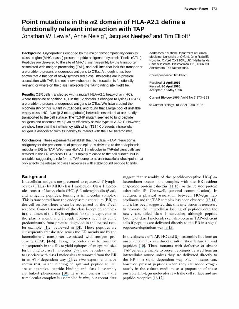
To test whether T134K expressed in the TAP1–TAP2-competent cell line C1R displayed any of the othercharacteristics shown by TAP1–TAP2-negative cell lines,we first determined its cell-surface expression using afluorescence-activated cell sorter (FACS). In contrast toan earlier report [20], where cell-surface expression ofT134K seemed to be normal, we found that C1R-T134Kcells expressed A2.1 at the cell surface at around 20 % ofwild-type levels, as detected by either BB7.2 or MA2.1 —antibodies specific for determinants on the a1 and a2domains of native A2.1 molecules (Table 2). Althoughcell-surface levels are low, this does not reflect low levelsof HLA-A2.1 biosynthesis, as equivalent amounts of A2.1could be immunoprecipitated from lysates of C1R-T134K and C1R-A2.1 cells containing the stabilizingpeptide HIV-pol(476–484) (Fig. 1, compare lanes c ande). Interestingly, in the absence of stabilizing peptide,only around 20 % of wild-type levels of T134K could berecovered from lysates by immunoprecipitation with theconformation-sensitive antibody BB7.2 (Fig. 1, comparelanes d and f). These results suggest that T134Kexpressed in C1R cells behaves in a similar way to wild-type molecules expressed in TAP1–TAP2-negative celllines (Fig. 1, lanes a and b).
T134K molecules are rapidly transported to the cell surfaceas unstable complexesIntracellular maturation of the mutant HLA-A2.1 molec-ules in C1R cells was compared by pulse–chase analysiswith the maturation of wild-type molecules expressed inboth C1R and .174 cells. The rate at which A2.1 moleculesacquired resistance to endoglycosidase-H (endo-H) wasused as a measure of egress from the ER–cis-Golgi networkto the medial Golgi. At time points during the chase, dupli-cate samples were taken and lysed in the presence orabsence of the stabilizing peptide HIV-pol(476–484).Labelled HLA-A2.1 molecules were then immunoprecipi-tated with the conformation-sensitive antibody BB7.2.
874 Current Biology 1996, Vol 6 No 7
Table 1
The presentation of HIV-pol(476–484) by C1R-T134K cellsfollowing infection by recombinant vaccinia virus.
C1R-A2.1 C1R-T134K
10 nM peptide*10:1† 52‡ 672:1 40 51
WR vacc10:1 7 82:1 5 3
HIV-pol vacc10:1 30 72:1 7 5
*HIV-pol(476–484) (ILKEPVHGV); †effector to target ratio; ‡percentspecific lysis.
Table 2
Cell-surface expression of wild-type and T134K HLA-A2.1 byC1R transfectants.
Control antibody* BB7.2 MA2.1
C1R 15† 20 20
C1R-A2.1 9 898 940
C1R-T134K 12 214 205
.174 10 292 315
*B22.249, an IgG2a monoclonal antibody recognizing H2-Db; †meanfluorescence intensity.
A2.1 molecules recovered from lysates lacking addedpeptide represent those that assemble and mature as stableheterodimers. The population of A2.1 molecules thatwould normally fall apart during an overnight incubation inthe absence of peptide can be preserved by the addition ofstabilizing peptide. This technique allowed us to measurethe intracellular fate of both stable and unstable class Imolecules during a 1-hour chase.
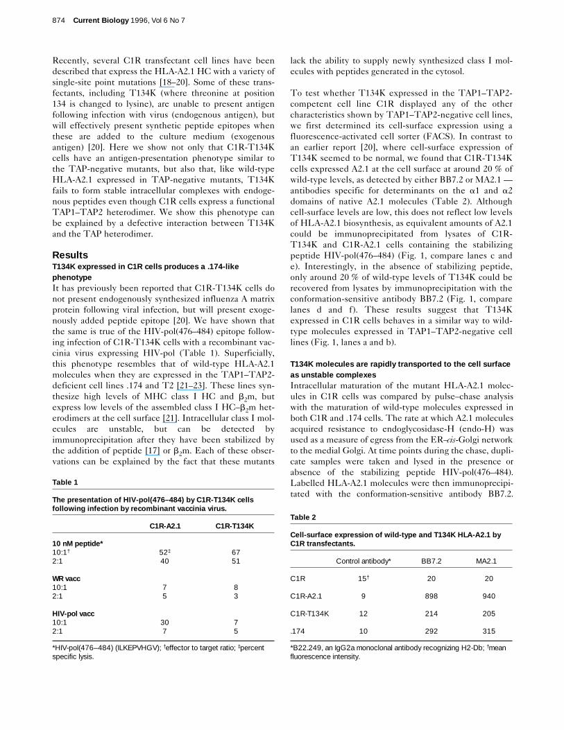
Unlike an earlier report [20] indicating that the threonine-to-lysine mutation in A2.1 does not affect its rate of intracel-lular trafficking, we found that in C1R cells, T134Kacquires endo-H resistance with a half-time of 12 minutes,which is considerably faster than wild-type A2.1, whichmatures with a half-time of around 30 minutes (Fig. 2). InC1R cells, around 50 % of wild-type A2.1 molecules seemto be stabilized rapidly. In five separate experiments, thisproportion ranged from 50 to 70 %. This is consistent withprevious studies, which show that HC–b2m heterodimersare stabilized within 2 minutes of their synthesis. The
remaining molecules are apparently degraded in an endo-H-sensitive compartment. A stark contrast is observed withT134K in C1R cells. In this case, although a small propor-tion (around 25 %) of T134K molecules mature as stablecomplexes, the majority enter the secretory pathway asunstable heterodimers — they are recoverable only fromlysates to which the A2.1-binding peptide has been added.These unstable molecules were released from the ER and,having acquired endo-H resistance, disappeared over thenext 30 minutes (compare t = 32 min and t = 64 min in Fig.2d). Thus, in C1R cells, all T134K molecules maturerapidly, and the majority of these are detectable only asunstable complexes which seem to be peptide-receptive.
The biogenesis of T134K molecules in C1R cells (Fig. 2)has some similarities with the behaviour of wild-type A2.1molecules in the TAP-negative cell line .174 in that, heretoo, a significant proportion of A2.1 synthesized enters thesecretory pathway as unstable complexes. However,unlike unstable T134K molecules, which are transportedmuch more rapidly, these acquire endo-H resistance witha half-time comparable to wild-type A2.1 in C1R cells(30 minutes). In longer pulse–chase experiments (data notshown) these unstable endo-H-resistant wild-type molec-ules were also seen to disappear over a period of around30 minutes — presumably indicating their clearance fromthe cell surface.
A small fraction of wild-type molecules in .174 cells andT134K molecules in C1R cells do, however, assemble asstable molecules, mature and can be detected at the cellsurface by FACS staining. As a result of work using theTAP-negative cell line T2, it has been suggested thatthese stable molecules represent wild-type A2.1 thatbinds to peptides delivered to the ER in a TAP-indepen-dent way [24,25]. To determine whether this was also thecase for T134K in C1R cells, we eluted the peptides fromT134K immunopurified from 1 × 10¹⁰ cells. The mostabundant peptide isolated in this way from T134K hadthe sequence LLDVPTAAV, derived from the IP-30signal sequence and which has been identified previouslyas the most abundant peptide associated with wild-typeA2.1 molecules in a TAP-negative cell line [24,25]. Nopeptides derived from cytosolic proteins were found asso-ciated with T134K. In contrast, the most abundantpeptide eluted from wild-type A2.1 in control experi-ments was derived from the cytosolic 60S ribosomalprotein. No signal-sequence-derived peptides werefound associated with wild-type A2.1 in these experi-ments (J.W.L., unpublished observations). The main dif-ferences between wild-type A2.1 in .174 and T134K inC1R cells are therefore: the rate of trafficking of bothstable and unstable molecules; and the fact that themajority of wild-type molecules in .174 never enter thesecretory pathway, whereas in T134K the majority leavethe ER and mature as unstable molecules.
Research Paper Interaction between HLA-A2.1 and TAP Lewis et al. 875
Figure 1
Most T134K molecules are unstable in C1R cells. Immunoprecipitationof class I molecules with BB7.2 from metabolically labelled .174 (lanes aand b), C1R-A2.1 (lanes c and d) and C1R-T134K cells (lanes e and f),following addition of a saturating concentration (10 mM) of the A2.1-stabilizing peptide ILKEPVHGV (lanes a, c and e) or PBS (lanes b, dand f). Note that the ratio of intensity of heavy chain (HC) to b2m is thesame in C1R-A2.1 (lane c) and C1R-T134K (lane e) cells, indicatingthat the stochiometry of HC–b2m complexes is the same for both wild-type and mutant class I molecules.
HC
β2m
a b c d e f
The T134K heavy chain binds both peptide and b2m normallyOne explanation for our observation that in TAP-compe-tent cells most T134K assembles as unstable molecules isthat the mutation leads to an inherent inability to bindpeptide and b2m normally. First, we measured the abilityof three different A2.1-binding peptides to stabilize‘empty’ wild-type A2.1 (expressed in .174 cells) andT134K (expressed in C1R cells). This has been shown pre-viously to be a reasonable estimate of the affinity ofpeptide-binding at equilibrium [26]. Binding was dose-dependent in all cases, and showed half-maximal effects atcomparable concentrations for both wild-type A2.1 and
T134K, as shown in Table 3. We also showed there was nodifference in the binding of peptides to immature (endo-H-sensitive) versus mature (endo-H-resistant) class I mole-cules, for wild-type or T134K (Table 3). This is important,as it shows there is no destabilizing effect specific for theT134K mutation of sialic-acid-containing complex sugars.In addition, the rate of dissociation of one of the peptides(radiolabelled ILKEPVHGV) was shown to be the samefor both wild-type and T134K, indicating that the threo-nine-to-lysine mutation did not significantly alter thebinding kinetics of the A2.1 molecule, and that T134K wasable to form very stable complexes with peptide.
876 Current Biology 1996, Vol 6 No 7
Figure 2
0 8 16 32 64 (min) 0 8 16 32 64 (min) 0 8 16 32 64 (min)
0
0.4
0.8
1.2
1.6
2
0
0.4
0.8
1.2
1.6
2
0
0.4
0.8
1.2
1.6
2
0 20 40 60 80
0
2
4
6
0
2
4
6
0
2
4
6
0 20 40 60 80
0 20 40 60 80
0 20 40 60 80
0 20 40 60 80
0 20 40 60 80
EHR
EHS
EHSEHR
EHS
EHR
EHR
EHS
EHR
EHS
EHSEHR
Time (min) Time (min) Time (min)
Time (min) Time (min) Time (min)
IOD
IOD
IOD
IOD
IOD
IOD
(a)
(b)
(c)
(d)
EHREHS
EHREHS
C1R-A2.1 cells C1R-T134K cells A2.1 in .174 cells
T134K molecules are transported rapidly to the cell surface as emptyreceptors. The intracellular trafficking of wild-type (C1R-A2.1 and.174) and mutant (C1R-T134K) A2.1 molecules. Pulse–chase analysisof a cohort of A2.1 molecules was carried out and BB7.2-reactivemolecules were recovered from cell lysates after overnight preclearingin the absence (a) or presence (b) of a saturating concentration(15 mM) of stabilizing peptide. Only the heavy-chain bands are shown,as human b2m is poorly visualized following a 10-minute pulse label.(c) Quantitation of (a), representing the maturation of stably assembled
A2.1. (d) The fate of unstable A2.1 molecules, calculated from thedifference in band intensities between (a) and (b) (d = b–a). EHR:endo-H resistant. EHS: endo-H sensitive. IOD: integrated opticaldensity in arbitrary units (see Materials and methods). All IODs werenormalized to the amount of immune complex recovered at the end ofthe experiment. This was achieved by comparing the relative intensityof the Coomassie blue-stained monoclonal antibody heavy-chain bandobserved before autoradiography.
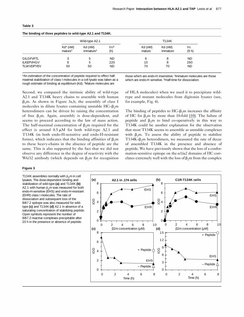
Second, we compared the intrinsic ability of wild-typeA2.1 and T134K heavy chains to assemble with humanb2m. As shown in Figure 3a,b, the assembly of class Imolecules in dilute lysates containing unstable HC–b2mheterodimers can be driven by raising the concentrationof free b2m. Again, assembly is dose-dependent, andseems to proceed according to the law of mass action.The half-maximal concentration of b2m required for theeffect is around 0.5 mM for both wild-type A2.1 andT134K (in both endo-H-sensitive and endo-H-resistantforms), which indicates that the binding affinities of b2mto these heavy-chains in the absence of peptide are thesame. This is also supported by the fact that we did notobserve any difference in the degree of reactivity with theW6/32 antibody (which depends on b2m for recognition
of HLA molecules) when we used it to precipitate wild-type and mutant molecules from digitonin lysates (see,for example, Fig. 4).
The binding of peptides to HC–b2m increases the affinityof HC for b2m by more than 10-fold [10]. The failure ofpeptide and b2m to bind co-operatively in this way toT134K could be another explanation for the observationthat most T134K seems to assemble as unstable complexeswith b2m. To assess the ability of peptide to stabilizeT134K–b2m heterodimers, we measured the rate of decayof assembled T134K in the presence and absence ofpeptide. We have previously shown that the loss of a confor-mation-sensitive epitope on the a1/a2 domains of HC corr-elates extremely well with the loss of b2m from the complex
Research Paper Interaction between HLA-A2.1 and TAP Lewis et al. 877
Table 3
The binding of three peptides to wild-type A2.1 and T134K.
Wild-type A2.1 T134K
Kd* (nM) Kd (nM) t½§ Kd (nM) Kd (nM) t½mature† immature‡ (h) mature immature (9 h)
GILGFVFTL 3 5 ND 6 8 NDILKEPVHGV 8 5 220 10 8 260TLWVDPYEV 50 50 ND 70 70 ND
*An estimation of the concentration of peptide required to effect half-maximal stabilization of class I molecules in a cell lysate was taken as arough estimate of binding at equilibrium (Kd). †Mature molecules are
those which are endo-H insensitive. ‡Immature molecules are thosewhich are endo-H sensitive. §Half-time for dissociation.
Figure 3
T134K assembles normally with b2m in celllysates. The dose-dependent binding andstabilization of wild-type (a) and T134K (b)A2.1 with human b2m was measured for bothendo-H-sensitive (EHS) and endo-H-resistant(EHR) class I molecules. The rate ofdissociation and subsequent loss of theBB7.2 epitope was also measured for wild-type (c) and T134K (d) A2.1 in absence of asaturating concentration of stabilizing peptide.Open symbols represent the number ofBB7.2-reactive complexes precipitable after20 h in the presence or absence of peptide. 0
2
4
6
8
0
2
4
6
8
0
2
4
6
8
0 2 4 6 8 10 0 2 4 6 8 10
EHR
EHS
EHS
EHR
β2m concentration (µM) β2m concentration (µM)
C1R-T134K cells
EHS
Time (h) Time (h)0 2 4 6 8
IOD
IOD
IOD
IOD
0
3
6
9
12
15
0 2 4 6 8
EHS
A2.1 in .174 cells(a) (b)
(c) (d)
+ Peptide
– Peptide
+ Peptide
– Peptide
[10]. We therefore measured the rate of decay of totalT134K–b2m heterodimers (this includes a mixture of bothmature and immature molecules) using this technique.Figure 3c,d shows that the rate of decay of T134K has ahalf-time of around 2.5 hours in the absence of peptide, andmore than 20 hours in the presence of peptide, and is con-sistent with the equivalent rates of decay of wild-type A2.1assembled in .174 cells. It seems, therefore, that the muta-tion at position 134 does not alter the intrinsic ability of theA2.1 molecule to assemble normally with b2m and peptides,to give rise to a long-lived heterotrimeric complex in vitro.
To show that the same was true in vivo, we delivered a‘pre-processed’ CTL epitope directly to the ER of C1R-T134K cells. TAP-independent delivery of the influenzaA matrix protein (MP) epitope GILGFVFTL is opti-mized by delivering it in the form of a minigene precededby an ER translocation signal sequence [25,27]. Table 4shows that delivering the epitope in this way overcomesthe antigen-presentation defect seen with full-length MPin both C1R-T134K and .174 cells. Influenza A MP isclearly able to be processed and delivered to the ER ofC1R cells, as shown by the ability of wild-type A2.1 topresent this epitope in C1R cells infected with influenzaA/X31 (Table 4).
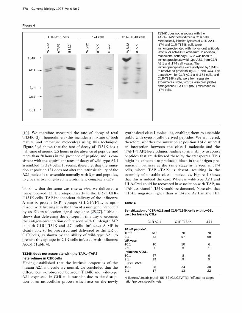
T134K does not associate with the TAP1–TAP2heterodimer in C1R cellsHaving established that the intrinsic properties of themutant A2.1 molecule are normal, we concluded that thedifferences we observed between T134K and wild-typeA2.1 expressed in C1R cells must be due to the disrup-tion of an intracellular process which acts on the newly
synthesized class I molecules, enabling them to assemblestably with cytosolically derived peptides. We wondered,therefore, whether the mutation at position 134 disruptedan interaction between the class I molecule and theTAP1–TAP2 heterodimer, leading to an inability to accesspeptides that are delivered there by the transporter. Thismight be expected to produce a block in the antigen-pre-sentation pathway at the same stage as is seen in .174cells, where TAP1–TAP2 is absent, resulting in theassembly of unstable class I molecules. Figure 4 showsthat this is indeed the case. Whereas wild-type A2.1 andHLA-Cw4 could be recovered in association with TAP, noTAP-associated T134K could be detected. Note also thatT134K migrates higher than wild-type A2.1 in the IEF
878 Current Biology 1996, Vol 6 No 7
Table 4
Sensitization of C1R-A2.1 and C1R-T134K cells with L+G9Lvacc for lysis by CTLs.
C1R-A2.1 C1R-T134K .174
10 nM peptide*10:1† 61‡ 70 782:1 52 57 60WR vacc10:1 10 10 62:1 7 3 1Influenza A/X3110:1 67 8 92:1 39 9 5L+G9L vacc10:1 28 24 382:1 17 13 22
*Influenza A matrix protein 55–63 (GILGFVFTL); †effector to targetratio; ‡percent specific lysis.
Figure 4
T134K does not associate with theTAP1–TAP2 heterodimer in C1R cells.Metabolically labelled lysates of C1R-A2.1,.174 and C1R-T134K cells wereimmunoprecipitated with monoclonal antibodyW6/32 or anti-TAP2 antiserum. In addition,monoclonal antibody BB7.2 was used toimmunoprecipitate wild-type A2.1 from C1R-A2.1 and .174 cell lysates. Theimmunoprecipitates were analysed by 1D-IEFto resolve co-precipitating A2.1 and Cw4. Thedata shown for C1R-A2.1 and .174 cells, andC1R-T134K cells, were from separateexperiments. Note, W6/32 also precipitatesendogenous HLA-B51 (B51) expressed in.174 cells.
T134K
A2.1
β2m
Cw4
B51
W6/
32
αTA
P2
BB
7.2
W6/
32
αTA
P2
αTA
P2
BB
7.2
W6/
32
C1R-A2.1 cells .174 cells C1R-T134K cells
gel, which separates proteins according to their isoelectricpoint (pI). This is due to the addition of positive charge bythe threonine-to-lysine mutation. HLA-Cw4 in associationwith TAP is recovered equally well from lysates of C1R-A2.1 and C1R-T134K cells. We do not know the origin ofthe additional bands that co-precipitate with the anti-TAPantiserum, nor whether they are specifically co-precipi-tated. Only the band that equilibrates at a pI similar tothat of mono-sialylated T134K was reproducibly co-pre-cipitated with anti-TAP (see, for example, Fig. 6). Thisband is unlikely to be mono- or non-sialylated T134Kheavy chain because: it equilibrates at a different pI toeither of these; A2.1 in all its sialylated forms migrates as adoublet, but this band is a singlet; the band persists afterthe b2m band disappears, indicating that, unlike Cw4 inthe same cell, it is not associated with b2m, as might beexpected if it were T134K; and, sialylation occurs in thetrans-Golgi network whereas TAP localizes to the ER andcis-Golgi network [28].
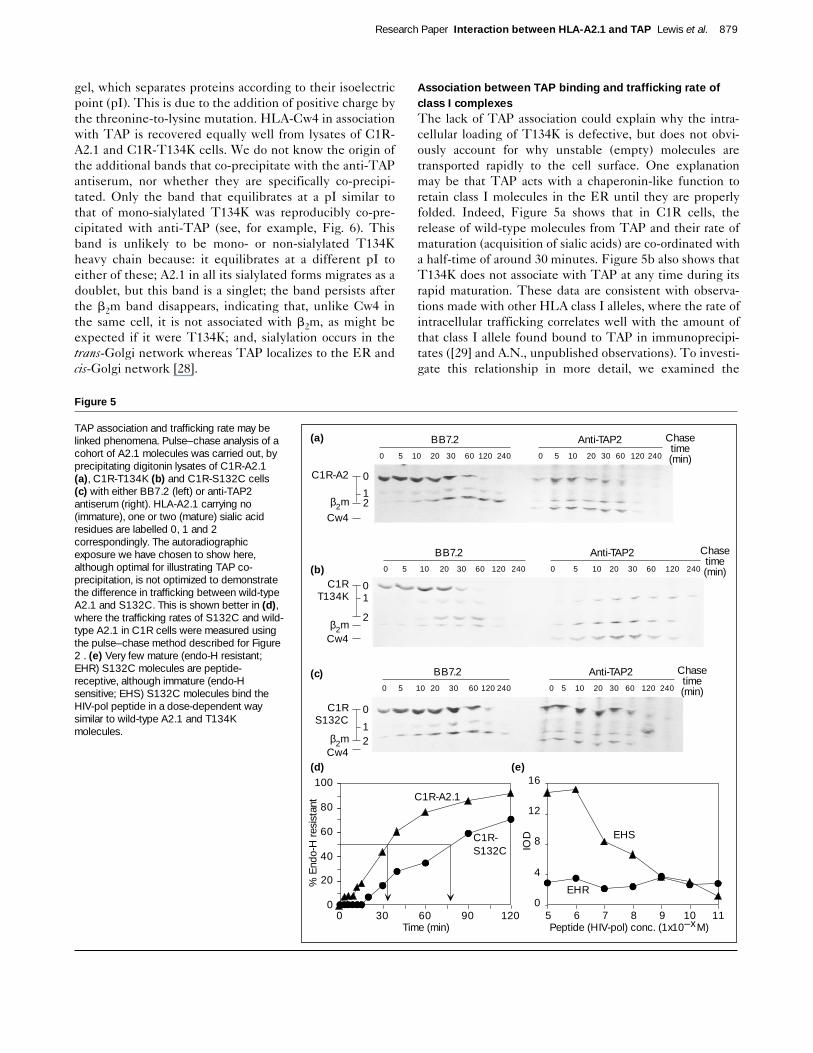
Association between TAP binding and trafficking rate ofclass I complexesThe lack of TAP association could explain why the intra-cellular loading of T134K is defective, but does not obvi-ously account for why unstable (empty) molecules aretransported rapidly to the cell surface. One explanationmay be that TAP acts with a chaperonin-like function toretain class I molecules in the ER until they are properlyfolded. Indeed, Figure 5a shows that in C1R cells, therelease of wild-type molecules from TAP and their rate ofmaturation (acquisition of sialic acids) are co-ordinated witha half-time of around 30 minutes. Figure 5b also shows thatT134K does not associate with TAP at any time during itsrapid maturation. These data are consistent with observa-tions made with other HLA class I alleles, where the rate ofintracellular trafficking correlates well with the amount ofthat class I allele found bound to TAP in immunoprecipi-tates ([29] and A.N., unpublished observations). To investi-gate this relationship in more detail, we examined the
Research Paper Interaction between HLA-A2.1 and TAP Lewis et al. 879
Figure 5
TAP association and trafficking rate may belinked phenomena. Pulse–chase analysis of acohort of A2.1 molecules was carried out, byprecipitating digitonin lysates of C1R-A2.1(a), C1R-T134K (b) and C1R-S132C cells(c) with either BB7.2 (left) or anti-TAP2antiserum (right). HLA-A2.1 carrying no(immature), one or two (mature) sialic acidresidues are labelled 0, 1 and 2correspondingly. The autoradiographicexposure we have chosen to show here,although optimal for illustrating TAP co-precipitation, is not optimized to demonstratethe difference in trafficking between wild-typeA2.1 and S132C. This is shown better in (d),where the trafficking rates of S132C and wild-type A2.1 in C1R cells were measured usingthe pulse–chase method described for Figure2 . (e) Very few mature (endo-H resistant;EHR) S132C molecules are peptide-receptive, although immature (endo-Hsensitive; EHS) S132C molecules bind theHIV-pol peptide in a dose-dependent waysimilar to wild-type A2.1 and T134Kmolecules.
5 6 7 8 9 10 110
4
8
12
16
EHR
EHS
Peptide (HIV-pol) conc. (1x10–x M)
IOD
0
20
40
60
80
100
0 30 60 90 120Time (min)
% E
ndo-
H re
sist
ant C1R-A2.1
C1R-S132C
0 5 10 20 30 60 120 240 0 5 10 20 30 60 120 240
(a)
(b)
(c)
(d) (e)
BB7.2
0 5 10 20 30 60 120 240 0 5 10 20 30 60 120 240
BB7.2
Anti-TAP2
Anti-TAP2
0 5 10 20 30 60120240 0 5 10 20 30 60 120 240
BB7.2 Anti-TAP2
C1R-A2
Cw4
β2m
0
12
C1RT134K
Cw4β2m
01
2
C1RS132C
Cw4β2m
0
12
Chase time (min)
Chase time (min)
Chase time (min)
biogenesis of a second point mutant, in which serine atposition 132 is substituted for cysteine (S132C). Thismutant presents endogenous antigen efficiently, butexogenous peptides very poorly [20]. S132C is transportedvery slowly in C1R cells, having a half-time for maturationof around 85 minutes (Fig. 5d). Significantly, it associateswith TAP for much of this time (Fig. 5c) and results in theegress of stably assembled complexes. Figure 5e shows thatno S132C molecules leave the ER in a peptide-receptivestate, as the addition of peptides to endo-H-resistant mol-ecules did not increase the number of complexes recoveredby BB7.2. There are sufficient numbers of immatureS132C molecules, however, to show that S132C bindspeptide normally in a lysate (half-maximal effect at around1 × 10–8 M, compared to 0.5 × 10⁻⁸ M for wild-type A2.1 and0.8 × 10–8 M for T134K, see Table 3). Thus, the biochem-istry of S132C is consistent with its antigen-presentationphenotype, and strongly supports the notion that the dura-tion of TAP association is directly correlated with thenumber or proportion of class I molecules that leave the ERloaded with an optimal peptide. S132C and T134K maytherefore represent two ends of a spectrum along which allclass I molecules fall.
DiscussionOur results show that a single amino-acid substitution atposition 134 in the a2 domain of the HLA-A2.1. HC(T134K) results in the intracellular assembly of unstablecomplexes with b2m, which are transported to the cellsurface more than three times faster than the wild-typeA2.1 molecules expressed in the same cell. The mutantmolecules can bind to peptides and b2m normally in vitro.However, no detectable association between the mutantHC and the TAP1–TAP2 heterodimer was found in co-immunoprecipitation experiments. The most likely expla-nation to account for the observed behaviour of themutant molecules, therefore, is that they fail to becomeloaded with high-affinity peptides in the lumen of the ERas a consequence of this impaired association. This is thefirst demonstration that the MHC class I–TAP interactionis obligatory for the presentation of peptide epitopesdelivered to the ER by TAP. These results are consistentwith the functional phenotype of C1R-T134K cells, whichdoes not present endogenous antigen to CTLs, but pres-ents exogenously added peptides normally.
It remains to be seen whether the requirement for TAPassociation that we observe for HLA-A2.1 can be extendedto other class I molecules. Although the behaviour of themutant heavy chains could be a consequence of studyingthem in the cell line C1R (which has its origins in a g-irradi-ated EBV-transformed B-cell line [30]), it is of interest tonote that Neisig et al. [29] have found other naturally occur-ring HLA class I alleles that are capable of presentingantigen, but that bind only poorly or undetectably to TAP.The only difference among HLA alleles in the region
between residues 128 and 137 is an arginine/serine dimor-phism at position 131. However, alleles with both arginineand serine are represented among both efficient and ineffi-cient TAP associators. Also, a recent report by Lee et al. [31]showed that a soluble form of HLA-G loaded the same setof endogenous peptides as its membrane-anchored counter-part, despite a lack of demonstrable TAP association.
The behaviour of the T134K molecule in being unable toform stable complexes with peptides in TAP-competentcells is similar to that of another class I molecule, Ddm6 [32],which has a non-conservative amino-acid substitutionwithin the same highly conserved region of the a2 domainbetween residues 128 and 137 (the loop preceding, andmost of, the b4 strand). In all classical and non-classicalmurine class I molecules, this sequence is EDLK-TWTAAD (the human equivalent is EDLRSWTAAD). InDdm6, residue 133 is changed from tryptophan to arginine(W133R), the only different residue between the Dd andDdm6 class I molecules. The level of cell-surface expressionof the mutant in normal cells is less than 10 % of the levelsof Kd or Dd expression in the same cells, and Ddm6 couldonly be precipitated from pre-cleared lysates with antibod-ies to the a3 domain, and not to the a1/a2 domains. Never-theless, the mutant heavy chains were shown to acquiremature N-linked oligosaccharides. Although experimentswere not carried out to test whether Ddm6 could be stabi-lized with Dd-binding peptides, or whether it was able topresent Dd-restricted endogenous antigens, it is tempting tospeculate that the mutant molecules behave like T134K.
Taken together with our observations that a threonine-to-lysine substitution at position 134 ablates the class I inter-action with TAP, and that another mutation in this region(S132C) results in slow trafficking and a phenotype consis-tent with ‘hyper-efficient’ intracellular loading of pep-tides, we suggest that residues in this region of themolecule are responsible for dictating the efficiency ofloading by controlling the interaction of HC with TAP,either directly or indirectly. Recent reports have alsoimplicated residues in both the a3 domain [33] and thepeptide-binding groove [29] in controlling the efficacy ofTAP binding. It remains to be seen whether these obser-vations, along with our observation that solvent-exposedresidues in the a2 domain are also involved, will result inan unified description of the class I–TAP interaction.
Various models can be proposed for the in vivo function ofTAP in the biogenesis of stable class I–peptide complexesand their regulated release into the anteriograde transportpathway. All of these must accommodate the observedbehaviour of T134K in CIR cells. The simplest is that theconserved stretch between residues 128 and 137 forms allor part of the interface between class I and TAP, and thatTAP has a chaperonin function, retaining class I het-erodimers in the ER until a suitable peptide for binding is
880 Current Biology 1996, Vol 6 No 7
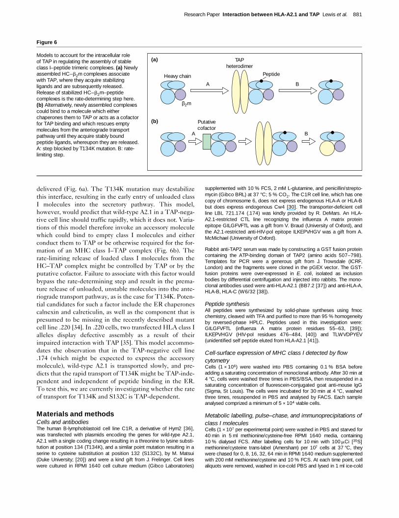
delivered (Fig. 6a). The T134K mutation may destabilizethis interface, resulting in the early entry of unloaded classI molecules into the secretory pathway. This model,however, would predict that wild-type A2.1 in a TAP-nega-tive cell line should traffic rapidly, which it does not. Varia-tions of this model therefore invoke an accessory moleculewhich could bind to empty class I molecules and eitherconduct them to TAP or be otherwise required for the for-mation of an MHC class I–TAP complex (Fig. 6b). Therate-limiting release of loaded class I molecules from theHC–TAP complex might be controlled by TAP or by theputative cofactor. Failure to associate with this factor wouldbypass the rate-determining step and result in the prema-ture release of unloaded, unstable molecules into the ante-riograde transport pathway, as is the case for T134K. Poten-tial candidates for such a factor include the ER chaperonescalnexin and calreticulin, as well as the component that ispresumed to be missing in the recently described mutantcell line .220 [34]. In .220 cells, two transfected HLA class Ialleles display defective assembly as a result of theirimpaired interaction with TAP [35]. This model accommo-dates the observation that in the TAP-negative cell line.174 (which might be expected to express the accessorymolecule), wild-type A2.1 is transported slowly, and pre-dicts that the rapid transport of T134K might be TAP-inde-pendent and independent of peptide binding in the ER.To test this, we are currently investigating whether the rateof transport for T134K and S132C is TAP-dependent.
Materials and methodsCells and antibodiesThe human B-lymphoblastoid cell line C1R, a derivative of Hym2 [36],was transfected with plasmids encoding the genes for wild-type A2.1,A2.1 with a single coding change resulting in a threonine to lysine substi-tution at position 134 (T134K), and a similar point mutation resulting in aserine to cysteine substitution at position 132 (S132C), by M. Matsui(Duke University; [20]) and were a kind gift from J. Frelinger. Cell lineswere cultured in RPMI 1640 cell culture medium (Gibco Laboratories)
supplemented with 10 % FCS, 2 mM L-glutamine, and penicillin/strepto-mycin (Gibco BRL) at 37 °C; 5 % CO2. The C1R cell line, which has onecopy of chromosome 6, does not express endogenous HLA-A or HLA-Bbut does express endogenous Cw4 [30]. The transporter-deficient cellline LBL 721.174 (.174) was kindly provided by R. DeMars. An HLA-A2.1-restricted CTL line recognizing the influenza A matrix proteinepitope GILGFVFTL was a gift from V. Braud (University of Oxford), andthe A2.1-restricted anti-HIV-pol epitope ILKEPVHGV was a gift from A.McMichael (University of Oxford).
Rabbit anti-TAP2 serum was made by constructing a GST fusion proteincontaining the ATP-binding domain of TAP2 (amino acids 507–798).Templates for PCR were a generous gift from J. Trowsdale (ICRF,London) and the fragments were cloned in the pGEX vector. The GST-fusion proteins were over-expressed in E. coli, isolated as inclusionbodies by differential centrifugation and injected into rabbits. The mono-clonal antibodies used were anti-HLA-A2.1 (BB7.2 [37]) and anti-HLA-A,HLA-B, HLA-C (W6/32 [38]).
Peptide synthesisAll peptides were synthesized by solid-phase syntheses using fmocchemistry, cleaved with TFA and purified to more than 95 % homogeneityby reversed-phase HPLC. Peptides used in this investigation were:GILGFVFTL (influenza A matrix protein residues 55–63, [39]);ILKEPVHGV (HIV-pol residues 476–484, [40]) and TLWVDPYEV(unidentified self peptide eluted from HLA-A2.1 [41]).
Cell-surface expression of MHC class I detected by flowcytometryCells (1 × 10⁶) were washed into PBS containing 0.1 % BSA beforeadding a saturating concentration of monoclonal antibody. After 30 min at4 °C, cells were washed three times in PBS/BSA, then resuspended in asaturating concentration of fluorescein-conjugated goat anti-mouse IgG(Sigma, St Louis). The cells were incubated for 30 min at 4 °C, washedthree times, resuspended in PBS and analysed by FACS. Each sampleanalysed comprized a minimum of 5 × 10⁴ viable cells.
Metabolic labelling, pulse–chase, and immunoprecipitations ofclass I moleculesCells (1 × 10⁷ per experimental point) were washed in PBS and starved for40 min in 5 ml methionine/cysteine-free RPMI 1640 media, containing10 % dialysed FCS. After labelling cells for 10 min with 100mCi [35S]methionine/cysteine trans-label (Amersham) per 107 cells at 37 °C, theywere chased for 0, 8, 16, 32, 64 min in RPMI 1640 medium supplementedwith 200 mM methionine/cysteine and 10 % FCS. At each time point, cellaliquots were removed, washed in ice-cold PBS and lysed in 1 ml ice-cold
Research Paper Interaction between HLA-A2.1 and TAP Lewis et al. 881
Figure 6
Models to account for the intracellular role of TAP in regulating the assembly of stableclass I–peptide trimeric complexes. (a) Newlyassembled HC–b2m complexes associatewith TAP, where they acquire stabilizingligands and are subsequently released.Release of stabilized HC–b2m–peptidecomplexes is the rate-determining step here.(b) Alternatively, newly assembled complexescould bind to a molecule which eitherchaperones them to TAP or acts as a cofactorfor TAP binding and which rescues emptymolecules from the anteriograde transportpathway until they acquire stably boundpeptide ligands, whereupon they are released.A: step blocked by T134K mutation. B: rate-limiting step.
A B
A B
(a)
(b)
Heavy chain
β2m
Peptide
TAP heterodimer
Putative cofactor
Tris lysis buffer (pH 7.4), containing 0.5 % NP-40 and 0.5 % Mega-9, 5 mMiodoacetamide and 2 mM PMSF (Sigma). For aliquots lysed in the pres-ence of stabilizing peptide, 15 mM HIV-pol(476–484) was added to thelysis mix. After 15 min at 4 °C, the nuclei were removed by centrifugationand the supernatants were precleared overnight at 4 °C with 20ml of fixedStaphylococcus aureus (Sigma) made up in lysis buffer.
Assembled class I molecules were immunoprecipitated with BB7.2(10 mg ml–1 for 1 h) and 5 % (w/w) protein A–Sepharose beads (Sigma).After washing the immunoprecipitates three times in lysis buffer, theywere subjected to endoglycosidase-H digestion overnight before beinganalysed on a 10 % SDS–PAGE gel under reducing conditions. Gelswere fixed, stained, treated with Amplify (Amersham), dried and visualizedby autoradiography at –80 °C. Quantitation of autoradiograms wasachieved by scanning using a MilliGen Bioimage Analyser whichexpresses band intensities as an integrated optical density (IOD).
Endoglycosidase-H treatmentEndoglycosidase-H (Boehringer Mannheim) digestion of immunoprecipi-tates was performed by resuspending the samples in 30 ml of 50 mMsodium citrate buffer (pH 5.5), containing 0.2 % SDS, heating to 95 °Cfor 5 min, and digesting with 3 U of endoglycosidase-H overnight at37 °C.
Peptide or b2m binding in vitroIncreasing concentrations of b2m or stabilizing peptide were added tocell lysates to determine binding affinities for mutant and wild-type classI molecules. Cells were labelled for 15 min with [35S] methionine/cysteine as described above. To enable the determination of bindingaffinities for both endoglycosidase-H-resistant (EHR) and endo-glycosidase-H-sensitive (EHS) forms, each cell line was chased withcold RPMI 1640 medium supplemented with 200 mM methionine/cys-teine and 10 % FCS (as above) until equal amounts of both EHR andEHS class I molecules were obtained. (For pulse–chase experimentsabove, C1R and .174 cells were chased for 30 min, C1R-T134K cellswere chased for 12 min, and C1R-S132C cells were chased for60 min.) After washing cells in cold PBS, samples were lysed in ice-coldTris lysis buffer (as above) in the presence of a decreasing concentrationof either b2m or peptide. Samples were then precleared for 1 h withStaph A and immunoprecipitated using BB7.2, washed three times,exposed to endoglycosidase-H overnight and analysed as above by12 % SDS–PAGE.
b2m dissociation assayThe stability of wild-type A2 and T134K class I complexes was deter-mined in vitro by measuring the rate of decay of the BB7.2 epitope result-ing from the dissociation of b2m from HC. A2.1 molecules in .174 andC1R-T134K cells were labelled with [35S] methionine/cysteine andchased as described previously to obtain equal amounts of EHS andEHR class I molecules. Cells were then lysed in Tris lysis buffer contain-ing 0.5 % NP-40, aliquotted and kept on ice. At intervals following lysis(0, 1, 2, 3, 8 and 20 h), stabilizing HIV-pol peptide and BB7.2 antibodywere added together to the lysate and immunoprecipitated after 1 h (asabove). For one sample, peptide was added at the time of lysis (0 h) andBB7.2 was added at 20 h. Samples were analysed on SDS–PAGE afterendoglycosidase-H treatment (as above).
Association of class I molecules with TAPWe starved 2 × 10⁷ cells of each cell line for 40 min in 1 mlmethionine/cysteine-free RPMI 1640 medium supplemented with 10 %FCS. The cells were pelletted and resuspended in 500 ml fresh methion-ine/cysteine-free RPMI 1640 + 10 % FCS and labelled with 250 mCi35S-methionine/cysteine for 10 min at 37 °C. Cells were washed twicewith cold PBS and lysed in 500 ml 1 % digitonin-containing lysis buffer(1 % (w/v) digitonin (Sigma), 50 mM Tris–HCl (pH 7.5), 5 mM MgCl2 and150 mM NaCl) for 30 min on ice. Each lysate was split into equal por-tions, pre-cleared overnight with normal rabbit serum (NRS) and immuno-precipitated with either BB7.2, W6/32 or anti-TAP2 antibodies. Theimmunoprecipitations were recovered with protein A–Sepharose beads(Sigma) for 1 h and the beads were washed three times in lysis buffercontaining 0.5 % digitonin before analysis by 1D-IEF.
Pulse–chase analysis to follow assembly of class I moleculesand TAP–class I interactionsWe starved 1 × 10⁸ cells for 40 min in 2 ml methionine/cysteine-freeRPMI 1640 medium with 10 % FCS. Cells were pelletted, resuspendedin 500 ml fresh RPMI 1640 medium supplemented with 10 % FCS andlabelled with 500 mCi 35S-methionine/cysteine for 10 min at 37 °C. Cellswere chased for 0, 5, 10, 20, 30, 60, 120, 240 and 480 min in RPMI1640 medium supplemented with 1 mM methionine/cysteine and 10 %FCS. Cells were washed in ice-cold PBS before lysis in 1 % digitonin-containing lysis buffer for 1 h on ice. Each lysate was divided into twoportions, pre-cleared with NRS and immunoprecipitated with eitherBB7.2 or anti-TAP2 antibodies. All samples were analysed by 1D-IEF.
One-dimensional isoelectric focusing (1D-IEF)1D-IEF was performed as described [42].
Dissociation rate of peptide from class IThe peptide ILKEVPHGV was radio-iodinated to a specific activity of1.3 × 1016 cpm mol–1 using the chloramine T method. C1R, .174 andC1R-T134K cells (1 × 10⁸) were lysed in 1 ml cold lysis buffer and nucleiwere removed by centrifugation. Peptide (5 × 10⁷ cpm) was then addedto each sample along with human b2m and diluted to a final concentrationof 2 × 10⁷ cell equivalents per ml and 3 mg ml–1 b2m. This mixture wasallowed to equilibrate overnight at 4 °C. The next day the equilibriummixture was diluted 1:1 with a 0.2 mM solution of unlabelled ILKEPVHGVin cold lysis buffer, thus effecting a 100-fold molar excess of cold overhot. At various times during the following 300 h, 2 × 10⁷ cell equivalentswere removed and precipitated with BB7.2, as described above. Theimmunoprecipitates were washed four times and the resulting isolateswere quantitated with a Rackgamma g-counter (LKB). Backgroundbinding was determined by allowing 2 × 10⁷ cell equivalents of lysate toequilibrate with 5 × 10⁶ cpm hot ILKEPVHGV in a solution of 0.1 mM coldpeptide. Specific peptide-binding to wild-type A2.1 and T134K were cal-culated by subtracting background cpm obtained in the sham bindingcontrol with a lysate of untransfected C1R cells.
Construction of recombinant vaccinia virus and CTL assaysRecombinant vaccinia virus expressing a minigene encoding the optimalinfluenza A matrix protein epitope GILGFVFTL preceded by the haemag-glutinin ER translocation signal sequence MKANLLVLLCALAAADA(L+G9L-vacc) was constructed as described in [8]. Briefly, the syntheticminigene was cloned into the plasmid pSC11.302.R.L+ as anNcoI–BamHI fragment. The resulting construct was then introduced intothe thymidine kinase locus of WR vaccinia by homologous recombinationusing standard techniques.
The ability of recombinant vaccinias to sensitize C1R-A2.1 and C1R-T134K cells was assessed by infecting 1 × 10⁶ of each for 1 h during51Cr-labelling at a multiplicity of infection of 20. Influenza A/X31 infec-tions were with 200 ml allantoic fluid. The media with virus and 51Cr werethen washed away and the cells were incubated for an additional 2 h at37 °C. Following two further washes in R10, the target cells wereassayed for their susceptibility to lysis by CTLs in a 4-hour chromium-release assay. Where peptide sensitization was assayed, targets werepulsed with 10 nM peptide 1 h prior to the start of the CTL assay.
AcknowledgementsWe would like to thank Phil Wood for stimulating discussions and insightfulsuggestions throughout, and Enzo Cerundolo for his continued friendshipand interest. We also thank J. Edwards for providing us with the plasmidpsc11L+G9L. This work was supported by the Wellcome Trust. T.E. is aWellcome Senior Research Fellow in Basic Biomedical Science and J.W.L.is a Wellcome Prize Student.
References1. Townsend A, Bastin J, Gould K, Brownlee G, Andrew M, Coupar B,
Boyle D, Chan S, Smith G: Defective presentation to class I-restricted cytotoxic T lymphocytes in vaccinia-infected cells isovercome by enhanced degradation of antigen. J Exp Med 1988,168:1211–1224.
882 Current Biology 1996, Vol 6 No 7
2. Moore MW, Carbone FR, Bevan MJ: Introduction of soluble proteininto the class I pathway of antigen processing and presentation.Cell 1988, 54:777–785.
3. Heemels MT, Ploegh H: Generation, translocation and presentationof MHC class I-restricted peptides. Annu Rev Biochem 1995,64:463.
4. Deverson EV, Gow IR, Coadwell WJ, Monaco JJ, Butcher GW,Howard JC: MHC class II region encoding proteins related to themultidrug resistance family of transmembrane transporters.Nature 1990, 348:738–741.
5. Trowsdale J, Hanson I, Mockridge I, Beck S, Townsend A, Kelly A:Sequences encoded in the class II region of the MHC related tothe ‘ABC’ superfamily of transporters. Nature 1990, 348:741–744.
6. Spies T, Bresnahan M, Bahram S, Arnold D, Blanck G, Mellins E, etal.: A gene in the human major histocompatibility complex class IIregion controlling the class I antigen presentation pathway.Nature 1990, 348:744–747.
7. Roelse J, Gromme M, Momburg F, Hammerling G, Neefjes J:Trimming of TAP-translocated peptides in the endoplasmicreticulum and in the cytosol during recycling. J Exp Med 1994,180:1591–1597.
8. Elliott T, Willis A, Cerundolo V, Townsend A: Processing of majorhistocompatibility class I-restricted antigens in the endoplasmicreticulum. J Exp Med 1995, 181:1481–1491.
9. Snyder HL, Yewdell JW, Bennink J: Trimming of antigenic peptidesin an early secretory compartment. J Exp Med 1994,180:2389–2394.
10. Elliott T, Cerundolo V, Elvin J, Townsend A: Peptide-inducedconformational change of the class I heavy chain. Nature 1991,351:402–406.
11. Degen E, Williams DB: Participation of a novel 88-kD protein in thebiogenesis of murine class I histocompatibility molecules. J CellBiol 1991, 112:1099–1115.
12. Hochstenbach F, David V, Watkins S, Brenner MB: Endoplasmicreticulum resident protein of 90 kilodaltons associates with the T-and B-cell antigen receptors and major histocompatibilitycomplex antigens during their assembly. Proc Natl Acad Sci USA1992, 89:4734–4738.
13. Suh WK, Cohen-Doyle MF, Fruh K, Wang K, Peterson PA, WilliamsDB: Interaction of MHC class I molecules with the transporterassociated with antigen processing. Science 1994, 264:1322.
14. Ortmann B, Androlewicz MJ, Cresswell P: MHC class I/b-2microglobulin complexes associate with TAP transporters beforepeptide binding. Nature 1994, 368:864–867.
15. Anderson K, Cresswell P, Gammon M, Hermes J, Williamson A,Zweerink H: Endogenously synthesized peptide with anendoplasmic reticulum signal sequence sensitizes antigenprocessing mutant cells to class I-restricted cell-mediated lysis. JExp Med 1991, 174:489–492.
16. Townsend A, Ohlen C, Bastin J, Ljunggren HG, Foster L, Karre K:Association of class I major histocompatibility heavy and lightchains induced by viral peptides. Nature 1989, 340:443–448.
17. Cerundolo V, Alexander J, Anderson K, Lamb C, Cresswell P,McMichael A, et al.: Presentation of viral antigen controlled by agene in the major histocompatibility complex. Nature 1990,345:449–452.
18. Moots RJ, Matsui M, Pazmany L, McMichael AJ, Frelinger JA: Acluster of mutations in HLA-A2 alpha 2 helix abolishes peptiderecognition by T cells. Immunogenetics 1991, 34:141–148.
19. Lombardi G, Matsui M, Moots R, Aichinger G, Sidhu S, Batchelor R,et al.: Structural relationship between HLA-A2-restricted and anti-A2 allospecific T-cell recognition: Analysis by mutation of thecodons for polymorphic and conserved residues. Transplant Proc1991, 23:446–448.
20. Matsui M, Moots RJ, Warburton RJ, Peace-Brewer AL, Quinn DG,McMichael AJ, Frelinger JA: Genetic evidence for difference betweenintracellular and extracellular peptides in Influenza A matrixpeptide-specific CTL recognition. J Immunol 1995, 154:1088–1096.
21. DeMars R, Chang CC, Shaw C, Reitnauer J, Sondel PM: Homozygousdeletions that simultaneously eliminate expressions of class I andclass II antigens of EBV-transformed B lymphoblastoid cells.Reduced proliferative responses of autologous and allogenic Tcells to mutant cells that have decreased expression of class Iantigens. Hum Immunol 1984, 11:77–82.
22. Ceman S, Rudersdorf R, Long EO, DeMars R: MHC class II deletionmutant expresses normal levels of transgene encoded class IImolecules that have abnormal conformation and impairedantigen presentation ability. J Immunol 1992, 149:754–761.
23. Riberdy JM, Cresswell P: The antigen-processing mutant T2suggests a role for MHC-linked genes in class II antigenpresentation. J Immunol 1992, 148:2586–2590.
24. Wei ML, Cresswell P: HLA-A2 molecules in an antigen processingmutant cell contain signal sequence-derived peptides. Nature1992, 356:443–446.
25. Henderson RA, Michel H, Sakaguchi K, Shabanowitz J, Appella EF,Hunt D, Engelhard E: HLA-A2.1-associated peptides from a mutantcell line: a second pathway of antigen presentation. Science 1992,255:1264–1266.
26. Elvin J, Potter C, Elliott T, Cerundolo V, Townsend A: A method toquantify binding of unlabeled peptides to class I MHC moleculesand detect their allele specificity. J Immunol Methods 1993, 158:161–171.
27. Zweerink HJ, Gammon MC, Utz U, Sauma SY, Harrer T, Hawkins JC,et al.: Presentation of endogenous peptides to MHC class I-restricted cytotoxic T lymphocytes in transport deletion mutant T2cells. J Immunol 1993, 150:1763–1771.
28. Kleijmeer MJ, Kelly A, Geuze HJ, Slot JW, Townsend A, Trowsdale J:Location of MHC-encoded transporters in the endoplasmicreticulum and cis-Golgi. Nature 1992, 357:342–344.
29. Neisig A, Wubbolts R, Zang X, Melief C, Neefjes J: The specificity ofTAP-class I interactions displays two routes of peptide loading ofMHC class I molecules. J Immunol 1996, in press.
30. Zemmour J, Little A, Schendel DJ, Parham P: The HLA-A,B ‘negative’mutant cell line C1R expresses a novel HLA-B35 allele, whichalso has a point mutation in the translation initiation codon. JImmunol 1992, 148:1941–1948.
31. Lee N, Malacko NR, Ishitani A, Chen M, Bajorath J, Marquardt H,Geraghty DE: The membrane-bound and soluble forms of HLA-Gbind identical sets of endogenous peptides but differ with respectto TAP association. Immunity 1995, 3:591–600.
32. Rubocki RJ, Connolly JM, Hansen TH, Melvold RW, Kim BS,Hildebrand WH, Martinko J: Mutation at amino acid position 133 ofH-2Dd prevents b2m association and immune recognition but notsurface expression. J ImmunoL 1991, 146:2352–2357.
33. Carreno BM, Solheim JC, Harris M, Stroynowski I, Connolly JM,Hansen TH: TAP associates with a unique class I conformation,whereas calnexin associates with multiple class I forms in mouseand man. J Immunol 1995, 155:4726–4733.
34. Greenwood R, Shimizu Y, Sekhon GS, DeMars R: Novel allele-specific, post-translational reduction in HLA class I surfaceexpression in a mutant human B-cell line. J Immunol 1994,153:5525–5536.
35. Grandea AG III, Androlewicz MJ, Athwal RS, Geraghty DE, Spies T:Dependence of peptide binding by MHC class I molecules ontheir interaction with TAP. Science 1995, 270:105–108.
36. Edwards PAW, Smith CM, Neville AM, O’Hare MJ: A human–humanhybridoma system based on a fast-growing mutant of the ARH-77plasma cell leukemia-derived line. Eur J Immunol 1982,12:641–648.
37. Parham P, Brodsky FM: Partial purification and some properties ofBB7.2 a cytotoxic monoclonal antibody with specificity for HLA-A2and a variant of HLA-A28. Hum Immunol 1981, 3:277–299.
38. Parham P, Barnstable CJ, Bodmer WF: Use of a monoclonalantibody (W6/32) in structural studies of HLA-A,-B,-C antigens. JImmunol 1979, 123:342–349.
39. Morrison J, Elvin J, Latron F, Gotch F, Moots R, Strominger JL,McMichael A: Identification of the nonamer peptide from influenzaA matrix protein and the role of pockets of HLA-A2 in itsrecognition by cytotoxic T lymphocytes. Eur J Immunol 1992,22:903–907.
40. Tsomides TJ, Walker BD, Eisen HN: An optimal viral peptiderecognized by CD8+ T cells binds very tightly to the restrictingclass I major histocompatibility complex protein on intact cellsbut not to the purified class I protein. Proc Natl Acad Sci USA1991, 88:11276–11280.
41. Hunt DF, Henderson RA, Shabanowitz J, Sakaguchi K, Michel M,Sevilir N, et al.: Characterization of peptides bound to the class IMHC molecule HLA-A2.1 by mass spectrometry. Science 1992,255:1261–1263.
42. Neefjes J, Breur-Vriesendorp BS, van Seventer GA, Ivanyi P, PloeghHL: An improved biochemical method for the analysis of HLAclass I antigens. Definition of new subtypes. Hum Immunol 1986,16:169–181.
Research Paper Interaction between HLA-A2.1 and TAP Lewis et al. 883
Related Documents