Molecular Cell Article Plk1 and CK2 Act in Concert to Regulate Rad51 during DNA Double Strand Break Repair Keiko Yata, 1 Janette Lloyd, 2 Sarah Maslen, 3 Jean-Yves Bleuyard, 1 Mark Skehel, 3 Stephen J. Smerdon, 2 and Fumiko Esashi 1, * 1 Sir William Dunn School of Pathology, University of Oxford, South Parks Road, Oxford OX1 3RE, UK 2 Division of Molecular Structure, MRC National Institute for Medical Research, The Ridgeway NW7 1AA, UK 3 Cancer Research UK, London Research Institute, Clare Hall Laboratories, South Mimms, Herts EN6 3LD, UK *Correspondence: [email protected] DOI 10.1016/j.molcel.2011.12.028 SUMMARY Homologous recombination (HR) plays an important role in the maintenance of genome integrity. HR repairs broken DNA during S and G2 phases of the cell cycle but its regulatory mechanisms remain elusive. Here, we report that Polo-like kinase 1 (Plk1), which is vital for cell proliferation and is frequently upregulated in cancer cells, phosphory- lates the essential Rad51 recombinase at serine 14 (S14) during the cell cycle and in response to DNA damage. Strikingly, S14 phosphorylation licenses subsequent Rad51 phosphorylation at threonine 13 (T13) by casein kinase 2 (CK2), which in turn triggers direct binding to the Nijmegen breakage syndrome gene product, Nbs1. This mechanism facilitates Rad51 recruitment to damage sites, thus enhancing cellular resistance to genotoxic stresses. Our results uncover a role of Plk1 in linking DNA damage recog- nition with HR repair and suggest a molecular mech- anism for cancer development associated with elevated activity of Plk1. INTRODUCTION Precise repair of DNA double-strand breaks (DSBs) that are caused during DNA replication and by exogenous stresses such as ionizing radiation (IR) is critical for the maintenance of genome integrity. Accurate regulation of homologous recom- bination (HR), which repairs DSBs using the replicated sister chromatid as a repair template, is important during S and G2 phases of the cell cycle. Downregulation of HR results in chro- mosomal rearrangements due to the engagement of alternative error-prone DSB repair mechanisms such as nonhomologous end-joining (NHEJ), whereas hyperrecombination also causes various genome instability phenotypes including loss of hetero- zygosity, gene amplification, and gene deletion (Stankiewicz and Lupski, 2002; van Gent et al., 2001). Nijmegen breakage syndrome (NBS) is an autosomal reces- sive chromosomal instability syndrome, and cells defective in the NBS1 gene exhibit increased sensitivity to IR (Digweed et al., 1999; Varon et al., 1998). Nbs1, together with its binding partners Mre11 and Rad50, is efficiently recruited to damaged chromatin via Mdc1 (mediator of DNA damage checkpoint 1) and also directly recruited to single-stranded DNA (ssDNA) (Bekker-Jensen et al., 2006; Chapman and Jackson, 2008). These events are critical for checkpoint activation and signal amplification. The recruited Mre11-Rad50-Nbs1 (MRN) com- plex also assists in the repair of DSBs; the complex holds two DSB ends together to facilitate nonhomologous end- joining (Rass et al., 2009; Xie et al., 2009) or, when cells are in S or G2, promotes DSB resection to initiate HR (Stracker and Petrini, 2011; Tauchi et al., 2002). The ssDNA generated from resection of double-stranded DNA (dsDNA) is subse- quently bound by the single-strand binding protein RPA (replication protein A), which is then replaced by the Rad51 recombinase, which catalyzes homologous pairing and strand transfer during HR (West, 2003; Wyman and Kanaar, 2004). Recruitment and activity of Rad51 are stimulated by additional factors, most critically by the tumor suppressor, breast cancer 2 (BRCA2) (Venkitaraman, 2002; West, 2003). BRCA2 was orig- inally identified through germ-line mutations that predispose individuals to the development of breast and ovarian cancers (Lancaster et al., 1996). BRCA2-defective cell lines exhibit spontaneous gross chromosomal instability, HR-defective phenotypes, and elevated sensitivity to IR during S and G2 (Connor et al., 1997; Tutt et al., 2003). Studies using purified full-length BRCA2 suggest that BRCA2 stimulates Rad51 loading onto RPA-coated ssDNA (Jensen et al., 2010; Liu et al., 2010; Thorslund et al., 2010). Nonetheless, Rad51 asso- ciates with chromatin during DNA replication in BRCA2-defec- tive cells (Tarsounas et al., 2003), and elevated expression of Rad51, which is often found in radioresistant cancer cells, bypasses the BRCA2 dependency of HR repair (Brown and Holt, 2009; Lee et al., 2009). A recent epistasis study using the DT40 system also supports the notion that Rad51 performs HR independently of BRCA2 (Qing et al., 2011). Together, these observations suggest that Rad51 recruitment to da- mage sites can also be mediated through BRCA2-independent mechanisms. HR processes are temporally controlled by the central cell- cycle regulators, cyclin-dependent kinases (CDKs) (Esashi et al., 2005; Huertas et al., 2008; Ira et al., 2004; Jazayeri et al., 2006; Yun and Hiom, 2009) but a complete picture of cell cycle-dependent HR regulation remains elusive. In addition to CDKs, Polo-like kinase 1 (Plk1) is increasingly recognized Molecular Cell 45, 371–383, February 10, 2012 ª2012 Elsevier Inc. 371

Welcome message from author
This document is posted to help you gain knowledge. Please leave a comment to let me know what you think about it! Share it to your friends and learn new things together.
Transcript
Molecular Cell
Article
Plk1 and CK2 Act in Concert to Regulate Rad51during DNA Double Strand Break RepairKeiko Yata,1 Janette Lloyd,2 Sarah Maslen,3 Jean-Yves Bleuyard,1 Mark Skehel,3 Stephen J. Smerdon,2
and Fumiko Esashi1,*1Sir William Dunn School of Pathology, University of Oxford, South Parks Road, Oxford OX1 3RE, UK2Division of Molecular Structure, MRC National Institute for Medical Research, The Ridgeway NW7 1AA, UK3Cancer Research UK, London Research Institute, Clare Hall Laboratories, South Mimms, Herts EN6 3LD, UK
*Correspondence: [email protected]
DOI 10.1016/j.molcel.2011.12.028
SUMMARY
Homologous recombination (HR) plays an importantrole in the maintenance of genome integrity. HRrepairs broken DNA during S and G2 phases of thecell cycle but its regulatory mechanisms remainelusive. Here, we report that Polo-like kinase 1(Plk1), which is vital for cell proliferation and isfrequently upregulated in cancer cells, phosphory-lates the essential Rad51 recombinase at serine 14(S14) during the cell cycle and in response to DNAdamage. Strikingly, S14 phosphorylation licensessubsequent Rad51 phosphorylation at threonine 13(T13) by casein kinase 2 (CK2), which in turn triggersdirect binding to the Nijmegen breakage syndromegene product, Nbs1. This mechanism facilitatesRad51 recruitment to damage sites, thus enhancingcellular resistance to genotoxic stresses. Our resultsuncover a role of Plk1 in linking DNA damage recog-nition with HR repair and suggest a molecular mech-anism for cancer development associated withelevated activity of Plk1.
INTRODUCTION
Precise repair of DNA double-strand breaks (DSBs) that are
caused during DNA replication and by exogenous stresses
such as ionizing radiation (IR) is critical for the maintenance
of genome integrity. Accurate regulation of homologous recom-
bination (HR), which repairs DSBs using the replicated sister
chromatid as a repair template, is important during S and G2
phases of the cell cycle. Downregulation of HR results in chro-
mosomal rearrangements due to the engagement of alternative
error-prone DSB repair mechanisms such as nonhomologous
end-joining (NHEJ), whereas hyperrecombination also causes
various genome instability phenotypes including loss of hetero-
zygosity, gene amplification, and gene deletion (Stankiewicz
and Lupski, 2002; van Gent et al., 2001).
Nijmegen breakage syndrome (NBS) is an autosomal reces-
sive chromosomal instability syndrome, and cells defective in
the NBS1 gene exhibit increased sensitivity to IR (Digweed
et al., 1999; Varon et al., 1998). Nbs1, together with its binding
Mole
partners Mre11 and Rad50, is efficiently recruited to damaged
chromatin via Mdc1 (mediator of DNA damage checkpoint 1)
and also directly recruited to single-stranded DNA (ssDNA)
(Bekker-Jensen et al., 2006; Chapman and Jackson, 2008).
These events are critical for checkpoint activation and signal
amplification. The recruited Mre11-Rad50-Nbs1 (MRN) com-
plex also assists in the repair of DSBs; the complex holds
two DSB ends together to facilitate nonhomologous end-
joining (Rass et al., 2009; Xie et al., 2009) or, when cells are
in S or G2, promotes DSB resection to initiate HR (Stracker
and Petrini, 2011; Tauchi et al., 2002). The ssDNA generated
from resection of double-stranded DNA (dsDNA) is subse-
quently bound by the single-strand binding protein RPA
(replication protein A), which is then replaced by the Rad51
recombinase, which catalyzes homologous pairing and strand
transfer during HR (West, 2003; Wyman and Kanaar, 2004).
Recruitment and activity of Rad51 are stimulated by additional
factors, most critically by the tumor suppressor, breast cancer
2 (BRCA2) (Venkitaraman, 2002; West, 2003). BRCA2 was orig-
inally identified through germ-line mutations that predispose
individuals to the development of breast and ovarian cancers
(Lancaster et al., 1996). BRCA2-defective cell lines exhibit
spontaneous gross chromosomal instability, HR-defective
phenotypes, and elevated sensitivity to IR during S and G2
(Connor et al., 1997; Tutt et al., 2003). Studies using purified
full-length BRCA2 suggest that BRCA2 stimulates Rad51
loading onto RPA-coated ssDNA (Jensen et al., 2010; Liu
et al., 2010; Thorslund et al., 2010). Nonetheless, Rad51 asso-
ciates with chromatin during DNA replication in BRCA2-defec-
tive cells (Tarsounas et al., 2003), and elevated expression of
Rad51, which is often found in radioresistant cancer cells,
bypasses the BRCA2 dependency of HR repair (Brown and
Holt, 2009; Lee et al., 2009). A recent epistasis study using
the DT40 system also supports the notion that Rad51 performs
HR independently of BRCA2 (Qing et al., 2011). Together,
these observations suggest that Rad51 recruitment to da-
mage sites can also be mediated through BRCA2-independent
mechanisms.
HR processes are temporally controlled by the central cell-
cycle regulators, cyclin-dependent kinases (CDKs) (Esashi
et al., 2005; Huertas et al., 2008; Ira et al., 2004; Jazayeri
et al., 2006; Yun and Hiom, 2009) but a complete picture of
cell cycle-dependent HR regulation remains elusive. In addition
to CDKs, Polo-like kinase 1 (Plk1) is increasingly recognized
cular Cell 45, 371–383, February 10, 2012 ª2012 Elsevier Inc. 371
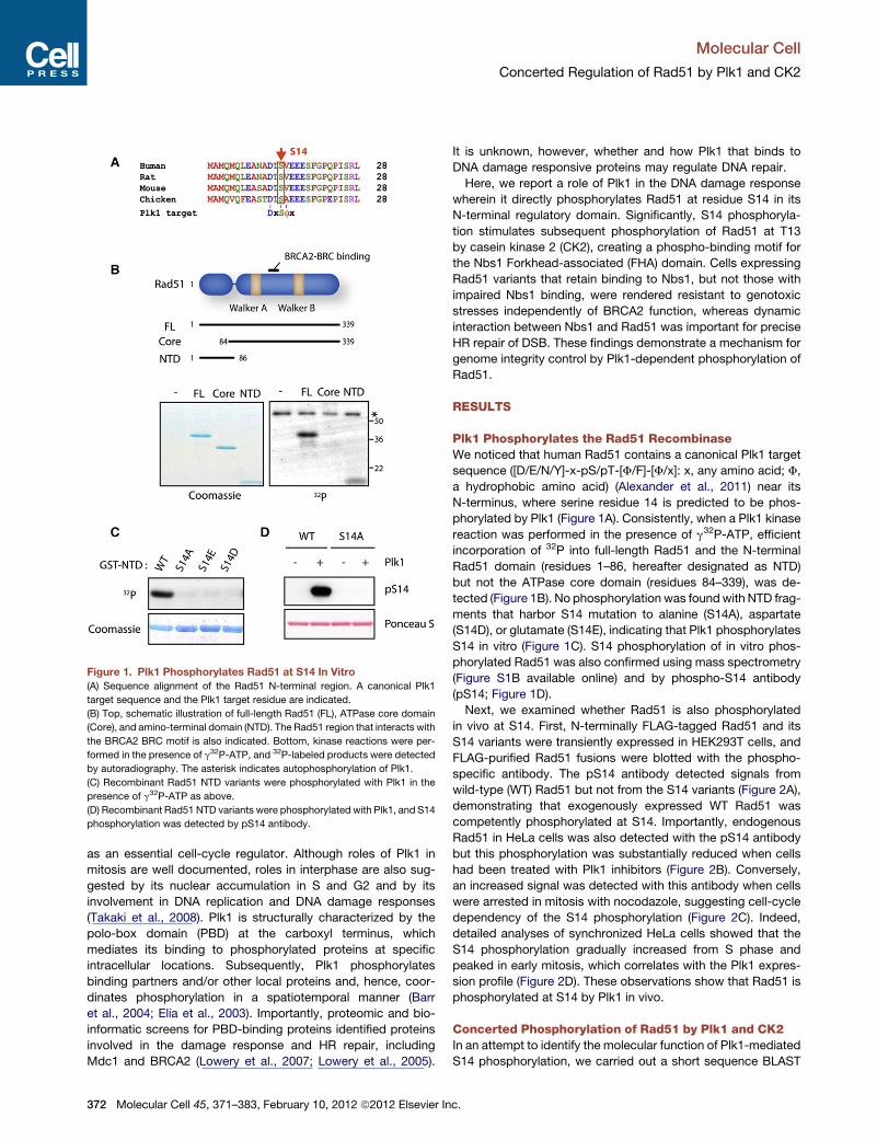
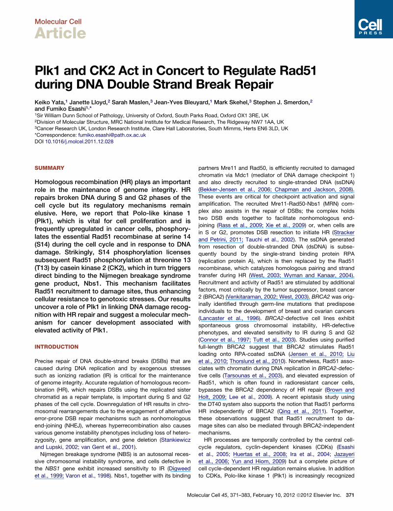
Figure 1. Plk1 Phosphorylates Rad51 at S14 In Vitro
(A) Sequence alignment of the Rad51 N-terminal region. A canonical Plk1
target sequence and the Plk1 target residue are indicated.
(B) Top, schematic illustration of full-length Rad51 (FL), ATPase core domain
(Core), and amino-terminal domain (NTD). The Rad51 region that interacts with
the BRCA2 BRC motif is also indicated. Bottom, kinase reactions were per-
formed in the presence of g32P-ATP, and 32P-labeled products were detected
by autoradiography. The asterisk indicates autophosphorylation of Plk1.
(C) Recombinant Rad51 NTD variants were phosphorylated with Plk1 in the
presence of g32P-ATP as above.
(D) Recombinant Rad51 NTD variants were phosphorylated with Plk1, and S14
phosphorylation was detected by pS14 antibody.
Molecular Cell
Concerted Regulation of Rad51 by Plk1 and CK2
as an essential cell-cycle regulator. Although roles of Plk1 in
mitosis are well documented, roles in interphase are also sug-
gested by its nuclear accumulation in S and G2 and by its
involvement in DNA replication and DNA damage responses
(Takaki et al., 2008). Plk1 is structurally characterized by the
polo-box domain (PBD) at the carboxyl terminus, which
mediates its binding to phosphorylated proteins at specific
intracellular locations. Subsequently, Plk1 phosphorylates
binding partners and/or other local proteins and, hence, coor-
dinates phosphorylation in a spatiotemporal manner (Barr
et al., 2004; Elia et al., 2003). Importantly, proteomic and bio-
informatic screens for PBD-binding proteins identified proteins
involved in the damage response and HR repair, including
Mdc1 and BRCA2 (Lowery et al., 2007; Lowery et al., 2005).
372 Molecular Cell 45, 371–383, February 10, 2012 ª2012 Elsevier In
It is unknown, however, whether and how Plk1 that binds to
DNA damage responsive proteins may regulate DNA repair.
Here, we report a role of Plk1 in the DNA damage response
wherein it directly phosphorylates Rad51 at residue S14 in its
N-terminal regulatory domain. Significantly, S14 phosphoryla-
tion stimulates subsequent phosphorylation of Rad51 at T13
by casein kinase 2 (CK2), creating a phospho-binding motif for
the Nbs1 Forkhead-associated (FHA) domain. Cells expressing
Rad51 variants that retain binding to Nbs1, but not those with
impaired Nbs1 binding, were rendered resistant to genotoxic
stresses independently of BRCA2 function, whereas dynamic
interaction between Nbs1 and Rad51 was important for precise
HR repair of DSB. These findings demonstrate a mechanism for
genome integrity control by Plk1-dependent phosphorylation of
Rad51.
RESULTS
Plk1 Phosphorylates the Rad51 RecombinaseWe noticed that human Rad51 contains a canonical Plk1 target
sequence ([D/E/N/Y]-x-pS/pT-[F/F]-[F/x]: x, any amino acid; F,
a hydrophobic amino acid) (Alexander et al., 2011) near its
N-terminus, where serine residue 14 is predicted to be phos-
phorylated by Plk1 (Figure 1A). Consistently, when a Plk1 kinase
reaction was performed in the presence of g32P-ATP, efficient
incorporation of 32P into full-length Rad51 and the N-terminal
Rad51 domain (residues 1–86, hereafter designated as NTD)
but not the ATPase core domain (residues 84–339), was de-
tected (Figure 1B). No phosphorylation was foundwith NTD frag-
ments that harbor S14 mutation to alanine (S14A), aspartate
(S14D), or glutamate (S14E), indicating that Plk1 phosphorylates
S14 in vitro (Figure 1C). S14 phosphorylation of in vitro phos-
phorylated Rad51 was also confirmed using mass spectrometry
(Figure S1B available online) and by phospho-S14 antibody
(pS14; Figure 1D).
Next, we examined whether Rad51 is also phosphorylated
in vivo at S14. First, N-terminally FLAG-tagged Rad51 and its
S14 variants were transiently expressed in HEK293T cells, and
FLAG-purified Rad51 fusions were blotted with the phospho-
specific antibody. The pS14 antibody detected signals from
wild-type (WT) Rad51 but not from the S14 variants (Figure 2A),
demonstrating that exogenously expressed WT Rad51 was
competently phosphorylated at S14. Importantly, endogenous
Rad51 in HeLa cells was also detected with the pS14 antibody
but this phosphorylation was substantially reduced when cells
had been treated with Plk1 inhibitors (Figure 2B). Conversely,
an increased signal was detected with this antibody when cells
were arrested in mitosis with nocodazole, suggesting cell-cycle
dependency of the S14 phosphorylation (Figure 2C). Indeed,
detailed analyses of synchronized HeLa cells showed that the
S14 phosphorylation gradually increased from S phase and
peaked in early mitosis, which correlates with the Plk1 expres-
sion profile (Figure 2D). These observations show that Rad51 is
phosphorylated at S14 by Plk1 in vivo.
Concerted Phosphorylation of Rad51 by Plk1 and CK2In an attempt to identify the molecular function of Plk1-mediated
S14 phosphorylation, we carried out a short sequence BLAST
c.
Figure 2. Rad51 Is Phosphorylated at S14
in a Plk1-Dependent Manner In Vivo
(A) FLAG-tagged Rad51 variants purified from
HEK293T cells were blotted with pS14 antibody or
FLAG antibody. NT, nontransfected cells. The
asterisk indicates endogenous Rad51 copurified
with FLAG-Rad51.
(B) HeLa cells were treated with DMSO or Plk1
inhibitors (BTO-1 or BI-2536), and immunoprecip-
itated Rad51 was analyzed using pS14 antibody.
(C) Rad51 from HeLa cells treated with DMSO
or nocodazole (Noc) was analyzed using pS14
antibody as above. No signal was detected after
l phosphatase (l PPase) treatment, showing the
phospho-specificity of the antibody.
(D) HeLa cells were synchronized by double
thymidine-block release, and Rad51 phosphory-
lation was analyzed. Cell-cycle progression was
monitored by cell-cycle markers cyclin E (for
G1/S), cyclin A (for S/G2), and Plk1 (for S/G2/M)
and by FACS. As, asynchronous cells.
Molecular Cell
Concerted Regulation of Rad51 by Plk1 and CK2
analysis using the Plk1 phosphorylation motif in Rad51 and its
surrounding sequence (hereafter designated as Rad51 DTSV
motif). This analysis revealed a remarkable similarity to regions
of Mdc1 containing SDTD motifs, a known target of CK2,
where CK2 phosphorylation of the threonine between two acidic
aspartate residues creates a binding site for Nbs1 (Figure 3A)
(Chapman and Jackson, 2008; Lloyd et al., 2009; Melander
et al., 2008; Spycher et al., 2008;Williams et al., 2009). Therefore,
we examined the ability of CK2 to phosphorylate the threonine in
the Rad51 DTSV motif. Notably, in vitro CK2 kinase assays
revealed efficient phosphorylation of full-length Rad51 and the
NTD fragment but not the ATPase core or a NTD fragment
harboring an alanine substitution at threonine 13 (T13A) (Figures
3B and 3C), demonstrating that CK2 phosphorylates Rad51 at
T13. Our mass spectrometric analysis of full-length Rad51 phos-
phorylated in vitro with CK2 also identified a single phosphoryla-
tion at T13 (Figure S1C).
Because CK2 is an acidophilic kinase, we next explored
whether a negative charge at S14 modifies CK2-mediated
phosphorylation at T13. Indeed, a Rad51 NTD fragment with
S14 substituted by an acidic residue (D or E) was more suscep-
Molecular Cell 45, 371–383,
tible to CK2 phosphorylation, whereas
a NTD with S14A substitution was less
efficiently phosphorylated (Figure 3D).
These observations are consistent with
the documented CK2 target preferences
for highly acidic substrate motifs (Son-
gyang et al., 1996) and the substantial
deleterious effects on both Km and Vmax
values for a CK2 peptide substrate with
an alanine substitution at the pS/T +1
position (Sarno et al., 1997). We further
evaluated whether Plk1 phosphorylation
at S14, which renders this residue nega-
tively charged, might modify the effi-
ciency of CK2-mediated phosphorylation
at T13. To this end, we first phosphory-
lated a GST fusion of Rad51 NTD immobilized on glutathione
beads in the presence of nonradiolabeled ATP; after washing
off the kinase and ATP, a second kinase reaction was carried
out in the presence of g32P-ATP (Figure 3E). Strikingly, prephos-
phorylation of Rad51 NTD with Plk1 substantially facilitated
subsequent phosphorylation by CK2; in contrast, CK2-prephos-
phorylation of Rad51 did not alter the efficiency of Plk1-depen-
dent phosphorylation (Figure 3E, compare lanes 2, 4, and 5).
This result supports the view that Rad51 is sequentially modified
by Plk1-dependent priming phosphorylation at S14, followed by
CK2-mediated phosphorylation at T13.
Dynamic Phosphorylation of Rad51 In VivoThe sequential phosphorylation of the Rad51 DTSVmotif by Plk1
and CK2 in vitro prompted us to further investigate whether
endogenous Rad51 is phosphorylated at T13 or at both T13
and S14 in cells. For this purpose, additional phospho-specific
antibodies were raised against Rad51 monophosphorylated
at T13 or diphosphorylated at T13 and S14 (pT13 and pT13/
pS14 antibody, respectively; Figures 4A and 4B), and used to
analyze in vivo phosphorylated Rad51. As shown in Figure 4C,
February 10, 2012 ª2012 Elsevier Inc. 373
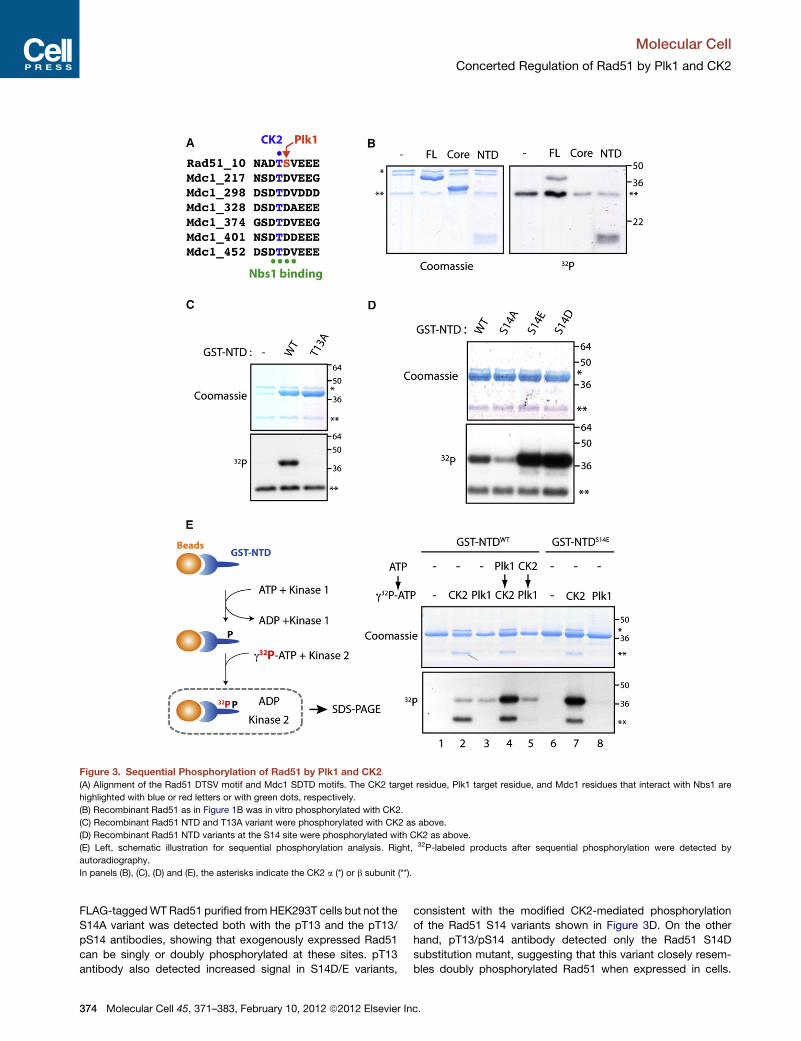
Figure 3. Sequential Phosphorylation of Rad51 by Plk1 and CK2
(A) Alignment of the Rad51 DTSV motif and Mdc1 SDTD motifs. The CK2 target residue, Plk1 target residue, and Mdc1 residues that interact with Nbs1 are
highlighted with blue or red letters or with green dots, respectively.
(B) Recombinant Rad51 as in Figure 1B was in vitro phosphorylated with CK2.
(C) Recombinant Rad51 NTD and T13A variant were phosphorylated with CK2 as above.
(D) Recombinant Rad51 NTD variants at the S14 site were phosphorylated with CK2 as above.
(E) Left, schematic illustration for sequential phosphorylation analysis. Right, 32P-labeled products after sequential phosphorylation were detected by
autoradiography.
In panels (B), (C), (D) and (E), the asterisks indicate the CK2 a (*) or b subunit (**).
Molecular Cell
Concerted Regulation of Rad51 by Plk1 and CK2
FLAG-taggedWTRad51 purified fromHEK293T cells but not the
S14A variant was detected both with the pT13 and the pT13/
pS14 antibodies, showing that exogenously expressed Rad51
can be singly or doubly phosphorylated at these sites. pT13
antibody also detected increased signal in S14D/E variants,
374 Molecular Cell 45, 371–383, February 10, 2012 ª2012 Elsevier In
consistent with the modified CK2-mediated phosphorylation
of the Rad51 S14 variants shown in Figure 3D. On the other
hand, pT13/pS14 antibody detected only the Rad51 S14D
substitution mutant, suggesting that this variant closely resem-
bles doubly phosphorylated Rad51 when expressed in cells.
c.
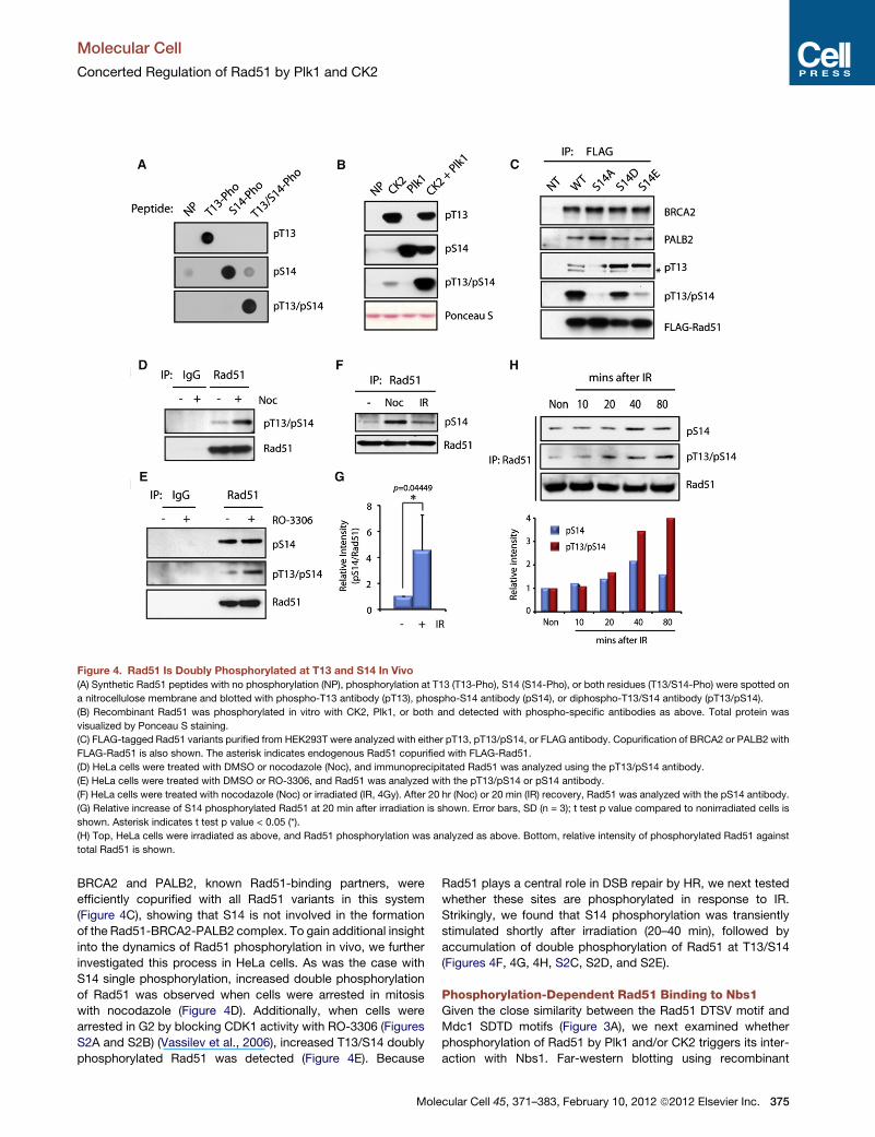
Figure 4. Rad51 Is Doubly Phosphorylated at T13 and S14 In Vivo
(A) Synthetic Rad51 peptides with no phosphorylation (NP), phosphorylation at T13 (T13-Pho), S14 (S14-Pho), or both residues (T13/S14-Pho) were spotted on
a nitrocellulose membrane and blotted with phospho-T13 antibody (pT13), phospho-S14 antibody (pS14), or diphospho-T13/S14 antibody (pT13/pS14).
(B) Recombinant Rad51 was phosphorylated in vitro with CK2, Plk1, or both and detected with phospho-specific antibodies as above. Total protein was
visualized by Ponceau S staining.
(C) FLAG-tagged Rad51 variants purified from HEK293T were analyzed with either pT13, pT13/pS14, or FLAG antibody. Copurification of BRCA2 or PALB2 with
FLAG-Rad51 is also shown. The asterisk indicates endogenous Rad51 copurified with FLAG-Rad51.
(D) HeLa cells were treated with DMSO or nocodazole (Noc), and immunoprecipitated Rad51 was analyzed using the pT13/pS14 antibody.
(E) HeLa cells were treated with DMSO or RO-3306, and Rad51 was analyzed with the pT13/pS14 or pS14 antibody.
(F) HeLa cells were treated with nocodazole (Noc) or irradiated (IR, 4Gy). After 20 hr (Noc) or 20 min (IR) recovery, Rad51 was analyzed with the pS14 antibody.
(G) Relative increase of S14 phosphorylated Rad51 at 20 min after irradiation is shown. Error bars, SD (n = 3); t test p value compared to nonirradiated cells is
shown. Asterisk indicates t test p value < 0.05 (*).
(H) Top, HeLa cells were irradiated as above, and Rad51 phosphorylation was analyzed as above. Bottom, relative intensity of phosphorylated Rad51 against
total Rad51 is shown.
Molecular Cell
Concerted Regulation of Rad51 by Plk1 and CK2
BRCA2 and PALB2, known Rad51-binding partners, were
efficiently copurified with all Rad51 variants in this system
(Figure 4C), showing that S14 is not involved in the formation
of the Rad51-BRCA2-PALB2 complex. To gain additional insight
into the dynamics of Rad51 phosphorylation in vivo, we further
investigated this process in HeLa cells. As was the case with
S14 single phosphorylation, increased double phosphorylation
of Rad51 was observed when cells were arrested in mitosis
with nocodazole (Figure 4D). Additionally, when cells were
arrested in G2 by blocking CDK1 activity with RO-3306 (Figures
S2A and S2B) (Vassilev et al., 2006), increased T13/S14 doubly
phosphorylated Rad51 was detected (Figure 4E). Because
Mole
Rad51 plays a central role in DSB repair by HR, we next tested
whether these sites are phosphorylated in response to IR.
Strikingly, we found that S14 phosphorylation was transiently
stimulated shortly after irradiation (20–40 min), followed by
accumulation of double phosphorylation of Rad51 at T13/S14
(Figures 4F, 4G, 4H, S2C, S2D, and S2E).
Phosphorylation-Dependent Rad51 Binding to Nbs1Given the close similarity between the Rad51 DTSV motif and
Mdc1 SDTD motifs (Figure 3A), we next examined whether
phosphorylation of Rad51 by Plk1 and/or CK2 triggers its inter-
action with Nbs1. Far-western blotting using recombinant
cular Cell 45, 371–383, February 10, 2012 ª2012 Elsevier Inc. 375
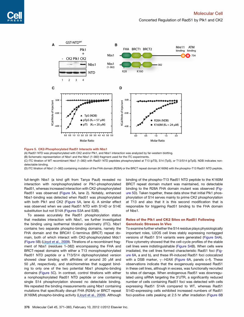
Figure 5. CK2-Phosphorylated Rad51 Interacts with Nbs1
(A) Rad51 NTD was phosphorylated with CK2 and/or Plk1, and Nbs1 interaction was analyzed by far-western blotting.
(B) Schematic representation of Nbs1 and the Nbs1 (1-382) fragment used for the ITC experiments.
(C) ITC titration of WT recombinant Nbs1 (1–382) with Rad51 NTD peptides phosphorylated at T13 (pTS), S14 (TpS), or T13/S14 (pTpS). NDB indicates non-
detectable binding.
(D) ITC titration of Nbs1 (1–382) containing mutation of the FHA domain (R28A) or the BRCT repeat domain (K160M) with the phospho-T13 Rad51 NTD peptide.
Molecular Cell
Concerted Regulation of Rad51 by Plk1 and CK2
full-length Nbs1 (a kind gift from Tanya Paull) revealed no
interaction with nonphosphorylated or Plk1-phosphorylated
Rad51, whereas increased interaction with CK2-phosphorylated
Rad51 was observed (Figure 5A, lane 2). Notably, enhanced
Nbs1-binding was detected when Rad51 was phosphorylated
with both Plk1 and CK2 (Figure 5A, lane 4). A similar effect
was observed when we used Rad51 NTD with S14D or S14E
substitution but not S14A (Figures S3A and S3B).
To assess accurately the Rad51 phosphorylation status
that mediates interaction with Nbs1, we further investigated
the binding using isothermal titration calorimetry (ITC). Nbs1
contains two separate phospho-binding domains, namely the
FHA domain and the BRCA1 C-terminus (BRCT) repeat do-
main, both of which interact with CK2-phosphorylated Mdc1
(Figure 5B) (Lloyd et al., 2009). Titrations of a recombinant frag-
ment of Nbs1 (residues 1–382) encompassing the FHA and
BRCT-repeat domains with either a T13 monophosphorylated
Rad51 NTD peptide or a T13/S14 diphosphorylated version
showed clear binding with affinities of around 20 mM and
50 mM, respectively, and stoichiometries that suggest bind-
ing to only one of the two potential Nbs1 phospho-binding
domains (Figure 5C). In contrast, control titrations with either
a nonphosphorylated Rad51 NTD peptide or one containing
single S14 phosphorylation showed no detectable binding.
We repeated the binding measurements using Nbs1 containing
mutations that specifically disrupt FHA (R28A) or BRCT repeat
(K160M) phospho-binding activity (Lloyd et al., 2009). Although
376 Molecular Cell 45, 371–383, February 10, 2012 ª2012 Elsevier In
binding of the phospho-T13 Rad51 NTD peptide to the K160M
BRCT repeat domain mutant was maintained, no detectable
binding to the R28A FHA domain mutant was observed (Fig-
ure 5D). Taken together, these data show that initial Plk1 phos-
phorylation of S14 serves mainly to prime CK2 phosphorylation
at T13 and also that it is this second modification that is
responsible for triggering Rad51 binding to the FHA domain
of Nbs1.
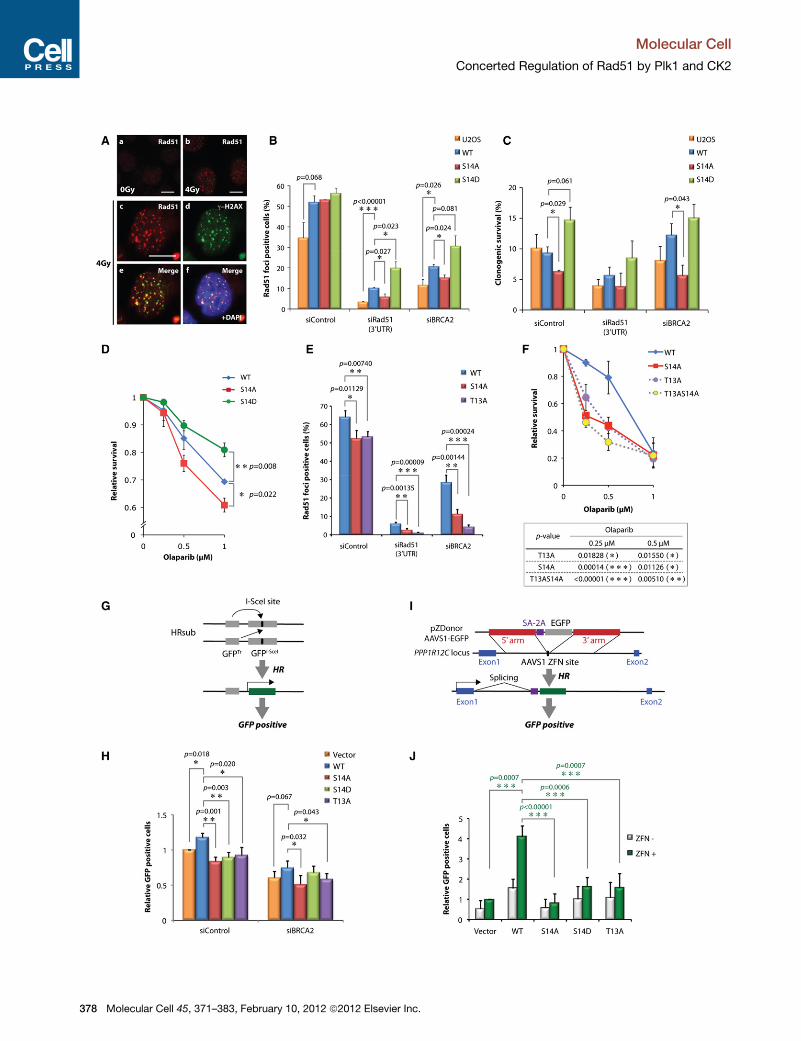
Roles of the Plk1 and CK2 Sites on Rad51 FollowingGenotoxic Stresses In VivoTo examine further whether the S14 residue plays physiologically
important roles, U2OS cell lines stably expressing nontagged
versions of Rad51 S14 variants were generated (Figure S4A).
Flow cytometry showed that the cell-cycle profiles of the stable
cell lines were indistinguishable (Figure S4B). When cells were
irradiated, the cell lines formed characteristic Rad51 foci (Fig-
ure 6A, a and b), and these IR-induced Rad51 foci colocalized
with a DSB marker, g�H2AX (Figure 6A, panels c–f). These
observations indicate that the exogenously expressed Rad51
in these cell lines, although in excess, was functionally recruited
to sites of damage. When endogenous Rad51 was downregu-
lated using siRNA targeting the 30UTR, a significantly reduced
number of cells containing Rad51 foci was detected with cells
expressing Rad51 S14A compared to WT, whereas Rad51
S14D-expressing cells exhibited increased numbers of Rad51
foci-positive cells peaking at 2.5 hr after irradiation (Figure 6B
c.
Molecular Cell
Concerted Regulation of Rad51 by Plk1 and CK2
and S4C). These results show the importance of the Rad51 S14
residue in damage-induced focus formation.
Our biochemical analyses shown in Figure 5 provided
evidence that sequential Rad51 phosphorylation within the
DSTVmotif stimulates its binding to a major DSB sensor protein,
Nbs1, which accumulates on damaged chromatin in a BRCA2-
independent manner. Hence we speculated that the differential
recruitment of Rad51 S14 variants was likely to be a BRCA2-
independent process. Indeed, the cells expressing exogenous
WT Rad51 effectively formed Rad51 foci after BRCA2 downre-
gulation, whereas those expressing Rad51 S14A exhibited
reduced focus formation (Figures 6B and S4D).We further tested
clonogenic survival of these cell lines after downregulating
endogenous Rad51 or BRCA2. In otherwise unperturbed cells,
all variants exhibited comparable survival (Figure S4E). By
contrast, cells expressing WT or S14D-substituted Rad51 but
not the S14A variant showed enhanced survival after IR
treatment following BRCA2 downregulation (Figure 6C). These
observations further confirm the importance of the S14 residue
in resistance to IR, independently of BRCA2 function.
HR-defective cells, including BRCA2-defective cancer cells,
exhibit profound sensitivity to inhibitors of poly(ADP-ribose)
polymerase (PARP); hence, cancers harboring mutations in
BRCA2 can be treated effectively with PARP inhibitors (Bryant
et al., 2005; Farmer et al., 2005). However, the genome of
BRCA2-defective cancer cells is highly unstable and some
cancers gain resistance to PARP inhibitors through de novo
mutations (Edwards et al., 2008; Sakai et al., 2008). Given that
Plk1 upregulation is often associated with malignancy (Streb-
hardt, 2010; Taylor and Peters, 2008), we addressed the poten-
tial role of Plk1-dependent Rad51 phosphorylation in PARP
inhibitor resistance. Remarkably, where BRCA2 was downregu-
lated, cells expressing WT and the phospho-mimetic S14D
Rad51 variant exhibited significantly higher resistance to
a PARP inhibitor compared to cells expressing S14A (Figure 6D).
These results indicate that Plk1-mediated phosphorylation of
Rad51 at S14 facilitates resistance to PARP inhibition.
We further addressed whether the cellular phenotypes associ-
ated with Rad51 S14 substitution are the direct result of altered
phospho-T13-dependent binding to Nbs1. Indeed, cells ex-
pressing the T13A variant exhibited impaired IR-induced
Rad51 focus formation (Figure 6E) and reduced resistance to
the PARP inhibitor in BRCA2 downregulated cells (Figure 6F),
similar to those expressing the S14A variant. Taken together,
these observations support the notion that CK2 and Plk1 act in
concert to regulate damage-induced Rad51 localization and
resistance to genotoxic stresses.
Roles of the Plk1 andCK2Sites onRad51 inHomologousRecombinationFinally, we investigated the importance of the CK2 and Plk1 sites
on Rad51 during HR in vivo. To this end, we used a well-estab-
lished HR reporter system based on the rare-cutting homing
endonuclease I-SceI to introduce a DSB (Moynahan and Jasin,
2010). Specifically, Rad51 variants were stably expressed in
a U2OS cell line that carries tandem modified GFP genes on
chromosome 18: a GFP mutant containing an I-SceI cleavage
site (GFPI-SceI) and a truncated GFP (GFPTr) (U2OS-SCR18)
Mole
(Puget et al., 2005). Following I-SceI expression, HR events
were measured by quantifying the GFP-expressing cell popula-
tion (Figures 6G and S4F). As shown in Figure 6H, increased
HR events were detected in cells expressing WT Rad51
compared to those with empty vector, whereas no increase
was found with Rad51 variants at S14 and T13. Similar pheno-
types were also observed following BRCA2 downregulation,
although S14D supported modest HR recovery.
We further assessed HR in the absence of functional
BRCA2 by DSB-mediated gene targeting in BRCA2-defective
EUFA423 cells stably expressing Rad51 variants. We exploited
a zinc-finger nuclease (ZFN) that introduces a DSB at a native
AAVS1 locus within the PPP1R12C gene and a donor plasmid
containing a promoterless GFP gene between sequences
homologous to those flanking the AAVS1 site (Figures 6I, S4G,
and S4H) (Brunet et al., 2009; Hockemeyer et al., 2009). In this
system, DSB-promoted gene targeting results in GFP expres-
sion from the native PPP1R12C promoter. Indeed, a clear
increase of GFP expression was observed in EUFA423 cells
expressing WT Rad51 compared to those containing empty
vector (Figure 6J). By contrast, EUFA423 cells expressing
Rad51 variants at either T13 or S14 exhibited significantly lower
targeting efficiency. These observations support the notion that
dynamic phosphorylation of Rad51 by Plk1 and CK2 is important
for the coordination of precise recombination.
DISCUSSION
In this study, we show that 1) Rad51 recombinase is directly
phosphorylated by Plk1 at S14 in a cell cycle- and DNA
damage-responsive manner; 2) Plk1-mediated phosphorylation
stimulates subsequent CK2-mediated phosphorylation at T13;
3) T13 phosphorylation of Rad51 by CK2 triggers a direct
interaction with the FHA domain of the MRN component,
Nbs1; and 4) Rad51 phosphorylation at either S14 or T13 is
important for accurate HR and for cellular resistance to IR and
to PPAR inhibition. Collectively, these data support the model
illustrated in Figure 7. Upon DSB induction, the MRN complex
is efficiently recruited to the site of damage, mediating DNA
resection during S and G2 phases of the cell cycle. During these
cell-cycle phases, Plk1-mediated Rad51 phosphorylation in-
creases, stimulating CK2-dependent T13 phosphorylation and
triggering its interaction with the FHA domain of Nbs1. This
mechanism helps increase the Rad51 concentration at the site
of DNA damage and facilitates HR.
Early Roles of Plk1 after DNA DamageMounting evidence points toward active roles of Plk1 in DNA
damage responses (Mac�urek et al., 2008; Syljuasen et al.,
2006; Toczyski et al., 1997; van Vugt et al., 2004; Yoo et al.,
2004) but there is no established role for Plk1 immediately after
DNA damage. Unexpectedly, we observed an immediate and
transient increase of Rad51 phosphorylation after DNA damage:
S14 phosphorylation reproducibly peaked at 20–40 min after
irradiation (Figures 4F, 4G, 4H, S2C, S2D, and S2E). It is not clear
whether total Plk1 activity is stimulated immediately after DNA
damage or whether Plk1 becomes locally activated at the site
of DNA damage. The latter possibility is particularly attractive
cular Cell 45, 371–383, February 10, 2012 ª2012 Elsevier Inc. 377
Molecular Cell
Concerted Regulation of Rad51 by Plk1 and CK2
378 Molecular Cell 45, 371–383, February 10, 2012 ª2012 Elsevier Inc.
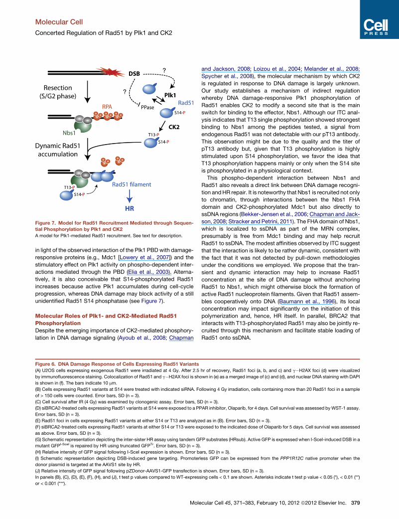
Figure 7. Model for Rad51 Recruitment Mediated through Sequen-
tial Phosphorylation by Plk1 and CK2
A model for Plk1-mediated Rad51 recruitment. See text for description.
Molecular Cell
Concerted Regulation of Rad51 by Plk1 and CK2
in light of the observed interaction of the Plk1 PBDwith damage-
responsive proteins (e.g., Mdc1 [Lowery et al., 2007]) and the
stimulatory effect on Plk1 activity on phospho-dependent inter-
actions mediated through the PBD (Elia et al., 2003). Alterna-
tively, it is also conceivable that S14-phosphorylated Rad51
increases because active Plk1 accumulates during cell-cycle
progression, whereas DNA damage may block activity of a still
unidentified Rad51 S14 phosphatase (see Figure 7).
Molecular Roles of Plk1- and CK2-Mediated Rad51PhosphorylationDespite the emerging importance of CK2-mediated phosphory-
lation in DNA damage signaling (Ayoub et al., 2008; Chapman
Figure 6. DNA Damage Response of Cells Expressing Rad51 Variants
(A) U2OS cells expressing exogenous Rad51 were irradiated at 4 Gy. After 2.5
by immunofluorescence staining. Colocalization of Rad51 and g�H2AX foci is sho
is shown in (f). The bars indicate 10 mm.
(B) Cells expressing Rad51 variants at S14 were treated with indicated siRNA. Fo
of > 150 cells were counted. Error bars, SD (n = 3).
(C) Cell survival after IR (4 Gy) was examined by clonogenic assay. Error bars, S
(D) siBRCA2-treated cells expressing Rad51 variants at S14 were exposed to a PP
Error bars, SD (n = 3).
(E) Rad51 foci in cells expressing Rad51 variants at either S14 or T13 are analyz
(F) siBRCA2-treated cells expressing Rad51 variants at either S14 or T13 were ex
as above. Error bars, SD (n = 3).
(G) Schematic representation depicting the inter-sister HR assay using tandem G
mutant GFPI-SceI is repaired by HR using truncated GFPTr. Error bars, SD (n = 3)
(H) Relative intensity of GFP signal following I-SceI expression is shown. Error b
(I) Schematic representation depicting DSB-induced gene targeting. Promoter
donor plasmid is targeted at the AAVS1 site by HR.
(J) Relative intensity of GFP signal following pZDonor-AAVS1-GFP transfection i
In panels (B), (C), (D), (E), (F), (H), and (J), t test p values compared to WT-express
or < 0.001 (***).
Mole
and Jackson, 2008; Loizou et al., 2004; Melander et al., 2008;
Spycher et al., 2008), the molecular mechanism by which CK2
is regulated in response to DNA damage is largely unknown.
Our study establishes a mechanism of indirect regulation
whereby DNA damage-responsive Plk1 phosphorylation of
Rad51 enables CK2 to modify a second site that is the main
switch for binding to the effector, Nbs1. Although our ITC anal-
ysis indicates that T13 single phosphorylation showed strongest
binding to Nbs1 among the peptides tested, a signal from
endogenous Rad51 was not detectable with our pT13 antibody.
This observation might be due to the quality and the titer of
pT13 antibody but, given that T13 phosphorylation is highly
stimulated upon S14 phosphorylation, we favor the idea that
T13 phosphorylation happens mainly or only when the S14 site
is phosphorylated in a physiological context.
This phospho-dependent interaction between Nbs1 and
Rad51 also reveals a direct link between DNA damage recogni-
tion andHR repair. It is noteworthy that Nbs1 is recruited not only
to chromatin, through interactions between the Nbs1 FHA
domain and CK2-phosphorylated Mdc1 but also directly to
ssDNA regions (Bekker-Jensen et al., 2006; Chapman and Jack-
son, 2008; Stracker and Petrini, 2011). The FHA domain of Nbs1,
which is localized to ssDNA as part of the MRN complex,
presumably is free from Mdc1 binding and may help recruit
Rad51 to ssDNA. The modest affinities observed by ITC suggest
that the interaction is likely to be rather dynamic, consistent with
the fact that it was not detected by pull-down methodologies
under the conditions we employed. We propose that the tran-
sient and dynamic interaction may help to increase Rad51
concentration at the site of DNA damage without anchoring
Rad51 to Nbs1, which might otherwise block the formation of
active Rad51 nucleoprotein filaments. Given that Rad51 assem-
bles cooperatively onto DNA (Baumann et al., 1996), its local
concentration may impact significantly on the initiation of this
polymerization and, hence, HR itself. In parallel, BRCA2 that
interacts with T13-phosphorylated Rad51 may also be jointly re-
cruited through this mechanism and facilitate stable loading of
Rad51 onto ssDNA.
hr of recovery, Rad51 foci (a, b, and c) and g�H2AX foci (d) were visualized
wn in (e) as a merged image of (c) and (d), and nuclear DNA staining with DAPI
llowing 4 Gy irradiation, cells containing more than 20 Rad51 foci in a sample
D (n = 3).
AR inhibitor, Olaparib, for 4 days. Cell survival was assessed by WST-1 assay.
ed as in (B). Error bars, SD (n = 3).
posed to the indicated dose of Olaparib for 5 days. Cell survival was assessed
FP substrates (HRsub). Active GFP is expressed when I-SceI-induced DSB in a
.
ars, SD (n = 3).
less GFP can be expressed from the PPP1R12C native promoter when the
s shown. Error bars, SD (n = 3).
ing cells < 0.1 are shown. Asterisks indicate t test p value < 0.05 (*), < 0.01 (**)
cular Cell 45, 371–383, February 10, 2012 ª2012 Elsevier Inc. 379
Molecular Cell
Concerted Regulation of Rad51 by Plk1 and CK2
Roles of Plk1-Mediated Rad51 Phosphorylation inUnperturbed CellsWe found no evidence for a role of cell cycle-dependent Rad51
phosphorylation by Plk1 in unperturbed U2OS cells (Figures
S4B and S4E), although it is possible that such phosphorylation
may have a role in primary cells. Supporting this idea, active and
protective roles of Rad51 in later phases of the cell cycle were
shown using chicken DT40 and Xenopus systems; Rad51
depletion caused G2 arrest with accumulated ssDNA lesions
or DSBs (Hashimoto et al., 2010; Su et al., 2008). In this context,
Plk1-mediated Rad51 phosphorylation in G2 may promote its
recruitment to a ssDNA lesion, providing a final opportunity to
complete sister chromatid synthesis before onset of mitosis.
This mechanism may also play a crucial role during meiosis,
where HR facilitates crossover between homologous chromo-
somes. In line with this notion, a direct role of Cdc5, which is
a yeast ortholog of Plk1, during meiotic crossover was recently
reported (Matos et al., 2011). Further studies will be needed to
fully illuminate Plk1-dependent HR regulation in somatic and
germline cells.
A Model for Genome Instability Phenotypes Mediatedthrough Rad51 PhosphorylationIn HR reporter assays using site-specific endonucleases, Rad51
variants at the CK2 or Plk1 sites that were substituted with either
nonphosphorylatable alanine (S14A and T13A) or phospho-
mimetic aspartate (S14D) were less proficient in HR than WT
Rad51. This observation suggests that both impaired and unduly
stable interaction between Rad51 and Nbs1 have negative
effects on canonical error-free HR, highlighting the importance
of dynamic quality control of HR proteins during DSB repair
(Kanaar et al., 2008). Intriguingly, the S14D variant exhibited
increased survival following IR and Olaparib treatment (Figures
6C and 6D), leading us to propose that enhanced Rad51-binding
to Nbs1 may support survival by promoting nonlethal but low
quality recombination events that were undetectable with the
HR reporter systems used in this study. This idea is supported
by the observation that elevated Rad51 expression in mouse
embryonic stem cells leads to aberrant interchromosomal repair
following induction of multiple DSBs within short homologous
sequences, consequently resulting in a genome instability
phenotype (Richardson et al., 2004). We also found that
BRCA2-defective EUFA423 cells exhibited increased random
integration of donor plasmid when WT Rad51 was exogenously
expressed (Figures 6J, S4G, and S4H). Therefore, Plk1-medi-
ated Rad51 phosphorylation may promote gross chromosomal
instability, particularly when fine-tuning of HR is disrupted in
BRCA-defective cells. Importantly, increased activity of Plk1 is
closely linked to malignancy, and Plk1 inhibition sensitizes
cancer cells to DNA damage treatment (Strebhardt, 2010; Sur
et al., 2009; Taylor and Peters, 2008). Clinical trials in cancer
patients are currently underway to evaluate the effects of
PARP and Plk1 inhibitors (Carden et al., 2010; Lord and Ash-
worth, 2008). It is tempting to speculate that a combined therapy
using inhibitors of both Plk1 and PARP may be an effective
approach to improve prognosis of BRCA-defective cancers.
In summary, we have established a direct link between DNA
damage recognition and HR repair, mediated through a phos-
380 Molecular Cell 45, 371–383, February 10, 2012 ª2012 Elsevier In
pho-dependent interaction between Nbs1 and Rad51. A primary
cell-cycle regulator, Plk1, plays a critical role in the regulation of
this interaction, which can modulate the BRCA2 dependency of
HR repair. Our findings represent a significant step toward a
comprehensive understanding of HR regulation by cell-cycle
regulators, which may be exploited in the further development
of effective cancer treatments.
EXPERIMENTAL PROCEDURES
Cell Culture
HeLa, HEK293T, and U2OS cells were cultured at 37�C with 5% CO2 in
Dulbecco’s modified Eagle’s medium supplemented with streptomycin
(0.1 mg/ml), penicillin (100 units/ml), and 10% v/v fetal bovine serum. Where
indicated, cells were treated with 0.2 mM nocodazole (Sigma-Aldrich) or
9 mM RO-3306 (Enzo Life Sciences) for 20 hr, or 50 mM BTO-1 (Sigma-Aldrich)
or 0.1 mM BI-2536 (Axon Medchem) for 2.5 hr. The 137Cs-source of an IBL 637
(CIS Bio International; Figures 4F, 4G, S2C, and S2D) or a GRAVITRON RX
30/55 (Gravatom; Figures 4H and 6) was used to irradiate cells at 4 Gy (59 s
and 69 s, respectively). Cell synchronization was carried out as previously
described (Esashi et al., 2005). U2OS stable cell lines expressing Rad51 vari-
ants were generated by cotransfecting pcDNA5/FRT encoding nontagged
Rad51 and pcDNA-DEST26 or pcDNA-DEST53 (Invitrogen) using jetPrime
(Polyplus Transfection), followed by G418 selection at 400 mg/ml. U2OS-
SCR18 was a kind gift from Ralph Scully. U2OS-SCR18 cells expressing
Rad51variantsweregeneratedby transfectingpT-Rex-DEST30encodingnon-
tagged Rad51, followed by selection with 300 mg/ml G418 and 1 mg/ml puro-
mycin. EUFA423 cell lines stably expressing Rad51 variants were generated
in two steps: EUFA423 cells were transfected with pFRT/lacZeo plasmid
(Invitrogen), and a cell line containing a Flp-In recombination site was cloned
following Zeocin selection at 25 mg/ml. Established EUFA423 Flp-In cells
were then used to generate stable cell lines by cotransfecting pOG44 (Invitro-
gen) and nontagged Rad51 variant in pcDNA5/FRT, followed by hygromycin
selection at 50 mg/ml. For siRNA treatments, cells were seeded at a density
of 1.5 3 105 cells in 6-well plates and then transfected, on the following day,
with siRad51 (20 nM), siBRCA2 (100 nM), or control siRNA with DharmaFECT1
(100 nM) (Dharmacon); cells were further incubated for 24 hr before analyses.
Extract Preparation, Immunoprecipitation, and Western Blotting
Cell extract was prepared using extraction buffer (150 mMKCl, 20 mMHEPES
pH7.6, 2 mM EGTA, 1.5 mM MgCl2, 50 mM NaF, 0.1% NP40, 10% glycerol,
1 mM Na3VO4, 20 mM b-glycerophosphate, 1 mM dithiothreitol, 10 mM
benzamidine HCl, 25 units/ml Benzonase nuclease [Novagen]) supplemented
with Protease inhibitor cocktail (Sigma-Aldrich, P2714). For immunoprecipita-
tion, the extract was precleared with 10 ml of control IgG beads, followed by
incubation with antibody cross-linked to beads. After extensive washing,
immune complexes were separated by SDS-PAGE and analyzed by western
blotting, following standard protocols. Where indicated, the membrane was
treated with Re-Blot Plus Mild Solution (Millipore) before incubating with
another antibody; for Rad51 phosphorylation analyses, pT13/pS14, pS14,
and Rad51 antibodies were applied in this order.
In Vitro Kinase Reactions and Far-Western Blotting
Protein substrates (1 mg in 15 ml total volume) were phosphorylated in kinase
buffer (25 mM MOPS pH 7.2, 25 mM b-glycerophosphate, 15 mM MgCl2,
1% DMSO, 7.5 mM ATP, 1 mM DTT and 1 mCi g32P-ATP) by the addition of
1 ml of recombinant Plk1 or CK2. Following incubation at 30�C for 30 min,
reactions were stopped by heating at 95�C for 5 min in SDS sample buffer.
Proteins were then resolved by SDS-PAGE and visualized by staining with
InstantBlue (Expedeon). After drying gel with DryEase (Invitrogen), 32P-labeled
products were detected by autoradiography. For far-western analysis, kinase
reaction was carried out in kinase buffer supplemented with 250 mMATP, with
no g32P-ATP. The reaction mixture was then resolved by SDS-PAGE and
transferred to a Protran nitrocellulose membrane (Whatman, BA85), followed
by incubation with recombinant full-length FLAG-Nbs1 (� 2 mg). Anti-Nbs1
antibody was then applied to the membrane to detect Nbs1 protein.
c.
Molecular Cell
Concerted Regulation of Rad51 by Plk1 and CK2
Isothermal Titration Calorimetry
The affinities and thermodynamic parameters for human Nbs1-Rad51 phos-
phopeptide interactions were determined by isothermal titration calorimetry
with a ITC-200 instrument (MicroCal) at 18�C. Protein samples were dialyzed
extensively into 50mMHEPES (pH 7.5), 150mMNaCl, 2mM b-mercaptoetha-
nol and peptides were desalted and buffer exchanged using NAP-5 purifica-
tion columns (GE Healthcare) into the relevant buffer (Lloyd et al., 2009).
In general, peptides (0.5–1.5 mM) were titrated into 0.05–0.1 mM Nbs1.
Peptides were synthesized by W. Mawby (University of Bristol) and their
composition was verified by mass spectrometry. Data were analyzed with
Origin 7.0 software.
Cell Survival Assay
For clonogenic assay, U2OS cells stably expressing Rad51 variants were
treated with siRNA for 24 hr before plating at a density of 500 or 5,000 cells
in a 100 mm plate. At 48 hr after siRNA transfection, plates seeded with
5,000 cells were irradiated at 4 Gy from a 137Cs-source of a GRAVITRON RX
30/55 irradiator (Gravatom) and incubated further for 14 days. Cells were
then fixed and stained with Coomassie stain, and colonies of > 50 cells were
counted. The mean surviving fraction was calculated as a percentage of the
mean seen in the nonirradiated control. To assess sensitivity to the PARP1
inhibitor Olaparib, cells treated with siRNA were seeded at a density of
5,000 cells (for Figure 6D) or 1,500 cells (for Figure 6F) in 96-well plates at
24 hr after siRNA transfection. Once cells had adhered to the plate, Olaparib/
AZD 2281 (Axon Medchem) was added at the indicated concentration. For
Figure 6F, medium containing Olaparib or vehicle was replenished at 48 hr
and cells were further incubated for 72 hr (Turner et al., 2008). Cell survival
relative to vehicle-treated cells was then assessed using theWST-1 kit (Roche)
according to the manufacturer’s protocol.
In Vivo Recombination Assay Using Site-Specific Endonucleases
HR assay using U2OS-SCR18 was performed as described previously (Puget
at al., 2005). For HR-mediated gene targeting assay, EUFA423 Flp-In cells
stably expressing Rad51 variants were seeded at a density of 1.9 3 105 per
6-wells, and transfected 24 hr later with pZDonor-AAVS1-SA-2P-GFP
(800 mg) (DeKelver et al., 2010; Hockemeyer et al., 2009) with or without
100 ng of each AAVS1 zinc-finger nuclease (ZFN) encoding plasmids
(pZFN1-AAVS1L and pZFN2-AAVS1R). Four hours posttransfection, cells
were trypsinized and reseeded on a 10 cm dish and further incubated for
4 days. Frequency of GFP-positive cells was quantified by FACS using a
FACSCalibur flow cytometer and analyzed on a green (FL1 channel) against
red (FL2 channel) autofluorescence plot with CellQuest Pro software (Becton
Dickinson).
SUPPLEMENTAL INFORMATION
Supplemental Information includes Supplemental Experimental Procedures
and four figures and can be found with this article online at doi:10.1016/
j.molcel.2011.12.028.
ACKNOWLEDGMENTS
We thank Drs. Stephen C. West for the Rad51 antibody (FBE2), Tanya Paull for
the recombinant full-length Nbs1, Tim Hunt for the Cyclin A antibody, and
Ralph Scully for U2OS-SCR18 cell line, as well as Profs. Hans Joenje for the
EUFA423 cell line and Peter Cook for pZFN1 and pZFN2 vectors. We also
thank Profs. Ian Hickson, Stephen Bell, Jordan Raff, and Hiroshi Iwasaki and
Drs. Tim Humphrey and Chris Norbury for productive discussions. This work
was supported by Cancer Research UK (F.E. and M.S.) and the Breast Cancer
Campaign (F.E.), and S.J.S is grateful to the Medical Research Council, UK
(Program Ref: U117584228) for continued support.
Received: June 3, 2011
Revised: October 27, 2011
Accepted: December 13, 2011
Published: February 9, 2012
Mole
REFERENCES
Alexander, J., Lim, D., Joughin, B.A., Hegemann, B., Hutchins, J.R.A.,
Ehrenberger, T., Ivins, F., Sessa, F., Hudecz, O., Nigg, E.A., et al. (2011).
Spatial exclusivity combined with positive and negative selection of phosphor-
ylation motifs is the basis for context-dependent mitotic signaling. Sci. Signal.
4, ra42.
Ayoub, N., Jeyasekharan, A.D., Bernal, J.A., and Venkitaraman, A.R. (2008).
HP1-beta mobilization promotes chromatin changes that initiate the DNA
damage response. Nature 453, 682–686.
Barr, F.A., Sillje, H.H., and Nigg, E.A. (2004). Polo-like kinases and the orches-
tration of cell division. Nat. Rev. Mol. Cell Biol. 5, 429–440.
Baumann, P., Benson, F.E., and West, S.C. (1996). Human Rad51 protein
promotes ATP-dependent homologous pairing and strand transfer reactions
in vitro. Cell 87, 757–766.
Bekker-Jensen, S., Lukas, C., Kitagawa, R., Melander, F., Kastan, M.B.,
Bartek, J., and Lukas, J. (2006). Spatial organization of the mammalian
genome surveillance machinery in response to DNA strand breaks. J. Cell
Biol. 173, 195–206.
Brown, E.T., and Holt, J.T. (2009). Rad51 overexpression rescues radiation
resistance in BRCA2-defective cancer cells. Mol. Carcinog. 48, 105–109.
Brunet, E., Simsek, D., Tomishima, M., DeKelver, R., Choi, V.M., Gregory, P.,
Urnov, F., Weinstock, D.M., and Jasin, M. (2009). Chromosomal translocations
induced at specified loci in human stem cells. Proc. Natl. Acad. Sci. USA 106,
10620–10625.
Bryant, H.E., Schultz, N., Thomas, H.D., Parker, K.M., Flower, D., Lopez, E.,
Kyle, S., Meuth, M., Curtin, N.J., and Helleday, T. (2005). Specific killing of
BRCA2-deficient tumours with inhibitors of poly(ADP-ribose) polymerase.
Nature 434, 913–917.
Carden, C.P., Yap, T.A., and Kaye, S.B. (2010). PARP inhibition: targeting the
Achilles’ heel of DNA repair to treat germline and sporadic ovarian cancers.
Curr. Opin. Oncol. 22, 473–480.
Chapman, J.R., and Jackson, S.P. (2008). Phospho-dependent interactions
between NBS1 and MDC1 mediate chromatin retention of the MRN complex
at sites of DNA damage. EMBO Rep. 9, 795–801.
Connor, F., Bertwistle, D., Mee, P.J., Ross, G.M., Swift, S., Grigorieva, E.,
Tybulewicz, V.L., and Ashworth, A. (1997). Tumorigenesis and a DNA repair
defect in mice with a truncating Brca2 mutation. Nat. Genet. 17, 423–430.
DeKelver, R.C., Choi, V.M., Moehle, E.A., Paschon, D.E., Hockemeyer, D.,
Meijsing, S.H., Sancak, Y., Cui, X.X., Steine, E.J., Miller, J.C., et al. (2010).
Functional genomics, proteomics, and regulatory DNA analysis in isogenic
settings using zinc finger nuclease-driven transgenesis into a safe harbor locus
in the human genome. Genome Res. 20, 1133–1142.
Digweed, M., Reis, A., and Sperling, K. (1999). Nijmegen breakage syndrome:
consequences of defective DNA double strand break repair. Bioessays 21,
649–656.
Edwards, S.L., Brough, R., Lord, C.J., Natrajan, R., Vatcheva, R., Levine, D.A.,
Boyd, J., Reis-Filho, J.S., and Ashworth, A. (2008). Resistance to therapy
caused by intragenic deletion in BRCA2. Nature 451, 1111–1115.
Elia, A.E., Rellos, P., Haire, L.F., Chao, J.W., Ivins, F.J., Hoepker, K.,
Mohammad, D., Cantley, L.C., Smerdon, S.J., and Yaffe, M.B. (2003). The
molecular basis for phosphodependent substrate targeting and regulation of
Plks by the Polo-box domain. Cell 115, 83–95.
Esashi, F., Christ, N., Gannon, J., Liu, Y., Hunt, T., Jasin, M., and West, S.C.
(2005). CDK-dependent phosphorylation of BRCA2 as a regulatory mecha-
nism for recombinational repair. Nature 434, 598–604.
Farmer, H., McCabe, N., Lord, C.J., Tutt, A.N., Johnson, D.A., Richardson,
T.B., Santarosa, M., Dillon, K.J., Hickson, I., Knights, C., et al. (2005).
Targeting the DNA repair defect in BRCA mutant cells as a therapeutic
strategy. Nature 434, 917–921.
Hashimoto, Y., Chaudhuri, A.R., Lopes, M., and Costanzo, V. (2010). Rad51
protects nascent DNA from Mre11-dependent degradation and promotes
continuous DNA synthesis. Nat. Struct. Mol. Biol. 17, 1305–1311.
cular Cell 45, 371–383, February 10, 2012 ª2012 Elsevier Inc. 381
Molecular Cell
Concerted Regulation of Rad51 by Plk1 and CK2
Hockemeyer, D., Soldner, F., Beard, C., Gao, Q., Mitalipova, M., DeKelver,
R.C., Katibah, G.E., Amora, R., Boydston, E.A., Zeitler, B., et al. (2009).
Efficient targeting of expressed and silent genes in human ESCs and iPSCs
using zinc-finger nucleases. Nat. Biotechnol. 27, 851–857.
Huertas, P., Cortes-Ledesma, F., Sartori, A.A., Aguilera, A., and Jackson, S.P.
(2008). CDK targets Sae2 to control DNA-end resection and homologous
recombination. Nature 455, 689–692.
Ira, G., Pellicioli, A., Balijja, A., Wang, X., Fiorani, S., Carotenuto, W., Liberi, G.,
Bressan, D., Wan, L., Hollingsworth, N.M., et al. (2004). DNA end resection,
homologous recombination and DNA damage checkpoint activation require
CDK1. Nature 431, 1011–1017.
Jazayeri, A., Falck, J., Lukas, C., Bartek, J., Smith, G.C., Lukas, J., and
Jackson, S.P. (2006). ATM- and cell cycle-dependent regulation of ATR in
response to DNA double-strand breaks. Nat. Cell Biol. 8, 37–45.
Jensen, R.B., Carreira, A., and Kowalczykowski, S.C. (2010). Purified human
BRCA2 stimulates RAD51-mediated recombination. Nature 467, 678–683.
Kanaar, R., Wyman, C., and Rothstein, R. (2008). Quality control of DNA break
metabolism: in the ‘end’, it’s a good thing. EMBO J. 27, 581–588.
Lancaster, J.M., Wooster, R., Mangion, J., Phelan, C.M., Cochran, C., Gumbs,
C., Seal, S., Barfoot, R., Collins, N., Bignell, G., et al. (1996). BRCA2 mutations
in primary breast and ovarian cancers. Nat. Genet. 13, 238–240.
Lee, S.A., Roques, C., Magwood, A.C., Masson, J.Y., and Baker, M.D. (2009).
Recovery of deficient homologous recombination in Brca2-depleted mouse
cells by wild-type Rad51 expression. DNA Repair (Amst.) 8, 170–181.
Liu, J., Doty, T., Gibson, B., and Heyer, W.D. (2010). Human BRCA2 protein
promotes RAD51 filament formation on RPA-covered single-stranded DNA.
Nat. Struct. Mol. Biol. 17, 1260–1262.
Lloyd, J., Chapman, J.R., Clapperton, J.A., Haire, L.F., Hartsuiker, E., Li, J.J.,
Carr, A.M., Jackson, S.P., and Smerdon, S.J. (2009). A supramodular FHA/
BRCT-repeat architecture mediates Nbs1 adaptor function in response to
DNA damage. Cell 139, 100–111.
Loizou, J.I., El-Khamisy, S.F., Zlatanou, A., Moore, D.J., Chan, D.W., Qin, J.,
Sarno, S., Meggio, F., Pinna, L.A., and Caldecott, K.W. (2004). The protein
kinase CK2 facilitates repair of chromosomal DNA single-strand breaks. Cell
117, 17–28.
Lord, C.J., and Ashworth, A. (2008). Targeted therapy for cancer using PARP
inhibitors. Curr. Opin. Pharmacol. 8, 363–369.
Lowery, D.M., Lim, D., and Yaffe, M.B. (2005). Structure and function of
Polo-like kinases. Oncogene 24, 248–259.
Lowery, D.M., Clauser, K.R., Hjerrild, M., Lim, D., Alexander, J., Kishi, K., Ong,
S.E., Gammeltoft, S., Carr, S.A., and Yaffe, M.B. (2007). Proteomic screen
defines the Polo-box domain interactome and identifies Rock2 as a Plk1
substrate. EMBO J. 26, 2262–2273.
Mac�urek, L., Lindqvist, A., Lim, D., Lampson, M.A., Klompmaker, R., Freire, R.,
Clouin, C., Taylor, S.S., Yaffe, M.B., and Medema, R.H. (2008). Polo-like
kinase-1 is activated by aurora A to promote checkpoint recovery. Nature
455, 119–123.
Matos, J., Blanco, M.G., Maslen, S., Skehel, J.M., and West, S.C. (2011).
Regulatory control of the resolution of DNA recombination intermediates
during meiosis and mitosis. Cell 147, 158–172.
Melander, F., Bekker-Jensen, S., Falck, J., Bartek, J., Mailand, N., and Lukas,
J. (2008). Phosphorylation of SDT repeats in the MDC1 N terminus triggers
retention of NBS1 at the DNA damage-modified chromatin. J. Cell Biol. 181,
213–226.
Moynahan, M.E., and Jasin, M. (2010). Mitotic homologous recombination
maintains genomic stability and suppresses tumorigenesis. Nat. Rev. Mol.
Cell Biol. 11, 196–207.
Puget, N., Knowlton, M., and Scully, R. (2005). Molecular analysis of sister
chromatid recombination in mammalian cells. DNA Repair (Amst.) 4, 149–161.
Qing, Y., Yamazoe, M., Hirota, K., Dejsuphong, D., Sakai, W., Yamamoto,
K.N., Bishop, D.K., Wu, X., and Takeda, S. (2011). The epistatic relationship
between BRCA2 and the other RAD51 mediators in homologous recombina-
tion. PLoS Genet. 7, e1002148.
382 Molecular Cell 45, 371–383, February 10, 2012 ª2012 Elsevier In
Rass, E., Grabarz, A., Plo, I., Gautier, J., Bertrand, P., and Lopez, B.S. (2009).
Role of Mre11 in chromosomal nonhomologous end joining in mammalian
cells. Nat. Struct. Mol. Biol. 16, 819–824.
Richardson, C., Stark, J.M., Ommundsen, M., and Jasin, M. (2004). Rad51
overexpression promotes double-strand break repair pathways and genome
instability. Oncogene 23, 546–553.
Sakai, W., Swisher, E.M., Karlan, B.Y., Agarwal, M.K., Higgins, J., Friedman,
C., Villegas, E., Jacquemont, C., Farrugia, D.J., Couch, F.J., et al. (2008).
Secondary mutations as a mechanism of cisplatin resistance in BRCA2-
mutated cancers. Nature 451, 1116–1120.
Sarno, S., Vaglio, P., Marin, O., Issinger, O.G., Ruffato, K., and Pinna, L.A.
(1997). Mutational analysis of residues implicated in the interaction between
protein kinase CK2 and peptide substrates. Biochemistry 36, 11717–11724.
Songyang, Z., Lu, K.P., Kwon, Y.T., Tsai, L.H., Filhol, O., Cochet, C., Brickey,
D.A., Soderling, T.R., Bartleson, C., Graves, D.J., et al. (1996). A structural
basis for substrate specificities of protein Ser/Thr kinases: primary sequence
preference of casein kinases I and II, NIMA, phosphorylase kinase, calmod-
ulin-dependent kinase II, CDK5, and Erk1. Mol. Cell. Biol. 16, 6486–6493.
Spycher, C., Miller, E.S., Townsend, K., Pavic, L., Morrice, N.A., Janscak, P.,
Stewart, G.S., and Stucki, M. (2008). Constitutive phosphorylation of MDC1
physically links the MRE11-RAD50-NBS1 complex to damaged chromatin.
J. Cell Biol. 181, 227–240.
Stankiewicz, P., and Lupski, J.R. (2002). Genome architecture, rearrange-
ments and genomic disorders. Trends Genet. 18, 74–82.
Stracker, T.H., and Petrini, J.H. (2011). The MRE11 complex: starting from the
ends. Nat. Rev. Mol. Cell Biol. 12, 90–103.
Strebhardt, K. (2010). Multifaceted polo-like kinases: drug targets and
antitargets for cancer therapy. Nat. Rev. Drug Discov. 9, 643–660.
Su, X., Bernal, J.A., and Venkitaraman, A.R. (2008). Cell-cycle coordination
between DNA replication and recombination revealed by a vertebrate N-end
rule degron-Rad51. Nat. Struct. Mol. Biol. 15, 1049–1058.
Sur, S., Pagliarini, R., Bunz, F., Rago, C., Diaz, L.A., Jr., Kinzler, K.W.,
Vogelstein, B., and Papadopoulos, N. (2009). A panel of isogenic human
cancer cells suggests a therapeutic approach for cancers with inactivated
p53. Proc. Natl. Acad. Sci. USA 106, 3964–3969.
Syljuasen, R.G., Jensen, S., Bartek, J., and Lukas, J. (2006). Adaptation to the
ionizing radiation-induced G2 checkpoint occurs in human cells and depends
on checkpoint kinase 1 and Polo-like kinase 1 kinases. Cancer Res. 66, 10253–
10257.
Takaki, T., Trenz, K., Costanzo, V., andPetronczki, M. (2008). Polo-like kinase 1
reaches beyond mitosis—cytokinesis, DNA damage response, and develop-
ment. Curr. Opin. Cell Biol. 20, 650–660.
Tarsounas, M., Davies, D., and West, S.C. (2003). BRCA2-dependent and
independent formation of RAD51 nuclear foci. Oncogene 22, 1115–1123.
Tauchi, H., Kobayashi, J., Morishima, K., van Gent, D.C., Shiraishi, T., Verkaik,
N.S., vanHeems, D., Ito, E., Nakamura, A., Sonoda, E., et al. (2002). Nbs1 is
essential for DNA repair by homologous recombination in higher vertebrate
cells. Nature 420, 93–98.
Taylor, S., and Peters, J.M. (2008). Polo and Aurora kinases: lessons derived
from chemical biology. Curr. Opin. Cell Biol. 20, 77–84.
Thorslund, T., McIlwraith, M.J., Compton, S.A., Lekomtsev, S., Petronczki, M.,
Griffith, J.D., and West, S.C. (2010). The breast cancer tumor suppressor
BRCA2 promotes the specific targeting of RAD51 to single-stranded DNA.
Nat. Struct. Mol. Biol. 17, 1263–1265.
Toczyski, D.P., Galgoczy, D.J., and Hartwell, L.H. (1997). CDC5 and CKII
control adaptation to the yeast DNA damage checkpoint. Cell 90, 1097–1106.
Turner, N.C., Lord, C.J., Iorns, E., Brough, R., Swift, S., Elliott, R., Rayter, S.,
Tutt, A.N., and Ashworth, A. (2008). A synthetic lethal siRNA screen identifying
genes mediating sensitivity to a PARP inhibitor. EMBO J. 27, 1368–1377.
Tutt, A., Connor, F., Bertwistle, D., Kerr, P., Peacock, J., Ross, G., and
Ashworth, A. (2003). Cell cycle and genetic background dependence of the
effect of loss of BRCA2 on ionizing radiation sensitivity. Oncogene 22, 2926–
2931.
c.
Molecular Cell
Concerted Regulation of Rad51 by Plk1 and CK2
van Gent, D.C., Hoeijmakers, J.H., and Kanaar, R. (2001). Chromosomal
stability and the DNA double-stranded break connection. Nat. Rev. Genet.
2, 196–206.
van Vugt, M.A., Bras, A., andMedema, R.H. (2004). Polo-like kinase-1 controls
recovery from a G2 DNA damage-induced arrest in mammalian cells. Mol. Cell
15, 799–811.
Varon, R., Vissinga, C., Platzer, M., Cerosaletti, K.M., Chrzanowska, K.H.,
Saar, K., Beckmann, G., Seemanova, E., Cooper, P.R., Nowak, N.J., et al.
(1998). Nibrin, a novel DNA double-strand break repair protein, is mutated in
Nijmegen breakage syndrome. Cell 93, 467–476.
Vassilev, L.T., Tovar, C., Chen, S., Knezevic, D., Zhao, X., Sun, H., Heimbrook,
D.C., and Chen, L. (2006). Selective small-molecule inhibitor reveals critical
mitotic functions of human CDK1. Proc. Natl. Acad. Sci. USA 103, 10660–
10665.
Venkitaraman, A.R. (2002). Cancer susceptibility and the functions of BRCA1
and BRCA2. Cell 108, 171–182.
Mole
West, S.C. (2003). Molecular views of recombination proteins and their control.
Nat. Rev. Mol. Cell Biol. 4, 435–445.
Williams, R.S., Dodson, G.E., Limbo, O., Yamada, Y., Williams, J.S., Guenther,
G., Classen, S., Glover, J.N., Iwasaki, H., Russell, P., and Tainer, J.A. (2009).
Nbs1 flexibly tethers Ctp1 and Mre11-Rad50 to coordinate DNA double-
strand break processing and repair. Cell 139, 87–99.
Wyman, C., and Kanaar, R. (2004). Homologous recombination: down to the
wire. Curr. Biol. 14, R629–R631.
Xie, A., Kwok, A., and Scully, R. (2009). Role of mammalian Mre11 in classical
and alternative nonhomologous end joining. Nat. Struct. Mol. Biol. 16,
814–818.
Yoo, H.Y., Kumagai, A., Shevchenko, A., Shevchenko, A., and Dunphy, W.G.
(2004). Adaptation of a DNA replication checkpoint response depends upon
inactivation of Claspin by the Polo-like kinase. Cell 117, 575–588.
Yun, M.H., and Hiom, K. (2009). CtIP-BRCA1 modulates the choice of DNA
double-strand-break repair pathway throughout the cell cycle. Nature 459,
460–463.
cular Cell 45, 371–383, February 10, 2012 ª2012 Elsevier Inc. 383
Related Documents