PLGA/Ag nanocomposites: in vitro degradation study and silver ion release E. Fortunati • L. Latterini • S. Rinaldi • J. M. Kenny • I. Armentano Received: 5 August 2011 / Accepted: 27 September 2011 / Published online: 15 October 2011 Ó Springer Science+Business Media, LLC 2011 Abstract New nanocomposite films based on a biode- gradable poly (DL-Lactide-co-Glycolide) copolymer (PLGA) and different concentration of silver nanoparticles (Ag) were developed by solvent casting. In vitro degradation studies of PLGA/Ag nanocomposites were conducted under physiological conditions, over a 5 week period, and com- pared to the behaviour of the neat polymer. Furthermore the silver ions (Ag ? ) release upon degradation was monitored to obtain information on the properties of the nanocomposites during the incubation. The obtained results suggest that the PLGA film morphology can be modified introducing a small percentage of silver nanoparticles that do not affect the degradation mechanism of PLGA polymer in the nano- composite. However results clearly evinced the stabilizing effect of the Ag nanoparticles in the PLGA polymer and the mineralization process induced by the combined effect of silver and nanocomposite surface topography. The Ag ? release can be controlled by the polymer degradation pro- cesses, evidencing a prolonged antibacterial effect. 1 Introduction Poly(DL-Lactide-co-Glycolide) (PLGA) copolymers have been widely utilized as biomaterials [1, 2] for implanted medical devices, e.g., endotrached tubes, urinary cathe- ters, etc. [2–4]; however, such devices often cause bac- terial infections limiting their applications [5]. Moreover, due to the non-bioactivity, they cannot bond directly with the bone and promote some mechanism as new bone formation on their surface at the early stage after implantation. Coating bone-like apatite on their surface through biomimetic process is considered a useful method to improve the bioactivity of polymer implants since bone-like apatite, particularly calcium-deficient and car- bonated apatite, has been proven to be highly beneficial for bonding to bone compared with the current bioactive ceramics [6, 7]. Over the past decade nanocomposites obtained by dispersion of inorganic nanoparticles in polymeric matrices have attracted great interest, both in industry and in academia, because the presence of nanofillers affords a remarkable improvement of the material properties when compared to those of the virgin poly- mer or of conventional macro- and micro-composites. The improvements can include mechanical properties, heat resistance, flammability, gas permeability, and biodegradability of biodegradable polymers [8–11]. Moreover, the composites may show additional specific properties if the fillers are properly designed, e.g. linear and non-linear optical properties [12, 13] or biological activity. Metal nanoparticle filled polymers have attracted great interest for their unique optical, electri- cal, catalytic and biomedical properties [14, 15]. In particular, biodegradable nanocomposites based on metal nanoparticles as gold, titanium and silver, have E. Fortunati J. M. Kenny I. Armentano (&) Materials Engineering Centre, UdR INSTM, NIPLAB, University of Perugia, Terni, Italy e-mail: [email protected] L. Latterini S. Rinaldi Department of Chemistry and CEMIN, University of Perugia, Perugia, Italy Present Address: S. Rinaldi Department of Chemistry, University of Siena, Via A. De Gasperi, 2 (Quartiere di S. Miniato), Siena 53100, Italy Present Address: J. M. Kenny Institute of Polymer Science and Technology, ICTP-CSIC, Juan de la Cierva 3, Madrid, Spain 123 J Mater Sci: Mater Med (2011) 22:2735–2744 DOI 10.1007/s10856-011-4450-0

Welcome message from author
This document is posted to help you gain knowledge. Please leave a comment to let me know what you think about it! Share it to your friends and learn new things together.
Transcript

PLGA/Ag nanocomposites: in vitro degradation study and silverion release
E. Fortunati • L. Latterini • S. Rinaldi •
J. M. Kenny • I. Armentano
Received: 5 August 2011 / Accepted: 27 September 2011 / Published online: 15 October 2011
� Springer Science+Business Media, LLC 2011
Abstract New nanocomposite films based on a biode-
gradable poly (DL-Lactide-co-Glycolide) copolymer
(PLGA) and different concentration of silver nanoparticles
(Ag) were developed by solvent casting. In vitro degradation
studies of PLGA/Ag nanocomposites were conducted under
physiological conditions, over a 5 week period, and com-
pared to the behaviour of the neat polymer. Furthermore the
silver ions (Ag?) release upon degradation was monitored to
obtain information on the properties of the nanocomposites
during the incubation. The obtained results suggest that the
PLGA film morphology can be modified introducing a small
percentage of silver nanoparticles that do not affect the
degradation mechanism of PLGA polymer in the nano-
composite. However results clearly evinced the stabilizing
effect of the Ag nanoparticles in the PLGA polymer and the
mineralization process induced by the combined effect of
silver and nanocomposite surface topography. The Ag?
release can be controlled by the polymer degradation pro-
cesses, evidencing a prolonged antibacterial effect.
1 Introduction
Poly(DL-Lactide-co-Glycolide) (PLGA) copolymers have
been widely utilized as biomaterials [1, 2] for implanted
medical devices, e.g., endotrached tubes, urinary cathe-
ters, etc. [2–4]; however, such devices often cause bac-
terial infections limiting their applications [5]. Moreover,
due to the non-bioactivity, they cannot bond directly with
the bone and promote some mechanism as new bone
formation on their surface at the early stage after
implantation. Coating bone-like apatite on their surface
through biomimetic process is considered a useful method
to improve the bioactivity of polymer implants since
bone-like apatite, particularly calcium-deficient and car-
bonated apatite, has been proven to be highly beneficial
for bonding to bone compared with the current bioactive
ceramics [6, 7].
Over the past decade nanocomposites obtained by
dispersion of inorganic nanoparticles in polymeric
matrices have attracted great interest, both in industry
and in academia, because the presence of nanofillers
affords a remarkable improvement of the material
properties when compared to those of the virgin poly-
mer or of conventional macro- and micro-composites.
The improvements can include mechanical properties,
heat resistance, flammability, gas permeability, and
biodegradability of biodegradable polymers [8–11].
Moreover, the composites may show additional specific
properties if the fillers are properly designed, e.g. linear
and non-linear optical properties [12, 13] or biological
activity. Metal nanoparticle filled polymers have
attracted great interest for their unique optical, electri-
cal, catalytic and biomedical properties [14, 15]. In
particular, biodegradable nanocomposites based on
metal nanoparticles as gold, titanium and silver, have
E. Fortunati � J. M. Kenny � I. Armentano (&)
Materials Engineering Centre, UdR INSTM, NIPLAB,
University of Perugia, Terni, Italy
e-mail: [email protected]
L. Latterini � S. Rinaldi
Department of Chemistry and CEMIN, University of Perugia,
Perugia, Italy
Present Address:S. Rinaldi
Department of Chemistry, University of Siena, Via A. De
Gasperi, 2 (Quartiere di S. Miniato), Siena 53100, Italy
Present Address:J. M. Kenny
Institute of Polymer Science and Technology, ICTP-CSIC, Juan
de la Cierva 3, Madrid, Spain
123
J Mater Sci: Mater Med (2011) 22:2735–2744
DOI 10.1007/s10856-011-4450-0

been applied as sensors or transducers, for the diagnosis
and treatment of diseases [16, 17]. However the nano-
particles act as a filler of the polymer matrix thus
affecting its morphology and structure which can result
in a modification of the polymer properties [18, 19]. It
has been recently demonstrated that loading PLGA with
silver nanoparticles strongly reduced the bacterial
development, mainly through a modification of the
surface properties [20]. In particular, low concentrations
of silver nanoparticles are able to induce surface mor-
phological changes in the polymer film and affect sur-
face nanocomposite wettability and roughness; all of
these aspects influence the bacterial adhesion process on
the nanocomposite surface [21]. These changes turn out
in preventing bacterial colony growth and hence in an
antibacterial action.
PLGA is a degradable polymer since in water media it
undergoes hydrolytical chain scission and the mechanism
of the degradation occurs in different steps involving
water penetration, chain scission and transport phenom-
ena of the products [22]. Several factors can affect the
degradation rate of PLGA including chemical architecture
[23–25] (e.g. molecular weight, length of lactic and
glycolic blocks, ratio of lactic and glycolic acids),
structure and morphology [26, 27] (e.g. crystallinity,
shape of the specimen) and, therefore, the process tech-
nique [28–30] and the environment in which the polymer
is placed [31, 32] (e.g.: body fluid, digestive fluid). When
the water molecules attack the ester bonds in the polymer
chains, the average length of the degraded chains
becomes smaller. The process results in short oligomeric
fragments having carboxylic end groups that render the
polymer soluble in water. Very often, the molecular
weight of some fragments are still relatively large such
that the corresponding diffusion rates are slow. As a
result, the remaining oligomers will lower the local pH,
catalyze the hydrolysis of other ester bonds and speed up
the degradation process. This mechanism is termed
autocatalysis, which is frequently observed in thick bio-
degradable materials [33]. However, it has been reported
that inorganic nanoparticles alter the degradation behav-
iour of PLGA since they can buffer the environment and
reduce the autocatalytic action of the acid end groups
created by chain scission [19]. Moreover, the dispersion
of a reinforcement phase as Ag nanoparticles in the
polymer and consequently the modification of surface
properties, can induce changes in the polymer degrada-
tion process.
In this research, the degradation of PLGA/Ag nano-
composite films was investigated and compared to the
behaviour of the neat polymer; furthermore the Ag?
release upon degradation was monitored to obtain
information on the degradation mechanism and on the
properties of the nanocomposites during degradation.
2 Materials and methods
2.1 Materials
Poly(DL-Lactide-co-Glycolide) (PLGA) (I.V. 0.95-1.20
dl/g) ether terminated, an amorphous copolymer with a
50/50 ratio (PLA/PGA), was purchased from Absorbable
Polymers-Lactel (Durect Corporation, UK). Commercial
silver nanopowder (Ag), P203, with a size distribution
ranged from 20 to 80 nm, was supplied by Cima NanoTech
(Corporate Headquarters Saint Paul, MN USA).
2.2 Preparation of solvent cast PLGA/Ag films
Neat PLGA films were obtained by solvent casting, dis-
solving polymer granules in chloroform (CHCl3) (10%w/v)
and using a magnetic stirring at room temperature (RT) to
obtain a complete polymer dissolution. PLGA nanocom-
posites were produced by dispersing the Ag powder in
CHCl3 at different percentages (0.1, 0.5 and 0.7% w/v), by
means of sonication for 5 h (Ultrasonic bath-mod.AC-5,
EMMEGI, Italy). The ultrasonic bath was used to improve
the dispersion in the solvent promoting the following
interaction with the biodegradable matrix. PLGA was
mixed with Ag nanoparticles, by means of magnetic stir-
ring until it was completely dissolved. Nanocomposite
films were produced adding silver nanoparticles at 1, 5 and
7wt% (designed as PLGA/1Ag, PLGA/5Ag and PLGA/
7Ag respectively) with respect to the polymer matrix. The
dispersion was cast in a Teflon� sheet, to obtain films of
rectangular shape (0.3 mm in thickness). Samples were air
dried for 24 h, and for further 48 h in vacuum at 37�C,
allowing the solvent to evaporate.
2.3 In vitro degradation studies
The degradation of the PLGA and PLGA/Ag films was
investigated in phosphate buffered saline, PBS (1 l deion-
ized H2O, 80 g NaCl, 2 g KCl, 2.4 g KH2PO4, 11.45 g
Na2HPO4) under physiological conditions (pH 7.4 and
37�C). Neat PLGA and nanocomposite samples (PLGA/
1Ag, PLGA/5Ag and PLGA/7Ag) were maintained in PBS
at 37�C for 5 weeks and the buffer solution was changed
once a week. Sample weight loss, thermal, morphological
and chemical changes were regularly analyzed over a
5 week period [34].
2736 J Mater Sci: Mater Med (2011) 22:2735–2744
123

2.3.1 Thermal analysis (TGA)
Thermogravimetric analysis (TGA) was performed using a
quartz rod microbalance (Seiko Exstar 6000, Cheshire,
UK) on PLGA/Ag composite systems in the following
conditions: 10 mg weight samples, nitrogen flow (250 ml/
min), temperature range from 30 to 900�C, 10�C/min
heating rate. Thermal degradation temperature (Td) was
evaluated from TGA thermograms.
2.3.2 Weight loss
Samples of neat polymer and nanocomposites with
dimensions of 1 mm 9 2 mm, 0.3 mm thick and weighing
approxi 60 mg (M0) were cut for the degradation experi-
ments. At each time point, 5 samples of each formulation
were removed from the buffer and weighed (M) after
drying in vacuum for 1 h. The mass was measured to an
accuracy of 0.01 mg using a Sartorius precision balance.
Samples of each composition were measured and the
results averaged. All measurements were expressed as an
average ± the mean standard deviation.
2.3.3 FT-IR
Fourier infrared (FT-IR) spectra of the PLGA and nano-
composite films in the 400–4000 cm-1 range, were recor-
ded using a Jasco FT-IR 615 spectrometer with attenuated
total reflection spectroscopy (ATR).
2.3.4 Field emission scanning electron microscopy
Field emission scanning electron microscopy (FESEM,
Supra 25-Zeiss, Germany) was used to examine the surface
morphology of nanocomposite films before and after in
vitro degradation. Energy dispersive X-ray spectroscopy
(EDX INCA, Oxford Instruments, UK) was used to mea-
sure the chemical composition of samples after immersion
in PBS. Surface of the samples were sputtered with gold
and analyzed.
2.3.5 Optical absorption
The optical properties of silver nanoparticle suspensions
were investigated by a Perkin–Elmer spectrophotometer
(Lambda 800, USA). The absorption spectra of the solid
samples before and after degradation, were recorded by a
Varian (Cary 4000, USA) spectrophotometer which is
equipped with a 150 mm integration sphere for reflectance
spectra recording. A bar of barium sulphate was used as
reference to calibrate the spectrophotometer. The recorded
spectra were analyzed with the Kubelka–Munk equation in
order to make possible the comparison among different
samples.
2.4 Silver ion release
The release of metal cation (Ag?) by the composite
materials was monitored by Varian 700-ES series Induc-
tively Coupled Plasma-Optical Emission Spectrometers
(ICP-OES) analyzing solutions obtained by the interaction
of the solvent with the nanocomposite samples at different
times. In order to avoid any interferences between ICP
measurements and the present in buffers, the polymer
degradation effects on the Ag? release was carried out in
water solutions. In particular, nanocomposites samples
(area 2 cm2) were incubated in 15 ml of deionized water
for up to 100 days to monitor the amount of silver ions
released upon polymer degradation; the solutions were
stored at 37� C in dark conditions. The solutions were
regularly analyzed by ICP to determine the concentration
of Ag?, once the instrumental setup has been calibrated
with a standard solution. Experiments were conducted in
duplicate. The obtained Ag? concentrations recorded were
then correlated with the degradation time.
3 Results and discussion
3.1 In vitro degradation study
In vitro degradation studies were conducted by weight loss
measurements, thermal degradation, infrared spectroscopy,
physical alterations and FESEM imaging performed as a
function of the incubation time.
3.1.1 Thermogravimetric investigations
The effects of the hydrolytic degradation on the thermal
behaviour of PLGA and PLGA/Ag nanocomposites were
investigated at different incubation times and the TGA
results are reported in Fig. 1. The obtained derivative
curves (DTG) for PLGA and PLGA/7Ag nanocomposites
before and at different incubation times were reported in
Fig. 1a. Pristine PLGA degrades with a single peak at
about 365�C with a shoulder at lower temperature ranged
from 250 to 320�C, in agreement with literature data [20,
34]. Before hydrolysis process, silver nanoparticles have a
very little influence on the thermal degradation of PLGA,
with an evident slight decrease (10�C) in degradation
temperature (Td) only in nanocomposite with the higher
silver nanoparticle content (7 wt%) [20, 34] indicating that
the thermal degradation of PLGA/Ag systems occurs
according to the typical PLGA chain scission mechanism
(Fig. 1a). The small difference in the degradation
J Mater Sci: Mater Med (2011) 22:2735–2744 2737
123
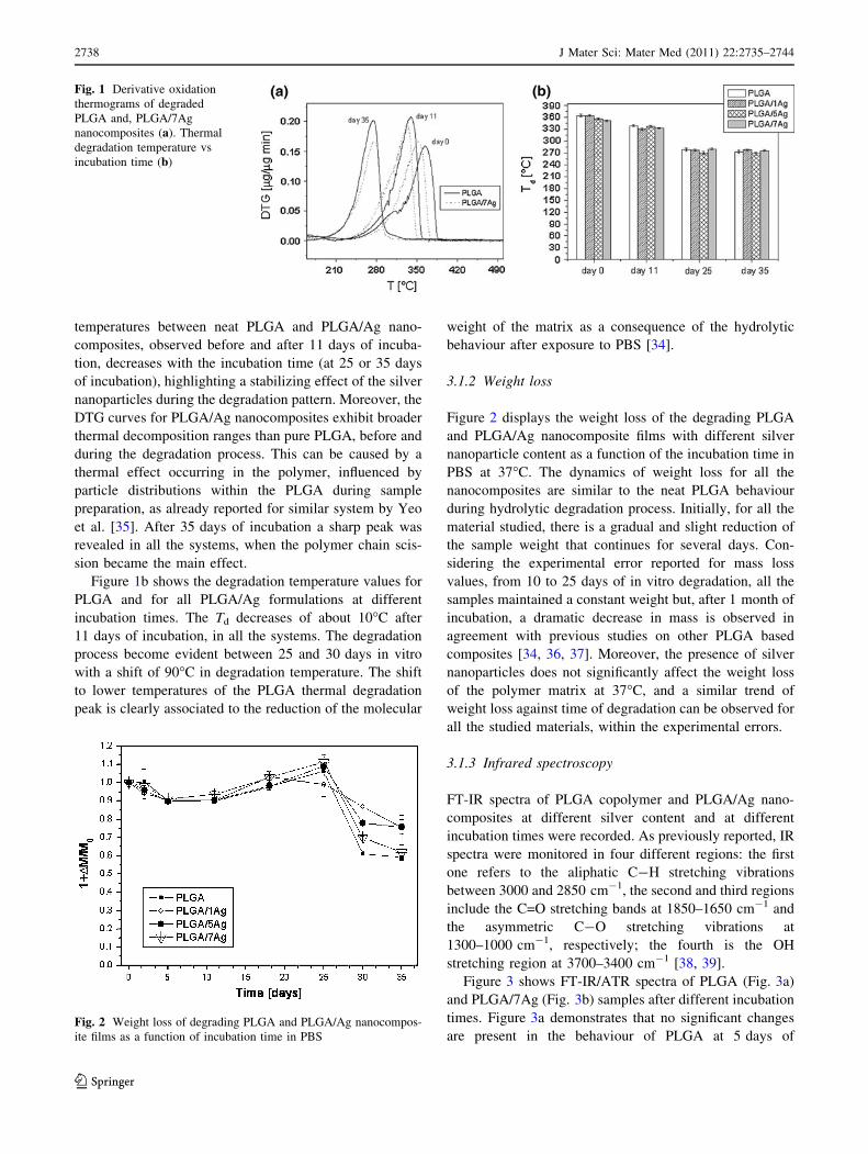
temperatures between neat PLGA and PLGA/Ag nano-
composites, observed before and after 11 days of incuba-
tion, decreases with the incubation time (at 25 or 35 days
of incubation), highlighting a stabilizing effect of the silver
nanoparticles during the degradation pattern. Moreover, the
DTG curves for PLGA/Ag nanocomposites exhibit broader
thermal decomposition ranges than pure PLGA, before and
during the degradation process. This can be caused by a
thermal effect occurring in the polymer, influenced by
particle distributions within the PLGA during sample
preparation, as already reported for similar system by Yeo
et al. [35]. After 35 days of incubation a sharp peak was
revealed in all the systems, when the polymer chain scis-
sion became the main effect.
Figure 1b shows the degradation temperature values for
PLGA and for all PLGA/Ag formulations at different
incubation times. The Td decreases of about 10�C after
11 days of incubation, in all the systems. The degradation
process become evident between 25 and 30 days in vitro
with a shift of 90�C in degradation temperature. The shift
to lower temperatures of the PLGA thermal degradation
peak is clearly associated to the reduction of the molecular
weight of the matrix as a consequence of the hydrolytic
behaviour after exposure to PBS [34].
3.1.2 Weight loss
Figure 2 displays the weight loss of the degrading PLGA
and PLGA/Ag nanocomposite films with different silver
nanoparticle content as a function of the incubation time in
PBS at 37�C. The dynamics of weight loss for all the
nanocomposites are similar to the neat PLGA behaviour
during hydrolytic degradation process. Initially, for all the
material studied, there is a gradual and slight reduction of
the sample weight that continues for several days. Con-
sidering the experimental error reported for mass loss
values, from 10 to 25 days of in vitro degradation, all the
samples maintained a constant weight but, after 1 month of
incubation, a dramatic decrease in mass is observed in
agreement with previous studies on other PLGA based
composites [34, 36, 37]. Moreover, the presence of silver
nanoparticles does not significantly affect the weight loss
of the polymer matrix at 37�C, and a similar trend of
weight loss against time of degradation can be observed for
all the studied materials, within the experimental errors.
3.1.3 Infrared spectroscopy
FT-IR spectra of PLGA copolymer and PLGA/Ag nano-
composites at different silver content and at different
incubation times were recorded. As previously reported, IR
spectra were monitored in four different regions: the first
one refers to the aliphatic C-H stretching vibrations
between 3000 and 2850 cm-1, the second and third regions
include the C=O stretching bands at 1850–1650 cm-1 and
the asymmetric C-O stretching vibrations at
1300–1000 cm-1, respectively; the fourth is the OH
stretching region at 3700–3400 cm-1 [38, 39].
Figure 3 shows FT-IR/ATR spectra of PLGA (Fig. 3a)
and PLGA/7Ag (Fig. 3b) samples after different incubation
times. Figure 3a demonstrates that no significant changes
are present in the behaviour of PLGA at 5 days of
Fig. 1 Derivative oxidation
thermograms of degraded
PLGA and, PLGA/7Ag
nanocomposites (a). Thermal
degradation temperature vs
incubation time (b)
Fig. 2 Weight loss of degrading PLGA and PLGA/Ag nanocompos-
ite films as a function of incubation time in PBS
2738 J Mater Sci: Mater Med (2011) 22:2735–2744
123

incubation respect to pristine PLGA and a similar trend
was detected for PLGA/7Ag (Fig. 3b) and the other
nanocomposites (data not shown) until 3 weeks of in vitro
degradation. Starting from 25 days of incubation, some
differences in the peak intensity appear in the region of the
characteristic carbonyl stretching vibrations between 1850
and 1650 cm-1. The spectra showed an absorption increase
in the frequency regions where the carbonyl (C=O) and the
OH stretching absorb, the latter appearing as broad band
centred at 3400 cm-1 [40, 41]. The signals are due to the
hydrolitic process leading to the formation of carboxylic
acid end chains which is occurring in the neat PLGA and in
the composites in a similar time-scale. To evaluate in
deeper details the degradation process the IR spectra were
monitored in the 1500–1300 cm-1 region for nanocom-
posite samples at different degradation times (Fig. 3c). In
this region, two bands are observed at 1452 cm-1 and
1422 cm-1 which correspond to the asymmetric bending of
CH3 from the lactic units and the bending of CH2 from the
glycolic units of the polymer, respectively. The relative
intensities of these two bands were used to estimate the
relative quantity of glycolic (CG) and lactic (CL) units
present in the sample. For this purpose the two bands were
de-convoluted and their intensity estimated. The relative
quantity of lactic, CL, and glycolic, CG, units present in our
samples was obtained through
CG ¼I1422
I1422 þ I1452
; CL ¼I1452
I1422 þ I1452
ð1Þ
where I1422 is the intensity of the band at 1422 cm-1 and
I1452 is the intensity of the band at 1452 cm-1. The vari-
ation of CG as a function of the degradation time for PLGA
and PLGA/7Ag nanocomposites is given in Fig. 3d. As can
be seen the relative quantity of glycolic units decrease in
the PLGA neat polymer already during the first week of
degradation, as expected confirming the preferential deg-
radation of these units. The remaining polymer, therefore,
becomes richer in lactic units [42]. In PLGA/7Ag nano-
composite a less evident decrease was measured, evi-
dencing the stability effect of the silver nanoparticles
during polymer degradation and confirming the TGA
results.
3.1.4 Physical and morphological alterations
The gross appearance of all PLGA systems changed during
degradation. The amorphous PLGA samples were initially
almost transparent. As degradation proceeded, they became
whitish due to water absorption. Then became brittle and
began to disintegrate in agreement with previous publica-
tions [43]. The visual aspects of PLGA and PLGA/Ag
nanocomposite films during degradation seem to be
Fig. 3 FT-IR/ATR spectra of
PLGA (a), and PLGA/7Ag (b),
samples for different incubation
times. Infrared spectra
(1500–1300 cm-1 region)
collected for PLGA
nanocomposite films degraded
in 20 ml PBS at different
degradation times (c). Relative
glycolic unit content, CG, of
remaining polymer versus
degradation time for PLGA
films degraded in 20 ml PBS at
378C (d)
J Mater Sci: Mater Med (2011) 22:2735–2744 2739
123

similar. The higher opacity for samples upon degradation
has been reported due to various phenomena, such as an
enhancement of the light diffusion through the material for
the presence of water and/or degradation products formed
[44–46], and to the formation of micro holes and cracks in
the bulk of the specimen during degradation of the polymer
matrix.
The changes in surface morphology caused by the
hydrolysis were observed using a Field Emission Scanning
Electron Microscopy (FESEM). Figure 4 shows the
FESEM images of PLGA neat film and PLGA/7Ag nano-
composite before and after different incubation times in
PBS at 37�C. FESEM observations were conducted until
11 days of incubation since after this period the samples
lost their stiffness and the FESEM observation became
difficult.
FESEM images show the formation of superficial
defects due to degradation just after 5 days of incubation in
vitro for PLGA neat films since the appearance of micro-
holes and cracks on the film surface were observed. At
longer incubation times the defects appeared much more
enlarged confirming the continuation of the degradation
process. The FESEM images recorded on the upper surface
of PLGA/7Ag system (Fig. 4) show the odd topography
previously described [20]. Upon exposure to the degrading
environment a round off of the pore edges was observed
and no other defects were detected, thus suggesting that the
degradation process is essentially localized in pore prox-
imity. Furthermore, particle-like structures are observable
around the pore edges as a consequence of the incubation
in PBS [47]. These micron-size particles are better
observed in PLGA/7Ag images recoded at higher
Fig. 4 FESEM images of pristine PLGA and PLGA/7Ag films (a, d), after 5 days (b, e), and after 11 days (c, f) of degradation
Fig. 5 FESEM images of PLGA/7Ag nanocomposite surface after 5 days (a), and after 11 days (b, c, d) of degradation
2740 J Mater Sci: Mater Med (2011) 22:2735–2744
123

magnification (Fig. 5a, b) after 5 and 11 days in vitro,
where the presence of these structures inside the superficial
pores is evident. Moreover, observing the lateral surface of
PLGA/Ag nanocomposite at 11 days in vitro, it is possible
to see the presence of silver nanoparticles on the pore wall
as a consequence of polymer chain degradation process
(Fig. 5b-d).
Figure 6 shows EDX spectra of PLGA/7Ag surface
pristine (a) and after 11 days of incubation (b). The anal-
ysis shows strong peaks due to calcium and phosphorus on
the surface of nanocomposite after 11 days of incubation,
with calcium-phosphorus atomic ratios of 1.36. The ratio
is close to that of octacalcium phosphate (OCP,
Ca8(HPO4)2(PO4)4_5H2O, Ca/P = 1.33). These findings
suggest that the specific surface chemistry and topography
with regular pore structure of PLGA/Ag composites
assisted the nucleation of mineral nanocrystals and the
mineralization process. This apatite-forming ability is
presumably due to interactions of the buffer ions (see
experimental section for composition) with the surface
pores of nanocomposites, which are favoured by the deg-
radation of the PLGA surface layer and are assisted by the
presence of Ag nanoparticles (Fig. 5c-d). In contrast,
apatite particles did not form on PLGA neat films [48].
3.1.5 Optical absorption
The absorption spectra of pristine PLGA and PLGA/Ag
nanocomposites before and after exposure to hydrolytic
medium are shown in Fig. 7. PLGA/Ag nanocomposites
before degradation (Fig. 7a) present absorption bands at
300 and 375 nm which are assigned to plasmon bands
(SPR) of the nanoparticles (Fig. 7a insert), since the
polymer contribution is at lower wavelengths [20]. It has
to be noted that the SPR absorption in the composites
appeared red shifted compared to the spectra recorded on
the neat Ag particles in suspensions; this behaviour can
be due to changes in the refractive index of the medium
around the metal particles, as reported in the literature
[49–51]. Once the nanocomposites were exposed to the
hydrolytic environment, a significant enhancement of the
SPR bands (Fig. 7b), respect to the intensity before the
exposure to the medium, was observed. This behaviour is
likely due to an alteration of the particle environments
induced by degradation of polymer matrix which resulted
in a reduction of the screening effects imposed by the
matrix, as already supported by FESEM images. Fur-
thermore, the SPR bands of the metal nanoparticles are
blue shifted (ca. 10 nm) in degraded nanocomposite
Fig. 6 EDX spectra of PLGA/
7Ag nanocomposite surface as
prepared (a), and after 11 days
(b) of degradation
Fig. 7 UV–vis absorption spectra of materials (a), before incubation
and (b), after 20 days of incubation. Panel a: PLGA (dotted line),
PLGA/1Ag (circles) and PLGA/7Ag (squares) recorded at the lower
(full symbols) and upper (empty symbols) surfaces; insert: absorption
spectra of silver nanoparticles. Panel b: PLGA/1Ag (circles) and
PLGA/7Ag (squares) recorded at the lower (full symbols) and upper
(empty symbols) surfaces after 20 days of exposure to the degradation
environment
J Mater Sci: Mater Med (2011) 22:2735–2744 2741
123
compared to the spectra of the original nanocomposites;
this observation together with the spectrum of the neat
nanoparticles in water, indicated a closer contact of the
metal with the aqueous medium. Optical absorption
results are in agreement with the morphological out-
comes, confirming the superficial presence of silver
nanoparticles as a consequence of polymer chain degra-
dation process.
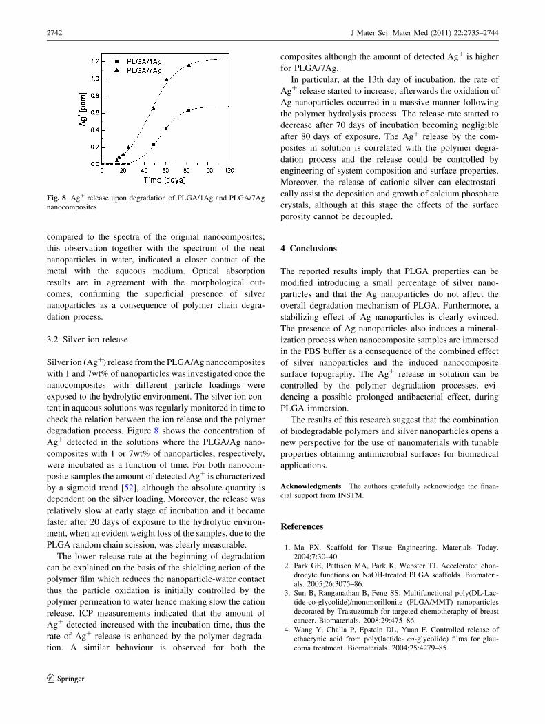
3.2 Silver ion release
Silver ion (Ag?) release from the PLGA/Ag nanocomposites
with 1 and 7wt% of nanoparticles was investigated once the
nanocomposites with different particle loadings were
exposed to the hydrolytic environment. The silver ion con-
tent in aqueous solutions was regularly monitored in time to
check the relation between the ion release and the polymer
degradation process. Figure 8 shows the concentration of
Ag? detected in the solutions where the PLGA/Ag nano-
composites with 1 or 7wt% of nanoparticles, respectively,
were incubated as a function of time. For both nanocom-
posite samples the amount of detected Ag? is characterized
by a sigmoid trend [52], although the absolute quantity is
dependent on the silver loading. Moreover, the release was
relatively slow at early stage of incubation and it became
faster after 20 days of exposure to the hydrolytic environ-
ment, when an evident weight loss of the samples, due to the
PLGA random chain scission, was clearly measurable.
The lower release rate at the beginning of degradation
can be explained on the basis of the shielding action of the
polymer film which reduces the nanoparticle-water contact
thus the particle oxidation is initially controlled by the
polymer permeation to water hence making slow the cation
release. ICP measurements indicated that the amount of
Ag? detected increased with the incubation time, thus the
rate of Ag? release is enhanced by the polymer degrada-
tion. A similar behaviour is observed for both the
composites although the amount of detected Ag? is higher
for PLGA/7Ag.
In particular, at the 13th day of incubation, the rate of
Ag? release started to increase; afterwards the oxidation of
Ag nanoparticles occurred in a massive manner following
the polymer hydrolysis process. The release rate started to
decrease after 70 days of incubation becoming negligible
after 80 days of exposure. The Ag? release by the com-
posites in solution is correlated with the polymer degra-
dation process and the release could be controlled by
engineering of system composition and surface properties.
Moreover, the release of cationic silver can electrostati-
cally assist the deposition and growth of calcium phosphate
crystals, although at this stage the effects of the surface
porosity cannot be decoupled.
4 Conclusions
The reported results imply that PLGA properties can be
modified introducing a small percentage of silver nano-
particles and that the Ag nanoparticles do not affect the
overall degradation mechanism of PLGA. Furthermore, a
stabilizing effect of Ag nanoparticles is clearly evinced.
The presence of Ag nanoparticles also induces a mineral-
ization process when nanocomposite samples are immersed
in the PBS buffer as a consequence of the combined effect
of silver nanoparticles and the induced nanocomposite
surface topography. The Ag? release in solution can be
controlled by the polymer degradation processes, evi-
dencing a possible prolonged antibacterial effect, during
PLGA immersion.
The results of this research suggest that the combination
of biodegradable polymers and silver nanoparticles opens a
new perspective for the use of nanomaterials with tunable
properties obtaining antimicrobial surfaces for biomedical
applications.
Acknowledgments The authors gratefully acknowledge the finan-
cial support from INSTM.
References
1. Ma PX. Scaffold for Tissue Engineering. Materials Today.
2004;7:30–40.
2. Park GE, Pattison MA, Park K, Webster TJ. Accelerated chon-
drocyte functions on NaOH-treated PLGA scaffolds. Biomateri-
als. 2005;26:3075–86.
3. Sun B, Ranganathan B, Feng SS. Multifunctional poly(DL-Lac-
tide-co-glycolide)/montmorillonite (PLGA/MMT) nanoparticles
decorated by Trastuzumab for targeted chemotheraphy of breast
cancer. Biomaterials. 2008;29:475–86.
4. Wang Y, Challa P, Epstein DL, Yuan F. Controlled release of
ethacrynic acid from poly(lactide- co-glycolide) films for glau-
coma treatment. Biomaterials. 2004;25:4279–85.
Fig. 8 Ag? release upon degradation of PLGA/1Ag and PLGA/7Ag
nanocomposites
2742 J Mater Sci: Mater Med (2011) 22:2735–2744
123

5. Darouiche RO. Anti-Infective Efficacy of Silver-Coated Medical
Prostheses. Clin Infect Dis. 1999;29:1371–7.
6. Ohura K, Nakamura T, Yamamuro T. Bone-bonding ability of
P2O5-free CaO.SiO2 glasses. J Biomed Mater Res.
1991;25:357–65.
7. Neo M, Kotani S, Nakamura T. A comparative study of ultra-
structure of the interfaces between four kinds of surface-active
ceramic and bone. J Biomed Mater Res. 1992;26:1419–32.
8. Bockstaller MR, Mickiewicz RA, Thomas EL. Block copolymer
nanocomposites: perspectives for tailored functional materials.
Adv. Mater. 2005;17:1331–49.
9. Tjong SC. Structural and mechanical properties of polymer
nanocomposites. Mater. Sci. Eng. R. 2006;53:73–197.
10. Mai YW, Yu ZZ, editors. Polymer nanocomposites. Chambridge:
RCR Press; 2006.
11. Aloisi GG, Costantino U, Latterini L, Nocchetti M, Camino G,
Frache A. Preparation and spectroscopic characterisation of
intercalation products of clay and of clay-polypropylene com-
posites with rhodamine B. J. Phys. Chem. Solids. 2006;67:
909–14.
12. Aloisi GG, Elisei F, Nocchetti M, Camino G, Frache A, Co-
stantino U, Latterini L. Mater. Chem. Phys. 2010;123:3777–9.
13. Latterini L, Nocchetti M, Costantino U, Aloisi GG, Elisei F.
Organized chromophores in layered inorganic matrices. Inorg
Chim Acta. 2007;360:728–40.
14. Lee JY, Nagahata JLR, Horiuchi S. Effect of metal nanoparticles
on thermal stabilization of polymer/metal nanocomposites pre-
pared by a one-step dry process. Polymer. 2006;47:7970–9.
15. Gautam A, Ram S. Preparation and thermomechanical properties
of Ag-PVA nanocomposite films. Mater. Chem. Phys. 2010;119:
266–71.
16. Wang JF, Liu XY, Loan B. Fabrication of Ti/polymer biocom-
posites for load-bearing implant applications. J. Mater. Process.
Tech. 2008;1–3:428–33.
17. Ren J, Hong H, Ren T, Teng T. Preparation and characterization
of magnetic PLA-PEG composite nanoparticles for drug target-
ing. React Funct Polym. 2006;66:944–51.
18. Palza H, Vergara R, Zapata P. Macromol Mater Eng.
2010;295:899–905.
19. Yang Z, Best SM, Cameron RE. Adv. Mater. 2009;21:1900–4.
20. Armentano I, Fortunati E, Latterini L, Rinaldi S, Saino E, Visai
L, Elisei F, Kenny JM. J. Nanostruc. Polym. Nanocomp. 2010;
6(4):110–8.
21. An YH, Friedman RJ. Concise review of mechanism of bacterial
adhesion of biomaterials surface. J Biomed Mater Res.
1998;43:338–48.
22. Kulkarni A, Reiche J, Lendlein A. Hydrolytic degradation of
poly(rac-lactide) and poly[(rac-lactide)-co-glycolide] at the air-
water interface. InterfaceAnal. 2007;39:740–6.
23. Cam D, Hyon SH, Ikada Y. Degradation of high-molecular-
weight poly(L-Lac-tide) in alkaline medium. Biomaterials.
1995;16(11):833–43.
24. Heya T, Okada H, Ogawa Y, Toguchi H. Factors influencing the
profiles of TRH release from co-poly(DL-lactic-glycolic acid)
microspheres. Int J Pharm. 1991;72(3):199–205.
25. Omelczuk MO, McGinity JW. The influence of polymer glass-
transition temperature and molecular-weight on drug release from
tablets containing poly(DL-lactic acid). Pharm Res.
1992;9(1):26–32.
26. Fu BX, Hsiao BS, Chen G, Zhou J, Koyfman I, Jamiolkowski
DD, et al. Structure and property studies of bioabsorbable
poly(glycolide-co-lactide) fiber during processing and in vitro
degradation. Polymer. 2002;43(20):5527–34.
27. Hurrell S, Cameron RE. The effect of initial polymer morphology
on the degradation and drug release from polyglycolide. Bio-
materials. 2002;23(11):2401–9.
28. Lu L, Garcia CA, Mikos AG. In vitro degradation of thin
poly(DL-lactic-co-glycolic acid) films. J Biomed Mater Res.
1999;46(2):236–44.
29. Panyam J, Dali MA, Sahoo SK, Ma WX, Chakravarthi SS,
Amidon GL, et al. Polymer degradation and in vitro release of a
model protein from poly(DL-Lactide-co- glycolide) nano- and
microparticles. J Control Release. 2003;92(1–2):173–87.
30. Williams HE, Huxley J, Claybourn M, Booth J, Hobbs M,
Meehan E, et al. The effect of gamma-irradiation and polymer
composition on the stability of PLGA polymer and microspheres.
Polym Degrad Stab. 2006;91(9):2171–81.
31. Chu CC. An in vitro study of the effect of buffer on the degra-
dation of poly(-glycolic acid) sutures. J Biomed Mater Res.
1981;15(1):19–27.
32. Chu CC. The in vitro degradation of poly(glycolic acid) sutures-
effect of pH. J Biomed Mater Res. 1981;15(6):795–804.
33. Bikiaris DN, Chrissafis K, Paraskevopoulos KM, Triantafyllidis
KS, Antonakou EV. Investigation of thermal degradation mech-
anism of an aliphatic polyester using pyrolysis–gas chromatog-
raphy–mass spectrometry and a kinetic study of the effect of the
amount of polymerisation catalyst. Polym Degrad Stab.
2007;92:525–36.
34. Armentano I, Dottori M, Puglia D, Kenny JM. Effects of carbon
nanotubes (CNTs) on the processing and in vitro degradation of
poly(DL-lactide-co-glycolide)/CNT films. J Mater Sci Mater Med.
2008;19:2377–87.
35. Yeo SY, Tan WL, Bakar M Abu, Ismail J. Silver sulfide/poly(3-
hydroxybutyrate) nanocomposites: Thermal stability and kinetic
analysis of thermal degradation. Polym Degrad Stab. 2010;95:
1299–304.
36. Lusa LU, Garcia CA, Mikos AG. In vitro degradation of thin
poly(DL-lactic-co-glycolic acid) films. J Biomed Mater Res.
1999;46:236–44.
37. Siepmann J, Elkharraz K, Siepmann F, Klose D. How autoca-
talysis accelerates drug release from PLGA-based microparticles:
a quantitative treatment. Biomacromolecules. 2005;6:2312–9.
38. Aguilar CA, Lu Y, Mao S, Chen S. Direct micro-patterning of
biodegradable polymers using ultraviolet and femtosecond lasers.
Biomaterials. 2005;26:7642–9.
39. Kister G, Cassanas G, Vert M. Morphology of poly(glycolic acid)
by IR and Raman spectroscopies. Spectrochim. Acta Part. A.
1997;53:1399–403.
40. Mercado AL, Allmond CE, Hoekstra JG, Fitz-Gerald JM. Pulsed
laser deposition vs. matrix assisted pulsed laser evaporation for
growth of biodegradable polymer thin films. Appl. Phys.
A Mater. Sci. Process. 2005;81:591–9.
41. Huang MH, Li S, Vert M. Synthesis and degradation of PLA-PCL-
PLA triblock copolymer prepared by successive polymerization of
caprolactone and DL-Lactide. Polymer. 2004;45:8675–81.
42. Vey E, Roger C, Meehan L, Booth J, Claybourn M, Miller AF,
Spiani A. Degradation mechanism of poly(lactic-co-glycolic)
acid block copolymer cast films in phosphate buffer solution.
Polym Degrad Stab. 2008;93:1869–76.
43. Loo SCJ, Ooi CP, Boey YCF. Radiation effects on poly(lactide-
co-glycolide) (PLGA) and poly(L-lactide) (PLLA). Polym Degrad
Stab. 2004;83:259–65.
44. Jarerat A, Tokiwa Y. Degradation of poly (L-lactide) by a fungus.
Macromol Biosci. 2001;1:136–40.
45. Jarerat A, Pranamuda H, Tokiwa Y. Poly(L-lactide)-degrading
activity in various actinomycetes. Macromol Biosci. 2002;2:
420–8.
46. Torres A, Li SM, Roussos S, Vert M. Poly (lactic acid) degra-
dation in soil or under controlled conditions. J. of Applied
Polymer Science. 1996;62:2295–302.
47. Qu X, Cui W, Yang F, Min C, Shen H, Bei J, Wang S. The effect
of oxygen plasma pretreatment and incubation in modified
J Mater Sci: Mater Med (2011) 22:2735–2744 2743
123

simulated body fluids on the formation of bone-like apatite on
poly(lactide-co-glycolide) (70/30). Biomaterials. 2007;28:9–18.
48. Lee JC, Cho SB, Lee SJ, Rhee SH. Nucleation and growth
mechanism of apatite on a bioactive and degradable ceramic/
polymer composite with a thick polymer layer. J Mater Sci.
2009;44:4531–8.
49. Kulkarni AP, Munechika K, Noone KM, Smith JM, Ginger DS.
Phase Transfer of Large Anisotropic Plasmon Resonant Silver
Nanoparticles from Aqueous to Organic Solution. Langmuir.
2009;25:7932–9.
50. Kazim S, Pfleger J, Prochazka M, Bondarev D, Vohlidal J. New
Phosphorus-Containing Spherical Carbon Adsorbents as Prom-
ising Materials in Drug Adsorption and Release. Coll. Interf. Sci.
2011;354:611–9.
51. Kubo S, Diaz A, Tang Y, Mayer TS, Khoo IC, Mallouk TE.
Tunability of the Refractive Index of Gold Nanoparticle Dis-
persions. Nano Lett. 2007;7:3418–23.
52. Benn TM, Westerhoff P. Nanoparticle silver released into water
from commercially available sock fabrics. Environ Sci Technol.
2008;42:7025–6.
2744 J Mater Sci: Mater Med (2011) 22:2735–2744
123
Related Documents