Degradation, Bioactivity, and Osteogenic Potential of Composites Made of PLGA and Two Different Sol–Gel Bioactive Glasses ELZBIETA PAMULA, 1 JUSTYNA KOKOSZKA, 2 KATARZYNA CHOLEWA-KOWALSKA, 2 MARIA LACZKA, 2 LUKASZ KANTOR, 3 LUKASZ NIEDZWIEDZKI, 4 GWENDOLEN C. REILLY, 5 JOANNA FILIPOWSKA, 6 WOJCIECH MADEJ, 6 MALGORZATA KOLODZIEJCZYK, 6 GRZEGORZ TYLKO, 6 and ANNA M. OSYCZKA 6 1 Department of Biomaterials, Faculty of Materials Science and Ceramics, AGH-University of Science and Technology, Mickiewicza Ave. 30, 30-059 Krakow, Poland; 2 Department of Glass Technology and Amorphous Coatings, Faculty of Materials Science and Ceramics, AGH-University of Science and Technology, Mickiewicza Ave. 30, 30-059 Krakow, Poland; 3 Multidisciplinary School of Engineering in Biomedicine, AGH-University of Science and Technology, Mickiewicza Ave. 30, 30-059 Krakow, Poland; 4 Department of Orthopedics and Musculoskeletal Traumatology, Institute of Physiotherapy, Faculty of Health Care, School of Medicine, Jagiellonian University, Michalowskiego 12, 31-126 Krakow, Poland; 5 Department of Materials Science and Engineering, University of Sheffield, The Kroto Research Institute, Broad Lane, Sheffield S3 7HQ, UK; and 6 Department of Cell Biology and Imaging, Faculty of Biology and Earth Sciences, Jagiellonian University, Ingardena 6, 30-060 Krakow, Poland (Received 27 October 2010; accepted 28 March 2011; published online 13 April 2011) Associate Editor Kent Leach oversaw the review of this article. Abstract—We have developed poly(L-lactide-co-glycolide) (PLGA) based composites using sol–gel derived bioactive glasses (S-BG), previously described by our group, as composite components. Two different composite types were manufactured that contained either S2—high content silica S-BG, or A2—high content lime S-BG. The composites were evaluated in the form of sheets and 3D scaffolds. Sheets containing 12, 21, and 33 vol.% of each bioactive glass were characterized for mechanical properties, wettability, hydro- lytic degradation, and surface bioactivity. Sheets containing A2 S-BG rapidly formed a hydroxyapatite surface layer after incubation in simulated body fluid. The incorporation of either S-BG increased the tensile strength and Young’s modulus of the composites and tailored their degradation rates compared to starting compounds. Sheets and 3D scaffolds were evaluated for their ability to support growth of human bone marrow cells (BMC) and MG-63 cells, respectively. Cells were grown in non-differentiating, osteo- genic or osteoclast-inducing conditions. Osteogenesis was induced with either recombinant human BMP-2 or dexa- methasone, and osteoclast formation with M-CSF. BMC viability was lower at higher S-BG content, though specific ALP/cell was significantly higher on PLGA/A2-33 compos- ites. Composites containing S2 S-BG enhanced calcification of extracellular matrix by BMC, whereas incorporation of A2 S-BG in the composites promoted osteoclast formation from BMC. MG-63 osteoblast-like cells seeded in porous scaffolds containing S2 maintained viability and secreted collagen and calcium throughout the scaffolds. Overall, the presented data show functional versatility of the composites studied and indicate their potential to design a wide variety of implant materials differing in physico-chemical properties and biological applications. We propose these sol–gel derived bioactive glass–PLGA composites may prove excellent potential orthopedic and dental biomaterials supporting bone formation and remodeling. Keywords—Bioactive glass, Sol–gel method, Poly(L-lactide- co-glycolide), Bioactive composites, Bone morphogenetic proteins, Human bone marrow cells, MG-63 cells, Alkaline phosphatase, Mineralization, Osteoclasts. INTRODUCTION Current treatments of bone defects include the use of autografts, allografts, and artificial biomaterials. 2 The availability of auto- and allografts continues to be limited by supply, donor-site morbidity, the risk of immune rejection and infections and, often, high costs. 15 Orthopedic implants made of artificial mate- rials offer an alternative to supply the growing needs for bone replacement therapies. Beyond the widely recognized titanium implants, the latter can be made of bioresorbable polymers (e.g., polylactides, poly- glycolide, and their copolymers) and/or bioactive ceramics (e.g., hydroxyapatite, tricalcium phosphate, bioactive glasses, glass ceramics). Poly(L-lactide- co-glycolide) (PLGA) is among the few synthetic polymers approved for human clinical use because of Address correspondence to Anna M. Osyczka, Department of Cell Biology and Imaging, Faculty of Biology and Earth Sciences, Jagiellonian University, Ingardena 6, 30-060 Krakow, Poland. Electronic mail: [email protected] Annals of Biomedical Engineering, Vol. 39, No. 8, August 2011 (ȑ 2011) pp. 2114–2129 DOI: 10.1007/s10439-011-0307-4 0090-6964/11/0800-2114/0 ȑ 2011 The Author(s). This article is published with open access at Springerlink.com 2114

Welcome message from author
This document is posted to help you gain knowledge. Please leave a comment to let me know what you think about it! Share it to your friends and learn new things together.
Transcript
Degradation, Bioactivity, and Osteogenic Potential of Composites
Made of PLGA and Two Different Sol–Gel Bioactive Glasses
ELZBIETA PAMULA,1 JUSTYNA KOKOSZKA,2 KATARZYNA CHOLEWA-KOWALSKA,2 MARIA LACZKA,2
LUKASZ KANTOR,3 LUKASZ NIEDZWIEDZKI,4 GWENDOLEN C. REILLY,5 JOANNA FILIPOWSKA,6
WOJCIECH MADEJ,6 MALGORZATA KOLODZIEJCZYK,6 GRZEGORZ TYLKO,6 and ANNA M. OSYCZKA6
1Department of Biomaterials, Faculty of Materials Science and Ceramics, AGH-University of Science and Technology,Mickiewicza Ave. 30, 30-059 Krakow, Poland; 2Department of Glass Technology and Amorphous Coatings, Faculty of
Materials Science and Ceramics, AGH-University of Science and Technology, Mickiewicza Ave. 30, 30-059 Krakow, Poland;3Multidisciplinary School of Engineering in Biomedicine, AGH-University of Science and Technology, Mickiewicza Ave. 30,
30-059 Krakow, Poland; 4Department of Orthopedics and Musculoskeletal Traumatology, Institute of Physiotherapy,Faculty of Health Care, School of Medicine, Jagiellonian University, Michalowskiego 12, 31-126 Krakow, Poland;
5Department of Materials Science and Engineering, University of Sheffield, The Kroto Research Institute, Broad Lane,Sheffield S3 7HQ, UK; and 6Department of Cell Biology and Imaging, Faculty of Biology and Earth Sciences, Jagiellonian
University, Ingardena 6, 30-060 Krakow, Poland
(Received 27 October 2010; accepted 28 March 2011; published online 13 April 2011)
Associate Editor Kent Leach oversaw the review of this article.
Abstract—We have developed poly(L-lactide-co-glycolide)(PLGA) based composites using sol–gel derived bioactiveglasses (S-BG), previously described by our group, ascomposite components. Two different composite types weremanufactured that contained either S2—high content silicaS-BG, or A2—high content lime S-BG. The composites wereevaluated in the form of sheets and 3D scaffolds. Sheetscontaining 12, 21, and 33 vol.% of each bioactive glass werecharacterized for mechanical properties, wettability, hydro-lytic degradation, and surface bioactivity. Sheets containingA2 S-BG rapidly formed a hydroxyapatite surface layer afterincubation in simulated body fluid. The incorporation ofeither S-BG increased the tensile strength and Young’smodulus of the composites and tailored their degradationrates compared to starting compounds. Sheets and 3Dscaffolds were evaluated for their ability to support growthof human bone marrow cells (BMC) and MG-63 cells,respectively. Cells were grown in non-differentiating, osteo-genic or osteoclast-inducing conditions. Osteogenesis wasinduced with either recombinant human BMP-2 or dexa-methasone, and osteoclast formation with M-CSF. BMCviability was lower at higher S-BG content, though specificALP/cell was significantly higher on PLGA/A2-33 compos-ites. Composites containing S2 S-BG enhanced calcificationof extracellular matrix by BMC, whereas incorporation ofA2 S-BG in the composites promoted osteoclast formationfrom BMC. MG-63 osteoblast-like cells seeded in porousscaffolds containing S2 maintained viability and secretedcollagen and calcium throughout the scaffolds. Overall, the
presented data show functional versatility of the compositesstudied and indicate their potential to design a wide varietyof implant materials differing in physico-chemical propertiesand biological applications. We propose these sol–gel derivedbioactive glass–PLGA composites may prove excellentpotential orthopedic and dental biomaterials supportingbone formation and remodeling.
Keywords—Bioactive glass, Sol–gel method, Poly(L-lactide-
co-glycolide), Bioactive composites, Bone morphogenetic
proteins, Human bone marrow cells, MG-63 cells, Alkaline
phosphatase, Mineralization, Osteoclasts.
INTRODUCTION
Current treatments of bone defects include the useof autografts, allografts, and artificial biomaterials.2
The availability of auto- and allografts continues tobe limited by supply, donor-site morbidity, the risk ofimmune rejection and infections and, often, highcosts.15 Orthopedic implants made of artificial mate-rials offer an alternative to supply the growing needsfor bone replacement therapies. Beyond the widelyrecognized titanium implants, the latter can be madeof bioresorbable polymers (e.g., polylactides, poly-glycolide, and their copolymers) and/or bioactiveceramics (e.g., hydroxyapatite, tricalcium phosphate,bioactive glasses, glass ceramics). Poly(L-lactide-co-glycolide) (PLGA) is among the few syntheticpolymers approved for human clinical use because of
Address correspondence to Anna M. Osyczka, Department of
Cell Biology and Imaging, Faculty of Biology and Earth Sciences,
Jagiellonian University, Ingardena 6, 30-060 Krakow, Poland.
Electronic mail: [email protected]
Annals of Biomedical Engineering, Vol. 39, No. 8, August 2011 (� 2011) pp. 2114–2129
DOI: 10.1007/s10439-011-0307-4
0090-6964/11/0800-2114/0 � 2011 The Author(s). This article is published with open access at Springerlink.com
2114

good biocompatibility, controllable degradability,and relatively good processability. PLGA has beenused as scaffolds for bone tissue engineering32 andfor drug delivery purposes, including delivery ofgrowth factors and hormones involved in boneregeneration.34 However, the polymer itself is bioin-ert and does not elicit a biological response in bonecells.23 Bioactive materials such as bioactive glass, onthe other hand, promote attachment, growth, andosteogenic differentiation of progenitor cells in vitroand are capable of forming interfacial bonds withbone tissue in vivo.13
Combining polymers and bioactive glass into com-posites is a relatively new concept in bone tissueengineering and the benefits of such compositesinclude: (1) enhanced mechanical properties enablingbetter integration with bone tissue,26 (2) enhancedbioactivity promoting new bone formation,4 and (3)tailored degradation rates allowing the body’s ownbone to heal, grow, remodel, and eventually replace thegraft materials.25 Specifically, incorporation of bioac-tive glass into a polymer base has been shown to delaythe degradation rate of the polymer compared to theneat polymer foam,25 enhance the composite’s bioac-tivity,4 improve the processing of porous poly(L-lac-tide)/bioactive glass composites44 and improve themechanical properties of these composites.26,28 Severalapproaches employed biodegradable polymers andthe melt-derived 45S5 Bioglass� developed byHench8,22,39,42,43 but very few reports applied sol–gelderived bioactive glass instead.24 In general, gel-der-ived bioactive glasses have a larger surface area andOH groups are present in their structure. Thus, theirbioactivity is usually higher than bioactive glassesprepared by melting. Consequently, they may stimu-late faster regeneration in living bone. Our earlierstudies reported the production, physico-chemical, andbiological properties of gel-derived bioactive glasses ofthe CaO–P2O5–SiO2 system.19,27 We showed thatCaO:SiO2 ratio in these glasses influenced their physi-cal and chemical properties and affected the growthand differentiation of bone marrow-derived cells.6,17,20
Our data further indicate that incorporation of thesegel-derived glasses into a HA matrix enhanced growthand osteogenic differentiation of human mesenchymalstem cells (MSC).5 Recently, Wu et al.41 suggested thatPLGA-based composites prepared with a sol–gelderived mesoporous bioactive glass possess advanta-geous mechanical and biological properties comparedto those containing non-mesoporous bioactive glass.Mesoporous, gel-derived bioactive glasses (MBG) havea highly ordered structure and generally higher bio-activity than non-mesoporous bioactive glass obtainedby melting. MBG/PLGA films have been characterizedby excellent physicochemical, biological, and drug
release properties and have been processed into 3Dscaffolds suitable for bone tissue engineering.41
In this work we have employed either silica-rich (S2)or calcium-rich (A2) bioactive glasses derived froma basic CaO–P2O5–SiO2 system and obtained by asol–gel method, to prepare PLGA-based composites.These composites were prepared in the form of sheetsand 3D scaffolds and evaluated for their mechanicalproperties, bioactivity and the ability to enhance BMP-2 mediated human bone marrow cell osteogenesis,support osteoclast formation from human bone mar-row and osteogenic gene expression of MG-63 osteo-blastic cells.
MATERIALS AND METHODS
Materials
PLGA was synthesized via a ring opening process inthe presence of low toxicity zirconium acetylacetonateas a copolymerization initiator.11 The molar ratio ofL-lactide to glycolide in the copolymer was 85:15(as studied by 1H NMR), and molecular masses ofPLGA were: Mn = 80 kDa and Mw = 152 kDa. Bio-active glasses (S2 and A2) of the composition of: S2:80 mol% SiO2–16 mol% CaO–4 mol% P2O5, and A2:40 mol% SiO2–54 mol% CaO–6 mol% P2O5, wereproduced using the sol–gel method.19 Tetraeth-oxysilane (TEOS; Si(OC2H5)4), triethylphosphate(TEP; OP(OC2H5)3), and calcium nitrate tetra-hydrate(Ca(NO3)2Æ4H2O) were used as base components tostart the sol–gel process. HCl solution was used as acatalyst for the hydrolysis and condensation reactions.The formed gel was dried at 40–120 �C for 7 days andthen subjected to thermal treatment at 700 �C for 20 h.Afterwards it was milled to obtain a bioactive glasspowder with particle sizes <50 lm.
Composite Sheets Fabrication
The PLGA–bioactive glass composites were fabri-cated by mixing glass particles with 10% w/v PLGAsolution in methylene chloride on a magnetic stirrer for3 h, followed by partial evaporation of the solvent inair, slip casting of the viscous mixture on glass Petridishes, then drying in air and vacuum to a constantweight. The volume fraction of bioactive glass in thecomposites was 12, 21, and 33%. The composite sheetsobtained were 0.11 mm thick.
Composite Scaffolds Fabrication
3D scaffolds were obtained by addition ofsodium chloride (grain fraction 320–400 lm) into the
Degradation, Bioactivity, and Osteogenic Potential of PLGA-S-BG Composites 2115
copolymer–bioactive glass solution in methylene chlo-ride. The amount of sodium chloride provided 85%porosity. NaCl particles were mixed with the PLGA–S-BG suspension with the use of spatula until most of themethylene chloride had evaporated. The dense sus-pension was tightly packed into polypropylene cylin-drical vials (12 mm in diameter, volume 5 mL), driedin air for 24 h and then in vacuum conditions for 48 h.Then the vials with the rigid salt/PLGA–S-BG mixturewere cut into slices of the thickness of 3 mm andextensively washed in ultra-high quality water (UHQ-water, produced by UHQ-PS purification system, Elga,UK). The water was exchanged several times until theconductivity of the water was about 2 lS/cm. Subse-quently the samples were air-dried and then vacuum-dried for at least 24 h.
Material Evaluation
The mechanical properties of the sheets, i.e., tensilestrength, Young’s modulus, and elongation at break ofthe PLGA–bioactive glass composites were determinedwith the use of an universal testing machine (Zwick1435, Germany) and compared to pure PLGA sheets.The parallel specimen length was set to 40 mm, thespecimen width was 5 mm, the pre-load force was0.1 N, the test speed was 100 mm/min and six sampleswere evaluated from each experimental group.
Wettability was evaluated by water contact anglemeasurements. The contact angle was determined bysessile drop method by an automatic drop shapeanalysis system DSA 10 Mk2 (Kruss, Germany).UHQ-water droplets of 0.2 lL were applied on eachsample and the contact angle was calculated by aver-aging 10 measurements.
To study hydrolytic degradation, the composites,and reference samples (PLGA, S2, and A2) weighing0.15 g were placed in separate plastic vials and incu-bated in 30 mL UHQ-water at 37 �C for 3 months.The evaluation was performed according to interna-tional standard (ISO 10993-13).16 Hydrolytic degra-dation process was monitored by pH and conductivitymeasurements of incubation medium with the use ofpH meter (CP-315 Elmetron, Poland) and conduc-tometer (CC-315, Elmetron, Poland), respectively.
The arithmetical mean roughness (Ra) and ten-point mean roughness depth (Rz) of a surface weredetermined. These parameters were quantified using aprofilometer (TR-100 Surface Roughness Tester).
Bioactivity of the composites was assessed by anin vitro simulated body fluid (SBF) test according tothe method described in the work of Niemiel et al.28 Inbrief, SBF was prepared by dissolving the followingchemicals (POCh, Gliwice, Poland) in UHQ-water:141 mM NaCl, 4 mM KCl, 0.5 mM MgSO4, 1 mM
MgCl2, 4.2 mM NaHCO3, 2.5 mM CaCl2, and1.0 mM KH2PO4. The resulting SBF was buffered topH 7.28 with Tris(hydroxymethyl aminomethane)/HCl. The sheets weighing 1 g were immersed in100 mL of SBF solution and incubated at 37 �C for10 days. The SBF solution was exchanged every 3 daysto ensure sufficient ion concentrations for mineralgrowth. Afterwards the samples were washed in UHQ-water, air- and vacuum-dried to a constant weight.
Microstructure and chemical composition of PLGAand composite sheets before and after soaking in SBFwere examined by SEM/EDAX analysis (NanoSEM,FEI, USA, accelerating voltage 10 and 18 kV, magni-fications 5009 and 5,0009). The EDAX spectra wereaveraged for the whole analyzed surface at the mag-nification of 5,0009.
Material Sterilization
For cell culture, composite sheets (round samplesmatching the size of culture vessels) were sterilized bysoaking in 70% ethanol for 30 min, washed withphosphate buffered saline (PBS) and further sterilizedby UV light for 15 min. 3D scaffolds were sterilized in70% ethanol for 72 h, washed in PBS and air-driedbefore cell seeding.
Cell Cultures
Evaluation of 2D Sheets
Adult human bone marrow cells (BMC) were har-vested from iliac crest of 10 adult patients (age 19–71,both genders) under Institutional Review Board-approved protocol (KBET/17/L/2007). The mononu-clear fraction, which contains both mesenchymal andhematopoetic cells, was isolated as described previ-ously.30 Cells were either expanded in primary culture toselect forMSC or they were directly seeded on the sheets(BMC mononuclear fraction). For selection of MSC,primaryBMCwere cultured in tissue culture flasks usingalpha-MEM supplemented with 10% pre-selectedMSC-qualified fetal bovine serum (MSC-FBS) and 1%antibiotics (Penicillin/Streptomycin; Invitrogen). Med-ium was changed twice weekly until a confluent cellmonolayer was developed. Cells were then detachedfrom culture flasks using 0.25% Trypsin–EDTA(Invitrogen) and seeded onto the composite sheets orfurther expanded. To induce alkaline phosphatase(ALP) activity, the early marker of osteogenesis, MSCwere seeded on composite sheets (round samples of15 mm in diameter) at a density of 104 cells/cm2 andcultured in either alpha-MEM/MSC-FBS/antibiotics orsupplemented with 100 lg/mL ascorbate-2-phosphate(ascorbate; Sigma) and 100 ng/mL recombinant humanBMP-2 (BMP-2; R&D Systems). Gene expression of
PAMULA et al.2116
osteogenic markers was analyzed in parallel culturesestablished on composite sheets of higher surface area(round samples of 28 mm in diameter) and treated withascorbate and BMP-2. For calcium deposition, cultureswere supplemented with 100 lg/mL ascorbate, 10 mMb-glycerophosphate (Sigma), 100 ng/mL BMP-2 or1027 M dexamethasone (Sigma). Analyzes were per-formed after 1 week (MTS, ALP, gene expressionstudies) or 3 weeks (calcification of extracellularmatrix)of culture. Osteoclast formation was induced usingBMC mononuclear fraction. After fractionation ofwhole bone marrow sample over Ficol gradient amononuclear fraction of BMCwas isolated and washedin sterile PBS to get rid of Ficol residues.30Mononuclearcells were seeded on PLGAand composite sheets (roundsamples of 28 mm in diameter) at a density of4.7 9 106 cell/well/6-well plates in alpha-MEM/MSC-FBS/antibiotics and supplemented with 100 ng/mLhuman recombinant macrophage colony stimulatingfactor (M-CSF, Peprotech). After 3 days of culture,medium, and floating cells were collected, transferred tocorresponding standard tissue culture 6-well plates andfurther cultured in standard alpha-MEM/MSC-FBS/antibiotics for a total of 23 culture days. Medium waschanged every 3 days starting from culture day 8.
Evaluation of 3D Scaffolds
MG-63 cells (human bone osteosarcoma-derivedcells; Lonza USA) were expanded using DMEM HighGlucose (Biosera, UK) supplemented with 10% FBS(PAA), 1% antibiotics, and 0.25% fungizone (Sigma).For experiments with 3D composites and controlPLGA 3D scaffolds MG-63 cells were suspended at adensity of 2 9 105 cells/150 lL of PBS containing1.75 mg/mL of fibrinogen (Sigma). To induce clotformation, cell suspension was supplemented withthrombin (1 unit/mL). Each 3D scaffold (12 mm indiameter, 2 mm thickness) was placed in a separatewell of 6-well plate and held by stainless steel ring tofacilitate cell loading. Aliquots of 150 lL cell suspen-sions were loaded into the scaffolds and incubated for40 min at 37 �C in a humidified, 5% CO2 atmosphere,followed by addition of standard DMEM/FBS/anti-biotics. After 24 h of initial culture, the steel ring wasremoved and medium was supplemented with 50 lg/mL ascorbate-2-phosphate, 1027 M dexamethasone,and 5 mM b-glycerophosphate (Sigma). Media andsupplements were changed every 2–3 days and ana-lyzes done after 2 weeks of culture.
2D Culture Analyzes
Seven-day osteogenic cultures were analyzed forcell metabolic activity using CellTiter96AQueous One
Solution Cell Proliferation Assay (MTS, Promega), atetrazolium salt assay. Cell numbers of human MSC orMG-63 cells were estimated based on standard curvesestablished with cells cultured at different densities andA490nm MTS values that were measured at appropriatetime intervals. The linear regressions between cellnumbers and A490nm MTS values were evaluated andthe linear regression equations from the data fittingwere used to determine the experimental cell numbers.ALP activity was assayed as described previously,37
and the results were normalized to live cell number toobtain specific ALP activity/cell values. We havepreviously determined that, using our hMSC cultureregimens, ALP peaks at day 7 and reaches plateau byday 10,9,10,29,30 and day 7 culture time point was pre-viously used to analyze cellular ALP activity on dif-ferent bioglass surfaces.17 Analyzes of gene expressionlevels in osteogenic cultures were performed after 7-day cultures on selected composite sheets and PLGA,and compared to gene expression levels in cells cul-tured on standard tissue culture plastic (TCP). Briefly:2 lg of total RNA from each culture (NucleoSpin�
RNA II, MACHEREY–NAGEL) were reverse-tran-scribed to cDNA (SuperScript III First Strand Syn-thesis System, Invitrogen) and used for the analyzes ofgene expression levels in StepOnePlus Real-Time PCRapparatus (Applied Biosystems). Samples of cDNAwere diluted 109 and 2-lL aliquots of diluted cDNAwere used for PCR reactions. Each reaction mixture(15 lL total volume) contained cDNA, gene-specificprimers, SYBR� Green I, AmpliTaq Gold� DNA Poly-merase and the reaction buffer as recommended by themanufacturer (SYBR� Green PCRMasterMix, AppliedBiosystems). The following primers were used forthe PCR analyzes: TATA (house keeping gene) for-ward 5¢-GGAGCTGTGATGTGAAGTTTACTA-3¢,reverse 5¢-CCAGGAAATAACTCTGGCTCATAAC-3¢; BMP-2 forward 5¢-TGCTAGTAACTTTTGGCATGATG-3¢; reverse 5¢-GCGTTTCCGCTGTTTGTGTT-3¢; MSX-2 forward 5¢-GCACCCTGAGGAAACACAAG-3¢, reverse 5¢-GGTGGTCGCTTCGGTAAA-3¢; osterix (OSX) forward 5¢-ACTCACACCCGGGAGAAGAA-3¢, reverse 5¢-GGTGGTCGCTTCGGGTAAA-3¢; osteopontin (OPN) forward 5¢-TGGAAAGCGAGGAGTTGAATG-3¢, reverse 5¢-CATCCAGCTGACTCGTTTCATAA-3¢, bone sialoprotein(BSP) forward: 5¢-AATGAAAACGAAGAAAGCGAAG-3¢, reverse 5¢-ATCATAGCCATCGTAGCCTTGT-3¢, osteocalcin (OC) forward 5¢-AAGAGACCCAGGCGCTACCT-3¢, reverse 5¢-AACTCGTCACAGTCCGGATTG-3¢. The PCR reactions were performedfor 40 cycles with denaturation step at 95 �C for 30 s,annealing at 60 �C for 1 min, and elongation at 72 �Cfor 30 s. Relative quantification (ddCT method) wasused to analyze the results. Osteoclast formation was
Degradation, Bioactivity, and Osteogenic Potential of PLGA-S-BG Composites 2117

monitored throughout the culture period using aninverted contrast-phase microscope (Axiovert 40, CarlZeiss). At the end of culture, pre-osteoclasts andosteoclasts were stained for tartrate-resistance acidphosphatase, as previously described.20
3D Culture Analyzes
3D cultures were analyzed at day 14 for live cellnumber by MTS, collagen production and calcificationof extracellular matrix. After MTS assay the scaffoldswere washed with PBS, fixed with 0.5 mL of 8% for-malin solution and cut in halves to facilitate thestaining and destaining procedures. For collagendetection, 1 mL of Sirus Red (Sigma) was added perscaffold and staining was performed for 18 h at roomtemperature with gentle rocking. Afterwards, the stainwas removed, scaffolds extensively washed withdeionized water and destained for few minutes using0.5 mL of a solution containing equal volumes of0.2 M sodium hydroxide and methanol. For calcifica-tion of extracellular matrix, 1 mL of 0.1% AlizarinRed S water solution was added per scaffold andstaining was performed at room temperature for40 min with vigorous shaking. Destaining was per-formed for 10 min using 0.5 mL of 5% perchloric acidwater solution per scaffold and gentle rocking.Recovered stains were then transferred to a clear96-well plate in 200-lL aliquots and the absorbancewas read at 490 nm using a plate reader. Collagen andcalcification results were normalized to live cell numberto obtain specific collagen/cell and mineral/cell values.For the analyzes of cell distribution with the confocalmicroscope, the scaffolds were fixed in 8% formalinsolution at room temperature for 20 min. Then thescaffolds were washed three times with PBS. Actincytoskeleton of the cells was visualized with phalloidin-TRITC solution (Molecular Probes, Invitrogen) inPBS (1:2000 dilution of the original stock). 1 mL ofstaining solution was added to each scaffold for40 min. Then the scaffolds were washed three timeswith PBS followed by nuclei staining with DAPI inPBS (1 mL DAPI/scaffold; 1:1000 dilution of originalDAPI stock) for 30 min. The confocal images weretaken as 12-bit z-stack with LSM 510 Meta microscopeusing 409 magnification of achroplan objective.
Statistical Analysis
Multi-way ANOVA was used for statistical analyzesof mechanical properties and contact angle values.Biological evaluations were done with at least threeseparate cultures and data from each culture wereusually collected in triplicates. Because the number ofbiological samples from each studied group was less
than 10, data were analyzed by nonparametricKruskal–Wallis test, followed by Tukey post hoc test:p< 0.05 was considered significant.
RESULTS
Mechanical Properties and Water Contact Angle
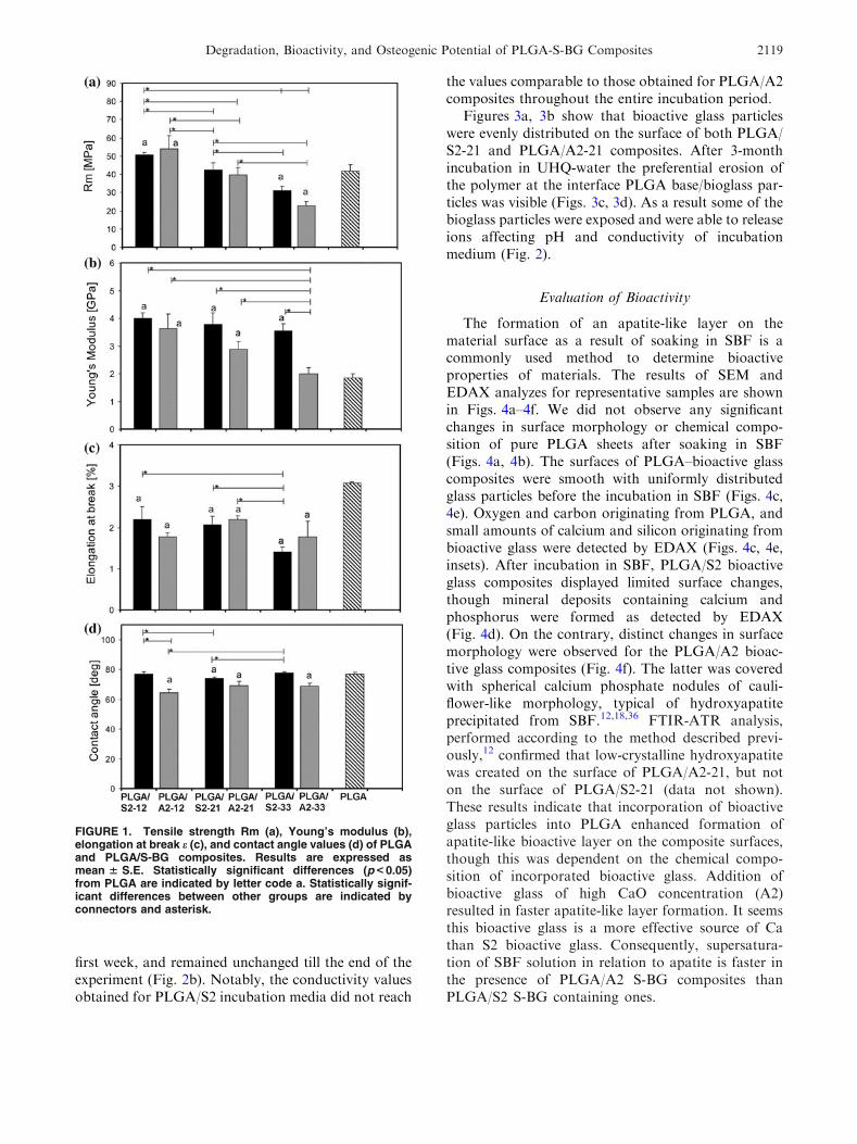
The mechanical properties of PLGA and PLGA/S-BG composites are given in Figs. 1a–1c. Tensilestrength (Rm) was comparable for pure PLGA andcomposites containing either 21%S2 or 21%A2 S-BG.Low S-BG contents (12%) in the compositesenhanced, and high (33%) reduced tensile strength(Fig. 1a). All composites, except for PLGA/A2-33 hadsignificantly higher Young’s modulus (Fig. 1b) andall exhibited significantly lower elongation at break(Fig. 1c) compared to pure PLGA (Figs. 1b, 1c).Contact angle values for PLGA/A2 composites weresignificantly lower than for PLGA/S2 composites(Fig. 1d). Contact angle values for the latter werecomparable to pure PLGA. Surface roughness param-eters, i.e., the arithmetical mean roughness (Ra)and ten-point mean roughness depth (Rz) (Table 1)increased with increased S-BG content in the com-posites. Ra values for composites containing 33% ofeither S-BG were comparable to values obtained forpure PLGA. In contrast, Rz values for compositescontaining either S-BG were approximately 5–13 foldhigher compared to Rz of pure PLGA, depending onS-BG content.
Degradation Rate
Degradation rates of the samples were evaluatedbased on pH and conductivity measurements of theincubation medium (Fig. 2) and compared to degra-dation rates of pure PLGA and pure bioactive glassesS2 or A2. Materials in the form of sheets were soakedin UHQ-water for 3 months. Pure PLGA did notinfluence pH and conductivity of the incubation med-ium up to the tenth week of incubation; this was fol-lowed by a rapid decrease of pH and increase ofconductivity of the incubation medium. In contrast,the pH of the composites’ incubation media stabi-lized at neutral level by second week and remainedunchanged until the end of experiments (Figs. 2a, 2c).Notably, PLGA/A2 composites containing either 21%or 33% A2 alkalized the incubation media duringinitial 2-week incubation period (Fig. 2c) and theconductivities of these incubation media were mark-edly increased compared to other materials studied(Fig. 2d). For PLGA/S2 bioactive glass, conductivitiesof their incubation media increased slightly within the
PAMULA et al.2118
first week, and remained unchanged till the end of theexperiment (Fig. 2b). Notably, the conductivity valuesobtained for PLGA/S2 incubation media did not reach
the values comparable to those obtained for PLGA/A2composites throughout the entire incubation period.
Figures 3a, 3b show that bioactive glass particleswere evenly distributed on the surface of both PLGA/S2-21 and PLGA/A2-21 composites. After 3-monthincubation in UHQ-water the preferential erosion ofthe polymer at the interface PLGA base/bioglass par-ticles was visible (Figs. 3c, 3d). As a result some of thebioglass particles were exposed and were able to releaseions affecting pH and conductivity of incubationmedium (Fig. 2).
Evaluation of Bioactivity
The formation of an apatite-like layer on thematerial surface as a result of soaking in SBF is acommonly used method to determine bioactiveproperties of materials. The results of SEM andEDAX analyzes for representative samples are shownin Figs. 4a–4f. We did not observe any significantchanges in surface morphology or chemical compo-sition of pure PLGA sheets after soaking in SBF(Figs. 4a, 4b). The surfaces of PLGA–bioactive glasscomposites were smooth with uniformly distributedglass particles before the incubation in SBF (Figs. 4c,4e). Oxygen and carbon originating from PLGA, andsmall amounts of calcium and silicon originating frombioactive glass were detected by EDAX (Figs. 4c, 4e,insets). After incubation in SBF, PLGA/S2 bioactiveglass composites displayed limited surface changes,though mineral deposits containing calcium andphosphorus were formed as detected by EDAX(Fig. 4d). On the contrary, distinct changes in surfacemorphology were observed for the PLGA/A2 bioac-tive glass composites (Fig. 4f). The latter was coveredwith spherical calcium phosphate nodules of cauli-flower-like morphology, typical of hydroxyapatiteprecipitated from SBF.12,18,36 FTIR-ATR analysis,performed according to the method described previ-ously,12 confirmed that low-crystalline hydroxyapatitewas created on the surface of PLGA/A2-21, but noton the surface of PLGA/S2-21 (data not shown).These results indicate that incorporation of bioactiveglass particles into PLGA enhanced formation ofapatite-like bioactive layer on the composite surfaces,though this was dependent on the chemical compo-sition of incorporated bioactive glass. Addition ofbioactive glass of high CaO concentration (A2)resulted in faster apatite-like layer formation. It seemsthis bioactive glass is a more effective source of Cathan S2 bioactive glass. Consequently, supersatura-tion of SBF solution in relation to apatite is faster inthe presence of PLGA/A2 S-BG composites thanPLGA/S2 S-BG containing ones.
FIGURE 1. Tensile strength Rm (a), Young’s modulus (b),elongation at break e (c), and contact angle values (d) of PLGAand PLGA/S-BG composites. Results are expressed asmean 6 S.E. Statistically significant differences (p < 0.05)from PLGA are indicated by letter code a. Statistically signif-icant differences between other groups are indicated byconnectors and asterisk.
Degradation, Bioactivity, and Osteogenic Potential of PLGA-S-BG Composites 2119

Interaction of Cells with Composite Materials
Evaluation of 2D Cultures
Cell numbers were estimated based on MTS stan-dard curve for human MSC cultures that were seededat different densities and culture media were assayedfor A490nm changes at appropriate time intervals. Cellnumbers of human MSC cultured for 7 days onPLGA/S2-12 and PLGA/S2-21 composite sheets instandard medium were comparable to cultures on purePLGA and significantly higher than on TCP (Fig. 5a).The number of cells grown on other composite sheetswas not different from those on TCP.
Human MSC cell numbers decreased with increasedbioactive glass content as a result of culturing hMSCon composite sheets in osteogenic medium containingbone morphogenetic protein 2 (BMP-2) (Fig. 5b).We have previously indicated BMP-2 to be a weekinducer of osteogenesis in human compared to rodentMSC cultures29 and also a poor osteogenic inducer
in standard serum-containing human MSC culturescompared to dexamethasone.9,10 Thus, it was ofinterest to test the BMP response of human MSC onbioactive composite sheets. We determined that spe-cific ALP activity (i.e., ALP/cell) significantly increasedfor PLGA/A2 composites compared to TCP and theincreases were proportional to the bioactive glasscontent (Fig. 5c). Pure PLGA sheets provided similarresults. Specific ALP activity for PLGA/S2-33 sheetswas higher than PLGA/S2-12 and PLGA/S2-21,but the results were not statistically significant andnone was significantly different from the specific ALPactivity of cells grown on TCP. Real-time PCR ana-lyzes of gene expression levels that are characteristicfor cells undergoing osteogenic differentiation wereperformed after 7-day hMSC culture on PLGA andcomposite sheets containing either 21%S2 or 21%A2S-BG, and results referred to gene expression levels forcells cultured on TCP (Fig. 6). The selection of com-posites for gene expression studies was based on
TABLE 1. Surface roughness parameters (Ra, Rz) of PLGA and PLGA/S-BG composites (mean 6 S.D.).
PLGA PLGA/S2-12 PLGA/S2-21 PLGA/S2-33 PLGA/A2-12 PLGA/A2-21 PLGA/A2-33
Ra (lm) 1.54 ± 0.28 0.59 ± 0.08 0.8 ± 0.1 1.5 ± 1.5 0.75 ± 0.07 1.6 ± 0.1 1.7 ± 0.1
Rz (lm) 1.15 ± 0.81 5.30 ± 0.68 7.4 ± 1.0 13.7 ± 1.6 6.7 ± 1.1 11.8 ± 1.4 12.7 ± 1.1
FIGURE 2. UHQ-water pH (a, c) and conductivity (b, d) changes during 3-month incubation of PLGA/S-BG composite samples andstarting compounds (PLGA and bioactive glass S2 or A2).
PAMULA et al.2120

moderate mechanical and biological properties of thesesheets. The analyzed transcripts included BMP-2mRNA, since BMP-2 can regulate its own expres-sion, and MSX-2 and OSTERIX that are tran-scription factors expressed early during osteogenesisand regulated by BMP-2.21 In addition, we ana-lyzed OSTEOPONTIN, BONE SIALOPROTEIN andOSTEOCALCIN that are synthesized by osteoblastsand deposited in bone matrix. The results showedcomparable expression levels for most selected geneson all studied surfaces (Figs. 6a–6f) suggesting thatosteogenic differentiation of hMSC by day 7 cultureswas not significantly affected by the composite surfaceproperties and/or their dissolution products. It shouldbe noted, however, that hMSC grown on compositesheets expressed significantly higher levels of BMP-2(PLGA/A2-21 vs. PLGA/S2-21; PLGA/A2-21 vs.TCP; Fig. 6a) and OSTEOPONTIN (OPN) mRNA(PLGA/S2-21 and PLGA/A2-21 vs. TCP; PLGA/S2-21 vs. PLGA/A2-21; Fig. 5d) and significantly lowerlevels of MSX-2 mRNA (PLGA/A2-21 vs. TCP;Fig. 5b).
Human MSC grown for 3 weeks on TCP in BMP-2containing osteogenic conditions stained mildly forAlizarin Red S, but staining was clearly visible forcells for which osteogenesis was induced by dexa-methasone (data not shown). When hMSC weregrown on PLGA/S-BG composites, strong AlizarinRed S staining was apparent on all PLGA/S2 andPLGA/A2-12 composite sheets. Corresponding sheetsincubated in cell-free conditions remained unstained.Thus, these materials enhanced cell-mediated calcifi-cation of extracellular matrix, and this was the casefor either dexamethasone or BMP-2 treated cells(Fig. 7). For PLGA/A2-21 and PLGA/A2-33 theAlizarin red staining intensity was very high withoutcells, thus it was difficult to distinguish betweenmaterial-induced and cell-induced calcification. PurePLGA sheets remained unstained with or withoutcells and regardless of culture treatments.
When primary BMC were pre-treated with M-CSFfor 3 days on PLGA-bioactive glass sheets, followedby 3-week culture of floaters on TCP in stan-dard medium, the morphology of cells that came into
FIGURE 3. SEM pictures of PLGA/S2-21 before (a) and after incubation in UHQ-water for 3 months (c), PLGA/A2-21 before (b) andafter incubation in UHQ-water for 3 months (d); original magnification 3500.
Degradation, Bioactivity, and Osteogenic Potential of PLGA-S-BG Composites 2121

contact with PLGA/A2-21 sheets was distinct fromthese pre-cultured on PLGA/S2-21 and pure PLGAsheets. Round, TRAP-positive and multinucleatedcells could be observed in cultures established fromfloating cells aspirated from PLGA/A2-21 (Figs. 8c,8d), whereas cells transferred from PLGA/S2-21 andpure PLGA sheets developed into fibroblast-like cellsnegative for TRAP (Figs. 8a, 8b).
Evaluation of 3D Cultures
MG-63 cells loaded into 3D PLGA, PLGA/S2-21,and PLGA/A2-21 attached to the sides of the scaffoldpores and remained viable after 2 weeks of culture, asdetermined by phalloidin/DAPI staining (Fig. 9a,confocal images). Cell numbers were comparable inall studied scaffolds as determined by MTS assay
FIGURE 4. SEM pictures and EDAX spectra (insets) of: PLGA surface before (a) and after 10-day incubation in SBF (b), PLGA/S2-21 before (c) and after 10-day incubation in SBF (d), PLGA/A2-21 before (e) and after 10-day incubation in SBF (f); originalmagnification 35,000.
PAMULA et al.2122

described in ‘‘Materials and Methods’’ section(Fig. 9b). In contrast, the production of collagen/cell(Fig. 9c) and calcium/cell (Fig. 9d) was significantlyincreased for cells cultured on composite scaffoldscompared to pure PLGA.
DISCUSSION
We report here that the incorporation of either S2or A2 sol–gel derived bioactive glass into PLGA resultsin composites of enhanced mechanical properties andtailored degradation rates, but biological activities ofthese composites depend both on the content andcomposition of the bioactive glass components.
Incorporation of either S2 or A2 S-BG into PLGAresulted in creation of stiffer materials compared topure PLGA, as determined by increased Young’smodulus and lowered elongation at break (Fig. 1).Tensile strength of the composites depended on thebioactive glass content and was significantly higher forcomposites containing 12% of either S-BG comparedto pure PLGA. Notably, tensile strengths of thesecomposites exceeded 50 MPa (Fig. 1a), a value com-parable to that of trabecular bone.7 The increase intensile strength and Young’s modulus of PLGA/S-BGcomposites suggests that S-BG particles act as a rein-forcing phase within the polymer matrix. In additionSEM observations show that the S-BG particles areuniformly distributed within the PLGA base and donot form agglomerates (Figs. 3a, 3b). Thus, it is pos-sible to obtain composites of mechanical propertiessimilar to those of natural bone simply by adjustingbioactive glass content. Interestingly, similarly torecent work of Wu et al.41 increasing A2 S-BG contentto 33% significantly decreased the tensile strength ofthe composites compared to pure PLGA; also themodulus of PLGA/A2-33 was significantly decreasedcompared to other composites (Fig. 1b). However, ourSEM analyzes of composites containing 33% of eitherS-BG indicated bioactive glass grains uniformlyembedded in the polymer matrix with no microcracks(data not shown).
Both S2 and A2 containing composites displayedmodified degradation rates compared to pure PLGA,S2 and A2 S-BG, and neutralized acidic degradationby-products of pure PLGA at later incubation stages,as determined by pH and conductivity of incubationsolutions. Notably, higher contents of A2 S-BG in thecomposites increased both pH and conductivity of theenvironment; the latter was not observed for S2 S-BG(Fig. 2). PLGA degrades through chain scission ofpolyester bonds due to hydrolysis. This results inoligomers formation leading to final degradationproducts of glycolic and lactic acids.33 Thus, at tenth
FIGURE 5. Growth and differentiation of human MSC after7-day culture on PLGA/S-BG composites, pure PLGA andtissue culture plastic (TCP). Cell number after culture instandard conditions without osteogenic inducers (a) and afterstimulation with rhBMP-2 (b). Alkaline phosphatase (ALP)activity of cells after stimulation with rhBMP-2 (c). Results areexpressed as mean 6 S.E. Statistically significant differences(p < 0.05) from a PLGA and b TCP are indicated by letter code,respectively. Statistically significant differences betweenother groups are indicated by connectors and asterisk. Cellnumbers were significantly higher on composite sheets con-taining low and mid w/v% of S2 S-BG compared to TCP whencultured without inducers, and significantly lower on com-posite sheets containing low and high w/v% of A2 S-BGcompared to PLGA (a). When stimulated with rhBMP-2, cellnumbers on composites containing high w/v% S-BG weresignificantly lower compared to either PLGA or TCP (b). Cellscultured on PLGA/A2-33 and stimulated with rhBMP-2 dis-played significantly higher ALP activity levels compared toPLGA and TCP (c). ALP activity levels of cells stimulated withrhBMP-2 were significantly higher on all studied A2 S-BG andPLGA/S2-33 compared to TCP (c).
Degradation, Bioactivity, and Osteogenic Potential of PLGA-S-BG Composites 2123

week in aqueous medium, the final PLGA degradationproducts decreased pH and increased conductivity ofthe incubation medium (Fig. 2). Immersion of S-BG inwater provokes the increase of pH and conductivitydue to release of calcium ions from the materials to theincubation medium, and protons uptake. AlthoughPLGA/S2 composites did not provoke significantchanges in pH and conductivity of the incubationmedium, past the tenth week in water, their degrada-tion rates remained constant in contrast to purePLGA. This may be due to (i) lower degradation of thePLGA base in the S2 containing composites, or (ii) theacidic degradation products of PLGA reacting withalkaline degradation products of S2 bioactive glass.
The former can be ruled out based on reports indi-cating that the presence of ceramic particles or carbonfibers in PLGA composites speeds up degradation3,31
and forms a new interface that improves diffusion ofwater molecules into the bulk of the composites. Thus,it is plausible that the PLGA/S2 composites degradefaster than pure PLGA, however, due to the bufferingcapacity of S2 S-BG, pH and conductivity of theincubation medium do not change. Compared toPLGA/S2 composites, the PLGA/A2 composites dis-played significantly higher pH and conductivity valuesand this positively correlated with the concentration ofbioactive glass (Figs. 2c, 2d). Notably, after 3 weeksimmersion of PLGA/A2 composites in UHQ-water,
FIGURE 6. Real-time RT-PCR analyzes of gene expression levels in hMSC cultured for 7 days on PLGA, PLGA/S2-21, and PLGA/A2-21. Cells were stimulated with ascorbate and BMP-2 and RNA harvested and analyzed as described in respective ‘‘Materials andMethods’’ sections. Fold changes relative to TCP 5 1 are presented (ddCT method). Results are expressed as mean 6 S.E.Statistically significant differences (p < 0.05) from a PLGA and b TCP are indicated by letter code, respectively. Statisticallysignificant differences between other groups are indicated by connectors and asterisk. Human MSC cultured on PLGA/A2-21sheets displayed significantly higher levels of BMP-2 and OPN mRNAs and significantly lower levels of MSX-2 mRNA.
FIGURE 7. Comparison of Alizarin Red S staining intensities of PLGA/S-BG composite sheets and pure PLGA in cell-free con-ditions (control) and after osteogenic human MSC cultures induced with dexamethasone (DEX) and BMP-2. PLGA/S2 compositesheets enhanced cell-mediated calcification of ECM—these sheets stained strongly for Alizarin Red S only when subjected toosteogenic culture. Similar results were obtained for either Dex or BMP induced osteogenic cultures.
PAMULA et al.2124

the pH stabilized at the level of 8 (Fig. 2c), whereasconductivity increased, especially for PLGA/A2-33composites (Fig. 2d). This may be due to continuousrelease of calcium ions from A2 S-BG and local buf-fering effects of potential PLGA degradation products.Overall, both the chemical composition of the S-BGand the amount in the PLGA base contribute to thedegradation rate of the resulting composites. Using thecomposites studied here, it is feasible to design a widevariety of biodegradable materials differing in degra-dation kinetics.
The cell numbers of bone marrow human MSCgrown in non-differentiating conditions were signifi-cantly higher on PLGA/S2 composite sheets comparedto PLGA/A2 (Fig. 5a). The latter was less hydrophobic(Fig. 1d) and this could have affected attachment andgrowth of osteoprogenitor cells. In contrast, when cellswere grown in osteogenic BMP-containing medium,the number of cells was comparable on either S-BG-containing composites and decreased with increasedS-BG content (Fig. 5b). This corresponded to increasedsurface roughness parameters of the PLGA/S-BGcomposites with increased S-BG contents (Table 1).The effects of surface roughness and topography onbone cells have been well documented in the litera-ture,14,22,40 who reported lower ALP activity of human
osteoblast-like cells with increased bioactive glasscontent in PLAGA-55S5 BG composites. We observedsignificant increases of specific ALP/cell (Fig. 5c) inhMSC cultures grown on composites containing thehighest proportion of A2 S-BG. It is plausible thatPLGA/A2-33 composites slowed cell adhesion and/orproliferation rates, but stimulated osteogenic differen-tiation instead. Further analyzes of gene expressionlevels of hMSC grown on composite sheets of moderateproperties (i.e., PLGA-21% of either A2 or S2 BG)showed, in most cases, comparable expression of oste-ogenic markers that was not significantly different fromcells cultured on either PLGA or TCP. This indicatesthat the studied surfaces support BMP-mediated oste-ogenic progression of hMSC at least to the same extentas polymer itself or TCP. However, hMSC grown onPLGA/A2-21 sheets had significantly higher BMP-2mRNA expression levels compared to TCP suggestingthey may enhance BMP-mediated signaling. In thisrespect, it is unclear why MSX-2 expression wasdecreased on PLGA/A2-21 sheets. Though, if thesecomposites stimulated osteogenic differentiation ofhMSC, they might as well induce expression of MSX-2at earlier culture stages. Further studies are required todetermine molecular mechanisms of BMP signaling onthese composite sheets.
FIGURE 8. Tartrate-resistant acid phosphatase (TRAP) staining of bone marrow mononuclear cells pre-cultured for 3 days onpure PLGA and PLGA/S-BG composites in the presence of MCS-F, followed by 3-week culture in standard medium on tissueculture plastic. Cells pre-cultured on pure PLGA (a) and PLGA/S2-21 composites (b) displayed typical fibroblastic morphology andthey were negative for TRAP. Positive TRAP staining and osteoclast-like morphology of cells was observed for cells pre-culturedon PLGA/A2-21 composites (c, d).
Degradation, Bioactivity, and Osteogenic Potential of PLGA-S-BG Composites 2125

In the previous work of Karpov et al.17 pure A2S-BG coatings induced moderate osteogenic effects inhuman bone marrow-derived MSC cultures, but actedas a potent inducer of osteoclast formation and sur-vival. In this work hMSC grown on PLGA/A2-21
sheets expressed significantly higher levels of OSTE-OPONTIN mRNA as compared to PLGA/S2-21 orTCP. While elevated osteopontin expression is anearly marker of osteoblast differentiation, it is alsorequired for osteoclast formation.1,38 Furthermore,
FIGURE 9. Phalloidin/DAPI staining (a) of MG-63 cells cultured in 3D PLGA, PLGA/S2-21, and PLGA/A2-21 scaffolds and stimu-lated with ascorbate, dexamethasone, and b-glycerophosphate for 2 weeks. Cells were also analyzed for cell number (b), collagen(c), and calcium (d) levels after 2-week culture. Results for b–d are expressed as mean 6 S.E. Statistically significant differences(p < 0.05) from PLGA are indicated by letter code a. Statistically significant differences between other groups are indicated byconnectors and asterisk. Comparable cell numbers were detected in all studied 3D scaffolds, but MG-63 cells cultured in PLGA-S-BG 3D composites produced significantly higher levels of collagen and calcium compared to pure PLGA 3D scaffolds.
PAMULA et al.2126
round, TRAP-positive multinucleated cells could beobserved in cultures established from floating BMCaspirated after 3-day primary BMC culture fromPLGA/A2-21 composite sheets, whereas cells trans-ferred from PLGA/S2-21 and pure PLGA sheetsdeveloped into fibroblast-like cells negative for TRAP.This may be due to high surface reactivity of PLGA/A2 composite sheets and their high bioactivity asdetermined by SBF studies (Fig. 4f) and Alizarin RedS staining of these sheets in cell-free conditions(Fig. 7). Crystallization of hydroxyapatite on thecomposite surface and/or osteopontin-rich extra-cellular matrix would attract osteoclast progenitorssimilarly to natural bone. Alternatively, PLGA/A2composite surfaces promoted the attachment ofosteoprogenitors through, i.e., enhanced BMP-2 sig-naling and these in turn signaled for osteoclast devel-opment. Regardless of the mechanisms, our presentdata support the concept that calcium-rich bioactiveglasses may be a key component of PLGA-basedcomposites where the requirement is to support boneremodeling.
An exciting new finding of this work is the ability ofPLGA/S-BG composites to support BMP-mediatedosteogenesis of bone marrow-derived human MSC instandard, serum-containing medium. BMP-2 was pre-viously shown to be ineffective at elevating ALPactivity in MSC derived from bone marrow of adultpatients and cultured on TCP in these same cultureconditions.30 Recent attempts to improve the osteo-genic response of human MSC to BMP-2 and/or Dexby culturing these cells on pure 45S5 bioactive glassalso did not have much success.35 In this study, cul-turing human MSC on the PLGA/A2-33 compositesheets significantly increased ALP/cell levels comparedto standard TCP. A similar tendency was observed forPLGA/S2-33 composites. PLGA/A2-21 sheets alsoenhanced BMP-2 mRNA expression of hMSC.Furthermore, all the PLGA/S2 composites studiedenhanced cell-mediated calcification of extracellularmatrix (ECM) in either BMP-2 or Dex-mediatedosteogenic conditions. We compared Alizarin Red Sstaining of composites in parallel cultures with andwithout cells. Increased Alizarin Red S staining wasapparent for PLGA/S2 composite sheets that wereused as growth surfaces for bone marrow-derivedhuman MSC, regardless of cell treatments. Similardata were obtained for PLGA/A2-12 composites, buthigh staining intensity of composites containing higherproportion of A2 S-BG in cell-free conditions did notallow us to quantify the effect. In contrast, pure PLGAdid not support cell-mediated calcification of ECM ineither BMP-2 or Dex-enriched osteogenic mediumdespite some ability of these cells to calcify ECMwith Dex on TCP (data not shown). Since PLGA/S2
composites showed moderate surface activity and/orbioactivity in cell-free solutions, we suggest thesecomposite features may be a key to promotion of theBMP-mediated osteogenic response of cells in vitro.
The potential for the presently studied compositematerials to be used as a bone graft substitute or tissueengineering scaffold is further supported by our 3Dculture studies, despite they were done with anothercell population of known osteoblastic properties (i.e.,MG-63 cell line) (Fig. 9). Cell distributions werecomparable within all studied constructs and compa-rable numbers of live cells were detected after 2-weekMG-63 culture in all studied 3D scaffolds. However,MG-63 cells cultured in 3D PLGA–BG compositesproduced significantly higher levels of collagen andcalcium suggesting osteoinductive properties of com-posite scaffolds.
CONCLUSIONS
Our present studies have shown that it is possibleto produce a wide variety of bioactive compositematerials made of poly(L-lactide-co-glycolide) and sol–gel derived bioactive glass. These composites differin mechanical performance, wettability, degradationkinetics, bioactivity, and biological properties depend-ing on the content and composition of bioactive glass.The composites made of PLGA and silica-rich S2 bio-active glass exhibit: (i) similar wettability as purePLGA, (ii) slow degradation kinetics, manifested byneutral pH and stable conductivity values of theincubation medium, (iii) good biological propertiesmanifested by enhanced human bone marrow cellproliferation and osteoinductive properties in both 2Dand 3D culture systems. The composites made ofPLGA and calcium-rich A2 bioactive glass exhibit: (i)high bioactivity manifested by crystallization ofhydroxyapatite and (ii) ability to promote both osteo-blast and osteoclast formation. Thus, the compositesmade of PLGA and sol–gel derived bioactive glassesshould find wide applications where a mechanicallystrong osteoinductive material is required for bonetissue engineering and bone tissue regeneration.
ACKNOWLEDGMENTS
We would like to thank Dr P. Dobrzynski from theCentre of Polymer and Carbon Materials, PolishAcademy of Sciences, Zabrze, Poland, for synthesizingpolymers used in this work and Mr. M. Dworak(AGH, Department of Biomaterials, Krakow, Poland)for his help in mechanical tests. The authors thank
Degradation, Bioactivity, and Osteogenic Potential of PLGA-S-BG Composites 2127

Prof. T. Niedzwiedzki, the Head of the Polytrauma,Orthopedic and Neuroorthopedic Department of theRydygier Hospital in Krakow, for procuring humanbone marrow samples. We also want to thank JossAtkinson of the University of Sheffield for providingMTS calibration data. Funding for this study wasprovided in part by MIRG-CT-2007-046479 grantwithin EC 6FP (AMO), Polish Ministry of Scienceand Education matching funds 465/6.PRUE/2007/7(AMO), Polish State Committee for ScientificResearch grants no. NN401 006135 (AMO), no.NN508 476338 (ML), no. NN 507280736 (EP), and no.11.11.60.365 (ML).
OPEN ACCESS
This article is distributed under the terms of theCreative Commons Attribution NoncommercialLicense which permits any noncommercial use, distri-bution, and reproduction in any medium, provided theoriginal author(s) and source are credited.
REFERENCES
1Aitken, C. J., J. M. Hodge, and G. C. Nicholson. Aden-oviral down-regulation of osteopontin inhibits humanosteoclast differentiation in vitro. J. Cell. Biochem. 93:896–903, 2004.2AlGhamdi, A. S., O. Shibly, and S. G. Ciancio. Osseousgrafting part I: autografts and allografts for periodontalregeneration—a literature review. J. Int. Acad. Periodontol.12:34–38, 2010.3Boccaccini, A. R., and V. Marquet. Bioresorbable andbioactive polymer/Bioglass� composites with tailored porestructure for tissue engineering applications. Compos. Sci.Technol. 63:2417–2429, 2003.4Boccaccini, A. R., J. A. Roether, L. L. Hench, V. Maquet,and R. Jerome. A composites approach to tissue engi-neering. Ceram. Eng. Sci. Proc. 23:805–816, 2002.5Cholewa-Kowalska, K., J. Kokoszka, M. Łaczka, Ł.Niedzwiedzki, W. Madej, and A. M. Osyczka. Gel-derivedbioglass as a compound of hydroxyapatite composites.Biomed. Mater. 4:1–11, 2009.6Cholewa-Kowalska, K., M. Łaczka, and A. Osyczka.Surface phenomena on gel-derived bioceramics–chemicaland clinical aspects. Key Eng. Mater. 206–213:1559–1562,2002.7Cullinane, D. M., and K. T. Salisbury. Biomechanics. In:Bone Tissue Engineering, edited by J. O. Hollinger, T. A.Einhorn, B. A. Doll, and C. Sfeir. Boca Raton: CRC Press,2005, pp. 245–276.8Day, R. M., V. Maquet, A. R. Boccaccini, R. Jerome, andA. Forbes. In vitro and in vivo analysis of macroporousbiodegradable poly(d,l-lactide-co-glycolide) scaffolds con-taining bioactive glass. J. Biomed. Mater. Res. 75A:778–787, 2005.9Diefenderfer, D. L., A. M. Osyczka, J. P. Garino, and P. S.Leboy. Regulation of BMP-induced transcription in
cultured human bone marrow stromal cells. J. Bone JointSurg. Am. 85 A(Suppl. 3):19–28, 2003.
10Diefenderfer, D. L., A. M. Osyczka, G. C. Reilly, and P. S.Leboy. BMP responsiveness in human mesenchymal stemcells. Connect. Tissue Res. 44(Suppl. 1):305–311, 2003.
11Dobrzynski, P., J. Kasperczyk, H. Janeczek, and M. Bero.Synthesis of biodegradable copolymers with the use of lowtoxic zirconium compounds. 1. Copolymerization of gly-colide with L-lactide initiated by Zr(Acac)4. Macromole-cules 34:5090–5103, 2001.
12Douglas, T., E. Pamula, D. Hauk, J. Wiltfang, S. Sivanan-than, E. Sherry, and P. H. Warnke. Porous polymer/hydroxyapatite scaffolds: characterization and biocompati-bility investigations. J. Mater. Sci. Mater. Med. 20:1909–1915, 2009.
13El-Ghannam, A. Bone reconstruction: from bioceramics totissue engineering.Expert Rev.Med. Devices 2:87–101, 2005.
14Geblinger, D., L. Addadi, and B. Geiger. Nano-topogra-phy sensing by osteoclasts. J. Cell Sci. 123:1503–1510,2010.
15Graham, S. M., A. Leonidou, N. Aslam-Pervez, A. Hamza,P. Panteliadis, M. Heliotis, A. Mantalaris, and E. Tsiridis.Biological therapy of bone defects: the immunology ofbone allo-transplantation. Expert Opin. Biol. Ther. 10:885–901, 2010.
16ISO 10993-13. Biological evaluation of medical devices Part13: Identification and quantification of degradation prod-ucts from polymeric medical devices, 1998.
17Karpov, M., M. Laczka, P. S. Leboy, and A. M. Osyczka.Sol-gel bioactive glasses support both osteoblast andosteoclast formation from human bone marrow cells.J. Biomed. Mater. Res. 84A:718–726, 2008.
18Kokubo, T., H.-M. Kim, and M. Kawashita. Novel bio-active materials with different mechanical properties. Bio-materials 24:2161–2175, 2003.
19Łaczka, M., K. Cholewa-Kowalska, K. Kulgawczyk, M.Klisch, and W. Mozgawa. Structural examinations ofgel-derived materials of the CaO–P2O5–SiO2 system.J. Mol. Str. 511–512:223–231, 1999.
20Łaczka-Osyczka, A., M. Łaczka, S. Kasugai, and K. Ohya.Behaviour of bone marrow cells cultured on three differentcoatings of gel-derived bioactive glass–ceramics at earlystages of cell differentiation. J. Biomed. Mater. Res. 42:433–442, 1998.
21Lian, J. B., G. S. Stein, A. Javed, A. J. van Wijnen, J. L.Stein, M. Montecino, M. Q. Hassan, T. Gaur, C. J.Lengner, and D. W. Young. Networks and hubs for thetranscriptional control of osteoblastogenesis. Rev. Endocr.Metab. Disord. 7:1–16, 2006.
22Lu, H. H., A. Tang, S. C. Oh, J. P. Spalazzi, and K.Dionisio. Compositional effects on the formation of acalcium phosphate layer and the response of osteoblast-likecells on polymer–bioactive glass composites. Biomaterials26:6323–6334, 2005.
23Lu, J. M., X. Wang, C. Marin-Muller, H. Wang, P. H. Lin,Q. Yao, and C. Chen. Current advances in research andclinical applications of PLGA-based nanotechnology.Expert Rev. Mol. Diagn. 9:325–341, 2009.
24Mahony, O., and J. R. Jones. Porous bioactive nano-structured scaffolds for bone regeneration: a sol–gel solu-tion. Nanomedicine 3:233–245, 2008.
25Maquet, V., A. R. Boccaccini, L. Pravata, I. Notingher,and R. Jerome. Porous poly(a-hydroxyacid)/Bioglass com-posite scaffolds for bone tissue engineering. I: preparation
PAMULA et al.2128
and in vitro characterization. Biomaterials 25:4185–4194,2004.
26Misra, S. K., D. Mohn, T. J. Brunner, W. J. Stark, S. E.Philip, I. Roy, V. Salih, J. C. Knowles, and A. R. Boc-caccini. Comparison of nanoscale and microscale bioactiveglass on the properties of P(3HB)/Bioglass composites.Biomaterials 29:1750–1761, 2008.
27Niedzielski, K., R. Sindut, K. Cholewa-Kowalska,M. Łaczka, and J. Kokoszka. New generation bioactiveglass–ceramics as a substitute of bone—in vivo study. Eng.Biomater. 67–68:48–51, 2007.
28Niemiel, T., H. Niiranen, M. Kellomaki, and P. Tormala.Self-reinforced composites of bioabsorbable polymer andbioactive glass with different bioactive glass contents. PartI: initial mechanical properties and bioactivity. Acta Bio-mater. 1:235–242, 2005.
29Osyczka, A. M., D. L. Diefenderfer, G. A. Bhargave, andP. S. Leboy. Different effects of BMP-2 on marrow stro-mal cells from human and rat bone. Cells Tissues Organs176(1-3):109–119, 2004.
30Osyczka, A. M., and P. S. Leboy. Bone morphogeneticprotein regulation of early osteoblast genes in humanmarrow stromal cells is mediated by extracellular signal-regulated kinase and phosphatidylinositol 3-kinase signal-ing. Endocrinology 146:3428–3437, 2005.
31Pamula, E., M. Blazewicz, C. Paluszkiewicz, and P.Dobrzynski. FTIR study of degradation products of ali-phatic polyesters–carbon fibres composites. J. Mol. Str.596:69–75, 2001.
32Pamula, E., E. Filova, L. Bacakova, V. Lisa, and D.Adamczak. Resorbable polymeric scaffolds for bone tissueengineering: the influence of their microstructure on thegrowth of human osteoblast-like MG 63 cells. J. Biomed.Mater. Res. 89A:432–443, 2009.
33Pamula, E., and E. Menaszek. In vitro and in vivo degra-dation of poly(L-lactide-co-glycolide) films and scaffolds.J. Mater. Sci. Mater. Med. 19:2063–2070, 2008.
34Porter, J. R., T. T. Ruckh, and K. C. Popat. Bone tissueengineering: a review in bone biomimetics and drug deliv-ery strategies. Biotechnol. Prog. 25:1539–1560, 2009.
35Reilly, G. C., S. Radin, A. T. Chen, and P. Ducheyne.Differential alkaline phosphatase responses of rat andhuman bone marrow derived mesenchymal stem cells to45S5 bioactive glass. Biomaterials 28:4091–4097, 2007.
36Rezwan, K., Q. Z. Chen, J. J. Blaker, and A. R. Boccaccini.Review. Biodegradable and bioactive porous polymer/inorganic composite scaffolds for bone tissue engineering.Biomaterials 27:3413–3431, 2006.
37Rickard, D. J., T. A. Sullivan, B. J. Shenker, P. S. Leboy,and I. Kazhdan. Induction of rapid osteoblast differentia-tion in rat bone marrow stromal cell cultures by dexa-methasone and BMP-2. Dev. Biol. 161:218–228, 1994.
38Rittling, S. R., H. N. Matsumoto, M. D. McKee, A. Nanci,X. R. An, K. E. Novick, A. J. Kowalski, M. Noda, andD. T. Denhardt. Mice lacking osteopontin show normaldevelopment and bone structure but display alteredosteoclast formation in vitro. J. Bone Miner. Res. 13:1101–1111, 1998.
39Roether, J. A., A. R. Boccaccini, L. L. Hench, V. Maquet,S. Gautier, and R. Jerome. Development and in vitrocharacterisation of novel bioresorbable and bioactivecomposite materials based on polylactide foams and bio-glass for tissue engineering applications. Biomaterials23:3871–3878, 2002.
40Schwartz, Z., E. Nasazky, and B. D. Boyan. Surface mic-rotopography regulates osteointegration: the role of im-plant surface microtopography in osteointegration. AlphaOmega. 98:9–19, 2005.
41Wu, C., Y. Ramaswamy, Y. F. Zhu, R. Zheng, R. Apple-yard, A. Howard, and H. Zreigat. The effect of mesoporousbioactive glass on the physiochemical, biological and drug-release properties of poly(dl-lactide-co-glycolide) films.Biomaterials 30:2199–2208, 2009.
42Yao, J., S. Radin, S. Leboy, and P. Ducheyne. The effect ofbioactive glass content on synthesis and bioactivity ofcomposite poly(lactic-co-glycolic acid)/bioactive glass sub-strate for tissue engineering. Biomaterials 26:1935–1943,2005.
43Yao, J., S. Radin, G. Reilly, P. S. Leboy, and P. Ducheyne.Solution-mediated effect of bioactive glass in poly (lactic-co-glycolic acid)–bioactive glass composites on osteogene-sis of marrow stromal cells. J. Biomed. Mater. Res.75A:794–801, 2005.
44Zhang, K., Y. Wang, M. A. Hillmyer, and L. F. Francis.Processing and properties of porous poly(L-lactide)/bio-active glass composites. Biomaterials 25:2489–2500, 2004.
Degradation, Bioactivity, and Osteogenic Potential of PLGA-S-BG Composites 2129
Related Documents