DOI 10.1212/01.wnl.0000271077.82508.a0 ; Published online before print August 29, 2007; 2007;69;1569 Neurology H. M. Nielsen, L. Minthon, E. Londos, et al. Lewy bodies Plasma and CSF serpins in Alzheimer disease and dementia with April 15, 2012 This information is current as of http://www.neurology.org/content/69/16/1569.full.html located on the World Wide Web at: The online version of this article, along with updated information and services, is rights reserved. Print ISSN: 0028-3878. Online ISSN: 1526-632X. All since 1951, it is now a weekly with 48 issues per year. Copyright © 2007 by AAN Enterprises, Inc. ® is the official journal of the American Academy of Neurology. Published continuously Neurology

Welcome message from author
This document is posted to help you gain knowledge. Please leave a comment to let me know what you think about it! Share it to your friends and learn new things together.
Transcript

DOI 10.1212/01.wnl.0000271077.82508.a0; Published online before print August 29, 2007; 2007;69;1569Neurology
H. M. Nielsen, L. Minthon, E. Londos, et al.Lewy bodies
Plasma and CSF serpins in Alzheimer disease and dementia with
April 15, 2012This information is current as of
http://www.neurology.org/content/69/16/1569.full.html
located on the World Wide Web at: The online version of this article, along with updated information and services, is
rights reserved. Print ISSN: 0028-3878. Online ISSN: 1526-632X.Allsince 1951, it is now a weekly with 48 issues per year. Copyright © 2007 by AAN Enterprises, Inc.
® is the official journal of the American Academy of Neurology. Published continuouslyNeurology
Plasma and CSF serpins in Alzheimerdisease and dementia with Lewy bodies
H.M. Nielsen, MScL. Minthon, MD, PhDE. Londos, MD, PhDK. Blennow, MD, PhDE. Miranda, PhDJ. Perez, PhDD.C. Crowther, PhD,MRCP
D.A. Lomas, PhD,FRCP
S.M. Janciauskiene,PhD
ABSTRACT
Objective: Serine protease inhibitors (serpins), the acute phase reactants and regulators of theproteolytic processing of proteins, have been recognized as potential contributors to the patho-genesis of Alzheimer disease (AD). We measured plasma and CSF levels of serpins in controls andpatients with dementia.
Methods: Using rocket immunoelectrophoresis, ELISA, and Luminex xMAP technology, we ana-lyzed plasma levels of �1-antichymotrypsin and �1-antitrypsin, and CSF levels of �1-antichymotrypsin, �1-antitrypsin, and neuroserpin along with three standard biomarkers (totaltau, tau phosphorylated at threonine-181, and the A�1-42) in patients with AD (n � 258), patientswith dementia with Lewy bodies (DLB; n � 38), and age-matched controls (n � 37).
Results: The level of CSF neuroserpin was significantly higher in AD compared with controls andDLB, whereas CSF �1-antichymotrypsin and �1-antitrypsin were significantly higher in both ADand DLB groups than in controls. Results from logistic regression analyses demonstrate a rela-tionship between higher CSF levels of �1-antichymotrypsin and neuroserpin and increased pre-dicted probability and odds ratios (ORs) of AD (OR 5.3, 95% CI 1.3 to 20.8 and OR 3.3, CI 1.3 to8.8). Furthermore, a logistic regression model based on CSF �1-antichymotrypsin, neuroserpin,and A�1-42 enabled us to discriminate between AD patients and controls with a sensitivity of94.7% and a specificity of 77.8%.
Conclusions: Higher CSF levels of neuroserpin and �1-antichymotrypsin were associated with theclinical diagnosis of Alzheimer disease (AD) and facilitated the diagnostic classification of AD vscontrols. CSF serpin levels did not improve the diagnostic classification of AD vs dementia withLewy bodies. Neurology® 2007;69:1569–1579
GLOSSARYAAT � �1-antitrypsin; ACT � �1-antichymotrypsin; AD � Alzheimer disease; ApoE � apolipoprotein E; AUC � area under thecurve; BBB � blood–brain barrier; COPD � chronic obstructive pulmonary disease; %CV � coefficients of variation percent-age; DLB � dementia with Lewy bodies; IL � interleukin; MMSE � Mini-Mental State Examination; NSAIDs � nonsteroidalanti-inflammatory drugs; OR � odds ratio; P-tau � tau phosphorylated at threonine-181; ROC � receiver operating charac-teristic; T-tau � total tau.
In addition to �-amyloid plaques and neurofibrillary tangles, the pathology of Alzheimerdisease (AD) is characterized by excessive inflammation.1 Inflammation is driven by cy-tokines (particularly interleukin [IL]-1) that are released from activated microglia andastrocytes,2 and this in turn drives the expression of IL-63 and inducible nitric oxidesynthase.4 Neuronal proteases that are released as part of the inflammatory response arecontrolled by a variety of inhibitors including members of the serine protease inhibitor(serpin) superfamily.5 These include �1-antichymotrypsin, �1-antitrypsin, and neuroser-pin. The important role of �1-antichymotrypsin in the pathogenesis of AD was demon-
e-Pub ahead of print on August 29, 2007, at www.neurology.org.
From the Chronic Inflammatory and Degenerative Disease Research Unit (H.M.N., S.M.J.) and Clinical Memory Research Unit (L.M., E.L.),Department of Clinical Sciences, Lund University, Malmo University Hospital, Malmo, Sweden; Department of Clinical Neuroscience (K.B.),Section of Experimental Neuroscience, The Sahlgrenska Academy at Goteborg University, Goteborg, Sweden; and Department of Medicine(E.M., J.P., D.C.C., D.A.L.), University of Cambridge, Cambridge Institute for Medical Research, Hills Road, Cambridge, UK.
Supported by the Swedish Research Council, the King Gustaf V and Queen Victoria Foundation, the Medical Research Council (UK), theWellcome Trust, and Papworth NHS Trust.
Disclosure: The authors report no conflicts of interest.
Address correspondence andreprint requests to Dr. SabinaJanciauskiene, Lund University,Department of ClinicalSciences, WallenbergLaboratory, 2nd Floor, MalmoUniversity Hospital Entrance46, SE-205 02 Malmo, [email protected]
Copyright © 2007 by AAN Enterprises, Inc. 1569
strated by the finding of raised levels inbrain homogenates from affected individu-als6; the finding that it is tightly associatedwith virtually all �-amyloid plaques7; andthe demonstration that it interacts with,8,9
and affects the clearance of, A�1-42.10-14
Moreover elevated levels of plasma andCSF �1-antichymotrypsin correlate withcognitive decline in elderly nondementedpersons and those with AD.15-17 Similarly,�1-antitrypsin18 is present in �-amyloidplaques of patients with AD, and isoformsof �1-antitrypsin are significantly altered inCSF of affected individuals when com-pared with controls.19,20 �1-Antitrypsin lev-els are increased in AD plasma andcorrelate with heme oxygenase-1 activityand cognitive decline.21
It has been shown that mutants of theneuron-specific serpin, neuroserpin, underliethe inclusion body dementia familial enceph-alopathy with neuroserpin inclusion bod-ies.22,23 Neuroserpin is expressed throughoutthe nervous system24 and inhibits serine pro-teases such as the tissue plasminogen activa-tor; urokinase-type plasminogen activator;and, to a lesser extent, plasmin.25-27 Recently,we have shown that neuroserpin is a plaque-associated protein in the brains of patientswith AD and that it forms a 1:1 binary com-plex with the A�1-42 peptide. This in turnprevents fibril formation and renders theA�1-42 peptide less toxic to neuronal cells.28
Whether neuroserpin is present within theCSF or is involved in AD pathogenesis hashitherto been unknown.
Taken together, these studies suggestthat serpins may be associated with ADthrough initiating some of the neuropatho-logic changes and reflect the developmentof the disease. We therefore measured theCSF levels of �1-antitrypsin, �1-antichymo-trypsin, and neuroserpin as well as thestandard markers of AD, i.e., total tau(T-tau), tau phosphorylated at threonine-181(P-tau), and A�1-42 in patients clinically di-agnosed with AD, dementia with Lewybodies (DLB), and in age-matched nonde-mented controls.
METHODS Patients. Subjects with dementia who en-rolled in this study (n � 296) are a sample of the patients thatare included in the Malmo Alzheimer Study. Patients wereseen in the Neuropsychiatric Clinic at Malmo UniversityHospital for evaluation of cognitive dysfunction between1999 and 2003. Healthy elderly controls (n � 37) were re-cruited among relatives of health care personnel and throughadvertisements at senior citizen clubs. The cognitive statusof the subjects was evaluated with the Mini-Mental StateExamination (MMSE)29 and Alzheimer’s Disease AssessmentScale–Cognitive subscale.30 The criteria of the Diagnosticand Statistical Manual of Mental Disorders, Fourth Edition,by the American Psychiatric Association (1994) were usedfor the clinical diagnosis of dementia, and the criteria of theNational Institute of Neurological and Communicative Dis-orders and Stroke and the Alzheimer Disease and RelatedDisorders Association31 were used for probable AD. Proba-ble dementia with Lewy bodies was diagnosed according tothe DLB consensus criteria.32 All patients and controls under-went routine laboratory tests, including determination of theapolipoprotein E (ApoE) genotype and measurement of theCSF/serum albumin ratio as an indicator of the blood–brainbarrier (BBB) function. In addition, the regular use (as pre-scribed by a physician) of nonsteroidal anti-inflammatorydrugs (NSAIDs) and the presence of other chronic inflamma-tory diseases, such as atherosclerosis, chronic obstructivepulmonary disease (COPD), and rheumatoid diseases, werealso recorded. This study was approved by the ethics com-mittee of Lund University.
Sample collection of blood and CSF. Lumbar puncturewas performed in the L3–L4 or L4–L5 interspace with thesubject in the sitting position. The first milliliter of CSF wasdiscarded, 1 mL was sent for cell analysis, and 10 mL wascollected in plastic (polypropylene) tubes. All CSF sampleswere gently mixed to avoid possible gradient effects. No CSFsample contained more than 500 erythrocytes/�L. The CSFsamples were centrifuged at 2,000g at 4 °C for 10 minutes toeliminate cells and other insoluble material, and were thenimmediately frozen and stored at �80 °C pending biochemi-cal analyses. Plasma and serum samples were collected at thesame time as the lumbar puncture. Blood for plasma analysiswas collected in tubes containing EDTA (B-D VacutainerSystem, Franklin Lakes, NJ) and centrifuged at 2,000g at 4°C for 10 minutes. The aliquots were immediately frozen at�80 °C and stored until assayed.
Determination of the concentration of �1-antichymotrypsin and �1-antitrypsin. Plasma and CSFlevels of �1-antichymotrypsin and �1-antitrypsin were deter-mined using rocket immunoelectrophoresis as described byLaurell33 with in-house modifications. In brief, aliquots ofplasma and CSF were run for 1.5 hours at 200 V on 1 mm0.9% w/v agarose gels containing 11 mg/L anti-human �1-antitrypsin antibody (DakoCytomation, Glostrup, Den-mark), and 6.98 mg/L (for plasma analysis) and 2.79 mg/L(for CSF analysis) anti-human �1-antichymotrypsin anti-body (DakoCytomation, Glostrup, Denmark). Gels werepressed between filter paper and dried before staining withCoomassie blue. To quantify �1-antitrypsin and �1-antichymotrypsin, the distance between the tip of the rocket-shaped immunoprecipitates and the application well wasmeasured. Standard curves were generated by serial dilutionsof a standard (Seronorm, Sero AS, Norway) that was run inparallel to samples on every gel. The coefficients of variation
1570 Neurology 69 October 16, 2007

percentage (%CV) for the interbatch and intrabatch vari-ability were 7.9% and 5.8%.
Determination of A�1-42, T-tau, and P-tau. Totaltau, P-tau, and A�1-42 levels were determined using LuminexxMAP technology as described previously.34 In brief, thistechnology is based on flow cytometric separation ofantibody-coated microspheres that are labeled with a spe-cific mixture of two fluorescent dyes. After binding of a bio-tinylated reporter antibody, quantification is made bybinding of a third fluorochrome coupled to streptavidin. Thetechnique allows for simultaneous measurement of severalanalytes in the same tube. The CSF levels of T-tau, P-tau,and A�1-42 correlated well with the levels obtained by con-ventional ELISA measurements.34 The intra-assay and inter-assay %CV for the multiparametric assay for A�1-42, T-tau,and P-tau were less than 9%.
Determination of the concentration of neuroserpin.A sandwich ELISA was developed using the antigen-purifiedfraction of a rabbit anti-human neuroserpin antibody35 as thecapture antibody and a pool of three high-affinity mousemonoclonal anti-human neuroserpin antibodies produced inProf. D. Lomas’ laboratory (1A10, 10B8, and 10G12) as thesecondary antibody. The ELISA plates (Corning Inc. Costar3590) were coated with capture antibody diluted at 2 �g/mLin 0.2 M Na2CO3/NaHCO3 pH 9.4 overnight at 4°C. Afterthree washes (0.9% w/v NaCl, 0.05% v/v Tween20), thewells were blocked for at least 1 hour at room temperaturewith blocking buffer (phosphate-buffered saline, 0.25% w/vbovine serum albumin, and 0.05% v/v Tween20, 0.025%w/vNaN3). Recombinant purified wild-type human neuroserpinwas used for the standard curve. It was sequentially diluted1:2, 10 times in blocking buffer for a standard range of 500 to1.0 ng/mL, and blocking buffer alone was used for the blank.The CSF samples were diluted at an assay-dependent concen-tration in the same blocking buffer. Standards and sampleswere added to the plate and incubated at room temperature for2 hours. After washing, the secondary antibody (monoclonalpool, 333 ng/mL each antibody) diluted in blocking buffer wasadded to the plate and further incubated at room temperaturefor 2 hours. After washing, horseradish peroxidase–labeledrabbit anti-mouse detection antibody (Sigma-Aldrich Co.,Dorset, UK), diluted 1:20,000 in blocking buffer withoutNaN3,was added to the plate and incubated at room temperature for 1hour. The plate was washed again, and each well was treatedwith developing solution (Sigma-Aldrich Co.) at room temper-ature for 10 minutes. The reaction was stopped with 1 MH2SO4, and the color reactionwas quantified in aThermo-maxmicroplate reader (Molecular Devices) at 450 nm. The detec-tion limit was 1 ng/mL, and the interplate and intraplate coeffi-cients of variation were both less than 5%.
Statistical analysis. Statistical analysis was performed us-ing Statistica software (Series 1203b, version 6.1 for Win-dows, Statsoft, Tulsa, OK), SPSS software (version 12.0.1 forWindows, SPSS Inc., Chicago, IL), and GraphPad Prism soft-ware (version 4 for Windows, GraphPad Software, Inc., SanDiego, CA). The Kruskal–Wallis test was used for compari-sons between more than two groups, and if significance wasreached, groups were compared using the Mann–Whitney Utest with correction for multiple comparisons (Bonferroni).Correlation coefficients were calculated using the test forSpearman rank order correlations. Because of the lack of thestandardized reference values for the measured variables, themedian of each variable in the control group was used as acut-point for defining “high” levels of the variable. The asso-ciations between marker levels in controls and AD and de-mentia with Lewy bodies were calculated as odds ratios(ORs) with 95% CIs. The differences in A�1-42 levels be-tween the controls and dementia patients were large. There-fore, the inverse highest quintile of the control group wasused as the cut-point when comparing controls with the twodementia groups. The two-sided �2 test was used to test ORsignificance and to test frequency differences among thegroups. With the attempt to discriminate between the studygroups using the analyzed markers, logistic regression analy-ses were conducted with controls against AD and with ADagainst DLB using a step-forward method. Variables wereentered based on a significant improvement in log likelihoodratios in every model. To avoid problems with multicol-linearity, highly correlated variables (significantly correlatedabove r � 0.5) were excluded from the analysis. Receiveroperating characteristic (ROC) curves were created using theaveraged predicted probabilities for each model to show therelationship between the logistic regression models’ specific-ity and sensitivity. The results are expressed as mean � SDor median and range. P � 0.05 was considered significant.
RESULTS Patient characteristics. Table 1 gives thedemographic data, MMSE scores, albumin ratio,and presence of the ApoE4 allele in patients withdementia and in controls. There was no signifi-cant difference in age at investigation, sex distri-bution, or CSF/serum albumin ratio (as a measureof the BBB function). As expected, AD patientsand DLB patients had significantly lower MMSEscores than controls. The distribution of theApoE4 allele between the groups was significantlydifferent. The occurrence of one or more of thechronic inflammatory diseases arteriosclerosis,chronic obstructive pulmonary disease, and rheu-
Table 1 Demographic data, MMSE, albumin ratio, and ApoE4 frequency
Diagnosis nSex,M/F, n (%)
Age at investigation,mean � SD
MMSE,mean � SD
CSF/serumalbumin ratio,mean � SD
ApoE4†carriers, %
Controls 37 14/23 (38/62) 72.4 � 7.5 29.1 � 1.0 7.2 � 2.7 27.0
AD 258 84/174 (33/67) 74.7 � 6.3 21.4 � 5.0* 7.5 � 3.2 70.2*
DLB 38 19/19 (50/50) 75.8 � 5.9 21.8 � 4.7* 8.1 � 4.3 55.3*
* Indicates a significant difference at the p�0.001 level, compared to controls.† ApoE4 carriers include both heterozygous and homozygous ApoE4 carriers.MMSE � Mini-Mental State Examination; AD � Alzheimer disease; DLB � dementia with Lewy bodies.
Neurology 69 October 16, 2007 1571

matoid disease and the regular use of NSAIDswas similar in all groups.
Levels of �1-antichymotrypsin, �1-antitrypsin, and neu-roserpin. In both AD and DLB patient groups, wefound higher levels of CSF �1-antichymotrypsin(44%, p � 0.001 and 36%, p � 0.001) and �1-antitrypsin (42.1%, p � 0.001 and 34.2%, p �0.05) than in controls. Plasma levels of �1-antichymotrypsin were elevated in the AD group(19.5%, p � 0.05) compared with controls,whereas plasma �1-antitrypsin levels did not dif-fer between the groups (table 2). Plasma levels ofneuroserpin were nondetectable. The CSF con-centration of neuroserpin was 25.5% higher in theAD patients than in controls (p� 0.001) but did notdiffer in patients with DLB vs controls. Interest-ingly, CSF neuroserpinwas the onlymarker that dif-fered between the two groups of dementia patients.AD patients had 15.4% higher neuroserpin levelsrelative to the DLB patients (p � 0.01). There wasconsiderable overlap, however, in CSF neuroserpinlevels between AD and DLB patients.
When analyzed across all groups, women hadhigher plasma levels of �1-antichymotrypsin thanmen (416mg/L vs 392mg/L, p� 0.01), whereasmenhad a higher albumin ratio (8.1 vs 6.3, p � 0.001)and higher CSF levels of �1-antitrypsin (12.1 mg/Lvs 9.9 mg/L, p � 0.001) and �1-antichymotrypsin(3.6 mg/L vs 3.3 mg/L, p � 0.05) than women. Wefound no sex-associated differences in CSF levels ofneuroserpin.
CSF levels of T-tau, P-tau, and A�1-42. Signifi-cantly higher CSF concentrations of T-tau andP-tau, but lower A�1-42 concentrations, werefound in the AD group compared with the control
group (table 2). DLB patients also exhibited sig-nificantly higher concentrations of P-tau butlower A�1-42 levels than controls; however, nodifference was found between the two dementiagroups. Previous studies have suggested that theP-tau/A�1-42 ratio can improve the separation ofAD and controls,36,37 so we also determined theP-tau/A�1-42 ratio in all groups. As shown in table2, both AD and DLB groups had significantlyhigher P-tau/A�1-42 ratios than controls; how-ever, no difference was found between the twogroups with dementia.
Intercorrelations between measured variables, age,and cognitive function. Correlations between themeasured markers, age, and cognitive functionare given in table 3. We found a strong linkagebetween higher CSF levels of �1-antichymo-trypsin (p � 0.001) and �1-antitrypsin (p � 0.001)and BBB dysfunction (increased CSF/serum albu-min ratio) that was independent of diagnosticgroup (table 3). This linkage is supported by thecorrelations between the CSF/plasma ACT orCSF/plasma AAT ratio and the CSF/serum albu-min ratio (r � 0.613 and r � 0.667, p � 0.001)(not shown in table 3). Lower MMSE scores wereassociated with increased CSF T-tau in AD(p � 0.001) and DLB (p � 0.05).
Among AD patients, higher CSF levels of neu-roserpin were strongly associated with increasedP-tau/A�1-42 ratio (p � 0.001). BBB dysfunction(higher CSF/serum albumin ratio) and higher CSF�1-antichymotrypsin were associated with lowerT-tau (p � 0.05) in the DLB group. Lower cogni-tive performance was linked to higher levels ofplasma �1-antichymotrypsin in AD (p � 0.01)
Table 2 Levels of AD markers and serpins
Marker Controls, n � 37 AD, n � 258 DLB, n � 38
CSF T-tau, ng/L 307 (117–846) 539‡ (153–2,144) 330 (87–811)
CSF P-tau, ng/L 57 (38–112) 73§ (15–211) 68� (26–129)
CSF A�1-42*, ng/L 754 (260–958) 397‡ (242–781) 463‡ (227–834)
P-tau/A� 1-42 7.6 (4.7–37.7) 18.8‡ (2.2–70.7) 15.2‡ (5.0–50.4)
Plasma ACT, mg/L 348 (232–600) 416‡ (196–1,256) 392 (256–1276)
CSF ACT, mg/L 2.5 (1.2–4.9) 3.6‡ (1.6–17.8) 3.4‡ (2.4–9.8)
Plasma AAT, g/L 1.36 (0.32–2.0) 1.52 (0.56–12.4) 1.59 (0.80–2.69)
CSF AAT, mg/L 7.6 (3.7–21.0) 10.8‡ (4.4–52.5) 10.2� (4.6–22.9)
CSF neuroserpin†, �g/L 7.41 (5.48–10.00) 9.30‡ (4.80–17.16) 8.06 (4.85–13.05)
Values are presented as median (range).* CSF A �1-42 was obtained from n � 257 Alzheimer disease (AD) patients.† CSF neuroserpin was obtained from n � 18 controls, n � 238 AD patients, and n � 37 dementia with Lewy body (DLB)patients.‡, §, and � indicate a significant difference at the p � 0.001, p � 0.01, and p � 0.05 levels, compared with controls.T-tau � total tau; P-tau � tau phosphorylated at threonine-181; ACT � �1-antichymotrypsin; AAT � �1-antitrypsin.
1572 Neurology 69 October 16, 2007
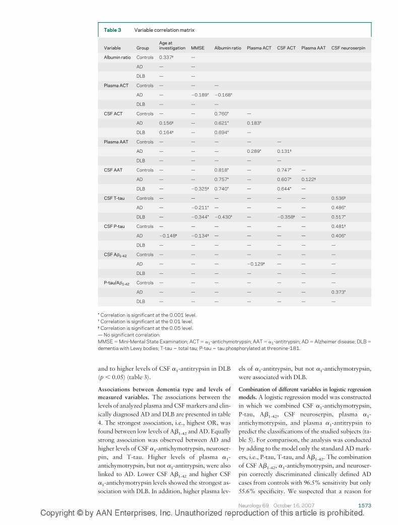
and to higher levels of CSF �1-antitrypsin in DLB(p � 0.05) (table 3).
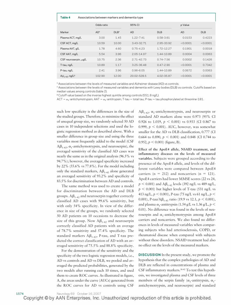
Associations between dementia type and levels ofmeasured variables. The associations between thelevels of analyzed plasma andCSFmarkers and clin-ically diagnosed AD and DLB are presented in table4. The strongest association, i.e., highest OR, wasfound between low levels of A�1-42 andAD. Equallystrong association was observed between AD andhigher levels of CSF �1-antichymotrypsin, neuroser-pin, and T-tau. Higher levels of plasma �1-antichymotrypsin, but not �1-antitrypsin, were alsolinked to AD. Lower CSF A�1-42 and higher CSF�1-antichymotrypsin levels showed the strongest as-sociation with DLB. In addition, higher plasma lev-
els of �1-antitrypsin, but not �1-antichymotrypsin,were associated with DLB.
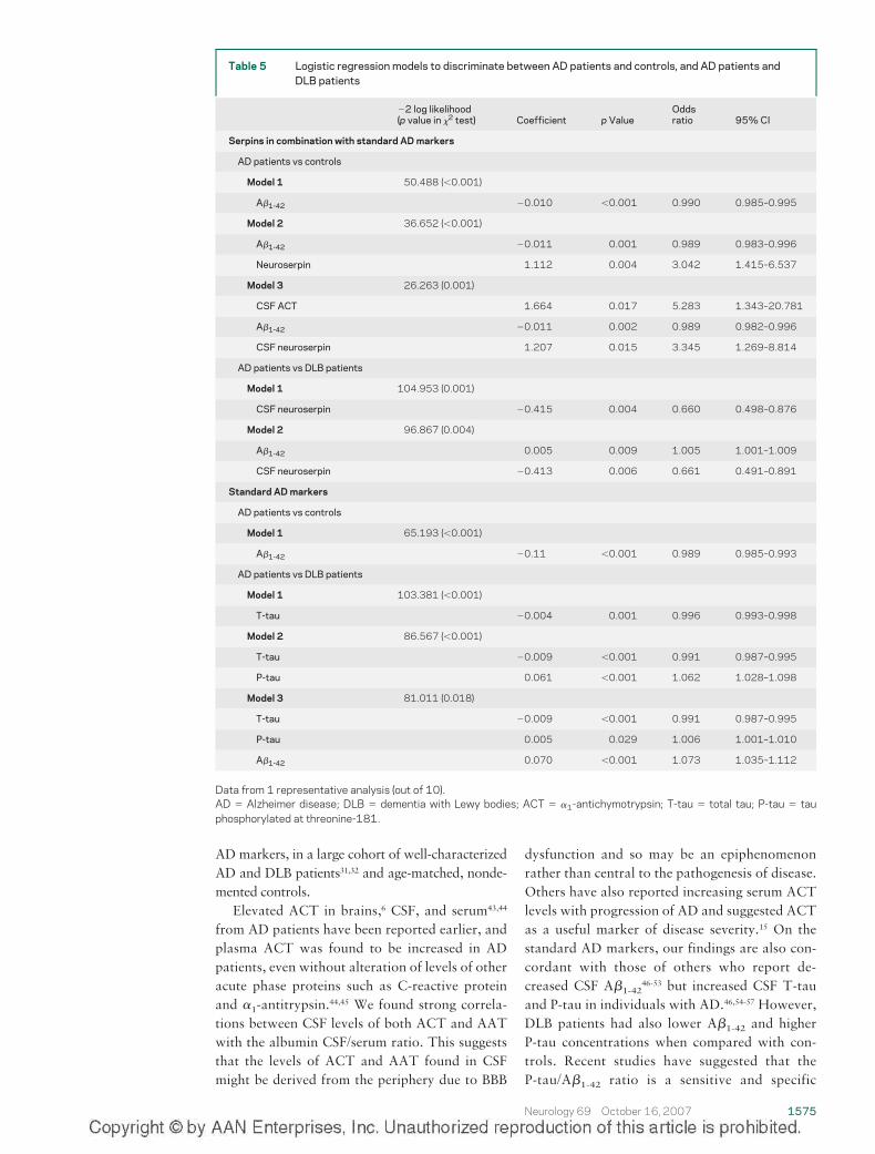
Combination of different variables in logistic regressionmodels. A logistic regression model was constructedin which we combined CSF �1-antichymotrypsin,P-tau, A�1-42, CSF neuroserpin, plasma �1-antichymotrypsin, and plasma �1-antitrypsin topredict the classifications of the studied subjects (ta-ble 5). For comparison, the analysis was conductedby adding to the model only the standard ADmark-ers, i.e., P-tau, T-tau, and A�1-42. The combinationof CSF A�1-42, �1-antichymotrypsin, and neuroser-pin correctly discriminated clinically defined ADcases from controls with 96.5% sensitivity but only55.6% specificity. We suspected that a reason for
Table 3 Variable correlation matrix
Variable GroupAge atinvestigation MMSE Albumin ratio Plasma ACT CSF ACT Plasma AAT CSF neuroserpin
Albumin ratio Controls 0.337‡ —
AD — —
DLB — —
Plasma ACT Controls — — —
AD — �0.189† �0.168†
DLB — — —
CSF ACT Controls — — 0.760* —
AD 0.156‡ — 0.621* 0.183†
DLB 0.164‡ — 0.694* —
Plasma AAT Controls — — — — —
AD — — — 0.289* 0.131‡
DLB — — — — —
CSF AAT Controls — — 0.818* — 0.747* —
AD — — 0.757* — 0.607* 0.122‡
DLB — �0.325‡ 0.740* — 0.644* —
CSF T-tau Controls — — — — — — 0.536‡
AD — �0.211* — — — — 0.486*
DLB — �0.344* �0.430† — �0.358‡ — 0.517*
CSF P-tau Controls — — — — — — 0.481‡
AD �0.148‡ �0.134‡ — — — — 0.406*
DLB — — — — — — —
CSF A�1-42 Controls — — — — — — —
AD — — — �0.129‡ — — —
DLB — — — — — — —
P-tau/A�1-42 Controls — — — — — — —
AD — — — — — — 0.373*
DLB — — — — — — —
* Correlation is significant at the 0.001 level.† Correlation is significant at the 0.01 level.‡ Correlation is significant at the 0.05 level.— No significant correlation.MMSE � Mini-Mental State Examination; ACT � �1-antichymotrypsin; AAT � �1-antitrypsin; AD � Alzheimer disease; DLB �
dementia with Lewy bodies; T-tau � total tau; P-tau � tau phosphorylated at threonine-181.
Neurology 69 October 16, 2007 1573
such low specificity is the differences in the size ofthe studied groups. Therefore, tominimize the effectof unequal group size, we randomly selected 50 ADcases in 10 independent selections and used the lo-gistic regression method as described above. With asmaller difference in group size and using the threevariables most frequently added to the model (CSFA�1-42, �1-antichymotrypsin, and neuroserpin), theaveraged sensitivity of the classified AD cases wasnearly the same as in the original analysis (96.5% vs94.7%); however, the averaged specificity increasedby 22% (55.6% vs 77.8%). For the model includingonly the standard markers, A�1-42 alone generatedan averaged sensitivity of 93.2% and specificity of83.5% for discrimination between AD and controls.
The same method was used to create a modelfor discrimination between the AD and DLBgroups. A�1-42 and neuroserpin together correctlyclassified AD cases with 99.6% sensitivity, butwith only 54% specificity. In view of the differ-ence in size of the groups, we randomly selected50 AD patients on 10 occasions to decrease thesize of this group. Now A�1-42 and neuroserpincorrectly classified AD patients with an averageof 78.7% sensitivity and 57.4% specificity. Thestandard markers A�1-42, P-tau, and T-tau pre-dicted the correct classification of AD with an av-eraged sensitivity of 75.5% and 88.8% specificity.
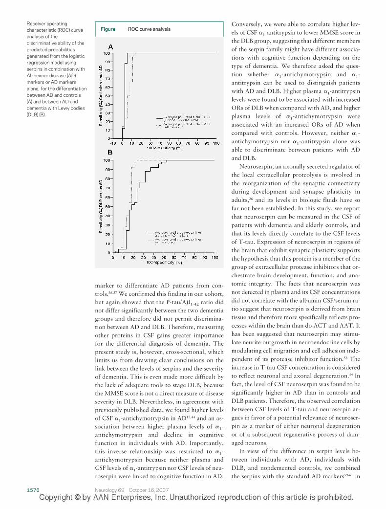
For the demonstration of the sensitivity and thespecificity of the two logistic regression models, i.e.,AD vs controls and AD vs DLB, we pooled and av-eraged the predicted probabilities, generated by thetwo models after running each 10 times, and usedthem to create ROC curves. As illustrated in figure,A, the areas under the curve (AUCs) generated fromthe ROC curves for AD vs controls using CSF
A�1-42, �1-antichymotrypsin, and neuroserpin orstandard AD markers alone were 0.973 (95% CI0.926 to 1.019, p � 0.001) vs 0.933 (CI 0.867 to0.999, p � 0.001). AUC, however, was somewhatsmaller for the AD vs DLB classification, 0.777 (CI0.664 to 0.890, p � 0.001) and 0.848 (CI 0.744 to0.952, p � 0.001) (figure, B).
Effect of the ApoE4 allele, NSAID treatment, andinflammatory diseases on the levels of measuredvariables. Subjects were grouped according to thepresence of the ApoE4 allele, and levels of the dif-ferent variables were compared between ApoE4carriers (n � 212) and noncarriers (n � 121).ApoE4 carriers had lowerMMSE scores (22 vs 24,p � 0.001) and A�1-42 levels (392 ng/L vs 489 ng/L,p � 0.001) but higher levels of T-tau (531 ng/L vs413 ng/L, p � 0.001), P-tau (75 ng/L vs 61 ng/L, p �
0.001), P-tau/A�1-42 ratio (19.9 vs 12.1, p � 0.001),and plasma �1-antitrypsin (1.54 g/L vs 1.36 g/L, p �
0.01). No difference was found in the levels of neu-roserpin and �1-antichymotrypsin among ApoE4carriers and noncarriers. We also found no differ-ences in levels of measured variables when compar-ing subjects who had arteriosclerosis, COPD, orrheumatoid disease when compared with subjectswithout these disorders. NSAID treatment had alsono effect on the levels of the measured markers.
DISCUSSION In the present study, we promote thehypothesis that the complex pathologies of AD andDLB are reflected in concentrations of plasma andCSF inflammatory markers.38-42 To test this hypoth-esis, we investigated plasma and CSF levels of threemembers of the serpin family (�1-antitrypsin, �1-antichymotrypsin, and neuroserpin) and standard
Table 4 Associations between markers and dementia type
Odds ratio 95% CI p Value
Marker AD* DLB† AD DLB AD DLB
Plasma ACT, mg/L 3.00 1.45 1.22–7.41 0.58–3.61 0.0153 0.4223
CSF ACT, mg/L 10.59 10.00 3.43–32.71 2.95–33.92 �0.0001 �0.0001
Plasma AAT, g/L 1.78 4.60 0.75–4.23 1.72–12.27 0.1901 0.0018
CSF AAT, mg/L 5.54 3.96 2.05–14.97 1.44–10.89 0.0004 0.0063
CSF neuroserpin, �g/L 10.75 2.36 2.71–42.73 0.74–7.56 0.0002 0.1426
T-tau, ng/L 10.89 1.17 3.25–36.48 0.47–2.90 �0.0001 0.7342
P-tau, ng/L 2.41 3.96 0.96–6.05 1.44–10.89 0.0672 0.0063
A�1-42, ng/L‡ 102.90 12.00 20.02–528.5 4.02–35.87 �0.0001 �0.0001
* Associations between the levels of measured variables and Alzheimer disease (AD) vs controls.† Associations between the levels of measured variables and dementia with Lewy bodies (DLB) vs controls. Cutoffs based onmedian values among controls (table 2).‡ Cutoff value based on the inverse highest quintile among controls (551.8 ng/L).ACT � �1-antichymotrypsin; AAT � �1-antitrypsin; T-tau � total tau; P-tau � tau phosphorylated at threonine-181.
1574 Neurology 69 October 16, 2007
ADmarkers, in a large cohort of well-characterizedAD and DLB patients31,32 and age-matched, nonde-mented controls.
Elevated ACT in brains,6 CSF, and serum43,44
from AD patients have been reported earlier, andplasma ACT was found to be increased in ADpatients, even without alteration of levels of otheracute phase proteins such as C-reactive proteinand �1-antitrypsin.44,45 We found strong correla-tions between CSF levels of both ACT and AATwith the albumin CSF/serum ratio. This suggeststhat the levels of ACT and AAT found in CSFmight be derived from the periphery due to BBB
dysfunction and so may be an epiphenomenonrather than central to the pathogenesis of disease.Others have also reported increasing serum ACTlevels with progression of AD and suggested ACTas a useful marker of disease severity.15 On thestandard AD markers, our findings are also con-cordant with those of others who report de-creased CSF A�1-42
46-53 but increased CSF T-tauand P-tau in individuals with AD.46,54-57 However,DLB patients had also lower A�1-42 and higherP-tau concentrations when compared with con-trols. Recent studies have suggested that theP-tau/A�1-42 ratio is a sensitive and specific
Table 5 Logistic regression models to discriminate between AD patients and controls, and AD patients andDLB patients
�2 log likelihood(p value in �2 test) Coefficient p Value
Oddsratio 95% CI
Serpins in combination with standard AD markers
AD patients vs controls
Model 1 50.488 (�0.001)
A�1-42 �0.010 �0.001 0.990 0.985–0.995
Model 2 36.652 (�0.001)
A�1-42 �0.011 0.001 0.989 0.983–0.996
Neuroserpin 1.112 0.004 3.042 1.415–6.537
Model 3 26.263 (0.001)
CSF ACT 1.664 0.017 5.283 1.343–20.781
A�1-42 �0.011 0.002 0.989 0.982–0.996
CSF neuroserpin 1.207 0.015 3.345 1.269–8.814
AD patients vs DLB patients
Model 1 104.953 (0.001)
CSF neuroserpin �0.415 0.004 0.660 0.498–0.876
Model 2 96.867 (0.004)
A�1-42 0.005 0.009 1.005 1.001–1.009
CSF neuroserpin �0.413 0.006 0.661 0.491–0.891
Standard AD markers
AD patients vs controls
Model 1 65.193 (�0.001)
A�1-42 �0.11 �0.001 0.989 0.985–0.993
AD patients vs DLB patients
Model 1 103.381 (�0.001)
T-tau �0.004 0.001 0.996 0.993–0.998
Model 2 86.567 (�0.001)
T-tau �0.009 �0.001 0.991 0.987–0.995
P-tau 0.061 �0.001 1.062 1.028–1.098
Model 3 81.011 (0.018)
T-tau �0.009 �0.001 0.991 0.987–0.995
P-tau 0.005 0.029 1.006 1.001–1.010
A�1-42 0.070 �0.001 1.073 1.035–1.112
Data from 1 representative analysis (out of 10).AD � Alzheimer disease; DLB � dementia with Lewy bodies; ACT � �1-antichymotrypsin; T-tau � total tau; P-tau � tauphosphorylated at threonine-181.
Neurology 69 October 16, 2007 1575
marker to differentiate AD patients from con-trols.36,37 We confirmed this finding in our cohort,but again showed that the P-tau/A�1-42 ratio didnot differ significantly between the two dementiagroups and therefore did not permit discrimina-tion between AD and DLB. Therefore, measuringother proteins in CSF gains greater importancefor the differential diagnosis of dementia. Thepresent study is, however, cross-sectional, whichlimits us from drawing clear conclusions on thelink between the levels of serpins and the severityof dementia. This is even made more difficult bythe lack of adequate tools to stage DLB, becausethe MMSE score is not a direct measure of diseaseseverity in DLB. Nevertheless, in agreement withpreviously published data, we found higher levelsof CSF �1-antichymotrypsin in AD15,44 and an as-sociation between higher plasma levels of �1-antichymotrypsin and decline in cognitivefunction in individuals with AD. Importantly,this inverse relationship was restricted to �1-antichymotrypsin because neither plasma andCSF levels of �1-antitrypsin nor CSF levels of neu-roserpin were linked to cognitive function in AD.
Conversely, we were able to correlate higher lev-els of CSF �1-antitrypsin to lower MMSE score inthe DLB group, suggesting that different membersof the serpin family might have different associa-tions with cognitive function depending on thetype of dementia. We therefore asked the ques-tion whether �1-antichymotrypsin and �1-antitrypsin can be used to distinguish patientswith AD and DLB. Higher plasma �1-antitrypsinlevels were found to be associated with increasedORs of DLBwhen compared with AD, and higherplasma levels of �1-antichymotrypsin wereassociated with an increased ORs of AD whencompared with controls. However, neither �1-antichymotrypsin nor �1-antitrypsin alone wasable to discriminate between patients with ADand DLB.
Neuroserpin, an axonally secreted regulator ofthe local extracellular proteolysis is involved inthe reorganization of the synaptic connectivityduring development and synapse plasticity inadults,26 and its levels in biologic fluids have sofar not been established. In this study, we reportthat neuroserpin can be measured in the CSF ofpatients with dementia and elderly controls, andthat its levels directly correlate to the CSF levelsof T-tau. Expression of neuroserpin in regions ofthe brain that exhibit synaptic plasticity supportsthe hypothesis that this protein is a member of thegroup of extracellular protease inhibitors that or-chestrate brain development, function, and ana-tomic integrity. The facts that neuroserpin wasnot detected in plasma and its CSF concentrationsdid not correlate with the albumin CSF/serum ra-tio suggest that neuroserpin is derived from braintissue and therefore more specifically reflects pro-cesses within the brain than do ACT and AAT. Ithas been suggested that neuroserpin may stimu-late neurite outgrowth in neuroendocrine cells bymodulating cell migration and cell adhesion inde-pendent of its protease inhibitor function.58 Theincrease in T-tau CSF concentration is consideredto reflect neuronal and axonal degeneration.54 Infact, the level of CSF neuroserpin was found to besignificantly higher in AD than in controls andDLB patients. Therefore, the observed correlationbetween CSF levels of T-tau and neuroserpin ar-gues in favor of a potential relevance of neuroser-pin as a marker of either neuronal degenerationor of a subsequent regenerative process of dam-aged neurons.
In view of the difference in serpin levels be-tween individuals with AD, individuals withDLB, and nondemented controls, we combinedthe serpins with the standard AD markers59-61 in
Figure ROC curve analysisReceiver operatingcharacteristic (ROC) curveanalysis of thediscriminative ability of thepredicted probabilitiesgenerated from the logisticregression model usingserpins in combination withAlzheimer disease (AD)markers or AD markersalone, for the differentiationbetween AD and controls(A) and between AD anddementia with Lewy bodies(DLB) (B).
1576 Neurology 69 October 16, 2007
an attempt to discriminate between the threegroups. Results from logistic regression analysesdemonstrate a relationship between higher CSFlevels of �1-antichymotrypsin and neuroserpinand increased predicted probability and ORs ofAD. Furthermore, a logistic regression modelbased on CSF �1-antichymotrypsin, neuroserpin,and A�1-42 enabled us to discriminate betweenAD patients and controls with a sensitivity andspecificity comparable to standard markers. Thelevels of sensitivity and specificity that were de-rived in our analyses should, however, be viewedwith caution until they are replicated.
ACKNOWLEDGMENTThe authors thank Carina Wattmo and Anders Wallmark, Depart-ment of Clinical Sciences, Lund University, for critical discussionsregarding the statistical analyses.
Received January 6, 2007. Accepted in final form April 2,2007.
REFERENCES1. Pratico D, Sung S. Lipid peroxidation and oxidative
imbalance: early functional events in Alzheimer’s dis-ease. J Alzheimers Dis 2004;6:171–175.
2. Griffin WS, Sheng JG, Roberts GW, Mrak RE.Interleukin-1 expression in different plaque types inAlzheimer’s disease: significance in plaque evolution.J Neuropathol Exp Neurol 1995;54:276–281.
3. Strauss S, Bauer J, Ganter U, Jonas U, Berger M, VolkB. Detection of interleukin-6 and alpha 2-macro-globulin immunoreactivity in cortex and hippocampusof Alzheimer’s disease patients. Lab Invest 1992;66:223–230.
4. Lee SC, Zhao ML, Hirano A, Dickson DW. Induciblenitric oxide synthase immunoreactivity in the Alzhei-mer disease hippocampus: association with Hiranobodies, neurofibrillary tangles, and senile plaques. JNeuropathol Exp Neurol 1999;58:1163–1169.
5. Silverman GA, Bird PI, Carrell RW, et al. The serpinsare an expanding superfamily of structurally similarbut functionally diverse proteins: evolution, mecha-nism of inhibition, novel functions, and a revised no-menclature. J Biol Chem 2001;276:33293–33296.
6. Licastro F, Mallory M, Hansen LA, Masliah E. In-creased levels of alpha-1-antichymotrypsin in brains ofpatients with Alzheimer’s disease correlate with acti-vated astrocytes and are affected by APOE 4 genotype.J Neuroimmunol 1998;88:105–110.
7. Abraham CR, Selkoe DJ, Potter H. Immunochemicalidentification of the serine protease inhibitor alpha1-antichymotrypsin in the brain amyloid deposits ofAlzheimer’s disease. Cell 1988;52:487–501.
8. Janciauskiene S, Eriksson S, Wright HT. A specificstructural interaction of Alzheimer’s peptide A beta1-42 with alpha 1-antichymotrypsin. Nat Struct Biol1996;3:668–671.
9. Janciauskiene S, Rubin H, Lukacs CM, Wright HT.Alzheimer’s peptide Abeta1-42 binds to two beta-sheets of alpha1-antichymotrypsin and transforms it
from inhibitor to substrate. J Biol Chem 1998;273:28360–28364.
10. Abraham CR, McGraw WT, Slot F, Yamin R. Alpha1-antichymotrypsin inhibits A beta degradation invitro and in vivo. Ann NY Acad Sci 2000;920:245–248.
11. Melchor JP, Pawlak R, Chen Z, Strickland S. The pos-sible role of tissue-type plasminogen activator (tPA)and tPA blockers in the pathogenesis and treatment ofAlzheimer’s disease. J Mol Neurosci 2003;20:287–289.
12. Melchor JP, Pawlak R, Strickland S. The tissue plas-minogen activator-plasminogen proteolytic cascade ac-celerates amyloid-beta (Abeta) degradation andinhibits Abeta-induced neurodegeneration. J Neurosci2003;23:8867–8871.
13. Mucke L, Yu GQ, McConlogue L, Rockenstein EM,Abraham CR, Masliah E. Astroglial expression of hu-man alpha(1)-antichymotrypsin enhances Alzheimer-like pathology in amyloid protein precursor transgenicmice. Am J Pathol 2000;157:2003–2010.
14. Nilsson LN, Bales KR, DiCarlo G, et al. Alpha-1-antichymotrypsin promotes beta-sheet amyloid plaquedeposition in a transgenic mouse model of Alzheimer’sdisease. J Neurosci 2001;21:1444–1451.
15. DeKosky ST, Ikonomovic MD, Wang X, et al. Plasmaand cerebrospinal fluid alpha1-antichymotrypsin levelsin Alzheimer’s disease: correlation with cognitive im-pairment. Ann Neurol 2003;53:81–90.
16. Dik MG, Jonker C, Hack CE, Smit JH, Comijs HC,Eikelenboom P. Serum inflammatory proteins and cog-nitive decline in older persons. Neurology 2005;64:1371–1377.
17. Lieberman J, Schleissner L, Tachiki KH, Kling AS. Se-rum alpha 1-antichymotrypsin level as a marker forAlzheimer-type dementia. Neurobiol Aging 1995;16:747–753.
18. Gollin PA, Kalaria RN, Eikelenboom P, Rozemuller A,Perry G. Alpha 1-antitrypsin and alpha 1-antichy-motrypsin are in the lesions of Alzheimer’s disease.Neuroreport 1992;3:201–203.
19. Puchades M, Hansson SF, Nilsson CL, Andreasen N,Blennow K, Davidsson P. Proteomic studies of poten-tial cerebrospinal fluid protein markers for Alzheimer’sdisease. Brain Res Mol Brain Res 2003;118:140–146.
20. Yu HL, Chertkow HM, Bergman H, Schipper HM.Aberrant profiles of native and oxidized glycoproteinsin Alzheimer plasma. Proteomics 2003;3:2240–2248.
21. Maes OC, Kravitz S, Mawal Y, et al. Characterizationof alpha1-antitrypsin as a heme oxygenase-1 suppres-sor in Alzheimer plasma. Neurobiol Dis 2006;24:89–100.
22. Davis RL, Holohan PD, Shrimpton AE, et al. Familialencephalopathy with neuroserpin inclusion bodies.Am J Pathol 1999;155:1901–1913.
23. Davis RL, Shrimpton AE, Holohan PD, et al. Familialdementia caused by polymerization of mutant neuro-serpin. Nature 1999;401:376–379.
24. Osterwalder T, Contartese J, Stoeckli ET, Kuhn TB,Sonderegger P. Neuroserpin, an axonally secretedserine protease inhibitor. Embo J 1996;15:2944–2953.
25. Hastings GA, Coleman TA, Haudenschild CC, et al.Neuroserpin, a brain-associated inhibitor of tissueplasminogen activator is localized primarily in neu-rons: implications for the regulation of motor learning
Neurology 69 October 16, 2007 1577

and neuronal survival. J Biol Chem 1997;272:33062–33067.
26. Osterwalder T, Cinelli P, Baici A, et al. The axonallysecreted serine proteinase inhibitor, neuroserpin, in-hibits plasminogen activators and plasmin but notthrombin. J Biol Chem 1998;273:2312–2321.
27. Belorgey D, Crowther DC, Mahadeva R, Lomas DA.Mutant neuroserpin (S49P) that causes familial enceph-alopathy with neuroserpin inclusion bodies is a poorproteinase inhibitor and readily forms polymers invitro. J Biol Chem 2002;277:17367–17373.
28. Kinghorn KJ, Crowther DC, Sharp LK, et al. Neuro-serpin binds Abeta and is a neuroprotective componentof amyloid plaques in Alzheimer’s disease. J Biol Chem2006;281:29268–29277.
29. Folstein MF, Folstein SE, McHugh PR. “Mini-mentalstate”: a practical method for grading the cognitivestate of patients for the clinician. J Psychiatr Res 1975;12:189–198.
30. Mohs RC, Rosen WG, Davis KL. The Alzheimer’s dis-ease assessment scale: an instrument for assessing treat-ment efficacy. Psychopharmacol Bull 1983;19:448–450.
31. McKhann G, Drachman D, Folstein M, Katzman R,Price D, Stadlan EM. Clinical diagnosis of Alzheimer’sdisease: report of the NINCDS-ADRDA Work Groupunder the auspices of Department of Health and Hu-man Services Task Force on Alzheimer’s Disease. Neu-rology 1984;34:939–944.
32. McKeith IG, Galasko D, Kosaka K, et al. Consensusguidelines for the clinical and pathologic diagnosis ofdementia with Lewy bodies (DLB): report of the con-sortium on DLB international workshop. Neurology1996;47:1113–1124.
33. Laurell CB. Quantitative estimation of proteins byelectrophoresis in agarose gel containing antibodies.Anal Biochem 1966;15:45–52.
34. Olsson A, Vanderstichele H, Andreasen N, et al. Simul-taneous measurement of beta-amyloid(1-42), total tau,and phosphorylated tau (Thr181) in cerebrospinal fluidby the xMAP technology. Clin Chem 2005;51:336–345.
35. Miranda E, Romisch K, Lomas DA. Mutants of neuro-serpin that cause dementia accumulate as polymerswithin the endoplasmic reticulum. J Biol Chem 2004;279:28283–28291.
36. Maddalena A, Papassotiropoulos A, Muller-TillmannsB, et al. Biochemical diagnosis of Alzheimer disease bymeasuring the cerebrospinal fluid ratio of phosphory-lated tau protein to beta-amyloid peptide42. Arch Neu-rol 2003;60:1202–1206.
37. Blasko I, Lederer W, Oberbauer H, et al. Measurementof thirteen biological markers in CSF of patients withAlzheimer’s disease and other dementias. DementGeriatr Cogn Disord 2006;21:9–15.
38. Bots ML, Breteler MM, van Kooten F, et al. Coagula-tion and fibrinolysis markers and risk of dementia: theDutch Vascular Factors in Dementia Study. Haemosta-sis 1998;28:216–222.
39. Teunissen CE, Lutjohann D, von Bergmann K, et al.Combination of serum markers related to severalmechanisms in Alzheimer’s disease. Neurobiol Aging2003;24:893–902.
40. Engelhart MJ, Geerlings MI, Meijer J, et al. Inflamma-tory proteins in plasma and the risk of dementia: theRotterdam study. Arch Neurol 2004;61:668–672.
41. Seshadri S, Beiser A, Selhub J, et al. Plasma homocys-teine as a risk factor for dementia and Alzheimer’s dis-ease. N Engl J Med 2002;346:476–483.
42. Galasko D, Clark C, Chang L, et al. Assessment ofCSF levels of tau protein in mildly demented patientswith Alzheimer’s disease. Neurology 1997;48:632–635.
43. Licastro F, Morini MC, Polazzi E, Davis LJ. Increasedserum alpha 1-antichymotrypsin in patients with prob-able Alzheimer’s disease: an acute phase reactant with-out the peripheral acute phase response. JNeuroimmunol 1995;57:71–75.
44. Licastro F, Parnetti L, Morini MC, et al. Acute phasereactant alpha 1-antichymotrypsin is increased in cere-brospinal fluid and serum of patients with probableAlzheimer disease. Alzheimer Dis Assoc Disord 1995;9:112–118.
45. Licastro F, Pedrini S, Caputo L, et al. Increased plasmalevels of interleukin-1, interleukin-6 and alpha-1-antichymotrypsin in patients with Alzheimer’s disease:peripheral inflammation or signals from the brain? JNeuroimmunol 2000;103:97–102.
46. Motter R, Vigo-Pelfrey C, Kholodenko D, et al. Reduc-tion of beta-amyloid peptide42 in the cerebrospinalfluid of patients with Alzheimer’s disease. Ann Neurol1995;38:643–648.
47. Hulstaert F, Blennow K, Ivanoiu A, et al. Improveddiscrimination of AD patients using beta-amyloid(1-42) and tau levels in CSF. Neurology 1999;52:1555–1562.
48. Andreasen N, Hesse C, Davidsson P, et al. Cerebrospi-nal fluid beta-amyloid(1-42) in Alzheimer disease: dif-ferences between early- and late-onset Alzheimerdisease and stability during the course of disease. ArchNeurol 1999;56:673–680.
49. Sjogren M, Minthon L, Davidsson P, et al. CSF levelsof tau, beta-amyloid(1-42) and GAP-43 in frontotem-poral dementia, other types of dementia and normalaging. J Neural Transm 2000;107:563–579.
50. Kanai M, Matsubara E, Isoe K, et al. Longitudinalstudy of cerebrospinal fluid levels of tau, A beta1-40,and A beta1-42(43) in Alzheimer’s disease: a study inJapan. Ann Neurol 1998;44:17–26.
51. Vanderstichele H, Van Kerschaver E, Hesse C, et al.Standardization of measurement of beta-amyloid(1-42)in cerebrospinal fluid and plasma. Amyloid 2000;7:245–258.
52. Stefani A, Bernardini S, Panella M, et al. AD with sub-cortical white matter lesions and vascular dementia:CSF markers for differential diagnosis. J Neurol Sci2005;237:83–88.
53. Wallin AK, Blennow K, Andreasen N, Minthon L. CSFbiomarkers for Alzheimer’s disease: levels of beta-amyloid, tau, phosphorylated tau relate to clinicalsymptoms and survival. Dement Geriatr Cogn Disord2006;21:131–138.
54. Blennow K, Wallin A, Agren H, Spenger C, Siegfried J,Vanmechelen E. Tau protein in cerebrospinal fluid: abiochemical marker for axonal degeneration in Alz-heimer disease? Mol Chem Neuropathol 1995;26:231–245.
55. Kohnken R, Buerger K, Zinkowski R, et al. Detectionof tau phosphorylated at threonine 231 in cerebrospi-
1578 Neurology 69 October 16, 2007
nal fluid of Alzheimer’s disease patients. Neurosci Lett2000;287:187–190.
56. Buerger K, Zinkowski R, Teipel SJ, et al. Differentialdiagnosis of Alzheimer disease with cerebrospinal fluidlevels of tau protein phosphorylated at threonine 231.Arch Neurol 2002;59:1267–1272.
57. Ishiguro K, Ohno H, Arai H, et al. Phosphorylatedtau in human cerebrospinal fluid is a diagnosticmarker for Alzheimer’s disease. Neurosci Lett 1999;270:91–94.
58. Hill RM, Parmar PK, Coates LC, Mezey E, Pearson JF,Birch NP. Neuroserpin is expressed in the pituitary and
adrenal glands and induces the extension of neurite-like processes in AtT-20 cells. Biochem J 2000;345 (pt3):595–601.
59. Andreasen N, Blennow K. CSF biomarkers for mildcognitive impairment and early Alzheimer’s disease.Clin Neurol Neurosurg 2005;107:165–173.
60. Blennow K, Hampel H. CSF markers for incipient Alz-heimer’s disease. Lancet Neurol 2003;2:605–613.
61. Hampel H, Mitchell A, Blennow K, et al. Core biologicalmarker candidates of Alzheimer’s disease: perspectivesfor diagnosis, prediction of outcome and reflection of bio-logical activity. J Neural Transm 2004;111:247–272.
Disagree? Agree? Have a Question? Have an Answer?Respond to an article inNeurology through our online Correspondence system:
• Visit www.neurology.org
• Access specific article on which you would like to comment
• Click on “Correspondence: Submit a response” in the content box
• Enter contact information
• Upload your Correspondence
• Press “Send Response”
Correspondence will then be transmitted to theNeurology Editorial Office for review. Corre-spondence must be received within six weeks of the publication date of the article. Selectedcorrespondence will subsequently appear in the print Journal. See our Information for Au-thors at www.neurology.org for format requirements.
Neurology 69 October 16, 2007 1579
DOI 10.1212/01.wnl.0000271077.82508.a0; Published online before print August 29, 2007; 2007;69;1569Neurology
H. M. Nielsen, L. Minthon, E. Londos, et al.Plasma and CSF serpins in Alzheimer disease and dementia with Lewy bodies
April 15, 2012This information is current as of
ServicesUpdated Information &
http://www.neurology.org/content/69/16/1569.full.htmlincluding high resolution figures, can be found at:
References
1http://www.neurology.org/content/69/16/1569.full.html#ref-list-at:This article cites 61 articles, 19 of which can be accessed free
Citations
urlshttp://www.neurology.org/content/69/16/1569.full.html#related-This article has been cited by 3 HighWire-hosted articles:
Subspecialty Collections
bodieshttp://www.neurology.org/cgi/collection/dementia_with_lewy_Dementia with Lewy bodies
http://www.neurology.org/cgi/collection/alzheimers_diseaseAlzheimer's diseasefollowing collection(s):This article, along with others on similar topics, appears in the
Permissions & Licensing
http://www.neurology.org/misc/about.xhtml#permissionstables) or in its entirety can be found online at: Information about reproducing this article in parts (figures,
Reprints http://www.neurology.org/misc/addir.xhtml#reprintsus
Information about ordering reprints can be found online:
Related Documents