Novel Scabies Mite Serpins Inhibit the Three Pathways of the Human Complement System Mika, Angela; Reynolds, Simone L.; Mohlin, Frida; Willis, Charlene; Swe, Pearl M.; Pickering, Darren A.; Halilovic, Vanja; Wijeyewickrema, Lakshmi C.; Pike, Robert N.; Blom, Anna; Kemp, David J.; Fischer, Katja Published in: PLoS ONE DOI: 10.1371/journal.pone.0040489 2012 Link to publication Citation for published version (APA): Mika, A., Reynolds, S. L., Mohlin, F., Willis, C., Swe, P. M., Pickering, D. A., ... Fischer, K. (2012). Novel Scabies Mite Serpins Inhibit the Three Pathways of the Human Complement System. PLoS ONE, 7(7). https://doi.org/10.1371/journal.pone.0040489 General rights Copyright and moral rights for the publications made accessible in the public portal are retained by the authors and/or other copyright owners and it is a condition of accessing publications that users recognise and abide by the legal requirements associated with these rights. • Users may download and print one copy of any publication from the public portal for the purpose of private study or research. • You may not further distribute the material or use it for any profit-making activity or commercial gain • You may freely distribute the URL identifying the publication in the public portal Take down policy If you believe that this document breaches copyright please contact us providing details, and we will remove access to the work immediately and investigate your claim.

Welcome message from author
This document is posted to help you gain knowledge. Please leave a comment to let me know what you think about it! Share it to your friends and learn new things together.
Transcript
LUND UNIVERSITY
PO Box 117221 00 Lund+46 46-222 00 00
Novel Scabies Mite Serpins Inhibit the Three Pathways of the Human ComplementSystem
Mika, Angela; Reynolds, Simone L.; Mohlin, Frida; Willis, Charlene; Swe, Pearl M.; Pickering,Darren A.; Halilovic, Vanja; Wijeyewickrema, Lakshmi C.; Pike, Robert N.; Blom, Anna; Kemp,David J.; Fischer, KatjaPublished in:PLoS ONE
DOI:10.1371/journal.pone.0040489
2012
Link to publication
Citation for published version (APA):Mika, A., Reynolds, S. L., Mohlin, F., Willis, C., Swe, P. M., Pickering, D. A., ... Fischer, K. (2012). Novel ScabiesMite Serpins Inhibit the Three Pathways of the Human Complement System. PLoS ONE, 7(7).https://doi.org/10.1371/journal.pone.0040489
General rightsCopyright and moral rights for the publications made accessible in the public portal are retained by the authorsand/or other copyright owners and it is a condition of accessing publications that users recognise and abide by thelegal requirements associated with these rights.
• Users may download and print one copy of any publication from the public portal for the purpose of private studyor research. • You may not further distribute the material or use it for any profit-making activity or commercial gain • You may freely distribute the URL identifying the publication in the public portalTake down policyIf you believe that this document breaches copyright please contact us providing details, and we will removeaccess to the work immediately and investigate your claim.
Novel Scabies Mite Serpins Inhibit the Three Pathways ofthe Human Complement SystemAngela Mika1, Simone L. Reynolds1,2, Frida C. Mohlin3, Charlene Willis1, Pearl M. Swe1,
Darren A. Pickering1, Vanja Halilovic1, Lakshmi C. Wijeyewickrema4, Robert N. Pike4, Anna M. Blom3,
David J. Kemp1, Katja Fischer1*
1 Infectious Diseases Program, Biology Department, Queensland Institute of Medical Research, Brisbane, Queensland, Australia, 2 School of Veterinary Sciences, University
of Queensland, Gatton, Queensland, Australia, 3 Department of Laboratory Medicine, Lund University, Malmo, Sweden, 4 Department of Biochemistry and Molecular
Biology, Monash University, Melbourne, Victoria, Australia
Abstract
Scabies is a parasitic infestation of the skin by the mite Sarcoptes scabiei that causes significant morbidity worldwide, inparticular within socially disadvantaged populations. In order to identify mechanisms that enable the scabies mite to evadehuman immune defenses, we have studied molecules associated with proteolytic systems in the mite, including two novelscabies mite serine protease inhibitors (SMSs) of the serpin superfamily. Immunohistochemical studies revealed that withinmite-infected human skin SMSB4 (54 kDa) and SMSB3 (47 kDa) were both localized in the mite gut and feces. Recombinantpurified SMSB3 and SMSB4 did not inhibit mite serine and cysteine proteases, but did inhibit mammalian serine proteases,such as chymotrypsin, albeit inefficiently. Detailed functional analysis revealed that both serpins interfered with all threepathways of the human complement system at different stages of their activation. SMSB4 inhibited mostly the initial andprogressing steps of the cascades, while SMSB3 showed the strongest effects at the C9 level in the terminal pathway.Additive effects of both serpins were shown at the C9 level in the lectin pathway. Both SMSs were able to interfere withcomplement factors without protease function. A range of binding assays showed direct binding between SMSB4 andseven complement proteins (C1, properdin, MBL, C4, C3, C6 and C8), while significant binding of SMSB3 occurred exclusivelyto complement factors without protease function (C4, C3, C8). Direct binding was observed between SMSB4 and thecomplement proteases C1s and C1r. However no complex formation was observed between either mite serpin and thecomplement serine proteases C1r, C1s, MASP-1, MASP-2 and MASP-3. No catalytic inhibition by either serpin was observedfor any of these enzymes. In summary, the SMSs were acting at several levels mediating overall inhibition of thecomplement system and thus we propose that they may protect scabies mites from complement-mediated gut damage.
Citation: Mika A, Reynolds SL, Mohlin FC, Willis C, Swe PM, et al. (2012) Novel Scabies Mite Serpins Inhibit the Three Pathways of the Human ComplementSystem. PLoS ONE 7(7): e40489. doi:10.1371/journal.pone.0040489
Editor: Charaf Benarafa, University of Bern, Switzerland
Received February 17, 2012; Accepted June 7, 2012; Published July 11, 2012
Copyright: � 2012 Mika et al. This is an open-access article distributed under the terms of the Creative Commons Attribution License, which permitsunrestricted use, distribution, and reproduction in any medium, provided the original author and source are credited.
Funding: This work was supported by the Australian National Health and Medical Research Council (http://www.nhmrc.gov.au/), Program Grant 496600, ProjectGrant 613626 and a fellowship to DJK. AB would like to acknowledge the financial support of the Swedish Research Council (K2009-68X-14928-06-3), the SwedishFoundation for Strategic Research and the Knut and Alice Wallenberg and Inga-Britt and Arne Lundberg Foundations. The funders had no role in study design,data collection and analysis, decision to publish, or preparation of the manuscript.
Competing Interests: The authors have declared that no competing interests exist.
* E-mail: [email protected]
Introduction
Scabies is a common transmissible parasitic skin infestation
caused by the mite Sarcoptes scabiei. It spreads rapidly under
crowded conditions [1] and is a major public health problem in
socially disadvantaged communities, such as Australian Indigenous
populations and in developing countries [2]. In some remote
Australian Aboriginal communities over 70% of children present
to clinics with scabies in their first two years of life, most of them
acquiring their first infection at the age of one to two months [3].
Pruritic scabies lesions facilitate opportunistic secondary bacterial
infections, particularly with Group A streptococci and Staphylococ-
cus aureus, causing significant sequelae, i.e. cellulitis, septicemia and
glomerulonephritis [4] and leading to the most extreme levels of
Acute Rheumatic Fever and Rheumatic Heart Disease worldwide
[5]. Pyoderma affects more than 111 million children globally,
making it one of the three most common skin disorders in children
along with scabies and tinea. Despite the alarming numbers,
scabies remains a truly neglected infectious disease, which is in
part due to the difficulty in obtaining sufficient numbers of mites
for molecular studies. Emerging resistance to the currently
available therapeutics against scabies, permethrin and ivermectin,
emphasizes the need to identify novel drug targets [6].
In the epidermis the human complement system is an
immediate host defense, which operates as a network of more
than 35 plasma proteins. Activation of the system is triggered by
immune complexes, carbohydrates or foreign surfaces and
proceeds via one of three enzymatic cascades: the classical (CP),
lectin (LP) and alternative (AP) pathways [7]. This leads to
opsonisation and phagocytosis of the target, the release of
anaphylatoxins, followed by the induction of inflammation and
the formation of a membrane attack complex, which creates a
pore in the target membrane causing cell lysis. Any successful
human pathogen that is in contact with host plasma must have
evolved a strategy to resist complement-mediated killing.
PLoS ONE | www.plosone.org 1 July 2012 | Volume 7 | Issue 7 | e40489
Burrowing scabies mites imbibe epidermal protein and plasma
[8,9], containing a multitude of diverse factors of the human
innate immunity including complement proteins, such as C1q or
C9, which were previously found in the mite gut [10,11]. Thus,
the mite has to be equipped with specific mechanisms to protect its
gut membrane against the adverse effects of complement
activation. Recently, we have characterized a multigene family
of proteolytically active and inactive serine proteases [12,13] that
are present in the mite digestive system. Two of the catalytically
inactive scabies mite proteases were found to interfere with the
human complement activation by binding to complement proteins
[10]. This resonates with previous observations that many
pathogens avoid complement attack or complement-mediated
inflammation by evading recognition via antibodies or MBL and/
or by expressing complement inhibitors (reviewed in [14,15]).
Characterization of countermeasures against complement, evolved
in hematophagous parasites such as trematodes, nematodes,
leeches, mosquitoes, flies, triatomine bugs, ticks and mites, is a
rapidly developing research field [16]. Schistosomes for example
are exposed to complement in the bloodstream of their definitive
hosts and employ several strategies to evade complement on
multiple levels of the system [17]. A high turnover of surface
antigens and a low intrinsic immunogenicity of the mambrano-
calyx were proposed as broad mechanisms [18]. Their effects are
enhanced by multiple regulatory proteins, some of them acquired
from the host, that slow down complement function by binding
and inhibiting various complement factors (reviewed in [16]). At
the other end of the broad spectrum of blood-feeding parasites,
and most closely related to mites, are the acarid ticks [19–21].
Feeding times of ticks vary from less than 1 h to weeks, implying
that they must possess mechanisms to overcome host defenses. The
feeding success of a tick depends on its ability to locally suppress
the host complement and coagulation systems, and a major
adaptation to blood feeding was proposed to be a complex
assortment of pharmacologically active saliva components that are
released into the bite site [22–24]. To date transcriptomes of
twelve tick species have been analysed [25–38], and as anticipated
an increasing number of complement-inhibitory molecules are
identified, concurrent with the observation of anti-complement
activity in tick salivary gland extracts [39].
The complement inhibitor OmCI, a salivary protein released by
the soft tick Ornithodoros moubata, [40–42] has been shown to
directly bind to the central complement component C5 in a stable
complex, thereby inhibiting cleavage of C5 to the anaphylatoxin
C5a and the subunit of the membrane attack complex C5b.
Further lipocalins with high sequence identity to OmCI have
recently been described, namely TSGP2 and TSGP3 in O. Savignyi,
and are proposed to target C5 by a common mechanism of action
[43]. Ixodes scapularis and I. ricinus produce a family of homologous
Isac-like proteins that behave as regulators primarily of the
alternative pathway by preventing C3b and factor B inhibition
[44]. Further homologous sequences have been described in
several Ixodes species [28,29,45–48]. Tyson et al. investigated the
specific mechanism by which one of these homologs, I. scapularis
salivary protein 20 (Salp20), inhibits the alternative pathway [49],
demonstrating that Salp20 directly binds to the regulatory
complement protein properdin, thereby destabilising the comple-
ment C3 convertase (C3bBb) in the alternative complement
pathway.
The classical, lectin and alternative complement pathways are
activated and controlled by serine proteases. These proteases are
endogenously regulated by human serpins [50], which usually
function as irreversible inhibitors of serine and occasionally
cysteine proteases [51]. Serpins control proteolytic pathways in
animals, plants, fungi, bacteria and certain viruses [52] and
regulate diverse physiological functions in vertebrates, such as the
blood clotting, fibrinolysis, complement, hormone transport and
inflammatory responses. The protein superfamily of serpins shows
a highly conserved secondary structure [51], consisting of three
major b-sheets, eight to nine a-helices, and an exposed reactive
center loop (RCL). The RCL is located 30–40 residues from the
C-terminus. After binding to the active site of a target protease
similarly to a binding substrate, the RCL of an inhibitory serpin is
specifically cleaved by the protease. This triggers a major
conformational change in the inhibitor that disrupts the active
site residues, thus facilitating the formation of an inactive,
covalently linked serpin/protease-complex. The specificity and
rate of interaction of inhibitory serpins can be regulated by
interactions between proteases and cofactors with serpin exosites
(reviewed in [51]). In addition, increasing numbers of non-
inhibitory serpins have been described that use alternative binding
sites and/or unknown mechanisms to serve, for example, as
chaperones, hormone transporters, tumor suppressors and anti-
angiogenic factors [52].
It has been previously hypothesized that serpins released with
the tick saliva may interfere with host protease cascades involved
in innate immunity [53,54]. To date over 30 serpin-encoding tick
cDNA sequences have been described [54–58], most of them
contain signal sequences and are secreted in midgut and salivary
glands [56]. Several studies on tick serpins provide evidence that
these play important roles in the parasite – host interface and may
be valuable targets for tick control. We describe here novel serpins
that are secreted into the gut of scabies mites, which feed in the
epidermal layers [59]. Skin protein [60] and serum components,
such as host immunoglobulins [61] and complement components
including C1q [10] and C9 [62], have been localized by
immunohistology in the digestive tract of scabies mites infesting
human skin. Hence, as seen in ticks, mites presumably have to
overcome inflammation, complement activation and blood coag-
ulation to successfully feed. Here we provide evidence that scabies
mites may utilize two serpins to manipulate the human comple-
ment system.
Methods
Ethics StatementAll animals were handled in strict accordance with good animal
practice as defined by the Australian code of practice for the care
and use of animals for scientific purposes, and the National Health
& Medical Research Council’s (NHMRC) Animal Code of
Practice. Ethical approval for the production of polyclonal
antibodies in mice was obtained from the Queensland Institute
of Medical Research Animal Ethics Committee in compliance
with the Code of practice, the NHMRC, and the Queensland
government responsible authority [63]. Written informed consent
was obtained from the crusted scabies patient for the collection of
shed skin crusts, with the approval of the Human Research Ethics
Committee of the Northern Territory Department of Health and
Families and the Menzies School of Health Research.
CloningA sequence analysis of cDNA libraries made from human
scabies mites (Sarcoptes scabiei; [64]) revealed several EST clones
with high similarities to serpins. The cDNA sequences of two
scabies mite serpins (SMSB3a, cDNA clone Yv7088B02; GenBank
accession no. JF317220, www.ncbi.nlm.nih.gov/GenBank;
SMSB4, cDNA clone Yv5004A04, GenBank accession no.
JF317222) were amplified using specific primers (59 AC-
Scabies Mite Serpins
PLoS ONE | www.plosone.org 2 July 2012 | Volume 7 | Issue 7 | e40489
CGGTCGACGATTGTGATGAAGCTCAATTGGATC, 39
ACCGCTGCAGCTAGAATCGATGCACTTCACCGAT-
GAAC) for SMSB3 and (59 ACCGGTCGACAAACCTCAG
CACCATTCTCAATCG, 39 ACCGCTGCAGTCATGCAA-
CAATCGCTTGCGATGCATAGGGGCG) for SMSB4, respec-
tively, including the restriction sites SalI and PstI required for
directional cloning into the pQE9 expression vector (Qiagen). The
PCR products were digested at these sites, ligated into the vector,
and transformed into E.coli XL1-blue (Stratagene; SMSB3) and
BL21 (DE3) cells (Qiagen; SMSB4). Transformants were con-
firmed by DNA sequencing with BigDye 3.1 (Applied Biosystems)
using T3 and T7 primers. Serpin names were given according to
human homologues and their nomenclature.
Heterologous Expression and Purification of SMSsRecombinant proteins were expressed in E. coli and purified
under denaturing conditions. Briefly, E. coli cells were cultivated in
LB medium containing 100 mg/mL ampicillin at 37uC over night.
After inoculation of 2YT medium containing the same concen-
tration of ampicillin, the cells were grown at 37uC until an OD600
of 0.5–0.6 was reached and expression was induced by addition of
1 mM IPTG for 4 hours. Cells were collected by centrifugation at
40006g at 4uC for 20 min, resuspended in 50 mM Tris, pH 8.0,
100 mM NaCl, 10 mM EDTA, 1 mM PMSF and lysed in the
presence of 250 mg/ml lysozyme and 10 mg/ml DNAse at room
temperature under continuous rotation. All of the following
purification steps were performed at 4uC. After sonication of the
spheroplasts by a Sonifier 250 (Branson), inclusion bodies were
washed six times using 50 mM Tris, pH 8.0, 100 mM NaCl,
10 mM EDTA, 0.5% (v/v) Triton X-100 and retrieved by
centrifugation (16,0006g for 20 min at 4uC), followed by
solubilisation in 6 M guanidine hydrochloride, 50 mM Tris,
pH 7.8, 1 mM DTT for 60 min. Proteins were further purified
by nickel immobilized metal affinity chromatography (Qiagen):
Solubilised protein was diluted 1:1 with 6 M urea, 100 mM
NaH2PO4, 10 mM Tris, pH 8.0, 5 mM imidazole, 150 mM
NaCl, 1% (v/v) glycerol, 1 mM DTT and bound over night to a
pre-equilibrated Ni-NTA matrix (Qiagen) on a rotating shaker.
The matrix was loaded into a column (PolyPrep, BioRad) and
washed with 10 column volumes of 6 M urea, 100 mM
NaH2PO4, 10 mM Tris, pH 6.3, 5 mM imidazole, 150 mM
NaCl, 1% (v/v) glycerol, 1 mM DTT. Bound proteins were eluted
using 4 column volumes of 6 M urea, 100 mM NaH2PO4, 10 mM
Tris, pH 8.0, 250 mM imidazole, 150 mM NaCl, 1% (v/v)
glycerol and 1 mM DTT. Purified recombinant proteins were
refolded for 3 hours in 300 mM L-arginine, 50 mM Tris, 50 mM
NaCl and 5 mM DTT at pH 8.0 for SMSB3 and pH 10.5 for
SMSB4 by drop wise addition using a Minipuls 3 pump (Gilson) at
a flow rate of 0.03 ml/min under continuous stirring. Refolded
proteins were concentrated using an Ultrasette Lab Tangential
Flow Device (10 kDa cut off; PALL Life Sciences), followed by
further concentration in centrifugal filters (Amicon Ultra, Milli-
pore). Protein concentrations were determined by Bradford [65].
Molecular masses and purity were confirmed using SDS-PAGE
analysis with silver and Coomassie blue R-250 staining. For all
following assays, SMSs were buffer exchanged into the corre-
sponding assay buffers using Zeba Desalt Spin columns (Pierce)
directly before use. Chemical control experiments were performed
for all assays to exclude buffer effects.
Site-directed MutagenesisFor functional analysis, the SMSs sequences were systematically
mutated in the predicted hinge region [clones A (T368E, A372V)
and B (T368P)] and RCL [clones C (G381H) and D (L382P)] of
SMSB3 using oligonucleotide-based site-directed mutagenesis.
Equivalent SMSB4 mutations in the hinge region [F (T422P)]
and RCL [H (S435H)] were also introduced. The mutant
molecules were expressed, purified and refolded as specified above
for the wild type molecules.
Complement ProteinsC1, C1r, C1s, C2, C3, C3b, C4, C4b, C5, C6, C7, C8, C9,
factor B, factor D, and properdin were purchased from
Complement Technology (Tyler, USA). Human MBL was
purchased from Statens Serum Institute (Copenhagen, Denmark).
C1q [66] and factor I [67] were purified from human plasma as
described previously.
Localization of SMSs and Human IgG byImmunohistochemistry
The production of antisera against scabies mite proteins, tissue
preparation of human mite-infested skin samples and the
immunolocalization of scabies mite proteins were accomplished
as previously outlined [68]. Antibodies against purified recombi-
nant SMSB3 and SMSB4 proteins were raised in C57BL/6 female
mice. In order to confirm the specificity of the anti-sera against
SMSB3 and SMSB4, purified SMS proteins were separated by
SDS-PAGE and transferred onto an Immubilon-FL PVDF
membrane (Millipore), blocked with Odyssey blocking buffer (LI-
COR Biosciences) overnight at 4uC, and incubated for 1 h with
1:500 dilutions of primary antibody raised against the recombinant
SMS proteins. Membranes were washed four times in PBS,
followed by incubation for 1 h with a fluorescent secondary
antibody (Goat anti mouse-IR dye l800 nm) at 1:10,000 dilution.
Proteins were visualized using an Odyssey Infrared Imaging
System (LI-COR Biosciences).
Tissue samples of scabies mite-infected human skin were
formalin-fixed overnight, paraffin embedded and cut into 4 mm
serial sections. After deparaffinisation, the slides were washed with
Milli Q water for 10 min followed by 15 min washes in PBS at
pH 7.2. In order to reduce non-specific background staining,
sections were blocked with casein (BACKGROUND Sniper,
BioCare Medical) for 10 min, followed by overnight incubation at
4uC with 10% (v/v) goat serum in 1% (w/v) BSA in PBS.
Endogenous peroxidase activity was blocked with 3% (v/v) H2O2
in blocking buffer. For the primary immunodetection of SMSB3
and SMSB4, the produced polyclonal mouse sera were incubated
for 2 h at a dilution of 1:1000. Pre-bleeds from mice used for
antibody production served as negative controls and were
incubated on sections of the same series. Incubation with
secondary anti-mouse-HRP antibodies (from DAKO EnVision
system for SMSB4 and from BioCare Medical for SMSB3) was
done for 40 min at room temperature. Anti-human IgG was used
as a positive control to identify the mite gut on adjacent sections.
These sections were incubated for 40 min at 4uC with an anti-
human IgG-HRP polyclonal antibody (1:500; Sapphire Biosci-
ences). All slides were washed in PBS and stained using the Vector
NovaRed peroxidase substrate kit (Vector Laboratories). Coun-
terstaining with hematoxylin was carried out as previously
described. Slides were scanned using a Scan Scope XT microscope
(Aperio Technologies) at 406magnification.
Enzyme AssaysEnzyme assays were performed in standard assay buffer
(100 mM Tris-HCl, pH 8.2 10 mM CaCl2 and 0.05% (v/v)
PEG 8000) for serine proteases, with commercially available
methylcoumarin substrates (Sigma Aldrich) at 37uC in a final
Scabies Mite Serpins
PLoS ONE | www.plosone.org 3 July 2012 | Volume 7 | Issue 7 | e40489
volume of 100 ml in 96-well plates. Enzyme assays with cysteine
proteases were analyzed in 100 mM potassium phosphate,
pH 6.0, 2.5 mM cysteine for Sar s 1c and 100 mM sodium
acetate-HCl, pH 5.5, 1.5 mM EDTA and 1 mM dithiothreitol for
cathepsin L. The house dust mite serine protease Der p 3 was
kindly provided by Professor Wayne Thomas (Telethon Inst for
Child Health Research, Perth, Australia) and the scabies mite
cysteine protease Sar s 1c by Dr Masego Johnstone (QIMR,
Brisbane, Australia). The recombinant human cathepsin L was
kindly provided by Weiwen Dai (Monash University, Melbourne,
Australia). Trypsin, Chymotrypsin, Elastase, Thrombin and
Cathepsin L were purchased from Sigma Aldrich. Thrombin
and cathepsin L were separately pre-incubated in their assay
buffers at 37uC for 15 min before use. SMSs and active proteases
were pre-incubated at 37uC for 10 min in the corresponding
buffer, followed by addition of the substrate to start the enzyme
reaction. Rates obtained in the absence of SMSs were defined as
100%. Fluorescence was continuously measured on a POLARstar
Optima fluorescent microtiter plate reader (BMG Labtech,
Melbourne, Australia) at 30 s intervals at excitation and emission
wavelengths of 370 and 460 nm, respectively. Generally, the
hydrolysis rate was recorded for 75 minutes and the linear part of
the enzymatic reaction was taken to calculate the rates depending
on the enzyme used.
SMS B3 was buffer exchanged into 100 mM Tris, pH 8.2,
10 mM CaCl2 and 100 mg/ml (10 mg total) of the serpin was
incubated with 25 mg/ml (2.5 mg total) human leukocyte (neutro-
phil) elastase (Sigma Aldrich) at 37uC for 15 min. Subsequently,
the reaction was inhibited by addition of the elastase-specific
inhibitor N-methoxysuccinyl-Ala-Ala-Pro-Val-chloromethyl ke-
tone (Sigma Aldrich) to a 10 mM final concentration and
incubated at room temperature for 30 min. Control reactions
containing no elastase were incubated under the same conditions
with or without addition of the inhibitor. Aliquots (3 mg) of the
produced samples were tested in duplicate assays for the deposition
of complement component C9, as outlined below. Each experi-
ment was independently repeated three times.
Analysis of Protease/serpin ComplexesIn order to analyze serpin/protease complexes, serpins were
further purified by additional size exclusion chromatography to
remove putative serpin aggregates. Refolded and concentrated
protein samples were applied at 4uC to a Superdex 75 column
(HR 10/30) using an AKTA HPLC system (GE Healthcare) and a
500 ml loop equilibrated with 4 column volumes of size exclusion
buffer (50 mM Tris-HCl, pH 8.0, 150 mM NaCl, 50 mM
arginine and 1 mM dithiothreitol). Proteins were eluted using
1.5 column volumes of size exclusion buffer at a flow rate of
0.5 ml/min with 0.5 ml fractions being collected. Fractions
containing the serpins were identified by SDS-PAGE in conjunc-
tion with an assay examining the inhibitory effects of fractions on
chymotrypsin activity. Serpin/protease complexes were analyzed
using SDS-PAGE under non-reducing conditions and Western
blotting after pre-incubation of the serpin with each protease in
serpin buffer at 37uC for 15 min at serpin/protease ratios of 4:1,
followed by 1 min incubation at 95uC and the addition of SDS-
PAGE loading buffer.
For the formation of serpin/protease complexes between the
purified, refolded SMSs and purified complement proteases (C1r,
C1s), the molecules were incubated for 1 h at 37uC in GVB2+
buffer (5 mM veronal buffer, pH 7.35, 140 mM NaCl, 0.1% (w/v)
gelatin, 1 mM MgCl2, 0.15 mM CaCl2) at serpin/protease ratios
of 3:1 and boiled in non-reducing loading buffer at 95uC. The
samples were separated by gradient SDS-PAGE (4-12%), trans-
ferred onto an Immubilon-FL PVDF membrane (Millipore),
blocked with Odyssey blocking buffer (LI-COR Biosciences)
overnight at 4uC, and incubated for 1 h with 1:2000 dilutions of
primary antibody raised against the recombinant SMS proteins.
Immunodetection was performed using fluorescent secondary
antibody (Goat anti mouse-IR dye l800 nm) at a dilution of
1:15000, followed by coomassie staining. Proteins were visualized
using an Odyssey Infrared Imaging System (LI-COR Biosciences).
Hemolytic AssaysTo assess the activity of the classical pathway of complement,
sheep erythrocytes (Swedish National Veterinary Institute) were
washed three times using cold DGVB2+ buffer (2.5 mM veronal
buffer, pH 7.35, 70 mM NaCl, 140 mM glucose, 0.1% (w/v)
gelatin, 1 mM MgCl2, 0.15 mM CaCl2) and centrifugation
(9506g, 4uC, 4 min), resuspended to a concentration of 109
cells/ml and incubated, with gentle shaking, for 20 min at 37uC,
with an equal volume of sensitizing antibodies (amboceptor, Dade
Behring) diluted 1:3000 in DGVB2+ buffer [10]. After two washes
with DGVB2+ at 4uC, 86107 cells/ml were incubated for 1 h at
37uC with 0.125% normal human serum (NHS) in DGVB2+
buffer, in a total volume of 150 ml in 96-well plates under
continuous shaking. Before incubation with erythrocytes, NHS
was pre-incubated with various concentrations of SMSs or BSA, as
a negative control, for 10 min at 37uC. NHS was prepared from
blood of six to nine healthy volunteers after informed consent and
following review by the local ethical board at Lund University or
the Queensland Institute of Medical Research, respectively.
Residual erythrocytes were separated by centrifugation (9506g,
4uC, 3 min) and the hemolytic activity, i.e. the amount of lysed
erythrocytes, was determined by spectrophotometric measurement
of the amount of released hemoglobin (l= 405 nm). The lysis
obtained in the absence of SMSs was defined as 100% hemolytic
activity.
To assess the activity of the alternative pathway, rabbit
erythrocytes (HeamoView Diagnostics, Pullenvale, Qld, Australia)
were washed three times with Mg2+ EGTA buffer (2.5 mM
veronal buffer (pH 7.3) containing 70 mM NaCl, 140 mM
glucose, 0.1% gelatin, 7 mM MgCl2, 10 mM EGTA). Erythro-
cytes at a concentration of 66107 cells/ml were incubated for 1 h
at 37uC with 1.6% NHS diluted in Mg2+ EGTA buffer in a total
volume of 150 ml. Before incubation with erythrocytes, NHS was
preincubated with various concentrations of SMSs or BSA, as a
negative control, for 10 min at 37. Residual erythrocytes were
separated by centrifugation (9506g at 4uC for 3 min) and the
hemolytic activity was determined spectrophotometrically as
described above. The lysis obtained in the absence of SMSs was
defined as 100% hemolytic activity.
Complement Deposition AssaysUnless stated otherwise, all incubation steps were performed at
room temperature, in a total volume of 50 ml, followed by
extensive washing with 50 mM Tris-HCl, pH 8.0, 150 mM NaCl
and 0.1% (v/v) Tween 20. Microtiter plates (96-well; Maxisorp,
Nunc) were incubated overnight at 4uC with 50 mM sodium
carbonate, pH 9.6 containing the following components: for the
classical pathway, 2.5 mg/ml aggregated human IgG (Immuno);
for the lectin pathway, 100 mg/ml mannan (Sigma-Aldrich); and
for the alternative pathway, 20 mg/ml zymosan (Sigma-Aldrich).
Coating with 1% (w/v) BSA was used as negative control. The
wells were blocked with 250 ml of blocking solution (1% (w/v) BSA
in PBS) for 2 h. To analyze the classical and lectin pathways, 1%
and 2% NHS, respectively, were incubated in GVB2+ buffer
(5 mM veronal buffer, pH 7.35, 140 mM NaCl, 0.1% (w/v)
Scabies Mite Serpins
PLoS ONE | www.plosone.org 4 July 2012 | Volume 7 | Issue 7 | e40489
gelatin, 1 mM MgCl2, 0.15 mM CaCl2) for 20 min (for detection
of the complement proteins C4b and C3b) or 60 min (for detection
of C1q, MBL and C9) at 37uC. For the alternative pathway, 4%
(v/v) NHS was incubated in Mg2+ EGTA buffer (2.1 mM veronal
buffer, pH 7.35, 60 mM NaCl, 120 mM glucose, 0.1% (w/v)
gelatin, 7 mM MgCl2, 10 mM EGTA) for 35 min for detection of
C3b or for 1 h for detection of C9. Serum concentrations were
chosen on the basis of initial titrations and represent conditions
under which each assay is most sensitive to changes. The
alternative pathway is known to require higher concentrations of
serum to function properly. NHS was pre-incubated for 10 min at
37uC with various concentrations of SMSs or BSA, as a negative
control, before addition to the microtiter plate. Complement
activation was assessed by detection of deposited complement
proteins using rabbit polyclonal antibodies against C1q, C4c, and
C3d (Dako), or goat polyclonal antibodies against MBL (R&D
Systems) and C9 (Complement Technology) diluted in blocking
solution. After 1 h incubation with the primary antibody, HRP-
conjugated secondary antibodies against rabbit or goat IgG (Dako)
were diluted in blocking solution and added for 30 min (for C4b
and C3b detection) or 60 min (for C1q, MBL, C9). Bound enzyme
was detected using o-1,2-phenylenediamine dihydrochloride tab-
lets (OPD, Dako) in presence of hydrogen peroxide and the
absorbance was measured at a wavelength of 490 nm. The
absorbance obtained in the absence of SMSs was defined as 100%.
Direct Binding AssaysUnless stated otherwise, all incubation steps were performed in
a total volume of 50 ml, followed by extensive washing, as
described for the complement activation assays. Various purified
complement proteins were coated onto microtiter plates as
described above at a concentration of 10 mg/ml 1% (w/v) BSA
was used as a negative control. The wells were blocked with 250 ml
of blocking solution and incubated at room temperature for 2 h.
SMS proteins (5-20 mg/ml) were diluted in binding buffer (50 mM
HEPES, pH 7.4, 100 mM NaCl, 2 mM CaCl2) and allowed to
bind for 4 h at 4uC. Specific mouse polyclonal antibodies against
SMSs were then added to the wells, followed by an HRP-
conjugated goat anti-mouse IgG antibody (Dako). All antibodies
were diluted in blocking buffer at a ratio of 1:4000 for SMSs and
1:2000 for HRP-IgG and incubated at room temperature for 1 h.
Bound HRP was detected as described above.
For NHS binding assays, microtiter plates were coated with
10 mg/ml recombinant purified SMSB3, SMSB4 and BSA as
negative control in 50 mM sodium carbonate, pH 9.6 at 4uC over
night. Coating with 1% (v/v) NHS was used as positive control.
ELISA conditions were optimized for specific binding of the
antibodies against individual complement proteins. After blocking
as described above, wells were incubated with serial dilutions of
NHS (0-100%) for 10, 20, 40 or 60 min at 37uC. Bound
complement proteins were detected by incubation with primary
antibodies against human complement factors for 1 h at room
temperature (for immunodetection of C1r, MBL, C2, factor B,
factor D and C8 at a dilution of 1:1000; for detection of C1s at
1:2500; for detection of C1q, C3d, C4c, C6 and properdin at
1:4000). To further confirm that complement serine proteases
were not interfering with the ELISA, i.e. digesting the antibodies,
assays for the detection of the heat-sensitive proteins factor B and
C2 were additionally performed with and without heat-inactiva-
tion (15 min at 56uC) after NHS binding. Depending on antibody
specificity and signal intensity, rabbit anti-goat/HRP and goat
anti-rabbit/HRP secondary antibodies were applied for 30 min or
1 h (at dilutions of 1:1000-1:5000) in blocking buffer. Bound HRP
was detected at 490 nm as described above.
Statistical AnalysisAll experiments were carried out at least three times in
duplicate, unless stated otherwise. Statistical significance was
determined using one-way ANOVA (Origin 5.0 Professional
software). Values of p,0.05 were considered significant.
Ethics StatementAll animals were handled in strict accordance with good animal
practice as defined by the Australian code of practice for the care
and use of animals for scientific purposes, and the National Health
& Medical Research Council’s (NHMRC) Animal Code of
Practice. The Queensland Institute of Medical Research Animal
Ethics Committee approved the production of polyclonal
antibodies in mice for this study, in compliance with the Code
of practice, the NHMRC, and the Queensland government
responsible authority. Written informed consent was obtained
from the crusted scabies patient for the collection of shed skin
crusts. The protocol was approved by the Human Research Ethics
Committee of the Northern Territory Department of Health and
Families and the Menzies School of Health Research.
Results
Sequence Analysis and Recombinant Expression of SMSsTwo novel cDNA sequences coding for the scabies mite serpins
SMSB3 and SMSB4 were amplified by PCR from scabies mite
cDNA libraries. The corresponding full-length amino acid
sequences consisted of 427 and 498 residues, respectively, and
had predicted signal sequences indicating that they are secretory
proteins. The theoretical molecular masses and isoelectric points
were 46.5 kDa and 5.61 for SMSB3 and 53.6 kDa and 5.92 for
SMSB4, respectively. Both SMSs showed sequence similarities to
human clade B serpins and to serpins from other arthropods such
as ticks, lice and house dust mites (47-62%). Both scabies mite
sequences revealed the typical domains and highly conserved
residues of the serpin superfamily. SMSB4 contained an additional
103 amino acid residue long N-terminal elongation, which is
different from human homologues and other mammalian serpins.
In order to analyze the inhibitory mechanism of the scabies mite
serpins, six mutants were created introducing point mutations
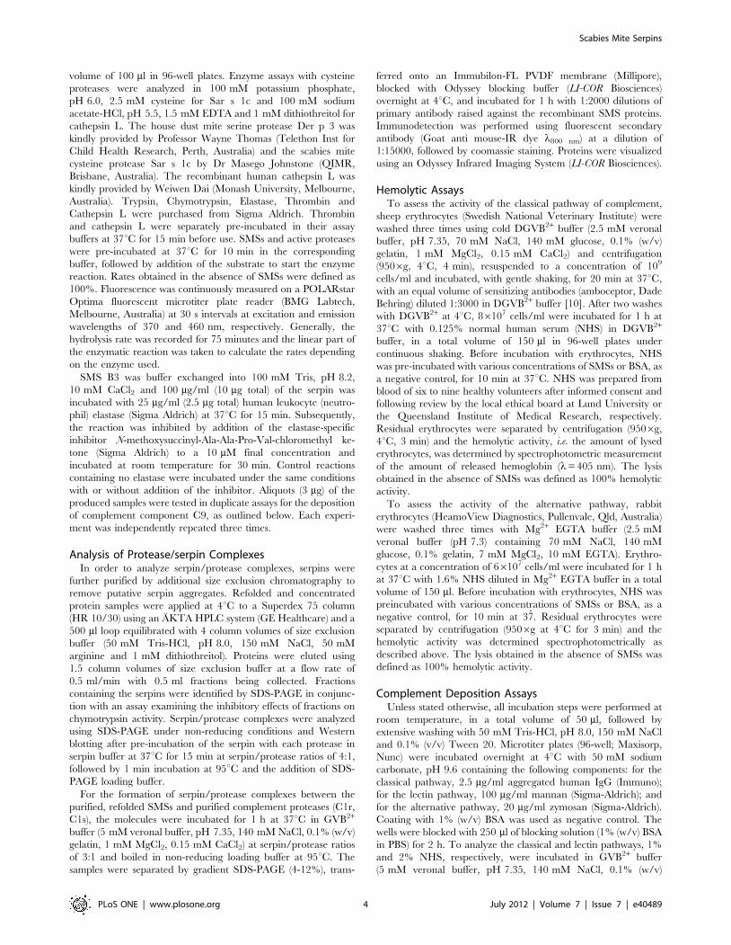
within the RCL hinge or RCL variable regions (Fig. 1A).
All proteins investigated in this study were expressed in E. coli
with N-terminal hexaHis-tags and purified from thoroughly
washed inclusion bodies under denaturing conditions by affinity
chromatography. Subsequent refolding in presence of L-arginine
and dithiothreitol yielded an average of 6 mg SMSB3 and 160 mg
SMSB4 per liter of induced culture, i.e. 20–30% of the purified
proteins were refolded into soluble proteins. The purified proteins
exhibited the expected molecular masses of 47 and 54 kDa,
respectively (Fig. 1B). The six mutated serpins were successfully
produced following the same procedure and showed identical
molecular masses and purity compared to the corresponding wild
type SMSs on SDS-PAGE (data not shown).
SMSs are Localized in the Mite Digestive System andExcreted into the Epidermis
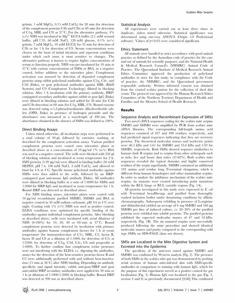
The specificity of the anti-sera raised against SMSB3 and
SMSB4 was confirmed by Western analysis (Fig. 2). The presence
of both SMSs in the scabies mite gut was demonstrated by probing
serial sections of human mite-infested skin with SMS-specific
antibodies in comparison to staining with human IgG, which for
the purpose of this experiment served as a positive control for gut
localization (Fig. 3). Human IgG was localized to the gut (Fig. 3,
section 4 and 8) as previously documented [9,68].This confirmed
Scabies Mite Serpins
PLoS ONE | www.plosone.org 5 July 2012 | Volume 7 | Issue 7 | e40489
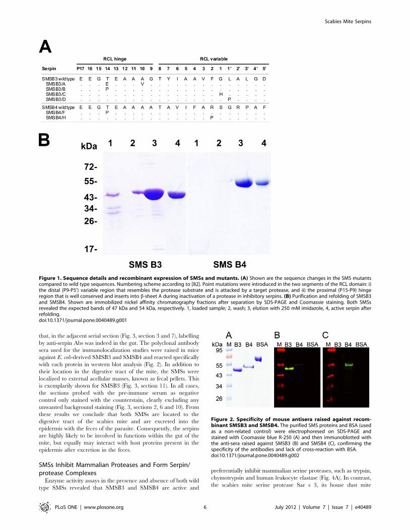
that, in the adjacent serial section (Fig. 3, section 3 and 7), labelling
by anti-serpin Abs was indeed in the gut. The polyclonal antibody
sera used for the immunolocalization studies were raised in mice
against E. coli-derived SMSB3 and SMSB4 and reacted specifically
with each protein in western blot analysis (Fig. 2). In addition to
their location in the digestive tract of the mite, the SMSs were
localized to external acellular masses, known as fecal pellets. This
is exemplarily shown for SMSB3 (Fig. 3, section 11). In all cases,
the sections probed with the pre-immune serum as negative
control only stained with the counterstain, clearly excluding any
unwanted background staining (Fig. 3, sections 2, 6 and 10). From
these results we conclude that both SMSs are located to the
digestive tract of the scabies mite and are excreted into the
epidermis with the feces of the parasite. Consequently, the serpins
are highly likely to be involved in functions within the gut of the
mite, but equally may interact with host proteins present in the
epidermis after excretion in the feces.
SMSs Inhibit Mammalian Proteases and Form Serpin/protease Complexes
Enzyme activity assays in the presence and absence of both wild
type SMSs revealed that SMSB3 and SMSB4 are active and
preferentially inhibit mammalian serine proteases, such as trypsin,
chymotrypsin and human leukocyte elastase (Fig. 4A). In contrast,
the scabies mite serine protease Sar s 3, its house dust mite
Figure 1. Sequence details and recombinant expression of SMSs and mutants. (A) Shown are the sequence changes in the SMS mutantscompared to wild type sequences. Numbering scheme according to [82]. Point mutations were introduced in the two segments of the RCL domain: i)the distal (P9-P5’) variable region that resembles the protease substrate and is attacked by a target protease, and ii) the proximal (P15-P9) hingeregion that is well conserved and inserts into b-sheet A during inactivation of a protease in inhibitory serpins. (B) Purification and refolding of SMSB3and SMSB4. Shown are immobilized nickel affinity chromatography fractions after separation by SDS-PAGE and Coomassie staining. Both SMSsrevealed the expected bands of 47 kDa and 54 kDa, respectively. 1, loaded sample; 2, wash; 3, elution with 250 mM imidazole, 4, active serpin afterrefolding.doi:10.1371/journal.pone.0040489.g001
Figure 2. Specificity of mouse antisera raised against recom-binant SMSB3 and SMSB4. The purified SMS proteins and BSA (usedas a non-related control) were electrophoresed on SDS-PAGE andstained with Coomassie blue R-250 (A) and then immunoblotted withthe anti-sera raised against SMSB3 (B) and SMSB4 (C), confirming thespecificity of the antibodies and lack of cross-reaction with BSA.doi:10.1371/journal.pone.0040489.g002
Scabies Mite Serpins
PLoS ONE | www.plosone.org 6 July 2012 | Volume 7 | Issue 7 | e40489
homolog Der p 3 and the cysteine proteases Sar s 1c and human
cathepsin L were not inhibited by either SMS. Values for the
stoichiometry of inhibition (SI) of the three mammalian proteases
by SMSB3 (Fig. 4B) were elucidated using linear regression to
determine the inhibitor/enzyme ratio ([I]0/[E]0) resulting in the
complete inhibition of the enzyme. While trypsin and elastase
inhibition by SMSB3 and SMSB4 were inefficient, the SI value
calculated for SMSB3 inhibition of chymotrypsin was 12.9,
indicating that this serpin inhibited chymotrypsin-like proteases
most effectively.
Serpin/protease complexes formed by the classical mechanism
involving RCL cleavage and formation of an acyl-enzyme
intermediate are heat and SDS-stable and hence can be visualized
after SDS-PAGE. Such analysis of SMSB3/protease complexes
confirmed that the inhibition of chymotrypsin was due to the
formation of a covalent bond between the enzyme and the
inhibitor (Fig. 4C). In contrast, the slight inhibition of trypsin and
elastase activity in presence of SMSB3 apparently occurred due to
the serpin acting as an alternative substrate for the enzymes, based
on the degradation products visible on SDS-PAGE analysis.
Scabies mite serine protease Sar s 3 did not affect the scabies mite
serpin (Fig. 4C). Western blotting of the SDS-PAGE gels after
complex formation and immunodetection with antibodies against
SMSB3 confirmed that the high molecular mass complex band
formed contained SMSB3 (data not shown).
SMSB3/A and SMSB3/B showed strongly impaired inhibitory
activity against chymotrypsin in comparison with the wild type
serpin (Fig. 4D). These mutants contain point mutations at P14 in
the RCL-associated hinge region of SMSB3 (Fig. 1A), indicating
that this region, which is integral to the conformational change
occurring during the serpin mechanism, was indeed involved in
the inhibition of this protease.
SMSsB3 and SMSB4 do not Inhibit Blood CoagulationWe tested the functionality of the intrinsic and the extrinsic
coagulation pathways, involving multiple serine proteases, in the
presence of the two recombinant mite serine protease inhibitors
SMSB3 and SMSB4. Two standardized tests, the Activated Partial
Thromboplastin Time (APTT) and the Prothrombin Time (PT)
were assessed. Both SMSs had no effect on either pathway when
tested at concentrations of 10, 100 and 200 mg/ml (215 nM,
2.1 mM and 4.2 mM for SMSB3 and 186 nM, 1.8 mM and 3.6 mM
for SMSB4) (Fig. S1).
SMSs inhibit the hemolytic activity of human serumA detailed functional analysis was performed to investigate the
anti-complement properties of the scabies mite serpins. Both SMSs
inhibited human complement in a hemolytic assay measuring the
activation of the classical pathway, while the six mutants (Fig. 1A)
showed distinct changes in inhibition (Fig. 5). SMSB3 and SMSB4
Figure 3. Immunolocalization of SMSs and IgG in scabies mite-infested human skin. Schematic diagrams 1 and 5 outline the featuresvisible in serial histological sections through a scabies mite in infested human skin. Red staining indicates binding of antibody to protein. Section 2and 6 probed with pre-immune mouse serum as a negative control remained unstained, while equivalent regions were detected in section 4 and 8when probed with anti-human IgG, a marker for mite gut [9], and in section 3 and 7 when probed with antibodies against SMSB4 and SMSB3,respectively. Schematic diagrams outline the features visible in serial histological sections through human epidermis containing scabies mite feces 9.Section 10 probed with pre-immune mouse serum as a negative control remained unstained, while equivalent regions were detected in section 11when probed with anti-SMSB3 and in section 12 with anti-IgG. Staining of feces was also seen in equivalent experiments performed with antibodiesagainst SMSB4 (not shown). Immunohistological co-localization of SMSs and host IgG indicated that both serpins are localized in the mite gut andmite feces within the burrows. b, burrow; f, feces; g, gut; m, mite. Scale bars (100 mM) indicate the magnification level.doi:10.1371/journal.pone.0040489.g003
Scabies Mite Serpins
PLoS ONE | www.plosone.org 7 July 2012 | Volume 7 | Issue 7 | e40489
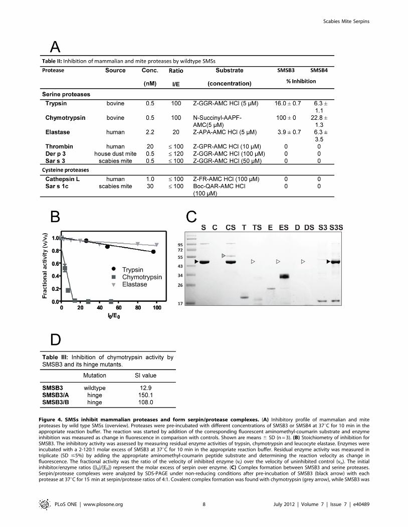
Figure 4. SMSs inhibit mammalian proteases and form serpin/protease complexes. (A) Inhibitory profile of mammalian and miteproteases by wild type SMSs (overview). Proteases were pre-incubated with different concentrations of SMSB3 or SMSB4 at 37uC for 10 min in theappropriate reaction buffer. The reaction was started by addition of the corresponding fluorescent aminomethyl-coumarin substrate and enzymeinhibition was measured as change in fluorescence in comparison with controls. Shown are means 6 SD (n = 3). (B) Stoichiometry of inhibition forSMSB3. The inhibitory activity was assessed by measuring residual enzyme activities of trypsin, chymotrypsin and leucocyte elastase. Enzymes wereincubated with a 2-120:1 molar excess of SMSB3 at 37uC for 10 min in the appropriate reaction buffer. Residual enzyme activity was measured intriplicate (SD #5%) by adding the appropriate aminomethyl-coumarin peptide substrate and determining the reaction velocity as change influorescence. The fractional activity was the ratio of the velocity of inhibited enzyme (vi) over the velocity of uninhibited control (vo). The initialinhibitor/enzyme ratios ([I0]/[E0]) represent the molar excess of serpin over enzyme. (C) Complex formation between SMSB3 and serine proteases.Serpin/protease complexes were analyzed by SDS-PAGE under non-reducing conditions after pre-incubation of SMSB3 (black arrow) with eachprotease at 37uC for 15 min at serpin/protease ratios of 4:1. Covalent complex formation was found with chymotrypsin (grey arrow), while SMSB3 was
Scabies Mite Serpins
PLoS ONE | www.plosone.org 8 July 2012 | Volume 7 | Issue 7 | e40489
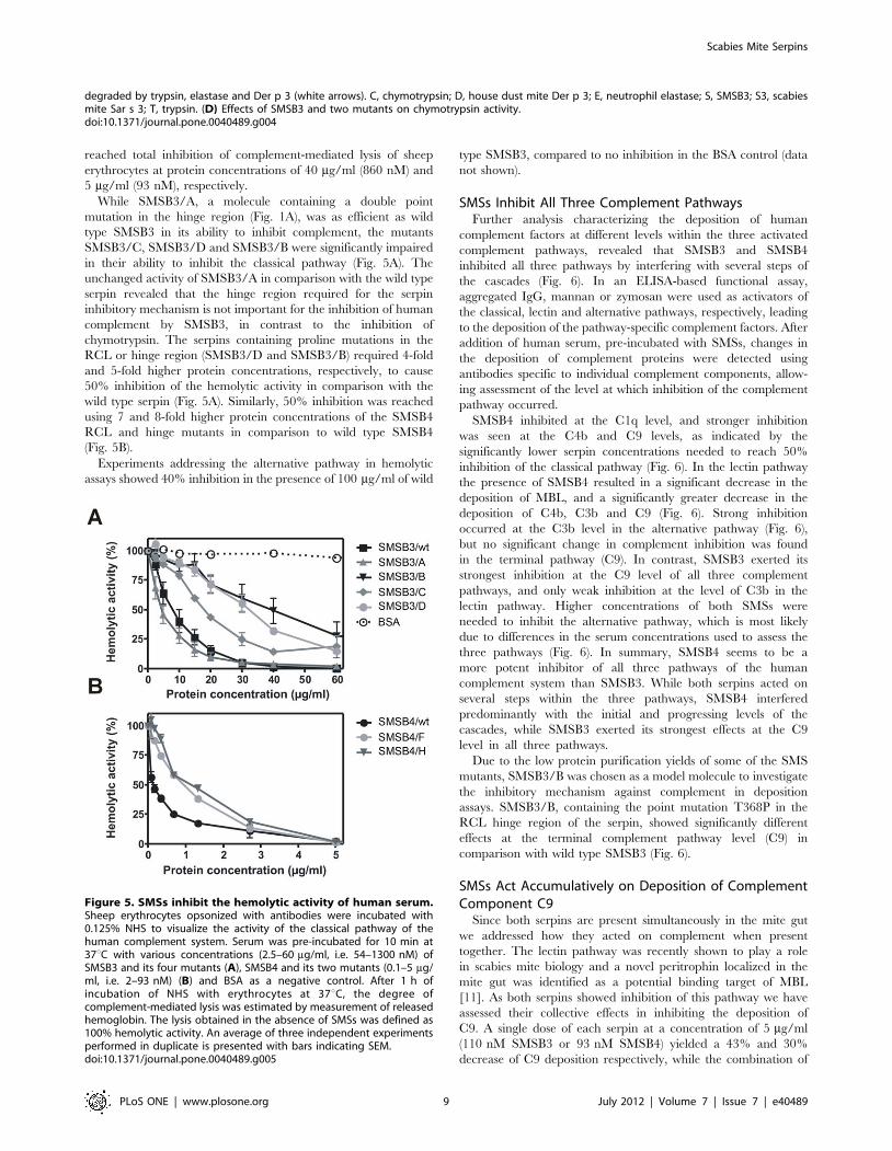
reached total inhibition of complement-mediated lysis of sheep
erythrocytes at protein concentrations of 40 mg/ml (860 nM) and
5 mg/ml (93 nM), respectively.
While SMSB3/A, a molecule containing a double point
mutation in the hinge region (Fig. 1A), was as efficient as wild
type SMSB3 in its ability to inhibit complement, the mutants
SMSB3/C, SMSB3/D and SMSB3/B were significantly impaired
in their ability to inhibit the classical pathway (Fig. 5A). The
unchanged activity of SMSB3/A in comparison with the wild type
serpin revealed that the hinge region required for the serpin
inhibitory mechanism is not important for the inhibition of human
complement by SMSB3, in contrast to the inhibition of
chymotrypsin. The serpins containing proline mutations in the
RCL or hinge region (SMSB3/D and SMSB3/B) required 4-fold
and 5-fold higher protein concentrations, respectively, to cause
50% inhibition of the hemolytic activity in comparison with the
wild type serpin (Fig. 5A). Similarly, 50% inhibition was reached
using 7 and 8-fold higher protein concentrations of the SMSB4
RCL and hinge mutants in comparison to wild type SMSB4
(Fig. 5B).
Experiments addressing the alternative pathway in hemolytic
assays showed 40% inhibition in the presence of 100 mg/ml of wild
type SMSB3, compared to no inhibition in the BSA control (data
not shown).
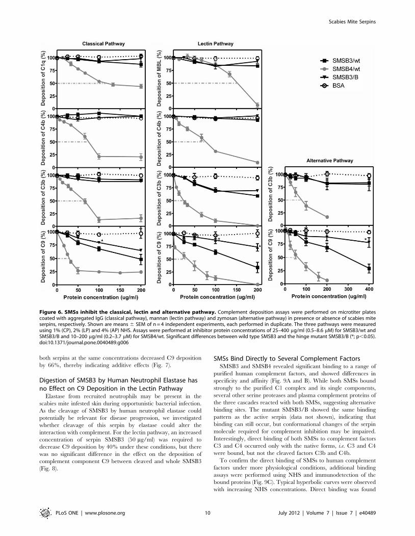
SMSs Inhibit All Three Complement PathwaysFurther analysis characterizing the deposition of human
complement factors at different levels within the three activated
complement pathways, revealed that SMSB3 and SMSB4
inhibited all three pathways by interfering with several steps of
the cascades (Fig. 6). In an ELISA-based functional assay,
aggregated IgG, mannan or zymosan were used as activators of
the classical, lectin and alternative pathways, respectively, leading
to the deposition of the pathway-specific complement factors. After
addition of human serum, pre-incubated with SMSs, changes in
the deposition of complement proteins were detected using
antibodies specific to individual complement components, allow-
ing assessment of the level at which inhibition of the complement
pathway occurred.
SMSB4 inhibited at the C1q level, and stronger inhibition
was seen at the C4b and C9 levels, as indicated by the
significantly lower serpin concentrations needed to reach 50%
inhibition of the classical pathway (Fig. 6). In the lectin pathway
the presence of SMSB4 resulted in a significant decrease in the
deposition of MBL, and a significantly greater decrease in the
deposition of C4b, C3b and C9 (Fig. 6). Strong inhibition
occurred at the C3b level in the alternative pathway (Fig. 6),
but no significant change in complement inhibition was found
in the terminal pathway (C9). In contrast, SMSB3 exerted its
strongest inhibition at the C9 level of all three complement
pathways, and only weak inhibition at the level of C3b in the
lectin pathway. Higher concentrations of both SMSs were
needed to inhibit the alternative pathway, which is most likely
due to differences in the serum concentrations used to assess the
three pathways (Fig. 6). In summary, SMSB4 seems to be a
more potent inhibitor of all three pathways of the human
complement system than SMSB3. While both serpins acted on
several steps within the three pathways, SMSB4 interfered
predominantly with the initial and progressing levels of the
cascades, while SMSB3 exerted its strongest effects at the C9
level in all three pathways.
Due to the low protein purification yields of some of the SMS
mutants, SMSB3/B was chosen as a model molecule to investigate
the inhibitory mechanism against complement in deposition
assays. SMSB3/B, containing the point mutation T368P in the
RCL hinge region of the serpin, showed significantly different
effects at the terminal complement pathway level (C9) in
comparison with wild type SMSB3 (Fig. 6).
SMSs Act Accumulatively on Deposition of ComplementComponent C9
Since both serpins are present simultaneously in the mite gut
we addressed how they acted on complement when present
together. The lectin pathway was recently shown to play a role
in scabies mite biology and a novel peritrophin localized in the
mite gut was identified as a potential binding target of MBL
[11]. As both serpins showed inhibition of this pathway we have
assessed their collective effects in inhibiting the deposition of
C9. A single dose of each serpin at a concentration of 5 mg/ml
(110 nM SMSB3 or 93 nM SMSB4) yielded a 43% and 30%
decrease of C9 deposition respectively, while the combination of
degraded by trypsin, elastase and Der p 3 (white arrows). C, chymotrypsin; D, house dust mite Der p 3; E, neutrophil elastase; S, SMSB3; S3, scabiesmite Sar s 3; T, trypsin. (D) Effects of SMSB3 and two mutants on chymotrypsin activity.doi:10.1371/journal.pone.0040489.g004
Figure 5. SMSs inhibit the hemolytic activity of human serum.Sheep erythrocytes opsonized with antibodies were incubated with0.125% NHS to visualize the activity of the classical pathway of thehuman complement system. Serum was pre-incubated for 10 min at37uC with various concentrations (2.5–60 mg/ml, i.e. 54–1300 nM) ofSMSB3 and its four mutants (A), SMSB4 and its two mutants (0.1–5 mg/ml, i.e. 2–93 nM) (B) and BSA as a negative control. After 1 h ofincubation of NHS with erythrocytes at 37uC, the degree ofcomplement-mediated lysis was estimated by measurement of releasedhemoglobin. The lysis obtained in the absence of SMSs was defined as100% hemolytic activity. An average of three independent experimentsperformed in duplicate is presented with bars indicating SEM.doi:10.1371/journal.pone.0040489.g005
Scabies Mite Serpins
PLoS ONE | www.plosone.org 9 July 2012 | Volume 7 | Issue 7 | e40489
both serpins at the same concentrations decreased C9 deposition
by 66%, thereby indicating additive effects (Fig. 7).
Digestion of SMSB3 by Human Neutrophil Elastase hasno Effect on C9 Deposition in the Lectin Pathway
Elastase from recruited neutrophils may be present in the
scabies mite infested skin during opportunistic bacterial infection.
As the cleavage of SMSB3 by human neutrophil elastase could
potentially be relevant for disease progression, we investigated
whether cleavage of this serpin by elastase could alter the
interaction with complement. For the lectin pathway, an increased
concentration of serpin SMSB3 (50 mg/ml) was required to
decrease C9 deposition by 40% under these conditions, but there
was no significant difference in the effect on the deposition of
complement component C9 between cleaved and whole SMSB3
(Fig. 8).
SMSs Bind Directly to Several Complement FactorsSMSB3 and SMSB4 revealed significant binding to a range of
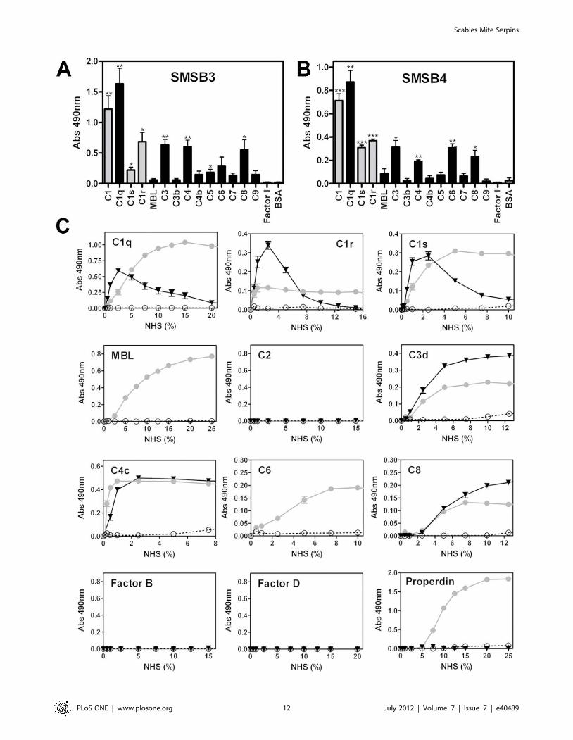
purified human complement factors, and showed differences in
specificity and affinity (Fig. 9A and B). While both SMSs bound
strongly to the purified C1 complex and its single components,
several other serine proteases and plasma complement proteins of
the three cascades reacted with both SMSs, suggesting alternative
binding sites. The mutant SMSB3/B showed the same binding
pattern as the active serpin (data not shown), indicating that
binding can still occur, but conformational changes of the serpin
molecule required for complement inhibition may be impaired.
Interestingly, direct binding of both SMSs to complement factors
C3 and C4 occurred only with the native forms, i.e. C3 and C4
were bound, but not the cleaved factors C3b and C4b.
To confirm the direct binding of SMSs to human complement
factors under more physiological conditions, additional binding
assays were performed using NHS and immunodetection of the
bound proteins (Fig. 9C). Typical hyperbolic curves were observed
with increasing NHS concentrations. Direct binding was found
Figure 6. SMSs inhibit the classical, lectin and alternative pathway. Complement deposition assays were performed on microtiter platescoated with aggregated IgG (classical pathway), mannan (lectin pathway) and zymosan (alternative pathway) in presence or absence of scabies miteserpins, respectively. Shown are means 6 SEM of n = 4 independent experiments, each performed in duplicate. The three pathways were measuredusing 1% (CP), 2% (LP) and 4% (AP) NHS. Assays were performed at inhibitor protein concentrations of 25–400 mg/ml (0.5–8.6 mM) for SMSB3/wt andSMSB3/B and 10–200 mg/ml (0.2–3.7 mM) for SMSB4/wt. Significant differences between wild type SMSB3 and the hinge mutant SMSB3/B (*; p,0.05).doi:10.1371/journal.pone.0040489.g006
Scabies Mite Serpins
PLoS ONE | www.plosone.org 10 July 2012 | Volume 7 | Issue 7 | e40489
between SMSB4 and the serine proteases C1r and C1s,
respectively (Fig. 9C). In addition, SMSB4 bound to seven other
complement factors (C1q, MBL, properdin, C3, C4, C6, and C8;
Fig. 9C). The mechanism by which the serpin binds these non-
protease factors is unknown, but the data confirm the inhibition of
the complement pathways at many levels, including the start of the
cascades (Fig. 6).
Low amounts of non-specific binding occurred between SMSB3
and the C1 complement components at low NHS concentrations
(Fig. 9C). These were not seen at higher concentrations.
Experiments using NHS confirmed the observed inhibitory effects
on the terminal pathway by revealing typical binding curves for
three complement factors (C3, C4, C8; Fig. 9C) indicating high
affinity binding for these proteins. None of these factors have a
serine protease function.
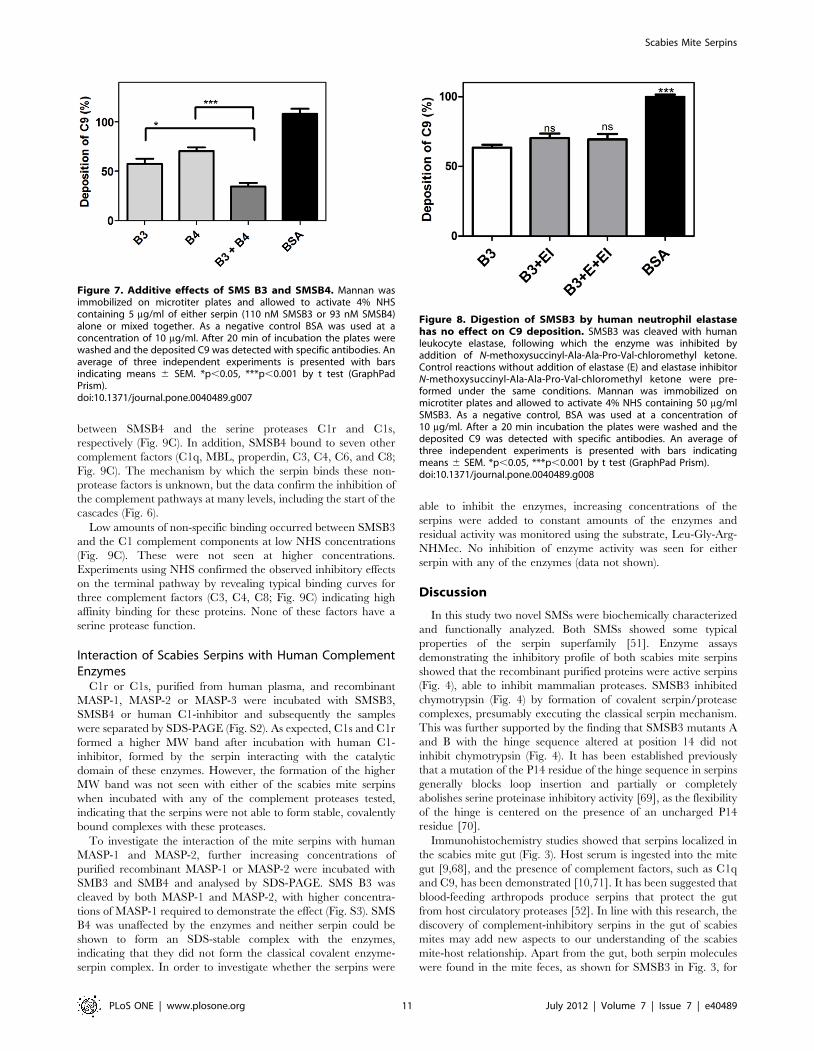
Interaction of Scabies Serpins with Human ComplementEnzymes
C1r or C1s, purified from human plasma, and recombinant
MASP-1, MASP-2 or MASP-3 were incubated with SMSB3,
SMSB4 or human C1-inhibitor and subsequently the samples
were separated by SDS-PAGE (Fig. S2). As expected, C1s and C1r
formed a higher MW band after incubation with human C1-
inhibitor, formed by the serpin interacting with the catalytic
domain of these enzymes. However, the formation of the higher
MW band was not seen with either of the scabies mite serpins
when incubated with any of the complement proteases tested,
indicating that the serpins were not able to form stable, covalently
bound complexes with these proteases.
To investigate the interaction of the mite serpins with human
MASP-1 and MASP-2, further increasing concentrations of
purified recombinant MASP-1 or MASP-2 were incubated with
SMB3 and SMB4 and analysed by SDS-PAGE. SMS B3 was
cleaved by both MASP-1 and MASP-2, with higher concentra-
tions of MASP-1 required to demonstrate the effect (Fig. S3). SMS
B4 was unaffected by the enzymes and neither serpin could be
shown to form an SDS-stable complex with the enzymes,
indicating that they did not form the classical covalent enzyme-
serpin complex. In order to investigate whether the serpins were
able to inhibit the enzymes, increasing concentrations of the
serpins were added to constant amounts of the enzymes and
residual activity was monitored using the substrate, Leu-Gly-Arg-
NHMec. No inhibition of enzyme activity was seen for either
serpin with any of the enzymes (data not shown).
Discussion
In this study two novel SMSs were biochemically characterized
and functionally analyzed. Both SMSs showed some typical
properties of the serpin superfamily [51]. Enzyme assays
demonstrating the inhibitory profile of both scabies mite serpins
showed that the recombinant purified proteins were active serpins
(Fig. 4), able to inhibit mammalian proteases. SMSB3 inhibited
chymotrypsin (Fig. 4) by formation of covalent serpin/protease
complexes, presumably executing the classical serpin mechanism.
This was further supported by the finding that SMSB3 mutants A
and B with the hinge sequence altered at position 14 did not
inhibit chymotrypsin (Fig. 4). It has been established previously
that a mutation of the P14 residue of the hinge sequence in serpins
generally blocks loop insertion and partially or completely
abolishes serine proteinase inhibitory activity [69], as the flexibility
of the hinge is centered on the presence of an uncharged P14
residue [70].
Immunohistochemistry studies showed that serpins localized in
the scabies mite gut (Fig. 3). Host serum is ingested into the mite
gut [9,68], and the presence of complement factors, such as C1q
and C9, has been demonstrated [10,71]. It has been suggested that
blood-feeding arthropods produce serpins that protect the gut
from host circulatory proteases [52]. In line with this research, the
discovery of complement-inhibitory serpins in the gut of scabies
mites may add new aspects to our understanding of the scabies
mite-host relationship. Apart from the gut, both serpin molecules
were found in the mite feces, as shown for SMSB3 in Fig. 3, for
Figure 7. Additive effects of SMS B3 and SMSB4. Mannan wasimmobilized on microtiter plates and allowed to activate 4% NHScontaining 5 mg/ml of either serpin (110 nM SMSB3 or 93 nM SMSB4)alone or mixed together. As a negative control BSA was used at aconcentration of 10 mg/ml. After 20 min of incubation the plates werewashed and the deposited C9 was detected with specific antibodies. Anaverage of three independent experiments is presented with barsindicating means 6 SEM. *p,0.05, ***p,0.001 by t test (GraphPadPrism).doi:10.1371/journal.pone.0040489.g007
Figure 8. Digestion of SMSB3 by human neutrophil elastasehas no effect on C9 deposition. SMSB3 was cleaved with humanleukocyte elastase, following which the enzyme was inhibited byaddition of N-methoxysuccinyl-Ala-Ala-Pro-Val-chloromethyl ketone.Control reactions without addition of elastase (E) and elastase inhibitorN-methoxysuccinyl-Ala-Ala-Pro-Val-chloromethyl ketone were pre-formed under the same conditions. Mannan was immobilized onmicrotiter plates and allowed to activate 4% NHS containing 50 mg/mlSMSB3. As a negative control, BSA was used at a concentration of10 mg/ml. After a 20 min incubation the plates were washed and thedeposited C9 was detected with specific antibodies. An average ofthree independent experiments is presented with bars indicatingmeans 6 SEM. *p,0.05, ***p,0.001 by t test (GraphPad Prism).doi:10.1371/journal.pone.0040489.g008
Scabies Mite Serpins
PLoS ONE | www.plosone.org 11 July 2012 | Volume 7 | Issue 7 | e40489
Scabies Mite Serpins
PLoS ONE | www.plosone.org 12 July 2012 | Volume 7 | Issue 7 | e40489
example. As house dust mite allergens are known to be highly
active proteases after excretion [72,73] scabies mite gut molecules
may be equally functional externally after being released into the
upper epidermis, as well as internally within the gut.
SMSB4 exhibited effects as early as the initial and progressing
steps of the complement cascades, while SMSB3 showed the
strongest effects on the terminal pathway. Differences between
protein concentrations needed in hemolytic and complement
deposition assays were based on differences in NHS concentrations
and other assay conditions. C1q is a pattern recognition molecule,
which activates the classical pathway and assembles into a
complex with its associated serine proteases, C1s and C1r [7].
The strong binding of SMSB4 to the components of the C1
complex (C1q, C1s and C1r; Figs. 9B and C) corresponds with the
inhibitory effects at the C1q level demonstrated in deposition
assays (Fig. 6). In contrast, the presumably non-specific binding
between C1q and SMSB3 at low NHS concentrations (Fig. 9C)
did not inhibit complement deposition (Fig. 6). Proteins binding to
the complement factor C1q without affecting its function, or
causing activation of complement instead, have been previously
described [74–76].
C3 acts as the central molecule of the alternative pathway. Its
hydrolyzed form is activated by the serine proteases B and D
generating an initial C3 convertase, which cleaves C3 into C3a
and C3b [7]. Properdin recognizes pathogen- or damage-
associated molecular patterns and is involved in the initiation of
the alternative pathway and the stabilization of AP convertases
[77]. Thus, the binding of C3 and properdin by SMSB4 correlates
with the strong inhibition at the C3b level in the alternative
pathway (Fig. 6).
In contrast, SMSB3 only targeted non-serine proteases in
human serum (Fig. 9C) and exhibited its strongest effect at the C9
level of the three pathways (Fig. 6) by interaction with C8, which
takes part in the assembly of the terminal membrane attack
complex along with the factors C5b, C6, C7 and C9.
An increasing number of serpins have been found to be non-
inhibitory towards proteases and show alternative binding
mechanisms [51,52,78–80]. The changes in anti-complement
activity of the mutants, in comparison with the wild type SMSs
(Fig. 5), and the binding of both SMSs to complement factors
without protease function (Fig. 9), suggested that possibly one or
several exosites might be used to inhibit these proteins. While the
ability of both SMSB3 hinge mutants A and B to inhibit bovine
chymotrypsin in enzyme assays was impaired, most likely by
inhibition of the serpin mechanism, the anti-complement activity
of the hinge mutant SMSB3/A was not changed. In contrast, the
hinge mutants SMSB3/B and SMSB4/F showed impaired anti-
complement activity (Fig. 5), suggesting that domains outside the
hinge region may have to be structurally changed to impair the
anti-complement function of the serpins. In the SMSB3/B mutant,
a proline residue was inserted into the hinge region. Proline
insertions often disrupt the secondary structure and thus misfold-
ing of the protein can occur and/or conformational changes
necessary for binding or inhibitory mechanisms elsewhere in the
molecule can be affected. No complex formation was seen with
either of the scabies mite serpins when incubated with any of the
complement proteases C1s, C1r, MASP-1, MASP-2 and MASP-3
tested, indicating that the mite serpins were not able to form
stable, covalently bound complexes with these proteases. Thus, it
appears that the effect of the mite serpins is not due to classical
serpin inhibition of the serine proteases of the complement
cascade. Based on the data presented here, it can be hypothesized
that both the complement proteases tested and the non-proteolytic
complement factors likely bind to an exosite of the SMSs or that
the SMSs bind to exosites on the proteases, causing these proteins
to be sterically inhibited from completing further binding
interactions required for complement activation. However, the
potential alternative binding mechanisms of the serpins to the
complement proteases and factors have yet to be elucidated.
SMSs may act in concert with each other (Fig. 7) and with the
catalytically inactive scabies mite serine proteases [10], which also
inhibit human complement. It appears that the scabies mite has
evolved a multitude of mechanisms to ensure human complement
inactivation at all pathway levels, similarly to the broad range of
anti-complement factors evolved by other pathogens [17,81]. We
hypothesize that the compilation of mite complement inhibitors
accumulates to high anti-complement activities in the confined
space of the gut and epidermal burrows. Importantly, while
complement factors are ingested by mites infesting human skin,
MAC formation is not detected in the gut [71], suggesting that this
anti-complement machinery may be very efficient in vivo. While
prevention of gut lysis seems to be the obvious role of intestinal
mite complement inhibitors, they may also act external to the mite
and their presence in the epidermis may possibly have further
consequences for the host. We have previously proposed that mite
excretory proteins may effectively enhance the survival of scabies
associated pathogenic bacteria by interfering locally with host
complement [10]. The serpins described here may take part in this
role. Further studies are needed to elucidate this hypothetical
model of host, parasite and bacteria interactions in order to further
the development of novel preventative and therapeutic strategies.
Supporting Information
Figure S1 Scabies mite serpins SMSB3 and SMSB4 donot interfere with the coagulation pathway. The function-
ality of the intrinsic and the extrinsic coagulation pathways in the
presence of the two recombinant serine protease inhibitors,
SMSB3 and SMSB4, was assessed by measuring the Activated
Partial Thromboplastin Time (APTT) and the Prothrombin Time
(PT). The dotted boxes represent reference ranges for clotting
times of healthy donors.
(PDF)
Figure S2 Interaction of scabies mite serpins withhuman complement enzymes. C1r (hC1r) or C1s (hC1s)
purified from human plasma and recombinant MASP-1 (M1),
MASP-2 (M2) or MASP-3 (M3) were incubated with SMSB3 (B3),
SMSB4 (B4) or human C1-inhibitor (hC1i) for 1 h at room
Figure 9. Direct binding of SMSs to various complement proteins. Microtiter plates were coated with various purified human complementproteins or BSA as a negative control and incubated with 20 mg/ml SMSB3 (A) or SMSB4 (B). Bound serpins were detected using specific polyclonalantibodies against SMSB3 and SMSB4. Shown are means 6 SEM of n = 3 independent experiments. The statistical significance of differences betweenBSA and the rest of the data groups was estimated using one-way ANOVA. *, p,0.05; **, p,0.01; ***, p,0.001. Grey, serine proteases; black, othercomplement factors. C Direct binding of complement factors from NHS to SMSs. Increasing concentrations of NHS were added to wells coated withSMSB3 (., black), SMSB4 (N, grey) or BSA as a negative control (#, white) and bound complement factors were detected by specific antibodies.Shown are means 6 SEM of n = 3 independent experiments measured in duplicates. NHS was tested from 0–100%. Positive controls were used forcomplement proteins, where no binding to the SMSs was detectable confirming strong immunodetection of the complement factor on 1% NHScoating.doi:10.1371/journal.pone.0040489.g009
Scabies Mite Serpins
PLoS ONE | www.plosone.org 13 July 2012 | Volume 7 | Issue 7 | e40489
temperature. Samples were electrophoresed on 10% SDS-PAGE
as indicated in the labels above each lane. The positions of the
catalytic chains of the enzymes are indicated [e.g. C1r (c)]. The
positions of the complexes between hC1r and hC1s and hC1i are
also indicated.
(PDF)
Figure S3 Interaction of scabies mite serpins withhuman MASP-1 and MASP-2. Increasing concentrations of
purified recombinant MASP-1 (M1) or MASP-2 (M2) were
incubated with SMSB3 (B3) and SMSB4 (B4) for 1 h at room
temperature. Samples were separated on 10% SDS-PAGE as
indicated in the labels above each lane shown. Molecular weight
markers (Precision Plus ProteinTM Dual Colour Standard, BIO
RAD) are shown in the first lane at the left of each SDS-PAGE gel.
(PDF)
Acknowledgments
The authors wish to thank Cassandra Lane and Yonghong Zhou for
excellent technical assistance and Joanne Beggs, Advanced Scientist Core
Coagulation Pathology, Queensland for expertise with coagulation
experiments.
Author Contributions
Conceived and designed the experiments: AM CW RNP AMB DJK KF.
Performed the experiments: AM FCM SLR VH CW DAP PMS LCW KF.
Analyzed the data: AM RNP AMB DJK KF. Contributed reagents/
materials/analysis tools: RNP AMB DJK KF. Wrote the paper: AM RNP
AMB DJK KF.
References
1. Hengge UR, Currie BJ, Jager G, Lupi O, Schwartz RA (2006) Scabies: a
ubiquitous neglected skin disease. The Lancet Infectious Diseases 6: 769–779.
2. Walton SF, Currie BJ (2007) Problems in diagnosing scabies, a global disease in
human and animal populations. Clin Microbiol Rev 20: 268–279.
3. Clucas DB, Carville KS, Connors C, Currie BJ, Carapetis JR, et al. (2008)
Disease burden and health-care clinic attendances for young children in remote
aboriginal communities of northern Australia. Bull World Health Organ 86:
275–281.
4. Brook I (1995) Microbiology of secondary bacterial infection in scabies lesions.
J Clin Microbiol 33: 2139–2140.
5. McDonald M, Currie BJ, Carapetis JR (2004) Acute rheumatic fever: a chink in
the chain that links the heart to the throat? Lancet Infect Dis 4: 240–245.
6. Mounsey KE, Holt DC, McCarthy J, Currie BJ, Walton SF (2008) Scabies:
molecular perspectives and therapeutic implications in the face of emerging drug
resistance. Future Microbiology 3: 57–66.
7. Ricklin D, Hajishengallis G, Yang K, Lambris JD (2010) Complement: a key
system for immune surveillance and homeostasis. Nat Immunol 11: 785–797.
8. Beckham SA, Boyd SE, Reynolds S, Willis C, Johnstone M, et al. (2009)
Characterization of a serine protease homologous to house dust mite group 3
allergens from the scabies mite Sarcoptes scabiei. J Biol Chem 284: 34413–
34422.
9. Rapp CM, Morgan MS, Arlian LG (2006) Presence of host immunoglobulin in
the gut of Sarcoptes scabiei (Acari: Sarcoptidae). J Med Entomol 43: 539–542.
10. Bergstrom FC, Reynolds S, Johnstone M, Pike RN, Buckle AM, et al. (2009)
Scabies mite inactivated serine protease paralogs inhibit the human complement
system. J Immunol 182: 7809–7817.
11. Mika A, Goh P, Holt DC, Kemp DJ, Fischer K (2011) Scabies mite peritrophins
are potential targets of human host innate immunity. PLoS Negl Trop Dis 5:
e1331.
12. Holt DC, Fischer K, Allen GE, Wilson D, Wilson P, et al. (2003) Mechanisms
for a novel immune evasion strategy in the scabies mite sarcoptes scabiei: a
multigene family of inactivated serine proteases. J Invest Dermatol 121: 1419–
1424.
13. Fischer K, Langendorf CG, Irving JA, Reynolds S, Willis C, et al. (2009)
Structural mechanisms of inactivation in scabies mite serine protease paralogues.
J Mol Biol 390: 635–645.
14. Zipfel PF, Wurzner R, Skerka C (2007) Complement evasion of pathogens:
common strategies are shared by diverse organisms. Mol Immunol 44: 3850–
3857.
15. Lambris JD, Ricklin D, Geisbrecht BV (2008) Complement evasion by human
pathogens. Nat Rev Microbiol 6: 132–142.
16. Schroeder H, Skelly PJ, Zipfel PF, Losson B, Vanderplasschen A (2009)
Subversion of complement by hematophagous parasites. Dev Comp Immunol
33: 5–13.
17. Skelly PJ (2004) Intravascular schistosomes and complement. Trends Parasitol
20: 370–374.
18. Skelly PJ, Alan Wilson R (2006) Making sense of the schistosome surface. Adv
Parasitol 63: 185–284.
19. Parola P, Raoult D (2006) Tropical rickettsioses. Clin Dermatol 24: 191–200.
20. Parola P, Raoult D (2001) Ticks and tickborne bacterial diseases in humans: an
emerging infectious threat. Clin Infect Dis 32: 897–928.
21. Nicholson WL, Sonenshine DE, Lane RS, Uilenberg G (2009) Ticks (Ixodida.
In: Mullen GR, Durden LA, editors. Medical and Veterinary Entomology. 2nd
edition ed. San Diego, CA: Academic Press. 493–541.
22. Francischetti IM, Sa-Nunes A, Mans BJ, Santos IM, Ribeiro JM (2009) The role
of saliva in tick feeding. Front Biosci 14: 2051–2088.
23. Hovius JW, Levi M, Fikrig E (2008) Salivating for knowledge: potential
pharmacological agents in tick saliva. PLoS Med 5: e43.
24. Steen NA, Barker SC, Alewood PF (2006) Proteins in the saliva of the Ixodida
(ticks): pharmacological features and biological significance. Toxicon 47: 1–20.
25. Valenzuela JG, Francischetti IM, Pham VM, Garfield MK, Mather TN, et al.
(2002) Exploring the sialome of the tick Ixodes scapularis. J Exp Biol 205: 2843–2864.
26. Nene V, Lee D, Quackenbush J, Skilton R, Mwaura S, et al. (2002) AvGI, anindex of genes transcribed in the salivary glands of the ixodid tick Amblyomma
variegatum. Int J Parasitol 32: 1447–1456.
27. Nene V, Lee D, Kang’a S, Skilton R, Shah T, et al. (2004) Genes transcribed in
the salivary glands of female Rhipicephalus appendiculatus ticks infected withTheileria parva. Insect Biochem Mol Biol 34: 1117–1128.
28. Francischetti IM, My Pham V, Mans BJ, Andersen JF, Mather TN, et al. (2005)The transcriptome of the salivary glands of the female western black-legged tick
Ixodes pacificus (Acari: Ixodidae). Insect Biochem Mol Biol 35: 1142–1161.
29. Ribeiro JM, Alarcon-Chaidez F, Francischetti IM, Mans BJ, Mather TN, et al.
(2006) An annotated catalog of salivary gland transcripts from Ixodes scapularisticks. Insect Biochem Mol Biol 36: 111–129.
30. Alarcon-Chaidez FJ, Sun J, Wikel SK (2007) Transcriptome analysis of the
salivary glands of Dermacentor andersoni Stiles (Acari: Ixodidae). Insect
Biochem Mol Biol 37: 48–71.
31. Francischetti IM, Mans BJ, Meng Z, Gudderra N, Veenstra TD, et al. (2008) Aninsight into the sialome of the soft tick, Ornithodorus parkeri. Insect Biochem
Mol Biol 38: 1–21.
32. Mans BJ, Andersen JF, Francischetti IM, Valenzuela JG, Schwan TG, et al.
(2008) Comparative sialomics between hard and soft ticks: implications for theevolution of blood-feeding behavior. Insect Biochem Mol Biol 38: 42–58.
33. Mans BJ, Andersen JF, Schwan TG, Ribeiro JM (2008) Characterization of anti-hemostatic factors in the argasid, Argas monolakensis: implications for the
evolution of blood-feeding in the soft tick family. Insect Biochem Mol Biol 38:22–41.
34. Francischetti IM, Meng Z, Mans BJ, Gudderra N, Hall M, et al. (2008) Aninsight into the salivary transcriptome and proteome of the soft tick and vector of
epizootic bovine abortion, Ornithodoros coriaceus. J Proteomics 71: 493–512.
35. Chmelar J, Anderson JM, Mu J, Jochim RC, Valenzuela JG, et al. (2008) Insight
into the sialome of the castor bean tick, Ixodes ricinus. BMC Genomics 9: 233.
36. Batista IF, Chudzinski-Tavassi AM, Faria F, Simons SM, Barros-Batestti DM, et
al. (2008) Expressed sequence tags (ESTs) from the salivary glands of the tickAmblyomma cajennense (Acari: Ixodidae). Toxicon 51: 823–834.
37. Aljamali MN, Hern L, Kupfer D, Downard S, So S, et al. (2009) Transcriptome
analysis of the salivary glands of the female tick Amblyomma americanum
(Acari: Ixodidae). Insect Mol Biol 18: 129–154.
38. Francischetti IM, Anderson JM, Manoukis N, Pham VM, Ribeiro JM (2011) Aninsight into the sialotranscriptome and proteome of the coarse bontlegged tick,
Hyalomma marginatum rufipes. J Proteomics 74: 2892–2908.
39. Lawrie CH, Sim RB, Nuttall PA (2005) Investigation of the mechanisms of anti-
complement activity in Ixodes ricinus ticks. Mol Immunol 42: 31–38.
40. Nunn MA, Sharma A, Paesen GC, Adamson S, Lissina O, et al. (2005)
Complement Inhibitor of C5 Activation from the Soft Tick Ornithodorosmoubata. J Immunol 174: 2084–2091.
41. Hepburn NJ, Williams AS, Nunn MA, Chamberlain-Banoub JC, Hamer J, et al.(2007) In Vivo Characterization and Therapeutic Efficacy of a C5-specific
Inhibitor from the Soft Tick Ornithodoros moubata. J Biol Chem 282: 8292–8299.
42. Roversi P, Lissina O, Johnson S, Ahmat N, Paesen GC, et al. (2007) Thestructure of OMCI, a novel lipocalin inhibitor of the complement system. J Mol
Biol 369: 784–793.
43. Mans BJ, Ribeiro JM (2008) Function, mechanism and evolution of the
moubatin-clade of soft tick lipocalins. Insect Biochem Mol Biol 38: 841–852.
44. Valenzuela JG, Charlab R, Mather TN, Ribeiro JMC (2000) Purification,Cloning, and Expression of a Novel Salivary Anticomplement Protein from the
Tick, Ixodes scapularis. J Biol Chem 275: 18717–18723.
45. Daix V, Schroeder H, Praet N, Georgin JP, Chiappino I, et al. (2007) Ixodes
ticks belonging to the Ixodes ricinus complex encode a family of anticomplement
proteins. Insect Mol Biol 16: 155–166.
Scabies Mite Serpins
PLoS ONE | www.plosone.org 14 July 2012 | Volume 7 | Issue 7 | e40489
46. Schroeder H, Daix V, Gillet L, Renauld JC, Vanderplasschen A (2007) The
paralogous salivary anti-complement proteins IRAC I and IRAC II encoded byIxodes ricinus ticks have broad and complementary inhibitory activities against
the complement of different host species. Microbes Infect 9: 247–250.
47. Couvreur B, Beaufays J, Charon C, Lahaye K, Gensale F, et al. (2008)Variability and action mechanism of a family of anticomplement proteins in
Ixodes ricinus. PLoS ONE 3: e1400.48. Tyson K, Elkins C, Patterson H, Fikrig E, de Silva A (2007) Biochemical and
functional characterization of Salp20, an Ixodes scapularis tick salivary protein
that inhibits the complement pathway. Insect Mol Biol 16: 469–479.49. Tyson KR, Elkins C, de Silva AM (2008) A novel mechanism of complement
inhibition unmasked by a tick salivary protein that binds to properdin. J Immunol180: 3964–3968.
50. Kerr FK, Thomas AR, Wijeyewickrema LC, Whisstock JC, Boyd SE, et al.(2008) Elucidation of the substrate specificity of the MASP-2 protease of the
lectin complement pathway and identification of the enzyme as a major
physiological target of the serpin, C1-inhibitor. Mol Immunol 45: 670–677.51. Gettins PG, Olson ST (2009) Exosite determinants of serpin specificity. J Biol
Chem 284: 20441–20445.52. Silverman GA, Whisstock JC, Bottomley SP, Huntington JA, Kaiserman D, et
al. (2010) Serpins flex their muscle: I. Putting the clamps on proteolysis in diverse
biological systems. J Biol Chem 285: 24299–24305.53. Mulenga A, Sugino M, Nakajima M, Sugimoto C, Onuma M (2001) Tick
encoded serine proteinase inhibitors (serpin); potential target antigens for tickvaccine development. J Vet Med Sci 63: 1063–1069.
54. Mulenga A, Tsuda A, Onuma M, Sugimoto C (2003) Four serine proteinaseinhibitors (serpin) from the brown ear tick, Rhiphicephalus appendiculatus;
cDNA cloning and preliminary characterization. Insect Biochem Mol Biol 33:
267–276.55. Chalaire KC, Kim TK, Garcia-Rodriguez H, Mulenga A (2011) Amblyomma
americanum (L.) (Acari: Ixodidae) tick salivary gland serine protease inhibitor(serpin) 6 is secreted into tick saliva during tick feeding. J Exp Biol 214: 665–673.
56. Mulenga A, Khumthong R, Chalaire KC (2009) Ixodes scapularis tick serine
proteinase inhibitor (serpin) gene family; annotation and transcriptional analysis.BMC Genomics 10: 217.
57. Mulenga A, Sugimoto C, Ingram G, Ohashi K, Misao O (2001) Characteriza-tion of two cDNAs encoding serine proteinases from the hard tick
Haemaphysalis longicornis. Insect Biochem Mol Biol 31: 817–825.58. Sugino M, Imamura S, Mulenga A, Nakajima M, Tsuda A, et al. (2003) A serine
proteinase inhibitor (serpin) from ixodid tick Haemaphysalis longicornis; cloning
and preliminary assessment of its suitability as a candidate for a tick vaccine.Vaccine 21: 2844–2851.
59. Levi A, Mumcuoglu K, Ingber A, Enk C (2011) Assessment of Sarcoptes scabiei
viability in vivo by reflectance confocal microscopy. Lasers in Medical Science 26:
291–292.
60. Beckham SA, Boyd SE, Reynolds S, Willis C, Johnstone M, et al. (2009)Characterization of a serine protease homologous to house dust mite group 3
allergens from the scabies mite Sarcoptes scabiei. Journal of Biological Chemistry284: 34413–34422.
61. Rapp C, Morgan M, Arlian L (2006) Presence of host immunoglobulin in the gutof Sarcoptes scabiei (Acari: Sarcoptidae). J Med Entomol 43: 539–542.
62. Mika A, Goh P, Holt DC, Kemp DJ, Fischer K (2011) Scabies mite peritrophins
are potential targets of human host innate immunity. PLoS Neglected TropicalDiseases 5: e1331.
63. Australian Government NHMRC (2004) Australian code of practice for the careand use of animals for scientific purposes. 7th edition ed. 1–84.
64. Fischer K, Holt DC, Harumal P, Currie BJ, Walton SF, et al. (2003) Generation
and characterization of cDNA clones from Sarcoptes scabiei var. hominis for an
expressed sequence tag library: identification of homologues of house dust mite
allergens. American Journal of Tropical Medicine and Hygiene 68: 61–64.
65. Bradford MM (1976) Rapid and Sensitive Method for Quantitation of
Microgram Quantities of Protein Utilizing Principle of Protein-Dye Binding.
Analytical Biochemistry 72: 248–254.
66. Tenner AJ, Lesavre PH, Cooper NR (1981) Purification and radiolabeling of
human C1q. J Immunol 127: 648–653.
67. Crossley LG, Porter RR (1980) Purification of the human complement control
protein C3b inactivator. Biochem J 191: 173–182.
68. Willis C, Fischer K, Walton SF, Currie BJ, Kemp DJ (2006) Scabies mite
inactivated serine protease paralogues are present both internally in the mite gut
and externally in feces. Am J Trop Med Hyg 75: 683–687.
69. Lawrence DA, Olson ST, Palaniappan S, Ginsburg D (1994) Serpin reactive
center loop mobility is required for inhibitor function but not for enzyme
recognition. J Biol Chem 269: 27657–27662.
70. Schick C, Bromme D, Bartuski AJ, Uemura Y, Schechter NM, et al. (1998) The
reactive site loop of the serpin SCCA1 is essential for cysteine proteinase
inhibition. Proc Natl Acad Sci U S A 95: 13465–13470.
71. Mika A, Goh P, Holt D, Kemp DJ, Fischer K (2011) Scabies Mite Peritrophins
are Potential Targets of Human Host Innate Immunity. PLoS Negl Trop Dis in
press.
72. Wan H, Winton HL, Soeller C, Tovey ER, Gruenert DC, et al. (1999) Der p 1
facilitates transepithelial allergen delivery by disruption of tight junctions. J Clin
Invest 104: 123–133.
73. Stewart GA, Ward LD, Simpson RJ, Thompson PJ (1992) The group III
allergen from the house dust mite Dermatophagoides pteronyssinus is a trypsin-
like enzyme. Immunology 75: 29–35.
74. Alberti S, Marques G, Camprubi S, Merino S, Tomas JM, et al. (1993) C1q
binding and activation of the complement classical pathway by Klebsiella
pneumoniae outer membrane proteins. Infect Immun 61: 852–860.
75. Sjoberg AP, Manderson GA, Morgelin M, Day AJ, Heinegard D, et al. (2009)
Short leucine-rich glycoproteins of the extracellular matrix display diverse
patterns of complement interaction and activation. Mol Immunol 46: 830–839.
76. Manderson GA, Martin M, Onnerfjord P, Saxne T, Schmidtchen A, et al.
(2009) Interactions of histidine-rich glycoprotein with immunoglobulins and
proteins of the complement system. Mol Immunol 46: 3388–3398.
77. Chen H, Ricklin D, Hammel M, Garcia BL, McWhorter WJ, et al. (2010)
Allosteric inhibition of complement function by a staphylococcal immune
evasion protein. Proc Natl Acad Sci U S A 107: 17621–17626.
78. Higgins WJ, Fox DM, Kowalski PS, Nielsen JE, Worrall DM (2010) Heparin
enhances serpin inhibition of the cysteine protease cathepsin L. J Biol Chem 285:
3722–3729.
79. Zhou A, Wei Z, Stanley PL, Read RJ, Stein PE, et al. (2008) The S-to-R
transition of corticosteroid-binding globulin and the mechanism of hormone
release. J Mol Biol 380: 244–251.
80. Whisstock JC, Silverman GA, Bird PI, Bottomley SP, Kaiserman D, et al. (2010)
Serpins flex their muscle: II. Structural insights into target peptidase recognition,
polymerization, and transport functions. J Biol Chem 285: 24307–24312.
81. Rooijakkers SH, van Strijp JA (2007) Bacterial complement evasion. Mol
Immunol 44: 23–32.
82. Schechter I, Berger A (1967) On the size of the active site in proteases. I. Papain.
Biochem Biophys Res Commun 27: 157–162.
Scabies Mite Serpins
PLoS ONE | www.plosone.org 15 July 2012 | Volume 7 | Issue 7 | e40489
Related Documents