1 23 Molecular Biotechnology Part B of Applied Biochemistry and Biotechnology ISSN 1073-6085 Volume 50 Number 1 Mol Biotechnol (2012) 50:87-97 DOI 10.1007/s12033-011-9418-2 Plant–Pathogen Interactions: What Microarray Tells About It? T. D. Lodha & J. Basak

Welcome message from author
This document is posted to help you gain knowledge. Please leave a comment to let me know what you think about it! Share it to your friends and learn new things together.
Transcript
1 23
Molecular BiotechnologyPart B of Applied Biochemistry andBiotechnology ISSN 1073-6085Volume 50Number 1 Mol Biotechnol (2012) 50:87-97DOI 10.1007/s12033-011-9418-2
Plant–Pathogen Interactions: WhatMicroarray Tells About It?
T. D. Lodha & J. Basak

1 23
Your article is protected by copyright and
all rights are held exclusively by Springer
Science+Business Media, LLC. This e-offprint
is for personal use only and shall not be self-
archived in electronic repositories. If you
wish to self-archive your work, please use the
accepted author’s version for posting to your
own website or your institution’s repository.
You may further deposit the accepted author’s
version on a funder’s repository at a funder’s
request, provided it is not made publicly
available until 12 months after publication.
REVIEW
Plant–Pathogen Interactions: What Microarray Tells About It?
T. D. Lodha • J. Basak
Published online: 27 May 2011
� Springer Science+Business Media, LLC 2011
Abstract Plant defense responses are mediated by ele-
mentary regulatory proteins that affect expression of
thousands of genes. Over the last decade, microarray
technology has played a key role in deciphering the
underlying networks of gene regulation in plants that lead
to a wide variety of defence responses. Microarray is an
important tool to quantify and profile the expression of
thousands of genes simultaneously, with two main aims:
(1) gene discovery and (2) global expression profiling.
Several microarray technologies are currently in use; most
include a glass slide platform with spotted cDNA or oli-
gonucleotides. Till date, microarray technology has been
used in the identification of regulatory genes, end-point
defence genes, to understand the signal transduction pro-
cesses underlying disease resistance and its intimate links
to other physiological pathways. Microarray technology
can be used for in-depth, simultaneous profiling of host/
pathogen genes as the disease progresses from infection to
resistance/susceptibility at different developmental stages
of the host, which can be done in different environments,
for clearer understanding of the processes involved. A
thorough knowledge of plant disease resistance using
successful combination of microarray and other high
throughput techniques, as well as biochemical, genetic, and
cell biological experiments is needed for practical appli-
cation to secure and stabilize yield of many crop plants.
This review starts with a brief introduction to microarray
technology, followed by the basics of plant–pathogen
interaction, the use of DNA microarrays over the last
decade to unravel the mysteries of plant–pathogen inter-
action, and ends with the future prospects of this
technology.
Keywords Microarray � Plant–pathogen interaction �Expression profiling � Systemic acquired resistance �Nonhost resistance
Abbreviations
EST Expressed sequence tag
RT-PCR Reverse transcriptase polymerase chain reaction
MeV MultiExperiment Viewer
EDGE Extraction of differential gene expression
FiRe Find Regulon
ROS Reactive oxygen species
SA Salicylic acid
NO Nitric oxide
JA Jasmonic acid
SAR Systemic acquired resistance
ISR Induced systemic resistance
PR Pathogenesis-related
GR Glucocorticord receptor
Dex Dexamethasone
NPR Nonexpressor of pathogenesis related genes
MJ Methyl jasmonate
TSWV Tomato spotted wilt virus
HR Hypersensitive response
DRG Differentially regulated genes
HSP Heat shock protein
PEBV Pea early browning virus
CELO Chicken embryo lethal orphan
PAMP Pathogen-associated molecular patterns
DAMP Danger-associated molecular patterns
PTI PAMPs-triggered immunity
ETI Effector-triggered immunity
T. D. Lodha � J. Basak (&)
Centre for Biotechnology, Visva-Bharati University,
Santiniketan 731235, West Bengal, India
e-mail: [email protected]
123
Mol Biotechnol (2012) 50:87–97
DOI 10.1007/s12033-011-9418-2
Author's personal copy

ORMV Oilseed rape mosaic virus
MGED Microarray Gene Expression Data Society
Introduction
The interactions between plants and pathogens are complex
[1, 2]. At the onset of plant pathogen interaction, plants
develop two strategies to detect and defend pathogen
attack. One strategy involves the generation of pathogen-
associated molecular patterns (PAMPs) and danger-asso-
ciated molecular patterns (DAMPs) while the other
involves recognition by pathogen effectors, resulting in
PAMPs-triggered immunity (PTI) and effector-triggered
immunity (ETI), respectively [1, 2]. As a consequence, the
plant switches on downstream signaling pathways and
produces antimicrobial compounds to kill the pathogen and
maintain homeostasis [1–4]. This very precisely controlled
complex process involves a number of genes and a number
of signaling pathways [5]. It is this complexity of plant–
pathogen interactions, which makes it very difficult to
discern, which anatomical features, metabolites, and sig-
naling pathways are activated: traditional biochemical and
genetic experimental methods are inadequate tools for the
task. Nowadays, the field of genomics provides powerful
tools to investigate these critical factors. Transcript pro-
filing techniques allow the simultaneous examination of
thousands of genes, and are used to study changes in gene
expression that are transcriptionally regulated [6]. DNA
microarray is among the most common of profiling tools,
and is becoming more and more advanced with the avail-
ability of the genomic and EST sequences of plants
simultaneous with the advancement in the computational
biology tools. It helps in the study of defense mechanism of
plants after pathogen attack, in the identification of path-
ogenesis-related genes and also to understand the interac-
tions between different signaling pathways [7–9]. This
review begins with concise information of microarray
technology and the basics of plant–pathogen interaction
and focuses mainly on the use of DNA microarrays over
the last decade to unravel the mysteries of plant–pathogen
interaction at the transcript level, ending with the future
prospects of this technology.
DNA Microarray
Microarray technology provides a suitable platform to
measure the expression levels of thousands of genes in a
sample in a single experiment, thereby creating an
expression profile or ‘‘transcriptome’’ for the sample under
study to create a global picture of cellular function [10, 11].
Although there are many protocols available for DNA
microarray, the basic technique involves extraction of
mRNA from two biological samples, a control sample and
the other an experimental. The isolated mRNAs are con-
verted to cDNA by reverse transcriptase polymerase chain
reaction (RT-PCR). Each of the two cDNA pools is fluo-
rescently labeled by two different fluorochromes, mixed
together and hybridized for a period of time to a large
number of gene sequences placed as individual spots on a
microarray slide [11]. After hybridization, the excess
cDNA is washed off. Hybridization results are analyzed by
determining the relative intensity of fluorescence at each
gene spot with a laser scanner. Spots that fluoresce pre-
dominately with one label or the other indicate a gene that
is differentially upregulated or downregulated in the sam-
ple under the conditions of the study [11].
There are two basic types of microarray:
cDNA Microarray
The spotted arrays are created by the deposition of con-
centrated solution of double-stranded DNA onto a solid
support, using robotic pins [11, 12].
Oligonucleotide Microarray
Oligonucleotides are shorter sequences; usually 16–20 bp
in length. Specific oligonucleotides synthesized in a pre-
determined spatial orientation on a solid surface using a
technique called photolithography generate oligonucleotide
arrays. Affeymatrix, the pioneer of this technology, have
come up with a variety of commercially available arrays
representing different organisms [13]. Sometimes the oli-
gonucleotides are deposited onto glass slides by spotting or
using miniature devices similar to ink jet printers. The
oligonucleotides density that can be achieved on such
arrays is quite high, with recent arrays representing 12,000
sequences at 16–20 oligomers per sequence for a total of
192,000–240,000 oligonucleotides per chip.
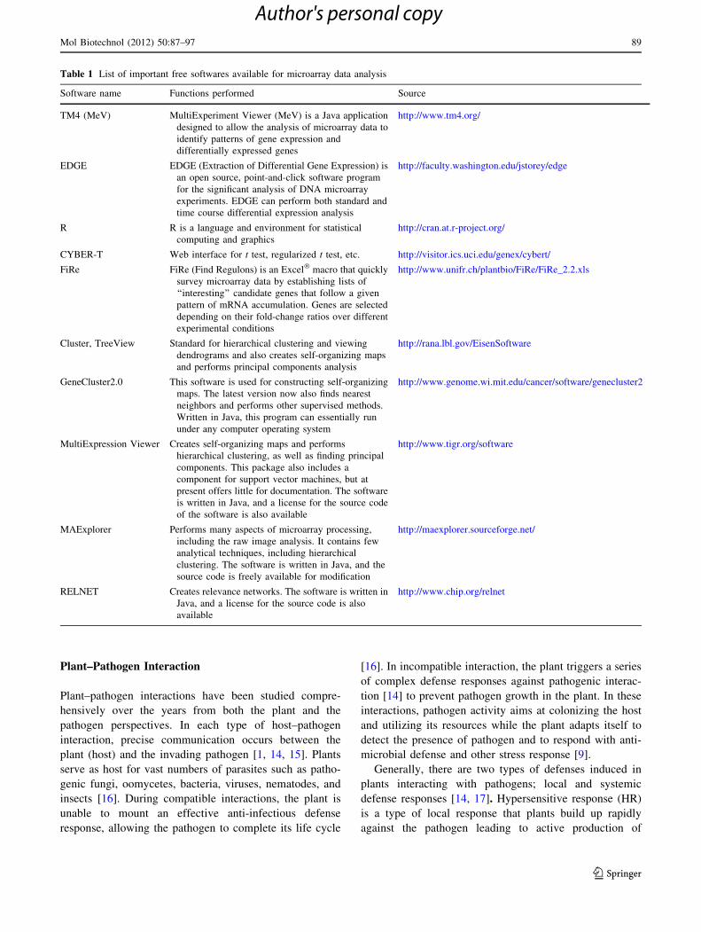
Many free and commercial software packages are now
available to analyze microarray data sets, although it is still
difficult to find a single complete software package that
answers all functional-genomics questions. As the field is
still young, when developing a bioinformatics analysis
pipeline, it is more important to have a good understanding
of both the biology involved and the analytical techniques
rather than having the right software. Although many
bioinformatics companies sell software that assists in
microarray analysis, there are several freely available
software packages that can be used to perform the analyt-
ical techniques. Only the important softwares are listed in
Table 1.
88 Mol Biotechnol (2012) 50:87–97
123
Author's personal copy
Plant–Pathogen Interaction
Plant–pathogen interactions have been studied compre-
hensively over the years from both the plant and the
pathogen perspectives. In each type of host–pathogen
interaction, precise communication occurs between the
plant (host) and the invading pathogen [1, 14, 15]. Plants
serve as host for vast numbers of parasites such as patho-
genic fungi, oomycetes, bacteria, viruses, nematodes, and
insects [16]. During compatible interactions, the plant is
unable to mount an effective anti-infectious defense
response, allowing the pathogen to complete its life cycle
[16]. In incompatible interaction, the plant triggers a series
of complex defense responses against pathogenic interac-
tion [14] to prevent pathogen growth in the plant. In these
interactions, pathogen activity aims at colonizing the host
and utilizing its resources while the plant adapts itself to
detect the presence of pathogen and to respond with anti-
microbial defense and other stress response [9].
Generally, there are two types of defenses induced in
plants interacting with pathogens; local and systemic
defense responses [14, 17]. Hypersensitive response (HR)
is a type of local response that plants build up rapidly
against the pathogen leading to active production of
Table 1 List of important free softwares available for microarray data analysis
Software name Functions performed Source
TM4 (MeV) MultiExperiment Viewer (MeV) is a Java application
designed to allow the analysis of microarray data to
identify patterns of gene expression and
differentially expressed genes
http://www.tm4.org/
EDGE EDGE (Extraction of Differential Gene Expression) is
an open source, point-and-click software program
for the significant analysis of DNA microarray
experiments. EDGE can perform both standard and
time course differential expression analysis
http://faculty.washington.edu/jstorey/edge
R R is a language and environment for statistical
computing and graphics
http://cran.at.r-project.org/
CYBER-T Web interface for t test, regularized t test, etc. http://visitor.ics.uci.edu/genex/cybert/
FiRe FiRe (Find Regulons) is an Excel� macro that quickly
survey microarray data by establishing lists of
‘‘interesting’’ candidate genes that follow a given
pattern of mRNA accumulation. Genes are selected
depending on their fold-change ratios over different
experimental conditions
http://www.unifr.ch/plantbio/FiRe/FiRe_2.2.xls
Cluster, TreeView Standard for hierarchical clustering and viewing
dendrograms and also creates self-organizing maps
and performs principal components analysis
http://rana.lbl.gov/EisenSoftware
GeneCluster2.0 This software is used for constructing self-organizing
maps. The latest version now also finds nearest
neighbors and performs other supervised methods.
Written in Java, this program can essentially run
under any computer operating system
http://www.genome.wi.mit.edu/cancer/software/genecluster2
MultiExpression Viewer Creates self-organizing maps and performs
hierarchical clustering, as well as finding principal
components. This package also includes a
component for support vector machines, but at
present offers little for documentation. The software
is written in Java, and a license for the source code
of the software is also available
http://www.tigr.org/software
MAExplorer Performs many aspects of microarray processing,
including the raw image analysis. It contains few
analytical techniques, including hierarchical
clustering. The software is written in Java, and the
source code is freely available for modification
http://maexplorer.sourceforge.net/
RELNET Creates relevance networks. The software is written in
Java, and a license for the source code is also
available
http://www.chip.org/relnet
Mol Biotechnol (2012) 50:87–97 89
123
Author's personal copy

reactive oxygen species (ROS) and localized cell death
[14]. Hypersensitive response fails in the case when the
virulent pathogen is necrotrophic, i.e., obtains nutrient
from dead cells [17]. The important features of the local
response are pathogen recognition, amplification of path-
ogenesis related (PR) proteins, and expression of the genes
related to plant defense response [17].
A systematic or long-term response elevates the level of
salicylic acid (SA), nitric oxide (NO), ethylene, jasmonic
acid (JA), calcium and other ion fluxes and also protein
kinases, which in-turn activates many downstream pro-
cesses [9, 18, 19]. Systemic host responses are of two sub-
types; systemic acquired resistance (SAR) and induced
systemic resistance (ISR) and they share two components:
elevated production of certain antimicrobial compounds
and ability of defense activation machinery so that anti-
microbial responses are activated more strongly in
response to subsequent infection [20, 21].
DNA Microarrays to Study Plant–Pathogen Interaction
Over the last decade, microarray technology has proven to
be an essential tool for discovery of genes related to plant
defense and for giving comprehensive picture of global
expression profiles in plants upon attack by pathogens.
Using microarray, researchers have gained novel infor-
mations about plant–pathogen interactions.
Maleck et al. [22] have applied microarray technology
to provide a comprehensive description of the SAR genes
from Arabidopsis thaliana. They constructed microarray
chip with 10,000 expressed sequence tags (ESTs) repre-
senting 7,000 genes (30% of all Arabidopsis genes). Gene
expression changes of 14 different conditions related to
SAR generated by chemical or biological means were
examined including the study with plant mutants. Com-
parison of the gene expression profile of all the 14 exper-
iments resulted in the identification of 413 ESTs
(approximately 300 genes, many of which are novel) that
appeared to be associated with SAR. Using different
clustering algorithms, groups of genes with common reg-
ulation patterns (regulons) were derived [22]. The regulon
containing PR-1, a reliable marker gene for SAR in
A. thaliana, contains known PR genes and novel genes that
functions during SAR and disease resistance. The induction
of PR-1 is under the control of NPR1 [Nonexpressor of
pathogenesis-related (PR) genes 1] protein which interacts
with members of the TGA family of basic leucine zipper
(TGA-bZIP) transcription factors. The promoter of the
Arabidopsis PR-1 gene contains a binding site for the
TGA-bZIP factors (the sequence TGACG) that serves as
the positive cis acting element for SAR induction [23].
Thus, it was expected that all genes of the PR-1 regulon
would contain a TGA-bZIP binding site in their promoters.
However, Maleck et al. [22] found that TGA-bZIP recog-
nition site is not present in each and every PR-1 regulon.
With the aid of microarray analysis Maleck et al. identified
a common promoter element called W box (WRKY factor
binding site, the sequence TTGAC); a major sequence
element in the promoters of genes co-regulated with PR-1.
The analysis with DNA microarray showed that the PR-1
gene is regulated by three transcription factors; a TGA-
bZIP factor, an unknown transcription factor that activate
the transcription, and a WRKY factor that represses the
transcription. WRKY factor could act together with other
types of transcription factors to achieve precise regulation
of gene expression during SAR. Maleck et al. [22]
proposed that PR-1 regulon genes may be co-repressed
by WRKY factors and during SAR these genes would be
de-repressed. This microarray analysis results extend
expression profiling to define regulatory networks and gene
discovery in plants.
A path-breaking finding that induction of protein
secretory pathway is required for SAR was carried out by
Wang et al. [24] utilizing microarray, based primarily on
the aforementioned findings. To identify additional NPR1
target genes, Wang et al. [25] used the 35S::NPR1-GR
transgenic line generated in the npr1-3 mutant where
nuclear translocation of NPR1-GR (GR, glucocorticoid
receptor) required not only SA but also dexamethasone
(Dex). Treatment of 35S::NPR1-GR plants first with SA
and then with Dex specifically activated NPR1 target genes
[24]. Using Affymetrix GeneChips (8200 genes), they
identified putative NPR1 primary target genes by com-
paring transcriptional profiles of NPR1 and NPR1/
35S::NPR1-GR that were both treated with SA and then
Dex. Induced genes clearly fell into two categories; one
group contained genes known to be involved in defense,
including several PR genes, while the other group encoded
members of the protein secretory pathway (most of which
are endoplasmic reticulum localized proteins) [24]. For the
first time, Wang et al. [24] provided genetic evidence that
during SAR, there is a massive buildup of PR proteins in
vacuoles and the apoplast and to ensure proper folding,
modification, and transport of these PR proteins, a coor-
dinated upregulation in the protein secretory machinery is
required.
Gene expression profiling by microarray analysis has
demonstrated a substantial crosstalk between different
defence signaling pathways. Expression profiling of 2,375
selected genes were carried out by cDNA microarray in
A. thaliana after inoculation with an incompatible fungal
pathogen Alternaria brassicicola, or treatment with the
defense-related signaling molecules including ethylene,
methyl jasmonate (MJ), and salicylic acid (SA) [26]. Dif-
ferential expression of 705 mRNAs was observed in
90 Mol Biotechnol (2012) 50:87–97
123
Author's personal copy

response to one or more of the treatments, including known
and putative defense-related genes and 106 genes with no
previously described function or homology. In leaf tissue
inoculated with A. brassicicola, 168 mRNAs were upreg-
ulated, whereas 39 mRNAs were downregulated. After
treatment with ethylene, MJ, and SA, the number of
mRNAs that were highly upregulated ([2.5-fold) were 55,
221, and 192, respectively. A coordinated defense response
was observed, including 169 mRNAs regulated by multiple
treatments/defense pathways. The highest number of dif-
ferentially expressed genes was found after treatments with
SA and MJ. Moreover, 50% of the genes induced by eth-
ylene treatment were also induced by MJ treatment. These
results demonstrated that a substantial network of regula-
tory interactions exists and that significant interaction
occurs among the different defense signaling pathways,
especially between the SA and MJ pathways, which were
previously believed to act in an antagonistic mode. Salz-
man et al. [27] conducted a large-scale study of gene
expression in sorghum in response to the signaling com-
pounds SA, MJ, and the ethylene precursor aminocyclo-
propane carboxylic acid using a microarray containing
12,982 nonredundant elements. Numerous gene clusters
were identified in which expression was correlated with
particular signaling compound and tissue combinations.
Many genes previously identified in defense response
responded to the treatments, including numerous patho-
genesis-related genes and most members of the phenyl-
propanoid pathway, and several other genes that may
represent novel activities or pathways. Genes of the octa-
decanoic acid pathway of jasmonic acid (JA) synthesis
were induced by SA as well as by MJ. Measurement of JA
content confirmed that increased SA could lead to
increased endogenous JA production. Comparison of
responses to SA, MJ, and combined SA ? MJ revealed
patterns of one-way and mutual antagonisms, as well as
synergistic effects on regulation of some genes [27]. This
shows that crosstalk and fine-tuning of different defence
pathways are vital for enabling the plant to build up
appropriate defence responses without draining energy
resources to unsustainable levels. The discovery of regu-
latory defense signaling networks by microarrays has
demonstrated that ultimately genes and their products, and
not pathways are controlled by signaling [27].
The microarray profiling has also been used to examine
gene expression of the biotrophic fungus Blumeria gra-
minis f. sp. hordei during infection on barley [28, 29]. With
the help of published cDNA sequences [30], the microarray
containing 2,027 unigenes was used to study Blumeria–
barley interaction. In this study, the mRNA extracts were
used from four developmental stages prior to penetration
on the host by the pathogen, and four stages thereafter.
Contrasting expression patterns of genes encoding enzymes
in various primary metabolic pathways were observed. At
the onset of infection, lipid catabolism genes were highly
upregulated. As the infection progressed, the expression of
these genes gradually decreased implying that lipids are
dominant carbon storage source for germination of fungal
spores. This result matches with previous findings in other
fungal pathogens that intact lipid catabolic pathways are
required for germination and fungal pathogenicity [31–34].
The results and conclusions from these studies help in
understanding the primary metabolism of the hosts during
infection.
Global gene expression analyses during plant defence
responses have identified new physiological processes
involved in induced defence responses. Scheideler et al.
[35] used cDNA arrays comprising 13,000 unique ESTs
from Arabidopsis leaves after infection with the bacterial
biotrophic pathogen Pseudomonas syringe. They observed
expression change from housekeeping to defence metabo-
lism, showing an increased demand for energy and bio-
synthetic capacity in plants fighting off a pathogen attack
[35]. Differential regulation patterns were observed on the
genes encoding enzymes in glycolysis, the Krebs cycle, the
pentose phosphate pathway, the biosynthesis of aromatic
amino acids, phenylpropanoids, and ethylene [35]. Fur-
thermore, the results showed potentially important changes
in areas of metabolism, such as the glyoxylate metabolism,
hitherto not suspected to be components of plant defense.
Likewise, genes for the b-oxidation pathway of fatty acids
in Arabidopsis were upregulated in both local and systemic
tissue when plants were inoculated with the incompatible
fungus A. brassicicola [26]. Apart from fatty acid metab-
olism, this pathway is also important for the synthesis of JA
during plant defence, an essential contributor for resistance
against this necrotrophic pathogen [2].
Expression profiling is highly cell-type-specific and
results are affected by the question, whether whole organs
(e.g., leaves) have been used for RNA isolation (as done in
the majority of studies), or whether cells have been isolated
from specific organs and used for this purpose. In case of
whole organs, expression profiles for particular genes get
leveled out across different cell and tissue types. To
overcome this problem, Gjetting et al. [36] for the first time
microextracted mRNA from B. graminis f. sp. hordei-
infected cells, papilla-containing penetration-resistant
cells, and unattacked cells of barley leaves 18 h after
inoculation and carried out cDNA array analysis. They
used Gatersleben barley PGRC1 10,000 cDNA arrays
(10 K array) for this purpose [37]. Gjetting et al. success-
fully obtained separate gene expression profiles for specific
mildew-resistant and -infected barley cells. Analysis of the
differentially expressed genes showed that 46 genes were
upregulated only in samples from infected cells, 98 genes
were upregulated only in resistant cell samples, and 54
Mol Biotechnol (2012) 50:87–97 91
123
Author's personal copy
genes were upregulated in both infected and resistant cell
samples [36]. The clear separation between control, resis-
tant, and infected samples indicated that (i) a large number
of genes were induced or repressed and (ii) affected genes
displayed large changes in expression. For the first time,
Gjetting et al. identified candidate genes specifically reg-
ulated in infected host cells during haustorium formation
and establishment of biotrophy. Catoni et al. [38] per-
formed a comparative analysis of expression profiling in
shoots and roots of tomato systemically infected by tomato
spotted wilt virus (TSWV). Tom2 12K oligonucleotide-
based tomato array (available from Boyce Thompson
Institute for Plant Research) was used for this purpose.
Microarray analysis of 14 days post-inoculated roots and
shoots revealed that the number of genes regulated in
shoots is approximately twice that regulated in roots. In the
shoots, genes related to defense and signal transduction
were induced, while genes related to primary, secondary, as
well as amino acid metabolism were repressed [38]. On the
contrary, in roots, expression of genes involved in signal
transduction, primary metabolism, and amino acid metab-
olism (except for those involved in synthesis of secondary
compounds) were unaltered by TSWV infection. Also, in
roots, genes involved in biotic stress were induced and
those associated to the response to abiotic stress were
repressed. All these results indicate organ-specific tran-
scriptional responses, although the virus was present in
similar concentration in both the organs [38].
Non-host resistance, a rarely studied defence phenom-
enon, was believed to be genetically complex of the fact
that activation of any specific defense component may not
be sufficient to render a plant resistance reaction. Micro-
array experiments played a key role in delineating the
molecular mechanism of non-host resistance. Zellerhof
et al. [39] studied the transcriptional responses of one
particular genotype of barley (Hordeum vulgare subsp.
vulgare ‘Ingrid’) to three different pairs of adapted (host)
and non-adapted (non-host) isolates of fungal pathogens,
which belong to the genera Blumeria (powdery mildew),
Puccinia (rust), and Magnaporthe (blast). They used the
barley PGRC1 array carrying 10,000 spotted cDNAs [37].
Non-host resistance against each of these pathogens was
associated with changes in transcript abundance of distinct
sets of non-host-specific genes, although general (not non-
host-associated) transcriptional responses to the different
pathogens overlapped considerably [39]. The powdery
mildew- and blast-induced differences in transcript
abundance between host and non-host interactions were
significantly correlated with differences between a near-
isogenic pair of barley lines that carry either the Mlo
wild-type allele or the mutated Mlo5 allele, which medi-
ates basal resistance to powdery mildew [39]. Moreover,
they found similar patterns of overrepresented and
underrepresented functional categories of genes during the
interactions of barley with the different host or non-host
pathogens. Their results suggest that non-host resistance
and basal host defense of barley are functionally related
and that non-host resistance to different fungal pathogens
is associated with more robust regulation of complex but
largely non-overlapping sets of pathogen-responsive genes
involved in similar metabolic or signaling pathways. Zhou
et al. [40] studied the molecular mechanisms underlying
hypersensitive response (HR) of rice to its bacterial
pathogen, Xanthomonas oryzae pv. oryzicola (Xoc) med-
iated by a non-host maize R gene Rxo1, using a micro-
array experiment and a pair of transgenic (9804-Rxo1) and
non-transgenic (9804) rice lines. Affymetrix Genechips�
Rice Genome Array chips, which contain 51,279 tran-
scripts from two rice cultivars was used for this experi-
ment. They detected 2,450 and 1,950 differentially
regulated genes (DRGs) in 9804-Rxo1 and 9804 using
stringent statistical conditions. The difference between
9804-Rxo1 and 9804 in expression patterns of these up- or
downregulated genes was very striking [40]. Of the 1,239
and 963 upregulated genes in 9804-Rxo1 and 9804
induced by Xoc, only 143 genes were in common between
the transgenic line and its recipient. Similarly, of the
1,211 and 987 downregulated genes in 9804-Rxo1 and
9804 induced by Xoc, 83 genes were commonly repressed
in both the transgenic line and its recipient [40]. In par-
ticular, 107 genes were regulated in opposite directions
between 9804-Rxo1 and 9804, including 61 genes upreg-
ulated in 9804-Rxo1 but downregulated in 9804 and 46
genes down-regulated in 9804-Rxo1 but upregulated in
9804, respectively [40]. Analysis of all the DRGs indi-
cated that Rxo1 appeared to function in the very early step
of the interaction between rice and Xoc, and could spe-
cifically activate large numbers of genes involved in sig-
naling pathways leading to HR and some basal defensive
pathways such as SA and ET pathways. In the former
case, Rxo1 appeared to differ from the typical host R
genes in that it could lead to HR without activating NDR1
(non-race-specific disease resistance 1) [40]. This study
explored the molecular mechanism of the non-host resis-
tance of rice mediated by Rxo1 and provided useful
information to understand the evolution of plant resistance
genes.
Microarray technology not only identifies the signal
transduction pathways induced by the pathogen, but also
identifies genes that are specifically induced by the path-
ogen to support the infection process, namely the host
factors used by viruses. Senthil et al. [41] constructed EST
microarray derived from potato cDNA libraries to analyze
expression profile in Nicotiana benthamiana to the biotic
stress induced by Impatiens necrotic spot virus (INSV); a
plant virus that replicates in the host cytoplasm. The
92 Mol Biotechnol (2012) 50:87–97
123
Author's personal copy
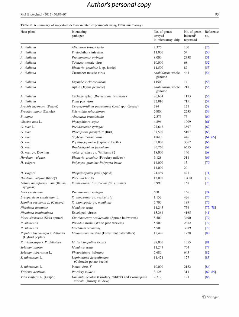
Table 2 A summary of important defense-related experiments using DNA microarrays
Host plant Interacting
pathogen
No. of genes
arrayed
in microarray chip
No. of genes
induced/
repressed
Reference
no.
A. thaliana Alternaria brassicicola 2,375 100 [26]
A. thaliana Phytophthora infestans 11,000 54 [50]
A. thaliana Pseudomonas syringae 8,000 2338 [51]
A. thaliana Tobacco mosaic virus 10,000 68 [52]
A. thaliana Blumeria graminis f. sp. hordei 11,500 89 [53]
A. thaliana Cucumber mosaic virus Arabidopsis whole
genome
444 [54]
A. thaliana Erysiphe cichoracearum 11500 14 [53]
A. thaliana Aphid (Myzus persicae) Arabidopsis whole
genome
2181 [55]
A. thaliana Cabbage aphid (Brevicoryne brassicae) 26,604 1133 [56]
A. thaliana Plum pox virus 22,810 7151 [57]
Arachis hypogaea (Peanut) Cercosporidium personatum (Leaf spot disease) 384 121 [58]
Brassica napus (Canola) Sclerotinia sclerotiorum 26000 2233 [59]
B. napus Alternaria brassicicola 2,375 75 [60]
Glycine max L. Phytophthora sojae 4,896 1009 [61]
G. max L. Pseudomonas syringae 27,648 3897 [62]
G. max Phakopsora pachyrhizi (Rust) 37,500 5107 [63]
G. max Soybean mosaic virus 18613 446 [64, 65]
G. max Popillia japonica (Japanese beetle) 35,000 3062 [66]
G. max Bradyrhizobium japonicum 36,760 6555 [67]
G. max cv. Dowling Aphis glycines cv. Williams 82 18,000 140 [68]
Hordeum vulgare Blumeria graminis (Powdery mildew) 3,128 311 [69]
H. vulgare Polymyxa graminis Polymyxa betae 14,000 13 [70]
14,000 20
H. vulgare Rhopalosiphum padi (Aphid) 21,439 497 [71]
Hordeum vulgare (barley) Puccinia hordei 15,000 1,410 [72]
Lolium multiflorum Lam (Italian
ryegrass)
Xanthomonas translucens pv. graminis 9,990 158 [73]
Lens esculentum Pseudomonas syringae 500 156 [74]
Lycopersicon esculentum L. X. campestris pv. vesicatoria 1,152 426 [75]
Manihot esculenta L. (Cassava) X. axonopodis pv. manihotis 5,700 199 [76]
Nicotiana attenuate Manduca sexta 11,243 754 [77, 78]
Nicotiana benthamiana Enveloped viruses 15,264 4165 [41]
Picea sitchensis (Sitka spruce) Choristoneura occidentalis (Spruce budworms) 5,500 3490 [79]
P. sitchensis Pissodes strobe (White pine weevils) 5,500 2382 [79]
P. sitchensis Mechnical wounding 5,500 3089 [79]
Populus trichocarpa x deltoides(Hybrid poplar)
Malacosoma disstria (Forest tent caterpillars) 15,496 1728 [80]
P. trichocarpa x P. deltoides M. laricipopulina (Rust) 28,000 1055 [81]
Solanum nigrum Manduca sexta 11,243 754 [77]
Solanum tuberosum L. Phytophthora infestans 7,680 643 [82]
S. tuberosum L. Leptinotarsa decemlineata(Colorado potato beetle)
11,421 127 [83]
S. tuberosum L. Potato virus Y 10,000 2132 [84]
Triticum aestivum Powdery mildew 3,128 311 [69, 85]
Vitis vinifera L. (Grape.) Uncinula necator (Powdery mildew) and Plasmoparaviticola (Downy mildew)
2,712 121 [86]
Mol Biotechnol (2012) 50:87–97 93
123
Author's personal copy

microarray analysis revealed that INSV-induced expression
of small heat-shock proteins genes to high levels (HSP18,
HSP20, HSP70). Earlier reports (with different techniques)
involving potyviruses and tobamoviruses [42–44] sup-
ported the finding that induced expression of HSPs are
associated with plant virus replication. Activating HSPs,
which have been shown to play a direct role in viral tran-
scription complexes [45], is a specific viral function
ensuring proper synthesis of viral proteins and virions. Carr
et al. [46] showed that the expression of HSP101 and
HSP70 was independent of the SA and JA defense sig-
naling pathways in A. thaliana during Oilseed rape mosaic
virus (ORMV) infection, confirming that ORMV activates
the expression of HSP. RNA1 of Pea early browning virus
(PEBV) alone was responsible for the induction of HSP70
expression in pea cotyledons [44]. Several significant
microarray experiments has been carried out [47–49],
providing information about the plant genes that are
modulated by viruses.
Till date, hundreds of microarray experiments have been
successfully carried out in the area of molecular plant–
microbe interaction, focusing mainly on the mechanisms
controlling plant disease resistance, crosstalk among the
signaling pathways involved, and the strategies used by the
pathogens to suppress the defense. However, as space is
limited, it is not possible to discuss all these interactions;
Table 2 gives a summary of the important experiments
involving plant–pathogen interaction using DNA micro-
arrays. Biological experimenters should interpret micro-
array data carefully as different laboratories use different
platforms and different algorithms to decide up- or down-
regulation of transcripts. Microarray data should be vali-
dated by secondary methods such as Northern hybridization
or real time PCR. In majority of the examples described in
this review, the microarray data were confirmed either by
Northern hybridization method or by Real time PCR
method. Absence of a unified ‘‘language’’ for exchange of
microarray data between different laboratories prompted
the ‘‘Microarray Gene Expression Data Society’’ (MGED)
to develop guidelines for the publication of DNA micro-
array data [87]. MGED also develop a Microarray Markup
Language to provide a standard platform for submitting and
analyzing the microarray expression data generated by
different laboratories around the world [87].
Future Prospects
With the experiments of plant–pathogen interaction com-
pleted to date, we can truly consider microarray as a mature
platform for gene expression analysis in plant–pathogen
interaction. Till date, microarray technology has been used in
the identification of regulatory genes, end-point defence
genes, to understand the role of particular transcription factor,
as well as to understand the signal transduction processes
underlying disease resistance and its intimate links to other
physiological pathways. The gene expression profiling can be
used as a tool for the study of effect of starvation and stress
condition. Sequencing projects will help in the construction of
the ESTs and give better understanding about the results. In
future, microarray technology can have its fruitful application
with more in-depth studies on simultaneous profiling of plant/
pathogen gene expression and on the influence of multiple
environmental factors on plants. The detailed information
regarding the plant defense system will be known with more
microarray experiments. Microarray containing the full
complement of Arabidopsis genes will provide a more com-
plete analysis. Similarly, the microarray experiments devel-
oped for crop plants will provide important information.
Recently, direct sequencing of transcripts by high-throughput
sequencing technologies (RNA-Seq) has become an addi-
tional alternative to microarrays. RNA-Seq does not depend
on genome annotation for prior probe selection and avoids
biases introduced during hybridization of microarrays [88].
On the other hand, RNA-Seq poses novel algorithmic and
logistic challenges, and current wet-lab RNA-Seq strategies
require lengthy library preparation procedures [88]. There-
fore, RNA-Seq is the method of choice in projects using non-
model organisms and for transcript discovery and genome
annotation [88]. Because of their robust sample processing
and analysis pipelines, microarrays are the choice for projects
that involve large numbers of samples for profiling tran-
scripts in model organisms with well-annotated genomes [88].
A symbiotic relationship between microarray technology and
high-throughput sequencing in the future will enable new
questions to be addressed in the area of plant–pathogen
interaction.
Acknowledgments TD Lodha is thankful to the Department of
Biotechnology, Govt. of India, for providing financial assistance.
Authors are thankful to Dr. Tapas Kumar Ghose, Division of Plant
Biology, Bose Institute, for his insightful discussion on the
manuscript.
References
1. Dodds, P. N., & Rathjen, J. P. (2010). Plant immunity: Towards
an integrated view of plant-pathogen interactions. NatureReviews Genetics, 11, 539–548.
2. De Wit, P. J. (2007). How plants recognize pathogens and defend
themselves. Cellular and Molecular Life Sciences, 64, 2726–2732.
3. Gachomo, E. W., Shonukan, O. O., & Kotchoni, S. O. (2003).
The molecular initiation and subsequent acquisition of disease
resistance in plants. African Journal of Biotechnology, 2, 26–32.
4. Dangl, J. L., & Jones, J. D. G. (2001). Plant pathogens and
integrated defense responses to infection. Nature, 411, 826–833.
5. Zipfel, C. (2009). Early molecular events in PAMP-triggered
immunity. Current Opinion in Plant Biology, 12, 414–420.
94 Mol Biotechnol (2012) 50:87–97
123
Author's personal copy
6. Wang, Z., Gerstein, M., & Snyder, M. (2009). RNA-Seq: A
revolutionary tool for transcriptomics. Nature Reviews Genetics,10, 57–63.
7. Libault, M., Farmer, A., Brechenmacher, L., Drnevich, J.,
Langley, R. J., Bilgin, D. D., et al. (2010). Complete transcrip-
tome of the soybean root hair cell, a single-cell model, and its
alteration in response to Bradyrhizobium japonicum infection.
Plant Physiology, 152, 541–552.
8. Santner, A., & Estelle, M. (2007). The JAZ proteins link jasm-
onate perception with transcriptional changes. The Plant Cell, 19,
3839–3842.
9. Andrew, F. B., Jinrong, W., & Mark, D. F. (2002). Probing plant-
pathogen interaction and downstream defense signaling using
DNA microarray. Functional & Integrative Genomics, 2,
259–273.
10. Schena, M., Shalon, D., Davis, R. W., & Brown, P. O. (1995).
Quantitative monitoring of gene expression patterns with a
complementary DNA microarray. Science., 20, 368–371.
11. Tan, K. C., Ipcho, S. V. S., Trengove, R. D., Oliver, R. P., &
Solomon, P. S. (2009). Assessing the impact of transcriptomics,
proteomics and metabolomics on fungal phytopathology.
Molecular Plant Pathology, 10, 703–715.
12. Stoughton, R. B. (2005). Applications of DNA microarrays in
biology. Annual Review of Biochemistry, 74, 53–82.
13. Webster, C. G., Wylie, S. J., & Jones, M. G. K. (2004). Diagnosis
of plant viral pathogen. Current Science., 86, 1604–1607.
14. Hammond-Kosack, K. E., & Jones, J. D. G. (2000). Responses
to plant pathogens. In Biochemistry and molecular biology ofplants (pp. 1102–1156). Rockville: American Society of Plant
Physiology.
15. Lucas, J. A. (1998). Plant pathology and plant pathogens (p. 8).
Oxford: Blackwell Science.
16. Schenk, P. M., Kazan, K., Wilson, L., Anderson, J. P., Richmond,
T., Somerville, S. C., et al. (2000). Coordinated plant defense
responses in Arabidopsis revealed by microarray analysis. Pro-ceedings of the National Academy of Science, 97, 11655–11660.
17. Schenk, P. M., Choo, J. H., & Wong, C. L. (2009). Microarray
analyses to study plant defense and rhizosphere microbe inter-
action. CAB: Perspective in Agriculture, Veterinary, Science,Nutrition, and Natural Resources., 4, 45–46.
18. Torres, M. A., Jonathan, D. G., & Dangl, J. L. (2006). Reactive
oxygen species signaling in response to pathogen. Plant Physi-ology, 141, 373–378.
19. Vallad, G. E., & Goodman, R. M. (2004). Systemic acquired
resistance and induced systemic resistance in conventional agri-
culture. Crop Science, 44, 1920–1934.
20. Dong, X. (2001). Genetic dissection of systemic acquired resis-
tance. Current Opinion in Plant Biology, 4, 309–314.
21. Glazebrook, J. (2001). Genes controlling expression of defense
responses in Arabidopsis—2001 status. Current Opinion in PlantBiology, 4, 301–308.
22. Maleck, K., Levine, A., Euglem, T., Morgan, A., Schmids, J.,
Lawton, K. A., et al. (2000). The Transcriptome of Arabidopsisthaliana during systemic acquired resistance. Nature Genetics,26, 403–410.
23. Lebel, E., Heifetz, P., Thorne, L., Uknes, S., Ryals, J., & Ward, E.
(1998). Functional analysis of regulatory sequences controlling
PR-1 gene expression in Arabidopsis. The Plant Journal, 16,
223–233.
24. Wang, D., Weaver, N. D., Kesarwani, M., & Dong, X. (2005).
Induction of protein secretory pathway is required for systemic
acquired resistance. Science., 308, 1036–1040.
25. Kinkema, M., Fan, W., & Dong, X. (2000). Nuclear localization
of NPR1 is required for activation of PR gene expression. PlantCell., 12, 2339–2350.
26. Schenk, P. M., Kazan, K., Manners, J. M., Anderson, J. P.,
Simpson, R. S., Wilson, I. W., et al. (2003). Systemic gene
expression In Arabidopsis during an incompatible interaction
with Alternaria brassicicola. Plant Physiology, 132, 999–1010.
27. Salzman, R. A., Brady, J. A., Finlayson, S. A., Buchanan, C. D.,
Summer, E. J., Sun, F., et al. (2005). Transcriptional profiling of
sorghum induced by methyl jasmonate, salicylic acid, and am-
inocyclopropane carboxylic acid reveals cooperative regulation
and novel gene responses. Plant Physiology, 138, 352–368.
28. Both, M., Csukai, M., Stumpf, M. P., & Spanu, P. D. (2005).
Gene expression profiles of Blumeria graminis indicate dynamic
changes to primary metabolism during development of an obli-
gate biotrophic pathogen. Plant Cell, 17, 2107–2122.
29. Both, M., Eckert, S. E., Csukai, M., Muller, E., Dimopoulos, G.,
& Spanu, P. D. (2005). Transcript profiles of Blumeria graminisdevelopment during infection reveal a cluster of genes that are
potential virulence determinants. Molecular Plant-MicrobeInteractions, 18, 125–133.
30. Thomas, S. W., Rasmussen, S. W., Glaring, M. A., Rouster, J. A.,
Christiansen, S. K., & Oliver, R. P. (2001). Gene identification in
the obligate fungal pathogen Blumeria graminis by expressed
sequence tag analysis. Fungal Genetics and Biology, 33, 195–211.
31. Idnurm, A., & Howlett, B. J. (2002). Isocitrate lyase is essential
for the pathogenicity of the fungus Leptosphaeria maculans to
Canola (Brassica napus). Eukaryotic Cell, 1, 719–724.
32. Solomon, P. S., Lee, R. C., Wilson, T. J. G., & Oliver, R. P.
(2004). Pathogenicity of Stagonospora nodorum requires malate
synthase. Molecular Microbiology, 53, 1065–1073.
33. Solomon, P. S., Tan, K. C., Sanchez, P., Cooper, R. M., & Oliver,
R. P. (2004). The disruption of a Ga subunit sheds new light on
the pathogenicity of Stagonospora nodorum on wheat. MolecularPlant-Microbe Interactions, 7, 456–466.
34. Wang, Z. Y., Thornton, C. R., Kershaw, M. J., Li, D. B., &
Talbot, N. J. (2003). The glyoxylate cycle is required for tem-
poral regulation of virulence by the plant pathogenic fungus
Magnaporthe oryzae. Molecular Microbiology, 47, 1601–1612.
35. Scheideler, M., Schlaich, N. L., Fellenberg, K., Beissbarth, T.,
Hauser, N. C., Vingron, M., et al. (2002). Monitoring the switch
from housekeeping to pathogen defence metabolism in Arabid-opsis thaliana using cDNA Arrays. Journal of BiologicalChemistry, 277, 10555–10561.
36. Gjetting, T., Hagedorn, P. H., Schweizer, P., Thordal-Christen-
sen, H., Carver, T. L. W., & Lyngkjær, M. F. (2007). Single-cell
transcript profiling of barley attacked by the powdery mildew
fungus. Molecular Plant-Microbe Interactions, 20, 235–246.
37. Sreenivasulu, N., Altschmied, L., Panitz, R., Hahnel, U., Mic-
halek, W., Weschke, W., et al. (2002). Identification of genes
specifically expressed in maternal and filial tissues of barley
caryopses: A cDNA array analysis. Molecular Genetics andGenomics, 266, 758–767.
38. Catoni, M., Miozzi, L., Fiorilli, V., Lanfranco, L., & Accotto, G.
P. (2009). Comparative analysis of expression profiles in shoots
and roots of tomato systemically infected by tomato spotted wilt
virus reveals organ-specific transcriptional responses. MolecularPlant-Microbe Interactions, 22, 1504–1513.
39. Zellerhoff, N., Himmelbach, A., Dong, W., Bieri, S., Schaffrath,U., & Schweizer, P. (2010). Nonhost resistance of barley to
different fungal pathogens is associated with largely distinct,
quantitative transcriptional responses. Plant Physiology, 152,
2053–2066.
40. Zhou, Y. L., Xu, M. R., Zhao, M. F., Xie, X. W., Zhu, L. H., Fu,
B. Y., et al. (2010). Genome-wide gene responses in a transgenic
rice line carrying the maize resistance gene Rxo1 to the rice
bacterial streak pathogen, Xanthomonas oryzae pv. oryzicola.
BMC Genomics., 11, 78–88.
Mol Biotechnol (2012) 50:87–97 95
123
Author's personal copy

41. Senthil, G., Liu, H., Puram, V. G., Clark, A., Stromberg, A., &
Goodin, M. M. (2005). Specific and common changes in Nico-tiana benthamiana gene expression in response to infection by
enveloped viruses. Journal of General Virology, 86, 2615–2625.
42. Aranda, M. A., Escaler, M., Wang, D., & Maule, A. J. (1996).
Induction of HSP70 and polyubiquitin expression associated with
plant virus replication. Proceedings of the National Academy ofSciences of the United States of America, 93, 15289–15293.
43. Aranda, M., & Maule, A. (1998). Virus-induced host gene shutoff
in animals and plants. Virology, 243, 261–267.
44. Whitham, S. A., Quan, S., Chang, H. S., Cooper, B., Estes, B.,
Zhu, T., et al. (2003). Diverse RNA viruses elicit the expression
of common sets of genes in susceptible Arabidopsis thaliana
plants. The Plant Journal, 33, 271–283.
45. Qanungo, K. R., Shaji, D., Mathur, M., & Banerjee, A. K. (2004).
Two RNA polymerase complexes from vesicular stomatitis virus-
infected cells that carry out transcription and replication of gen-
ome RNA. Proceedings of the National Academy of Sciences ofthe United States of America, 101, 5952–5957.
46. Carr, T., Wang, Y., Huang, Z., Yeakley, J. M., Fan, J. B., & Whitham,
S. (2006). Tobamovirus infection is independent of HSP101 mRNA
induction and protein expression. Virus Research, 121, 33–41.
47. Chen, W., Provart, N., Glazebrook, J., Katagiri, F., Chang, H. S.,
Eulgem, T., et al. (2002). Expression profile matrix of Arabid-
opsis transcription factor genes implies their putative functions in
response to environmental stresses. Plant Cell., 14, 559–574.
48. Huang, Z., Yeakley, J. M., Garcia, E. W., Holdridge, J. D., Fan, J.
B., & Whitham, S. A. (2005). Salicylic acid-dependent expres-
sion of host genes in compatible Arabidopsis-virus interactions.
Plant Physiology, 137, 1147–1159.
49. Itaya, A., Matsuda, Y., Gonzales, R. A., Nelson, R. S., & Ding, B.
(2002). Potato spindle tuber viroid strains of different pathoge-
nicity induces and suppresses expression of common and unique
genes in infected tomato. Molecular Plant-Microbe Interactions,15, 990–999.
50. Huitema, E., Vleeshouwers, V., Francis, D., & Kamoun, S.
(2003). Active defence responses associated with non-host
resistance of Arabidopsis thaliana to the oomycete pathogen
Phytophthora infestans. Molecular Plant Pathology, 4, 487–500.
51. Tao, Y., Xie, Z., Chen, W., Glazebrook, J., Chang, H., Han, B.,
et al. (2003). Quantitative nature of Arabidopsis responses during
compatible and incompatible interactions with the bacterial
pathogen Pseudomonas syringae. Plant Cell., 15, 317–330.
52. Golem, S., & Culver, J. (2003). Tobacco mosaic virus induced
alterations in the gene expression profile of Arabidopsis thaliana.
Molecular Plant-Microbe Interactions, 16, 681–688.
53. Zimmerli, L., Stein, M., Lipka, V., Schulze-Lefert, P., & Som-
erville, S. (2004). Host and non-host pathogens elicit different
jasmonate/ethylene responses in Arabidopsis. The Plant Journal,40, 633–646.
54. Marathe, R., Guan, Z., Anandalakshmi, R., Zhao, H., & Dines-
hKumar, S. (2004). Study of Arabidopsis thaliana resistome in
response to cucumber mosaic virus infection using whole genome
microarray. Plant Molecular Biology, 55, 501–520.
55. Couldridge, C., Newbury, H. J., Ford-Lloyd, B., Bale, J., &
Pritchard, J. (2007). Exploring plant responses to aphid feeding
using a full Arabidopsis microarray reveals a small number of
genes with significantly altered expression. Bulletin of Entomo-logical Research, 97, 523–532.
56. Kusnierczyk, A., Winge, P., Jørstad, T. S., Troczynska, J.,
Rossiter, J. T., & Bones, A. M. (2008). Towards global under-
standing of plant defense against aphids - timing and dynamics of
early Arabidopsis defense responses to cabbage aphid (Brevico-ryne brassicae) attack. Plant, Cell & Environment, 31,
1097–1115.
57. Babu, M., Griffiths, J. S., Huang, T., & Wang, A. (2008). Altered
gene expression changes in Arabidopsis leaf tissues and protop-
lasts in response to plum pox virus infection. BMC Genomics., 9,
325.
58. Luo, M., Dang, P., Bausher, M., Holbrook, C., Lee, R., Lynch, R.,
et al. (2005). Identification of transcripts involved in resistance
responses to leaf spot disease caused by Cercosporidium per-sonatum in peanut (Arachis hypogaea). Phytopathology, 95,
381–387.
59. Zhao, J., Wang, J., An, L., Doerge, R. W., Chen, Z. J., Grau, C.
R., et al. (2007). Analysis of gene expression profiles in response
to Sclerotinia sclerotiorum in Brassica napus. Planta., 227,
13–24.
60. Schenk, P. M., Thomas-Hall, S., Nguyen, A. V., Manners, J. M.,
Kazan, K., & Spangenberg, G. (2008). Identification of plant
defense genes in canola using Arabidopsis cDNA microarrays.
Plant Biology, 10, 539–547.
61. Moy, P., Qutob, D., Chapman, B., Atkinson, I., & Gijzen, M.
(2004). Patterns of gene expression upon infection of soybean
plants by Phytophthora sojae. Molecular Plant-Microbe Inter-actions, 17, 1051–1062.
62. Zou, J., Rodriguez-Zas, S., Li, M. A. M., Zhu, J., Gonzalez, D.,
Vodkin, L., et al. (2005). Expression profiling soybean response
to Pseudomonas syringae reveals new defense-related genes and
rapid HR-specific downregulation of photosynthesis. MolecularPlant-Microbe Interactions, 8, 1161–1174.
63. van de Mortel, M., Recknor, J. C., Graham, M. A., Nettleton, D.,
Dittman, J. D., Nelson, R. T., et al. (2007). Distinct biphasic
mRNA changes in response to Asian soybean rust infection.
Molecular Plant-Microbe Interactions, 20, 887–999.
64. Babu, M., Gagarinova, A. G., Brandle, J. E., & Wang, A. (2008).
Association of the transcriptional response of soybean plants with
soybean mosaic virus systemic infection. Journal of GeneralVirology, 89, 1069–1080.
65. Bilgin, D. D., Aldea, M., O’Neill, B. F., Benitez, M., Li, M.,
Clough, S. J., et al. (2008). Elevated ozone alters soybean–virus
interaction. Molecular Plant-Microbe Interactions, 21,
1297–1308.
66. Casteel, C. L., O’Neill, B. F., Zavala, J. A., Bilgin, D. D., Ber-
enbaum, M. R., & DeLucia, E. H. (2008). Transcriptional pro-
filing reveals elevated CO2 and elevated O3 alter resistance of
soybean (Glycine max) to Japanese beetles (Popillia japonica).
Plant, Cell & Environment, 31, 419–434.
67. Brechenmacher, L., Kim, M.-Y., Benitez, M., Li, M., Joshi, T.,
Calla, B., et al. (2008). Transcription profiling of soybean nod-
ulation by Bradyrhizobium japonicum. Molecular Plant-MicrobeInteractions, 21, 631–645.
68. Li, Y., Zou, J., Li, M., Bilgin, D. D., Vodkin, L. O., Hartman, G.
L., et al. (2008). Soybean defense responses to the soybean aphid.
New Phytologist, 179, 185–195.
69. Zierold, U., Scholz, U., & Schweizer, P. (2005). Transcriptome
analysis of mlo-mediated resistance in the epidermis of barley.
Molecular Plant Pathology, 6, 139–151.
70. McGrann, G. R. D., Townsend, B. J., Antoniw, J. F., Asher, M.
J. C., & Mutasa-Gottgens, E. S. (2009). Barley elicits a similar
early basal defense response during host and non-host interac-
tions with Polymyxa root parasites. European Journal of PlantPathology, 123, 5–15.
71. Delp, G., Gradin, T., Ahman, I., & Jonsson, L. M. V. (2009).
Microarray analysis of the interaction between the aphid Rho-palosiphum padi and host plants reveals both differences and
similarities between susceptible and partially resistant barley
lines. Mol Genet Genomics., 281, 233–248.
72. Chen, X., Niks, R. E., Hedley, R. E., Morris, J., Druka, A.,
Marcel, T. C., et al. (2010). Differential gene expression in nearly
96 Mol Biotechnol (2012) 50:87–97
123
Author's personal copy

isogenic lines with QTL for partial resistance to Puccinia hordeiin barley. BMC Genomics., 11, 629.
73. Wichmann, F., Asp, T., Widmer, F., & Kolliker, R. (2011).
Transcriptional responses of Italian ryegrass during interaction
with Xanthomonas translucens pv. graminis reveal novel candi-
date genes for bacterial wilt resistance. Theoretical and AppliedGenetics, 122, 567–579.
74. Zhao, Y., Thilmony, R., Bender, C., Schaller, A., He, S., &
Howe, G. (2003). Virulence systems of Pseudomonas syringaepv. tomato promotes bacterial speck disease in tomato by tar-
geting the jasmonate signaling pathway. The Plant Journal, 36,
485–499.
75. Gibly, A., Bonshtien, A., Balaji, V., Debbie, P., Martin, G., &
Sessa, G. (2004). Identification and expression profiling of tomato
genes differentially regulated during a resistance response to
Xanthomonas campestris pv. vesicatoria. Molecular Plant-Microbe Interactions, 17, 1212–1222.
76. Lopez, C., Soto, M., Restrepo, S., Piegu, B., Cooke, R., Delseny,
M., et al. (2005). Gene expression profile in response to Xan-thomonas axonopodis pv. manihotis infection in cassava using a
cDNA microarray. Plant Molecular Biology, 57, 393–410.
77. Schmidt, D. D., Voelckel, C., Hartl, M., Schmidt, S., & Baldwin,
I. T. (2005). Specificity in ecological interactions: Attack from
the same lepidopteran herbivore results in species-specific tran-
scriptional responses in two solanaceous host plants. PlantPhysiology, 138, 1763–1773.
78. Voelckel, C., & Baldwin, I. T. (2004). Herbivore-induced plant
vaccination. Part II. Array-studies reveal the transience of her-
bivorespecific transcriptional imprints and a distinct imprint from
stress combinations. The Plant Journal, 38, 650–663.
79. Ralph, S. G., Yueh, H., Friedmann, M., Aeschliman, D., Zeznik,
J. A., Nelson, C. C., et al. (2006). Conifer defence against insects:
Microarray gene expression profiling of Sitka spruce (Picea
sitchensis) induced by mechanical wounding or feeding by spruce
budworms (Choristoneura occidentalis) or white pine weevils
(Pissodes strobi) reveals large-scale changes of the host tran-
scriptome. Plant, Cell & Environment, 29, 1545–1570.
80. Ralph, S., Oddy, C., Cooper, D., Yueh, H., Jancsik, S., Kolosov,
N., et al. (2006). Genomics of hybrid poplar (Populus trichocarpa9 deltoides) interacting with forest tent caterpillars (Malacosoma
disstria): Normalized and full-length cDNA libraries, expressed
sequence tags, and a cDNA microarray for the study of insect-
induced defences in poplar. Molecular Ecology, 15, 1275–1297.
81. Rinaldi, C., Kohler, A., Frey, P., Duchaussoy, F., Ningre, N.,
Couloux, A., et al. (2007). Transcript profiling of poplar leaves
upon infection with compatible and incompatible strains of the
foliar rust Melampsora larici-populina. Plant Physiology, 144,
347–366.
82. Restrepo, S., Myers, K., Pozo, Od., Martin, G., Hart, A., Buell,
C., et al. (2005). Gene profiling of a compatible interaction
between Phytotphthora infestans and Solanum tuberosum sug-
gests a role for carbonic anhydrase. Molecular Plant-MicrobeInteractions, 18, 913–922.
83. Lawrence, S. D., Novak, N. G., Ju, C. J.-T., & Cooke, J. E. K.
(2008). Potato, Solanum tuberosum, defense against colorado
potato beetle, Leptinotarsa decemlineata (Say): Microarray gene
expression profiling of potato by colorado potato beetle regur-
gitant treatment of wounded leaves. Journal of Chemical Ecol-ogy, 34, 1013–1025.
84. Baebler, S., Krecic-Stres, H., Rotter, A., Kogovsek, P., Cankar,
K., Kok, E. J., et al. (2009). PVYNTN elicits a diverse gene
expression response in different potato genotypes in the first 12 h
after inoculation. Molecular Plant Pathology, 10, 263–275.
85. Bruggmann, R., Abderhalden, O., Reymond, P., & Dudler, R.
(2005). Analysis of epidermis- and mesophyll-specific transcript
accumulation in powdery mildew-inoculated wheat leave. PlantMolecular Biology, 58, 247–267.
86. Figueiredo, A., Fortes, A. M., Ferreira, S., Sebastiana, M., Choi,
Y. H., Sousa, L., et al. (2008). Transcriptional and metabolic
profiling of grape (Vitis vinifera L.) leaves unravel possible innate
resistance against pathogenic fungi. Journal of ExperimentalBotany, 59, 3371–3381.
87. Brazma, A., Parkinson, H., Sarkans, U., Shojatalab, M., Vilo, J.,
Abeygunawardena, N., et al. (2003). ArrayExpress—a public
repository for microarray gene expression data at the EBI.
Nucleic Acids Research, 31, 68–71.
88. Baginsky, S., Hennig, L., Zimmermann, P., & Gruissem, W.
(2010). Gene expression analysis, proteomics, and network dis-
covery. Plant Physiology, 152, 402–410.
Mol Biotechnol (2012) 50:87–97 97
123
Author's personal copy
Related Documents