PKCb Phosphorylates PI3Kc to Activate It and Release It from GPCR Control Romy Walser 1 , John E. Burke 2 , Elena Gogvadze 1 , Thomas Bohnacker 1 , Xuxiao Zhang 2 , Daniel Hess 3 , Peter Ku ¨ enzi 1 , Michael Leitges 4 , Emilio Hirsch 5 , Roger L. Williams 2 , Muriel Laffargue 6 , Matthias P. Wymann 1 * 1 Department of Biomedicine, University of Basel, Basel, Switzerland, 2 Medical Research Council, Laboratory of Molecular Biology, Cambridge, United Kingdom, 3 Friedrich Miescher Institute for Biomedical Research, Basel, Switzerland, 4 Biotechnology Centre, University of Oslo, Oslo, Norway, 5 Department of Genetics, Biology and Biochemistry, University of Torino, Torino, Italy, 6 INSERM, UMR1048, Institut des Maladies Me ´ taboliques et Cardiovasculaires, Toulouse, France Abstract All class I phosphoinositide 3-kinases (PI3Ks) associate tightly with regulatory subunits through interactions that have been thought to be constitutive. PI3Kc is key to the regulation of immune cell responses activated by G protein-coupled receptors (GPCRs). Remarkably we find that PKCb phosphorylates Ser582 in the helical domain of the PI3Kc catalytic subunit p110c in response to clustering of the high-affinity IgE receptor (FceRI) and/or store-operated Ca 2+ - influx in mast cells. Phosphorylation of p110c correlates with the release of the p84 PI3Kc adapter subunit from the p84-p110c complex. Ser582 phospho-mimicking mutants show increased p110c activity and a reduced binding to the p84 adapter subunit. As functional p84-p110c is key to GPCR-mediated p110c signaling, this suggests that PKCb-mediated p110c phosphorylation disconnects PI3Kc from its canonical inputs from trimeric G proteins, and enables p110c to operate downstream of Ca 2+ and PKCb. Hydrogen deuterium exchange mass spectrometry shows that the p84 adaptor subunit interacts with the p110c helical domain, and reveals an unexpected mechanism of PI3Kc regulation. Our data show that the interaction of p110c with its adapter subunit is vulnerable to phosphorylation, and outline a novel level of PI3K control. Citation: Walser R, Burke JE, Gogvadze E, Bohnacker T, Zhang X, et al. (2013) PKCb Phosphorylates PI3Kc to Activate It and Release It from GPCR Control. PLoS Biol 11(6): e1001587. doi:10.1371/journal.pbio.1001587 Academic Editor: Len Stephens, The Babraham Institute, United Kingdom Received November 14, 2012; Accepted May 8, 2013; Published June 25, 2013 Copyright: ß 2013 Walser et al. This is an open-access article distributed under the terms of the Creative Commons Attribution License, which permits unrestricted use, distribution, and reproduction in any medium, provided the original author and source are credited. Funding: This work was supported by the Swiss National Science Foundation (310030_127574 & 31EM30-126143; www.snf.ch), the ESF EuroMEMBRANE programme grant FP-018 (www.esf.org), and the Medical Research Council (file reference number U105184308). JEB was supported by an EMBO long-term fellowship (ALTF268-2009; www.embo.org) and the British Heart Foundation (PG11/109/29247; www.bhf.org.uk). The funders had no role in study design, data collection and analysis, decision to publish, or preparation of the manuscript. Competing Interests: The authors have declared that no competing interests exist. Abbreviations: A3AR, A3 adenosine receptor; ADA, adenosine deaminase; BAPTA-AM, 1,2-Bis(2-aminophenoxy)ethane-N,N,N9,N9-tetraacetic acid tetrakis(ace- toxymethyl ester); BMMC, bone marrow-derived mast cell; CAMK, calmodulin-dependent kinase; FceRI, high-affinity IgE receptor; GPCR, G protein-coupled receptor; GST, glutathione S-transferase; HDX-MS, hydrogen deuterium exchange mass spectrometry; MRM, multiple reaction monitoring; PAF, platelet activating factor; PI3K, phosphoinositide 3-kinase; PKC, protein kinase C; PMA, phorbol 12-myristate 13-acetate; PtdIns, phosphatidylinositol; PKB/Akt, protein kinase B; PTx, B. pertussis toxin; SCF, stem cell factor, c-kit ligand; SOCE, store-operated Ca2+ entry. * E-mail: [email protected] Introduction Class I phosphoinositide 3-kinases (PI3Ks) produce the lipid second messenger phosphatidylinositol(3,4,5)-trisphosphate [PtdIns(3,4,5)P 3 ] and consist of a p110 catalytic and a regulatory subunit. The class IA catalytic subunits, p110a, b, and d, are constitutively bound to p85- related regulatory proteins that link them to the activation by protein tyrosine kinase receptors. The only class IB PI3K member, p110c, is activated downstream of G protein-coupled receptors (GPCRs), and interacts with p101 or p84 (also known as p87 PIKAP ) adaptor subunits [1–3]. A tight complex of the p110c catalytic subunit (PK3CG) with p101 was first discovered in neutrophils [1]. The p101 subunit (PI3R5) sensitizes PI3Kc for activation by Gbc subunits of trimeric G proteins, and is essential for chemotaxis of neutrophils towards GPCR-ligands [1,4,5]. Mast cells do not express p101; however, they do express the homologous adaptor protein p84 ([PI3R6]) [6], which shares 30% sequence identity with p101. Both p101 and p84 potentiate the activation of p110c by Gbc, but the p110c-p101 complex is significantly more sensitive towards Gbc, and displays an enhanced translocation to the plasma membrane as compared with p110c-p84 [7]. Although p84 is absolutely required to relay GPCR signals to protein kinase B (PKB/Akt) phosphorylation and degranulation [6], its role is not completely understood: contrary to p110c-p101, p110c-p84 requires additionally the presence of the small G protein Ras, and might operate in distinct membrane micro-domains [6,7]. Interestingly, genetic ablation of p110c blocks high-affinity IgE receptor (FceRI)-dependent mast cell degranulation in vitro and in vivo [8]. In part this is due to the fact that initial IgE/antigen- mediated mast cell stimulation triggers the release of adenosine and other GPCR ligands to feed an autocrine/paracrine activation of PI3Kc, which then functions as an amplifier of mast cell degranulation. Interestingly, a substantial part of the observed PI3Kc-dependent histamine-containing granule release (ca. 40%) was found to be resistant to Bordetella pertussis toxin (PTx) pretreatment [9,10]. Furthermore, although adenosine activates PI3Kc via the A3 adenosine receptor (A 3 AR; [ADORA3]), A 3 AR PLOS Biology | www.plosbiology.org 1 June 2013 | Volume 11 | Issue 6 | e1001587

Welcome message from author
This document is posted to help you gain knowledge. Please leave a comment to let me know what you think about it! Share it to your friends and learn new things together.
Transcript
PKCb Phosphorylates PI3Kc to Activate It and Release Itfrom GPCR ControlRomy Walser1, John E. Burke2, Elena Gogvadze1, Thomas Bohnacker1, Xuxiao Zhang2, Daniel Hess3,
Peter Kuenzi1, Michael Leitges4, Emilio Hirsch5, Roger L. Williams2, Muriel Laffargue6,
Matthias P. Wymann1*
1 Department of Biomedicine, University of Basel, Basel, Switzerland, 2 Medical Research Council, Laboratory of Molecular Biology, Cambridge, United Kingdom,
3 Friedrich Miescher Institute for Biomedical Research, Basel, Switzerland, 4 Biotechnology Centre, University of Oslo, Oslo, Norway, 5 Department of Genetics, Biology and
Biochemistry, University of Torino, Torino, Italy, 6 INSERM, UMR1048, Institut des Maladies Metaboliques et Cardiovasculaires, Toulouse, France
Abstract
All class I phosphoinositide 3-kinases (PI3Ks) associate tightly with regulatory subunits through interactions that have beenthought to be constitutive. PI3Kc is key to the regulation of immune cell responses activated by G protein-coupledreceptors (GPCRs). Remarkably we find that PKCb phosphorylates Ser582 in the helical domain of the PI3Kc catalytic subunitp110c in response to clustering of the high-affinity IgE receptor (FceRI) and/or store-operated Ca2+- influx in mast cells.Phosphorylation of p110c correlates with the release of the p84 PI3Kc adapter subunit from the p84-p110c complex. Ser582phospho-mimicking mutants show increased p110c activity and a reduced binding to the p84 adapter subunit. Asfunctional p84-p110c is key to GPCR-mediated p110c signaling, this suggests that PKCb-mediated p110c phosphorylationdisconnects PI3Kc from its canonical inputs from trimeric G proteins, and enables p110c to operate downstream of Ca2+ andPKCb. Hydrogen deuterium exchange mass spectrometry shows that the p84 adaptor subunit interacts with the p110chelical domain, and reveals an unexpected mechanism of PI3Kc regulation. Our data show that the interaction of p110cwith its adapter subunit is vulnerable to phosphorylation, and outline a novel level of PI3K control.
Citation: Walser R, Burke JE, Gogvadze E, Bohnacker T, Zhang X, et al. (2013) PKCb Phosphorylates PI3Kc to Activate It and Release It from GPCR Control. PLoSBiol 11(6): e1001587. doi:10.1371/journal.pbio.1001587
Academic Editor: Len Stephens, The Babraham Institute, United Kingdom
Received November 14, 2012; Accepted May 8, 2013; Published June 25, 2013
Copyright: � 2013 Walser et al. This is an open-access article distributed under the terms of the Creative Commons Attribution License, which permitsunrestricted use, distribution, and reproduction in any medium, provided the original author and source are credited.
Funding: This work was supported by the Swiss National Science Foundation (310030_127574 & 31EM30-126143; www.snf.ch), the ESF EuroMEMBRANEprogramme grant FP-018 (www.esf.org), and the Medical Research Council (file reference number U105184308). JEB was supported by an EMBO long-termfellowship (ALTF268-2009; www.embo.org) and the British Heart Foundation (PG11/109/29247; www.bhf.org.uk). The funders had no role in study design, datacollection and analysis, decision to publish, or preparation of the manuscript.
Competing Interests: The authors have declared that no competing interests exist.
Abbreviations: A3AR, A3 adenosine receptor; ADA, adenosine deaminase; BAPTA-AM, 1,2-Bis(2-aminophenoxy)ethane-N,N,N9,N9-tetraacetic acid tetrakis(ace-toxymethyl ester); BMMC, bone marrow-derived mast cell; CAMK, calmodulin-dependent kinase; FceRI, high-affinity IgE receptor; GPCR, G protein-coupledreceptor; GST, glutathione S-transferase; HDX-MS, hydrogen deuterium exchange mass spectrometry; MRM, multiple reaction monitoring; PAF, platelet activatingfactor; PI3K, phosphoinositide 3-kinase; PKC, protein kinase C; PMA, phorbol 12-myristate 13-acetate; PtdIns, phosphatidylinositol; PKB/Akt, protein kinase B; PTx,B. pertussis toxin; SCF, stem cell factor, c-kit ligand; SOCE, store-operated Ca2+ entry.
* E-mail: [email protected]
Introduction
Class I phosphoinositide 3-kinases (PI3Ks) produce the lipid second
messenger phosphatidylinositol(3,4,5)-trisphosphate [PtdIns(3,4,5)P3]
and consist of a p110 catalytic and a regulatory subunit. The class IA
catalytic subunits, p110a, b, and d, are constitutively bound to p85-
related regulatory proteins that link them to the activation by protein
tyrosine kinase receptors. The only class IB PI3K member, p110c, is
activated downstream of G protein-coupled receptors (GPCRs), and
interacts with p101 or p84 (also known as p87PIKAP) adaptor subunits
[1–3]. A tight complex of the p110c catalytic subunit (PK3CG) with
p101 was first discovered in neutrophils [1]. The p101 subunit (PI3R5)
sensitizes PI3Kc for activation by Gbc subunits of trimeric G proteins,
and is essential for chemotaxis of neutrophils towards GPCR-ligands
[1,4,5].
Mast cells do not express p101; however, they do express the
homologous adaptor protein p84 ([PI3R6]) [6], which shares 30%
sequence identity with p101. Both p101 and p84 potentiate the
activation of p110c by Gbc, but the p110c-p101 complex is
significantly more sensitive towards Gbc, and displays an
enhanced translocation to the plasma membrane as compared
with p110c-p84 [7]. Although p84 is absolutely required to relay
GPCR signals to protein kinase B (PKB/Akt) phosphorylation and
degranulation [6], its role is not completely understood: contrary
to p110c-p101, p110c-p84 requires additionally the presence of
the small G protein Ras, and might operate in distinct membrane
micro-domains [6,7].
Interestingly, genetic ablation of p110c blocks high-affinity IgE
receptor (FceRI)-dependent mast cell degranulation in vitro and in
vivo [8]. In part this is due to the fact that initial IgE/antigen-
mediated mast cell stimulation triggers the release of adenosine
and other GPCR ligands to feed an autocrine/paracrine activation
of PI3Kc, which then functions as an amplifier of mast cell
degranulation. Interestingly, a substantial part of the observed
PI3Kc-dependent histamine-containing granule release (ca. 40%)
was found to be resistant to Bordetella pertussis toxin (PTx)
pretreatment [9,10]. Furthermore, although adenosine activates
PI3Kc via the A3 adenosine receptor (A3AR; [ADORA3]), A3AR
PLOS Biology | www.plosbiology.org 1 June 2013 | Volume 11 | Issue 6 | e1001587
null mice are still sensitive to passive systemic anaphylaxis, and
degranulation in A3AR2/2 bone marrow-derived mast cells
(BMMCs) upon antigen stimulation remains functional [11,12].
This and the strong degranulation phenotype of PI3Kc2/2
BMMCs suggest that GPCR signaling does not generate the full
input to PI3Kc-dependent degranulation, but a GPCR-indepen-
dent activation mechanism for PI3Kc has yet to be defined.
Here we identify a mechanism that activates PI3Kc indepen-
dently of GPCRs: we demonstrate that (i) IgE/antigen complexes
and extracellular Ca2+ influx activate PI3Kc, (ii) PI3Kc is
operationally linked to the FceRI specifically by PKCb (PRKCB),
(iii) and that the phosphorylation of Ser582 located in the helical
domain of p110c by PKCb leads to the dissociation of the p84
adapter to decouple phosphorylated p110c from GPCR inputs.
Further we characterize the p110c-p84 interface, and delineate an
activation process that seems to be conserved among class I PI3Ks.
Results
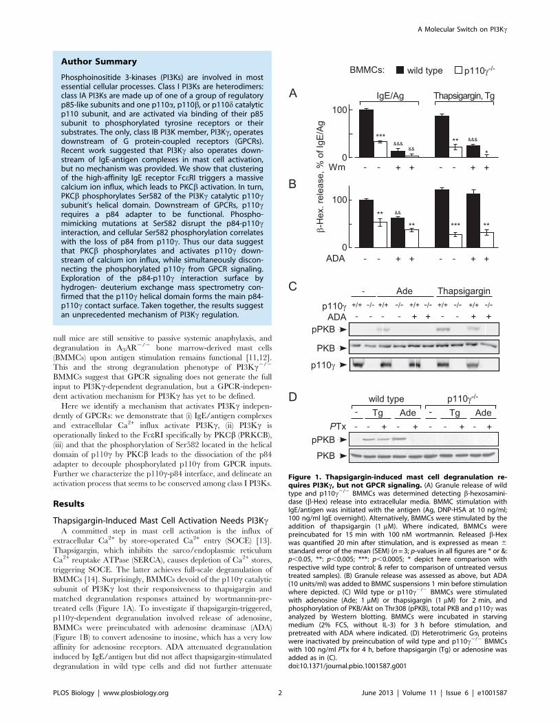
Thapsigargin-Induced Mast Cell Activation Needs PI3KcA committed step in mast cell activation is the influx of
extracellular Ca2+ by store-operated Ca2+ entry (SOCE) [13].
Thapsigargin, which inhibits the sarco/endoplasmic reticulum
Ca2+ reuptake ATPase (SERCA), causes depletion of Ca2+ stores,
triggering SOCE. The latter achieves full-scale degranulation of
BMMCs [14]. Surprisingly, BMMCs devoid of the p110c catalytic
subunit of PI3Kc lost their responsiveness to thapsigargin and
matched degranulation responses attained by wortmannin-pre-
treated cells (Figure 1A). To investigate if thapsigargin-triggered,
p110c-dependent degranulation involved release of adenosine,
BMMCs were preincubated with adenosine deaminase (ADA)
(Figure 1B) to convert adenosine to inosine, which has a very low
affinity for adenosine receptors. ADA attenuated degranulation
induced by IgE/antigen but did not affect thapsigargin-stimulated
degranulation in wild type cells and did not further attenuate
γ
β
γ
γ
γ
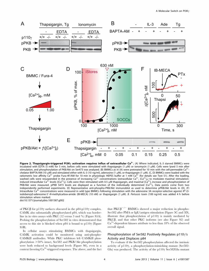
Figure 1. Thapsigargin-induced mast cell degranulation re-quires PI3Kc, but not GPCR signaling. (A) Granule release of wildtype and p110c2/2 BMMCs was determined detecting b-hexosamini-dase (b-Hex) release into extracellular media. BMMC stimulation withIgE/antigen was initiated with the antigen (Ag, DNP-HSA at 10 ng/ml;100 ng/ml IgE overnight). Alternatively, BMMCs were stimulated by theaddition of thapsigargin (1 mM). Where indicated, BMMCs werepreincubated for 15 min with 100 nM wortmannin. Released b-Hexwas quantified 20 min after stimulation, and is expressed as mean 6standard error of the mean (SEM) (n = 3; p-values in all figures are * or &:p,0.05, **: p,0.005; ***: p,0.0005; * depict here comparison withrespective wild type control; & refer to comparison of untreated versustreated samples). (B) Granule release was assessed as above, but ADA(10 units/ml) was added to BMMC suspensions 1 min before stimulationwhere depicted. (C) Wild type or p110c2/2 BMMCs were stimulatedwith adenosine (Ade; 1 mM) or thapsigargin (1 mM) for 2 min, andphosphorylation of PKB/Akt on Thr308 (pPKB), total PKB and p110c wasanalyzed by Western blotting. BMMCs were incubated in starvingmedium (2% FCS, without IL-3) for 3 h before stimulation, andpretreated with ADA where indicated. (D) Heterotrimeric Gai proteinswere inactivated by preincubation of wild type and p110c2/2 BMMCswith 100 ng/ml PTx for 4 h, before thapsigargin (Tg) or adenosine wasadded as in (C).doi:10.1371/journal.pbio.1001587.g001
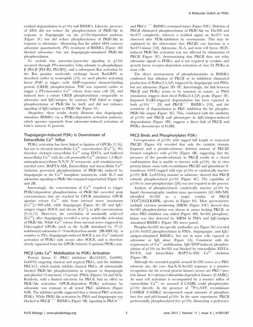
Author Summary
Phosphoinositide 3-kinases (PI3Ks) are involved in mostessential cellular processes. Class I PI3Ks are heterodimers:class IA PI3Ks are made up of one of a group of regulatoryp85-like subunits and one p110a, p110b, or p110d catalyticp110 subunit, and are activated via binding of their p85subunit to phosphorylated tyrosine receptors or theirsubstrates. The only, class IB PI3K member, PI3Kc, operatesdownstream of G protein-coupled receptors (GPCRs).Recent work suggested that PI3Kc also operates down-stream of IgE-antigen complexes in mast cell activation,but no mechanism was provided. We show that clusteringof the high-affinity IgE receptor FceRI triggers a massivecalcium ion influx, which leads to PKCb activation. In turn,PKCb phosphorylates Ser582 of the PI3Kc catalytic p110csubunit’s helical domain. Downstream of GPCRs, p110crequires a p84 adapter to be functional. Phospho-mimicking mutations at Ser582 disrupt the p84-p110cinteraction, and cellular Ser582 phosphorylation correlateswith the loss of p84 from p110c. Thus our data suggestthat PKCb phosphorylates and activates p110c down-stream of calcium ion influx, while simultaneously discon-necting the phosphorylated p110c from GPCR signaling.Exploration of the p84-p110c interaction surface byhydrogen- deuterium exchange mass spectrometry con-firmed that the p110c helical domain forms the main p84-p110c contact surface. Taken together, the results suggestan unprecedented mechanism of PI3Kc regulation.
A Molecular Switch on PI3Kc
PLOS Biology | www.plosbiology.org 2 June 2013 | Volume 11 | Issue 6 | e1001587
residual degranulation in p110c null BMMCs. Likewise, presence
of ADA did not reduce the phosphorylation of PKB/Akt in
response to thapsigargin via the p110c-dependent pathway
(Figure 1C) but did reduce phosphorylation of PKB/Akt in
response to adenosine—illustrating that the added ADA removes
adenosine quantitatively. PTx treatment of BMMCs (Figure 1D)
blocked adenosine—but not thapsigargin-stimulated PKB/Akt
phosphorylation.
To exclude that autocrine/paracrine signaling to p110coccurred through PTx-insensitive Gaq subunits to phospholipase
b (PLCb [PLCB2, PLCB3]), and a subsequent Ras activation by
the Ras guanine nucleotide exchange factor RasGRP4 as
described earlier in neutrophils [15], we used platelet activating
factor (PAF) to trigger cyclic AMP-responsive element-binding
protein (CREB) phosphorylation. PAF was reported earlier to
trigger a PTx-insensitive Ca2+ release from mast cells [9], and
induced here a robust CREB phosphorylation comparable to
adenosine and IgE/antigen. In contrast, PAF failed to trigger
phosphorylation of PKB/Akt by itself, and did not enhance
signaling of IgE/antigen to PKB/Akt (Figure S1).
Altogether, these results clearly illustrate that thapsigargin
stimulates BMMCs via a PI3Kc-dependent activation pathway,
which operates separately from adenosine-induced activation of
Gai/o trimeric G proteins.
Thapsigargin-Induced PI3Kc Is Downstream ofExtracellular Ca2+ Influx
PI3Kc activation has been linked to ligation of GPCRs [1,16],
but not to elevated intracellular Ca2+ concentration ([Ca2+]i). We
therefore chelated extracellular Ca2+ using EDTA, and buffered
intracellular Ca2+ with the cell permeable Ca2+ chelator 1,2-Bis(2-
aminophenoxy)ethane-N,N,N9,N9-tetraacetic acid tetrakis(acetox-
ymethyl ester) (BAPTA-AM). Extracellular and intracellular Ca2+
chelation prevented phosphorylation of PKB/Akt induced by
thapsigargin or the Ca2+ ionophore ionomycin, while IL-3 and
adenosine signaling to PKB/Akt remained unperturbed (Figure 2A
and 2B).
Interestingly, the concentration of Ca2+ required to trigger
PI3Kc-dependent phosphorylation of PKB/Akt exceeded peak
concentrations that are reached by GPCR stimulation: GPCR
agonists release Ca2+ only from internal stores (maximum
[Ca2+]i,300 nM), while thapsigargin (Figure 2C–2F) and IgE/
antigen trigger SOCE and elevate [Ca2+]i to mM concentrations
[9,14,17]. Moreover, the correlation of maximally achieved
[Ca2+]i after thapsigargin revealed a steep, switch-like activation
of PKB/Akt. While Ca2+ release from internal stores triggered by
Gai-coupled GPCRs (such as the A3AR stimulated by N6-(3-
iodobenzyl)-adenosine-59-N-methylcarbox-amide [IB-MECA]) is
sensitive to PTx, thapsigargin-induced SOCE is not. Ca2+-induced
activation of PI3Kc only occurs after SOCE, and is therefore
clearly separated from the GPCR/trimeric G protein/PI3Kc axis.
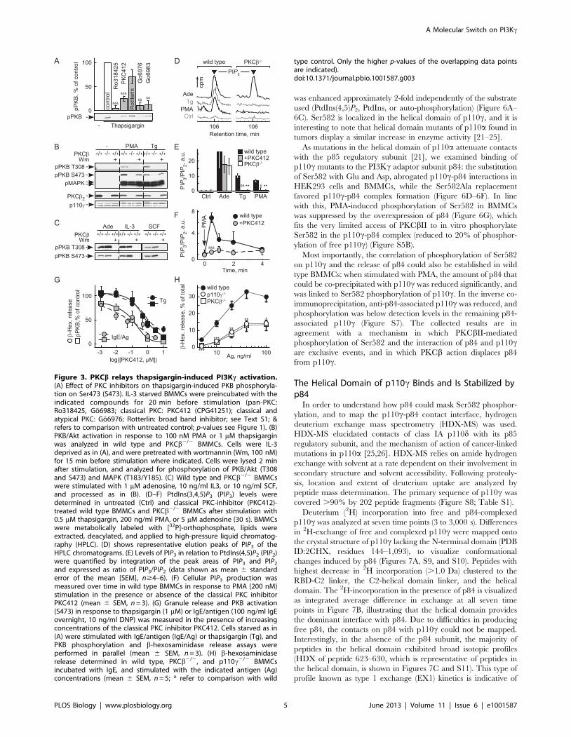
PKCb Links Ca2+ Mobilization to PI3Kc ActivationProtein kinase C (PKC) inhibitors (Ro318425, Go6983,
Go6976) targeting classical and atypical PKCs, and the inhibitor
PKC412, which mainly inhibits classical PKCs, all substantially
blocked PKB/Akt phosphorylation in response to thapsigargin
and phorbol 12-myristate 13-acetate (PMA) (Figures 3A and S2A).
Rottlerin, with a limited selectivity for PKCd, had no effect on
PKB/Akt activation. GPCR-dependent PI3Kc activation by
adenosine was resistant to all tested PKC inhibitors (Figure
S2B). The inhibitor profile suggested that a classical PKC activates
PI3Kc. While PKB/Akt activation by PMA and thapsigargin was
blocked in PKCb2/2 BMMCs (Figure 3B), signaling in PKCa2/2
and PKCc2/2 BMMCs remained intact (Figure S2C). Deletion of
PKCb eliminated phosphorylation of PKB/Akt on Thr308 and
Ser473 completely, whereas a residual signal on Ser473 was
observed after PI3K-inhibition by wortmannin. This may be
explained by the observation that PKCb2 can function as a
Ser473 kinase [18]. Adenosine, IL-3, and stem cell factor (SCF)-
induced PKB/Akt activation was not affected by elimination of
PKCb (Figure 3C), demonstrating that PKCb does not relay
adenosine signals to PI3Kc, and is not required in cytokine and
growth factor receptor-dependent activation of class IA PI3Ks in
mast cells.
The direct measurement of phosphoinositides in BMMCs
confirmed that ablation of PKCb or its inhibition eliminated
production of PtdIns(3,4,5)P3 triggered by thapsigargin and PMA,
but not adenosine (Figure 3D–3F). Interestingly, the link between
PKCb and PI3Kc seems to be transient in nature, as PMA
stimulation triggers short lived PtdIns(3,4,5)P3 peaks (Figure 3F).
Impaired FceRI-triggered degranulation has been reported in
both p110c2/2 [9] and PKCb2/2 BMMCs [19], and the
sensitivity of degranulation to PKC inhibition fits the phospho-
PKB/Akt output (Figure 3G). This, combined with the similarity
of p110c and PKCb null phenotypes in IgE/antigen-induced
degranulation (Figure 3H), suggests a direct link of PKCb and
PI3Kc downstream of FceRI.
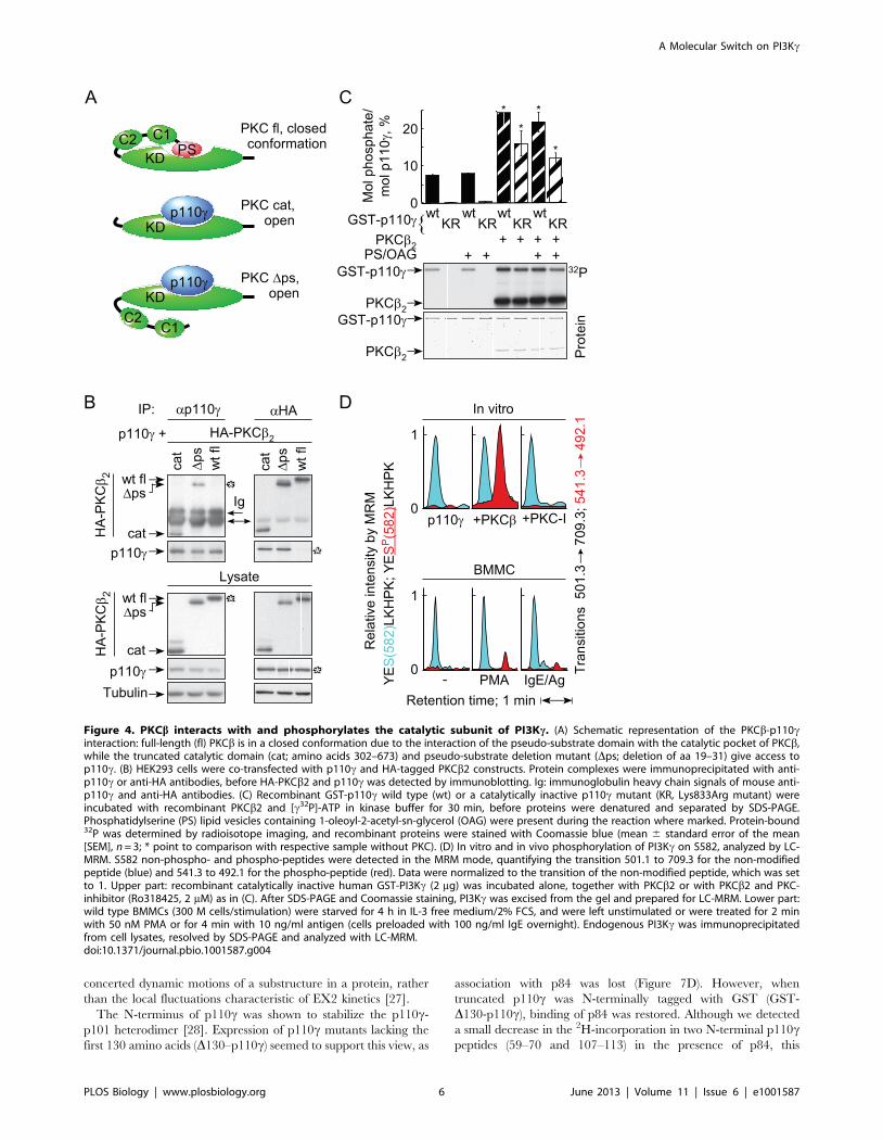
PKCb Binds and Phosphorylates PI3KcCo-expression of p110c with tagged full length or truncated
PKCb2 (Figure 4A) revealed that only the catalytic domain
fragment and a pseudo-substrate deletion mutant of PKCb2
formed complexes with p110c (Figure 4B), suggesting that the
presence of the pseudo-substrate in PKCb results in a closed
conformation that is unable to interact with p110c. An in vitro
protein kinase assay with recombinant PKCb2 and glutathione S-
transferase (GST)-tagged wild type p110c or catalytically inactive
p110c (KR; Lys833Arg mutant) as substrate, showed that PKCbrobustly phosphorylated p110c (Figure 4C). The capability of
p110c to auto-phosphorylate [20] was not required in the process.
Analysis of phosphorylated, catalytically inactive p110c by
liquid chromatography tandem mass spectrometry (LC-MS/MS)
identified Ser582 as a target residue of PKCb(YESP[582]LKHPK; spectra in Figure S3). Mass spectrometric
multiple reaction monitoring (MRM) (Figure S3C) showed that
Ser582 phosphorylation was absent in assays lacking PKCb, or
when PKC-inhibitor was added (Figure 4D). Ser582 phosphory-
lation was also detected by MRM in PMA and IgE/antigen
stimulated BMMCs (Figure 4D, lower panel).
Phospho-Ser582 site-specific antibodies (see Figure S4) revealed
p110c Ser852 phosphorylation in PMA-, thapsigargin-, and IgE/
antigen-stimulated BMMCs, but not in mast cells exposed to
adenosine or IgE alone (Figure 5A). Consistent with the
requirement of Ca2+ mobilization, IgE/DNP-induced phosphor-
ylation of p110c on Ser582 was blocked by extracellular (EDTA,
EGTA) and intracellular (BAPTA/AM) Ca2+ chelation
(Figure 5B).
Although the extended peptide around Ser582 scores as a PKC
substrate site, the core Arg-X-X-Ser582 sequence is a putative
recognition site for several protein kinases (scores are PKC.pro-
tein kinase A.calcium/calmodulin-dependent kinases [CAMK]).
As mast cell activation is accompanied by a massive influx of
extracellular Ca2+, we assessed if CAMK could phosphorylate
p110c directly. In the presence of 32P-c-ATP, recombinant
CAMKII (CAMK2) incorporated equal amounts of phosphate
into free and p84-bound p110c. In the same experiment, PKCbpreferentially phosphorylated free p110c, illustrating a preference
A Molecular Switch on PI3Kc
PLOS Biology | www.plosbiology.org 3 June 2013 | Volume 11 | Issue 6 | e1001587
of PKCb for p110c surfaces obscured in the p84-p110c complex.
CAMK also substantially phosphorylated p84, which was border-
line in in vitro assays with PKC (12 versus 3 mol %) (Figure S5A).
Probing the phosphorylation of Ser582 in vitro demonstrated that
access to this site is blocked when p84 is bound to p110c (Figure
S5B).
In cellular assays stimulating BMMCs with thapsigargin,
CAMK activation could be monitored using anti-phospho-
CAMKII antibodies. While PKC inhibitors left CAMKII phos-
phorylation .50% intact, Ser582 and PKB/Akt phosphorylation
were both reduced to background levels (Figure S6), even in a
context favoring Ca2+-triggered responses. The above, and the fact
that PKCb2/2 BMMCs showed a major reduction in phospho-
Ser582 after PMA or IgE/antigen stimulation (Figure 5C and 5D),
illustrate that phosphorylation of p110c is mainly mediated by
PKCb, and that other PKC isoforms (see also Figure S2) and
Ca2+-dependent kinases attribute to less than 18% of the observed
overall signal.
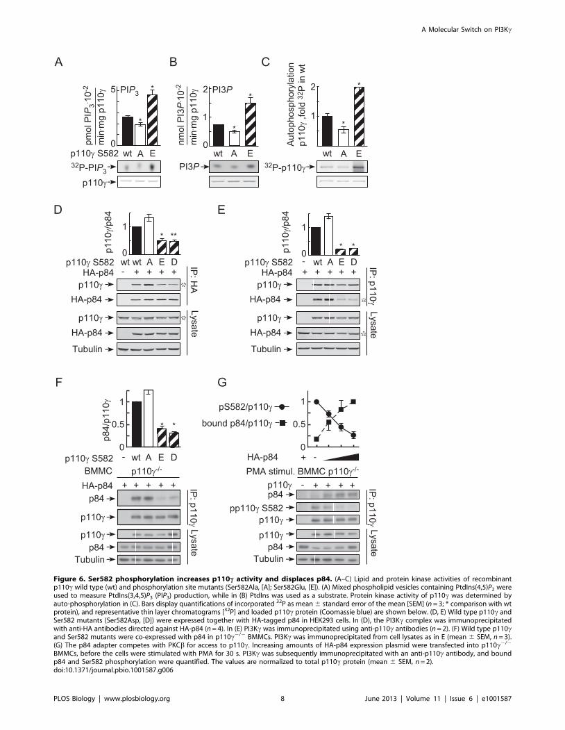
Phosphorylation of Ser582 Positively Regulates p110c’sActivity and Displaces p84
To evaluate if the Ser582 phosphorylation affected the intrinsic
activity of p110c, a phosphorylation-mimicking mutant (Ser582-
Glu) was produced. The activity of the p110c Ser582Glu mutant
Δ
γ
Figure 2. Thapsigargin-triggered PI3Kc activation requires influx of extracellular Ca2+. (A) Where indicated, IL-3 starved BMMCs wereincubated with EDTA (5 mM) for 5 min, before cells were stimulated with thapsigargin (1 mM) or ionomycin (1 mM). Cells were lysed 5 min afterstimulation, and phosphorylation of PKB/Akt on Ser473 was analyzed. (B) BMMCs as in (A) were pretreated for 10 min with the cell-permeable Ca2+-chelator BAPTA/AM (10 mM) and stimulated either with IL-3 (10 ng/ml), adenosine (1 mM), or thapsigargin (1 mM). (C, D) BMMCs were loaded with theratiometric low affinity Ca2+ probe Fura-4F/AM for 10 min in physiologic HEPES buffer at 1 mM Ca2+ (for details see Text S1). After the loading,washed cells were resuspended in the presence of increasing Ca2+ concentrations (extracellular Ca2+, [Ca2+]e) to modulate maximal stimulation-induced intracellular Ca2+ levels ([Ca2+]i). Cells were then stimulated with 0.5 mM thapsigargin, and maximal [Ca2+]i increase and phosphorylation ofPKB/Akt were measured. pPKB S473 levels are displayed as a function of the individually determined [Ca2+]i. Data points come from twoindependently performed experiments. (E) Representative anti-phospho-PKB/Akt immunoblot as used to determine pPKB/Akt levels in (D). (F)Intracellular Ca2+ concentrations were measured in wild type BMMCs following stimulation with the adenosine 3A receptor-selective agonist N6-(3-iodobenzyl)-adenosine-59-N-methylcarbox-amide (IB-MECA) (10 nM) or thapsigargin (1 mM). B. Pertussis toxin (100 ng/ml) was added 4 h beforestimulation where marked.doi:10.1371/journal.pbio.1001587.g002
A Molecular Switch on PI3Kc
PLOS Biology | www.plosbiology.org 4 June 2013 | Volume 11 | Issue 6 | e1001587
was enhanced approximately 2-fold independently of the substrate
used (PtdIns(4,5)P2, PtdIns, or auto-phosphorylation) (Figure 6A–
6C). Ser582 is localized in the helical domain of p110c, and it is
interesting to note that helical domain mutants of p110a found in
tumors display a similar increase in enzyme activity [21–25].
As mutations in the helical domain of p110a attenuate contacts
with the p85 regulatory subunit [21], we examined binding of
p110c mutants to the PI3Kc adaptor subunit p84: the substitution
of Ser582 with Glu and Asp, abrogated p110c-p84 interactions in
HEK293 cells and BMMCs, while the Ser582Ala replacement
favored p110c-p84 complex formation (Figure 6D–6F). In line
with this, PMA-induced phosphorylation of Ser582 in BMMCs
was suppressed by the overexpression of p84 (Figure 6G), which
fits the very limited access of PKCbII to in vitro phosphorylate
Ser582 in the p110c-p84 complex (reduced to 20% of phosphor-
ylation of free p110c) (Figure S5B).
Most importantly, the correlation of phosphorylation of Ser582
on p110c and the release of p84 could also be established in wild
type BMMCs: when stimulated with PMA, the amount of p84 that
could be co-precipitated with p110c was reduced significantly, and
was linked to Ser582 phosphorylation of p110c. In the inverse co-
immunoprecipitation, anti-p84-associated p110c was reduced, and
phosphorylation was below detection levels in the remaining p84-
associated p110c (Figure S7). The collected results are in
agreement with a mechanism in which PKCbII-mediated
phosphorylation of Ser582 and the interaction of p84 and p110care exclusive events, and in which PKCb action displaces p84
from p110c.
The Helical Domain of p110c Binds and Is Stabilized byp84
In order to understand how p84 could mask Ser582 phosphor-
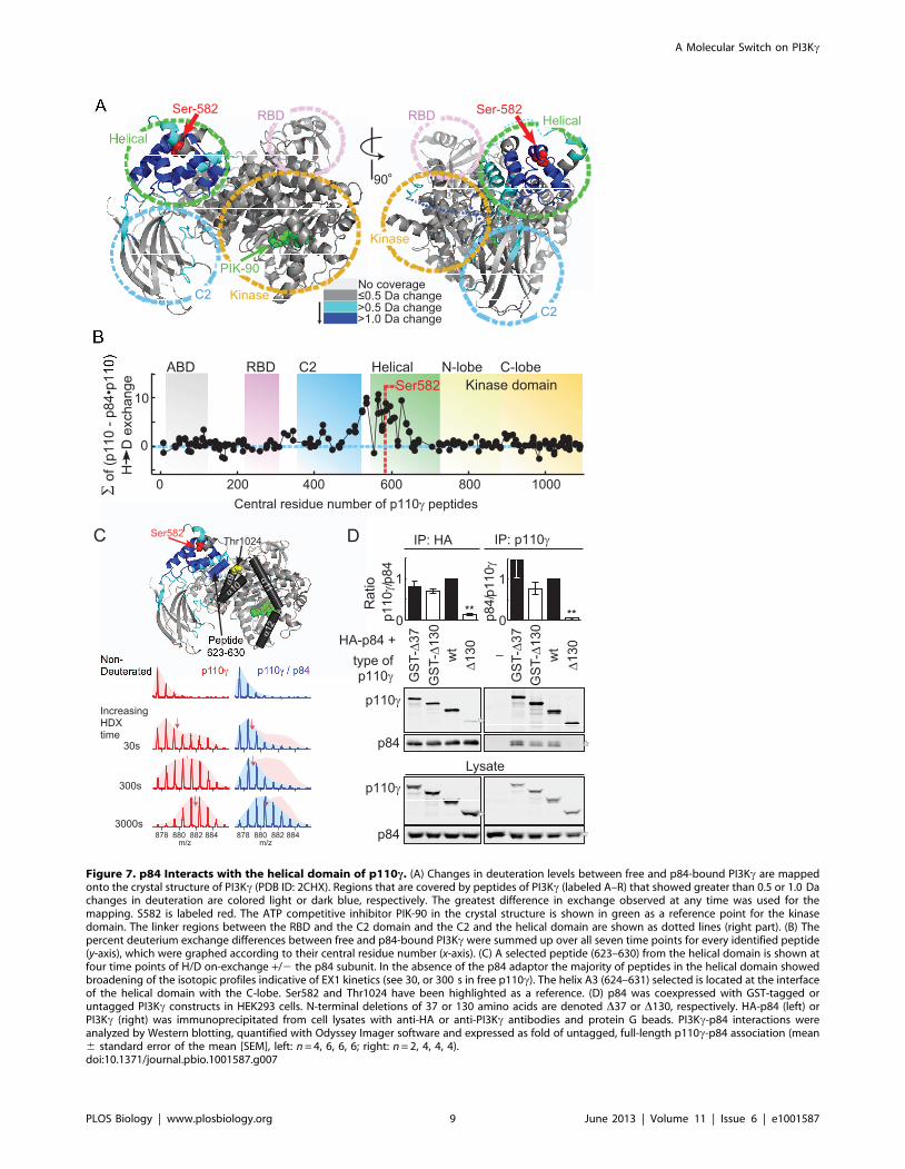
ylation, and to map the p110c-p84 contact interface, hydrogen
deuterium exchange mass spectrometry (HDX-MS) was used.
HDX-MS elucidated contacts of class IA p110d with its p85
regulatory subunit, and the mechanism of action of cancer-linked
mutations in p110a [25,26]. HDX-MS relies on amide hydrogen
exchange with solvent at a rate dependent on their involvement in
secondary structure and solvent accessibility. Following proteoly-
sis, location and extent of deuterium uptake are analyzed by
peptide mass determination. The primary sequence of p110c was
covered .90% by 202 peptide fragments (Figure S8; Table S1).
Deuterium (2H) incorporation into free and p84-complexed
p110c was analyzed at seven time points (3 to 3,000 s). Differences
in 2H-exchange of free and complexed p110c were mapped onto
the crystal structure of p110c lacking the N-terminal domain (PDB
ID:2CHX, residues 144–1,093), to visualize conformational
changes induced by p84 (Figures 7A, S9, and S10). Peptides with
highest decrease in 2H incorporation (.1.0 Da) clustered to the
RBD-C2 linker, the C2-helical domain linker, and the helical
domain. The 2H-incorporation in the presence of p84 is visualized
as integrated average difference in exchange at all seven time
points in Figure 7B, illustrating that the helical domain provides
the dominant interface with p84. Due to difficulties in producing
free p84, the contacts on p84 with p110c could not be mapped.
Interestingly, in the absence of the p84 subunit, the majority of
peptides in the helical domain exhibited broad isotopic profiles
(HDX of peptide 623–630, which is representative of peptides in
the helical domain, is shown in Figures 7C and S11). This type of
profile known as type 1 exchange (EX1) kinetics is indicative of
γβ
β
β
β
β
β
γ
β
β
Figure 3. PKCb relays thapsigargin-induced PI3Kc activation.(A) Effect of PKC inhibitors on thapsigargin-induced PKB phosphoryla-tion on Ser473 (S473). IL-3 starved BMMCs were preincubated with theindicated compounds for 20 min before stimulation (pan-PKC:Ro318425, Go6983; classical PKC: PKC412 (CPG41251); classical andatypical PKC: Go6976; Rotterlin: broad band inhibitor; see Text S1; &refers to comparison with untreated control; p-values see Figure 1). (B)PKB/Akt activation in response to 100 nM PMA or 1 mM thapsigarginwas analyzed in wild type and PKCb2/2 BMMCs. Cells were IL-3deprived as in (A), and were pretreated with wortmannin (Wm, 100 nM)for 15 min before stimulation where indicated. Cells were lysed 2 minafter stimulation, and analyzed for phosphorylation of PKB/Akt (T308and S473) and MAPK (T183/Y185). (C) Wild type and PKCb2/2 BMMCswere stimulated with 1 mM adenosine, 10 ng/ml IL3, or 10 ng/ml SCF,and processed as in (B). (D–F) PtdIns(3,4,5)P3 (PIP3) levels weredetermined in untreated (Ctrl) and classical PKC-inhibitor (PKC412)-treated wild type BMMCs and PKCb2/2 BMMCs after stimulation with0.5 mM thapsigargin, 200 ng/ml PMA, or 5 mM adenosine (30 s). BMMCswere metabolically labeled with [32P]-orthophosphate, lipids wereextracted, deacylated, and applied to high-pressure liquid chromatog-raphy (HPLC). (D) shows representative elution peaks of PIP3 of theHPLC chromatograms. (E) Levels of PIP3 in relation to PtdIns(4,5)P2 (PIP2)were quantified by integration of the peak areas of PIP3 and PIP2
and expressed as ratio of PIP3/PIP2 (data shown as mean 6 standarderror of the mean [SEM], n$4–6). (F) Cellular PIP3 production wasmeasured over time in wild type BMMCs in response to PMA (200 nM)stimulation in the presence or absence of the classical PKC inhibitorPKC412 (mean 6 SEM, n = 3). (G) Granule release and PKB activation(S473) in response to thapsigargin (1 mM) or IgE/antigen (100 ng/ml IgEovernight, 10 ng/ml DNP) was measured in the presence of increasingconcentrations of the classical PKC inhibitor PKC412. Cells starved as in(A) were stimulated with IgE/antigen (IgE/Ag) or thapsigargin (Tg), andPKB phosphorylation and b-hexosaminidase release assays wereperformed in parallel (mean 6 SEM, n = 3). (H) b-hexosaminidaserelease determined in wild type, PKCb2/2, and p110c2/2 BMMCsincubated with IgE, and stimulated with the indicated antigen (Ag)concentrations (mean 6 SEM, n = 5; * refer to comparison with wild
type control. Only the higher p-values of the overlapping data pointsare indicated).doi:10.1371/journal.pbio.1001587.g003
A Molecular Switch on PI3Kc
PLOS Biology | www.plosbiology.org 5 June 2013 | Volume 11 | Issue 6 | e1001587
concerted dynamic motions of a substructure in a protein, rather
than the local fluctuations characteristic of EX2 kinetics [27].
The N-terminus of p110c was shown to stabilize the p110c-
p101 heterodimer [28]. Expression of p110c mutants lacking the
first 130 amino acids (D130–p110c) seemed to support this view, as
association with p84 was lost (Figure 7D). However, when
truncated p110c was N-terminally tagged with GST (GST-
D130-p110c), binding of p84 was restored. Although we detected
a small decrease in the 2H-incorporation in two N-terminal p110cpeptides (59–70 and 107–113) in the presence of p84, this
γ β
γ
γ
β
γ
β
γβ{
Δ
γ β
γ
Δ
α γ α
β
Δ
β
Δ
γ
γ
γ Δ
Figure 4. PKCb interacts with and phosphorylates the catalytic subunit of PI3Kc. (A) Schematic representation of the PKCb-p110cinteraction: full-length (fl) PKCb is in a closed conformation due to the interaction of the pseudo-substrate domain with the catalytic pocket of PKCb,while the truncated catalytic domain (cat; amino acids 302–673) and pseudo-substrate deletion mutant (Dps; deletion of aa 19–31) give access top110c. (B) HEK293 cells were co-transfected with p110c and HA-tagged PKCb2 constructs. Protein complexes were immunoprecipitated with anti-p110c or anti-HA antibodies, before HA-PKCb2 and p110c was detected by immunoblotting. Ig: immunoglobulin heavy chain signals of mouse anti-p110c and anti-HA antibodies. (C) Recombinant GST-p110c wild type (wt) or a catalytically inactive p110c mutant (KR, Lys833Arg mutant) wereincubated with recombinant PKCb2 and [c32P]-ATP in kinase buffer for 30 min, before proteins were denatured and separated by SDS-PAGE.Phosphatidylserine (PS) lipid vesicles containing 1-oleoyl-2-acetyl-sn-glycerol (OAG) were present during the reaction where marked. Protein-bound32P was determined by radioisotope imaging, and recombinant proteins were stained with Coomassie blue (mean 6 standard error of the mean[SEM], n = 3; * point to comparison with respective sample without PKC). (D) In vitro and in vivo phosphorylation of PI3Kc on S582, analyzed by LC-MRM. S582 non-phospho- and phospho-peptides were detected in the MRM mode, quantifying the transition 501.1 to 709.3 for the non-modifiedpeptide (blue) and 541.3 to 492.1 for the phospho-peptide (red). Data were normalized to the transition of the non-modified peptide, which was setto 1. Upper part: recombinant catalytically inactive human GST-PI3Kc (2 mg) was incubated alone, together with PKCb2 or with PKCb2 and PKC-inhibitor (Ro318425, 2 mM) as in (C). After SDS-PAGE and Coomassie staining, PI3Kc was excised from the gel and prepared for LC-MRM. Lower part:wild type BMMCs (300 M cells/stimulation) were starved for 4 h in IL-3 free medium/2% FCS, and were left unstimulated or were treated for 2 minwith 50 nM PMA or for 4 min with 10 ng/ml antigen (cells preloaded with 100 ng/ml IgE overnight). Endogenous PI3Kc was immunoprecipitatedfrom cell lysates, resolved by SDS-PAGE and analyzed with LC-MRM.doi:10.1371/journal.pbio.1001587.g004
A Molecular Switch on PI3Kc
PLOS Biology | www.plosbiology.org 6 June 2013 | Volume 11 | Issue 6 | e1001587

interaction seems to be dispensable. The N-terminus of p110cinstead has a role in stabilizing the intact catalytic subunit. The
helical domain is the main location of interaction with p84; however,
it appears that this interaction is vulnerable and easily broken, as a
single phosphorylation at Ser582 is able to disrupt the contact.
Discussion
The activation of PI3Kc has been tightly linked to GPCRs-
triggered dissociation of trimeric Gai proteins, and has been
shown to require the interaction of Gbc subunits with p110c and
the PI3Kc adaptor subunits p101 and p84 [1,2,4,16,29,30].
Moreover, GPCRs generate PI3K signals typically through PI3Kc,
thereby controlling extravasation of hematopoietic cells [31,32],
cardiovascular parameters [33,34], and metabolic output [35,36].
Non-GPCR-mediated activation of PI3Kc has not been
reported so far, but it has been shown that phorbol esters and
Ca2+ ionophores can modulate phosphoinositide levels in a variety
of cells, including platelets [37], adipocites [38], fibroblasts [39],
and hematopoietic cells [40]. The proposed mechanisms have
β
γγ
ββ
γγ
γγ
γ
β− γ− β− γ−
γ γ
γγ
γ
γ
γγ
γ
γ γ
Figure 5. Phosphorylation of PI3Kc requires Ca2+ and is PKCb-dependent. (A) Stimulus-induced phosphorylation of endogenous p110c onSer582 in wild type BMMCs. IL-3 deprived cells were stimulated with 100 nM PMA, 1 mM thapsigargin, 1 mM adenosine, or 20 ng/ml DNP for 2 min.Where indicated (IgE), BMMCs were loaded with IgE (100 ng/ml) overnight. PI3Kc was immunoprecipitated from cell lysates with an anti-PI3Kcantibody, before precipitated protein was probed for phosphorylated p110c (pp110c) with a phospho-specific anti-pSer582 antibody (validation ofantibody see Figure S4). PI3Kc phosphorylation is shown normalized to total PI3Kc levels (mean 6 standard error of the mean [SEM], n = 3; * depictanalysis using unstimulated control. & reference point is IgE only). (B) IgE/antigen-induced Ser582 phosphorylation of p110c requires Ca2+ influx. Cellswere stimulated as in (A), but exposed to EDTA, EGTA, or loaded with BAPTA/AM where indicated (see Figure 2). Phosphorylated p110c was detectedas in (A); mean 6 SEM, n = 3; * comparison with unstimulated control; &analysis of stimulated versus chelator treated). (C) Phosphorylation of p110c inwild type and PKCb2/2 BMMCs. Experimental settings were as in (A), and (D) depicts quantification of pp110c in relation to total p110c protein (mean6 SEM; PMA n = 4, antigen n = 3). Cells devoid of p110c were included as negative control.doi:10.1371/journal.pbio.1001587.g005
A Molecular Switch on PI3Kc
PLOS Biology | www.plosbiology.org 7 June 2013 | Volume 11 | Issue 6 | e1001587
γ
γ
γ
γ
γ
γ
γ
γγ
γ
γ
γ
γ
γ
γ
γ
γ
γ
γ
γ γ
γ
γ
γ
γ
γ
γ
γ
γ
Figure 6. Ser582 phosphorylation increases p110c activity and displaces p84. (A–C) Lipid and protein kinase activities of recombinantp110c wild type (wt) and phosphorylation site mutants (Ser582Ala, [A]; Ser582Glu, [E]). (A) Mixed phospholipid vesicles containing PtdIns(4,5)P2 wereused to measure PtdIns(3,4,5)P3 (PIP3) production, while in (B) PtdIns was used as a substrate. Protein kinase activity of p110c was determined byauto-phosphorylation in (C). Bars display quantifications of incorporated 32P as mean 6 standard error of the mean [SEM] (n = 3; * comparison with wtprotein), and representative thin layer chromatograms [32P] and loaded p110c protein (Coomassie blue) are shown below. (D, E) Wild type p110c andSer582 mutants (Ser582Asp, [D]) were expressed together with HA-tagged p84 in HEK293 cells. In (D), the PI3Kc complex was immunoprecipitatedwith anti-HA antibodies directed against HA-p84 (n = 4). In (E) PI3Kc was immunoprecipitated using anti-p110c antibodies (n = 2). (F) Wild type p110cand Ser582 mutants were co-expressed with p84 in p110c2/2 BMMCs. PI3Kc was immunoprecipitated from cell lysates as in E (mean 6 SEM, n = 3).(G) The p84 adapter competes with PKCb for access to p110c. Increasing amounts of HA-p84 expression plasmid were transfected into p110c2/2
BMMCs, before the cells were stimulated with PMA for 30 s. PI3Kc was subsequently immunoprecipitated with an anti-p110c antibody, and boundp84 and Ser582 phosphorylation were quantified. The values are normalized to total p110c protein (mean 6 SEM, n = 2).doi:10.1371/journal.pbio.1001587.g006
A Molecular Switch on PI3Kc
PLOS Biology | www.plosbiology.org 8 June 2013 | Volume 11 | Issue 6 | e1001587
Non-Deuterated
IncreasingHDXtime
878 880 882 884 878 880 882 884m/z m/z
30s
300s
3000s
α10
α12
α11α9
Thr1024Ser582
No coverage≤0.5 Da change>0.5 Da change>1.0 Da change
PIK-90
Σof(p110-p84•p110)
HDexchange
Central residue number of p110γ peptides
0
10Ser582
0 200 400 600 800 1000
RBD C2 Helical N-lobe C-lobeKinase domain
ABDp110γ /p8 4 1
0 p84/p110γ
0
1
Ratio
p110γ
p84
GST-Δ 130
wt
Δ130
GST-Δ37
–type ofp110γ
HA-p84 +
GST-Δ130
wt
Δ130
GST-Δ37
IP: HA IP: p110γ
Lysatep110γ
p84
DC
90°
Ser-582 Ser-582Helical
Kinase
RBDRBD
Helical
C2C2 Kinase
**
****
**
****
Figure 7. p84 Interacts with the helical domain of p110c. (A) Changes in deuteration levels between free and p84-bound PI3Kc are mappedonto the crystal structure of PI3Kc (PDB ID: 2CHX). Regions that are covered by peptides of PI3Kc (labeled A–R) that showed greater than 0.5 or 1.0 Dachanges in deuteration are colored light or dark blue, respectively. The greatest difference in exchange observed at any time was used for themapping. S582 is labeled red. The ATP competitive inhibitor PIK-90 in the crystal structure is shown in green as a reference point for the kinasedomain. The linker regions between the RBD and the C2 domain and the C2 and the helical domain are shown as dotted lines (right part). (B) Thepercent deuterium exchange differences between free and p84-bound PI3Kc were summed up over all seven time points for every identified peptide(y-axis), which were graphed according to their central residue number (x-axis). (C) A selected peptide (623–630) from the helical domain is shown atfour time points of H/D on-exchange +/2 the p84 subunit. In the absence of the p84 adaptor the majority of peptides in the helical domain showedbroadening of the isotopic profiles indicative of EX1 kinetics (see 30, or 300 s in free p110c). The helix A3 (624–631) selected is located at the interfaceof the helical domain with the C-lobe. Ser582 and Thr1024 have been highlighted as a reference. (D) p84 was coexpressed with GST-tagged oruntagged PI3Kc constructs in HEK293 cells. N-terminal deletions of 37 or 130 amino acids are denoted D37 or D130, respectively. HA-p84 (left) orPI3Kc (right) was immunoprecipitated from cell lysates with anti-HA or anti-PI3Kc antibodies and protein G beads. PI3Kc-p84 interactions wereanalyzed by Western blotting, quantified with Odyssey Imager software and expressed as fold of untagged, full-length p110c-p84 association (mean6 standard error of the mean [SEM], left: n = 4, 6, 6, 6; right: n = 2, 4, 4, 4).doi:10.1371/journal.pbio.1001587.g007
A Molecular Switch on PI3Kc
PLOS Biology | www.plosbiology.org 9 June 2013 | Volume 11 | Issue 6 | e1001587

been diverse and involved protein tyrosine kinases and GPCR
signaling. A recent finding that protein kinase D (PKD) can
phosphorylate two distinct sites on the p85 regulatory subunit to
control class IA PI3K activity [41] is an indication that PI3K
control is more complex than anticipated.
PI3Kc has been shown to be a key element in enhancing IgE/
antigen output by the release of adenosine. This process involves
signaling downstream of Gai-coupled A3AR, and is sensitive to
PTx and ADA [9]. The resistance of thapsigargin-induced
degranulation to ADA shown here, and the fact that PTx did
not diminish the PI3Kc-dependent, thapsigargin-induced phos-
phorylation of PKB/Akt, points to a novel mechanism of PI3Kcactivation, which is clearly distinct from GPCR action. This Ca2+-
mediated PI3Kc activation requires SOCE and [Ca2+]i
.600 nM. In contrast, GPCRs yield phosphorylation of PKB/
Akt in mast cells even in the absence of a change in [Ca2+]i.
Furthermore, increases in [Ca2+]i triggered via GPCRs remain at
levels incapable of engaging a Ca2+-dependent activation of
PI3Kc.
Thapsigargin bypasses the signaling chain from IgE/antigen-
clustered FceRI to the activation of phospholipase Cc (PLCc) and
inositol(1,4,5)-trisphosphate (Ins(1,4,5)P3) production, and triggers
SOCE by the depletion of Ca2+ stores. That thapsigargin requires
functional PI3K activity to induce mast cell degranulation was first
demonstrated using the PI3K inhibitors wortmannin and
LY294002 [14], but no link between [Ca2+]i rise and PI3Kcactivity was established previously. The fact that inhibitors for
classical PKCs only prevented the thapsigargin- and PMA-
induced phosphorylation of PKB/Akt, while the GPCR-mediated
phosphorylation of PKB/Akt remained intact, points to a link
between classical PKCs and PI3Kc. Experiments on BMMCs
lacking PKCa, PKCb, and PKCc, showed that only the PKCbnull cells lost the ability to activate PKB/Akt in response to
thapsigargin or PMA stimulation. As the PMA- and thapsigargin-
induced phosphorylation of PKB/Akt on Ser473 showed a partial
resistance to wortmannin, and as it has been reported that PKCbcan directly phosphorylate Ser473 in the hydrophobic motif of
PKB/Akt [18], the effect of genetic and pharmacological targeting
of PKCb was also validated measuring PtdIns(3,4,5)P3 production
directly. The lack of PtdIns(3,4,5)P3 production in thapsigargin or
PMA-stimulated BMMCs treated with the PKC inhibitor
PKC412, and in cells devoid of PKCb, is in agreement with a
requirement of PKCb upstream of PI3Kc. That signaling from
PKC to PI3K plays a role in mast cell degranulation is further
supported by the close correlation of PKC inhibitor sensitivity of
phosphorylated PKB/Akt and degranulation responses. More-
over, the loss of PKCb or PI3Kc results in a similar reduction of
degranulation over a wide range of IgE/antigen concentrations.
The results obtained here are in agreement with previous findings
that mast cells and mice devoid of p85a/p55a/p50a [42,43] and
p85b [44] remain fully responsive to IgE/antigen complexes. A
previous report showed a biphasic activation of PI3K with PI3Kchaving an early role and PI3Kd a later role downstream of FceRI
in murine mast cells [45,46].
A mechanistic link between PKCb and the catalytic subunit of
PI3Kc was initially difficult to establish, as the direct
PtdIns(3,4,5)P3 response to PMA-stimulation was transient (main
peak half life ,1 min), and the two full-length enzymes interacted
only weakly. The observation that truncated, activated forms of
PKCb formed stable complexes with p110c, suggested that PKCbmust attain an open conformation to interact with p110c, and that
PKCb binds to p110c via its catalytic domain. This contact
resulted in phosphorylation of Ser582 on p110c, which could be
detected both in vitro and in PMA or IgE/antigen-stimulated
BMMCs by mass spectrometry. The PKCb-mediated phosphor-
ylation of p110c was confirmed using site-specific anti-phospho-
Ser582 antibodies. Stimuli like PMA, thapsigargin, and IgE/
antigen complexes all required PKC to signal to PKB/Akt, which
correlated with the phosphorylation of Ser582 on p110c.
Moreover, the phosphorylation of Ser582 on p110c was sensitive
to removal of extracellular Ca2+, buffering of [Ca2+]i, and the
genetic deletion of PKCb. Adenosine stimulates PI3Kc via
GPCRs and PTx-sensitive trimeric G proteins [9] in a Ca2+-
independent process, and did not yield a detectable phosphory-
lation of Ser582.
The increased turnover of PtdIns and PtdIns(4,5)P2, and the
increased rate of auto-phosphorylation displayed by p110c with a
phosphate-mimicking mutation (Ser582Glu), suggests that a
structural change in the helical domain of p110c is sufficient to
increase the catalytic activity independent of the presence of the
p84 subunit. Previous work examining the activation of the class
IA p110a, p110b, and p110d catalytic subunits has shown that
part of the activation mechanism occurs through a conformational
change from a closed cytosolic form to an open form on
membranes [25,26]. The helical domain of p110c is exquisitely
well placed to propagate conformational changes due to the fact
that it is in contact with every other domain in p110c. In the
crystal structure of N-terminally truncated (D144) p110c, the side
chain of Ser582 points inward [47,48], and has to rotate to
accommodate a phosphate. Our HDX-MS results showed a
dynamic ‘‘breathing’’ motion in the helical domain in the free
p110c catalytic subunit that may allow for temporary exposure of
Ser582, enabling modification by PKC. HDX results showed that
the p84 subunit slowed or prevented this dynamic motion, and this
correlated with a decreased efficiency of phosphorylation by PKC
in cells in the presence of p84.
Although Ser582 is not in a direct contact with the kinase
domain, it is structurally linked to it: the heat repeat HA1/HB1
housing Ser582, and the connecting intra-helical loop (residues
560 to 570), along with helix A3 (624–631) are in contact with
helices ka9 and ka10 in the C-lobe of the kinase domain (known as
the regulatory arch [49,50]) and could transduce a conformational
change to the catalytic center of p110c (Figure 7C). The
phosphorylated Ser582 and phosphorylation-mimicking mutants
may activate lipid kinase activity by causing a conformational shift
at this interface. It has been shown recently that this region of the
p110c kinase domain is critical in regulating lipid kinase activity,
as phosphorylation of Thr1024 in the ka9 by protein kinase A
(PKA) negatively regulates p110c activity in vitro and in
cardiomyocytes (Figure 7C) [51].
In contrast to the p110a-p85 heterodimer, stabilized by the N-
terminal adaptor-binding domain (ABD)/inter SH2 domain
interaction, the association of the p110c subunit with its adaptor
subunit is quite vulnerable, and the Ser582Glu mutant, but not
Ser582Ala, abrogated the formation of a p110c-p84 complex.
HDX-MS revealed that the helical domain of p110c was stabilized
by p84. Ser582 is located in the center of the p110c-p84 contact
surface, which explains how a change in charge (Ser582Glu)
breaks the interaction with p84, either by direct contact or by
destabilization of the helical domain. Overexpression of p84
shields p110c from a PMA-induced phosphorylation, and suggests
that binding of p84 to p110c, and Ser582 phosphorylation by
PKCb are mutually exclusive. This implies that the two activation
modes of PI3Kc—by GPCRs or PKCb—are completely separat-
ed. At low [Ca2+]i, PI3Kc is exclusively activated by Gbc subunits.
It has been demonstrated that a PI3Kc adapter protein is
absolutely needed for functional GPCR inputs to p110c [6]. If the
interaction of p84 with p110c is blocked by the phosphorylation of
A Molecular Switch on PI3Kc
PLOS Biology | www.plosbiology.org 10 June 2013 | Volume 11 | Issue 6 | e1001587

Ser582, p110c is decoupled from its GPCR input (for a schematic
view of the process see Figure 8). The PKCb-mediated activation
and phosphorylation of p110c constitutes therefore an unprece-
dented PI3K molecular switch, which enables the operation of
p110c downstream of FceRI signaling, and will elucidate cell type-
specific activation processes in allergy and chronic inflammation.
Materials and Methods
Cells and MiceBMMCs were derived from bone marrow of 8–12-wk-old
C57BL/6J wild type, p110c2/2, PKCa2/2, PKCb2/2, and
PKCc2/2 mice, and cultured and characterized as described in
[9]. Animal experiments were carried out in accordance with
institutional guidelines and national legislation. Human embryonic
kidney 293 (HEK293) cells were grown in DMEM supplemented
with 10% HI-FCS, 2 mM L-glutamine, 100 units/ml penicillin,
100 mg/ml streptomycin. Sf9 cells were cultivated in IPL-41
medium (Genaxxon Bioscience) supplemented with 10% HI-FCS,
2% yeastolate, 1% lipid concentrate, 50 mg/ml gentamicin
(Invitrogen), and 2.5 mg/ml amphotericin B (Genaxxon Biosci-
ence). Detailed descriptions and references are available in Text
S1.
Cellular PdtIns(3,4,5)P3 MeasurementsPtdIns(3,4,5)P3 levels have been measured as described in [9]
with some modifications. BMMCs were cultured for 2 h in
phosphate-free RPMI medium/2% FCS at 37uC and 5% CO2,
followed by labelling with 1 mCi/ml [32P]-orthophosphate for 4 h.
Cells were washed, stimulated, and lysed by the addition of
chloroform/methanol (1:2, v/v, with butylated hydroxytoluene
and carrier phosphoinositides). Lipids were extracted, deacylated,
and separated by high-pressure liquid chromatography (HPLC).
In Vitro Lipid Kinase AssayPI3Kc-His6 was incubated with PtdIns(4,5)P2-containing lipid
vesicles (PE/PS/PC/SM/PIP2 = 30/20/10/4.5/1.2; PIP2 final
5 mM), 10 mM ATP, and 4 mCi of [c32P]-ATP in lipid kinase
buffer (40 mM HEPES [pH 7.4], 150 mM NaCl, 4 mM MgCl2,
1 mM DTT [1,4-Dithio-DL-threitol], 0.1 mg/ml fatty-acid free
BSA) for 10 min at 30uC. Alternatively, PtdIns/PS vesicles
(,200 mM each) were used. Reactions were terminated by
addition of 1 M HCl and CHCl3/MeOH. Lipids were isolated
by chloroform extraction, separated by TLC and quantified on a
Typhoon 9400.
Statistical AnalysisNumeric results were tested for significance using a two-tailed
Student’s t test, (paired or unpaired, as imposed by datasets). * or&, ** or &&, and *** or &&& refer to p-values p,0.05, p,0.005, and
p,0.0005. * and & were used for comparison of different
genotypes, stimuli an conditions as indicated. Calculations were
carried out using Graph Pad Prism, Microsoft Excel, or
Kaleidagraph software.
Deuterium Exchange ReactionsProtein stock solutions (5 ml; Hsp110c-C-His6: 30 mM;
Hsp110c-C-His6/Mmp84-C-His6: 35 mM) were prepared in
20 mM Tris [pH 7.5], 100 mM NaCl, 1 mM ammonium sulfate,
γ
γβα γβ
γ
γ
Figure 8. Phosphorylation of Ser582—loss of GPCR coupling of p110c. In a resting mast cell, the PI3Kc complex is responsive to GPCR-mediated dissociation of trimeric G proteins. An adapter protein (here p84) is required for a productive relay of the GPCR signal to PI3Kc. When FceRIreceptors are clustered via IgE/antigen complexes, a signaling cascade is initiated, which triggers the depletion of intracellular Ca2+ stores and theopening of store-operated Ca2+ channels. The resulting increase in [Ca2+]i and PLCc-derived diacylglycerol activate PKCb, which binds to p110c andsubsequently phosphorylates Ser582 (Rpp110c). Phosphorylated p110c cannot interact with p84, and is therefore unresponsive to GPCR inputs.GPCR input to PI3Kc coincides with migration and adhesion, while Ca2+/PKCb activation of p110c occurs when mast cells degranulate. Thephosphorylation of PKB/Akt occurs downstream of PtdIns(3,4,5)P3, which originates from G protein-activated p84-p110c complex or PKCb-activatedpp110c. The phosphorylated residues Thr308 and Ser473 of PKB/Akt are used to monitor PI3K activation. More detailed effector signaling eventschemes can be found in [52].doi:10.1371/journal.pbio.1001587.g008
A Molecular Switch on PI3Kc
PLOS Biology | www.plosbiology.org 11 June 2013 | Volume 11 | Issue 6 | e1001587
and 5 mM DTT. Exchange reactions were initiated by addition of
25 ml of a 98% D2O solution containing 10 mM HEPES (pH 7.2),
50 mM NaCl, and 2 mM DTT, giving a final concentration of
82% D2O. Deuterium exchange reactions were allowed to carry
on for seven time periods, 3, 10, 30, 100, 300, 1,000, and 3,000 s
of on-exchange at 23uC, before addition of quench buffer. On-
exchange was stopped by the addition of 40 ml of a quench buffer
containing 1.2% formic acid and 0.833 M guanidine-HCl, which
lowered the pH to 2.6. Samples were then immediately frozen in
liquid nitrogen until mass analysis. The full HDX-MS protocol
can be found in Text S1.
Supporting Information
Figure S1 PAF-mediated signaling does not activatePI3K, and does not synergize with FceRI co-stimulation(related to Figure 1). IgE-sensitized (100 ng/ml IgE, overnight)
or non-sensitized wild type BMMCs were IL-3 depleted for 3 h
and stimulated with either 1 mM adenosine (Ade), 1 mM PAF, or
5 ng/ml antigen (post IgE-sensitization) 6 PAF for 2 min.
Subsequently, cell lysates were subjected to SDS-PAGE. Phos-
phorylation of Ser473 in PKB/Akt (A), (B) Ser133 in cyclic AMP-
responsive element-binding protein (CREB) and Ser660 in
PKCbII was monitored by immunodetection with phosphosite-
specific antibodies (n = 3, *: p,0.05; * refers to unstimulated
control).
(EPS)
Figure S2 Effect of PKC-inhibitors on PMA- or adeno-sine-induced PKB phosphorylation (S473) (related toFigure 3). (A/B) Wild type BMMCs were starved for 3 h in IL-3
free medium supplemented with 2% FCS, and preincubated with
the inhibitors for 20 min before stimulation (pan-PKC: Ro318425,
Go6983; classical PKC: PKC412 [CPG41251]; classical and
atypical PKC: Go6976; Rotterlin: broad band inhibitor; all 1 mM;
& refers to comparison with untreated, stimulated control). (C)
PKB activation in response to 100 nM PMA or 1 mM Thapsi-
gargin (2 min) was analyzed in wild type, PKCa2/2 and PKCc2/
2 BMMCs. Cells were deprived of IL-3 as in (A/B). PKB (S473)
and MAPK (T183/Y185) phosphorylation was determined by
Western blotting. (D) Wild type, PKCb2/2 and PI3Kc2/2
BMMCs were stimulated with different concentrations of
thapsigargin, before degranulation was quantified by the mea-
surement of b-hexosaminidase release into cell supernatants (data
are the average of three independent experiments 6 standard
error of the mean [SEM]; * comparison to wild type (both
genotypes); & only the p110c2/2 dataset reached significance).
(EPS)
Figure S3 Identification of PI3Kc phosphorylation sitesby MS (related to Figure 4). (A/B) Recombinant, catalytically
inactive GST-PI3Kc (K833R mutant; GST fused to p110c amino
acids 38–1,102) was phosphorylated in vitro by recombinant
PKCb in the presence of 100 mM ATP/[c32P]-ATP. Proteins
were separated by SDS-PAGE and trypsin-digested PI3Kc was
analyzed by LC-MSMS. (A) Enhanced product ion spectra of the
tryptic phospho-S582-peptide of PI3Kc. The y- and b-fragments
detected are indicated in the sequence. Fragments showing a
H3PO4 loss are marked with an asterisk. The b2, y6, and y7
fragments allow assignment of the phosphorylation to serine 3 in
the peptide. (B) Enhanced product ion spectra of the non-
phosphorylated form of this peptide. (C) Relevant information for
the MRM analysis of the peptides containing Ser582. The amino
acid numbering is as in Swiss-Prot entry P48736. (D) Sequence
alignment of the beginning of the helical domain of class I PI3Ks.
Alignments were done by inspection of the crystal structures of
PI3Kc (1E8Y), PI3Ka (3HHM), PI3Kb (2Y3A), and PI3Kd(2WXR). Secondary structure elements are labeled as indicated in
the legend. S582 is colored red, while cancer-associated PI3Kamutations are marked as blue.
(TIF)
Figure S4 Anti-phospho-Ser582 antibody validation (re-lated to Figure 5). (A) Wild type BMMCs were transfected with
empty vector, expression plasmid for GFP-PI3Kc wild type or the
GFP-PI3Kc S582A mutant. On the next day, cells were
stimulated with 200 nM PMA for 45 s, and PI3Kc was
immunoprecipitated from cell lysates. Specificity of the anti-S582
antibodies was validated by Western blotting.
(EPS)
Figure S5 Global and site-specific in vitro phosphory-lation of monomeric p110c and p110c-p84 complexes byPKCb and CamKII (related to Figure 5). (A) Equal amounts
of purified recombinant p110c-His6 (2.5 pmole) or p110c-His6/
EE-p84 complexes were incubated with 20 pmole wortmannin to
eliminate auto-phosphorylation signals. Free or complexed p110c(with p84 protein) was incubated with 10 mM ATP and 5 mCi of
[32P]-c-ATP, and equal specific activity of recombinant PKCbII
and CamKII (Life Technologies assays: 30 pmol phosphate
incorporation/min.) for 30 min at 30uC. Subsequently, proteins
were denatured and separated by SDS-PAGE followed by
Coomassie staining (lower panel). 32P-incorporation was visualized
by autoradiography and quantified on a phospho-imager (Ty-
phoon 9400, middle panel). Band intensities were quantified with
ImageQuant TL Software (Amersham Biosciences, top panel;
n = 4, * p,0.05). Insert: quantification of p84 phosphorylation
from the reactions shown in (A), (open bars, n = 4, * p,0.05).
Phospho-PKCbII levels were subtracted from phospho-p84 signals
(due to identical apparent Mr on SDS-PAGE). The filled bar
represents phosphorylated p84-His6 after Ni2+-NTA pull-down to
minimize contaminating PKCbII autophosphorylation signals
(n = 3). (B) Equal amounts of purified recombinant p110c-His6
(2.5 pmole) or p110c-His6/EE-p84 complexes were incubated as
indicated with ATP (100 mM), recombinant PKCbII and CamKII
for 30 min at 30uC. Proteins were denatured, separated by SDS-
PAGE followed by immunological detection of p110c and p84
protein, as well as site-specific phosphorylation of residue Ser582
(pp110c S582) in p110c. PKCbII and CamKII input was adjusted
to equal protein kinase activity (30 pmol of transferred phosphate/
min). For quantification, Ser582 phosphorylation levels were
normalized (n = 4, *: p,0.05, ***: p,0.0005).
(EPS)
Figure S6 Phosphorylation of p110c requires activePKCb (related to Figure 5). (A) Effect of PKC inhibitors on
thapsigargin induced p110c Ser582 phosphorylation. IL-3 de-
prived BMMCs were preincubated with PKC inhibitors (1 mM
Ro318425 or 1 mM AEB071) for 20 min before stimulation with
1 mM thapsigargin for 2 min. PI3Kc was immunoprecipitated
from cell lysates with anti-p110c antibody (see Methods).
Precipitated protein was then probed for phosphorylated p110c(pp110c) using phospho-specific anti-pSer582 antibodies. PI3Kcphosphorylation is shown normalized to total p110c levels (mean
6 standard error of the mean [SEM], n = 3; * depict comparison
with stimulated control). (B) Cells were stimulated as in (A). PKB
(T308) and CamKII (T286) phosphorylation was determined by
Western blotting. Data are the average of three independent
experiments 6 SEM.
(EPS)
A Molecular Switch on PI3Kc
PLOS Biology | www.plosbiology.org 12 June 2013 | Volume 11 | Issue 6 | e1001587
Figure S7 Ser582 phosphorylation releases p84 fromp110c (related to Figure 6). IL-3 deprived BMMCs were
stimulated with 100 nM PMA for 2 min. PI3Kc complex was co-
immunoprecipitated from cell lysates using either (A) anti-p110cor (B) anti-p84 antibodies. The amount of p84 co-immunoprecip-
itated with p110c (A) or p110c co-immunoprecipitated with p84
(B) was normalized to the total amount of p110c or p84,
respectively. Data are the average of five independent experiments
6 standard error of the mean [SEM].
(EPS)
Figure S8 PI3Kc domain order and peptide coverageafter pepsin digestion (related to Figure 7). Identified and
analyzed peptides are shown under the primary sequence of
PI3Kc, which has been colored according to the domain
boundaries shown above.
(EPS)
Figure S9 Changes in deuteration levels of PI3Kc in thepresence of p84 (related to Figure 7). Peptides spanning
PI3Kc (labeled A–Q) that showed greater than 0.5 Da changes in
deuteration in the presence and absence of p84 were mapped onto
the PI3Kc structure (PDB ID: 2CHX, residues 144–1,093). The
greatest difference in exchange observed at any time was used for
the mapping. S582 is shown as red balls. The ATP competitive
inhibitor PIK-90 is shown in green as a reference point for the
kinase domain. The linker regions between the RBD and the C2
domain and the C2 and helical domain are shown as dotted lines.
(EPS)
Figure S10 Changes in deuteration levels of PI3Kcpeptides in the presence of p84 (related to Figure 7).The graphs showing the number of incorporated deuterium atoms
in the presence (o) and absence (N) of p84 at seven time points in
peptides that showed a greater than 0.5 Da H/D exchange
difference. Data represent mean 6 standard deviation (SD) of two
independent experiments.
(EPS)
Figure S11 Deuteration levels in free and p84-boundp110c (related to Figure 7). Changes in deuteration levels
were mapped onto the crystal structure of PI3Kc (PDB ID:
2CHX) as in Figure 7.The isotopic profiles of two selected peptides
(579–592, 623–630) from the helical domain are shown at three or
four time points of H/D on exchange +/2 the p84 subunit. In the
absence of the p84 adaptor the majority of peptides in the helical
domain showed broadening of the isotopic profiles indicative of
EX1 kinetics (see 30 s of HDX in free p110c). The helices HB1,
HA2 (579–592), and HA3 (624–631) selected are all structurally
linked, with HA3 located at the interface of the helical domain
with the C-lobe. Ser582 (red) and Thr1024 (yellow) have been
highlighted as a reference.
(TIF)
Table S1 Deuterium exchange data of all analyzedpeptides of PI3Kc in the absence or presence of p84are summarized in tabular form (related to Figure 7).Percent hydrogen deuterium exchange was calculated for each of
the seven time points and colored according to the legend. Data
show the mean of two independent experiments. The charge state
(Z), maximal number of exchangeable amides (#D), starting
residue number (S), and ending residue number (E) are displayed
for every peptide.
(XLSX)
Text S1 Extended experimental procedures, referenceto animals and plasmids. Detailed description of experimen-
tal procedures, materials, and further reference to the origin of
genetically modified mice used here, and a primer to the
determination of deuterium incorporation (HDX_MS).
(DOCX)
Acknowledgments
We would like to thank Priska Reinhard and Jan Volzmann for valuable
technical help, Sophie Tornay and Katja Bjorklof for the help with initial
experiments, and Olga Perisic for critical comments and discussions. We
would like to thank Mark Skehel, Elaine Stephens, Sew Yeu Peak-Chew,
and Farida Bergum for help with the HDX-MS setup.
Author Contributions
The author(s) have made the following declarations about their
contributions: Conceived and designed the experiments: RW JEB EG
TB RLW MPW. Performed the experiments: RW JEB EG TB XZ DH PK
MuL MPW. Analyzed the data: RW JEB EG TB DH RLW MPW.
Contributed reagents/materials/analysis tools: MiL EH. Wrote the paper:
RW JEB EG TB RLW MPW.
References
1. Stephens LR, Eguinoa A, Erdjument-Bromage H, Lui M, Cooke F, et al. (1997)
The G beta gamma sensitivity of a PI3K is dependent upon a tightly associated
adaptor, p101. Cell 89: 105–114.
2. Suire S, Coadwell J, Ferguson GJ, Davidson K, Hawkins P, et al. (2005) p84, a
new Gbetagamma-activated regulatory subunit of the type IB phosphoinositide
3-kinase p110gamma. Curr Biol 15: 566–570.
3. Voigt P, Dorner MB, Schaefer M (2006) Characterization of p87PIKAP, a novel
regulatory subunit of phosphoinositide 3-kinase gamma that is highly expressed
in heart and interacts with PDE3B. J Biol Chem 281: 9977–9986.
4. Brock C, Schaefer M, Reusch HP, Czupalla C, Michalke M, et al. (2003) Roles
of G beta gamma in membrane recruitment and activation of p110 gamma/
p101 phosphoinositide 3-kinase gamma. J Cell Biol 160: 89–99.
5. Suire S, Condliffe AM, Ferguson GJ, Ellson CD, Guillou H, et al. (2006)
Gbetagammas and the Ras binding domain of p110gamma are both important
regulators of PI3Kgamma signalling in neutrophils. Nat Cell Biol 8: 1303–1309.
6. Bohnacker T, Marone R, Collmann E, Calvez R, Hirsch E, et al. (2009)
PI3Kgamma adaptor subunits define coupling to degranulation and cell motility
by distinct PtdIns(3,4,5)P3 pools in mast cells. Sci Signal 2: ra27.
7. Kurig B, Shymanets A, Bohnacker T, Prajwal Brock C, et al. (2009) Ras is an
indispensable coregulator of the class IB phosphoinositide 3-kinase p87/
p110gamma. Proc Natl Acad Sci U S A 106: 20312–20317.
8. Collmann E, Bohnacker T, Marone R, Dawson J, Rehberg M, et al. (2013) Transient
targeting of PI3K acts as a roadblock in mast cells’ route to allergy. J Allergy Clin Immunol.
doi:pii: S0091-6749(13)00428-4.10.1016/j.jaci.2013.03.008. [Epub ahead of print].
9. Laffargue M, Calvez R, Finan P, Trifilieff A, Barbier M, et al. (2002)
Phosphoinositide 3-kinase gamma is an essential amplifier of mast cell function.
Immunity 16: 441–451.
10. Endo D, Gon Y, Nunomura S, Yamashita K, Hashimoto S, et al. (2009)
PI3Kgamma differentially regulates FcepsilonRI-mediated degranulation and
migration of mast cells by and toward antigen. Int Arch Allergy Immunol 149
Suppl 1: 66–72.
11. Tilley SL, Wagoner VA, Salvatore CA, Jacobson MA, Koller BH (2000)
Adenosine and inosine increase cutaneous vasopermeability by activating A(3)
receptors on mast cells. J Clin Invest 105: 361–367.
12. Gao Z, Li BS, Day YJ, Linden J (2001) A3 adenosine receptor activation triggers
phosphorylation of protein kinase B and protects rat basophilic leukemia 2H3
mast cells from apoptosis. Mol Pharmacol 59: 76–82.
13. Ma HT, Beaven MA (2009) Regulation of Ca2+ signaling with particular focus
on mast cells. Crit Rev Immunol 29: 155–186.
14. Huber M, Hughes MR, Krystal G (2000) Thapsigargin-induced degranulation
of mast cells is dependent on transient activation of phosphatidylinositol-3
kinase. J Immunol 165: 124–133.
15. Suire S, Lecureuil C, Anderson KE, Damoulakis G, Niewczas I, et al. (2012)
GPCR activation of Ras and PI3Kc in neutrophils depends on PLCb2/b3 and
the RasGEF RasGRP4. EMBO J 31: 3118–3129.
16. Stoyanov B, Volinia S, Hanck T, Rubio I, Loubtchenkov M, et al. (1995)
Cloning and characterization of a G protein-activated human phosphoinositide-
3 kinase. Science 269: 690–693.
A Molecular Switch on PI3Kc
PLOS Biology | www.plosbiology.org 13 June 2013 | Volume 11 | Issue 6 | e1001587
17. Baba Y, Nishida K, Fujii Y, Hirano T, Hikida M, et al. (2008) Essential function
for the calcium sensor STIM1 in mast cell activation and anaphylactic responses.Nat Immunol 9: 81–88.
18. Kawakami Y, Nishimoto H, Kitaura J, Maeda-Yamamoto M, Kato RM, et al.
(2004) Protein kinase C betaII regulates Akt phosphorylation on Ser-473 in a celltype- and stimulus-specific fashion. J Biol Chem 279: 47720–47725.
19. Nechushtan H, Leitges M, Cohen C, Kay G, Razin E (2000) Inhibition ofdegranulation and interleukin-6 production in mast cells derived from mice
deficient in protein kinase Cbeta. Blood 95: 1752–1757.
20. Stoyanova S, Bulgarelli-Leva G, Kirsch C, Hanck T, Klinger R, et al. (1997)Lipid kinase and protein kinase activities of G-protein-coupled phosphoinositide
3-kinase gamma: structure-activity analysis and interactions with wortmannin.Biochem J 324: 489–495.
21. Miled N, Yan Y, Hon WC, Perisic O, Zvelebil M, et al. (2007) Mechanism oftwo classes of cancer mutations in the phosphoinositide 3-kinase catalytic
subunit. Science 317: 239–242.
22. Carson JD, Van Aller G, Lehr R, Sinnamon RH, Kirkpatrick RB, et al. (2008)Effects of oncogenic p110alpha subunit mutations on the lipid kinase activity of
phosphoinositide 3-kinase. Biochem J 409: 519–524.23. Gymnopoulos M, Elsliger MA, Vogt PK (2007) Rare cancer-specific mutations
in PIK3CA show gain of function. Proc Natl Acad Sci U S A 104: 5569–5574.
24. Hon WC, Berndt A, Williams RL (2012) Regulation of lipid binding underliesthe activation mechanism of class IA PI3-kinases. Oncogene 31: 3655–3666.
25. Burke JE, Perisic O, Masson GR, Vadas O, Williams RL (2012) Oncogenicmutations mimic and enhance dynamic events in the natural activation of
phosphoinositide 3-kinase p110alpha (PIK3CA). Proc Natl Acad Sci U S A 109:15259–15264
26. Burke JE, Vadas O, Berndt A, Finegan T, Perisic O, et al. (2011) Dynamics of
the phosphoinositide 3-kinase p110delta interaction with p85alpha andmembranes reveals aspects of regulation distinct from p110alpha. Structure
19: 1127–1137.27. Weis DD, Wales TE, Engen JR, Hotchko M, Ten Eyck LF (2006) Identification
and characterization of EX1 kinetics in H/D exchange mass spectrometry by
peak width analysis. J Am Soc Mass Spectrom 17: 1498–1509.28. Krugmann S, Hawkins PT, Pryer N, Braselmann S (1999) Characterizing the
interactions between the two subunits of the p101/p110gamma phosphoinosi-tide 3-kinase and their role in the activation of this enzyme by G beta gamma
subunits. J Biol Chem 274: 17152–17158.29. Maier U, Babich A, Nurnberg B (1999) Roles of non-catalytic subunits in
gbetagamma-induced activation of class I phosphoinositide 3-kinase isoforms
beta and gamma. J Biol Chem 274: 29311–29317.30. Shymanets A, Ahmadian MR, Kossmeier KT, Wetzker R, Harteneck C, et al.
(2012) The p101 subunit of PI3Kgamma restores activation by Gbeta mutantsdeficient in stimulating p110gamma. Biochem J 441: 851–858.
31. Hirsch E, Katanaev VL, Garlanda C, Azzolino O, Pirola L, et al. (2000) Central
role for G protein-coupled phosphoinositide 3-kinase gamma in inflammation.Science 287: 1049–1053.
32. Del Prete A, Vermi W, Dander E, Otero K, Barberis L, et al. (2004) Defectivedendritic cell migration and activation of adaptive immunity in PI3Kgamma-
deficient mice. EMBO J 23: 3505–3515.33. Patrucco E, Notte A, Barberis L, Selvetella G, Maffei A, et al. (2004)
PI3Kgamma modulates the cardiac response to chronic pressure overload by
distinct kinase-dependent and -independent effects. Cell 118: 375–387.34. Vecchione C, Patrucco E, Marino G, Barberis L, Poulet R, et al. (2005)
Protection from angiotensin II-mediated vasculotoxic and hypertensive responsein mice lacking PI3Kgamma. J Exp Med 201: 1217–1228.
35. Kobayashi N, Ueki K, Okazaki Y, Iwane A, Kubota N, et al. (2011) Blockade of
class IB phosphoinositide-3 kinase ameliorates obesity-induced inflammation
and insulin resistance. Proc Natl Acad Sci U S A 108: 5753–5758.
36. Becattini B, Marone R, Zani F, Arsenijevic D, Seydoux J, et al. (2011)
PI3Kgamma within a nonhematopoietic cell type negatively regulates diet-
induced thermogenesis and promotes obesity and insulin resistance. Proc Natl
Acad Sci U S A 108: E854–E863.
37. Yamamoto K, Lapetina EG (1990) Protein kinase C-mediated formation of
phosphatidylinositol 3,4-bisphosphate in human platelets. Biochem Biophys Res
Commun 168: 466–472.
38. Nave BT, Siddle K, Shepherd PR (1996) Phorbol esters stimulate phosphati-
dylinositol 3,4,5-trisphosphate production in 3T3-L1 adipocytes: implications for
stimulation of glucose transport. Biochem J 318: 203–205.
39. Petritsch C, Woscholski R, Edelmann HM, Parker PJ, Ballou LM (1995)
Selective inhibition of p70 S6 kinase activation by phosphatidylinositol 3-kinase
inhibitors. Eur J Biochem 230: 431–438.
40. Stephens L, Eguinoa A, Corey S, Jackson T, Hawkins PT (1993) Receptor
stimulated accumulation of phosphatidylinositol (3,4,5)-trisphosphate by G-
protein mediated pathways in human myeloid derived cells. EMBO J 12: 2265–
2273.
41. Lee JY, Chiu YH, Asara J, Cantley LC (2011) Inhibition of PI3K binding to
activators by serine phosphorylation of PI3K regulatory subunit p85alpha Src
homology-2 domains. Proc Natl Acad Sci U S A 108: 14157–14162.
42. Lu-Kuo JM, Fruman DA, Joyal DM, Cantley LC, Katz HR (2000) Impaired kit-
but not FcepsilonRI-initiated mast cell activation in the absence of phospho-
inositide 3-kinase p85alpha gene products. J Biol Chem 275: 6022–6029.
43. Fukao T, Yamada T, Tanabe M, Terauchi Y, Ota T, et al. (2002) Selective loss
of gastrointestinal mast cells and impaired immunity in PI3K-deficient mice. Nat
Immunol 3: 295–304.
44. Krishnan S, Mali RS, Ramdas B, Sims E, Ma P, et al. (2012) p85beta regulatory
subunit of class IA PI3 kinase negatively regulates mast cell growth, maturation,
and leukemogenesis. Blood 119: 3951–3961.
45. Ali K, Bilancio A, Thomas M, Pearce W, Gilfillan AM, et al. (2004) Essential
role for the p110delta phosphoinositide 3-kinase in the allergic response. Nature
431: 1007–1011.
46. Ali K, Camps M, Pearce WP, Ji H, Ruckle T, et al. (2008) Isoform-specific
functions of phosphoinositide 3-kinases: p110 delta but not p110 gamma
promotes optimal allergic responses in vivo. J Immunol 180: 2538–2544.
47. Walker EH, Perisic O, Ried C, Stephens L, Williams RL (1999) Structural
insights into phosphoinositide 3-kinase catalysis and signalling. Nature 402: 313–
320.
48. Pacold ME, Suire S, Perisic O, Lara-Gonzalez S, Davis CT, et al. (2000) Crystal
structure and functional analysis of Ras binding to its effector phosphoinositide
3-kinase gamma. Cell 103: 931–943.
49. Zhang X, Vadas O, Perisic O, Anderson KE, Clark J, et al. (2011) Structure of
lipid kinase p110beta/p85beta elucidates an unusual SH2-domain-mediated
inhibitory mechanism. Mol Cell 41: 567–578.
50. Vadas O, Burke JE, Zhang X, Berndt A, Williams RL (2011) Structural basis for
activation and inhibition of class I phosphoinositide 3-kinases. Sci Signal 4: re2.
51. Perino A, Ghigo A, Ferrero E, Morello F, Santulli G, et al. (2011) Integrating
cardiac PIP(3) and cAMP signaling through a PKA anchoring function of
p110gamma. Mol Cell 42: 84–95.
52. Wymann MP, Marone R (2005) Phosphoinositide 3-kinase in disease: timing,
location, and scaffolding. Curr Opin Cell Biol 17: 141–149.
A Molecular Switch on PI3Kc
PLOS Biology | www.plosbiology.org 14 June 2013 | Volume 11 | Issue 6 | e1001587
Related Documents