MOLECULAR AND CELLULAR BIOLOGY, 0270-7306/00/$04.0010 Aug. 2000, p. 6114–6126 Vol. 20, No. 16 Copyright © 2000, American Society for Microbiology. All Rights Reserved. Sik (BRK) Phosphorylates Sam68 in the Nucleus and Negatively Regulates Its RNA Binding Ability JASON J. DERRY, 1 STE ´ PHANE RICHARD, 2 HE ´ CTOR VALDERRAMA CARVAJAL, 2 XIN YE, 1 VALERI VASIOUKHIN, 1 ALAN W. COCHRANE, 3 TAIPING CHEN, 2 AND ANGELA L. TYNER 1 * Departments of Molecular Genetics and Medicine, University of Illinois at Chicago, Chicago, Illinois 60607, 1 and Terry Fox Molecular Oncology Group, Lady Davis Institute for Medical Research, Sir Mortimer B. Davis Jewish General Hospital, Departments of Oncology, Medicine and Microbiology and Immunology, McGill University, Montreal, Quebec H3T 1E2, 2 and Department of Medical Genetics and Microbiology, University of Toronto, Toronto, Ontario M5S 1A8, 3 Canada Received 11 January 2000/Returned for modification 2 March 2000/Accepted 23 May 2000 Sik (mouse Src-related intestinal kinase) and its orthologue BRK (human breast tumor kinase) are intra- cellular tyrosine kinases that are distantly related to the Src family and have a similar structure, but they lack the myristoylation signal. Here we demonstrate that Sik and BRK associate with the RNA binding protein Sam68 (Src associated during mitosis, 68 kDa). We found that Sik interacts with Sam68 through its SH3 and SH2 domains and that the proline-rich P3 region of Sam68 is required for Sik and BRK SH3 binding. In the transformed HT29 adenocarcinoma cell cell line, endogenous BRK and Sam68 colocalize in Sam68-SLM nuclear bodies (SNBs), while transfected Sik and Sam68 are localized diffusely in the nucleoplasm of non- transformed NMuMG mammary epithelial cells. Transfected Sik phosphorylates Sam68 in SNBs in HT29 cells and in the nucleoplasm of NMuMG cells. In functional studies, expression of Sik abolished the ability of Sam68 to bind RNA and act as a cellular Rev homologue. While Sam68 is a substrate for Src family kinases during mitosis, Sik/BRK is the first identified tyrosine kinase that can phosphorylate Sam68 and regulate its activity within the nucleus, where it resides during most of the cell cycle. The Src-related intestinal kinase Sik is an intracellular ty- rosine kinase that we identified in a screen for tyrosine kinases in intestinal epithelial cells (34). Although it is related to the Src family and contains SH2 and SH3 domains, it has a very short unique amino terminus and is not myristoylated (41). Sik expression is restricted to differentiating epithelial cells, and it is found in the skin and all linings of the alimentary canal. Addition of calcium to cultured primary mouse keratinocytes induces cell differentiation and rapid activation of Sik (42). Overexpression of Sik in an embryonic mouse keratinocyte cell line resulted in increased expression of the differentiation marker filaggrin during calcium-induced differentiation, sug- gesting that Sik is involved in a signal transduction pathway that may promote differentiation (42). The human orthologue of Sik is called BRK (breast tumor kinase) (24, 25). Increased BRK expression has been detected in colon tumors (24), breast tumors (2, 25), and melanomas (11, 22). Neither Sik nor BRK expression has been detected in normal mammary tissue, but both proteins are expressed in normal epithelial cells that are undergoing terminal differen- tiation in the gastrointestinal tract (24, 41). While BRK ap- pears to play a role in signal transduction in normal epithelial linings, its overexpression appears to be linked to the develop- ment of a variety of epithelial tumors. The seemingly paradox- ical roles of Sik and BRK during differentiation and tumori- genesis are poorly understood. To date, no substrates of Sik and BRK have been identified. Here we report that Sam68 (Src associated in mitosis; 68 kDa) is a substrate of Sik that can be phosphorylated by Sik within the nucleus. Sam68 is an RNA binding protein (47) that was first identified as a major target of Src during mitosis (14, 39). Thus far, Sam68 has been shown to be a substrate of Src family kinases (14, 29, 39, 46), ZAP70 (20), and the insulin receptor (31). Although Sam68 resides in the nucleus during most of the cell cycle, none of these tyrosine kinases colocalize with Sam68 within the nucleus. Sam68 has also been shown to be a sub- strate of Cdc2 during mitosis (28). Sam68 has been proposed to function as a multifunctional adapter protein for Src kinases (29, 38), and it can associate with phospholipase Cg1, the p85 subunit of phosphatidylinositol-3-kinase (31), and the adapter proteins Grb2 (29), Nck (21), and Grap (40). Sam68 has been shown to preferentially bind RNA with UAAA motifs (23). The RNA binding activity of Sam68 is negatively regulated by Src kinases (45), and Sam68 may func- tion as a protein that links signaling cascades by Src kinases to RNA metabolism. The type of RNA binding motif present in Sam68 is called the hnRNP K homology (KH) domain (15, 33). Sam68 is part of a subfamily of KH domain-containing pro- teins, because it contains an extended KH domain embedded in a larger domain called the GSG (GRP33-Sam68-GLD1) domain (10, 19). This protein module is also referred to as the STAR (signal transduction and activation of RNA) domain (43). The GSG domain of Sam68 has been shown to be re- quired for RNA binding (5, 23), RNA-dependent oligomeriza- tion (5), and protein localization (4). Sam68 has been observed to localize in novel nuclear bodies called Sam68-SLM nuclear bodies (SNBs) in cancer cell lines (4). Although the function of Sam68 is unknown, Sam68 has been shown to be required for cell cycle progression (3) and can function as a cellular homo- logue of Rev by transporting unspliced human immunodefi- ciency virus (HIV) RNA into the cytoplasm (27). Here we report that both Sik and BRK colocalize with Sam68 within the nucleus. We show that Sik is active within the * Corresponding author. Mailing address: University of Illinois Col- lege of Medicine, Department of Molecular Genetics, M/C 669, 900 S. Ashland Ave., Chicago, IL 60607. Phone: (312) 996-7964. Fax: (312) 413-0353. E-mail: [email protected]. 6114 on September 21, 2015 by guest http://mcb.asm.org/ Downloaded from

Welcome message from author
This document is posted to help you gain knowledge. Please leave a comment to let me know what you think about it! Share it to your friends and learn new things together.
Transcript

MOLECULAR AND CELLULAR BIOLOGY,0270-7306/00/$04.0010
Aug. 2000, p. 6114–6126 Vol. 20, No. 16
Copyright © 2000, American Society for Microbiology. All Rights Reserved.
Sik (BRK) Phosphorylates Sam68 in the Nucleus and NegativelyRegulates Its RNA Binding Ability
JASON J. DERRY,1 STEPHANE RICHARD,2 HECTOR VALDERRAMA CARVAJAL,2 XIN YE,1
VALERI VASIOUKHIN,1 ALAN W. COCHRANE,3 TAIPING CHEN,2
AND ANGELA L. TYNER1*
Departments of Molecular Genetics and Medicine, University of Illinois at Chicago, Chicago, Illinois 60607,1 andTerry Fox Molecular Oncology Group, Lady Davis Institute for Medical Research, Sir Mortimer B. Davis
Jewish General Hospital, Departments of Oncology, Medicine and Microbiology and Immunology,McGill University, Montreal, Quebec H3T 1E2,2 and Department of Medical Genetics
and Microbiology, University of Toronto, Toronto, Ontario M5S 1A8,3 Canada
Received 11 January 2000/Returned for modification 2 March 2000/Accepted 23 May 2000
Sik (mouse Src-related intestinal kinase) and its orthologue BRK (human breast tumor kinase) are intra-cellular tyrosine kinases that are distantly related to the Src family and have a similar structure, but they lackthe myristoylation signal. Here we demonstrate that Sik and BRK associate with the RNA binding proteinSam68 (Src associated during mitosis, 68 kDa). We found that Sik interacts with Sam68 through its SH3 andSH2 domains and that the proline-rich P3 region of Sam68 is required for Sik and BRK SH3 binding. In thetransformed HT29 adenocarcinoma cell cell line, endogenous BRK and Sam68 colocalize in Sam68-SLMnuclear bodies (SNBs), while transfected Sik and Sam68 are localized diffusely in the nucleoplasm of non-transformed NMuMG mammary epithelial cells. Transfected Sik phosphorylates Sam68 in SNBs in HT29 cellsand in the nucleoplasm of NMuMG cells. In functional studies, expression of Sik abolished the ability of Sam68to bind RNA and act as a cellular Rev homologue. While Sam68 is a substrate for Src family kinases duringmitosis, Sik/BRK is the first identified tyrosine kinase that can phosphorylate Sam68 and regulate its activitywithin the nucleus, where it resides during most of the cell cycle.
The Src-related intestinal kinase Sik is an intracellular ty-rosine kinase that we identified in a screen for tyrosine kinasesin intestinal epithelial cells (34). Although it is related to theSrc family and contains SH2 and SH3 domains, it has a veryshort unique amino terminus and is not myristoylated (41). Sikexpression is restricted to differentiating epithelial cells, and itis found in the skin and all linings of the alimentary canal.Addition of calcium to cultured primary mouse keratinocytesinduces cell differentiation and rapid activation of Sik (42).Overexpression of Sik in an embryonic mouse keratinocyte cellline resulted in increased expression of the differentiationmarker filaggrin during calcium-induced differentiation, sug-gesting that Sik is involved in a signal transduction pathwaythat may promote differentiation (42).
The human orthologue of Sik is called BRK (breast tumorkinase) (24, 25). Increased BRK expression has been detectedin colon tumors (24), breast tumors (2, 25), and melanomas(11, 22). Neither Sik nor BRK expression has been detected innormal mammary tissue, but both proteins are expressed innormal epithelial cells that are undergoing terminal differen-tiation in the gastrointestinal tract (24, 41). While BRK ap-pears to play a role in signal transduction in normal epitheliallinings, its overexpression appears to be linked to the develop-ment of a variety of epithelial tumors. The seemingly paradox-ical roles of Sik and BRK during differentiation and tumori-genesis are poorly understood.
To date, no substrates of Sik and BRK have been identified.Here we report that Sam68 (Src associated in mitosis; 68 kDa)is a substrate of Sik that can be phosphorylated by Sik within
the nucleus. Sam68 is an RNA binding protein (47) that wasfirst identified as a major target of Src during mitosis (14, 39).Thus far, Sam68 has been shown to be a substrate of Src familykinases (14, 29, 39, 46), ZAP70 (20), and the insulin receptor(31). Although Sam68 resides in the nucleus during most of thecell cycle, none of these tyrosine kinases colocalize with Sam68within the nucleus. Sam68 has also been shown to be a sub-strate of Cdc2 during mitosis (28). Sam68 has been proposedto function as a multifunctional adapter protein for Src kinases(29, 38), and it can associate with phospholipase Cg1, the p85subunit of phosphatidylinositol-3-kinase (31), and the adapterproteins Grb2 (29), Nck (21), and Grap (40).
Sam68 has been shown to preferentially bind RNA withUAAA motifs (23). The RNA binding activity of Sam68 isnegatively regulated by Src kinases (45), and Sam68 may func-tion as a protein that links signaling cascades by Src kinases toRNA metabolism. The type of RNA binding motif present inSam68 is called the hnRNP K homology (KH) domain (15, 33).Sam68 is part of a subfamily of KH domain-containing pro-teins, because it contains an extended KH domain embeddedin a larger domain called the GSG (GRP33-Sam68-GLD1)domain (10, 19). This protein module is also referred to as theSTAR (signal transduction and activation of RNA) domain(43). The GSG domain of Sam68 has been shown to be re-quired for RNA binding (5, 23), RNA-dependent oligomeriza-tion (5), and protein localization (4). Sam68 has been observedto localize in novel nuclear bodies called Sam68-SLM nuclearbodies (SNBs) in cancer cell lines (4). Although the function ofSam68 is unknown, Sam68 has been shown to be required forcell cycle progression (3) and can function as a cellular homo-logue of Rev by transporting unspliced human immunodefi-ciency virus (HIV) RNA into the cytoplasm (27).
Here we report that both Sik and BRK colocalize withSam68 within the nucleus. We show that Sik is active within the
* Corresponding author. Mailing address: University of Illinois Col-lege of Medicine, Department of Molecular Genetics, M/C 669, 900 S.Ashland Ave., Chicago, IL 60607. Phone: (312) 996-7964. Fax: (312)413-0353. E-mail: [email protected].
6114
on Septem
ber 21, 2015 by guesthttp://m
cb.asm.org/
Dow
nloaded from

nucleus and that it can phosphorylate Sam68 in vivo. In addi-tion, we demonstrate that phosphorylation of Sam68 by Siknegatively regulates its RNA binding ability and its ability tofunction as a Rev cellular homologue. Phosphorylation ofSam68 within the nucleus may have important physiologicalsignificance and may contribute to the posttranscriptional con-trol of gene expression during the differentiation of epitheliallinings.
MATERIALS AND METHODS
Expression constructs. For the preparation of the mutant Sik cDNAs, we usedthe oligonucleotide-mediated Altered sites in vitro mutagenesis system (Pro-mega). The Sik cDNA was cloned into the pAlter plasmid, and the oligonucle-otide with the sequence 59-CACCAGGTTTGAGAACC-39, with a substitutionof A for T resulting in substitution of the tyrosine at position 447 with phenyl-alanine, was used to generate the Sik Y-F construct. This type of mutation hasbeen shown to lead to constitutive activation of the Src family of tyrosine kinases(8). Preparation of the kinase defective Sik expression construct Sik K-M waspreviously described (42). Wild-type Sik, Sik Y-F, and Sik K-M coding sequenceswere cloned into the vector pcDNA3. The GST-Sik constructs were as previouslydescribed (42).
The Fyn expression construct and Myc-Sam68 (68–347) (also called P1234)were previously described (29), as was Myc-Sam68 (5). The coding region ofmouse hnRNPK was amplified using the expressed sequence tags AA544863 andAA183839 and the oligonucleotides with the sequences 59-CAGGAATTCACTAGTCTTAGAAAA-39 and 59-AATGAATTCCGAACAGCCAGAAGA-39,and it was digested with EcoRI and subcloned in frame in Myc-Bluescript (29).The DNA fragments encoding the Sam68 proline motifs P0, P1P2, P3, P4, and P5were amplified by PCR using Myc-Sam68 as a DNA template (5). The DNA wasdigested with BamHI and EcoRI and subcloned in the respective sites ofpGEX-KG (16). The sequences of the oligonucleotides are as follows: P0, 59-CGT GGA TCC AAG GAC CCG TCA GGT-39 and 59-GCG GAA TTC TCAAGC GCC TCC TCT GGG CCC AC-39; P1P2, 59-CGG GGA TCC CCC GCCACC CAG CCG CCG-39 and 59-GCG GAA TTC TCA CGG CTG TGG CTGACG GGG GC-39; P3, 59-AAC GGA TCC CCT GAA CCC TCT CGT GGT-39and 59-GCG GAA TTC TCA AGC TCC TCT AGG TGG TCC AAC-39; P4,59-CGT GGA TCC CCA GTG AGA GCT CCA TCA CC-39 and 59-GCG GAATTC TCA CCC AGC TGT CCG AGC TCT TG-39. For construction of GST-Sam68 (331–443), Myc-Sam68 was digested with XhoI and the DNA fragmentcorresponding to amino acids 331 to 443 was subcloned in frame into Myc-BS.The resulting plasmid was digested with BamHI and KpnI, and the fragment wassubcloned into the BamHI-HindIII sites of pGEX-KG. The KpnI and HindIIIsites were made blunt. For construction of GST-Sam68 (354-393), Myc-Sam68was used as a template for the following primers: forward, CCCGGATCCATTCAGAGAATACCTTTG, and reverse, ATAGAATTCTTACTCCCCTTGACTCTGGC. The DNA fragment was digested with BamHI and EcoRI and sub-cloned into the corresponding sites in pGEX-KG. Myc-pcDNA Sam68 wasconstructed by digesting Myc-BS Sam68f (5) with EcoRI and subcloning thefragment in frame in Myc-pcDNA (6). Myc-pcDNA Sam68D C was constructedby digesting Myc-pcDNA Sam68 with XhoI and religating. This deletes aminoacids 348 to 443 of Sam68, and the translation terminates in the vector.
Peptides P0, P3, and P4 used in competition assays were synthesized by theW. M. Keck Biotechnology Resource Center, New Haven, Conn., and theirsequences are as follows: P0, biotin-RLTPSRPSPLPHRPRGGGGGPRGG; P3,biotin-GVSVRGRGAAPPPPPVPRGRGVGP; P4, biotin-TRGATVTRGVPPPPTVRGAPTPR.
Cell lines. Cell lines were obtained from the American Type Culture Collec-tion. NMuMG cells were generally transfected using the LipofectAMINE Re-agent (Gibco/BRL). HeLa cells were maintained in Dulbecco modified Eaglemedium (DMEM) with 1.0 mM sodium pyruvate and 10% bovine calf serum(HyClone, Logan, Utah) and were transfected with the vaccinia virus T7 expres-sion system and lysed as previously described (29). COS7 cells were maintainedin DMEM supplemented with 10% bovine calf serum and were transfected usingthe DEAE-dextran method.
Subcellular fractionation. Cells were washed two times in 13 phosphate-buffered saline (PBS) and one time in hypotonic lysis buffer (HLB; 20 mMTris-HCl [pH 7.5], 1 mM MnCl2, 2 mM EGTA) for 5 min on ice. Cells were thentreated with 1.5 ml of HLB (with 20 mg of leupeptin/ml and 1 mM phenylmeth-ylsulfonyl fluoride [PMSF]) and shaken for 20 min on ice. Cells were scraped andhomogenized in a Dounce homogenizer (50 to 60 strokes) and spun for 10 minat 2,300 rpm, 4°C. The supernatant from this spin was kept as cytosolic andmembrane fractions. The pellet was washed in 1 ml of HLB, spun 4 min at 5,000rpm at 4°C, and resuspended in 1 ml of Dignum buffer (20 mM HEPES [pH 7.9],420 mM NaCl, 1.5 mM MnCl2, 0.1 mM EDTA, 25% glycerol, 0.5 mM dithio-threitol [DTT], 0.5 mM PMSF, 2 mg of leupeptin/ml, 2 mg of aprotinin/ml, 1 mMNaVO4). After shaking for 15 min at 4°C, samples were spun at 14,000 rpm for10 min at 4°C. The supernatant was kept as the nuclear protein fraction.
Antibodies, immunoprecipitations, and immunoblotting. Anti-Sik polyclonalantibodies N20 (sc-915) and C17 (sc-916) were purchased from Santa Cruz
Biotechnology. Immunoblot analyses were performed with a combination of thetwo mouse Sik antibodies, N20 and C17, at a 1:5,000 dilution for increasedsensitivity. BRK was detected with the Santa Cruz Biotechnology BRK antibody(C-17, sc-1188) or the Sik N20 antibody. The monoclonal antibody anti-Myc9E10 (Santa Cruz Biotechnology) and antiphosphotyrosine antibodies 4G10(1:10,000) and PY-20 (1:2,000) (both from Santa Cruz Biotechnology) andRC20-HRPO (1:5,000) (Transduction Laboratories) were also used. The anti-AD1 rabbit polyclonal antibody specific for Sam68 was generated using a peptidefrom amino acids 330 to 348 of mouse Sam68 (4). For immunoblotting, thedesignated primary antibody was followed by either goat anti-rabbit antibody,goat anti-mouse antibody conjugated to horseradish peroxidase (HRP) (ICN),HRP-conjugated donkey anti-rabbit antibody, or HRP-conjugated protein A(Transduction Laboratories), and chemiluminescence was used for protein de-tection.
Immunoprecipitations were performed as previously described (42). Anti-BRK antibodies or anti-Sam68 AD-1 and 50 ml of protein G-Sepharose (Amer-sham or Pharmacia Biotech) were incubated with 1 to 2 mg of cell lysate for 3 to16 h at 4°C. As controls, lysates were incubated with Sepharose beads and rabbitserum, rabbit immunoglobulin G (IgG), or alone.
Sik-GST fusion protein in vitro binding assays. Glutathione S-transferase–Sik(GST-Sik) and GST-Sam68 fusion proteins were prepared as described previ-ously (29, 42). Cell lysates were precleared by incubating with GST-saturatedglutathione beads for 30 min. Precipitations were performed by incubating ly-sates with GST, GST-SikSH2/3, Sik SH2, or Sik SH3 for 45 min at 4°C, followedby incubation with glutathione-Sepharose beads (Amersham) for 30 min. Pre-cipitates were eluted with sample buffer and subjected to sodium dodecyl sulfate-polyacrylamide gel electrophoresis (SDS-PAGE), followed by immunoblottingwith anti-Sam68 antibody.
Immunofluorescence. Cells were grown on chamber slides (Falcon) and fixedin methanol at 220°C for 5 min or Carnoy’s fixative for 5 min at room temper-ature. Cells transfected with green fluorescent protein (GFP) constructs werefixed in 4% paraformaldehyde for 5 min at room temperature and permeabilizedin 50% methanol–50% acetone for 15 min at 220°C. Slides were blocked in 2%goat serum or 3% bovine serum albumin in TNT buffer (0.1 M Tris-HCl [pH 7.5],0.15 M NaCl, 0.05% Tween 20) for 30 to 40 min. Slides were then incubated withanti-BRK or anti-Sam68 AD-1 antibodies (1:250) overnight at 4°C, washed,incubated with biotinylated goat anti-rabbit antibody (Vector Laboratories) (1:250) for 1 h at room temperature, washed, blocked 30 min with blocking reagent(from DuPont NEN), and incubated with streptavidin-HRP (DuPont NEN)(1:100). After washing, tyramide amplification was performed using the TSA-Indirect Kit (DuPont NEN) according to manufacturer’s directions. Reactionswere visualized with rhodamine-avidin (Vector Laboratories) (1:500), and slideswere mounted with Vectashield mounting medium (Vector Laboratories). Con-trols for specificity of signal were performed by preincubating BRK antibodieswith the immunogenic BRK peptide (1:4) for 30 min at room temperature or byincubating sections with normal rabbit sera alone.
For double antibody labeling in HT29 and MCF-7 cells, cells were stained asabove with rabbit anti-BRK antibody and visualized with rhodamine, followed byincubation with anti-Sam68 (Transduction Laboratories) (1:50) for 1 h at roomtemperature and anti-mouse IgG fluorescein isothiocyanate (FITC) conjugate(Sigma) (1:64), and they were analyzed by confocal microscopy.
For double antibody labeling of cells transfected with GFP-Sam68 and Sikconstructs, cells were stained overnight with antiphosphotyrosine antibody con-jugated to HRP (RC20-HRPO; Transduction Laboratories) at a 1:2,000 dilutionat 4°C and were incubated with biotinyl tyramide and rhodamine-avidin (1:500).Next, slides were treated with 1.0% H2O2 for 15 min to inactivate HRP, followedby incubation with the second antibody, anti-Sik (C-17) at a 1:250 dilution,followed by HRP-conjugated goat anti-rabbit antibody, biotinyl tyramide, andthen streptavidin Alexa 350 conjugate (Molecular Probes) (1:500) for visualiza-tion. Controls were performed by substituting rabbit serum, rabbit IgG, orblocking buffer alone for the first and second antibodies. Omission of Sik anti-body in the second antibody incubation, followed by tyramide amplification,resulted in no Alexa 350 signal in these double staining experiments. Nuclei werestained with DAPI (49,69-diamidino-2-phenylindole) (Boehringer Mannheim)for 3 min and washed before mounting.
poly(U) binding assays. Following transfections with Sam68D1–67 (45) andSik Y-F or Fyn, HeLa cells were lysed on ice in 1% Triton X-100, 25 mM Tris(pH 7.4), 150 mM NaCl, 25 mM NaF, and 100 mM sodium orthovanadate.Lysates were centrifuged to remove insoluble material, and one-fourth of thetotal cell lysate was added to 20 ml of agarose-poly(U) beads (Pharmacia BiotechInc.) or agarose beads as a control for 30 min, 4°C. Beads were washed twice withlysis buffer and eluted in Laemmli sample buffer. For assessment of total proteinexpression, 2.5% of the cell lysate was blotted.
REV assays. COS7 cells were transfected with a total of 3.5 mg of DNAsupplemented with empty pcDNA3.1. Each transfection contained 0.125 mg ofRev response element (RRE) chloramphenicol acetyltransferase (CAT) reporterplasmid pDM128 (17), with 1.5 mg of Rev expression vector B1-SVH6Rev (7,37), 1.5 mg of Rev mutant B1-SVH6RevM10, 1.5 mg of pcDNA-Sam68DC, or 1.5mg of pcDNA-Sam68. Increasing amounts of expression vectors for Sik K-M andSik Y-F (0.05, 0.8, and 1.6 mg) were added with pcDNA-Sam68. The b-galacto-sidase expression vector, pCH110 (0.125 mg; Pharmacia-Amersham Inc.) wasincluded in all transfections for measuring the efficiency of transfection. Forty-
VOL. 20, 2000 Sik IS A NUCLEAR TYROSINE KINASE THAT REGULATES Sam68 6115
on Septem
ber 21, 2015 by guesthttp://m
cb.asm.org/
Dow
nloaded from
eight hours after transfection, the cells were collected and resuspended in 150 mlof 0.25 M Tris-HCl, pH 7.8. The cell extracts were prepared by three freeze-thawcycles, followed by a brief centrifugation to remove cell debris. CAT and b-ga-lactosidase assays were performed as previously described (30). CAT activity wasnormalized to the b-galactosidase activity and did not exceed twofold.
RESULTS
BRK associates with Sam68 in human tumor cell lines.BRK is expressed in breast and colon tumors and tumor celllines (2, 24). To better understand the role of BRK, we per-formed indirect immunofluorescence microscopy to visualize
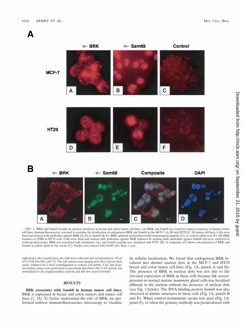
its cellular localization. We found that endogenous BRK lo-calized into distinct nuclear dots in the MCF-7 and HT29breast and colon tumor cell lines (Fig. 1A, panels A and D).The presence of BRK in nuclear dots was not due to theelevated expression of BRK in these cells because Sik overex-pression in normal murine mammary gland cells was localizeddiffusely in the nucleus without the presence of nuclear dots(see Fig. 5 below). The RNA binding protein Sam68 was alsoobserved in similar structures in these cells (Fig. 1A, panels Band E). When control nonimmune serum was used (Fig. 1A,panel F), or when the primary antibody was preincubated with
FIG. 1. BRK and Sam68 localize in nuclear structures in breast and colon tumor cell lines. (A) BRK and Sam68 are found in nuclear structures in human tumorcell lines. Immunofluorescence was used to examine the localization of endogenous BRK and Sam68 in the MCF-7 (A, B) and HT29 (C, D) tumor cell lines. Cells werefixed and stained with antibodies against BRK (A, D) or Sam68 (B, E), BRK antibody preincubated with immunogenic peptide (C), or control rabbit sera (F). (B) BRKlocalizes to SNBs in HT29 cells. Cells were fixed and stained with antibodies against BRK followed by staining with antibodies against Sam68 and were analyzed byconfocal microscopy. BRK was visualized with rhodamine (A), and Sam68 staining was visualized with FITC (B). A composite (C) shows colocalization of BRK andSam68 as yellow spots in the nuclei (C). Nuclei were stained with DAPI (D). Bars, 5 mm.
6116 DERRY ET AL. MOL. CELL. BIOL.
on Septem
ber 21, 2015 by guesthttp://m
cb.asm.org/
Dow
nloaded from
the immunogenic peptide (Fig. 1A, panel C; BRK antibodyplus BRK peptide, long exposure), no dots were observed,confirming the specificity of the signal.
Previously, Sam68 was reported to localize to novel nuclearstructures termed SNBs that are novel and distinct from coiledbodies, gems, PML nuclear bodies, the perinucleolar compart-ment, and SC-35 speckles (4). SNBs contain nucleic acid that ismost likely RNA, but their function remains unknown (4).We examined whether BRK colocalized with Sam68 in SNBs.HT29 cells were fixed and stained with antibodies againstSam68 (4) and BRK (BRK C-17), followed by secondary an-tibodies conjugated to FITC and rhodamine, respectively. Thefluorescent signals were imaged using confocal microscopy. Itwas observed that most of the BRK nuclear dots colocalizedwith Sam68 SNBs (Fig. 1B). These findings demonstrate thatBRK is a nuclear kinase that appears to be a component ofSNBs in the HT29 colon cancer cell line.
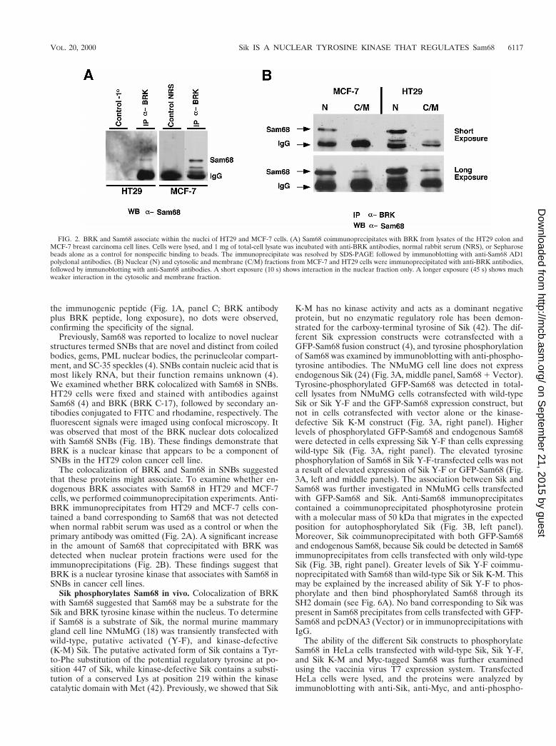
The colocalization of BRK and Sam68 in SNBs suggestedthat these proteins might associate. To examine whether en-dogenous BRK associates with Sam68 in HT29 and MCF-7cells, we performed coimmunoprecipitation experiments. Anti-BRK immunoprecipitates from HT29 and MCF-7 cells con-tained a band corresponding to Sam68 that was not detectedwhen normal rabbit serum was used as a control or when theprimary antibody was omitted (Fig. 2A). A significant increasein the amount of Sam68 that coprecipitated with BRK wasdetected when nuclear protein fractions were used for theimmunoprecipitations (Fig. 2B). These findings suggest thatBRK is a nuclear tyrosine kinase that associates with Sam68 inSNBs in cancer cell lines.
Sik phosphorylates Sam68 in vivo. Colocalization of BRKwith Sam68 suggested that Sam68 may be a substrate for theSik and BRK tyrosine kinase within the nucleus. To determineif Sam68 is a substrate of Sik, the normal murine mammarygland cell line NMuMG (18) was transiently transfected withwild-type, putative activated (Y-F), and kinase-defective(K-M) Sik. The putative activated form of Sik contains a Tyr-to-Phe substitution of the potential regulatory tyrosine at po-sition 447 of Sik, while kinase-defective Sik contains a substi-tution of a conserved Lys at position 219 within the kinasecatalytic domain with Met (42). Previously, we showed that Sik
K-M has no kinase activity and acts as a dominant negativeprotein, but no enzymatic regulatory role has been demon-strated for the carboxy-terminal tyrosine of Sik (42). The dif-ferent Sik expression constructs were cotransfected with aGFP-Sam68 fusion construct (4), and tyrosine phosphorylationof Sam68 was examined by immunoblotting with anti-phospho-tyrosine antibodies. The NMuMG cell line does not expressendogenous Sik (24) (Fig. 3A, middle panel, Sam68 1 Vector).Tyrosine-phosphorylated GFP-Sam68 was detected in total-cell lysates from NMuMG cells cotransfected with wild-typeSik or Sik Y-F and the GFP-Sam68 expression construct, butnot in cells cotransfected with vector alone or the kinase-defective Sik K-M construct (Fig. 3A, right panel). Higherlevels of phosphorylated GFP-Sam68 and endogenous Sam68were detected in cells expressing Sik Y-F than cells expressingwild-type Sik (Fig. 3A, right panel). The elevated tyrosinephosphorylation of Sam68 in Sik Y-F-transfected cells was nota result of elevated expression of Sik Y-F or GFP-Sam68 (Fig.3A, left and middle panels). The association between Sik andSam68 was further investigated in NMuMG cells transfectedwith GFP-Sam68 and Sik. Anti-Sam68 immunoprecipitatescontained a coimmunoprecipitated phosphotyrosine proteinwith a molecular mass of 50 kDa that migrates in the expectedposition for autophosphorylated Sik (Fig. 3B, left panel).Moreover, Sik coimmunoprecipitated with both GFP-Sam68and endogenous Sam68, because Sik could be detected in Sam68immunoprecipitates from cells transfected with only wild-typeSik (Fig. 3B, right panel). Greater levels of Sik Y-F coimmu-noprecipitated with Sam68 than wild-type Sik or Sik K-M. Thismay be explained by the increased ability of Sik Y-F to phos-phorylate and then bind phosphorylated Sam68 through itsSH2 domain (see Fig. 6A). No band corresponding to Sik waspresent in Sam68 precipitates from cells transfected with GFP-Sam68 and pcDNA3 (Vector) or in immunoprecipitations withIgG.
The ability of the different Sik constructs to phosphorylateSam68 in HeLa cells transfected with wild-type Sik, Sik Y-F,and Sik K-M and Myc-tagged Sam68 was further examinedusing the vaccinia virus T7 expression system. TransfectedHeLa cells were lysed, and the proteins were analyzed byimmunoblotting with anti-Sik, anti-Myc, and anti-phospho-
FIG. 2. BRK and Sam68 associate within the nuclei of HT29 and MCF-7 cells. (A) Sam68 coimmunoprecipitates with BRK from lysates of the HT29 colon andMCF-7 breast carcinoma cell lines. Cells were lysed, and 1 mg of total-cell lysate was incubated with anti-BRK antibodies, normal rabbit serum (NRS), or Sepharosebeads alone as a control for nonspecific binding to beads. The immunoprecipitate was resolved by SDS-PAGE followed by immunoblotting with anti-Sam68 AD1polyclonal antibodies. (B) Nuclear (N) and cytosolic and membrane (C/M) fractions from MCF-7 and HT29 cells were immunoprecipitated with anti-BRK antibodies,followed by immunoblotting with anti-Sam68 antibodies. A short exposure (10 s) shows interaction in the nuclear fraction only. A longer exposure (45 s) shows muchweaker interaction in the cytosolic and membrane fraction.
VOL. 20, 2000 Sik IS A NUCLEAR TYROSINE KINASE THAT REGULATES Sam68 6117
on Septem
ber 21, 2015 by guesthttp://m
cb.asm.org/
Dow
nloaded from

tyrosine antibodies. Several proteins, including one comi-grating with Sam68, appeared heavily phosphorylated by theSik Y-F construct (Fig. 4A, right panel). These data provideadditional evidence that Sik is negatively regulated by phos-phorylation of the carboxy-terminal tyrosine at position 447and that substitution of Sik Y447F activates the kinase. Theanti-Sik and anti-Myc immunoblots show equivalent expres-sion of the proteins (Fig. 4A, left and middle panels).
The ability of Sik Y-F to phosphorylate other nuclear KH
domain proteins, such as hnRNPK, was examined. A truncatedform of Sam68, Sam68 (68–347), which contains amino acids68 to 347 and lacks part of the amino terminus and the ty-rosine-rich carboxy terminus, was also investigated as a sub-strate for Sik. HeLa cells were transfected with Sik Y-F aloneor were cotransfected with Sik Y-F and Myc-Sam68, Myc-hnRNPK, or Myc-Sam68 (68–347). The cells were lysed andimmunoprecipitated with control IgG or anti-Myc antibodies.The bound proteins were analyzed by immunoblotting with
FIG. 3. Sik phosphorylates Sam68 in vivo. (A) Sam68 is tyrosine phosphorylated in NMuMG cells expressing wild-type Sik and Sik Y-F. NMuMG cells werecotransfected with GFP-Sam68 and Vector alone, wild-type (WT) Sik, Sik Y-F, or kinase-defective Sik K-M. Total-cell lysates were divided equally and immunoblottedwith antibodies against Sam68, Sik, and phosphotyrosine. Tyrosine-phosphorylated Sam68 was detected only in lysates containing wild-type Sik or Sik Y-F. Althoughlower levels of Sik Y-F were expressed than wild-type Sik, the highest levels of tyrosine-phosphorylated Sam68 were detected in cells cotransfected with Sik Y-F,suggesting that tyrosine 447 of Sik negatively regulates its activity. (B) Sik associates with Sam68. Immunoprecipitations were performed with Sam68 antibody or IgGas a control and with lysates from the transfected cells in panel A. Immunoblotting was performed with antiphosphotyrosine or anti-Sik antibodies. Sik was observedto coprecipitate with Sam68 from lysates of cells transfected with Sik expression constructs. Sik antibody binding was detected using HRP-conjugated protein A.Tyrosine-phosphorylated Sik could also be detected in the Sam68 immunoprecipitates (left panel).
6118 DERRY ET AL. MOL. CELL. BIOL.
on Septem
ber 21, 2015 by guesthttp://m
cb.asm.org/
Dow
nloaded from

FIG. 4. Sik Y-F has increased kinase activity and specifically phosphorylates Sam68. (A) The carboxy-terminal tyrosine of Sik functions as a negative regulatory site.HeLa cells were cotransfected with Myc-tagged Sam68 and wild-type (WT) Sik, Sik Y-F, or Sik K-M (kinase defective). Cells were lysed, the proteins were separatedby SDS-PAGE, and immunoblotting with anti-Sik, anti-Myc, and antiphosphotyrosine antibodies was performed. Increased phosphorylation of Myc-Sam68 was visiblein total-cell lysates from Sik Y-F-transfected cells (right panel). (B) HeLa cells were transfected with Sik Y-F or were cotransfected with Sik Y-F and Myc-Sam68,Myc-hnRNPK, or truncated Myc-Sam68 (68-347). The cells were lysed and immunoprecipitated with IgG (control) or anti-Myc antibodies. The bound proteins wereanalyzed by immunoblotting with antiphosphotyrosine antibodies (left panel). The same membrane was subsequently immunoblotted with anti-Myc antibodies (middlepanel). Total-cell extracts (TCL) were immunoblotted with anti-Sik antibodies (right panel). The band at ;55 kDa in lanes 1 to 16 represents the heavy chain of theimmunoprecipitating antibodies. (C) Sik phosphorylates the carboxy terminus of Sam68. In vitro kinase assays were performed using full-length GST-Sik, [g-32P]ATP,and 2 mg of the following substrates: GST-Grb2-N-SH3 (negative control), GST alone, GST-Sam68 (331–443), or GST-Sam68 (354–393). The proteins were separatedby SDS-PAGE and stained with Coomassie blue (left). The gel was dried, and the phosphorylated proteins were visualized by autoradiography (right).
VOL. 20, 2000 Sik IS A NUCLEAR TYROSINE KINASE THAT REGULATES Sam68 6119
on Septem
ber 21, 2015 by guesthttp://m
cb.asm.org/
Dow
nloaded from

antiphosphotyrosine, anti-Myc, and anti-Sik antibodies (Fig.4B). A phosphotyrosine-containing protein with a molecularmass of 68 kDa (lane 6) was observed in anti-Myc immuno-precipitates from extracts transfected with wild-type Myc-tagged Sam68, but not with Myc-hnRNPK or Myc-Sam68 (68–347) (Fig. 4B, left panel). The membrane was reimmunoblottedwith anti-Myc antibodies to confirm the equivalent expressionof Myc-Sam68, Myc-hnRNPK, or Myc-Sam68 (68–347) (Fig.4B, middle panel). Total-cell extracts were also verified for theequivalent expression of Sik Y-F by immunoblotting with anti-Sik antibodies (Fig. 4B, right panel). These data suggest thatthe C terminus of Sam68 is the target for the Sik tyrosinekinase.
To confirm that the carboxy terminus of Sam68 is directlyphosphorylated by Sik, we incubated full-length GST-Sik withGST-Sam68 (331-443) and GST-Sam68 (354-393), two Sam68carboxy terminus fusion proteins containing amino acids 331 to443 and 354 to 393, respectively, in the presence of [g-32P]ATP. These two carboxy-terminal fragments of Sam68 wereefficiently phosphorylated by Sik, whereas a control GST pro-tein containing the amino-terminal SH3 domain of Grb2 andGST alone was not phosphorylated (Fig. 4C, right panel). Theamounts of proteins used were determined to be equivalent asvisualized by Coomassie blue staining (left panel). These datasuggest that Sik directly and specifically phosphorylates the Cterminus of Sam68.
Colocalization of Sik, Sam68, and phosphotyrosine in thenuclei of transfected cells. The localization of Sik and Sam68was investigated in the NMuMG cell line. NMuMG cells weretransfected with both wild-type Sik and GFP-Sam68 and wereanalyzed by confocal microscopy. The pattern of wild-type Sikexpression was visualized by avidin-Alexa 350 (blue) (Fig. 5A,panel C) and was detected in the nucleus and at the mem-brane. Expression of the Sik Y-F and Sik K-M expressionconstructs was also detected in the nuclei and at the mem-branes of transfected cells (data not shown). GFP-Sam68, wild-type Sik, and phosphotyrosine colocalized in the nucleoplasmof the cells (Fig. 5A, panel D). Nuclear bodies were morecommonly seen in cancer cell lines, but were not generallyobserved in the nontransformed NMuMG cell line (Fig. 5Aand B). In experiments with control IgG, no specific fluores-cent signal was detected.
The localization and tyrosine phosphorylation of Sam68 inthe presence of wild-type Sik, Sik Y-F, and Sik K-M wereexamined (Fig. 5B). Phosphotyrosine was readily detected onlyin the nuclei of cells transfected with active Sik and Sam68,suggesting that Sik is active within the nuclei and phosphory-lates Sam68. Phosphotyrosine was detected in the nuclei ofcells transfected with wild-type Sik and Sik Y-F (Fig. 5B, pan-els B and F), but not in kinase-defective Sik K-M-transfectedcells (Fig. 5B, panel J). The majority of Sik-phosphorylatedprotein colocalized with Sam68 (Fig. 5B, panels C and G). Wealso observed that the intensity of the antiphosphotyrosine
staining was greatest in cells transfected with Sik Y-F andSam68. Cotransfection of the GFP expression vectorpEGFP-C1 and the empty Sik expression vector pcDNA3resulted in diffuse GFP fluorescence throughout the cell (Fig.5B, panel M) and no detectable antiphosphotyrosine staining(Fig. 5B, panel N). These data provide further support thatSam68 is a substrate for Sik in vivo.
Since BRK and Sam68 were observed to colocalize in SNBs(Fig. 1), the presence of phosphotyrosine in these SNBs fol-lowing transfection of the Sik expression constructs was exam-ined. HT29 cells were transfected with GFP-Sam68 and wild-type Sik, Sik Y-F, or kinase-defective Sik K-M. The transfectedcells were fixed, stained with antiphosphotyrosine antibodies,and analyzed by confocal microscopy. Tyrosine-phosphorylat-ed protein colocalized with Sam68 in SNBs in these trans-formed cells transfected with wild-type Sik and Sik Y-F, butnot with Sik K-M. The Sik Y-F transfection is shown in Fig. 5C.These data demonstrate that Sik most likely targets Sam68 inSNBs in cancer cell lines.
Sik-Sam68 interaction is mediated by both the SH3 andSH2 domains. To determine which part of Sik interacts withSam68, different domains of Sik were expressed in bacteria asGST-fusion proteins and used in GST pull down assays. Ly-sates from NMuMG cells transfected with wild-type Sik, SikY-F, or Sik K-M and GFP-Sam68 expression constructs wereincubated with GST alone, GST-Sik SH21SH3, GST-Sik SH2,and GST-Sik SH3 bound to beads. Bound proteins were de-tected by immunoblotting with anti-Sam68 AD1 antibody (Fig.6A). GFP-Sam68 and the endogenous Sam68 were observed toassociate with the Sik SH3 and Sik SH21SH3 domain fusionproteins (Fig. 6A). Association of GFP-Sam68 with the SikSH2 domain was observed only when it was coexpressed withwild-type Sik or Sik Y-F, but not kinase-defective Sik K-M(Fig. 6A). We also observed association of endogenous phos-phorylated Sam68 protein following longer exposures of theimmunoblots. These data demonstrate that Sik associates withSam68 through both its SH3 and SH2 domains.
Sam68 contains at least five proline motifs and interacts withthe SH3 domains of several proteins, including Src, Fyn, andPLCg-1 (14, 29, 39, 46). To determine which proline motifwithin Sam68 mediates interaction with the Sik SH3 domain,we used a GST pull down approach, with lysates from HeLacells that express human BRK and GST fusion proteins rep-resenting the five proline (P0, P1P2, P3, and P4) motifs inSam68. We found that GST-P3 was the main polypeptide thatinteracted with BRK (Fig. 6B). To further confirm the specificinteraction of Sam68 P3 with the Sik SH3 domain, we testedthe ability of peptides representing the proline-rich sequencesP0, P3, and P4 to compete with binding of Sam68 with theGST-Sik SH3 domain. The Sam68 P3 peptide competed withthe binding between Sam68 and the Sik SH3 domain. Nosignificant competition was observed with the P0 and P4 pep-tides. Thus, the Sik SH3 domain appears to interact with one
FIG. 5. Wild-type Sik and Sik Y-F phosphorylate nuclear proteins that colocalize with GFP-Sam68 within the nucleus. (A) Localization of GFP-tagged Sam68,phosphotyrosine, and wild-type (WT) Sik in transfected NMuMG cells. Antiphosphotyrosine antibody was visualized with rhodamine (red), while anti-Sik antibodybinding was visualized with avidin-Alexa 350 (blue). Wild-type Sik is present in the nucleus and at the membrane (panel C). (B) NMuMG cells were transfected withGFP-Sam68 and wild-type Sik (A to D), GFP-Sam68 and Sik Y-F (E to H), GFP-Sam68 and kinase-defective Sik K-M (I to L), or the GFP expression vector pEGFP-C1and pcDNA3 (M to P). Cells were fixed 24 h after transfection, and tyrosine-phosphorylated proteins were localized using anti-phosphotyrosine antibodies (B, F, J, N).DAPI was used to stain the nuclei (D, H, L, P). In NMuMG cells, Sam68 displays diffuse, nuclear localization visible by green fluorescence (A, E, I). Cells cotransfectedwith GFP-Sam68 and wild-type Sik or Sik Y-F also stain strongly with the anti-phosphotyrosine antibody visualized using rhodamine (B, F), while no phosphotyrosinewas detected in cells expressing kinase-defective Sik K-M (J). Panels C, G, K, and O are composites demonstrating colocalization that appears yellow. GFP alone is expressedthroughout the cell (M) and is negative for anti-phosphotyrosine staining (N). (C) Increased phosphotyrosine in SNBs in HT29 cells following introduction of theactivated Sik Y-F construct into HT29 cells. HT29 cells were transfected with GFP-Sam68 (A, C) and Sik Y-F, and tyrosine-phosphorylated proteins were localizedusing antiphosphotyrosine antibodies (B, C). Colocalization of Sam68 and the increased phosphotyrosine signal in SNBs are shown in panel C. No phosphotyrosinesignal was detected in control cells transfected with kinase-defective Sik K-M (not shown). DAPI was used to stain the nuclei (D). Bars represent 5 mm.
6120 DERRY ET AL. MOL. CELL. BIOL.
on Septem
ber 21, 2015 by guesthttp://m
cb.asm.org/
Dow
nloaded from

VOL. 20, 2000 Sik IS A NUCLEAR TYROSINE KINASE THAT REGULATES Sam68 6121
on Septem
ber 21, 2015 by guesthttp://m
cb.asm.org/
Dow
nloaded from

major proline motif in Sam68, P3, that is neither a type I nora type II proline motif (13).
Sik inhibits the RNA binding ability of Sam68. Tyrosinephosphorylation of Sam68D1–67 negatively regulates its abilityto bind to homopolymeric RNA (45). To determine whetherSik can regulate the ability of Sam68 to bind RNA, HeLa cells
were transfected with Myc-Sam68D1–67 or cotransfected withMyc-Sam68D1–67 and Sik Y-F or Fyn. The expression of Fynserved as a positive control, as we have shown previously thatFyn can negatively regulate Sam68D1–67 homopolymeric RNAbinding (45). HeLa cell lysates were divided equally and incu-bated with either poly(U) immobilized to agarose or agarose
FIG. 6. The Sik SH2 and SH3 domains bind Sam68. (A) The ability of Sam68 to bind the SH2 and SH3 domains of Sik was tested. NMuMG cells were transfectedwith GFP-Sam68 and the expression vector pcDNA3 (Vector) or the wild-type Sik, activated Sik Y-F, or kinase-defective Sik K-M expression constructs. Cell lysateswere divided equally and incubated with GST, GST-Sik SH21SH3, GST-Sik SH2, and GST-SH3 covalently coupled to beads. Bound proteins as well as an aliquot oftotal-cell lysate from GFP-Sam68-transfected cells were separated by SDS-PAGE and immunoblotted with Sam68 AD1 polyclonal antibody. Positions of GFP-Sam68and the endogenous Sam68 protein are indicated with arrows. GFP-Sam68 and endogenous Sam68 protein bound to the GST-Sik, SH21SH3, and GST-Sik SH3 fusionproteins in all of the cell lysates. GFP-Sam68 binding to the GST-Sik SH2 domain was detected only in cells transfected with wild-type Sik or Sik Y-F, suggesting thatphosphorylation by Sik is required for Sik SH2 binding. (B) Sam68 proline motif 3 (P3) associates with the SH3 domain of Sik. HeLa cell lysates were incubated withGST, GST-Sam68 P0, GST-Sam68 P1P2, GST-Sam68 P3, or GST Sam68 P4 covalently coupled to beads. The beads were washed, and the bound BRK was observedby immunoblotting. An aliquot of the HeLa cell lysate was used to represent total-cell lysate. BRK binding was only detected with the GST-Sam68 P3 fusion protein.(C) Beads containing covalently coupled GST-Sik SH3 protein were preincubated for 15 min at room temperature with the indicated concentration of Sam68proline-rich peptide. Subsequently, HeLa cell lysates were added to each tube for 30 min at 4°C. The beads were washed extensively, and the bound Sam68 wasquantitated by immunoblotting. Binding of Sam68 with the GST-Sik SH3 fusion protein was efficiently competed with the P3 peptide.
6122 DERRY ET AL. MOL. CELL. BIOL.
on Septem
ber 21, 2015 by guesthttp://m
cb.asm.org/
Dow
nloaded from

alone. Sam68 bound poly(U) homopolymeric RNA, when ex-pressed alone (Fig. 7A). However, little or no RNA bindingwas detected when Sam68 was coexpressed with Sik Y-F or Fyn(Fig. 7A). The reduction of bound Myc-Sam68 was not due topoor expression of Myc-Sam68 (Fig. 7A). Total-cell lysateswere immunoblotted with anti-Sik, anti-Fyn, and anti-phos-photyrosine antibodies, and we found that Sik Y-F and Fynwere both overexpressed and Myc-Sam68 was tyrosine phos-
phorylated (Fig. 7B). These data suggest that Sik is able tonegatively regulate the ability of Sam68 to bind RNA.
Sam68 has been shown to function as a cellular homologueof Rev in transporting HIV RNA (27). We examined whetherSik could regulate this nuclear function of Sam68 by determin-ing if Sik could modulate RRE-directed reporter gene expres-sion (Fig. 8). COS7 cells were transfected with an RRE-CATreporter plasmid in the presence of Sam68 and increasing
FIG. 7. Sik negatively regulates the ability of Sam68 to bind RNA. (A) HeLa cell lysates from cells either transfected with Myc-Sam68D1–67 alone or cotransfectedwith Myc-Sam68D1–67 and Sik Y-F or Fyn were divided equally and precipitated with agarose (Con) or poly(U)-agarose (pU), followed by anti-Myc immunoblotting.An aliquot of total-cell lysate (TCL) was also included to monitor expression of Myc-Sam68D1–67. The ability of Myc-Sam68D1–67 to bind poly(U) agarose wasinhibited in cells transfected with either Sik Y-F or Fyn. (B) Sik and Fyn are efficiently expressed and phosphorylate Myc-Sam68D1–67 in transfected cells. Cell lysatesfrom transfected cells were immunoblotted with anti-Sik and anti-Fyn antibodies to demonstrate that these kinases were efficiently expressed. Immunoprecipitation ofMyc-Sam68D1–67 followed by immunoblotting with antiphosphotyrosine confirmed that Myc-Sam68D1–67 was tyrosine phosphorylated by both Sik and Fyn.
VOL. 20, 2000 Sik IS A NUCLEAR TYROSINE KINASE THAT REGULATES Sam68 6123
on Septem
ber 21, 2015 by guesthttp://m
cb.asm.org/
Dow
nloaded from

amounts of kinase-active Sik Y-F or kinase-inactive Sik K-M.The transfection of Sam68 or Rev with the RRE-CAT reporterresulted in a 10-fold increase in CAT activation in comparisonto Sam68DC and RevM10, two proteins shown to be inactive inRev function. These data are consistent with previously pub-lished data (27). The cotransfection of Sam68 with Sik Y-F, butnot Sik K-M, decreased CAT activity in a dose-dependentmanner. These findings indicate that Sik can regulate a nuclearfunction of Sam68 and that its kinase activity is required.
DISCUSSION
The tyrosine kinases represent a large family of diverse pro-teins that play important roles in the regulation of growth anddifferentiation. Thus far, only a small number of nuclear ty-rosine kinases have been identified, including Abl, Rak, Fes,Fer, and the dual-specificity kinase Wee1 (reviewed in refer-ences 26 and 44). In these studies, we showed that Sik andBRK are present in the nucleus. The mechanisms by which Sikand BRK localize to the nucleus are unknown, as they lack aclear nuclear localization signal (41). It is possible that Sik and
BRK are transported to the nucleus through association withother proteins containing nuclear localization signals. LikeAbl, Sik and BRK are not nuclear specific. In addition to beingpresent in the nucleus, BRK was detected in the cytoplasm ofHT29 cells (Fig. 1). Sik protein and kinase activity were alsodetected at the membrane of transfected NMuMG cells (Fig.5A), consistent with the earlier observation that Sik can asso-ciate with a GAP-associated protein (37).
We demonstrate that the RNA binding protein Sam68 is asubstrate for Sik within the nucleus and that Sik can inhibit theability of Sam68 to bind RNA. RNA binding proteins mayregulate gene expression by a number of mechanisms (re-viewed in reference 32). They may alter RNA structure toregulate interaction with trans-acting factors or provide local-ization or targeting signals. Although its cellular function isunknown, Sam68 has been shown to be able to functionallysubstitute for the HIV-1 Rev protein, which plays an essentialrole in the nuclear export of unspliced and partially splicedviral transcripts and export of the HIV genome (27). We showthat Sik kinase activity can negatively regulate the ability ofSam68 to function as a cellular homologue of Rev. These data
FIG. 8. Sik negatively regulates the ability of Sam68 to function as a cellular Rev homologue. (A) A schematic diagram of the Rev responsive element reporter CATconstruct is shown. The splice acceptor and donor sites are indicated as SA and SD. CAT indicates the chloramphenicol acetyltransferase cDNA, and RRE is the HIVRev responsive element. (B) COS7 cells were transfected with the RRE-CAT reporter plasmid in the presence of the indicated expression vectors and pCH110. CATactivity was normalized to b-galactosidase activity. Each bar represents CAT activity from at least eight samples from at least three separate experiments and thestandard deviation indicated. The autoradiogram shown on the left represents a typical experiment: the two rows of dots on the left represent monoacetylated[14C]chloramphenicol and the row on the right represents unacetylated [14C]chloramphenicol.
6124 DERRY ET AL. MOL. CELL. BIOL.
on Septem
ber 21, 2015 by guesthttp://m
cb.asm.org/
Dow
nloaded from

strengthen the possibility that signaling cascades can regulatethe RNA function of GSG domain-containing proteins orSTAR proteins. The ability of Sam68 to act in RNA transportsuggests a role in posttranscriptional regulation of gene expres-sion, which may be negatively regulated by Sik within thenucleus. Sam68 may also serve as an adaptor for Sik, bringingit into proximity of other, as of yet unidentified, substrates.
Sik and its human orthologue BRK have only 80% aminoacid sequence identity (24). Nevertheless, the genes have beenmapped to regions of the mouse and human genomes thatshare conservation of synteny, and we have found that themouse and human proteins are expressed in similar patterns indifferentiated epithelial tissues (24). Here we show that themouse and human proteins both localize to the nucleus andthat they both associate with Sam68. These data provide fur-ther evidence that the functions of Sik and BRK in the twospecies are conserved.
NMuMG cells were isolated from the mammary glands ofNamru mice and have epithelial growth characteristics, andthey do not form malignant lesions when introduced into nudemice (18). Sik localization is diffuse within the nuclei of im-mortalized NMuMG cells, while BRK appears in SNBs in theHT29 colon adenocarcinoma cell line (Fig. 5). These datacomplement earlier studies by Chen et al. (4), who found thatSNBs were predominant in transformed cells. SNBs are novelunique dynamic structures that disassemble when transcriptionis inhibited with actinomycin D (4). When GFP-Sam68 andwild-type Sik are introduced into HT29 cells, they localize tothe SNBs, which become tyrosine phosphorylated (Fig. 5C).Tyrosine phosphorylation of Sam68 by Sik does not appear toalter its localization because Sam68 coexpressed with active SikY-F is retained in SNBs (Fig. 5).
We have shown that Sik can bind Sam68 through both itsSH3 and SH2 domains. It was previously shown that Sam68also binds SH3 and SH2 domains of Src kinases (14, 29, 39, 46).The binding affinities of specific SH2 domains are influencedby sequence context. For example, Src family members preferthe sequence pYEEI, while the SH2 domains of p85 andPLC-g select the general motif pY-hydrophobic-X-hydropho-bic (35, 36). Using a technique employing degenerate phos-phopeptide libraries to predict the specificity of individual SH2domains, it was determined that the Sik SH2 domain may bindto phosphorylated proteins with p-YEEY, YEDY, YDEY,and YDDY motifs (Z. Songyang and L. C. Cantley, personalcommunication). Interestingly, Sam68 contains the sequenceYEDY in its carboxy terminus, and this is a putative bindingsite for the Sik SH2 domain. This sequence may also be thetarget of Sik, as we show here that Sik can phosphorylate thecarboxy terminus of Sam68.
Sam68 is the first substrate identified for the Sik kinase.Related KH domain-containing proteins have been shown toplay important roles in development, and these include humanFMR1 (fragile X mental retardation syndrome) (9); mouseQk1 (quaking), required for myelination (12); Caenorhabditiselegans GLD-1, required for germ cell differentiation (19); andDrosophila Who/How, required for muscle differentiation (1).The tissue- and differentiation-specific expression and activa-tion of Sik (41, 42) suggest that Sik is involved in regulatingepithelial cell differentiation. Sik is unique in that it is the onlyknown tyrosine kinase that can phosphorylate Sam68 withinthe nucleus, where it can modulate its RNA binding ability andperhaps the pattern of gene expression associated with epithe-lial cell differentiation. Since Sam68 has also been shown to beinvolved in cell cycle regulation (3), overexpression of Sik andBRK may contribute to the development of epithelial cancersby altering the ability of Sam68 to regulate cell growth.
ACKNOWLEDGMENTS
This work was supported by National Institutes of Health grantDK44525, Department of the Army grant DAMD17-96-1-6175(A.L.T.), and Medical Research Council of Canada grant MT 13377S.R. is a Scholar of the MRC, and T.C. is supported by a DoctoralResearch Award from the MRC. J.J.D. is supported by the SignalTransduction and Cellular Endocrinology NIH training grant DK07739.
We thank Michael Serfas and Shahab Uddin for helpful discussionsand critical reading of the manuscript.
J.J.D. and S.R. contributed equally to this work.
REFERENCES
1. Baehrecke, E. H. 1997. who encodes a KH RNA binding protein that func-tions in muscle development. Development 124:1323–1332.
2. Barker, K. T., L. E. Jackson, and M. R. Crompton. 1997. BRK tyrosinekinase expression in a high proportion of human breast carcinomas. Onco-gene 15:799–805.
3. Barlat, I., F. Maurier, M. Duchesne, E. Guitard, B. Tocque, and F. Schweig-hoffer. 1997. A role for Sam68 in cell cycle progression antagonized by aspliced variant within the KH domain. J. Biol. Chem. 272:3129–3132.
4. Chen, T., F. M. Boisvert, D. P. Bazett-Jones, and S. Richard. 1999. A role forthe GSG domain in localizing Sam68 to novel nuclear structures in cancercell lines. Mol. Biol. Cell 10:3015–3033.
5. Chen, T., B. B. Damaj, C. Herrera, P. Lasko, and S. Richard. 1997. Self-association of the single-KH-domain family members Sam68, GRP33,GLD-1, and Qk1: role of the KH domain. Mol. Cell. Biol. 17:5707–5718.
6. Chen, T., and S. Richard. 1998. Structure-function analysis of Qk1: a lethalpoint mutation in mouse quaking prevents homodimerization. Mol. Cell.Biol. 18:4863–4871.
7. Cochrane, A. W., C. H. Chen, R. Kramer, L. Tomchak, and C. A. Rosen.1989. Purification of biologically active human immunodeficiency virus revprotein from Escherichia coli. Virology 173:335–337.
8. Cooper, J. A., and B. Howell. 1993. The when and how of Src regulation. Cell73:1051–1054.
9. De Boulle, K., A. J. Verkerk, E. Reyniers, L. Vits, J. Hendrickx, B. Van Roy,F. Van den Bos, E. de Graaff, B. A. Oostra, and P. J. Willems. 1993. A pointmutation in the FMR-1 gene associated with fragile X mental retardation.Nat. Genet. 3:31–35.
10. Di Fruscio, M., T. Chen, S. Bonyadi, P. Lasko, and S. Richard. 1998. Theidentification of two Drosophila K homology domain proteins. Kep1 andSAM are members of the Sam68 family of GSG domain proteins. J. Biol.Chem. 273:30122–30130.
11. Easty, D. J., P. J. Mitchell, K. Patel, V. A. Florenes, R. A. Spritz, and D. C.Bennett. 1997. Loss of expression of receptor tyrosine kinase family genesPTK7 and SEK in metastatic melanoma. Int. J. Cancer 71:1061–1065.
12. Ebersole, T. A., Q. Chen, M. J. Justice, and K. Artzt. 1996. The quaking geneproduct necessary in embryogenesis and myelination combines features ofRNA binding and signal transduction proteins. Nat. Genet. 12:260–265.
13. Feng, S., C. Kasahara, R. J. Rickles, and S. L. Schreiber. 1995. Specificinteractions outside the proline-rich core of two classes of Src homology 3ligands. Proc. Natl. Acad. Sci. USA 92:12408–12415.
14. Fumagalli, S., N. F. Totty, J. J. Hsuan, and S. A. Courtneidge. 1994. A targetfor Src in mitosis. Nature 368:871–874.
15. Gibson, T. J., J. D. Thompson, and J. Heringa. 1993. The KH domain occursin a diverse set of RNA-binding proteins that include the antiterminatorNusA and is probably involved in binding to nucleic acid. FEBS Lett. 324:361–366.
16. Guan, K. L., and J. E. Dixon. 1991. Eucaryotic proteins expressed in Esch-erichia coli: an improved thrombin cleavage and purification procedure offusion proteins with glutathione S-transferase. Anal. Biochem. 192:262–267.
17. Hope, T. J., X. J. Huang, D. McDonald, and T. G. Parslow. 1990. Steroid-receptor fusion of the human immunodeficiency virus type 1 Rev transacti-vator: mapping cryptic functions of the arginine-rich motif. Proc. Natl. Acad.Sci. USA 87:7787–7791.
18. Hynes, N. E., R. Jaggi, S. C. Kozma, R. Ball, D. Muellener, N. T. Wetherall,B. W. Davis, and B. Groner. 1985. New acceptor cell for transfected genomicDNA: oncogene transfer into a mouse mammary epithelial cell line. Mol.Cell. Biol. 5:268–272.
19. Jones, A. R., and T. Schedl. 1995. Mutations in gld-1, a female germ cell-specific tumor suppressor gene in Caenorhabditis elegans, affect a conserveddomain also found in Src-associated protein Sam68. Genes Dev. 9:1491–1504.
20. Lang, V., D. Mege, M. Semichon, H. Gary-Gouy, and G. Bismuth. 1997. Adual participation of ZAP-70 and scr protein tyrosine kinases is required forTCR-induced tyrosine phosphorylation of Sam68 in Jurkat T cells. Eur.J. Immunol. 27:3360–3367.
21. Lawe, D. C., C. Hahn, and A. J. Wong. 1997. The Nck SH2/SH3 adaptorprotein is present in the nucleus and associates with the nuclear proteinSAM68. Oncogene 14:223–231.
22. Lee, S. T., K. M. Strunk, and R. A. Spritz. 1993. A survey of protein tyrosine
VOL. 20, 2000 Sik IS A NUCLEAR TYROSINE KINASE THAT REGULATES Sam68 6125
on Septem
ber 21, 2015 by guesthttp://m
cb.asm.org/
Dow
nloaded from

kinase mRNAs expressed in normal human melanocytes. Oncogene 8:3403–3410.
23. Lin, Q., S. J. Taylor, and D. Shalloway. 1997. Specificity and determinants ofSam68 RNA binding. Implications for the biological function of K homologydomains. J. Biol. Chem. 272:27274–27280.
24. Llor, X., M. S. Serfas, W. Bie, V. Vasioukhin, M. Polonskaia, J. Derry, C. M.Abbott, and A. L. Tyner. 1999. BRK/Sik expression in the gastrointestinaltract and in colon tumors. Clin. Cancer Res. 5:1767–1777.
25. Mitchell, P. J., K. T. Barker, J. E. Martindale, T. Kamalati, P. N. Lowe, M. J.Page, B. A. Gusterson, and M. R. Crompton. 1994. Cloning and characteri-sation of cDNAs encoding a novel non-receptor tyrosine kinase, brk, ex-pressed in human breast tumours. Oncogene 9:2383–2390.
26. Pendergast, A. M. 1996. Nuclear tyrosine kinases: from Abl to WEE1. Curr.Opin. Cell Biol. 8:174–181.
27. Reddy, T. R., W. Xu, J. K. Mau, C. D. Goodwin, M. Suhasini, H. Tang, K.Frimpong, D. W. Rose, and F. Wong-Staal. 1999. Inhibition of HIV replica-tion by dominant negative mutants of Sam68, a functional homolog of HIV-1Rev. Nat. Med. 5:635–642.
28. Resnick, R. J., S. J. Taylor, Q. Lin, and D. Shalloway. 1997. Phosphorylationof the Src substrate Sam68 by Cdc2 during mitosis. Oncogene 15:1247–1253.
29. Richard, S., D. Yu, K. J. Blumer, D. Hausladen, M. W. Olszowy, P. A.Connelly, and A. S. Shaw. 1995. Association of p62, a multifunctional SH2-and SH3-domain-binding protein, with src family tyrosine kinases, Grb2, andphospholipase C gamma-1. Mol. Cell. Biol. 15:186–1897.
30. Richard, S., and H. H. Zingg. 1992. Analysis of cis-acting elements of theoxytocin gene by DNA-mediated gene transfer, p. 324–343. In P. M. Conn(ed.), Methods in neuroscience. Academic Press Inc, Orlando, Fla.
31. Sanchez-Margalet, V., and S. Najib. 1999. p68 Sam is a substrate of theinsulin receptor and associates with the SH2 domains of p85 PI3K. FEBSLett. 455:307–310.
32. Siomi, H., and G. Dreyfuss. 1997. RNA-binding proteins as regulators ofgene expression. Curr. Opin. Genet. Dev. 7:345–353.
33. Siomi, H., M. J. Matunis, W. M. Michael, and G. Dreyfuss. 1993. Thepre-mRNA binding K protein contains a novel evolutionarily conservedmotif. Nucleic Acids Res. 21:1193–1198.
34. Siyanova, E. Y., M. S. Serfas, I. A. Mazo, and A. L. Tyner. 1994. Tyrosinekinase gene expression in the mouse small intestine. Oncogene 9:2053–2057.
35. Songyang, Z., S. E. Shoelson, M. Chaudhuri, G. Gish, T. Pawson, W. G.
Haser, F. King, T. Roberts, S. Ratnofsky, R. J. Lechleider, et al. 1993. SH2domains recognize specific phosphopeptide sequences. Cell 72:767–778.
36. Songyang, Z., S. E. Shoelson, J. McGlade, P. Olivier, T. Pawson, X. R.Bustello, M. Barbacid, H. Sabe, H. Hanafusa, T. Yi, R. Ren, D. Baltimore, S.Ratnofsky, R. A. Feldman, and L. C. Cantley. 1994. Specific motifs recog-nized by the SH2 domains of Csk, 3BP2, fps/fes, GRB-2, HCP, SHC, Syk,and Vav. Mol. Cell. Biol. 14:2777–2785.
37. Swenarchuk, L., P. Harakidas, and A. Cochrane. 1999. Regulated expressionof HIV-1 Rev function in mammalian cell lines. Can. J. Microbiol. 45:480–490.
38. Taylor, S. J., M. Anafi, T. Pawson, and D. Shalloway. 1995. Functionalinteraction between c-Src and its mitotic target, Sam 68. J. Biol. Chem. 270:10120–10124.
39. Taylor, S. J., and D. Shalloway. 1994. An RNA-binding protein associatedwith Src through its SH2 and SH3 domains in mitosis. Nature 368:867–871.
40. Trub, T., J. D. Frantz, M. Miyazaki, H. Band, and S. E. Shoelson. 1997. Therole of a lymphoid-restricted, Grb2-like SH3-SH2-SH3 protein in T cellreceptor signaling. J. Biol. Chem. 272:894–902.
41. Vasioukhin, V., M. S. Serfas, E. Y. Siyanova, M. Polonskaia, V. J. Costigan,B. Liu, A. Thomason, and A. L. Tyner. 1995. A novel intracellular epithelialcell tyrosine kinase is expressed in the skin and gastrointestinal tract. Onco-gene 10:349–357.
42. Vasioukhin, V., and A. L. Tyner. 1997. A role for the epithelial-cell-specifictyrosine kinase Sik during keratinocyte differentiation. Proc. Natl. Acad. Sci.USA 94:14477–14482.
43. Vernet, C., and K. Artzt. 1997. STAR, a gene family involved in signaltransduction and activation of RNA. Trends Genet. 13:479–484.
44. Wang, J. Y. 1994. Nuclear protein tyrosine kinases. Trends Biochem. Sci. 19:373–376.
45. Wang, L. L., S. Richard, and A. S. Shaw. 1995. P62 association with RNA isregulated by tyrosine phosphorylation. J. Biol. Chem. 270:2010–2013.
46. Weng, Z., S. M. Thomas, R. J. Rickles, J. A. Taylor, A. W. Brauer, C.Seidel-Dugan, W. M. Michael, G. Dreyfuss, and J. S. Brugge. 1994. Identi-fication of Src, Fyn, and Lyn SH3-binding proteins: implications for a func-tion of SH3 domains. Mol. Cell. Biol. 14:4509–4521.
47. Wong, G., O. Muller, R. Clark, L. Conroy, M. F. Moran, P. Polakis, and F.McCormick. 1992. Molecular cloning and nucleic acid binding properties ofthe GAP-associated tyrosine phosphoprotein p62. Cell 69:551–558.
6126 DERRY ET AL. MOL. CELL. BIOL.
on Septem
ber 21, 2015 by guesthttp://m
cb.asm.org/
Dow
nloaded from
Related Documents