MOLECULAR AND CELLULAR BIOLOGY, Oct. 2011, p. 3997–4006 Vol. 31, No. 19 0270-7306/11/$12.00 doi:10.1128/MCB.05808-11 Copyright © 2011, American Society for Microbiology. All Rights Reserved. Pirh2 E3 Ubiquitin Ligase Monoubiquitinates DNA Polymerase Eta To Suppress Translesion DNA Synthesis † Yong-Sam Jung, 1 Anne Hakem, 2,3 Razqallah Hakem, 2,3 and Xinbin Chen 1 * Comparative Oncology Laboratory, University of California, Davis, California 95616, 1 and Department of Medical Biophysics, University of Toronto, 2 and Ontario Cancer Institute, University Health Network, 3 Toronto, Ontario, Canada Received 14 June 2011/Returned for modification 28 June 2011/Accepted 18 July 2011 Polymerase eta (PolH) is necessary for translesion DNA synthesis, and PolH deficiency predisposes xero- derma pigmentosum variant (XPV) patients to cancer. Due to the critical role of PolH in translesion DNA synthesis, the activity of PolH is tightly controlled and subjected to multiple regulations, especially posttrans- lational modifications. Here, we show that PolH-dependent lesion bypass and intracellular translocation are regulated by Pirh2 E3 ubiquitin ligase through monoubiquitination. Specifically, we show that Pirh2, a target of the p53 tumor suppressor, monoubiquitinates PolH at one of multiple lysine residues. We also show that monoubiquitination of PolH inhibits the ability of PolH to interact with PCNA and to bypass UV-induced lesions, leading to decreased viability of UV-damaged cells. Moreover, we show that monoubiquitination of PolH alters the ability of PolH to translocate to replication foci for translesion DNA synthesis of UV-induced DNA lesions. Considering that Pirh2 is known to be overexpressed in various cancers, we postulate that in addition to mutation of PolH in XPV patients, inactivation of PolH by Pirh2 via monoubiquitination is one of the mechanisms by which PolH function is controlled, which might be responsible for the development and progression of some spontaneous tumors wherein PolH is not found to be mutated. The hereditary material of an organism is under constant attack by endogenous genotoxic stresses and exogenous sources such as UV light and alkylation agents. UV light causes DNA lesions that block the accurate and processive DNA synthesis. Although the majority of DNA damage is recovered by excision repair systems, the remaining DNA lesions can hinder the activity of DNA polymerase during S phase (11). Under these conditions, many organisms require the action of specialized translesion DNA polymerases that can bypass var- ious DNA lesions (6, 8, 27). Many of these translesion DNA synthesis (TLS) polymerases, including PolH, belong to the Y-family DNA polymerases (8, 27). Xeroderma pigmentosum variant (XPV) cells were unable to replicate past UV-induced DNA lesions due to inactivating mutations in the PolH gene (14, 24). Thus, it is postulated that PolH-mediated replication bypass across these DNA lesions is particularly important to prevent UV-induced carcinogenesis in XPV patients. Ubiquitin (Ub) was originally identified as a 76-amino-acid (aa) ubiquitous, eukaryotic protein. Ubiquitin is conjugated to target proteins via an isopeptide bond between the C terminus of ubiquitin and a lysine residue; thereby, ubiquitinated pro- teins can be degraded by the 20S proteasome (13). In addition, ubiquitin can serve as a reversible modification that regulates the activity of target proteins without proteasomal degrada- tion, such as signal transduction, autophagy, chromatin remod- eling, membrane trafficking, and DNA repair (9). The conju- gation of a single ubiquitin molecule to a target protein is known as monoubiquitination. Histones, membrane proteins, and ubiquitin binding domain (UBD)-containing proteins are usually monoubiquitinated (1, 9, 12, 25). Due to the require- ment of a functional UBD, monoubiquitination of UBD-con- taining proteins is referred to as coupled monoubiquitination, which is also used as a signal for proteasomal degradation (3, 4, 18). Previously, we showed that Pirh2 physically interacts with PolH and subsequently promotes PolH degradation (16). However, Pirh2 does not catalyze PolH polyubiquitination. In addition, recent studies showed that the ubiquitination status of PolH controls its interaction with PCNA (1, 2, 26). These findings prompted us to examine whether Pirh2 monoubiquiti- nates PolH and regulates PolH-mediated TLS. Here, we dem- onstrated that Pirh2 promotes PolH monoubiquitination at K682, K686, and K694, located within the nuclear localization signal (NLS), and at K709. We also found that that Pirh2- mediated monoubiquitination inhibits PolH to interact with PCNA and its ability in the translesion DNA synthesis of UV- induced DNA lesions, which leads to decreased viability of UV-damaged cells. Our results indicate that Pirh2-mediated monoubiquitination is a mechanism by which PolH expression and activity are controlled. MATERIALS AND METHODS Cell culture. RKO, Pirh2-KD RKO (clone 11) in which Pirh2 can be inducibly knocked down, and primary human fibroblasts derived from XPV patients (GM03617) were used as described previously (21). Wild-type and Pirh2-knock- out mouse embryonic fibroblast (MEF) cells were generated from 13.5-day embryos according to standard procedures. Generation of Pirh2-knockout mice and Pirh2-knockout MEFs. Embryonic stem (ES) cells were electroporated with a linearized target construct to generate Pirh2 fl2-3-neo ES clones. Pirh2 2–3 ES clones lacking exons 2 and 3 and a neo- mycin resistance cassette were obtained following transient transfection of two targeted Pirh2 fl2-3-neo ES clones with cytomegalovirus (CMV)-Cre recombinase. All mice were in 129/C57BL/6 genetic background and were maintained in the animal facility of the Ontario Cancer Institute in accordance with the established * Corresponding author. Mailing address: Comparative Oncology Laboratory, University of California, Davis, CA 95616. Phone: (530) 754-8404. Fax: (530) 752-6042. E-mail: [email protected]. † Supplemental material for this article may be found at http://mcb .asm.org/. Published ahead of print on 26 July 2011. 3997

Welcome message from author
This document is posted to help you gain knowledge. Please leave a comment to let me know what you think about it! Share it to your friends and learn new things together.
Transcript
MOLECULAR AND CELLULAR BIOLOGY, Oct. 2011, p. 3997–4006 Vol. 31, No. 190270-7306/11/$12.00 doi:10.1128/MCB.05808-11Copyright © 2011, American Society for Microbiology. All Rights Reserved.
Pirh2 E3 Ubiquitin Ligase Monoubiquitinates DNA Polymerase EtaTo Suppress Translesion DNA Synthesis�†
Yong-Sam Jung,1 Anne Hakem,2,3 Razqallah Hakem,2,3 and Xinbin Chen1*Comparative Oncology Laboratory, University of California, Davis, California 95616,1 and Department of Medical Biophysics,
University of Toronto,2 and Ontario Cancer Institute, University Health Network,3 Toronto, Ontario, Canada
Received 14 June 2011/Returned for modification 28 June 2011/Accepted 18 July 2011
Polymerase eta (PolH) is necessary for translesion DNA synthesis, and PolH deficiency predisposes xero-derma pigmentosum variant (XPV) patients to cancer. Due to the critical role of PolH in translesion DNAsynthesis, the activity of PolH is tightly controlled and subjected to multiple regulations, especially posttrans-lational modifications. Here, we show that PolH-dependent lesion bypass and intracellular translocation areregulated by Pirh2 E3 ubiquitin ligase through monoubiquitination. Specifically, we show that Pirh2, a targetof the p53 tumor suppressor, monoubiquitinates PolH at one of multiple lysine residues. We also show thatmonoubiquitination of PolH inhibits the ability of PolH to interact with PCNA and to bypass UV-inducedlesions, leading to decreased viability of UV-damaged cells. Moreover, we show that monoubiquitination ofPolH alters the ability of PolH to translocate to replication foci for translesion DNA synthesis of UV-inducedDNA lesions. Considering that Pirh2 is known to be overexpressed in various cancers, we postulate that inaddition to mutation of PolH in XPV patients, inactivation of PolH by Pirh2 via monoubiquitination is one ofthe mechanisms by which PolH function is controlled, which might be responsible for the development andprogression of some spontaneous tumors wherein PolH is not found to be mutated.
The hereditary material of an organism is under constantattack by endogenous genotoxic stresses and exogenoussources such as UV light and alkylation agents. UV light causesDNA lesions that block the accurate and processive DNAsynthesis. Although the majority of DNA damage is recoveredby excision repair systems, the remaining DNA lesions canhinder the activity of DNA polymerase during S phase (11).Under these conditions, many organisms require the action ofspecialized translesion DNA polymerases that can bypass var-ious DNA lesions (6, 8, 27). Many of these translesion DNAsynthesis (TLS) polymerases, including PolH, belong to theY-family DNA polymerases (8, 27). Xeroderma pigmentosumvariant (XPV) cells were unable to replicate past UV-inducedDNA lesions due to inactivating mutations in the PolH gene(14, 24). Thus, it is postulated that PolH-mediated replicationbypass across these DNA lesions is particularly important toprevent UV-induced carcinogenesis in XPV patients.
Ubiquitin (Ub) was originally identified as a 76-amino-acid(aa) ubiquitous, eukaryotic protein. Ubiquitin is conjugated totarget proteins via an isopeptide bond between the C terminusof ubiquitin and a lysine residue; thereby, ubiquitinated pro-teins can be degraded by the 20S proteasome (13). In addition,ubiquitin can serve as a reversible modification that regulatesthe activity of target proteins without proteasomal degrada-tion, such as signal transduction, autophagy, chromatin remod-eling, membrane trafficking, and DNA repair (9). The conju-gation of a single ubiquitin molecule to a target protein isknown as monoubiquitination. Histones, membrane proteins,
and ubiquitin binding domain (UBD)-containing proteins areusually monoubiquitinated (1, 9, 12, 25). Due to the require-ment of a functional UBD, monoubiquitination of UBD-con-taining proteins is referred to as coupled monoubiquitination,which is also used as a signal for proteasomal degradation (3,4, 18).
Previously, we showed that Pirh2 physically interacts withPolH and subsequently promotes PolH degradation (16).However, Pirh2 does not catalyze PolH polyubiquitination. Inaddition, recent studies showed that the ubiquitination statusof PolH controls its interaction with PCNA (1, 2, 26). Thesefindings prompted us to examine whether Pirh2 monoubiquiti-nates PolH and regulates PolH-mediated TLS. Here, we dem-onstrated that Pirh2 promotes PolH monoubiquitination atK682, K686, and K694, located within the nuclear localizationsignal (NLS), and at K709. We also found that that Pirh2-mediated monoubiquitination inhibits PolH to interact withPCNA and its ability in the translesion DNA synthesis of UV-induced DNA lesions, which leads to decreased viability ofUV-damaged cells. Our results indicate that Pirh2-mediatedmonoubiquitination is a mechanism by which PolH expressionand activity are controlled.
MATERIALS AND METHODS
Cell culture. RKO, Pirh2-KD RKO (clone 11) in which Pirh2 can be induciblyknocked down, and primary human fibroblasts derived from XPV patients(GM03617) were used as described previously (21). Wild-type and Pirh2-knock-out mouse embryonic fibroblast (MEF) cells were generated from 13.5-dayembryos according to standard procedures.
Generation of Pirh2-knockout mice and Pirh2-knockout MEFs. Embryonicstem (ES) cells were electroporated with a linearized target construct to generatePirh2fl2-3-neo ES clones. Pirh2�2–3 ES clones lacking exons 2 and 3 and a neo-mycin resistance cassette were obtained following transient transfection of twotargeted Pirh2fl2-3-neo ES clones with cytomegalovirus (CMV)-Cre recombinase.All mice were in 129/C57BL/6 genetic background and were maintained in theanimal facility of the Ontario Cancer Institute in accordance with the established
* Corresponding author. Mailing address: Comparative OncologyLaboratory, University of California, Davis, CA 95616. Phone: (530)754-8404. Fax: (530) 752-6042. E-mail: [email protected].
† Supplemental material for this article may be found at http://mcb.asm.org/.
� Published ahead of print on 26 July 2011.
3997
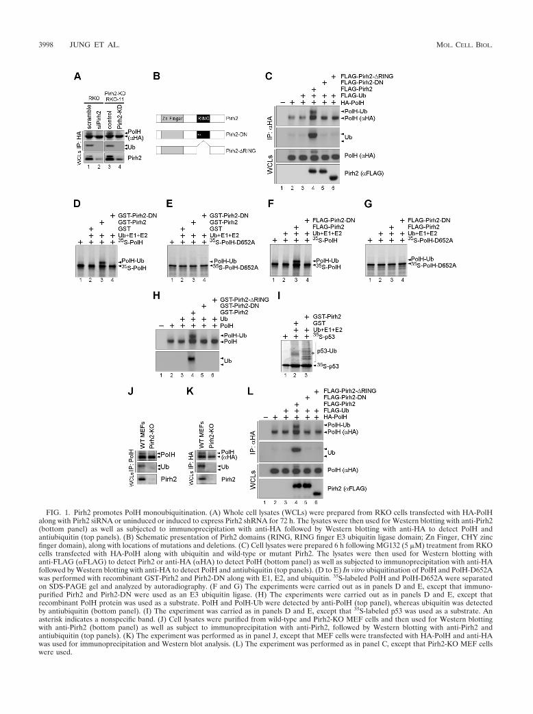
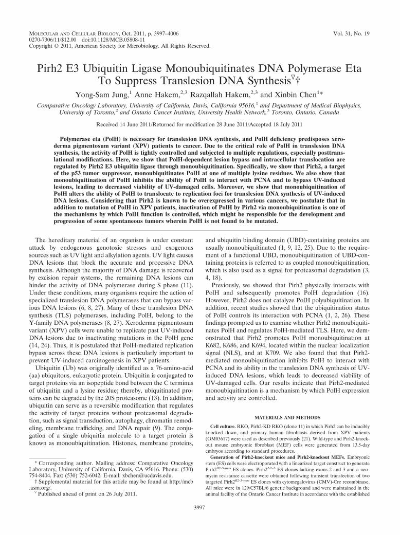
FIG. 1. Pirh2 promotes PolH monoubiquitination. (A) Whole cell lysates (WCLs) were prepared from RKO cells transfected with HA-PolHalong with Pirh2 siRNA or uninduced or induced to express Pirh2 shRNA for 72 h. The lysates were then used for Western blotting with anti-Pirh2(bottom panel) as well as subjected to immunoprecipitation with anti-HA followed by Western blotting with anti-HA to detect PolH andantiubiquitin (top panels). (B) Schematic presentation of Pirh2 domains (RING, RING finger E3 ubiquitin ligase domain; Zn Finger, CHY zincfinger domain), along with locations of mutations and deletions. (C) Cell lysates were prepared 6 h following MG132 (5 �M) treatment from RKOcells transfected with HA-PolH along with ubiquitin and wild-type or mutant Pirh2. The lysates were then used for Western blotting withanti-FLAG (�FLAG) to detect Pirh2 or anti-HA (�HA) to detect PolH (bottom panel) as well as subjected to immunoprecipitation with anti-HAfollowed by Western blotting with anti-HA to detect PolH and antiubiquitin (top panels). (D to E) In vitro ubiquitination of PolH and PolH-D652Awas performed with recombinant GST-Pirh2 and Pirh2-DN along with E1, E2, and ubiquitin. 35S-labeled PolH and PolH-D652A were separatedon SDS-PAGE gel and analyzed by autoradiography. (F and G) The experiments were carried out as in panels D and E, except that immuno-purified Pirh2 and Pirh2-DN were used as an E3 ubiquitin ligase. (H) The experiments were carried out as in panels D and E, except thatrecombinant PolH protein was used as a substrate. PolH and PolH-Ub were detected by anti-PolH (top panel), whereas ubiquitin was detectedby antiubiquitin (bottom panel). (I) The experiment was carried as in panels D and E, except that 35S-labeled p53 was used as a substrate. Anasterisk indicates a nonspecific band. (J) Cell lysates were purified from wild-type and Pirh2-KO MEF cells and then used for Western blottingwith anti-Pirh2 (bottom panel) as well as subject to immunoprecipitation with anti-Pirh2, followed by Western blotting with anti-Pirh2 andantiubiquitin (top panels). (K) The experiment was performed as in panel J, except that MEF cells were transfected with HA-PolH and anti-HAwas used for immunoprecipitation and Western blot analysis. (L) The experiment was performed as in panel C, except that Pirh2-KO MEF cellswere used.
3998 JUNG ET AL. MOL. CELL. BIOL.
ethical care regulations of the Canadian Council on Animal Care. The 13.5-dayembryos were used to generate MEF cells according to standard procedures.
Antibodies. Anti-PCNA, rabbit polyclonal and mouse monoclonal anti-PolH,mouse antiubiquitin, and rabbit anti-Pirh2 were purchased from Santa CruzBiotechnology. The other antibodies were antihemagglutinin (anti-HA) (HA11;Covance), anti-FLAG (Sigma), anti-p53 (DO-1, PAb1801, PAb240, andPAb421), and antiactin (Sigma).
Immunoprecipitation-Western blot analyses. The immunoprecipitation-West-ern blotting experiment was carried out as described previously (16). Threehundred to 500 �g of total proteins was immunoprecipitated with 2 �g of variousantibodies and then subjected to Western blot analysis.
siRNA. Scramble and Pirh2 small interfering RNAs (siRNAs) (sense 5�-CCAACA GAC UUG UGA AGA A dTdT-3� and antisense 5�-UUC UUC ACAAGU CUG UUG G dTdT-3�) were purchased from Dharmacon.
Plasmids and mutagenesis. pcDNA3 vectors expressing HA-PolH, FLAG-PolH, FLAG-PolH-D652A, FLAG-ubiquitin, Pirh2, FLAG-Pirh2, FLAG-Pirh2-DN, and FLAG-Pirh2-�RING were described previously (16). FLAG-PolH-Ubwas constructed by in-frame insertion of ubiquitin at the end of the PolH codingsequence.
Replacement of lysine(s) with arginine(s) in PolH was generated by site-directed mutagenesis (Quick Change; Stratagene). The following primers wereused: forward primer K682R-FR (5�-GTA TCT CAT CAA GGC AGA AGAAAT CCC AAG AGC-3�) and reverse primer K682R-RR (5�-GCT CTT GGGATT TCT TCT GCC TTG ATG AGA TAC-3�) for PolH-K682R, forwardprimer K694R-FR (5�-TTG GCC TGC ACT AAT AGA CGC CCC AGG CCTGAG-3�) and reverse primer K694R-RR (5�-CTC AGG CCT GGG GCG TCTATT AGT GCA GGC CAA-3�) for PolH-K694R, forward primer K709R-FR(5�-CAA CTG GAT CCG AGA TGG ATT TGG CCC ACA ACA GCC AAAG-3�) and reverse primer K709R-RR (5�-CTA ATG TGT TAA TGG CCT AAAAAA TGA TTC CAA-3�) for PolH-K709R, and forward primer K682.686R-FR(5�-GGC AGA AGA AAT CCC AGG AGC CCT TTG GCC TGC-3�) and
reverse primer K682.686R-RR (5�-GCA GGC CAA AGG GCT CCT GGGATT TCT TCT GCC-3�) for PolH-K682.686R. Other constructs (PolH-K682.694R, PolH-K682.709R, PolH-K682.686.709R, PolH-K682.694.709R,PolH-K682.686.694R, and PolH-K682.686.694.709R [PolH-4KR]) were gen-erated by combining single or double K-R mutations. PolH-4KR-NLS con-struct was generated by in-frame insertion of 2� NLS sequence from theSV40 large T antigen at the end of the PolH-4KR coding sequence. Greenfluorescent protein (GFP)-tagged PolH, PolH-4KR, PolH-4KR-NLS, andPolH-Ub were cloned in pEGFP-C1 for imaging.
GST pulldown assay. Glutathione S-transferase (GST)-tagged Pirh2, Pirh2-DN, and PCNA were expressed by pGEX-4T-3 (Amersham Pharmacia Biotech).The GST pulldown assay was performed as described previously (15).
Ubiquitination assay. The ubiquitination assay was performed as describedpreviously (20) with in vitro-synthesized 35S-labeled PolH, PolH-D652A, p53 (theTNT T7-coupled reticulocyte lysate system; Promega), or recombinant PolHalong with E1, E2, and ubiquitin (Boston Biochem). 35S-labeled proteins wereseparated on an SDS-PAGE gel and analyzed by autoradiography.
Immunofluorescence assay. FLAG-PCNA along with GFP-tagged wild-typeor mutated PolH was cotransfected into XPV cells on coverslips in 6-well plates.Six hours after UV irradiation (15 J/m2), cells were fixed with formaldehyde andthen incubated with primary antibody followed by secondary antibody. Nucleiwere visualized using 4�,6-diamidino-2-phenylindole (DAPI).
DNA histogram analysis. DNA histogram analysis was performed as previ-ously described (21). Both floating cells in the medium and live cells on the plateswere collected 24 h following 15 J/m2 UV irradiation.
Mass spectrometric analysis. The samples were run in one-dimensional SDS-PAGE, and potential PolH-containing bands were cut out, which were thensubjected to in-gel tryptic digestion overnight. The Paradigm MG4 high-perfor-mance liquid chromatography (HPLC) system (Michrom Bioresources, Auburn,CA), which is coupled to a Thermo Finnigan LTQ-FT Ultra ion trap massspectrometer (Thermo Fisher) through a Michrom advance captive spray ion-
FIG. 2. Characterization of PolH and monoubiquitinated PolH. (A) RKO cells were cotransfected with HA-tagged PolH and FLAG-taggedPirh2. After immunoprecipitation of RKO cell extracts with anti-HA antibody, the immunocomplex was separated and visualized by Coomassieblue stain. The bands corresponding to PolH (band 2) and monoubiquitinated PolH (band 1) were cut out and then subjected to mass spectrometry.(B) Band 2 was subjected to in-gel tryptic digestion and analyzed by mass spectrometry using an LTG-FT Ultra ion trap mass spectrometer asdescribed in Materials and Methods. A representative peptide sequence of PolH from band 2 is shown at the top of the mass spectrum. (C) Band1 was analyzed as described in panel B. Representative peptide sequences of PolH and ubiquitin from band 1 are shown at the top of the massspectra.
VOL. 31, 2011 POLYMERASE ETA MONOUBIQUITINATION BY Pirh2 3999

ization source, was used for peptide separation and analysis. Each sample wasloaded onto a trap column (Zorbax300SB C18, 5 �m, 5 by 0.3 mm; AgilentTechnologies, Santa Clara, CA) and desalted online. Peptides were then elutedfrom the trap and separated by a reverse-phase Michrom Magic C18 AQ (200-�mby 150-mm) capillary column at a flow rate of 2 �l/min. Samples were resus-pended in 2% acetonitrile (ACN) and 0.1% trifluoroacetic acid (TFA) as therunning buffer and directly loaded onto the mass spectrometer. The mass spec-trometer was operated with a spray voltage of 1.2 kV, a heated capillary tem-perature of 200°C, and a full scan range with a mass/charge ratio of 300 to 1,400.The protein database (both canonical and isoform versions) was downloadedfrom Uniprot.org. The cRAP (Common Repository of Adventitious Proteins)Database, which contains the most common contaminants of samples, such ashuman keratin, bovine serum albumin, etc., was appended to the database toidentify contaminations prior to analysis.
Clonogenic assay. XPV (GM03617) cells on 100-mm and 60-mm culturedishes in triplicate were transfected with a control vector or a vector expressingPolH, PolH-Ub, PolH-4KR, or PolH-4KR-NLS for 24 h. Cells were then washedwith phosphate-buffered saline (PBS) and exposed to UV irradiation (0 to 9J/m2). After UV irradiation, cells were incubated in minimal essential medium(MEM) containing 0.375 mM caffeine for 13 days (2). Caffeine sensitizes XPVcells but not normal human fibroblast cells (such as GM00024 cells) upon UVirradiation. Colonies were stained with Giemsa staining solution and counted.
RESULTS
Pirh2 promotes PolH monoubiquitination. Recently, weshowed that Pirh2 physically interacts with PolH and promotesPolH degradation via 20S proteasome, but Pirh2 does notpolyubiquitinate PolH (16). In addition, other studies showedthat the status of PolH monoubiquitination controls its inter-action with PCNA (1, 2, 26). These findings prompted us toexamine whether Pirh2 monoubiquitinates PolH and regulatesits translesion DNA synthesis (TLS) activity. To test this, PolHubiquitination was examined in RKO cells in which endoge-nous Pirh2 was knocked down by Pirh2 siRNA or induciblyknocked down by Pirh2 short hairpin RNA (shRNA) alongwith ectopic expression of HA-tagged PolH. We showed thatthe level of Pirh2 was significantly decreased by Pirh2 siRNAor shRNA (Fig. 1A, Pirh2 panel, compare lanes 1 and 3 with 2and 4, respectively). Next, HA-tagged PolH expressed in RKOcells was immunoprecipitated by anti-HA and then used forWestern blot analysis. We found that HA-PolH was detectedalong with a slow-migrating band recognized by anti-HA anti-body (Fig. 1A, lanes 1 and 3). In addition, the slow-migratingband was markedly decreased upon knockdown of Pirh2 (Fig.1A, compare lanes 1 and 3 with 2 and 4, respectively). Inter-estingly, we found that the slow-migrating band was recognizedby antiubiquitin antibody (Fig. 1A, Ub panel). Based on themigration pattern, it is likely that the slow-migrating band is amonoubiquitinated form of PolH.
To validate PolH monoubiquitination, RKO cells werecotransfected with HA-tagged PolH, FLAG-tagged ubiquitinalong with wild-type Pirh2, E3 ligase-deficient Pirh2-DN, orPirh2-�RING (Fig. 1B). Pirh2-DN contains amino acid sub-stitutions (C145S and C148S) in the RING finger domain,whereas Pirh2-�RING lacks the entire RING finger domainfrom aa 145 to 186 (16, 20, 23, 28). Upon immunoprecipitationwith anti-HA antibody followed by Western blot analysis, weshowed that a slow-migrating band was detected in cellscotransfected with wild-type Pirh2, but not Pirh2-DN andPirh2-�RING (Fig. 1C, compare lane 4 with lanes 5 and 6). Inaddition, the slow-migrating band was found to be recognizedby antiubiquitin (Fig. 1C, Ub panel, lane 4).
To investigate whether PolH is a substrate of Pirh2 E3 li-
gase, ubiquitination assay was performed with in vitro-trans-lated 35S-labeled PolH (PolH) or PolH-D652A. PolH-D652A,which contains a substitution (D652A) in the UBZ domain, isunable to interact with ubiquitin (1). We showed that PolH wasmonoubiquitinated in the presence of recombinant GST-tagged Pirh2 (Fig. 1D, lane 3) or Flag-tagged Pirh2 purifiedfrom RKO cells (Fig. 1F, lane 3). In contrast, Pirh2-DN wasinert (Fig. 1D and F, lane 4). In addition, Pirh2 and Pirh2-DNwere incapable of monoubiquitinating PolH-D652A (Fig. 1Eand G). To further investigate whether PolH is a direct sub-strate of Pirh2, the in vitro ubiquitination assay was performedwith recombinant PolH and showed that PolH was found to bemonoubiquitinated by Pirh2, but not Pirh2-DN and Pirh2-�RING (Fig. 1H). As a control, p53 was polyubiquitinatedby recombinant GST-tagged Pirh2 but not by GST tag alone(Fig. 1I).
To validate the role of Pirh2 in PolH monoubiquitination,we examined the status of PolH monoubiquitination in Pirh2knockout mouse embryo fibroblast (MEF) cells. We found thatendogenous PolH was monoubiquitinated in Pirh2-competentbut not Pirh2-deficient MEF cells (Fig. 1J). We also found thatectopically expressed PolH was monoubiquitinated in Pirh2-competent but not Pirh2-deficient MEF cells (Fig. 1K). Inaddition, we showed that ectopically expressed PolH was
FIG. 3. Identification of lysine residues in PolH for monoubiquiti-nation by Pirh2. (A) Locations of the nuclear localization signal andlysine-to-arginine substitution(s). (B and C) Cell lysates were preparedfrom RKO cells (B) or Pirh2-KO MEF cells (C), which were trans-fected with FLAG-tagged wild-type (WT) or mutated PolH along withHA-tagged ubiquitin and Pirh2. The lysates were then immunoprecipi-tated with anti-FLAG followed by Western blotting with anti-FLAG todetect PolH and antiubiquitin.
4000 JUNG ET AL. MOL. CELL. BIOL.
monoubiquitinated in Pirh2 knockout (Pirh2-KO) MEF cellsupon reconstitution of wild-type Pirh2 but not Pirh2-DN andPirh2-�RING (Fig. 1L). Finally, mass spectrometry was per-formed to determine whether the slower-migrating band ismonoubiquitinated PolH. We found that the slower-migratingband contained peptides derived from both PolH and ubiquitin(Fig. 2A and C), whereas the fast-migrating band containedpeptides derived only from PolH (Fig. 2A and B). Together,these data suggest that PolH is a substrate of Pirh2 for mono-ubiquitination.
Identification of lysine residues in PolH for monoubiquiti-nation by Pirh2. Three lysine residues, K682, K686, andK694, which are located within the nuclear localization se-quence (NLS), and K709, which is located in the extreme Cterminus in PolH, were found to be monoubiquitinated (2).To test whether monoubiquitination of these lysines werecatalyzed by Pirh2, we generated 10 PolH mutants, whichcarry one, two, three, or four substitutions with arginine atK682, K686, K694, and K709 along with a FLAG tag (Fig.3A). These PolH mutants were then expressed in RKO cells(Fig. 3B) and Pirh2-KO MEF cells (Fig. 3C), along with HA-
tagged ubiquitin and Pirh2. We found that nine PolHmutants, which carry one to three arginine substitutions(K682R, K694R, K709R, K682.686R, K682.694R, K682.709R,K682.686.709R, K682.694.709R, and K682.686.694R), werestill found to be monoubiquitinated (Fig. 3B and C). In con-trast, little if any monoubiquitination was detected for thePolH mutant with a quadruple substitution of lysines witharginines (K682.686.694.709R, thereafter referred to as PolH-4KR) (Fig. 3B and C). These results suggest that Pirh2 mono-ubiquitinates PolH at one of the four lysine residues (K682,K686, K694, and K709).
Pirh2 suppresses PolH to interact with PCNA but not itssensitivity to proteasomal degradation via monoubiquitinationof PolH. Recent studies showed that the switching from repli-cative polymerase � (Pol �) to TLS pol � is promoted by PCNAmonoubiquitination (10, 17), but inhibited by PolH monoubiq-uitination (1, 2). These findings led us to test whether Pirh2modulates the PCNA-PolH interaction through PolH mono-ubiquitination. To address this, FLAG-tagged PolH was ex-pressed in RKO cells with or without Pirh2. We showed that alarge fraction of PolH was monoubiquitinated in the presence
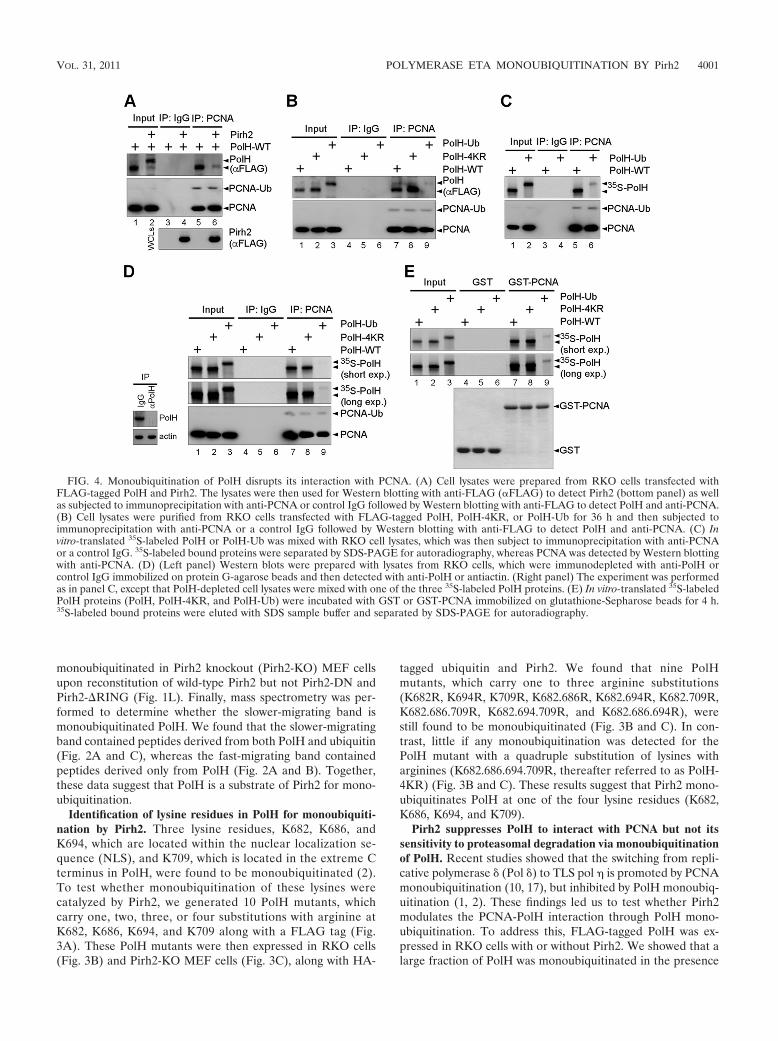
FIG. 4. Monoubiquitination of PolH disrupts its interaction with PCNA. (A) Cell lysates were prepared from RKO cells transfected withFLAG-tagged PolH and Pirh2. The lysates were then used for Western blotting with anti-FLAG (�FLAG) to detect Pirh2 (bottom panel) as wellas subjected to immunoprecipitation with anti-PCNA or control IgG followed by Western blotting with anti-FLAG to detect PolH and anti-PCNA.(B) Cell lysates were purified from RKO cells transfected with FLAG-tagged PolH, PolH-4KR, or PolH-Ub for 36 h and then subjected toimmunoprecipitation with anti-PCNA or a control IgG followed by Western blotting with anti-FLAG to detect PolH and anti-PCNA. (C) Invitro-translated 35S-labeled PolH or PolH-Ub was mixed with RKO cell lysates, which was then subject to immunoprecipitation with anti-PCNAor a control IgG. 35S-labeled bound proteins were separated by SDS-PAGE for autoradiography, whereas PCNA was detected by Western blottingwith anti-PCNA. (D) (Left panel) Western blots were prepared with lysates from RKO cells, which were immunodepleted with anti-PolH orcontrol IgG immobilized on protein G-agarose beads and then detected with anti-PolH or antiactin. (Right panel) The experiment was performedas in panel C, except that PolH-depleted cell lysates were mixed with one of the three 35S-labeled PolH proteins. (E) In vitro-translated 35S-labeledPolH proteins (PolH, PolH-4KR, and PolH-Ub) were incubated with GST or GST-PCNA immobilized on glutathione-Sepharose beads for 4 h.35S-labeled bound proteins were eluted with SDS sample buffer and separated by SDS-PAGE for autoradiography.
VOL. 31, 2011 POLYMERASE ETA MONOUBIQUITINATION BY Pirh2 4001
of Pirh2 (Fig. 4A, lane 2), consistent with the above observa-tion. We found that upon immunoprecipitation with anti-PCNA, PolH but not monoubiquitinated PolH was detected inPCNA immunocomplexes (Fig. 4A, lanes 5 and 6).
To further test this, we examined the interaction of PCNA
with PolH-ubiquitin fusion protein (PolH-Ub), which containsthe 76-aa ubiquitin fused at the C terminus of PolH polypep-tide, or PolH-4KR, which cannot be monoubiquitinated, asshown above (Fig. 3). We showed that upon expression inRKO cells, wild-type PolH and PolH-4KR, but very little
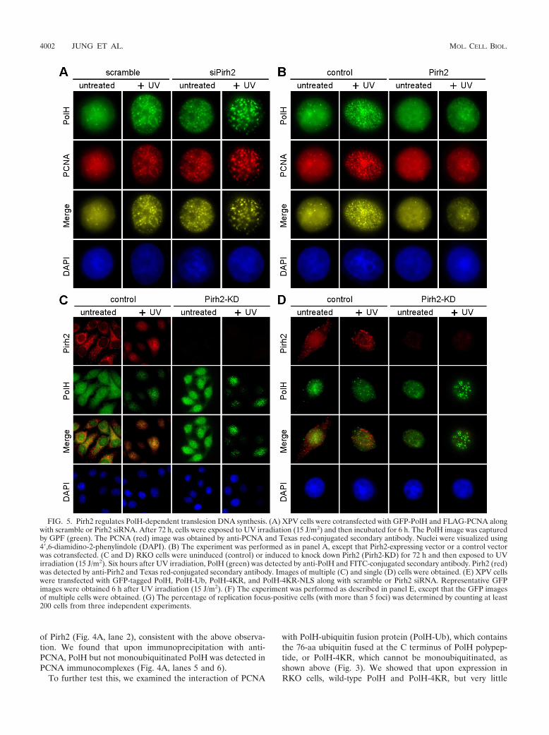
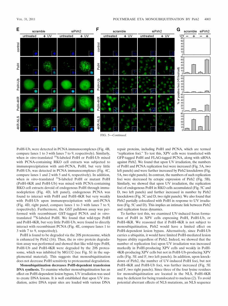
FIG. 5. Pirh2 regulates PolH-dependent translesion DNA synthesis. (A) XPV cells were cotransfected with GFP-PolH and FLAG-PCNA alongwith scramble or Pirh2 siRNA. After 72 h, cells were exposed to UV irradiation (15 J/m2) and then incubated for 6 h. The PolH image was capturedby GPF (green). The PCNA (red) image was obtained by anti-PCNA and Texas red-conjugated secondary antibody. Nuclei were visualized using4�,6-diamidino-2-phenylindole (DAPI). (B) The experiment was performed as in panel A, except that Pirh2-expressing vector or a control vectorwas cotransfected. (C and D) RKO cells were uninduced (control) or induced to knock down Pirh2 (Pirh2-KD) for 72 h and then exposed to UVirradiation (15 J/m2). Six hours after UV irradiation, PolH (green) was detected by anti-PolH and FITC-conjugated secondary antibody. Pirh2 (red)was detected by anti-Pirh2 and Texas red-conjugated secondary antibody. Images of multiple (C) and single (D) cells were obtained. (E) XPV cellswere transfected with GFP-tagged PolH, PolH-Ub, PolH-4KR, and PolH-4KR-NLS along with scramble or Pirh2 siRNA. Representative GFPimages were obtained 6 h after UV irradiation (15 J/m2). (F) The experiment was performed as described in panel E, except that the GFP imagesof multiple cells were obtained. (G) The percentage of replication focus-positive cells (with more than 5 foci) was determined by counting at least200 cells from three independent experiments.
4002 JUNG ET AL. MOL. CELL. BIOL.
PolH-Ub, were detected in PCNA immunocomplexes (Fig. 4B,compare lanes 1 to 3 with lanes 7 to 9, respectively). Similarly,when in vitro-translated 35S-labeled PolH or PolH-Ub mixedwith PCNA-containing RKO cell extracts was subjected toimmunoprecipitation with anti-PCNA, PolH, but very littlePolH-Ub, was detected in PCNA immunocomplexes (Fig. 4C,compare lanes 1 and 2 with 5 and 6, respectively). In addition,when in vitro-translated 35S-labeled PolH or mutant PolH(PolH-4KR and PolH-Ub) was mixed with PCNA-containingRKO cell extracts devoid of endogenous PolH through immu-nodepletion (Fig. 4D, left panel), endogenous PCNA wasfound to interact with PolH and PolH-4KR but very weaklywith PolH-Ub upon immunoprecipitation with anti-PCNA(Fig. 4D, right panel, compare lanes 1 to 3 with lanes 7 to 9,respectively). Furthermore, the GST pulldown assay was per-formed with recombinant GST-tagged PCNA and in vitro-translated 35S-labeled PolH. We found that wild-type PolHand PolH-4KR, but very little PolH-Ub, were found to directlyinteract with recombinant PCNA (Fig. 4E, compare lanes 1 to3 with 7 to 9, respectively).
PolH is found to be degraded via the 20S proteasome, whichis enhanced by Pirh2 (16). Thus, the in vitro protein degrada-tion assay was performed and showed that like wild-type PolH,PolH-Ub and PolH-4KR were degraded by the 20S protea-some, which was inhibited by MG132 (see Fig. S1 in the sup-plemental material). This suggests that monoubiquitinationdoes not decrease PolH sensitivity to proteasomal degradation.
Monoubiquitination decreases PolH-dependent translesionDNA synthesis. To examine whether monoubiquitination has aneffect on PolH-dependent lesion bypass, UV irradiation was usedto create DNA lesions. It is well established that upon UV irra-diation, active DNA repair sites are loaded with various DNA
repair proteins, including PolH and PCNA, which are termed“replication foci.” To test this, XPV cells were transfected withGFP-tagged PolH and FLAG-tagged PCNA, along with siRNAagainst Pirh2. We found that upon UV irradiation, the numbersof PolH and PCNA replication foci were increased (Fig. 5A, twoleft panels) and were further increased by Pirh2 knockdown (Fig.5A, two right panels). In contrast, the numbers of such replicationfoci were decreased by ectopic expression of Pirh2 (Fig. 5B).Similarly, we showed that upon UV irradiation, the replicationfoci of endogenous PolH in RKO cells accumulated (Fig. 5C andD, two left panels) and further increased in number by Pirh2knockdown (Fig. 5C and D, two right panels). We also found thatPirh2 partially colocalized with PolH in response to UV irradia-tion (Fig. 5C and D). This implies an intimate link between Pirh2and replication focus dynamics.
To further test this, we examined UV-induced focus forma-tion of PolH in XPV cells expressing PolH, PolH-Ub, orPolH-4KR. We reasoned that if PolH-4KR is not subject tomonoubiquitination, Pirh2 would have a limited effect onPolH-dependent lesion bypass. Alternatively, since PolH-Ubcarries a ubiquitin, it would have limited PolH-mediated lesionbypass ability regardless of Pirh2. Indeed, we showed that thenumber of replication foci upon UV irradiation was increasedmarkedly in PolH-producing XPV cells and weakly in PolH-4KR-producing XPV cells but not in PolH-Ub-producing XPVcells (Fig. 5E and F, two left panels). In addition, upon knock-down of Pirh2, the number of UV-induced PolH foci, but notPolH-4KR and PolH-Ub foci, was further increased (Fig. 5Eand F, two right panels). Since three of the four lysine residuesfor monoubiquitination are located in the NLS, PolH-4KRmay be deficient for being translocated to nucleus (2). To avoidpotential aberrant effects of NLS mutations, an NLS sequence
FIG. 5—Continued.
VOL. 31, 2011 POLYMERASE ETA MONOUBIQUITINATION BY Pirh2 4003
from SV40 large T antigen was fused to the C terminus ofPolH-4KR, and the resulting protein was designated PolH-4KR-NLS. Consistent with the previous report that PolH-4KR-NLS reacquires its ability to be translocated into nucleus(2), we found that upon UV irradiation, strong replication fociwere detected in PolH-4KR-NLS-producing XPV cells (Fig.5E and F, 4KR-NLS panel). Interestingly, while the numberand size of replication foci in PolH-4KR-NLS-producing cellswere slightly increased upon knockdown of Pirh2, the extent ofthe increase was much less than that of wild-type PolH (Fig. 5Eand G, compare the PolH and PolH-4KR-NLS panels). Theresult is consistent with the idea that Pirh2-mediated PolHmonoubiquitination regulates intracellular translocation andPolH-mediated lesion bypass.
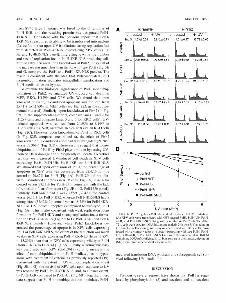
To examine the biological significance of PolH monoubiq-uitination by Pirh2, we analyzed UV-induced cell death inMEF, RKO, H1299, and XPV cells. We found that uponknockout of Pirh2, UV-induced apoptosis was reduced from25.41% to 11.83% in MEF cells (see Fig. S2A in the supple-mental material). Similarly, upon knockdown of Pirh2 (in Fig.S2E in the supplemental material, compare lanes 1 and 2 forH1299 cells and compare lanes 3 and 5 for RKO cells), UV-induced apoptosis was reduced from 20.58% to 8.19% inH1299 cells (Fig. S2B) and from 16.67% to 8.47% in RKO cells(Fig. S2C). However, upon knockdown of PolH in RKO cells(in Fig. S2E, compare lanes 4 and 6), the effect of Pirh2knockdown on UV-induced apoptosis was abrogated (21.38%versus 23.36%) (Fig. S2D). These results suggest that mono-ubiquitination of PolH by Pirh2 plays a role in bypassing UV-induced DNA damage and subsequently cell death. To furthertest this, we measured UV-induced cell death in XPV cellsexpressing PolH, PolH-Ub, PolH-4KR, or PolH-4KR-NLS.We showed that upon expression of PolH, the percentage ofapoptosis in XPV cells was decreased from 32.42% for thecontrol to 20.62% for PolH (Fig. 6A). PolH-Ub did not alle-viate UV-induced apoptosis in XPV cells (Fig. 6A, 32.42% forcontrol versus 33.11% for PolH-Ub), consistent with the lackof replication focus formation (Fig. 5E to G, PolH-Ub panel).Similarly, PolH-4KR had a weak effect (32.42% for controlversus 26.17% for PolH-4KR), whereas PolH-4KR-NLS had astrong effect (32.42% for control versus 18.75% for PolH-4KR-NLS) on UV-induced apoptosis compared to wild-type PolH(Fig. 6A). This is also consistent with weak replication focusformation for PolH-4KR and strong replication focus forma-tion for PolH-4KR-NLS (Fig. 5E to G, PolH-4KR, and PolH-4KR-NLS panels). However, while Pirh2 knockdown de-creased the percentage of apoptosis in XPV cells expressingPolH or PolH-4KR-NLS, the extent of the reduction was muchweaker in XPV cells expressing PolH-4KR-NLS (from 18.85%to 13.29%) than that in XPV cells expressing wild-type PolH(from 20.63% to 11.24%) (Fig. 6A). Finally, a clonogenic assaywas performed with XPV (GM03617) cells to measure theeffect of monoubiquitination on PolH-mediated lesion bypassalong with treatment of caffeine as previously reported (19).Consistent with the extent of UV-induced replication arrest(Fig. 5E to G), the survival of XPV cells upon exposure to UVwas rescued by PolH, PolH-4KR-NLS, and, to a lesser extent,by PolH-4KR compared to PolH-Ub (Fig. 6B). Together, thesedata suggest that PolH monoubiquitination modulates PolH-
mediated translesion DNA synthesis and subsequently cell sur-vival following UV irradiation.
DISCUSSION
Previously, several reports have shown that PolH is regu-lated by phosphorylation (5) and covalent and noncovalent
FIG. 6. Pirh2 regulates PolH-dependent resistance to UV irradiation.(A) XPV cells were transfected with GFP-tagged PolH, PolH-Ub, PolH-4KR, and PolH-4KR-NLS along with scramble or Pirh2 siRNA. After72 h, cells were used for DNA histogram analysis 24 h post-UV irradiation(15 J/m2). (B) The clonogenic assay was performed with XPV cells trans-fected with a control vector or a vector expressing wild-type PolH, PolH-Ub, PolH-4KR, or PolH-4KR-NLS. Cells were then incubated in DMEMcontaining 0.375 mM caffeine. Error bars represent the standard deviation(SD) from three independent experiments.
4004 JUNG ET AL. MOL. CELL. BIOL.
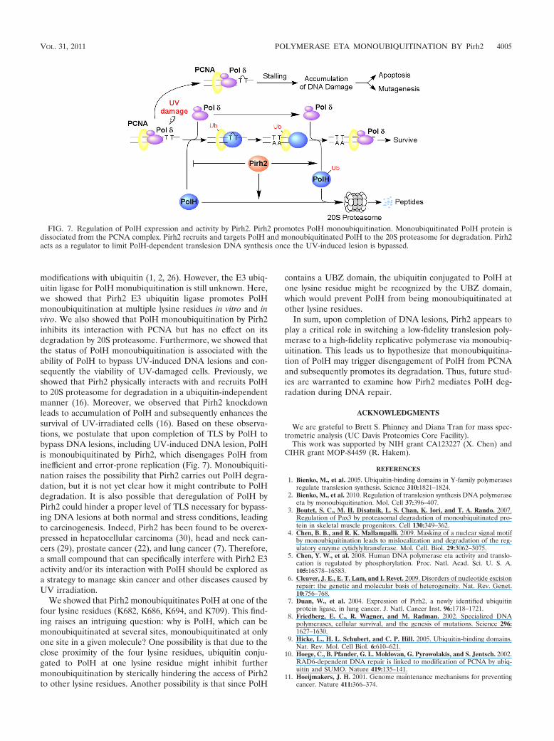
modifications with ubiquitin (1, 2, 26). However, the E3 ubiq-uitin ligase for PolH monubiquitination is still unknown. Here,we showed that Pirh2 E3 ubiquitin ligase promotes PolHmonoubiquitination at multiple lysine residues in vitro and invivo. We also showed that PolH monoubiquitination by Pirh2inhibits its interaction with PCNA but has no effect on itsdegradation by 20S proteasome. Furthermore, we showed thatthe status of PolH monoubiquitination is associated with theability of PolH to bypass UV-induced DNA lesions and con-sequently the viability of UV-damaged cells. Previously, weshowed that Pirh2 physically interacts with and recruits PolHto 20S proteasome for degradation in a ubiquitin-independentmanner (16). Moreover, we observed that Pirh2 knockdownleads to accumulation of PolH and subsequently enhances thesurvival of UV-irradiated cells (16). Based on these observa-tions, we postulate that upon completion of TLS by PolH tobypass DNA lesions, including UV-induced DNA lesion, PolHis monoubiquitinated by Pirh2, which disengages PolH frominefficient and error-prone replication (Fig. 7). Monoubiquiti-nation raises the possibility that Pirh2 carries out PolH degra-dation, but it is not yet clear how it might contribute to PolHdegradation. It is also possible that deregulation of PolH byPirh2 could hinder a proper level of TLS necessary for bypass-ing DNA lesions at both normal and stress conditions, leadingto carcinogenesis. Indeed, Pirh2 has been found to be overex-pressed in hepatocellular carcinoma (30), head and neck can-cers (29), prostate cancer (22), and lung cancer (7). Therefore,a small compound that can specifically interfere with Pirh2 E3activity and/or its interaction with PolH should be explored asa strategy to manage skin cancer and other diseases caused byUV irradiation.
We showed that Pirh2 monoubiquitinates PolH at one of thefour lysine residues (K682, K686, K694, and K709). This find-ing raises an intriguing question: why is PolH, which can bemonoubiquitinated at several sites, monoubiquitinated at onlyone site in a given molecule? One possibility is that due to theclose proximity of the four lysine residues, ubiquitin conju-gated to PolH at one lysine residue might inhibit furthermonoubiquitination by sterically hindering the access of Pirh2to other lysine residues. Another possibility is that since PolH
contains a UBZ domain, the ubiquitin conjugated to PolH atone lysine residue might be recognized by the UBZ domain,which would prevent PolH from being monoubiquitinated atother lysine residues.
In sum, upon completion of DNA lesions, Pirh2 appears toplay a critical role in switching a low-fidelity translesion poly-merase to a high-fidelity replicative polymerase via monoubiq-uitination. This leads us to hypothesize that monoubiquitina-tion of PolH may trigger disengagement of PolH from PCNAand subsequently promotes its degradation. Thus, future stud-ies are warranted to examine how Pirh2 mediates PolH deg-radation during DNA repair.
ACKNOWLEDGMENTS
We are grateful to Brett S. Phinney and Diana Tran for mass spec-trometric analysis (UC Davis Proteomics Core Facility).
This work was supported by NIH grant CA123227 (X. Chen) andCIHR grant MOP-84459 (R. Hakem).
REFERENCES
1. Bienko, M., et al. 2005. Ubiquitin-binding domains in Y-family polymerasesregulate translesion synthesis. Science 310:1821–1824.
2. Bienko, M., et al. 2010. Regulation of translesion synthesis DNA polymeraseeta by monoubiquitination. Mol. Cell 37:396–407.
3. Boutet, S. C., M. H. Disatnik, L. S. Chan, K. Iori, and T. A. Rando. 2007.Regulation of Pax3 by proteasomal degradation of monoubiquitinated pro-tein in skeletal muscle progenitors. Cell 130:349–362.
4. Chen, B. B., and R. K. Mallampalli. 2009. Masking of a nuclear signal motifby monoubiquitination leads to mislocalization and degradation of the reg-ulatory enzyme cytidylyltransferase. Mol. Cell. Biol. 29:3062–3075.
5. Chen, Y. W., et al. 2008. Human DNA polymerase eta activity and translo-cation is regulated by phosphorylation. Proc. Natl. Acad. Sci. U. S. A.105:16578–16583.
6. Cleaver, J. E., E. T. Lam, and I. Revet. 2009. Disorders of nucleotide excisionrepair: the genetic and molecular basis of heterogeneity. Nat. Rev. Genet.10:756–768.
7. Duan, W., et al. 2004. Expression of Pirh2, a newly identified ubiquitinprotein ligase, in lung cancer. J. Natl. Cancer Inst. 96:1718–1721.
8. Friedberg, E. C., R. Wagner, and M. Radman. 2002. Specialized DNApolymerases, cellular survival, and the genesis of mutations. Science 296:1627–1630.
9. Hicke, L., H. L. Schubert, and C. P. Hill. 2005. Ubiquitin-binding domains.Nat. Rev. Mol. Cell Biol. 6:610–621.
10. Hoege, C., B. Pfander, G. L. Moldovan, G. Pyrowolakis, and S. Jentsch. 2002.RAD6-dependent DNA repair is linked to modification of PCNA by ubiq-uitin and SUMO. Nature 419:135–141.
11. Hoeijmakers, J. H. 2001. Genome maintenance mechanisms for preventingcancer. Nature 411:366–374.
FIG. 7. Regulation of PolH expression and activity by Pirh2. Pirh2 promotes PolH monoubiquitination. Monoubiquitinated PolH protein isdissociated from the PCNA complex. Pirh2 recruits and targets PolH and monoubiquitinated PolH to the 20S proteasome for degradation. Pirh2acts as a regulator to limit PolH-dependent translesion DNA synthesis once the UV-induced lesion is bypassed.
VOL. 31, 2011 POLYMERASE ETA MONOUBIQUITINATION BY Pirh2 4005
12. Isasa, M., et al. 2010. Monoubiquitination of RPN10 regulates substraterecruitment to the proteasome. Mol. Cell 38:733–745.
13. Johnson, E. S., P. C. Ma, I. M. Ota, and A. Varshavsky. 1995. A proteolyticpathway that recognizes ubiquitin as a degradation signal. J. Biol. Chem.270:17442–17456.
14. Johnson, R. E., C. M. Kondratick, S. Prakash, and L. Prakash. 1999.hRAD30 mutations in the variant form of xeroderma pigmentosum. Science285:263–265.
15. Jung, Y. S., et al. 2008. Physical interactions and functional coupling betweenDaxx and sodium hydrogen exchanger 1 in ischemic cell death. J. Biol. Chem.283:1018–1025.
16. Jung, Y. S., G. Liu, and X. Chen. 2010. Pirh2 E3 ubiquitin ligase targets DNApolymerase eta for 20S proteasomal degradation. Mol. Cell. Biol. 30:1041–1048.
17. Kannouche, P. L., J. Wing, and A. R. Lehmann. 2004. Interaction of humanDNA polymerase eta with monoubiquitinated PCNA: a possible mechanismfor the polymerase switch in response to DNA damage. Mol. Cell 14:491–500.
18. Kirkpatrick, D. S., et al. 2006. Quantitative analysis of in vitro ubiquitinatedcyclin B1 reveals complex chain topology. Nat. Cell Biol. 8:700–710.
19. Laposa, R. R., L. Feeney, and J. E. Cleaver. 2003. Recapitulation of thecellular xeroderma pigmentosum-variant phenotypes using short interferingRNA for DNA polymerase H. Cancer Res. 63:3909–3912.
20. Leng, R. P., et al. 2003. Pirh2, a p53-induced ubiquitin-protein ligase, pro-motes p53 degradation. Cell 112:779–791.
21. Liu, G., and X. Chen. 2006. DNA polymerase eta, the product of the xero-
derma pigmentosum variant gene and a target of p53, modulates the DNAdamage checkpoint and p53 activation. Mol. Cell. Biol. 26:1398–1413.
22. Logan, I. R., et al. 2006. Human PIRH2 enhances androgen receptor sig-naling through inhibition of histone deacetylase 1 and is overexpressed inprostate cancer. Mol. Cell. Biol. 26:6502–6510.
23. Logan, I. R., V. Sapountzi, L. Gaughan, D. E. Neal, and C. N. Robson. 2004.Control of human PIRH2 protein stability: involvement of TIP60 and theproteosome. J. Biol. Chem. 279:11696–11704.
24. Masutani, C., et al. 1999. The XPV (xeroderma pigmentosum variant) geneencodes human DNA polymerase eta. Nature 399:700–704.
25. Penengo, L., et al. 2006. Crystal structure of the ubiquitin binding domainsof rabex-5 reveals two modes of interaction with ubiquitin. Cell 124:1183–1195.
26. Plosky, B. S., et al. 2006. Controlling the subcellular localization of DNApolymerases iota and eta via interactions with ubiquitin. EMBO J. 25:2847–2855.
27. Prakash, S., R. E. Johnson, and L. Prakash. 2005. Eukaryotic translesionsynthesis DNA polymerases: specificity of structure and function. Annu. Rev.Biochem. 74:317–353.
28. Sheng, Y., et al. 2008. Molecular basis of Pirh2-mediated p53 ubiquitylation.Nat. Struct. Mol. Biol. 15:1334–1342.
29. Shimada, M., et al. 2009. High expression of Pirh2, an E3 ligase for p27, isassociated with low expression of p27 and poor prognosis in head and neckcancers. Cancer Sci. 100:866–872.
30. Wang, L., et al. 2011. Interplay between MDM2, MDMX, Pirh2 and COP1:the negative regulators of p53. Mol. Biol. Rep. 38:229–236.
4006 JUNG ET AL. MOL. CELL. BIOL.
Related Documents