Discovery of a DNA Damage Response in Acinetobacter baumannii and Analysis of Translesion Synthesis DNA Polymerases of Both A. baumannii and Escherichia coli by Matthew David Norton B.S in Microbiology, University of Rhode Island B.S. in Biological Sciences, University of Rhode Island A dissertation submitted to The Faculty of the College of Science of Northeastern University in partial fulfillment of the requirements for the degree of Doctor of Philosophy November 21, 2013 Dissertation directed by Veronica Godoy-Carter, Ph.D. Associate Professor of Biology

Welcome message from author
This document is posted to help you gain knowledge. Please leave a comment to let me know what you think about it! Share it to your friends and learn new things together.
Transcript
Discovery of a DNA Damage Response in Acinetobacter baumannii and Analysis of Translesion Synthesis DNA Polymerases of Both A. baumannii and Escherichia coli
by Matthew David Norton
B.S in Microbiology, University of Rhode Island B.S. in Biological Sciences, University of Rhode Island
A dissertation submitted to
The Faculty of the College of Science of Northeastern University
in partial fulfillment of the requirements for the degree of Doctor of Philosophy
November 21, 2013
Dissertation directed by
Veronica Godoy-Carter, Ph.D. Associate Professor of Biology

ii
DEDICATION
This dissertation is dedicated to my family and fiancé for their endless support and love.

iii
ACKNOWLEDGEMENTS
I would like to thank my fellow Godoy lab members, past and present, for their helpful
discussions and for maintaining a fun work environment. I especially thank Ryan Benson and
William DuComb for always making me laugh and smile on the days I needed it most.
None of this would have been possible if it wasn’t for my advisor, Veronica Godoy. Her
positive attitude, enthusiasm, encouragement, and insights were immensely important to my
success. I thank her for not only training me as a molecular microbiologist, but for always getting
me to see the bright side of every experiment. She was always able to put things into perspective
and for that she has been an excellent role model both in the lab and in life. I also would like to
thank my committee members Dr. Kim Lewis, Dr. Erin Cram, Dr. Marin Vulic, and Dr. Daniel
Jarosz for their guidance, thoughtful insights, and contributions to this work.
Lastly, I want to thank my fiancé, Aimee, for her endless support and encouragement
throughout this journey. She brought out the best in me during this process, and made the light at
the end of the tunnel always shine brighter. I thank my family for their love and support and for
always enthusiastically cheering me on. I’m grateful to everyone in my life that has helped me
become the person I am today and for getting here whether they know it or not.
iv
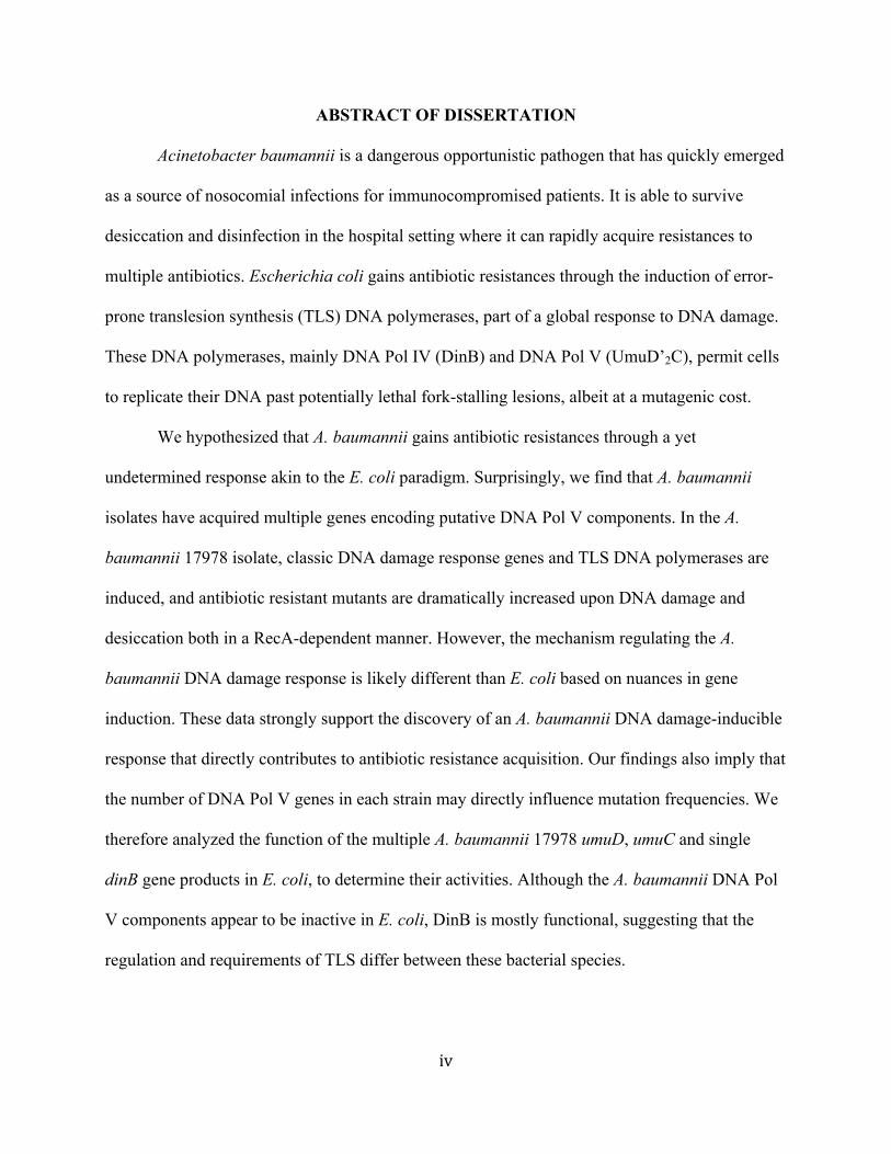
ABSTRACT OF DISSERTATION
Acinetobacter baumannii is a dangerous opportunistic pathogen that has quickly emerged
as a source of nosocomial infections for immunocompromised patients. It is able to survive
desiccation and disinfection in the hospital setting where it can rapidly acquire resistances to
multiple antibiotics. Escherichia coli gains antibiotic resistances through the induction of error-
prone translesion synthesis (TLS) DNA polymerases, part of a global response to DNA damage.
These DNA polymerases, mainly DNA Pol IV (DinB) and DNA Pol V (UmuD’2C), permit cells
to replicate their DNA past potentially lethal fork-stalling lesions, albeit at a mutagenic cost.
We hypothesized that A. baumannii gains antibiotic resistances through a yet
undetermined response akin to the E. coli paradigm. Surprisingly, we find that A. baumannii
isolates have acquired multiple genes encoding putative DNA Pol V components. In the A.
baumannii 17978 isolate, classic DNA damage response genes and TLS DNA polymerases are
induced, and antibiotic resistant mutants are dramatically increased upon DNA damage and
desiccation both in a RecA-dependent manner. However, the mechanism regulating the A.
baumannii DNA damage response is likely different than E. coli based on nuances in gene
induction. These data strongly support the discovery of an A. baumannii DNA damage-inducible
response that directly contributes to antibiotic resistance acquisition. Our findings also imply that
the number of DNA Pol V genes in each strain may directly influence mutation frequencies. We
therefore analyzed the function of the multiple A. baumannii 17978 umuD, umuC and single
dinB gene products in E. coli, to determine their activities. Although the A. baumannii DNA Pol
V components appear to be inactive in E. coli, DinB is mostly functional, suggesting that the
regulation and requirements of TLS differ between these bacterial species.

v
Lastly, we examined the poorly understood carboxy-terminal domain of E. coli UmuC,
the catalytic subunit of DNA Pol V, to determine its structural role in regulatory protein-protein
interactions. Using a carboxy-terminal fragment of UmuC, we find that expression causes
diverse changes in DNA damage-induced mutagenesis and cell viability, depending on the type
of damage or stress. These effects are independent of HtpG, the Hsp90 chaperone homologue
that we hypothesized to play a role in UmuC stability. C-terminal fragment solubility is
dependent on DNA damaging conditions, indicating the involvement of other damage-induced
interacting factors necessary for stability. Since UmuC orthologues are conserved in bacteria,
these results provide insights into the regulation of mutagenesis and the evolution of antibiotic
resistances in most bacteria, including A. baumannii.

vi
TABLE OF CONTENTS
Dedication ..................................................................................................................ii
Acknowledgements ....................................................................................................iii
Abstract of Dissertation .............................................................................................iv
Table of Contents .......................................................................................................vi
List of Figures ............................................................................................................vii
List of Tables .............................................................................................................ix
Introduction ................................................................................................................1
Chapter 1: Antibiotic resistance acquired through a DNA damage-inducible
response in Acinetobacter baumannii ................................................6
Chapter 2: Functional analysis of multiple, putative Acinetobacter baumannii
17978 DNA polymerase V gene products in Escherichia coli ..........49
Chapter 3: Examining the role of the carboxy-terminal domain of Escherichia
coli DNA polymerase V subunit, UmuC ...........................................72
Concluding Remarks ..................................................................................................106
References ..................................................................................................................109
vii
LIST OF FIGURES
CHAPTER 1 Figure 1.1 The A. baumannii 17978 predicted umuC and umuD genes are
organized differently than E. coli .......................................................36 Figure 1.2 Representative, evolutionarily conserved DNA damage response genes are expressed in A. baumannii 17978 .......................37 Figure 1.3 The predicted A. baumannii TLS DNA polymerases and other
DNA damage response genes are induced by DNA damage and regulated by RecA ..............................................................................38
Figure 1.4 Intracellular concentrations of A. baumannii 17978 DNA damage-inducible proteins increase upon UV irradiation .................40 Figure 1.5 Mutation frequency is elevated upon treatment with DNA damaging agents or upon desiccation in a recA-dependent manner ................................................................................................41 Figure 1.6 A. baumannii DinB shares sequence similarity to E. coli DinB ........42 Figure 1.7 Predicted UmuC proteins from A. baumannii 17978 are similar to
E. coli UmuC .....................................................................................43 Figure 1.8 Plasmid-borne A. baumannii dinB complements certain phenotypes of dinB-deficient E. coli ..................................................44 Figure 1.9 There is no effect of Ab-dinB on the frequency of MMS-induced
rifampicin mutants .............................................................................45 CHAPTER 2 Figure 2.1 All plasmid-borne A. baumannii genes are expressed upon UV-
irradiation in E. coli ∆umuDC ...........................................................65 Figure 2.2 A. baumannii 17978 umuDCs do not rescue E. coli ∆umuDC from
UV-sensitivity ....................................................................................66 Figure 2.3 A. baumannii 17978 umuDCs do not confer UV-induced mutagenesis in E. coli ∆umuDC ........................................................67

viii
Figure 2.4 A. baumannii 17978 umuDs do not complement E. coli ∆umuD for UV-induced mutagenesis ...................................................................68
Figure 2.5 A. baumannii 17978 umuDCs do not affect MMS-induced
mutation frequencies in an alkylation damage-sensitive strain of E. coli ......................................................................................................69
CHAPTER 3 Figure 3.1 Model of hypothesis and method .......................................................96 Figure 3.2 Schematic of UmuC carboxy terminus construct ..............................97 Figure 3.3 Complementation of the mutator strain, umuC122::Tn5, with
UmuC carboxy terminus results in decreased mutagenesis and increased hydroxyurea resistance ......................................................98
Figure 3.4 Cell viability of umuDC+ strains bearing pC-terminus varies depending on the treatment ................................................................99 Figure 3.5 The C-terminus construct increases the frequency of mutagenesis
upon treatment with MMS in a manner requiring dinB and umuDC but independent of htpG .....................................................................100
Figure 3.6 UmuC C-terminus increases mutagenesis upon treatment with
ciprofloxacin and decreases mutagenesis upon UV irradiation .........101 Figure 3.7 SOS induction is required to detect soluble UmuC C-terminus
protein ................................................................................................102 Figure 3.8 Solubility of UmuC C-terminus protein is not dependent on htpG,
active HtpG, or umuDC .....................................................................103
ix
LIST OF TABLES
CHAPTER 1 Table 1.1 Oligonucleotides used in this study ...................................................46 Table 1.2 Comparison of number of putative TLS DNA polymerase genes
from select isolates of A. baumannii ..................................................47 Table 1.3 Mutation signatures of desiccation-induced A. baumannii 17978
RifR mutants .......................................................................................48 CHAPTER 2 Table 2.1 Strains and plasmids ..........................................................................70 Table 2.2 Oligonucleotides used in this study ...................................................71 CHAPTER 3 Table 3.1 Strains and plasmids ..........................................................................104 Table 3.2 Oligonucleotides used in this study ...................................................105
1
INTRODUCTION
All cells, both prokaryotic and eukaryotic, must deal with damage to their DNA from
exogenous and endogenous sources such as ultraviolet (UV) light, ionizing radiation, chemicals,
and cellular metabolism. DNA damage will ultimately kill cells that are unable to deal with it,
thus they have evolved mechanisms to cope with this constant challenge (1). Bacteria in
particular have evolved sophisticated and interlinked DNA damage and stress responses to repair
or tolerate potentially lethal DNA lesions, survive environmental stress, and ultimately promote
genetic variability (2, 3). In Escherichia coli, the model prokaryotic organism, one such system
is called the SOS response (1, 4, 5).
The SOS response induces around 200 genes (6) involved in high-fidelity DNA repair
and homologous recombination (1, 7), low-fidelity DNA damage tolerance and mutagenesis (5,
8-10), persistence (11, 12), and virulence (13, 14). Enzymes part of high-fidelity repair processes
such as homologous recombination, nucleotide excision repair, and base excision repair are
called upon first to repair DNA lesions (1). When damage becomes too great, translesion
synthesis (TLS) DNA polymerases are activated to permit bypass of replication fork-stalling
lesions, i.e. the damage is tolerated and high-fidelity processes will repair the lesions in
subsequent rounds of replication (9). These enzymes are known to cause mutations by replicating
DNA in an error-prone manner, giving rise to profound consequences such as antibiotic
resistance acquisition in pathogenic bacteria and the formation of eukaryotic cancer cells (4, 8, 9,
15, 16).
The E. coli SOS gene network is negatively regulated by the LexA global repressor,
which binds to an operator region upstream of each SOS gene called the LexA box (or SOS box)
(17, 18). When replication forks become stalled by DNA lesions or other replication stress, the
2
single stranded DNA that builds up is coated by RecA, forming RecA/ssDNA nucleoprotein
filament (RecA*). RecA* promotes the autocleavage of LexA, thus SOS genes are derepressed
and transcription commences (1). High-fidelity repair enzymes have LexA boxes with low
binding specificities, allowing these genes to be quickly activated upon DNA damage. The polB
and dinB genes, encoding TLS DNA polymerases (Pols) II and IV (DinB), respectively, also
have weak LexA boxes because their gene products are able to bypass certain DNA lesions in a
mostly error-free manner. In contrast, the umuDC operon, encoding low-fidelity DNA Pol V
(UmuD’2C), is tightly bound by LexA, which ensures that it is one of the last enzymes to be
induced (1, 9, 18). DNA Pol V bypasses a variety of DNA lesions including those produced by
UV-light, but is highly error-prone and responsible for the majority of SOS-induced mutagenesis
(5, 19, 20). Because of this feature, it is highly regulated and used only as a last resort when
other high-fidelity mechanisms are exhausted (1, 9).
Orthologues of the Y-family of DNA polymerases, including E. coli DinB and UmuC,
Rev1, and Rad30 are found in all domains of life (21). DinB’s function remained elusive for
many years even though DinB orthologues are the most ubiquitous of the Y-family polymerases
(9, 19) and it is the most abundant DNA polymerase in SOS-induced E. coli cells (22). Recently,
DinB was shown to bypass certain N2-dG lesions with high fidelity (23) and be involved in non-
TLS functions such as replication check-points (24), error-prone homologous recombination (25,
26), and transcription coupled repair (27). The human DinB orthologue, Pol kappa, plays a role
in nucleotide excision repair in addition to TLS (28). Moreover, the human orthologue of E. coli
UmuC and S. cerevisiae Rad30, Pol eta, has been found to be impaired in those with xeroderma
pigmentosum variant (XP-V) syndrome, a disease that promotes both extreme sensitivity to
sunlight and skin cancer (9).
3
The Y-family DNA polymerases derive their error-prone replication feature from the lack
of a 3’ to 5’ exonuclease proofreading subunit (4, 19) and an enzyme active site that is more
structurally open in comparison to high-fidelity replicative DNA polymerases (e.g. E. coli DNA
Pol III; (29-33)). This open active site allows for the accommodation of distorted Watson-Crick
base pairing and damaged bases with bulky adducts, albeit while sacrificing stringent geometric
checking of incoming nucleotides (9, 29, 30). The chances of incorporating the wrong nucleotide
are therefore greater for Y-family Pols. The misincorporation of nucleotides, called single
nucleotide polymorphisms (SNPs), during cellular replication has vast implications in life; it is
not only part of the molecular basis of evolution in all organisms (34, 35), but is specifically one
way that bacteria are able to gain resistances to antibiotics (8, 15, 36, 37).
The rise of pathogenic bacteria that are multi-drug resistant (MDR) or in some cases,
pan-drug resistant, has steadily risen over the years and far surpassed the rate in which new
antibiotics are being discovered (38, 39). The emergence of the “ESKAPE” group of MDR
pathogens is presently of greatest concern because they cause the majority of clinical infections
and “escape” the effects of antibacterial drugs. This group includes: Enterococcus faecium,
Staphylococcus aureus, Klebsiella pneumoniae, Acinetobacter baumannii, Pseudomonas
aeruginosa, and Enterobacter species (38). A. baumannii, in particular, has become notorious for
infecting US military personnel that have been seriously injured in battle and transported to
military hospitals. For this reason, it has gained the nickname, “Iraqibacter,” but also infects
immunocompromised patients in hospitals around the world (40-44). Acinetobacter species
appear to be ubiquitous in nature, while A. baumannii does not seem to be typically found in the
environment (44). A. baumannii causes hospital- and community-acquired pneumonia,
bacteremia, meningitis, urinary tract infections, and wound and burn infections (from military

4
combat). It is so dangerous in the intensive care hospital setting because of its ability to form a
biofilm, withstand both desiccation and disinfection, and survive on equipment and surfaces for
very long periods of time (40-42, 44, 45).
A. baumannii and other pathogenic bacteria can gain antibiotic resistances through
horizontal gene transfer, plasmids, and the modification of genes that encode porins,
transmembrane efflux pumps, lipopolysaccharides, ribosomes, and other antibiotic targets such
as DNA gyrase and topoisomerase IV (40-42, 44). The genetic basis of these modifications is
changes in the genome that result in either altered expression of genes or in changes in the
primary amino acid sequence of these proteins. Since TLS DNA polymerases generate SNPs in
DNA sequences, their induction during times of stress is a way in which bacteria are able to
“speed up” the process of evolution and select for increased fitness (36, 46).
In this work, we wanted to uncover a yet to be determined DNA damage response in A.
baumannii to gain a better understanding of its role in antibiotic resistance acquisition. Although
the E. coli SOS response is considered the paradigm and A. baumannii appears to lack LexA
(47), bacterial DNA damage responses are broad and LexA-independent regulated systems exist
(3). In chapters one and two, our aim was to: (i) discover whether or not a regulated DNA
damage response exists in A. baumannii; (ii) if so, examine the mechanisms of its regulation; (iii)
assess the overall effect of DNA damaging conditions on A. baumannii’s ability to evolve
antibiotic resistance; and (iv) determine the specific roles TLS DNA polymerases play in this
response. We also focused in on the regulation of E. coli DNA Pol V to gain insights into the
regulation of this highly mutagenic DNA polymerase. Chapter three focuses on the carboxy-
terminal domain of UmuC, the catalytic subunit of DNA Pol V. This domain is a poorly
understood region of the enzyme but is thought to be involved in regulatory protein-protein
5
interactions. Taken together, this work provides crucial insights into how an important
opportunistic pathogen responds to DNA damage and gains antibiotic resistances. Furthermore,
we have enhanced our knowledge of the mechanisms of regulation of DNA damage responses
and of mutagenic TLS in both A. baumannii and E. coli.
6
CHAPTER 1
Antibiotic resistance acquired through a DNA damage-inducible response in
Acinetobacter baumannii
Published in the Journal of Bacteriology (2013)
Matthew D. Norton, Allison J. Spilkia and Veronica G. Godoy
J. Bacteriol. 2013, 195(6):1335.

7
ABSTRACT
Acinetobacter baumannii is an emerging nosocomial, opportunistic pathogen that
survives desiccation and quickly acquires resistance to multiple antibiotics. Escherichia coli
gains antibiotic resistances by expressing genes involved in a global response to DNA damage.
Therefore, we asked whether A. baumannii does the same through a yet undetermined DNA
damage response akin to the E. coli paradigm. We find that recA, and all of the multiple error-
prone DNA Polymerase V genes, those organized as umuDC operons and unlinked, are induced
upon DNA damage in a RecA-mediated fashion. Consequently, we found that the frequency of
rifampicin resistant (RifR) mutants is dramatically increased upon UV treatment, alkylation
damage and desiccation also in a RecA-mediated manner. However, in the recA insertion
knockout strain, in which we can measure recA transcript, we find recA is induced by DNA
damage, while uvrA and one of the unlinked umuC genes are somewhat derepressed in the
absence of DNA damage. Thus, the mechanism regulating the A. baumannii DNA damage
response is likely different than E. coli. Notably, it appears that the number of DNA Pol V genes
may directly contribute to desiccation-induced mutagenesis. Sequences of the rpoB gene from
desiccation-induced RifR mutants show a signature consistent with E. coli DNA Polymerase V-
generated base pair substitutions, and match that of sequenced A. baumannii clinical RifR
isolates. These data strongly support an A. baumannii DNA damage-inducible response that
directly contributes to antibiotic resistance acquisition, particularly in hospitals where A.
baumannii desiccates and tenaciously survives on equipment and surfaces.

8
INTRODUCTION
Acinetobacter baumannii is a gram-negative coccobacillus that has quickly become a
major nosocomial pathogen in hospitals worldwide, particularly infecting critically ill and
immunocompromised patients in intensive care units (40, 42). With its tenacious resistance to
desiccation and disinfectants (40), it is able to live on hospital equipment, including plastics,
fabrics, and dry surfaces for long periods of time (48-52). Due to A. baumannii’s ability to
readily gain multiple antibiotic resistances (42, 53), there is now a high incidence of multi-drug
resistant strains in many hospitals, which are sometimes resistant to every antibiotic available to
clinicians (54-58). Therefore, there is an increasing need to understand the underlying
mechanisms that permit A. baumannii to readily evolve in the hospital environment. Though
horizontal gene transfer and homologous recombination are important for A. baumannii to gain
antibiotic resistance (42, 59), it is unclear how A. baumannii regulates, if at all, systems that
govern recombination and mutagenesis.
A well understood mechanism by which Escherichia coli and possibly other bacteria can
become resistant to antibiotics is through the elevated expression of gene products that increase
mutagenesis (10, 36). The E. coli SOS response, a well-characterized global transcriptional
response triggered by DNA damage, replication stress, or antibiotics (1, 60), ultimately helps
cells survive poor environmental conditions. The SOS response induces over 40 genes (18)
involved in DNA repair (1); mutagenesis (1, 5, 8, 10); homologous recombination (7); virulence
(13); and tolerance and persistence to fluoroquinolones (11).
In E. coli, DNA damage (1) or other effectors such as nucleotide starvation (61) trigger
DNA replication fork arrest, which in turn signals induction of the SOS gene response. RecA
initiates the response by coating single stranded DNA that accumulates at stalled replication
9
forks, forming a nucleoprotein filament. Also known as RecA*, this filament promotes
autocleavage of LexA, the global transcriptional repressor of the SOS gene network, through an
endowed co-protease activity. It is LexA proteolysis which ultimately permits the expression of
SOS-regulated genes (1). RecA is also necessary for homologous recombination (62) and
participates in the DNA damage tolerance pathway by forming complexes with translesion
synthesis (TLS) DNA polymerases DinB (or DNA Pol IV; (63)) and DNA Pol V (63-65). A.
baumannii encodes a predicted recA gene that when knocked out sensitizes it to DNA damage
and a number of different stressors (66). Moreover, recA and ddrR (encoding a protein of
unknown function) are induced upon UV irradiation in Acinetobacter baylyi ADP1 (67, 68), a
non-pathogenic strain of Acinetobacter, suggesting a key role for RecA in mechanisms involved
in stress survival. Nevertheless, efforts to identify a global DNA damage response in
Acinetobacter have not been pursued. The lack of a LexA homologue in this genus has
undoubtedly hindered efforts to identify such a response (47).
Damaged DNA must be either repaired or tolerated for a cell to survive. UvrA is one of
the first gene products in which elevated expression can be detected upon DNA damage in the E.
coli DNA damage response (18). This enzyme is part of the nucleotide excision repair (NER)
pathway that detects DNA-distorting lesions, e.g. those produced by UV irradiation (69), and
recruits the NER components to repair them. The E. coli DNA damage response also induces
error-prone Y-family TLS DNA polymerases, Pol V and DinB, as well as B-family DNA Pol II,
to perform DNA synthesis past replication stalling lesions that have been left behind on the
template DNA. These lesions stall DNA replication because they cannot be used as template by
replicative DNA polymerases. Y-family DNA polymerases have a relatively open active site
compared to replicative DNA polymerases, permitting the accommodation of damaged bases. In
10
addition, they lack an exonuclease activity, which enables other DNA polymerases to proofread
DNA synthesis. Because of these features, Y-family DNA polymerases are generally more error-
prone on undamaged DNA than replicative, high-fidelity DNA polymerases (4, 5, 19, 30, 70).
This low fidelity DNA synthesis increases mutagenesis and can lead to acquisition of antibiotic
resistance through the modification of certain gene products (10, 36). The mutation signatures of
DNA Pol V and DinB are base-pair substitutions and -1 frameshifts, respectively (20, 63, 71).
Notably, sequenced clinical A. baumannii strains from different locations worldwide have
multiple mutations that result in quinolone resistance (72-74), possibly the result of base-pair
substitutions made by mutagenic Y-family DNA polymerases.
Y-family DNA polymerases are evolutionarily conserved from bacteria to humans (21).
DNA Pol V (UmuD’2C) is composed of the catalytic enzyme, UmuC, and a homodimer of the
accessory protein UmuD’. UmuD’ is the product of the co-protease activity of RecA* on UmuD;
it is a 24 residue amino-terminal truncation of full-length UmuD. The error-prone DNA Pol V is
known to bypass UV-induced DNA lesions and it is responsible for most UV-induced
mutagenesis; because of this, umuD and umuC are highly regulated in E. coli to minimize the
intracellular concentration of active DNA Pol V (1). A. baumannii is capable of UV-induced
mutagenesis and it has also been observed that it carries multiple umuD and umuC genes (75,
76). It has been assumed that these genes are responsible for the mutagenesis. However, since
there are multiple umuD and umuC genes, it is not yet known whether one or all of them are
expressed upon DNA damage.
Therefore, we sought to assess whether a common response to DNA damage exists in A.
baumannii by determining whether E. coli canonical DNA damage genes (e.g. recA, uvrA), as
well as the multiple error-prone DNA polymerase genes, are induced upon DNA damage. We
11
also investigated induced mutagenesis, an output of the DNA damage response, and assessed the
impact of having multiple umuD and umuC genes. In this report we present evidence that
supports the existence of an A. baumannii inducible DNA damage response in which RecA plays
a major regulatory role. We demonstrate that this response increases mutagenesis and is one of
the mechanisms used by A. baumannii to acquire antibiotic resistances upon clinically relevant
DNA damaging conditions.

12
MATERIALS AND METHODS
Strains and growth conditions. A. baumannii ATCC 17978 (77) and ATCC 19606 (78)
were purchased from The American Type Culture Collection (ATCC). The isogenic A.
baumannii 17978 recA deficient mutant (recA::Km) was the generous gift of the Bou Lab
(Universitario A Coruña, Spain). All GenBank accession numbers, including those of strains
used for in silico analyses, are shown in Table 1.2. A. baumannii and E. coli cultures were
routinely grown at 37º C in Luria Broth (LB) or on LB agar. MICs were determined using a
standard liquid broth dilution method (79). For all strains, 100 µg mL-1 of rifampicin (Rif,
Calbiochem), and 30 µg mL-1 of Kanamycin (Km, Sigma) were used.
Homology searches and sequence alignments. A. baumannii protein sequences were
obtained from the NCBI protein-protein BLAST search engine (80) using E. coli protein
sequences as query. Genomic sequences that were not annotated were hand-curated accordingly.
Genomic organization of ATCC 17978 umuDC operons were determined by finding the
predicted open reading frames of the genes of interest in the available genome sequence. Protein
sequences were aligned using the multiple sequence alignment tool of CLC Main Workbench
(CLC Bio). Gene locus tags for these A. baumannii 17978 genes are as follows:
umuD(A1S_0636) and umuC(A1S_0637), umuD(A1S_1174) and umuC(A1S_1173),
umuD(A1S_1389), umuC(A1S_2008), umuC(A1S_2015), and dinB(A1S_0186).
Construction of A. baumannii 17978 dinB::Km. The dinB::Km insertion knockout was
created using a method developed by Aranda et al. (66) with some modifications. An amplicon
of approximately 3000 bp was constructed by splicing by overlap extension PCR (81). This
fragment contains a kanamycin resistance gene insertion at bp 414-612 (resulting in a 198 bp
deletion) of the A. baumannii 17978 dinB gene (see Table 1.1 for oligonucleotide sequences).
13
The Km gene was amplified by PCR from pUA66 (82) using Kan-F and Kan-R oligonucleotides
(Table 1.1). dinB-int-R and dinB-nest-F (Table 1.1) were used to amplify the 5’ end of the dinB
gene and approximately 550 bp upstream of dinB. dinB-int-F and dinB-nest-R (Table 1.1) were
used to amplify the 3’ end of the dinB gene and approximately 500 bp downstream of dinB.
Finally, using dinB-nest-F and dinB-nest-R (Table 1.1), the three pieces were joined together by
PCR. All PCRs were carried out using Gotaq Green Master Mix (Promega). This 3000 bp
product was ligated into pGEM Easy T-Vector (Promega) using T4 DNA ligase (Promega) and
the resulting dinB::Km plasmid was introduced into A. baumannii 17978 cells by electroporation
at 1.8 mV for 5 ms following standard E. coli protocols (83). A. baumannii dinB::Km colonies
were confirmed by sequencing (Tufts Core Facility) using chromosomal flanking
oligonucleotides dinB-up-F with dinB-down-R; dinB-up-F with Kan-R; and dinB-down-R with
Kan-F (Table 1.1). Kanamycin was used at 35 ug ml-1 for selection in A. baumannii and plasmid
maintenance in E. coli.
UV, MMS, and ciprofloxacin treatment. Saturated cultures of A. baumannii 17978
(~109 cells; parental) and A. baumannii 17978 dinB::Km were diluted 1:1000 in LB broth and
grown for 2.5 hours. They were then sub-cultured for 2 hours, 3 consecutive times by diluting
cultures each time 1:50 to ensure cells were in exponential phase. A. baumannii 17978 recA::Km
cultures were grown similarly, with the exception of the final growth cycle being 4 hours. For
UV treatment, 10 mL saturated cultures were spun down, resuspended in equal volume of SMO
(100 mM NaCl, 20 mM Tris-HCl pH 7.5), and 2 mL samples were placed evenly in a sterile
glass petri dish. Samples were irradiated in the dark under a UV germicidal lamp with 270 J m-2
for parental and dinB::Km, or 5 J m-2 for recA::Km, resulting in approximately 2-20% survival.
Parallel samples of the parental strain were also irradiated with 100 J m-2.

14
For MMS and ciprofloxacin treatments, cultures were grown to exponential phase as
described for UV treatment. 25 mM methyl methanesulfonate (MMS; Sigma; 1X MIC) or 6 µg
ciprofloxacin mL-1 (Sigma; 10X MIC) were used to treat the parental and dinB::Km cultures for
1 hour. In addition, parental strain cultures were treated for 2 and 3 hours with ciprofloxacin. For
A. baumannii 17978 recA::Km cultures, 0.8 mM MMS (1X MIC) or 1 µg ciprofloxacin mL-1
(10X MIC) were used. After treatment, which resulted in 10-fold killing for all strains used after
1 hour, cells were spun down and washed in SMO two times.
Semi-quantitative RT-PCR. UV-treated samples were incubated for 1 hour prior to
RNA extraction to allow for gene expression. Total RNA was obtained by following the RNA
Protect and RNeasy protocols (Qiagen). Absence of DNA was verified by carrying out a PCR
with Go-taq 2X Master Mix (Promega) and the same oligonucleotide sets as described below for
RT-PCR (Table 1.1) at the highest concentration of total RNA used for RT-PCR (100 ng). Total
RNA concentration was measured by a spectrophotometer nanodrop at A260 (Nanodrop 2000,
Thermo Scientific). Equal amounts of total RNA (100 ng) from treated and untreated samples
were 10-fold serially diluted and used as template for the SuperScript III One-Step RT-PCR
System with Platinum Taq (Life Technologies) kit. The concentrations of the serially diluted
total RNA were measured, within the nanodrop’s limit of detection of 1 ng µL-1, and were
determined to be within approximately 10% of the predicted concentration. PCR conditions were
followed per manufacturer’s recommendations. Oligonucleotides (Table 1.1) were designed to be
specific for amplifying either the unique junctions between umuD and umuC in the umuDC
operons, or to the unlinked umuC, umuD, dinB, uvrA(A1S_3295), recA(A1S_1962) and 16s
rRNA (A1S_r01) open reading frames. cDNA was separated by electrophoresis in 1% agarose
(SeaKem) gels. Gel images were analyzed using ImageJ 1.46r software (Wayne Rasband, NIH,
15
USA). The software provides a measurement of the thickness and intensity of the separated
electrophoresis bands. The area of each band was determined to learn the specific mRNA
concentration present at each dilution from treated and untreated samples, which is in turn
divided by the total RNA dilution factor. Changes in relative expression were thus calculated.
Spontaneous and induced-mutagenesis. For all mutagenesis assays, bacterial cultures
were started with ≤100 cells to reduce the probability of preexisting mutants in the starting
inoculum. For UV-induced mutagenesis, samples were treated as described for UV-treatment
(270 J m-2 for parental), with the exception that cultures were grown 1 time at a 1:50 dilution
from the starting saturated culture. After treatment, samples were immediately diluted 1:10 in LB
medium-containing flasks wrapped in tin foil and grown to saturation. Then, the appropriate cell
dilutions were deposited on LB plates with and without rifampicin to assess, respectively, the
number of rifampicin resistant mutants (RifR) and the total number of CFUs. Colonies were
counted after 24 hours of incubation. Mutation frequency was calculated by dividing the number
of RifR mutants by the total number of CFUs. Spontaneous RifR mutants from untreated saturated
cultures were determined as described. Statistical significance was calculated using a student t-
test.
For MMS-induced mutagenesis, cultures were grown and treated as described for MMS-
treatment, with the exception that cultures were grown one time at a 1:50 dilution directly from
the saturated cultures. After the 1-hour treatment, washed cultures were diluted 1:3 in LB
medium and grown to saturation. RifR mutation frequency was determined as described above.
The protocol used for desiccation-induced mutagenesis is a modification of the one used
by Aranda et al. (66). 0.5 mL of saturated cultures were deposited onto sterile 0.45 µm, black
gridded 47 mm filters (Millipore) by filtration. Filters were dried inside a closed, sterile petri

16
dish at 37º C for 24 hours (recA+ strains) or 6 hours (recA::Km). 3-5 fold killing was observed
for A. baumannii 17978, dinB::Km, and A. baumannii 19606 strains and 15-fold killing for
recA::Km. In addition, an exponential phase culture of A. baumannii 17978 was desiccated as
described above for 24 hours, which resulted in 15-fold killing.
Sequencing of RifR mutants. Colony PCR was performed according to the Go-Taq 2X
Master Mix (Promega) protocol on 32 individual desiccation-induced RifR mutants from 6
independent A. baumannii 17978 recA+ experiments and also on 10 individual dinB::Km RifR
mutants from 5 independent A. baumannii 17978 dinB::Km experiments. Oligonucleotides rpoB-
1441F and rpoB-2095R (Table 1.1) amplify a 654 bp region of rpoB (locus A1S_0287) where
RifR-inducing base pair substitutions are frequently located (37). Sequencing (Tufts Core
Facility) was carried out using the same oligonucleotide set. The data obtained were analyzed
using CLC Main Workbench (CLC Bio).
Immunoblotting. Cells were spun down and lysed with Bugbuster (Novagen) after UV
treatment. Total protein concentration was determined for each sample with Bradford reagent
(Biorad) following the manufacturer’s protocol. Equal amounts of total protein per sample,
mixed 1:1 with Laemmli Sample Buffer (2x; Sigma), were separated by SDS-PAGE on a 4-12%
Bis-Tris gel (Life Technologies) with 1x MOPS buffer (Life Technologies). After
electrophoresis, proteins were transferred to a PVDF membrane (Immobilon-P; Millipore) and
incubation with primary and secondary antibodies was carried out according to published
procedures (83). Bound antibodies were detected with Luminata Crescendo Western HRP
Substrate (Millipore) followed by autoradiography or imaging on a Typhoon 8600 (GE
Healthcare) using ImageQuant 5.2 software (Molecular Dynamics). Gel images were analyzed
using ImageJ 1.46r software (Wayne Rasband, NIH, USA; see prior methods section). Relative
17
fold-change in expression was determined by dividing the obtained intensities by the intensity of
the untreated sample.
Polyclonal rabbit anti-DinB antibody, the generous gift of Dr. Takehiko Nohmi (84), was
affinity purified (85) and diluted 1:100. Polyclonal rabbit anti-UvrA antibody (Covance) was
generated using purified UvrA protein (the generous gift of Dr. Ben Van Houten) and used at a
1:10,000 dilution. Rabbit polyclonal anti-RecA (Abcam, Cambridge, MA) was used at a
1:10,000 dilution while the mouse monoclonal anti-RpoB (Abcam, Cambridge, MA) was used at
a 1:5,000 dilution.
E. coli strains, plasmids, and growth conditions. Escherichia coli strain P90C (86)
∆dinB::Km (lab stock) derivative was used as wild-type. MG1655 ∆alkA tag dinB (87) is the
base excision repair-deficient strain (gracious gift of Ivan Matic, Université Paris Descartes).
Plasmids used in this study include: pVector (pWKS30, (88)), pEc-dinBnative (pYG768, (22)),
pEc-dinBlac (pYG782, (22)); pAb-dinBnative and pAb-dinBlac were constructed for this work. E.
coli strains were routinely grown in Luria broth (LB) and supplemented with 200 µg/mL
ampicillin (Ap; Sigma) for plasmid maintenance.
Construction of pAb-dinBlac. Acinetobacter baumannii dinB (gene locus A1S_0186)
from strain ATCC 17978 was amplified by PCR using the oligonucleotides 5’-ATG CGC AAA
ATC ATT CAT ATC G-3’ and 5’-TTA CCA TAA GGA CAA CTG AAA GTC G-3’ with
Platinum Taq DNA polymerase High Fidelity (Life Technologies). The amplification product
was purified and ligated into pGEM cloning vector (Promega). The PstI and SacII Ab-dinB
fragment was subcloned into the low copy number plasmid pWKS30 under the lac promoter.
The resulting pAb-dinBlac plasmid was sequenced with M13 forward and reverse
oligonucleotides (Tufts Core Facility).
18
Construction of pEc-dinBnative. Site-directed mutagenesis was performed on plasmid
pYG768 (contains E. coli dinB under its native promoter; (22)) using the Gene-Tailor kit (Life
Technologies), according to manufacturer’s instructions. Using oligonucleotides 5’-ACC AGT
GTT GAG AGG TGA GCT AGC AAT GCG TAA AAT CAT TC-3’ and 5’-GCT CA CCT
CTC AAC ACT GGT AAA GTA TAC AGT GAT TTC AGG-3’, a NheI restriction site was
inserted between the starting E. coli dinB methionine codon and the native promoter region.
Resulting plasmid was confirmed by sequencing (Tufts Core Facility) using oligonucleotides 5’-
GGG ATA ATT GGC GGT GCT GAT CAC-3’ and 5’-CCG GCG CAT TGAG ATT ATG GTG
C-3’. The NheI restriction site was added so that the A. baumannii dinB gene could be inserted
into the plasmid directly downstream of the E. coli dinB promoter.
Construction of pAb-dinBnative. A. baumannii dinB was amplified by PCR with
oligonucleotides that introduced restriction site NheI on the 5’ end and HindIII on the 3’ end of
the gene (5’ GGG GGC TAG CAA TGC GCA AAA TCA TTC ATA TCG-3’, 5’-CTG CAA
GCT TTT ACC ATA AGG ACA ACT GAA AGT CG-3’). The amplification product was
cloned into the NheI and HindIII sites of pEc-dinBnative, resulting in Ab-dinB directly
downstream of the native E. coli dinB promoter. The newly constructed plasmid was sequenced
(Tufts Core Facility) with 5’-CCG GCG CAT TGA GAT TAT GGT GC-3’, 5'-TAA TAC GAC
TCA CTA TAG GG-3’, 5’-CTC ATG GAC ATG GCA GAG CG-3’, and 5’-GCA ACT GAA
TGC CCG AGG TG-3’.
E. coli Survival Assays and DNA damage treatments. For survival assays, three
independent E. coli cultures were grown to saturation. Cultures were serially diluted in SMO and
10 µL spots were deposited on LB-Ap agar with methyl methanesulfonate (MMS; Acros
Organics), ethyl methanesulfonate (EMS; Acros Organics), 4-nitroquinolone-1-oxide (4-NQO;

19
Sigma), or nitrofurazone (NFZ; Sigma) at the concentrations specified in figure legends. NFZ
and 4-NQO plates were incubated in the dark for 20 hours, and MMS plates were incubated for
20-40 hours depending on the strain and concentration. Percent survival was determined by
calculating the fraction of colony forming units (CFUs) grown with the DNA-damaging agent
per total number of CFUs grown on LB.
20
RESULTS
Most A. baumannii genomes encode multiple error-prone DNA polymerases genes
organized either as operons or as unlinked genes. We wanted to know if A. baumannii
regulates the error-prone translesion synthesis (TLS) DNA polymerases in response to DNA
damage or environmental stress, because this would account for a yet undetermined mechanism
of genomic evolution and antibiotic resistance acquisition in this organism.
To gain insights into the expression, genetic context and relevance of these predicted TLS
DNA polymerase genes in A. baumannii 17978, we searched the sequenced genomes of 10
independent A. baumannii isolates (Table 1.2) for genes whose products show similarity with the
E. coli TLS DNA polymerases UmuC, DinB, and DNA Pol II and the accessory protein, UmuD.
This was done using the standard protein-protein BLAST search engine made available by NCBI
(80); genomic sequences that were not annotated were hand-curated accordingly. Interestingly,
we found no polB genes (encoding TLS DNA Polymerase II) in these genomes (Table 1.2). As in
E. coli, A. baumannii isolates have only one putative dinB gene. DinB homologues from A.
baumannii share sequence similarity with E. coli DinB with E values less than or equal to 2x10-
69, and were found to have nearly 100% sequence conservation between A. baumannii isolates
(Fig. 1.6). Not surprisingly, we discovered that A. baumannii DinB is also recognized by E. coli
polyclonal antibody (see below and Fig. 1.4).
Because E. coli DNA Pol V (composed of UmuD’2C) is extensively regulated to
minimize unnecessary mutagenesis (1), it is very surprising that the majority of A. baumannii
genomes encode multiple, putative umuC and umuD homologues (Table 1.2). There is even one
isolate, A. baumannii ATCC 17978, with four putative umuC and three umuD homologues. We
found that isolates have acquired different combinations of the number of umuC and umuD genes

21
(Table 1.2), both on the chromosome and on plasmids (e.g. strain ACICU; Table 1.2). The total
intracellular concentration of active DNA Pol V will depend on the expression of these multiple
umuC and umuD genes. However, even if an isolate has acquired numerous umuC genes, A.
baumannii DNA Pol V activity likely depends on enough supporting umuD gene products (89).
Because A. baumannii 17978 has more copies of both umuC and umuD genes, it may have the
potential for more DNA damage-induced mutagenesis (or DNA Pol V-induced) than the other
isolates listed (Table 1.2).
Conserved catalytic residues of the active site (90) were used to validate A. baumannii
17978 umuC gene products’ homology to E. coli UmuC (Fig. 1.7). Each of the putative UmuC
homologues, annotated in Genbank as either RumB, DNA-directed DNA polymerase, or DNA
repair protein, share sequence similarity with E. coli UmuC throughout the protein sequences
with E values less than or equal to 7x10-82 (Fig. 1.7). Similar E values were found for all putative
A. baumannii umuC genes listed in Table 1.2. UmuD protein sequences of all A. baumannii
isolates share sequence similarity with E. coli UmuD with E values less than or equal to 7x10-18,
in agreement with previous reports (76, 91).
In A. baumannii 17978, we found that the four umuC genes are uniquely organized, and
different than E. coli. Figure 1.1 diagrams the arrangements of the two umuDC operons, the two
unlinked umuCs, and the one unlinked umuD gene of A. baumannii 17978. There are interesting
differences between A. baumannii and E. coli even within the umuDC operons: for instance, in
E. coli, umuD and umuC genes overlap by one nucleotide (Fig. 1.1, top; (1)). In contrast, we
found that the open reading frame (ORF) of umuC(A1S_0637) overlaps the ORF of
umuD(A1S_0636) by 20 nucleotides, and the ORF of umuC(A1S_1173) does not overlap the
umuD(A1S_1174) ORF at all. Instead, the umuC(A1S_1173) ORF starts 3 nucleotides after the

22
stop codon of umuD (Fig. 1.1). In E. coli, the -1 frameshift within the ORF of the umuDC operon
is part of the regulation of expression of the umuD and umuC gene products, resulting in
significantly less translation of umuC than umuD, and thus a low intracellular concentration of
DNA Pol V molecules (1). Therefore, it is likely that these gene arrangements in A. baumannii
would influence the synthesis of their gene products as well.
Predicted TLS DNA polymerase and other DNA damage response genes are
expressed in A. baumannii 17978. We wanted to ascertain whether the predicted multiple
umuC, umuD and the single dinB genes are expressed in A. baumannii 17978, since this isolate
has acquired the most TLS DNA polymerases of those sequenced (Table 1.2). To also examine
the role of RecA, if any, in gene expression, we obtained an isogenic A. baumannii 17978 strain
with a kanamycin gene cassette inserted within recA, rendering its gene product functionally
inactive (recA::Km; (66)). We hypothesized that RecA would play a key role in the induction of
the aforementioned genes as well as other DNA damage response genes in A. baumannii, despite
lacking a discernable LexA. We measured mRNA transcript levels by semi-quantitative RT-PCR
to determine basal level gene induction (Fig. 1.2). Total RNA was purified from untreated A.
baumannii cells; then the same amount of starting RNA template was used for subsequent RT-
PCRs. The relative mRNA expression levels were thus obtained using gel electrophoresis image
analysis (refer to Materials and Methods). Each gene’s basal level of expression was calculated
as a percentage of 16S rRNA expression, a standard housekeeping gene, in both the recA+ and
recA::Km strains. This analysis permits the assessment of any differences in the relative basal
level of expression between the examined genes. It should be noted here that we are able to
measure recA expression in the recA::Km strain, because of the kanamycin cassette insertion
23
(66). The recA oligonucleotides are specific to the 5’ end of the gene (first 260 bp), a region that
remains intact on the chromosome of the recA::Km strain.
We found that the A. baumannii umuDC operons, the unlinked umuD and umuCs, dinB,
uvrA and recA are expressed because we detected their respective transcripts (Fig. 1.2). Notably,
umuD(1389) and recA had the highest relative basal level of expression in the recA+ strain (Fig.
1.2). The umuDC(0636-0637) operon, unlinked umuC(2008), uvrA and dinB had the second
highest level of relative expression in the recA+ strain. Lastly, the umuDC(1174-1173) operon
and unlinked umuC(2015) had lowest relative basal level expression in the recA+ suggesting that
these genes may be the most tightly regulated of those analyzed in A. baumannii 17978. In the
recA::Km, we found a similar gene expression profile, however one surprising difference is
evident: umuC(2015) and uvrA have marked higher relative basal level expression in recA::Km
compared to the recA+ (Fig. 1.2). This suggests a role for RecA in the regulation of these genes;
possibly an involvement in repression.
A. baumannii TLS DNA polymerases are upregulated as part of a RecA-mediated
DNA damage response. Escherichia coli and other bacteria manage genomic instability in
response to DNA damage or environmental stress by regulating a globally induced response, the
SOS gene network (1, 5). The lack of an identifiable LexA homologue has made it difficult to
characterize a similar damage response in Acinetobacter (47, 76). In the classic E. coli DNA
damage response, the orchestrated upregulation of stress-response proteins is controlled at the
level of transcription (13, 18, 92). We assessed whether we could detect changes in gene
expression after treatment with three different DNA damaging agents: MMS, ciprofloxacin and
UV. These agents are known to induce the DNA damage regulatory system in E. coli through
varying mechanisms. MMS is a cytotoxic DNA alkylating agent that produces replication fork-
24
stalling 3-methyladenine (3-meA) lesions (87). Ciprofloxacin is an antibiotic that is a strong
inducer of the SOS response in E. coli (11, 93); it causes replication stress because it traps the
gyrase-DNA complex and blocks DNA replication, potentiating DNA double-strand breaks (94).
UV irradiation is also classically used as a strong inducer of the SOS response (95) because it
produces fork-stalling DNA lesions such as thymine-thymine dimers (1). Like E. coli, A.
baumannii is sensitive to killing by UV, and the recA::Km strain is extremely sensitive as
predicted ((66); data not shown). A. baumannii 17978 recA+ or recA::Km strains were each
treated with MMS at their respective MIC and ciprofloxacin was used at a clinically relevant
concentration of 10X the MIC. To compare between strains with dramatically different
sensitivities to DNA damaging agents, we used doses of drugs or UV treatments in which they
had the same viability. Otherwise, cells would either die (e.g. if a UV dose typically used for a
recA+ is used for a recA::Km) or the treatment would not elicit a response (e.g. if a UV dose
typically used for a recA::Km strain is used for a recA+).
We determined first whether there is induction of A. baumannii DNA Pol V genes upon
treatment with DNA damaging agents. In the recA+ strain the levels of expression for all umuDC
operons and unlinked umuD and umuC loci increased upon all three treatments (Fig. 1.3A; black
bars). The gene expression profiles differ for each treatment, but the umuDC(1174-1173) operon
has the highest fold increase in expression in each case. We also saw drastic differences in
induction between treatments. For instance, umuC(2015) was only modestly upregulated upon
MMS-treatment (~1.5-fold), but upon ciprofloxacin- and UV-treatment, its expresion increased
~10- and ~4-fold, respectively (Fig. 1.3A). umuD(1389) gene expression was induced ~10-fold
for MMS- and ciprofloxacin-treatment and only 2-fold upon UV-treatment (Fig. 1.3A).

25
Conversely, it is apparent that in recA::Km induced levels were either greatly reduced
when compared to recA+ or not induced at all (Fig. 1.3A; white bars). Remarkably, we found
some genes are induced even in the absence of recA, as exemplified by the umuDC(0636-0637)
operon during ciprofloxacin- and UV-treatment and the umuDC(1174-1173) operon during UV-
treatment (Fig. 1.3A).
We next examined the induction of two DNA damage response genes recA and uvrA, and
the other Y-family DNA polymerase, DinB (or DNA Pol IV). Like the DNA Pol V genes (Fig.
1.3A), there was induction of expression of recA and uvrA in the recA+ strain (Fig. 1.3B; black
bars). The induction of recA upon ciprofloxacin treatment was quite dramatic (34-fold; Fig.
1.3B), suggesting that RecA is likely an important part of this response, and that ciprofloxacin is
a strong inducer of the A. baumannii DNA damage response, as it is for E. coli (11, 93).
Because we were able to measure recA expression in the A. baumannii recA::Km strain,
we were able to see that its expression in recA::Km was almost equal to that of recA+ during
MMS treatment (Fig. 1.3B). Upon ciprofloxacin treatment, recA was expressed comparatively
higher than other genes in the recA::Km strain (Fig. 1.3B). It was also expressed approximately
one-third as much as was seen in recA+. In contrast, recA was significantly induced (~30-fold)
upon UV-treatment in recA::Km compared to recA+ (~5-fold; Fig. 1.3B), suggesting deregulation
of recA in the absence of RecA.
No significant changes in expression were observed for dinB in either recA+ or recA::Km,
which is similar to the 16s rRNA control (Fig. 1.3B). In a time course with ciprofloxacin- or UV-
treatment, we found no detectable differences in the levels of induction of many genes, including
dinB, in comparison to the results shown in Fig. 1.3 (recA+ ; data not shown).
26
In response to persistent DNA damage or replication stress, transcript upregulation
should coincide with an increase in protein levels (1, 18). We tested for a change in abundance of
three DNA damage-inducible proteins in response to UV induced-damage. We selected RecA,
UvrA, and DinB, because each of these is encoded by a single gene in A. baumannii 17978.
DinB was of particular interest since we were unable to see a detectable increase in transcript.
We predicted that antibodies raised against the E. coli proteins would recognize the respective A.
baumannii homologues, given the similarity in their predicted primary sequences.
Increasing levels of all three proteins in response to increasing doses of UV irradiation
were observed in A. baumannii 17978 (Fig. 1.4). The relative increase in RecA protein
expression at 160 J m-2 compared to untreated was 40-fold; UvrA, 2.5-fold; and DinB, 3-fold
(Fig. 1.4). No change was observed in the housekeeping protein, RpoB, the RNA polymerase ß
subunit (Fig. 1.4). Although the use of different antibodies precludes comparison of the
amplitude of induction of the three proteins, the simultaneous increase in abundance of all three
in response to DNA damage is consistent with a DNA damage regulatory program in A.
baumannii. While a change in the expression of dinB at the level of transcription was
undetectable (Fig. 1.3B), the observable increase in protein over time strongly suggests that
DinB is induced upon DNA damage.
Taken together, these data provide evidence for a bona fide DNA damage-inducible
response in A. baumannii with TLS as a key component. The induction of a host of genes,
including the multiple DNA polymerase V components, was shown using a DNA alkylating
agent, UV irradiation and treatment with an antibiotic frequently used by clinicians at clinically
relevant concentrations. High-level induction of these genes is dependent on RecA, but the data
also suggests that the role of RecA in A. baumannii gene regulation is different than the E. coli
27
paradigm. These results are also consistent with the hypothesis that A. baumannii may induce
this DNA damage response as a possible mechanism for genomic evolution upon multiple
stressors. We therefore sought to gain evidence for the role of the DNA damage response in A.
baumannii induced-mutagenesis.
A. baumannii recA-dependent DNA damage response contributes to induced
mutagenesis. We set forth to test whether this response is responsible for DNA damage-induced
mutagenesis by using an established rifampicin resistance (RifR) assay (96). Rifampicin is an
antibiotic frequently coupled with colistin and used by clinicians to treat multidrug-resistant A.
baumannii infections (37). Rifampicin targets the ß subunit of the bacterial RNA polymerase
holoenzyme, RpoB. Only base pair substitutions, i.e. not frameshifts, in the rpoB gene lead to
select residue changes in the target site of RpoB, decreasing the effectiveness of rifampicin
binding (96). These base pair substitutions can be the result of error-prone DNA polymerase
such as DNA Pol V (20). A. baumannii clinical RifR isolates have been shown to have mutations
in rpoB (37), validating this assay for use in A. baumannii. Both parental A. baumannii 17978
and the recA::Km isogenic strains were tested for induced mutagenesis by selecting for RifR
mutants after exposure to UV and to the alkylating agent MMS. We also constructed an A.
baumannii 17978 dinB::Km insertion knockout strain to assess the impact of DinB on induced
mutagenesis. TLS DNA polymerase gene products are necessary in E. coli for both survival and
induced mutagenesis in cells that have accumulated UV- and MMS-induced DNA lesions (1, 87,
93), and we know (see previous sections) that these genes are induced by treatment with these
reagents in A. baumannii.
As shown in Figure 1.5A, in the parental recA+ strain there was a dramatic increase (~30-
fold for UV; ~400-fold for MMS) in the frequency of DNA damage-induced RifR mutation
28
frequency (UV, grey bars; MMS, black bars) when compared to spontaneous RifR mutation
frequency (white bars). No significant increase in MMS- or UV-induced RifR mutation
frequency was observed for recA::Km (Fig. 1.5A). Interestingly, a significantly lower
spontaneous mutation frequency (3.5-fold; P<0.01) was found for the dinB::Km strain when
compared to parental (Fig. 1.5A), and it is also not statistically different than that of recA::Km
(P>0.05). UV- and MMS-induced mutation frequencies for dinB::Km were the same as parental;
however, the fold-increase of induced compared to spontaneous mutation frequencies was larger
than dinB+ (70-fold for UV, 1400-fold for MMS).
Together these data demonstrate that rifampicin resistance can be acquired through the
recA-dependent DNA damage response in A. baumannii, likely resulting from DNA base pair
substitutions in the rpoB gene (37). The dinB::Km data also suggests a role for A. baumannii
DinB in generating spontaneous mutations, and emphasizes that the multiple DNA Pol Vs likely
have a greater role in induced-mutagenesis.
Desiccation-induced mutagenesis is recA-dependent. From these data, we became
intrigued by the possibility that A. baumannii may be able to mutate in the hospital setting as the
result of environmental processes likely to produce DNA damage. It is known that A. baumannii
is able to survive on hospital equipment for long periods of time and has considerable
desiccation tolerance (48, 49). Desiccation and desiccation-rehydration cause various DNA
lesions including alkylation, oxidation, cross-linking, base removal, and strand breaks (97); and
it has been reported that A. baumannii 17978 recA::Km cells are sensitive to desiccation stress
(66). It is likely that A. baumannii cells on the surfaces of hospital equipment incur these types of
desiccation-induced DNA lesions. We hypothesized that these DNA lesions would result in
elevated mutagenesis when cells are rehydrated. We simulated desiccation-induced DNA-

29
damage by drying A. baumannii cells on filters for a period of time resulting in standardized
killing (see materials and methods). As expected, and in agreement with previous findings (66),
recA::Km cultures were more sensitive to drying than the parental (data not shown). Cells were
rehydrated and grown in rich liquid medium to assess the frequency of RifR mutants. As seen in
Figure 1.5B, the mutation frequency post-desiccation (grey bars) compared to pre-desiccation
(white bars; spontaneously arising mutations only) was significantly increased (~50-fold) in the
A. baumannii 17978 recA+ strain. No significant increase in mutation frequency was observed in
the A. baumannii 17978 strain lacking recA post-desiccation (P=0.2). Post-desiccation mutation
frequency of A. baumannii 17978 dinB::Km matches the frequency of A. baumannii 17978 (data
not shown), and we can again infer that there is a lessened role for A. baumannii DinB in
induced-mutagenesis and a greater role for DNA Pol Vs. Together these results correlate with the
results of DNA alkyation-induced mutagenesis (MMS; Fig. 1.5A), since it is probable that cells
incur DNA alkylation lesions from desiccation (97).
Because A. baumannii 17978 has 4 predicted umuC and 3 predicted umuD genes (Fig. 1.1
and Table 1.2), we expected that a strain with fewer TLS genes would result in fewer RifR
mutants upon desiccation rehydration. As a proof of concept, we used the strain A. baumannii
19606, which possesses 2 predicted umuC loci (HMPREF0010_03135 and
HMPREF0010_00311) that are the same as those present in A. baumannii 17978 (A1S_1173 and
A1S_2008, respectively). The 2 predicted A. baumannii 19606 umuD loci,
HMPREF0010_00986 and HMPREF0010_03136, are also present in the A. baumannii 17978
genome as A1S_1389 and A1S_1174, respectively. Moreover, we have shown that these
common loci were induced upon DNA damage (Fig. 1.3). We compared the frequency of RifR
mutants after desiccation between these two strains. Like A. baumannii 17978, we found that

30
there were significantly more RifR mutants for A. baumannii 19606 post-desiccation compared to
pre-desiccation (~7-fold; P<0.01; Fig. 1.5B). Remarkably, this increase is significantly less (~7-
fold) than the increase observed for A. baumannii 17978 (Fig. 1.5B) even though both strains are
comparably sensitive to desiccation. Therefore, these data suggest a correlation between the
number of genes encoding error-prone DNA Pol V and the number of desiccation-induced RifR
mutants.
We then tested the hypothesis that the A. baumannii 17978 recA+ desiccation-induced
RifR mutants were the result of rpoB base pair substitutions. The rpoB gene from 32 individual
colonies was sequenced and it was found that all isolates had indeed acquired mutations in this
gene (Table 1.3). Sequence analysis revealed single base pair substitutions that result in amino
acid substitutions. Our data coincides with published clinical RifR isolates containing amino acid
substitutions for aspartic acid at position 525, histidine at position 535, serine at position 540,
leucine at position 542 and isoleucine at position 581 (37). At these positions, we found the
recognized D525Y, H535L and S540Y substitutions (37) as well as a number of novel
substitutions that are indicated in Table 1.3. We also found new substitutions of the glutamic
acid in position 522 for lysine, leucine or arginine.
In addition, the rpoB sequence from 10 A. baumannii dinB::Km desiccation-induced RifR
mutants was sequenced. Many of the same mutations as those in dinB+ were found, including
amino acid substitutions at positions 522, 525, 535, 540, 542, 566, and 581 (Table 1.3). Two
mutations, D525V and R566C, were also found to be unique to dinB::Km. The majority of
dinB::Km mutations are transversions (7 out of 10), as are the majority of dinB+ mutations (21
out of 32). Analysis of the total dinB+ and dinB::Km sequences combined reveals the majority
(67%; 28 out of 42) of base pair substitutions to be transversions (Table 1.3), a signature of DNA

31
Pol V in E. coli, and all but one listed substitution (A to G transition) are also known to be DNA
Pol V generated (45).
DISCUSSION
A. baumannii is desiccation resistant, which permits long-term survival and transmission
in hospital environments. It also quickly becomes multidrug resistant, and has thus become a
major worldwide health concern (40, 42, 48, 49, 53). It is clear that homologous recombination,
horizontal gene transfer and plasmids play a role in antibiotic resistance acquisition (40, 42, 59),
though the underlying regulatory mechanisms, if any, have remained unknown. A global
response to DNA damage or harsh environmental conditions has been shown to play a key
function in antibiotic and virulence acquisition in other organisms (13, 14), but it has been
unclear whether such a response exists in A. baumannii. In this study, we present evidence for a
bona fide A. baumannii global DNA damage-inducible response, and identify this response as
one important mechanism of antibiotic resistance acquisition.
It has been unclear why A. baumannii isolates have acquired, most likely through
horizontal gene transfer (76), multiple umuDC operons and unlinked umuCs or umuDs (Fig. 1.1
and Table 1.2). This is in stark contrast to E. coli, which highly regulates a single umuDC operon
to minimize the intracellular concentration of active DNA Pol V (1). We found that these
multiple DNA Pol V gene components are all expressed at different levels in A. baumannii
17978 (Fig. 1.2) and induced upon DNA damage (Fig. 1.3A). Different DNA damaging agents
caused distinct expression of the multiple umuD and umuC genes (Fig. 1.3A), consistent with an
idea in which the multiple DNA Pol Vs may have different lesion-bypass abilities (and mutation
signatures; Table 1.3). Thus, these possibly provide A. baumannii 17978 with multiple
32
alternatives to cope with DNA damage. The unlinked umuD(1389) is ubiquitously present in all
the A. baumannii genomes analyzed (Table 1.2). Its role in the A. baumannii DNA damage
response is likely similar to its role in E. coli. Indeed, this umuD gene product is most closely
similar to the A. baylyi umuD gene product shown to be cleaved in E. coli in response to DNA
damage (91), suggesting its role in the DNA Pol V complex might be similar to that of E. coli
UmuD’.
We found that DinB is also induced by DNA damage based on a detectable increase in
protein levels upon UV treatment (Fig. 1.4), but we were unable to detect increased dinB
transcript (Fig. 1.3B) even over a time course of treatment (data not shown). We do not yet
understand the reason for this discrepancy. We tested whether A. baumannii DinB (Ab-DinB)
activity is conserved and we found that E. coli dinB::Km is complemented by Ab-dinB on a low
copy number plasmid (Fig. 1.8). Like E.coli DinB, which accurately bypasses N2-furfuryl-dG
lesions generated by nitrofurazone and other N2-dG lesions generated by 4-nitroquinolone-1-
oxide (23, 98), complementation with plasmid-borne Ab-dinB rescues cells from nitrofurazone-
and 4-NQO-induced death (Fig. 1.8). In contrast and to our surprise, Ab-dinB does not
complement E. coli dinB::Km upon treatment with alkylating agents (Figs. 1.8 & 1.9). In
addition, A. baumannii dinB::Km cells are neither sensitive to alkylating agents (data not shown)
nor are they more or less mutagenic upon treatment than dinB+ (Fig. 1.5A). These results suggest
that Ab-DinB has different lesion bypass activities than E.coli DinB, and also provides more
support for our hypothesis that mutagenesis and TLS are dominated by the DNA Pol Vs in A.
baumannii 17978, especially considering there is no DNA Pol II in the A. baumannii sequences
analyzed (Table 1.2).
33
Here we provide evidence for RecA regulating the induction of A. baumannii DNA
damage response genes (Fig. 1.3). RecA is essential to mount a DNA damage response in E. coli
(1) and is necessary for A. baumannii to survive DNA damage and general stress (66).
Interestingly, we also observed RecA-independent induction of some genes, and RecA may also
have an autoregulatory role (Fig. 1.3B). The precise mechanistic role of RecA in the regulation
of the A. baumannii DNA damage response remains unknown, as does the yet unidentified
LexA-like transcriptional repressor. DNA damage responses vary from bacteria to bacteria (13,
68, 99, 100) so it is possible that (i) a protein unidentifiable by primary and secondary structure
has evolved a similar function as LexA or (ii) there is no LexA-like repressor, and the regulation
in A. baumannii is different to that of E. coli’s. While both of these options are currently being
investigated, this study suggests the latter is most likely. In agreement with this idea, we find no
DinI homologue in A. baumannii, a protein that turns off the SOS response in E. coli by
inhibiting LexA cleavage promoted by RecA nucleoprotein filament (5). We also failed to
complement a lexA(Def) strain of E. coli with plasmids containing A. baumannii genes encoding
LexA-like candidates, including umuD(1389) (A. MacGuire and V.G. Godoy, unpublished data).
umuD(1389) may still have a regulatory role in Acinetobacter spp., as it does in A. baylyi, a
notion put forth by Hare et al. (91).
A. baumannii is notorious for readily incorporating foreign DNA such as transposons, IS
elements, and antibiotic resistance encoding islands into its genome (40, 42, 75). Therefore it has
the ability to acquire antibiotic resistances possibly from a wide range of bacteria. In
combination with error-prone DNA polymerases, both inherent and acquired through these
means, A. baumannii could evolve new resistances when faced with environmental stress by
generating base pair substitutions in a variety of cellular targets (58, 73, 74). Our finding that A.
34
baumannii mutates upon desiccation-rehydration (Fig. 1.5B), is not only novel, but it has
obvious implications in the clinical setting: improper disinfection of A. baumannii from surfaces
could lead to desiccation-induced mutagenesis. Importantly, current methods of disinfection are
lacking in their ability to kill A. baumannii or hinder further antibiotic resistance acquisitions.
Use of UV-light as a sterilizing agent in hospitals (101-104) may even promote mutagenesis
(Fig. 1.5A) if not done properly. Incorporation of a RecA inhibitor (105, 106), for example, into
new disinfectants may be a viable option in the near future as novel inhibitors continue to be
discovered and patented (107, 108). This would impede the DNA damage response, suppressing
both induced mutagenesis and homologous recombination in the hospital, and thus limiting
evolution of antibiotic resistance (15, 109, 110). Another intriguing use for a bacterial RecA
inhibitor includes combining it with antibiotic treatment as a combination therapy, thereby
increasing bacterial susceptibility and the therapeutic effects of the antibiotic (110).
In summary, we have uncovered a mechanism that may aid A. baumannii in genomic
evolution and acquisition of antibiotic resistance. This global DNA damage response has
hallmark features of those that are well understood; however, it is clear that the system in place is
by no means conventional. Elucidation of the more intricate details of this system will further
efforts to combat this deadly opportunistic pathogen.
35
ACKNOWLEDGEMENTS
This work was supported by the 1RO1GM088230-01A1 award from NIGMS to V.G.
Godoy. We would like to thank Marin Vulic for critical reading of the manuscript, the Bou lab
for generously sending us the A. baumannii recA::Km strain, Ivan Matic for the E. coli ∆alkA tag
dinB strain, and Dr. Ben Van Houten for the UvrA protein. We would also like to thank Ashley
MacGuire for providing her unpublished data and other members of the Godoy lab for helpful
discussions.

36
FIGURES
Figure 1.1. The A. baumannii 17978 predicted umuC and umuD genes are organized
differently than E. coli. (A) There is one umuDC operon in the E. coli (Ec) chromosome in
which the umuD open reading frame (ORF) is expressed approximately 10-fold better than umuC
due to a -1 frameshift between the two ORFs (17). This frameshift in the gene is depicted as
overlapping arrows. (B) A. baumannii 17978 (Ab) has two putative umuDC operons, an
organization similar to the one in E. coli, but within the umuDC(0636-0637) operon there is an
overlap between the umuD and umuC genes of 20 nucleotides (depicted by overlapping arrows).
In the umuDC(1174-1173) operon, we find no overlap between the two predicted genes. There
are also two unlinked predicted umuC genes and one unlinked predicted umuD gene. For easier
identification, locus tags (“A1S_” not included before number) are included as part of each A.
baumannii gene name. Arrows represent predicted ORFs and white boxes represent promoter (P)
or putative promoter (P*) regions.
37
Figure 1.2. Representative, evolutionarily conserved DNA damage response genes are
expressed in A. baumannii 17978. The predicted genes encoding DNA damage response genes
are all expressed in the recA+ strain though at different levels. Relative expression of each gene
is shown as a percent of 16s rRNA expression, a standard housekeeping gene. In the recA::Km
strain, most genes analyzed have no detectable change in relative basal level gene expression.
Some genes showed modest detectable decreases and modest to moderate increases in
expression, which suggests a role for RecA in gene regulation. Semi-quantitative RT-PCR was
performed on total RNA purified from untreated cultures of A. baumannii 17978. See Materials
and Methods section for details of this experimental procedure. Gene specific RT-PCR primers
were used to amplify approximately 300bp of either the unique junctions between the umuD and
umuC genes organized as operons, or unique sequences of the unlinked genes. Locus tags from
the A. baumannii ATCC 17978 genome (“A1S_” not included before number) are included as
part of the umuD and umuC names. Data from a representative experiment is shown.

38
Figure 1.3. The predicted A. baumannii TLS DNA polymerases and other DNA damage
response genes are induced by DNA damage and regulated by RecA. (A) Expression of
putative DNA Polymerase V genes. All umuD and umuC loci are upregulated upon MMS,

39
ciprofloxacin, or UV light treatment in the recA+ strain. In the recA::Km strain, most genes have
no change in expression, which is denoted as a fold change of 1. We also observed increased
expression for some of the genes though lower than in recA+. (B) Expression of other DNA
damage response genes. The three DNA damaging conditions examined resulted in upregulation
of recA and uvrA in the recA+ strain. uvrA is regulated by RecA, as shown by its high expression
in the recA+ strain. Notably, a large increase in recA expression is seen in the UV-treated
recA::Km strain. There is no increase in expression of dinB or the 16S rRNA control in either
strain. The recA+ and recA::Km strains were treated with 25 mM or 1.5 mM, respectively, of
MMS for 1 hour; 6 µg mL-1 or 1 µg mL-1, respectively, of ciprofloxacin for 1 hour; and 270 J m-2
or 5 J m-2 , respectively, of UV light. Semi-quantitative RT-PCR was performed on total RNA
purified from treated and untreated cultures as described in Figure 1.2 legend and Materials and
Methods. Locus tags from the A. baumannii ATCC 17978 genome (“A1S_” not included before
number) are included as part of the gene names. Data from a representative experiment is shown.
40
Figure 1.4. Intracellular concentrations of A. baumannii 17978 DNA damage-inducible
proteins increase upon UV irradiation. There is 40-fold more RecA protein at 160 J m-2
compared to untreated, 2.5-fold more UvrA, and 3-fold more DinB, while RpoB remains
constant. A. baumannii cultures were grown to exponential phase as indicated in Materials and
Methods section and irradiated with increasing amounts of UV (J m-2). Equal amounts of whole
cell lysates per treatment were probed with polyclonal anti-RecA, polyclonal anti-UvrA,
polyclonal anti-DinB, and monoclonal anti-RpoB (refer to Materials and Methods). Antibodies
used were raised against the E.coli proteins. A comparative experiment using the isogenic
recA::Km strain could not be performed due to its extreme sensitivity to UV irradiation.

41
Figure 1.5. Mutation frequency is elevated upon treatment with DNA damaging agents or
upon desiccation in a recA-dependent manner. (A) A. baumannii 17978 strain has higher
frequency of rifampicin mutants upon both UV- and MMS-treatment compared to untreated
cultures. There is no significant increase in induced mutation frequency for isogenic recA::Km.
The isogenic dinB::Km shows a modest, but significant, decrease (3.5-fold) in spontaneous
mutants compared to parental, but has the same frequency of induced rifampicin mutants as
parental upon both treatments. There is also no significant difference between recA::Km and
dinB::Km untreated spontaneous mutation frequency. Error bars represent the standard error of
the mean for at least 3 independently tested cultures and statistical significance was determined
using a student t-test. A statistically significant increase in mutation frequency between treated
and untreated cultures (P≤0.02) is marked by *. (B) A. baumannii 17978 has a dramatically
increased frequency of rifampicin mutants after desiccation only in a recA+ background. The A.
baumannii 17978 recA::Km strain shows no difference in pre-desiccation to post-desiccation
rifampicin mutants (P=0.2). A. baumannii ATCC 19606, a strain containing fewer isogenic
umuD and umuC genes than the A. baumannii 17978 strain, has increased desiccation-induced
RifR frequency, but fewer RifR mutants than the A. baumannii 17978 recA+ strain. A statistically

42
significant increase in mutation frequency between post-desiccation and pre-desiccation cultures
(P<0.01) is marked by *. A. baumannii 17978 and 19606 cells were desiccated for 24 hours
resulting in 3-5 fold killing compared to non-desiccated cells. Cells were then rehydrated in LB
medium, outgrown and deposited on plates with rifampicin (100 µg mL-1). The recA::Km strain,
treated for 6 hours, was killed 15-fold compared to non-desiccated cells. A. baumannii 17978
recA+ cultures at 15-fold killing show no difference in mutation frequency compared to the
cultures that resulted in 3-5 fold killing (not shown). Error bars represent the standard error of
the mean for at least 5 independently tested cultures. Statistical significance was determined
using a student t-test.
Figure 1.6. A. baumannii DinB shares sequence similarity to E. coli DinB. An alignment of
E. coli DinB and a DinB consensus sequence from 21 strains of A. baumannii. Known E. coli
catalytic residues D8, F12, F13, Y79, and D103 are highlighted in boxes. Bar graph represents
conservation with full bars as 100%. Dashes in overall consensus sequence represent ambiguity.
Alignment was generated using the CLC Main Workbench (CLC Bio).

43
Figure 1.7. Predicted UmuC proteins from A. baumannii 17978 are similar to E. coli
UmuC. Full alignment of 17978 UmuC sequences with E. coli UmuC. Conserved catalytic
residues are highlighted in boxes. Bar graph represents conservation with full bars as 100%.
Dashes in overall consensus sequence represent ambiguity. E values are all less than or equal to
7x10-82. Alignment was generated using the CLC Main Workbench (CLC Bio).

44
Figure 1.8. Plasmid-borne A. baumannii dinB complements certain phenotypes of dinB-
deficient E. coli. (A) Wild-type P90C ∆dinB cells bearing A. baumannii 17978 dinB on a
plasmid (pAb-dinBlac) are rescued as well as those with E. coli dinB (pEc-dinB) upon
nitrofurazone (NFZ) and 4-nitroquinolone-1-oxide (4-NQO) treatment. There is no rescue of
∆dinB strains upon methyl methanesulfonate (MMS) treatment. Ab-dinB expression is driven by
the lac promoter and Ec-dinB expression is driven by its native promoter. Percent survival was
determined by calculating the fraction of colony forming units (CFUs) that grew on LB medium
supplemented with NFZ (7.5 µM), 4-NQO (8 µM), or MMS (7.5 mM) per total number of
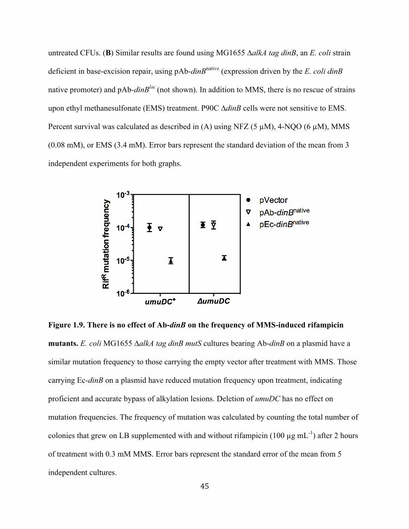
45
untreated CFUs. (B) Similar results are found using MG1655 ∆alkA tag dinB, an E. coli strain
deficient in base-excision repair, using pAb-dinBnative (expression driven by the E. coli dinB
native promoter) and pAb-dinBlac (not shown). In addition to MMS, there is no rescue of strains
upon ethyl methanesulfonate (EMS) treatment. P90C ∆dinB cells were not sensitive to EMS.
Percent survival was calculated as described in (A) using NFZ (5 µM), 4-NQO (6 µM), MMS
(0.08 mM), or EMS (3.4 mM). Error bars represent the standard deviation of the mean from 3
independent experiments for both graphs.
Figure 1.9. There is no effect of Ab-dinB on the frequency of MMS-induced rifampicin
mutants. E. coli MG1655 ∆alkA tag dinB mutS cultures bearing Ab-dinB on a plasmid have a
similar mutation frequency to those carrying the empty vector after treatment with MMS. Those
carrying Ec-dinB on a plasmid have reduced mutation frequency upon treatment, indicating
proficient and accurate bypass of alkylation lesions. Deletion of umuDC has no effect on
mutation frequencies. The frequency of mutation was calculated by counting the total number of
colonies that grew on LB supplemented with and without rifampicin (100 µg mL-1) after 2 hours
of treatment with 0.3 mM MMS. Error bars represent the standard error of the mean from 5
independent cultures.
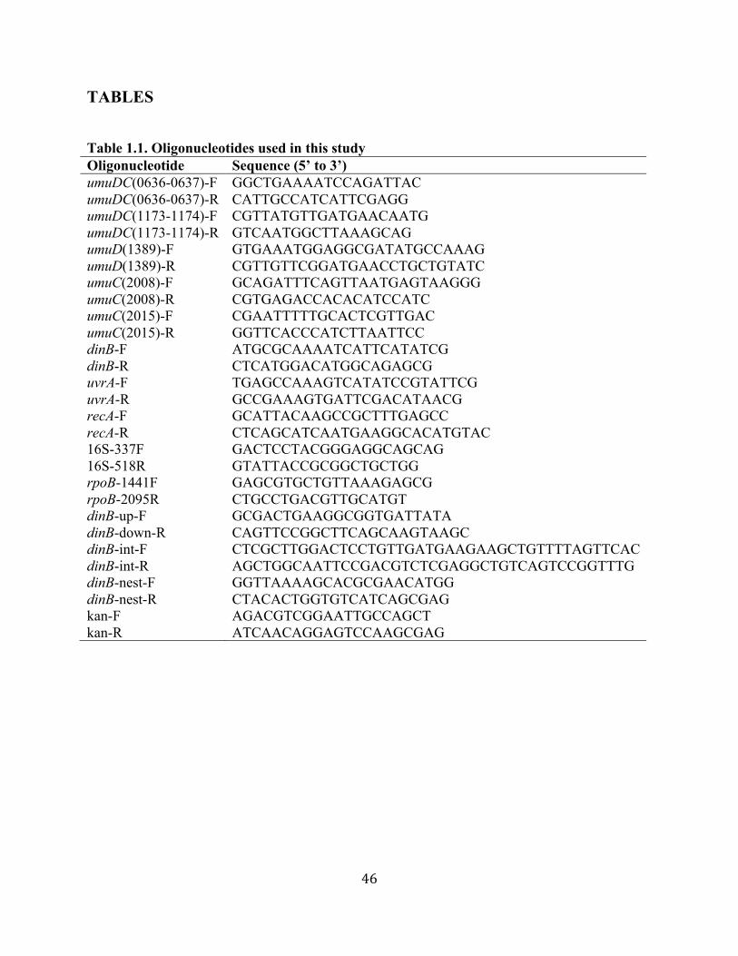
46
TABLES Table 1.1. Oligonucleotides used in this study Oligonucleotide Sequence (5’ to 3’) umuDC(0636-0637)-F GGCTGAAAATCCAGATTAC umuDC(0636-0637)-R CATTGCCATCATTCGAGG umuDC(1173-1174)-F CGTTATGTTGATGAACAATG umuDC(1173-1174)-R GTCAATGGCTTAAAGCAG umuD(1389)-F GTGAAATGGAGGCGATATGCCAAAG umuD(1389)-R CGTTGTTCGGATGAACCTGCTGTATC umuC(2008)-F GCAGATTTCAGTTAATGAGTAAGGG umuC(2008)-R CGTGAGACCACACATCCATC umuC(2015)-F CGAATTTTTGCACTCGTTGAC umuC(2015)-R GGTTCACCCATCTTAATTCC dinB-F ATGCGCAAAATCATTCATATCG dinB-R CTCATGGACATGGCAGAGCG uvrA-F uvrA-R recA-F recA-R 16S-337F
TGAGCCAAAGTCATATCCGTATTCG GCCGAAAGTGATTCGACATAACG GCATTACAAGCCGCTTTGAGCC CTCAGCATCAATGAAGGCACATGTAC GACTCCTACGGGAGGCAGCAG
16S-518R GTATTACCGCGGCTGCTGG rpoB-1441F GAGCGTGCTGTTAAAGAGCG rpoB-2095R dinB-up-F dinB-down-R dinB-int-F dinB-int-R dinB-nest-F dinB-nest-R kan-F kan-R
CTGCCTGACGTTGCATGT GCGACTGAAGGCGGTGATTATA CAGTTCCGGCTTCAGCAAGTAAGC CTCGCTTGGACTCCTGTTGATGAAGAAGCTGTTTTAGTTCAC AGCTGGCAATTCCGACGTCTCGAGGCTGTCAGTCCGGTTTG GGTTAAAAGCACGCGAACATGG CTACACTGGTGTCATCAGCGAG AGACGTCGGAATTGCCAGCT ATCAACAGGAGTCCAAGCGAG

47
Table 1.2. Comparison of number of putative TLS DNA polymerase genes from select isolates of A. baumannii.
A. baumannii Strain GenBank accession number
Number of putative genes
umuC umuD dinB polB
ATCC 17978 CP000521 4 3 1 0
TCDC-AB0715 CP002522 3 2 1 0
AB059 ADHB00000000 3 2 1 0
ATCC 19606 ACQB00000000 2 2 1 0
AB0057 CP001182 2 2 1 0
AB058 ADHA00000000 2 1 1 0
ABNIH3 AFTB00000000 2 1 1 0
ACICU CP000863 2 1 1 0
AYE CU459141 1 1 1 0
MDR-ZJ06 CP001937 1 1 1 0

48
Table 1.3. Mutation signatures of desiccation-induced A. baumannii 17978 RifR mutants
Mutation type rpoB nucleotide change
RpoB amino acid substitution
Mutation frequency (%, N=42)
Transversion 67 1564 CAGàAAG 522 GlnàLys a 7 1565 CAGàCTG 522 GlnàLeu a 10 1573 GACàTAC 525 AspàTyr b 5 1574 GACàGTC 525 AspàVal c 5 1603 CATàGAT 535 HisàAsp a 2 1604 CATàCTT 535 HisàLeu b 12 1619 TCT àTAT 540 SeràTyr b 12 1741 ATCàTTC 581 IleàPhe a b 14 Transition
33
1565 CAGàCGG 522 GlnàArg a b 12 1603 CATàTAT 535 HisàTyr a 2 1619 TCTàTTT 540 SeràPhe a 7 1625 CTTàCCT 542 LeuàPro a b 10 1696 CGTàTGT 566 ArgàCys a c 2 a Indicates novel Acinetobacter substitution b Indicates mutation was also found in an A. baumannii 17978 dinB::Km strain c Indicates mutation was unique to A. baumannii 17978 dinB::Km strain

49
CHAPTER 2
Functional analysis of multiple, putative Acinetobacter baumannii 17978
DNA polymerase V gene products in Escherichia coli

50
ABSTRACT
Acinetobacter baumannii is a clinically important and dangerous opportunistic pathogen.
It quickly gains antibiotic resistances through horizontal gene transfer and a DNA damage
response that we have recently discovered. Many A. baumannii strains have acquired multiple
umuD and umuC orthologues, which encode putative subunits of DNA Polymerase V (Pol V).
We have previously shown that these gene products are induced upon DNA damage in A.
baumannii 17978, and we hypothesize that multiple DNA Pol Vs are likely to contribute to DNA
damage-induced mutagenesis and thus enhance A. bamannii’s mutational capacity. To test this,
we cloned the umuD and umuC genes of A. baumannii 17978 into E. coli to measure in vivo
translesion synthesis (TLS) activity using established DNA damage assays. We find that these
genes are expressed in E. coli, but ∆umuDC, ∆umuD, or umuC-deficient E. coli strains are not
complemented by expression of the A. baumannii genes. Therefore, they are still sensitive to
UV-irradiation and incapable of UV-induced mutagenesis. Our results indicate that A. baumannii
umuD and umuC gene products are not active in E. coli. It is likely that an A. baumannii
component required for DNA Pol V-mediated TLS is missing in E. coli, the implications of
which are discussed.
51
INTRODUCTION
Acinetobacter baumannii is a nosocomial pathogen that has become a serious health
threat worldwide (41). Notably, its resistance to desiccation and disinfection allows it to live for
long periods of time in intensive care units, where it infects immunocompromised patients (40-
42). Strains of A. baumannii are inherently resistant to many classes of antibiotics (40-42) and
some resistant to every antibiotic available have emerged (58). It appears that A. baumannii
readily incorporates foreign DNA through horizontal gene transfer processes (59), which permit
the acquisition of new antibiotic resistances. Although bacteria, such as Escherichia coli, possess
DNA damage response systems known to increase mutagenesis and therefore the evolution of
antibiotic resistances (1, 15, 36), it was unknown whether A. baumannii did the same. In our
previous work (Chapter 1; (111)) we uncovered a regulated system in A. baumannii capable of
inducing mutagenesis upon DNA damage and thus increases the evolution of antibiotic
resistance.
We found that in A. baumannii, DNA-damage induces classic DNA repair and error-
prone DNA polymerase genes, such as those encoding Y-family DNA Pol IV and Pol V (Chapter
1, Figs. 1.3 & 1.4). RecA, the main activator of the SOS response in E. coli, mediates this
response and is required for increased frequency of antibiotic resistant mutants after desiccation,
UV-irradiation, or alkylation damage (Chapter 1, Fig. 1.5). These results are consistent with
Aranda et al., who used DNA microarrays to show a RecA-dependent increase in DNA damage
response genes following mitomycin-C treatment (112).
Intriguingly, A. baumannii strains have acquired multiple umuD and umuC orthologues
(Chapter 1, Table 1.1), encoding putative subunits of DNA Pol V. In particular, A. baumanii
strain ATCC 17978 has acquired two umuDC operons, two unlinked umuCs, and one unlinked

52
umuD (Chapter 1, Fig. 1.1). These genes may have been acquired through horizontal gene
transfer (76). One of the umuDC operons (1174-1173) encodes a DNA Pol V with high sequence
similarity to Pol VR391, encoded by rumA’B from the conjugative IncJ transposon R391 (113).
This Y-family DNA polymerase is capable of generating even higher mutation rates than E. coli
DNA Pol V (113). Indeed, we found that desiccation-induced rifampicin resistant mutants
contained base-pair substitutions that match the signature of E. coli DNA Pol V and Pol VR391
(Chapter 1, Table 1.3; (111, 113)). However, while we and others have shown that A. baumannii
17978 upregulates these multiple umuD and umuC orthologues upon DNA damage (111, 112), it
is still unclear whether or not they are all active in this strain. E. coli contains one single umuDC
operon, which is highly regulated at the levels of both transcription and post-translation (5).
DNA Pol V (UmuD’2C) complex requires UmuC, the catalytic DNA polymerase subunit, a
homodimer of the amino terminally cleaved form of UmuD, UmuD’2, and RecA* for activity
(114). It bypasses UV photoproducts, abasic lesions, and guanine oxidation products through a
process termed translesion-synthesis (TLS) (115, 116). It also participates in TLS past cytotoxic
lesions produced by alkylating agents such as methyl methanesulfonate (87). Because DNA Pol
V is very error-prone, it is the major cause of UV- and SOS-induced mutagenesis in E. coli (1,
9).
Our evidence suggests that the A. baumannii 17978 DNA Pol Vs are involved in DNA-
damage induced mutagenesis (Chapter 1, Fig. 1.5, Table 1.3). The acquisition of multiple genes
encoding DNA Pol V orthologues could potentiate an increase in A. baumannii’s ability to gain
antibiotic resistance. In this work, we examined the TLS activities of A. baumannii 17978 umuD
and umuC gene products in E. coli to gain a better understanding of TLS-mediated mutagenesis
in A. baumannii.

53
MATERIALS AND METHODS
Strains and growth conditions. Strains used in this study are listed in Table 2.1.
Acinetobacter baumannii ATCC 17978 was purchased from The American Type Culture
Collection (ATCC). E. coli strains were routinely grown at 37º C in Luria Broth (LB) or on LB
agar. For plasmid maintenance, 200 µg mL-1 of ampicillin (Ap; Sigma), 30 µg mL-1 of
kanamycin (Km; Sigma), or 12 µg mL-1 tetracycline (Tet; Sigma) were used. When required, 100
µg mL-1 of rifampicin (Rif; Calbiochem) was added to LB agar.
Construction of plasmids. E. coli umuDC and A. baumannii 17978 umuDC genes were
cloned in the low copy-number vector, pWSK29, all under the native E. coli umuDC promoter.
First, pumuDC1 (Table 2.1) was constructed by using the In-fusion (Clontech) kit and by
following the manufacturer’s protocol. Briefly, E. coli umuDC and its native promoter region
were amplified by PCR from strain P90C (Table 2.1) using fusionDC-F and fusionDC-R
oligonucleotides (Table 2.2). pumuDC1 was then used as the template for the construction of
pEc-umuDC (Table 2.1), which contains a NheI restriction site inserted precisely between the
promoter region and the start codon of umuD. To insert the NheI restriction site, the GeneTailor
(Life Technologies) site-directed mutagenesis protocol was followed using umuCNheIF and
umuCNheIR (Table 2.2) oligonucleotides. Next, A. baumannii 17978 (Table 2.1) umuDC genes
were amplified using oligonucleotides containing NheI and SacI restriction sites (Table 2.2).
Gene loci amplified from A. baumannii 17978 are as follows:
umuD(A1S_0636)umuC(A1S_0637), umuD(A1S_1174)umuC(A1S_1173), umuC(A1S_2008),
and umuC(A1S_2015). A. baumannii umuDC genes were cloned into the NheI and SacI sites of
pEc-umuDC, effectively creating isogenic plasmids under the Ec-umuDC promoter (Table 2.1).
All constructs were confirmed by sequencing (Tufts University Core Facility).
54
pNLAC1-Ec-umuD and pNLAC-Ab-umuD(1389) plasmids (Table 2.1) were constructed
by cloning the respective amplification products from P90C or A. baumannii 17978 into the PstI
and PvuI sites of pNLAC1 (Table 2.1; generous gift from Tom Russo, University at Buffalo).
These sites are within the bla gene (encodes ß-lactamase), putting the umuD genes under the
control of the bla promoter and disrupting ß-lactamase production. Oligonucleotides sets used
for the PCR amplifications were umuDPvuI-F and umuDPstI-R and 1389PvuI-F and 1389PstI-R
(Table 2.2). pWSK129-Ec-umuD and pWSK129-Ab-umuD(1389) (Table 2.1) were constructed
by cloning the respective amplification products (as above) into the SacI and ApaI sites of
pWSK129 (Table 2.1). Control of these genes is under the lac promoter. Oligonucleotide sets
used for the PCR reactions were umuDSacI-F and umuDApaI-R and 1389SacI-F and 1389ApaI-
R (Table 2.2). Constructs were confirmed by sequencing (Tufts University Core Facility).
UV-irradiation and induced-mutagenesis. For UV-survival, saturated E. coli P90C
cultures (~109 cells) were serially diluted in SMO (100 mM NaCl, 20mM Tris-HCl pH 7.5) and
10 µL spots were deposited on LB-Ap plates. Plates were individually irradiated in the dark
under a UV germicidal lamp with increasing amounts of UV-exposure (J m-2). Survival was
calculated by dividing the number of colony forming units (CFUs) that grew on each UV-treated
plate by the number of CFUs grown on LB-Ap alone after 20-24 hours.
For UV-induced mutagenesis, E. coli P90C cultures were started from ≤100 cells to
minimize the probability of preexisting mutants in the starting inoculum. Saturated cultures were
spun down, resuspended in equal volume of SMO, and 2 mL samples were spread evenly in a
petri dish. Samples were irradiated in the dark under a UV germicidal lamp with 55 J m-2 and
immediately transferred to LB-Ap medium at a 1:10 dilution following irradiation. Upon
saturation, appropriate cell dilutions were deposited on LB-Ap with and without rifampicin to

55
assess, respectively, the number of rifampicin resistant mutants (RifR) and total number of CFUs.
Colonies were counted after 24 hours of incubation. Mutation frequency was calculated by
dividing the number of RifR mutants by the total number of CFUs.
MMS-induced mutagenesis. E. coli MG1655 (Table 2.1) cultures were started from
≤100 cells, grown to saturation, then diluted 1:100 and grown for 4 hours. Cultures were then
treated with 0.3 mM methyl methanesulfonate (MMS; Sigma) for 1 hour. Cells were washed
with SMO, serially diluted, and deposited on LB-Ap plates with and without rifampicin. After 24
hours of incubation, mutation frequency was determined as described above.
Semi-quantitative RT-PCR. Cultures were treated as described in the UV-induced
mutagenesis section (see above) with three exceptions. First, to ensure cells were in exponential
phase growth, saturated cultures were diluted 1:1000 and grown for 2.5 hours, then diluted 1:50
and grown for 2 hours 3 consecutive times. Second, after UV-irradiation, samples were
incubated for an additional 1 hour at 37º C prior to RNA extraction to allow for gene expression.
Third, untreated cultures were not used. The procedure was carried out as described in Chapter 1
Materials and Methods (111). E. coli rpoB RNA expression was used as the control and was
equal in each sample. Gene specific RT-PCR oligonucleotides are listed in Table 2.2.

56
RESULTS
A. baumannii umuDCs and umuCs are expressed upon UV-irradiation from their
respective plasmid constructs in E. coli. To test the activity of A. baumannii 17978 umuDC
(Ab-umuDC) gene products in E. coli, we constructed isogenic low copy-number plasmids
containing each umuDC operon or unlinked umuC from the 17978 strain (See Materials and
Methods). E. coli umuDC (Ec-umuDC), Ab-umuDCs and Ab-umuCs were cloned into pWSK29
(Table 2.1) under the control of the SOS-inducible Ec-umuDC native promoter (Table 2.1). We
choose not to use the Ab-umuDC or Ab-umuC promoters because it is unknown whether these
are functional in E. coli. Use of the Ec-umuDC promoter ensures relatively equal expression and
induction by the SOS response (1).
Figure 2.1 shows mRNA expression levels of each umuDC construct (pWSK29; grey
bars) as determined by semi-quantitative RT-PCR after ∆umuDC cells were treated with UV-
light, a strong inducer of the SOS response (95). Ec-umuDC, Ab-umuDCs, and Ab-umuCs are
expressed at similar levels (within an approximately 10-fold range (Fig. 2.1; grey bars)).
Ec-umuD and the unlinked umuD(1389) from A. baumannii were also cloned into a
higher copy-number plasmid, pNLAC1 (Table 2.1), which should result in constitutive
expression from the bla promoter. We find that Ec-umuD and Ab-umuD(1389) are equally
expressed in E. coli ∆umuDC (Fig. 2.1; white bars) upon UV-irradiation. Relative expression
levels of these two genes are on par with the umuDCs and umuCs expressed from the Ec-umuDC
promoter in pWSK29 (Fig. 2.1; grey bars). These data suggested to us that we would be able to
assay A. baumannii umuDC gene products for activity in E. coli.
A. baumannii umuDC gene products do not rescue E. coli ∆umuDC from UV-
sensitivity. We first wanted to test A. baumannii umuDC gene products for their ability to form

57
active DNA Pol V in vivo and thus bypass UV-induced lesions. This is one of the main functions
of E. coli DNA Pol V and a conserved feature across domains (5, 21). E. coli ∆umuDC cells
carrying the pWSK29-umuDCs were exposed to increasing amounts of UV-light. As expected,
Ec-umuDC rescues cells from UV-sensitivity (Fig. 2.2A; compare pEc-umuDC to pWSK29).
However, cells carrying the plasmids with A. baumannii umuDC(0636-0637), umuDC(1174-
1173), umuC(2008), or umuC(2015) did not have improved survival over the vector alone (Fig.
2.2A). We did not expect the Ab-umuC gene products to rescue E. coli ∆umuDC, because
formation of a typical DNA Pol V complex would not happen in the absence of Ab-umuD or Ec-
umuD.
To determine whether the A. baumannii umuC gene products are able to form an active
DNA Pol V hybrid in E. coli, we provided them with Ec-umuD+. We hypothesized that the
presence of native Ec-umuD+ would facilitate DNA Pol V formation if the Ab-UmuCs are
similar in structure and function to Ec-UmuC. This strategy eliminates the variable of both Ab-
UmuD and Ab-UmuC needing to be active in E. coli, since it is possible that these particular Ab-
UmuDs might not be cleaved to Ab-UmuD’ in E. coli. Moreover, the dependence of RecA*
might be particular for the A. baumannii gene products (9). We used the strain, umuC122::Cm
(Table 2.1), which is umuD+ and phenotypically ∆umuC to test our hypothesis. We find that the
Ab-umuCs and Ab-umuDCs do not rescue cells from UV-sensitivity (Fig. 2.2B), in stark contrast
to Ec-umuDC (Fig. 2.2B). The results thus far suggest that A. baumannii UmuCs are either
unable to form active DNA Pol V complex in E. coli or are unable to perform TLS past a UV-
induced DNA lesion.
A. baumannii umuD and umuC gene products do not confer UV-induced
mutagenesis in E. coli. Although cells expressing the A. baumannii umuDC gene products were
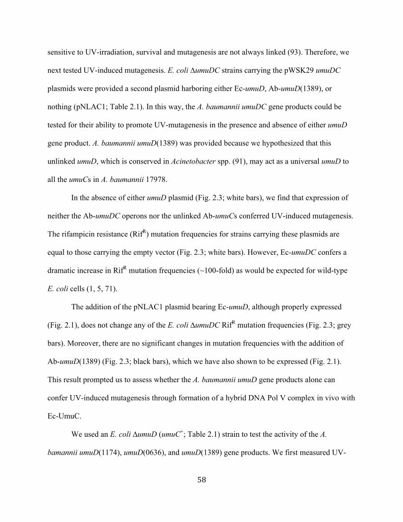
58
sensitive to UV-irradiation, survival and mutagenesis are not always linked (93). Therefore, we
next tested UV-induced mutagenesis. E. coli ∆umuDC strains carrying the pWSK29 umuDC
plasmids were provided a second plasmid harboring either Ec-umuD, Ab-umuD(1389), or
nothing (pNLAC1; Table 2.1). In this way, the A. baumannii umuDC gene products could be
tested for their ability to promote UV-mutagenesis in the presence and absence of either umuD
gene product. A. baumannii umuD(1389) was provided because we hypothesized that this
unlinked umuD, which is conserved in Acinetobacter spp. (91), may act as a universal umuD to
all the umuCs in A. baumannii 17978.
In the absence of either umuD plasmid (Fig. 2.3; white bars), we find that expression of
neither the Ab-umuDC operons nor the unlinked Ab-umuCs conferred UV-induced mutagenesis.
The rifampicin resistance (RifR) mutation frequencies for strains carrying these plasmids are
equal to those carrying the empty vector (Fig. 2.3; white bars). However, Ec-umuDC confers a
dramatic increase in RifR mutation frequencies (~100-fold) as would be expected for wild-type
E. coli cells (1, 5, 71).
The addition of the pNLAC1 plasmid bearing Ec-umuD, although properly expressed
(Fig. 2.1), does not change any of the E. coli ∆umuDC RifR mutation frequencies (Fig. 2.3; grey
bars). Moreover, there are no significant changes in mutation frequencies with the addition of
Ab-umuD(1389) (Fig. 2.3; black bars), which we have also shown to be expressed (Fig. 2.1).
This result prompted us to assess whether the A. baumannii umuD gene products alone can
confer UV-induced mutagenesis through formation of a hybrid DNA Pol V complex in vivo with
Ec-UmuC.
We used an E. coli ∆umuD (umuC+; Table 2.1) strain to test the activity of the A.
bamannii umuD(1174), umuD(0636), and umuD(1389) gene products. We first measured UV-

59
induced RifR mutation frequencies using strains carrying the Ab-umuDC operons. The presence
of umuC on the plasmids seemed to have no ill effects, because we find a significant increase in
RifR mutation frequency (>100-fold) when cells are provided with Ec-umuDC (Fig. 2.4A). In
contrast, cells carrying Ab-umuDC(1174-1173) or Ab-umuDC(0636-0637) operons have RifR
mutation frequencies equal to those with the empty vector (Fig. 2.4A). Furthermore, similar
results were found when we tested Ab-umuD(1389) in the E. coli ∆umuD strain using two
different plasmid constructs (pNLAC1 and pWSK129; Table 2.1). We find that with either
plasmid, umuD(1389) does not confer UV-induced mutagenesis (Fig. 2.4B), in contrast to the
significant increase in mutation frequency obtained using Ec-umuD (Fig. 2.4B). This result
suggests that Ab-umuD(1389) is unable to form active DNA Pol V with Ec-UmuC, even though
it has been shown to be cleaved to a UmuD’-like form in E. coli (91). Taken together, these
results provide further evidence for the lack of activity of A. baumannii umuD and umuC gene
products in E. coli, at least upon conditions of UV-damage.
MMS-induced mutation frequencies are unaffected by the expression of A.
baumannii umuDCs. Lastly, we wanted to see if we could detect TLS activity using a different
DNA-damaging agent; we chose the alkylating agent methyl methanesulfonate (MMS). Using
the E. coli ∆umuDC strain, it was difficult to detect significant differences in MMS-induced
mutagenesis between those carrying Ec-umuDC and the empty vector control (data not shown).
We therefore used the ∆alkA tag mutS umuDC dinB background (Table 2.1) that we (Chapter 1,
Fig. 1.9) and others (87) have successfully used to measure differences in MMS-induced
mutagenesis. Here, the presence of dinB dramatically reduces the frequency of RifR mutants
because DinB accurately bypasses DNA alkylation lesions produced by MMS (87). Therefore,
the accuracy of TLS past alkylation lesions can be measured in vivo. Interestingly, we find a
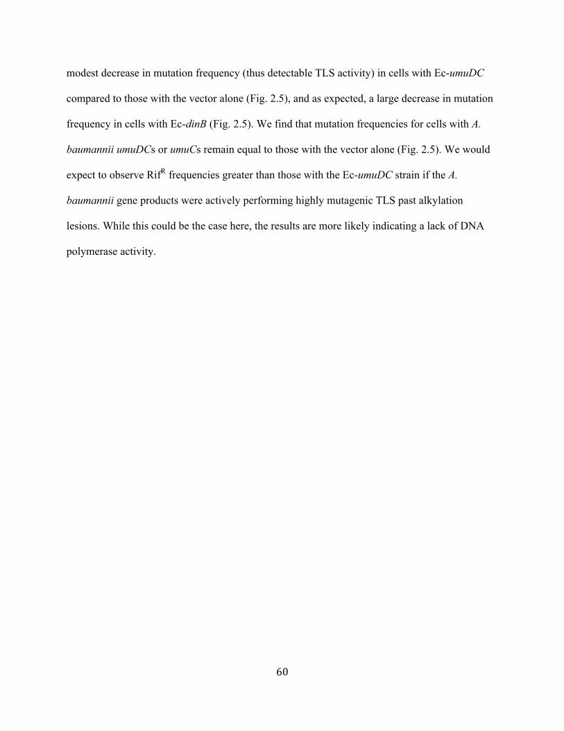
60
modest decrease in mutation frequency (thus detectable TLS activity) in cells with Ec-umuDC
compared to those with the vector alone (Fig. 2.5), and as expected, a large decrease in mutation
frequency in cells with Ec-dinB (Fig. 2.5). We find that mutation frequencies for cells with A.
baumannii umuDCs or umuCs remain equal to those with the vector alone (Fig. 2.5). We would
expect to observe RifR frequencies greater than those with the Ec-umuDC strain if the A.
baumannii gene products were actively performing highly mutagenic TLS past alkylation
lesions. While this could be the case here, the results are more likely indicating a lack of DNA
polymerase activity.

61
DISCUSSION
Acinetobacter baumannii integrates foreign DNA from its environment and other bacteria
through horizontal gene transfer processes such as plasmid acquisition, transposons, and
homologous recombination (40, 42, 59). We have previously shown that Acinetobacter
baumannii isolates have acquired varying numbers of umuD and umuC genes, which encode
putative subunits of DNA Pol V (Chapter 1, Figs. 1.1 & 1.7, Table 1.2). It is believed that these
genes were acquired through horizontal gene transfer (76), but the question of whether or not
they are active still remains. We set out to answer this question by assaying these genes for
classic signs of TLS activity in E. coli. A. baumannii 17978 contains two umuDC operons, two
unlinked umuCs, and one unlinked umuD (Chapter 1, Fig. 1.1), bringing the total number of
putative DNA Pol V component genes to seven; five more than E. coli. The potential for DNA
damage-induced mutagenesis is thus greater than E. coli’s, if the systems of regulation are
identical. However, we know that the mechanisms regulating the DNA-damage response in A.
baumannii 17978 are different than E. coli’s (Chapter 1 & (112)).
Multiple lines of evidence suggest that the A. baumannii 17978 umuD and umuC genes
are inactive in E. coli. We tested different combinations of A. baumannii (Ab-) and E. coli (Ec-)
umuDs and umuCs to see if active DNA Pol V complex could be formed in vivo. Ab-umuDCs
alone or combined with Ec-umuD failed to rescue cells from UV-irradiation (Fig. 2.2) or confer
UV induced-mutagenesis (Fig. 2.3). This suggests that Ab-umuC’s are unable to form active
DNA Pol V with either Ab-umuD or Ec-umuD. We also tested the opposite combination, Ab-
umuD’s with Ec-umuC, and failed again to observe UV-induced mutagenesis (Fig. 2.4A). These
results imply that some factor may be missing for these enzymes to be active in E. coli; i.e. a
native A. baumannii protein.

62
We hypothesized that Ab-umuD(1389), whose gene product has been shown to be
cleaved to a UmuD’-like protein in E. coli (91), was the missing component necessary for
activity. This Ab-UmuD(1389) has characteristics of both UmuD and LexA, and has recently
been shown to repress error-prone DNA polymerase genes in A. baumannii (112). While this
could be the main function of Ab-UmuD(1389), the evidence for DNA damage-inducible
cleavage (91) led us to believe it could still be functioning like Ec-UmuD’ in the DNA Pol V
complex. Our results suggest that Ab-UmuD(1389), unlike Ec-UmuD, cannot complement
∆umuD E. coli cells (Fig. 2.4B). Furthermore, the addition Ab-UmuD(1389) does not change the
mutation frequencies of E. coli ∆umuDC cells carrying any of the Ab-umuDC operons or Ab-
umuCs (Fig. 2.3). Therefore, even though Ab-umuD(1389) is cleaved to a UmuD’ form, our
evidence suggests that it does not form DNA Pol V in E. coli with either Ab-UmuCs or Ec-
UmuC. The data point to a larger issue of whether the Ab-UmuDs or Ab-UmuC proteins have
even the potential to be active in E. coli, independent of their ability to form DNA Pol V
molecules.
Ec-UmuC is quickly degraded by the Lon protease, and the umuD gene products by Lon
and ClpXP (117), a level of post-translational regulation that ensures minimal time for potential
mutagenesis. Although we have shown that the A. baumannii genes are transcribed in E. coli
nearly as well as native genes (Fig. 2.1), it is unknown whether they are being translated
efficiently or how quickly their gene products are being degraded. Since they are foreign genes
in E. coli, it is entirely possible that they are translated at lower levels (rare codons) and/or are
degraded faster than native Ec-umuDC gene products. We would need to have specific
antibodies made against each of these A. baumannii gene products in order to know the levels of
protein in the cell at multiple time points. Remarkably, we have previously shown that Ab-DinB

63
is active in E. coli. Cells bearing plasmid-borne Ab-dinB are rescued upon treatment with either
nitrofurazone or 4-nitroquinolone-1-oxide (Chapter 1, Fig. 1.8). While the DinB family of
enzymes are more evolutionarily conserved than the UmuC family (21), these results
demonstrate that some A. baumannii enzymes do have the potential to be active in E. coli.
One factor that could be missing from our transplanted A. baumannii DNA Pol V TLS
system in E. coli is the A. baumannii ß-binding clamp of the DNA Pol III holoenzyme (encoded
by dnaN). This essential replication factor is important for recruitment and management of DNA
polymerases at the replication fork (118). In order to access the replication fork and perform
TLS, E. coli TLS DNA polymerases, as well as UmuD and UmuD’, require interaction with the
ß-clamp through conserved motifs (119-123). The UmuC canonical ß-binding motif is required
for UV-induced mutagenesis (124), and mutating the ß-binding motif of DinB (DNA Pol IV)
abolishes its activity in vivo (93). The system of governing access to the replication fork is
delicate, and even a single residue change in the ß-binding motif can dramatically alter the
activity of Ec-UmuC (124). We therefore hypothesize that even if Ab-umuDC gene products are
successfully produced in E. coli, then they may be inactive because of their inability to bind to
the native E. coli ß-binding clamp.
The Ab-DinB ß-binding motif (QLSLW_) is similar to the QLVLGL motif found in Ec-
DinB (Chapter 1, Fig. 1.6; (125)). One residue is missing in the A. baumannii motif and only two
residues differ between the two. However, an alignment of all the A. baumannii 17978 umuC
gene products shows that the E. coli ß-binding motif is not as conserved as DinB’s (Chapter 1,
Fig. 1.7). Residues 357-361 (QLNLF) at the C-terminal end of Ec-UmuC (118, 123) do not align
with a similar motif in the Ab-UmuC sequences. A consensus sequence of TYDLL at this
location (Chapter 1, Fig. 1.7) could very well be an A. baumannii alternative ß-binding motif.

64
Therefore, if Ab-UmuCs are translated and stable in E. coli, then a proper ß-clamp interaction
could be a limiting factor in our assays. We plan on providing an E. coli dnaN temperature
sensitive mutant with Ab-dnaN to further test the TLS activity of the Ab-umuDC gene products.
Preliminary evidence suggests that Ab-dnaN is able to complement growth proficiency in this
mutant E. coli strain at high temperatures (data not shown).
This work offers an interesting perspective on how we think of bacterial foreign gene
acquisition. It is easy to presume that bacteria, such as pathogens, uptake foreign DNA through
horizontal gene transfer processes and immediately use it to their advantage. It is alarmingly
common for pathogens like A. baumannii to acquire resistance islands, transposons, and
plasmids containing antibiotic resistance genes or virulence factors (41, 58, 126, 127). However,
our results suggest that successful utilization of a new gene, such as a mutagenic DNA
polymerase, may require modification of that gene or other components of the TLS system to
make it work – an evolutionary process that could take many generations. It remains unclear at
this point whether the multiple A. baumannii 17978 DNA Pol Vs are truly active polymerases.
Nevertheless, independent of whether or not Ab-DnaN or some other component will confer
activity in E. coli, the complexity of the situation lends support to this perspective.
ACKNOWLEDGEMENTS
I would like to thank Kathrin Abele, who helped me perform some of the experiments.
Thank you to Nicole Connelly for her umuC122:Cm strain, Ivan Matic (Université Paris
Descartes) for the MG1655 strain, and Tom Russo (University at Buffalo) for the pNLAC1
plasmid. Also thanks to Linda Nguyen for her help gathering preliminary data for this work.
65
FIGURES
Figure 2.1. All plasmid-borne A. baumannii genes are expressed upon UV-irradiation in E.
coli ∆umuDC. Relative expression level in arbitrary units is shown for each pWSK29 (gray) and
pNLAC1 (white) construct. pWSK29 genes are all expressed from the native promoter of the E.
coli umuDC operon. pNLAC1 genes are expressed from the bla (encoding ß-lactamase)
promoter. Semi-quantitative RT-PCR was performed on total RNA purified from 55 J/m2 UV-
irradiated cultures. See Materials and Methods section for details. Gene specific RT-PCR
primers were used to amplify approximately 300bp regions of each umuD, umuC, or umuDC
operon. E. coli genes are denoted by “Ec-”. A. baumannii 17978 gene names include genomic
locus tags (“A1S_” not included before number) in parenthesis. Data from a representative
experiment is shown.
66
Figure 2.2. A. baumannii 17978 umuDCs do not rescue E. coli ∆umuDC from UV-
sensitivity. (A) E. coli ∆umuDC cells carrying pWSK29 with A. baumannii 17978 umuDCs are
sensitive to increasing amounts of UV-light. In contrast, native Ec-umuDC confers survival. (B)
umuC122::Cm cells are umuD+ but have a C-terminal UmuC truncation, rendering them
sensitive to UV (see pWSK29). Cells with native Ec-umuDC survive much better than those with
A. baumannii 17978 umuDCs, even though the cells are umuD+. Error bars represent the standard
deviation of the mean from at least 3 independent experiments.
∆umuDC(a)
(b)umuC122::Cm

67
Figure 2.3. A. baumannii 17978 umuDCs do not confer UV-induced mutagenesis in E. coli
∆umuDC. All strains carry both pWSK29 constructs (X-axis) and pNLAC1 umuD derivatives.
Strains with plasmid-borne A. baumannii 17978 umuDCs and Ec-umuDC were assayed for UV-
induced mutagenesis in the absence (white bars) and presence of Ec-umuD (grey bars) or Ab-
umuD(1389) (black bars). Ec-umuDC confers an increase in rifampicin resistance (RifR)
mutation frequency regardless of the presence of Ec-umuD or Ab-umuD(1389). A. baumannii
umuDCs do not confer any increase in RifR mutation frequency compared to the vector. The
addition of either Ec-umuD or Ab-umuD(1389) has no significant effect on mutation frequency.
Error bars represent the standard deviation of the mean from at least 3 independent experiments.
pWSK29
pEc-umuDC
p1174-1173
p0636-0637
p2008
p2015
Plasmid
UV-induced Mutagenesis
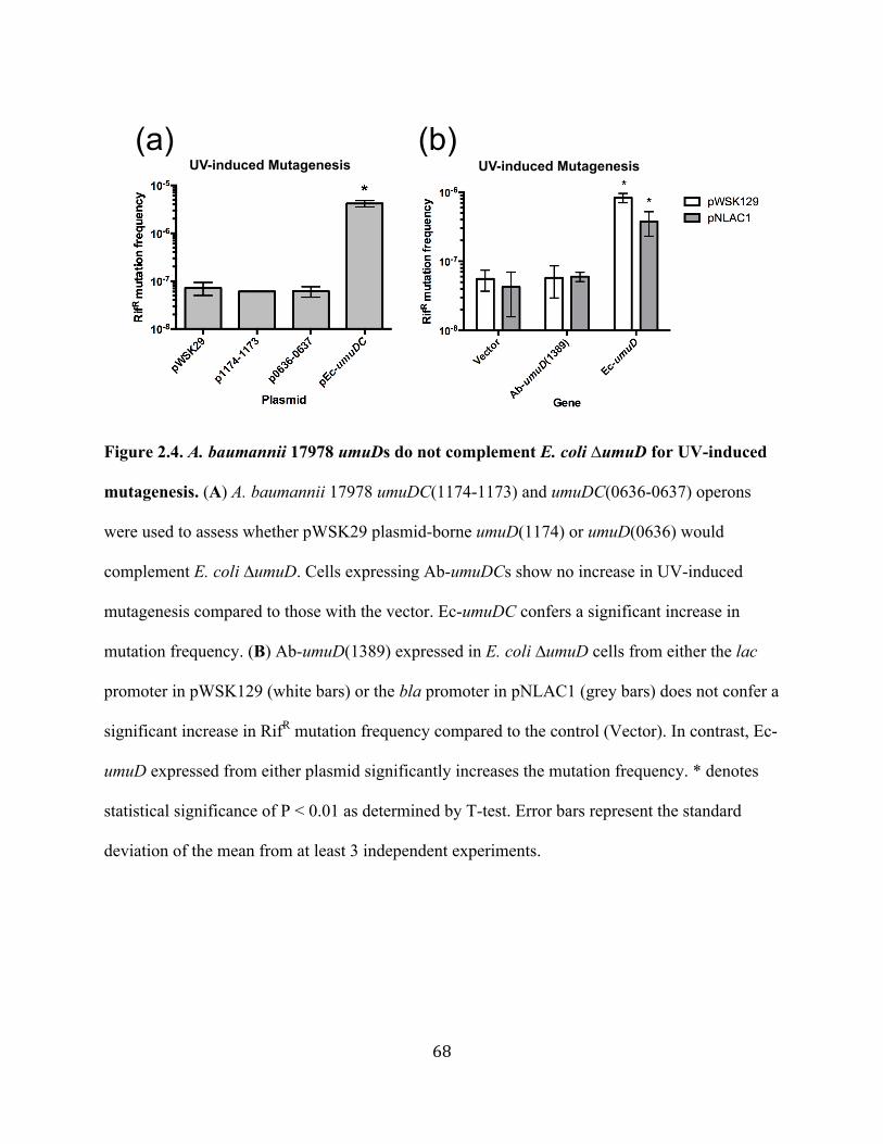
68
Figure 2.4. A. baumannii 17978 umuDs do not complement E. coli ∆umuD for UV-induced
mutagenesis. (A) A. baumannii 17978 umuDC(1174-1173) and umuDC(0636-0637) operons
were used to assess whether pWSK29 plasmid-borne umuD(1174) or umuD(0636) would
complement E. coli ∆umuD. Cells expressing Ab-umuDCs show no increase in UV-induced
mutagenesis compared to those with the vector. Ec-umuDC confers a significant increase in
mutation frequency. (B) Ab-umuD(1389) expressed in E. coli ∆umuD cells from either the lac
promoter in pWSK129 (white bars) or the bla promoter in pNLAC1 (grey bars) does not confer a
significant increase in RifR mutation frequency compared to the control (Vector). In contrast, Ec-
umuD expressed from either plasmid significantly increases the mutation frequency. * denotes
statistical significance of P < 0.01 as determined by T-test. Error bars represent the standard
deviation of the mean from at least 3 independent experiments.
UV-induced Mutagenesis(a) (b)
UV-induced Mutagenesis

69
Figure 2.5. A. baumannii 17978 umuDCs do not affect MMS-induced mutation frequencies
in an alkylation damage-sensitive strain of E. coli. MG1655 ∆alkA tag mutS umuDC dinB
cells bearing A. baumannii 17978 umuDCs have RifR mutation frequencies no different than the
vector (pWSK29). In this background, RifR mutation frequencies are lower in the presence of
dinB because DinB accurately bypasses DNA alkylation lesions (compare pEc-dinB to
pWSK29). The RifR mutation frequencies of cells with Ec-umuDC are modestly lower than those
with the vector alone. Error bars represent the standard deviation of the mean from at 6
independent experiments.
RifR
mut
atio
n fr
eque
ncy

70
TABLES Table 2.1. Strains and plasmids Name Genotype/Description Reference Strains E. coli P90C ∆(lac-pro)XIII thi ara (86) ∆umuDC As P90C with ∆umuDC Godoy Lab ∆umuD As P90C with ∆umuD, KanR Godoy Lab umuC122::Cm As P90C with CmR cassette inserted
precisely into umuC, deleting 306 bp from C-terminus
Godoy Lab, N. Connelly
E. coli MG1655 ∆alkA tag mutS dinB umuDC I. Matic, (87) A. baumannii 17978 Multi-drug resistant (77) Plasmids
pWSK29 ApR, lacZα, pSC101 ori (88) pumuDC1 As pWSK29 with E. coli umuDC and
native promoter This work
pEc-umuDC As pumuDC with NheI site inserted between umuDC promoter and start codon
This work
p0636-0637 As pumuDC-NheI, E. coli umuDC promoter, replaces umuDC with A. baumannii umuDC(0636-0637)
This work
p1174-1173 As p0636-0637 but with A. baumannii umuDC(1174-1173)
This work
p2008 As p0636-0637 but with A. baumannii umuC(2008)
This work
p2015 As p0636-0637 but with A. baumannii umuC(2015)
This work
pEc-dinB pYG768; ApR; pSC101 ori (22) pNLAC1 ApR, TetR, pBR322 ori (128) pNLAC1-Ab-umuD(1389)
As pNLAC1, with Ab-umuD1389 cloned into PstI and PvuI sites of the bla gene
This work
pNLAC1-Ec-umuD As pNLAC1, with Ec-umuD cloned into PstI and PvuI sites of the bla gene
This work
pWSK129 KanR, lacZα, pSC101 ori (88) pWSK129-Ab-(umuD1389)
As pWSK129, with Ab-umuD cloned into SacI and ApaI sites
This work
pWSK129-Ec-umuD
As pWSK129, with Ec-umuD cloned into SacI and ApaI sites
This work

71
Table 2.2. Oligonucleotides used in this study Name Sequence (5’ to 3’) umuCNheIF GTATAACTTCAGGCAGATTAGCTAGCTTATGTTGTTTATCAAGCC
TGCGG umuCNheIR TAATCTGCCTGAAGTTATACTGTTTTTATATACAGTAGTCTGTTC
TTGCCAGC fusionDC-F TATCGAATTCCTGCACTCCATCTGCGGTTTCGATTGC fusionDC-R ATCCCCCGGGCTGCACGTGATCTGTTCGGTCGCTAATCC umuC200inF GCG CAG CTC AAT TTA TTC G umuD200inR CACCGTCACTAATTCCACCATC 2008FNheI CTAGGCTAGCATGAAAGATATCTCACACCG 2008RSacI GCTAGAGCTCTCATGTATTTTGTGTAACGGG 2015FNheI ACTGGCTAGCATGAAAAGGCGAATTTTTGC 2015RSacI CACTGGAGCTCTTATCTTGATATTTTTAGCATACCTTCCC 1174FNheI ATAAGCTAGCATGAGCGAAATTGCACCATC 1174RSacI CGGCGAGCTCTTAATTCAAAATAGTTAATAACTCATCCC 0636FNheI ACGGCTCGCTAGCATGAATAGAGTAATTAATTCAGAATTGGAGC 0636RSacI GGCCACGCTGAGCTCTTAATTCTTAATTTTTATCATTCCATCAAA
ACT AbdnaNPstI GCTGCCTGCAGGATTAAACACGCATCGG AbdnaNPvuI GCAGCACGATCGATGCGTTTGAAAATCGC 1174RT-F GCGAAATTGCACCATCCATTATCC 1174RT-R GTGCTCGTTCATGTCGAGAG 2015RT-F GTTAGAAGCGATGGGAATAAATACCG 2015RT-R GTCCTTCTGGCAATAGCTTCCTT 2008RT-F CACACCGTGAAGTTTATGCACT 2008RT-R CGTGAGACCACACATCCATC 0636RT-F GGCTGAAAATCCAGATTAC 0636RT-R CATTGCCATCATTCGAGG 1389RT-F GTGAAATGGAGGCGATATGCCAAAG 1389RT-R CGTTGTTCGGATGAACCTGCTGTATC RTrpoB-F CCCTATGGTTTACTCCTATACCGAG RTrpoB-R GGACGTCAAACACCGGTTCGC RTumuDC-F AGCGCGTACTCGCCCATTACC RTumuDC-R CCCATAAATCAGGGCGAAACACCG umuD200-R CACCGTCACTAATTCCACCATC umuDPvuI-F CGACGACGATCGTATGTTGTTTATCAAGCCTGC umuDPstI-R TATATCTGCAGATCAGCGCATCGCCTTAACG 1389PvuI-F GCACTACGATCGCATTTCGCATGTCTCCATACCA 1389PstI-R GCGCTGCTGCAGGTTACTGACCATCATTTCTGG umuDSacI-F GCGACGGAGCTCGATTATTATGTTGTTTATCAAGCC umuDApaI-R ATATAGGGCCCGGCAAACATCAGCGCATCG 1389SacI-F GTTATAGAGCTCTGAAATGGAGGCGATATGCCAAAG 1389ApaI-R GAGCTGGGCCCGTTACTGACCATCATTTCTGG
72
CHAPTER 3
Examining the role of the carboxy-terminal domain of Escherichia coli
DNA polymerase V subunit, UmuC

73
ABSTRACT
Cells have a variety of mechanisms to cope with DNA replication fork stalling. One of
these is translesion DNA synthesis (TLS), in which cells alleviate fork stalling by way of
specialized, low-fidelity DNA polymerases. Escherichia coli TLS DNA polymerase, DNA Pol
V, is composed of the accessory subunit, UmuD’, and the catalytic subunit, UmuC. DNA Pol V
is responsible for the majority of UV- and SOS- induced mutagenesis and is therefore regulated
at both the level of transcription and post-translation. The carboxy-terminal domain of UmuC
may mediate such protein-protein interactions, but this is poorly understood. To gain insights
into the role of this domain, we constructed an IPTG inducible plasmid that expresses a C-
terminal fragment of UmuC lacking its catalytic domain. We hypothesized that this domain may
be involved in UmuC regulatory interactions, and expression of this fragment would reveal these
interactions through sequestration or modulation of certain factors. We find that expression of
the C-terminus lowers the frequency of induced mutagenesis upon hydroxyurea treatment in a
mutagenic strain that lacks the C-terminus of UmuC. We also find that expression of this C-
terminal fragment in an UmuC+ background has diverse effects on cell viability and is only
detectable upon SOS-inducing conditions. We hypothesized that bacterial Hsp90 (HtpG), an
enigmatic heat shock protein, may have a role in this phenomenon since it is known to directly
interact with and regulate eukaryotic TLS DNA polymerases. Results suggest that HtpG does not
interact with UmuC at the C-terminus; therefore, the role of this heat shock protein in E. coli
TLS regulation remains elusive. Together these data support the notion that the C-terminus of E.
coli UmuC is likely involved in DNA Pol V regulatory protein-protein interactions under
conditions of stress.
74
INTRODUCTION
Replicative DNA polymerases copy templates proficiently and with high fidelity, but they
cannot incorporate nucleotides opposite of lesions on the template DNA. Thus, DNA damage
can cause replication fork stalling and therefore genomic instability and cell death by hindering
the progress of replicative DNA polymerases. In a process termed translesion synthesis (TLS),
specialized DNA polymerases can take over replication at stalled forks and continue synthesis
past DNA lesions (1). These DNA polymerases, in particular those of the Y-family, have a
comparatively open active site, which allows for accommodation of damaged bases (30).
Because of this feature, Y-family DNA polymerases have poor processivity and are, in general,
error prone on undamaged DNA compared to replicative, high-fidelity DNA polymerases (1).
This low fidelity, potentially mutagenic incorporation of nucleotides may be helpful for bacteria
to evolve and survive conditions of stress (15), but it also makes it necessary for these DNA
polymerases to be extensively regulated (1). Y-family DNA polymerases are highly conserved
throughout all domains of life (1, 21, 129, 130). They have been implicated in human cancers
(131, 132) and in the growing problem of bacterial antibiotic resistance (15, 133). Therefore,
studying the function and regulation of these DNA polymerases in the model system of
Escherchia coli is translatable and significant.
E. coli has two Y-family DNA polymerases, DNA Pol IV (DinB) and DNA Pol V
(UmuD’2C), encoded by the dinB and umuDC genes, respectively. They are both
transcriptionally regulated by the LexA repressor as part of the SOS gene network and induced
upon DNA damage or replication stress (1). UmuC is the catalytic subunit of DNA Pol V, and it
has the ability to insert nucleotides on templates with abasic sites and UV-photoproducts such as
thymine-thymine dimers and thymine-thymine cyclobutane pyrimidine dimers (115, 116). It is

75
responsible for the majority of UV- and SOS-induced mutagenesis in E. coli, thus it is highly
regulated at the levels of transcription and post-translation (1). While the number of DinB
molecules in the cell increases from approximately 25 to 2,500 on SOS induction, the number of
DNA Pol V molecules increases from nearly zero to approximately 15 (19). Indeed, the umuDC
promoter has the highest affinity for the LexA repressor (18), meaning it is tightly repressed and
one of the last SOS genes to be turned on. The umuC gene is translated less efficiently from the
umuDC operon than umuD, because its start codon is out of the umuD reading frame in the
messenger RNA by one nucleotide (1). Active UmuC requires both the active form of UmuD,
UmuD’, and RecA* for efficient translesion synthesis (114). Chaperone proteins such as GroEL
(Hsp60), DnaK (Hsp70) and DnaJ have also been shown to be required for DNA Pol V activity
(134-136); furthermore, both UmuC and UmuD are rapidly degraded in the cell by proteases
(117). Full-length UmuD is translated from the umuDC operon first and undergoes cleavage of
the first 24 amino acids of the amino terminus to become UmuD’. Cleavage of UmuD to UmuD’
requires RecA bound to single stranded DNA, which builds up when the replicative DNA
polymerase stalls. RecA::ssDNA nucleoprotein filament (RecA*) then stimulates the auto-
cleavage of UmuD to UmuD’ (1). Strikingly, though these requirements are clearly understood,
there is still nothing known about the regions on UmuC that interact with UmuD’2 or with
RecA*, assuming the latter is via direct interaction with UmuC (137).
The ß-sliding clamp is an essential replication factor important for recruitment and
management of DNA polymerases at the replication fork (118). Replicative DNA polymerases as
well as TLS polymerases benefit from interactions with the ß-clamp by increased polymerase
processivity (119, 122). Four of E. coli’s DNA polymerases as well as UmuD and UmuD’ are
able to bind to the ß-clamp through protein-protein interactions at their ß-binding motifs (119-

76
123). The UmuC canonical ß-binding motif, residues 357-361 (QLNLF) at the C-terminal end
(118, 123), has been shown to be necessary for UV-induced mutagenesis (124).
Chaperone proteins, also known as heat shock proteins, mitigate the detrimental effects of
heat and other stresses by maintaining protein homeostasis. They facilitate folding of newly
synthesized proteins to allow for active conformations, assemble and disassemble protein
complexes, and help the refolding of misfolded proteins (138). Hsp90, known as HtpG in
bacteria, is a highly conserved chaperone protein (139) that is well understood and essential in
eukaryotes (140). However, its function in bacteria is obscure (141) and only recently has it
begun to be understood. It is known that HtpG stabilizes phycobilisome protein in
cyanobacterium (142). In E. coli it cooperates with DnaK in client protein remodeling (143) and
is essential for the activity of the CRISPR/Cas system (144). Intriguingly, Hsp90 has been found
to chaperone folding of two eukaryotic orthologues of UmuC, DNA polymerase eta and REV1,
into their active forms through direct interactions, thus mediating mutagenic TLS (145, 146).
These findings led us to hypothesize that HtpG may also do the same for UmuC in bacteria.
DNA replication fork progress can be stalled in vivo in a damage-independent manner with
hydroxyurea (147). Hydroxyurea causes a depletion in the pool of deoxyribonucleotide
triphosphates (dNTPs) by inhibiting class I ribonucleotide reductases in E. coli (148). This leads
to cell death by apparently increasing intracellular hydroxyl radical formation (149). DNA Pols
IV and V are involved in the response to DNA damage-independent replication fork stalling
(61). A unique allele of UmuC, UmuC122, which lacks 102 residues of the carboxy-terminus
(150), confers to cells a remarkable resistance to hydroxyurea (61). This may mean that when the
C-terminus is intact, interactions occur in a pathway that leads to cell death during hydroxyurea
induced replication fork stalling. The catalytic activity of both UmuC122 and DinB are required

77
for the resistance phenotype, suggesting that DNA Pols IV and V are responsible for most of the
DNA replication that occurs during low dNTP conditions (61); a phenomenon distinct from TLS.
Mutagenesis is also elevated upon hydroxyurea treatment, suggesting that Pols IV and V may
generate mutations to permit cell survival during this stressful, low dNTP condition (28, 61).
Important questions arise from the UmuC122 data. What does the C-terminus of UmuC
normally interact with and what is its function in vivo? Environmental bacterial samples have
revealed the existence of natural C-terminal truncations of UmuC homologues, suggesting an
evolutionary benefit to losing this domain in some bacteria (Godoy, V.G., unpublished data). We
hypothesize that if the C-terminus of UmuC is involved in the regulation of DNA Pol V activity,
then expressing high intracellular concentrations of a UmuC C-terminal fragment in E. coli cells
will reveal inherent UmuC interactions and possibly functions of the C-terminal domain. In vivo
excess of the C-terminal fragment of a TLS polymerase already has precedence as a molecular
tool. D’Souza et al. demonstrated that the C-terminus of yeast polymerase Rev1 is involved in
protein-protein interactions and is required for S. cerevisiae Rev1-dependent survival and
mutagenesis (151).
Our hypothesis that the C-terminus may be involved in the regulation of DNA Pol V
activity is outlined in Figure 3.1. Unknown factors (X, Y, Z; Fig. 3.1A) may interact with UmuC
at its C-terminus, albeit transiently. With excess concentrations of a C-terminal fragment in the
cell, these transient interactions may be magnified and possibly result in sequestration or
modulation of these factors (Fig. 3.1B). An exaggeration of transient or regulatory interactions
should result in an observable phenotype, indicating the involvement of the C-terminus of
UmuC.

78
Due to their conserved function, studying TLS DNA polymerases in E. coli, in particular
the error-prone UmuC, will enhance our understanding of other prokaryotic and eukaryotic TLS
polymerases and their roles in mutagenesis. In this study, we looked for phenotypes generated by
the expression of a C-terminal domain fragment of E. coli UmuC to gain a better understanding
of its role. We also analyzed the solubility of the C-terminal fragment to determine factors that
may help stabilize UmuC, such as HtpG, and/or to determine C-terminal binding partners. We
ultimately provide evidence that the C-terminus of UmuC interacts with SOS-induced factors.
79
MATERIALS AND METHODS
Strains and growth conditions. Escherichia coli strains used in this study are listed in
Table 3.1. Bacterial cultures were routinely grown at 37º C in Luria Broth (LB) or on LB agar
and supplemented with 20 µg mL-1 of chloramphenicol (Sigma; Cm) for plasmid maintenance
unless otherwise specified.
P90C ∆umuDC and ∆htpG::kan were made using a standard P1 bacteriophage
transduction method (152). The CmR cassette was deleted from P90C ∆umuDC::cat by
introducing pCP20 (expressing the flippase recombinase) into the cells by transformation, plating
on LB agar containing ampicillin (Ap), and incubating at 30 °C. Colonies obtained were streaked
on LB agar, grown overnight at 42 °C, and then streaked on LB agar containing Ap or Cm to test
for sensitivity.
P90C ∆htpG(D80N) was constructed using the SOE-LRed method (153). Briefly, htpG
and its native promoter were amplified from P90C using htpG-fwd-PstI and htpG-rev-SacII
oligonucleotides (Table 3.2), GoTaq Green 2X Master Mix (Promega), and the manufacturer’s
protocol. The approximately 2 Kb PCR product was cloned into the PstI and SacII sites of
pWSK29 (ApR; Table 3.1). GeneTailor™ Site Directed Mutagenesis System (Life Technologies)
was performed on phtpG (Table 3.1) using the oligonucleotides htpGD80N-fwd and htpGD80N-
rev (Table 3.2) to introduce the mutation (base pair 347 G to A). The procedure was performed
following the manufacturer’s procedure. phtpGD80N (Table 3.1) was then used to carry out the
SOE-LRed procedure to move the htpG(D80N) mutation onto the chromosome of P90C. The
mutant was confirmed by sequencing using oligonucleotides: htpG-F-flank, htpG-R-flank, htpG-
mid-F, and htpG-mid-R (Table 3.2).

80
Construction of pVector, pC-terminus, and pC-terminus(ß). JW1173 was a gift from
Dr. Kim Lewis and is originally from the ASKA collection (154). This plasmid (pCA24N; Table
3.1) contains a N-terminal 6xHis-tagged umuC with a C-terminal green fluorescence protein
(GFP) fusion. Located between the His-tag and GFP fusion are two SfiI restriction sites for
removal of umuC. Using SfiI (New England Biolabs), pCA24N was digested and the vector was
re-ligated to itself to create the pVector control plasmid (containing 6xHis-tagged GFP; Table
3.1). The pC-terminus plasmid (Table 3.1) was created using N175SfiI-F and pCA24N-SfiI-R
(Table 3.2) to amplify approximately half of the umuC gene, resulting in 175 codons deleted
from the N-terminus. PCR was performed using GoTaq Green 2X Master Mix (Promega)
following the manufacturer’s protocol. PCR fragments were cleaned using QIA Quick PCR
Purification Kit (Qiagen) and cut with SfiI restriction enzyme. Digested products were ligated
into SfiI digested pCA24N vector using T4 DNA ligase (Promega). The umuC ß-binding motif
variant (pC-terminus(ß); Table 3.1) was constructed using the GeneTailor™ Site Directed
Mutagenesis System (Life Technologies) and by following the manufacturer’s procedure.
Oligonucleotides used to mutate the umuC gene product’s residues 357-361 (QLNLF) to alanines
(AAAAA) were umuCbeta5xA-F and umuCbeta5xA-R (Table 3.2). All constructs were
confirmed by sequencing using pCA24N-Fwd and pCA24N-Rev (Tufts Core Facility; Table
3.2). Sequences were analyzed using CLC Main Workbench (CLC Bio).
Survival Assays. Three independent cultures were grown to saturation then serially
diluted in SMO. 10 µL spots were deposited on LB-Cm agar containing 0.1mM IPTG and
hydroxyurea (HU; Calbiochem), methyl methanesulfonate (MMS; Acros Organics), or
ciprofloxacin (Cip; Sigma) at concentrations specified in the figure legends. For UV treatment,
samples were spot plated as described on LB-Cm plates and irradiated in the dark under a UV
81
germicidal lamp. Percent survival was determined by calculating the fraction of colony forming
units (CFUs) grown with the DNA-damaging agent (or after UV irradiation) per total number of
CFUs grown on untreated LB-Cm. HU, Cip, and UV-treated plates were incubated in the dark
for 20 hours and MMS plates were incubated in the dark for 40 hours. The umuC122::Tn5 strain
cultures were grown to saturation, diluted 1:1000 in 10 mL LB-Cm, then either left untreated or
treated with 100 mM HU for 24 hours. At time points specified in the figure legends, samples
were taken, diluted in SMO and deposited on LB-Cm plates. CFUs were determined as
described.
Mutagenesis Assays. For MMS and HU-induced mutagenesis, independent cultures
were grown to saturation then sub-cultured 1:1000 in 125 mL baffled flasks containing 10 mL of
LB-Cm, 0.1mM IPTG and either 7.5 mM MMS or 75 mM HU. Cultures were incubated for 24
hours with shaking then serially diluted in SMO and plated at the appropriate dilutions on
tetrazolium galactose agar (TG). TG contains Cm, 1% galactose (Sigma), and 75 µM 2,3,5-
Triphenyltetrazolium chloride (tetrazolium indicator dye). For UV mutagenesis, appropriate
dilutions of saturated cultures were deposited on TG plates and irradiated with 46 J m-2 under a
UV germicidal lamp. After 18-20 hours of incubation, Gal+ colonies appear white in color, and
gal mutants appear dark pink or red. Only those colonies that were indistinguishably dark
pink/red in color were counted. Mutation frequency was determined by dividing the total number
of red CFUs by the total number of both white and red CFUs. Between 1,000 and 20,000
colonies were screened for each culture.
Immunoblotting. Cells were spun down and lysed with Bugbuster (Novagen) after MMS
treatment (as described for MMS-induced mutagenesis). Total protein concentration was
measured for each sample using a spectrophotometer nanodrop at A280 (Nanodrop 2000, Thermo

82
Scientific). Equal amounts of total protein per sample, mixed 1:1 with Laemmli Sample Buffer
(2x; Sigma), were separated by SDS-PAGE on a 4-12% Bis-Tris gel (Life Technologies) with
either 1x MOPS buffer (Life Technologies) or 1x MES buffer (Life technologies). After
electrophoresis, proteins were transferred to a PVDF membrane (Immobilon-P; Millipore).
Incubation with primary or secondary antibodies was carried out according to published
procedures (83) and the QIAexpress Detection and Assay Handbook (Qiagen).
Polyclonal mouse anti-penta-His antibody (Qiagen) was used at a 1:2000 dilution.
Polyclonal rabbit anti-E. coli HtpG was the generous gift of Dr. Costa Georgopoulos (University
of Utah) and was used at a 1:1000 dilution. Horse anti-mouse horseradish peroxidase (HRP)
conjugated secondary antibody (Cell Signaling Technologies) was used at a 1:3000 dilution.
HRP conjugated goat anti-rabbit secondary antibody (Pierce) was used at a 1:50,000 dilution.
83
RESULTS
A plasmid encoding the C-terminus of UmuC was constructed to investigate the role
of this functionally unknown domain. The amino terminus of UmuC contains the palm and
finger domains (Fig. 3.2A), which includes conserved catalytic residues such as D6, Y11 and
D101 (90, 155). The middle region of UmuC contains the thumb and some of the little finger
domains, while the carboxy terminus is composed of the rest of little finger domain and an
unknown region (Fig. 3.2A; (90)). To try and gain insights into the enigmatic role of this domain,
we constructed a plasmid that encodes only the C-terminus of UmuC. As diagramed in Figure
3.2B, the pC-terminus encodes a hexahistidine tagged C-terminal derivative of UmuC fused to
green fluorescent protein (GFP). The first 738 basepairs of umuC were removed by cloning only
the last 528 basepairs of the gene into the ASKA plasmid pCA24N (154). The gene product is
catalytically inactive because it is missing the N-terminal catalytic residues (90). The GFP fusion
was originally included to facilitate visual confirmation of gene expression. pCA24N is a high
copy number plasmid with an IPTG inducible promoter and it contains lacIq for strict repression
of expression (154). The control plasmid, pVector, encodes hexahistidine tagged GFP (Table 3.1;
Fig. 3.2C).
Complementation of a mutator strain, umuC122::Tn5, with pC-terminus affects
both mutagenesis and survival. The umuC122::Tn5 (umuC122) strain has a unique allele of
umuC whose gene product lacks 102 residues of the C-terminus (61), mainly the unknown
domain diagrammed in Figure 3.2. When cells are treated with hydroxyurea (HU), umuC122
confers rescue of cell viability compared to the wild type but also leads to a significant increase
in mutagenesis (i.e. 100x; (61)). When the C-terminus is intact, interactions occur in a pathway
that lead to cell death during HU-induced replication fork stalling. UmuC122 may not be

84
subjected to these normal regulatory protein-protein interactions mediated by the missing C-
terminal domain (61). We hypothesized that the C-terminal truncation protein that we
constructed would complement this UmuC allele because it contains the missing domain.
Because this is a non-catalytic fragment, the roles we are looking for are structural and likely
involved in protein-protein interactions. Thus, one of the first questions was whether there would
be any effect on mutagenesis. We find that for the umuC122 strain bearing pC-terminus, HU-
induced galactose utilization deficient (gal) mutants are undetectable (Fig. 3.3A), which is in
stark contrast to the high frequency of gal mutants observed for the strain with the vector only
(Fig. 3.3A). To our surprise, we also find that the strain with pC-terminus survives
approximately 15-fold and 300-fold better than the control after 12 and 24 hours, respectively
(Fig. 3.3B). These results suggest that the C-terminus is likely sequestering or modulating factors
involved in both the UmuC122 mutagenesis pathway and a general HU-induced lethality
pathway. It has been previously shown that dinB is required for UmuC122 mediated survival
(61), and here we show that the increase in survival gained by pC-terminus is also dependent on
dinB (Fig. 3.3B). This result implies that DinB is also involved in the C-terminus survival
pathway, at least in the umuC122 background.
UmuC C-terminus fragment causes diverse changes in cell viability in an umuDC+
background. We tested the survival of wild-type (umuDC+) cells bearing pC-terminus and
pVector using a variety of different DNA-damaging treatments. Similar to the results found in
the umuC122 background, we find that umuDC+ cells with pC-terminus are dramatically rescued
from HU-induced cell death (Fig. 3.4A). Deletion of umuDC (Fig. 3.4A) or dinB (data not
shown) has no effect on this trend, suggesting survival in an umuDC+ background involves a
different pathway compared to umuC122. In contrast, we find that cell viability of the strain
85
carrying pC-terminus is decreased when cells are treated with an SOS-inducing DNA alkylating
agent (MMS; Fig. 3.4B; (93)) or ciprofloxacin (Fig. 3.4C), an antibiotic that potentiates double
strand breaks in E. coli (94) and is a strong inducer of the SOS response (11, 93). These trends
are independent of the presence of umuDC (Figs. 3.4B & C; ∆umuDC). Lastly, cells carrying
pC-terminus display a modest sensitivity to UV-irradiation in the umuDC+ background (Fig.
3.4D), but no effect on viability is seen in a ∆umuDC background (Fig. 3.4D).
We next tested the hypothesis that the observed phenotypes may be due to C-terminus-
DNA Pol III ß-clamp subunit interactions. DNA polymerases, including both UmuC and DinB,
interact with the ß-clamp subunit of the holoenzyme, giving them access to the replication fork
(118, 121, 122). In fact, this interaction is necessary for in vivo activity of these translesion DNA
polymerases (118, 124, 156) and is mediated by a universal ß-binding motif, which for E. coli
UmuC is residues 357QLNLF361 (123). We constructed a ß-binding motif variant of C-
terminus::GFP (pC-terminus(ß)) using site directed mutagenesis and by changing the basepairs
encoding QLNLF of the C-terminus to encode alanines (AAAAA). The umuDC+ strains carrying
pC-terminus(ß) are only partially rescued upon hydroxyurea treatment compared to those
carrying pC-terminus (Fig. 3.4A) and they are also completely viable upon MMS treatment (Fig.
3.4B). In addition, upon UV-treatment the strains carrying pC-terminus(ß) survive the same as
those with pC-terminus in both backgrounds (Fig. 3.4D; umuDC+ and ∆umuDC). These results
suggest that the survival phenotypes may depend on different degrees of C-terminus-ß-clamp
interactions occurring at the replication fork. The C-terminal fragment dependent increase in cell
viability upon HU treatment may be partially dependent on this interaction; MMS sensitivity
may be completely dependent on it; and UV sensitivity may be independent of this interaction.

86
Interestingly, these data may reveal different degrees of replication fork access for UmuC,
dependening on the type of replication fork stalling or DNA damage.
The UmuC C-terminus fragment alters SOS-induced mutagenesis in umuDC+ cells.
We next measured the mutation frequencies of strains bearing pC-terminus upon treatment with
various DNA damaging agents. We find that umuDC+ cells carrying pC-terminus have a
significantly increased frequency of MMS-induced gal mutants compared to those carrying the
empty vector (Fig. 3.5A). Conversely, spontaneous gal mutation frequency is not significantly
different between strains carrying each plasmid (Fig. 3.5D). Furthermore, induction with IPTG is
required for this phenotype (Fig. 3.5A), strengthening the case that expression of C-terminus
protein is the cause of this mutator phenotype. This mutator effect is not observed in either a
∆umuDC or ∆dinB background (Fig. 3.5B), indicating that both DNA Pol V (or UmuD alone)
and DinB are required, along with C-terminus protein, in this mutagenic pathway.
To test our hypothesis that the bacterial Hsp90 homologue, HtpG, may mediate TLS and
mutagenesis in E. coli, we introduced a deletion of htpG into the umuDC+ strain and measured
gal mutation frequency. We still find an increase in gal mutants for the ∆htpG strain carrying
pC-terminus compared to the vector alone (Fig. 3.5C), indicating that the pathway remains intact
without a requirement for HtpG. However, we do find that the use of geldanamycin, which
specifically inhibits Hsp90/HtpG by inhibiting ATP binding (157), modestly reduces
mutagenesis in umuDC+ cells bearing pC-terminus (Fig. 3.5C). These data suggest that
inactivation of HtpG may affect this mutagenesis pathway more than deletion of HtpG.
Upon ciprofloxacin treatment, there is a dramatic increase in mutation frequency when
comparing umuDC+ strains bearing pC-terminus versus pVector (Fig. 3.6A). Indeed, we were
unable to detect ciprofloxacin-induced gal mutants in cells carrying the vector (Fig. 3.6A). In

87
contrast, we find a modest but significant decrease in UV-induced mutagenesis in umuDC+
strains with pC-terminus compared to the vector (4-fold; Fig. 3.6B). In the ∆htpG background,
the trend is similar (11-fold; Fig. 3.6B), suggesting no involvement of HtpG in this UV-
mutagenesis pathway. These data demonstrate that expression of the C-terminus of UmuC has a
diverse array of cellular effects in vivo. Depending on the type of DNA damage, the C-terminus
may sequester different factors or disrupt normal stress response interactions, resulting in
changes in both survival and induced-mutagenesis.
The UmuC C-terminus protein is only detected upon SOS-inducing conditions. In
order to confirm that the phenotypes we had thus far observed were due to C-terminus protein
expression, we performed a series of immunoblots to detect protein levels. umuDC+ Cells were
treated as they were for MMS-induced mutagenesis (see Materials and Methods section) and the
soluble fraction of the whole cell lysates were probed with anti pentahistidine antibodies. As
shown in Figure 3.7A, the hexahistidine (his)-tagged C-terminus::GFP fusion protein is detected
only in the lane with both MMS treatment and IPTG induction (indicated with arrow). As
expected, equal amounts of his-tagged GFP are detected in both lanes (Fig. 3.7A; pVector),
indicating GFP production is independent of MMS treatment and our in vivo findings are
independent of GFP. The results strongly infer that C-terminus stability and solubility are
dependent on SOS factors, thus the in vivo phenotypes are observed only upon SOS inducing
conditions.
We next wanted to test if inhibition of HtpG would have an effect on in vivo solubility by
repeating the experiment with geldanamycin added to the cultures. Since an increase in MMS-
induced mutagenesis appeared to be mitigated by the addition of geldanamycin (Fig. 3.5C), we
were not surprised to observe undetectable levels of C-terminus protein here as well (Fig. 3.7B).
88
Furthermore, by using a HtpG specific antibody, we find equal amounts of soluble HtpG in all
cultures (Fig. 3.7B), indicating that geldanamycin does not affect the concentration of HtpG
protein. There is, however, a significant decrease in soluble his-GFP suggesting perhaps the
addition of geldanamycin has non-specific effects on GFP or all cellular protein concentrations.
This makes sense, since HtpG is a chaperone protein that works in concert with other chaperones
to fold and stabilize client proteins (142, 143). GFP is a non-native protein in E. coli and likely
requires the concerted effort of multiple chaperone proteins for folding and stability (158, 159).
Expression of an inactive mutant of HtpG does not have an effect on UmuC C-
terminus solubility. Based on the geldanamycin results, we hypothesized that active HtpG could
be required to help stabilize and/or fold the UmuC C-terminus during SOS-inducing conditions,
resulting in detectable levels of soluble protein. We repeated the experiments using MMS (as in
Fig. 3.7) to induce the SOS response and looked for soluble C-terminus in a ∆htpG background.
We also constructed a chromosomal point mutant of htpG that would render HtpG inactive, the
idea being it would mimick the effect of geldanamycin. An aspartic acid to asparagine mutation
(D79N) in the yeast Hsp90 orthologue, Hsp82, has been shown to render the chaperone
catalytically inactive by inhibiting binding of ATP (157). Using a multi-sequence alignment of
yeast Hsp82 and HtpG (data not shown), we determined that this aspartic acid residue at position
79 was conserved at position 80 in HtpG, thus we constructed htpG(D80N). Again we find that
soluble C-terminus protein is detectable only with MMS treatment in the umuDC+ htpG+ (wild-
type) background (WT; Fig. 3.8A). Intriguingly, the results are similar in both the ∆htpG and
htpG(D80N) backgrounds. Nearly equal amounts of HtpG protein were found in both the wild-
type and htpG(D80N) strains, while no HtpG is detected in the ∆htpG (Fig. 3.8A). MMS
treatment has no effect on the concentration of HtpG (Fig. 3.8A), which strengthens the notion

89
that C-terminus solubility is dependent on MMS treatment (i.e. SOS induction). Although the
amount of C-terminus protein appears to be less in the htpG(D80N) strain (Fig. 3.8A), similar
immunoblots using these strains show no differences in detectable protein (data not shown).
Lastly, a similar experiment (as Figs. 3.7 & 3.8A) was done to determine if UmuD was a
required factor for C-terminus stability/solubility. Here we find no change in detectable C-
terminus protein concentration between the ∆umuDC and umuDC+ cultures (Fig. 3.8B). Taken
together, these data suggest that neither UmuD nor HtpG are required to stabilize the C-terminus
protein, at least upon MMS treatment. However, these data do not rule out the possibility that the
C-terminus could sequester these factors in vivo, resulting in the phenotypes we have observed.

90
DISCUSSION
DNA Pol V is a highly regulated, evolutionarily conserved DNA polymerase (1, 129,
130). The N-terminal catalytic domain is most conserved (90) while the poorly understood C-
terminal domain has an unknown, but likely important function (160). Our analysis of E. coli
cells expressing a C-terminal fragment of UmuC provides evidence that this domain is of
functional importance. We hypothesized that excess C-terminal fragment would reveal structural
interactions normally made by this domain of UmuC and lead us to uncover the function of this
domain.
It is likely that the UmuC122 protein is deregulated in some way, which in turn leads to
an increase in mutagenesis in conditions of nucleotide starvation (i.e. hydroxyurea treatment;
(61)). We determined that the C-terminus complements mutagenesis in the umuC122 background
(Fig. 3.3A). Because UmuC122 is missing its C-terminal domain, reduction of mutagenesis in
this strain by replacement of the missing component makes sense. Complementation with the C-
terminus brings back interactions in some pathway leading to a decrease in mutagenesis. The
effect on survival in the umuC122 background is unexpected (Fig. 3.3B), but suggests that the C-
terminus of UmuC is involved in various pathways that affect cell survival. Additionally, we
show that DinB is part of this pathway and is necessary for survival in the umuC122 background
with or without the C-terminus. It is possible that the C-terminus either allows DinB greater
access to the replication fork, or blocks other factors from preventing DinB from gaining access.
Increased DinB replication fork access during conditions of HU-induced nucleotide starvation
may cause sustained cell viability. Another possibility is that the C-terminus prevents prolonged
fork access by UmuC122, which would decrease mutagenesis (Fig. 3.3A) and increase viability
over the longer time frame (Fig. 3.3B).

91
The diverse effects of high intracellular C-terminus expression are especially evident in
the umuDC+ background. Here, we can assume that DNA Pol V is normally produced upon
SOS-inducing conditions. Interestingly, deletion of umuDC does not have an effect on the C-
terminus-dependent phenotypes. For instance, cells expressing C-terminus survive equally in
both umuDC+ and ∆umuDC backgrounds when treated with hydroxyurea (HU), MMS, or
ciprofloxacin (Fig. 3.4). The different effects on cell viability between the treatments may be due
to different forms of replication stress. HU causes replication fork stalling in the absence of DNA
damage (61), MMS and UV-light produce lesions on the DNA, and ciprofloxacin causes DNA
double strand breaks. Only upon DNA damaging conditions does expression of the C-terminus
result in a decrease in survival (Fig. 3.4B, C, D). The C-terminus may be modulating or
activating factors involved in cell death under these conditions; but upon HU-induced replication
fork stalling, the C-terminus interacts with or signals factors in a pathway that promotes cell
survival. Therefore, there may be more than one UmuC pathway that has become apparent using
the C-terminal fragment.
We had theorized that expression of the C-terminus fragment might cause changes in the
cell by sequestering factors such as UmuD. N-terminal UmuC derivatives missing residues of the
C-terminus are unable to interact with UmuD/UmuD’ (160-162), suggesting that UmuD/UmuD’
interacts with UmuC via the C-terminal domain (160-162). In our studies, UmuD or UmuD’ may
be sequestered by the C-terminus under certain conditions. We find a modest decrease in
viability when cells expressing C-terminus are UV-irradiated, with survival approaching
∆umuDC levels (Fig. 3.4D). The fact that no change in survival is seen in ∆umuDC cells
expressing C-terminus compared to the vector suggests that the effect depends on umuDC.
Decreased survival in this case may be due to the excess concentrations of C-terminus titrating

92
out UmuD or UmuD’, limiting the available pool for DNA Pol V formation, and decreasing TLS
past UV-induced lesions.
Another factor that appears to be involved in this complex story is the ß-clamp of the
DNA Pol III holoenzyme. It is known that the TLS DNA polymerases of E. coli require the ß-
clamp for access to the replication fork, and in turn, TLS activity (118, 121). DinB variants
containing mutations in the ß-binding motif have been shown to lack activity in vivo (93).
Likewise, direct interactions with the ß-clamp are critical for UmuC TLS participation (124,
163), and mutations in the canonical ß-binding motif affect the ability of UmuC to interact with
the ß-clamp (124, 155, 163). Using this knowledge, we constructed the ß-clamp mutant, C-
terminus(ß), to inhibit binding of the fragment to the ß-clamp.
We found that the ß-binding motif of the C-terminus is partially required for HU survival
(Fig. 3.4A) and completely required for MMS sensitivity (Fig. 3.4B). In contrast, the ß-binding
motif mutation had no effect on UV-treated cells (Fig. 3.4D). Assuming that the C-terminus(ß)
mutant is expressed as well as the wild-type C-terminus, we can conclude two things. Either the
mutant is more unstable than the wild-type fragment and therefore has less of an effect in vivo,
or C-terminus fragment produces some of its effects due to ß-clamp binding. During conditions
of nucleotide starvation (Fig. 3.4A), DNA Pol V and DinB are recruited to the replication fork
because of their increased Km for dNTPs (61). In these conditions, the C-terminus fragment may
help recruit one or both of these TLS polymerases to the replication fork through an unknown
mechanism that partially involves the ß-clamp. When cells are treated with MMS, binding of the
ß-clamp may inhibit DNA polymerases such as DinB from accessing the replication fork, thus
causing sensitivity to alkylation lesions (87, 93). Upon UV damage, umuDC+ cells expressing C-
terminus(ß) are just as sensitive as those expressing C-terminus, indicating the effect is ß-clamp

93
independent and mainly due to something else (i.e. UmuD sequestration).
It is intriguing that we found increased MMS-induced mutagenesis in strains expressing
the C-terminus fragment (Fig. 3.5). We also demonstrate that MMS treatment itself is required
for the detection of soluble C-terminus (Figs. 3.7 & 3.8) in comparison to untreated cells. Since
UmuC is quickly degraded in vivo and the Lon-protease signal may be located on the C-terminus
(117), the C-terminal fragment may also be quickly degraded. Taken together, we conclude that
an SOS-induced factor is required to stabilize intracellular C-terminus protein levels, possibly by
prevention of degradation. In this model (Fig. 3.9), cells are flooded with excess C-terminal
fragment by IPTG induction. Upon treatment with an alkylating agent, SOS protein
concentrations increase and there is a need for UmuC (DNA Pol V) in the cell. Participation in
the SOS-induced mutagenesis pathway is the most likely role of UmuC in this scenario (1).
Without interacting SOS factors, the C-terminus is likely degraded and undetectable by
immunoblot (Fig. 3.9). Indeed, it is thought that the C-terminus of UmuC contains a Lon-
protease signal, which targets UmuC for degradation (117). These SOS-induced protein factors
may not only bind to the C-terminus and structurally stabilize it, but they might also cover the
signal, preventing Lon-targeted degradation and leaving UmuC more vulnerable to Lon.
We have show that both umuDC and dinB are required for the MMS-induced increase in
mutagenesis (Fig. 3.5B). To explain this mechanism there are a few interesting scenarios: (i)
sequestering of UmuD/UmuD’ by C-terminus modulates DinB-dependent mutagenesis (63), (ii)
C-terminus-ß-clamp interactions alter the TLS DNA pols access hierarchy to the replication fork
(164), (iii) C-terminus sequesters or modifies an unknown factor that alters the TLS pols activity
or access to the fork. Furthermore, upon UV-irradiation, where mutagenesis is completely
dependent on DNA Pol V (1), we find that mutagenesis is significantly decreased (Fig. 3.6B).

94
This lends more weight to the idea that UmuD’ is likely a factor sequestered by the C-terminus
fragment. But to make matters complicated, umuD is not required for C-terminus solubility (Fig.
3.8B). This may be explained by solubility/stability being dependent on factors other than UmuD
such as chaperones or RecA. A C-terminus-RecA interaction could account for some of the
observed in vivo effects. This interaction could not only change basic RecA functions such as
homologous recombination (62), but could alter the modulation of DinB (63) and DNA Pol V
activity (114, 165), thus changing SOS-induced mutagenesis and survival.
Throughout our studies we envisioned a scenario where the bacterial Hsp90 homologue,
HtpG, would be one such factor involved in UmuC stability and mutagenesis. Since Hsp90 has
been shown to regulate UmuC homologue-mediated TLS in eukaryotes (145, 146), the perfect
scenario would be filling the evolutionary gap with similar evidence in prokaryotes. However,
our data did not support this idea. Notably, there were no significant changes in MMS- or UV-
induced mutagenesis in the htpG deletion strain (Figs. 3.5C & 3.6B). If HtpG was required for
DNA Pol V-induced mutagenesis, we would have at least seen a difference between the htpG+
and ∆htpG strains carrying the vector upon UV-irradiation (Fig. 3.6B).
Interestingly, when geldanamycin was added to cultures along with MMS (Fig. 3.5C),
mutation frequency in the umuDC+ htpG+ strain expressing C-terminus was modestly lowered.
Additionally, C-terminus protein is not detected in soluble cell lysate from these geldanamycin
cultures (Fig. 3.7B). This led us to think that there may be differences between a complete
absence of HtpG, and a present but inactive HtpG. We hypothesized that the htpG(D80N) mutant
would mimic the results of geldanamycin treatment, since both should inhibit HtpG’s ability to
bind ATP (143, 157). We found that soluble C-terminus protein levels were relatively unchanged
in the D80N background (Fig. 3.8A), leading us to conclude that HtpG is probably not involved
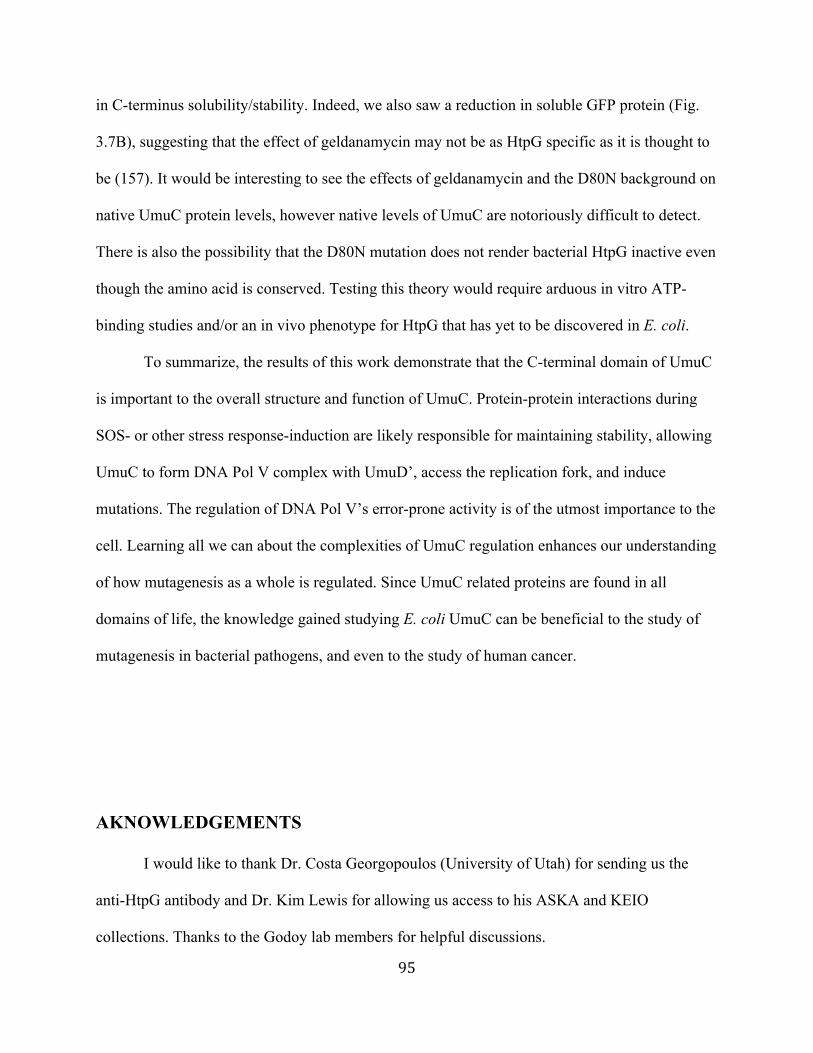
95
in C-terminus solubility/stability. Indeed, we also saw a reduction in soluble GFP protein (Fig.
3.7B), suggesting that the effect of geldanamycin may not be as HtpG specific as it is thought to
be (157). It would be interesting to see the effects of geldanamycin and the D80N background on
native UmuC protein levels, however native levels of UmuC are notoriously difficult to detect.
There is also the possibility that the D80N mutation does not render bacterial HtpG inactive even
though the amino acid is conserved. Testing this theory would require arduous in vitro ATP-
binding studies and/or an in vivo phenotype for HtpG that has yet to be discovered in E. coli.
To summarize, the results of this work demonstrate that the C-terminal domain of UmuC
is important to the overall structure and function of UmuC. Protein-protein interactions during
SOS- or other stress response-induction are likely responsible for maintaining stability, allowing
UmuC to form DNA Pol V complex with UmuD’, access the replication fork, and induce
mutations. The regulation of DNA Pol V’s error-prone activity is of the utmost importance to the
cell. Learning all we can about the complexities of UmuC regulation enhances our understanding
of how mutagenesis as a whole is regulated. Since UmuC related proteins are found in all
domains of life, the knowledge gained studying E. coli UmuC can be beneficial to the study of
mutagenesis in bacterial pathogens, and even to the study of human cancer.
AKNOWLEDGEMENTS
I would like to thank Dr. Costa Georgopoulos (University of Utah) for sending us the
anti-HtpG antibody and Dr. Kim Lewis for allowing us access to his ASKA and KEIO
collections. Thanks to the Godoy lab members for helpful discussions.

96
FIGURES
Figure 3.1. Model of hypothesis and method. If the C-terminus of UmuC is involved in the
regulation of DNA Pol V activity, then studying the effects of excess concentration of a C-
terminal fragment will reveal the existence of inherent C-terminal interactions. (A) The C-
terminus of UmuC may interact with and possibly be regulated by factors X, Y, and Z normally
in vivo. UmuD’ interacts with UmuC to form DNA Pol V but is not specifically represented in
this diagram. (B) Excess C-terminal fragment may result in sequestration or activation of
factor(s) X, Y, or Z. The result may be an altered effect on a UmuC pathway and an observable
downstream phenotype. Figures are not drawn to scale and are only representative of
hypothetical structures.
Interaction(s), altered downstream effect
C-terminus
C-terminus
X Y
Z
(a) Excess
C-terminus fragment
(b) UmuC

97
Figure 3.2. Schematic of UmuC carboxy terminus construct. (A) Diagram of full length
UmuC and its five structural domains. Proposed domains are adapted from Boudsocq et al.
(2002) and are approximations. (B) pC-terminus is a high copy number plasmid (pCA24N) that
expresses His6x-C-terminus::GFP fusion protein from an IPTG inducible promoter. The C-
terminus::GFP fusion protein contains approximately half the residues of full length UmuC; all
N-terminal catalytic residues are deleted. (C) The control, pVector, expresses His6x-GFP protein
from the same IPTG inducible promoter.
UmuC
1 422
Palm and finger domainsincluding catalytic
residues
pVector (pCA24N)
Little finger
domainThumb domain
166
pC-terminus
His6X - GFP
His6X -176 422
GFP
(a)
Unknowndomain
(b)
(c)

98
Figure 3.3. Complementation of the mutator strain, umuC122::Tn5, with UmuC carboxy
terminus results in decreased mutagenesis and increased hydroxyurea resistance. (A)
Frequency of galactose deficient mutants is decreased for the umuC122::Tn5 strain bearing pC-
terminus compared to the control (pVector). Undetectable frequency of mutation is represented
by * symbol. (B) Survival over time is increased in the umuC122::Tn5 strain bearing pC-
terminus compared to the control (pVector). Deletion of dinB results in decreased survival for
both strains. Liquid cultures were treated for 24 hours with 75 mM (A; mutagenesis) or 100mM
(B; survival) hydroxyurea. Averages of at least 3 independent cultures are shown with error bars
representing the standard deviation.
(a)
umuC122::
Tn5 pVector
umuC122::
Tn5 pC-terminusumuC122::Tn5
pVector
umuC122::Tn5 pC-terminus
umuC122::Tn5 ∆dinBpC-terminus
umuC122::Tn5 ∆dinBpVector
Time in hydroxyurea (hours)
Hydroxyurea Induced Mutagenesis
Plasmid
*
Per
cent
Sur
viva
l
Mut
atio
n Fr
eque
ncy
Hydroxyurea Survival
(b)
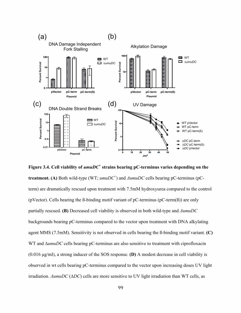
99
Figure 3.4. Cell viability of umuDC+ strains bearing pC-terminus varies depending on the
treatment. (A) Both wild-type (WT; umuDC+) and ΔumuDC cells bearing pC-terminus (pC-
term) are dramatically rescued upon treatment with 7.5mM hydroxyurea compared to the control
(pVector). Cells bearing the ß-binding motif variant of pC-terminus (pC-term(ß)) are only
partially rescued. (B) Decreased cell viability is observed in both wild-type and ΔumuDC
backgrounds bearing pC-terminus compared to the vector upon treatment with DNA alkylating
agent MMS (7.5mM). Sensitivity is not observed in cells bearing the ß-binding motif variant. (C)
WT and ∆umuDC cells bearing pC-terminus are also sensitive to treatment with ciprofloxacin
(0.016 µg/ml), a strong inducer of the SOS response. (D) A modest decrease in cell viability is
observed in wt cells bearing pC-terminus compared to the vector upon increasing doses UV light
irradiation. ΔumuDC (∆DC) cells are more sensitive to UV light irradiation than WT cells, as
Alkylation Damage
(a) (b)
DNA Double Strand Breaks
DNA Damage IndependentFork Stalling
(c) (d)
Plasmid
WT∆umuDC
WT∆umuDC
pVector pC-term
Plasmid
pC-term(ß) pVector pC-termPlasmid
pC-term(ß)
WT∆umuDC
∆DC pVector∆DC pC-term(ß)
WT pC-term(ß)
UV Damage
WT pC-termWT pVector
∆DC pC-term
Per
cent
Sur
viva
l
Per
cent
Sur
viva
l
Per
cent
Sur
viva
l
Per
cent
Sur
viva
l

100
expected, however there is no significant difference between cells bearing pC-terminus
compared to pVector in this background. No differences in survival in either background are
seen when comparing pC-terminus to pC-terminus(ß). The average of 3 replicates is shown with
error bars representing the standard deviation of the mean for all experiments.
Figure 3.5. The C-terminus construct increases the frequency of mutagenesis upon
treatment with MMS in a manner requiring dinB and umuDC but independent of htpG. (A)
The frequency of galactose deficient (gal) mutants for the umuDC+ strain bearing pC-terminus
increases compared to the umuDC+ strain carrying the control plasmid upon treatment with
MMS (9mM). The uninduced strain carrying pC-terminus (IPTG-) has a mutation frequency
comparable to the control. (B) Deletion of htpG has no effect on the C-terminus mutator
phenotype. The umuDC+ strain carrying pC-terminus but supplemented with 30µM
Geldanamycin (Gm) has decreased mutation frequency; geldanamycin inhibits the activity of
HtpG. (C) Deletion of either umuDC or dinB mitigates the mutator effect of C-terminal
fragment. (D) The frequency of gal mutants is the same between IPTG induced wild-type strains
(a)
pVec
torpC
-term
pC-te
rm (I
PTG- )
pVec
tor +
GmpC
-term
+ Gm
∆htpG
pVec
tor∆h
tpG C
-term
∆umuD
C pVec
tor
∆umuD
C C-te
rm∆d
inb pV
ector
∆dinb
pC-te
rm
(b) (c) (d)
StrainpV
ector
pC-te
rm
Plasmid
MMS Induced Mutagenesis
pVecto
r
pC-term
10-5
10-4
10-3
Plasmid
Untreated Gal- MutantsSpontaneous Mutagenesis
Mut
atio
n Fr
eque
ncy

101
carrying pVector and pC-terminus. The difference seen is not significant (P>0.05). For all
experiments shown, the average of at least 3 independent cultures is shown with error bars
representing the standard error of the mean.
Figure 3.6. UmuC C-terminus increases mutagenesis upon treatment with ciprofloxacin
and decreases mutagenesis upon UV irradiation. (A) umuDC+ strain carrying pC-terminus has
an increased frequency of gal mutants compared to the control upon treatment with 0.016 µg/ml
ciprofloxacin. Undetectable frequency of mutation is represented by *** symbol. (B) In both
wild-type (umuDC+ htpG+) and ΔhtpG backgrounds, the strains carrying pC-terminus have
decreased mutation frequency compared to the strains carrying pVector upon UV treatment (46
J/m2). The average of at least 3 independent experiments is shown. Error bars represent the
standard error of the mean. P<0.01 = *, P≤0.01 = **.
Text
(a) (b)
pVec
tor
∆htpG
pVec
tor∆h
tpG C
-term
inus
pC-te
rminu
s
pVec
tor
pC-te
rminu
s
***
Ciprofloxacin-induced Mutagenesis
UV-induced Mutagenesis
PlasmidPlasmid
Mut
atio
n Fr
eque
ncy
Mut
atio
n Fr
eque
ncy
* **
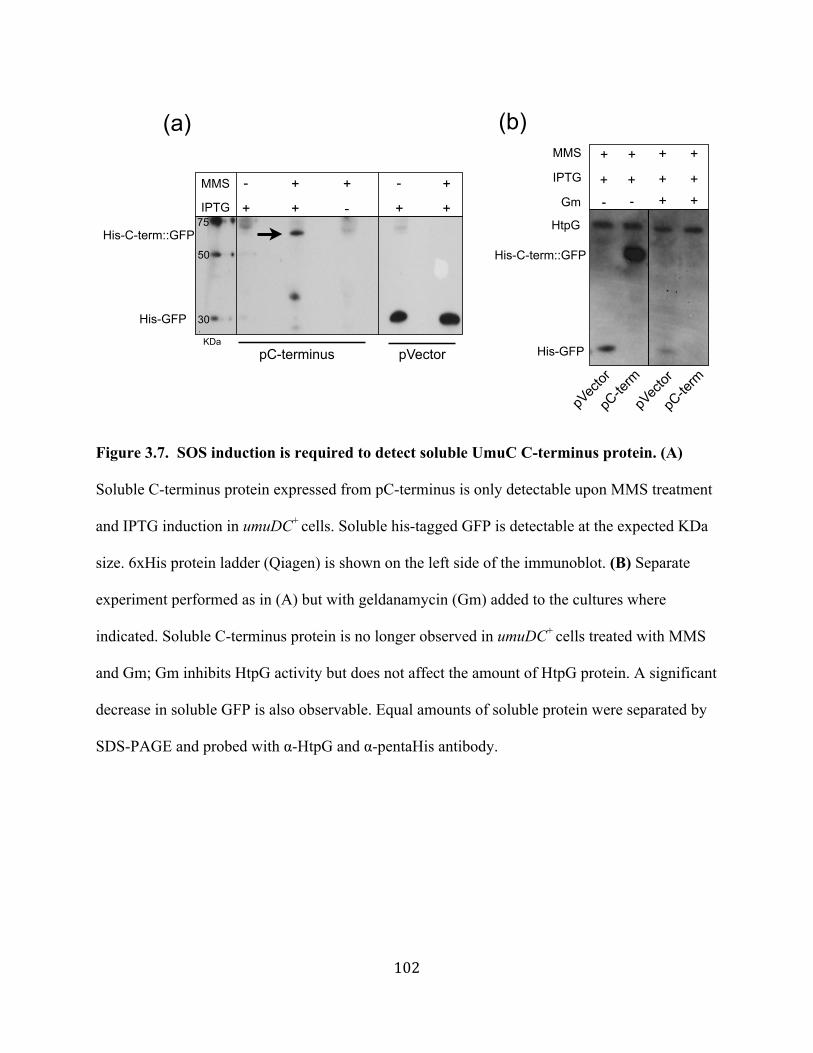
102
Figure 3.7. SOS induction is required to detect soluble UmuC C-terminus protein. (A)
Soluble C-terminus protein expressed from pC-terminus is only detectable upon MMS treatment
and IPTG induction in umuDC+ cells. Soluble his-tagged GFP is detectable at the expected KDa
size. 6xHis protein ladder (Qiagen) is shown on the left side of the immunoblot. (B) Separate
experiment performed as in (A) but with geldanamycin (Gm) added to the cultures where
indicated. Soluble C-terminus protein is no longer observed in umuDC+ cells treated with MMS
and Gm; Gm inhibits HtpG activity but does not affect the amount of HtpG protein. A significant
decrease in soluble GFP is also observable. Equal amounts of soluble protein were separated by
SDS-PAGE and probed with α-HtpG and α-pentaHis antibody.
+
+-Gm
+
+
+
-
MMS
+IPTG
+ + ++
His-GFP
HtpG
His-C-term::GFP
pVector
pC-term
pVector
pC-term
MMS
IPTG
pC-terminus pVector
His-GFP
His-C-term::GFP
+ + + +-
+ + +--
(a) (b)
50
75
KDa
30
103
Figure 3.8. Solubility of UmuC C-terminus protein is not dependent on htpG, active HtpG,
or umuDC. (A) Soluble C-terminus protein expressed from pC-terminus is detected upon MMS
treatment in the wild-type (WT; umuDC+), ∆htpG, and htpG(D80N) strains. The hptG(D80N)
contains a chromosomal mutation of htpG and produces inactive HtpG. Soluble HtpG protein is
seen in the WT and htpG(D80N) strains, but not the ∆htpG. (B) Soluble C-terminus protein is
detectable in both umuDC+ (WT) and ∆umuDC strains. Equal amounts of soluble protein were
separated by SDS-PAGE and probed with α-HtpG or α-pentaHis antibody.
75
50
KDa
His-C-term::GFP
WT ∆htpG htpG(D80N)
+ +++MMS -
IPTG
++
-- +++
HtpG
50
75
+MMS
IPTG
+++
WT ∆umuDC
His-C-term::GFP
(a) (b)
KDa
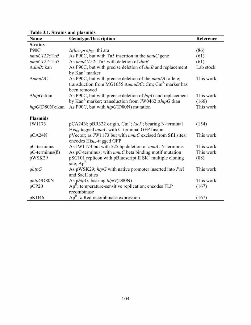
104
Table 3.1. Strains and plasmids Name Genotype/Description Reference Strains P90C ∆(lac-pro)XIII thi ara (86) umuC122::Tn5 As P90C, but with Tn5 insertion in the umuC gene (61) umuC122::Tn5 As umuC122::Tn5 with deletion of dinB (61) ∆dinB::kan As P90C, but with precise deletion of dinB and replacement
by KanR marker Lab stock
∆umuDC As P90C, but with precise deletion of the umuDC allele; transduction from MG1655 ∆umuDC::Cm; CmR marker has been removed
This work
∆htpG::kan As P90C, but with precise deletion of htpG and replacement by KanR marker; transduction from JW0462 ∆htpG::kan
This work; (166)
htpG(D80N)::kan As P90C, but with htpG(D80N) mutation This work Plasmids JW1173 pCA24N; pBR322 origin, CmR; lacIq; bearing N-terminal
His6x-tagged umuC with C-terminal GFP fusion (154)
pCA24N pVector; as JW1173 but with umuC excised from SfiI sites; encodes His6x-tagged GFP
This work
pC-terminus As JW1173 but with 525 bp deletion of umuC N-terminus This work pC-terminus(ß) As pC-terminus; with umuC beta binding motif mutation This work pWSK29 pSC101 replicon with pBluescript II SK+ multiple cloning
site, ApR (88)
phtpG As pWSK29; htpG with native promoter inserted into PstI and SacII sites
This work
phtpGD80N As phtpG; bearing htpG(D80N) This work pCP20 ApR; temperature-sensitive replication; encodes FLP
recombinase (167)
pKD46 ApR; λ Red recombinase expression (167)
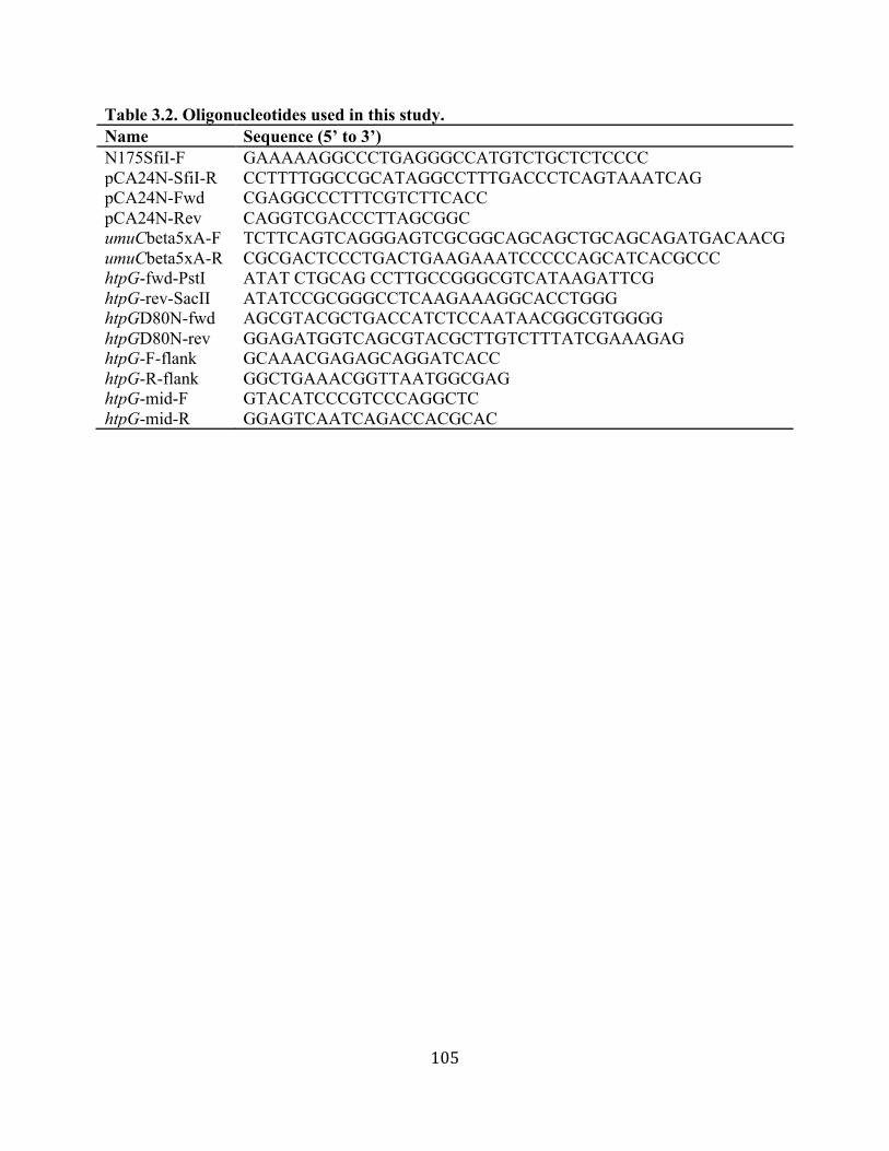
105
Table 3.2. Oligonucleotides used in this study. Name Sequence (5’ to 3’) N175SfiI-F GAAAAAGGCCCTGAGGGCCATGTCTGCTCTCCCC pCA24N-SfiI-R CCTTTTGGCCGCATAGGCCTTTGACCCTCAGTAAATCAG pCA24N-Fwd CGAGGCCCTTTCGTCTTCACC pCA24N-Rev CAGGTCGACCCTTAGCGGC umuCbeta5xA-F TCTTCAGTCAGGGAGTCGCGGCAGCAGCTGCAGCAGATGACAACG umuCbeta5xA-R CGCGACTCCCTGACTGAAGAAATCCCCCAGCATCACGCCC htpG-fwd-PstI ATAT CTGCAG CCTTGCCGGGCGTCATAAGATTCG htpG-rev-SacII ATATCCGCGGGCCTCAAGAAAGGCACCTGGG htpGD80N-fwd AGCGTACGCTGACCATCTCCAATAACGGCGTGGGG htpGD80N-rev GGAGATGGTCAGCGTACGCTTGTCTTTATCGAAAGAG htpG-F-flank GCAAACGAGAGCAGGATCACC htpG-R-flank GGCTGAAACGGTTAATGGCGAG htpG-mid-F GTACATCCCGTCCCAGGCTC htpG-mid-R GGAGTCAATCAGACCACGCAC
106
CONCLUDING REMARKS Over the past two decades, great strides have been made in Escherichia coli DNA
damage and Y-family DNA polymerase research (70). It has become apparent that these DNA
polymerases are involved in not only error-prone translesion DNA synthesis (TLS), but also a
multitude of activities and diverse roles (9, 28). Their precise regulation is critical to cell survival
and maintenance of genomic integrity. Because DNA damage and environmental stress is ever
present for bacteria, they have evolved sophisticated and interconnected coping mechanisms (2,
3). The remarkable process of evolution gave rise to stress-responses and repair systems that can
actually allow bacteria to further increase genetic diversity and fitness during these stressful
times (2, 8, 29, 46). This balancing act of maintaining genomic integrity and increasing genetic
diversity has been fine tuned over the course of billions of years (168). Likewise, bacteria have
also had hundreds of thousands of years to either become experts at infecting the human species
(169), skillfully evading our immune response, or to survive in a human host when given the
opportunity (170). Moreover, over time they have also evolved resistance mechanisms (8) to
combat antibiotics first produced by their fellow bacterial neighbors (171), and now produced by
us (39).
In this work we have uncovered a DNA damage response in the opportunistic pathogen,
Acinetobacter baumannii. This emerging pathogen is a worldwide source of nosocomial
infections and has become resistant to almost every antibiotic available to clinicians (40-42, 44).
We have shed light on the mechanisms of its regulation, including the involvement of the highly
conserved bacterial protein, RecA (1). We demonstrated that in the absence of recA, A.
baumannii does not induce putatively mutagenic Y-family DNA polymerases nor does it mutate
upon DNA damaging conditions such as desiccation. Others have shown that the recA mutant is

107
sensitive to a variety of DNA damaging agents in addition to decreased pathogenicity (66). The
implications of these findings are clinically important because A. baumannii survives desiccation
in the hospital setting; enduring on equipment and surfaces and getting transmitted between staff
and patients (40, 41, 43). RecA thus becomes an attractive target for specific treatment and
control of A. baummannii outbreaks. A stable chemical inhibitor that could be universally used
as part of the disinfection process would be an ideal scenario. Similarly, a stable drug that could
be taken along with antibiotics to increase the potency (110) and prevent further mutation is also
ideal. The current development of these inhibitors will hopefully yield efficacious candidates, as
the need for new antimicrobials has never been greater (39).
The Y-family DNA polymerases are also interesting target candidates for a compound
that could prevent bacterial evolution (15, 36). Although they are dispensable in bacteria under
ideal laboratory conditions (9, 70), in long-term stationary phase competition experiments,
bacteria that lack translesion DNA polymerases lose (in both survival and evolutionary fitness)
to those that have them (172). Therefore, in theory, it seems as though inhibiting them could
prevent growth and/or mutagenesis under natural conditions, though bacteria are still left with
other ways to increase genetic diversity such as spontaneous mutations (1), horizontal gene
transfer mechanisms (59), and copious other mechanisms of mutagenesis/evolvability (173-179).
Thus, the challenge of preventing bacteria from evolving in the hospital or in the host is an uphill
battle. A highly conserved target such as RecA, which modulates the activity of these DNA
polymerases and activates the entire DNA damage response, is clearly one of the more useful
directions to take (15, 36).
The insights we have gained in studying the Y-family DNA polymerases and DNA
damage response of both A. baumannii and E. coli have nevertheless been worthwhile. Our work
108
in A. baumannii has shed new light on establishing DNA damage response mechanisms different
than the E. coli paradigm. Although E. coli works well as a model system and we should
continue our efforts, we must not be constrained by this one species, or what we can accomplish
and learn will be severely impeded. Finally, there is still much to be learned about how
mutagenic DNA polymerases are regulated and what their functional roles are. It is not only
about finding a drug or inhibitor to combat pathogenic bacteria. Every scientific contribution
made to the greater pool of knowledge leads us closer to understanding mutagenesis, and hence
molecular evolution, in both bacteria and humans. Additionally, the more that is learned in
bacteria the more we are able to translate those findings to the diagnosis (131, 132) and some day
prevention of cancer.

109
REFERENCES
1. Friedberg EC, Walker GC, Siede W, Wood RD, Schultz RA, Ellenberger T. 2006. DNA repair and mutagenesis, 2nd ed. ASM Press, Washington, D.C.
2. Al Mamun AA, Lombardo MJ, Shee C, Lisewski AM, Gonzalez C, Lin D, Nehring RB, Saint-Ruf C, Gibson JL, Frisch RL, Lichtarge O, Hastings PJ, Rosenberg SM. 2012. Identity and function of a large gene network underlying mutagenic repair of DNA breaks. Science 338:1344-1348.
3. Kreuzer KN. 2013. DNA damage responses in prokaryotes: regulating gene expression, modulating growth patterns, and manipulating replication forks. Cold Spring Harb Perspect Biol 5.
4. Beuning PJ, Simon SM, Godoy VG, Jarosz DF, Walker GC. 2006. Characterization of Escherichia coli translesion synthesis polymerases and their accessory factors. Methods Enzymol 408:318-340.
5. Sutton MD, Smith BT, Godoy VG, Walker GC. 2000. The SOS response: recent insights into umuDC-dependent mutagenesis and DNA damage tolerance. Annu Rev Genet 34:479-497.
6. Khil PP, Camerini-Otero RD. 2002. Over 1000 genes are involved in the DNA damage response of Escherichia coli. Mol Microbiol 44:89-105.
7. Kogoma T. 1997. Stable DNA replication: interplay between DNA replication, homologous recombination, and transcription. Microbiol Mol Biol Rev 61:212-238.
8. Galhardo RS, Hastings PJ, Rosenberg SM. 2007. Mutation as a stress response and the regulation of evolvability. Crit Rev Biochem Mol Biol 42:399-435.
9. Goodman MF, Woodgate R. 2013. Translesion DNA polymerases. Cold Spring Harb Perspect Biol 5.
10. Petrosino JF, Galhardo RS, Morales LD, Rosenberg SM. 2009. Stress-induced beta-lactam antibiotic resistance mutation and sequences of stationary-phase mutations in the Escherichia coli chromosome. J Bacteriol 191:5881-5889.
11. Dorr T, Lewis K, Vulic M. 2009. SOS response induces persistence to fluoroquinolones in Escherichia coli. PLoS Genet 5:e1000760.
12. Dorr T, Vulic M, Lewis K. 2010. Ciprofloxacin causes persister formation by inducing the TisB toxin in Escherichia coli. PLoS Biol 8:e1000317.
13. Kelley WL. 2006. Lex marks the spot: the virulent side of SOS and a closer look at the LexA regulon. Mol Microbiol 62:1228-1238.
110
14. Li B, Smith P, Horvath DJ, Jr., Romesberg FE, Justice SS. 2010. SOS regulatory elements are essential for UPEC pathogenesis. Microbes and infection / Institut Pasteur 12:662-668.
15. Cirz RT, Chin JK, Andes DR, de Crecy-Lagard V, Craig WA, Romesberg FE. 2005. Inhibition of mutation and combating the evolution of antibiotic resistance. PLoS Biol 3:e176.
16. Lange SS, Takata K, Wood RD. 2011. DNA polymerases and cancer. Nat Rev Cancer 11:96-110.
17. Zhang AP, Pigli YZ, Rice PA. 2010. Structure of the LexA-DNA complex and implications for SOS box measurement. Nature 466:883-886.
18. Courcelle J, Khodursky A, Peter B, Brown PO, Hanawalt PC. 2001. Comparative gene expression profiles following UV exposure in wild-type and SOS-deficient Escherichia coli. Genetics 158:41-64.
19. Fuchs RP, Fujii S, Wagner J. 2004. Properties and functions of Escherichia coli: Pol IV and Pol V. Advances in protein chemistry 69:229-264.
20. Maor-Shoshani A, Reuven NB, Tomer G, Livneh Z. 2000. Highly mutagenic replication by DNA polymerase V (UmuC) provides a mechanistic basis for SOS untargeted mutagenesis. Proc Natl Acad Sci U S A 97:565-570.
21. Ohmori H, Friedberg EC, Fuchs RP, Goodman MF, Hanaoka F, Hinkle D, Kunkel TA, Lawrence CW, Livneh Z, Nohmi T, Prakash L, Prakash S, Todo T, Walker GC, Wang Z, Woodgate R. 2001. The Y-family of DNA polymerases. Mol Cell 8:7-8.
22. Kim SR, Maenhaut-Michel G, Yamada M, Yamamoto Y, Matsui K, Sofuni T, Nohmi T, Ohmori H. 1997. Multiple pathways for SOS-induced mutagenesis in Escherichia coli: an overexpression of dinB/dinP results in strongly enhancing mutagenesis in the absence of any exogenous treatment to damage DNA. Proc Natl Acad Sci U S A 94:13792-13797.
23. Jarosz DF, Godoy VG, Delaney JC, Essigmann JM, Walker GC. 2006. A single amino acid governs enhanced activity of DinB DNA polymerases on damaged templates. Nature 439:225-228.
24. Uchida K, Furukohri A, Shinozaki Y, Mori T, Ogawara D, Kanaya S, Nohmi T, Maki H, Akiyama M. 2008. Overproduction of Escherichia coli DNA polymerase DinB (Pol IV) inhibits replication fork progression and is lethal. Mol Microbiol 70:608-622.
25. Pomerantz RT, Goodman MF, O'Donnell ME. 2013. DNA polymerases are error-prone at RecA-mediated recombination intermediates. Cell Cycle 12:2558-2563.

111
26. Pomerantz RT, Kurth I, Goodman MF, O'Donnell ME. 2013. Preferential D-loop extension by a translesion DNA polymerase underlies error-prone recombination. Nat Struct Mol Biol 20:748-755.
27. Cohen SE, Walker GC. 2010. The transcription elongation factor NusA is required for stress-induced mutagenesis in Escherichia coli. Curr Biol 20:80-85.
28. Lehmann AR. 2006. New functions for Y family polymerases. Mol Cell 24:493-495.
29. Goodman MF. 2002. Error-prone repair DNA polymerases in prokaryotes and eukaryotes. Annu Rev Biochem 71:17-50.
30. Ling H, Boudsocq F, Woodgate R, Yang W. 2001. Crystal structure of a Y-family DNA polymerase in action: a mechanism for error-prone and lesion-bypass replication. Cell 107:91-102.
31. Silvian LF, Toth EA, Pham P, Goodman MF, Ellenberger T. 2001. Crystal structure of a DinB family error-prone DNA polymerase from Sulfolobus solfataricus. Nat Struct Biol 8:984-989.
32. Trincao J, Johnson RE, Escalante CR, Prakash S, Prakash L, Aggarwal AK. 2001. Structure of the catalytic core of S. cerevisiae DNA polymerase eta: implications for translesion DNA synthesis. Mol Cell 8:417-426.
33. Zhou BL, Pata JD, Steitz TA. 2001. Crystal structure of a DinB lesion bypass DNA polymerase catalytic fragment reveals a classic polymerase catalytic domain. Mol Cell 8:427-437.
34. Gillespie JH. 1991. The causes of molecular evolution. Oxford University Press, New York ; Oxford.
35. Graur D, Li W-H. 1999. Fundamentals of molecular evolution, 2nd ed. Sinauer Associates, Sunderland, Mass.
36. Cirz RT, Romesberg FE. 2007. Controlling mutation: intervening in evolution as a therapeutic strategy. Crit Rev Biochem Mol Biol 42:341-354.
37. Giannouli M, Di Popolo A, Durante-Mangoni E, Bernardo M, Cuccurullo S, Amato G, Tripodi MF, Triassi M, Utili R, Zarrilli R. 2012. Molecular epidemiology and mechanisms of rifampicin resistance in Acinetobacter baumannii isolates from Italy. International journal of antimicrobial agents 39:58-63.
38. Boucher HW, Talbot GH, Bradley JS, Edwards JE, Gilbert D, Rice LB, Scheld M, Spellberg B, Bartlett J. 2009. Bad bugs, no drugs: no ESKAPE! An update from the Infectious Diseases Society of America. Clin Infect Dis 48:1-12.
39. Lewis K. 2013. Platforms for antibiotic discovery. Nat Rev Drug Discov 12:371-387.

112
40. Bergogne-Berezin E, Friedman H, Bendinelli M. 2008. Acinetobacter Biology and Pathogenesis, vol. xvi. Springer Verlag, New York.
41. Dijkshoorn L, Nemec A, Seifert H. 2007. An increasing threat in hospitals: multidrug-resistant Acinetobacter baumannii. Nat Rev Microbiol 5:939-951.
42. Gerischer U. 2008. Acinetobacter Molecular Biology. Caister Academic Press, Norfolk, UK.
43. Peleg AY, Hooper DC. 2010. Hospital-acquired infections due to gram-negative bacteria. The New England journal of medicine 362:1804-1813.
44. Peleg AY, Seifert H, Paterson DL. 2008. Acinetobacter baumannii: emergence of a successful pathogen. Clin Microbiol Rev 21:538-582.
45. Tomaras AP, Dorsey CW, Edelmann RE, Actis LA. 2003. Attachment to and biofilm formation on abiotic surfaces by Acinetobacter baumannii: involvement of a novel chaperone-usher pili assembly system. Microbiology 149:3473-3484.
46. Friedberg EC, Wagner R, Radman M. 2002. Specialized DNA polymerases, cellular survival, and the genesis of mutations. Science 296:1627-1630.
47. Robinson A, Brzoska AJ, Turner KM, Withers R, Harry EJ, Lewis PJ, Dixon NE. 2010. Essential biological processes of an emerging pathogen: DNA replication, transcription, and cell division in Acinetobacter spp. Microbiol Mol Biol Rev 74:273-297.
48. Jawad A, Seifert H, Snelling AM, Heritage J, Hawkey PM. 1998. Survival of Acinetobacter baumannii on dry surfaces: comparison of outbreak and sporadic isolates. J Clin Microbiol 36:1938-1941.
49. Kramer A, Schwebke I, Kampf G. 2006. How long do nosocomial pathogens persist on inanimate surfaces? A systematic review. BMC infectious diseases 6:130.
50. Neely AN. 2000. A survey of gram-negative bacteria survival on hospital fabrics and plastics. The Journal of burn care & rehabilitation 21:523-527.
51. Neely AN, Maley MP, Warden GD. 1999. Computer keyboards as reservoirs for Acinetobacter baumannii in a burn hospital. Clin Infect Dis 29:1358-1360.
52. Wendt C, Dietze B, Dietz E, Ruden H. 1997. Survival of Acinetobacter baumannii on dry surfaces. J Clin Microbiol 35:1394-1397.
53. Lockhart SR, Abramson MA, Beekmann SE, Gallagher G, Riedel S, Diekema DJ, Quinn JP, Doern GV. 2007. Antimicrobial resistance among Gram-negative bacilli causing infections in intensive care unit patients in the United States between 1993 and 2004. J Clin Microbiol 45:3352-3359.

113
54. Bergogne-Berezin E. 1995. The increasing significance of outbreaks of Acinetobacter spp.: the need for control and new agents. J Hosp Infect 30 Suppl:441-452.
55. Bergogne-Berezin E. 2001. The Increasing Role of Acinetobacter Species As Nosocomial Pathogens. Curr Infect Dis Rep 3:440-444.
56. Bergogne-Berezin E, Joly-Guillou ML. 1991. Hospital infection with Acinetobacter spp.: an increasing problem. J Hosp Infect 18 Suppl A:250-255.
57. Bergogne-Berezin E, Towner KJ. 1996. Acinetobacter spp. as nosocomial pathogens: microbiological, clinical, and epidemiological features. Clin Microbiol Rev 9:148-165.
58. Vila J, Marti S, Sanchez-Cespedes J. 2007. Porins, efflux pumps and multidrug resistance in Acinetobacter baumannii. J Antimicrob Chemother 59:1210-1215.
59. Snitkin ES, Zelazny AM, Montero CI, Stock F, Mijares L, Murray PR, Segre JA. 2011. Genome-wide recombination drives diversification of epidemic strains of Acinetobacter baumannii. Proc Natl Acad Sci U S A 108:13758-13763.
60. Miller C, Thomsen LE, Gaggero C, Mosseri R, Ingmer H, Cohen SN. 2004. SOS response induction by beta-lactams and bacterial defense against antibiotic lethality. Science 305:1629-1631.
61. Godoy VG, Jarosz DF, Walker FL, Simmons LA, Walker GC. 2006. Y-family DNA polymerases respond to DNA damage-independent inhibition of replication fork progression. EMBO J 25:868-879.
62. Kowalczykowski SC, Dixon DA, Eggleston AK, Lauder SD, Rehrauer WM. 1994. Biochemistry of homologous recombination in Escherichia coli. Microbiol Rev 58:401-465.
63. Godoy VG, Jarosz DF, Simon SM, Abyzov A, Ilyin V, Walker GC. 2007. UmuD and RecA directly modulate the mutagenic potential of the Y family DNA polymerase DinB. Mol Cell 28:1058-1070.
64. Rehrauer WM, Bruck I, Woodgate R, Goodman MF, Kowalczykowski SC. 1998. Modulation of RecA nucleoprotein function by the mutagenic UmuD'C protein complex. J Biol Chem 273:32384-32387.
65. Tang M, Bruck I, Eritja R, Turner J, Frank EG, Woodgate R, O'Donnell M, Goodman MF. 1998. Biochemical basis of SOS-induced mutagenesis in Escherichia coli: reconstitution of in vitro lesion bypass dependent on the UmuD'2C mutagenic complex and RecA protein. Proc Natl Acad Sci U S A 95:9755-9760.
66. Aranda J, Bardina C, Beceiro A, Rumbo S, Cabral MP, Barbe J, Bou G. 2011. Acinetobacter baumannii RecA protein in repair of DNA damage, antimicrobial resistance, general stress response, and virulence. J Bacteriol 193:3740-3747.
114
67. Hare JM, Perkins SN, Gregg-Jolly LA. 2006. A constitutively expressed, truncated umuDC operon regulates the recA-dependent DNA damage induction of a gene in Acinetobacter baylyi strain ADP1. Appl Environ Microbiol 72:4036-4043.
68. Rauch PJ, Palmen R, Burds AA, Gregg-Jolly LA, van der Zee JR, Hellingwerf KJ. 1996. The expression of the Acinetobacter calcoaceticus recA gene increases in response to DNA damage independently of RecA and of development of competence for natural transformation. Microbiology 142 ( Pt 4):1025-1032.
69. Yang W. 2011. Surviving the sun: repair and bypass of DNA UV lesions. Protein Sci 20:1781-1789.
70. Yang W, Woodgate R. 2007. What a difference a decade makes: insights into translesion DNA synthesis. Proc Natl Acad Sci U S A 104:15591-15598.
71. Reuven NB, Tomer G, Livneh Z. 1998. The mutagenesis proteins UmuD' and UmuC prevent lethal frameshifts while increasing base substitution mutations. Molecular cell 2:191-199.
72. Seward RJ, Towner KJ. 1998. Molecular epidemiology of quinolone resistance in Acinetobacter spp. Clinical microbiology and infection : the official publication of the European Society of Clinical Microbiology and Infectious Diseases 4:248-254.
73. Vila J, Ruiz J, Goni P, Jimenez de Anta T. 1997. Quinolone-resistance mutations in the topoisomerase IV parC gene of Acinetobacter baumannii. J Antimicrob Chemother 39:757-762.
74. Vila J, Ruiz J, Goni P, Marcos A, Jimenez de Anta T. 1995. Mutation in the gyrA gene of quinolone-resistant clinical isolates of Acinetobacter baumannii. Antimicrob Agents Chemother 39:1201-1203.
75. Di Nocera PP, Rocco F, Giannouli M, Triassi M, Zarrilli R. 2011. Genome organization of epidemic Acinetobacter baumannii strains. BMC Microbiol 11:224.
76. Hare J, Bradley J, Lin CL, Elam T. 2012. Diverse DNA damage responses in Acinetobacter include the capacity for DNA damage-induced mutagenesis in the opportunistic pathogens Acinetobacter baumannii and Acinetobacter ursingii. Microbiology:10.1099/mic.1090.054668-054660.
77. Smith MG, Gianoulis TA, Pukatzki S, Mekalanos JJ, Ornston LN, Gerstein M, Snyder M. 2007. New insights into Acinetobacter baumannii pathogenesis revealed by high-density pyrosequencing and transposon mutagenesis. Genes Dev 21:601-614.
78. Schaub IG, Hauber FD. 1948. A biochemical and serological study of a group of identical unidentifiable gram-negative bacilli from human sources. J Bacteriol 56:379-385.

115
79. Amsterdam D. 1996. Susceptibility testing of antimicrobials in liquid media, p. 52-111. In Lorian V (ed.), Antibiotics in laboratory medicine, 4th ed. ed. Williams and Wilkins, Baltimore, MD.
80. Altschul SF, Madden TL, Schaffer AA, Zhang J, Zhang Z, Miller W, Lipman DJ. 1997. Gapped BLAST and PSI-BLAST: a new generation of protein database search programs. Nucleic acids research 25:3389-3402.
81. Horton RM, Hunt HD, Ho SN, Pullen JK, Pease LR. 1989. Engineering hybrid genes without the use of restriction enzymes: gene splicing by overlap extension. Gene 77:61-68.
82. Zaslaver A, Bren A, Ronen M, Itzkovitz S, Kikoin I, Shavit S, Liebermeister W, Surette MG, Alon U. 2006. A comprehensive library of fluorescent transcriptional reporters for Escherichia coli. Nature methods 3:623-628.
83. Ausubel FM. 1994. Current protocols in molecular biology. J. Wiley, Brooklyn, N.Y.
84. Nohmi T. 2006. Environmental stress and lesion-bypass DNA polymerases. Annu Rev Microbiol 60:231-253.
85. Tang WJ. 1993. Blot-affinity purification of antibodies. Methods in cell biology 37:95-104.
86. Cairns J, Foster PL. 1991. Adaptive reversion of a frameshift mutation in Escherichia coli. Genetics 128:695-701.
87. Bjedov I, Dasgupta CN, Slade D, Le Blastier S, Selva M, Matic I. 2007. Involvement of Escherichia coli DNA polymerase IV in tolerance of cytotoxic alkylating DNA lesions in vivo. Genetics 176:1431-1440.
88. Wang RF, Kushner SR. 1991. Construction of versatile low-copy-number vectors for cloning, sequencing and gene expression in Escherichia coli. Gene 100:195-199.
89. Woodgate R, Rajagopalan M, Lu C, Echols H. 1989. UmuC mutagenesis protein of Escherichia coli: purification and interaction with UmuD and UmuD'. Proceedings of the National Academy of Sciences of the United States of America 86:7301-7305.
90. Boudsocq F, Ling H, Yang W, Woodgate R. 2002. Structure-based interpretation of missense mutations in Y-family DNA polymerases and their implications for polymerase function and lesion bypass. DNA Repair (Amst) 1:343-358.
91. Hare JM, Adhikari S, Lambert KV, Hare AE, Grice AN. 2012. The Acinetobacter regulatory UmuDAb protein cleaves in response to DNA damage with chimeric LexA/UmuD characteristics. FEMS Microbiol Lett.

116
92. Markham BE, Harper JE, Mount DW, Sancar GB, Sancar A, Rupp WD, Kenyon CJ, Walker GC. 1984. Analysis of mRNA synthesis following induction of the Escherichia coli SOS system. J Mol Biol 178:237-248.
93. Benson RW, Norton MD, Lin I, Du Comb WS, Godoy VG. 2011. An active site aromatic triad in Escherichia coli DNA Pol IV coordinates cell survival and mutagenesis in different DNA damaging agents. PLoS One 6:e19944.
94. Malik M, Zhao X, Drlica K. 2006. Lethal fragmentation of bacterial chromosomes mediated by DNA gyrase and quinolones. Mol Microbiol 61:810-825.
95. Schlacher K, Goodman MF. 2007. Lessons from 50 years of SOS DNA-damage-induced mutagenesis. Nat Rev Mol Cell Biol 8:587-594.
96. Garibyan L, Huang T, Kim M, Wolff E, Nguyen A, Nguyen T, Diep A, Hu K, Iverson A, Yang H, Miller JH. 2003. Use of the rpoB gene to determine the specificity of base substitution mutations on the Escherichia coli chromosome. DNA Repair (Amst) 2:593-608.
97. Potts M. 1994. Desiccation tolerance of prokaryotes. Microbiol Rev 58:755-805.
98. Jarosz DF, Godoy VG, Walker GC. 2007. Proficient and accurate bypass of persistent DNA lesions by DinB DNA polymerases. Cell Cycle 6:817-822.
99. Davis EO, Springer B, Gopaul KK, Papavinasasundaram KG, Sander P, Bottger EC. 2002. DNA damage induction of recA in Mycobacterium tuberculosis independently of RecA and LexA. Molecular microbiology 46:791-800.
100. Yasbin RE, Cheo DL, Bayles KW. 1992. Inducible DNA repair and differentiation in Bacillus subtilis: interactions between global regulons. Molecular microbiology 6:1263-1270.
101. Botzenhart K, Ruden H, Tolon M, von KSM. 1976. [Clinical uses of ultraviolet light radiation]. Prakt Anaesth 11:320-327.
102. Hart D. 1960. Bactericidal ultraviolet radiation in the operating room. Twenty-nine-year study for control of infections. J Am Med Assoc 172:1019-1028.
103. Riley RL, Kaufman JE. 1971. Air disinfection in corridors by upper air irradiation with ultraviolet. Arch Environ Health 22:551-553.
104. Riley RL, Nardell EA. 1989. Clearing the air. The theory and application of ultraviolet air disinfection. Am Rev Respir Dis 139:1286-1294.
105. Sexton JZ, Wigle TJ, He Q, Hughes MA, Smith GR, Singleton SF, Williams AL, Yeh LA. 2010. Novel Inhibitors of E. coli RecA ATPase Activity. Current chemical genomics 4:34-42.

117
106. Wigle TJ, Sexton JZ, Gromova AV, Hadimani MB, Hughes MA, Smith GR, Yeh LA, Singleton SF. 2009. Inhibitors of RecA activity discovered by high-throughput screening: cell-permeable small molecules attenuate the SOS response in Escherichia coli. Journal of biomolecular screening 14:1092-1101.
107. Romesberg F, David N.E., Cirz, R. 2009. Compositions and methods to reduce mutagenesis. USA patent US 2009/0136518 A1.
108. Singleton SF. 2012. Inhibitors of RecA activities for control of antibiotic-resistant bacterial pathogens. USA patent US 2012/0135092 A1.
109. Lee AM, Ross CT, Zeng BB, Singleton SF. 2005. A molecular target for suppression of the evolution of antibiotic resistance: inhibition of the Escherichia coli RecA protein by N(6)-(1-naphthyl)-ADP. Journal of medicinal chemistry 48:5408-5411.
110. Thi TD, Lopez E, Rodriguez-Rojas A, Rodriguez-Beltran J, Couce A, Guelfo JR, Castaneda-Garcia A, Blazquez J. 2011. Effect of recA inactivation on mutagenesis of Escherichia coli exposed to sublethal concentrations of antimicrobials. J Antimicrob Chemother 66:531-538.
111. Norton MD, Spilkia AJ, Godoy VG. 2013. Antibiotic resistance acquired through a DNA damage-inducible response in Acinetobacter baumannii. J Bacteriol 195:1335-1345.
112. Aranda J, Poza M, Shingu-Vazquez M, Cortes P, Boyce JD, Adler B, Barbe J, Bou G. 2013. Identification of a DNA-damage-inducible regulon in Acinetobacter baumannii. J Bacteriol.
113. Mead S, Vaisman A, Valjavec-Gratian M, Karata K, Vandewiele D, Woodgate R. 2007. Characterization of polVR391: a Y-family polymerase encoded by rumA'B from the IncJ conjugative transposon, R391. Mol Microbiol 63:797-810.
114. Jiang Q, Karata K, Woodgate R, Cox MM, Goodman MF. 2009. The active form of DNA polymerase V is UmuD'(2)C-RecA-ATP. Nature 460:359-363.
115. Reuven NB, Arad G, Maor-Shoshani A, Livneh Z. 1999. The mutagenesis protein UmuC is a DNA polymerase activated by UmuD', RecA, and SSB and is specialized for translesion replication. J Biol Chem 274:31763-31766.
116. Tang M, Pham P, Shen X, Taylor JS, O'Donnell M, Woodgate R, Goodman MF. 2000. Roles of E. coli DNA polymerases IV and V in lesion-targeted and untargeted SOS mutagenesis. Nature 404:1014-1018.
117. Frank EG, Ennis DG, Gonzalez M, Levine AS, Woodgate R. 1996. Regulation of SOS mutagenesis by proteolysis. Proc Natl Acad Sci U S A 93:10291-10296.
118. Becherel OJ, Fuchs RP, Wagner J. 2002. Pivotal role of the beta-clamp in translesion DNA synthesis and mutagenesis in E. coli cells. DNA Repair (Amst) 1:703-708.
118
119. Lopez de Saro FJ, Georgescu RE, Goodman MF, O'Donnell M. 2003. Competitive processivity-clamp usage by DNA polymerases during DNA replication and repair. EMBO J 22:6408-6418.
120. Sutton MD, Farrow MF, Burton BM, Walker GC. 2001. Genetic interactions between the Escherichia coli umuDC gene products and the beta processivity clamp of the replicative DNA polymerase. Journal of bacteriology 183:2897-2909.
121. Wagner J, Etienne H, Fuchs RP, Cordonnier A, Burnouf D. 2009. Distinct beta-clamp interactions govern the activities of the Y family PolIV DNA polymerase. Mol Microbiol 74:1143-1151.
122. Wagner J, Fujii S, Gruz P, Nohmi T, Fuchs RP. 2000. The beta clamp targets DNA polymerase IV to DNA and strongly increases its processivity. EMBO reports 1:484-488.
123. Dalrymple BP, Kongsuwan K, Wijffels G, Dixon NE, Jennings PA. 2001. A universal protein-protein interaction motif in the eubacterial DNA replication and repair systems. Proc Natl Acad Sci U S A 98:11627-11632.
124. Beuning PJ, Sawicka D, Barsky D, Walker GC. 2006. Two processivity clamp interactions differentially alter the dual activities of UmuC. Mol Microbiol 59:460-474.
125. Bunting KA, Roe SM, Pearl LH. 2003. Structural basis for recruitment of translesion DNA polymerase Pol IV/DinB to the beta-clamp. EMBO J 22:5883-5892.
126. Villa L, Carattoli A. 2005. Integrons and transposons on the Salmonella enterica serovar typhimurium virulence plasmid. Antimicrob Agents Chemother 49:1194-1197.
127. Herrero A, Mendoza MC, Rodicio R, Rodicio MR. 2008. Characterization of pUO-StVR2, a virulence-resistance plasmid evolved from the pSLT virulence plasmid of Salmonella enterica serovar Typhimurium. Antimicrob Agents Chemother 52:4514-4517.
128. Russo TA, MacDonald U, Beanan JM, Olson R, MacDonald IJ, Sauberan SL, Luke NR, Schultz LW, Umland TC. 2009. Penicillin-binding protein 7/8 contributes to the survival of Acinetobacter baumannii in vitro and in vivo. J Infect Dis 199:513-521.
129. Gerlach VL, Aravind L, Gotway G, Schultz RA, Koonin EV, Friedberg EC. 1999. Human and mouse homologs of Escherichia coli DinB (DNA polymerase IV), members of the UmuC/DinB superfamily. Proc Natl Acad Sci U S A 96:11922-11927.
130. Johnson RE, Washington MT, Prakash S, Prakash L. 1999. Bridging the gap: a family of novel DNA polymerases that replicate faulty DNA. Proc Natl Acad Sci U S A 96:12224-12226.
131. Pillaire MJ, Selves J, Gordien K, Gourraud PA, Gentil C, Danjoux M, Do C, Negre V, Bieth A, Guimbaud R, Trouche D, Pasero P, Mechali M, Hoffmann JS, Cazaux C. 2010. A 'DNA replication' signature of progression and negative outcome in colorectal cancer. Oncogene 29:876-887.

119
132. Wang H, Wu W, Wang HW, Wang S, Chen Y, Zhang X, Yang J, Zhao S, Ding HF, Lu D. 2010. Analysis of specialized DNA polymerases expression in human gliomas: association with prognostic significance. Neuro Oncol 12:679-686.
133. Wagner J, Gruz P, Kim SR, Yamada M, Matsui K, Fuchs RP, Nohmi T. 1999. The dinB gene encodes a novel E. coli DNA polymerase, DNA pol IV, involved in mutagenesis. Mol Cell 4:281-286.
134. Donnelly CE, Walker GC. 1992. Coexpression of UmuD' with UmuC suppresses the UV mutagenesis deficiency of groE mutants. J Bacteriol 174:3133-3139.
135. Grudniak AM, Kuc M, Wolska KI. 2005. Role of Escherichia coli DnaK and DnaJ chaperones in spontaneous and induced mutagenesis and their effect on UmuC stability. FEMS Microbiol Lett 242:361-366.
136. Petit MA, Bedale W, Osipiuk J, Lu C, Rajagopalan M, McInerney P, Goodman MF, Echols H. 1994. Sequential folding of UmuC by the Hsp70 and Hsp60 chaperone complexes of Escherichia coli. J Biol Chem 269:23824-23829.
137. Freitag N, McEntee K. 1989. "Activated"-RecA protein affinity chromatography of LexA repressor and other SOS-regulated proteins. Proc Natl Acad Sci U S A 86:8363-8367.
138. Taipale M, Jarosz DF, Lindquist S. 2010. HSP90 at the hub of protein homeostasis: emerging mechanistic insights. Nat Rev Mol Cell Biol 11:515-528.
139. Lindquist S, Craig EA. 1988. The heat-shock proteins. Annu Rev Genet 22:631-677.
140. Young JC, Moarefi I, Hartl FU. 2001. Hsp90: a specialized but essential protein-folding tool. J Cell Biol 154:267-273.
141. Buchner J. 2010. Bacterial Hsp90--desperately seeking clients. Mol Microbiol 76:540-544.
142. Sato T, Minagawa S, Kojima E, Okamoto N, Nakamoto H. 2010. HtpG, the prokaryotic homologue of Hsp90, stabilizes a phycobilisome protein in the cyanobacterium Synechococcus elongatus PCC 7942. Mol Microbiol 76:576-589.
143. Genest O, Hoskins JR, Camberg JL, Doyle SM, Wickner S. 2011. Heat shock protein 90 from Escherichia coli collaborates with the DnaK chaperone system in client protein remodeling. Proc Natl Acad Sci U S A 108:8206-8211.
144. Yosef I, Goren MG, Kiro R, Edgar R, Qimron U. 2011. High-temperature protein G is essential for activity of the Escherichia coli clustered regularly interspaced short palindromic repeats (CRISPR)/Cas system. Proc Natl Acad Sci U S A 108:20136-20141.

120
145. Sekimoto T, Oda T, Pozo FM, Murakumo Y, Masutani C, Hanaoka F, Yamashita T. 2010. The molecular chaperone Hsp90 regulates accumulation of DNA polymerase eta at replication stalling sites in UV-irradiated cells. Mol Cell 37:79-89.
146. Pozo FM, Oda T, Sekimoto T, Murakumo Y, Masutani C, Hanaoka F, Yamashita T. 2011. Molecular chaperone Hsp90 regulates REV1-mediated mutagenesis. Molecular and cellular biology 31:3396-3409.
147. Lopes M, Cotta-Ramusino C, Pellicioli A, Liberi G, Plevani P, Muzi-Falconi M, Newlon CS, Foiani M. 2001. The DNA replication checkpoint response stabilizes stalled replication forks. Nature 412:557-561.
148. Stubbe J. 2003. Di-iron-tyrosyl radical ribonucleotide reductases. Current opinion in chemical biology 7:183-188.
149. Davies BW, Kohanski MA, Simmons LA, Winkler JA, Collins JJ, Walker GC. 2009. Hydroxyurea induces hydroxyl radical-mediated cell death in Escherichia coli. Mol Cell 36:845-860.
150. Koch WH, Ennis DG, Levine AS, Woodgate R. 1992. Escherichia coli umuDC mutants: DNA sequence alterations and UmuD cleavage. Molecular & general genetics : MGG 233:443-448.
151. D'Souza S, Waters LS, Walker GC. 2008. Novel conserved motifs in Rev1 C-terminus are required for mutagenic DNA damage tolerance. DNA Repair (Amst) 7:1455-1470.
152. Miller JH. 1972. Experiments in molecular genetics. Cold Spring Harbor Laboratory, Cold Spring Harbor, N.Y.
153. Benson RW, Cafarelli TM, Godoy VG. 2011. SOE-LRed: A simple and time-efficient method to localize genes with point mutations onto the Escherichia coli chromosome. J Microbiol Methods 84:479-481.
154. Kitagawa M, Ara T, Arifuzzaman M, Ioka-Nakamichi T, Inamoto E, Toyonaga H, Mori H. 2005. Complete set of ORF clones of Escherichia coli ASKA library (a complete set of E. coli K-12 ORF archive): unique resources for biological research. DNA Res 12:291-299.
155. Shurtleff BW, Ollivierre JN, Tehrani M, Walker GC, Beuning PJ. 2009. Steric gate variants of UmuC confer UV hypersensitivity on Escherichia coli. Journal of bacteriology 191:4815-4823.
156. Lenne-Samuel N, Wagner J, Etienne H, Fuchs RP. 2002. The processivity factor beta controls DNA polymerase IV traffic during spontaneous mutagenesis and translesion synthesis in vivo. EMBO reports 3:45-49.
121
157. Panaretou B, Prodromou C, Roe SM, O'Brien R, Ladbury JE, Piper PW, Pearl LH. 1998. ATP binding and hydrolysis are essential to the function of the Hsp90 molecular chaperone in vivo. EMBO J 17:4829-4836.
158. Fukuda H, Arai M, Kuwajima K. 2000. Folding of green fluorescent protein and the cycle3 mutant. Biochemistry 39:12025-12032.
159. Sacchetti A, Cappetti V, Marra P, Dell'Arciprete R, El Sewedy T, Crescenzi C, Alberti S. 2001. Green fluorescent protein variants fold differentially in prokaryotic and eukaryotic cells. J Cell Biochem Suppl Suppl 36:117-128.
160. Woodgate R, Singh M, Kulaeva OI, Frank EG, Levine AS, Koch WH. 1994. Isolation and characterization of novel plasmid-encoded umuC mutants. J Bacteriol 176:5011-5021.
161. Jonczyk P, Nowicka A. 1996. Specific in vivo protein-protein interactions between Escherichia coli SOS mutagenesis proteins. J Bacteriol 178:2580-2585.
162. Sutton MD, Walker GC. 2001. umuDC-mediated cold sensitivity is a manifestation of functions of the UmuD(2)C complex involved in a DNA damage checkpoint control. Journal of bacteriology 183:1215-1224.
163. Hawver LA, Gillooly CA, Beuning PJ. 2011. Characterization of Escherichia coli UmuC active-site loops identifies variants that confer UV hypersensitivity. J Bacteriol 193:5400-5411.
164. Sutton MD, Duzen JM. 2006. Specific amino acid residues in the beta sliding clamp establish a DNA polymerase usage hierarchy in Escherichia coli. DNA Repair (Amst) 5:312-323.
165. Fujii S, Gasser V, Fuchs RP. 2004. The biochemical requirements of DNA polymerase V-mediated translesion synthesis revisited. J Mol Biol 341:405-417.
166. Baba T, Ara T, Hasegawa M, Takai Y, Okumura Y, Baba M, Datsenko KA, Tomita M, Wanner BL, Mori H. 2006. Construction of Escherichia coli K-12 in-frame, single-gene knockout mutants: the Keio collection. Mol Syst Biol 2:2006 0008.
167. Datsenko KA, Wanner BL. 2000. One-step inactivation of chromosomal genes in Escherichia coli K-12 using PCR products. Proc Natl Acad Sci U S A 97:6640-6645.
168. DeLong EF, Pace NR. 2001. Environmental diversity of bacteria and archaea. Syst Biol 50:470-478.
169. Stringer C. 2003. Human evolution: Out of Ethiopia. Nature 423:692-693, 695.
170. Groisman EA. 2001. Principles of bacterial pathogenesis. Academic Press, San Diego, Calif.

122
171. Walsh C. 2003. Antibiotics : actions, origins, resistance. ASM Press, Washington, D.C.
172. Yeiser B, Pepper ED, Goodman MF, Finkel SE. 2002. SOS-induced DNA polymerases enhance long-term survival and evolutionary fitness. Proc Natl Acad Sci U S A 99:8737-8741.
173. Gordon AJ, Halliday JA, Blankschien MD, Burns PA, Yatagai F, Herman C. 2009. Transcriptional infidelity promotes heritable phenotypic change in a bistable gene network. PLoS Biol 7:e44.
174. Gordon AJ, Satory D, Halliday JA, Herman C. 2013. Heritable change caused by transient transcription errors. PLoS Genet 9:e1003595.
175. Satory D, Gordon AJ, Halliday JA, Herman C. 2011. Epigenetic switches: can infidelity govern fate in microbes? Curr Opin Microbiol 14:212-217.
176. Lin D, Gibson IB, Moore JM, Thornton PC, Leal SM, Hastings PJ. 2011. Global chromosomal structural instability in a subpopulation of starving Escherichia coli cells. PLoS Genet 7:e1002223.
177. Slack A, Thornton PC, Magner DB, Rosenberg SM, Hastings PJ. 2006. On the mechanism of gene amplification induced under stress in Escherichia coli. PLoS Genet 2:e48.
178. Hersh MN, Ponder RG, Hastings PJ, Rosenberg SM. 2004. Adaptive mutation and amplification in Escherichia coli: two pathways of genome adaptation under stress. Res Microbiol 155:352-359.
179. Kim H, Lee BS, Tomita M, Kanai A. 2010. Transcription-associated mutagenesis increases protein sequence diversity more effectively than does random mutagenesis in Escherichia coli. PLoS One 5:e10567.
Related Documents