Gold Bulletin Volume 43 No 2 2010 94 Pigments based on silica-coated gold nanorods: Synthesis, colouring strength, functionalisation, extrusion, thermal stability and colour evolution Cyrille Gautier 1 , Alastair Cunningham 2 , Lynda Si-Ahmed 1 , Gilles Robert 1 and Thomas Bürgi 2* www.goldbulletin.org Abstract The intense plasmon absorption bands of gold nanorods (GNRs) with peak extinction coefficients up to 6.4 x 10 9 M -1 cm -1 as well as their expected high stability make GNRs promising candidates for the colouration of bulk materials. The comparison of the integrated absorption in the visible region of GNRs with those of commercial organic pigments shows that the colouring strength of GNRs is 4 to 8 times higher. In order to improve their stability, GNRs were encapsulated in a silica shell of around 15 nm thickness using an optimized Stöber method. The silica surface was modified with octadecylsilane to enable their dispersion in non-polar media. Different plastics were successfully coloured with a tiny quantity of bare and functionalised GNRs@SiO 2 . These rods were homogeneously dispersed using extrusion. The shape of the rods was effectively stabilised by the silica shell at high temperature during the extrusion process. Surprisingly, a slight modification of the rods colour was observed due to a decrease of the refractive index in the mesoporous silica shell. However, this effect is greatly limited after the functionalisation. Introduction The plastic pigment market is considerable and a large portfolio of pigments is commercially available. However, the current pigment technology mainly based on metals, heavy metals and organics still suffers from major drawbacks ranging from low stability against heat, light and weather, poor migration resistance, difficulty of dispersion and strong impact on the physical properties of the polymers. Furthermore, one has to mention the problem of toxicity of plastic containing environmentally unfriendly pigments (for example cadmium based yellow pigments). Finally, the pollution resulting from the plastic waste and recycling is also noteworthy, as pigmented plastic degradation leads to pollution with heavy metals or fragmented organic molecules. The development of more versatile pigments that will satisfy the majority of application requirements will help reduce the total amount of pigments used and simplify formulations. Gold nanoparticles (GNPs) are extremely interesting pigment candidates. They were used as pigment of ruby-coloured stained glass dating back to the 17th century. The red colouration of GNPs arises from their surface plasmon resonance (SP) due to the coherent oscillation of surface electrons when interacting with incident electromagnetic radiation. Gold nanorods (GNRs) are fascinating nanomaterials with potential applications in various fields such as biosensing, [1-4] imaging, [5] therapy, [6] displays, [7] high-density optical storage, [8] patterning, [9] solar cells, [10] and spectrally selective coating for windows. [11] For GNRs, the SP resonance splits into two SP bands, the lateral (SP l ) and the longitudinal (SP L ) surface plasmon resonances that correspond 1 Metalor Technologies SA, Avenue du Vignoble Case postale 9, CH-2009 Neuchâtel, Switzerland 2 Physikalisch Chemisches Institut, Ruprecht-Karls-Universität Heidelberg, Im Neuenheimer Feld 253, D-69120 Heidelberg, Germany E-mail: [email protected] * Corresponding author: Prof. Dr. T. Bürgi, Physikalisch Chemisches Institut, Ruprecht-Karls-Universität Heidelberg, Im Neuenheimer Feld 253, D-69120 Heidelberg, Germany Fax: (+) 49 6221 54 49 18 E-Mail: [email protected] Title running head: GNRs@SiO 2 based pigment Keywords: gold, silica, nanorods, thermal stability, porosity, coloring strength

Welcome message from author
This document is posted to help you gain knowledge. Please leave a comment to let me know what you think about it! Share it to your friends and learn new things together.
Transcript
Gold Bulletin
Volume 43 No 2 2010
94
Pigments based on silica-coated gold nanorods: Synthesis, colouring strength, functionalisation, extrusion, thermal stability and colour evolutionCyrille Gautier1, Alastair Cunningham2, Lynda Si-Ahmed1, Gilles Robert1 and Thomas Bürgi2*
www.goldbulletin.org
Abstract
The intense plasmon absorption bands of gold
nanorods (GNRs) with peak extinction coefficients
up to 6.4 x 10⁹ M-1 cm-1 as well as their expected
high stability make GNRs promising candidates for
the colouration of bulk materials. The comparison
of the integrated absorption in the visible region
of GNRs with those of commercial organic
pigments shows that the colouring strength of
GNRs is 4 to 8 times higher.
In order to improve their stability, GNRs were
encapsulated in a silica shell of around 15 nm
thickness using an optimized Stöber method. The
silica surface was modified with octadecylsilane to
enable their dispersion in non-polar media. Different
plastics were successfully coloured with a tiny
quantity of bare and functionalised GNRs@SiO2.
These rods were homogeneously dispersed using
extrusion. The shape of the rods was effectively
stabilised by the silica shell at high temperature
during the extrusion process. Surprisingly, a slight
modification of the rods colour was observed
due to a decrease of the refractive index in the
mesoporous silica shell. However, this effect is
greatly limited after the functionalisation.
Introduction
The plastic pigment market is considerable and
a large portfolio of pigments is commercially
available. However, the current pigment technology
mainly based on metals, heavy metals and organics
still suffers from major drawbacks ranging from
low stability against heat, light and weather, poor
migration resistance, difficulty of dispersion and
strong impact on the physical properties of the
polymers.
Furthermore, one has to mention the problem
of toxicity of plastic containing environmentally
unfriendly pigments (for example cadmium based
yellow pigments). Finally, the pollution resulting from
the plastic waste and recycling is also noteworthy,
as pigmented plastic degradation leads to pollution
with heavy metals or fragmented organic molecules.
The development of more versatile pigments that will
satisfy the majority of application requirements will
help reduce the total amount of pigments used and
simplify formulations.
Gold nanoparticles (GNPs) are extremely interesting
pigment candidates. They were used as pigment
of ruby-coloured stained glass dating back to the
17th century. The red colouration of GNPs arises
from their surface plasmon resonance (SP) due
to the coherent oscillation of surface electrons
when interacting with incident electromagnetic
radiation.
Gold nanorods (GNRs) are fascinating nanomaterials
with potential applications in various fields such as
biosensing, [1-4] imaging, [5] therapy, [6] displays,
[7] high-density optical storage, [8] patterning, [9]
solar cells, [10] and spectrally selective coating for
windows. [11] For GNRs, the SP resonance splits into
two SP bands, the lateral (SPl) and the longitudinal
(SPL) surface plasmon resonances that correspond
1 Metalor Technologies SA, Avenue du Vignoble Case postale 9,
CH-2009 Neuchâtel, Switzerland2 Physikalisch Chemisches Institut, Ruprecht-Karls-Universität
Heidelberg, Im Neuenheimer Feld 253, D-69120 Heidelberg,
Germany
E-mail: [email protected]
* Corresponding author: Prof. Dr. T. Bürgi, Physikalisch
Chemisches Institut, Ruprecht-Karls-Universität Heidelberg,
Im Neuenheimer Feld 253, D-69120 Heidelberg, Germany
Fax: (+) 49 6221 54 49 18 E-Mail: [email protected]
Title running head: GNRs@SiO2 based pigment
Keywords: gold, silica, nanorods, thermal stability, porosity,
coloring strength
Gold Bulletin
Volume 43 No 2 2010
95
to electron oscillations perpendicular and parallel to
the long rod axis, respectively.
GNRs are even more interesting pigment candidates
than spherical GNPs. The strong dependence
of the SPL on the aspect ratio (AR, length divided
by width) of the GNRs allows fine-tuning of this
absorption band from the visible to the infrared
region. Hence, GNRs with different aspect ratios will
have different colours, which is a unique property in
pigment technology. SP resonance is very effective
resulting in unrivalled extinction coefficient that can
lead to a lower loading of pigment compared to
the equivalent dyes used today in plastics. Indeed,
the molar extinction coefficients for the maxima of
the SPL band of GNRs were previously reported
to be 4 orders of magnitude higher than those of
commercial organic pigments.[12] These systems
could then be used as dyes or pigments, affording
many advantages over the organic dyes currently in
use, not least of which includes the fact that GNRs
are not photosensitive, ensuring colour stability
over time. This is proven by examples such as the
Lycurgus Cup, which was manufactured in Late
Roman times and which still has a vibrant colour.[13]
An additional advantage is that different colours can
be produced relatively easily, simply by tuning the
AR while it is often necessary to develop completely
different synthetic routes to produce distinct colours
using organic dyes.
We have exploited the well known Ag-assisted seed-
mediated synthesis for the preparation of GNRs.[14]
We have compared the integrated absorption in
the visible region of solutions containing GNRs
and a commercial organic pigment at the same
mass concentration. This shows that the colouring
strength of the rods is 4 to 8 times higher. In order
to improve their stability, GNRs were encapsulated
in a mesoporous silica shell of around 15 nm
thickness using an optimised Stöber method. The
silica coating makes the GNR@SiO2 soluble in
alcohols, allows the engineering of their surface
via the versatile silica surface chemistry, improves
their thermal stability and may preserve their colour
after insertion in different media. The silica surface
was modified with octadecylsilane (ODS) in order
to disperse the GNRs@SiO2 in non-polar media.
Different plastics were successfully coloured with
a tiny quantity of bare and functionalised GNRs@
SiO2 using a high volume manufacturing process
(extrusion). The shape of the rods was effectively
stabilised by the silica shell during extrusion at high
temperature. Surprisingly, a slight modification of
the rods colour was observed due to a decrease
of the refractive index in the mesoporous silica
shell. However, this effect is greatly limited after
functionalisation. This study reveals the large
potential of GNRs@SiO2 as pigment for plastics
and other bulk materials.
Experimental procedures
Preparation of 4.2 aspect ratio GNRs@CTAB
GNRs@CTAB, AR = 4.2 ± 0.9 were prepared
according to procedures reported elsewhere.
[14-17] Briefly, a growth solution was prepared by
mixing at 27°C, 500 mL of a fresh 1 mM aqueous
chloroauric acid solution, 500 mL of CTAB (0.2 M)
and 30 mL of a freshly prepared aqueous AgNO3
(4 mM) solution and 5.39mL of 78mM Ascorbic acid
solution. [18, 19] Concentrated chloroauric acid
solution and silver nitrate powder were produced by
Metalor.
Spherical gold seed particles were prepared by
reducing 2.5 x 10⁻⁴ M aqueous chloroauric acid in
10 mL of 0.1 M CTAB at 27°C with 600 μL of ice
cold NaBH4 (0.001 M in water) while the solution
was stirred. A few minutes after the reduction with
NaBH4, 1.6 mL of this brownish yellow seed solution
was added to the growth solution. The solution was
left under gentle magnetic stirring for 3 hours.
GNRs@CTAB were separated from excess CTAB
and unreacted gold and silver ions in solutions by
centrifugation.
Preparation of GNRs@SiO2
The pH of the clean GNRs@CTAB solution was
adjusted to 10.5 through the addition of NaOH at
0.1 M. Subsequently, three additions of tetraethyl
orthosilicate (TEOS, 4.23 mL, 20% in methanol)
were introduced into the flask, at half-hour intervals.
For each addition, the TEOS was added drop-wise
and the reaction mixture was allowed to react for 12
hours. The AuNRs@SiO2 were then centrifuged, and
redispersed in ethanol.
Preparation of GNRs@SiO2@ODS
Surface modification with ODS was carried out
following the procedure published by Pastoriza-
Santos et al. [20] Briefly, 33.54 mL of ODS (2.4%
in chloroform) was added drop wise to 560 mL of
AuNRs@SiO2 (0.021 g.L-1 in ethanol) with a pH
adjusted to 10.5 with ammonia (25%) and with
vigorous stirring. After 24 hours, the particles
were separated and washed by centrifugations in
ethanol.
Gold Bulletin
Volume 43 No 2 2010
96
Results and discussion
Preparation of GNRs@CTAB of 4.2 aspect ratio
GNRs can be prepared using mainly four wet
chemistry methods, the templated method, [21] the
electrochemical method, [22] the photochemical
method [23] and the seeded growth method. [10] In
this study we used the silver-assisted seed mediated
growth method which selectively gives access to
GNRs with AR from 1 to 5 and thus to a wide panel
of colour. [15] The shape and the optical properties
of the rods are mainly controlled by the silver
concentration in the growth solution. Furthermore the
presence of silver enhances the selectivity towards
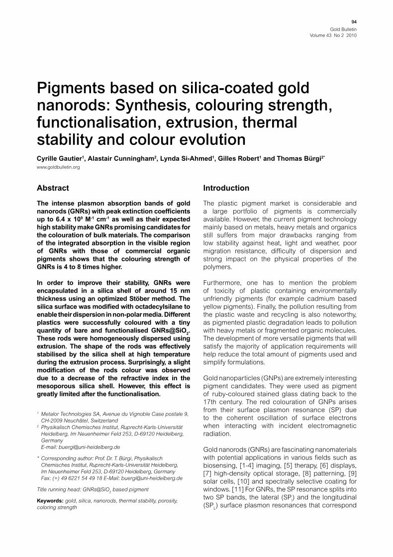
the formation of GNRs up to 99%. As shown by the
TEM image in Figure 1, the rods have an averaged
length and width of 50 nm and 12 nm, respectively.
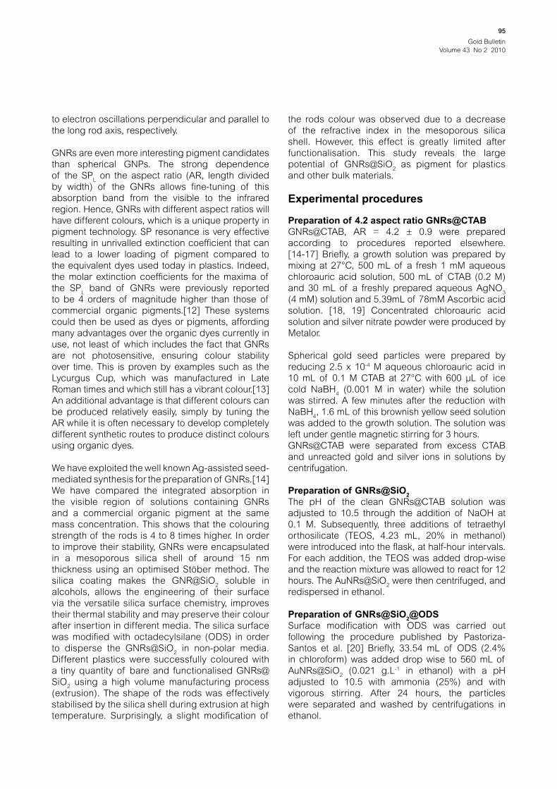
These GNRs have an AR of 4.2 ± 0.9 and exhibit
the two characteristic SPl and SP
L bands at around
520 and 820 nm, respectively (see Figure 2). The
SPL/SP
l intensity ratio is superior to 3 showing that
the proportion of sphere is negligible.
Colouring strength
El-Sayed et al. have published a systematic
quantitative study which uses Mie theory and discrete
dipole approximation method to calculate the trend
of the absorption and scattering efficiencies for
spherical nanoparticles, nanoshells and GNRs
according to their diameter, to their AR and to their
volume. [24] This study clearly shows that GNRs are
better candidates for pigmentation than nanospheres
and nanoshells since a defined volume of gold in the
rod shape gives higher absorption and scattering
efficiencies. Their calculations are in good agreement
with the extinction coefficients reported in literature
and with our results. [25, 26] According to TEM, UV-
vis and inductively coupled plasma atomic emission,
our GNRs exhibit a molar extinction coefficient of 6.4
x 109 M-1.cm-1 at 820 nm. Such extinction coefficients
are at least 4 orders of magnitude higher than
those of organic pigments. It is however difficult
to compare the colouring strength of GNRs with
those of commercial pigments based on extinction
coefficients. Indeed, GNRs have very high molecular
weight and their SPL bands are broader than typical
absorption bands of organic dyes.
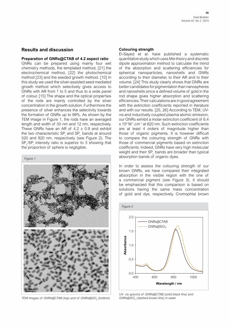
In order to assess the colouring strength of our
brown GNRs, we have compared their integrated
absorption in the visible region with the one of
a commercial pigment (see Figure 3). It should
be emphasized that this comparison is based on
solutions having the same mass concentration
of gold and dye, respectively. Cromophtal brown
TEM images of GNRs@CTAB (top) and of GNRs@SiO2 (bottom)
Figure 1
UV- vis spectra of GNRs@CTAB (solid black line) and
GNRs@SiO2 (dashed brown line) in water
Figure 2
400
0.0
0.5
1.0
1.5
2.0
GNRs@SiO2
GNRs@CTAB
600
Wavelength / nm
Ab
so
rban
ce
800 1000
Gold Bulletin
Volume 43 No 2 2010
97
was selected because its La*b* coordinates are
close to those of our GNRs. The result shows that
the colouring strength or the integrated absorption
of the rods is 3.7 higher even if a large part of the
SPL band is outside of the visible window. A similar
comparison using gold nanorods with an AR of 2
and a blue colouration and Cromophtal blue shows
that the colouring strength of the rods is up to 8
times higher due to the fact that the two plasmon
bands are fully in the visible window.
Preparation of GNRs@SiO2
The encapsulation of GNRs by oxide such as
silica is expected to avoid the optical coupling of
neighbouring GNRs, to preserve their colour upon
environment modification, to protect the GNRs from
aggregation, from thermal coalescence and from
thermal rod-to-sphere shape transition. The latter
was observed by increasing the temperature of the
rods either in an oven or by laser irradiation. [27-29]
Wang and co-workers have shown that the rod-to-
sphere shape transition induced by laser irradiation
requires more energy when the rods are coated with
silica. [27]
The well-known Stöber process is widely used for
the formation of more or less dense spherical silica
NPs by hydrolysis-condensation of TEOS in alkaline
water/ethanol mixture. [30] The silica coating of
AuNRs@SiO2 has been reported several times after
removing the excess of CTAB by centrifugation
Visible spectra of GNRs@CTAB with AR 4.2 (0.012 g.L-1of gold
in water, black line) and of Cromophtal brown (0.012 g.L-1, EtOH:
H2O:NaOH, 6:4:0.001, brown line). The brown surface shows
the difference of absorption between brown GNRs@CTAB (AR
~4.2) and Cromophtal brown
Figure 3
1.6brown AuNRs@CTAB 0.012 g.L-1 in H
2O (AR~4.2)
Cromophtal brown 0.012 g.L-1 in EtOH:H2O;
NaOH 6:4:0.001 (v:v:v:)
1.4
1.2
1.0
0.8
0.6
0.4
0.2
0.0
400 500
Wavelength / nm
Ab
so
rba
nc
e
600 700
either by direct hydrolysis-condensation of TEOS
or sodium silicate on bare NRDs [31] or by using
a primer to render the surface vitreophilic before
silica coating. [27, 32-34] Both the direct hydrolysis-
condensation method and the use of primer yield a
silica shell with a uniform thickness over the entire
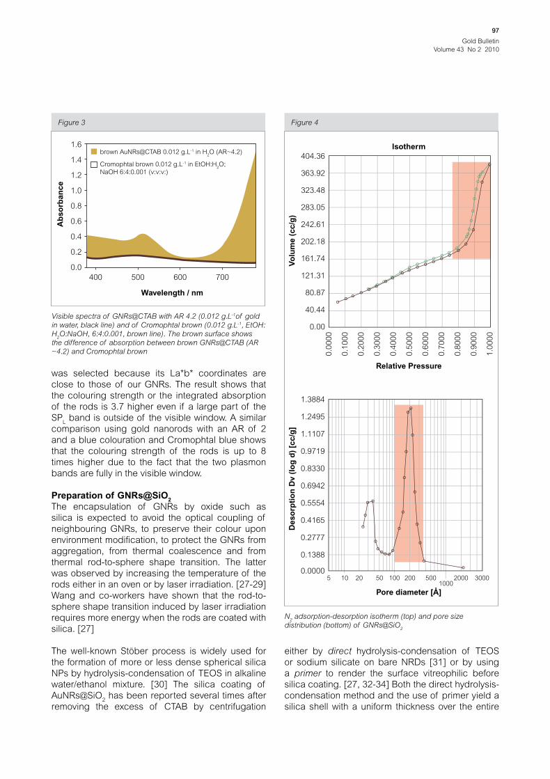
N2 adsorption-desorption isotherm (top) and pore size
distribution (bottom) of GNRs@SiO2
Figure 4
Relative Pressure
Pore diameter [Å]
Vo
lum
e (
cc
/g)
De
so
rpti
on
Dv
(lo
g d
) [c
c/g
]
404.36
Isotherm
363.92
323.48
283.05
242.61
202.18
161.74
121.31
80.87
40.44
0.00
0.0000
0.1388
0.2777
0.4165
0.5554
0.6942
0.8330
0.9719
1.1107
1.2495
1.3884
5 10 20 50 100 200 5001000
2000 3000
0.0
00
0
0.1
00
0
0.2
00
0
0.3
00
0
0.4
00
0
0.5
00
0
0.6
00
0
0.7
00
0
0.8
00
0
0.9
00
0
1.0
00
0
Gold Bulletin
Volume 43 No 2 2010
98
surface of the GNRs. Furthermore, the silica shell
thickness can be controlled with reaction time and
reagent concentrations.
Here, GNRs@CTAB were coated using a direct
improved Stöber method. Figure 1 shows a TEM
image of the GNRs@SiO2. It can be seen that the
initial AR is preserved during the coating, that the
amorphous silica shell has a homogeneous thickness
of ca. 15 nm, and that the polymerisation of TEOS
selectively takes place on the GNRs without visible
agglomeration.
After the silica coating, the SPL band is red shifted
from 820 nm to 840 nm and the absorbance slightly
increased due to the rise of the local refractive index
around the GNRs (Figure 2). According to the results
reported by Pastoriza-Santos et al., this 20 nm red
shift is in good agreement with the thickness of 15
nm of the silica shell measured by TEM. [20]
The N2 adsorption–desorption isotherms (Figure 4)
show that the silica shell obtained by this method
is porous with a high surface area and a bimodal
pore size distribution. Indeed, the surface area of
the GNRs@SiO2 is 303 m2.g-1 and the curves show
two well-defined adsorption steps. The first, at an
intermediate relative pressure, is related to capillary
condensation of N2 inside the silica shell mesopores
which have a diameter of 3 to 4 nm. The second
step, at a high relative pressure, corresponds to the
filling of the large meso- or macro-pores in between
individual GNRs@SiO2. In both cases, the curves
show hysteresis due to a relatively wide pore size
distribution as shown by the pore size distribution
computed using the BJH treatment. [35] The metal
core can be dissolved away to produce hollow silica
nanotubes (using cyanide or aqua regia for gold or
ammonia for silver). [32, 36] This porosity is likely due
to the remaining CTAB micelles which are commonly
used for the preparation of ordered mesoporous
silica NPs. [37]
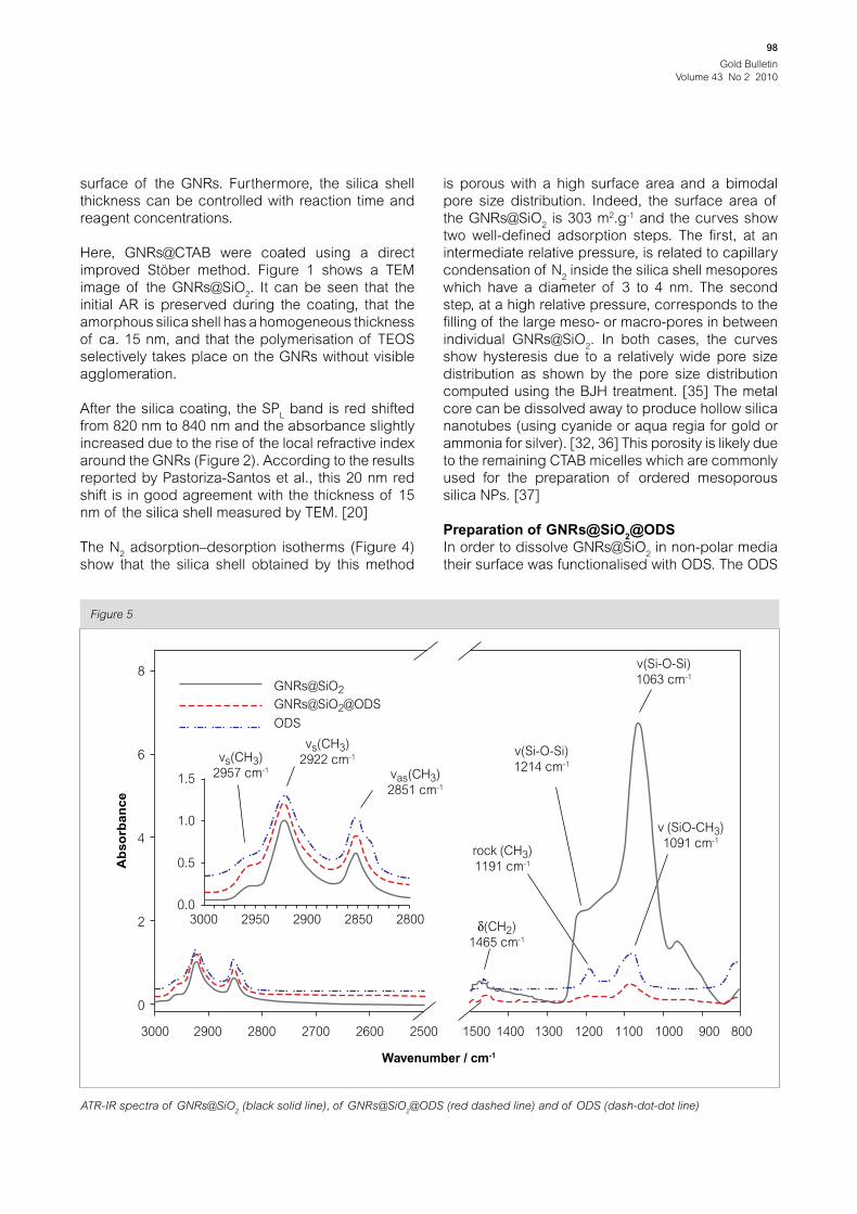
Preparation of GNRs@SiO2@ODS
In order to dissolve GNRs@SiO2 in non-polar media
their surface was functionalised with ODS. The ODS
ATR-IR spectra of GNRs@SiO2 (black solid line), of GNRs@SiO
2@ODS (red dashed line) and of ODS (dash-dot-dot line)
Figure 5
8
GNRs@SiO2
GNRs@SiO2@ODS
ODS
vs(CH3)
2922 cm-1vs(CH3)
2957 cm-1 vas(CH3)
2851 cm-1
rock (CH3)
1191 cm-1
v (SiO-CH3)
1091 cm-1
(CH2)
1465 cm-1
v(Si-O-Si)
1214 cm-1
v(Si-O-Si)
1063 cm-1
6
1.5
1.0
0.5
0.03000 2950 2900 2850 2800
4
2
0
3000 2900 2800 2700 2600 2500
Wavenumber / cm-1
Ab
so
rba
nc
e
1500 1400 1300 1200 1100 1000 900 800

Gold Bulletin
Volume 43 No 2 2010
99
grafting was monitored by attenuated total reflection
infrared spectroscopy (ATR-IR). As shown in
Figure 5, ODS, GNRs@CTAB and GNRs@SiO2@ODS
display characteristic bands of long alkyl chains.
Additionally, ODS possesses bands at 1191 and
1091 cm-1 which can be assigned to the CH3 rocking
and SiO-C modes of SiO-CH3, respectively. After
the modification of the GNRs@SiO2 with ODS, these
bands were replaced by bands at 1214 and 1063
cm-1 which were assigned to the stretching modes of
Si-O-Si. The grafting of ODS is clearly demonstrated
by the fact that the Si-to-alkyl intensity ratio of related
peaks decreases upon addition of ODS even after
several centrifugations, by their solubility in non-
polar solvents and solid-state MAS NMR (Figure 6).
The 13C MAS NMR spectrum shows two signals
centered at around 33 and 35 ppm which can
be assigned to gauche and trans methylene
carbon conformations, respectively. Their relative
intensity shows that ODS chains are mainly in trans
conformation and that they are densely packed on
the surface. The very small signal at 54.1 ppm shows
that CTAB is almost completely removed.
The 29Si MAS NMR spectrum shows mainly six peaks
at around -110, -100, -92, - 65, – 58 and - 50 ppm which
can be assigned to Q4 (Si(OSi)4), Q3 (Si(OSi)
3(OH or
OR), Q2 (Si(OSi)2(OH
2, OR
2 or (OH)(OR)), T3 (RSiO
3)
and T2 (RSiO2(OH)) and T1 (RSiO(OH)
2) silicon sites,
respectively. Their relative intensities 0.9(Q3):0.55
(Q3):0.1(Q2):0.45(T3):1(T2):0.35(T1) reveal a high
ODS coverage and a highly condensed framework. 13C and 29Si MAS NMR tend to show that both the
surface and the pores of the silica shell are highly
functionalised. The relatively high T2 signal might
be attributed to not fully condensed ODS polymers
either attached, or not, to the silica surface.
Thermal stability
The insertion of pigments in bulk material such as
plastics often requires processes at high temperature
such as extrusion and therefore the thermal stability
of GNRs@SiO2 was investigated. It has been known
for a long time that the melting point of a wide variety
of materials such as metals, semiconductors and
organic crystals decreases as their size is reduced
to the nanometer scale. [38-44]
The rod shape of GNRs@CTAB was believed to
be stable up to ~ 600°C.[45, 46] Figure 7 shows the
UV-vis spectra of GNRs@CTAB dissolved in water,
after drying in air at room temperature on a glass slide
and after subsequent firing of this glass slide in air at
260°C for 90 minutes. After drying, the SPL band was
broader and red shifted. This can be explained by
interplasmon coupling due to the close proximity of
the rods on the glass slide. Surprisingly, after heating
the same glass slide for 90 minutes at only 260°C,
the SPL band disappeared completely while the SP
l
remained. This clearly demonstrates that a rod-to-
sphere shape transition occurs. The TEM image
confirms this shape evolution. Petrova et al. and
Xu and co-workers have already reported a similar
transformation in the same range of temperatures
for PVP coated and bare GNRs respectively. [11, 47]
The formers also observed that a slow reduction of
the AR is already observed at only 100°C.
Silica coating of spherical gold NPs has been proven
to be a very efficient approach to preserve the identity
(volume) of the NPs even at high temperature. [29]
Silica has a high melting temperature (~1700 K) Solid-state MAS 13C (top) and 29Si (down) NMR spectra of
GNRs@SiO2@ODS
Figure 6
1.0
0.8
0.6
0.4
-40 -20
-150 -100 -50 0 50
0 20
ppm
ppm
Sig
na
l In
ten
sit
y [
Arb
. U
.]S
ign
al
Inte
nsit
y [
Arb
. U
.]
40 60
C3-16 trans-gauche
C3-16 trans
C2 & C17
T2
T3
T1
Q4
Q2
Q3
C1 & C18
80
0.2
1.0
0.8
0.6
0.4
0.2
0.0
0
-O-SiCH2CH2(CH2)14CH2CH3
O
O
1 2 3-16 17 18--

Gold Bulletin
Volume 43 No 2 2010
100
and the silica shell of silica coated GNRs is known
to withstand the GNRs reshaping induced by laser
irradiation without deformation. [8]
In a similar way, the thermal stability of ODS-
functionalised and of bare GNRs@SiO2 at 260°C
was investigated. In both cases, interplasmon band
coupling was not observed since the 15 nm shell is
thick enough. However, a surprising colour change
was observed when the GNRs@SiO2 were dried in air
on a glass substrate at room temperature. The UV-
vis spectrum of the GNR@SiO2 film indicates that the
SPL shifts from 802 nm to 774 nm during the drying
process (see Figure 8-A). This can be attributed to
the decrease of the refractive index when changing
the GNR surrounding from ethanol to air. Wang et al.
already observed this phenomenon for GNRs@SiO2.
[48] They observed that this colour change is highly
reversible when the rods are repeatedly dried and
dispersed in ethanol. This is in good agreement with
the porosity of the silica shell discussed above.
Interestingly, in the case of the GNRs@SiO2@ODS
(Figure 8-B), we observed that the SPL did not shift
upon drying at room temperature. This might be due
to the presence of the ODS chains on the surface and
UV-vis spectra of GNRs@SiO2 (A) and GNRs@SiO
2@ODS (B) in
water (solid black line) and on glass slide after drying in air at
room temperature (red dashed line) and after heating at 260°C
for 90 minutes (dotted green line)
Figure 8
400
wet
A – GNRs@SiO2
dry
heated
0.0
0.2
0.4
0.6
0.8
1.0
1.2
600 800
Wavelength / nm
Ab
so
rba
nc
e1000
400
wet
B – GNRs@SiO2@ODS
dry
heated
0.0
0.2
0.4
0.6
0.8
1.0
1.2
600 800
Wavelength / nm
Ab
so
rba
nc
e
1000
UV-vis spectra (top) of GNRs@CTAB in water (solid black line),
on glass after drying in air at room temperature (red dashed
line) and after heating at 260°C for 90 minutes (dotted green
line). TEM image (bottom) of GNRs@CTAB after 90 minutes at
260°C
Figure 7
400
wet
dry
heated
0.0
0.1
0.2
0.3
0.4
0.5
600 800
Wavelength / nm
Ab
so
rba
nc
e
1000
Gold Bulletin
Volume 43 No 2 2010
101
in the pores of the shell which does not evaporate
upon drying and which stabilises the refractive index
in the silica shell.
Upon heating at 260°C for 90 minutes, the SPL of
both GNRs@SiO2 and GNRs@SiO
2@ODS are shifted
from 774 nm to 662 nm and from 851 nm to 632 nm,
respectively. These blue shifts are comparable to
those observed after insertion in plastics (vide infra).
In the case of the GNRs@SiO2@ODS, the strong blue
shift of the SPL band observed after heating above
the ODS melting point might be attributed to the loss
of the grafted ODS.
The TEM images of GNRs@SiO2 observed on the
same grid at the same position before and after
heating at 260°C for 90 minutes reveal that the shape
of the rods is well preserved by the silica shell which
may act as a mould.
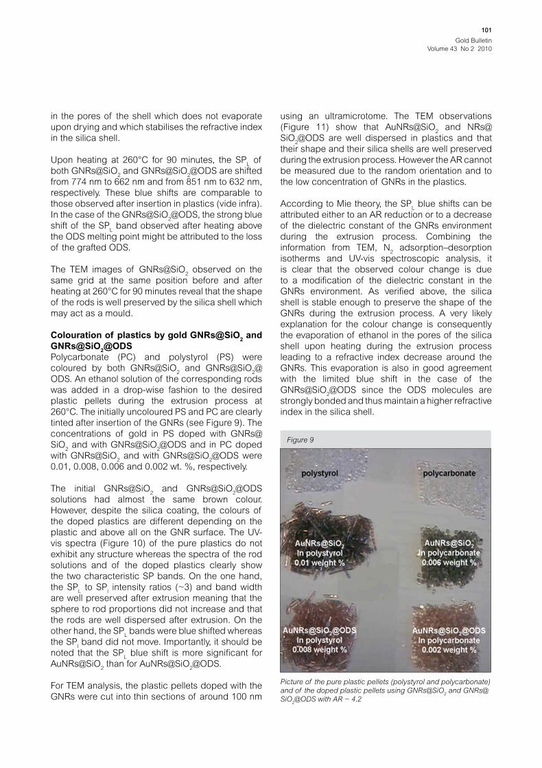
Colouration of plastics by gold GNRs@SiO2 and
GNRs@SiO2@ODS
Polycarbonate (PC) and polystyrol (PS) were
coloured by both GNRs@SiO2 and GNRs@SiO
2@
ODS. An ethanol solution of the corresponding rods
was added in a drop-wise fashion to the desired
plastic pellets during the extrusion process at
260°C. The initially uncoloured PS and PC are clearly
tinted after insertion of the GNRs (see Figure 9). The
concentrations of gold in PS doped with GNRs@
SiO2 and with GNRs@SiO
2@ODS and in PC doped
with GNRs@SiO2 and with GNRs@SiO
2@ODS were
0.01, 0.008, 0.006 and 0.002 wt. %, respectively.
The initial GNRs@SiO2 and GNRs@SiO
2@ODS
solutions had almost the same brown colour.
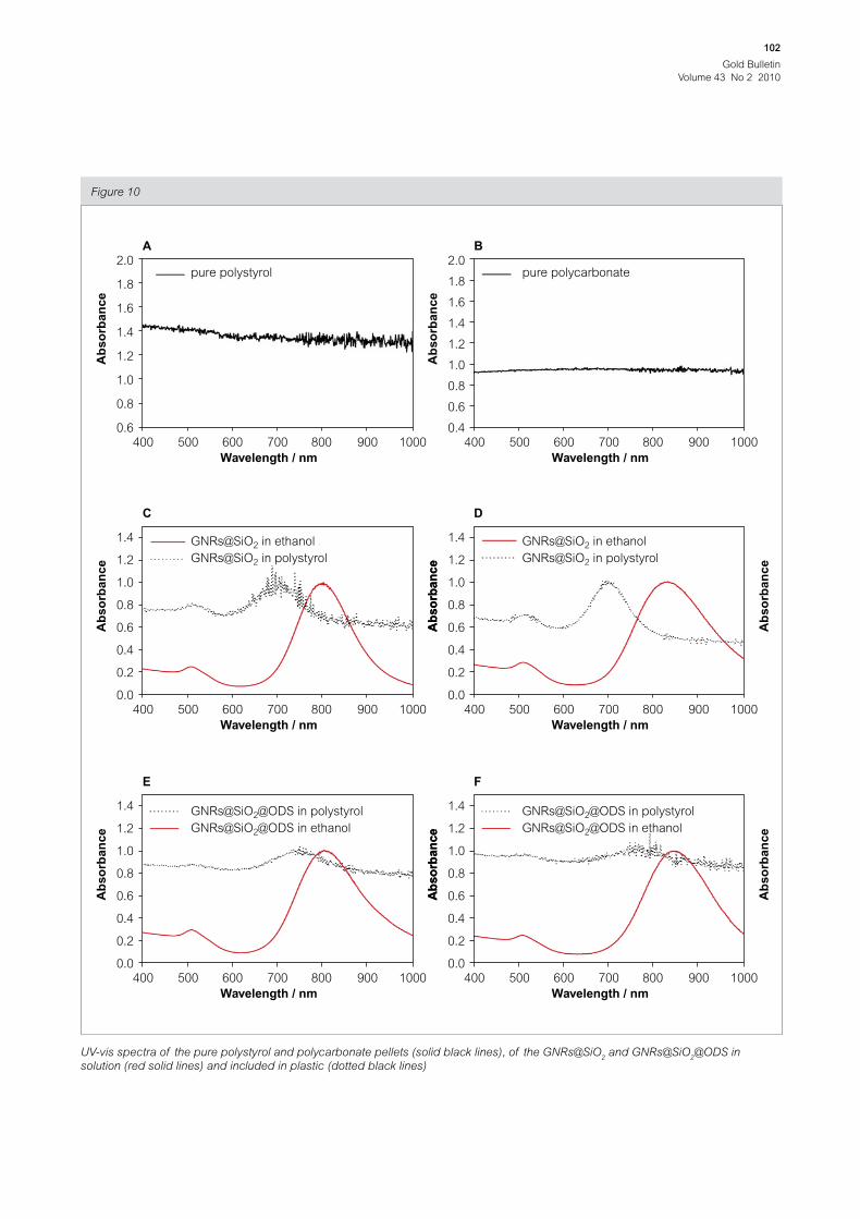
However, despite the silica coating, the colours of
the doped plastics are different depending on the
plastic and above all on the GNR surface. The UV-
vis spectra (Figure 10) of the pure plastics do not
exhibit any structure whereas the spectra of the rod
solutions and of the doped plastics clearly show
the two characteristic SP bands. On the one hand,
the SPL to SP
l intensity ratios (~3) and band width
are well preserved after extrusion meaning that the
sphere to rod proportions did not increase and that
the rods are well dispersed after extrusion. On the
other hand, the SPL bands were blue shifted whereas
the SPl band did not move. Importantly, it should be
noted that the SPL blue shift is more significant for
AuNRs@SiO2 than for AuNRs@SiO
2@ODS.
For TEM analysis, the plastic pellets doped with the
GNRs were cut into thin sections of around 100 nm
using an ultramicrotome. The TEM observations
(Figure 11) show that AuNRs@SiO2 and NRs@
SiO2@ODS are well dispersed in plastics and that
their shape and their silica shells are well preserved
during the extrusion process. However the AR cannot
be measured due to the random orientation and to
the low concentration of GNRs in the plastics.
According to Mie theory, the SPL blue shifts can be
attributed either to an AR reduction or to a decrease
of the dielectric constant of the GNRs environment
during the extrusion process. Combining the
information from TEM, N2 adsorption–desorption
isotherms and UV-vis spectroscopic analysis, it
is clear that the observed colour change is due
to a modification of the dielectric constant in the
GNRs environment. As verified above, the silica
shell is stable enough to preserve the shape of the
GNRs during the extrusion process. A very likely
explanation for the colour change is consequently
the evaporation of ethanol in the pores of the silica
shell upon heating during the extrusion process
leading to a refractive index decrease around the
GNRs. This evaporation is also in good agreement
with the limited blue shift in the case of the
GNRs@SiO2@ODS since the ODS molecules are
strongly bonded and thus maintain a higher refractive
index in the silica shell.
Picture of the pure plastic pellets (polystyrol and polycarbonate)
and of the doped plastic pellets using GNRs@SiO2 and GNRs@
SiO2@ODS with AR ~ 4.2
Figure 9
Gold Bulletin
Volume 43 No 2 2010
102
UV-vis spectra of the pure polystyrol and polycarbonate pellets (solid black lines), of the GNRs@SiO2 and GNRs@SiO
2@ODS in
solution (red solid lines) and included in plastic (dotted black lines)
Figure 10
400
A
pure polystyrol
0.6
0.8
1.0
1.2
1.4
1.6
2.0
1.8
500 600 700 800
Wavelength / nm
Ab
so
rba
nc
e
900 1000 400
B
pure polycarbonate
0.4
0.6
0.8
1.0
1.2
1.4
1.6
2.0
1.8
500 600 700 800
Wavelength / nm
Ab
so
rba
nc
e900 1000
400
C
GNRs@SiO2 in ethanol
GNRs@SiO2 in polystyrol
0.0
0.2
0.4
0.6
0.8
1.0
1.4
1.2
500 600 700 800
Wavelength / nm
Ab
so
rba
nc
e
900 1000
Ab
so
rba
nc
e
400
D
GNRs@SiO2 in ethanol
GNRs@SiO2 in polystyrol
0.0
0.2
0.4
0.6
0.8
1.0
1.4
1.2
500 600 700 800
Wavelength / nm
Ab
so
rba
nc
e
900 1000
Ab
so
rba
nc
e
400
E
GNRs@SiO2@ODS in polystyrol
GNRs@SiO2@ODS in ethanol
0.0
0.2
0.4
0.6
0.8
1.0
1.4
1.2
500 600 700 800
Wavelength / nm
Ab
so
rba
nc
e
900 1000
Ab
so
rba
nc
e
400
F
GNRs@SiO2@ODS in polystyrol
GNRs@SiO2@ODS in ethanol
0.0
0.2
0.4
0.6
0.8
1.0
1.4
1.2
500 600 700 800
Wavelength / nm
Ab
so
rba
nc
e
900 1000
Ab
so
rba
nc
e

Gold Bulletin
Volume 43 No 2 2010
103
Conclusions
During this study, the well known silver-assisted
seeded method was used to prepare GNRs with
AR ~ 4.2. The colouring strength of the rods prepared
during this study was demonstrated to be 4 to 8 times
higher than those of commercial pigments. The
GNRs were coated with a mesoporous silica shell of
around 15 nm using a single-step Stöber approach
and further functionalised with ODS. The silica shell
greatly enhances the thermal stability of the rods up
to at least 260°C. GNRs@SiO2 and GNRs@SiO
2@
ODS were successfully dispersed in both polystyrol
and polycarbonate by extrusion. These plastics
were greatly coloured using a tiny amount of GNRs.
However, the colour of the rods evolves during
extrusion due to the mesoporosity of the silica shell.
Interestingly, the ODS functionalisation stabilises the
colouration of the rods upon extrusion and drying
in air.
About the authors
Cyrille Gautier is a R&D chemist at
Metalor Technologies in Neuchâtel,
Switzerland. He has obtained his PhD
degree at Neuchâtel University in the
Surface Spectroscopy and
Nanoscience Laboratory. His research
activities include the production, the
characterization and the applications
of nanomaterials based on noble
metals.
Alastair Cunningham is a PhD student
at the University of Heidelberg working
on novel means of functionalising and
organising gold nanorods. Previously,
he completed a Masters in Micro and
Nanotechnology at the University of
Neuchâtel in Switzerland and obtained
an MSc in Chemistry from the
University of Strathclyde in Glasgow.
Lynda Si-Ahmed, PhD, is the
Nanotech manager at Metalor
Technologies SA in Neuchâtel,
Switzerland. The Nanotech Unit is
dedicated to the development and
manufacturing of Precious Metal
Nanoparticles.
Gilles Robert, PhD, is the manager of
the Technology Product Department at
Metalor Technologies SA in Neuchâtel,
Switzerland. The Technology Product
Department is dedicated to the
development and market introduction
of innovations based on Metalor core
competences in metallurgy and
inorganic chemistry of precious metals.
TEM images of the GNRs@SiO2 and GNRs@SiO
2@ODS with AR ~ 4.2 inserted in polystyrol and in polycarbonate by extrusion.
The image A shows a small agglomerate of GNRs@SiO2 in polystyrol. However, it should be noted that in this sample most of the rods
are well dispersed as shown for samples B, C and D
Figure 11
A B C D
A – GNRs@SiO2 in polystyrol
B – GNRs@SiO2 in polycarbonate
C – GNRs@SiO2@ODS in polystyrol
D – GNRs@SiO2@ODS in polycarbonate
Gold Bulletin
Volume 43 No 2 2010
104
Thomas Bürgi obtained his PhD
degree from the University of Bern,
Switzerland. After a postdoc at MIT he
made his habilitation thesis at ETH
Zurich. In 2003 he moved to the
University of Neuchâtel as a Professor
of Physical Chemistry funded by the
Swiss National Science Foundation. In
2008 he was appointed as a professor
of Physical Chemistry at the University of Heidelberg. He is
working in the fields of in situ spectroscopy of solid − liquid
interfaces, photocatalysis and the preparation, characterization
and application of nanomaterials.
Acknowledgements
We are grateful to the World Gold Council GROW
program for financial support of this project. We
cordially thank Prof. Clemens Holzer from INKA
institute and Michèle Vlimant from Neuchâtel
University for providing facilities and help in extrusion
and ultramicrotomy, respectively.
References
1 C. Wang, J. Irudayaraj, Small 2008, 4, 2204
2 C.Z. Li, K.B. Male, S. Hrapovic, J.H.T. Luong, Chem.
Commun. 2005, 3924
3 P.K. Sudeep, S.T.S. Joseph, K.G. Thomas, J. Am. Chem.
Soc. 2005, 127, 6516
4 K.G. Thomas, S. Barazzouk, B.I. Ipe, S.T.S. Joseph, P.V.
Kamat, J. Phys. Chem. B 2004, 108, 13066
5 H. Ding, K.T. Yong, I. Roy, H.E. Pudavar, W.C. Law,
E.J. Bergey, P.N. Prasad, J. Phys. Chem. C 2007, 111, 12552
6 L. Tong, Y. Zhao, T.B. Huff, M.N. Hansen, A. Wei, J.X. Cheng,
Adv. Mater. 2007, 19, 3136
7 N.M. Lawandy, A.Y. Smuk, J. Soc. Inf. Display 2005, 13, 755
8 J.W.M. Chon, C. Bullen, P. Zijlstra, M. Gu, Adv. Funct. Mater.
2007, 17, 875
9 P. Zijlstra, J.W.M. Chon, M. Gu, Opt. Express 2007, 15,
12151
10 J. Pérez-Juste, I. Pastoriza-Santos, L.M. Liz-Marzán,
P. Mulvaney, Coord. Chem. Rev. 2005, 249, 1870
11 X. Xu, T. Gibbons, M.B. Cortie, Gold Bull. 2006, 39, 156
12 C.J. Orendorff, C.J. Murphy, J. Phys. Chem. B 2006, 110,
3990
13 I. Freestone, N. Meeks, M. Sax, C. Higgitt, Gold Bull. 2007,
40, 270
14 N.R. Jana, L. Gearheart, C.J. Murphy, Adv. Mater. 2001, 13,
1389
15 B. Nikoobakht, M.A. El-Sayed, Chem. Mater. 2003, 15, 1957
16 T.K. Sau, C.J. Murphy, Langmuir 2004, 20, 6414
17 M.Z. Liu, P. Guyot-Sionnest, J. Phys. Chem. B 2005, 109,
22192
18 L.I. Elding, A.B. Gronong, Acta. Chem. Scand. A 1978, 32,
867
19 J. Pouradier, M. Coquard, J. Chim. Phys. 1966, 63, 1072
20 I. Pastoriza-Santos, J. Perez-Juste, L.M. Liz-Marzan, Chem.
Mater. 2006, 18, 2465
21 C.A.J. Foss, G.L. Hornyak, J.A. Stockert, C.R. Martin,
J. Phys. Chem. 1992, 96, 7497
22 Y.Y. Yu, S.S. Chang, C.L. Lee, C.R.C. Wang, J. Phys. Chem.
B 1997, 101, 6661
23 F. Kim, J.H. Song, P. Yang, J. Am. Chem. Soc. 2002, 124,
14316
24 P.K. Jain, K.S. Lee, I.H. El-Sayed, M.A. El-Sayed, J. Phys.
Chem. B 2006, 110, 7238
25 B. Nikoobakht, J. Wang, M.A. El-Sayed, Chem. Phys. Lett.
2002, 366, 17
26 H. Liao, J.H. Hafner, Chem. Mater. 2005, 17, 4636
27 S.S. Chang, C.W. Shih, C.D. Chen, W.C. Lai, C.R. Chris
Wang, Langmuir 1999, 15, 701
28 M. Zhu, G. Qian, Z. Wang, M. Wang, Mat. Chem. and Phys.
2006, 100, 333
29 K. Dick, T. Dhanasekaran, Z. Zhang, D. Meisel, J. Am.
Chem. Soc. 2002, 124, 2312
30 W. Stöber, A. Fink, E. Bohn, J. Colloid Interface Sci. 1968,
62, 26
31 I. Gorelikov, N. Matsuura, Nano Lett. 2008, 8, 369
32 S.O. Obare, N.R. Jana, C.J. Murphy, Nano Lett. 2001, 1, 601
33 J. Perez-Juste, M.A. Correa-Duarte, L.M. Liz-Marzan, Appl.
Surf. Sci. 2004, 226, 137
34 C.J. Murphy, T.K. Sau, A.M. Gole, C.J. Orendorff, J. Gao, L.
Gou, S.E. Hunyadi, T. Li, J. Phys. Chem. B 2005, 109, 13857
35 E.P. Barrett, L.G. Joyner, P.P. Halenda, J. Am. Chem. Soc.
2002, 73, 373
36 Y. Yin, Y. Lu, Y. Sun, Y. Xia, Nano Lett. 2002, 2, 427
37 C.T. Kresge, M.E. Leonowicz, W.J. Roth, J.C. Vartuli, J.S.
Beck, Nature 1992, 359, 710
38 J.R. Sambles, Proc. R. Soc. London, Ser. A 1971, 324, 339
39 H.W. Sheng, K. Lu, E. Ma, Nanostruct. Mater. 1998, 10, 865
40 K.F. Peters, J.B. Cohen, Y.W. Chung, Phys. Rev. B 1998, 57,
13430
41 A.N. Goldstein, C.M. Echer, A.P. Alivisatos, Science 1992,
256, 1425
42 C.L. Cleveland, W.D. Luedtke, U. Landman, Phys. Rev. B
1999, 60, 5065
43 P. Buffat, J. P. Borel, Phys. Rev. A 1976, 13, 2287
44 J. P. Borel, Surf. Sci. 1981, 106, 1
45 M. B. Mohamed, K.Z. Ismail, S. Link, M.A. El-Sayed,
J. Phys. Chem. B 1998, 102, 9370
46 El-Sayed A.M. Al-Sherbini, Colloids Surf. A 2004, 246, 61
47 H. Petrova, J.P. Juste, I. Pastoriza-Santos, G.V. Hartland,
L.M. Liz-Marzan, P. Mulvaney, Phys. Chem. Chem. Phys.
2006, 8, 814
48 C. Wang, Z. Ma, Z. Su, Nanotechnology 2006, 17, 1819
Related Documents