PHYSIOLOGY OF VISUAL CYCLE Othman Al-Abbadi, M.D

Physiology of visual cycle
Jul 24, 2015
Welcome message from author
This document is posted to help you gain knowledge. Please leave a comment to let me know what you think about it! Share it to your friends and learn new things together.
Transcript

PHYSIOLOGY OF VISUAL CYCLE
Othman Al-Abbadi, M.D

INDEX
Vitamin A Dietary sources Absorption & storage Transport Utilization
Visual pigments Rhodopsin Cone pigments
Light-induced changes Rhodopsin bleaching Rhodopsin regeneration Visual cycle
DIETARY SOURCES

DIETARY SOURCES
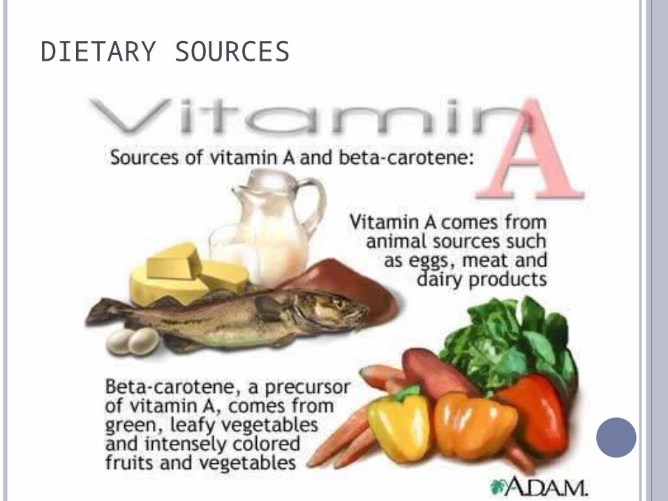
Dietary sources of vitamin A include animal food & plant food.
Animal food contain vitamin A as Retnol. The liver is always the best source
Plant foods contain vitamin A as Carotenes which is a precursor.
Carotenes must be converted to Retinol by metabolic activity in the wall of the small intestine.
Three types of carotenes (alpha, beta, & gamma) are present in plant food
Beta Carotenes yield 2 molecules of vitamin A, while Alpha & Gamma yield one molecule.

Absorption Storage Transport
Attach to receptors in RPE
Transported inside RPE to photoreceptors

UTILIZATION OF VITAMIN A
Retinol passes through RPE to phtoreceptors unchanged.
Oxidation of Retinol to Retinene (11-cis Retinal) by Retinene Reductase in photoreceptor outer segment.
Retinene combines with Opsin to form Rhodopsin.
The freshly formed Rhodopsin is incorporated into the newly forming double discs at the innermost portion of outer segment of photoreceptors.

VISUAL PIGMENTS
Substances that have the property of absorbing light.
The peak of each pigment’s absorption curve is called “absorption maximum.”

RHODOPSIN
Membrane-bound glycolipid. Consists of Scotopsin & Retinal. Molecular weight of 40,000 Fat soluble Sensitive to heat & chemical agents which
denature the protein. Scotopsin is a 348 amino acid protein &
crosses the disc membrane 7 times.
The light absorbing form is 11-cis retinal. The absorption spectrum for rhodopsin has a
peak of 493-505 nm; absorbing yellow wavelength primarily & transmitting violet & red to appear purple (visual purple).

CONE PIGMENTS
NOT intensively studied. Three kinds of cones. Cone pigments are different than rhodopsin in
that they respond to specific wavelengths of light… giving us the ability for colour vision.
Amino acid sequence for all human cone opsins is almost identical except for few different amino acids that makes the difference in spectral absorbance.
11-cis retinal remains the same. Peak absorbance of blue, green & red is 435, 535
& 580 nm respectively. Blue-sensitive cones are the least prevalent.
The light falling on the retina is absorbed by the photosensitive pigments & initiates phtotchemical changes that initiate electrical changes.
Photochemical changes take place in the outer segments. Bleaching (light dependent) Regeneration (light independent) Visual cycle

RHODOPSIN –RETINAL VISUAL CYCLE IN THE RODS. Rhodopsin and its decomposition by light energy:• The outer segment of the rod that projects into
the pigment layer of retina has a concentration of about 40% of light sensitive pigments called Rhodopsin or visual purple.
• Rhodopsin = scotopsin(protein) + retinal(carotenoid protein).
• Retinal is present in the form of 11-cis retinal known as retinene.
• cis form of retinal is important because only this form can bind with scotopsin to synthesize rhodopsin.

CHANGES OCCURING IN RHODOPSIN
Rhodopsin Light barthorhodopsin
lumirhodopsin
BLEACHING
metarhodopsin I
metarhodopsin II
Scotopsin
11-cis retinal isomerase all-trans retinal
REGENERATION
11-cis retinol isomerase all trans retinol


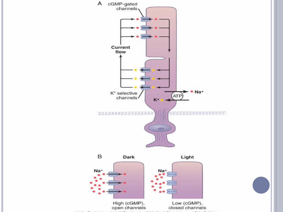
PHOTOTRANSDUCTION
The transduction of light into a neural signal takes place in the outer segment of a retinal rod or cone photoreceptor


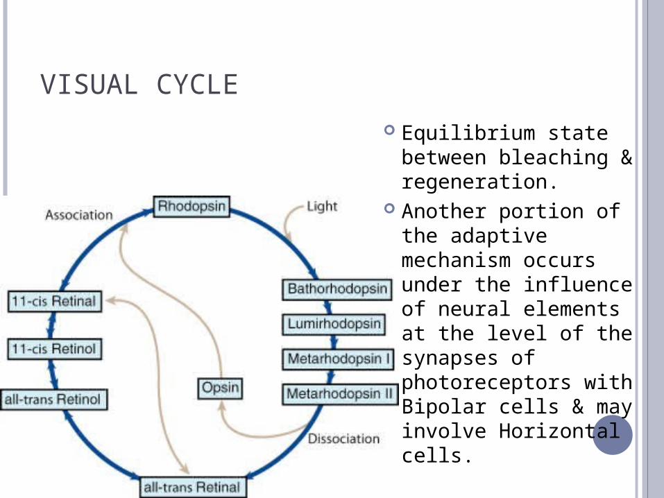
VISUAL CYCLE
Equilibrium state between bleaching & regeneration.
Another portion of the adaptive mechanism occurs under the influence of neural elements at the level of the synapses of photoreceptors with Bipolar cells & may involve Horizontal cells.

DIFFERENCES THAN OTHER RECEPTORS
Receptor potential of photoreceptors is local graded potential. (not all or non)
Hyperpolarization rather than depolarization.

THANK YOU
Related Documents