Physiology of the menstrual cycle. Conception. Early pregnancy Gyula Richárd Nagy MD, PhD

Welcome message from author
This document is posted to help you gain knowledge. Please leave a comment to let me know what you think about it! Share it to your friends and learn new things together.
Transcript
Physiology of the
menstrual cycle.
Conception.
Early pregnancy
Gyula Richárd Nagy MD, PhD
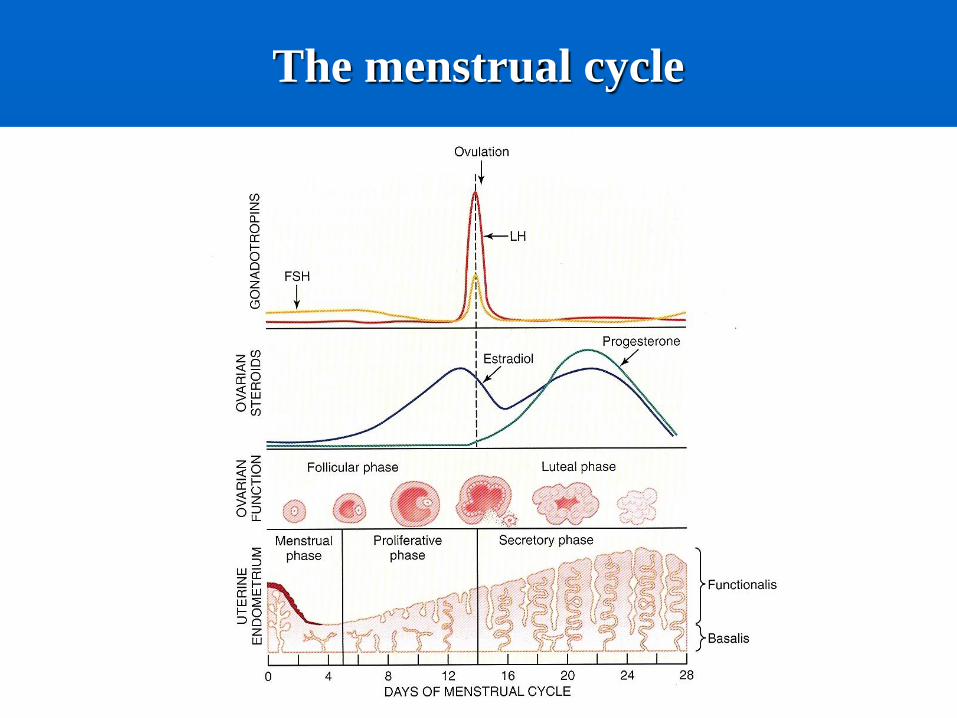
The menstrual cycle
Complex interaction between the hypothalamus,
pituitary gland, ovaries, and endometrium
Cyclic changes in gonadotropin and steroid hormones
result in
• Follicular maturation, ovulation, and corpus luteum
formation
• Changes at the level of endometrium for successful
implantation of the developing embryo

The menstrual cycle
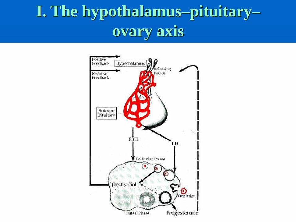
I. The hypothalamus–pituitary–
ovary axis
Pituitary gland
Pituitary gland lies below the hypothalamus at the base of the brain within a bony cavity (sella turcica), separated from the cranial cavity by the diaphragma sellae
Two parts
• Neurohypophysis, derived from neural tissue, direct continuity with the hypothalamus and CNS
• Adenohypophysis, derived from ectoderm
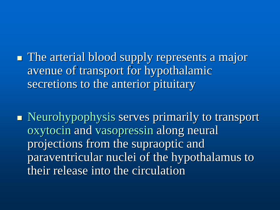
The arterial blood supply represents a major avenue of transport for hypothalamic secretions to the anterior pituitary
Neurohypophysis serves primarily to transport oxytocin and vasopressin along neural projections from the supraoptic and paraventricular nuclei of the hypothalamus to their release into the circulation
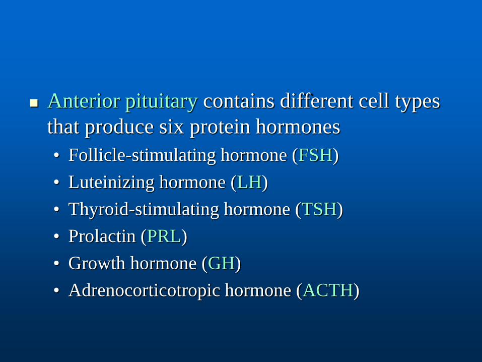
Anterior pituitary contains different cell types
that produce six protein hormones
• Follicle-stimulating hormone (FSH)
• Luteinizing hormone (LH)
• Thyroid-stimulating hormone (TSH)
• Prolactin (PRL)
• Growth hormone (GH)
• Adrenocorticotropic hormone (ACTH)
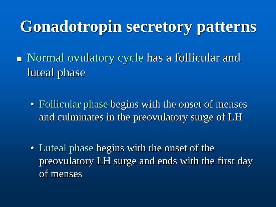
Gonadotropin secretory patterns
Normal ovulatory cycle has a follicular and
luteal phase
• Follicular phase begins with the onset of menses
and culminates in the preovulatory surge of LH
• Luteal phase begins with the onset of the
preovulatory LH surge and ends with the first day
of menses
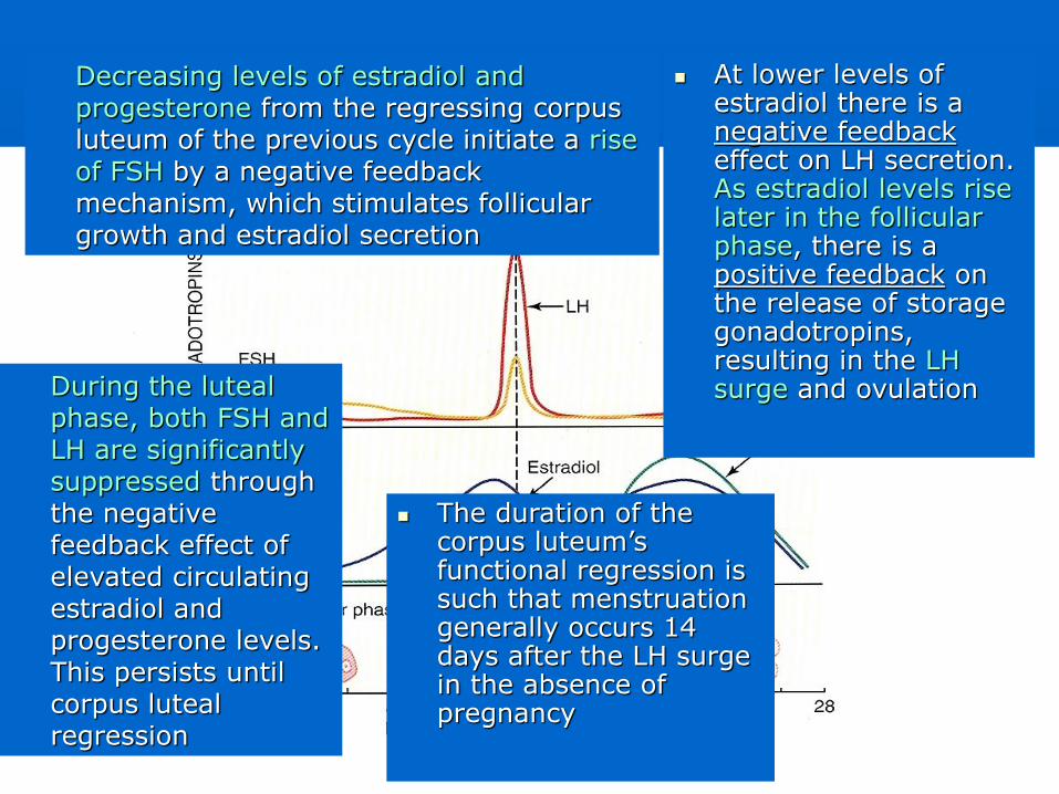
Decreasing levels of estradiol and progesterone from the regressing corpus luteum of the previous cycle initiate a rise of FSH by a negative feedback mechanism, which stimulates follicular growth and estradiol secretion
At lower levels of estradiol there is a negative feedbackeffect on LH secretion. As estradiol levels rise later in the follicular phase, there is a positive feedback on the release of storage gonadotropins, resulting in the LH surge and ovulationDuring the luteal
phase, both FSH and LH are significantly suppressed through the negative feedback effect of elevated circulating estradiol and progesterone levels. This persists until corpus luteal regression
The duration of the corpus luteum’sfunctional regression is such that menstruation generally occurs 14 days after the LH surge in the absence of pregnancy
Hypothalamus
GnRH
• a decapeptide that is synthesized primarily in the
arcuate nucleus
• Responsible for the synthesis and release of both
FSH and LH
• Reaches the anterior pituitary via the hypophyseal
portal vessels
• Secreted in a pulsatile fashion
Iv. and sc. administration of exogenous pulsatile
GnRH has been used to induce ovulation in selected
women who are not ovulating as a result of
hypothalamic dysfunction
Continuous infusion of GnRH results in a reversible
inhibition of gonadotropin secretion through a
process of „downregulation” or desensitization of
pituitary gonadotrophs
Gonadotropins have an inhibitory effect on
GnRH release
Estradiol appears to enhance hypothalamic
release of GnRH and may help induce the
midcycle LH surge by increasing GnRH
release or by enhancing pituitary
responsiveness to the decapeptide
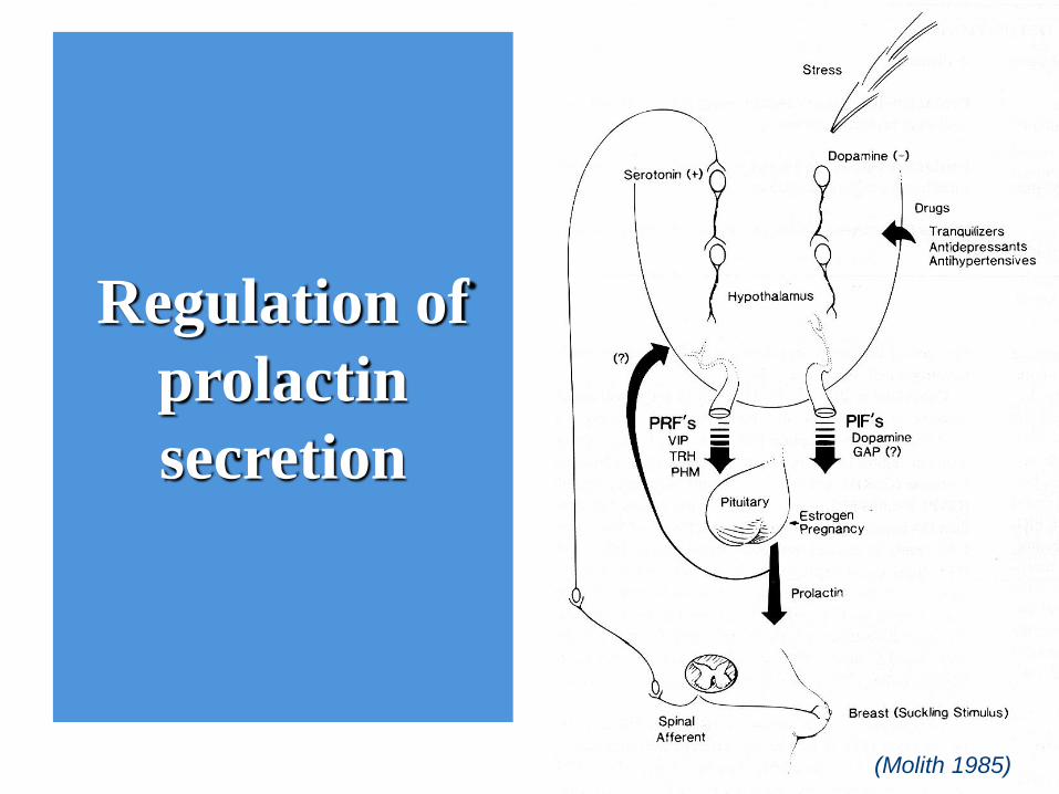
Regulation of
prolactin
secretion
(Molith 1985)
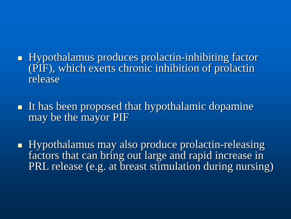
Hypothalamus produces prolactin-inhibiting factor (PIF), which exerts chronic inhibition of prolactin release
It has been proposed that hypothalamic dopamine may be the mayor PIF
Hypothalamus may also produce prolactin-releasing factors that can bring out large and rapid increase in PRL release (e.g. at breast stimulation during nursing)
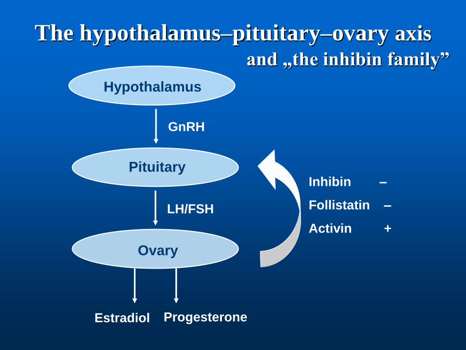
Hypothalamus
Pituitary
Ovary
GnRH
LH/FSH
The hypothalamus–pituitary–ovary axis
and „the inhibin family”
Estradiol Progesterone
Inhibin –
Follistatin –
Activin +
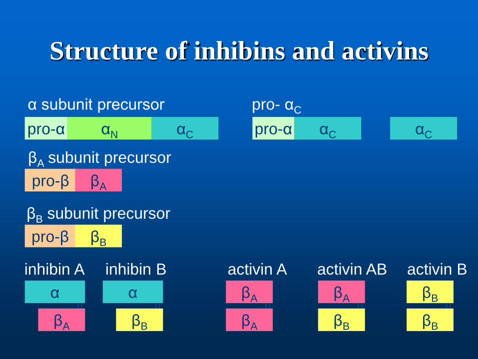
Structure of inhibins and activins
pro-α αN αC
pro-β βA
pro-β βB
α
βA
α
βB
βA
βA
βA
βB
βB
βB
pro-α αC αC
α subunit precursor
βA subunit precursor
βB subunit precursor
inhibin A inhibin B activin A activin AB activin B
pro- αC
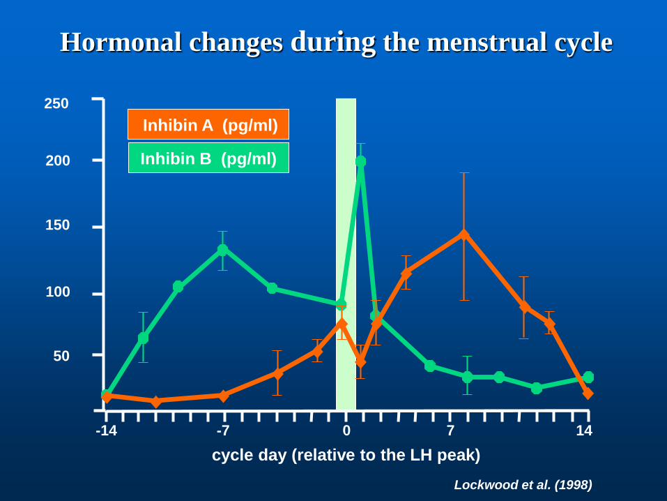
cycle day (relative to the LH peak)
Lockwood et al. (1998)
Inhibin B (pg/ml)
Inhibin A (pg/ml)
200
150
100
50
250
-14 -7 0 7 14
Hormonal changes during the menstrual cycle
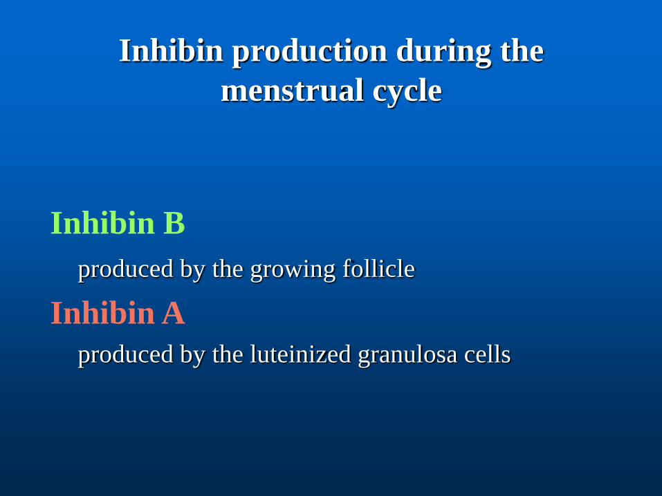
Inhibin production during the
menstrual cycle
Inhibin B
produced by the growing follicle
Inhibin A
produced by the luteinized granulosa cells
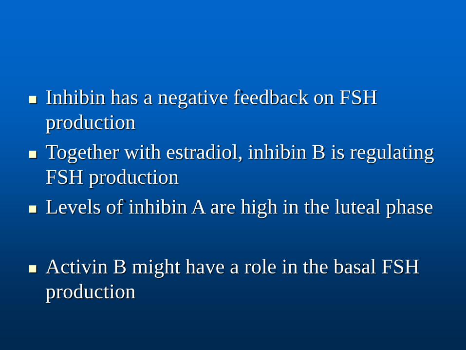
Inhibin has a negative feedback on FSH
production
Together with estradiol, inhibin B is regulating
FSH production
Levels of inhibin A are high in the luteal phase
Activin B might have a role in the basal FSH
production
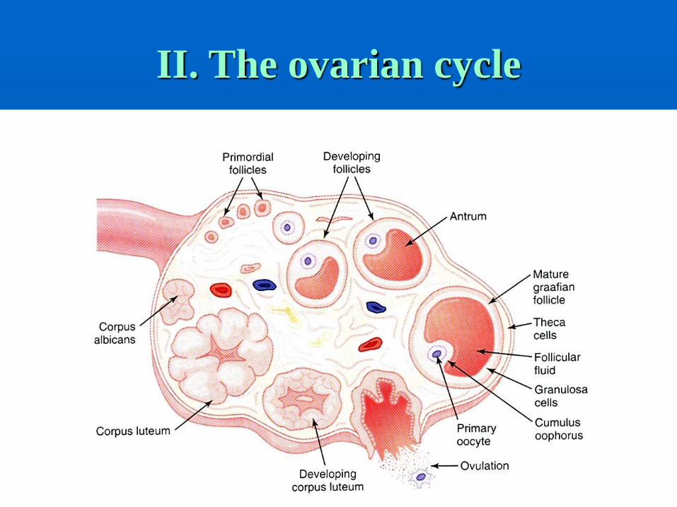
II. The ovarian cycle
The ovarian cycle
I. Folliculogenesis
II. Hormonogenesis
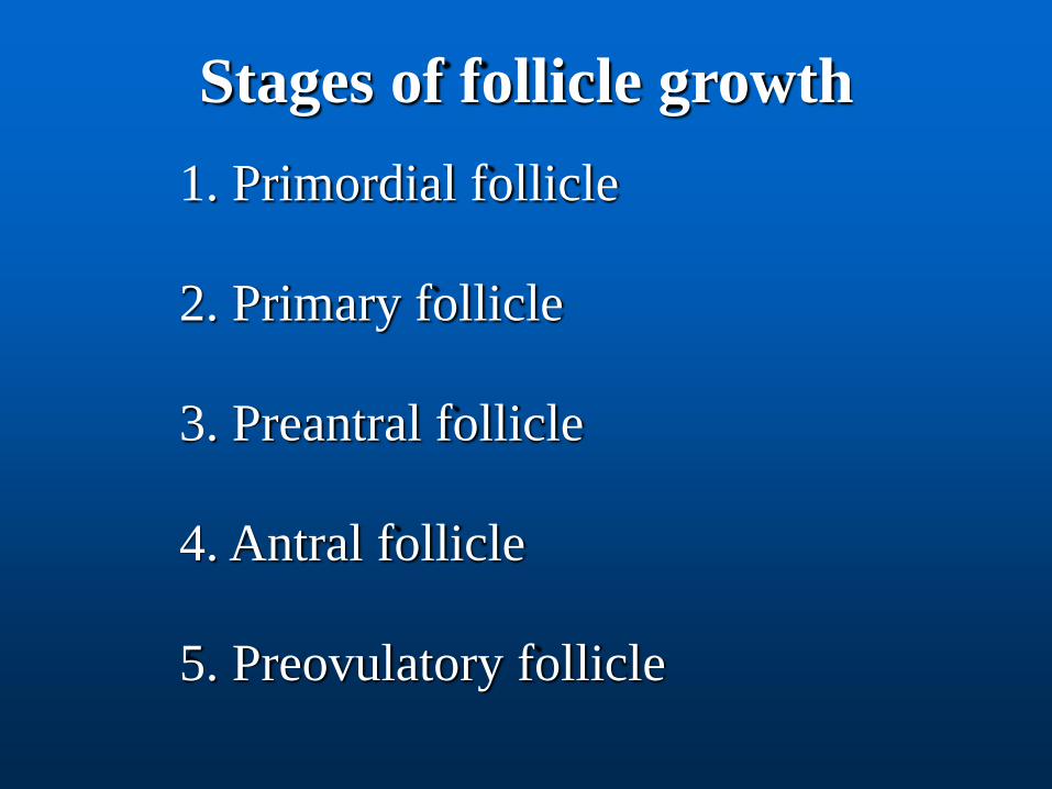
Stages of follicle growth
1. Primordial follicle
2. Primary follicle
3. Preantral follicle
4. Antral follicle
5. Preovulatory follicle
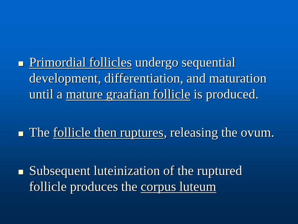
Primordial follicles undergo sequential
development, differentiation, and maturation
until a mature graafian follicle is produced.
The follicle then ruptures, releasing the ovum.
Subsequent luteinization of the ruptured
follicle produces the corpus luteum
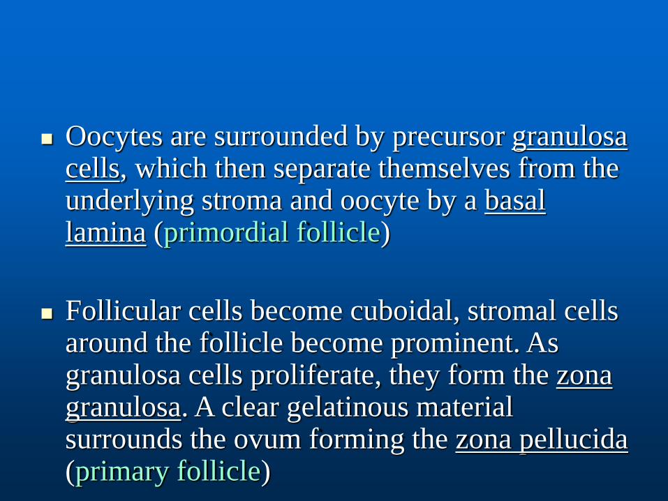
Oocytes are surrounded by precursor granulosa cells, which then separate themselves from the underlying stroma and oocyte by a basal lamina (primordial follicle)
Follicular cells become cuboidal, stromal cells around the follicle become prominent. As granulosa cells proliferate, they form the zona granulosa. A clear gelatinous material surrounds the ovum forming the zona pellucida(primary follicle)
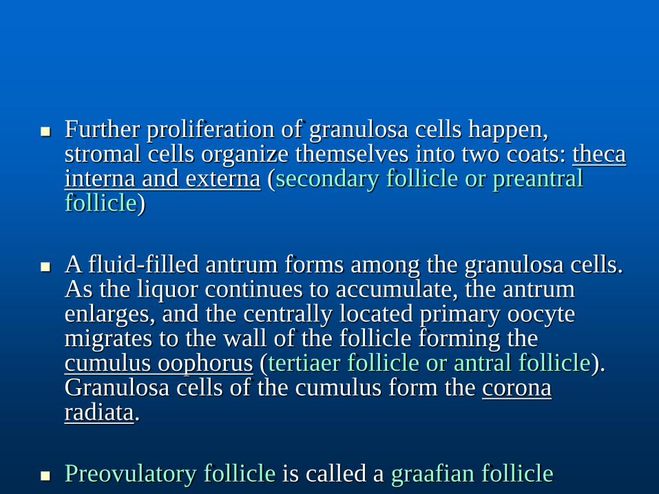
Further proliferation of granulosa cells happen, stromal cells organize themselves into two coats: theca interna and externa (secondary follicle or preantral follicle)
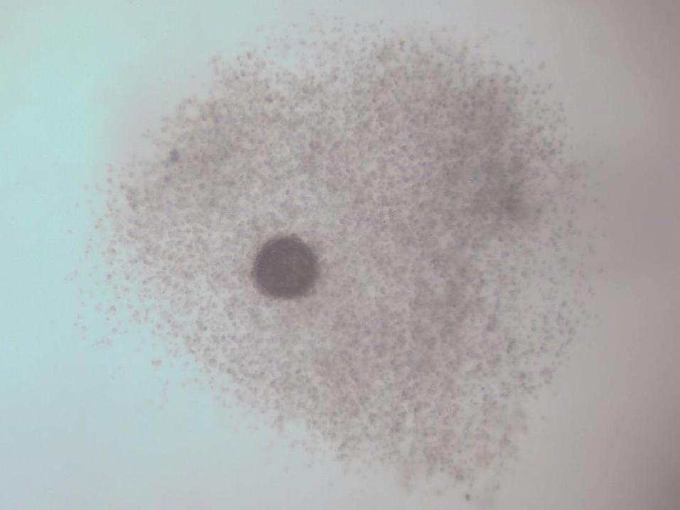
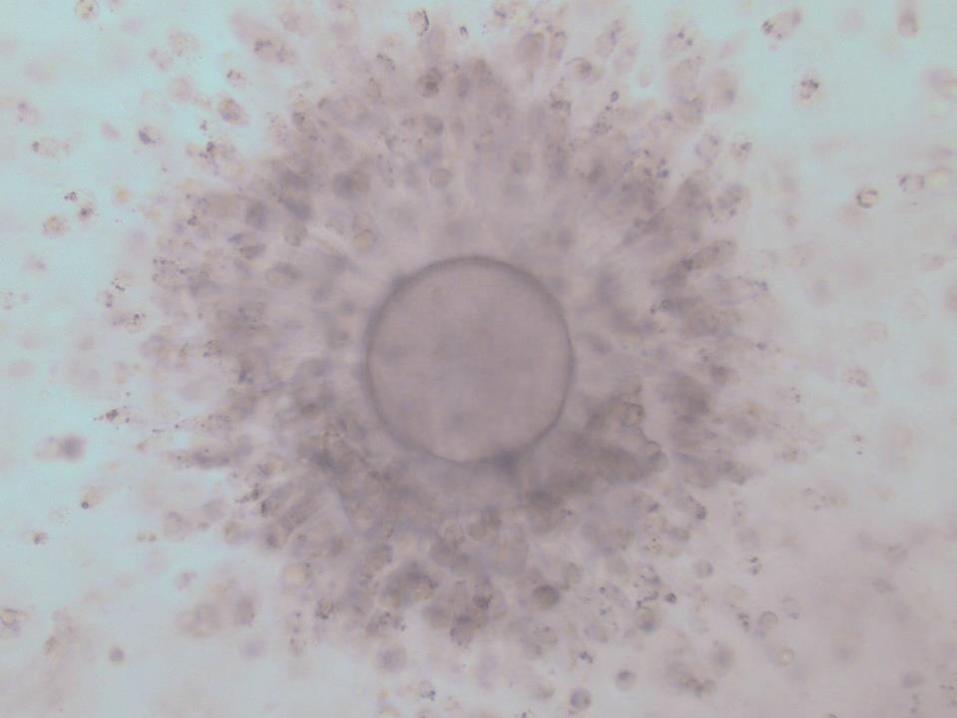
A fluid-filled antrum forms among the granulosa cells. As the liquor continues to accumulate, the antrum enlarges, and the centrally located primary oocyte migrates to the wall of the follicle forming the cumulus oophorus (tertiaer follicle or antral follicle). Granulosa cells of the cumulus form the corona radiata.
Preovulatory follicle is called a graafian follicle
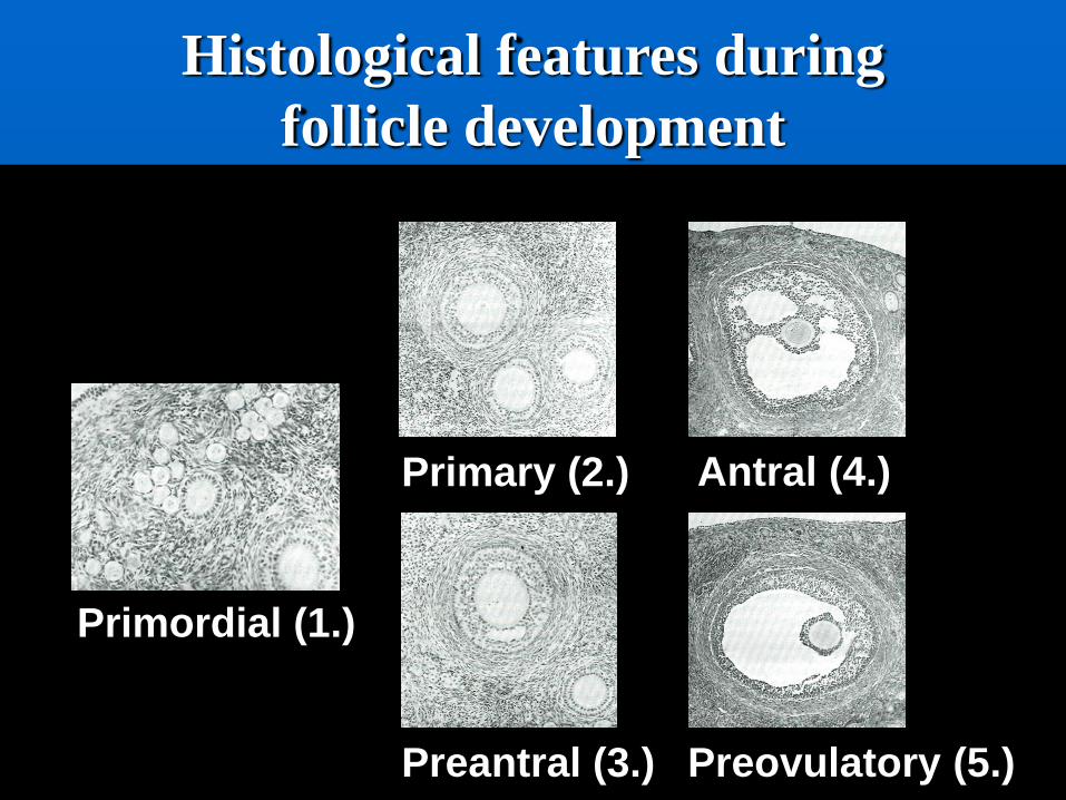
Histological features during
follicle development
Primordial (1.)
Preovulatory (5.)
Primary (2.)
Preantral (3.)
Antral (4.)
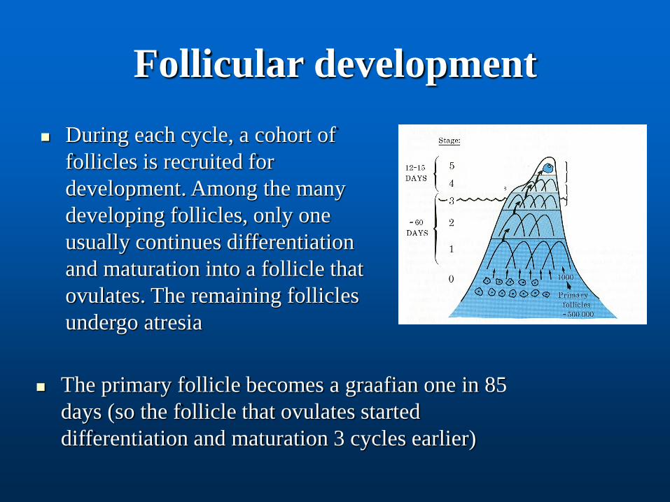
Follicular development
During each cycle, a cohort of
follicles is recruited for
development. Among the many
developing follicles, only one
usually continues differentiation
and maturation into a follicle that
ovulates. The remaining follicles
undergo atresia
The primary follicle becomes a graafian one in 85
days (so the follicle that ovulates started
differentiation and maturation 3 cycles earlier)
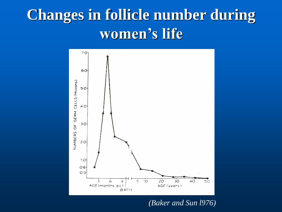
Changes in follicle number during
women’s life
(Baker and Sun l976)
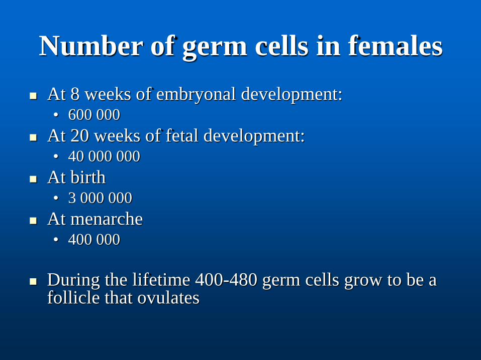
Number of germ cells in females
At 8 weeks of embryonal development: • 600 000
At 20 weeks of fetal development:• 40 000 000
At birth• 3 000 000
At menarche• 400 000
During the lifetime 400-480 germ cells grow to be a follicle that ovulates
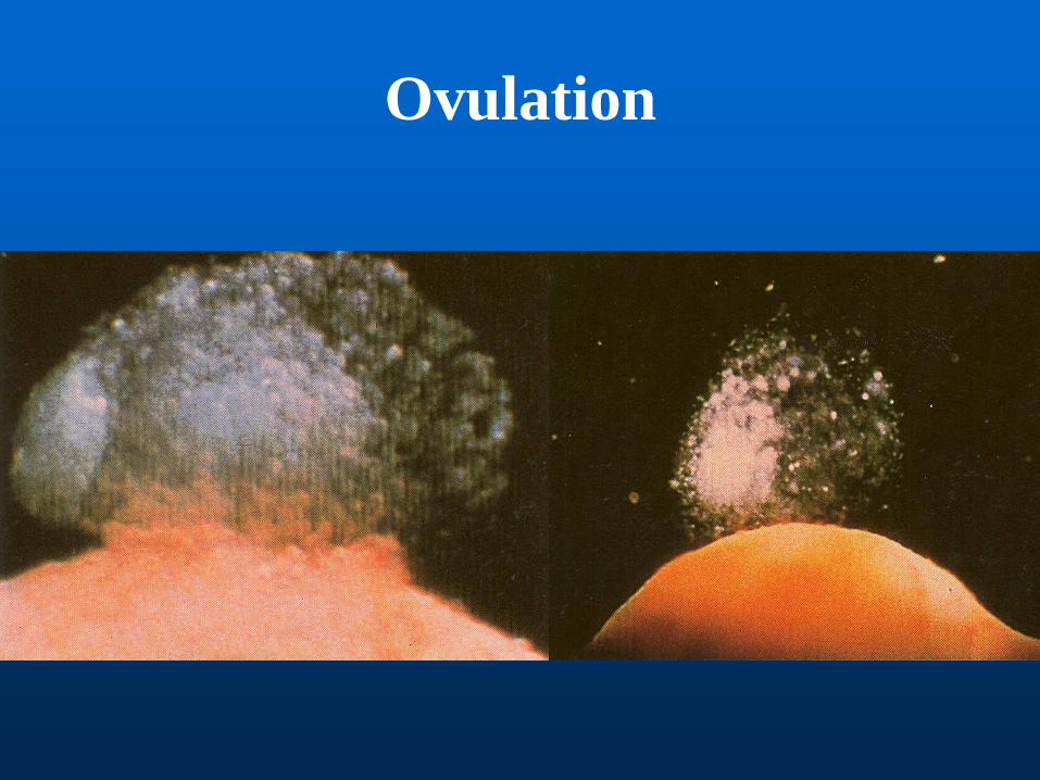
Ovulation
Before ovulation a general dissolution of the entire
follicular wall occurs (action of proteolytic enzimes)
A stigma forms and follicular basement membrane
finally bulges through
When this ruptures, the oocyte and corona radiata are
expelled into the peritoneal cavity, and ovulation
takes place
Ovulation
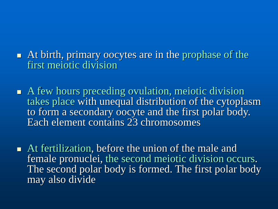
At birth, primary oocytes are in the prophase of the first meiotic division
A few hours preceding ovulation, meiotic division takes place with unequal distribution of the cytoplasm to form a secondary oocyte and the first polar body. Each element contains 23 chromosomes
At fertilization, before the union of the male and female pronuclei, the second meiotic division occurs. The second polar body is formed. The first polar body may also divide
Corpus luteum
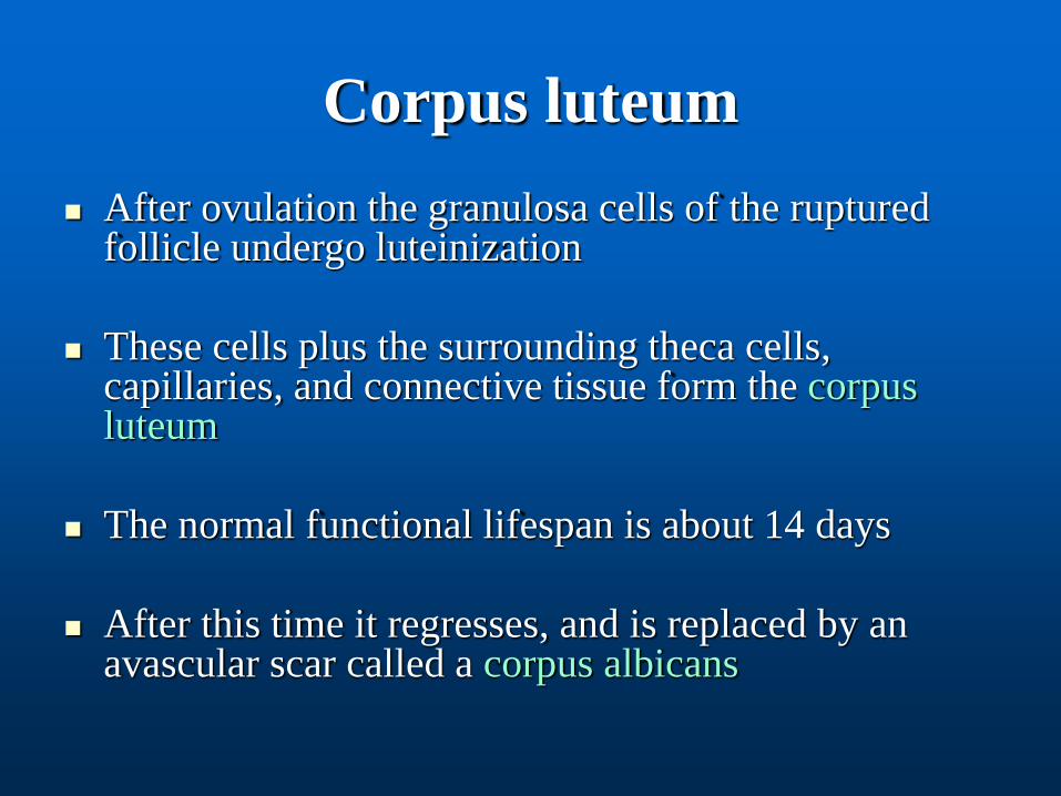
After ovulation the granulosa cells of the ruptured follicle undergo luteinization
These cells plus the surrounding theca cells, capillaries, and connective tissue form the corpus luteum
The normal functional lifespan is about 14 days
After this time it regresses, and is replaced by an avascular scar called a corpus albicans
The ovarian cycle
I. Folliculogenesis
II. Hormonogenesis
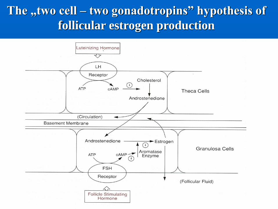
The „two cell – two gonadotropins” hypothesis of
follicular estrogen production
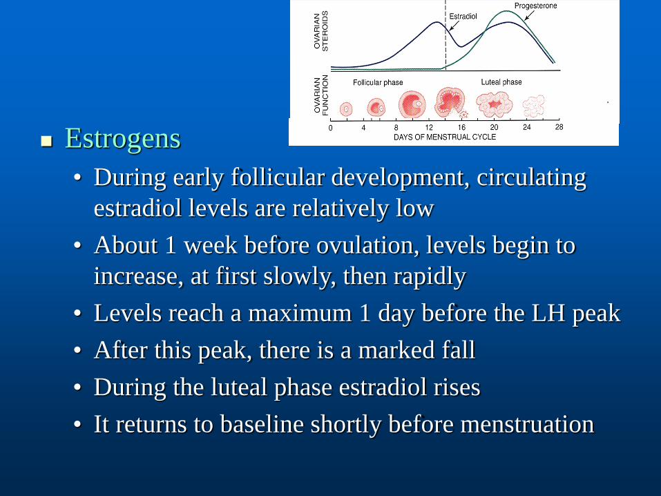
Estrogens
• During early follicular development, circulating
estradiol levels are relatively low
• About 1 week before ovulation, levels begin to
increase, at first slowly, then rapidly
• Levels reach a maximum 1 day before the LH peak
• After this peak, there is a marked fall
• During the luteal phase estradiol rises
• It returns to baseline shortly before menstruation
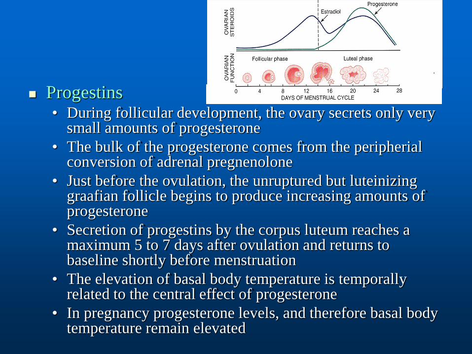
Progestins• During follicular development, the ovary secrets only very
small amounts of progesterone
• The bulk of the progesterone comes from the peripherial conversion of adrenal pregnenolone
• Just before the ovulation, the unruptured but luteinizing graafian follicle begins to produce increasing amounts of progesterone
• Secretion of progestins by the corpus luteum reaches a maximum 5 to 7 days after ovulation and returns to baseline shortly before menstruation
• The elevation of basal body temperature is temporally related to the central effect of progesterone
• In pregnancy progesterone levels, and therefore basal body temperature remain elevated
Androgens
• Both the ovary and the adrenal glands secrete small
amounts of testosterone, but most of it is derived from the
metabolism of androstendione (also secreted by both the
ovary and the adrenal gland)
Serum-binding proteins
• Circulating sex hormones are mostly bound to SHBG or to
serum albumin. The remaining fraction is free
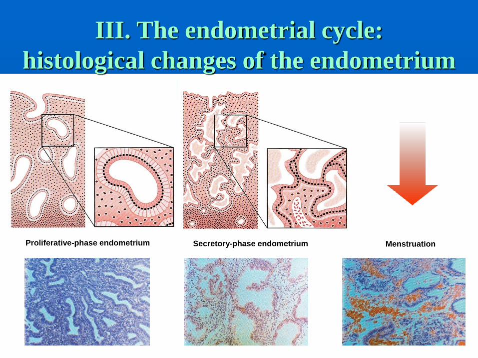
MenstruationSecretory-phase endometriumProliferative-phase endometrium
III. The endometrial cycle:
histological changes of the endometrium
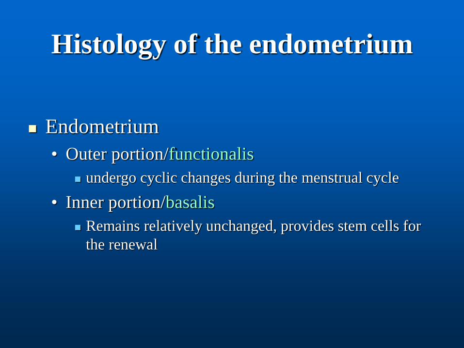
Endometrium
• Outer portion/functionalis
undergo cyclic changes during the menstrual cycle
• Inner portion/basalis
Remains relatively unchanged, provides stem cells for
the renewal
Histology of the endometrium
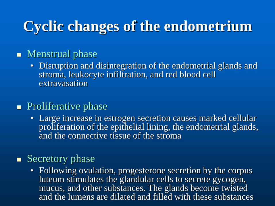
Cyclic changes of the endometrium
Menstrual phase• Disruption and disintegration of the endometrial glands and
stroma, leukocyte infiltration, and red blood cell extravasation
Proliferative phase• Large increase in estrogen secretion causes marked cellular
proliferation of the epithelial lining, the endometrial glands, and the connective tissue of the stroma
Secretory phase• Following ovulation, progesterone secretion by the corpus
luteum stimulates the glandular cells to secrete gycogen, mucus, and other substances. The glands become twisted and the lumens are dilated and filled with these substances
The menstrual cycle
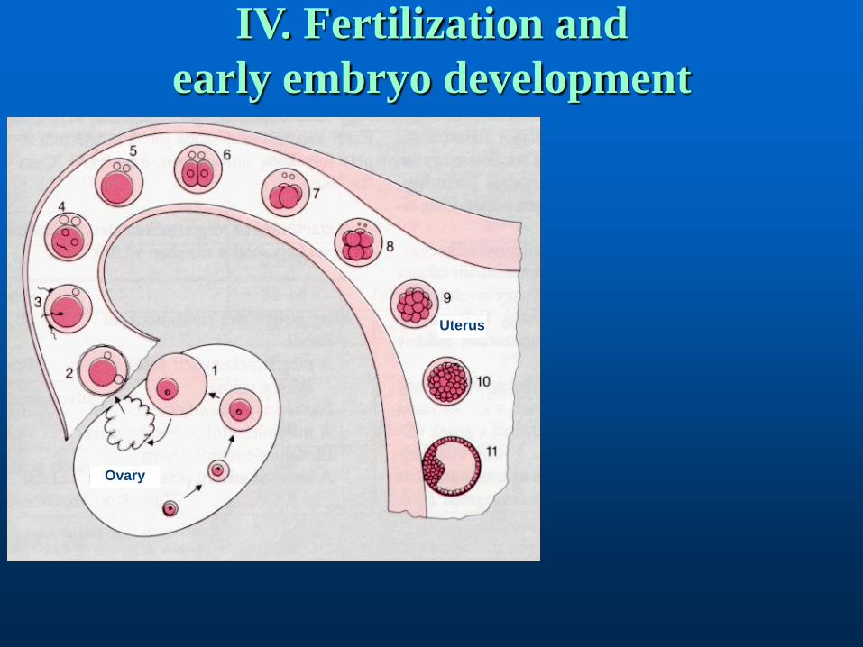
IV. Fertilization and
early embryo development
Uterus
Ovary
Spermatogenesis requires about 74 days
The average ejaculate contains 2 to 5 mL of semem; 20 to 250 million sperm may be deposited in the vagina, >30% of which are morphologically normal. Fewer than 200 sperm achieve proximity to the egg
Only one sperm fertilizes a single egg released at ovulation
Ova are usually fertilized within 12 hours of ovulation
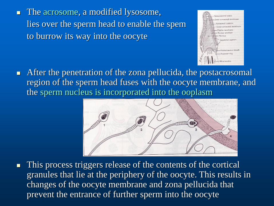
The acrosome, a modified lysosome,
lies over the sperm head to enable the spem
to burrow its way into the oocyte
After the penetration of the zona pellucida, the postacrosomal region of the sperm head fuses with the oocyte membrane, and the sperm nucleus is incorporated into the ooplasm
This process triggers release of the contents of the cortical granules that lie at the periphery of the oocyte. This results in changes of the oocyte membrane and zona pellucida that prevent the entrance of further sperm into the oocyte
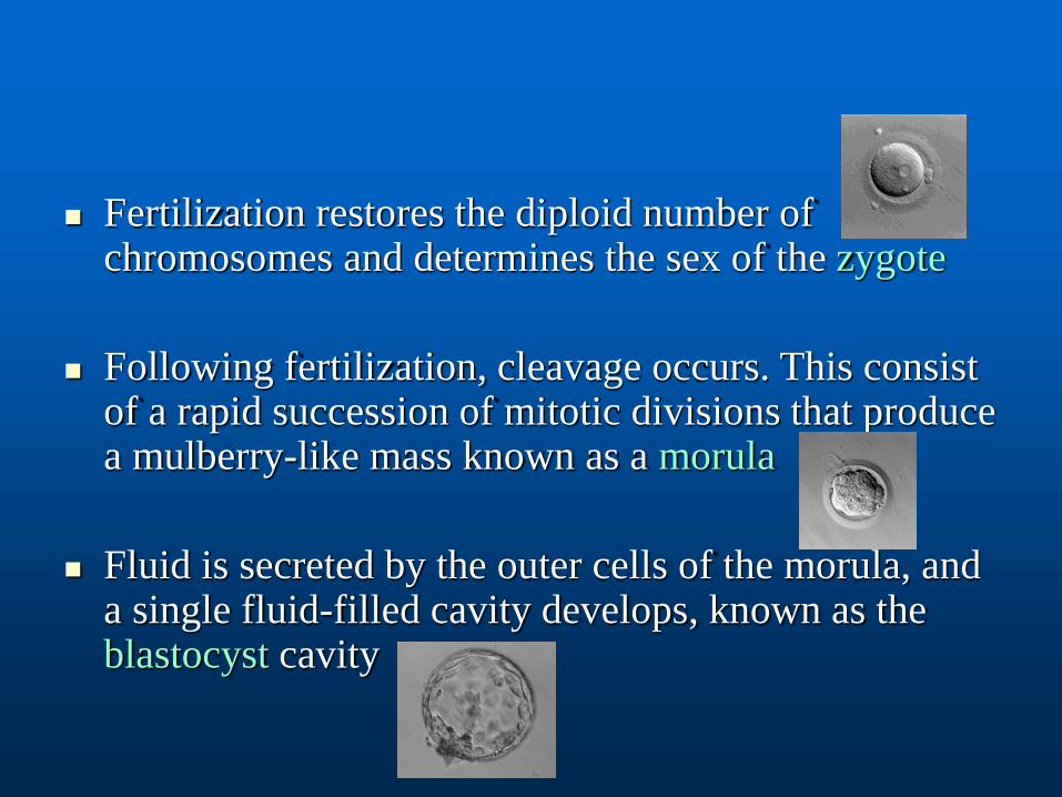
Fertilization restores the diploid number of chromosomes and determines the sex of the zygote
Following fertilization, cleavage occurs. This consist of a rapid succession of mitotic divisions that produce a mulberry-like mass known as a morula
Fluid is secreted by the outer cells of the morula, and a single fluid-filled cavity develops, known as the blastocyst cavity
The fertilized ovum reaches the endometrial cavity about 3 days after ovulation
The wall of the blastocyst facing the uterine lumen consists of a single layer of flattened cells. The thicker opposite wall has two zones: the trophoblastsand the embryoblasts
Between the embryonic disk and the trophoblasts an enclosed space becomes the amniotic cavity
Implantation
Under the influence of progesterone, decidual
changes occur in the endometrium of the pregnant
uterus (endometrial stromal cells enlarge and form
polygonal or round decidual cells)
Decidua thickens: 5-10 mm
As the blastocyst borrows deeper into the
endometrium, the trophoblastic strands branch to
form the solid, primitive villi
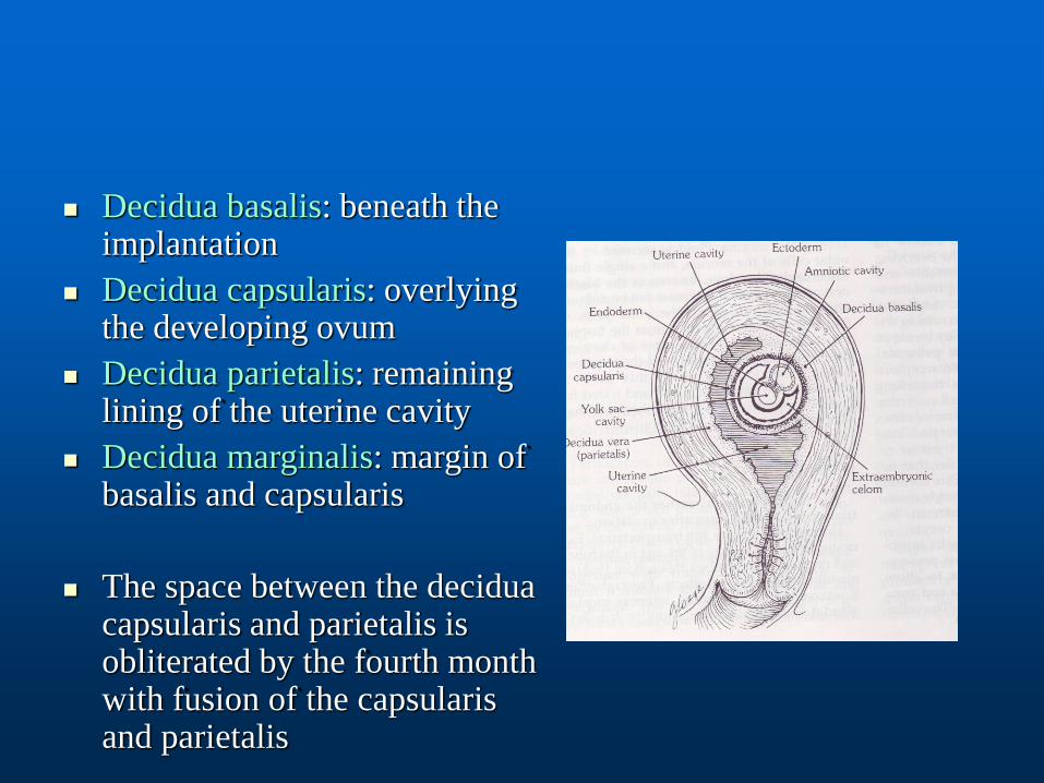
Decidua basalis: beneath the implantation
Decidua capsularis: overlying the developing ovum
Decidua parietalis: remaining lining of the uterine cavity
Decidua marginalis: margin of basalis and capsularis
The space between the decidua capsularis and parietalis is obliterated by the fourth month with fusion of the capsularis and parietalis
Human chorionic gonadotropin (hCG) (coming from
the syncytiotrophoblasts) can be detected from the
21-23rd day of the cycle in the serum and the urine of
a pregnant. hCG has an LH-like activity, it lets the
corpus luteum to keep on functioning
Preconception care
Unplanned pregnancies
Medications – impact on fertility & pregnancy
Family and genetic history (DM, age risk…)
Nutritional assessment
• Underweight low birth weight, prematurity
• Obesity obstetric complications (PIH, GD/DM)
• Balanced nutrition for at least 3 months before conception
• Folic acid 400 μg daily (4 mg if previous Hx of NTD)
Early pregnancy loss
Biochemical pregnancies
(subclinical abortions)
• hCG present in blood 7–10 days after ovulation
• Menstruation occurs when expected
• No gestational sac on US
• Incidence unknown
Spontaneous abortions
• 10–15% of clinically recognized pregnancies
Spontaneous abortions
Threatened abortion
• Vaginal bleeding
• Lower abdominal pain (±)
• Cervix is closed
• 20–25% pregnancy loss
Inevitable (incipient) abortion
• Vaginal bleeding
• Cramp-like lower abdominal pain
• Cervix partially dilated
Spontaneous abortions
Incomplete abortion• Vaginal bleeding
• Cramp-like lower abdominal pain
• Cervix partially dilated
• Passage of products of conception
Complete abortion• Passage of all products of conception
• Uterine contractions & bleeding
• Cervix closes
• Uterus smaller than based on period of amenorrhea
• Symptoms of pregnancy
• Pregnancy test negative
Spontaneous abortions
Missed abortion
• Embryo/fetus died
• Retained in uterus
• Coagulation problems (DIC)
Habitual abortion
• 2 previous spontaneous abortions
Related Documents