Physiology of Optics Dr. Abdul Ahad Shaikh 1

Physiology of Optics Dr. Abdul Ahad Shaikh 1. Objectives Describe the refraction of light as it passes through the eye to the retina, identifying the.
Dec 17, 2015
Welcome message from author
This document is posted to help you gain knowledge. Please leave a comment to let me know what you think about it! Share it to your friends and learn new things together.
Transcript

1
Physiology of Optics
Dr. Abdul Ahad Shaikh

2
Objectives• Describe the refraction of light as it passes
through the eye to the retina, identifying the eye components that account for refraction of light at the center of the eye and away from the center.
• Describe the refractive deficits that account for myopia, hyperopia, presbyopia and astigmatism, and their correction by eyeglasses or contact lenses.
• Describe the mechanism of measuring visual acuity using Snellen chart.

3
Horizontal section of the right eye. AP, anterior pole; PP, posterior pole; VA, visual axis.
Eyeball• Our brain derives knowledge
of the world around us from visual stimuli
• The three tunics of eyeball are;– Outer fibrous tunic made up of
sclera and cornea– Middle vascular tunic made up
of choroid , ciliary body and iris
– Inner nervous tunic called retina covers only posterior 5/6th portion of the eye ball

4
Refraction• Bending of light at an angulated
interface is known as refraction• If the interface between two
media is perpendicular to the beam of light there is no change in the direction of light
• When the interface between two media is angulated the light rays change their direction at the interface
• Refraction depends on two factors– Degree of angulation interface
between two media – Difference in the refractive indices of
two media

5
Optics - Basic Principles
AIR AIRTransparent substance
Refractive index of a transparent substance - ratio of velocity of light in air to the velocity in the substance.
Refractive index
Perpendicular interface - decreased velocity and shorter wavelength.

6
Types of Lenses
• Two types of lenses• Spherical (convex or concave)
refracts the light in all planes • Cylindrical (convex or concave)
refracts the light only in one plane
• The signs of the lens radii indicate whether the corresponding surfaces are convex (R > 0, bulging outwards from the lens) or concave (R < 0, depressed into the lens). If R is infinite, the surface is flat, or has zero curvature, and is said to be planar.

7
Optics - Basic Principles
Refraction
A convex lens focuses light rays while concave lenses diverge parallel rays.
Focal length distance at which parallel rays converge to a focal point

8
Optics - Basic Principles
Parallel rays
Point source
Point source
Parallel rays - considered to be coming from infinity. Light sources closer than infinity (>6 meters) are divergent.
Parallel rays and divergent rays (point source) may converge at the same focal point however, provided the lens changes its convexity.

9
Optics - Basic Principles• Refractive power of a lens -
measured in diopters• In case of a convex lens the
refractive power is equal to 1 divided by its focal length of the lens in meter
• If focal length is 1 m refractive power of the lens is +1D
• The power of concave lens is expressed by comparing it with convex lens
Refractive power of a convex lens = 1/focal length in meters

10
Refractive Interfaces of the Eye
The eye has 4 refractive interfaces• - Air/anterior cornea• - posterior cornea/aqueous
humour• - Ant. Lens/aqueous
humour• - Post. Lens/vitreous
humour

11
The cornea provides most of the refractive power of the eye because its refractive index is markedly different from that of air (1.38 - 1.0 = 0.38)
Refractive power of the optical system of the eye

12
Refractive Surface • Anterior surface of cornea + 48D • Posterior surface of cornea - 4D• Lens of the eye +15D• Maximum refraction occurs at the anterior surface of cornea because;
– Greater curvature– Greater difference between refractive indices
• Refractive power of a lens inside the eye is +15D and outside the eye is +150D
• Reduced eye is schematic representation of the eye considering the eye has a single lens of +59 D placed at a distance of 17 mm from retina (refractive power= 1000/17 )
• The axial length of the eye at birth is approximately 17 mm and reaches approximately 24 mm in adulthood.
• It is typically longer than 24 mm in myopes and shorter than 24 mm in hyperopes.
• The centre of the lens is called nodal point lies 7.08 mm behind the cornea

13
The Eye as a camera
• The human eye is a camera!• Iris - colored annulus with radial muscles
• Pupil - the hole (aperture) whose size is controlled by the iris• What’s the “film”?
– photoreceptor cells (rods and cones) in the retina

14
• The degree to which the details and contours of objects are perceived.
• Defined in terms of the minimum separable i.e. the minimum distance by which two lines can be separated and still be perceived as two lines.
• Determined using Snellen charts. An emmetropic eye has VA of 20/20 or 6/6.
• The design of the VA chart is based on the subtended visual angle. For example, the letters of the 20/20 line of a projected Snellen chart is usually E V O T Z 2. Look at the E
• Each "stroke" of the E subtends an angle of 1 min, or an arc on a circle with a radius of 20 ft (6m). This one-min arc seems the minimum for the retinal photoreceptors to resolve .
Visual Acuity

15
• Visual acuity– determined using Snellen’s chart– V=d/D
• d=distance at which characters are read• D=distance at which characters should be
read
• Subject 6 meters from test chart. Test one eye at a time.
• Normally should be 6/6• The average diameter of the cones
in the fovea of the retina—the central part of the retina, where visionm is most highly developed—is about 1.5 micrometers
Visual acuity – how to measure

16
Visual Acuity
• When visual acuity is below the largest letter on the chart, the reading distance is reduced until the patient can read it. Once the patient is able to read the chart, the letter size and test distance are noted. If the patient is unable to read the chart at any distance, he or she is tested as follows:
Name Abbreviation Definition
Counting Fingers CF Ability to count fingers at a given distance.
Hand Motion HMAbility to distinguish a hand if it is moving or not in front of the patient's face.
Light Perception LP Ability to perceive any light.
No Light Perception NLP Inability to see any light. Total blindness.
17
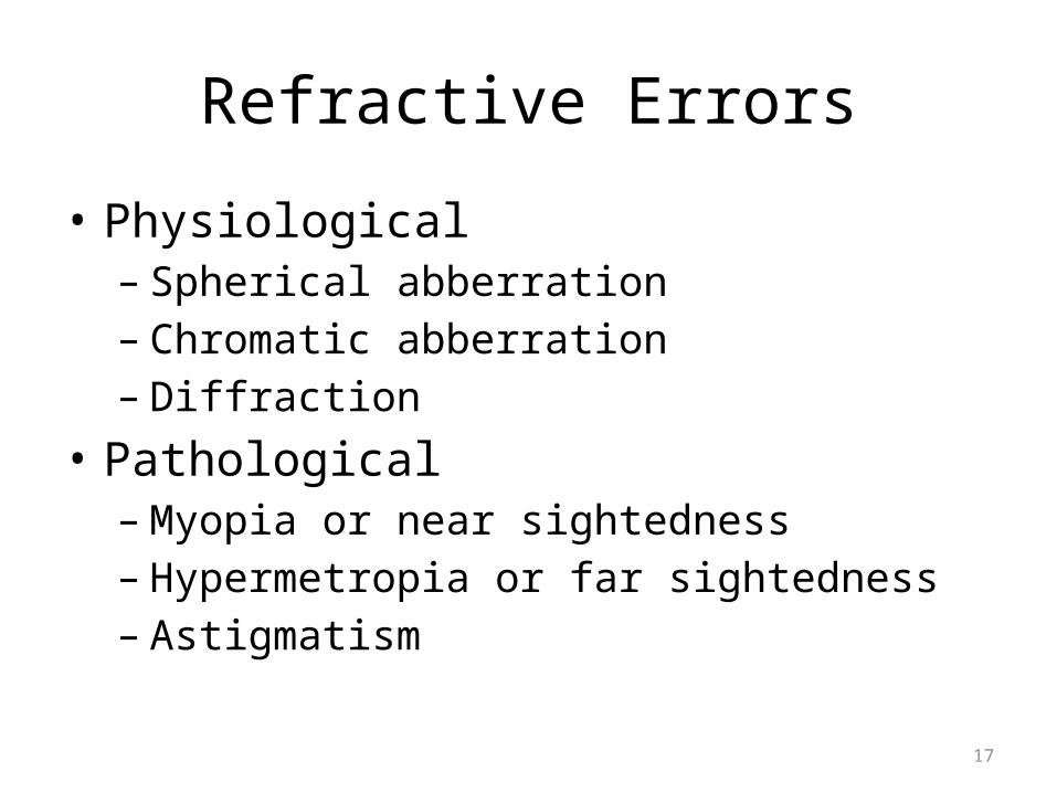
Refractive Errors
• Physiological– Spherical abberration– Chromatic abberration– Diffraction
• Pathological– Myopia or near sightedness– Hypermetropia or far sightedness– Astigmatism

18
Refractive Errors• Physiological
– Spherical Abberration• Parallel rays of light passing through the peripheral portion of
the eye lens do not focus at the same point where rays passing through central portion of the lens focus
– Chromatic Abberration• Refractive power of the lens of the eye is different for
different colors, there fore light rays of differen colors get focused at different distances behind the lens
– Diffraction• Bending of the light rays as they pass over the sharp borders
of the pupil . It interferes with the image formation when pupilary size is small(2.5 -1.5 mm)

19
Errors of refraction
Common defects of the optical system of the eye. In hyperopia, the eyeball is too short and light rays come to a focus behind the retina. A biconvex lens corrects this by adding to the refractive power of the lens of the eye. In myopia, the eyeball is too long and light rays focus in front of the retina. Placing a biconcave lens in front of the eye causes the light rays to diverge slightly before striking the eye, so that they are brought to a focus on the retina.

20
Presbyopia
• With aging, the elasticity of the lens gradually declines• Accommodation of the lens for near vision becomes
progressively less effective• A young person can change power of lens as much as 14 D • By age 40 years accommodation become half• By age of 50 it decreases to 2D or less• The lens become almost non-elastic solid mass and remains
focused permanently at constant distance• It becomes non accommodative for near and far vision• It can be corrected by bifocal glasses with upper segment
focused for far objects and lower segment for near objects

21
• The nearest point to the eye to which an object can be brought to clear focus by accommodation.
Near Point Of Vision
(D) Changes in the ability of the lens to round up (accommodate) with age. The graph also shows how the near point (the closest point to the eye that can be brought into focus) changes. Accommodation, which is an optical measurement of the refractive power of the lens, is given in diopters. (After Westheimer, 1974.)

22
Astigmatism
• Defect in the refractive power of a lens only in one plane of the lens
• The curvature of the cornea is different in one plane from that in another plane giving different degree of refraction in different plane
• Person is asked to look at the chart with one eye normally bars in all directions will be seen clearly

23
AstigmatismDue to uneven curvature of the cornea, causing the visual image in one plane to focus at a different distance from that of the plane at right angles.Corrected using cylindrical lenses.
Astigmatism testing chart - 2 meters away
Are any of the lines clearer than others?

25
Glaucoma
• Degeneration of optic nerve associated with increased intraoccular pressure (IOP) in majority of cases
• Normal intraocular pressure 12 – 20 mmHg with average about 15 mmHg
• Intraocular pressure is measured using a tonometer

26
References
• Guyton and Hall Textbook of Medical Physiology 12th Edition
• Ganong's Review of Medical Physiology, 24th Edition
• Berne & Levy Physiology, 6th Updated Edition
Thank you
Related Documents