Photosynthetic electron transport activity in heat-treated barley leaves: The role of internal alternative electron donors to photosystem II Szilvia Z. Tóth a,b, ⁎ , Gert Schansker a , Győző Garab b , Reto J. Strasser a a Laboratory of Bioenergetics, University of Geneva, Chemin des Embrouchis 10, CH-1254 Jussy, Geneva, Switzerland b Institute of Plant Biology, Biological Research Center of the Hungarian Academy of Sciences, PO Box 521, H-6701 Szeged, Hungary Received 10 October 2006; received in revised form 5 February 2007; accepted 26 February 2007 Available online 3 March 2007 Abstract Electron transport processes were investigated in barley leaves in which the oxygen-evolution was fully inhibited by a heat pulse (48 °C, 40 s). Under these circumstances, the K peak (∼ F 400 μs ) appears in the chl a fluorescence (OJIP) transient reflecting partial Q A reduction, which is due to a stable charge separation resulting from the donation of one electron by tyrozine Z. Following the K peak additional fluorescence increase (indicating Q A − accumulation) occurs in the 0.2–2 s time range. Using simultaneous chl a fluorescence and 820 nm transmission measurements it is demonstrated that this Q A − accumulation is due to naturally occurring alternative electron sources that donate electrons to the donor side of photosystem II. Chl a fluorescence data obtained with 5-ms light pulses (double flashes spaced 2.3–500 ms apart, and trains of several hundred flashes spaced by 100 or 200 ms) show that the electron donation occurs from a large pool with t 1/2 ∼ 30 ms. This alternative electron donor is most probably ascorbate. © 2007 Elsevier B.V. All rights reserved. Keywords: Alternative electron donor; Chlorophyll a fluorescence; Heat pulse; OJIP-transient; Photosystem II; 820 nm transmission 1. Introduction Exposure of plants to high temperatures has two opposite effects on the photosynthetic electron transport chain. Photo- system I (PSI) is stimulated by heat (measured as the reduction rate of P700 + ), which is related to an enhanced reduction of the plastoquinone (PQ) pool by either ferredoxin (Fd) or NADPH [1,2]. In contrast, photosystem II (PSII), and especially the oxygen evolving complex (OEC) is inactivated at relatively low temperatures (e.g. [3,4]). A further heat-induced effect is a shift of the redox equilibrium between Q A and Q B , increasing the residence time of an electron on Q A relative to Q B [5–7] that could lead to back flow of electrons from the PQ-pool to PSII [8,9]. The chl a fluorescence (OJIP) transient is a very sensitive indicator of photosynthetic electron transport processes. Light- dependent changes in the chl a fluorescence yield are thought to be determined by changes in the redox state of Q A (reviewed in e.g. [10–14]). At the same time, it has been shown that the OJIP transient reflects the reduction of the photosynthetic electron transport chain (e.g. [15]). The OJ phase is the photochemical part of the transient. Its kinetics depend strongly on the light intensity [16,17]. The JI phase has been shown to parallel the reduction of the PQ-pool [18] and the IP phase depends on PSI activity representing the reduction of the ferredoxin (Fd) pool in Biochimica et Biophysica Acta 1767 (2007) 295 – 305 www.elsevier.com/locate/bbabio Abbreviations: Asc, ascorbate; DCMU, 3-(3′,4′-dichlorophenyl)-1,1- dimethylurea; Fd, ferredoxin; F 0 , initial fluorescence (measured at 20 μs), all PSII RCs are open; F J , fluorescence intensity at ∼ 3 ms (at 3000 μmol photons m − 2 s − 1 ); F M , maximum fluorescence, all PSII RCs are closed; FNR, ferredoxin-NADP + -reductase; F P , maximum measured fluorescence intensity; FR, far-red light; I 820nm , transmission at 820 nm; I, step in the OJIP transient at 30 ms; J, step in the OJIP transient at ∼ 3 ms (at 3000 μmol photons m − 2 s − 1 ); K, peak in the OJIP transient at ∼ 400 μs (at 3000 μmol photons m − 2 s − 1 ); MV, methylviologen, 1,1′-dimethyl-4,4′-bipyridinium-dichloride; OEC, oxygen evolving complex; OJIP, chl a fluorescence transient defined by the names of its intermediate steps; PpBQ, phenyl-p-benzoquinone; P680, primary electron donor of PSII; P700, primary electron donor of PSI; PC, plastocyanin; PQ, plastoquinone; PSI, photosystem I; PSII, photosystem II; Q A , primary electron acceptor quinone of PSII; Q B , secondary electron acceptor quinone of PSII; Tyr Z , tyrozine Z; φ Po , maximum quantum yield for primary photochemistry ⁎ Corresponding author. Institute of Plant Biology, Biological Research Center of the Hungarian Academy of Sciences, PO Box 521, H-6701 Szeged, Hungary. Tel.: +36 62 433 131; fax: +36 62 433 434. E-mail address: [email protected] (S.Z. Tóth). 0005-2728/$ - see front matter © 2007 Elsevier B.V. All rights reserved. doi:10.1016/j.bbabio.2007.02.019

Welcome message from author
This document is posted to help you gain knowledge. Please leave a comment to let me know what you think about it! Share it to your friends and learn new things together.
Transcript
a 1767 (2007) 295–305www.elsevier.com/locate/bbabio
Biochimica et Biophysica Act
Photosynthetic electron transport activity in heat-treated barley leaves:The role of internal alternative electron donors to photosystem II
Szilvia Z. Tóth a,b,⁎, Gert Schansker a, Győző Garab b, Reto J. Strasser a
a Laboratory of Bioenergetics, University of Geneva, Chemin des Embrouchis 10, CH-1254 Jussy, Geneva, Switzerlandb Institute of Plant Biology, Biological Research Center of the Hungarian Academy of Sciences, PO Box 521, H-6701 Szeged, Hungary
Received 10 October 2006; received in revised form 5 February 2007; accepted 26 February 2007Available online 3 March 2007
Abstract
Electron transport processes were investigated in barley leaves in which the oxygen-evolution was fully inhibited by a heat pulse (48 °C, 40 s).Under these circumstances, the K peak (∼F400 μs) appears in the chl a fluorescence (OJIP) transient reflecting partial QA reduction, which is dueto a stable charge separation resulting from the donation of one electron by tyrozine Z. Following the K peak additional fluorescence increase(indicating QA
− accumulation) occurs in the 0.2–2 s time range. Using simultaneous chl a fluorescence and 820 nm transmission measurements itis demonstrated that this QA
− accumulation is due to naturally occurring alternative electron sources that donate electrons to the donor side ofphotosystem II. Chl a fluorescence data obtained with 5-ms light pulses (double flashes spaced 2.3–500 ms apart, and trains of several hundredflashes spaced by 100 or 200 ms) show that the electron donation occurs from a large pool with t1/2 ∼30 ms. This alternative electron donor ismost probably ascorbate.© 2007 Elsevier B.V. All rights reserved.
Keywords: Alternative electron donor; Chlorophyll a fluorescence; Heat pulse; OJIP-transient; Photosystem II; 820 nm transmission
1. Introduction
Exposure of plants to high temperatures has two oppositeeffects on the photosynthetic electron transport chain. Photo-
Abbreviations: Asc, ascorbate; DCMU, 3-(3′,4′-dichlorophenyl)-1,1-dimethylurea; Fd, ferredoxin; F0, initial fluorescence (measured at 20 μs),all PSII RCs are open; FJ, fluorescence intensity at ∼3 ms (at 3000 μmolphotons m−2 s−1); FM, maximum fluorescence, all PSII RCs are closed; FNR,ferredoxin-NADP+-reductase; FP, maximum measured fluorescence intensity;FR, far-red light; I820nm, transmission at 820 nm; I, step in the OJIP transient at30 ms; J, step in the OJIP transient at∼3 ms (at 3000 μmol photons m−2 s−1); K,peak in the OJIP transient at ∼400 μs (at 3000 μmol photons m−2 s−1); MV,methylviologen, 1,1′-dimethyl-4,4′-bipyridinium-dichloride; OEC, oxygenevolving complex; OJIP, chl a fluorescence transient defined by the names ofits intermediate steps; PpBQ, phenyl-p-benzoquinone; P680, primary electrondonor of PSII; P700, primary electron donor of PSI; PC, plastocyanin; PQ,plastoquinone; PSI, photosystem I; PSII, photosystem II; QA, primary electronacceptor quinone of PSII; QB, secondary electron acceptor quinone of PSII; TyrZ,tyrozine Z; φPo, maximum quantum yield for primary photochemistry⁎ Corresponding author. Institute of Plant Biology, Biological Research Center
of the Hungarian Academy of Sciences, PO Box 521, H-6701 Szeged, Hungary.Tel.: +36 62 433 131; fax: +36 62 433 434.
E-mail address: [email protected] (S.Z. Tóth).
0005-2728/$ - see front matter © 2007 Elsevier B.V. All rights reserved.doi:10.1016/j.bbabio.2007.02.019
system I (PSI) is stimulated by heat (measured as the reductionrate of P700+), which is related to an enhanced reduction of theplastoquinone (PQ) pool by either ferredoxin (Fd) or NADPH[1,2]. In contrast, photosystem II (PSII), and especially theoxygen evolving complex (OEC) is inactivated at relatively lowtemperatures (e.g. [3,4]). A further heat-induced effect is a shiftof the redox equilibrium between QA and QB, increasing theresidence time of an electron on QA relative to QB [5–7] thatcould lead to back flow of electrons from the PQ-pool to PSII[8,9].
The chl a fluorescence (OJIP) transient is a very sensitiveindicator of photosynthetic electron transport processes. Light-dependent changes in the chl a fluorescence yield are thought tobe determined by changes in the redox state of QA (reviewed ine.g. [10–14]). At the same time, it has been shown that the OJIPtransient reflects the reduction of the photosynthetic electrontransport chain (e.g. [15]). The OJ phase is the photochemicalpart of the transient. Its kinetics depend strongly on the lightintensity [16,17]. The JI phase has been shown to parallel thereduction of the PQ-pool [18] and the IP phase depends on PSIactivity representing the reduction of the ferredoxin (Fd) pool in
296 S.Z. Tóth et al. / Biochimica et Biophysica Acta 1767 (2007) 295–305
the presence of inactive ferredoxin-NADP+-reductase (FNR)[15]. The shape of the OJIP transient drastically changes after aheat treatment, especially if oxygen evolution is completelyinhibited: the J and I steps disappear and the K peak develops at300–400 μs if the measurements are carried out at a standard,3000 μmol photons m−2 s−1 light intensity [19]. After the Kpeak the fluorescence intensity decreases. The K peak isthought to result from the only stable charge separationpossible when the OEC is completely destroyed [20,21]because TyrZ can provide only one electron. However, inleaves in the 0.2–2 s time range another peak appears in thefluorescence transient [22,23], which might represent a secondphase of QA
− accumulation.In this study, we investigated the origin of the fluorescence
increase in the 0.2–2 s time range in leaves in which the oxygenevolution was fully inhibited with a heat pulse (by immersingthe leaves for 40 s in a water bath of 48 °C). This treatmentinduces typical heat stress effects, but causes virtually no visiblesymptoms and secondary effects [23]. (A similar method, astrong but short pulse instead of a longer, weaker treatment hasbeen used for inducing UV-A stress in order to separateinactivation of PSII and repair [24].) We used simultaneous820 nm transmission and chl a fluorescence inductionmeasurements, and also recorded chl a fluorescence transientsinduced by short (5 ms), repetitive light pulses. It was found thatalternative electron sources provide electrons to the donor sideof PSII with t1/2 ∼30 ms. We also show that the electronsoriginate from a large pool of donors (probably ascorbatemolecules).
2. Materials and methods
2.1. Plant material and growth conditions
Measurements were carried out on 7-day-old barley seedlings (Hordeumvulgare cv. Triangel). Plants were grown in a greenhouse where the temperaturewas 20–25 °C during the day and ∼14 °C at night.
2.2. Heat pulse
The heat pulse was given by submerging the leaves of barley seedlings. Thepots with seedlings were turned upside down into a water bath of 48 °C (if notstated otherwise) for 40 s. In the case of the DCMU+heat treatment and theMV+heat treatment leaf segments were used and submerged in 48 °C water for40 s. In our previous study [23], we used a heat pulse of 50 °C to inactivate allOECs. In this case, 48 °C was enough to achieve the same effect, which isprobably due to the lower growth temperature of the plants. We verified that thisheat pulse fully inactivated the OECs. The B thermoluminescence band [25] (thatis a reflection of recombination reactions betweenQB
− and the S2 or S3 states of theOEC) was completely eliminated; this was confirmed by oxygen-evolutionmeasurements carried out on thylakoids isolated from heat-treated leaves usingPpBQ (phenyl-p-benzoquinone) as an electron acceptor (data not shown).
2.3. DCMU+heat treatment
Leaf segments of ∼2 cm long were submerged in DCMU (3-(3′,4′-dichlorophenyl)-1,1-dimethylurea) solution (200 μM, containing 1% ethanol todissolve DCMU) or 1% ethanol for 5 h. Half of the leaf segments treated withDCMU and half of the leaf segments kept in 1% ethanol were given a heat pulse(48 °C for 40 s in water bath). There were four types of samples: DCMU-treated,DCMU+heat-treated, heat-treated (treated in 1% ethanol before the heat
treatment) and control (1% ethanol treated). It was important to carry out theDCMU treatment before the heat pulse otherwise the F0 value of leaves becamevery high (∼78% of FM, data not shown).
2.4. Methylviologen (MV)+heat treatment
For MV treatments, a 200 μM MV (1,1′-dimethyl-4,4′-bipyridinium-dichloride, Fluka) solution was applied to both sides of the leaf with a fine brush.The leaves were not detached and the plants were kept overnight in completedarkness before the measurements (see also [15]). Following the MV treatment,leaf segments were prepared and half of them were given a heat pulse (48 °C,40 s). Control plants were kept overnight in darkness.
2.5. Chl a fluorescence and 820 nm transmission measurements
Samples were kept in darkness after the heat pulse and measured within 30–60 min. In the case of fluorescence measurements carried out by the Handy-PEA(Hansatech Instruments Ltd, UK), the leaf samples were illuminated withcontinuous red light (650 nm peak wavelength, the spectral line half-width is22 nm; the light emitted by the LEDs is cut-off at 700 nm by NIR short-passfilters) at the indicated light intensities for 1–5 s. The light was provided by anarray of three light-emitting diodes focused on a circle of 5 mm diameter of thesample surface. The first reliably measured point of the fluorescence transient isat 20 μs, which was taken as F0. Chl a fluorescence emission is measured atwavelengths longer than 700 nm (Kopp Corning RG-9 long-pass filter).
For some of the experiments (Figs. 2 and 3) we made use of a “fast” versionof the Handy-PEA instrument that allows reducing the pulse length to 300 μs.Either two, or trains of several hundred 5-ms light pulses were applied. Thedark-time intervals between the light pulses were 2.3, 9.6, 16.9, 31.5, 38.8, 53.4,75.3, 100, 200 or 500 ms.
A special high-intensity measuring head was used to induce chl afluorescence at a maximum intensity of 15000 μmol photons m−2 s−1 producedby a single 650 nm LED (Hansatech Instruments Ltd, UK).
820 nm transmission measurements parallel to chl a fluorescence werecarried out using a PEA Senior instrument (Hansatech Instruments Ltd, UK).The excitation light intensity used was 1800 μmol photons m−2 s−1, producedby four 650 nm LEDs. Far-red light (718 nm peak wavelength, 200 μmolphotons m−2 s−1 light intensity) and modulated far-red measuring light (820 nmpeak wavelength) were provided by two additional LEDs. Further technicaldetails and applications are described in several papers by Schansker et al.[15,26,27].
3. Results
3.1. Chl a fluorescence transients of heat-treated barley leavesmeasured at different light intensities
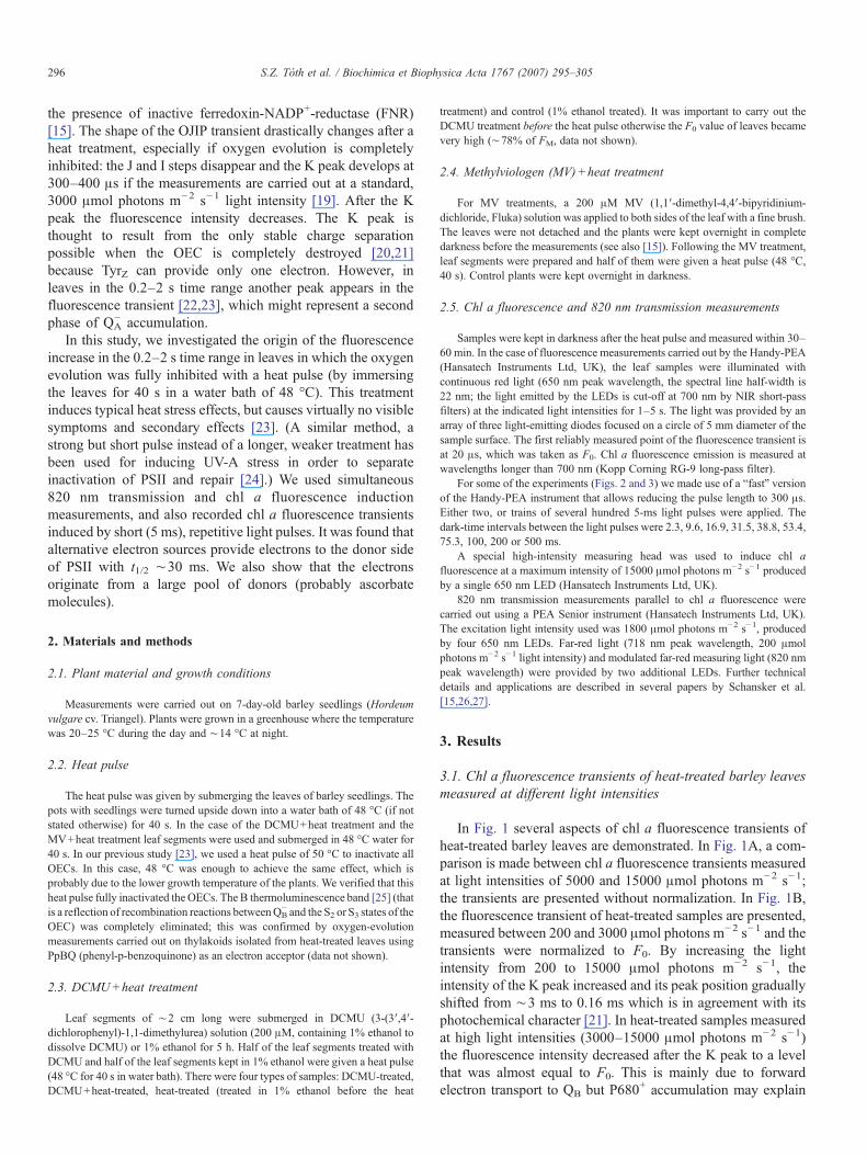
In Fig. 1 several aspects of chl a fluorescence transients ofheat-treated barley leaves are demonstrated. In Fig. 1A, a com-parison is made between chl a fluorescence transients measuredat light intensities of 5000 and 15000 μmol photons m−2 s−1;the transients are presented without normalization. In Fig. 1B,the fluorescence transient of heat-treated samples are presented,measured between 200 and 3000 μmol photons m−2 s−1 and thetransients were normalized to F0. By increasing the lightintensity from 200 to 15000 μmol photons m−2 s−1, theintensity of the K peak increased and its peak position graduallyshifted from ∼3 ms to 0.16 ms which is in agreement with itsphotochemical character [21]. In heat-treated samples measuredat high light intensities (3000–15000 μmol photons m−2 s−1)the fluorescence intensity decreased after the K peak to a levelthat was almost equal to F0. This is mainly due to forwardelectron transport to QB but P680+ accumulation may explain
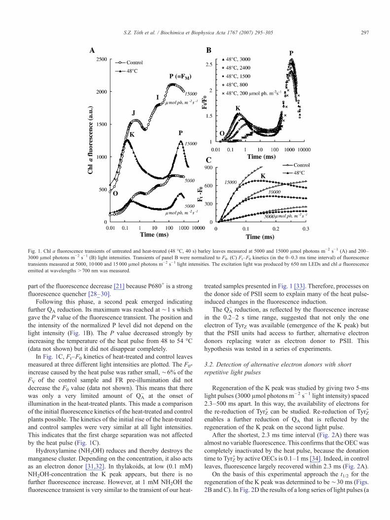
Fig. 1. Chl a fluorescence transients of untreated and heat-treated (48 °C, 40 s) barley leaves measured at 5000 and 15000 μmol photons m−2 s−1 (A) and 200–3000 μmol photons m−2 s−1 (B) light intensities. Transients of panel B were normalized to F0. (C) Ft–F0 kinetics (in the 0–0.3 ms time interval) of fluorescencetransients measured at 5000, 10 000 and 15000 μmol photons m−2 s−1 light intensities. The excitation light was produced by 650 nm LEDs and chl a fluorescenceemitted at wavelengths >700 nm was measured.
297S.Z. Tóth et al. / Biochimica et Biophysica Acta 1767 (2007) 295–305
part of the fluorescence decrease [21] because P680+ is a strongfluorescence quencher [28–30].
Following this phase, a second peak emerged indicatingfurther QA reduction. Its maximum was reached at ∼1 s whichgave the P value of the fluorescence transient. The position andthe intensity of the normalized P level did not depend on thelight intensity (Fig. 1B). The P value decreased strongly byincreasing the temperature of the heat pulse from 48 to 54 °C(data not shown) but it did not disappear completely.
In Fig. 1C, Ft–F0 kinetics of heat-treated and control leavesmeasured at three different light intensities are plotted. The F0-increase caused by the heat pulse was rather small, ∼6% of theFV of the control sample and FR pre-illumination did notdecrease the F0 value (data not shown). This means that therewas only a very limited amount of QA
− at the onset ofillumination in the heat-treated plants. This made a comparisonof the initial fluorescence kinetics of the heat-treated and controlplants possible. The kinetics of the initial rise of the heat-treatedand control samples were very similar at all light intensities.This indicates that the first charge separation was not affectedby the heat pulse (Fig. 1C).
Hydroxylamine (NH2OH) reduces and thereby destroys themanganese cluster. Depending on the concentration, it also actsas an electron donor [31,32]. In thylakoids, at low (0.1 mM)NH2OH-concentration the K peak appears, but there is nofurther fluorescence increase. However, at 1 mM NH2OH thefluorescence transient is very similar to the transient of our heat-
treated samples presented in Fig. 1 [33]. Therefore, processes onthe donor side of PSII seem to explain many of the heat pulse-induced changes in the fluorescence induction.
The QA− reduction, as reflected by the fluorescence increase
in the 0.2–2 s time range, suggested that not only the oneelectron of TyrZ was available (emergence of the K peak) butthat the PSII units had access to further, alternative electrondonors replacing water as electron donor to PSII. Thishypothesis was tested in a series of experiments.
3.2. Detection of alternative electron donors with shortrepetitive light pulses
Regeneration of the K peak was studied by giving two 5-mslight pulses (3000 μmol photons m−2 s−1 light intensity) spaced2.3–500 ms apart. In this way, the availability of electrons forthe re-reduction of TyrZ
+ can be studied. Re-reduction of TyrZ+
enables a further reduction of QA that is reflected by theregeneration of the K peak on the second light pulse.
After the shortest, 2.3 ms time interval (Fig. 2A) there wasalmost no variable fluorescence. This confirms that the OECwascompletely inactivated by the heat pulse, because the donationtime to TyrZ
+ by active OECs is 0.1–1 ms [34]. Indeed, in controlleaves, fluorescence largely recovered within 2.3 ms (Fig. 2A).
On the basis of this experimental approach the t1/2 for theregeneration of the K peak was determined to be ∼30 ms (Figs.2B and C). In Fig. 2D the results of a long series of light pulses (a
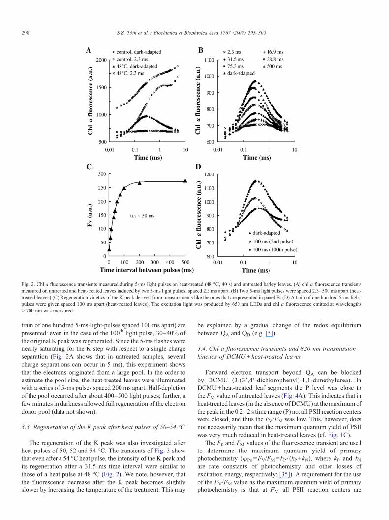
Fig. 2. Chl a fluorescence transients measured during 5-ms light pulses on heat-treated (48 °C, 40 s) and untreated barley leaves. (A) chl a fluorescence transientsmeasured on untreated and heat-treated leaves induced by two 5-ms light pulses, spaced 2.3 ms apart. (B) Two 5-ms light pulses were spaced 2.3–500 ms apart (heat-treated leaves) (C) Regeneration kinetics of the K peak derived from measurements like the ones that are presented in panel B. (D) A train of one hundred 5-ms light-pulses were given spaced 100 ms apart (heat-treated leaves). The excitation light was produced by 650 nm LEDs and chl a fluorescence emitted at wavelengths>700 nm was measured.
298 S.Z. Tóth et al. / Biochimica et Biophysica Acta 1767 (2007) 295–305
train of one hundred 5-ms-light-pulses spaced 100 ms apart) arepresented: even in the case of the 100th light pulse, 30–40% ofthe original K peak was regenerated. Since the 5-ms flashes werenearly saturating for the K step with respect to a single chargeseparation (Fig. 2A shows that in untreated samples, severalcharge separations can occur in 5 ms), this experiment showsthat the electrons originated from a large pool. In the order toestimate the pool size, the heat-treated leaves were illuminatedwith a series of 5-ms pulses spaced 200 ms apart. Half-depletionof the pool occurred after about 400–500 light pulses; further, afewminutes in darkness allowed full regeneration of the electrondonor pool (data not shown).
3.3. Regeneration of the K peak after heat pulses of 50–54 °C
The regeneration of the K peak was also investigated afterheat pulses of 50, 52 and 54 °C. The transients of Fig. 3 showthat even after a 54 °C heat pulse, the intensity of the K peak andits regeneration after a 31.5 ms time interval were similar tothose of a heat pulse at 48 °C (Fig. 2). We note, however, thatthe fluorescence decrease after the K peak becomes slightlyslower by increasing the temperature of the treatment. This may
be explained by a gradual change of the redox equilibriumbetween QA and QB (e.g. [5]).
3.4. Chl a fluorescence transients and 820 nm transmissionkinetics of DCMU+heat-treated leaves
Forward electron transport beyond QA can be blockedby DCMU (3-(3′,4′-dichlorophenyl)-1,1-dimethylurea). InDCMU+heat-treated leaf segments the P level was close tothe FM value of untreated leaves (Fig. 4A). This indicates that inheat-treated leaves (in the absence of DCMU) at themaximum ofthe peak in the 0.2–2 s time range (P) not all PSII reaction centerswere closed, and thus the FV/FM was low. This, however, doesnot necessarily mean that the maximum quantum yield of PSIIwas very much reduced in heat-treated leaves (cf. Fig. 1C).
The F0 and FM values of the fluorescence transient are usedto determine the maximum quantum yield of primaryphotochemistry (φPo=FV/FM=kP / (kP+kN), where kP and kNare rate constants of photochemistry and other losses ofexcitation energy, respectively; [35]). A requirement for the useof the FV/FM value as the maximum quantum yield of primaryphotochemistry is that at FM all PSII reaction centers are

Fig. 3. Regeneration of the K peak after heat treatments at 50 °C (A), 52 °C (B) or 54 °C (C). The light pulses were 5 ms long and the time interval was 31.5 ms betweenthe first (dark-adapted) and second light pulse. The excitation light was produced by 650 nm LEDs and chl a fluorescence emitted at wavelengths >700 nm wasmeasured.
299S.Z. Tóth et al. / Biochimica et Biophysica Acta 1767 (2007) 295–305
closed. This requirement is not fulfilled in the case of heat-treated samples (Fig. 4A). For this reason, the FV/FM valueshould not be used as an indicator of maximum quantum yieldof primary photochemistry in such samples.
We also note that in the case of non-heat-treated samples theFM values were almost identical in the presence or absence ofDCMU. This confirms an earlier study in which it was shownthat PQ-pool quenching does not occur if DCMU is allowed todiffuse slowly into intact leaves [36].
The shape of the chl a fluorescence transient of DCMU+heat-treated leaf segments was biphasic: a large fast and asmaller slow phase can be distinguished. This may suggest thatthe phenomenon is related to α and β centers [37]. However, thet1/2 of this slow phase is in the order of tens of ms, which ismuch slower than the closure of the β centers at this lightintensity [38]. We explain the very slow phase of the heat-treated samples by assuming that charge recombinationsbetween QA
− and TyrZ+ slow down the reduction of QA
−. These
Fig. 4. Chl a fluorescence (A) and simultaneously measured 820 nm transmission kbarley leaves. The excitation light was produced by 650 nm LEDs and chl a fluores
recombination reactions may be gradually blocked when thealternative electron donors re-reduce TyrZ
+ (Fig. 2).In Fig. 4, simultaneously measured Chl a fluorescence
transients (Fig. 4A) and 820 nm transmission kinetics (Fig. 4B)are presented. Transmission changes at 820 nm reflect the redoxstates of P700, plastocyanin (PC) and Fd, with Fd being a smallcomponent [15,26,39]. Transmission changes were used here tomonitor electron flow through PSI. In control leaves, a red lightpulse induced an initial oxidation of P700 and PC (decrease inthe transmission level) followed by a re-reduction whenelectrons arrive from PSII [26,36]. Re-reduction occurred alsoin the case of heat-treated samples, but it happened later (after20 ms for the control and 80 ms for the heat-treated samples)and it was also much slower (t1/2 of ∼40 ms for the control and∼140 ms for the heat-treated leaves). This indicates that in thecase of the heat-treated samples the electrons responsible forP700+ and PC+ reduction arrived much more slowly than in thecontrol samples. This is in agreement with the data showing that
inetics (B) of heat-treated, DCMU-treated, DCMU+heat-treated and untreatedcence emitted at wavelengths >700 nm was measured.
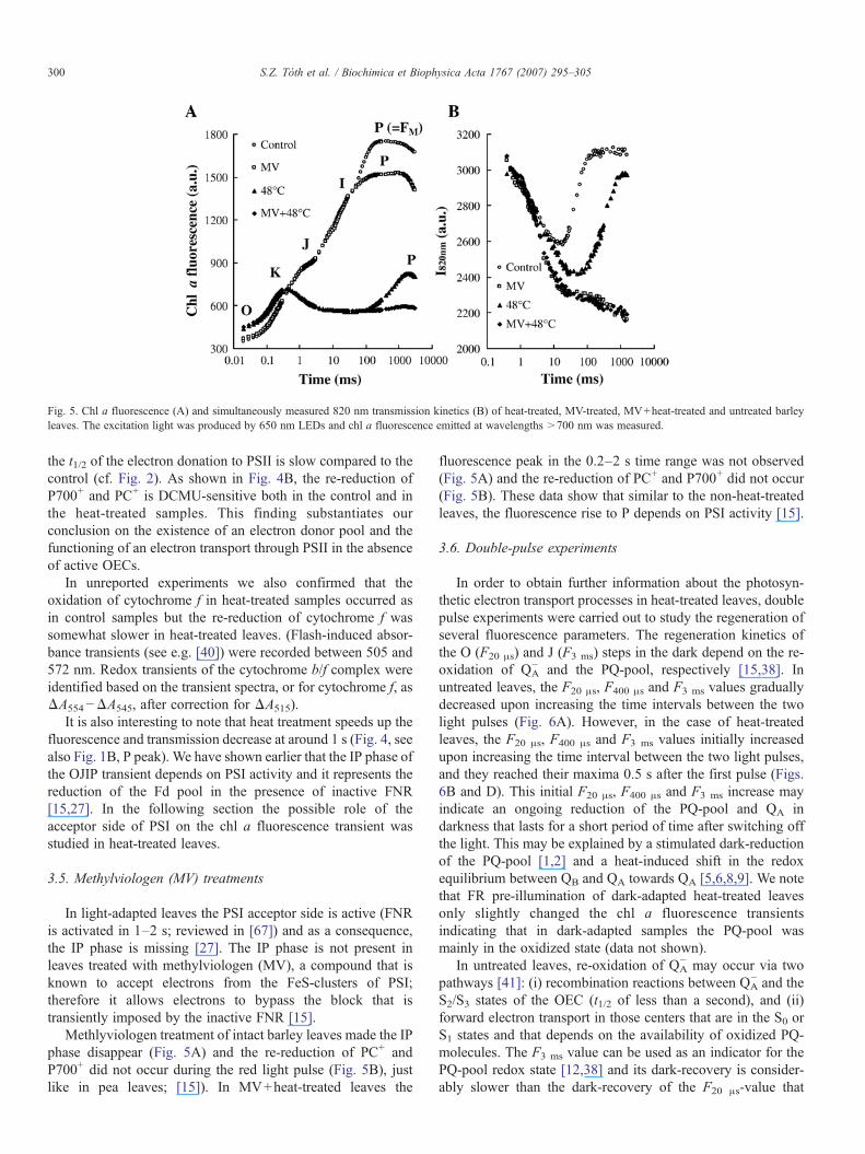
Fig. 5. Chl a fluorescence (A) and simultaneously measured 820 nm transmission kinetics (B) of heat-treated, MV-treated, MV+heat-treated and untreated barleyleaves. The excitation light was produced by 650 nm LEDs and chl a fluorescence emitted at wavelengths >700 nm was measured.
300 S.Z. Tóth et al. / Biochimica et Biophysica Acta 1767 (2007) 295–305
the t1/2 of the electron donation to PSII is slow compared to thecontrol (cf. Fig. 2). As shown in Fig. 4B, the re-reduction ofP700+ and PC+ is DCMU-sensitive both in the control and inthe heat-treated samples. This finding substantiates ourconclusion on the existence of an electron donor pool and thefunctioning of an electron transport through PSII in the absenceof active OECs.
In unreported experiments we also confirmed that theoxidation of cytochrome f in heat-treated samples occurred asin control samples but the re-reduction of cytochrome f wassomewhat slower in heat-treated leaves. (Flash-induced absor-bance transients (see e.g. [40]) were recorded between 505 and572 nm. Redox transients of the cytochrome b/f complex wereidentified based on the transient spectra, or for cytochrome f, asΔA554−ΔA545, after correction for ΔA515).
It is also interesting to note that heat treatment speeds up thefluorescence and transmission decrease at around 1 s (Fig. 4, seealso Fig. 1B, P peak). We have shown earlier that the IP phase ofthe OJIP transient depends on PSI activity and it represents thereduction of the Fd pool in the presence of inactive FNR[15,27]. In the following section the possible role of theacceptor side of PSI on the chl a fluorescence transient wasstudied in heat-treated leaves.
3.5. Methylviologen (MV) treatments
In light-adapted leaves the PSI acceptor side is active (FNRis activated in 1–2 s; reviewed in [67]) and as a consequence,the IP phase is missing [27]. The IP phase is not present inleaves treated with methylviologen (MV), a compound that isknown to accept electrons from the FeS-clusters of PSI;therefore it allows electrons to bypass the block that istransiently imposed by the inactive FNR [15].
Methlyviologen treatment of intact barley leaves made the IPphase disappear (Fig. 5A) and the re-reduction of PC+ andP700+ did not occur during the red light pulse (Fig. 5B), justlike in pea leaves; [15]). In MV+heat-treated leaves the
fluorescence peak in the 0.2–2 s time range was not observed(Fig. 5A) and the re-reduction of PC+ and P700+ did not occur(Fig. 5B). These data show that similar to the non-heat-treatedleaves, the fluorescence rise to P depends on PSI activity [15].
3.6. Double-pulse experiments
In order to obtain further information about the photosyn-thetic electron transport processes in heat-treated leaves, doublepulse experiments were carried out to study the regeneration ofseveral fluorescence parameters. The regeneration kinetics ofthe O (F20 μs) and J (F3 ms) steps in the dark depend on the re-oxidation of QA
− and the PQ-pool, respectively [15,38]. Inuntreated leaves, the F20 μs, F400 μs and F3 ms values graduallydecreased upon increasing the time intervals between the twolight pulses (Fig. 6A). However, in the case of heat-treatedleaves, the F20 μs, F400 μs and F3 ms values initially increasedupon increasing the time interval between the two light pulses,and they reached their maxima 0.5 s after the first pulse (Figs.6B and D). This initial F20 μs, F400 μs and F3 ms increase mayindicate an ongoing reduction of the PQ-pool and QA indarkness that lasts for a short period of time after switching offthe light. This may be explained by a stimulated dark-reductionof the PQ-pool [1,2] and a heat-induced shift in the redoxequilibrium between QB and QA towards QA [5,6,8,9]. We notethat FR pre-illumination of dark-adapted heat-treated leavesonly slightly changed the chl a fluorescence transientsindicating that in dark-adapted samples the PQ-pool wasmainly in the oxidized state (data not shown).
In untreated leaves, re-oxidation of QA− may occur via two
pathways [41]: (i) recombination reactions between QA− and the
S2/S3 states of the OEC (t1/2 of less than a second), and (ii)forward electron transport in those centers that are in the S0 orS1 states and that depends on the availability of oxidized PQ-molecules. The F3 ms value can be used as an indicator for thePQ-pool redox state [12,38] and its dark-recovery is consider-ably slower than the dark-recovery of the F20 μs-value that
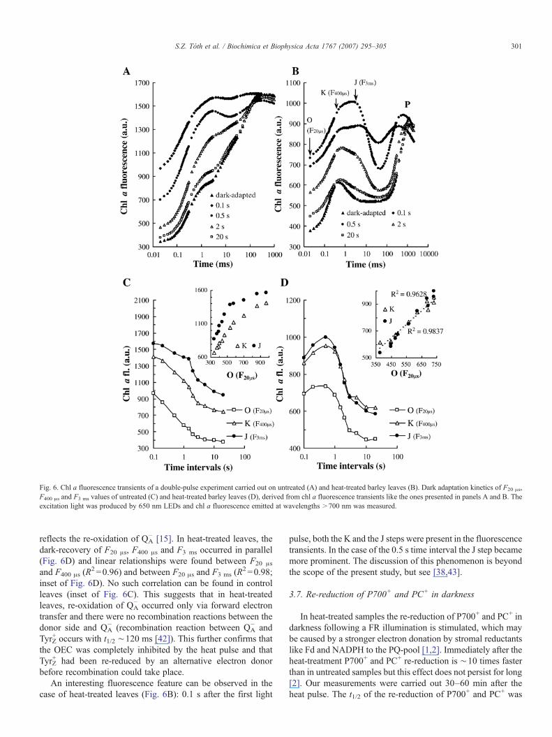
Fig. 6. Chl a fluorescence transients of a double-pulse experiment carried out on untreated (A) and heat-treated barley leaves (B). Dark adaptation kinetics of F20 μs,F400 μs and F3 ms values of untreated (C) and heat-treated barley leaves (D), derived from chl a fluorescence transients like the ones presented in panels A and B. Theexcitation light was produced by 650 nm LEDs and chl a fluorescence emitted at wavelengths >700 nm was measured.
301S.Z. Tóth et al. / Biochimica et Biophysica Acta 1767 (2007) 295–305
reflects the re-oxidation of QA− [15]. In heat-treated leaves, the
dark-recovery of F20 μs, F400 μs and F3 ms occurred in parallel(Fig. 6D) and linear relationships were found between F20 μs
and F400 μs (R2 =0.96) and between F20 μs and F3 ms (R
2 =0.98;inset of Fig. 6D). No such correlation can be found in controlleaves (inset of Fig. 6C). This suggests that in heat-treatedleaves, re-oxidation of QA
− occurred only via forward electrontransfer and there were no recombination reactions between thedonor side and QA
− (recombination reaction between QA− and
TyrZ+ occurs with t1/2 ∼120 ms [42]). This further confirms that
the OEC was completely inhibited by the heat pulse and thatTyrZ
+ had been re-reduced by an alternative electron donorbefore recombination could take place.
An interesting fluorescence feature can be observed in thecase of heat-treated leaves (Fig. 6B): 0.1 s after the first light
pulse, both the K and the J steps were present in the fluorescencetransients. In the case of the 0.5 s time interval the J step becamemore prominent. The discussion of this phenomenon is beyondthe scope of the present study, but see [38,43].
3.7. Re-reduction of P700+ and PC+ in darkness
In heat-treated samples the re-reduction of P700+ and PC+ indarkness following a FR illumination is stimulated, which maybe caused by a stronger electron donation by stromal reductantslike Fd and NADPH to the PQ-pool [1,2]. Immediately after theheat-treatment P700+ and PC+ re-reduction is ∼10 times fasterthan in untreated samples but this effect does not persist for long[2]. Our measurements were carried out 30–60 min after theheat pulse. The t1/2 of the re-reduction of P700+ and PC+ was
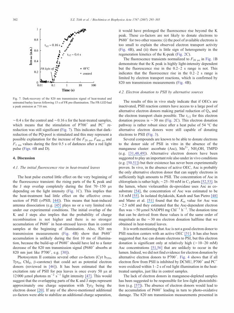
Fig. 7. Dark-recovery of the 820 nm transmission signal of heat-treated anduntreated barley leaves following 15 s of FR pre-illumination. The FR-LED hada peak emission at 718 nm.
302 S.Z. Tóth et al. / Biochimica et Biophysica Acta 1767 (2007) 295–305
∼0.4 s for the control and ∼0.16 s for the heat-treated samples,which means that the stimulation of P700+ and PC+ re-reduction was still significant (Fig. 7). This indicates that dark-reduction of the PQ-pool is stimulated and this may represent apossible explanation for the increase of the F20 μs, F400 μs andF3 ms values during the first 0.5 s of darkness after a red lightpulse (Figs. 6B and D).
4. Discussion
4.1. The initial fluorescence rise in heat-treated leaves
The heat pulse exerted little effect on the very beginning ofthe fluorescence transient: the rising parts of the K peak andthe J step overlap completely during the first 70–150 μsdepending on the light intensity (Fig. 1C). This implies thatthe heat-treatment had little effect on the effective cross-section of PSII (σPSII; [44]). This means that heat-inducedantenna dissociation (e.g. [45] plays no or a very limited roleunder our experimental conditions. The initial overlap of theK and J steps also implies that the probability of chargerecombination is not higher and there is no strongeraccumulation of P680+ in heat-stressed leaves than in controlsamples at the beginning of illumination. Also, 820 nmtransmission measurements (Fig. 4B) show that P680+
accumulation is unlikely during the first 10 ms of illumina-tion, because the build-up of P680+ should have led to a fasterdecrease of the 820 nm transmission signal (P680+ absorbs at820 nm just like P700+, e.g. [30]).
Photosystem II contains several other co-factors (Cyt b559,TyrD, ChlZ, β-carotene) that could act as potential electrondonors (reviewed in [46]). It has been estimated that theexcitation rate of PSII for pea leaves is once every 50 μs at12 000 μmol photons m−2 s−1 light intensity [47]. This wouldsuggest that the overlapping parts of the K and J steps representapproximately one charge separation with TyrZ being theelectron donor [20]. If any of the above-mentioned additionalco-factors were able to stabilize an additional charge separation,
it would have prolonged the fluorescence rise beyond the Kpeak. These co-factors are not likely to donate electrons toP680+ for two other reasons: (i) the pool of available electrons istoo small to explain the observed electron transport activity(Fig. 4B), and (ii) there is little sign of heterogeneity in theregeneration kinetics of the K-peak (Fig. 2C).
The fluorescence transients normalized to F20 μs in Fig. 1Bdemonstrate that the K peak is highly light-intensity dependentbut the fluorescence rise in the 0.2–2 s range is not. Thisindicates that the fluorescence rise in the 0.2–2 s range islimited by electron transport reactions, which is confirmed by820 nm transmission measurements (Fig. 4B).
4.2. Electron donation to PSII by alternative sources
The results of this in vivo study indicate that if OECs areinactivated, PSII reaction centers have access to a large pool ofalternative electron donors making partial reduction of QA andthe electron transport chain possible. The t1/2 for this electrondonation process is ∼30 ms (Fig. 2C). This electron donationpathway is rather robust since after a heat pulse of 54 °C thealternative electron donors were still capable of donatingelectrons to PSII (Fig. 3).
Several compounds are known to be able to donate electronsto the donor side of PSII in vitro in the absence of themanganese cluster: ascorbate (Asc), Mn2+, NH2OH, TMPD(e.g. [31,48,49]). Alternative electron donors have beensuggested to play an important role also under in vivo conditions(e.g. [50,51]) but their existence has never been experimentallyproven. In vivo, in the absence of active OEC, Asc is probablythe only alternative electron donor that can supply electrons insufficiently high amounts to PSII. The concentration of Asc inchloroplasts is rather high;∼25–50 mM in C3 plants [52,53]. Inthe lumen, where violaxanthin de-epoxidase uses Asc as co-substrate [54], the concentration of Asc was estimated to be3.8 mM [55]. In isolated thylakoids, Katoh and San Pietro [48]and Mano et al. [51] found that the Km value for Asc was∼2.5 mM and they estimated that the Asc-dependent electronflux was ∼50 μmol NADPH mg Chl−1 h−1. The donation timethat can be derived from these values is of the same order ofmagnitude as the ∼30 ms electron donation halftime that weobtained in heat-treated leaves.
It is worth mentioning that Asc is not a good electron donor toPSII reaction centers with an active OEC [51]. It has also beensuggested that Asc can donate electrons to PSI, but this electrondonation is significant only at relatively high (>10–20 mM)Asc concentrations [51,56] that are unlikely to occur in thelumen. Indeed, we did not find evidence for electron donation byalternative electron donors to P700+. Fig. 4 shows that if allelectron flow from PSII is inhibited by DCMU, P700+ and PC+
were oxidized within 1–2 s of red light illumination in the heat-treated samples, just like in control samples.
The lack of electron donors in manganese-depleted sampleshas been suggested to be responsible for low-light photoinhibi-tion (e.g. [57]). The absence of electron donors would lead tothe accumulation of P680+ leading in turn to photo-oxidativedamage. The 820 nm transmission measurements presented in
303S.Z. Tóth et al. / Biochimica et Biophysica Acta 1767 (2007) 295–305
Fig. 4B demonstrate that alternative donors can maintain alimited electron flow through PSII. In low light, this might beenough to protect PSI and PSII reaction centers from furtherdamage even in the total absence of active OECs. Thisprotection can be physiologically relevant given the fact thatunder natural stress conditions the inactivation of OECs occursstochastically. Indeed, it has been observed that there is nodifference between the photosynthetic activities of plants kept indarkness or in relatively low light (100 μmol photons m−2 s−1)for 30 min following a heat pulse [23]. However, a few hours oflight treatment following a heat pulse makes the K peakdisappear indicating a further damage (or degradation) of thePSII reaction centers [23].
Photosystem I itself is heat-tolerant but exposure of isolatedchloroplasts to heat in the light causes a selective destruction ofPSI pigments. It has been shown that it is a direct consequenceof the interruption of the electron flow originating from PSII[58]. Therefore, it is conceivable that the alternative electrondonors of PSII may have some protective role also for PSI.
A pathway for the maintenance of the Asc-concentration in thelumen has been proposed by Mano et al. [51,59]. Upon donatingelectrons Asc is oxidized to monodehydroascorbate (MDA). TheMDA radicals in the lumen are then disproportionated to Asc anddehydroascorbate (DHA). DHA is able to diffuse to the stromawhere DHA reductase catalyses the reduction of DHA byglutathione. Asc− is then thought to be transferred to the lumen bya postulated transporter [51,59] or by diffusion [55].
4.3. Dark-reduction of the PQ-pool
The heat-induced stimulation of electron transport aroundPSI has been in the focus of research on heat-stress effects onthe thylakoid membrane over the last 10 years. Re-reduction ofP700+ and PC+ after a far-red pulse was shown to be stimulatedup to 10-fold after a heat treatment [2]. However, it was alsoshown that this stimulatory effect disappears quite quickly afterthe heat-treatment [2]. The only two-fold stimulation that isobserved here (Fig. 7) could very well be due to the fact that ourmeasurements were carried out ∼30 min after the treatment. Astimulated non-photochemical reduction of the PQ-pool can bedue to an increased cyclic electron transport around PSI [60–63]or to increased chlororespiration [64,65]. However, FR-preillumination did not decrease the F0 value of dark-adapted,heat-treated leaves (data not shown). Therefore it is likely thatthe PQ-pool and QA were mainly in the oxidized state and thestimulation of chlororespiration was not significant.
Two cyclic PSI electron transport pathways have beendescribed in higher plants: (i) a NADPH-dehydrogenase (NDH)dependent pathway [66], and (ii) a ferredoxin-PQ-reductase(FQR) dependent pathway [60]. Of these, the NDH pathwaydoes not seem to be relevant to our study, because the dataindicate (Fig. 4) that in heat-treated barley leaves FNR isinactive in darkness, as expected (e.g. [67]). Havaux et al. [68]have shown that the relative contribution of the two cyclicpathways varies from one plant species to another. In barleyprotoplasts, it has been shown that the slow component of thefar-red light stimulated luminescence (related to PSI cyclic
electron transfer) is sensitive to antimycin A [69]. Therefore it islikely that in non-stressed barley plants the FQR pathway isactive. In other words, a heat-stimulation of the FQR-pathwaycould explain the two-fold increase in P700+ and PC+ reduction(Fig. 7) and the increase of F20 μs, F400 μs and F3 ms that lasts for∼0.5 s after switching off the light (Fig. 6). However, the820 nm transmission measurements (Fig. 4B) demonstrate thatin the light the electron transport activity of heat-treated leaveswas very sensitive to DCMU. This means that cyclic PSIelectron transport contributed very little and the photosyntheticelectron transport was dominated by PSII-mediated electrontransport that originates from the alternative electron donors ofPSII.
Acknowledgements
We thank also the Hansatech Instruments Ltd for theconstruction of the high intensity measuring head and the“fast” Handy-PEA instrument. We thank Milán Szabó for thehelp with the cytochrome f flash-induced absorbance measure-ments and Dr. László Kovács for the help with thermolumines-cence and oxygen-evolution measurements. This work was inpart supported by the Hungarian Research Foundation (OTKAK 63252). Dr. S.Z. Tóth received support from the NationalOffice for Research and Technology (NKTH, Hungary, ÖvegesFellowship Program).
References
[1] M. Havaux, Short-term responses of photosystem I to heat stress,Photosynth. Res. 47 (1996) 85–97.
[2] N.G. Bukhov, C. Wiese, S. Neimanis, U. Heber, Heat sensitivity ofchloroplasts and leaves: leakage of protons from thylakoids andreversible activation of cyclic electron transport, Photosynth. Res. 59(1999) 81–93.
[3] I. Enami, M. Kitamura, T. Tomo, Y. Isokawa, H. Ohta, S. Katoh, Is theprimary cause of thermal inactivation of oxygen evolution in spinach PSIImembranes release of the extrinsic 33 kDa protein or of Mn? Biochim.Biophys. Acta 1186 (1994) 52–58.
[4] Y. Yamane, Y. Kashino, H. Koike, K. Satoh, Effects of high temperatureson the photosynthetic systems in spinach: oxygen-evolving activities,fluorescence characteristics and the denaturation process, Photosynth. Res.57 (1998) 51–59.
[5] J.-M. Ducruet, Y. Lemoine, Increased heat sensitivity of the photosyntheticapparatus in triazine-resistant biotypes from different plant species, PlantCell Physiol. 26 (1985) 419–429.
[6] M. Havaux, Comparison of atrazine-resistant and -susceptible biotypes ofSenecio vulgaris L.: effects of high temperatures on the in vivophotosynthetic electron transfer in intact leaves, J. Exp. Bot. 40 (1989)849–854.
[7] J.-M. Ducruet, Relation between the heat-induced increase of F0
fluorescence and a shift in the electronic equilibrium at the acceptor sideof photosystem 2, Photosynthetica 37 (1999) 335–338.
[8] L.A. Sazanov, P.A. Burrows, P.J. Nixon, The chloroplast Ndh complexmediates the dark reduction of the plastoquinone pool in response to heatstress in tobacco leaves, FEBS Lett. 429 (1998) 115–118.
[9] Y. Yamane, T. Shikanai, Y. Kashino, H. Koike, K. Satoh, Reduction of QA
in the dark: another cause of fluorescence F0 increases by hightemperatures in higher plants, Photosynth. Res. 63 (2000) 23–34.
[10] G. Samson, O. Prášil, B. Yaakoubd, Photochemical and thermal phases ofchlorophyll a fluorescence, Photosynthetica 37 (1999) 163–182.
[11] Govindjee, Chlorophyll a fluorescence: a bit of basics and history, in: G.C.
304 S.Z. Tóth et al. / Biochimica et Biophysica Acta 1767 (2007) 295–305
Papageorgiou, Govindjee (Eds.), Chlorophyll a Fluorescence: A Signatureof Photosynthesis, Advances in Photosynthesis and Respiration, vol. 19,Sringer, Dordrecht, 2004, pp. 1–42.
[12] R.J. Strasser, A. Srivastava, M. Tsimilli-Michael, Analysis of thechlorophyll a fluorescence transient, in: G.C. Papageorgiou, Govindjee(Eds.), Chlorophyll a Fluorescence: A Signature of Photosynthesis,Advances in Photosynthesis and Respiration, vol. 19, Sringer, Dordrecht,2004, pp. 321–362.
[13] U. Schreiber, Pulse-amplitude-modulation (PAM) fluorometry andsaturation pulse method: an overview, in: G.C. Papageorgiou, Govindjee(Eds.), Chlorophyll a Fluorescence: A Signature of Photosynthesis,Advances in Photosynthesis and Respiration, vol. 19, Springer,Dordrecht, 2004, pp. 279–319.
[14] D. Lazár, The polyphasic chlorophyll a fluorescence rise measured underhigh intensity of exciting light, Funct. Plant Biol. 33 (2006) 9–30.
[15] G. Schansker, S.Z. Tóth, R.J. Strasser, Methylviologen and dibromothymo-quinone treatments of pea leaves reveal the role of photosystem I in the Chl afluorescence rise OJIP, Biochim. Biophys. Acta 1706 (2005) 250–261.
[16] C. Neubauer, U. Schreiber, The polyphasic rise of chlorophyll fluorescenceupon onset of strong illumination: I. Saturation characteristics and partialcontrol by the photosystem II acceptor side, Z. Naturforsch. 42c (1987)1246–1254.
[17] R.J. Strasser, A. Srivastava, Govindjee, Polyphasic chlorophyll afluorescence transient in plants and cyanobacteria, Photochem. Photobiol.61 (1995) 32–42.
[18] U. Schreiber, C. Neubauer, C. Klughammer, Devices and methods forroom-temperature fluorescence analysis, Philos. Trans. R. Soc. Lond. B323 (1989) 241–251.
[19] A. Srivastava, B. Guissé, H. Greppin, R.J. Strasser, Regulation of antennastructure and electron transport in Photosystem II of Pisum sativum underelevated temperature probed by the fast polyphasic chlorophyll-afluorescence transient: OKJIP, Biochim. Biophys. Acta 1320 (1997)95–106.
[20] B.R. Velthuys, J. Amesz, Charge accumulation at the reducing side ofsystem 2 of photosynthesis, Biochim. Biophys. Acta 333 (1974) 85–94.
[21] B.J. Strasser, Donor side capacity of photosystem II probed bychlorophyll-a fluorescence transients, Photosynth. Res. 52 (1997)147–155.
[22] D. Lazár, P. Ilík, High-temperature induced chlorophyll fluorescencechanges in barley leaves. Comparison of the critical temperaturesdetermined from fluorescence induction and from fluorescence tempera-ture curve, Plant Sci. 124 (1997) 159–164.
[23] S.Z. Tóth, G. Schansker, J. Kissimon, L. Kovács, G. Garab, R.J. Strasser,Biophysical studies of photosystem II-related recovery processes after aheat pulse in barley seedlings (Hordeum vulgare L.), J. Plant Physiol. 162(2005) 181–194.
[24] O. Zsiros, S.I. Allakhverdiev, S. Higashi, M. Watanabe, Y. Nishiyama,N. Murata, Very strong UV-A light temporally separates the photo-inhibition of photosystem II into light-induced inactivation and repair,Biochim. Biophys. Acta 1757 (2006) 123–129.
[25] I. Vass, Y. Inoue, Thermoluminescence in the study of photosystem two,in: J. Barber (Ed.), The Photosystems: Structure, Function and MolecularBiology, Topics in Photosynthesis, vol. 11, Elsevier, Amsterdam, TheNetherlands, pp. 259–294.
[26] G. Schansker, A. Srivastava, W. Govindjee, R.J. Strasser, Characterizationof the 820-nm transmission signal paralleling the chlorophyll afluorescence rise (OJIP) in pea leaves, Funct. Plant Biol. 30 (2003)785–796.
[27] G. Schansker, S.Z. Tóth, R.J. Strasser, Dark-recovery of the Chl afluorescence transient (OJIP) after light adaptation: the qT-component ofnon-photochemical quenching is related to an activated photosystem Iacceptor side, Biochim. Biophys. Acta 1757 (2006) 787–797.
[28] D. Mauzerall, Light-induced fluorescence changes in Chlorella, and theprimary photoreactions for the production of oxygen, Proc. Natl. Acad.Sci. U. S. A. 69 (1972) 1358–1362.
[29] W.L. Butler, On the primary nature of fluorescence yield changesassociated with photosynthesis, Proc. Natl. Acad. Sci. U. S. A. 69(1972) 3420–3422.
[30] R. Steffen, H.-J. Eckert, A.A. Kelly, P. Dörmann, G. Renger, Investigationson the reaction pattern of photosystem II in leaves from Arabidopsisthaliana by time-resolved fluorometric analysis, Biochemistry 44 (2005)3123–3133.
[31] B. Bouges, Action de faibles concentrations d'hydroxylamine surl'émission d'oxygène des algues Chlorella et des chloroplastes d'épinards,Biochim. Biophys. Acta 234 (1971) 103–112.
[32] G.M. Cheniae, I.F. Martin, Effects of hydroxylamine on photosystem II: I.Factors affecting the decay of O2 evolution, Plant Physiol. 47 (1971)568–575.
[33] A.A. Bulychev, W.J. Vredenberg, Modulation of photosystem IIchlorophyll fluorescence by electrogenic events generated by photosystemI, Bioelectrochemistry 54 (2001) 157–168.
[34] G.T. Babcock, R.E. Blankenship, K. Sauer, Reaction kinetics for positivecharge accumulation on the water side of chloroplast photosystem II,FEBS Lett. 61 (1976) 286–289.
[35] M. Kitajima, W.L. Butler, Quenching of chlorophyll fluorescence andprimary photochemistry in chloroplasts by dibromothymoquinone,Biochim. Biophys. Acta 376 (1975) 105–115.
[36] S.Z. Tóth, G. Schansker, R.J. Strasser, In intact leaves, the maximumfluorescence level (FM) is independent of the redox state of theplastoquinone pool: a DCMU inhibition study, Biochim. Biophys. Acta1708 (2005) 275–282.
[37] A. Melis, P.H. Homann, Heterogeneity of the photochemical centers insystem II of chloroplasts, Photochem. Photobiol. 23 (1976) 343–350.
[38] S.Z. Tóth, Analysis and application of the fast chl a fluorescence (OJIP)transient complemented with simultaneous 820 nm transmission measure-ments, Doctoral thesis, N° 3741, University of Geneva, Switzerland, 2006,Available at: http://www.unige.ch/cyberdocuments/theses2006/TothSZ/meta.html.
[39] C. Klughammer, U. Schreiber, Analysis of light-induced absorbancechanges in the near-infrared spectral region: I. Characterization of variouscomponents in isolated chloroplasts, Z. Naturforsch. 46c (1991)233–244.
[40] C. Büchel, G. Garab, Electrochromic absorbance changes in thechlorophyll-c-containing alga Pleurochloris meiringensis (Xanthophy-ceae), Photosynth. Res. 43 (1995) 49–56.
[41] V. Petrouleas, A.R. Crofts, The iron–quinone acceptor complex, in: T.J.Wydrzynski, K. Satoh (Eds.), Photosystem II: The Light-Driven Water:Plastoquinone Oxidoreductase, Advances in Photosynthesis and Respira-tion, vol. 22, Springer, Dordrecht, The Netherlands, 2005, pp. 177–206.
[42] J.P. Dekker, H.J. van Gorkom, M. Brok, L. Ouwehand, Opticalcharacterization of photosystem II electron donors, Biochim. Biophys.Acta 764 (1984) 301–309.
[43] B. Guissé, A. Srivastava, R.J. Strasser, The polyphasic rise of thechlorophyll a fluorescence (O–K–J–I–P) in heat-stressed leaves, Archs.Sci. Genève 48 (1995) 147–160.
[44] A.C. Ley, D. Mauzerall, Absolute absorbance cross sections forPhotosystem II and the minimum quantum requirement for photosynthesisin Chlorella vulgaris, Biochim. Biophys. Acta 680 (1982) 95–106.
[45] U. Schreiber, P.A. Armond, Heat-induced changes of chlorophyllfluorescence in isolated chloroplasts and related heat-damaged at thepigment level, Biochim. Biophys. Acta 502 (1978) 138–151.
[46] P. Faller, C. Fufezan, A.W. Rutherford, Side-path electron donors:cytochrome b559, chlorophyll Z and β-carotene, in: T.J. Wydrzynski,K. Satoh (Eds.), Photosystem II: The Light-Driven Water:PlastoquinoneOxidoreductase, Advances in Photosynthesis and Respiration, vol. 22,Springer, Dordrecht, The Netherlands, 2005, pp. 347–365.
[47] U. Schreiber, C. Neubauer, O2-dependent electron flow, membraneenergization and the mechanism of non-photochemical quenching ofchlorophyll fluorescence, Photosynth. Res. 25 (1990) 279–293.
[48] S. Katoh, A. San Pietro, Ascorbate-supported NADP photoreduction byheated Euglena chloroplasts, Arch. Biochem. Biophys. 122 (1967)144–152.
[49] T. Yamashita, W.L. Butler, Photoreduction and photophosphorylation withTRIS-washed chloroplasts, Plant Physiol. 43 (1968) 1978–1986.
[50] J.A. De Ronde, W.A. Cress, G.H.J. Krüger, R.J. Strasser, J. Van Staden,Photosynthetic response of transgenic soybean plants, containing an
305S.Z. Tóth et al. / Biochimica et Biophysica Acta 1767 (2007) 295–305
Arabidopsis P5CR gene, during heat and drought stress, J. Plant Physiol.161 (2004) 1211–1224.
[51] J. Mano, É. Hideg, K. Asada, Ascorbate in thylakoid lumen functions as analternative electron donor to photosystem II and to photosystem I, Arch.Biochem. Biophys. 429 (2004) 71–80.
[52] M. Eskling, H.-E. Åkerlund, Changes in the quantities of violaxanthin de-epoxidase, xanthophylls and ascorbate in spinach upon shift from low tohigh light, Photosynth. Res. 57 (1998) 41–50.
[53] N. Smirnoff, Ascorbate biosynthesis and function in photoprotection,Philos. Trans. R. Soc. Lond., B 355 (2000) 1455–1464.
[54] C.E. Bratt, P.-E. Arvidsson, A. Carlsson, H.-E. Åkerlund, Regulation ofviolaxanthin de-epoxidase activity by pH and ascorbate concentration,Photosynth. Res. 45 (1995) 169–175.
[55] C.H. Foyer, M. Lelandais, A comparison of the relative rates of transport ofascorbate and glucose across the thylakoid, chloroplast and plasmalemmamembranes of pea leaf mesophyll cells, J. Plant Physiol. 148 (1996) 391–398.
[56] G. Hauska, Artificial acceptors and donors, in: A. Trebst, M. Avron (Eds.),Encyclopedia of Plant Physiology, New Series, vol. 5, Springer-Verlag,Berlin, 1977, pp. 251–265.
[57] D.J. Blubaugh, M. Atamian, G.T. Babcock, J.H. Golbeck, G.M. Cheniae,Photoinhibition of hydroxylamine-extracted photosystem II membranes:Identification of the sites of photodamage, Biochemistry 30 (1991)7586–7597.
[58] W.P. Williams, A. Sen, D.C. Fork, Selective photobleaching of PSI-relatedchlorophylls in heat-stressed pea chloroplasts, Photosynth. Res. 10 (1986)75–92.
[59] J. Mano, T. Ushimaru, K. Asada, Ascorbate in thylakoid lumen as anendogenous electron donor to photosystem II: Protection of thylakoidsfrom photoinhibition and regeneration of ascorbate in stroma bydehydroascorbate reductase, Photosynth. Res. 53 (1997) 197–204.
[60] D.S. Bendall, R.S. Manasse, Cyclic photophosphorylation and electrontransport, Biochim. Biophys. Acta 1229 (1995) 23–38.
[61] T. Joët, L. Cournac, G. Peltier, M. Havaux, Cyclic electron flow aroundphotosystem I in C3 plants. In vivo control by the redox state ofchloroplasts and involvement of the NADH-dehydrogenase complex,Plant Physiol. 128 (2002) 760–769.
[62] P. Joliot, A. Joliot, Cyclic electron transfer in plant leaf, Proc. Natl. Acad.Sci. U. S. A. 99 (2002) 10209–10214.
[63] N.G. Bukhov, R. Carpentier, Alternative photosystem-I-driven electrontransport routes: mechanisms and functions, Photosynth. Res. 82 (2004)17–33.
[64] P. Bennoun, Evidence for a respiratory chain in the chloroplast, Proc. Natl.Acad. Sci. U. S. A. 79 (1982) 4352–4356.
[65] G. Garab, F. Lajkó, L. Mustárdy, L. Márton, Respiratory control overphotosynthetic electron transport in chloroplasts of higher-plant cells:evidence for chlororespiration, Planta 179 (1989) 349–358.
[66] J. Ravenel, G. Peltier, M. Havaux, The cyclic electron pathways aroundphotosystem I in Chlamydomonas reinhardtii as determined in vivo byphotoacoustic measurements of energy storage, Planta 193 (1994)251–259.
[67] N. Carrillo, R.H. Vallejos, Ferredoxin-NADP+ oxidoreductase, in: J. Barber(Ed.), The Light Reactions, Topics in Photosynthesis, vol. 8, Elsevier,Amsterdam, 1987, pp. 527–560.
[68] M. Havaux, D. Rumeau, J.-M. Ducruet, Probing the FQR and NDHactivities involved in cyclic electron transport around Photosystem I bythe ‘afterglow’ luminescence, Biochim. Biophys. Acta 1709 (2005)203–213.
[69] H. Nakamoto, L.-G. Sundblad, P. Gardeström, E. Sundbom, Far-redstimulated long-lived luminescence from barley protoplasts, Plant Sci. 55(1988) 1–7.
Related Documents