High-Frequency Electron Nuclear Double-Resonance Spectroscopy Studies of the Mechanism of Proton-Coupled Electron Transfer at the Tyrosine‑D Residue of Photosystem II Ruchira Chatterjee, † Christopher S. Coates, † Sergey Milikisiyants, † Cheng-I Lee, § Arlene Wagner, ‡ Oleg G. Poluektov, ‡ and K. V. Lakshmi* ,† † Department of Chemistry and Chemical Biology and The Baruch ’60 Center for Biochemical Solar Energy Research, Rensselaer Polytechnic Institute, Troy, New York 12180, United States ‡ Chemical Sciences and Engineering Division, Argonne National Laboratory, 9700 South Cass Avenue, Argonne, Illinois 60439, United States § Department of Life Science, National Chung Cheng University, 168 University Road, Min-Hsiung, Chia-Yi 621, Taiwan ABSTRACT: The solar water-splitting protein complex, photo- system II, catalyzes one of the most energetically demanding reactions in Nature by using light energy to drive the catalytic oxidation of water. Photosystem II contains two symmetrically placed tyrosine residues, Y D and Y Z , one on each subunit of the heterodimeric core. The Y Z residue is kinetically competent and is proposed to be directly involved in the proton-coupled electron transfer reactions of water oxidation. In contrast, the Y D proton-coupled electron transfer redox poises the catalytic tetranuclear manganese cluster and may electrostatically tune the adjacent monomeric redox-active chlorophyll and β-carotene in the secondary electron transfer pathway of photosystem II. In this study, we apply pulsed high-frequency electron paramagnetic resonance (EPR) and electron nuclear double- resonance (ENDOR) spectroscopy to study the photochemical proton-coupled electron transfer (PCET) intermediates of Y D . We detect the “unrelaxed” and “relaxed” photoinduced PCET intermediates of Y D using high-frequency EPR spectroscopy and observe an increase of the g anisotropy upon temperature-induced relaxation of the unrelaxed intermediate to the relaxed state as previously observed by Faller et al. [(2002) Biochemistry 41, 12914−12920; (2003) Proc. Natl. Acad. Sci. U.S.A. 100, 8732−8735]. This observation suggests the presence of structural differences between the two intermediates. We probe the possible structural differences by performing high-frequency 2 H ENDOR spectroscopy experiments. On the basis of numerical simulations of the experimental 2 H ENDOR spectra, we confirm that (i) there is a significant change in the H-bond length of the tyrosyl radical in the unrelaxed (1.49 Å) and relaxed (1.75 Å) PCET intermediates. This observation suggests that the D2-His189 residue is deprotonated prior to electron transfer at the Y D residue and (ii) there are negligible changes in the conformation of the tyrosyl ring in the unrelaxed and relaxed PCET intermediates of Y D . T he versatility of charge transfer processes in biological energy transduction is demonstrated by the photo- synthetic reaction centers (RCs) that are present in plants and bacteria. 1 In particular, light-induced proton-coupled electron transfer (PCET) reactions are of central importance in the energetics of photosynthetic type II RCs. 2 PCET reactions are also known to participate in small-molecule activation, redox-driven proton pumps, and hydrogen atom abstraction in a variety of proteins. 3 PCET reactions are vital for the solar water oxidation reaction of photosystem II (PSII). 1 PSII catalyzes one of the most energetically demanding reactions in Nature by using light energy to drive a catalyst capable of oxidizing water. 4−9 In PSII, PCET reactions diminish the large energetic penalty for the multielectron (4e) transfers that are required for the oxidation of water to dioxygen by the coupling of proton (PT) and electron transfer (ET) processes. Thus, PCET greatly enhances the electrochemical driving force of the water oxidation reaction of PSII. 10,11 This is important as PCET reactions hold a great deal of potential for the design of bioinspired systems for solar fuel production. The major challenges at present are in attaining the high efficiency of the early charge transfer steps and understanding the role of PCET reactions in natural photosynthesis. However, the mechanistic details of PCET are lacking because of the inability of current methods to directly probe these reactions in natural photosynthetic systems. Received: September 5, 2012 Revised: June 12, 2013 Published: June 17, 2013 Article pubs.acs.org/biochemistry © 2013 American Chemical Society 4781 dx.doi.org/10.1021/bi3012093 | Biochemistry 2013, 52, 4781−4790

Welcome message from author
This document is posted to help you gain knowledge. Please leave a comment to let me know what you think about it! Share it to your friends and learn new things together.
Transcript
High-Frequency Electron Nuclear Double-Resonance SpectroscopyStudies of the Mechanism of Proton-Coupled Electron Transfer at theTyrosine‑D Residue of Photosystem IIRuchira Chatterjee,† Christopher S. Coates,† Sergey Milikisiyants,† Cheng-I Lee,§ Arlene Wagner,‡
Oleg G. Poluektov,‡ and K. V. Lakshmi*,†
†Department of Chemistry and Chemical Biology and The Baruch ’60 Center for Biochemical Solar Energy Research, RensselaerPolytechnic Institute, Troy, New York 12180, United States‡Chemical Sciences and Engineering Division, Argonne National Laboratory, 9700 South Cass Avenue, Argonne, Illinois 60439,United States§Department of Life Science, National Chung Cheng University, 168 University Road, Min-Hsiung, Chia-Yi 621, Taiwan
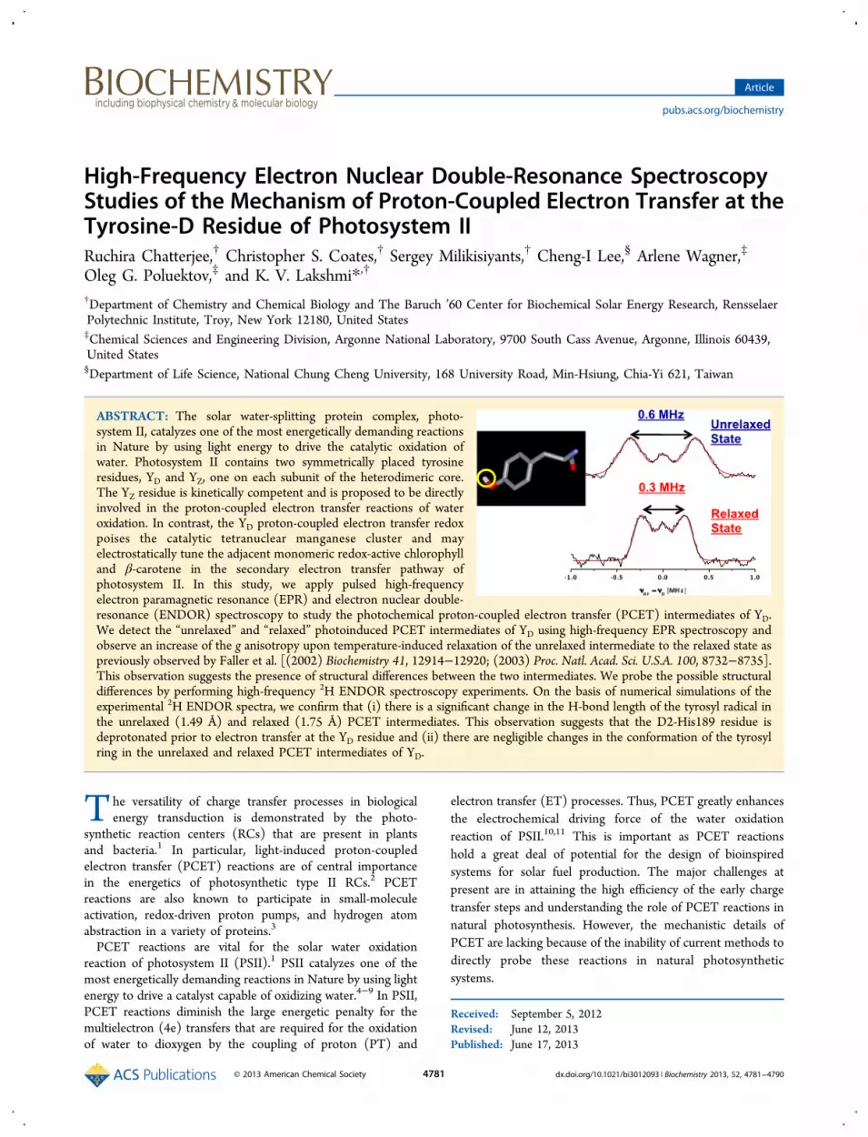
ABSTRACT: The solar water-splitting protein complex, photo-system II, catalyzes one of the most energetically demanding reactionsin Nature by using light energy to drive the catalytic oxidation ofwater. Photosystem II contains two symmetrically placed tyrosineresidues, YD and YZ, one on each subunit of the heterodimeric core.The YZ residue is kinetically competent and is proposed to be directlyinvolved in the proton-coupled electron transfer reactions of wateroxidation. In contrast, the YD proton-coupled electron transfer redoxpoises the catalytic tetranuclear manganese cluster and mayelectrostatically tune the adjacent monomeric redox-active chlorophylland β-carotene in the secondary electron transfer pathway ofphotosystem II. In this study, we apply pulsed high-frequencyelectron paramagnetic resonance (EPR) and electron nuclear double-resonance (ENDOR) spectroscopy to study the photochemical proton-coupled electron transfer (PCET) intermediates of YD.We detect the “unrelaxed” and “relaxed” photoinduced PCET intermediates of YD using high-frequency EPR spectroscopy andobserve an increase of the g anisotropy upon temperature-induced relaxation of the unrelaxed intermediate to the relaxed state aspreviously observed by Faller et al. [(2002) Biochemistry 41, 12914−12920; (2003) Proc. Natl. Acad. Sci. U.S.A. 100, 8732−8735].This observation suggests the presence of structural differences between the two intermediates. We probe the possible structuraldifferences by performing high-frequency 2H ENDOR spectroscopy experiments. On the basis of numerical simulations of theexperimental 2H ENDOR spectra, we confirm that (i) there is a significant change in the H-bond length of the tyrosyl radical inthe unrelaxed (1.49 Å) and relaxed (1.75 Å) PCET intermediates. This observation suggests that the D2-His189 residue isdeprotonated prior to electron transfer at the YD residue and (ii) there are negligible changes in the conformation of the tyrosylring in the unrelaxed and relaxed PCET intermediates of YD.
The versatility of charge transfer processes in biologicalenergy transduction is demonstrated by the photo-
synthetic reaction centers (RCs) that are present in plantsand bacteria.1 In particular, light-induced proton-coupledelectron transfer (PCET) reactions are of central importancein the energetics of photosynthetic type II RCs.2 PCETreactions are also known to participate in small-moleculeactivation, redox-driven proton pumps, and hydrogen atomabstraction in a variety of proteins.3
PCET reactions are vital for the solar water oxidationreaction of photosystem II (PSII).1 PSII catalyzes one of themost energetically demanding reactions in Nature by using lightenergy to drive a catalyst capable of oxidizing water.4−9 In PSII,PCET reactions diminish the large energetic penalty for themultielectron (4e) transfers that are required for the oxidationof water to dioxygen by the coupling of proton (PT) and
electron transfer (ET) processes. Thus, PCET greatly enhancesthe electrochemical driving force of the water oxidationreaction of PSII.10,11 This is important as PCET reactionshold a great deal of potential for the design of bioinspiredsystems for solar fuel production. The major challenges atpresent are in attaining the high efficiency of the early chargetransfer steps and understanding the role of PCET reactions innatural photosynthesis. However, the mechanistic details ofPCET are lacking because of the inability of current methods todirectly probe these reactions in natural photosyntheticsystems.
Received: September 5, 2012Revised: June 12, 2013Published: June 17, 2013
Article
pubs.acs.org/biochemistry
© 2013 American Chemical Society 4781 dx.doi.org/10.1021/bi3012093 | Biochemistry 2013, 52, 4781−4790
The solar water oxidation reaction of PSII results from PCETreactions at the redox-active tyrosine and quinone cofactors.12
These cofactors have the remarkable ability to control andmodulate ET and PT chemistry in PSII. It is thought that thelocation of the cofactor, the geometry of its binding site, theredox potential, and dynamics from the surrounding proteinenvironment greatly influence the unrivaled efficiency of PCETreactions in the photochemical reactions of PSII. To under-stand the mechanisms and the versatility of PCET reactions, weneed to understand the factors that control the functionaltuning of these cofactors. The recent 1.9−3.8 Å resolution X-ray crystal structures reveal atomic-level information about thedark state of PSII.13−19 The preliminary information availablefrom the X-ray crystal structures sets the stage for thedevelopment and application of state-of-the-art magneticresonance spectroscopy methods for interrogating the molec-ular basis of the light-driven PCET reactions of PSII.PSII contains two symmetrically placed tyrosine residues, YD
and YZ, on each subunit of the heterodimeric polypeptidecore.12,20 The function of the symmetry-related tyrosineresidues is quite distinct as the protein environment greatlyinfluences tyrosine function in PSII. It is proposed that thePCET redox at YD poises the catalytic Mn4Ca cluster
21 and mayelectrostatically tune the adjacent monomeric redox-activechlorophyll and β-carotene in the secondary ET pathway ofPSII.22,23 In contrast, YZ is kinetically competent and directlyinvolved in the PCET reactions of the water oxidation reactionof PSII.24,25 The redox-active tyrosine residues of PSII areespecially attractive as an experimental system because (a) YZand YD have distinct properties, including time scales andspecificity of redox activity, and (b) the PCET reactions at YZand YD can be studied at cryogenic temperatures.26,27
Electron paramagnetic resonance (EPR) and electron nucleardouble-resonance (ENDOR) spectroscopy are powerful toolsfor the study of the structure and dynamics of paramagneticcenters in photosynthetic RCs.28−30 In ENDOR spectroscopy,the nuclear resonance frequencies are monitored indirectlythrough EPR transitions. Typically, the nuclear transitionsdetected in an ENDOR spectroscopy experiment havesensitivities higher than those of conventional nuclear magneticresonance (NMR) detection and higher selectivity because ofdetection through the electron spin. In addition, the applicationof high-frequency (HF) EPR spectroscopy methods providesenhanced sensitivity and g tensor resolution. The application ofENDOR spectroscopy at high frequency facilitates thedetection of weak electron−nuclear interactions and affordsorientation selection that can remove interactions fromoverlapping signals. This provides accurate information aboutthe interaction of the radical with the surrounding proteinenvironment.31
Previous studies have used EPR spectroscopy to illustrate thepresence of photoinduced intermediates of the YD residue ofPSII.26,27,32−35 It was demonstrated that photo-oxidation of YDat 7 K results in the formation of the low-temperature early“unrelaxed” PCET intermediate of YD
● where the proton andprotein motion are limited. The illumination of PSII at a highertemperature (240 K) allows for unrestricted proton movementand possibly the relaxation of the protein environment thatleads to the formation of the late “relaxed” PCET intermediateof YD
●.26,27 These studies identified changes in the g tensor ofthe unrelaxed and relaxed intermediates that were suggested toarise from a change in the hydrogen bond distance of the YD
●
radical. However, it was not possible to disentangle the
hydrogen bond changes and possible conformational effects ofYD in the unrelaxed and relaxed intermediate of the YD
● radical.In this study, we demonstrate the application of pulsed HF
EPR and HF 2H ENDOR spectroscopy to obtain structuraldetails of the unrelaxed and relaxed photoinduced PCETintermediates of the YD residue of PSII. A combination ofelectron transfer, proton movement, and local conformationaleffects of YD
● can result in PCET reactions in the oxygen-evolving complex (OEC) of PSII. We report pulsed HF 2HENDOR measurements of the hydrogen (H)-bonded deuteronof YD
●, which provides quantitative knowledge of the H-bondstrength and proton movement in the respective PCETintermediates.36 We also report HF 2H ENDOR measurementsof deuterons at the 2, 6 ring and β-methylene positions of YD
●
that yield information about the relative geometry of thetyrosine ring in the PCET intermediates.In this study, we observe changes in the H-bond strength
from possible conformational effects during photoinducedPCET at the tyrosine-D residue of PSII that cannot bediscerned by other structural methods. The results provideimportant insight into the mechanism of PCET at the tyrosine-D residue of photosystem II.
■ MATERIALS AND METHODSPreparation of Photosystem II. Preparation of Deu-
terated Photosystem II from Synechococcus lividus. The S.lividus cells were grown at 43 °C in deuterated Ac medium.37
The photosystem II (PSII) core complexes were isolated fromthe deuterated cells using the procedure of Tang and Diner38
with minor modifications.37 The cell extract containingdeuterated PSII was loaded onto a diethylaminoethyl(DEAE) cellulose ion-exchange column (Tosoh Biosciences,King of Prussia, PA) in the absence of magnesium sulfate(MgSO4), and the PSII was eluted with a 1 L gradient from 0 to50 mM MgSO4 in buffer containing 50 mM 2-(N-morpholino)-ethanesulfonic acid (MES-NaOH), 20 mM calcium chloride(CaCl2), and 5 mM magnesium chloride (MgCl2) at pH 6.0with 25% (w/v) glycerol and 0.03% (w/v) dodecyl β-D-maltoside (β-DM).37 The deuterated PSII fractions werecollected and concentrated to a volume of 2 mL. Theconcentrated eluent was desalted using an EconoPac 10 D6desalting column (Bio-Rad, Hercules, CA). Following this, thedeuterated PSII was resuspended in buffer containing 50 mMMES-NaOH, 20 mM CaCl2, and 5 mM MgCl2 at pH 6.0 with25% (w/v) glycerol and 0.03% (w/v) β-DM. The purifieddeuterated PSII was characterized using 12% sodium dodecylsulfate−polyacrylamide gel electrophoresis (SDS−PAGE)analysis. The isolation and purification of deuterated PSIIwere performed in the dark at 4 °C, and the samples werestored at −80 °C in the dark until further use.
Preparation of Histidine-Tagged (HT) Photosystem II fromthe PsbB Variant of Synechocystis PCC 6803. The PsbBvariant of Synechocystis PCC 6803 cells that was previouslydeveloped by Brudvig, Lakshmi, and co-workers waspropagated on BG-11-glucose agar plates.39,40 The cells weregrown in 18 L cultures under constant illumination at ∼30 °Cusing BG-11 medium with 5 mM 2-[tris(hydroxymethyl)-methylamino]-1-ethanesulfonic acid/potassium hydroxide(TES-KOH) and 5 mM glucose (pH 8.2). The cells wereharvested in buffer containing 50 mM MES-NaOH, 5 mMCaCl2, and 5 mM MgCl2 (pH 6.0) with 25% (w/v) glycerol.Upon being harvested, the cells were broken using a beadbeater (BioSpec, Bartlesville, OK) and processed in the dark to
Biochemistry Article
dx.doi.org/10.1021/bi3012093 | Biochemistry 2013, 52, 4781−47904782
prepare a detergent-solubilized extract of thylakoid proteins.The extract was suspended in buffer containing 50 mM MES-NaOH, 20 mM CaCl2, and 5 mM MgCl2 (pH 6.0) with 25%(w/v) glycerol and 0.03% (w/v) β-DM in the dark and wassubjected to purification on a Ni2+-NTA metal-affinity column(QIAgen, Valencia, CA). The Ni2+-NTA column was washedwith a solution of 25% (w/v) glycerol, 5 mM MgCl2, 20 mMCaCl2, 50 mM MES-NaOH (pH 6.0), and 0.03% (w/v) β-DMwith 5 mM imidazole. The HT-PSII was eluted with a buffercontaining 25% (w/v) glycerol, 5 mM MgCl2, 20 mM CaCl2,50 mM MES-NaOH (pH 6.0), and 0.03% (w/v) β-DM with250 mM imidazole.40 The HT-PSII was desalted using anEconoPac 10 D6 desalting column (Bio-Rad). The isolationand purification of HT-PSII were performed in the dark at 4°C. The purified HT-PSII was characterized using 12% SDS−PAGE analysis, optical assays, and oxygen evolution measure-ments. Following this, the HT-PSII was stored in buffercontaining 50 mM MES-NaOH, 20 mM CaCl2, and 5 mMMgCl2 (pH 6.0) with 25% (w/v) glycerol and 0.03% (w/v) β-DM at −80 °C in the dark.Manganese Depletion and Cyanide Treatment of Photo-
system II. The purified deuterated PSII and HT-PSII from S.lividus and the PsbB variant of Synechocystis PCC 6803,respectively, were depleted of manganese (Mn) using a buffercontaining 50 mM MES-NaOH, 20 mM CaCl2, and 5 mMMgCl2 (pH 6.0) with 25% (w/v) glycerol, 0.03% (w/v) β-DM,10 mM hydroxylamine hydrochloride (NH2OH·HCl), and 10mM sodium ethylenediaminetetraacetic acid (EDTA).37,41 Theaddition of NH2OH·HCl results in the disassembly of theMn4Ca−oxo cluster of PSII. The Mn-depleted PSII complexeswere repeatedly washed in a buffer containing 50 mM MES-NaOH, 20 mM CaCl2, and 5 mM MgCl2 (pH 6.0) with 25%(w/v) glycerol, 0.03% (w/v) β-DM, and 5 mM EDTA.The high-spin (electron spin S = 2) non-heme iron center,
Fe(II), in Mn-depleted deuterated PSII and Mn-depleted HT-PSII was converted to its low-spin (electron spin S = 0) state byincubation with 350 mM potassium cyanide (KCN) at pH 6.0for 3.5 h in the dark at 4 °C.42 Although manganese depletionand cyanide (CN) treatment are not required for theobservation of the PCET intermediates of YD
●, these protocolswere applied as the deuterated PSII preparation was also usedfor HF ENDOR spectroscopy experiments with the primarysemiquinone, QA
−.The Mn-depleted, CN-treated deuterated PSII was sus-
pended in tricine buffer or “protonated” buffer at pH 8.7. TheMn-depleted, CN-treated HT-PSII was suspended in tricinebuffer in D2O or “deuterated” buffer at pH 8.7.Photochemical Trapping of the Proton-Coupled Electron
Transfer Intermediates of the YD Residue of Photosystem II.The early unrelaxed PCET intermediate of YD was trapped byilluminating the Mn-depleted, CN-treated deuterated PSII andMn-depleted, CN-treated HT-PSII from S. lividus and the PsbBvariant of Synechocystis PCC 6803, respectively, at pH 8.7 and 7K for 2 min using 550 nm laser pulses (∼1 mJ/pulse) with aflash frequency of 10 Hz. The late relaxed PCET intermediateof YD was trapped by illuminating Mn-depleted, CN-treateddeuterated PSII and Mn-depleted, CN-treated HT-PSII at pH8.7 for 2 min at 240 K, which allows for temperature-inducedrelaxation. The experimental HF EPR and HF 2H ENDORspectra of the unrelaxed and relaxed PCET intermediate of YDwere recorded at 20 K.Pulsed High-Frequency EPR and ENDOR Spectrosco-
py. The high-frequency (HF) EPR spectra were obtained on a
home-built D-band (130 GHz, 4.5 T) continuous wave (cw)/pulsed spectrometer.37,43,44 The pulsed HF ENDOR spectrawere recorded using a Mims sequence31 of microwave (MW)and radiofrequency (RF) pulses (π/2MW−τ−π/2MW−πRF−π/2MW) by monitoring the electron-spin−echo intensity as afunction of the frequency of the RF pulse. The RF pulses weregenerated by an Agilent RF signal generator (model E4400B)and amplified by a 1 kW pulsed amplifier (CPC, model5T1000). For the HF 2H ENDOR experiment, the duration ofthe MW pulses was 60 ns and that of the RF π-pulse was 170μs. The light excitation of the sample was achieved with anoptical parametric oscillator (Opotek, Carlsbad, CA) pumpedby a Nd:YAG laser (Quantel, Bozeman, MT), the output ofwhich was coupled to an optical fiber. The optical fiber allowsdelivery of up to 1 mJ/pulse to the sample. The PSII sampleswere held in quartz tubes (inside diameter of 0.5 mm andoutside diameter of 0.6 mm) and placed in the MW resonator.The resonator was held in an Oxford flow cryostat (OxfordInstruments, Oxford, U.K.). The samples were frozen in thedark in the resonator/cryostat, and the temperature wascontrolled with an ITC Oxford temperature control system(Oxford Instruments).
Spin−Echo-Detected Magnetic Field-Sweep EPRSpectra of the Relaxed and Unrelaxed PCET Intermedi-ates of YD
●. The spin−echo-detected magnetic field-sweepEPR spectra of the relaxed and unrelaxed intermediates of theYD residue are represented by solid black lines in Figure 1. Weused the “pepper” function of the EasySpin software packagefor numerical simulations of the experimental spectra.45 In thenumerical simulations, we neglected the hyperfine interactionsand used an orientation-dependent line broadening parameter“HStrain”. For the relaxed intermediate, we reproduce theexperimental spectra assuming contributions from threespecies: the tyrosyl radical, YD
●, primary semiquinone anionradical, QA
−, and a species with much smaller g anisotropycorresponding to the monomeric chlorophyll (ChlZ
+) and/orβ-carotene (Car+) radical of PSII.37,46 For the unrelaxedintermediate, a small additional contribution from the relaxedstate was added to the simulated spectrum. The principalcomponents of the g tensor of all of the species are presented inTable 1.
Numerical Simulation of the HF 2H ENDOR Spectra.The numerical simulations of the HF 2H ENDOR spectra wereperformed with the “salt” function of the EasySpin softwarepackage.45 Orientation selection was achieved by using the gtensor determined from the numerical simulations of the spin−echo-detected magnetic field-sweep EPR spectra describedabove and assuming microwave excitation corresponding to a
Table 1. Relative g Tensor of the Unrelaxed and RelaxedPCET Intermediates of YD
●, QA−, and ChlZ
+/Car+ Residuesin Mn-Depleted, CN-Treated Deuterated PSII That WereObtained from the Spectral Simulation of the HF EPRSpectraa
cofactor gx gy gz
YD• (unrelaxed) 2.00673 2.00453 2.00232
YD• (relaxed) 2.00774 2.00447 2.00232
QA− 2.0067 2.0052 2.00232
ChlZ+/Car+ 2.0031 2.0027 2.00232
aThe gz component of the g tensor is normalized to the g value of afree electron.
Biochemistry Article
dx.doi.org/10.1021/bi3012093 | Biochemistry 2013, 52, 4781−47904783

rectangular-shaped microwave pulse length of 60 ns. Tosimulate the experimental spectra of the intrinsic deuteronson the tyrosine residue, we used the data available in theliterature as an initial estimate.47,48 The orientation of theelectron−nuclear hyperfine tensors has been described inpreviously published reports.47,48 The orientation of the nuclearquadrupole tensor is assumed to be collinear with theelectron−nuclear hyperfine tensor.Determination of the Hydrogen Bond Distance. For
the numerical simulations of the hydrogen (H)-bondeddeuteron in the relaxed and unrelaxed states, we assumed theH-bond to be in the plane containing the tyrosyl ring of YD
●.This is in agreement with the recently determined high-resolution crystal structure of PSII,18 in which the τ-nitrogen ofD2-His189 is in plane with the tyrosyl ring of YD. To reducethe number of variables, we calculated the hyperfine tensorusing the McConnell equations that suggest that the unpairedelectron is located in the 2p Slater orbital of the oxygen atom:49
ρ= − − + + + + +
+ −
⎜
⎟
⎡⎣⎢
⎛⎝
⎞⎠
⎤⎦⎥
AR a
a a aa
a
21
94 10 17
18
9e
x
a
3 23 2
22
(1)
ρ= − + + + + + −⎜ ⎟⎡⎣⎢
⎛⎝
⎞⎠
⎤⎦⎥A
R aa a
a a1
19
24 8
9 92
eya
3 22
22
(2)
ρ= − + + + + +
+ −
⎜
⎟
⎡⎣⎢
⎛⎝
⎞⎠
⎤⎦⎥
AR a
a a aa
a
11
272
2 7 16 2627
272
e
z
a
3 23 2
22
(3)
where ρ is the spin density on the oxygen atom and a = (ZR)/(2a0), where Z is the effective charge of oxygen for the 2porbital and a0 is the Bohr radius. The electron spin density onthe oxygen atom varies from ∼0.24 to 0.34 in tyrosyl radicals,and such changes are most likely due to the presence of a H-bond that can lower the electron spin density.33,50,51 In thisstudy, a value of 0.28 was selected for the electron spin densityon the oxygen atom of YD
● in both the unrelaxed and relaxedintermediates. The effective charge, Z, was assumed to be4.45.52,53 The electron−nuclear interspin distance, R, the angleof the H-bond in the plane of the tyrosyl ring, and thequadrupole tensor were treated as variables in the numericalsimulations of the experimental HF 2H ENDOR spectra.
■ RESULTS AND DISCUSSIONIn Situ Observation of the PCET Intermediates of YD
●.This study is focused on understanding the mechanism of thephotoinduced PCET reaction that leads to the formation of theredox-active tyrosyl radical, YD
●, of PSII. It has beendemonstrated that it is possible to photo-oxidize YD at 7 Kwhere both the proton and protein motions are limited.26,27
This leads to the formation of the low-temperature earlyunrelaxed PCET intermediate of YD
●. However, illumination ofPSII at a higher temperature (240 K) allows for unrestrictedproton movement and possibly the relaxation of the proteinenvironment.26,27 This results in the formation of the laterelaxed PCET intermediate of YD
●. Here, we trap the unrelaxedand relaxed PCET intermediates of YD
● of wild-type Mn-
depleted, CN-treated deuterated PSII and Mn-depleted, CN-treated HT-PSII from S. lividus and the PsbB variant ofSynechocystis PCC 6803, respectively. We utilize the enhancedresolution and sensitivity of HF EPR and HF 2H ENDORspectroscopy37 to elucidate the structure of the unrelaxed andrelaxed PCET intermediates and, hence, the sequence of eventsduring the light-induced PCET reaction at the YD residue ofPSII.At high frequencies, the line shape of an EPR spectrum is
typically determined by the anisotropy of the Zeemaninteraction of the paramagnetic center in the presence of anexternal magnetic field. This results in an EPR spectrum withthree characteristic features corresponding to the direction ofthe canonical axes of the g tensor with respect to the externalmagnetic field. As one can see in panels A and B of Figure 1,
the HF EPR spectra of the unrelaxed and relaxed PCETintermediates of YD exhibit multiple spectral features. The HFEPR spectra are dominated by features arising from the well-resolved g tensor of YD
● (these are marked by arrows in Figure1A,B). Also observed are minor additional spectral contribu-tions from other radicals, such as the primary semiquinone(QA
−), β-carotene (Car+), and monomeric chlorophyll (ChlZ+)
radical of PSII that are easily identified by their characteristicprincipal g values (Table 1).37,46,54,55
Further examination of the EPR spectra of the unrelaxed andrelaxed intermediates in panels A and B of Figure 1 and thecorresponding g tensors presented in Table 1 reveals that thevalue of the gx component of YD
● in the unrelaxed intermediateis 2.00673. However, in the case of the relaxed intermediate ofYD
●, the spectral feature corresponding to the gx componentshifts to a lower magnetic field with a value of 2.00774. Thus,there is a significant shift of the gx component of the YD
●
radical in the unrelaxed and relaxed PCET intermediates. Incontrast, numerical simulations indicate that the values of the gyand gz components of YD
● remain identical, within the accuracyof the experiment, in both the unrelaxed and relaxed PCETintermediates. These results confirm an earlier study performedby Faller et al. in which two YD
● intermediates were reportedfor the first time.26,27
Figure 1. HF 130 GHz spin−echo-detected magnetic field-sweep EPRspectra of Mn-depleted, CN-treated deuterated PSII in protonatedbuffer in the (A) unrelaxed and (B) relaxed PCET intermediate states.The experimental spectra are shown as solid black traces, and thesimulated spectra are shown as dashed red traces. The gx, gy, and gzcomponents of YD
● are marked by arrows in panels A and B.
Biochemistry Article
dx.doi.org/10.1021/bi3012093 | Biochemistry 2013, 52, 4781−47904784

In the case of organic radicals, the anisotropy of the g tensoris mostly determined by the spin−orbit coupling interaction atthe heavy atom, namely, the oxygen atom in the case of atyrosyl radical. The spin−orbit coupling interaction arisesbecause of relativistic effects that are significant in heavieratoms. The observed shift of the gx value in the spin−echo-detected magnetic field-sweep EPR spectra of the unrelaxedand relaxed intermediate shown in panels A and B of Figure 1reflects significant reduction of the electron spin density at theoxygen atom of the YD
● radical. Such a change in the electronspin density can be induced by either a change in the H-bondstrength at the carbonyl oxygen atom or conformationalchanges of the YD
● radical. It is thought that the tyrosine-Dresidue (D2-Tyr160), YD, has a single H-bond with theneighboring D2-His189 residue of PSII.56 An increase in theanisotropy of the g tensor upon relaxation of the intermediateat a higher temperature could reflect weakening of the H-bondwith the τ-nitrogen of the D2-His189 residue. Recently,Svistunenko et al.47 analyzed hyperfine parameters that wereobtained in previous HF ENDOR spectroscopy studies oftyrosyl radicals of different proteins. This study estimated therotation angle of the tyrosyl ring of the YD
● radical to be ∼50°in PSII.47 In principle, a significant change in the rotation angleof the tyrosyl ring could also affect the electron spin densitydistribution of the YD
● radical. This could result in the changesthat are observed in the g anisotropy of the unrelaxed andrelaxed PCET intermediate of the YD residue of PSII. Todirectly probe the strength of the H-bond and the rotationalconformation of the tyrosyl ring of YD
●, we performed HF 2HENDOR spectroscopy on the unrelaxed and relaxed PCETintermediates of YD
●.Proton Movement in the PCET Intermediates of YD
●.To characterize the hydrogen bonding interaction of theunrelaxed and relaxed PCET intermediates of YD
●, weperformed HF 2H ENDOR spectroscopy of protonated Mn-depleted, CN-treated HT-PSII from the PsbB variant ofSynechocystis PCC 6803 that is exchanged in deuterated buffer(where the buffer is prepared in D2O). The exchange ofprotonated Mn-depleted, CN-treated HT-PSII in deuteratedbuffer results in the replacement of the H-bonded proton with adeuteron57−61 that facilitates exclusive detection of theelectron−nuclear hyperfine interaction of the H-bondeddeuteron by HF 2H ENDOR spectroscopy. To obtainquantitative information about the principal components andcanonical orientation of the hyperfine tensor in the molecularframe, we conduct HF 2H ENDOR measurements at multiplespectral positions that correspond to different orientations ofthe g tensor with respect to the external magnetic field. In thecase of a tyrosyl radical, the HF 2H ENDOR measurements areorientation selective as the microwave excitation bandwidth ismuch smaller than the g anisotropy (and spectral width) of theHF EPR spectrum.The experimental HF 2H ENDOR spectra of the unrelaxed
and relaxed PCET intermediate of YD● are shown in panels A−
C of Figure 2. As can be seen in panels A and B, the HF 2HENDOR spectra that are measured at a magnetic field positioncorresponding to the gx component of the g tensor display asingle well-resolved splitting arising from the electron−nuclearhyperfine interaction of the electron spin with the H-bondeddeuteron. In this case, the nuclear quadrupole interaction ismuch weaker and contributes to the shape of the individualpeaks of the doublet in panels A and B of Figure 2. A qualitativecomparison of the HF 2H ENDOR spectra at the magnetic field
position corresponding to the gx component of the g tensorindicates that the hyperfine splitting that is observed in theunrelaxed intermediate (Figure 2A) is larger than the splittingthat is observed in the relaxed intermediate (Figure 2B). Incontrast with the single well-defined hyperfine splitting that isobserved in the HF 2H ENDOR spectra at the magnetic fieldposition corresponding to the gx component of the g tensor(Figure 2A,B), the spectrum that is measured at a magneticfield position corresponding to the gy component of the gtensor of the relaxed intermediate (Figure 2C) is morecomplex. The spectral complexity arises from the contributionof multiple molecular orientations to the gy component withrespect to the magnetic field.We performed numerical simulations of the experimental HF
2H ENDOR spectra to obtain quantitative information aboutthe hyperfine components of the H-bonded deuteron in theunrelaxed and relaxed PCET intermediates. To reduce thenumber of variables in the numerical simulations, we calculatedthe electron−nuclear hyperfine tensor using McConnellequations (see Materials and Methods).49 The distancebetween the carbonyl oxygen and H-bonded proton, theangle of the H-bond in the plane of the tyrosyl ring, theprincipal components, and the orientation (determined by theEuler angles) of the quadrupole tensor were varied to obtainthe best fit with the experimental spectra. The numericalsimulations of the experimental HF 2H ENDOR spectra for theunrelaxed and relaxed intermediate are shown as red traces inFigure 2A−C. The principal components of the electron−nuclear hyperfine tensor (Ax, Ay, and Az), the quadrupole tensor(Qx, Qy, and Qz), and the relative orientation of both tensors inthe frame of the g tensor are listed in Table 2. To facilitatecomparison with previous studies, the values of the principalcomponents of the hyperfine tensor, Ax, Ay, and Az, have beenrescaled for protons, and the proton hyperfine parameters areprovided in parentheses. The value of the hyperfine tensor ofthe H-bonded proton of the relaxed PCET intermediate is inagreement with previous reports on single crystals58 and frozensolutions of PSII.57 The hydrogen bonding distance of therelaxed PCET intermediate of YD
● is in qualitative agreementwith the recent high-resolution X-ray crystal structure of PSII.18
A quantitative comparison is not possible as the high-resolution
Figure 2. Mims HF 2H ENDOR spectrum of Mn-depleted, CN-treated HT-PSII in deuterated buffer in the (A) gx orientation of theunrelaxed intermediate and (B) gx orientation of the relaxedintermediate. (C) Mims HF 2H ENDOR spectrum of Mn-depleted,CN-treated HT-PSII in deuterated buffer in the gy orientation of therelaxed PCET intermediate. The experimental spectra are depicted asblack traces and the simulations as red traces.
Biochemistry Article
dx.doi.org/10.1021/bi3012093 | Biochemistry 2013, 52, 4781−47904785

X-ray crystal structure of PSII does not include coordinates forprotons.We observe a significant decrease in the H-bond distance in
the unrelaxed PCET intermediate (1.49 Å) in comparison withthat of the relaxed PCET intermediate (1.75 Å) of YD
●. Thechange in the interspin distance of the H-bonded deuteron inthe unrelaxed and relaxed PCET intermediates of YD
● that isobserved in this study can be reviewed in the context of the twoPCET models that were previously proposed by Faller et al.26,27
By measuring the pH dependence of tyrosine oxidation rates,we proposed that ET at the YD residue is coupled todeprotonation with a pKa value of ∼7.7. The pH dependencesof the rates of ET were explained by two possible PCETpathways for the photoinduced oxidation of the YD residue thatis thought to occur in concert with a conjugate base, D2-His189. In the first pathway, abstraction of a proton from thecarbonyl oxygen by the D2-His189 residue is proposed occurprior to ET from the YD residue. In this case, a stronginteraction between the carbonyl oxygen atom and the H-bonded proton (at a distance estimate of 1.58 Å) has beenpredicted in the initial unrelaxed His-Tyr● intermediate.Subsequent temperature-induced thermal relaxation by themotion of the histidine residue is predicted to increase thedistance between the carbonyl oxygen atom and the H-bondedproton to 1.93 Å in the relaxed intermediate. In the secondpathway, abstraction of a proton by the D2-His189 residue isproposed to either follow or occur simultaneously with ETfrom the YD residue. The distance between the carbonyl oxygenatom and the H-bonded proton has been predicted to be 1.85Å in the unrelaxed His-Tyr● intermediate. In this case, upontemperature-induced thermal relaxation of the histidine residue,the H-bond length is proposed to increase only slightly from1.85 to ∼1.93 Å. The significant difference in the H-bonddistances in these two possible pathways originates from thefact that the τ-nitrogen atom of the deprotonated D2-His189residue is expected to be <2.60 Å from the carbonyl oxygen ofthe YD residue in comparison with the distance in theprotonated D2-His189 residue (2.82 Å).The results of this study do not support the second pathway,
in which proton abstraction by D2-His189 is proposed toaccompany ET from the YD residue. The change in distance of0.26 Å that is observed in the HF ENDOR experiments is toolarge in comparison with the predicted change of 0.08 Å in thetheoretical model. The larger change in distance of 0.35 Å thatis predicted for the first pathway is in reasonable agreementwith the experimental observations in this study. The smallervalue obtained experimentally could be due to increasedelectron spin density on the oxygen atom of the carbonyl groupin the relaxed state. However, in the numerical calculations, weassumed a fixed value of the electron spin density in both theunrelaxed and relaxed intermediate. An increase in the value ofthe electron spin density would result in an underestimation of
the H-bond length in the relaxed intermediate in comparisonwith that in the unrelaxed intermediate.A recent study by Hienerwadel et al. has suggested that YD
remains protonated in the pH range of 6.0−10.0 and theprotonation state of the D2-His189 residue remains unchangedat these pH values.62 Thus, the first pathway that suggesteddeprotonation of YD prior to electron transfer is in disagree-ment with the results of the FTIR study. However, the secondpathway that suggested that the oxidation of YD is promoted athigher pH values by the deprotonation of the D2-His189residue is also in disagreement with the FTIR study.The short distance that is observed between the proton and
carbonyl oxygen of YD● in the unrelaxed state (1.49 Å) in this
study indicates the proximity of the hydrogen bonding partnerat pH 8.7. This observation is in agreement with the FTIRstudy that suggested the involvement of YD in very stronghydrogen bonding interaction at pH >7.5.62 The contradictionsbetween the theoretical models and FTIR spectroscopy datacould arise from the fact that additional amino acid residuesimpact the structure and function of the YD−D2-His189 couplein vivo. Thus, it remains unclear whether the formation of theunrelaxed intermediate is preceded by a tyrosinate moiety atpH >7.5, D2-His189 remains as the H-bonding partner of YD,or another residue is involved in the formation of the verystrong bond. These questions will be further addressed infuture investigations.For the relaxed PCET intermediate of YD
●, the numericalsimulations yield an angle of 120 ± 10° between the H-bondand C−O bond of the carbonyl group. This is reasonablebecause the sp2-hybridized oxygen has a lone pair orbital that isin the ring plane and makes an angle of 120° with respect to theC−O axis. For carbonyl radicals, there is a noticeablepreference for the H-bond to be in the direction of the lonepair at an angle of ∼120°.63 Thus, in the case of a carbonylgroup, the orientation of the H-bonded proton is expected tobe close to this angle. In a recent theoretical study, Brynda etal.64 calculated the angle of the optimized Tyr-His structure tobe 124°, which is in excellent agreement with our finding.Similarly, from numerical simulations, the H-bond angle for theunrelaxed YD
● intermediate is estimated to be 108 ± 20°. Thissuggests that there is negligible change in the angle between theH-bond and C−O bond of the carbonyl group in the relaxedand unrelaxed intermediates. The orientation of the H-bond ofthe relaxed YD
● intermediate that is determined in this study isin excellent agreement with the recent high-resolution X-raycrystal structure of PSII.18
Conformation of the Tyrosyl Ring in the PCETIntermediates of YD
●. To probe the conformation of thetyrosyl ring in the unrelaxed and relaxed intermediate of the YDresidue, we perform HF 2H ENDOR spectroscopy on Mn-depleted CN-treated deuterated PSII from S. lividus inprotonated buffer (where the buffer is prepared in H2O). Inthis case, the hyperfine interactions with the intrinsic deuterons
Table 2. Magnetic Parameters Obtained from Numerical Simulations of the Mims HF 2H ENDOR Spectra of Mn-Depleted,CN-Treated HT-PSII in Deuterated Buffer at the gx Orientation
a
Ax (MHz) Ay (MHz) Az (MHz) [α, β, γ]b (deg)Qx
(MHz)Qy
(MHz)Qz
(MHz) [α, β, γ]b (deg)
exchanged deuterons (unrelaxed) 1.59 (10.34) −0.91 (−5.93) −0.68 (−4.41) [108, 0, 0] 0.14 −0.066 −0.074 [126, 0, 0]exchanged deuterons (relaxed) 1.06 (6.88) −0.58 (−3.79) −0.48 (−3.10) [120, 0, 0] 0.11 −0.04 −0.07 [142, 0, 0]aThe hyperfine parameters rescaled for the proton gyromagnetic ratio are provided in parentheses. bThe Euler rotation angles are defined withrespect to the principal axis frame of the g tensor.
Biochemistry Article
dx.doi.org/10.1021/bi3012093 | Biochemistry 2013, 52, 4781−47904786

of the tyrosyl radical, YD●, contribute to the experimental HF
2H ENDOR spectra. As shown in Figure 3A−D, the HF 2H
ENDOR spectra for the unrelaxed and relaxed intermediate aremeasured at magnetic field positions that correspond to the gxand gy components of the g tensor, respectively. We observethat the spectral line shapes are determined by the hyperfineinteraction of the unpaired electron with the four deuterons onthe ring and the β-methylene deuterons of the tyrosyl radical(Figure 4A,B). To obtain quantitative information about thehyperfine interactions with the deuterons, we performnumerical simulations of the HF 2H ENDOR spectra. Thehyperfine parameters of the numerical simulations wereadjusted to obtain the best fit with the experimental spectra.
An initial estimate of the simulation parameters and assignmentof the observed spectral features were obtained from theliterature on the hyperfine tensors of the intrinsic protons ofthe relaxed intermediate.47,48 In this analysis, we assume thatthe pairs of deuterons at positions 2 and 6 and positions 3 and5 of the tyrosyl ring have identical hyperfine componentswithin the pair. Although a recent single-crystal EPR spectros-copy study suggests that this is not the case,65 the differencesbetween the hyperfine components are too small to be resolvedin powder samples of a frozen solution.The spectral features that are observed in the HF 2H
ENDOR spectra shown in Figure 3A−D arise from hyperfineinteractions with the deuterons at positions 2 and 6 of thetyrosyl ring and one deuteron at the β-methylene position, β1.The ring deuterons at positions 3 and 5 of the tyrosyl ring havenegligible contributions to the HF 2H ENDOR spectra. This isdue to the large anisotropy of the hyperfine tensor thatprecludes detection under the experimental conditions of theHF 2H ENDOR experiment. Similarly, a contribution from thesecond β-methylene proton, β2, is not observed in the HF 2HENDOR spectra because the hyperfine interaction of β2 withthe unpaired electron is too strong to be detected under theseexperimental conditions. The hyperfine parameters, theelectron−nuclear hyperfine tensor (Ax, Ay, Az), and the nuclearquadrupole tensor (Qx, Qy, Qz) that are obtained from thenumerical simulations are listed in Table 3. The values ofprincipal hyperfine components, Ax, Ay, and Az, have beenrescaled for the proton gyromagnetic ratio, and the protonhyperfine parameters are provided in parentheses.It is important to note here that contributions from the
intrinsic protons of the primary semiquinone (QA−), β-carotene
(Car+), or monomeric chlorophyll (ChlZ+) radical are not
detected in the HF 2H ENDOR spectra shown in Figure 3A−D. This confirms our earlier assumption that the observed HF2H ENDOR features in Figure 2A−C are entirely due to theYD
● radical and possible contributions from other unwantedradicals is negligible.To relate the obtained parameters to a certain conformation
of the radical, we utilize the McConnell equation that expressesthe isotropic component of the hyperfine tensor of a β-methylene proton via the unpaired electron spin density at C1(Figure 4A) and the dihedral angle, θ, the angle between theplane perpendicular to the tyrosyl ring and the plane containingC1, Cβ, and Hβ1, as shown in Figure 4B.66
ρ θ= ′ + ″βA B B( cos )iso C21
1 (4)
where Aisoβ1 is the isotropic hyperfine constant of the
corresponding β-methylene proton, ρC1is the spin density on
atom C1, and B′ and B″ are constants. B′ is neglected inpractical applications, and B″ is known to be 58 G.67 The angleof the second β-methylene proton can be easily evaluatedtaking into account that its dihedral angle is shifted by 120°(Figure 4B). The electron spin density, ρC1
, on atom C1 hasbeen estimated to be 0.37.68 Although eq 4 was derived forprotons, it can be used for deuterons by simply using a scalingfactor of 6.51 for the corresponding hyperfine components.Using the isotropic hyperfine component value of 9.2 MHz thatis obtained from the numerical simulations, eq 2 yields adihedral angle of 67.0° for the β1-methylene deuteron in therelaxed intermediate. This value is in excellent agreement withprevious studies of the relaxed state.47,69 The dihedral angle ofthe β-methylene deuteron for the relaxed intermediate of YD
Figure 3. Mims HF 2H ENDOR spectrum of Mn-depleted, CN-treated deuterated PSII in protonated buffer in the (A) gx orientationof the unrelaxed intermediate, (B) gx orientation of the relaxedintermediate, (C) gy orientation of the unrelaxed intermediate, and(D) gy orientation of the relaxed intermediate. The experimentalspectra are depicted as black traces and the simulations as red traces.
Figure 4. (A) Schematic depicting the definition of the rotation angle,θ, of the tyrosine ring plane as defined in this study. (B) View wherethe angle, θ, for proton β1 increases when it is measured clockwisefrom the perpendicular to the ring plane when the Cβ−R bond isbelow the horizontally oriented ring plane.
Biochemistry Article
dx.doi.org/10.1021/bi3012093 | Biochemistry 2013, 52, 4781−47904787
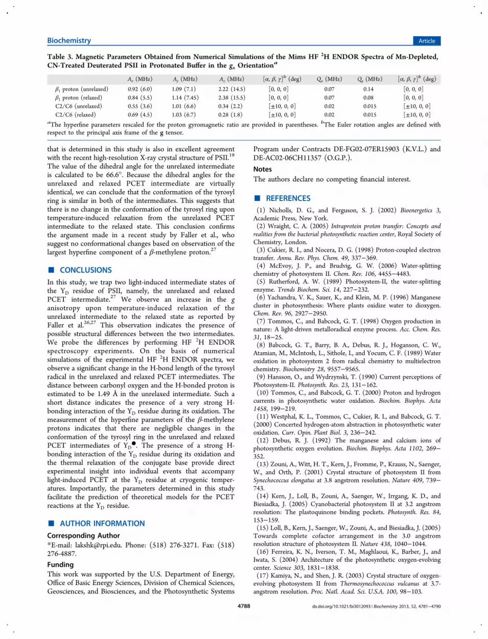
that is determined in this study is also in excellent agreementwith the recent high-resolution X-ray crystal structure of PSII.18
The value of the dihedral angle for the unrelaxed intermediateis calculated to be 66.6°. Because the dihedral angles for theunrelaxed and relaxed PCET intermediate are virtuallyidentical, we can conclude that the conformation of the tyrosylring is similar in both of the intermediates. This suggests thatthere is no change in the conformation of the tyrosyl ring upontemperature-induced relaxation from the unrelaxed PCETintermediate to the relaxed state. This conclusion confirmsthe argument made in a recent study by Faller et al., whosuggest no conformational changes based on observation of thelargest hyperfine component of a β-methylene proton.27
■ CONCLUSIONS
In this study, we trap two light-induced intermediate states ofthe YD residue of PSII, namely, the unrelaxed and relaxedPCET intermediate.27 We observe an increase in the ganisotropy upon temperature-induced relaxation of theunrelaxed intermediate to the relaxed state as reported byFaller et al.26,27 This observation indicates the presence ofpossible structural differences between the two intermediates.We probe the differences by performing HF 2H ENDORspectroscopy experiments. On the basis of numericalsimulations of the experimental HF 2H ENDOR spectra, weobserve a significant change in the H-bond length of the tyrosylradical in the unrelaxed and relaxed PCET intermediates. Thedistance between carbonyl oxygen and the H-bonded proton isestimated to be 1.49 Å in the unrelaxed intermediate. Such ashort distance indicates the presence of a very strong H-bonding interaction of the YD residue during its oxidation. Themeasurement of the hyperfine parameters of the β-methyleneprotons indicates that there are negligible changes in theconformation of the tyrosyl ring in the unrelaxed and relaxedPCET intermediates of YD
●. The presence of a strong H-bonding interaction of the YD residue during its oxidation andthe thermal relaxation of the conjugate base provide directexperimental insight into individual events that accompanylight-induced PCET at the YD residue at cryogenic temper-atures. Importantly, the parameters determined in this studyfacilitate the prediction of theoretical models for the PCETreactions at the YD residue.
■ AUTHOR INFORMATION
Corresponding Author*E-mail: [email protected]. Phone: (518) 276-3271. Fax: (518)276-4887.
FundingThis work was supported by the U.S. Department of Energy,Office of Basic Energy Sciences, Division of Chemical Sciences,Geosciences, and Biosciences, and the Photosynthetic Systems
Program under Contracts DE-FG02-07ER15903 (K.V.L.) andDE-AC02-06CH11357 (O.G.P.).
NotesThe authors declare no competing financial interest.
■ REFERENCES(1) Nicholls, D. G., and Ferguson, S. J. (2002) Bioenergetics 3,Academic Press, New York.(2) Wraight, C. A. (2005) Intraprotein proton transfer: Concepts andrealities from the bacterial photosynthetic reaction center, Royal Society ofChemistry, London.(3) Cukier, R. I., and Nocera, D. G. (1998) Proton-coupled electrontransfer. Annu. Rev. Phys. Chem. 49, 337−369.(4) McEvoy, J. P., and Brudvig, G. W. (2006) Water-splittingchemistry of photosystem II. Chem. Rev. 106, 4455−4483.(5) Rutherford, A. W. (1989) Photosystem-II, the water-splittingenzyme. Trends Biochem. Sci. 14, 227−232.(6) Yachandra, V. K., Sauer, K., and Klein, M. P. (1996) Manganesecluster in photosynthesis: Where plants oxidize water to dioxygen.Chem. Rev. 96, 2927−2950.(7) Tommos, C., and Babcock, G. T. (1998) Oxygen production innature: A light-driven metalloradical enzyme process. Acc. Chem. Res.31, 18−25.(8) Babcock, G. T., Barry, B. A., Debus, R. J., Hoganson, C. W.,Atamian, M., McIntosh, L., Sithole, I., and Yocum, C. F. (1989) Wateroxidation in photosystem 2 from radical chemistry to multielectronchemistry. Biochemistry 28, 9557−9565.(9) Hansson, O., and Wydrzynski, T. (1990) Current perceptions ofPhotosystem-II. Photosynth. Res. 23, 131−162.(10) Tommos, C., and Babcock, G. T. (2000) Proton and hydrogencurrents in photosynthetic water oxidation. Biochim. Biophys. Acta1458, 199−219.(11) Westphal, K. L., Tommos, C., Cukier, R. I., and Babcock, G. T.(2000) Concerted hydrogen-atom abstraction in photosynthetic wateroxidation. Curr. Opin. Plant Biol. 3, 236−242.(12) Debus, R. J. (1992) The manganese and calcium ions ofphotosynthetic oxygen evolution. Biochim. Biophys. Acta 1102, 269−352.(13) Zouni, A., Witt, H. T., Kern, J., Fromme, P., Krauss, N., Saenger,W., and Orth, P. (2001) Crystal structure of photosystem II fromSynechococcus elongatus at 3.8 angstrom resolution. Nature 409, 739−743.(14) Kern, J., Loll, B., Zouni, A., Saenger, W., Irrgang, K. D., andBiesiadka, J. (2005) Cyanobacterial photosystem II at 3.2 angstromresolution: The plastoquinone binding pockets. Photosynth. Res. 84,153−159.(15) Loll, B., Kern, J., Saenger, W., Zouni, A., and Biesiadka, J. (2005)Towards complete cofactor arrangement in the 3.0 angstromresolution structure of photosystem II. Nature 438, 1040−1044.(16) Ferreira, K. N., Iverson, T. M., Maghlaoui, K., Barber, J., andIwata, S. (2004) Architecture of the photosynthetic oxygen-evolvingcenter. Science 303, 1831−1838.(17) Kamiya, N., and Shen, J. R. (2003) Crystal structure of oxygen-evolving photosystem II from Thermosynechococcus vulcanus at 3.7-angstrom resolution. Proc. Natl. Acad. Sci. U.S.A. 100, 98−103.
Table 3. Magnetic Parameters Obtained from Numerical Simulations of the Mims HF 2H ENDOR Spectra of Mn-Depleted,CN-Treated Deuterated PSII in Protonated Buffer in the gx Orientation
a
Ax (MHz) Ay (MHz) Az (MHz) [α, β, γ]b (deg) Qx (MHz) Qy (MHz) [α, β, γ]b (deg)
β1 proton (unrelaxed) 0.92 (6.0) 1.09 (7.1) 2.22 (14.5) [0, 0, 0] 0.07 0.14 [0, 0, 0]β1 proton (relaxed) 0.84 (5.5) 1.14 (7.45) 2.38 (15.5) [0, 0, 0] 0.07 0.08 [0, 0, 0]C2/C6 (unrelaxed) 0.55 (3.6) 1.01 (6.6) 0.34 (2.2) [±10, 0, 0] 0.02 0.015 [±10, 0, 0]C2/C6 (relaxed) 0.69 (4.5) 1.03 (6.7) 0.28 (1.8) [±10, 0, 0] 0.02 0.015 [±10, 0, 0]
aThe hyperfine parameters rescaled for the proton gyromagnetic ratio are provided in parentheses. bThe Euler rotation angles are defined withrespect to the principal axis frame of the g tensor.
Biochemistry Article
dx.doi.org/10.1021/bi3012093 | Biochemistry 2013, 52, 4781−47904788
(18) Umena, Y., Kawakami, K., Shen, J. R., and Kamiya, N. (2011)Crystal structure of oxygen-evolving photosystem II at a resolution of1.9 angstrom. Nature 473, 55−60.(19) Guskov, A., Kern, J., Gabdulkhakov, A., Broser, M., Zouni, A.,and Saenger, W. (2009) Cyanobacterial photosystem II at 2.9-angstrom resolution and the role of quinones, lipids, channels andchloride. Nat. Struct. Mol. Biol. 16, 334−342.(20) Koulougliotis, D., Tang, X. S., Diner, B. A., and Brudvig, G. W.(1995) Spectroscopic evidence for the symmetrical location oftyrosine-D and tyrosine-Z in photosystem II. Biochemistry 34, 2850−2856.(21) Styring, S., and Rutherford, A. W. (1987) In the oxygen-evolvingcomplex of photosystem II the S0 state is oxidized to S1 state by D+
(signal-II slow). Biochemistry 26, 2401−2405.(22) Tracewell, C. A., and Brudvig, G. W. (2003) Two redox-activeβ-carotene molecules in photosystem II. Biochemistry 42, 9127−9136.(23) Diner, B. A., and Rappaport, F. (2002) Structure, dynamics, andenergetics of the primary photochemistry of photosystem II ofoxygenic photosynthesis. Annu. Rev. Plant Biol. 53, 551−580.(24) Vrettos, J. S., Limburg, J., and Brudvig, G. W. (2001)Mechanism of photosynthetic water oxidation: Combining biophysicalstudies of photosystem II with inorganic model chemistry. Biochim.Biophys. Acta 1503, 229−245.(25) Hoganson, C. W., and Babcock, G. T. (1997) A metalloradicalmechanism for the generation of oxygen from water in photosynthesis.Science 277, 1953−1956.(26) Faller, P., Rutherford, A. W., and Debus, R. J. (2002) TyrosineD oxidation at cryogenic temperature in photosystem II. Biochemistry41, 12914−12920.(27) Faller, P., Goussias, C., Rutherford, A. W., and Un, S. (2003)Resolving intermediates in biological proton-coupled electron transfer:A tyrosyl radical prior to proton movement. Proc. Natl. Acad. Sci.U.S.A. 100, 8732−8735.(28) Lakshmi, K. V., and Brudvig, G. W. (2000) Electronparamagnetic resonance distance measurements in photosystems, KluwerAcademic/Plenum Publishers, New York.(29) Lakshmi, K. V., and Brudvig, G. W. (2001) Pulsed electronparamagnetic resonance methods for macromolecular structuredetermination. Curr. Opin. Struct. Biol. 11, 523−531.(30) Prisner, T., Rohrer, M., and MacMillan, F. (2001) Pulsed EPRspectroscopy: Biological applications. Annu. Rev. Phys. Chem. 52, 279−313.(31) Mims, W. B. (1965) Pulsed ENDOR experiments. Proc. R. Soc.London, Ser. A 283, 452−457.(32) Un, S., Tang, X. S., and Diner, B. A. (1996) 245 GHz high-fieldEPR study of tyrosine-D degrees and tyrosine-Z degrees in mutants ofphotosystem II. Biochemistry 35, 679−684.(33) Farrar, C. T., Gerfen, G. J., Griffin, R. G., Force, D. A., and Britt,R. D. (1997) Electronic structure of the Y-D tyrosyl radical inphotosystem II: A high-frequency electron paramagnetic resonancespectroscopic and density functional theoretical study. J. Phys. Chem. B101, 6634−6641.(34) Un, S., Dorlet, P., and Rutherford, A. W. (2001) A high-fieldEPR tour of radicals in photosystems I and II. Appl. Magn. Reson. 21,341−361.(35) Un, S., Atta, M., Fontecave, M., and Rutherford, A. W. (1995)G-values as a probe of the local protein environment: High-field EPRof tyrosyl radicals in ribonucleotide reductase and photosystem-II. J.Am. Chem. Soc. 117, 10713−10719.(36) Wilson, J. C., Wu, G., Tsai, A. L., and Gerfen, G. J. (2005)Determination of the structural environment of the tyrosyl radical inprostaglandin H-2 synthase-1: A high frequency ENDOR/EPR study.J. Am. Chem. Soc. 127, 1618−1619.(37) Lakshmi, K. V., Reifler, M. J., Brudvig, G. W., Poluektov, O. G.,Wagner, A. M., and Thurnauer, M. C. (2000) High-field EPR study ofcarotenoid and chlorophyll cation radicals in photosystem II. J. Phys.Chem. B 104, 10445−10448.(38) Tang, X. S., and Diner, B. A. (1994) Biochemical andspectroscopic characterization of a new oxygen-evolving photosystem
II core complex from the cyanobacterium Synechocystis PCC6803.Biochemistry 33, 4594−4603.(39) Rippka, R., Deruelles, J., Waterbury, J. B., Herdman, M., andStanier, R. Y. (1979) Generic assignments, strain histories andproperties of pure cultures of cyanobacteria. J. Gen. Microbiol. 111, 1−61.(40) Lakshmi, K. V., Reifler, M. J., Chisholm, D. A., Wang, J. Y.,Diner, B. A., and Brudvig, G. W. (2002) Correlation of the cytochromec550 content of cyanobacterial photosystem II with the EPR propertiesof the oxygen-evolving complex. Photosynth. Res. 72, 175−189.(41) Tamura, N., and Cheniae, G. (1987) Photoactivation of thewater-oxidizing complex in photosystem II membranes depleted of Mnand extrinsic proteins. 1. Biochemical and kinetic characterization.Biochim. Biophys. Acta 890, 179−194.(42) Deligiannakis, Y., and Rutherford, A. W. (1998) Reaction centrephotochemistry in cyanide-treated photosystem II. Biochim. Biophys.Acta 1365, 354−362.(43) Poluektov, O. G., Paschenko, S. V., Utschig, L. M., Lakshmi, K.V., and Thurnauer, M. C. (2005) Bidirectional electron transfer inphotosystem I: Direct evidence from high-frequency time-resolvedEPR spectroscopy. J. Am. Chem. Soc. 127, 11910−11911.(44) Poluektov, O. G., Paschenko, S. V., and Utschig, L. M. (2009)Spin-dynamics of the spin-correlated radical pair in photosystem I.Pulsed time-resolved EPR at high magnetic field. Phys. Chem. Chem.Phys. 11, 6750−6756.(45) Stoll, S., and Schweiger, A. (2006) EasySpin, a comprehensivesoftware package for spectral simulation and analysis in EPR. J. Magn.Reson. 178, 42−55.(46) Faller, P., Rutherford, A. W., and Un, S. (2000) High-frequencyEPR study of carotenoid and the angular orientation of chlorophyll-zin photosystem II. J. Phys. Chem. B 104, 10960−10963.(47) Svistunenko, D. A., and Cooper, C. E. (2004) A new method ofidentifying the site of tyrosyl radicals in proteins. Biophys. J. 87, 582−595.(48) Hoganson, C. W., and Babcock, G. T. (1992) Protein tyrosylradical interactions in photosystem-II studied by electron-spin-resonance and electron nuclear double-resonance spectroscopy:Comparison with ribonucleotide reductase and in vitro tyrosine.Biochemistry 31, 11874−11880.(49) McConnell, H. M., and Strathdee, J. (1959) Theory ofanisotropic hyperfine interactions in π-electron radicals. Mol. Phys. 2,129−138.(50) Dole, F., Diner, B. A., Hoganson, C. W., Babcock, G. T., andBritt, R. D. (1997) Determination of the electron spin density on thephenolic oxygen of the tyrosyl radical of photosystem II. J. Am. Chem.Soc. 119, 11540−11541.(51) van Dam, P. J., Willems, J. P., Schmidt, P. P., Potsch, S., Barra, A.L., Hagen, W. R., Hoffman, B. M., Andersson, K. K., and Graslund, A.(1998) High-frequency EPR and pulsed Q-Band ENDOR studies onthe origin of the hydrogen bond in tyrosyl radicals of ribonucleotidereductase R2 proteins from mouse and herpes simplex virus type 1. J.Am. Chem. Soc. 120, 5080−5085.(52) Clementi, E., and Raimondi, D. L. (1963) Atomic screeningconstants from SCF functions. J. Chem. Phys. 38, 2686−2689.(53) Clementi, E., Raimondi, D. L., and Reinhard., W. P. (1967)Atomic screening constants from SCF functions. 2. Atoms with 37 to86 electrons. J. Chem. Phys. 47, 1300−1306.(54) Dorlet, P., Rutherford, A. W., and Un, S. (2000) Orientation ofthe tyrosyl D, pheophytin anion, and semiquinone QA
− radicals inphotosystem II determined by high-field electron paramagneticresonance. Biochemistry 39, 7826−7834.(55) Chatterjee, R., Coates, C. S., Milikisiyants, S., Poluektov, O. G.,and Lakshmi, K. V. (2012) Structure and function of quinones inbiological solar energy transduction: A high-frequency D-band EPRspectroscopy study of model benzoquinones. J. Phys. Chem. B 116,676−682.(56) Campbell, K. A., Peloquin, J. M., Diner, B. A., Tang, X. S.,Chisholm, D. A., and Britt, R. D. (1997) The tau-nitrogen of D2
Biochemistry Article
dx.doi.org/10.1021/bi3012093 | Biochemistry 2013, 52, 4781−47904789

histidine 189 is the hydrogen bond donor to the tyrosine radical Y-Dof photosystem II. J. Am. Chem. Soc. 119, 4787−4788.(57) Force, D. A., Randall, D. W., Britt, R. D., Tang, X. S., and Diner,B. A. (1995) H-2 ESE-ENDOR study of hydrogen bonding to thetyrosine radicals Y-D and Y-Z of photosystem II. J. Am. Chem. Soc. 117,12643−12644.(58) Kessen, S., Teutloff, C., Kern, J., Zouni, A., and Bittl, R. (2010)High-Field H-2-Mims-ENDOR Spectroscopy on PSII Single Crystals:Hydrogen Bonding of Y-D. ChemPhysChem 11, 1275−1282.(59) Tang, X. S., Chisholm, D. A., Dismukes, G. C., Brudvig, G. W.,and Diner, B. A. (1993) Spectroscopic evidence from site-directedmutagenesis of Synechocystis PCC6803 in favor of a close interactionbetween histidine-189 and redox active tyrosine-160 both ofpolypeptide-D2 of the photosystem II reaction center. Biochemistry32, 13742−13748.(60) Mino, H., Satoh, J., Kawamori, A., Toriyama, K., andZimmermann, J. L. (1993) Matrix ENDOR of tyrosine D+ in orientedphotosystem-II membranes. Biochim. Biophys. Acta 1144, 426−433.(61) Evelo, R. G., Hoff, A. J., Dikanov, S. A., and Tyryshkin, A. M.(1989) An ESEEM study of the oxidized electron-donor of plantphotosystem-II: Evidence that D• is a neutral tyrosine radical. Chem.Phys. Lett. 161, 479−484.(62) Hienerwadel, R., Diner, B. A., and Berthomieu, C. (2008)Molecular origin of the pH dependence of tyrosine D oxidationkinetics and radical stability in photosystem II. Biochim. Biophys. Acta1777, 525−531.(63) Braga, D., Grepioni, F., Biradha, K., Pedireddi, V. R., andDesiraju, G. R. (1995) Hydrogen-bonding in organometallic crystals.2. C-H···O hydrogen-bonds in bridged and terminal first-row metal-carbonyls. J. Am. Chem. Soc. 117, 3156−3166.(64) Brynda, M., and Britt, R. D. (2007) Density functional theorycalculations on the magnetic properties of the model tyrosine radical-histidine complex mimicking tyrosyl radical Y-D in photosystem II.Res. Chem. Intermed. 33, 863−883.(65) Teutloff, C., Pudollek, S., Kessen, S., Broser, M., Zouni, A., andBittl, R. (2009) Electronic structure of the tyrosine D radical and thewater-splitting complex from pulsed ENDOR spectroscopy onphotosystem II single crystals. Phys. Chem. Chem. Phys. 11, 6715−6726.(66) McConnell, H. M., and Chesnut, D. B. (1958) Theory ofisotropic hyperfine interactions in π-electron radicals. J. Chem. Phys.28, 107−117.(67) Fessenden, R. W., and Schuler, R. H. (1963) Electron spinresonance studies of transient alkyl radicals. J. Chem. Phys. 39, 2147−2195.(68) Warncke, K., Babcock, G. T., and McCracken, J. (1994)Structure of the Y-D tyrosine radical in photosystem-II as revealed by2H electron-spin echo envelope modulation (ESEEM) spectroscopicanalysis of hydrogen hyperfine interactions. J. Am. Chem. Soc. 116,7332−7340.(69) Nieuwenhuis, S. A. M., Hulsebosch, R. J., Raap, J., Gast, P.,Lugtenburg, J., and Hoff, A. J. (1998) Structure of the Y-D tyrosineradical in photosystem II. Determination of the orientation of thephenoxyl ring by enantioselective deuteration of the methylene group.J. Am. Chem. Soc. 120, 829−830.
Biochemistry Article
dx.doi.org/10.1021/bi3012093 | Biochemistry 2013, 52, 4781−47904790
Related Documents