David W Townsend PhD Departments of Medicine and Radiology University of Tennessee, Knoxville, TN PET/CT Scanner Designs and Characteristics PET/CT Have a seat Kermit. What I’m about to tell you might come as a big shock….. Making a diagnosis from imaging FDG brain scan Normal PET Normal CT Diagnosis from anatomy….. Diagnosis from function.. FDG-PET Dual-modality prototypes: 1995 - 1998 PET/CT PET/MR Cherry, Marsden et al Beyer, Nutt et al SPECT CT CT PET PET MR PET/CT SPECT/CT Hasegawa, Lang et al Mouse SPECT PET CT The Geneva PRT Camera Project 1990 - 1992 PRT-1 First FDG brain study on PRT-1, May 1991 CT detectors (Xe) PET detectors (BGO) PET/CT: artist’s impression

Welcome message from author
This document is posted to help you gain knowledge. Please leave a comment to let me know what you think about it! Share it to your friends and learn new things together.
Transcript
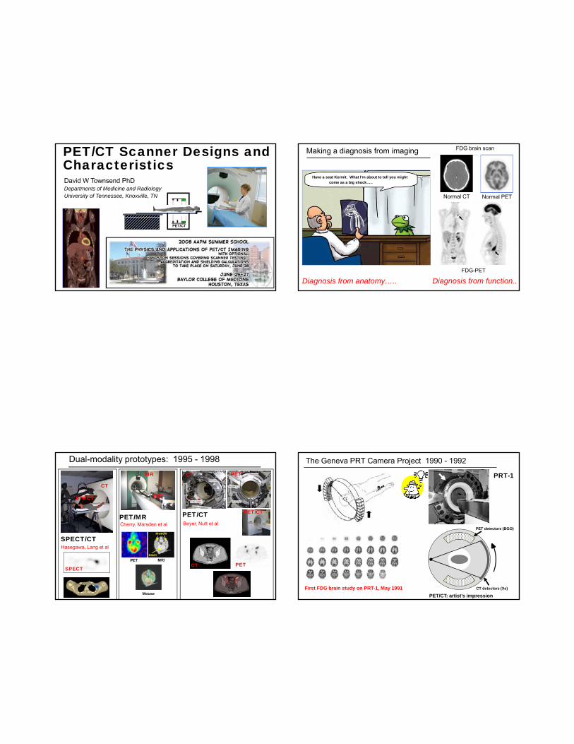
David W Townsend PhD Departments of Medicine and Radiology University of Tennessee, Knoxville, TN
PET/CT Scanner Designs and Characteristics
PET/CT
Have a seat Kermit. What I’m about to tell you might come as a big shock…..
Making a diagnosis from imaging FDG brain scan
Abnormal PETNormal PETNormal CT
Diagnosis from anatomy….. Diagnosis from function..FDG-PET
Dual-modality prototypes: 1995 - 1998
PET/CTPET/MRCherry, Marsden et al Beyer, Nutt et al
SPECT
CT
CT PET
PET
MR
PET/CT
SPECT/CTHasegawa, Lang et al
y,
Mouse
SPECTPETCT
The Geneva PRT Camera Project 1990 - 1992
PRT-1
First FDG brain study on PRT-1, May 1991 CT detectors (Xe)
PET detectors (BGO)
PET/CT: artist’s impression
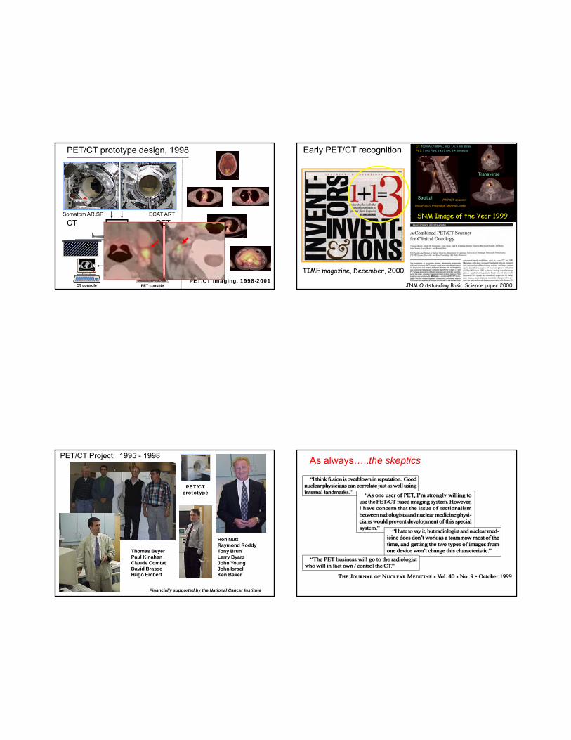
Somatom AR.SP ECAT ART
PET/CT prototype design, 1998
CT console PET console
PET/CT
So a o S C
CT PET
PET/CT imaging, 1998-2001
Early PET/CT recognition
PET/CT scanner
University of Pittsburgh Medical Center
CT: 160 mAs; 130 KVp; pitch 1.6; 5 mm slicesPET: 7 mCi FDG; 2 x 15 min; 3.4 mm slices
Sagittal
Transverse
SNM Image of the Year 1999
TIME magazine, December, 2000
SNM Image of the Year 1999
JNM Outstanding Basic Science paper 2000
PET/CT Project, 1995 - 1998
PET/CTprototype
Financially supported by the National Cancer Institute
Ron NuttRaymond RoddyTony BrunLarry ByarsJohn YoungJohn IsraelKen Baker
Thomas BeyerPaul KinahanClaude ComtatDavid BrasseHugo Embert
As always…..the skeptics
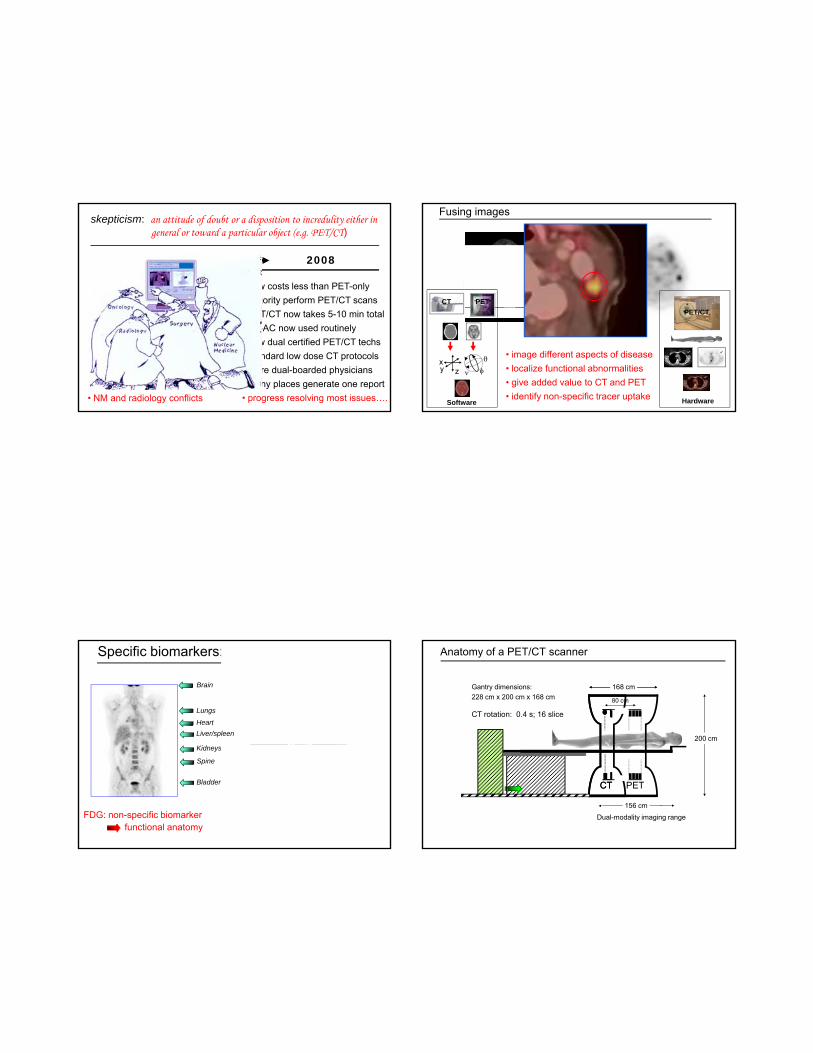
skepticism: an attitude of doubt or a disposition to incredulity either in general or toward a particular object (e.g. PET/CT)
• PET/CT will be too expensive • PET will restrict access to CT
• now costs less than PET-only• majority perform PET/CT scans
1999 2008
• CT: 5 min; but PET: 45 min• significant artifacts with CT-AC• makes poor use of CT tech time• gives unnecessary CT radiation• requires CT and NM physicians• results in two separate reports• NM and radiology conflicts
• PET/CT now takes 5-10 min total• CT-AC now used routinely• now dual certified PET/CT techs• standard low dose CT protocols• more dual-boarded physicians• many places generate one report• progress resolving most issues….
Fused image accuratelylocalizes uptake into alymph node and thus demonstrates spread of disease. Fused imagescan improve staging of head and neck cancer
Fusing images
CT PEThead and neck cancer
• image different aspects of disease• localize functional abnormalities• give added value to CT and PET• identify non-specific tracer uptake
xzy φ
θ
ν
Software Hardware
PET/CT
Specific biomarkers: 68Ga-DOTATOC
PET
PET/CT
Brain
HeartLiver/spleen
Lungs
58 year-old male. History of removal of carcinoid tumor. He then presented with a bone lesion read as degenerative bone disease from a bone scan. CT and MR were negative. Findings from the PET/CT changed patient management.
Michael Hofmann, MD
FDG: non-specific biomarkerfunctional anatomy
Kidneys
Bladder
Spine
Anatomy of a PET/CT scanner
80 cm
200 cm
168 cmGantry dimensions:228 cm x 200 cm x 168 cm
CT rotation: 0.4 s; 16 slice
CTCT PET
156 cmDual-modality imaging range
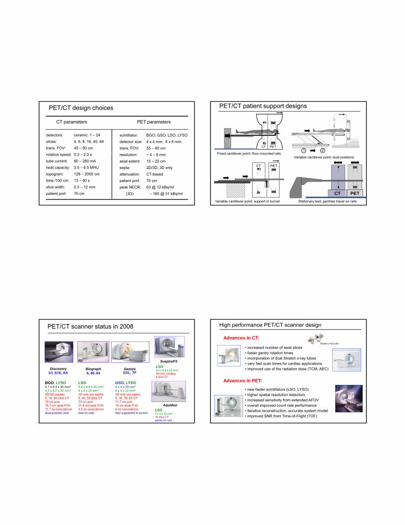
PET/CT design choices
detectors: ceramic; 1 – 24
slices: 4, 6, 8, 16, 40, 64
trans. FOV: 45 – 50 cm
t ti d 0 3 2 0
CT parameters PET parameters
scintillator: BGO; GSO; LSO; LYSO
detector size: 4 x 4 mm; 6 x 6 mm
trans. FOV: 55 – 60 cm
l i 4 6rotation speed: 0.3 – 2.0 s
tube current: 80 – 280 mA
heat capacity: 3.5 – 6.5 MHU
topogram: 128 – 2000 cm
time /100 cm: 13 – 90 s
slice width: 0.5 – 12 mm
patient port: 70 cm
resolution: ~ 4 – 6 mm
axial extent: 15 – 22 cm
septa: 2D/3D; 3D only
attenuation: CT-based
patient port: 70 cm
peak NECR: 63 @ 12 kBq/ml
(3D) – 160 @ 31 kBq/ml
PET/CT patient support designs
Fixed cantilever point; floor mounted rails
CT PET1 2
Fixed cantilever point; floor-mounted railsVariable cantilever point; dual positions
Variable cantilever point; support in tunnel
CT PET
CT PETStationary bed; gantries travel on rails
Discovery
PET/CT scanner status in 2008
Biograph GeminiGXL TF
SceptreP3LSO6.4 x 6.4 x 25 mm3
ST, STE, RX
BGO, LYSO4.7 x 6.3 x 30 mm3
4.2 x 6.2 x 30 mm3
2D/3D (septa)8, 16, 64 slice CT70 cm port15.7 cm axial FOV11.7 ns coincidencedual-position bed
6, 40, 64
LSO6.4 x 6.4 x 25 mm3
4 x 4 x 20 mm3
3D only (no septa)6, 40, 64 slice CT70 cm port21.8 cm axial FOV4.5 ns coincidencebed on rails
GXL, TF
GSO, LYSO4 x 4 x 30 mm3
4 x 4 x 22 mm3
3D only (no septa)6, 10, 16, 64 CT71.7 cm port18 cm axial FOV6 ns coincidencebed supported in tunnel
3D only; rotating4 slice CT
AquiduoLSO4 x 4 x 20 mm3
16 slice CTgantry on rails
High performance PET/CT scanner design
• increased number of axial slices• faster gantry rotation times• incorporation of dual Straton x-ray tubes• very fast scan times for cardiac applications• improved use of the radiation dose (TCM, AEC)
Advances in CT:Straton x-ray tube
• new faster scintillators (LSO, LYSO)• higher spatial resolution detectors• increased sensitivity from extended AFOV • overall improved count rate performance• iterative reconstruction, accurate system model• improved SNR from Time-of-Flight (TOF)
Advances in PET:
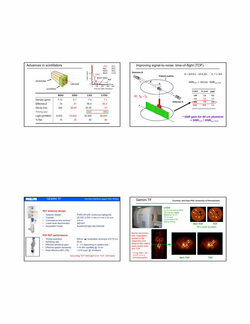
Advances in scintillators
BGO GSO LSO LYSO
DB
C γ (511 keV)
LSO: 69%NaI: 35%GSO: 20%BGO: 4.3%10
5
0
LSO
NaIBGO
0 100 200 300 400Time (ns) after interaction
Ligh
t out
put
GSO
scintillator
phototubes
7.13
74
300
8,200
15
7.4
66.4
35-45
500
30,000
80
BGO GSO LSO LYSO
6.7
61
30-60
10,000
25
Density (g/ml)
Effective Z
Decay (ns)
Timing (ps)
Light (ph/MeV)
% NaI
7.1
65.4
41
585
30,000
80
Improving signal-to-noise: time-of-flight (TOF)
δt (ps) δx (cm) SNR*
Detector B
e-e+
Patient outline
tA
tB
Δt = [(d+d1) – (d-d1)]/c ; d1 = c Δt/2
SNRTOF = √(D/Δd) · SNRnon-TOFd
d1
100300500
1200
1.54.57.5
18.0
5.23.02.31.5
* SNR gain for 40 cm phantom= SNRTOF / SNRnon-TOF
Detector A
Δt: tB - tA
GEMINI TF
PET detector design• Detector design: PIXELAR with continuous lightguide• Crystals: 28,336; LYSO: 4 mm x 4 mm x 22 mm• Coincidence time window: 3.8 ns• Lower level discriminator: 440 keV• Acquisition mode: Sustained high-rate listmode
Courtesy Matthias Egger PhD, Philips
TOF PET performance• Timing resolution: 650 ps localization accuracy of 9.75 cm• Sampling rate: 25 ps• Effective sensitivity gain: 2 – 4 x depending on patient size• Effective system sensitivity: > 14,400 cps/MBq @ 10 cm• Peak effective NEC (1R): > 210 kcps* @ 16 kBq/ml
*assuming TOF SNR gain (non-TOF: 105 kcps)
Gemini TF
LYSO4 x 4 x 22 mm3 (LYSO)3D only (no septa)Brilliance 16 CT70 cm port18 cm axial FOV585 ps timing
Non-TOF TOF60 s scan duration
Courtesy Joel Karp PhD, University of Pennsylvania
Non-TOF TOF
Rectal carcinoma, with metastases located in the mesentery and bilateral iliac chains more clearly seen with TOF.
114 kg; BMI = 38.112 mCi; 2 hr pi3min/bed position
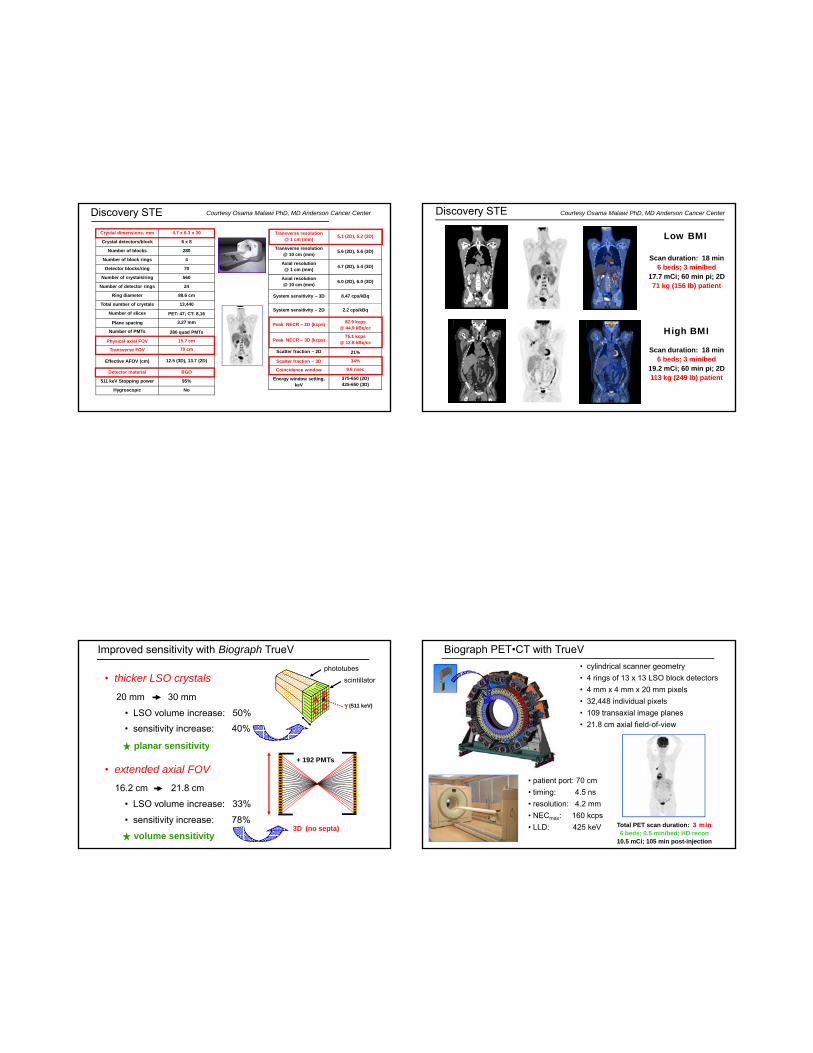
Discovery STE
Crystal dimensions, mm 4.7 x 6.3 x 30
Crystal detectors/block 6 x 8
Number of blocks 280
Number of block rings 4
Detector blocks/ring 70
Number of crystals/ring 560
Number of detector rings 24
Ring diameter 88.6 cm
Total number of crystals 13,440
Transverse resolution @ 1 cm (mm) 5.1 (2D), 5.2 (3D)
Transverse resolution @ 10 cm (mm) 5.6 (2D), 5.6 (3D)
Axial resolution @ 1 cm (mm) 4.7 (2D), 5.4 (3D)
Axial resolution @ 10 cm (mm) 6.0 (2D), 6.0 (3D)
System sensitivity – 3D 8.47 cps/kBq
Courtesy Osama Malawi PhD, MD Anderson Cancer Center
Number of slices PET: 47; CT: 8,16
Plane spacing 3.27 mm
Number of PMTs 280 quad PMTs
Physical axial FOV 15.7 cm
Transverse FOV 70 cm
Effective AFOV (cm) 12.5 (3D), 13.7 (2D)
Detector material BGO
511 keV Stopping power 95%
Hygroscopic No
System sensitivity – 2D 2.2 cps/kBq
Peak NECR – 2D (kcps) 87.9 kcps @ 44.9 kBq/cc
Peak NECR – 3D (kcps)75.1 kcps
@ 12.8 kBq/cc
Scatter fraction – 2D 21%
Scatter fraction – 3D 34%
Coincidence window 9.6 nsec
Energy window setting, keV
375-650 (2D)425-650 (3D)
Discovery STE
Scan duration: 18 min6 beds; 3 min/bed
17.7 mCi; 60 min pi; 2D71 kg (156 lb) patient
Courtesy Osama Malawi PhD, MD Anderson Cancer Center
Low BMI
Scan duration: 18 min6 beds; 3 min/bed
19.2 mCi; 60 min pi; 2D113 kg (249 lb) patient
High BMI
Improved sensitivity with Biograph TrueV
• thicker LSO crystals
DB
Cγ (511 keV)
20 mm 30 mm
• LSO volume increase: 50%
• sensitivity increase: 40%
scintillatorphototubes
planar sensitivity
• extended axial FOV
3D (no septa)
16.2 cm 21.8 cm
• sensitivity increase: 78%
• LSO volume increase: 33%
planar sensitivity
volume sensitivity
+ 192 PMTs
Biograph PET•CT with TrueV• cylindrical scanner geometry• 4 rings of 13 x 13 LSO block detectors• 4 mm x 4 mm x 20 mm pixels• 32,448 individual pixels• 109 transaxial image planes• 21.8 cm axial field-of-view
Total PET scan duration: 3 min6 beds; 0.5 min/bed; HD recon
10.5 mCi; 105 min post-injection
• patient port: 70 cm• timing: 4.5 ns• resolution: 4.2 mm• NECmax: 160 kcps• LLD: 425 keV
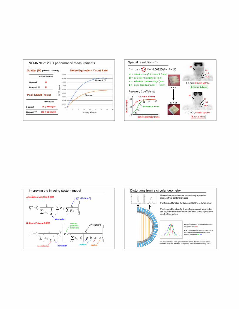
NEMA NU-2 2001 performance measurements
Noise Equivalent Count Rate
Biograph
Biograph TP 34
34
Scatter fraction
Scatter (%)
(kcp
s)
Biograph TP
(425 keV – 650 keV)
Peak NECR
96 @ 34 kBq/ml
161 @ 31 kBq/ml
Peak NECR (kcps)
Activity (kBq/ml)
NEC
R
Biograph
Biograph TP
Biograph
Spatial resolution (Γ)
8.6 mCi; 60 min uptake
2.0
3.91.3
3.3
6.4 mm x 6.4 mm
Γ = 1.25 √ {(d/2)2 + (0.0022D)2 + r2 + b2}
d = detector size (6.4 mm or 4.0 mm)D = detector ring diameter (mm)r = ‘effective’ positron range (mm)b = block decoding factor (~ 1 mm)
8 x 8
Sphere diameter (mm)
Rec
over
y (%
) 100
50
010
37282217
13
Recovery Coefficients
11.2 mCi; 90 min uptake
3.7
8.74.1
8.6
4 mm x 4 mm
6.4 mm x 6.4 mm
4.0 mm x 4.0 mm
13 x 13
Improving the imaging system model
⎥⎥⎥
⎦
⎤
⎢⎢⎢
⎣
⎡
⋅⋅⋅
⋅⋅= ∑ ∑∑
+
ij
kjji
iji
i iji
kj
kj ip
sp
ap
ii,
,
,
1
11
Attenuation-weighted OSEM
tt ti
{(P - R)·N – S}
attenuation
( )∑
∑∑⎥⎥⎥⎥⎥
⎦
⎤
⎢⎢⎢⎢⎢
⎣
⎡
+⋅⋅+⎟⎟⎠
⎞⎜⎜⎝
⎛⋅
⋅⋅⎟⎟⎠
⎞⎜⎜⎝
⎛⋅
⋅⋅=+
iiiii
j
kjji
iji
i iiji
kj
kj
cnraip
sp
anp
ii
,
,
,
1
11
Ordinary Poisson OSEM
attenuation
Prompts (P)
normalization
includes geometric distortions
randoms scatter
Distortions from a circular geometry
Point spread function for the central LORs is symmetrical
Point spread function for lines-of-response at large radius are asymmetrical and broader due to tilt of the crystal and depth of interaction
Lines-of-response become more closely spaced as distance from center increases
-12 -10 -8 -6 -4 -2 0 2 4 6 8 10 12
AW-OSEM linearly interpolates between sinogram bins ( )
PSF interpolates between sinogram bins with measured spatially-variant point spread functions ( )
The inclusion of the point spread function allows the simulation to better match the data with the effect of improving resolution and lowering noise

HD PET: modeling the system PSF
FORE + 2D OSEM
HD•PET(0mm filter)
3D-OSEM(2mm filter)
Brain Whole body
3D OSEM
HD•PET
Advances in reconstruction techniquesUT Molecular Imaging
3DRP7 thi
2D OSEM2i/14 thi
88 kg (194 lbs), BMI = 25Scan duration: 18 min
6 beds; 3 min/bed10.1 mCi; 88 min post-injection
168 x 168 matrix; CT-AC
7 mm smoothing
HD2i/14s; no smoothing
3D OSEM2i/14s; no smoothing
2i/14s; no smoothing
Reconstruction algorithms and SUV
FBP 2D: 1i / 8s 2D: 3i / 8s 2D: 5i / 8s 2D: 3i / 16s 3D: 3i / 8s
#1#3
#2
#1
#2
#3
8.7 / 6.1
3.8 / 2.8
8.2 / 5.0
9.8 / 6.6
5.9 / 4.2
10.1 / 6.2
9.9 / 6.6
5.6 / 4.1
9.9 / 6.6
9.6 / 6.5
5.7 / 3.9
10.0 / 5.9
9.7 / 6.6
5.9 / 4.2
10.0 / 6.4
9.5 / 6.4
5.2 / 3.8
9.0 / 5.7
SUVmax / SUVmeanTumor
Reconstruction method SUV @ 60 min SUV @ 90 min
2D AWOSEM 4i/8s 168 (5 mm)2D AWOSEM 4i/16s 168 (5 mm)3D AWOSEM 4i/8s 168 (5 mm)3D AWOSEM 4i/8s 336 (5 mm)3D AWOSEM 4i/8s 336 (none)
4.594.554.465.195.47
6.947.047.187.188.00
29% SUV h f diff t t ti• 29% SUV change for different reconstructions• 40% SUV change at two different time points
Iterations
Con
tras
t
Contrast = Mean ROI hot spot
Mean ROI background
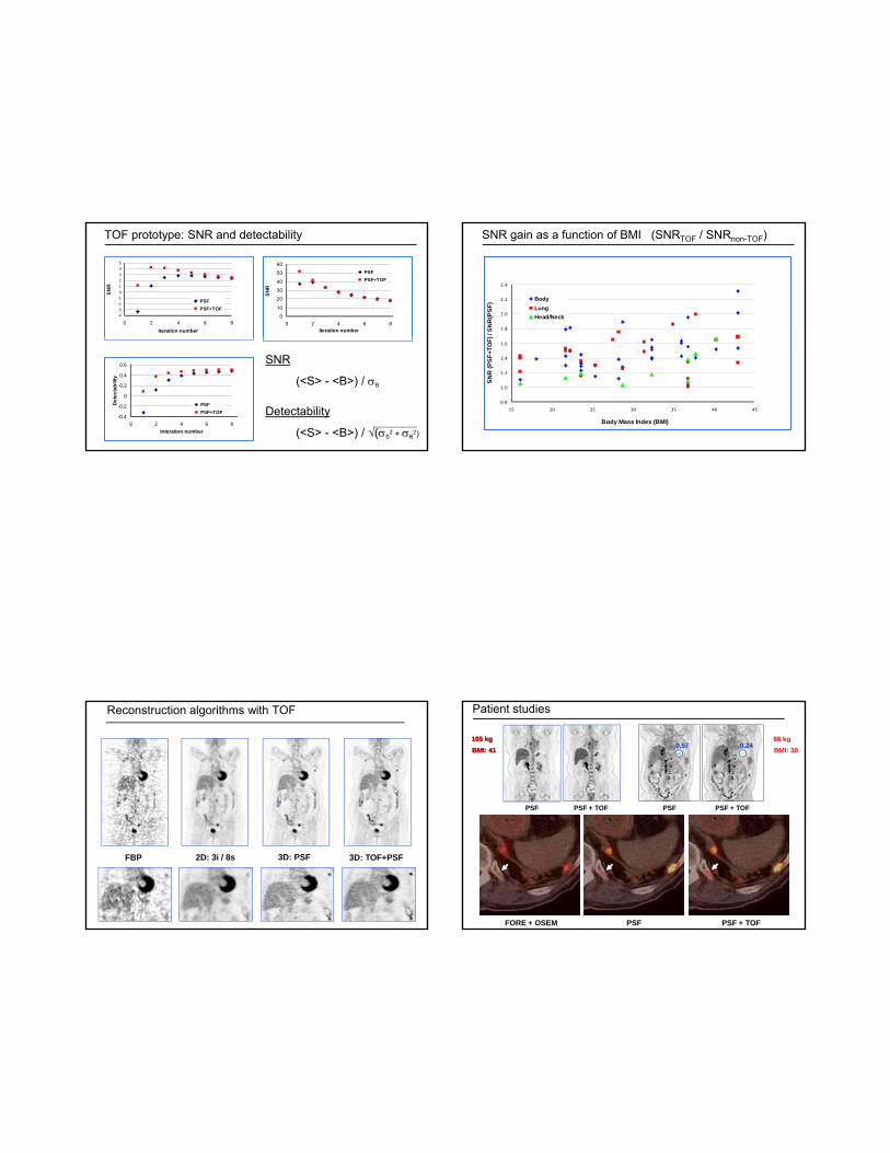
TOF prototype: SNR and detectability
‐4‐3
‐2‐10
123
45
0 2 4 6 8Iteration number
SNR
PSF PSF+TOF
0
10
20
30
40
50
60
0 2 4 6 8Iteration number
SNR
PSF PSF+TOF
-0.4
-0.2
0
0.2
0.4
0.6
0 2 4 6 8Interation number
Det
ecta
bilit
y
PSF PSF+TOF
SNR
(<S> - <B>) / σB
Detectability
(<S> - <B>) / √(σS2 + σB
2)
SNR gain as a function of BMI (SNRTOF / SNRnon-TOF)
1.8
2.0
2.2
2.4
/ S
NR
(PSF
) BodyLungHead/Neck
0.8
1.0
1.2
1.4
1.6
15 20 25 30 35 40 45
Body Mass Index (BMI)
SN
R (P
SF+
TOF)
Reconstruction algorithms with TOF
FBP 2D: 3i / 8s 3D: PSF 3D: TOF+PSF
Patient studies
PSF PSF + TOF PSF PSF + TOF
105 kgBMI: 41105 kgBMI: 41
88 kgBMI: 30
0.240.57
FORE + OSEM PSF PSF + TOF
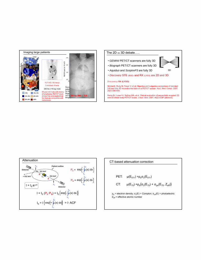
Imaging large patients
y = 79465e-0.0142x
y = 124843e-0.015x
1.E+04
1.E+05
1.E+06
NEC
R (c
ps) a
t 10
mC
i
106
105
104
NEC
R (c
ps) a
t 10
mC
i BiographBiograph TPNEMA phantom
1998
1990
10.7 mCi, 93 min pi 3 min/bed, 6 beds
260 lbs (118 kg) male
59 year-old male with history of lymphoma. PET/CT study shows hilar and mediastinal foci that are compatible with metastases.
1.E+0320 40 60 80 100 120 140 160
Weight (kg)
3 Rings (1080)4 Rings (1094)
Weight (kg)
103
20 40 60 80 100 120 140 160
N
Decrease in NECR as a function of patient weight. There are 15 patients for the Biograph and 9 patients for the Biograph TP. The NECR is for a 10 mCi injected dose. At 70 kg, the Biograph TP NEC1R is 50% higher.
204 kg; BMI = 70.515%–19% 20%–24%
2006
BMI ≥30
25%–29% ≥30%
<10% 10%–14%
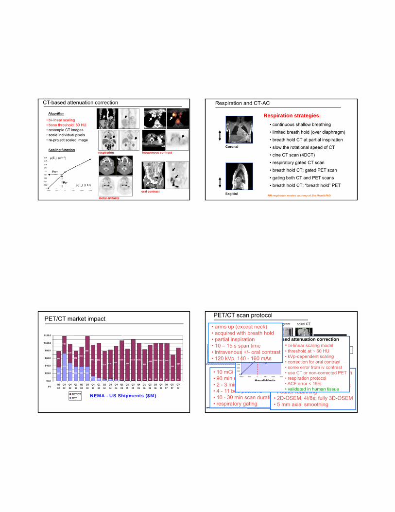
The 2D vs 3D debate…..
• GEMINI PET/CT scanners are fully 3D
• Biograph PET/CT scanners are fully 3D
• Aquiduo and SceptreP3 are fully 3D
• Discovery STE (BGO) and RX (LYSO) are 2D and 3D
2D3D
Discovery STE (BGO) and RX (LYSO) are 2D and 3D
Discovery RX (LYSO)
Strobel K, Rudy M, Treyer V, et al. Objective and subjective comparison of standard 2-D and fully 3D reconstructed data on a PET/CT system. Nucl. Med. Comm. 2007; 28(7):555-559.
Kemp BJ, Lowe VJ, Nathan MA, et al. Clinical evaluation of sequentially acquired 2D and 3D whole-body PET/CT scans. J Nucl. Med. 2007; 48(2):433P (abstract).
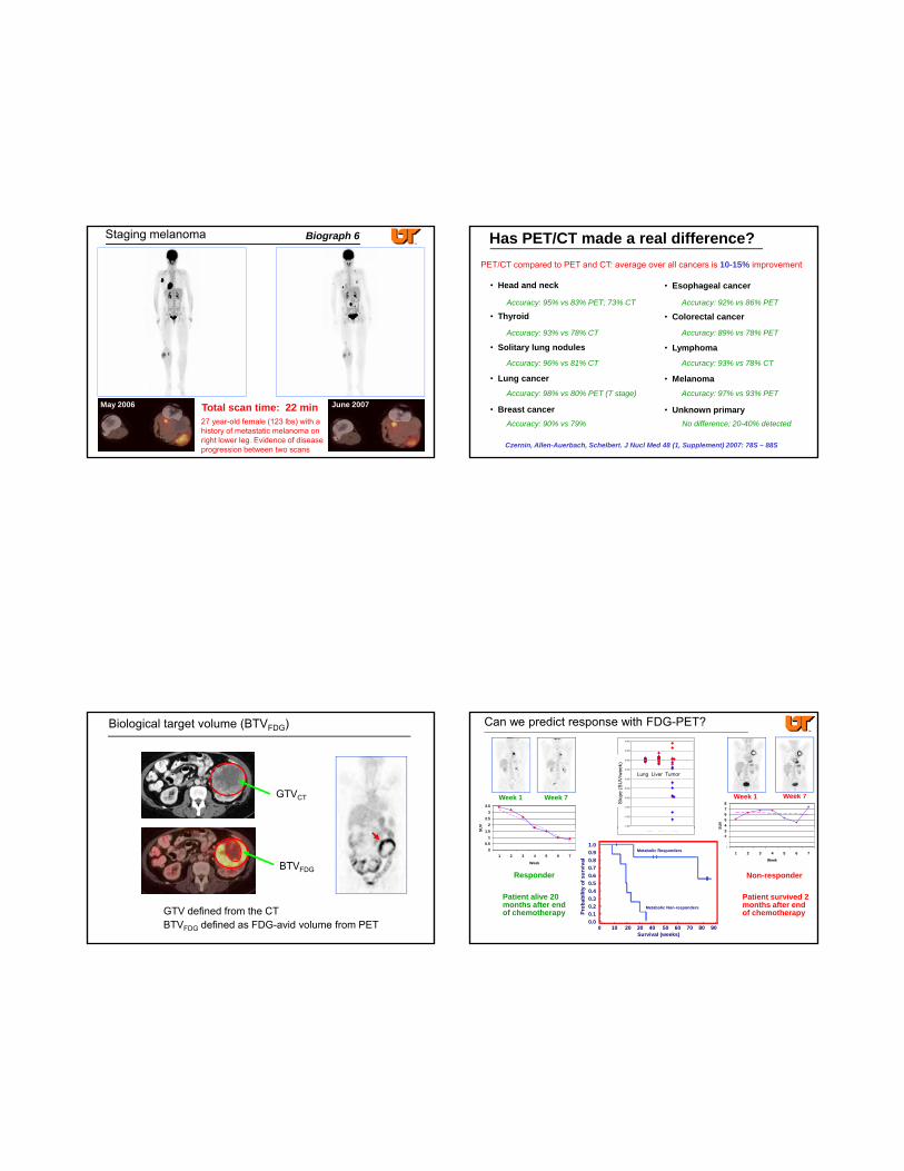
e-e+
511 keV< 511 keV
Detector511 keV
Patient outlinex2
Attenuation
x0
exp ∫x1
x0
A
B
PA = - μ(x) dx
PB = exp ∫x
x2
- μ(x) dx
I = I0 (PA·PB) = I0
Detectorx1
exp ∫x1
x2
- μ(x) dx
x0I = I0 e-μx
I0 = I exp ∫x1
x2
+ μ(x) dx = I· ACF
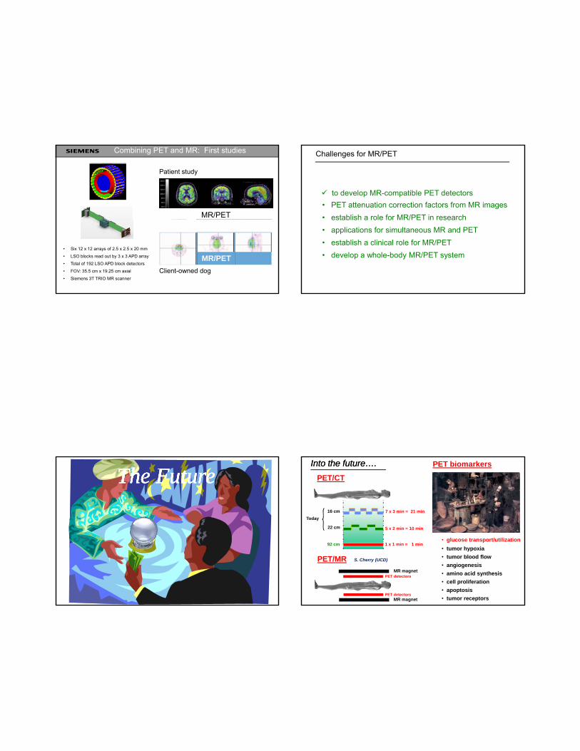
CT-based attenuation correction
PET: µ(E511) =ρeσc(E511)
CT: µ(E ) =ρ {σ (E ) + σ (E Z )}CT: µ(E70) =ρe{σc(E70) + σpe(E70, Zeff)}
ρe = electron density; σc(E) = Compton; σpe(E) = photoelectricZeff = effective atomic number
CT-based attenuation correction
• bi-linear scaling• bone threshold: 80 HU• resample CT images• scale individual pixels• re-project scaled image
Algorithm
Scaling functionintravenous contrast
oral contrast
respiration
metal artifacts
µ(Ex) (HU)
µ(Eγ) (cm-1)
µPET
µCT
Scaling function
Respiration and CT-AC
Respiration strategies:• continuous shallow breathing
• limited breath hold (over diaphragm)
• breath hold CT at partial inspiration
• slow the rotational speed of CTCoronal p
• cine CT scan (4DCT)
• respiratory gated CT scan
• breath hold CT; gated PET scan
• gating both CT and PET scans
• breath hold CT; “breath hold” PET
MR respiration movies courtesy of Jim Hamill PhDSagittal
$42
$49
$41$51
$82 $92$89
$60.0
$80.0
$100.0
$120.0
$110 $101$94 $100 $99$105
$45
$80
$99
PET/CT market impact
NEMA - US Shipments ($M) PET/CTPET
$37
$74
$48$39 $39 $39
$13 $8 $4 $6 $3
$74 $76 $89
$0.0
$20.0
$40.0
Q202
Q302
Q402
Q103
Q203
Q303
Q403
Q104
Q204
Q304
Q404
Q105FY
Q205
Q305
Q405
Q106
Q206
$1
$84
$1 $1 $0
$83
$0
Q306
Q406
$0 $0$1 $0 $0
$69
$0
$87
Q107
Q207
Q307
attenuation correction
Fusion
topogram spiral CT
CT PET
PET/CT scan protocol
CT scan
• bi-linear scaling model• threshold at ~ 60 HU• kVp-dependent scaling• correction for oral contrast0.08
0.1
0.12
0.14
0.16
0.18
water-air mix
water-bone mix
Mixing model: CT-based attenuation correction
• arms up (except neck)• acquired with breath hold• partial inspiration• 10 – 15 s scan time• intravenous +/- oral contrast• 120 kVp, 140 - 160 mAs
attenuation correction
Reconstruction
PET imagefused image
CT PET
CT PET
PET scan• 10 mCi of FDG• 90 min uptake period• 2 - 3 min per bed position• 4 - 11 bed positions• 10 - 30 min scan duration• respiratory gating
• CT-based attenuation correction• model-based scatter correction• 168 x 168 reconstruction matrix• Fourier rebinning• 2D-OSEM, 4i/8s; fully 3D-OSEM• 5 mm axial smoothing
• correction for oral contrast• some error from iv contrast• use CT or non-corrected PET• respiration protocol• ACF error < 15%• validated in human tissue
0
0.02
0.04
0.06
-1000 -500 0 500 1000 1500
Hounsfield units
mix

PET/CT:
- for staging diseaseg g- for therapy planning- for monitoring response
Molecular Imaging andTranslational Research
Mandibular cancer
83 year-old female withmandibular cancer. PET/CTscan acquired pre-surgeryidentified 3 left-side positivenodes 5-12 mm in size with increased FDG uptake. Postsurgery, pathology identified 35 nodes positive for cancer.
biograph 16
Primary (1.5 x 3.8 cm) Nodes (12 mm, 7 mm, 5 mm)
5 mm lytic spine lesion Bone lesions, 6-7 mm in diameter
Lung cancer Biograph 6 TrueVMolecular Imaging
Program
10.8 mCi, 92 min pi 2 min/bed, 5 beds
3i / 8s; 5 mm130 kVp; 50 mAs
Scan duration: 10 min PET/CT
44 year-old male (6’, 118 lbs) with recent diagnosis of lung cancer. Smoker for 26 years. Loss of voice and hoarseness. Shortness of breath and 25 lb weight loss in one month. Recent chest pain. Uptake in lymph node obstructing breathing and left pulmonary artery.
Restaging breast cancer Biograph 6
Feb 2007
CT
Total scan time: 18 min71 year-old female (BMI: 25.8) with a history of breast cancer and Merkel cell cancer of the chin. Extensive bone marrow disease of axial and proximal appendicular skeleton. Deceased 7/07.
Patient received radiation therapy to T11 and T12 and right and left lateral chin and neck. Sparing of T9-L1 and cervical spine10.2 mCi; 96 min; 6 beds @ 3 min
PET/CT
July 2007
Staging melanoma Biograph 6
Total scan time: 22 minMay 2006 June 2007
27 year-old female (123 lbs) with a history of metastatic melanoma on right lower leg. Evidence of disease progression between two scans
Has PET/CT made a real difference?PET/CT compared to PET and CT: average over all cancers is 10-15% improvement
• Head and neck
• Thyroid
• Esophageal cancer
• Colorectal cancerAccuracy: 95% vs 83% PET; 73% CT
Accuracy: 93% vs 78% CT
Accuracy: 92% vs 86% PET
Accuracy: 89% vs 78% PET
• Solitary lung nodules
• Lung cancer
• Breast cancer
• Lymphoma
• Melanoma
• Unknown primary
Accuracy: 96% vs 81% CT
Accuracy: 98% vs 80% PET (T stage)
Accuracy: 90% vs 79%
Accuracy: 93% vs 78% CT
Accuracy: 97% vs 93% PET
No difference; 20-40% detected
Czernin, Allen-Auerbach, Schelbert. J Nucl Med 48 (1, Supplement) 2007: 78S – 88S
Biological target volume (BTVFDG)
GTVCT
BTVFDG
GTV defined from the CTBTVFDG defined as FDG-avid volume from PET
Can we predict response with FDG-PET?
45678
SUV
Week 1 Week 7 Week 7Week 1
22.5
33.5
UV -1.400
-1.200
-1.000
-0.800
-0.600
-0.400
-0.200
0.000
0.200
0.400
Slop
e (S
UV
/ wee
k) Lung Liver Tumor
Slo
pe (S
UV
/wee
k)
Responder
Patient alive 20 months after end of chemotherapy
0123
1 2 3 4 5 6 7
Week
S
Non-responder
Patient survived 2 months after end of chemotherapy
00.5
11.5
1 2 3 4 5 6 7
Week
SU
Metabolic Responders
Metabolic Non-responders
Survival (weeks)0 10 20 30 40 50 60 70 80 90
1.00.90.80.70.60.50.40.30.20.10.0
Prob
abili
ty o
f sur
viva
l
Lung Liver Tumor
Combining PET and MR: First studies
PET
MR/PET
Patient study
MR
MR/PET• Six 12 x 12 arrays of 2.5 x 2.5 x 20 mm
• LSO blocks read out by 3 x 3 APD array
• Total of 192 LSO APD block detectors
• FOV: 35.5 cm x 19.25 cm axial
• Siemens 3T TRIO MR scannerClient-owned dog
Challenges for MR/PET
• PET attenuation correction factors from MR images • establish a role for MR/PET in research
to develop MR-compatible PET detectors
establish a role for MR/PET in research• applications for simultaneous MR and PET• establish a clinical role for MR/PET• develop a whole-body MR/PET system
The FutureThe FutureThe FutureThe FutureInto the future….Into the future….
7 x 3 min = 21 min
5 x 2 min = 10 min
16 cm
22 cm
Today
PET/CT
PET biomarkers
1 x 1 min = 1 min92 cm• glucose transport/utilization• tumor hypoxia• tumor blood flow• angiogenesis• amino acid synthesis• cell proliferation• apoptosis• tumor receptors
PET/MR S. Cherry (UCD)
MR magnet
MR magnet
PET detectors
PET detectors
Jon Wall, PhDKarl Hubner, MDChris Guglielmo, MDGeorge Kabalka, PhDSteve Kennel, PhDWenben Zeng, PhDW i i Mi PhD
UTGSM Molecular Imaging and Tracer Development Program Acknowledgements:
Weimin Miao, PhDBjoern Jakoby, MSCristina Lois, PhDJosh Schaefferkoetter, BSMisty Long, R.T. (R) (N)Chris Carr, R.T. (R) CNMTAlan Stuckey, CNMTPam TrenthamNational Cancer Institute
…and now on your iPhone
Related Documents


