REVIEW ARTICLE Perspectives of purinergic signaling in stem cell differentiation and tissue regeneration Talita Glaser & Angélica Regina Cappellari & Micheli Mainardi Pillat & Isabele Cristiana Iser & Márcia Rosângela Wink & Ana Maria Oliveira Battastini & Henning Ulrich Abstract Replacement of lost or dysfunctional tissues by stem cells has recently raised many investigations on therapeutic applications. Purinergic signaling has been shown to regulate proliferation, differentiation, cell death, and successful engraftment of stem cells originated from diverse origins. Adenosine triphosphate release occurs in a controlled way by exocytosis, transporters, and lysosomes or in large amounts from damaged cells, which is then subsequently degraded into adenosine. Paracrine and auto- crine mechanisms induced by immune responses present critical factors for the success of stem cell therapy. While P1 receptors generally exert beneficial effects including anti-inflammatory activity, P2 receptor-mediated actions depend on the subtype of stimulated receptors and localization of tissue repair. Pro-inflammatory actions and excitatory tissue damages mainly result from P2X7 receptor activation, while other purinergic receptor subtypes partic- ipate in proliferation and differentiation, thereby providing adequate niches for stem cell engraftment and novel mechanisms for cell therapy and endogenous tissue repair. Therapeutic applications based on regulation of purinergic signaling are foreseen for kidney and heart muscle regeneration, Clara-like cell replacement for pulmonary and bronchial epithelial cells as well as for induction of neurogenesis in case of neurodegenerative diseases. Keywords ATP . Adenosine nucleotides . Purinergic signaling . Tissue injury . Differentiation . Immune system An overview of purinergic signaling Receptors for purines and pyrimidines are classified based on their agonist specificity. P1 receptors subtypes are selective for adenosine and are classical 7-transmembrane metabotropic receptors coupled to several families of Gi, Go, and Gs proteins. There are four types of adenosine receptors (A 1 , A 2A , A 2B , and A 3 ) differing in their pharmacological and functional properties [1]. P2 receptors are divided into P2X and P2Y subtypes based on their structural characteristics. P2X receptors are ATP-activated, ligand-gated cationic (Na + /K + /Ca 2+ ) channels [2, 3], as- sembled in trimeric form from P2X1 to P2X7 subunits [1, 3]. Metabotropic P2Y purinoceptors expressed by mamma- lians are divided into P2Y 1,2,4,6,11,12,13,14 subtypes based on phylogenetic similarity and are stimulated by ATP, ADP, UTP, UDP, or UDP glucose [1]. Purinergic receptors are expressed by almost every cell type and are one of the first expressed neurotransmitter receptors in development [4–6]. The extracellular nucleotide/nucleoside availability is con- trolled by a highly efficient enzymatic cascade, which includes the members of the ectonucleoside triphosphate diphosphohydrolase (E-NTPDases, NTPDase1–8), ectonu- cleotide pyrophosphatase/phosphodiesterase (E-NPPs), ecto-alkaline phosphatases, and ecto-5′ -nucleotidase/ CD73. These enzymes catalyze the complete nucleotide T. Glaser : M. M. Pillat : H. Ulrich (*) Departamento de Bioquímica , Instituto de Química, Universidade São Paulo, Av. Prof. Lineu Prestes, 748-Bloco 8S/Room 0858, CEP: 05508-900 São Paulo, SP, Brazil e-mail: [email protected] A. R. Cappellari : A. M. O. Battastini Departamento de Bioquímica, ICBS, Universidade Federal do Rio Grande do Sul, Porto Alegre, RS, Brazil I. C. Iser : M. R. Wink Departamento de Ciências Básicas da Saúde, Universidade Federal de Ciências da Saúde de Porto Alegre, Porto Alegre, RS, Brazil Purinergic Signalling (2012) 8:523–537 DOI 10.1007/s11302-011-9282-3 Received: 13 September 2011 /Accepted: 9 November 2011 /Published online: 6 December 2011 # Springer Science+Business Media B.V. 2011

Welcome message from author
This document is posted to help you gain knowledge. Please leave a comment to let me know what you think about it! Share it to your friends and learn new things together.
Transcript

REVIEW ARTICLE
Perspectives of purinergic signaling in stem celldifferentiation and tissue regeneration
Talita Glaser & Angélica Regina Cappellari & Micheli Mainardi Pillat &Isabele Cristiana Iser & Márcia Rosângela Wink & Ana Maria Oliveira Battastini &Henning Ulrich
Abstract Replacement of lost or dysfunctional tissues bystem cells has recently raised many investigations ontherapeutic applications. Purinergic signaling has beenshown to regulate proliferation, differentiation, cell death,and successful engraftment of stem cells originated fromdiverse origins. Adenosine triphosphate release occurs in acontrolled way by exocytosis, transporters, and lysosomesor in large amounts from damaged cells, which is thensubsequently degraded into adenosine. Paracrine and auto-crine mechanisms induced by immune responses presentcritical factors for the success of stem cell therapy. WhileP1 receptors generally exert beneficial effects includinganti-inflammatory activity, P2 receptor-mediated actionsdepend on the subtype of stimulated receptors andlocalization of tissue repair. Pro-inflammatory actions andexcitatory tissue damages mainly result from P2X7 receptoractivation, while other purinergic receptor subtypes partic-ipate in proliferation and differentiation, thereby providingadequate niches for stem cell engraftment and novelmechanisms for cell therapy and endogenous tissue repair.Therapeutic applications based on regulation of purinergic
signaling are foreseen for kidney and heart muscleregeneration, Clara-like cell replacement for pulmonaryand bronchial epithelial cells as well as for induction ofneurogenesis in case of neurodegenerative diseases.
Keywords ATP. Adenosine nucleotides . Purinergicsignaling . Tissue injury . Differentiation . Immune system
An overview of purinergic signaling
Receptors for purines and pyrimidines are classified basedon their agonist specificity. P1 receptors subtypes areselective for adenosine and are classical 7-transmembranemetabotropic receptors coupled to several families of Gi,Go, and Gs proteins. There are four types of adenosinereceptors (A1, A2A, A2B, and A3) differing in theirpharmacological and functional properties [1]. P2 receptorsare divided into P2X and P2Y subtypes based on theirstructural characteristics. P2X receptors are ATP-activated,ligand-gated cationic (Na+/K+/Ca2+) channels [2, 3], as-sembled in trimeric form from P2X1 to P2X7 subunits [1,3]. Metabotropic P2Y purinoceptors expressed by mamma-lians are divided into P2Y1,2,4,6,11,12,13,14 subtypes based onphylogenetic similarity and are stimulated by ATP, ADP,UTP, UDP, or UDP glucose [1]. Purinergic receptors areexpressed by almost every cell type and are one of the firstexpressed neurotransmitter receptors in development [4–6].The extracellular nucleotide/nucleoside availability is con-trolled by a highly efficient enzymatic cascade, whichincludes the members of the ectonucleoside triphosphatediphosphohydrolase (E-NTPDases, NTPDase1–8), ectonu-cleotide pyrophosphatase/phosphodiesterase (E-NPPs),ecto-alkaline phosphatases, and ecto-5′-nucleotidase/CD73. These enzymes catalyze the complete nucleotide
T. Glaser :M. M. Pillat :H. Ulrich (*)Departamento de Bioquímica , Instituto de Química,Universidade São Paulo,Av. Prof. Lineu Prestes, 748-Bloco 8S/Room 0858,CEP: 05508-900 São Paulo, SP, Brazile-mail: [email protected]
A. R. Cappellari :A. M. O. BattastiniDepartamento de Bioquímica, ICBS,Universidade Federal do Rio Grande do Sul,Porto Alegre, RS, Brazil
I. C. Iser :M. R. WinkDepartamento de Ciências Básicas da Saúde,Universidade Federal de Ciências da Saúde de Porto Alegre,Porto Alegre, RS, Brazil
Purinergic Signalling (2012) 8:523–537DOI 10.1007/s11302-011-9282-3
Received: 13 September 2011 /Accepted: 9 November 2011 /Published online: 6 December 2011# Springer Science+Business Media B.V. 2011

hydrolysis (e.g., ATP) to nucleosides (e.g., adenosine) andrepresent a powerful tool for controlling the effectsmediated by extracellular purines [7–9].
Stem cells and purinergic signaling
Replacement of lost or dysfunctional tissues has recentlyraised many investigations on possible therapeutic applica-tion of stem cells. An impressive number of clinical trialsand animal studies have already been performed todetermine the therapeutic potential of various stem cellsmodels [10, 11]. The first isolation of embryonic stem (ES)cells from mouse goes back to 1981 followed by human EScell isolation and culture in 1998 [12, 13]. Organ-specificstem cells were isolated from embryonic and adult tissuesincluding brain, bone marrow, umbilical cord, skeletal andcardiac muscles, and adipose tissue [14]. Pluripotent EScells are capable to originate any somatic cell type, whiletissue-specific stem cells are mostly multipotent andsubsequently originate cell types found in these specifictissues. Both, ES and tissue-specific stem cells canproliferate symmetrically replicating themselves for self-renewal or asymmetrically giving rise to a stem cell andanother more differentiated cell type. The most promisingand recently discovered stem cell model for basic researchand even therapy is the induced pluripotent stem cell (iPScell), reprogrammed in 2006 from differentiated mousecells and in 2007 from human cells [15, 16]. The recentdescribed capacity of genetically reprogrammed somaticcells towards pluripotent ones could bypass obstacles, suchas the lack of histocompatibility and ethical concerns, byallowing the generation of autologous cells from thepatient. This new pluripotent cell source initially obtained byoverexpression of the genes Klf-4, Oct4, Sox2, and c-Mycresponsible for pluripotency [reviewed by 17] has openedexpectations for treatment of many diseases. Importantly, iPScells derived from different species demonstrated the potentialto differentiate into tissues derived from the three germ layers,such as known from ES cells. However, care must be takenon using these cells as well as ES cells for transplantationpurposes due to their possible tumorigenic potential.
Therapeutic application of stem cells in patients isparticularly promising for treatment of heart disease, wherenew cardiomyocytes could restore contractile function aftermyocardial infarction. Cell regeneration therapy could bealso relevant for repair of pancreatic function in diabeteswith the replacement of β insulin-secreting cells [18].Further possible applications are foreseen for the treatmentof the damaged neuronal system and neurodegenerativediseases. For instance, efforts are being made to replacedopaminergic neurons in Parkinson’s disease [19] or to usethe stem cell therapy to restore motorneuron function in
patients suffering from spinal cord injuries [16]. However,the little obtained progress in many cases did not satisfy thehigh expectations made. Moreover, observed functionalimprovements observed in the treated tissues did not oftenresult from the integration of stem cells into existing tissuearchitectures. It is evident that transplanted cells contributeto endogenous tissue repair through paracrine mechanismsmore than by differentiating themselves. For instance, thesuccess of neural progenitor cell (NPC) engrafting into thespinal cord of Sprague–Dawley rats, subjected to contusionat T8–T9 levels, was limited by allodynia due to the deathof transplanted cells [7]. However, injection of conditionedmedia recovered from cultured stem cells promoted arterio-genesis and functional improvement when injected into thedamaged heart [20]. Therefore, it has been postulated thattrophic factors represent the principle mechanism responsi-ble for tissue repair.
Usual strategies for cell replacement therapy are based onthe isolation of a stem cell source from a donor or the patient,followed by induction to proliferate and/or differentiate intotissue types which shall be repaired. Cell death and rejection oftransplanted cells are mostly due to immune responses and theabsence of adequate stem cell niches at the localization oftransplantation. Although mechanisms by which the localmilieu influences stem cell differentiation and tissue engraft-ment need yet to be elucidated, it seems that the fate of bonemarrow stem cells is determined by the environment in whichthey engraft rather than by an intrinsically programmed fate.As support for such hypothesis, positive inotropic (pharmaco-logic augmentation of contractility) or chronotropic stimuli(heart rate increase by exercise) promoted and intensified thedifferentiation of bone marrow-derived stem cells intocardiomyocyte phenotypes [21]. Furthermore, stem cellssecret trophic and immunomodulatory factors controllinglocal and systematic inflammatory responses. Such factors,liberated by, i.e., bone marrow stem cells are therapeuticallyimportant, since they stimulate local tissue regeneration and/or recruitment of endogenous stem or progenitor cells.Moreover, some studies have demonstrated that mesenchymalstem cells (MSC) can diminish the apoptosis degree andinfarct size of the damaged areas by secreting a wide range ofcytoprotective molecules like vascular endothelial growthfactor, basic fibroblast growth factor, insulin-like growthfactor 1, stromal cell-derived factor-1, platelet-derived growthfactor, interleukin-1 beta, or hepatocyte growth factor [22].
Other factors with such therapeutic potential are UTP,UDP, ADP, and adenosine acting through purinergicreceptors. Nucleotides, released after tissue injury and celldeath and hydrolyzed by ectonucleotidases, also regulateimmune cell function induced by damage-associated mo-lecular pattern molecules [23]. Moreover, ATP released fromimmune cells participates in autocrine as well as in paracrinefeedback loops with regulatory functions during T-cell
524 Purinergic Signalling (2012) 8:523–537

activation in the immune synapse (junction between T cell andantigen-presenting cell) [24]. During the inflammatory processfollowing cell transplantation and hindering repair, purinesexert trophic functions and keep several immune functionsunder control, including the release of prostanoids, activationof matrix metalloproteinase-9, cytokines and chemokines,proliferation, differentiation/maturation and stimulation ofimmune cells, endothelial adhesion, free radical production,degranulation, phagocytosis, fusion, and cell death [25].Depending on the involved purinergic receptor subtype, ATPoften exerts proinflammatory effects while adenosine inducesmainly anti-inflammatory effects [25]. Several studies demon-strated that the absence or inhibition of the P2X7 receptor (amediator of the pro-inflammatory effects of ATP) results in lesssevere outcomes in chronic inflammatory diseases andenhanced functional recovery [23, 26, 27].
Besides importance of purinergic receptor agonists indifferentiated immune cells, these compounds also modulatehematopoietic stem cell (HSC) self-renewal, expansion, anddifferentiation with implications not only in hematopoiesis,but also in tissue repair and regenerative medicine [28, 29].For instance, ATP induces the proliferation of human HSCand contributed through P2X receptor activation duringinflammation process [29, 30]. UTP also induces prolifera-tion and migration of HSCs [30, 31] while adenosinepotentiates the stimulatory effect of growth factors andcytokines on HSC proliferation and differentiation [8].Moreover, human MSCs at early stages of culture (P0–P5)spontaneously release ATP reducing cell proliferation.Increased human MSC proliferation is induced by theunselective P2 receptor antagonist pyridoxalphosphate-6-azophenyl-2′,4′-disulfonate (PPADS) and by the selectiveP2Y1 receptor antagonist 2′-deoxy-N6-methyladenosine-3′,5′-bisphosphate (MRS 2179). In summary, ATP modulatesHSC and MSC proliferation and likely acts as one of theearly factors determining their cell fate [32]. Furthermore,nucleotides also contribute to inflammatory responses andcell fate decisions occurring in the brain. P2X7 receptorsexpressed by NPCs are responsible for cell death, being inagreement with observations that high levels of extracellularATP in inflammatory central nervous system (CNS) lesionshinder successful NPC engraftment [33].
The extracellular nucleotide/nucleoside availability iscontrolled by a highly efficient enzymatic cascade, whichincludes the members of the E-NTPDases (NTPDase1–8),E-NPPs, ecto-alkaline phosphatases, and ecto-5′-nucleotid-ase/CD73. These enzymes are responsible for nucleotidehydrolysis (e.g. ATP) into nucleosides (e.g., adenosine) andrepresent a powerful mechanism for controlling the effectsmediated by extracellular purines [9, 34]. Althoughpurinergic signaling has been extensively studied, onlyfew studies are found in the literature demonstrating theinvolvement of extracellular nucleotide metabolizing
enzymes in stem cell biology. Expression and activities ofmembers of ectonucleotidase families as well as purinergicreceptor subtypes have been detected in different types ofstem and progenitor cells. Recent works have identified thepresence of NTPDase2 in adult mouse hippocampalprogenitors [35] and in type B cells of the subventricularzone (SVZ) [36], two neurogenic regions of the adultmammalian brain. In accordance, neurospheres culturedfrom the adult mouse SVZ express NTPDase2, the tissuenonspecific isoform of alkaline phosphatase (TNAP) andfunctional P2 receptors in synergism with growth factorsfor enhancing cell proliferation [37]. In addition, deletion ofTNAP expression or inhibition of its enzymatic activity inneural progenitors reduces cell proliferation and differenti-ation into neurons or oligodendrocytes [38]. These pub-lished data corroborate the importance of NTPDase2 andTNAP, two potential ATP scavengers, as novel markers forprogenitor cells both in the adult and developing brain [39].Reinforcing these results, spontaneous ATP release wasobserved in murine NPCs and, interestingly, purinergicreceptors antagonists were able to suppress progenitor cellproliferation [40]. Moreover, neuronal differentiation wasaccompanied by a decrease in ATP release and a loss offunctional P2Y receptors, suggesting that purine nucleo-tides act as proliferation-inducing factors for NPCs anddownregulators of neuronal differentiation, once againpointing at the importance of purinergic signaling andinvolved enzymes for neurogenesis in the adult brain [40].These data are in agreement with results of our laboratory[41], showing down-regulation of P2Y1 receptor expres-sion and activity in differentiating P19 mouse embryonalcarcinoma cells. This observation is in line with functionsof the P2Y1 subtype in promoting proliferation of undif-ferentiated cells, but not induction of neuronal differentia-tion. Finally, the studies presented here demonstrate thepotential participation of ectonucleotidases in the biology ofstem or progenitor cells from different tissues. Initial resultson roles of these ecto-enzymes will encourage more studiesfor better understanding of their importance in stem cellbiology, differentiation, and tissue repair. In the following,we will discuss new trends of stem cell research related topurinergic signaling and the perspectives of using thesediscoveries as tools for future tissue repair in clinical trialsas this new approach develops (see Fig. 1 for a scheme ofthe possible therapeutic use of purines in combination withstem cells).
Purinergic signaling and perspectives in tissueregeneration
Implications of the purinergic system in stem cell biologyand tissue regeneration will be discussed with emphasis on
Purinergic Signalling (2012) 8:523–537 525

the recent hypothesis that paracrine effects present the mostimportant mechanisms in this process. Since this idea isvery recent, few data are available directly relating thepurinergic system with stem cell differentiation and tissueregeneration; however, the authors of this review areconfident that the present article will encourage researchin order to better understand the participation of purinergicsignaling in this context.
Heart injury
The heart is an organ composed basically of fibroblasts andcardiomyocytes, terminal-differentiated cells which give theheart the pumping ability. During ischemia and otherinjuries, the most affected cells are the cardiomyocytesbecause they die and a scar is formed due to the inability ofrenewing these cells. The scar stiffens the heart, decreasingits capability and efficiency in pumping the blood.Therefore, intense efforts are being made for the restorationof lost cells by cell therapy and maintenance of cardiacfunction in patients with heart injury. Many stem cells typeshave been studied in order to select the best model forcardiac cell therapy. ES cells, iPS cells as well as adult stemcells (bone marrow, adipose tissue-derived, and cardiacstem cells) are already tested in animal models and humanswith often promising results [42, 43].
The most promising model is provided by cardiac stemcells (CSCs) that reside in small populations in the adultmammalian myocardium and have the potential to differ-entiate into cardiomyocytes and other cell types, such asendothelial and vascular smooth muscle cells [44–47].However, differentiation of these cells is rare underphysiological conditions [48]. For therapeutic purposes,CSCs can be generated by expanding autologous cells exvivo or stimulating the regeneration capacity of these cellsin vivo. Nevertheless, one of the biggest problemshindering the therapeutic use of stem cells lies still in thedifficulty of keeping stem cells alive following transplan-tation. Cell death occurs before cells can engraft in theirenvironment due to inflammation-signaling responses, orcells do not even identify the injured tissue site forengraftment. Therefore, signaling factors necessary for cellestablishment at the location of transplantation are beinginvestigated. Such paracrine factors include ATP andadenosine and their respective receptor subtypes. P2Y14receptors expressed by bone marrow HSCs induce migra-tion of these cells to the localization of injury followed byinduction of differentiation at the site mediated byactivation of other purinergic receptors [49] (Fig. 2).Adenosine plays many roles in the heart includingregulation of growth, differentiation, angiogenesis, coro-nary blood flow, cardiac conduction and heart rate, substrate
Fig. 1 Therapeutic potential of stem cells and supposed effects ofpurinergic signaling. Stem cells of diverse origins, such as fromadipose, cardiac, and neural tissues can restore and regeneratedamaged tissues by secreting paracrine factors including purines andpyrimidines. ATP and adenosine interfere with tissue reactionsfollowing transplantation of stem cells of various origins in differentways. (1) Nucleotides modulate the immune response and therebyreduce inflammation processes and the risk of transplant rejection andcell death. (2) Purines and pyrimidines promote proliferation anddifferentiation of transplanted and endogenous stem cells by providing
adequate stem cell niches. (3) Purines and pyrimidines inducemigration of endogenous stem cells to the site of injury and increaseengraftment rates. Stem cell types with therapeutic applications arehuman induced-pluripotent stem cells (hiPSC), human embryonicstem cells (hESC), adipose stem cells (ASC), cardiac stem cells(CSC), neural stem cells (NSC), bone marrow stem cells (BMSC), andumbilical cord stem cells (UCSC) which are transplanted by usingstereotaxic surgery (SS), intracoronary retrograde infusion throughcoronary sinus (IRICS) or intravenous, intramyocardial, or intraperi-toneal injection or lumbar puncture
526 Purinergic Signalling (2012) 8:523–537

metabolism, and sensitivity to adrenergic stimulation [50],and also functions as an endogenous determinant of ischemictolerance [50]. The two A2 receptor subtypes (A2A andA2BA) possess important anti-inflammatory and immuno-modulatory functions, and probably control the impact ofinflammatory processes during ischemic and post-ischemicdamage. Vinten-Johansen and colleagues confirm protectivefunctions of A2A receptors in cardiac tissue by inhibition ofneutrophil activation and neutrophil–vascular interactions asseen in Fig. 2 [51, 52].
Extracellular pyridoxal-5′-phosphate (PLP), a synthesisprecursor of PPADS, is considered a P2 receptor antagonist.When this compound is used in the micromolar concentra-tion range, it prevents ATP-induced calcium influx inisolated rat cardiomyocytes, inhibiting the positive inotro-pic effects of ATP on isolated perfused hearts and blockingATP binding to the cardiac sarcolemma. Recent researchsuggests that at least part of the protective effect observedduring reperfusion by PLP may be mediated through itsinhibitory action on purinergic receptors. The possiblereceptors expressed in cardiomyocytes and subject toinhibition by PLP are P2Y1, P2Y2, P2Y4, P2Y6, andP2Y11 subtypes [53]. Taking together, the strategy of celltherapy following a heart attack could base on activation ofP2Y14 purinergic receptors expressed by bone marrowstem cells which then would induce migration to the site ofinjury and thus could restore heart tissue before theformation of a scar. Furthermore, concomitant activationof A2 receptors would decrease the damage caused byischemia due to the anti-inflammatory activity of thesereceptors in preventing the activation of neutrophils whichmay cause further damage tissue. However, reservations
remain regarding stimulation of P2Y receptors in cardio-myocytes due to their involvement in apoptosis induction.Taken together, fundamental roles exist for the purinergicsystem in cardiac protection and preconditioning suggestingpossible applications together with stem cell therapy.
Bladder dysfunction and glomerular injury
Much effort has been spent for establishing a stem celltherapy for the regeneration of tissues, including nephronand bladder. The urinary system is composed basically bykidneys, ureters, bladder, and urethra, and disorders in anyof these structures can cause much pain and suffering forthe patient. Hemodialysis and implementation of tubes areusually used for the treatment of patients with urogenitaldiseases; however, unfortunately, there is no cure for manydiseases. Different stem cell types have been tested fortherapeutic applications with varying success. For therapyof bladder dysfunction, Nishijima et al. transplanted bonemarrow stem cells by intrabladder injection resulting inrestored bladder contraction in rats [54]. Huang et al.transplanted adipose-derived stem cells by intrabladder orintravenous injection resulting in improved tissue parame-ters and urodynamics in a rat model of overactive bladder[55]. Interestingly, De Coppi et al. showed that intrabladdertransplantation of amniotic fluid or bone marrow stem cellspromoted post-injury bladder remodeling by a paracrinemechanism [56]. According to Hallman et al., the repair ofinjured renal epithelium is thought to be mediated bysurviving renal proximal tubular cells that must dedifferen-tiate to allow for proliferation and migration necessary forepithelial regeneration. ATP and its intracellular signaling
Fig. 2 ATP-and adenosine-induced actions following car-diac ischemic insult. After myo-cardial injury following anischemic insult, dead cells re-lease ATP into the extracellularspace. The released ATP stimu-lates P2Y14 receptors expressedby hematopoietic stem cells andpossibly purinergic receptors oncardiac stem cells. NTPDases(ecto-nucleoside triphosphatediphosphohydrolases) dephos-phorylate ATP via ADP to AMP,and 5-nucleotidase (5′-NT) cat-alyzes the hydrolysis of AMP toadenosine inducing anti-inflammatory responses by acti-vation of A2A and A2B recep-tors, blocking neutrophilactivation and migration
Purinergic Signalling (2012) 8:523–537 527

have also crucial functions in this regeneration process.Kartha et al. showed that adenine nucleotides stimulatemigration of kidney epithelial cells in an in vitroculture resembling wounded kidney. In these experi-ments, cells were treated with 10 μM of differentadenine nucleotides, and the number of cells thatmigrated into the leasioned area of 1 mm2 in size wascounted 24 h later [57]. Increases in migration wereinduced by cAMP, adenosine, AMP, and ATP suggestingpurinergic receptors activation; however, P1 receptorsmay promote contrary functions in this context, asadenosine can induce apoptosis in glomerular mesangialcells causing glomerular injury [58]. Babelova et al.showed that the secretion of the pro-inflammatory mastercytokine interleukin (IL)-1β during inflammatory renalinjury interacts with purinergic P2X4/P2X7 receptors[59]. Moreover, P2X7 receptor expression in glomeruliwas augmented tenfold in diabetic and hypertensive ratmodels when compared to that of healthy rat glomeruli[60]. Purinergic signaling has also been related to renalprotection via A2a adenosine receptor activation inconditions of reperfusion injury [61]. In summary, fortreatment of renal epithelium injury, transplantation ofbone marrow or adipose tissue stem cells are promising.Migration to the injured sites can be induced by injectingcAMP, adenosine, AMP, and ATP suggesting purinergicreceptor activation. On the other site, P1 receptorinhibition is indicated due to the contribution of thesereceptors to apoptosis under these conditions.
Parkinson’s disease
Probably most effort has been put into the study of theapplicability of cellular therapy in the nervous system dueto its enormous impact on patient’s life and a lack oftherapeutic strategies to cure neurodegenerative diseasesand spinal cord injuries. We describe here some recentdiscoveries related to purinergic signaling with impact ontissue repair in the neuronal system.
Parkinson’s disease (PD) is a neurodegenerativeillness caused by death of dopaminergic neurons in thesubstantia nigra pars, but the underlying mechanisms ofneuronal death remain largely unknown. Dopaminergicneurons are responsible for dopamine neurotransmittersecretion and control body movements. The absence of thismolecule in patients with PD generates tremor rigidity,postural instability, and loss of motor coordination affectingwriting capability among other disturbances. Increased sur-vival is achieved by surgical therapies and medications,principally based on administration of L-DOPA, but itsprolonged use may generate uncontrollable movementsknown as dyskinesia [63, 64]. Purinergic signaling hasimplications in PD, since treatment with ATP enhances the
release of dopamine from dopaminergic neurons of thesubstantia nigra. However, at the same time, ATP releasemay activate P2X7 receptors expressed by neighboring cellsthereby promoting cell death and contributing to an increaseof the necrotic volume [62–65]. Furthermore, Feuvre et al.[66] provided evidence that P2X7 receptor activationfollowing ATP release induces expression of proteinsinvolved in the inflammatory response followed by liberationof cytokines. In addition, ATP together with glutamatereleased in neurodegenerative disorders may change intra-cellular Ca2+ homeostasis, mainly in neurons, with majorimportance for the disease progress [67].
Primary cultures of rat dopaminergic neurons expressP2X1-7 and P2Y1 receptors together with D1 and D2dopamine receptors [68]. P2Y receptor antagonists arepotent neuroprotecting agents in the brain cortex, hippo-campus, and cerebellum by modulating excessive neuro-transmitter release in brain disorders [69, 70]; however,these effects would be undesirable in PD, since evenPPADS blocking P2 receptors was shown to decreasedopamine secretion [71, 72]. A wide range of differentstrategies is under investigation for PD treatment, with amajor focus research on stem cell therapy applications.Exogenous molecules are known to guide neural differen-tiation and are responsible for the high grade of phenotypespecification, including induction of axonal growth andestablishment of synaptic contact [73]. Milosevic et al.[74] detected P2Y4, P2Y6, and P2X4 receptor expressionin cultured human NPCs from human fetal midbrain. UTPand UDP are known as agonists of the P2Y2/P2Y4 andP2Y6 receptors, respectively [75]. The treatment ofhNPCs with UTP, in the presence of EGF and FGF2,increases cell proliferation. Moreover, UTP and UDP inthe presence of specific culture medium enhance dopami-nergic cell differentiation, and these effects are reduced byantagonists of P2 receptors.
Adenosine A2A receptors are selectively located onstriatopallidal neurons and are capable of formingfunctional heteromeric complexes with dopamine D2and metabotropic glutamate mGlu5 receptors. A2A recep-tor antagonists have emerged as an attractive nondopa-minergic target to improve the motor deficits thatcharacterize PD, based on the regional and uniquecellular distribution of this receptor, being in agreementwith data showing that A2A receptor antagonists improvemotor symptoms in animal models of Parkinson’s diseaseand in initial clinical trials. Some experimental data alsoindicate that A2A receptor antagonists do not induceneuroplasticity phenomena which complicate long-termdopaminergic treatments [76].
These data suggest the involvement of purinergicsignaling in dopaminergic cell differentiation and possibleapplications for purinergic receptors in in vitro differenti-
528 Purinergic Signalling (2012) 8:523–537

ation cultures for posterior PD cell therapy [74]. However,more studies are needed to clarify whether extracellularnucleotides may contribute to favorable endogenous nichesfor stem cell transplantation or even recruit endogenousNPCs for dopaminergic differentiation.
Alzheimer’s disease
The pathogenicity of Alzheimer’s disease (AD) involvesamyloids plaques and neurofibrillary tangle formation inthe neuron extracellular medium. AD patients present anelevated production and secretion of the amyloid βpeptide (Aβ) by neurons into the extracellular mediumwith progressive deposit of fibrils with high-gradetoxicity, generating neuronal dysfunction and cell death[77–79]. This initial deposit triggers an inflammatoryprocess with microglia and astrocyte recruitment to theinjury site. Then, elevating cytokine secretion promotesAβ internalization by neurons elevating neuronal damage[80–82]. ATP is released in high concentrations as resultof cell death and enhances the local inflammatory effectsbesides increasing vulnerability of neurons by Aβ [61,81]. Microglial cells recruited to the injury site showedelevated increased P2X7 receptor expression, as observedin animal models and human patients [83, 84]. P2X7receptor activation by elevated ATP concentration pro-motes the secretion of the cytokines by microglial cellsand activated oxygen species, increasing inflammationand stimulating Aβ-plaque formation, which also stim-ulates ATP liberation [82, 85, 86]. Furthermore, the P2Y1subtype is expressed in AD typical structures such as Aβplaques and neurofibrillary tangles, and receptor immu-nostaining was notably high in AD brain suggesting thatP2Y1 receptors may participate in signaling eventstriggering neurodegenerative processes [61, 81, 82]. A1
and A2A adenosine receptor subtypes are expressed in thecortex, hippocampus, and microglia in the brain ofpatients suffering from AD. The A2A receptor wassuggested to contribute to memory deficits. The adminis-tration of caffeine, an antagonist of A1 and A2A receptors,promoted the protection against Aβ-induced neurotoxici-ty. Moreover, in vivo studies with A2A antagonists resultedin reduced Aβ production and still protected against Aβtoxicity [87].
Hippocampus and the subventricular zone are the brainstructures most affected in AD and are also the main sites ofNPC localization. Increased NPC proliferation was observedin different illness stages; however, subsequent differentiationof these cells was not detected [88, 89]. NPCs implanted intothe brain of a rat model of migrated to the disease site.Moreover, the presence of NPCs decreased microgliosisand the expression and secretion of pro-inflammatorycytokines, both characteristic conditions for AD. Elevated
neuroprotection was also observed together with augmentedexpression of MAP-2, a marker protein for mature neurons.However, NPCs were nestin–positive and negative forexpression of neuronal marker proteins in immunostainingassays, indicating that neuronal differentiation did not occur[90]. Secreted Aβ 1–42, a more toxic form of the amyloidpeptide causing cell death, evoked a reduction of NPCproliferation [91]. Nowadays, acetylcholinesterase inhibitorsare being used to enhance cholinergic function and induce atemporary cognition improvement. Implants of NPCs de-rived from the cholinergic regions of the forebrain, appear tobe a valid approach for cell therapy. ATP, a naturalcotransmitter of acetylcholine, may gain importance in thiscontext for helping to reestablish defective cholinergictransmission.
Several cell lines and animal models are used to assessmechanisms of neural differentiation and the inter-relationship of action of various metabotropic and iono-tropic receptors in this process. Trujillo et al. [62] suggestedthe intrinsic regulation between purinergic, cholinergic, andkallikrein–kinin systems for phenotype determination dur-ing neural differentiation. Using P19 embryonal carcinomacells as in vitro model for neuronal differentiation, ourgroup observed that functional purinergic receptors areessential for cell differentiation into neurons with functionalcholinergic receptors [41].
According to Delarasse et al. [92], activation of P2X7receptors stimulates soluble amyloid precursor protein αrelease from mouse neuroblastoma cells. In view of that, apossible treatment for AD could include inhibition of P2receptors to decrease inflammatory responses, together withNPC injection secreting factors for reduction of inflammatoryresponses. Further studies will also reveal whether stimulationwith ATP will help restoring cholinergic functions.
Epilepsy
Epilepsy is a brain disturbance manifested by frequent seizureswith constant neural activation. It may be accompanied bymassive glial cell proliferation, initiating following neurode-generative processes. Several anti-epileptic agents inhibit theability of astrocytes in transmitting intracellular Ca2+ waves. Inview of that, purinergic receptor antagonists should offer anovel treatment for blocking Ca2+ wave propagation stimu-lated by ATP [93]. As further proof for such mechanism,injection of high doses of ATP into rat cortex promoted anincrease in seizure occurrence, which could be antagonized bysuramin [94]. Hippocampi from chronic epileptic ratsdemonstrated elevated P2X7 receptor expression and abnor-mal responses to ATP, suggesting a possible participation ofthis system in the pathophysiology of epilepsy [95]. Potentdrugs are administrated in high doses in rats and mice topromote sequential seizures and behavioral and electrographic
Purinergic Signalling (2012) 8:523–537 529
changes [62]. In a rat epilepsy model, kainate applicationelevated microglial purinergic receptor expression, mainlyP2X7 and P2Y12 receptor subtypes. Both receptors areassociated with the active state of microglia, inducinginflammatory responses and microglia migration, respectively[95]. In a temporal lobe epilepsy model induced bypilocarpine injection, P2X4 receptor expression was signifi-cantly reduced in pyramidal neurons reflecting a neuronal lossin a chronic status, while elevated P2X7 receptor expressionwas observed in glial cells suggesting again its participation inthe inflammatory response [95]. Oses [96] observed adecrease in P2X receptor expression in rat hippocampusfollowing convulsive periods which may be associated withprogressing neurodegeneration and seizure worsening duringepilepsy. However, adenosine acting through A1 receptors inan epilepsy model induced by pilocarpine promoted signifi-cant protection against seizures [95, 97]. As a possiblemechanism, adenosine participates in cell proliferation regu-lation and apoptosis eliminating useless and damaged cellsduring repair, without the necessity of neurotoxic mediators orimmunomodulators. Furthermore, adenosine can controlastrocyte proliferation triggered by other purine nucleotides[98].
Cell transplantation strategies have been employed forthe treatment of epileptic disorders, but the effect ofexogenous neural stem cells is unknown. Chua et al.evaluated possible anti-epileptogenic effect of NSCs inadult rats with status epilepticus and showed that NSCsdifferentiate into inhibitory interneurons and decreaseneuronal excitability, preventing spontaneous recurrentseizure formation in adult rats with pilocarpine-inducedtemporal lobe epilespy [99]. Therefore, a novel cellularsource for the local therapeutic delivery of adenosine, astem cell-based delivery system for adenosine, was gener-ated by disruption of both alleles of adenosine kinase (AK)in mouse ES cells. These Ak−/− ES cells were differenti-ated into glial precursor cells and released significantamounts of adenosine. Rats with adenosine releasing Ak−/− ES cell-derived implants displayed transient protectionagainst convulsive seizures and a profound reduction ofafter-discharge activity in EEG recordings, providing aproof-of-principle evidence that Ak−/− ES cell-derivedbrain implants suppress seizure activity by a paracrinemode of action [100]. In summary, stem cell therapy maybe successful for epilepsy if transplanted NSCs feature aparacrine effect by releasing adenosine, which decreases thenumber of seizures, besides their ability to differentiate intoinhibitory interneurons.
Trauma, ischemia, and hypoxia in the CNS
During several injury conditions such as trauma, ischemia,and hypoxia, ATP secretion is an important signaling
molecule involved in repair of damaged tissue. After spinalcord injury, a large peritraumatic region sustains patholog-ical processes to keep high ATP concentrations in theextracellular medium [61] involving P2X7 receptor activa-tion and cell death as already discussed in this review. Forinstance, P2X7 receptors are expressed in neurons, astro-cytes, and microglia of brain tissue suffering from ischemicconditions [82, 101]. Accordingly, administration of P2receptor antagonists improved cell function and reducedcell death in the peritraumatic zone [81]. Moreover, afterlesions in the peripheral nervous system. P2X3 receptorexpression in intact neurons, suggesting a role for thisreceptor in post-traumatic repair [102]. Following trauma,astrocytes increased expression of P2X4 receptors therebyinducing trombospondin-1 secretion, which constitutes anextracellular molecule for synapse formation contributing toCNS remodeling [81, 103]. However, as already said, ATPalso promotes neuronal apoptosis, necrosis and astrocyticdeath after traumatic events. P2 receptors promote therecruitment of microglial cells from distal areas to thetraumatic core [104]. In vivo studies showed that P2X4 andP2Y12 receptors stimulated migration of microglial cells tothe injury area after trauma, followed by expression ofP2Y6 receptors favoring the secondary damage moment,the debris phagocytosis [82]. Experimental evidence indi-cates liberation of adenosine into the extracellular mediumafter tissue damage together with down-regulation of AKexpression, leading to adenosine accumulation and neuro-protection following injury [105]. ATP in the extracellularmedium may attract astrocytes and microglia to the site ofinjury in order to assist tissue repair [106]. In the normaladult brain, ATP secreted by astrocytes stimulates NPCproliferation and migration, while P2Y receptor antagonistsreversed this effect by inhibiting proliferation. During theneurogenesis process, NPCs revealed NTPDase activity forcontrolling ATP concentration and subsequently directingneuronal and glial differentiation [40]. In summary,treatment of spinal cord injury and other traumatic andischemic disorders of the CNS would benefit from P2receptor inhibition in order to reduce cell death, followedby activation of P2X3 and P2X4 receptors for induction ofsynapse formation. Thereby, extracellular adenosine accu-mulation leads to neuroprotection during injury while ATPmay attract astrocytes and microglia to the site of injury toassist tissue repair. In vitro NPC differentiation is directedby P2Y receptors; such mechanism should be furthervalidated in animal models.
Skin injury
Skin is a stratified epithelium, where the epidermis is theoutermost part of this tissue and dermis is innermost.Epidermis is mainly constituted by keratinocytes (90–95%)
530 Purinergic Signalling (2012) 8:523–537
and these cells are arranged in continuous layers from theinside towards outer layers: the basal layer, the stratumspinosum, the granular layer and stratum corneum. Theepidermis is capable of self-renewal by presenting adult stemcells, which proliferate and can originate a new epidermis tocover all body surfaces. These stem cells are located in aportion of the follicle hair known as bulge migrating upwardsto the proliferative basal layer. Keratinocytes migrate from thebasal layer to the skin surface with concomitant differentiation[107–109]. In physiological and in pathological conditions,many kind of cells related to nervous and immune systems,can generate ATP extravasation and accumulation in theextracellular medium of keratinocytes. Two importantfunctions are attributed to this nucleotide such as modulationof keratinocyte proliferation and differentiation [110]. Manystudies have shown that these effects are mediated by ATPaction through P2X and P2Y receptor subtypes, and it isknown that the epidermis expresses P2X5, P2X7, P2Y1, andP2Y2 subtypes with diverse functions [111–113].
P2Y1 and P2Y2 receptors expressed in basal layers of thefetal and adult epidermis [111, 113] were immune colocalizedwith the cell proliferation markers Ki67 and PCNA(proliferation cell nuclear antigen) [111]. The P2Y1 receptoragonist 2-methylthio-ADP (P2Y1 agonist) and UTP activat-ing P2Y2 receptors induced proliferation in cell cultures ofbasal keratinocytes [113]. P2Y1 and P2Y2 subtypes arecoupled to Phospholipase C via Gq/11 proteins with gener-ation of Inositol 3-phosphate and, in sequence, induceintracellular calcium mobilization [113] leading to Cl−
conductance and starting keratinocyte differentiation [112].In an in vivo wound-healing model, the P2Y1 receptor isexpressed in epidermal basal layers and the wound edge,while the P2Y2 subtype is expressed in basal and suprabasallayers, but is not expressed in the wound edge. Alterations ofdistribution patterns of purinergic receptors occur duringphenotype changes as keratinocytes become migratory cellsin the wound-healing process [110]. P2X5 receptors areexpressed in undifferentiated basal and intermediate layers offetal epidermis with high immunoreactivity for cytokeratin-10, an initial differentiation marker [111]. In woundedepidermis, keratinocytes of the wound edge increase P2X5receptor expression [111]. P2X7 receptor expression wasdetected together with labeling for caspase-3 and TUNEL,markers for terminal differentiation and apoptosis, respec-tively, suggesting that this receptor eliminates not any moreneeded cells during final epidermis development [111].Furthermore, the P2X7 subtype is also expressed in corneumstratum in adult epidermis suggesting its participation inapoptotic control [111, 113]. During wound healing process-es, P2X7 receptor expression was not detected [111]. ATP isreleased by keratinocytes into the extracellular space bymechanical stress and external damage and achieves elevatedextracellular, cytotoxic levels. Elevated ATP concentrations
(300 μM) were applied together with UV radiation asexternal damage model in cultured human epidermalkeratinocytes. Both situations augmented significantlyP2X7 receptor and reduced P2Y2 receptor expression whileP2X5 and P2Y1 subtype expression levels were not altered.These events associated with elevated extracellular ATPconcentration result in skin inflammation, demonstrating therole of purinergic signaling in skin physiology and diseaseinduction [112]. Purinergic signaling could promote skininjury therapy by selective activation of P2Y1 and P2Y2receptors favoring the phenotype of migratory cells withoutinduction of inflammatory responses.
Pulmonary epithelium injury
The airway epithelium is exposed to environmentalpollutants, allergens and pathogens that might lead to tissuedamage or the development of a variety of infectious andinflammatory diseases such as chronic bronchitis, chronicobstructive pulmonary disease, asthma, and fibrosis. In thiscontext, stem and progenitor cells are involved in lungregeneration. They are located within the basal layer of theupper airways, within or near pulmonary neuroendocrinecell rests, at the bronchoalveolar junction, and within theepithelial surface [114–116]. The airway epithelium repre-sents the first barrier to inhaled particles and pathogens andbecause of this, it suffers constant damages. Thus, themechanism of the repair of damaged epithelium has beenwidely studied. Epithelial progenitors termed Clara cells(transit-amplifying cells) are broadly distributed and afterinjury differentiate into ciliated cells [117, 118]. In additionto Clara cells, bronchiolar airways have also rare stem cellsthat contribute to repair of the tissue [119]. Both Clara andstem cells present the CD45neg CD31neg CD34neg Scal low
phenotype. However, it is possible to distinguish betweenthe two cell types based on high (AFhigh) and lowautofluorescence (AFlow), respectively [120]. Clara-likecells are another cell type that exhibits many features ofpluripotent stem cells and apparently contributes to epithe-lial regeneration [120–122]. They can be discriminatedfrom Clara cells by their resistance to naphthalene and theirclose association with pulmonary neuroepithelial bodies(NEBs) [123, 124]. ATP released from secretory vesicles ofrodent NEBs [125] in response to depolarization in lungslices promotes paracrine effects on surrounding Clara-likecells by activation of P2Y2 receptors. Considering the stemcell-like characteristics of Clara-like cells, this purinergicsignaling might be of great importance for airway epithelialrepair after injury [123].
Furthermore, ATP regulates diverse processes involved inhost defense such as anion transport, ciliary function andmucin expression and is also suggested to function in woundrepair [126–128]. ATP-mediated P2 purinergic receptor
Purinergic Signalling (2012) 8:523–537 531
activation promotes bronchial epithelial migration and epi-thelial repair. This is suggested to occur after activation ofdual oxidase 1 mediated by release of ATP during injury[123]. In addition, adenosine also stimulates cell migration,proliferation, and angiogenesis [129, 130]. Experimentalevidence suggests that adenosine evokes wound closure viaA2A receptor activation, since A2A agonists promote earlywound closure while A2A antagonists impede the healingprocess [131]. The continuous denudation and repair ofairway epithelium occurs especially in inflammatory airwaysdiseases such as asthma [132]. Asthma is a chronicinflammatory airway disease orchestrated by eosinophils,mast cells, Th2 lymphocytes, and dendritic cells (DCs) [133].ATP is reported to be important for the genesis andmaintenance of this disease. For instance, ATP triggers andmaintains asthmatic inflammation by activating DCs andenhancing its Th2-priming capacity [134, 135]. Anotherstudy demonstrated that this allergic inflammation in humansand mice is associated with the functional up-regulation ofP2X7 receptor expression on immune cells (macrophages andeosinophils) and that P2X7 receptor signaling (e.g., viamodulating of DC function) is involved in ATP-mediatedpro-asthmatic effects [136]. P2X7 receptor −/− knock-outanimals or animals treated with a selective P2X7 receptorantagonist showed a strong reduction in all cardinal featuresof acute allergic airway inflammation including airwayeosinophilia, goblet cell hyperplasia, and bronchial hyper-responsiveness to methacholine [137]. Thus, P2X7 receptorantagonists might be a new therapeutic option for the
treatment of severe asthma. Moreover, adenosine is alsoimportant in asthmatic inflammation. Inhaled adenosineinduced bronchoconstriction in patients suffering fromchronic asthma or obstructive pulmonary disorder (COPD),and adenosine receptor blockade prevented this bronchocon-striction [138]. Adenosine-mediated effects through A2B andA3 receptor activation play key roles in mast cells producingpro-inflammatory mediators (histamine, IL-8, and degranula-tion) [139, 140]. Therefore, CVT-6883, an A2B receptorantagonist, is being evaluated in phase I clinical studies forthe management of asthma and COPD in human patients.Mobilization of hematopoietic progenitor cells from the bonemarrow comprises also a feature of asthmatic inflammation[141–143]. However, in the airway, these progenitor cellshave the potential to generate in situ mature inflammatorycells, principally eosinophils [142, 144]. Moreover, it hasbeen suggested that purinergic signaling in HSCs is importantfor genesis of asthma. Some studies indicate that this allergyis transferable and curable with allogeneic hematopoietic celltransplantation, but more studies are still necessary [144,145]. In summary, for pulmonary epithelium repair, promo-tion of P2 purinergic receptor-mediated effects inducingbronchial epithelial migration and epithelial repair would bea valid strategy, while adenosine stimulates migration,proliferation and angiogenesis. Hematopoietic progenitorcells from the bone marrow have the potential to generatein situ mature inflammatory cells; therefore, it would benecessary to inhibit this effect while the epithelium isregenerating.
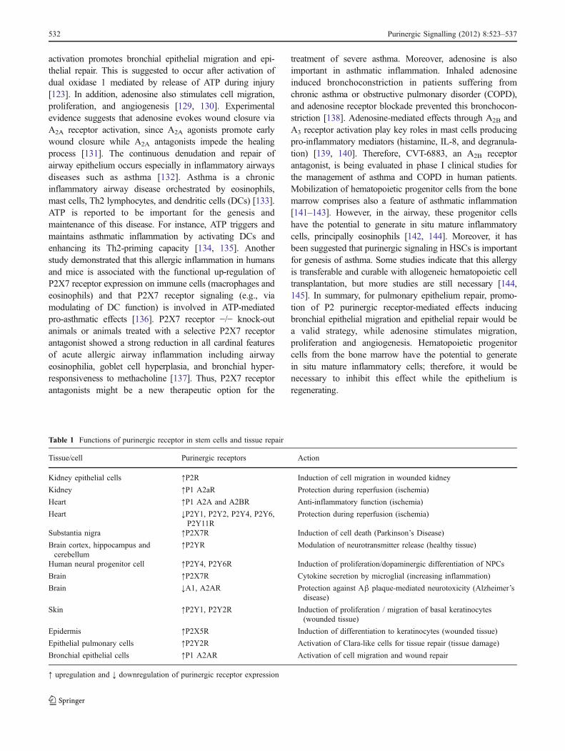
Table 1 Functions of purinergic receptor in stem cells and tissue repair
Tissue/cell Purinergic receptors Action
Kidney epithelial cells ↑P2R Induction of cell migration in wounded kidney
Kidney ↑P1 A2aR Protection during reperfusion (ischemia)
Heart ↑P1 A2A and A2BR Anti-inflammatory function (ischemia)
Heart ↓P2Y1, P2Y2, P2Y4, P2Y6,P2Y11R
Protection during reperfusion (ischemia)
Substantia nigra ↑P2X7R Induction of cell death (Parkinson’s Disease)
Brain cortex, hippocampus andcerebellum
↑P2YR Modulation of neurotransmitter release (healthy tissue)
Human neural progenitor cell ↑P2Y4, P2Y6R Induction of proliferation/dopaminergic differentiation of NPCs
Brain ↑P2X7R Cytokine secretion by microglial (increasing inflammation)
Brain ↓A1, A2AR Protection against Αβ plaque-mediated neurotoxicity (Alzheimer’sdisease)
Skin ↑P2Y1, P2Y2R Induction of proliferation / migration of basal keratinocytes(wounded tissue)
Epidermis ↑P2X5R Induction of differentiation to keratinocytes (wounded tissue)
Epithelial pulmonary cells ↑P2Y2R Activation of Clara-like cells for tissue repair (tissue damage)
Bronchial epithelial cells ↑P1 A2AR Activation of cell migration and wound repair
↑ upregulation and ↓ downregulation of purinergic receptor expression
532 Purinergic Signalling (2012) 8:523–537

Conclusions
Stem cell transplantation and engraftment depends on thesecretion of anti-inflammatory molecules, in addition toextrinsic and endogenous factors promoting differentiationinto distinct cell types depending on the injury site. Whileadenosine receptors often, but not every time, exertbeneficial effects in providing adequate stem cell niches,functions of P2Y and P2X receptors depend very much onthe tissue and the expression pattern of these receptors (seeTable 1). Therapeutic applications based on activation ofpurinergic signaling are foreseen for kidney and heartmuscle regeneration, while other disease conditions will yetneed further investigation. While nucleotides have beenshown to promote differentiation of dopaminergic neuronsdestroyed in Parkinson’s disease, other neuronal diseasesinvolve excitatory cell damage mostly due to P2X7 receptoraction. Therapeutic inhibition of such receptor activitywould be required for improving disease conditions.Finally, the need of P2Y2 and A2A receptor activationduring Clara-like cell differentiation into pulmonary andbronchial epithelial cells just corroborates the fact thatpurinergic signaling is well involved in tissue repair,specially mediated by stem cells. More work need to bedone for elucidation of crucial concepts which couldrevolutionize cell therapy.
Acknowledgments HU acknowledges grant support from Fundação deAmparo à Pesquisa do Estado de São Paulo (FAPESP) andCNPq (ConselhoNacional de Desenvolvimento Científico e Tecnológico), Brazil, project no.2006/61285-9. TG, PDN, and CL are supported by fellowships fromFAPESP.ARC,MMP, and IC are grateful for fellowships fromCNPq.Grantsupport by FAPERGS/CNPq - PRONEX, Brazil, is also acknowledged.
References
1. Verkhratsky A, Krishtal OA, Burnstock G (2009) Purinoceptorson neuroglia. Mol Neurobiol 39(3):190–208. doi:10.1007/s12035-009-8063-2
2. Burnstock G (1997) The past, present and future of purinenucleotides as signalling molecules. Neuropharmacology 36(9):1127–1139
3. North RA (1996) P2X receptors: a third major class of ligand-gatedion channels. Ciba Found Symp 198:91–105, discussion 105–109
4. Burnstock G, Campbell G, Satchell D, Smythe A (1997)Evidence that adenosine triphosphate or a related nucleotide isthe transmitter substance released by non-adrenergic inhibitorynerves in the gut. 1970. Br J Pharmacol 120(4 Suppl):337–357,discussion 334–336
5. Burnstock G (1986) The changing face of autonomic neuro-transmission. Acta Physiol Scand 126(1):67–91
6. Burnstock G, Ulrich H (2011) Purinergic signaling in embryonicand stem cell development. Cell Mol Life Sci 68(8):1369–1394.doi:10.1007/s00018-010-0614-1
7. Hofstetter CP, Holmstrom NA, Lilja JA, Schweinhardt P, Hao J,Spenger C, Wiesenfeld-Hallin Z, Kurpad SN, Frisen J, Olson L(2005) Allodynia limits the usefulness of intraspinal neural stem
cell grafts; directed differentiation improves outcome. NatNeurosci 8(3):346–353. doi:10.1038/nn1405
8. Hofer M, Vacek A, Pospisil M, Weiterova L, Hola J, Streitova D,Znojil V (2006) Adenosine potentiates stimulatory effects ongranulocyte-macrophage hematopoietic progenitor cells in vitroof IL-3 and SCF, but not those of G-CSF, GM-CSF and IL-11.Physiol Res 55(5):591–596
9. Zimmermann H (2001) Ectonucleotidases: some recent develop-ments and a note on nomenclature. Drug Dev Res 52(1–2):44–56. doi:10.1002/ddr.1097
10. Trounson A, Thakar RG, Lomax G, Gibbons D (2011) Clinicaltrials for stem cell therapies. BMC Med 10:9–52. doi:10.1186/1741-7015-9-52
11. Lodi D, Iannitti T, Palmieri B (2011) Stem cells in clinicalpractice: applications and warnings. J Exp Clin Cancer Res 17(30):9. doi:10.1186/1756-9966-30-9
12. Evans MJ, Kaufman MH (1981) Establishment in culture ofpluripotential cells from mouse embryos. Nature 292(5819):154–156
13. Thomson JA, Itskovitz-Eldor J, Shapiro SS, Waknitz MA,Swiergiel JJ, Marshall VS, Jones JM (1998) Embryonic stemcell lines derived from human blastocysts. Science 282(5391):1145–1147
14. Pelacho B, Mazo M, Gavira JJ, Prosper F (2011) Adult stem cells:from new cell sources to changes in methodology. J CardiovascTransl Res 4(2):154–160. doi:10.1007/s12265-010-9245-z
15. Takahashi K, Yamanaka S (2006) Induction of pluripotent stemcells frommouse embryonic and adult fibroblast cultures by definedfactors. Cell 126(4):663–676. doi:10.1016/j.cell.2006.07.024
16. Takahashi K, Tanabe K, Ohnuki M, Narita M, Ichisaka T,Tomoda K, Yamanaka S (2007) Induction of pluripotent stemcells from adult human fibroblasts by defined factors. Cell 131(5):861–872. doi:10.1016/j.cell.2007.11.019
17. Yamanaka S, Blau HM (2010) Nuclear reprogramming to apluripotent state by three approaches. Nature 465(7299):704–712. doi:10.1038/nature09229
18. Passier R, van Laake LW, Mummery CL (2008) Stem-cell-basedtherapy and lessons from the heart. Nature 453(7193):322–329.doi:10.1038/nature07040
19. Dambrot C, Passier R, Atsma D, Mummery CL (2011)Cardiomyocyte differentiation of pluripotent stem cells and theiruse as cardiac disease models. Biochem J 434(1):25–35.doi:10.1042/BJ20101707
20. Gnecchi M, He H, Noiseux N, Liang OD, Zhang L, Morello F(2006) Evidence supporting paracrine hypothesis for Akt-modified mesenchymal stem cell-mediated cardiac protectionand functional improvement. FASEB J 20(6):661–669
21. Laflamme MA, Myerson D, Saffitz JE (2002) Evidence forcardiomyocyte repopulation by extracardiac progenitors in trans-planted human hearts. Circ Res 90:634–640
22. Uemura R, Xu M, Ahmad N, Ashraf M (2006) Bone marrowstem cells prevent left ventricular remodeling of ischemic heartthrough paracrine signaling. Circ Res 98(11):1414–1421
23. Khakh BS, North RA (2006) P2X receptors as cell-surface ATPsensors in health and disease. Nature 442(7102):527–532.doi:10.1038/nature04886
24. Junger WG (2011) Immune cell regulation by autocrinepurinergic signalling. Nat Rev Immunol 11(3):201–212.doi:10.1038/nri2938
25. Di Virgilio F, Falzoni S, Mutini C, Sanz JM, Chiozzi P (1998)Purinergic P2X7 receptor: a pivotal role in inflammation andimmunomodulation. Drug Dev Res 45(3–4):doi:10.1002/(SICI)1098-2299(199811/12)45:3/4<207::AID-DDR18>3.0.CO;2-N
26. Surprenant A, North RA (2009) Signaling at purinergic P2Xreceptors. Annu Rev Physiol 71:333–359. doi:10.1146/annurev.physiol.70.113006.100630
Purinergic Signalling (2012) 8:523–537 533
27. Carroll WA, Donnelly-Roberts D, Jarvis MF (2009) SelectiveP2X(7) receptor antagonists for chronic inflammation and pain.Purinergic Signal 5(1):63–73. doi:10.1007/s11302-008-9110-6
28. Sak K, Boeynaems JM, Everaus H (2003) Involvement of P2Yreceptors in the differentiation of haematopoietic cells. J LeukocBiol 73(4):442–447
29. Casati A, Frascoli M, Traggiai E, Proietti M, Schenk U, Grassi F(2011) Cell-autonomous regulation of hematopoietic stem cellcycling activity by ATP. Cell Death Differ 18(3):396–404.doi:10.1038/cdd.2010.107
30. Lemoli RM, Ferrari D, Fogli M, Rossi L, Pizzirani C, Forchap S,Chiozzi P, Vaselli D, Bertolini F, Foutz T, Aluigi M, Baccarani M,Di Virgilio F (2004) Extracellular nucleotides are potent stimulatorsof human hematopoietic stem cells in vitro and in vivo. Blood 104(6):1662–1670. doi:10.1182/blood-2004-03-08342004-03-0834
31. Rossi L,Manfredini R, Bertolini F, Ferrari D, FogliM, Zini R, SalatiS, Salvestrini V, Gulinelli S, Adinolfi E, Ferrari S, Di Virgilio F,Baccarani M, Lemoli RM (2007) The extracellular nucleotide UTPis a potent inducer of hematopoietic stem cell migration. Blood 109(2):533–542. doi:10.1182/blood-2006-01-035634
32. Coppi E, Pugliese AM, Urbani S, Melani A, Cerbai E, MazzantiB, Bosi A, Saccardi R, Pedata F (2007) ATP modulates cellproliferation and elicits two different electrophysiologicalresponses in human mesenchymal stem cells. Stem Cells 25(7):1840–1849
33. Delarasse C, Gonnord P, Galante M, Auger R, Daniel H, Motta I,Kanellopoulos JM (2009) Neural progenitor cell death is inducedby extracellular ATP via ligation of P2X7 receptor. J Neurochem109(3):846–857
34. Robson SC, Sevigny J, Zimmermann H (2006) The E-NTPDasefamily of ectonucleotidases: structure function relationships andpathophysiological significance. Purinergic Signal 2(2):409–430.doi:10.1007/s11302-006-9003-5
35. Shukla V, Zimmermann H, Wang L, Kettenmann H, Raab S,Hammer K, Sevigny J, Robson SC, Braun N (2005) Functionalexpression of the ecto-ATPase NTPDase2 and of nucleotidereceptors by neuronal progenitor cells in the adult murinehippocampus. J Neurosci Res 80(5):600–610. doi:10.1002/jnr.20508
36. Braun N, Sevigny J, Mishra SK, Robson SC, Barth SW,Gerstberger R, Hammer K, Zimmermann H (2003) Expressionof the ecto-ATPase NTPDase2 in the germinal zones of thedeveloping and adult rat brain. Eur J Neurosci 17(7):1355–1364
37. Mishra SK, Braun N, Shukla V, Fullgrabe M, Schomerus C, KorfHW, Gachet C, Ikehara Y, Sevigny J, Robson SC, ZimmermannH (2006) Extracellular nucleotide signaling in adult neural stemcells: synergism with growth factor-mediated cellular prolifera-tion. Development 133(4):675–684. doi:10.1242/dev.02233
38. Kermer V, Ritter M, Albuquerque B, Leib C, Stanke M,Zimmermann H (2010) Knockdown of tissue nonspecificalkaline phosphatase impairs neural stem cell proliferation anddifferentiation. Neurosci Lett 485(3):208–211. doi:10.1016/j.neulet.2010.09.013
39. Langer D, Ikehara Y, Takebayashi H, Hawkes R, Zimmermann H(2007) The ectonucleotidases alkaline phosphatase and nucleo-side triphosphate diphosphohydrolase 2 are associated withsubsets of progenitor cell populations in the mouse embryonic,postnatal and adult neurogenic zones. Neuroscience 150(4):863–879. doi:10.1016/j.neuroscience.2007.07.064
40. Lin JH, Takano T, Arcuino G, Wang X, Hu F, Darzynkiewicz Z,Nunes M, Goldman SA, Nedergaard M (2007) Purinergic signalingregulates neural progenitor cell expansion and neurogenesis. DevBiol 302(1):356–366. doi:10.1016/j.ydbio.2006.09.017
41. Resende RR, Britto LR, Ulrich H (2008) Pharmacologicalproperties of purinergic receptors and their effects on prolifera-tion and induction of neuronal differentiation of P19 embryonal
carcinoma cells. Int J Dev Neurosci 26(7):763–777. doi:10.1016/j.ijdevneu.2008.07.008
42. Xie X, Sun A, Huang Z, Zhu W, Wang S, Zou Y, Ge J (2011)Another possible cell source for cardiac regenerative medicine:reprogramming adult fibroblasts to cardiomyocytes and endo-thelial progenitor cells. Med Hypotheses 76(3):365–367.doi:10.1016/j.mehy.2010.10.041
43. Martinez EC, Kofidis T (2011) Adult stem cells for cardiac tissueengineering. J Mol Cell Cardiol 50(2):312–319. doi:10.1016/j.yjmcc.2010.08.009
44. Beltrami AP, Barlucchi L, Torella D, BakerM, Limana F, Chimenti S,Kasahara H, RotaM,Musso E, UrbanekK, Leri A, Kajstura J, Nadal-Ginard B, Anversa P (2003) Adult cardiac stem cells are multipotentand support myocardial regeneration. Cell 114(6):763–776
45. Laugwitz KL, Moretti A, Lam J, Gruber P, Chen Y, Woodard S,Lin LZ, Cai CL, Lu MM, Reth M, Platoshyn O, Yuan JX, EvansS, Chien KR (2005) Postnatal isl1+ cardioblasts enter fullydifferentiated cardiomyocyte lineages. Nature 433(7026):647–653. doi:10.1038/nature03215
46. Martin CM, Meeson AP, Robertson SM, Hawke TJ, RichardsonJA, Bates S, Goetsch SC, Gallardo TD, Garry DJ (2004)Persistent expression of the ATP-binding cassette transporter,Abcg2, identifies cardiac SP cells in the developing and adultheart. Dev Biol 265(1):262–275
47. Oh H, Bradfute SB, Gallardo TD, Nakamura T, Gaussin V,Mishina Y, Pocius J, Michael LH, Behringer RR, Garry DJ,Entman ML, Schneider MD (2003) Cardiac progenitor cells fromadult myocardium: homing, differentiation, and fusion afterinfarction. Proc Natl Acad Sci USA 100(21):12313–12318.doi:10.1073/pnas.21321261002132126100
48. Hsieh PC, Segers VF, Davis ME, MacGillivray C, Gannon J,Molkentin JD, Robbins J, Lee RT (2007) Evidence from agenetic fate-mapping study that stem cells refresh adult mam-malian cardiomyocytes after injury. Nat Med 13(8):970–974.doi:10.1038/nm1618
49. Lee BC, Cheng T, Adams GB, Attar EC, Miura N, Lee SB, SaitoY, Olszak I, Dombkowski D, Olson DP, Hancock J, Choi PS,Haber DA, Luster AD, Scadden DT (2003) P2Y-like receptor,GPR105 (P2Y14), identifies and mediates chemotaxis of bone-marrow hematopoietic stem cells. Genes Dev 17(13):1592–1604.doi:10.1101/gad.107150317/13/1592
50. Headrick JP, Hack B, Ashton KJ (2003) Acute adenosinergiccardioprotection in ischemic-reperfused hearts. Am J PhysiolHeart Circ Physiol 285(5):H1797–H1818. doi:10.1152/ajpheart.00407.2003285/5/H1797
51. Jordan JE, Zhao ZQ, Sato H, Taft S, Vinten-Johansen J (1997)Adenosine A2 receptor activation attenuates reperfusion injuryby inhibiting neutrophil accumulation, superoxide generationand coronary endothelial adherence. J Pharmacol Exp Ther 280(1):301–309
52. Zhao ZQ, Todd JC, Sato H, Ma XL, Vinten-Johansen J (1997)Adenosine inhibition of neutrophil damage during reperfusiondoes not involve K(ATP)-channel activation. Am J Physiol 273(4Pt 2):H1677–H1687
53. Millart H, Alouane L, Oszust F, Chevallier S, Robinet A (2009)Involvement of P2Y receptors in pyridoxal-5′-phosphate-inducedcardiac preconditioning. Fundam Clin Pharmacol 23(3):279–292. doi:10.1111/j.1472-8206.2009.00677.x
54. Nishijima S, Sugaya K, Miyazato M, Kadekawa K, Oshiro Y,Uchida A, Hokama S, Ogawa Y (2007) Restoration of bladdercontraction by bone marrow transplantation in rats withunderactive bladder. Biomed Res 28(5):275–280
55. Huang YC, Shindel AW, Ning H, Lin G, Harraz AM, Wang G,Garcia M, Lue TF, Lin CS (2010) Adipose derived stem cellsameliorate hyperlipidemia associated detrusor overactivity in a ratmodel. J Urol 183(3):1232–1240. doi:10.1016/j.juro.2009.11.012
534 Purinergic Signalling (2012) 8:523–537

56. De Coppi P, Callegari A, Chiavegato A, Gasparotto L, Piccoli M,Taiani J, Pozzobon M, Boldrin L, Okabe M, Cozzi E, Atala A,Gamba P, Sartore S (2007) Amniotic fluid and bone marrowderived mesenchymal stem cells can be converted to smoothmuscle cells in the cryo-injured rat bladder and preventcompensatory hypertrophy of surviving smooth muscle cells. JUrol 177(1):369–376. doi:10.1016/j.juro.2006.09.103
57. Kartha S, Toback FG (1992) Adenine nucleotides stimulatemigration in wounded cultures of kidney epithelial cells. J ClinInvest 90(1):288–292. doi:10.1172/JCI115851
58. Zhao Z, Kapoian T, Shepard M, Lianos EA (2002) Adenosine-induced apoptosis in glomerular mesangial cells. Kidney Int 61(4):1276–1285. doi:10.1046/j.1523-1755.2002.00256.x
59. Babelova A, Moreth K, Tsalastra-Greul W, Zeng-Brouwers J,Eickelberg O, YoungMF, Bruckner P, Pfeilschifter J, Schaefer RM,Grone HJ, Schaefer L (2009) Biglycan, a danger signal thatactivates the NLRP3 inflammasome via toll-like and P2X receptors.J Biol Chem 284(36):24035–24048. doi:10.1074/jbc.M109.014266
60. Vonend O, Turner CM, Chan CM, Loesch A, Dell’Anna GC,Srai KS, Burnstock G, Unwin RJ (2004) Glomerular expressionof the ATP-sensitive P2X receptor in diabetic and hypertensiverat models. Kidney Int 66(1):157–166. doi:10.1111/j.1523-1755.2004.00717.xKID717
61. Lee HT, Emala CW (2001) Systemic adenosine given afterischemia protects renal function via A(2a) adenosine receptoractivation. Am J Kidney Dis 38(3):610–618. doi:10.1053/ajkd.2001.26888
62. Burnstock G (2008) Purinergic signalling and disorders of thecentral nervous system. Nat Rev Drug Discov 7(7):575–590.doi:10.1038/nrd2605
63. Trujillo CA, Schwindt TT, Martins AH, Alves JM, Mello LE,Ulrich H (2009) Novel perspectives of neural stem celldifferentiation: from neurotransmitters to therapeutics. Cytome-try A 75(1):38–53. doi:10.1002/cyto.a.20666
64. Morley JF, Hurtig HI (2010) Current understanding andmanagement of Parkinson disease: five new things. Neurology75(18 Suppl 1):S9–S15. doi:10.1212/WNL.0b013e3181fb3628
65. Jun D, Kim K (2004) ATP-mediated necrotic volume increase(NVI) in sustancia nigra pars compacta dopaminergic neuron.Abstract Viewer/Itinerary Planner; Program no. 222.18. Societyfor Neuroscience, Washington, DC
66. Le Feuvre R, Brough D, Rothwell N (2002) Extracellular ATPand P2X7 receptors in neurodegeneration. Eur J Pharmacol 447(2–3):261–269
67. Heine C, Wegner A, Grosche J, Allgaier C, Illes P, Franke H(2007) P2 receptor expression in the dopaminergic system of therat brain during development. Neuroscience 149(1):165–181.doi:10.1016/j.neuroscience.2007.07.015
68. Scheibler P, Pesic M, Franke H, Reinhardt R, Wirkner K, Illes P,Norenberg W (2004) P2X2 and P2Y1 immunofluorescence in ratneostriatal medium-spiny projection neurones and cholinergicinterneurones is not linked to respective purinergic receptorfunction. Br J Pharmacol 143(1):119–131. doi:10.1038/sj.bjp.0705916143/1/119
69. Zona C, Marchetti C, Volonte C, Mercuri NB, Bernardi G (2000)Effect of P2 purinoceptor antagonists on kainate-inducedcurrents in rat cultured neurons. Brain Res 882(1–2):26–35
70. Burnstock G (2006) Pathophysiology and therapeutic potentialof purinergic signaling. Pharmacol Rev 58(1):58–86.doi:10.1124/pr.58.1.5
71. Krugel U, Kittner H, Illes P (1999) Adenosine 5′-triphosphate-induced dopamine release in the rat nucleus accumbens in vivo.Neurosci Lett 265(1):49–52
72. Krugel U, Kittner H, Franke H, Illes P (2001) Acceleratedfunctional recovery after neuronal injury by P2 receptorblockade. Eur J Pharmacol 420(2–3):R3–R4
73. Gaspard N, Vanderhaeghen P (2011) From stem cells to neuralnetworks: recent advances and perspectives for neurodevelop-mental disorders. Develop Med Child Neurol 53(1):13–17.doi:10.1111/j.1469-8749.2010.03827.x
74. Milosevic J, Brandt A, Roemuss U, Arnold A, Wegner F,Schwarz SC, Storch A, Zimmermann H, Schwarz J (2006) Uracilnucleotides stimulate human neural precursor cell proliferationand dopaminergic differentiation: involvement of MEK/ERKsignalling. J Neurochem 99(3):913–923. doi:10.1111/j.1471-4159.2006.04132.x
75. Ralevic V, Burnstock G (1998) Receptors for purines andpyrimidines. Pharmacol Rev 50(3):413–492
76. Morellia M, Di Paolob T, Wardasc J, Calonb F, Xiaod D,Schwarzschildd MA (2007) Role of adenosine A2A receptors inparkinsonian motor impairment and L-DOPA-induced motorcomplications. Prog Neurobiol 83(5):293–309
77. Haughey NJ, Mattson MP (2003) Alzheimer’s amyloid beta-peptide enhances ATP/gap junction-mediated calcium-wavepropagation in astrocytes. Neuromolecular Med 3(3):173–180.doi:10.1385/NMM:3:3:173
78. Thathiah A, De Strooper B (2011) The role of G protein-coupledreceptors in the pathology of Alzheimer’s disease. Nat RevNeurosci 12(2):73–87. doi:10.1038/nrn2977
79. Ballard C, Gauthier S, Corbett A, Brayne C, Aarsland D, Jones E(2011) Alzheimer’s disease. Lancet 377(9770):1019–1031.doi:10.1016/S0140-6736(10)61349-9
80. Zhang YX, Yamashita H, Ohshita T, Sawamoto N, Nakamura S(1995) ATP increases extracellular dopamine level throughstimulation of P2Y purinoceptors in the rat striatum. Brain Res691(1–2):205–212
81. Franke H, Illes P (2006) Involvement of P2 receptors in thegrowth and survival of neurons in the CNS. Pharmacol Ther 109(3):297–324. doi:10.1016/j.pharmthera.2005.06.002
82. Sanz JM, Chiozzi P, Ferrari D, Colaianna M, Idzko M, Falzoni S,Fellin R, Trabace L, Di Virgilio F (2009) Activation of microgliaby amyloid beta requires P2X7 receptor expression. J Immunol182(7):4378–4385. doi:10.4049/jimmunol.0803612
83. McLarnon JG, Ryu JK, Walker DG, Choi HB (2006) Upregu-lated expression of purinergic P2X(7) receptor in Alzheimerdisease and amyloid-beta peptide-treated microglia and inpeptide-injected rat hippocampus. J Neuropathol Exp Neurol 65(11):1090–1097. doi:10.1097/01.jnen.0000240470.97295.d300005072-200611000-00008
84. RampeD,Wang L, RingheimGE (2004) P2X7 receptormodulationof beta-amyloid- and LPS-induced cytokine secretion from humanmacrophages and microglia. J Neuroimmunol 147(1–2):56–61
85. Majumder P, Trujillo CA, Lopes CG, Resende RR, Gomes KN,Yuahasi KK, Britto LR, Ulrich H (2007) New insights intopurinergic receptor signaling in neuronal differentiation, neuro-protection, and brain disorders. Purinergic Signal 3(4):317–331.doi:10.1007/s11302-007-9074-y
86. Haughey NJ, Nath A, Chan SL, Borchard AC, Rao MS, MattsonMP (2002) Disruption of neurogenesis by amyloid beta-peptide,and perturbed neural progenitor cell homeostasis, in models ofAlzheimer’s disease. J Neurochem 83(6):1509–1524
87. Ballard C, Gauthier S, Corbett A, Brayne C, Jones AD, Jones E(2011) Alzheimer’s disease. Lancet 377(9770):1019–1031
88. Resende RR, Majumder P, Gomes KN, Britto LR, Ulrich H(2007) P19 embryonal carcinoma cells as in vitro model forstudying purinergic receptor expression and modulation of N-methyl-D-aspartate-glutamate and acetylcholine receptors duringneuronal differentiation. Neuroscience 146(3):1169–1181.doi:10.1016/j.neuroscience.2007.02.041
89. Lindvall O, Kokaia Z (2010) Stem cells in human neurodegen-erative disorders—time for clinical translation? J Clin Invest 120(1):29–40. doi:10.1172/JCI40543
Purinergic Signalling (2012) 8:523–537 535

90. Ryu JK, Cho T, Wang YT, McLarnon JG (2009) Neuralprogenitor cells attenuate inflammatory reactivity and neuronalloss in an animal model of inflamed AD brain. J Neuro-inflammation 6:39. doi:10.1186/1742-2094-6-39
91. Chuang TT (2010) Neurogenesis in mouse models of Alz-heimer’s disease. Biochim Biophys Acta 1802(10):872–880.doi:10.1016/j.bbadis.2009.12.008
92. Delarasse C, Auger R, Gonnord P, Fontaine B, KanellopoulosJM (2011) The purinergic receptor P2X7 triggers α-secretase-dependent processing of the amyloid precursor protein. J BioChem 286:2596–2606. doi:10.1074/jbc.M110.200618
93. Knutsen LJ, Murray TF (1997) Adenosine and ATP epilepsy. In:Jacobson KA, Jarvis MF (eds) Purinergic approaches inexperimental therapeutics. Wiley, New York, pp 423–447
94. Avignone E, Ulmann L, Levavasseur F, Rassendren F, Audinat E(2008) Status epilepticus induces a particular microglial activationstate characterized by enhanced purinergic signaling. J Neurosci 28(37):9133–9144. doi:10.1523/JNEUROSCI.1820-08.2008
95. Dona F, Ulrich H, Persike DS, Conceicao IM, Blini JP,Cavalheiro EA, Fernandes MJ (2009) Alteration of purinergicP2X4 and P2X7 receptor expression in rats with temporal-lobeepilepsy induced by pilocarpine. Epilepsy Res 83(2–3):157–167.doi:10.1016/j.eplepsyres.2008.10.008
96. Oses JP (2006) Modification by kainate-induced convulsions ofthe density of presynaptic P2X receptors in the rat hippocampus.Purinergic Signalling 2:252–253
97. Burnstock G (2007) Physiology and pathophysiology of puri-nergic neurotransmission. Physiol Rev 87(2):659–797.doi:10.1152/physrev.00043.2006
98. Ciccarelli R, Ballerini P, Sabatino G, Rathbone MP, D’OnofrioM, Caciagli F, Di Iorio P (2001) Involvement of astrocytes inpurine-mediated reparative processes in the brain. Int J DevNeurosci 19(4):395–414
99. Chua K, Kima M, Junga K, Jeond D, Leea S, Kima J, Jeongf S,Kimg SU, Leea SK, Shind H, Roha J (2004) Human neural stemcell transplantation reduces spontaneous recurrent seizuresfollowing pilocarpine-induced status epilepticus in adult rats.Brain Res 1023(2):213–221. doi:10.1016/j.brainres.2004.07.045
100. Guttinger M, Fedele D, Koch P, Padrun V, Pralong WF, BrustleO, Boison D (2005) Suppression of kindled seizures by paracrineadenosine release from stem cell-derived brain implants. Epi-lepsia 46(8):1162–1169
101. Franke H, Gunther A, Grosche J, Schmidt R, Rossner S,Reinhardt R, Faber-Zuschratter H, Schneider D, Illes P (2004)P2X7 receptor expression after ischemia in the cerebral cortex ofrats. J Neuropathol Exp Neurol 63(7):686–699
102. Tsuzuki K, Kondo E, Fukuoka T, Yi D, Tsujino H, Sakagami M,Noguchi K (2001) Differential regulation of P2X(3) mRNAexpression by peripheral nerve injury in intact and injuredneurons in the rat sensory ganglia. Pain 91(3):351–360
103. Bianco F, Ceruti S, Colombo A, Fumagalli M, Ferrari D,Pizzirani C, Matteoli M, Di Virgilio F, Abbracchio MP, VerderioC (2006) A role for P2X7 in microglial proliferation. J Neuro-chem 99(3):745–758. doi:10.1111/j.1471-4159.2006.04101.x
104. Ceruti S, Villa G, Genovese T, Mazzon E, Longhi R, Rosa P,Bramanti P, Cuzzocrea S, Abbracchio MP (2009) The P2Y-likereceptor GPR17 as a sensor of damage and a new potential targetin spinal cord injury. Brain 132(Pt 8):2206–2218. doi:10.1093/brain/awp147
105. Cunha RA (2005) Neuroprotection by adenosine in the brain:from A(1) receptor activation to A (2A) receptor blockade.Purinergic Signal 1(2):111–134. doi:10.1007/s11302-005-0649-1
106. Nedeljkovic N, Bjelobaba I, Subasic S, Lavrnja I, Pekovic S,Stojkov D, Vjestica A, Rakic L, Stojiljkovic M (2006) Up-regulation of ectonucleotidase activity after cortical stab injury inrats. Cell Biol Int 30(6):541–546. doi:10.1016/j.cellbi.2006.03.001
107. Fuchs E, Raghavan S (2002) Getting under the skin of epidermalmorphogenesis. Nat Rev Genet 3(3):199–209. doi:10.1038/nrg758nrg758
108. Kanitakis J (2002) Anatomy, histology and immunohistochemistryof normal human skin. Eur J Dermatol 12(4):390–399, quiz 400–391
109. Fujishita K, Koizumi S, Inoue K (2006) Upregulation of P2Y2receptors by retinoids in normal human epidermal keratinocytes.Purinergic Signal 2(3):491–498. doi:10.1007/s11302-005-7331-5
110. Holzer AM, Granstein RD (2004) Role of extracellular adenosinetriphosphate in human skin. J Cutan Med Surg 8(2):90–96.doi:10.1007/s10227-004-0125-5
111. Greig AV, James SE, McGrouther DA, Terenghi G, Burnstock G(2003) Purinergic receptor expression in the regenerationepidermis in a rat model of normal and delayed wound healing.Exp Dermatol 12(6):860–871
112. Greig AV, Linge C, Cambrey A, Burnstock G (2003) Purinergicreceptors are part of a signaling system for keratinocyteproliferation, differentiation, and apoptosis in human fetalepidermis. J Invest Dermatol 121(5):1145–1149. doi:10.1046/j.1523-1747.2003.12567.x
113. Inoue K, Denda M, Tozaki H, Fujishita K, Koizumi S (2005)Characterization of multiple P2X receptors in cultured normalhuman epidermal keratinocytes. J Invest Dermatol 124(4):756–763. doi:10.1111/j.0022-202X.2005.23683.x
114. Burnstock G, Verkhratsky A (2010) Long-term (trophic) puri-nergic signalling: purinoceptors control cell proliferation, differ-entiation and death. Cell Death Dis 1:e9. doi:10.1038/cddis.2009.11
115. Volonte C, Amadio S, D’Ambrosi N, Colpi M, Burnstock G(2006) P2 receptor web: complexity and fine-tuning. PharmacolTher 112(1):264–280. doi:10.1016/j.pharmthera.2005.04.012
116. Snyder JC, Teisanu RM, Stripp BR (2009) Endogenous lungstem cells and contribution to disease. J Pathol 217(2):254–264.doi:10.1002/path.2473
117. ChistiakovDA (2010) Endogenous and exogenous stem cells: a rolein lung repair and use in airway tissue engineering and transplan-tation. J Biomed Sci 17:92. doi:10.1186/1423-0127-17-92
118. Sueblinvong V, Weiss DJ (2010) Stem cells and cell therapyapproaches in lung biology and diseases. Transl Res 156(3):188–205
119. Evans MJ, Cabral-Anderson LJ, Freeman G (1978) Role of theClara cell in renewal of the bronchiolar epithelium. Lab Invest 38(6):648–653
120. Stripp BR (2008) Hierarchical organization of lung progenitorcells: is there an adult lung tissue stem cell? Proc Am Thorac Soc5(6):695–698. doi:10.1513/pats.200801-011AW
121. Teisanu RM, Lagasse E, Whitesides JF, Stripp BR (2009)Prospective isolation of bronchiolar stem cells based uponimmunophenotypic and autofluorescence characteristics. StemCells 27(3):612–622. doi:10.1634/stemcells.2008-0838
122. Borthwick DW, Shahbazian M, Krantz QT, Dorin JR, RandellSH (2001) Evidence for stem-cell niches in the trachealepithelium. Am J Respir Cell Mol Biol 24(6):662–670
123. Reynolds SD, Giangreco A, Power JH, Stripp BR (2000)Neuroepithelial bodies of pulmonary airways serve as a reservoirof progenitor cells capable of epithelial regeneration. Am JPathol 156(1):269–278. doi:10.1016/S0002-9440(10)64727-X
124. De Proost I, Pintelon I, Wilkinson WJ, Goethals S, Brouns I, VanNassauw L, Riccardi D, Timmermans JP, Kemp PJ, AdriaensenD (2009) Purinergic signaling in the pulmonary neuroepithelialbody microenvironment unraveled by live cell imaging. FASEBJ 23(4):1153–1160. doi:10.1096/fj.08-109579
125. Reynolds SD, Hong KU, Giangreco A, Mango GW, Guron C,Morimoto Y, Stripp BR (2000) Conditional clara cell ablationreveals a self-renewing progenitor function of pulmonaryneuroendocrine cells. Am J Physiol Lung Cell Mol Physiol 278(6):L1256–L1263
536 Purinergic Signalling (2012) 8:523–537

126. Brouns I, Oztay F, Pintelon I, De Proost I, Lembrechts R,Timmermans JP, Adriaensen D (2009) Neurochemical pattern ofthe complex innervation of neuroepithelial bodies in mouselungs. Histochem Cell Biol 131(1):55–74. doi:10.1007/s00418-008-0495-7
127. Conway JD, Bartolotta T, Abdullah LH, Davis CW (2003)Regulation of mucin secretion from human bronchial epithelialcells grown in murine hosted xenografts. Am J Physiol LungCell Mol Physiol 284(6):L945–L954. doi:10.1152/ajplung.00410.200200410.2002
128. Schwiebert EM, Zsembery A (2003) Extracellular ATP as asignaling molecule for epithelial cells. Biochim Biophys Acta1615(1–2):7–32
129. Wesley UV, Bove PF, Hristova M, McCarthy S, van der Vliet A(2007) Airway epithelial cell migration and wound repair byATP-mediated activation of dual oxidase 1. J Biol Chem 282(5):3213–3220. doi:10.1074/jbc.M606533200
130. Ethier MF, Chander V, Dobson JG Jr (1993) Adenosinestimulates proliferation of human endothelial cells in culture.Am J Physiol 265(1 Pt 2):H131–H138
131. Montesinos MC, Desai A, Chen JF, Yee H, Schwarzschild MA,Fink JS, Cronstein BN (2002) Adenosine promotes woundhealing and mediates angiogenesis in response to tissue injuryvia occupancy of A(2A) receptors. Am J Pathol 160(6):2009–2018. doi:10.1016/S0002-9440(10)61151-0
132. Allen-Gipson DS, Wong J, Spurzem JR, Sisson JH, Wyatt TA(2006) Adenosine A2A receptors promote adenosine-stimulatedwound healing in bronchial epithelial cells. Am J Physiol Lung CellMol Physiol 290(5):L849–L855. doi:10.1152/ajplung.00373.2005
133. Erjefalt JS, Persson CG (1997) Airway epithelial repair:breathtakingly quick and multipotentially pathogenic. Thorax52(11):1010–1012
134. Wills-Karp M (1999) Immunologic basis of antigen-inducedairway hyperresponsiveness. Annu Rev Immunol 17:255–281.doi:10.1146/annurev.immunol.17.1.255
135. Idzko M, Hammad H, van Nimwegen M, Kool M, Willart MA,Muskens F, Hoogsteden HC, Luttmann W, Ferrari D, Di Virgilio F,Virchow JC Jr, Lambrecht BN (2007) Extracellular ATP triggersand maintains asthmatic airway inflammation by activating den-dritic cells. Nat Med 13(8):913–919. doi:10.1038/nm1617
136. la Sala A, Sebastiani S, Ferrari D, Di Virgilio F, Idzko M,Norgauer J, Girolomoni G (2002) Dendritic cells exposed to
extracellular adenosine triphosphate acquire the migratoryproperties of mature cells and show a reduced capacity to attracttype 1 T lymphocytes. Blood 99(5):1715–1722
137. Muller T, Vieira RP, Grimm M, Durk T, Cicko S, Zeiser R, JakobT, Martin SF, Blumenthal B, Sorichter S, Ferrari D, Di VirgillioF, Idzko M (2011) A potential role for P2X7R in allergic airwayinflammation in mice and humans. Am J Respir Cell Mol Biol44(4):456–464. doi:10.1165/rcmb.2010-0129OC
138. Polosa R, Holgate ST (2006) Adenosine receptors as promisingtherapeutic targets for drug development in chronic airwayinflammation. Curr Drug Targets 7(6):699–706
139. Zhong H, Shlykov SG, Molina JG, Sanborn BM, Jacobson MA,Tilley SL, Blackburn MR (2003) Activation of murine lung mastcells by the adenosine A3 receptor. J Immunol 171(1):338–345
140. Ryzhov S, Zaynagetdinov R, Goldstein AE, Novitskiy SV,Dikov MM, Blackburn MR, Biaggioni I, Feoktistov I (2008)Effect of A2B adenosine receptor gene ablation on proinflam-matory adenosine signaling in mast cells. J Immunol 180(11):7212–7220
141. Gibson PG, Manning PJ, O’Byrne PM, Girgis-Gabardo A,Dolovich J, Denburg JA, Hargreave FE (1991) Allergen-inducedasthmatic responses. Relationship between increases in airwayresponsiveness and increases in circulating eosinophils, basophils,and their progenitors. Am Rev Respir Dis 143(2):331–335
142. Dorman SC, Efthimiadis A, Babirad I, Watson RM, Denburg JA,Hargreave FE, O’Byrne PM, Sehmi R (2004) Sputum CD34+IL-5Ralpha+ cells increase after allergen: evidence for in situeosinophilopoiesis. Am J Respir Crit Care Med 169(5):573–577.doi:10.1164/rccm.200307-1004OC200307-1004OC
143. Catalli AE, Thomson JV, Babirad IM, Duong M, Doyle TM,Howie KJ, Newbold P, Craggs RI, Foster M, Gauvreau GM,O’Byrne PM, Sehmi R (2008) Modulation of beta1-integrins onhemopoietic progenitor cells after allergen challenge in asthmaticsubjects. J Allergy Clin Immunol 122(4):803–810. doi:10.1016/j.jaci.2008.07.021
144. Southam DS, Widmer N, Ellis R, Hirota JA, Inman MD, SehmiR (2005) Increased eosinophil-lineage committed progenitors inthe lung of allergen-challenged mice. J Allergy Clin Immunol115(1):95–102. doi:10.1016/j.jaci.2004.09.022
145. Daikeler T, Tyndall A (2007) Autoimmunity following haema-topoietic stem-cell transplantation. Best Pract Res Clin Haematol20(2):349–360. doi:10.1016/j.beha.2006.09.008
Purinergic Signalling (2012) 8:523–537 537
Related Documents