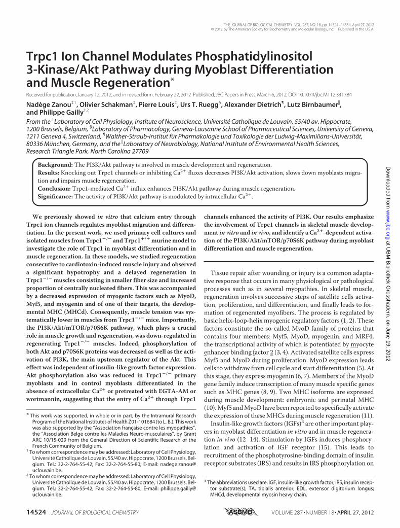
Trpc1 Ion Channel Modulates Phosphatidylinositol 3-Kinase/Akt Pathway during Myoblast Differentiation and Muscle Regeneration * Received for publication, January 12, 2012, and in revised form, February 22, 2012 Published, JBC Papers in Press, March 6, 2012, DOI 10.1074/jbc.M112.341784 Nadège Zanou ‡1 , Olivier Schakman ‡ , Pierre Louis ‡ , Urs T. Ruegg § , Alexander Dietrich ¶ , Lutz Birnbaumer , and Philippe Gailly ‡2 From the ‡ Laboratory of Cell Physiology, Institute of Neuroscience, Université Catholique de Louvain, 55/40 av. Hippocrate, 1200 Brussels, Belgium, § Laboratory of Pharmacology, Geneva-Lausanne School of Pharmaceutical Sciences, University of Geneva, 1211 Geneva 4, Switzerland, ¶ Walther-Straub-Institut für Pharmakologie und Toxikologie der Ludwig-Maximilians-Universität, 80336 München, Germany, and the Laboratory of Neurobiology, National Institute of Environmental Health Sciences, Research Triangle Park, North Carolina 27709 Background: The PI3K/Akt pathway is involved in muscle development and regeneration. Results: Knocking out Trpc1 channels or inhibiting Ca 2 fluxes decreases PI3K/Akt activation, slows down myoblasts migra- tion and impairs muscle regeneration. Conclusion: Trpc1-mediated Ca 2 influx enhances PI3K/Akt pathway during muscle regeneration. Significance: The activity of PI3K/Akt pathway is modulated by intracellular Ca 2 . We previously showed in vitro that calcium entry through Trpc1 ion channels regulates myoblast migration and differen- tiation. In the present work, we used primary cell cultures and isolated muscles from Trpc1 / and Trpc1 / murine model to investigate the role of Trpc1 in myoblast differentiation and in muscle regeneration. In these models, we studied regeneration consecutive to cardiotoxin-induced muscle injury and observed a significant hypotrophy and a delayed regeneration in Trpc1 / muscles consisting in smaller fiber size and increased proportion of centrally nucleated fibers. This was accompanied by a decreased expression of myogenic factors such as MyoD, Myf5, and myogenin and of one of their targets, the develop- mental MHC (MHCd). Consequently, muscle tension was sys- tematically lower in muscles from Trpc1 / mice. Importantly, the PI3K/Akt/mTOR/p70S6K pathway, which plays a crucial role in muscle growth and regeneration, was down-regulated in regenerating Trpc1 / muscles. Indeed, phosphorylation of both Akt and p70S6K proteins was decreased as well as the acti- vation of PI3K, the main upstream regulator of the Akt. This effect was independent of insulin-like growth factor expression. Akt phosphorylation also was reduced in Trpc1 / primary myoblasts and in control myoblasts differentiated in the absence of extracellular Ca 2 or pretreated with EGTA-AM or wortmannin, suggesting that the entry of Ca 2 through Trpc1 channels enhanced the activity of PI3K. Our results emphasize the involvement of Trpc1 channels in skeletal muscle develop- ment in vitro and in vivo, and identify a Ca 2 -dependent activa- tion of the PI3K/Akt/mTOR/p70S6K pathway during myoblast differentiation and muscle regeneration. Tissue repair after wounding or injury is a common adapta- tive response that occurs in many physiological or pathological processes such as in several myopathies. In skeletal muscle, regeneration involves successive steps of satellite cells activa- tion, proliferation, and differentiation, and finally leads to for- mation of regenerated myofibers. The process is regulated by basic helix-loop-helix myogenic regulatory factors (1, 2). These factors constitute the so-called MyoD family of proteins that contains four members: Myf5, MyoD, myogenin, and MRF4, the transcriptional activity of which is potentiated by myocyte enhancer binding factor 2 (3, 4). Activated satellite cells express Myf5 and MyoD during proliferation. MyoD expression leads cells to withdraw from cell cycle and start differentiation (5). At this stage, they express myogenin (6, 7). Members of the MyoD gene family induce transcription of many muscle specific genes such as MHC genes (8, 9). Two MHC isoforms are expressed during muscle development: embryonic and perinatal MHC (10). Myf5 and MyoD have been reported to specifically activate the expression of these MHCs during muscle regeneration (11). Insulin-like growth factors (IGFs) 3 are other important play- ers in myoblast differentiation in vitro and in muscle regenera- tion in vivo (12–14). Stimulation by IGFs induces phosphory- lation and activation of IGF receptor (15). This leads to recruitment of the phosphotyrosine-binding domain of insulin receptor substrates (IRS) and results in IRS phosphorylation on * This work was supported, in whole or in part, by the Intramural Research Program of the National Institutes of Health Z01-101684 (to L. B.). This work was also supported by the “Association française contre les myopathies”, the “Association Belge contre les Maladies Neuro-musculaires”, by Grant ARC 10/15-029 from the General Direction of Scientific Research of the French Community of Belgium. 1 To whom correspondence may be addressed: Laboratory of Cell Physiology, Université Catholique de Louvain, 55/40 av. Hippocrate, 1200 Brussels, Bel- gium. Tel.: 32-2-764-55-42; Fax: 32-2-764-55-80; E-mail: nadege.zanou@ uclouvain.be. 2 To whom correspondence may be addressed: Laboratory of Cell Physiology, Université Catholique de Louvain, 55/40 av. Hippocrate, 1200 Brussels, Bel- gium. Tel.: 32-2-764-55-42; Fax: 32-2-764-55-80; E-mail: philippe.gailly@ uclouvain.be. 3 The abbreviations used are: IGF, insulin-like growth factor; IRS, insulin recep- tor substrate(s); TA, tibialis anterior; EDL, extensor digitorium longus; MHCd, developmental myosin heavy chain. THE JOURNAL OF BIOLOGICAL CHEMISTRY VOL. 287, NO. 18, pp. 14524 –14534, April 27, 2012 © 2012 by The American Society for Biochemistry and Molecular Biology, Inc. Published in the U.S.A. 14524 JOURNAL OF BIOLOGICAL CHEMISTRY VOLUME 287 • NUMBER 18 • APRIL 27, 2012 at UBM Bibliothek Grosshadern, on June 19, 2012 www.jbc.org Downloaded from

Welcome message from author
This document is posted to help you gain knowledge. Please leave a comment to let me know what you think about it! Share it to your friends and learn new things together.
Transcript
Trpc1 Ion Channel Modulates Phosphatidylinositol3-Kinase/Akt Pathway during Myoblast Differentiationand Muscle Regeneration*
Received for publication, January 12, 2012, and in revised form, February 22, 2012 Published, JBC Papers in Press, March 6, 2012, DOI 10.1074/jbc.M112.341784
Nadège Zanou‡1, Olivier Schakman‡, Pierre Louis‡, Urs T. Ruegg§, Alexander Dietrich¶, Lutz Birnbaumer�,and Philippe Gailly‡2
From the ‡Laboratory of Cell Physiology, Institute of Neuroscience, Université Catholique de Louvain, 55/40 av. Hippocrate,1200 Brussels, Belgium, §Laboratory of Pharmacology, Geneva-Lausanne School of Pharmaceutical Sciences, University of Geneva,1211 Geneva 4, Switzerland, ¶Walther-Straub-Institut für Pharmakologie und Toxikologie der Ludwig-Maximilians-Universität,80336 München, Germany, and the �Laboratory of Neurobiology, National Institute of Environmental Health Sciences,Research Triangle Park, North Carolina 27709
Background: The PI3K/Akt pathway is involved in muscle development and regeneration.Results: Knocking out Trpc1 channels or inhibiting Ca2� fluxes decreases PI3K/Akt activation, slows down myoblasts migra-tion and impairs muscle regeneration.Conclusion: Trpc1-mediated Ca2� influx enhances PI3K/Akt pathway during muscle regeneration.Significance: The activity of PI3K/Akt pathway is modulated by intracellular Ca2�.
We previously showed in vitro that calcium entry throughTrpc1 ion channels regulates myoblast migration and differen-tiation. In the present work, we used primary cell cultures andisolatedmuscles fromTrpc1�/� andTrpc1�/� murinemodel toinvestigate the role of Trpc1 in myoblast differentiation and inmuscle regeneration. In these models, we studied regenerationconsecutive to cardiotoxin-inducedmuscle injury and observeda significant hypotrophy and a delayed regeneration inTrpc1�/� muscles consisting in smaller fiber size and increasedproportion of centrally nucleated fibers. This was accompaniedby a decreased expression of myogenic factors such as MyoD,Myf5, and myogenin and of one of their targets, the develop-mental MHC (MHCd). Consequently, muscle tension was sys-tematically lower in muscles from Trpc1�/� mice. Importantly,the PI3K/Akt/mTOR/p70S6K pathway, which plays a crucialrole in muscle growth and regeneration, was down-regulated inregenerating Trpc1�/� muscles. Indeed, phosphorylation ofboth Akt and p70S6K proteins was decreased as well as the acti-vation of PI3K, the main upstream regulator of the Akt. Thiseffect was independent of insulin-like growth factor expression.Akt phosphorylation also was reduced in Trpc1�/� primarymyoblasts and in control myoblasts differentiated in theabsence of extracellular Ca2� or pretreated with EGTA-AM orwortmannin, suggesting that the entry of Ca2� through Trpc1
channels enhanced the activity of PI3K. Our results emphasizethe involvement of Trpc1 channels in skeletal muscle develop-ment in vitro and in vivo, and identify a Ca2�-dependent activa-tion of the PI3K/Akt/mTOR/p70S6K pathway during myoblastdifferentiation and muscle regeneration.
Tissue repair after wounding or injury is a common adapta-tive response that occurs in many physiological or pathologicalprocesses such as in several myopathies. In skeletal muscle,regeneration involves successive steps of satellite cells activa-tion, proliferation, and differentiation, and finally leads to for-mation of regenerated myofibers. The process is regulated bybasic helix-loop-helix myogenic regulatory factors (1, 2). Thesefactors constitute the so-called MyoD family of proteins thatcontains four members: Myf5, MyoD, myogenin, and MRF4,the transcriptional activity of which is potentiated by myocyteenhancer binding factor 2 (3, 4). Activated satellite cells expressMyf5 and MyoD during proliferation. MyoD expression leadscells to withdraw from cell cycle and start differentiation (5). Atthis stage, they express myogenin (6, 7). Members of theMyoDgene family induce transcription of manymuscle specific genessuch as MHC genes (8, 9). Two MHC isoforms are expressedduring muscle development: embryonic and perinatal MHC(10).Myf5 andMyoDhave been reported to specifically activatethe expression of theseMHCs duringmuscle regeneration (11).Insulin-like growth factors (IGFs)3 are other important play-
ers in myoblast differentiation in vitro and in muscle regenera-tion in vivo (12–14). Stimulation by IGFs induces phosphory-lation and activation of IGF receptor (15). This leads torecruitment of the phosphotyrosine-binding domain of insulinreceptor substrates (IRS) and results in IRS phosphorylation on
* This work was supported, in whole or in part, by the Intramural ResearchProgram of the National Institutes of Health Z01-101684 (to L. B.). This workwas also supported by the “Association française contre les myopathies”,the “Association Belge contre les Maladies Neuro-musculaires”, by GrantARC 10/15-029 from the General Direction of Scientific Research of theFrench Community of Belgium.
1 To whom correspondence may be addressed: Laboratory of Cell Physiology,Université Catholique de Louvain, 55/40 av. Hippocrate, 1200 Brussels, Bel-gium. Tel.: 32-2-764-55-42; Fax: 32-2-764-55-80; E-mail: [email protected].
2 To whom correspondence may be addressed: Laboratory of Cell Physiology,Université Catholique de Louvain, 55/40 av. Hippocrate, 1200 Brussels, Bel-gium. Tel.: 32-2-764-55-42; Fax: 32-2-764-55-80; E-mail: [email protected].
3 The abbreviations used are: IGF, insulin-like growth factor; IRS, insulin recep-tor substrate(s); TA, tibialis anterior; EDL, extensor digitorium longus;MHCd, developmental myosin heavy chain.
THE JOURNAL OF BIOLOGICAL CHEMISTRY VOL. 287, NO. 18, pp. 14524 –14534, April 27, 2012© 2012 by The American Society for Biochemistry and Molecular Biology, Inc. Published in the U.S.A.
14524 JOURNAL OF BIOLOGICAL CHEMISTRY VOLUME 287 • NUMBER 18 • APRIL 27, 2012
at UB
M B
ibliothek Grosshadern, on June 19, 2012
ww
w.jbc.org
Dow
nloaded from
specific tyrosine residues (16). Activated IRS recruits andsequesters the p85 subunit of PI3K, liberating the p110 catalyticsubunit. The active p110 subunit generates 3�-phosphorylatedphosphoinositideswhich bind the pleckstrin homology domainof phosphoinositide-dependent kinase 1 andAkt inducing theirmembrane targeting (17–19). Phosphoinositide-dependentkinase 1phosphorylates Akt, which phosphorylates the mam-malian target of rapamycin mTOR, which in turn, phosphory-lates p70S6K and activates protein synthesis.Finally, extracellular Ca2� also is known to play an impor-
tant role in muscle development. Indeed, it has beenreported that migration and/or fusion which precedes myo-tubes formation require Ca2� influxes (20, 21). It has beensuggested that this Ca2� influx occurs through T-type Ca2�
channels (22). We recently reported that the process alsoinvolved the type 1 canonical subfamily of Trp (transient recep-tor potential) channels. Indeed, using a knockdown strategy invitro, we showed that Trpc1 channels were responsible for theincreased Ca2� influx observed at the onset of myoblast differ-entiation (23).To investigate the role of Trpc1 channels during skeletal
muscle regeneration in vivo, we used a model of cardiotoxin-induced muscle injury and compared muscle consecutiveregeneration in adult Trpc1�/� and Trpc1�/� mice. Weobserved that Trpc1�/� mice presented a delayed regeneration(smaller fibers, higher proportion of central nuclei, delayed anddiminished expression of myogenic transcription). We showthat the lack of Trpc1 or the inhibition of Ca2� entries reducesAkt phosphorylation and delaysmuscle cell differentiation.Wesuggest that the entry of Ca2� through Trpc1 channelsenhances the activity of PI3K/Akt/mTOR/p70S6K pathwayand accelerates muscle regeneration.
EXPERIMENTAL PROCEDURES
Trpc1�/� and Trpc1�/� Mice—Generation of Trpc1�/�
mice has been described previously (24). Trpc1�/� andTrpc1�/�were obtained fromheterozygous animals. Trpc1�/�
were compared with their Trpc1�/� control littermates.Muscle Injury—Three- to four-month-old Trpc1�/� and
Trpc1�/� mice were anesthetized by intraperitoneal injectionof a solution containing ketamine (10mg�ml�1 Pfizer, Brussels,Belgium) and xylazine (1 mg�ml�1 Bayer HC, Diegem,Machelen, Belgium). Tibialis anterior (TA) and extensor digi-torium longus (EDL) muscles were injured by intramuscularinjection of 50 and 20 �l, respectively, of a solution containing10 �M cardiotoxin from Naja Naja (Sigma) (unique injectionafter limb skin opening and identification ofmuscle; skin closedby surgical suture). Muscles were harvested after specific peri-ods of time to investigate the rate of regeneration.Mechanical Measurement—Trpc1�/� and Trpc1�/� mice
were anesthetized deeply (see above) to preserve muscle perfu-sion during dissection of both TA and EDL muscles. Depth ofanesthesia was assessed by the abolition of eyelid and pedalreflexes. After dissection, the animals were killed by rapid neckdislocation. This protocol has been approved by the AnimalEthics Committee of the Catholic University of Louvain (Brus-sels, Belgium).
EDL muscles were bathed in a 1-ml horizontal chambersuperfused continuously with Hepes buffered Krebs solution(100% O2) containing the following: 135.5 mM NaCl, 5.9 mM
KCl, 1.0 mMMgCl2, 2.5 mMCaCl2, 11.6 mMHepes sodium, and11.5 mM glucose, maintained at 20 � 0.1 °C. One end of themuscle was tied to an isometric force transducer and the otherwas tied to an electromagnetic motor and length transducer(25). Stimulation was delivered through platinum electrodesrunning parallel to themuscles. Restingmuscle length (L0) wasadjusted carefully for maximal isometric force using 100 ms(EDL)maximally fused tetani. Force was digitized at a samplingrate of 1 KHz, using a PCI 6023E i/o card (National Instrumentsunder a homemade Labview program). Tension was expressedrelative to cross-sectional area, obtained by multiplying abso-lute force by the quotient “muscle fiber length (mm)/muscleblotted weight (mg)” and considering the fiber length equal to0.5 � L0 (26). Maximal tension was then expressed as a per-centage of contralateral noninjected muscle tension.Histology Assessment—Histological investigations were per-
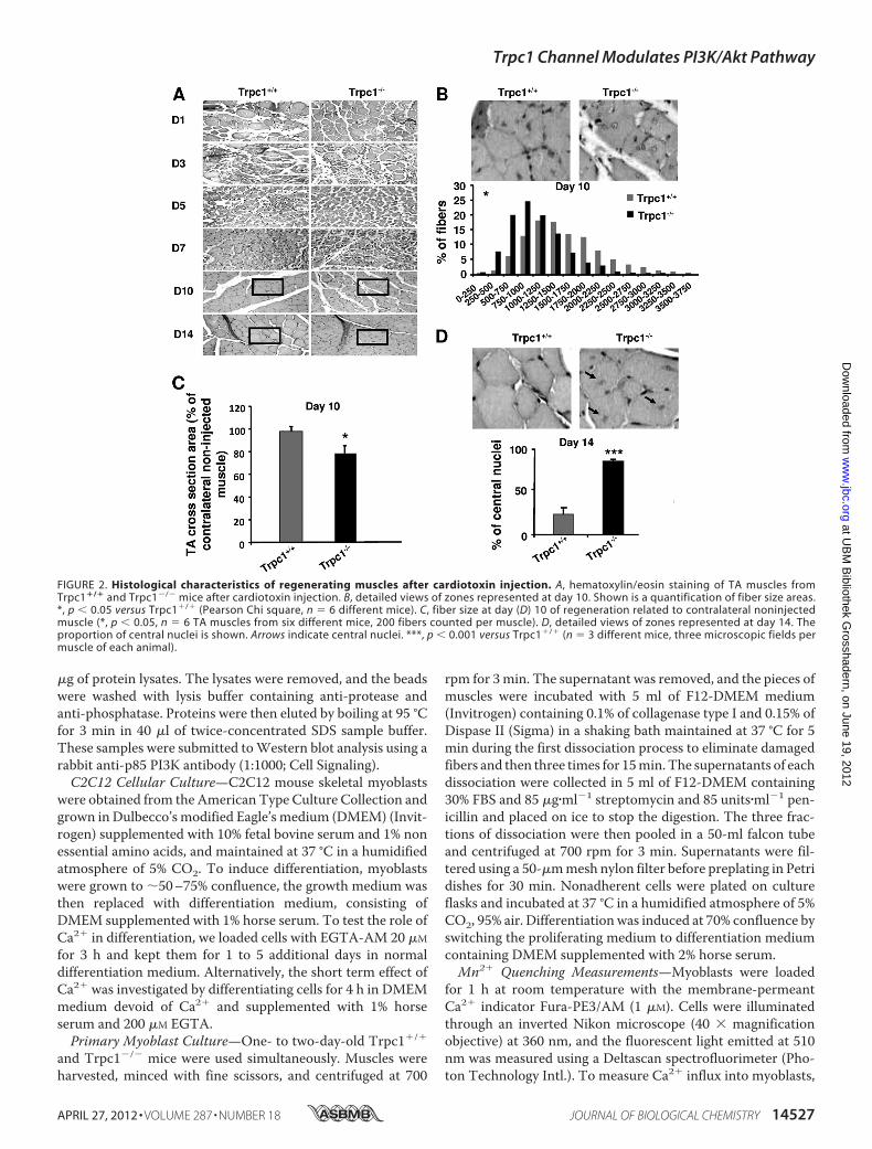
formedon cardiotoxin-injuredTAmuscles after a period of 1 to14 days of regeneration. Muscles were dissected, fixed in 4%paraformaldehyde on ice for 4 h, embedded in paraffin, andsectioned. Sections were stained with hematoxylin and eosin asdescribed previously (27). The size of muscle fiber sections wasmeasured using a homemade planimetry program (200 fiberswere counted per muscle).Immunohistochemistry—Five-�m thick paraffin embedded
sections of TA muscles at day 3 of regeneration were deparaf-finated, rehydrated, and blocked using a 0.5% bovine serumalbumin solution in phosphate-buffered saline (PBS) during 1 hat room temperature. Sections were then incubated at 4 °Covernight with mouse MHCd antibody (1:10, Novocastra, UK)diluted in blocking solution, washed three times in PBS for 10min, incubatedwith an anti-mouse antibody coupledwith alka-line phosphatase (1:50, Sigma) for 1 h, washed three timesagain, and revealed using alkaline phosphatase (Sigma). Thereaction was stopped with Tris-EDTA solution, pH 8, and sec-tions were fixed in formol and mounted with Mowiol (Calbi-ochem, La Jolla, CA).Measurement of Transcription Factor Activity—The global
activity ofmyogenic transcription factors wasmeasured using aluciferase plasmid gene reporter. The 4RTK-luciferase vectorcontaining four oligomerized MyoD-binding sites upstream ofa thymidine kinase promoter (28) (kindly provided by Dr. SteveTapscott, Fred Hutchinson Cancer Research Center, Seattle,WA) was amplified in Escherichia coli TOP10F� (Invitrogen)and purifiedwith an EndoFree PlasmidGiga kit (Qiagen, Venlo,Netherlands) (29). The day before injection, 30 �g of plasmidwere lyophilized and resuspended in 30 �l of 0.9% NaCl solu-tion. Three days before cardiotoxin injection, each mouse wasanesthetized, and 1 �g/�l plasmid solution was injected intoTAmuscles; these were electroporated as described previously(30). At day one of regeneration, animals were sacrificed, andTA muscles were removed. Whole TA muscles were homoge-nized with Ultraturax (IKA-Labortechnik, Staufen, Germany),and luciferase activity was quantified using a luciferase assaysystem (Promega, Madison, WI).
Trpc1 Channel Modulates PI3K/Akt Pathway
APRIL 27, 2012 • VOLUME 287 • NUMBER 18 JOURNAL OF BIOLOGICAL CHEMISTRY 14525
at UB
M B
ibliothek Grosshadern, on June 19, 2012
ww
w.jbc.org
Dow
nloaded from
Western Blot Analysis—Cells were scraped off, rinsed twicewith ice-cold PBS, centrifuged at 1500 � g for 10 min at 4 °C,and kept at �80 °C until use. Injured TA muscles were har-vested, frozen in liquid nitrogen, and also kept at �80 °C untiluse. Cells pellets were suspended in 60 �l and TA muscles in500 �l of lysis buffer containing the following: 50 mM Tris/HCl(pH 7.5), 1 mM EDTA (pH 8), 1 mM EGTA, 10 mM �-glycero-phosphate, 1 mM KH2PO4, 1 mM NaVO3, 50 mM NaF, 10 mM
NaPPi, and a protease inhibitor mixture (Roche, CompleteMini) and 0.5% Nonidet P-40, homogenized with pipette tipsfor cells or Ultraturax for muscles and incubated for 10 min at4 °C. Nuclei and unbroken cells were removed by centrifuga-tion at 10,000 � g for 10 min at 4 °C. Samples were incubatedwith Laemmli sample buffer containing SDS and 2-mercapto-ethanol for 3 min at 95 °C, electrophoresed on 10% SDS-poly-acrylamide gels, and transferred onto nitrocellulose mem-branes. Blots were incubated with rabbit anti-Myf5 (1:1000;Millipore, Billerica, MA), rabbit anti-MyoD (1:500; Santa CruzBiotechnology), mouse anti-myogenin (1:250; Santa Cruz Bio-technology), rabbit anti-phospho-Akt (1:500; Cell Signaling,Danvers, MA), mouse anti-PKB/Akt (1:1000; Bioke, Leiden,Netherlands), rabbit anti-phospho- and total p70S6K (1:1000;Santa Cruz Biotechnology), rabbit anti-GAPDH (1:1000; CellSignaling, Danvers,MA). After incubationwith the appropriatesecondary antibody coupled to peroxidase (Dako, Heverlee,Belgium), peroxidase was detected with ECL plus on ECLhyperfilm (Amersham Biosciences, Diegem, Belgium). Proteinexpressions were quantified by densitometry.
Real-time Polymerase Chain Reaction—Injured EDLmuscleswere homogenized in TRIzol (Invitrogen). Total RNA wastreated with DNase I and reverse-transcribed using qScriptReverse Transcriptase (Quanta Biosciences, Gaithersburg,ME). Gene-specific PCR primers were designed using Primer3.The GAPDH housekeeping gene and the genes of interest wereamplified in parallel. Real-time RT-PCRwas performed using 5�l of cDNA, 12.5 �l of qScript Reaction Mix (Quanta Biosci-ences, Gaithersburg, MD) and 300 nM of each primer in a totalreaction volume of 25�l. Data were recorded on aDNAEngineOpticon real-time RT-PCR detection system (Bio-Rad) andcycle threshold (Ct) values for each reaction were determinedusing analytical software from the same manufacturer. EachcDNA was amplified in duplicate, and Ct values were averagedfor each duplicate. The average Ct value for GAPDH was sub-tracted from the average Ct value for the gene of interest andnormalized to non-injected muscles. As amplification efficien-cies of the genes of interest and GAPDH were comparable, theamount of mRNA, normalized GAPDH, was given by the rela-tion 2���Ct. MyoD, Myf5, and myogenin primers and GAPDHand growth factor primers were designed as described previ-ously (31, 32).Immunoprecipitation Assay—Protein extracts were pre-
pared from C2C12 myoblasts cultured in differentiationmedium for 1 day or from TA muscles after 3 days of regener-ation. One �g of mouse anti-phosphotyrosine antibody (BDBiosciences) was incubated with 40 �l of Sepharose G beads(Sigma) for 2 h at 4 °C and then incubated overnight with 300
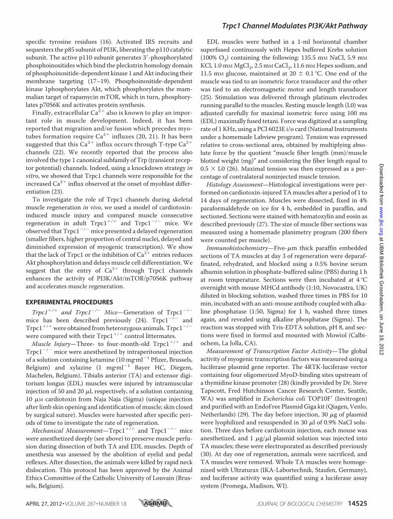
FIGURE 1. Weight and tension of normal and regenerating muscle in Trpc1�/� and Trpc1�/� mice. A and B, animal and EDL muscle weights during the first6 months of life (*, p � 0.05). C, maximal tension (force per cross-sectional area) measured after cardiotoxin-induced injury in EDL muscles at day 14 ofregeneration, stimulated during 300 ms and at 125 Hz. *, p � 0.05 versus Trpc1�/� (Student’s t test, n � 6). D, time course of muscle tension in regenerating EDLmuscles. Day zero is the day of cardiotoxin injection. Tension of regenerating muscle reported to that of contralateral noninjected muscle. *, p � 0.05 versusTrpc1�/� (two-way analysis of variance followed by Tukey’s test for multiple comparison, n � 6 per day).
Trpc1 Channel Modulates PI3K/Akt Pathway
14526 JOURNAL OF BIOLOGICAL CHEMISTRY VOLUME 287 • NUMBER 18 • APRIL 27, 2012
at UB
M B
ibliothek Grosshadern, on June 19, 2012
ww
w.jbc.org
Dow
nloaded from
�g of protein lysates. The lysates were removed, and the beadswere washed with lysis buffer containing anti-protease andanti-phosphatase. Proteins were then eluted by boiling at 95 °Cfor 3 min in 40 �l of twice-concentrated SDS sample buffer.These samples were submitted toWestern blot analysis using arabbit anti-p85 PI3K antibody (1:1000; Cell Signaling).C2C12 Cellular Culture—C2C12 mouse skeletal myoblasts
were obtained from the American Type Culture Collection andgrown in Dulbecco’s modified Eagle’s medium (DMEM) (Invit-rogen) supplemented with 10% fetal bovine serum and 1% nonessential amino acids, and maintained at 37 °C in a humidifiedatmosphere of 5% CO2. To induce differentiation, myoblastswere grown to 50–75% confluence, the growth medium wasthen replaced with differentiation medium, consisting ofDMEM supplemented with 1% horse serum. To test the role ofCa2� in differentiation, we loaded cells with EGTA-AM 20 �M
for 3 h and kept them for 1 to 5 additional days in normaldifferentiation medium. Alternatively, the short term effect ofCa2� was investigated by differentiating cells for 4 h in DMEMmedium devoid of Ca2� and supplemented with 1% horseserum and 200 �M EGTA.Primary Myoblast Culture—One- to two-day-old Trpc1�/�
and Trpc1�/� mice were used simultaneously. Muscles wereharvested, minced with fine scissors, and centrifuged at 700
rpm for 3min. The supernatant was removed, and the pieces ofmuscles were incubated with 5 ml of F12-DMEM medium(Invitrogen) containing 0.1% of collagenase type I and 0.15% ofDispase II (Sigma) in a shaking bath maintained at 37 °C for 5min during the first dissociation process to eliminate damagedfibers and then three times for 15min. The supernatants of eachdissociation were collected in 5 ml of F12-DMEM containing30% FBS and 85 �g�ml�1 streptomycin and 85 units�ml�1 pen-icillin and placed on ice to stop the digestion. The three frac-tions of dissociation were then pooled in a 50-ml falcon tubeand centrifuged at 700 rpm for 3 min. Supernatants were fil-tered using a 50-�mmesh nylon filter before preplating in Petridishes for 30 min. Nonadherent cells were plated on cultureflasks and incubated at 37 °C in a humidified atmosphere of 5%CO2, 95% air. Differentiationwas induced at 70% confluence byswitching the proliferating medium to differentiation mediumcontaining DMEM supplemented with 2% horse serum.Mn2� Quenching Measurements—Myoblasts were loaded
for 1 h at room temperature with the membrane-permeantCa2� indicator Fura-PE3/AM (1 �M). Cells were illuminatedthrough an inverted Nikon microscope (40 � magnificationobjective) at 360 nm, and the fluorescent light emitted at 510nm was measured using a Deltascan spectrofluorimeter (Pho-ton Technology Intl.). To measure Ca2� influx into myoblasts,
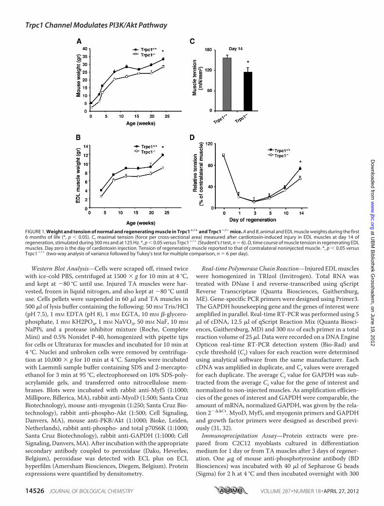
FIGURE 2. Histological characteristics of regenerating muscles after cardiotoxin injection. A, hematoxylin/eosin staining of TA muscles fromTrpc1�/� and Trpc1�/� mice after cardiotoxin injection. B, detailed views of zones represented at day 10. Shown is a quantification of fiber size areas.*, p � 0.05 versus Trpc1�/� (Pearson Chi square, n � 6 different mice). C, fiber size at day (D) 10 of regeneration related to contralateral noninjectedmuscle (*, p � 0.05, n � 6 TA muscles from six different mice, 200 fibers counted per muscle). D, detailed views of zones represented at day 14. Theproportion of central nuclei is shown. Arrows indicate central nuclei. ***, p � 0.001 versus Trpc1�/� (n � 3 different mice, three microscopic fields permuscle of each animal).
Trpc1 Channel Modulates PI3K/Akt Pathway
APRIL 27, 2012 • VOLUME 287 • NUMBER 18 JOURNAL OF BIOLOGICAL CHEMISTRY 14527
at UB
M B
ibliothek Grosshadern, on June 19, 2012
ww
w.jbc.org
Dow
nloaded from
500 �M MnCl2 was added to the Krebs medium, and the influxof Mn2� was evaluated by the quenching of Fura-PE3 fluores-cence excited at 360 nm (isosbestic point) (33, 34).Wound Healing Assay—The wound healing assay was per-
formed as described previously (23). Briefly, proliferation ofprimarymyoblasts at 70% confluence was stopped by switchingto differentiation medium for 24 h. Then, cells were scrappedoff to obtain a 600 �m wide acellular area and migrated myo-blasts into this area were counted after 15 h using the ImageJprogram.Chemicals—Cardiotoxin I isolated from Naja Naja Atra was
purchased from Sigma. Fura-PE3/AM, EGTA-AM, and wort-mannin were obtained from Calbiochem, Darmstadt, Ger-many. F12/DMEM, DMEM, serum, and streptomycin-penicil-lin solutions were purchased from Invitrogen.Statistical Analysis—Data are presented asmeans� S.E. Sta-
tistical significance was determined using t tests to comparetwo groups or analysis of variance to compare many groups.Analysis of the muscle cross-sectional area was performedusing a�2 Pearson test. The level of significancewas fixed at p�0.05.
RESULTS
TRPC1�/� Mice Present Delay of Skeletal MuscleRegeneration—We previously showed that TRPC1 proteinrepression reduces C2C12 myoblast migration and differentia-tion (23). We also observed that Trpc1�/� mice presented amild muscular hypotrophy consisting in smaller fibers size andin reduced content in myofibrillar proteins without any othersign ofmyopathy such as necrosis, central nulei, or fibrosis (27).Here, we studied animal development during the first sixmonths of life and confirmed amoderate but significant defaultof development (Fig. 1A) with in particular a reduced of muscleweight progression (Fig. 1B).To further investigate whether deletion of Trpc1 protein can
impair skeletal muscle development in vivo, we studied muscleregeneration after cardiotoxin-induced injury in Trpc1�/� andTrpc1�/� mice (35). EDL and TAmuscles were harvested afterdifferent periods of time post-injury and were characterizedfunctionally and histologically. First, we measured muscle ten-sion of regenerated Trpc1�/� and Trpc1�/� muscles at day 14of regeneration when muscle repair is almost completed (35).We observed that Trpc1�/� regenerating muscles presentedabout 25% lower tension than Trpc1�/� muscles (114.47 �13.18 in Trpc1�/� versus 155.78� 9.11mN/mm2 in Trpc1�/�;p � 0.05) (Fig. 1C). To relate this difference of tension to adeficit of regeneration, we performed muscles tension kineticsin the two groups from the first to the fourteenth day of regen-eration and related the tension produced by regenerating mus-cle to that produced by contralateral non injected muscle. Weobserved that one to 3 days after cardiotoxin injection, muscletension was dramatically but similarly decreased in bothTrpc1�/� and Trpc1�/� muscles (Fig. 1D). The importance ofthis loss of tension (10% residual tension) indicated thatdegeneration was almost complete in both groups. Interest-ingly, the time course of tension recovery between days 3 and 14post-injury suggests that muscle regeneration is slower inTrpc1�/� than Trpc1�/� mice (Fig. 1D).
The apparent delay of recovery observed in Trpc1�/� mus-cles was corroborated results point to a delayed repair inTrpc1�/� muscles in comparison with Trpc1�/� muscles: (i)adult non regenerated Trpc1�/� muscle fibers present asmaller cross-section area than Trpc1�/� muscles (1317 � 97�m2 versus 1638 � 103 �m2, n � 3 different animals of eachtype, 200muscle fibers counted permuscle, p� 0.05). Ten daysafter cardiotoxin injection, this difference was more important(1041 � 81 �m2 in Trpc1�/� versus 1638 � 110 �m2 inTrpc1�/� n � 6 different animals of each type, 200 musclefibers counted permuscle, p� 0.05; Fig. 2B). Indeed, Trpc1�/�
muscle fibers had recovered a normal size (98% of nonregener-ated fibers), whereas Trpc1�/� fibers remained significantlysmaller than fibers from the contralateral noninjected muscle(Fig. 2C); (ii) at day 14 of regeneration, themajority ofTrpc1�/�
fibers were still centrally nucleated, whereas in Trpc1�/� fibersmost of the nuclei hadmigrated to the periphery (84.25� 2.30%central nuclei in Trpc1�/� versus 23.53 � 7.55% in Trpc1�/�;
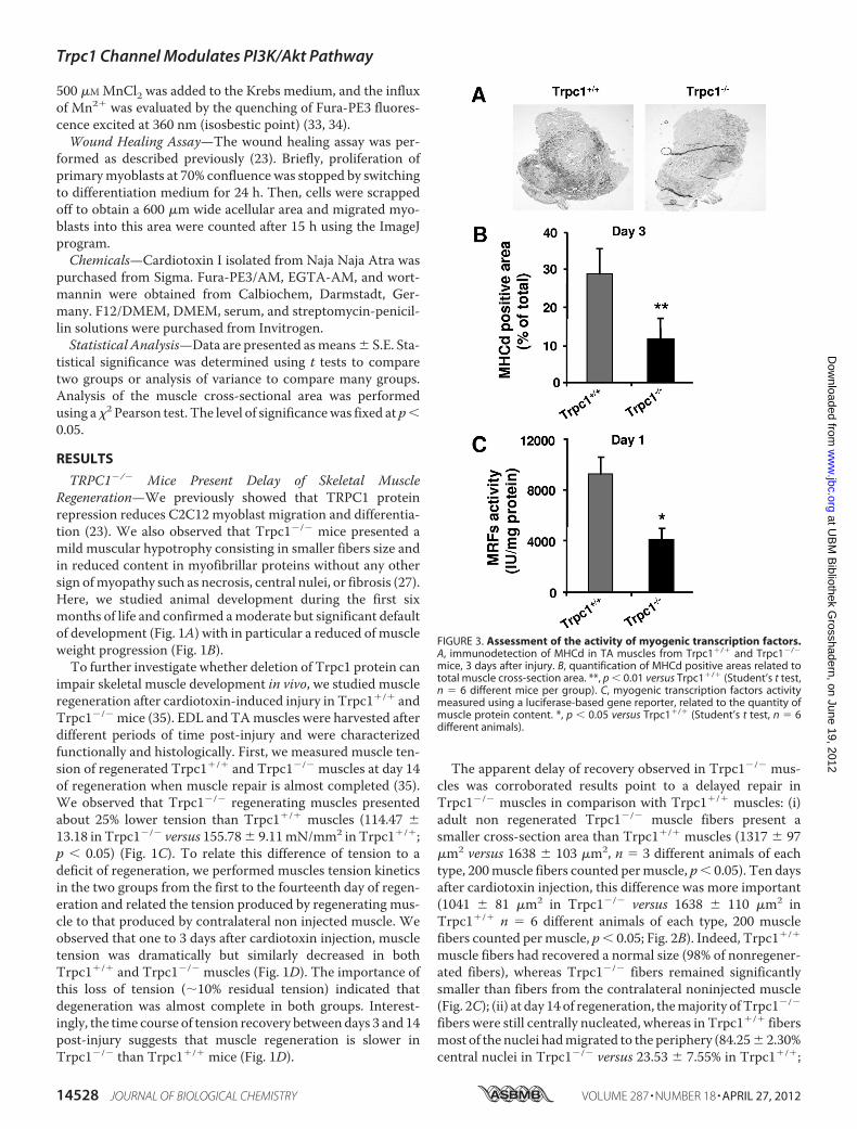
FIGURE 3. Assessment of the activity of myogenic transcription factors.A, immunodetection of MHCd in TA muscles from Trpc1�/� and Trpc1�/�
mice, 3 days after injury. B, quantification of MHCd positive areas related tototal muscle cross-section area. **, p � 0.01 versus Trpc1�/� (Student’s t test,n � 6 different mice per group). C, myogenic transcription factors activitymeasured using a luciferase-based gene reporter, related to the quantity ofmuscle protein content. *, p � 0.05 versus Trpc1�/� (Student’s t test, n � 6different animals).
Trpc1 Channel Modulates PI3K/Akt Pathway
14528 JOURNAL OF BIOLOGICAL CHEMISTRY VOLUME 287 • NUMBER 18 • APRIL 27, 2012
at UB
M B
ibliothek Grosshadern, on June 19, 2012
ww
w.jbc.org
Dow
nloaded from
p � 0.001) (Fig. 2D). Altogether, these results highlight a smallbut significant delay of muscle regeneration in Trpc1�/� micecompared with Trpc1�/� mice.Expression and Activity of Myogenic Transcription Factors
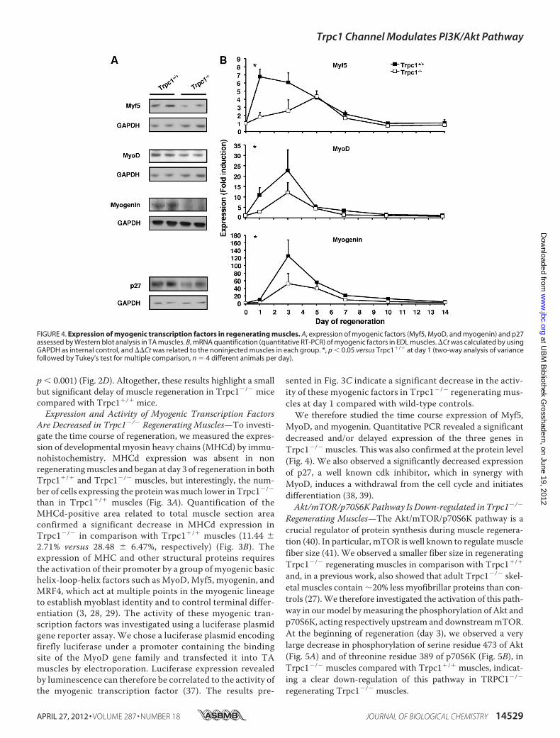
Are Decreased in Trpc1�/� Regenerating Muscles—To investi-gate the time course of regeneration, we measured the expres-sion of developmental myosin heavy chains (MHCd) by immu-nohistochemistry. MHCd expression was absent in nonregeneratingmuscles and began at day 3 of regeneration in bothTrpc1�/� and Trpc1�/� muscles, but interestingly, the num-ber of cells expressing the protein wasmuch lower in Trpc1�/�
than in Trpc1�/� muscles (Fig. 3A). Quantification of theMHCd-positive area related to total muscle section areaconfirmed a significant decrease in MHCd expression inTrpc1�/� in comparison with Trpc1�/� muscles (11.44 �2.71% versus 28.48 � 6.47%, respectively) (Fig. 3B). Theexpression of MHC and other structural proteins requiresthe activation of their promoter by a group of myogenic basichelix-loop-helix factors such as MyoD, Myf5, myogenin, andMRF4, which act at multiple points in the myogenic lineageto establish myoblast identity and to control terminal differ-entiation (3, 28, 29). The activity of these myogenic tran-scription factors was investigated using a luciferase plasmidgene reporter assay. We chose a luciferase plasmid encodingfirefly luciferase under a promoter containing the bindingsite of the MyoD gene family and transfected it into TAmuscles by electroporation. Luciferase expression revealedby luminescence can therefore be correlated to the activity ofthe myogenic transcription factor (37). The results pre-
sented in Fig. 3C indicate a significant decrease in the activ-ity of these myogenic factors in Trpc1�/� regenerating mus-cles at day 1 compared with wild-type controls.We therefore studied the time course expression of Myf5,
MyoD, and myogenin. Quantitative PCR revealed a significantdecreased and/or delayed expression of the three genes inTrpc1�/� muscles. This was also confirmed at the protein level(Fig. 4). We also observed a significantly decreased expressionof p27, a well known cdk inhibitor, which in synergy withMyoD, induces a withdrawal from the cell cycle and initiatesdifferentiation (38, 39).Akt/mTOR/p70S6KPathway Is Down-regulated in Trpc1�/�
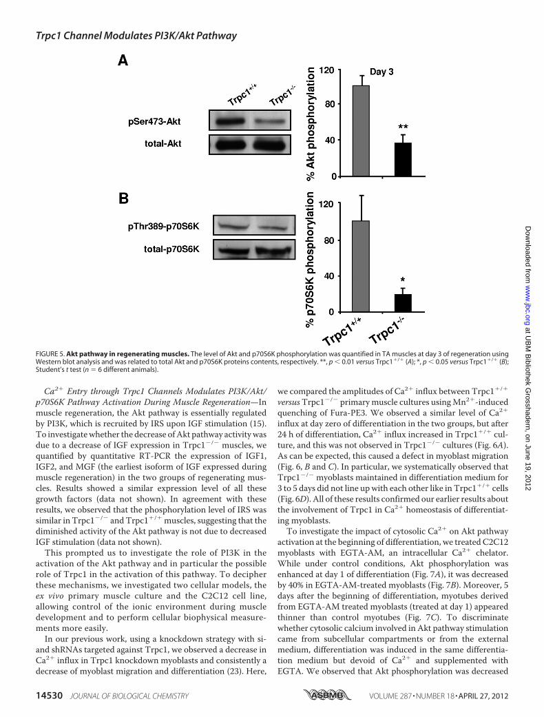
Regenerating Muscles—The Akt/mTOR/p70S6K pathway is acrucial regulator of protein synthesis during muscle regenera-tion (40). In particular, mTOR is well known to regulatemusclefiber size (41). We observed a smaller fiber size in regeneratingTrpc1�/� regenerating muscles in comparison with Trpc1�/�
and, in a previous work, also showed that adult Trpc1�/� skel-etal muscles contain 20% less myofibrillar proteins than con-trols (27).We therefore investigated the activation of this path-way in ourmodel bymeasuring the phosphorylation of Akt andp70S6K, acting respectively upstream and downstreammTOR.At the beginning of regeneration (day 3), we observed a verylarge decrease in phosphorylation of serine residue 473 of Akt(Fig. 5A) and of threonine residue 389 of p70S6K (Fig. 5B), inTrpc1�/� muscles compared with Trpc1�/� muscles, indicat-ing a clear down-regulation of this pathway in TRPC1�/�
regenerating Trpc1�/� muscles.
FIGURE 4. Expression of myogenic transcription factors in regenerating muscles. A, expression of myogenic factors (Myf5, MyoD, and myogenin) and p27assessed by Western blot analysis in TA muscles. B, mRNA quantification (quantitative RT-PCR) of myogenic factors in EDL muscles. �Ct was calculated by usingGAPDH as internal control, and ��Ct was related to the noninjected muscles in each group. *, p � 0.05 versus Trpc1�/� at day 1 (two-way analysis of variancefollowed by Tukey’s test for multiple comparison, n � 4 different animals per day).
Trpc1 Channel Modulates PI3K/Akt Pathway
APRIL 27, 2012 • VOLUME 287 • NUMBER 18 JOURNAL OF BIOLOGICAL CHEMISTRY 14529
at UB
M B
ibliothek Grosshadern, on June 19, 2012
ww
w.jbc.org
Dow
nloaded from
Ca2� Entry through Trpc1 Channels Modulates PI3K/Akt/p70S6K Pathway Activation During Muscle Regeneration—Inmuscle regeneration, the Akt pathway is essentially regulatedby PI3K, which is recruited by IRS upon IGF stimulation (15).To investigatewhether the decrease ofAkt pathway activitywasdue to a decrease of IGF expression in Trpc1�/� muscles, wequantified by quantitative RT-PCR the expression of IGF1,IGF2, and MGF (the earliest isoform of IGF expressed duringmuscle regeneration) in the two groups of regenerating mus-cles. Results showed a similar expression level of all thesegrowth factors (data not shown). In agreement with theseresults, we observed that the phosphorylation level of IRS wassimilar in Trpc1�/� and Trpc1�/� muscles, suggesting that thediminished activity of the Akt pathway is not due to decreasedIGF stimulation (data not shown).This prompted us to investigate the role of PI3K in the
activation of the Akt pathway and in particular the possiblerole of Trpc1 in the activation of this pathway. To decipherthese mechanisms, we investigated two cellular models, theex vivo primary muscle culture and the C2C12 cell line,allowing control of the ionic environment during muscledevelopment and to perform cellular biophysical measure-ments more easily.In our previous work, using a knockdown strategy with si-
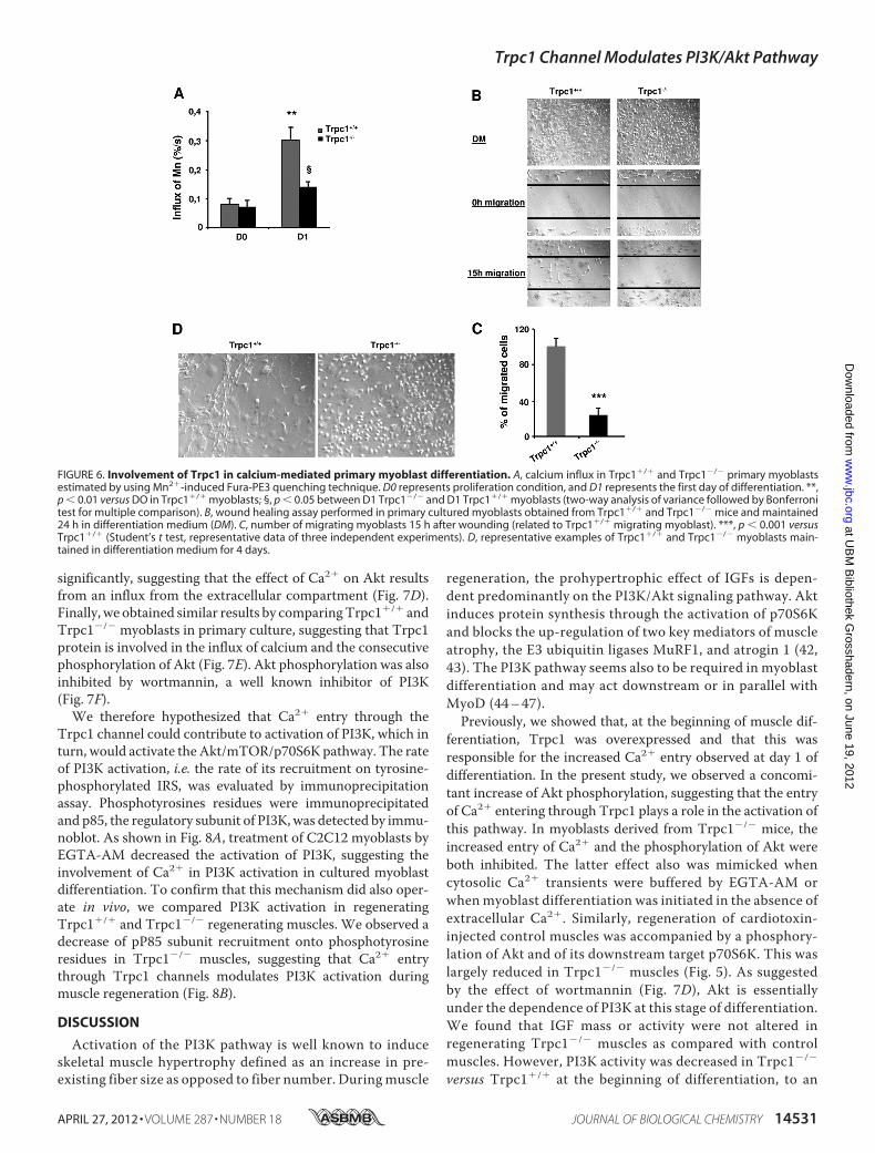
and shRNAs targeted against Trpc1, we observed a decrease inCa2� influx in Trpc1 knockdown myoblasts and consistently adecrease of myoblast migration and differentiation (23). Here,
we compared the amplitudes of Ca2� influx between Trpc1�/�
versusTrpc1�/� primarymuscle cultures usingMn2�-inducedquenching of Fura-PE3. We observed a similar level of Ca2�
influx at day zero of differentiation in the two groups, but after24 h of differentiation, Ca2� influx increased in Trpc1�/� cul-ture, and this was not observed in Trpc1�/� cultures (Fig. 6A).As can be expected, this caused a defect in myoblast migration(Fig. 6, B and C). In particular, we systematically observed thatTrpc1�/� myoblasts maintained in differentiation medium for3 to 5 days did not line upwith each other like in Trpc1�/� cells(Fig. 6D). All of these results confirmed our earlier results aboutthe involvement of Trpc1 in Ca2� homeostasis of differentiat-ing myoblasts.To investigate the impact of cytosolic Ca2� on Akt pathway
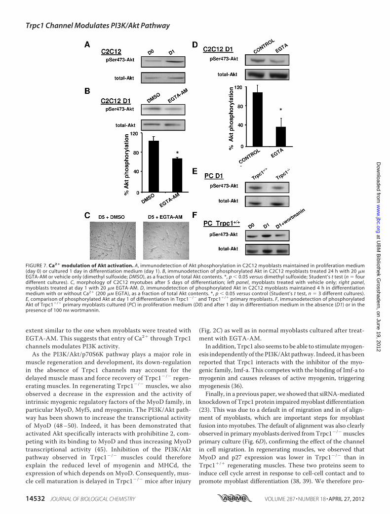
activation at the beginning of differentiation, we treated C2C12myoblasts with EGTA-AM, an intracellular Ca2� chelator.While under control conditions, Akt phosphorylation wasenhanced at day 1 of differentiation (Fig. 7A), it was decreasedby 40% in EGTA-AM-treated myoblasts (Fig. 7B). Moreover, 5days after the beginning of differentiation, myotubes derivedfrom EGTA-AM treated myoblasts (treated at day 1) appearedthinner than control myotubes (Fig. 7C). To discriminatewhether cytosolic calcium involved in Akt pathway stimulationcame from subcellular compartments or from the externalmedium, differentiation was induced in the same differentia-tion medium but devoid of Ca2� and supplemented withEGTA. We observed that Akt phosphorylation was decreased
FIGURE 5. Akt pathway in regenerating muscles. The level of Akt and p70S6K phosphorylation was quantified in TA muscles at day 3 of regeneration usingWestern blot analysis and was related to total Akt and p70S6K proteins contents, respectively. **, p � 0.01 versus Trpc1�/� (A); *, p � 0.05 versus Trpc1�/� (B);Student’s t test (n � 6 different animals).
Trpc1 Channel Modulates PI3K/Akt Pathway
14530 JOURNAL OF BIOLOGICAL CHEMISTRY VOLUME 287 • NUMBER 18 • APRIL 27, 2012
at UB
M B
ibliothek Grosshadern, on June 19, 2012
ww
w.jbc.org
Dow
nloaded from
significantly, suggesting that the effect of Ca2� on Akt resultsfrom an influx from the extracellular compartment (Fig. 7D).Finally, we obtained similar results by comparingTrpc1�/� andTrpc1�/� myoblasts in primary culture, suggesting that Trpc1protein is involved in the influx of calcium and the consecutivephosphorylation of Akt (Fig. 7E). Akt phosphorylation was alsoinhibited by wortmannin, a well known inhibitor of PI3K(Fig. 7F).We therefore hypothesized that Ca2� entry through the
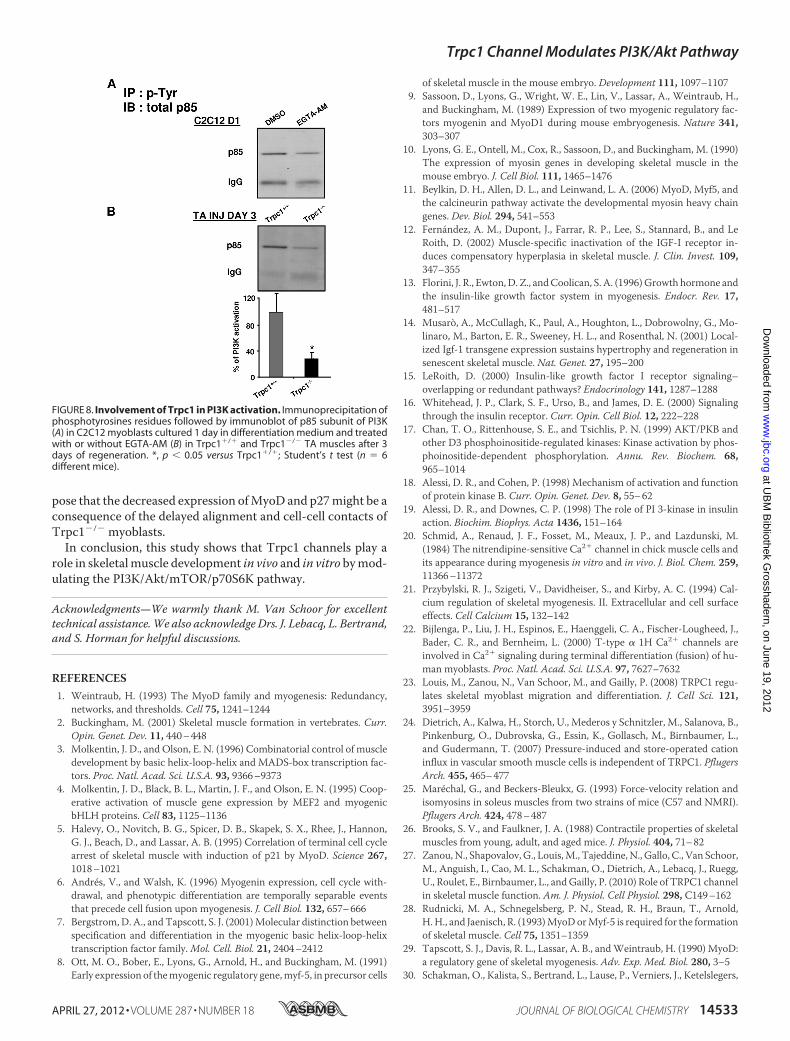
Trpc1 channel could contribute to activation of PI3K, which inturn, would activate the Akt/mTOR/p70S6K pathway. The rateof PI3K activation, i.e. the rate of its recruitment on tyrosine-phosphorylated IRS, was evaluated by immunoprecipitationassay. Phosphotyrosines residues were immunoprecipitatedand p85, the regulatory subunit of PI3K, was detected by immu-noblot. As shown in Fig. 8A, treatment of C2C12 myoblasts byEGTA-AM decreased the activation of PI3K, suggesting theinvolvement of Ca2� in PI3K activation in cultured myoblastdifferentiation. To confirm that this mechanism did also oper-ate in vivo, we compared PI3K activation in regeneratingTrpc1�/� and Trpc1�/� regenerating muscles. We observed adecrease of pP85 subunit recruitment onto phosphotyrosineresidues in Trpc1�/� muscles, suggesting that Ca2� entrythrough Trpc1 channels modulates PI3K activation duringmuscle regeneration (Fig. 8B).
DISCUSSION
Activation of the PI3K pathway is well known to induceskeletal muscle hypertrophy defined as an increase in pre-existing fiber size as opposed to fiber number. Duringmuscle
regeneration, the prohypertrophic effect of IGFs is depen-dent predominantly on the PI3K/Akt signaling pathway. Aktinduces protein synthesis through the activation of p70S6Kand blocks the up-regulation of two key mediators of muscleatrophy, the E3 ubiquitin ligases MuRF1, and atrogin 1 (42,43). The PI3K pathway seems also to be required in myoblastdifferentiation and may act downstream or in parallel withMyoD (44–47).Previously, we showed that, at the beginning of muscle dif-
ferentiation, Trpc1 was overexpressed and that this wasresponsible for the increased Ca2� entry observed at day 1 ofdifferentiation. In the present study, we observed a concomi-tant increase of Akt phosphorylation, suggesting that the entryof Ca2� entering through Trpc1 plays a role in the activation ofthis pathway. In myoblasts derived from Trpc1�/� mice, theincreased entry of Ca2� and the phosphorylation of Akt wereboth inhibited. The latter effect also was mimicked whencytosolic Ca2� transients were buffered by EGTA-AM orwhenmyoblast differentiation was initiated in the absence ofextracellular Ca2�. Similarly, regeneration of cardiotoxin-injected control muscles was accompanied by a phosphory-lation of Akt and of its downstream target p70S6K. This waslargely reduced in Trpc1�/� muscles (Fig. 5). As suggestedby the effect of wortmannin (Fig. 7D), Akt is essentiallyunder the dependence of PI3K at this stage of differentiation.We found that IGF mass or activity were not altered inregenerating Trpc1�/� muscles as compared with controlmuscles. However, PI3K activity was decreased in Trpc1�/�
versus Trpc1�/� at the beginning of differentiation, to an
FIGURE 6. Involvement of Trpc1 in calcium-mediated primary myoblast differentiation. A, calcium influx in Trpc1�/� and Trpc1�/� primary myoblastsestimated by using Mn2�-induced Fura-PE3 quenching technique. D0 represents proliferation condition, and D1 represents the first day of differentiation. **,p � 0.01 versus DO in Trpc1�/� myoblasts; §, p � 0.05 between D1 Trpc1�/� and D1 Trpc1�/� myoblasts (two-way analysis of variance followed by Bonferronitest for multiple comparison). B, wound healing assay performed in primary cultured myoblasts obtained from Trpc1�/� and Trpc1�/� mice and maintained24 h in differentiation medium (DM). C, number of migrating myoblasts 15 h after wounding (related to Trpc1�/� migrating myoblast). ***, p � 0.001 versusTrpc1�/� (Student’s t test, representative data of three independent experiments). D, representative examples of Trpc1�/� and Trpc1�/� myoblasts main-tained in differentiation medium for 4 days.
Trpc1 Channel Modulates PI3K/Akt Pathway
APRIL 27, 2012 • VOLUME 287 • NUMBER 18 JOURNAL OF BIOLOGICAL CHEMISTRY 14531
at UB
M B
ibliothek Grosshadern, on June 19, 2012
ww
w.jbc.org
Dow
nloaded from
extent similar to the one when myoblasts were treated withEGTA-AM. This suggests that entry of Ca2� through Trpc1channels modulates PI3K activity.As the PI3K/Akt/p70S6K pathway plays a major role in
muscle regeneration and development, its down-regulationin the absence of Trpc1 channels may account for thedelayed muscle mass and force recovery of Trpc1�/� regen-erating muscles. In regenerating Trpc1�/� muscles, we alsoobserved a decrease in the expression and the activity ofintrinsic myogenic regulatory factors of the MyoD family, inparticular MyoD, Myf5, and myogenin. The PI3K/Akt path-way has been shown to increase the transcriptional activityof MyoD (48–50). Indeed, it has been demonstrated thatactivated Akt specifically interacts with prohibitine 2, com-peting with its binding to MyoD and thus increasing MyoDtranscriptional activity (45). Inhibition of the PI3K/Aktpathway observed in Trpc1�/� muscles could thereforeexplain the reduced level of myogenin and MHCd, theexpression of which depends on MyoD. Consequently, mus-cle cell maturation is delayed in Trpc1�/� mice after injury
(Fig. 2C) as well as in normal myoblasts cultured after treat-ment with EGTA-AM.In addition, Trpc1 also seems to be able to stimulatemyogen-
esis independently of the PI3K/Akt pathway. Indeed, it has beenreported that Trpc1 interacts with the inhibitor of the myo-genic family, Imf-a. This competes with the binding of Imf-a tomyogenin and causes releases of active myogenin, triggeringmyogenesis (36).Finally, in a previous paper, we showed that siRNA-mediated
knockdown of Trpc1 protein impairedmyoblast differentiation(23). This was due to a default in of migration and in of align-ment of myoblasts, which are important steps for myoblastfusion into myotubes. The default of alignment was also clearlyobserved in primarymyoblasts derived fromTrpc1�/�musclesprimary culture (Fig. 6D), confirming the effect of the channelin cell migration. In regenerating muscles, we observed thatMyoD and p27 expression was lower in Trpc1�/� than inTrpc1�/� regenerating muscles. These two proteins seem toinduce cell cycle arrest in response to cell-cell contact and topromote myoblast differentiation (38, 39). We therefore pro-
FIGURE 7. Ca2� modulation of Akt activation. A, immunodetection of Akt phosphorylation in C2C12 myoblasts maintained in proliferation medium(day 0) or cultured 1 day in differentiation medium (day 1). B, immunodetection of phosphorylated Akt in C2C12 myoblasts treated 24 h with 20 �M
EGTA-AM or vehicle only (dimethyl sulfoxide; DMSO), as a fraction of total Akt contents. *, p � 0.05 versus dimethyl sulfoxide; Student’s t test (n � fourdifferent cultures). C, morphology of C2C12 myotubes after 5 days of differentiation; left panel, myoblasts treated with vehicle only; right panel,myoblasts treated at day 1 with 20 �M EGTA-AM. D, immunodetection of phosphorylated Akt in C2C12 myoblasts maintained 4 h in differentiationmedium with or without Ca2� (200 �M EGTA), as a fraction of total Akt contents. *, p � 0.05 versus control (Student’s t test, n � 3 different cultures).E, comparison of phosphorylated Akt at day 1 of differentiation in Trpc1�/� and Trpc1�/� primary myoblasts. F, immunodetection of phosphorylatedAkt of Trpc1�/� primary myoblasts cultured (PC) in proliferation medium (D0) and after 1 day in differentiation medium in the absence (D1) or in thepresence of 100 nM wortmannin.
Trpc1 Channel Modulates PI3K/Akt Pathway
14532 JOURNAL OF BIOLOGICAL CHEMISTRY VOLUME 287 • NUMBER 18 • APRIL 27, 2012
at UB
M B
ibliothek Grosshadern, on June 19, 2012
ww
w.jbc.org
Dow
nloaded from
pose that the decreased expression ofMyoD and p27might be aconsequence of the delayed alignment and cell-cell contacts ofTrpc1�/� myoblasts.In conclusion, this study shows that Trpc1 channels play a
role in skeletalmuscle development in vivo and in vitro bymod-ulating the PI3K/Akt/mTOR/p70S6K pathway.
Acknowledgments—We warmly thank M. Van Schoor for excellenttechnical assistance.We also acknowledge Drs. J. Lebacq, L. Bertrand,and S. Horman for helpful discussions.
REFERENCES1. Weintraub, H. (1993) The MyoD family and myogenesis: Redundancy,
networks, and thresholds. Cell 75, 1241–12442. Buckingham, M. (2001) Skeletal muscle formation in vertebrates. Curr.
Opin. Genet. Dev. 11, 440–4483. Molkentin, J. D., and Olson, E. N. (1996) Combinatorial control of muscle
development by basic helix-loop-helix and MADS-box transcription fac-tors. Proc. Natl. Acad. Sci. U.S.A. 93, 9366–9373
4. Molkentin, J. D., Black, B. L., Martin, J. F., and Olson, E. N. (1995) Coop-erative activation of muscle gene expression by MEF2 and myogenicbHLH proteins. Cell 83, 1125–1136
5. Halevy, O., Novitch, B. G., Spicer, D. B., Skapek, S. X., Rhee, J., Hannon,G. J., Beach, D., and Lassar, A. B. (1995) Correlation of terminal cell cyclearrest of skeletal muscle with induction of p21 by MyoD. Science 267,1018–1021
6. Andrés, V., and Walsh, K. (1996) Myogenin expression, cell cycle with-drawal, and phenotypic differentiation are temporally separable eventsthat precede cell fusion upon myogenesis. J. Cell Biol. 132, 657–666
7. Bergstrom, D. A., and Tapscott, S. J. (2001)Molecular distinction betweenspecification and differentiation in the myogenic basic helix-loop-helixtranscription factor family.Mol. Cell. Biol. 21, 2404–2412
8. Ott, M. O., Bober, E., Lyons, G., Arnold, H., and Buckingham, M. (1991)Early expression of themyogenic regulatory gene,myf-5, in precursor cells
of skeletal muscle in the mouse embryo. Development 111, 1097–11079. Sassoon, D., Lyons, G., Wright, W. E., Lin, V., Lassar, A., Weintraub, H.,
and Buckingham, M. (1989) Expression of two myogenic regulatory fac-tors myogenin and MyoD1 during mouse embryogenesis. Nature 341,303–307
10. Lyons, G. E., Ontell, M., Cox, R., Sassoon, D., and Buckingham, M. (1990)The expression of myosin genes in developing skeletal muscle in themouse embryo. J. Cell Biol. 111, 1465–1476
11. Beylkin, D. H., Allen, D. L., and Leinwand, L. A. (2006) MyoD, Myf5, andthe calcineurin pathway activate the developmental myosin heavy chaingenes. Dev. Biol. 294, 541–553
12. Fernández, A. M., Dupont, J., Farrar, R. P., Lee, S., Stannard, B., and LeRoith, D. (2002) Muscle-specific inactivation of the IGF-I receptor in-duces compensatory hyperplasia in skeletal muscle. J. Clin. Invest. 109,347–355
13. Florini, J. R., Ewton,D. Z., andCoolican, S. A. (1996)Growth hormone andthe insulin-like growth factor system in myogenesis. Endocr. Rev. 17,481–517
14. Musarò, A., McCullagh, K., Paul, A., Houghton, L., Dobrowolny, G., Mo-linaro, M., Barton, E. R., Sweeney, H. L., and Rosenthal, N. (2001) Local-ized Igf-1 transgene expression sustains hypertrophy and regeneration insenescent skeletal muscle. Nat. Genet. 27, 195–200
15. LeRoith, D. (2000) Insulin-like growth factor I receptor signaling–overlapping or redundant pathways? Endocrinology 141, 1287–1288
16. Whitehead, J. P., Clark, S. F., Urso, B., and James, D. E. (2000) Signalingthrough the insulin receptor. Curr. Opin. Cell Biol. 12, 222–228
17. Chan, T. O., Rittenhouse, S. E., and Tsichlis, P. N. (1999) AKT/PKB andother D3 phosphoinositide-regulated kinases: Kinase activation by phos-phoinositide-dependent phosphorylation. Annu. Rev. Biochem. 68,965–1014
18. Alessi, D. R., and Cohen, P. (1998) Mechanism of activation and functionof protein kinase B. Curr. Opin. Genet. Dev. 8, 55–62
19. Alessi, D. R., and Downes, C. P. (1998) The role of PI 3-kinase in insulinaction. Biochim. Biophys. Acta 1436, 151–164
20. Schmid, A., Renaud, J. F., Fosset, M., Meaux, J. P., and Lazdunski, M.(1984) The nitrendipine-sensitive Ca2� channel in chick muscle cells andits appearance during myogenesis in vitro and in vivo. J. Biol. Chem. 259,11366–11372
21. Przybylski, R. J., Szigeti, V., Davidheiser, S., and Kirby, A. C. (1994) Cal-cium regulation of skeletal myogenesis. II. Extracellular and cell surfaceeffects. Cell Calcium 15, 132–142
22. Bijlenga, P., Liu, J. H., Espinos, E., Haenggeli, C. A., Fischer-Lougheed, J.,Bader, C. R., and Bernheim, L. (2000) T-type � 1H Ca2� channels areinvolved in Ca2� signaling during terminal differentiation (fusion) of hu-man myoblasts. Proc. Natl. Acad. Sci. U.S.A. 97, 7627–7632
23. Louis, M., Zanou, N., Van Schoor, M., and Gailly, P. (2008) TRPC1 regu-lates skeletal myoblast migration and differentiation. J. Cell Sci. 121,3951–3959
24. Dietrich, A., Kalwa, H., Storch, U., Mederos y Schnitzler, M., Salanova, B.,Pinkenburg, O., Dubrovska, G., Essin, K., Gollasch, M., Birnbaumer, L.,and Gudermann, T. (2007) Pressure-induced and store-operated cationinflux in vascular smooth muscle cells is independent of TRPC1. PflugersArch. 455, 465–477
25. Maréchal, G., and Beckers-Bleukx, G. (1993) Force-velocity relation andisomyosins in soleus muscles from two strains of mice (C57 and NMRI).Pflugers Arch. 424, 478–487
26. Brooks, S. V., and Faulkner, J. A. (1988) Contractile properties of skeletalmuscles from young, adult, and aged mice. J. Physiol. 404, 71–82
27. Zanou,N., Shapovalov, G., Louis,M., Tajeddine,N., Gallo, C., Van Schoor,M., Anguish, I., Cao, M. L., Schakman, O., Dietrich, A., Lebacq, J., Ruegg,U., Roulet, E., Birnbaumer, L., andGailly, P. (2010) Role of TRPC1 channelin skeletal muscle function. Am. J. Physiol. Cell Physiol. 298, C149–162
28. Rudnicki, M. A., Schnegelsberg, P. N., Stead, R. H., Braun, T., Arnold,H.H., and Jaenisch, R. (1993)MyoDorMyf-5 is required for the formationof skeletal muscle. Cell 75, 1351–1359
29. Tapscott, S. J., Davis, R. L., Lassar, A. B., andWeintraub, H. (1990) MyoD:a regulatory gene of skeletal myogenesis. Adv. Exp. Med. Biol. 280, 3–5
30. Schakman, O., Kalista, S., Bertrand, L., Lause, P., Verniers, J., Ketelslegers,
FIGURE 8. Involvement of Trpc1 in PI3K activation. Immunoprecipitation ofphosphotyrosines residues followed by immunoblot of p85 subunit of PI3K(A) in C2C12 myoblasts cultured 1 day in differentiation medium and treatedwith or without EGTA-AM (B) in Trpc1�/� and Trpc1�/� TA muscles after 3days of regeneration. *, p � 0.05 versus Trpc1�/�; Student’s t test (n � 6different mice).
Trpc1 Channel Modulates PI3K/Akt Pathway
APRIL 27, 2012 • VOLUME 287 • NUMBER 18 JOURNAL OF BIOLOGICAL CHEMISTRY 14533
at UB
M B
ibliothek Grosshadern, on June 19, 2012
ww
w.jbc.org
Dow
nloaded from
J. M., and Thissen, J. P. (2008) Role of Akt/GSK-3beta/�-catenin trans-duction pathway in the muscle anti-atrophy action of insulin-like growthfactor-I in glucocorticoid-treated rats. Endocrinology 149, 3900–3908
31. Louis, M., Van Beneden, R., Dehoux, M., Thissen, J. P., and Francaux, M.(2004) Creatine increases IGF-I andmyogenic regulatory factor mRNA inC2C12 cells. FEBS Lett. 557, 243–247
32. Gilson, H., Schakman, O., Combaret, L., Lause, P., Grobet, L., Attaix, D.,Ketelslegers, J. M., and Thissen, J. P. (2007) Myostatin gene deletion pre-vents glucocorticoid-induced muscle atrophy. Endocrinology 148,452–460
33. Gailly, P., Hermans, E., and Gillis, J. M. (1996) Role of [Ca2�]i in “Ca2�
stores depletion Ca2� entry coupling” in fibroblasts expressing the ratneurotensin receptor. J. Physiol. 491, 635–646
34. Merritt, J. E., Jacob, R., and Hallam, T. J. (1989) Use of manganese todiscriminate between calcium influx and mobilization from internalstores in stimulated human neutrophils. J. Biol. Chem. 264, 1522–1527
35. Girgenrath, M., Weng, S., Kostek, C. A., Browning, B., Wang, M., Brown,S. A., Winkles, J. A., Michaelson, J. S., Allaire, N., Schneider, P., Scott,M. L., Hsu, Y. M., Yagita, H., Flavell, R. A., Miller, J. B., Burkly, L. C., andZheng, T. S. (2006) TWEAK, via its receptor Fn14, is a novel regulator ofmesenchymal progenitor cells and skeletal muscle regeneration. EMBO J.25, 5826–5839
36. Ma, R., Rundle, D., Jacks, J., Koch, M., Downs, T., and Tsiokas, L. (2003)Inhibitor of myogenic family, a novel suppressor of store-operated cur-rents through an interaction with TRPC1. J. Biol. Chem. 278,52763–52772
37. Weintraub, H., Davis, R., Lockshon, D., and Lassar, A. (1990)MyoD bindscooperatively to two sites in a target enhancer sequence: Occupancy oftwo sites is required for activation. Proc. Natl. Acad. Sci. U.S.A. 87,5623–5627
38. Vernon, A. E., and Philpott, A. (2003) A single cdk inhibitor, p27Xic1,functions beyond cell cycle regulation to promote muscle differentiationin Xenopus. Development 130, 71–83
39. Gavard, J., Marthiens, V., Monnet, C., Lambert, M., and Mège, R. M.(2004) N-cadherin activation substitutes for the cell contact control in cellcycle arrest and myogenic differentiation: Involvement of p120 and�-catenin. J. Biol. Chem. 279, 36795–36802
40. Bodine, S. C., Stitt, T. N., Gonzalez, M., Kline, W. O., Stover, G. L., Bau-
erlein, R., Zlotchenko, E., Scrimgeour, A., Lawrence, J. C., Glass, D. J., andYancopoulos, G. D. (2001) Akt/mTOR pathway is a crucial regulator ofskeletal muscle hypertrophy and can prevent muscle atrophy in vivo.Nat.Cell Biol. 3, 1014–1019
41. Miyabara, E. H., Conte, T. C., Silva, M. T., Baptista, I. L., Bueno, C., Jr.,Fiamoncini, J., Lambertucci, R. H., Serra, C. S., Brum, P. C., Pithon-Curi,T., Curi, R., Aoki, M. S., Oliveira, A. C., and Moriscot, A. S. (2010) Mam-malian target of rapamycin complex 1 is involved in differentiation ofregenerating myofibers in vivo.Muscle Nerve 42, 778–787
42. Glass, D. J. (2010) PI3 kinase regulation of skeletalmuscle hypertrophy andatrophy. Curr. Top Microbiol. Immunol. 346, 267–278
43. Latres, E., Amini, A. R., Amini, A. A., Griffiths, J., Martin, F. J.,Wei, Y., Lin,H. C., Yancopoulos, G. D., and Glass, D. J. (2005) Insulin-like growthfactor-1 (IGF-1) inversely regulates atrophy-induced genes via the phos-phatidylinositol 3-kinase/Akt/mammalian target of rapamycin (PI3K/Akt/mTOR) pathway. J. Biol. Chem. 280, 2737–2744
44. Wilson, E. M., Tureckova, J., and Rotwein, P. (2004) Permissive roles ofphosphatidyl inositol 3-kinase and Akt in skeletal myocyte maturation.Mol. Biol. Cell 15, 497–505
45. Sun, L., Liu, L., Yang, X. J., and Wu, Z. (2004) Akt binds prohibitin 2 andrelieves its repression ofMyoD andmuscle differentiation. J. Cell Sci. 117,3021–3029
46. Wilson, E. M., Hsieh, M. M., and Rotwein, P. (2003) Autocrine growthfactor signaling by insulin-like growth factor-II mediates MyoD-stimu-lated myocyte maturation. J. Biol. Chem. 278, 41109–41113
47. Small, E. M., O’Rourke, J. R., Moresi, V., Sutherland, L. B., McAnally, J.,Gerard, R. D., Richardson, J. A., and Olson, E. N. (2010) Regulation ofPI3-kinase/Akt signaling by muscle-enriched microRNA-486. Proc. Natl.Acad. Sci. U.S.A. 107, 4218–4223
48. Wilson, E. M., and Rotwein, P. (2007) Selective control of skeletal muscledifferentiation by Akt1. J. Biol. Chem. 282, 5106–5110
49. Xu, Q., and Wu, Z. (2000) The insulin-like growth factor-phosphatidyli-nositol 3-kinase-Akt signaling pathway regulates myogenin expression innormal myogenic cells but not in rhabdomyosarcoma-derived RD cells.J. Biol. Chem. 275, 36750–36757
50. Wilson, E. M., and Rotwein, P. (2006) Control of MyoD function duringinitiation of muscle differentiation by an autocrine signaling pathway ac-tivated by insulin-like growth factor-II. J. Biol. Chem. 281, 29962–29971
Trpc1 Channel Modulates PI3K/Akt Pathway
14534 JOURNAL OF BIOLOGICAL CHEMISTRY VOLUME 287 • NUMBER 18 • APRIL 27, 2012
at UB
M B
ibliothek Grosshadern, on June 19, 2012
ww
w.jbc.org
Dow
nloaded from
Related Documents