Cerebral Cortex September 2009;19:1990--2000 doi:10.1093/cercor/bhn226 Advance Access publication December 10, 2008 Performance Effects of Nicotine during Selective Attention, Divided Attention, and Simple Stimulus Detection: An fMRI Study Britta Hahn 1,3 , Thomas J. Ross 1 , Frank A. Wolkenberg 1 , Diaa M. Shakleya 2 , Marilyn A. Huestis 2 and Elliot A. Stein 1 1 Neuroimaging Research Branch, 2 Chemistry and Drug Metabolism Section, NIH/National Institute on Drug Abuse—Intramural Research Program (IRP), Biomedical Research Center, 251 Bayview Boulevard, Baltimore, MD 21224, USA and 3 Current address: Britta Hahn, Maryland Psychiatric Research Center, University of Maryland School of Medicine, PO Box 21247, Baltimore, MD 21228, USA Attention-enhancing effects of nicotine appear to depend on the nature of the attentional function. Underlying neuroanatomical mechanisms, too, may vary depending on the function modulated. This functional magnetic resonance imaging study recorded blood oxygen level--dependent (BOLD) activity in minimally deprived smokers during tasks of simple stimulus detection, selective attention, or divided attention after single-blind application of a transdermal nicotine (21 mg) or placebo patch. Smokers’ performance in the placebo condition was unimpaired as compared with matched nonsmokers. Nicotine reduced reaction time (RT) in the stimulus detection and selective attention but not divided attention condition. Across all task conditions, nicotine reduced activation in frontal, temporal, thalamic, and visual regions and enhanced deactivation in so-called ‘‘default’’ regions. Thalamic effects correlated with RT reduction selectively during stimulus detection. An interaction with task condition was observed in middle and superior frontal gyri, where nicotine reduced activation only during stimulus detection. A visuomotor control experiment provided evidence against nonspecific effects of nicotine. In conclusion, although prefrontal activity partly displayed differential modulation by nicotine, most BOLD effects were identical across tasks, despite differential performance effects, suggesting that common neuronal mechanisms can selectively benefit different attentional functions. Overall, the effects of nicotine may be explained by increased functional efficiency and downregulated task-independent ‘‘default’’ functions. Keywords: deactivation, default, fMRI, reaction time, skin patch, smokers Introduction There is ample evidence across species that nicotine possesses performance-enhancing properties (Wesnes and Warburton 1983; Heishman et al. 1994; Rezvani and Levin 2001), with improvements in attention particularly robust (Stolerman et al. 1995; Newhouse et al. 2004). The therapeutic potential of these effects motivates investigation of the precise attentional functions affected and their neuronal mediators. With regard to the type of attentional function, nicotine consistently improves performance in tasks of vigilance and simple stimulus detection (e.g., Wesnes and Warburton 1984; Koelega 1993; Foulds et al. 1996; Mancuso et al. 1999). These findings speak toward a generalized drug effect on alertness and intensity aspects of attention that could enhance perfor- mance across different paradigms. However, even if less robust, several findings also suggest improvements specific to pro- cesses of selective attention, such that the performance- enhancing effects of nicotine are relatively greater in mitigating the effects of distractors. For example, in both smokers and nonsmokers, nicotine or cigarette smoking reduced the Stroop effect, that is, performance costs of naming the ink color of an incongruent color word, in about half the studies investigating such effects (Wesnes and Warburton 1983; Provost and Woodward 1991; Hasenfratz and Battig 1992; Parrott and Craig 1992; Foulds et al. 1996; Poltavski and Petros 2006; Domier et al. 2007). Nicotine also reduced the Garner effect, that is, performance costs due to changes in the irrelevant stimulus dimension per se (Waters 1998). Furthermore, introducing sensory distractor stimuli helped reveal performance-enhanc- ing effects of nicotine in humans (Grobe et al. 1998), monkeys (Prendergast et al. 1998), and rats (Hahn et al. 2002; Hahn and Stolerman 2002). This reduced interference from irrelevant stimuli may reflect enhanced attentional filtering or an enhancement in control processes of attentional resource allocation. Evidence for the former can be deduced from findings that nicotine impaired incidental memory of material that subjects had not been instructed to remember and to which attention had presumably not been directed, while improving recall of attended material (Andersson and Hockey 1977). Further- more, improvements occurred mainly in those portions of a word list that were better recalled in the placebo condition and had probably been predominantly attended to (Warburton et al. 1992). Thus, nicotine appeared to increase attentional resources allocated to attended material and enhance the filtering of unattended material. In addition to selective attention, or attention to individual stimuli, there is the question of divided attention. Concepts of divided attention, too, are concerned with selectivity aspects of attention but more specifically with the optimal allocation of resources between different sets of input (Parasuraman 1998). Attention can be divided between locations in space, different features of one or more objects, and stimuli in one or more sensory modalities (Braun 1998). Nicotine has been reported to either improve (Leigh et al. 1977) or have no effect (Trimmel and Wittberger 2004) on dual-task performance. Effects on selective and divided attention have never been directly compared. Neuroimaging studies of the attention-enhancing effects of nicotine have mostly focused on nonselective alertness compo- nents of attention (with the exception of studies on specific spatial reorienting functions; Thiel et al. 2005; Giessing et al. 2006). Improvement in vigilance performance was accompa- nied by thalamic and parietal activation and enhanced insula and medial temporal deactivation (Lawrence et al. 2002). Nicotine also modulated cue-induced alerting-related activity in frontal, parietal, and superior temporal regions (Thiel and Fink 2007). Furthermore, in a study of visuospatial attention (Hahn et al. 2007), performance enhancement was associated with Published by Oxford University Press 2008. by guest on April 9, 2016 http://cercor.oxfordjournals.org/ Downloaded from

Welcome message from author
This document is posted to help you gain knowledge. Please leave a comment to let me know what you think about it! Share it to your friends and learn new things together.
Transcript
Cerebral Cortex September 2009191990--2000
doi101093cercorbhn226
Advance Access publication December 10 2008
Performance Effects of Nicotine duringSelective Attention Divided Attention andSimple Stimulus Detection An fMRI Study
Britta Hahn13 Thomas J Ross1 Frank A Wolkenberg1 Diaa
M Shakleya2 Marilyn A Huestis2 and Elliot A Stein1
1Neuroimaging Research Branch 2Chemistry and Drug
Metabolism Section NIHNational Institute on Drug
AbusemdashIntramural Research Program (IRP) Biomedical
Research Center 251 Bayview Boulevard Baltimore MD 21224
USA and 3Current address Britta Hahn Maryland Psychiatric
Research Center University of Maryland School of Medicine
PO Box 21247 Baltimore MD 21228 USA
Attention-enhancing effects of nicotine appear to depend on thenature of the attentional function Underlying neuroanatomicalmechanisms too may vary depending on the function modulatedThis functional magnetic resonance imaging study recorded bloodoxygen level--dependent (BOLD) activity in minimally deprivedsmokers during tasks of simple stimulus detection selectiveattention or divided attention after single-blind application ofa transdermal nicotine (21 mg) or placebo patch Smokersrsquoperformance in the placebo condition was unimpaired as comparedwith matched nonsmokers Nicotine reduced reaction time (RT) inthe stimulus detection and selective attention but not dividedattention condition Across all task conditions nicotine reducedactivation in frontal temporal thalamic and visual regions andenhanced deactivation in so-called lsquolsquodefaultrsquorsquo regions Thalamiceffects correlated with RT reduction selectively during stimulusdetection An interaction with task condition was observed inmiddle and superior frontal gyri where nicotine reduced activationonly during stimulus detection A visuomotor control experimentprovided evidence against nonspecific effects of nicotine Inconclusion although prefrontal activity partly displayed differentialmodulation by nicotine most BOLD effects were identical acrosstasks despite differential performance effects suggesting thatcommon neuronal mechanisms can selectively benefit differentattentional functions Overall the effects of nicotine may beexplained by increased functional efficiency and downregulatedtask-independent lsquolsquodefaultrsquorsquo functions
Keywords deactivation default fMRI reaction time skin patch smokers
Introduction
There is ample evidence across species that nicotine possesses
performance-enhancing properties (Wesnes and Warburton
1983 Heishman et al 1994 Rezvani and Levin 2001) with
improvements in attention particularly robust (Stolerman et al
1995 Newhouse et al 2004) The therapeutic potential of
these effects motivates investigation of the precise attentional
functions affected and their neuronal mediators
With regard to the type of attentional function nicotine
consistently improves performance in tasks of vigilance and
simple stimulus detection (eg Wesnes and Warburton 1984
Koelega 1993 Foulds et al 1996 Mancuso et al 1999) These
findings speak toward a generalized drug effect on alertness
and intensity aspects of attention that could enhance perfor-
mance across different paradigms However even if less robust
several findings also suggest improvements specific to pro-
cesses of selective attention such that the performance-
enhancing effects of nicotine are relatively greater in mitigating
the effects of distractors For example in both smokers and
nonsmokers nicotine or cigarette smoking reduced the Stroop
effect that is performance costs of naming the ink color of an
incongruent color word in about half the studies investigating
such effects (Wesnes and Warburton 1983 Provost and
Woodward 1991 Hasenfratz and Battig 1992 Parrott and Craig
1992 Foulds et al 1996 Poltavski and Petros 2006 Domier
et al 2007) Nicotine also reduced the Garner effect that is
performance costs due to changes in the irrelevant stimulus
dimension per se (Waters 1998) Furthermore introducing
sensory distractor stimuli helped reveal performance-enhanc-
ing effects of nicotine in humans (Grobe et al 1998) monkeys
(Prendergast et al 1998) and rats (Hahn et al 2002 Hahn and
Stolerman 2002)
This reduced interference from irrelevant stimuli may
reflect enhanced attentional filtering or an enhancement in
control processes of attentional resource allocation Evidence
for the former can be deduced from findings that nicotine
impaired incidental memory of material that subjects had not
been instructed to remember and to which attention had
presumably not been directed while improving recall of
attended material (Andersson and Hockey 1977) Further-
more improvements occurred mainly in those portions of
a word list that were better recalled in the placebo condition
and had probably been predominantly attended to (Warburton
et al 1992) Thus nicotine appeared to increase attentional
resources allocated to attended material and enhance the
filtering of unattended material
In addition to selective attention or attention to individual
stimuli there is the question of divided attention Concepts of
divided attention too are concerned with selectivity aspects
of attention but more specifically with the optimal allocation of
resources between different sets of input (Parasuraman 1998)
Attention can be divided between locations in space different
features of one or more objects and stimuli in one or more
sensory modalities (Braun 1998) Nicotine has been reported to
either improve (Leigh et al 1977) or have no effect (Trimmel and
Wittberger 2004) on dual-task performance Effects on selective
and divided attention have never been directly compared
Neuroimaging studies of the attention-enhancing effects of
nicotine have mostly focused on nonselective alertness compo-
nents of attention (with the exception of studies on specific
spatial reorienting functions Thiel et al 2005 Giessing et al
2006) Improvement in vigilance performance was accompa-
nied by thalamic and parietal activation and enhanced insula and
medial temporal deactivation (Lawrence et al 2002) Nicotine
also modulated cue-induced alerting-related activity in frontal
parietal and superior temporal regions (Thiel and Fink
2007) Furthermore in a study of visuospatial attention (Hahn
et al 2007) performance enhancement was associated with
Published by Oxford University Press 2008
by guest on April 9 2016
httpcercoroxfordjournalsorgD
ownloaded from
nicotine-induced deactivation of default regions of resting brain
function (Gusnard and Raichle 2001) suggesting that nicotine
improved performance by aiding the downregulation of task-
independent thought processes Thus insight has been gained
regarding mechanisms mediating effects on global intensity
aspects of attention However such generalized alerting
functions do not preclude the existence of mechanisms via
which nicotine may specifically enhance selectivity aspects of
attention in line with its behavioral profile described above
The present study aimed at identifying and dissociating
neuroanatomical substrates of nicotinersquos performance effects
under conditions that tax processes of selective or divided
attention and in a simple stimulus detection condition that
does not create any particular demands on attentional
selection A task setting was developed in which a single
foveally presented stimulus accommodated tasks related to
decisions about each of 2 stimulus dimensions or about both
dimensions combined A third stimulus detection task pre-
sented similar stimuli but required responses based on
subsequently presented signals We hypothesized that some
of the neuronal effects of nicotine would be specific to task
conditions with a selectivity component
Materials and Methods
ParticipantsEighteen right-handed smokers (9 females) participated in the study
Smokers were aged 19--49 years (mean plusmn standard deviation [SD]
301 plusmn 79 years) and smoked 21 plusmn 5 (range 15--30) cigarettes per day
for 129 plusmn 66 years (range 4--27 years) Smokerrsquos Intelligence Quotient
(IQ) determined by the Wechsler Abbreviated Scale of Intelligence
(Wechsler 1999) was 109 plusmn 10 Eighteen right-handed nonsmokers
(7 females) who reported no nicotine use within the past 12 months
were matched (no significant difference in independent-samples
t-tests) for age (296 plusmn 67 years range 18--44) and IQ (116 plusmn 11)
Neuroimaging and performance data from these nonsmoking partic-
ipants forming part of a larger group of control subjects were reported
in detail previously (Hahn et al 2008)
Subjects were recruited from the general population through
newspaper advertising flyers and referrals and gave written informed
consent for a protocol approved by the National Institute on Drug Abuse
(NIDA)--IRP Institutional Review Board Subjects were screened for major
medical illnesses claustrophobia history of neurological or psychiatric
disorders drug and alcohol abuse and pregnancy A urine sample was
collected and assessed for common drugs of abuse (TRIAGE)
ProcedureThe protocol required 3 visits During the first visit participants gave
informed consent and were trained on 2 cognitive tasks (1 reported
elsewhere) initially on a bench computer and then for 30 min in
a mock scanner that mimicked all properties of the magnetic resonance
imaging (MRI) scanner without the magnetic field Participants were
also familiarized with the computerized questionnaires to be com-
pleted in the scanner and with the wheel response device used for
their completion Sessions 2 and 3 were identical however for
smokers a Nicoderm patch (21 mg24 h GlaxoSmithKline Moon
Township PA) was applied to the upper back in one session 2--25 h
prior to being loaded into the MR scanner and a placebo patch in the
other The task paradigm reported here began approximately 3--35 h
following patch application The sequence of test sessions was
counterbalanced such that 9 smokers received nicotine in the first
and 9 in the second session Order of patch application was single blind
Nonsmokers completed both sessions without any skin patches The
purpose was to compare performance and regional activity between
groups at baseline and examine potential abstinence-related differences
in smokers Nicotine was not administered to nonsmokers because
initial exposure of drug-naive individuals typically leads to aversive side
effects that can overshadow and interfere with the measurement of
nicotinersquos cognitive effects (Heishman et al 1993 Perkins et al 1994
Heishman and Henningfield 2000) The 2 test sessions were scheduled
2--16 days apart in all except 2 subjects (both smokers) who were
tested 24 and 28 days apart due to scheduling availability
Smokers smoked a cigarette within 1 h prior to entering the NIDA--
IRP research facilities with MR scans starting approximately 3 h
following their last cigarette Participants were told not to ingest any
alcohol or over-the-counter medication in the 24 h preceding each
session and not to consume more than a half cup of coffee within the
preceding 12 h Prior to patch application participants were tested for
recent drug use (TRIAGE) and for alcohol intake via breath analysis
(Alco-Sensor IV Intoximeters Inc St Louis MO) Shortly after patch
application subjects received a 9-min reminder task training on
a bench computer and practiced a finger-tapping procedure In MR
scans three 842-min runs of the selectivedivided attention task (see
below) were performed separated by 1-min rest periods A 30-min
visuospatial cueing task (Stein et al 2004) was performed within the
same session the sequence of tasks was counterbalanced across
subjects Anatomical scans were performed between tasks At the end
of each session a perfusion MRI scan was obtained during performance
of a 65-min finger-tapping task described below
For all but one smoker a venous blood specimen (5 mL) was drawn
from a forearm vein within 10 min following each scan session
Specimens were stored on ice centrifuged within 2 h of collection and
plasma frozen at ndash20 C until analysis Plasma specimens (1 mL) from 10
participants were assayed for nicotine concentration via solid phase
extraction and liquid chromatography--atmospheric pressure chemical
ionization--mass spectrometry with selected ion monitoring (Kim and
Huestis 2006) The assay was linear from 25 to 500 ngmL with
a weighting factor of 1x correlation coefficients for calibration curves
were gt099 Intra- and interassay precision and accuracy were lt150Nicotine recovery ranged from 1082 to 1108 at 3 concentrations
across the dynamic range For the remaining specimens nicotine was
isolated and concentrated from 200 lL plasma by solid phase extraction
with preconditioned CleanScreen DAU columns Eluates were evapo-
rated to dryness under nitrogen reconstituted with 200 lL of mobile
phase and analyzed by a validated liquid chromatography--tandem mass
spectrometry method with electrospray ionization Identification and
quantification of nicotine were based on selected reaction monitoring
with 2 transitions The limit of quantification was 1 ngmL with a linear
dynamic range to 500 ngmL Extraction efficiency was greater than
90 with inter- and intraday imprecision lt20Subjective state was measured by computerized versions of 2 self-
report instruments while lying inside the MR scannermdashonce just before
and once just after the scan session One instrument given to both
smokers and control participants consisted of a list of bidirectional
visual analog scales sensitive to mood changes induced by tobacco
deprivation (Parrott et al 1996) tenserelaxed nervouscalm ener-
getictired alertdrowsy contentedirritated satisfieddissatisfied Ad-
ditional scales added to cover further nicotine withdrawal symptoms
were distractedfocused depressedhappy and satiatedhungry Data
are not available for 6 of the nonsmoking controls Smokers also
completed the 12-item version of the tobacco craving questionnaire
(TCQ) (Heishman et al 2003) For both scales participants used
a wheel response device to move a cursor on the screen to the desired
position on a horizontal bar relative to 2 anchors
Measurement of Selective and Divided AttentionThe task stimulus consisted of a circle containing 2 wedges displayed
against a gray background in the center of the screen (Fig 1) The
diameter of the circle based on a viewing distance of 80 cm was 36 ofvisual angle thus allowing foveal stimulus processing without
significant eye movement In the selective and divided attention task
conditions each wedge was divided into 3 sections of an inner middle
and outer ring of color (Fig 1AB) Within each wedge each segment
was always of a different color from the others (red blue and purple)
In 3 different forced choice tasks participants decided whether
specific features of the 2 wedges were the same or different In the 2
selective attention conditions they were instructed to attend either to
the color order of the rings (selective color SEL-C) or to the angles of
Cerebral Cortex September 2009 V 19 N 9 1991
by guest on April 9 2016
httpcercoroxfordjournalsorgD
ownloaded from
the wedges (selective angle SEL-A) and decide whether they were the
same or different The third was a divided attention (DIV) condition
during which subjects attended to both of these features and decided
whether or not the wedges were identical in both features A button for
same was pressed with the right index finger and a button for different
with the left
In the SEL-A and SEL-C tasks the wedges differed on the task-relevant
feature in 50 of trials The task-irrelevant feature also differed in 50
of trials independent of the status of the task-relevant feature Thus
stimulus characteristics remained constant and only task demands
defined the 2 different conditions In the DIV task the wedges differed
on either one of the 2 stimulus features in 50 of trials that is 25 on
the angle and 25 on the color feature in the other half of the trials
neither feature differed
The fourth was a simple stimulus detection task (SDT) designed not
to place any particular demands on selectivity aspects of attention The
wedged circle was presented for a fixed length of time that equaled the
display time (DT) entered at the beginning of the session (see below)
The visual stimulus properties were the same as in the other tasks
except that only 2 rings of color were presented and the color hues
were composed of multicolored dots (Fig 1C) These changes to the
stimulus appearance were made to prevent potential habitual focusing
on either of the 2 stimulus dimensions The circle stimulus was
followed by presentation of a letter on the left or on the right
Participants were instructed to respond not to any property of the
wedges but to the side on which a letter was presented
Each task trial started with a 500-ms central fixation cross followed
by 500 ms of blank screen The wedged circle was then presented for
the duration of DT (see below) followed by 48 ms of a back mask
consisting of the circle filled with colored dots to eliminate any
persisting afterimage of the task stimulus The letters lsquolsquodrsquorsquo and lsquolsquosrsquorsquo for
lsquolsquodifferentrsquorsquo and lsquolsquosamersquorsquo then appeared on the left and right respectively
of where the circle had been presented (on the left or right for SDT)
and stayed on display until a response was made for a maximum
duration of 2 s Trials where no response was recorded within this time
were excluded from analysis (11 of all trials) Trials were separated by
a variable interstimulus interval (ISI) of 0 2 4 or 6 s duration The ISI
was extended by the length of time needed to complete the preceding
repetition time (TR)
For Sel-A Sel-C and DIV performance accuracy was held at 75 by
manipulating the stimulus DT The purpose of this manipulation was to
minimize differences in error processing and response uncertainty
between the selective and divided attention conditions and to eliminate
such confounds when interpreting any differential effects of nicotine
Adjustments were made in 16 ms units Initial DT was determined
during training for each individual subject Early during the training
procedure the wedge angle difference was determined such that DT
for SEL-A was identical to SEL-C at 75 accuracy This difference value
was then adopted for all 3 tasks Angle difference values ranged from 6to 12 across participants (mean plusmn SD 71 plusmn 18) Throughout DT was
dynamically adjusted after every 4 trials If a correct response was made
in 3 out of the 4 preceding trials DT stayed the same If 2 or fewer trials
were correct DT increased by 16 ms and if all 4 trials were correct DT
decreased by 16 ms During scan sessions DT was adjusted in this
manner independently for SEL-A SEL-C and DIV starting with the
values obtained at completion of the training The same starting values
were used for both scans In this manner response accuracy was
successfully adjusted to vary around or just above 75 for each task
(Hahn et al 2008) Accuracy during SDT approached 100
In each scan session three 842-min task runs were completed Each
run started with one 8-trial block of SDT One 16-trial block each of
SEL-A SEL-C and DIV was then performed in a randomized sequence
followed by 8 more trials of SDT Each block began with the task
instruction displayed for 4 s followed by a 6-s epoch where
participants performed a forced choice test (lsquolsquopress the button on the
side that names this taskrsquorsquo) Blocks preceded by an incorrect answer
were excluded from further analyses (7 out of a total of 432 blocks
across subjects and sessions)
Controls for Nonspecific Effects of Nicotine on Blood Flow andCouplingTo test for potential nonspecific effects of nicotine on cerebral blood
flow (CBF) or coupling between neuronal and hemodynamic response
dynamics perfusion functional magnetic resonance imaging (fMRI)
scans were acquired from slices covering primary motor and visual
cortices while subjects performed cyclic (30 s on 30 s off) bilateral
finger tapping During on-periods a checkerboard of black and white
squares that filled the entire screen (spatial frequency ~026 cycles
degree) and whose contrast reversed 3 times per second served as
a visual metronome During off-periods participants fixated a central
cross The scan started and ended on an off-period Thirteen 30-s
periods were presented in total
Magnetic Resonance ImagingScanning was performed on a 3 Tesla Siemens Allegra scanner
(Erlangen Germany) Whole-brain functional EPI images were acquired
for measurement of T2-weighted blood oxygen level--dependent
(BOLD) effects (4 mm sagittal slices 64 3 64 matrix field of view
[FOV] = 22 3 22 cm TR = 2 s time echo [TE] = 27 ms FA = 75) Ineach scanning session a whole-brain sagittal T1-weighted structural
image (MPRAGE) was acquired for anatomical reference (1 mm3
isotropic voxels TR = 25 s TE = 438 ms FA = 8) Perfusion fMRI
scans were acquired in six 7-mm transaxial slices using a QUIPPS II
(Wong et al 1998) arterial spin labeling (ASL) imaging sequence
(FOV = 220 cm matrix = 64 3 64 TR = 3 s TE = 27 ms FA = 90TI1 = 700 ms TI2 = 1400 ms gap = 10 mm) Four subjects were
scanned with a Flow-sensitive Alternating Inversion Recovery (FAIR)-
based sequence (Kim 1995 TI = 1400 inversion slab thickness = 58 mm)
Analysis of Subjective Self-ReportsIndividual lsquolsquoParrottrsquorsquo subscales (Parrott et al 1996) were analyzed by 3-
factor analysis of variance (ANOVA) with GROUP (smokers controls) as
a between-subject factor and SESSION (nicotine vs placebo for
smokers no-drug vs no-drug for controls) and PRE--POST (pre- vs
postscan) as within-subject factors TCQ craving scores were obtained
only in smokers and were analyzed by 2-factor ANOVA (SESSION 3
PRE--POST)
Figure 1 Examples of the task stimuli Participants were instructed to detect a difference in either the angles of the 2 wedges in the sequence of color across the 3 rings or ineither aspect In (A) there is a difference in the color dimension In (B) there is a difference in the angle dimension (C) Represents a stimulus presented during SDT whereresponses did not depend on any stimulus aspects
1992 Performance Effects of Nicotine d Hahn et al
by guest on April 9 2016
httpcercoroxfordjournalsorgD
ownloaded from
Analysis of Behavioral DataData from the 2 scan sessions were analyzed DT and reaction time (RT)
were expressed as averages for each task condition and analyzed
separately by 3-factor ANOVA for repeated measures with DRUG
(nicotine placebo) and TASK (SDT Sel-A Sel-C DIV) as within-subject
factors and SEQUENCE OF TESTING (nicotine followed by placebo
placebo followed by nicotine) as between-subject factor ANOVAs were
followed by paired t-tests where indicated To compare performance in
the absence of nicotine between smokers and nonsmokers data from
nondrug days were analyzed by 2-factor ANOVA with GROUP
(smokers controls) as between-subject factor and TASK as within-
subject factor Session 1 data were included from 9 of the 18 controls
selected randomly and session 2 data from the other 9 thus matching
the amount of task preexposure to that of smokersrsquo placebo sessions
Analysis of fMRI DataData were processed using the AFNI software package (Cox 1996)
Motion correction was performed by registering each 3D volume to
a base volume The time series was then analyzed as an event-related
design by voxel-wise multiple regression Regressors were expressed as
a delta function time locked to the onset of each circle stimulus and
convolved with a model hemodynamic response function and its
temporal derivative Regressors corresponded to the 4 different task
conditions (SDT Sel-A Sel-C and DIV) and to the 6 motion parameters
as nuisance regressors Further nuisance regressors corresponded to
the display and retention test of the task instruction and if applicable
to trials in which no response was registered and blocks in which the
task instruction was not correctly repeated For each subject and test
session the voxel-wise average amplitude of signal change (b value)
produced by each task condition was determined relative to baseline
The resulting activation maps were resampled to a higher (1 lL)resolution converted to a standard stereotaxic coordinate system
(Talairach and Tournoux 1988) and spatially blurred using a Gaussian
5-mm rms isotropic kernel
Second-level random-effects analysis across smokers consisted of
voxel-wise 2-factor ANOVA for repeated measures (DRUG 3 TASK)
performed on the b values produced by each task condition A voxel-
wise threshold of P lt 001 was applied to the activation maps and
combined with a minimum cluster volume size of 450 lL Based on
Monte Carlo simulations taking account of spatial covariation in the
output dataset this yielded an overall false positive P lt 0005 To test
whether the effects of nicotine may have served to restore a normal
functional state aberrant in smokers in the absence of nicotine for
example due to neural adaptations with chronic nicotine exposure
average levels of activity in nonsmokers were determined within
functional Regions of Interest (ROIs) that displayed effects of nicotine
Activations in the drug-free state were compared between groups by
independent-samples t-tests Nine smokers received placebo in session
1 and 9 in session 2 accordingly session 1 data were used from 9 and
session 2 data from the other 9 nonsmoking controls Also to test for
group differences in brain regions not necessarily modulated by
nicotine whole-brain voxel-wise ANOVA (GROUP 3 TASK) was
performed on the no-drug data using the same significance criteria
as for the DRUG 3 TASK ANOVA
To examine the effects of nicotine on BOLD and CBF responses to
visuomotor stimulation during smokersrsquo ASL scan BOLD- (derived from
untagged images) and flow-weighted (derived by voxel-wise sub-
traction of untagged from tagged images) time series were analyzed
with a boxcar regressor following the 30-s on- and off-periods
convolved with a model hemodynamic response function Data from
3 subjects were corrupted and were excluded BOLD contrast values
(on- vs off-periods) were normalized and underwent a random-effects
1-sample t-test against 0 Voxel-wise P lt 0001 combined with
a minimum cluster volume of 368 lL yielded an overall false positive
P lt 005 as determined by Monte Carlo simulation Flow- and BOLD-
weighted contrast values were averaged across voxels within each
identified region For each participant only voxels with anatomical
coverage in both sessions were included Average regional BOLD
contrast values were compared between the placebo and nicotine
session by paired t-tests Flow-weighted values displayed large
variability in this dataset Although no effects of nicotine were seen
the large error variance would most likely preclude their detection
Flow-weighted values are thus excluded from this report
Head motion during the attention task was compared between test
sessions by calculating a composite motion index from the 3
translational and the 3 rotational parameters as described by Yang
et al (2005) This index reflects a subjectrsquos average head motion
between 2 consecutive TRs Values did not differ between the nicotine
and the placebo session (t17 = 168 not significant [NS] paired t-test)
CorrelationsEach smokerrsquos RT in the placebo session was subtracted from that in
the nicotine session Similarly for each brain area modulated by
nicotine average regional activation under placebo was subtracted
from that under nicotine The difference values in RT and regional
activation underwent partial correlation controlling for nicotine plasma
concentrations in both the nicotine and placebo sessions Plasma
concentrations were controlled for because they may underlie
interindividual variation in both performance and BOLD effects of
nicotine and may thus enhance correlations by acting as a common
antecedent For correlations P lt 0005 was considered significant
Results
Nicotine Plasma Levels
Smokersrsquo plasma nicotine levels were 57 plusmn 28 ngmL at
completion of the placebo scan and 377 plusmn 99 ngmL after
the nicotine scan (t16 = 138 P lt 0001) comparable with
results obtained previously under the same experimental
conditions (Hahn et al 2007)
Subjective State
Parrott Scale
Main effects of PRE--POST for 5 variables (F128 gt 480
P lt 005) indicated that all participants felt more tired drowsy
dissatisfied distracted and hungry after than before scan
sessions A main effect of GROUP (F128 = 479 P lt 005) for
the lsquolsquoenergetic--tiredrsquorsquo subscale reflected higher reports of
tiredness in the smokers than nonsmokers A GROUP 3
SESSION interaction occurred on lsquolsquoalert--drowsyrsquorsquo and lsquolsquofo-
cused--distractedrsquorsquo (F128 gt 542 P lt 005) Effects of SESSION
on these scales were seen in smokers (t17 gt 273 P lt 005)
who were more focused and alert in the nicotine than placebo
session but not in nonsmokers who were never administered
any drug Ratings never differed significantly between groups
but numerically smokers felt more alert and focused than
nonsmokers in the nicotine session and drowsier and more
distracted than nonsmokers in the placebo session Thus the
drug effect may represent a combination of alerting effects of
nicotine and impairment in the absence of nicotine
Tobacco Craving Questionnaire
Smokersrsquo craving ratings were higher in the placebo than
nicotine session (main effect of DRUG F117 = 725 P lt 005)
and higher after than before scan sessions (PRE--POST
F117 = 874 P lt 001) No DRUG 3 PRE--POST interaction
was observed
Effects of Nicotine on Smokersrsquo Performance
Figure 2A shows DT during Sel-A Sel-C and DIV for each of the
2 drug conditions A main effect of TASK (F232 = 1450
P lt 0001) reflects longer DT during DIV than during Sel-A or
Sel-C No difference between the nicotine and placebo
condition was seen as confirmed by the absence of a DRUG
Cerebral Cortex September 2009 V 19 N 9 1993
by guest on April 9 2016
httpcercoroxfordjournalsorgD
ownloaded from
main effect or DRUG 3 TASK interaction No main effect or
interactions involving SEQUENCE OF TESTING were identified
DT during SDT was fixed and not included in the analysis
Figure 2B shows that RT was fastest during SDT and slowest
during DIV as confirmed by a main effect of TASK
(F348 = 2592 P lt 0001) Both the main effect of DRUG
(F116 = 119 P lt 001) and the DRUG 3 TASK interaction
(F348 = 428 P lt 001) were significant Faster RT in the
presence of nicotine was seen during Sel-A Sel-C and to
a smaller degree also during SDT but not during DIV A
SEQUENCE 3 DRUG 3 TASK interaction was observed
(F348 = 334 P lt 005) Two-factor ANOVA in each task
condition revealed a DRUG 3 SEQUENCE interaction in SDT
where nicotine reduced RT only in participants who received
placebo first Thus for SDT session effects weakened the
nicotine effect in participants who were tested with nicotine
first while enhancing it in those receiving nicotine second
The counterbalancing of the sequence of testing canceled
these effects
Comparison of Drug-Free Performance between Smokersand Nonsmokers
DT and RT were compared between groups in the absence of
nicotine In 2-factor ANOVA there was no main effect of
GROUP on either performance measure (F134 lt 1) GROUP
interacted with TASK on RT (F3102 = 412 P lt 001) smokers
displayed somewhat slower RT in Sel-A and faster RT in Sel-C
and DIV than nonsmokers (data not shown) but independent-
samples t-tests did not reveal any significant group difference in
any of the 4 task conditions (P gt 02) No interaction was seen
on DT (F3102 lt 1) The wedge angle difference adopted for Sel-
A Sel-C and DIV did not differ between groups (t34 lt 1) It is
concluded that performance of smokers and nonsmokers in the
absence of nicotine was approximately equal
Functional Magnetic Resonance Imaging
In smokers voxel-wise 2-factor ANOVA (DRUG 3 TASK)
identified 15 regions displaying a main effect of DRUG (Table 1
Fig 3) These included the medial frontalrostral anterior
cingulate cortex (ACC) left middle and inferior frontal gyrus
(MFG IFG) middleinferior temporal gyrus (MTGITG) right
pre-postcentral gyrus fusiform and parahippocampal gyrus
striate and extrastriate occipital regions and bilateral
thalamus In each region nicotine either reduced activation
(regions 1--11 in Table 1 and Fig 3) or induced or enhanced
existing deactivation (regions 12--15) Three of the 4 regions
where nicotine induced deactivation (rostral ACC left MFG
and parahippocampal gyrus) were located in areas typically
deactivated by attention-demanding tasks termed the default
network of resting brain function (Gusnard and Raichle
2001) In the absence of nicotine the average BOLD signal
did not differ between smokers and nonsmokers in any of
the regions in independent-samples t-tests Post hoc ANOVA
of regional averages (DRUG 3 TASK 3 SEQUENCE) identified
no effects involving the sequence of testing
Two right frontal regions were identified as displaying
a DRUG 3 TASK interaction (Fig 4) 1 located in MFG
(Brodmann area [BA] 9 999 lL x y z 402 234 362) and 1
in superior frontal gyrus (SFG) extending into MFG (BA 6 650
lL x y z 326 ndash89 635) In both regions nicotine reduced
Figure 2 Average (plusmnstandard error of the mean) DT (A) and RT (B) of 18 smokersperforming the SDT the angle discrimination (Sel-A) color discrimination (Sel-C) orcombined angle and color discrimination task (divided attention DIV) while wearinga nicotine or placebo patch Significant differences between the nicotine and placebosession (P 001 P 0001 paired t-test) and between task conditions(P 0001 paired t-test) are indicated
Table 1Main effect of nicotine
Brain region Side Center of mass (mm) Brodmann areas Number of 1-lL voxels
x y z
Nicotine reduced activation1 MFG and IFG L 461 37 159 46 10022 MFG L 439 89 348 9 6623 Pre- and postcentral gyrus R 327 268 522 3 4 5114 MTG and ITG L 538 415 10 20 21 37 6765 Fusiform gyrus L 30 36 138 37 5106 Primary visual cortex R 136 89 6 17 18557 Middle occipital gyrus cuneus L 217 975 51 18 11448 Middle and inferior occipital gyrus R 303 783 31 18 19 6059 Middle and inferior occipital gyrus L 319 869 66 18 58810 Thalamus L 122 131 138 mdash 71211 Thalamus R 83 84 71 mdash 461Nicotine reduced deactivation12 Rostral anterior cingulate and medial frontal gyrus L 10 449 1 10 32 82713 MTG L 379 162 502 6 8 91614 Parahippocampal gyrus L 229 129 224 35 82815 White matter superior to insula R 348 182 255 mdash 480
Note The numbering corresponds to ROIs in Figure 3 L left R right
1994 Performance Effects of Nicotine d Hahn et al
by guest on April 9 2016
httpcercoroxfordjournalsorgD
ownloaded from
activation only during SDT whereas the other 3 task conditions
displayed trends toward increases When comparing average
regional BOLD signal in the absence of nicotine between
smokers and nonsmokers a significant difference was observed
for SDT in SFG In the placebo condition smokersrsquo SFG
activation (033 plusmn 006) was elevated as compared with non-
smokers (017 plusmn 005) which was reversed by nicotine
(018 plusmn 004) Post hoc ANOVA of regional averages identified
no effects involving the sequence of testing
To explore the relationship between nicotinersquos effects on
BOLD activity and on performance smokersrsquo RT and regional
BOLD difference values between the nicotine and placebo
sessions were correlated within each task condition Nicotine-
induced signal reductions in the left thalamus (region 10 in
Table 1 and Fig 3) correlated with RT reductions during SDT
(r = 069 P lt 0005) but not in any of the other task conditions
(P gt 04 in each case) At a lower P threshold the right
thalamus (region 11) also displayed a correlation during SDT
(r = 058 P lt 005)
To test whether task-related activity in the drug-free state
differed between smokers and nonsmokers in regions other
than those modulated by nicotine we performed voxel-wise
ANOVA (GROUP 3 TASK) on BOLD signal in the absence of
nicotine Only a small region in posterior MTGmiddle occipital
gyrus displayed a main effect of GROUP and was hypoactivated
across tasks in smokers ( signal change 005 plusmn 013) as
compared with nonsmokers (021 plusmn 014) However several
regions displayed a GROUP 3 TASK interaction dorsal anterior
Figure 3 Brain regions displaying a main effect of nicotine across all 4 task conditions Group activation maps are overlaid onto an individual anatomical scan in Talairach spaceSlices are displayed in neurological view (left is on the viewerrsquos left) In all regions nicotine decreased the BOLD signal causing either reductions in activation or significantdeactivations Regional BOLD activity is presented in the graph as averages plusmn standard error of the mean (n 5 18) Significant differences from zero in 1-sample t-tests areindicated (P 005 P 001 P 0001) The difference between the nicotine and placebo session was always significant in paired t-tests (P 0003 in all regions)The numbering corresponds to ROIs in Table 1
Cerebral Cortex September 2009 V 19 N 9 1995
by guest on April 9 2016
httpcercoroxfordjournalsorgD
ownloaded from
cingulate sulcus (probably the motor region of ACC)medial
frontal gyrus left IFG precuneus and cerebellum (Supplemen-
tary Table 1 and Supplementary Fig 1) Activation in non-
smokers was generally SDT Sel-A lt Sel-C lt DIV In the low-
activation task conditions smokers displayed greater activity
and in the high-activation conditions less activity than non-
smokers resulting in relatively even activation levels across
conditions (Supplementary Fig 2)
Lastly we determined the effects of nicotine on BOLD
responses to visuomotor stimulation Ten regions were
identified as responding to the flashing checkerboard and
finger-tapping manipulation (Table 2) The occipital and
thalamic regions had no coverage in one participant BOLD
contrast values (on- vs off-periods) never differed between the
nicotine and placebo session (t1314 lt 174 NS for each region
paired t-tests) indicating that nicotine did not alter BOLD
responses to neuronal stimulation in a nonspecific manner
Discussion
The aim of the present investigation was to test the hypothesis
that nicotine would exert qualitatively distinct neuronal effects
when selectivity aspects of attention were taxed consistent
with its behavioral profile to differentially alter such aspects
We employed a novel paradigm designed to explore the neural
substrates of nicotinersquos performance-enhancing effects in tasks
of selective attention divided attention and simple stimulus
detection
Behaviorally nicotine displayed a profile of action that
suggested task selectivity First nicotine-induced RT reduc-
tions were more prevalent during the 2 selective attention
tasks than during the stimulus detection condition (SDT)
suggesting that processes of selective attention are particu-
larly sensitive to modulation by nicotine However given the
substantially lower average RT during SDT floor effects
cannot be excluded Second nicotine did not reduce RT
during DIV in stark contrast to the robust reductions seen
during Sel-A and Sel-C Both single- and dual-task conditions
tax selectivity aspects of attention but nicotine clearly did not
enhance the additional mental operations engaged specifically
when attention was divided between 2 stimulus dimensions
The additional task requirements by DIV may indeed mask the
effects of nicotine on components shared with Sel-A and Sel-
C For example the presence of higher cognitive control
demands may diminish improvements by nicotine (see also
Parrott and Craig 1992 Spilich et al 1992 Rusted and Trawley
2006) That nicotine caused less performance enhancement
in the presence of greater control demands suggests that
improvements in selective attention were not due to
enhanced control of attentional resource allocation but
probably due to enhanced attentional focusing and filtering
as outlined in the Introduction
The effects of nicotine on neural activity mostly consisted of
main effects across task conditions that can be subdivided into
2 patterns 1) nicotine reduced activation in frontal temporal
thalamic and visual regions and 2) nicotine induced or
enhanced existing deactivation in areas of the default network
of resting brain function The latter effect has been suggested
to reflect an aided downregulation of task-independent mental
operations and shifts to externally oriented information
processing (Hahn et al 2007) Such an interpretation would
be consistent with a cholinergically mediated shift from
intracortical associational processing to enhanced cortical
processing of external sensory stimuli (Sarter et al 2005)
Specifically nicotine via presynaptic nicotinic receptors
strengthens thalamo-cortical but not cortico-cortical neuro-
transmission (Gil et al 1997)
Areas where nicotine-enhanced default deactivation con-
verged between Hahn et al (2007) and the present study in
rostral ACCmedial frontal gyrus and left MFG However Hahn
et al (2007) also found deactivation in the posterior cingulate
cortex and precuneus which correlated with nicotine-induced
improvements in visuospatial attention Because these later
regions were not altered in the present study their engage-
ment may constitute a mechanism specific to that subtype of
attention Modulation of frontal default regions in contrast
appears to reflect more global effects of nicotine across
different attentional functions This apparent subdivision of
nicotinersquos effects on the default network resonates with
suggested functional subdivisions of this network (Gusnard
and Raichle 2001) and deserves exploration in future studies
Nicotine-induced deactivation of the thalamus was corre-
lated with RT reduction selectively during SDT Thus although
BOLD effects of nicotine did not differ between task
conditions modulation of this region appears to benefit simple
stimulus detection but not more demanding selectivity aspects
of attention This agrees with a role of the thalamus in global
external information processing and alerting (Coull 1998)
Although required in all task conditions enhanced general
alertness was probably of particular benefit when performance
primarily depended on stimulus detection and not on more
involved processing
Figure 4 Brain regions displaying an interaction of the effects of nicotine with taskcondition Nicotine induced deactivation in right (R) MFG and SFG only in the SDTtask Significant differences between the nicotine and placebo session are indicated(P 005 P 001 paired t-test)
1996 Performance Effects of Nicotine d Hahn et al
by guest on April 9 2016
httpcercoroxfordjournalsorgD
ownloaded from
DRUG 3 TASK interactions in the BOLD signal reflected
differences in the effects of nicotine between SDT and task
conditions with a selectivity component (Sel-A Sel-C DIV)
Nicotine decreased activity in right MFG and SFG during SDT
while causing trends toward increases during the other tasks
The different-sized RT effects of nicotine in SDT versus Sel-A
and Sel-C would be consistent with a difference in underlying
neural mechanisms Given that BOLD and performance effects
of nicotine did not significantly correlate it is not clear
whether the signal decrease may account for the improvement
in SDT RT or for its smaller effect size as compared with the
other task conditions A trend-level correlation (r = 051
P = 0053) between MFG deactivation and RT reduction during
SDT is suggestive of the former alternative If activity decreases
with nicotine tended to be associated with greater perfor-
mance benefits could the trends toward activity increases in
the other task conditions be associated with smaller benefits
Findings by Hahn et al (2007) suggest that this may be the case
right MFG activation by nicotine was identified in the vicinity of
the current region in a condition requiring attentional
selection and this activation was associated with smaller RT
benefits Overall our current findings indicate that prefrontal
functions are differentially modulated by nicotine when
performing simple stimulus detection versus more involved
processes of attention selection
Surprisingly BOLD effects of nicotine did not differ between
DIV versus Sel-A and Sel-C corresponding to the behavioral
selectivity It is conceivable that the same effects on brain
activity benefited performance of selective but not divided
attention that is the neuronal effects of nicotine may converge
with neural mechanisms that determine performance of Sel-A
and Sel-C but not with those that are of specific importance for
DIV Alternatively the distinction may be quantitative in nature
such that nicotine-induced regional activity levels that are
optimal for selective but not for dual-task conditions The
finding that activation differences between Sel-A Sel-C and DIV
are mostly quantitative rather than qualitative in nature (Hahn
et al 2008) supports this explanation
The fact that all effects of nicotine consisted of reductions in
activity is of concern due to potential nonspecific effects on
CBF and coupling between neuronal responses and brain
hemodynamics Nicotine has sympathomimetic properties
(eg Perkins et al 2004 Yugar-Toledo et al 2005) and can
exert direct vascular effects (Toda 1975 Boyajian and Otis
2000 Sabha et al 2000) Notably cerebral blood vessels express
nicotinic receptors (Kalaria et al 1994 Macklin et al 1998)
The current effects of nicotine do not reflect absolute changes
but modulation of task-induced BOLD responses Thus given
that vascular effects can alter BOLD responses (Bruhn et al
1994 2001 Wang et al 2006) the relevant question is whether
BOLD responses to task stimuli were affected by direct vascular
effects of nicotine The lack of a nicotine effect in our
visuomotor control experiment indicated that nicotine did
not modulate BOLD responses to neuronal stimulation in
a nonspecific manner Robust activation in visual and motor
regions was observed but as in previous studies (Jacobsen
et al 2002 Hahn et al 2007) the presence of nicotine had no
effect on these responses
Reports of decreased BOLD or rCBF responses following
nicotine administration are not uncommon and are often
accompanied by activation in other brain regions (Ghatan et al
1998 Thiel et al 2005 Giessing et al 2006 Hahn et al 2007) A
common explanation of activity decreases that accompany
equal or improved performance is that of enhanced functional
efficiency such that the same cognitive operation requires less
energy This could reflect a greater ease or automaticity with
which the operation is performed A possible link to the
concomitantly observed downregulation of default activity is
that a reduction in task-independent thought processes may
have facilitated the execution of task-related operations making
them less effortful and resource demanding On a cellular level
enhanced neuronal efficiency may be related to a neuromodu-
latory potentiation of transmitter release via presynaptic
receptors (MacDermott et al 1999 Wonnacott et al 2006)
Thus nicotine can facilitate synaptic release of acetylcholine
dopamine noradrenalin serotonin c-aminobutyric acid and
glutamate in various cortical and subcortical structures in
a manner that does not depend on increased firing of the
presynaptic cell (Nisell et al 1994 Summers and Giacobini 1995
Lambe et al 2003 Mansvelder et al 2006) Via autoregulatory
mechanisms cells may thus maintain the same or enhanced
output with reduced firing and energy expenditure
A question then would be why another well-controlled study
identified predominantly increased activation by nicotine (Law-
rence et al 2002) A major difference between that report and
studies identifying nicotine-induced deactivations lies in the task
demands The rapid visual information processing (RVIP) task
used by Lawrence et al creates densely spaced information
processing requirements and the major performance-limiting
factor appears to be the sheer load of these processing demands
in the face of limited available processing resources The other
studies including the present required responses to more widely
Table 2Brain regions activated by visuomotor stimulation
Brain region Side Center of mass (mm) Brodmann areas Volume (lL)
x y z
Pre- and postcentral gyrus inferior parietal lobule R 44 263 418 1 2 3 4 40 9606Pre- and postcentral gyrus inferior parietal lobule L 494 248 355 1 2 3 4 40 24 121Supplementary motor area anterior cingulate sulcus B 06 62 426 6 24 3045Middlesuperior occipital gyrus cuneus R 304 794 226 19 1246Middlesuperior occipital gyrus cuneus L 302 752 24 19 629Precuneus L 249 695 404 7 620Cingulate gyrus precuneus L 115 274 415 31 409Inferior parietal lobule R 604 254 23 40 372Thalamus B 1 93 168 mdash 942Thalamus L 138 16 179 mdash 662
Note L left R right and B bilateral
Cerebral Cortex September 2009 V 19 N 9 1997
by guest on April 9 2016
httpcercoroxfordjournalsorgD
ownloaded from
spaced stimuli Thus it is possible that enhanced functional
efficiency by nicotine is observable in task conditions that do not
engage maximal processing capacity whereas in conditions
where capacity may be lsquolsquomaxed outrsquorsquo as in the RVIP task nicotine
may enable recruitment of additional resources
Comparing results from smokers and nonsmokers in the
absence of nicotine overall supported the concept of net effects
of nicotine rather than a restoration of a normal state This may
not be surprising as the length of pretest abstinence was chosen
to keep deprivation minimal Subjective self-reports gave some
evidence of an impaired attentional state in smokers but this
was not reflected by objective measures of performance This
raises the possibility that smokers assessed their subjective state
relative to a different reference point than nonsmokers given
that subjective alerting effects of nicotine are likely to form part
of a normal baseline state On BOLD activity only one regional
effect of nicotine appeared to reflect the restoration of a normal
functional state namely activity reduction in right SFG during
SDT Hyperactivity in prefrontal regions including right SFG has
been reported in deprived smokers during working memory
performance (Jacobsen et al 2007) In one report (Xu et al
2005) this was observed only under low task load consistent
with the current selectivity for SDT and nicotine reduced this
hyperactivity (Xu et al 2006) Our result in SFG may thus reflect
the beginning of a reduced functional efficiency in abstinent
smokers that was remedied by nicotine
Baseline differences between smokers and nonsmokers were
detected in regions not modulated by nicotine such as
posterior MTG the motor area of ACC left IFG precuneus
and cerebellum Here smokers differed from nonsmokers in
complex ways Increased activity in smokers in low-load task
conditions is consistent with our findings in SFG Decreased
activity in high-load task conditions agrees with findings by
Lawrence et al (2002) who employed a high-level processing
task Overall the data suggest that chronic tobacco exposure
may blunt task-adaptive changes in regional activity In
conclusion although most of the observed effects of nicotine
did not depend on baseline shifts in smokers there were
differences in task-related brain function between smokers and
nonsmokers as observed previously (eg Ernst et al 2001
Lawrence et al 2002) Clearly it is desirable to replicate the
observed effects of nicotine in a nonsmoking population
employing low doses to minimize aversive side effects
The present study provides evidence for global neuroana-
tomical mechanisms of nicotine-induced attentional enhance-
ment that span different attentional functions Namely the
neural effects of nicotine did not by and large differ with task
demands although they appeared to benefit some functions
more than others However the study also suggests that some
mechanisms contribute specifically to effects of nicotine on
simple stimulus detection but not on more cognitively involved
tasks that tax selectivity aspects of attention This conclusion is
based on the findings that 1) prefrontal regions displayed
modulation by nicotine selectively during SDT and 2) thalamic
effects of nicotine correlated with performance effects only
during SDT Furthermore comparing the present with a parallel
experiment (Hahn et al 2007) suggests that nicotine modulates
specific parts of the default network depending on the
attentional functions taxed
Considering the wide distribution of nicotinic receptors
throughout the brain and the variety of distinct structures and
pathways nicotine interacts with via multiple secondary
neurotransmitter systems (Gotti et al 1997 Wonnacott et al
2006) it may not be surprising to find different mechanisms
associated with different performance effects Thus it may be
time to replace the search for the neuroanatomical mechanism
of nicotine-induced attentional enhancement by a broader
characterization of effects on diverse task-induced neuronal
states This will enable more targeted attempts to match the
neurobehavioral profile of nicotinic compounds with clinical
conditions characterized by distinct attentional dysfunction
and functional brain abnormalities
Supplementary Material
Supplementary figures 1 and 2 and table 1 can be found at http
wwwcercoroxford journalsorg
Funding
Intramural Research Program of the National Institutes of
Health National Institute on Drug Abuse
Notes
We thank William Rea and Loretta Spurgeon for their assistance in the
conduct of the study Conflict of Interest None declared
Address correspondence to email bhahnmprcumarylandedu
References
Andersson K Hockey GR 1977 Effects of cigarette smoking on
incidental memory Psychopharmacology 52223--226
Boyajian RA Otis SM 2000 Acute effects of smoking on human cerebral
blood flow a transcranial Doppler ultrasonography study J Neuro-
imaging 10204--208
Braun J 1998 Divided attention narrowing the gap between brain and
behavior In Parasuraman R editor The attentive brain Cambridge
(MA) MIT Press p 327--351
Bruhn H Fransson P Frahm J 2001 Modulation of cerebral blood
oxygenation by indomethacin MRI at rest and functional brain
activation J Magn Reson Imaging 13325--334
Bruhn H Kleinschmidt A Boecker H Merboldt KD Hanicke W
Frahm J 1994 The effect of acetazolamide on regional cerebral
blood oxygenation at rest and under stimulation as assessed by MRI
J Cereb Blood Flow Metab 14742--748
Coull JT 1998 Neural correlates of attention and arousal insights from
electrophysiology functional neuroimaging and psychopharmacol-
ogy Prog Neurobiol 55343--361
Cox RW 1996 AFNI software for analysis and visualization of
functional magnetic resonance neuroimages Comput Biomed Res
29162--173
Domier CP Monterosso JR Brody AL Simon SL Mendrek A
Olmstead R Jarvik ME Cohen MS London ED 2007 Effects of
cigarette smoking and abstinence on Stroop task performance
Psychopharmacology 1951--9
Ernst M Matochik JA Heishman SJ Van Horn JD Jons PH
Henningfield JE London ED 2001 Effect of nicotine on brain
activation during performance of a working memory task Proc Natl
Acad Sci USA 984728--4733
Foulds J Stapleton J Swettenham J Bell N McSorley K Russell MAH
1996 Cognitive performance effects of subcutaneous nicotine in
smokers and never-smokers Psychopharmacology 12731--38
Ghatan PH Ingvar M Eriksson L Stone-Elander S Serrander M
Ekberg K Wahren J 1998 Cerebral effects of nicotine during
cognition in smokers and non-smokers Psychopharmacology
136179--189
Giessing C Thiel CM Rosler F Fink GR 2006 The modulatory effects
of nicotine on parietal cortex activity in a cued target detection task
depend on cue reliability Neuroscience 137853--864
1998 Performance Effects of Nicotine d Hahn et al
by guest on April 9 2016
httpcercoroxfordjournalsorgD
ownloaded from
Gil Z Connors BW Amitai Y 1997 Differential regulation of neocortical
synapses by neuromodulators and activity Neuron 19679--686
Gotti C Fornasari D Clementi F 1997 Human neuronal nicotinic
receptors Prog Neurobiol 53199--237
Grobe JE Perkins KA Goettler-Good J Wilson A 1998 Importance of
environmental distractors in the effects of nicotine on short-term
memory Exp Clin Psychopharmacol 6209--216
Gusnard DA Raichle ME 2001 Searching for a baseline functional
imaging and the resting human brain Nat Rev Neurosci 2685--694
Hahn B Ross TJ Yang Y Kim I Huestis MA Stein EA 2007 Nicotine
enhances visuospatial attention by deactivating areas of the resting
brain default network J Neurosci 273477--3489
Hahn B Shoaib M Stolerman IP 2002 Nicotine-induced enhancement
of attention in the five-choice serial reaction time task the
influence of task-demands Psychopharmacology 162129--137
Hahn B Stolerman IP 2002 Nicotine-induced attentional enhancement
in rats effects of chronic exposure to nicotine Neuropsychophar-
macology 27712--722
Hahn B Wolkenberg FA Ross TJ Myers CS Heishman SJ Stein DJ
Kurup P Stein EA 2008 Divided versus selective attention evidence
for common processing mechanisms Brain Res 1215137--146
Hasenfratz M Battig K 1992 Action profiles of smoking and caffeine
Stroop effect EEG and peripheral physiology Pharmacol Biochem
Behav 42155--161
Heishman SJ Henningfield JE 2000 Tolerance to repeated nicotine
administration on performance subjective and physiological
responses in nonsmokers Psychopharmacology 152321--333
Heishman SJ Singleton EG Moolchan ET 2003 Tobacco craving
questionnaire reliability and validity of a new multifactorial
instrument Nicotine Tob Res 5645--654
Heishman SJ Snyder FR Henningfield JE 1993 Performance subjective
and physiological effects of nicotine in non-smokers Drug Alcohol
Depend 3411--18
Heishman SJ Taylor RC Henningfield JE 1994 Nicotine and smoking
a review of effects on human performance Exp Clin Psychophar-
macol 2345--395
Jacobsen LK Gore JC Skudlarski P Lacadie CM Jatlow P Krystal JH
2002 Impact of intravenous nicotine on BOLD signal response to
photic stimulation Magn Reson Imaging 20141--145
Jacobsen LK Mencl WE Constable RT Westerveld M Pugh KR 2007
Impact of smoking abstinence on working memory neurocircuitry in
adolescent daily tobacco smokers Psychopharmacology 193557--566
Kalaria RN Homayoun P Whitehouse PJ 1994 Nicotinic cholinergic
receptors associated with mammalian cerebral vessels J Auton Nerv
Syst 49(Suppl)S3--S7
Kim SG 1995 Quantification of relative cerebral blood flow change by
flow-sensitive alternating inversion recovery (FAIR) technique
application to functional mapping Magn Reson Med 34293--301
Kim I Huestis MA 2006 A validated method for the determination of
nicotine cotinine trans-3-hydroxycotinine and norcotinine in human
plasma using solid-phase extraction and liquid chromatography-
atmospheric pressure chemical ionization-mass spectrometry J Mass
Spectrom 41815--821
Koelega HS 1993 Stimulant drugs and vigilance performance a review
Psychopharmacology 1111--16
Lambe EK Picciotto MR Aghajanian GK 2003 Nicotine induces
glutamate release from thalamocortical terminals in prefrontal
cortex Neuropsychopharmacology 28216--225
Lawrence NS Ross TJ Stein EA 2002 Cognitive mechanisms of
nicotine on visual attention Neuron 36539--548
Leigh G Tong JE Campbell JA 1977 Effects of ethanol and tobacco on
divided attention J Stud Alcohol 381233--1239
MacDermott AB Role LW Siegelbaum SA 1999 Presynaptic ionotropic
receptors and the control of transmitter release Annu Rev Neuro-
sci 22443--485
Macklin KD Maus AD Pereira EF Albuquerque EX Conti-Fine BM
1998 Human vascular endothelial cells express functional nicotinic
acetylcholine receptors J Pharmacol Exp Ther 287435--439
Mancuso G Andres P Ansseau M Tirelli E 1999 Effects of nicotine
administered via a transdermal delivery system on vigilance
a repeated measure study Psychopharmacology 14218--23
Mansvelder HD van Aerde KI Couey JJ Brussaard AB 2006 Nicotinic
modulation of neuronal networks from receptors to cognition
Psychopharmacology 184292--305
Newhouse PA Potter A Singh A 2004 Effects of nicotinic stimulation
on cognitive performance Curr Opin Pharmacol 436--46
Nisell M Nomikos GG Svensson TH 1994 Infusion of nicotine in the
ventral tegmental area or the nucleus accumbens of the rat
differentially affects accumbal dopamine release Pharmacol Tox-
icol 75348--352
Parasuraman R 1998 The attentive brain issues and prospects In
Parasuraman R editor The attentive brain Cambridge (MA) MIT
Press p 3--15
Parrott AC Craig D 1992 Cigarette smoking and nicotine gum (0 2 and
4 mg) effects upon four visual attention tasks Neuropsychobiology
2534--43
Parrott AC Garnham NJ Wesnes K Pincock C 1996 Cigarette smoking
and abstinence comparative effects upon cognitive task perfor-
mance and mood state over 24 hours Hum Psychopharmacol
11391--400
Perkins KA Grobe JE Fonte C Goettler J Caggiula AR Reynolds WA
Stiller RL Scierka A Jacob RG 1994 Chronic and acute tolerance to
subjective behavioral and cardiovascular effects of nicotine in
humans J Pharmacol Exp Ther 270628--638
Perkins KA Lerman C Keenan J Fonte C Coddington S 2004 Rate of
nicotine onset from nicotine replacement therapy and acute
responses in smokers Nicotine Tob Res 6501--507
Poltavski DV Petros T 2006 Effects of transdermal nicotine on
attention in adult non-smokers with and without attentional
deficits Physiol Behav 87614--624
Prendergast MA Jackson WJ Terry AV Jr Decker MW Arneric SP
Buccafusco JJ 1998 Central nicotinic receptor agonists ABT-418
ABT-089 and (--)-nicotine reduce distractibility in adult monkeys
Psychopharmacology 13650--58
Provost SC Woodward R 1991 Effects of nicotine gum on repeated
administration of the Stroop test Psychopharmacology 104536--540
Rezvani AH Levin ED 2001 Cognitive effects of nicotine Biol
Psychiatry 49258--267
Rusted JM Trawley S 2006 Comparable effects of nicotine in smokers
and nonsmokers on a prospective memory task Neuropsychophar-
macology 311545--1549
Sabha M Tanus-Santos JE Toledo JC Cittadino M Rocha JC
Moreno H Jr 2000 Transdermal nicotine mimics the smoking-
induced endothelial dysfunction Clin Pharmacol Ther 68167--174
Sarter M Hasselmo ME Bruno JP Givens B 2005 Unraveling the
attentional functions of cortical cholinergic inputs interactions
between signal-driven and cognitive modulation of signal detection
Brain Res Brain Res Rev 4898--111
Spilich GJ June L Renner J 1992 Cigarette smoking and cognitive
performance Br J Addict 871313--1326
Stein EA Ross TJ Zhang YQ Wolkenberg FA Differential neural
processing of selective attention vs intention 34th annual meeting
of the society for neuroscience 2004 San Diego (CA) [abstract]
Stolerman IP Mirza NR Shoaib M 1995 Nicotine psychopharmacology
addiction cognition and neuroadaptation Med Res Rev 1547--72
Summers KL Giacobini E 1995 Effects of local and repeated systemic
administration of (-)nicotine on extracellular levels of acetylcholine
norepinephrine dopamine and serotonin in rat cortex Neurochem
Res 20753--759
Talairach J Tournoux P 1988 Co-planar stereotaxic atlas of the human
brain New York Thieme
Thiel CM Fink GR 2007 Visual and auditory alertness modality-
specific and supramodal neural mechanisms and their modulation
by nicotine J Neurophysiol 972758--2768
Thiel CM Zilles K Fink GR 2005 Nicotine modulates reorienting of
visuospatial attention and neural activity in human parietal cortex
Neuropsychopharmacology 30810--820
Toda N 1975 Nicotine-induced relaxation in isolated canine cerebral
arteries J Pharmacol Exp Ther 193376--384
Trimmel M Wittberger S 2004 Effects of transdermally administered
nicotine on aspects of attention task load and mood in women and
men Pharmacol Biochem Behav 78639--645
Cerebral Cortex September 2009 V 19 N 9 1999
by guest on April 9 2016
httpcercoroxfordjournalsorgD
ownloaded from
Wang R Foniok T Wamsteeker JI Qiao M Tomanek B Vivanco RA
Tuor UI 2006 Transient blood pressure changes affect the
functional magnetic resonance imaging detection of cerebral
activation Neuroimage 311--11
Warburton DM Rusted JM Muller C 1992 Patterns of facilitation of
memory by nicotine Behav Pharmacol 3375--378
Waters AJ 1998 The effects of smoking on performance on the Garner
speeded classification task Hum Psychopharmacol 13477--491
Wechsler D 1999 Wechsler Abbreviated Scale of Intelligence (WASI)
San Antonio TX Harcourt Assessment
Wesnes K Warburton DM 1983 Smoking nicotine and human
performance Pharmacol Ther 21189--208
Wesnes K Warburton DM 1984 Effects of scopolamine and nicotine
on human rapid information processing performance Psychophar-
macology 82147--150
Wong EC Buxton RB Frank LR 1998 Quantitative imaging of perfusion
using a single subtraction (QUIPSS and QUIPSS II) Magn Reson Med
39702--708
Wonnacott S Barik J Dickinson J Jones IW 2006 Nicotinic receptors
modulate transmitter cross talk in the CNS nicotinic modulation of
transmitters J Mol Neurosci 30137--140
Xu J Mendrek A Cohen MS Monterosso J Rodriguez P Simon SL
Brody A Jarvik M Domier CP Olmstead R et al 2005 Brain activity
in cigarette smokers performing a working memory task effect of
smoking abstinence Biol Psychiatry 58143--150
Xu J Mendrek A Cohen MS Monterosso J Simon S Brody AL Jarvik M
Rodriguez P Ernst M London ED 2006 Effects of acute smoking
on brain activity vary with abstinence in smokers performing the
N-back task a preliminary study Psychiatry Res 148103--109
Yang S Ross TJ Zhang Y Stein EA Yang Y 2005 Head motion
suppression using real-time feedback of motion information and its
effects on task performance in fMRI Neuroimage 27153--162
Yugar-Toledo JC Ferreira-Melo SE Sabha M Nogueira EA Coelho OR
Consolin Colombo FM Irigoyen MC Moreno H Jr 2005 Blood
pressure circadian rhythmand endothelial function inheavy smokers
acute effects of transdermal nicotine J Clin Hypertens 7721--728
2000 Performance Effects of Nicotine d Hahn et al
by guest on April 9 2016
httpcercoroxfordjournalsorgD
ownloaded from
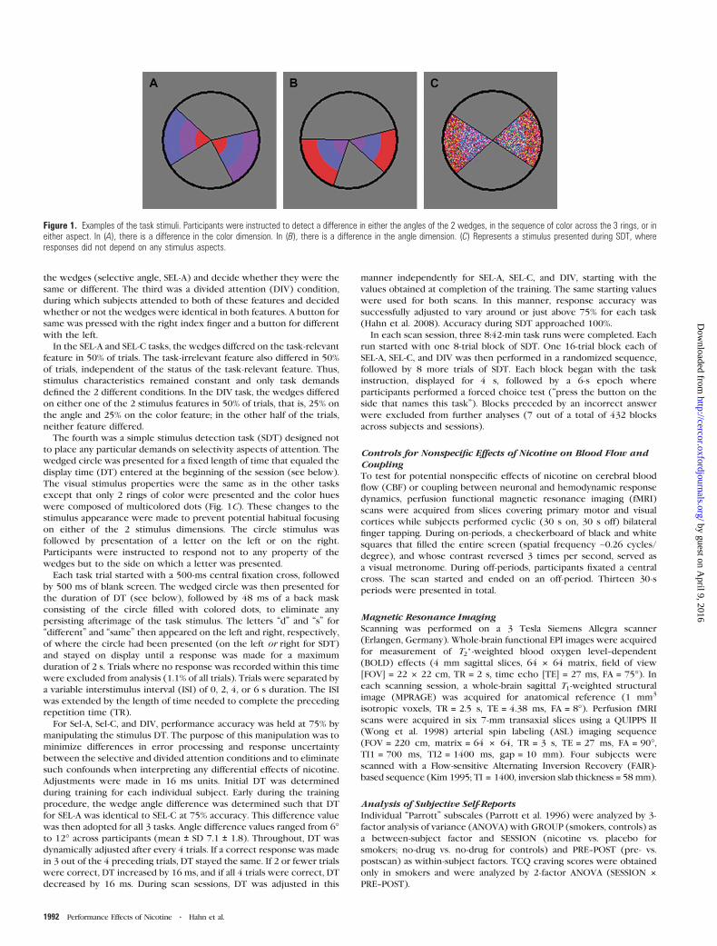
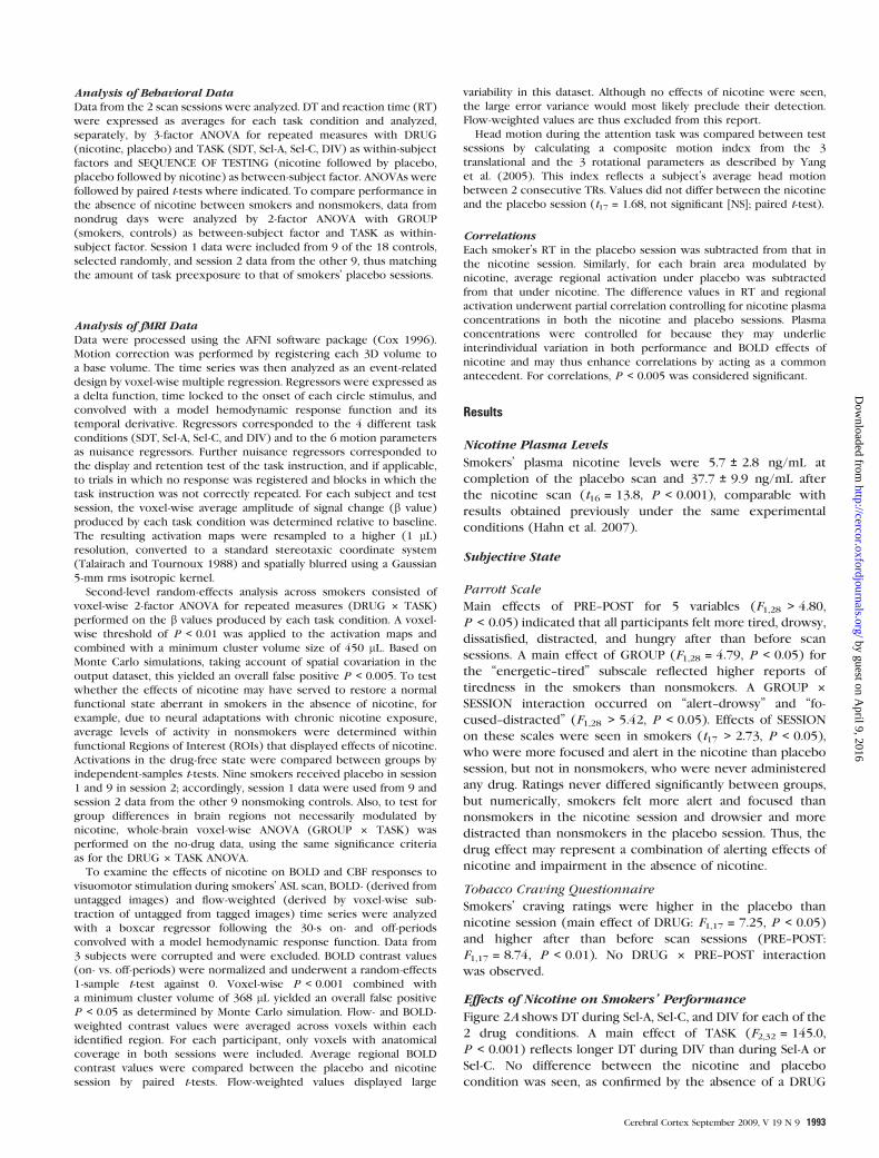
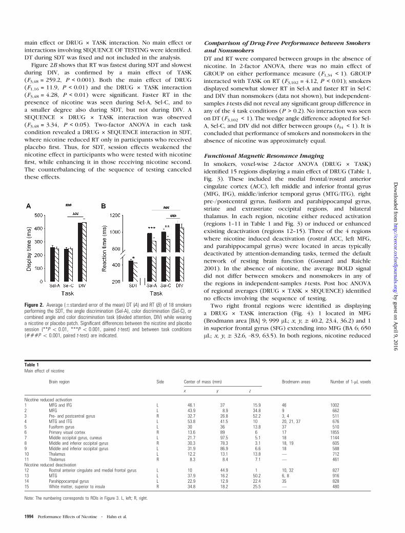
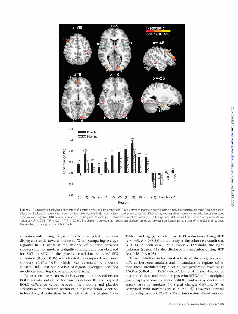
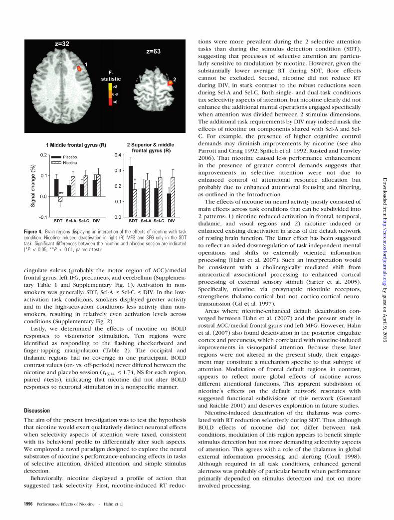
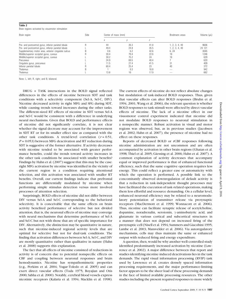
Related Documents