PATHOLOGY TEAM 2nd year / GIT block Med teams 431 2012 one by: Mamdouh Al-Enezi & Amjad Al shehry D

Welcome message from author
This document is posted to help you gain knowledge. Please leave a comment to let me know what you think about it! Share it to your friends and learn new things together.
Transcript

PATHOLOGYTEAM
2nd year / GIT block Med teams 431 2012
�������� � �������
��� ��� � ��� �
one by:
Mamdouh Al-Enezi & Amjad Al shehry
D
�����

Peptic ulcer disease
What is the difference between ulcers and erosions ?
rosionsE ulcers
*A breach in the epithelium of the mucosa only (superficial).
* May heal within days
*Defined histologically as a breach (break) in the mucosa that extends through the muscularis mucosae into the submucosa or deeper. * Takes much longer time to heal.
):Acute peptic ulcers ( acute gastric ulcers first:
Acutely developing gastric mucosal defects that may appear after severe physiologic stress called " stress ulcers".
Pathophysiology of Acute peptic ulcer:
1. As part of an acute gastritis (acute response to an irritant 'chemical' ( example, when children drink some chemicals) injury by drugs like NSAIDs – ibuprofen, vulturine, aspirin - or alcohol) 2. As a complication of a severe stress response (severe external extensive burns -leading to Curling's ulcer-, major trauma e.g: car accidents or cerebrovascular accidents after stroke, shock or operation to the brain). The body reacts to it by hyperacidity, which leads to ulceration.
ulcers
peptic ulcer(gastric or
duodenal)
chronic
stress ulcer(acute gastric ulcer
acute
ulcer
Erosions
These factors also cause sever pain and subsequent vasoconstriction that reduces blood flow to the stomach, which also helps in the formation of the ulcer. 3. As a result of extreme hyperacidity e.g: (Zollinger-Ellison syndrome).
in stomach. small multiple ulcerspeptic ulcer there will be ACUTEIn case of *
Characteristics:
-Acute stress ulcers are found anywhere in the stomach (multiple). -They range in depth from very superficial lesions (erosion) to deeper lesions that involve the entire mucosal thickness (true ulceration). -Clinically presented with hematemesis (vomiting blood). Prognosis: The gastric mucosa can recover completely if the person does not die from the primary disease .
Treatment: Antacids with treatment of underlying cause.
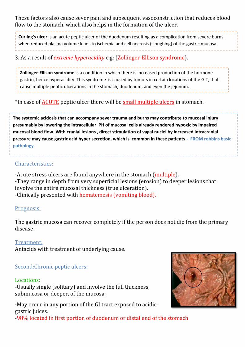
Second:Chronic peptic ulcers:
Locations: -Usually single (solitary) and involve the full thickness, submucosa or deeper, of the mucosa.
-May occur in any portion of the GI tract exposed to acidic gastric juices. -98% located in first portion of duodenum or distal end of the stomach
resulting as a complication from severe burns duodenum of the peptic ulcer acute is an Curling's ulcer.gastric mucosa (sloughing) of thevolume leads to ischemia and cell necrosis plasma when reduced
is a condition in which there is increased production of the hormone Ellison syndrome-Zollingergastrin, hence hyperacidity. This syndrome is caused by tumors in certain locations of the GIT, that
cause multiple peptic ulcerations in the stomach, duodenum, and even the jejunum.
The systemic acidosis that can accompany sever trauma and burns may contribute to mucosal injury presumably by lowering the intracellular PH of mucosal cells already rendered hypoxic by impaired mucosal blood flow. With cranial lesions , direct stimulation of vagal nuclei by increased intracranial pressure may cause gastric acid hyper secretion, which is common in these patients.- FROM robbins basic pathology-

-The ratio of duodenal ulcers to gastric ulcers is 4:1 * it occurs in duodenum(duodenal ulcer) more than stomach(gastric ulcer) * -Esophagus can be involved as a result of GERD or acid secretion by ectopic gastric mucosa. - Gastric mucosa within a Meckel diverticulum can result in peptic ulceration of adjacent mucosa.
- In Zollinger-Ellison syndrome: multiple peptic ulcerations in the stomach, duodenum, and even the jejunum. Pathophysiology: 1-gatric ulcers:" ulcer in the stomach" The mucosal defences against acid attack consist of: 1. Mucus-bicarbonate barrier: can be damaged by Duodeno-gastric reflux (bile) *bile is an irritant to the stomach 2. The surface epithelium: can be damaged by NSAIDs (blocking the synthesis of the prostaglandins), Helicobacter pylori infection (producing cytotoxins and ammonia that lead to ulceration). 3. Blood supply. 4. Cell renewal.
is to breakdown of mucosal defencepeptic ulcers in the stomach due In over all, much more important than excessive acid production.
Imbalance
is a most common congenital anomaly of GIT a small bulge of small intestine. its : Meckel diverticulumwall is covered by ectopic location of gastric type of epithelium (containing parietal cells) which is
abnormal and may cause ulcers.
Aggressive Factors
Defensive Factors

pathophysiology of chronic peptic ulcer in stomach:
duodenal ulcers:-2
-Increased production of acid (Hypersecreation) assumes more importance in the pathogenesis of duodenal ulceration. - H. pylori-infected individuals secrete 2-6 times as much acid (HCL) as non-infected indviduals. - Helicobacter Pylori does NOT colonise normal duodenal epithelium. - Helicobacter Pylori is involved in duodenal ulceration because there is gastric metaplasia in response to excess acid. Gastric metaplasia paves the way for colonisation by H. Pylori.
Hypersecreation (Hyperacidity) + H. Pylori = Duodenal ulcers

H.pylori
Pathophysiology of chronic peptic ulcer in duodenum:
Chronic peptic ulcer disease pathophysiology :
-H. pylori infection of the pyloric antrum is present in nearly all patients (almost 100%) with chronic duodenal ulcer and approximately 75% of patients with chronic gastric ulcer. - Although more than 70% of individuals with PUD are infected by H. pylori, fewer than 20% of H. pylori–infected individuals develop peptic ulcer. Morphology: *Grossly:
- Usually less than 20 mm in diameter but they may > 100 mm in diameter. - The classic peptic ulcer is a round to oval, sharply punched-out defect (that’s how we can differentiate between benign and malignant peptic ulcer). - In contrast, heaped-up margins are more characteristic of cancers (malignancy). *microscopically:
-The base consists of necrotic tissue and polymorph exudates overlying inflamed granulation tissue which merges with mature
Gastric ulcers
Duodenal ulcer
Duodenal-gastric reflex (bile)
NSAIDs
Hyperacidity

fibrous (scar) tissue. -The presence of neutrophils within the gastric glands signifies active inflammation and, most of the time, presence of H.pylori. what is the difference between benign and malignant peptic ulcer?
Malignant peptic ulcer Benign peptic ulcer
- Solitary and larger in size (up to 5 cm) - The margins are raised and inverted with a necrotic center. - For better diagnosis we take the biopsy from the edges of the lesion not the necrotic center.
- Solitary and < 20 mm - Well defined margins (shallow edge), rounded in shape and the surrounding area are usually edematous and inflamed
Clinical features of duodenal ulcer : -Epigastric pain (the most common symptom) - Gnawing or burning sensation. - Occurs 2-3 hours after meals (due to increased acid secretion) - Relieved by food or antacids (dudonal ulcers). -Patient awakens with pain at night. -Some present with complications: such as iron deficiency anemia, frank hemorrhage (bright in color) and it's usually seen in acute peptic ulcer, or perforation (can cause serious infections e.g. peritonitis). - hematemesis, Melena (which is black feces that are associated with gastrointestinal hemorrhage. The black color is caused by oxidation of the iron in hemoglobin during its passage through the ileum and colon) and it's usually seen in chronic peptic ulcer. - If hemorrhage is present in chronic cases it'll be coffee like (granular and brown). Therapy: 1. H. pylori eradication by antibiotics. 2. Acid suppression: a. Proton pump inhibitors (PPIs) . b. H2 blockers . Complications of Chronic Peptic Ulcers
o Hemorrhage (anemia ).
o Penetration : The ulcer penetrates the full thickness of the stomach or duodenal
wall, progressing into adherent underlying tissue, e.g. the pancreas or liver.
o Perforation: This leads to peritonitis.
o Fibrous stricture: In the stomach, ulcers may cause pyloric stenosis.
o Malignant change: This is extremely uncommon.
Present-day therapies including ( antibiotics active against H.pylori , proton pump inhibitors and hydrogen receptor antagonists ) most ulcer victims can be helped if not cured , and they usually escape the surgeon's knife. From Robbins

Inflammatory diseases of the stomach :
Chronic gastritis:
major cause is infection of H.Pylori, less commonly autoimmune in origin; characterized by mononuclear cell inflammation in the lamina propria with intestinal metaplasia and frequently, proliferation of lymphoid tissue; may be the precursor of peptic ulcer and carcinoma.
Acute gastritis :
Acute mucosal inflammation, usually transient, associated with use of NSAIDs, alcohol, heavy smoking and varies systemic abnormalities .
Peptic ulcer :
Breach in the epithelium caused most commonly by H.Pylori infection and mucosal exposure to gastric acid and enzymes(pepsin), or less frequently by use NSAIDs; H.Pylori clicits inflammatory reaction and damages epithelial cells; typically, sharply demarcated mucosal defects with underlying necrosis, acute inflammation, granulation tissue and scaring; manifested by bleeding and less commonly rupture.
Stress ulcer (acute gastric ulcer) :
Assusiated with severe trauma, burns, CNS trauma or hemorrhage; usually small, multiple, hemorrhagic ulcers that are often shallow.
Questions :
1- A 52-year-old male presents with epigastric pain that improves with meals. Endoscopy demonstrates a 2 Cm ulcerated area located 3 cm distal to the pyloric junction. Which of the following is most likely to have made the strongest contribution to the development of this disease?
A. Aspirin use B. Chronic antacid use C. Drinking alcohol D. Helicobacter pylori infection E. Smoking
2- All of the following are causes of acute peptic ulcer except
A. Severe burns B. Helicobacter pylori infection C. Major trauma D. Zollinger-Ellison syndrome E.
3- A 49-year-old secretary presents to medical outpatients with a 7-month history of epigastric pain. She has been treated with antacids by her GP, but this has not controlled the symptoms. In the clinic, she complains of epigastric pains which are sharp and burning and radiate her subcostal margin to the right. The pain is worse at night and is relieved by food. On examination, there is epigastric tenderness and clinical signs of anaemia. What is the possible cause of this clinical presentation?
A. Duodenal Peptic ulceration B. gastric Peptic ulceration C. euration of the esophagus D. pneumonia
1- D 2- B 3- A
Notes about Q1 :
• The correct answer is D. The patient has a duodenal peptic ulcer. The strongest risk factor for duodenal peptic ulcer is Helicobacter pylori infection, which is found in almost 100% of these cases (contrast to 70% Infection rate in gastric peptic ulcer).
• Aspirin use (choice A) and ethanol use (choice C) are more strongly implicated in gastric ulcer disease than duodenal ulcer disease.
• Chronic antacid use (choice B) is seen as a result of peptic ulcer disease, not as a cause of it. • Smoking (choice E) may also be a lesser contributing factor to the development of peptic ulcer.
Related Documents


