Respiratory CHAPTER

Welcome message from author
This document is posted to help you gain knowledge. Please leave a comment to let me know what you think about it! Share it to your friends and learn new things together.
Transcript
Respiratory CHAPTER

Editors
EditingFeedback
Contributors
o Allulu Alsulayhim
o Ghadah Almazrou
o Jawaher Abanumy
o Laila Hassan Mathkour
o Maha AlGhamdi
o Rawan Alqahtani
o Shatha Alghaihb
Abdulhakim Bin Onaiq
Arwa Alqahtani
Ashwaq Almajed
Shatha Alghaihb
Badr Alqarni
Bayan Al-Mugheerah
Dania Alkelabi
Duaa Alhumoudi
Fay Albuqami
Haifa Alwaily
Haneen Somily
Laila Alsabbagh
Manee Alkhalifah
Noura Alturki
Nasser AbuDujain
Rania Almutiri
Razan AlRabah
Reham Alturki
Renad Alhomaidi
Shoag Alahmari
Shahd Alsowaidan
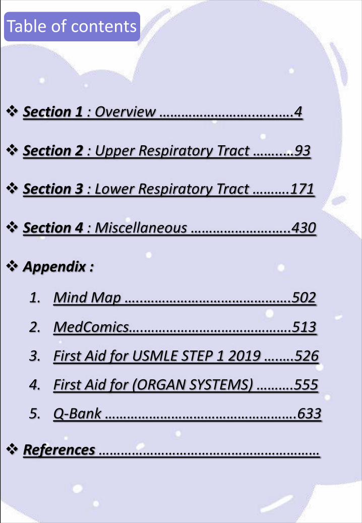
Tableofcontents
v Section1 :Overview……………………..…...….4
v Section2 :UpperRespiratoryTract……..…93
v Section3 :LowerRespiratoryTract……….171
v Section4 :Miscellaneous………………….…..430
v Appendix:
1. MindMap…..………………………………….502
2. MedComics….………………………………….513
3. FirstAidforUSMLESTEP1 2019 ….….526
4. FirstAidfor(ORGANSYSTEMS)……….555
5. Q-Bank…………………………………………….633
v References ……………………………………………………
SECTION 1 :
OVERVIEW
1 PHYSIOLOGY:Functional organization of the respiratory system
ANATOMY: Muscles involved in respiration
32510 39
18 47
PHYSIOLOGY:Gas transport
PHYSIOLOGY:Control of Breathing
53
PHYSIOLOGY:Hypoxia and Cyanosis
PHYSIOLOGY:Respiratory ventilation
PHYSIOLOGY:Gas transfer
PHYSIOLOGY:Mechanism of breathing
25BIOCHEMSTRY:Globular proteins
1 | Respiratory Chapter
SECTION 1 | Functional organization of the respiratory system
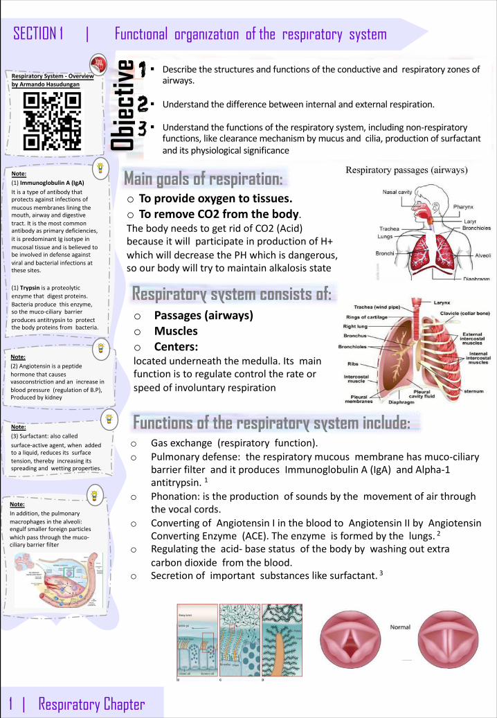
Main goals of respiration:
§ Describethestructuresandfunctionsoftheconductiveandrespiratoryzonesofairways.
§ Understandthedifferencebetweeninternalandexternalrespiration.
§ Understandthefunctionsoftherespiratorysystem,includingnon-respiratoryfunctions,likeclearancemechanismbymucusandcilia,productionofsurfactantanditsphysiologicalsignificanceOb
jectiv
e
Respiratory system consists of:
Functions of the respiratory system include:
o Toprovideoxygentotissues.o ToremoveCO2 fromthebody.ThebodyneedstogetridofCO2 (Acid)becauseitwillparticipateinproductionofH+whichwilldecreasethePHwhichisdangerous,soourbodywilltrytomaintainalkalosisstate
o Passages(airways)o Muscleso Centers:locatedunderneaththemedulla.Itsmainfunctionistoregulatecontroltherateorspeedofinvoluntaryrespiration
o Gasexchange(respiratoryfunction).o Pulmonarydefense:therespiratorymucousmembranehasmuco-ciliary
barrierfilteranditproducesImmunoglobulinA(IgA)andAlpha-1antitrypsin.1
o Phonation:istheproductionofsoundsbythemovementofairthroughthevocalcords.
o ConvertingofAngiotensinIinthebloodtoAngiotensinIIbyAngiotensinConvertingEnzyme(ACE).Theenzymeisformedbythelungs. 2
o Regulatingtheacid- basestatusofthebodybywashingoutextracarbondioxidefromtheblood.
o Secretionofimportantsubstanceslikesurfactant. 3
Note:Inaddition,thepulmonarymacrophagesinthealveoli:engulfsmallerforeignparticleswhichpassthroughthemuco-ciliarybarrierfilter
Note:(2)Angiotensinisapeptidehormonethatcausesvasoconstrictionandanincreaseinbloodpressure(regulationofB.P),Producedbykidney
Note:(3)Surfactant:alsocalledsurface-activeagent,whenaddedtoaliquid,reducesitssurfacetension,therebyincreasingitsspreadingandwettingproperties.
RespiratorySystem- OverviewbyArmandoHasudungan
Note:(1)ImmunoglobulinA(IgA)Itisatypeofantibodythatprotectsagainstinfectionsofmucousmembranesliningthemouth,airwayanddigestivetract.Itisthemostcommonantibodyasprimarydeficiencies,itispredominantIgisotypeinmucosaltissueandisbelievedtobeinvolvedindefenseagainstviralandbacterialinfectionsatthesesites.
(1)Trypsin isaproteolyticenzymethatdigestproteins.Bacteriaproducethisenzyme,sothemuco-ciliarybarrierproducesantitrypsintoprotectthebodyproteinsfrombacteria.
Respiratory Chapter | 2
SECTION 1 | Functional organization of the respiratory system
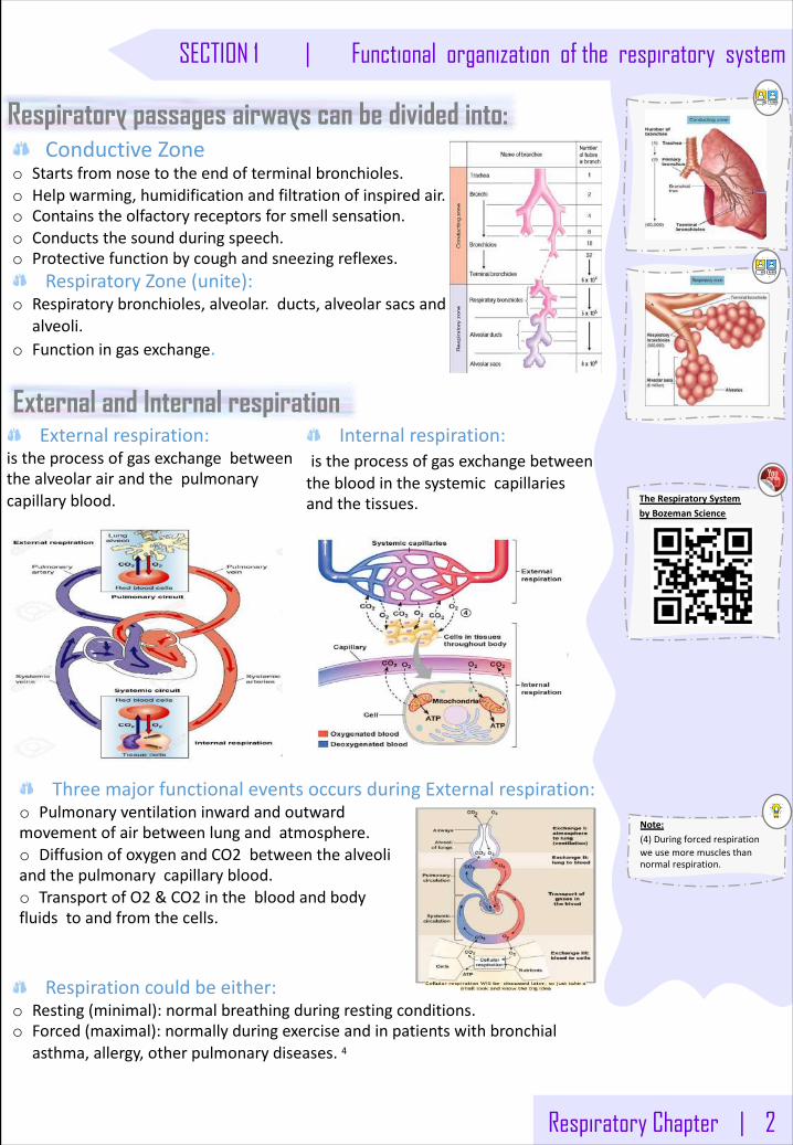
Respiratory passages airways can be divided into: ConductiveZone
o Startsfromnosetotheendofterminalbronchioles.o Helpwarming,humidificationandfiltrationofinspiredair.o Containstheolfactoryreceptorsforsmellsensation.o Conductsthesoundduringspeech.o Protectivefunctionbycoughandsneezingreflexes.
RespiratoryZone(unite):o Respiratorybronchioles,alveolar.ducts,alveolarsacsand
alveoli.o Functioningasexchange.
External and Internal respirationExternalrespiration:
istheprocessofgasexchangebetweenthealveolarairandthepulmonarycapillaryblood.
Internalrespiration:istheprocessofgasexchangebetweenthebloodinthesystemiccapillariesandthetissues.
Respirationcouldbeeither:o Resting(minimal):normalbreathingduringrestingconditions.o Forced(maximal):normallyduringexerciseandinpatientswithbronchial
asthma,allergy,otherpulmonarydiseases.4
ThreemajorfunctionaleventsoccursduringExternalrespiration:o Pulmonaryventilationinwardandoutwardmovementofairbetweenlungandatmosphere.o DiffusionofoxygenandCO2 betweenthealveoliandthepulmonarycapillaryblood.o TransportofO2 &CO2 inthebloodandbodyfluidstoandfromthecells.
Note:(4)Duringforcedrespirationweusemoremusclesthannormalrespiration.
TheRespiratorySystembyBozemanScience
3 | Respiratory Chapter
SECTION 1 | Functional organization of the respiratory system
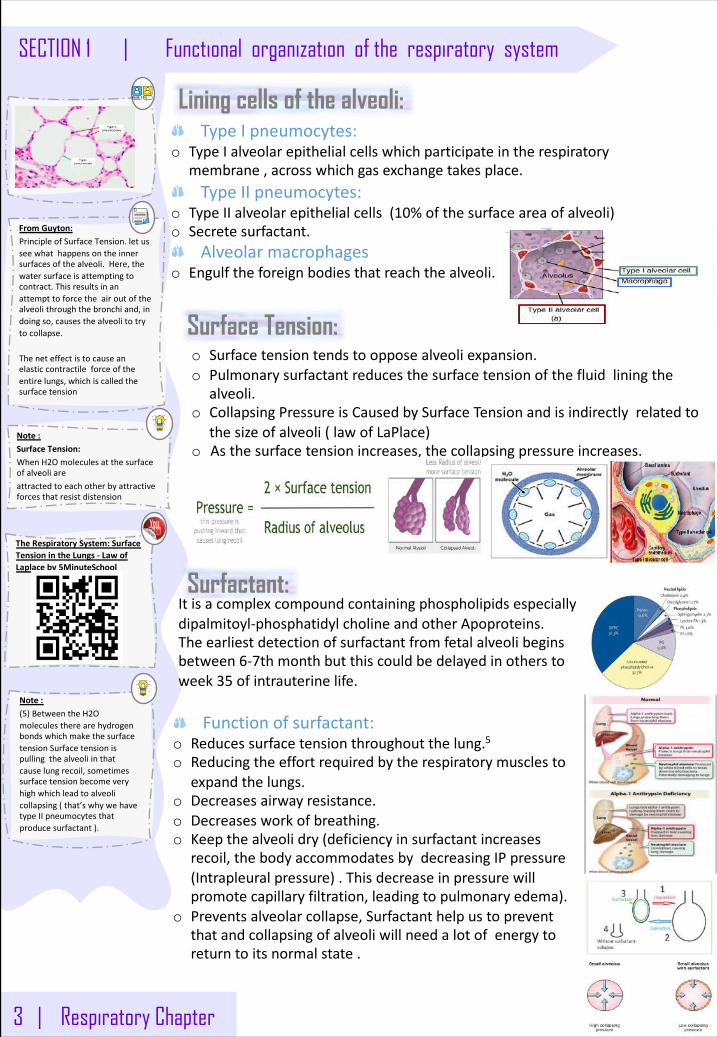
Lining cells of the alveoli:TypeIpneumocytes:
o TypeIalveolarepithelialcellswhichparticipateintherespiratorymembrane,acrosswhichgasexchangetakesplace.TypeIIpneumocytes:
o TypeIIalveolarepithelialcells(10%ofthesurfaceareaofalveoli)o Secretesurfactant.
Alveolarmacrophageso Engulftheforeignbodiesthatreachthealveoli.
Surface Tension:o Surfacetensiontendstoopposealveoliexpansion.o Pulmonarysurfactantreducesthesurfacetensionofthefluidliningthe
alveoli.o CollapsingPressureisCausedbySurfaceTensionandisindirectlyrelatedto
thesizeofalveoli(lawofLaPlace)o Asthesurfacetensionincreases,thecollapsingpressureincreases.
FromGuyton:PrincipleofSurfaceTension.letusseewhathappensontheinnersurfacesofthealveoli.Here,thewatersurfaceisattemptingtocontract.Thisresultsinanattempttoforcetheairoutofthealveolithroughthebronchiand,indoingso,causesthealveolitotrytocollapse.
Theneteffectistocauseanelasticcontractileforceoftheentirelungs,whichiscalledthesurfacetension
Note:SurfaceTension:WhenH2Omoleculesatthesurfaceofalveoliareattractedtoeachotherbyattractiveforcesthatresistdistension
Surfactant:Itisacomplexcompoundcontainingphospholipidsespeciallydipalmitoyl-phosphatidylcholineandotherApoproteins.Theearliestdetectionofsurfactantfromfetalalveolibeginsbetween6-7thmonthbutthiscouldbedelayedinotherstoweek35 ofintrauterinelife.
Note:(5)BetweentheH2OmoleculestherearehydrogenbondswhichmakethesurfacetensionSurfacetensionispullingthealveoliinthatcauselungrecoil,sometimessurfacetensionbecomeveryhighwhichleadtoalveolicollapsing(that’swhywehavetypeIIpneumocytesthatproducesurfactant).
Functionofsurfactant:o Reducessurfacetensionthroughoutthelung.5o Reducingtheeffortrequiredbytherespiratorymusclesto
expandthelungs.o Decreasesairwayresistance.o Decreasesworkofbreathing.o Keepthealveolidry(deficiencyinsurfactantincreases
recoil,thebodyaccommodatesbydecreasingIPpressure(Intrapleuralpressure).Thisdecreaseinpressurewillpromotecapillaryfiltration,leadingtopulmonaryedema).
o Preventsalveolarcollapse,Surfactanthelpustopreventthatandcollapsingofalveoliwillneedalotofenergytoreturntoitsnormalstate.
TheRespiratorySystem:SurfaceTensionintheLungs- LawofLaplaceby5MinuteSchool
Respiratory Chapter | 4
Functional organization of the respiratory system | SECTION 1
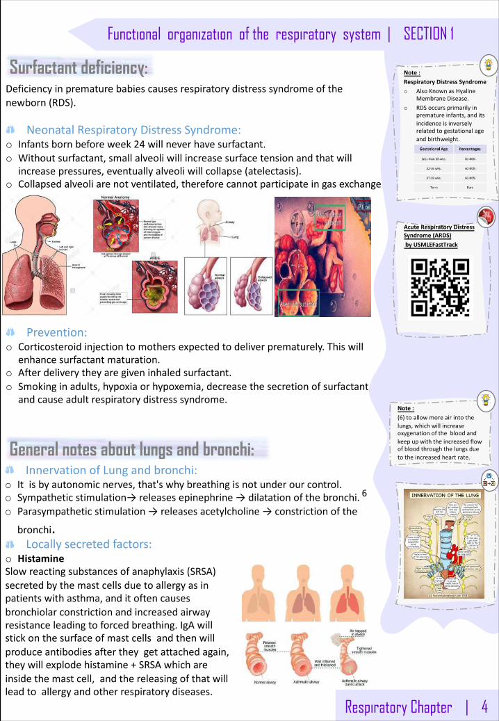
Surfactant deficiency:Deficiencyinprematurebabiescausesrespiratorydistresssyndromeofthenewborn(RDS).
NeonatalRespiratoryDistressSyndrome:o Infantsbornbeforeweek24 willneverhavesurfactant.o Withoutsurfactant,smallalveoliwillincreasesurfacetensionandthatwill
increasepressures,eventuallyalveoliwillcollapse(atelectasis).o Collapsedalveoliarenotventilated,thereforecannotparticipateingasexchange
Prevention:o Corticosteroidinjectiontomothersexpectedtodeliverprematurely.Thiswill
enhancesurfactantmaturation.o Afterdeliverytheyaregiveninhaledsurfactant.o Smokinginadults,hypoxiaorhypoxemia,decreasethesecretionofsurfactant
andcauseadultrespiratorydistresssyndrome.
General notes about lungs and bronchi:InnervationofLungandbronchi:
o Itisbyautonomicnerves,that'swhybreathingisnotunderourcontrol.o Sympatheticstimulation→releasesepinephrine→dilatationofthebronchi. 6
o Parasympatheticstimulation→releasesacetylcholine→constrictionofthe
bronchi.
Note:(6)toallowmoreairintothelungs,whichwillincreaseoxygenationofthebloodandkeepupwiththeincreasedflowofbloodthroughthelungsduetotheincreasedheartrate.
Note:RespiratoryDistressSyndromeo AlsoKnownasHyaline
MembraneDisease.o RDSoccursprimarilyin
prematureinfants,anditsincidenceisinverselyrelatedtogestationalageandbirthweight.
Locallysecretedfactors:o HistamineSlowreactingsubstancesofanaphylaxis(SRSA)secretedbythemastcellsduetoallergyasinpatientswithasthma,anditoftencausesbronchiolarconstrictionandincreasedairwayresistanceleadingtoforcedbreathing.IgAwillstickonthesurfaceofmastcellsandthenwillproduceantibodiesaftertheygetattachedagain,theywillexplodehistamine+SRSAwhichareinsidethemastcell,andthereleasingofthatwillleadtoallergyandotherrespiratorydiseases.
AcuteRespiratoryDistressSyndrome(ARDS)byUSMLEFastTrack
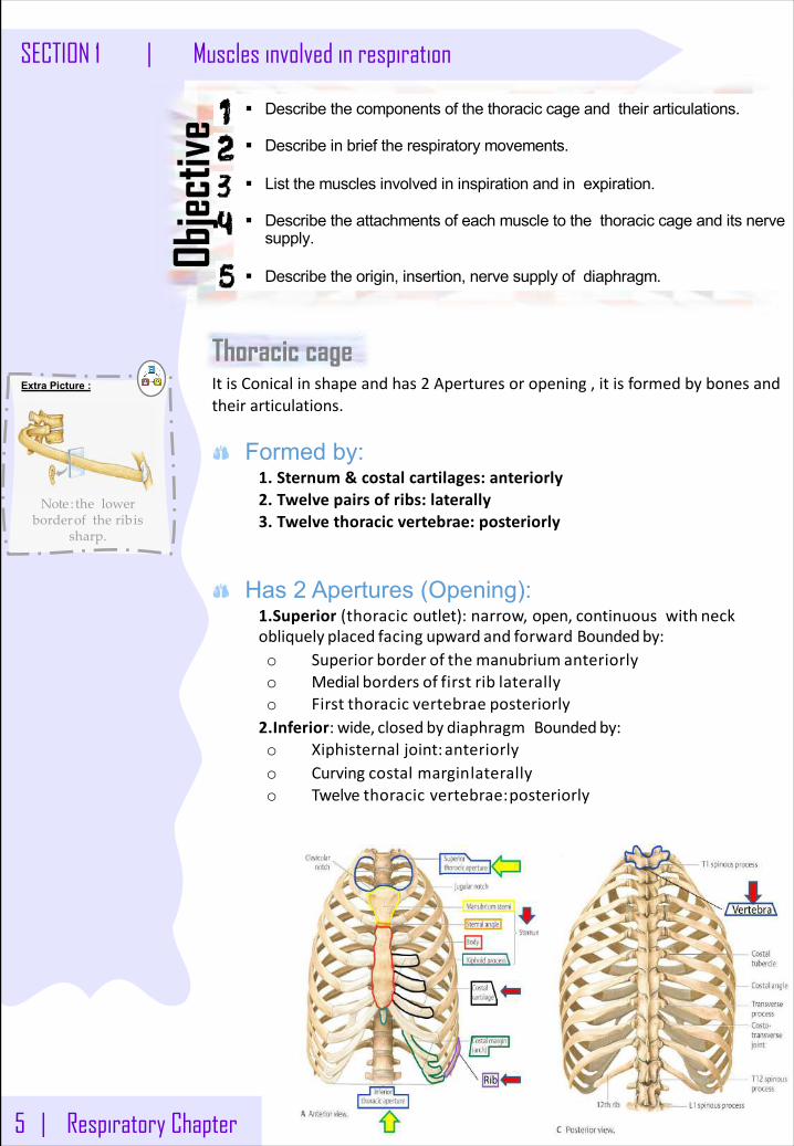
ItisConicalinshapeandhas2 Aperturesoropening,itisformedbybonesandtheirarticulations.
Formed by:1. Sternum&costalcartilages:anteriorly2. Twelvepairsofribs:laterally3. Twelvethoracicvertebrae:posteriorly
Has 2 Apertures (Opening): 1.Superior (thoracicoutlet):narrow,open,continuouswith neckobliquely placed facing upward and forward Boundedby:o Superior border of the manubrium anteriorlyo Medial borders of first rib laterallyo First thoracic vertebrae posteriorly2.Inferior: wide, closed by diaphragmBoundedby:o Xiphisternaljoint:anteriorlyo Curvingcostalmarginlaterallyo Twelvethoracicvertebrae:posteriorly
5 | Respiratory Chapter
SECTION 1 | Muscles involved in respiration
Thoracic cage
§ Describe the components of the thoracic cage and their articulations.
§ Describe in brief the respiratory movements.
§ List the muscles involved in inspiration and in expiration.
§ Describe the attachments of each muscle to the thoracic cage and its nerve supply.
§ Describe the origin, insertion, nerve supply of diaphragm.
Objec
tive
Extra Picture :
Note : the lower borderof the ribis
sharp.
Respiratory Chapter | 6
Muscles involved in respiration | SECTION 1
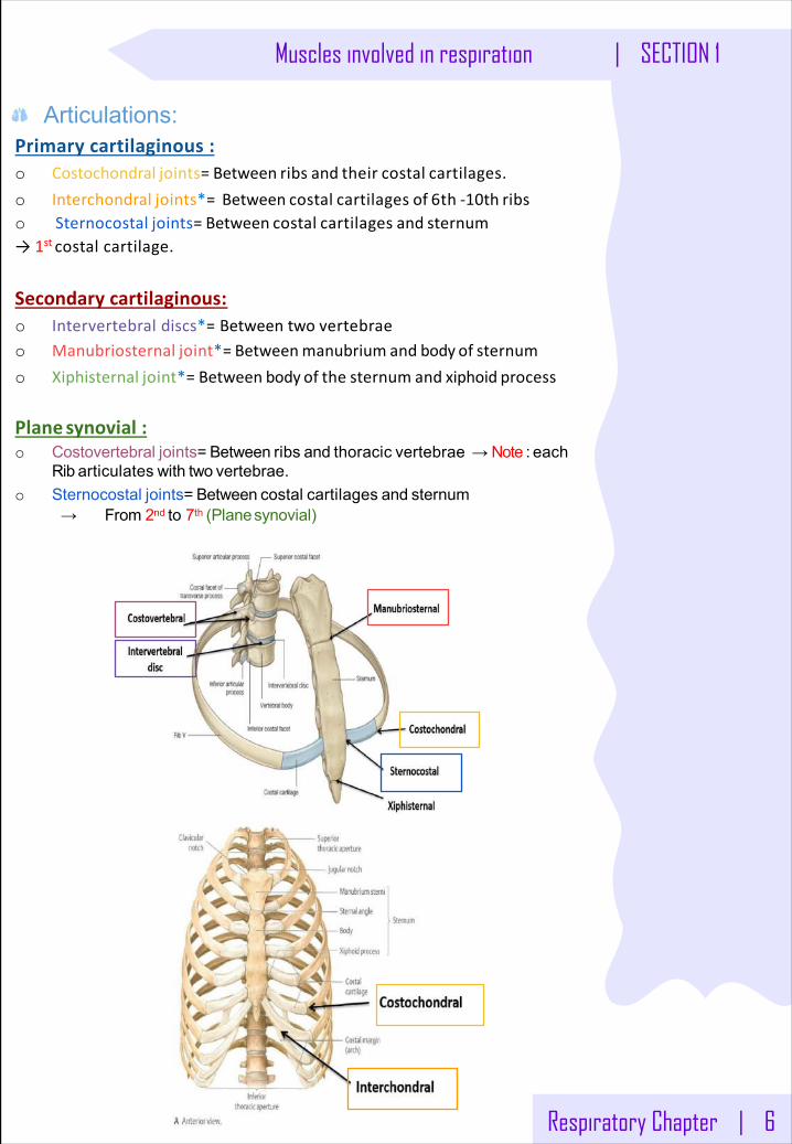
Articulations:Primarycartilaginous: o Costochondral joints= Between ribs and their costal cartilages.o Interchondral joints*= Between costal cartilages of 6th -10th ribso Sternocostal joints= Between costal cartilages and sternum→1stcostalcartilage.
Secondarycartilaginous: o Intervertebraldiscs*=Betweentwovertebraeo Manubriosternal joint*= Betweenmanubrium and body of sternumo Xiphisternal joint*= Between body of the sternum and xiphoid process
Plane synovial: o Costovertebral joints= Between ribs and thoracic vertebrae → Note : each
Rib articulates with two vertebrae.o Sternocostal joints= Between costal cartilages and sternum
→ From 2nd to 7th (Planesynovial)
7 | Respiratory Chapter
SECTION 1 | Muscles involved in respiration
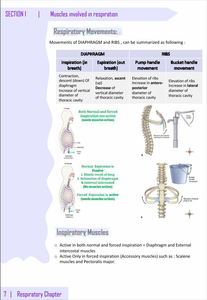
MovementsofDIAPHRAGMandRIBS,canbesummarizedasfollowing:
Contraction,descent(down)OfdiaphragmIncreaseofverticaldiameterofthoraciccavity
Relaxation,ascent(up)Decrease ofverticaldiameterofthoraciccavity
ElevationofribsIncreaseinantero-posteriordiameterofthoraciccavity
ElevationofribsIncreaseinlateraldiameterofthoraciccavity
Respiratory Movements:
Inspiratory Muscles
o Activeinbothnormalandforcedinspiration=DiaphragmandExternalintercostalmuscles
o ActiveOnlyinforcedinspiration(Accessorymuscles)suchas:ScalenemusclesandPectoralismajor.
Respiratory Chapter | 8
Muscles involved in respiration | SECTION 1
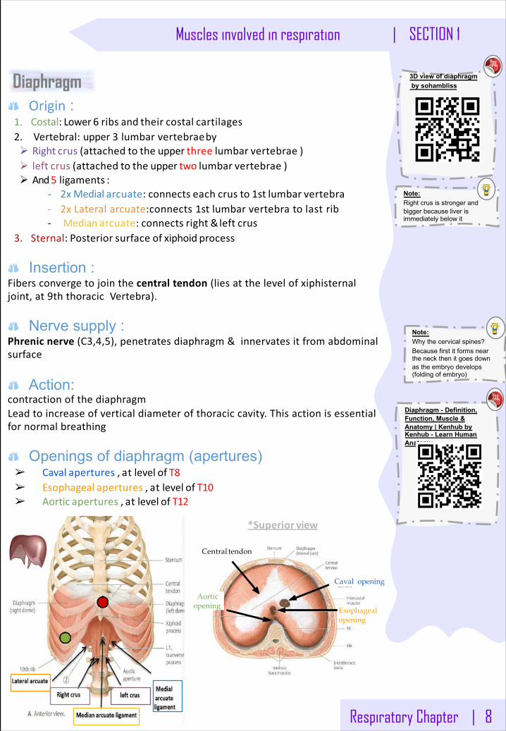
Central tendon
Caval opening
Esophageal opening
Aortic opening
*Superior view
DiaphragmOrigin :
1. Costal: Lower 6 ribs and their costal cartilages2. Vertebral:upper3 lumbarvertebraebyØ Right crus (attached to the upper three lumbar vertebrae )Ø left crus (attached to the upper two lumbar vertebrae )Ø And5 ligaments :
- 2x Medial arcuate: connects each crus to 1st lumbar vertebra- 2xLateralarcuate:connects 1stlumbarvertebratolastrib- Median arcuate: connects right &left crus
3. Sternal: Posterior surface of xiphoid process
Insertion :Fibersconvergetojointhecentraltendon(liesatthelevelofxiphisternaljoint,at9ththoracicVertebra).
Nerve supply :Phrenicnerve(C3,4,5),penetratesdiaphragm&innervatesitfromabdominalsurface
Action:contractionofthediaphragmLeadtoincreaseofverticaldiameterofthoraciccavity.Thisactionisessentialfornormalbreathing
Openings of diaphragm (apertures)➢ Caval apertures , at level of T8➢ Esophageal apertures , at level of T10➢ Aortic apertures , at level of T12
Note:Right crus is stronger and bigger because liver is immediately below it
Note:Why the cervical spines?Because first it forms near the neck then it goes down as the embryo develops (folding of embryo)
3D view of diaphragmby sohambliss
Diaphragm - Definition, Function, Muscle & Anatomy | Kenhub by Kenhub - Learn Human Anatomy
9 | Respiratory Chapter
SECTION 1 | Muscles involved in respiration
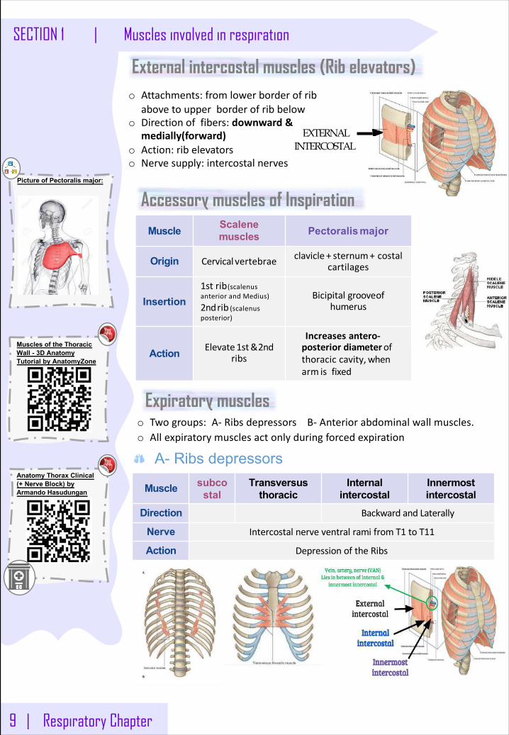
o Twogroups:A- RibsdepressorsB- Anteriorabdominalwallmuscles.o Allexpiratorymusclesactonlyduringforcedexpiration
External intercostal muscles (Rib elevators)
Accessory muscles of Inspiration
Muscle Scalenemuscles Pectoralis major
Origin Cervical vertebrae clavicle + sternum+ costalcartilages
Insertion1strib(scalenusanteriorandMedius)
2ndrib (scalenusposterior)
Bicipitalgrooveofhumerus
Action Elevate1st&2ndribs
Increases antero-posterior diameterofthoracic cavity, whenarm isfixed
o Attachments:fromlowerborderofribabovetoupperborderofribbelow
o Directionoffibers:downward&medially(forward)
o Action:ribelevatorso Nervesupply:intercostalnerves
EXTERNAL INTERCOSTAL
Picture of Pectoralis major:
Expiratory muscles
Muscle subcostal
Transversus thoracic
Internal intercostal
Innermost intercostal
Direction BackwardandLaterally
Nerve IntercostalnerveventralramifromT1 toT11
Action DepressionoftheRibs
A- Ribs depressors
Muscles of the Thoracic Wall - 3D Anatomy Tutorial by AnatomyZone
Anatomy Thorax Clinical (+ Nerve Block) by Armando Hasudungan
Respiratory Chapter | 10
Muscles involved in respiration | SECTION 1
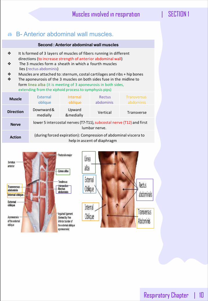
B- Anterior abdominal wall muscles. Second : Anterior abdominal wall muscles
❖ ItIsformedof3 layersofmusclesoffibersrunningindifferentdirections (to increase strength of anterior abdominal wall)
❖ The3 musclesformasheathinwhichafourthmuscleslies(rectus abdominis)
❖ Muscles are attached to: sternum, costal cartilages and ribs + hip bones❖ Theaponeurosisofthe3 musclesonbothsidesfuseinthemidlineto
formlineaalba(itismeetingof3 aponeurosisinbothsides,extending from the xiphoid process to symphysis pips)
Muscle Externaloblique
Internaloblique
Rectusabdominis
Transversusabdominis
Direction Downward&medially
Upward&medially
Vertical Transverse
Nerve lower 5 intercostal nerves (T7-T11), subcostal nerve (T12) and firstlumbar nerve.
Action (during forced expiration): Compression of abdominal viscera tohelp in ascent of diaphragm
MusclesInvolvedinRespiratory
• Themajormuscleofinspirationisthediaphragm.Contractionofthediaphragmenlargestheverticaldimensionsofthechest.Alsoutilizedaretheexternalintercostalmusclesofthechestwall.Contractionofthesemusclescausestheribstoriseandthusincreasestheanterior-posteriordimensionsofthechest.
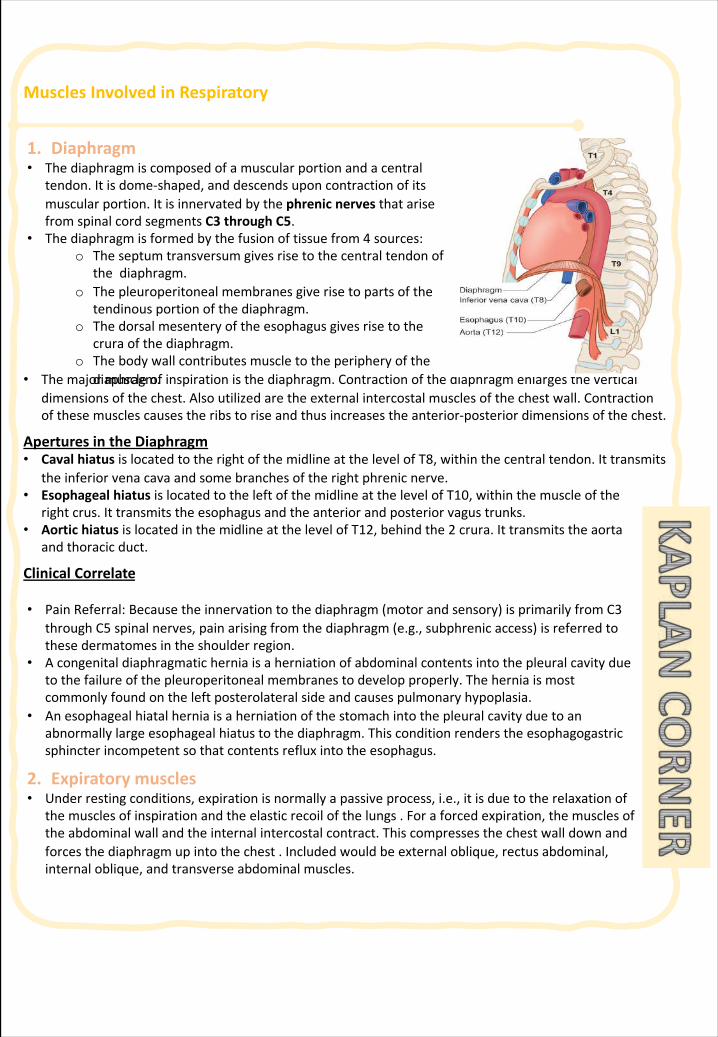
AperturesintheDiaphragm• Caval hiatusislocatedtotherightofthemidlineatthelevelofT8,withinthecentraltendon.Ittransmits
theinferiorvenacavaandsomebranchesoftherightphrenicnerve.• EsophagealhiatusislocatedtotheleftofthemidlineatthelevelofT10,withinthemuscleofthe
rightcrus.Ittransmitstheesophagusandtheanteriorandposteriorvagustrunks.• AortichiatusislocatedinthemidlineatthelevelofT12,behindthe2 crura.Ittransmitstheaorta
andthoracicduct.
ClinicalCorrelate
1. Diaphragm• Thediaphragmis composedofamuscularportionandacentral
tendon. Itisdome-shaped,anddescendsuponcontractionofitsmuscularportion.ItisinnervatedbythephrenicnervesthatarisefromspinalcordsegmentsC3 throughC5.
• Thediaphragmisformedbythefusionoftissuefrom4 sources:o Theseptumtransversum givesrisetothecentraltendonof
thediaphragm.o Thepleuroperitoneal membranesgiverisetopartsofthe
tendinousportionofthediaphragm.o Thedorsalmesenteryoftheesophagusgivesrisetothe
crura ofthediaphragm.o Thebodywallcontributesmuscletotheperipheryofthe
diaphragm.
• PainReferral:Becausetheinnervationtothediaphragm(motorandsensory)isprimarilyfromC3throughC5 spinalnerves,painarisingfromthediaphragm(e.g.,subphrenic access)isreferredtothesedermatomesintheshoulderregion.
• Acongenitaldiaphragmaticherniaisaherniationofabdominalcontentsintothepleuralcavityduetothefailureofthepleuroperitoneal membranestodevelopproperly.Theherniaismostcommonlyfoundontheleftposterolateralsideandcausespulmonaryhypoplasia.
• Anesophagealhiatalherniaisaherniationofthestomachintothepleuralcavityduetoanabnormallylargeesophagealhiatustothediaphragm.Thisconditionrenderstheesophagogastricsphincterincompetentsothatcontentsrefluxintotheesophagus.
2. Expiratorymuscles• Underrestingconditions,expirationisnormallyapassiveprocess,i.e.,itisduetotherelaxationof
themusclesofinspirationandtheelasticrecoilofthelungs.Foraforcedexpiration,themusclesoftheabdominalwallandtheinternalintercostalcontract.Thiscompressesthechestwalldownandforcesthediaphragmupintothechest.Includedwouldbeexternaloblique,rectusabdominal,internaloblique,andtransverseabdominalmuscles.
11 | Respiratory Chapter
SECTION 1 | Mechanism of Breathing
§ Listthemusclesofrespirationanddescribetheirrolesduringinspiration & expiration.
§ Identifytheimportanceofthefollowingpressuresinrespiration:atmospheric,intra-alveolar,intrapleural,andtranspulmonary.
§ Explainwhyintrapleuralpressureisalwayssubatmospheric undernormalconditions,andthesignificanceofthethinlayeroftheintrapleuralfluidsurroundingthelung.
§ Definelungcomplianceandlistthedeterminantsofcompliance
Objec
tive
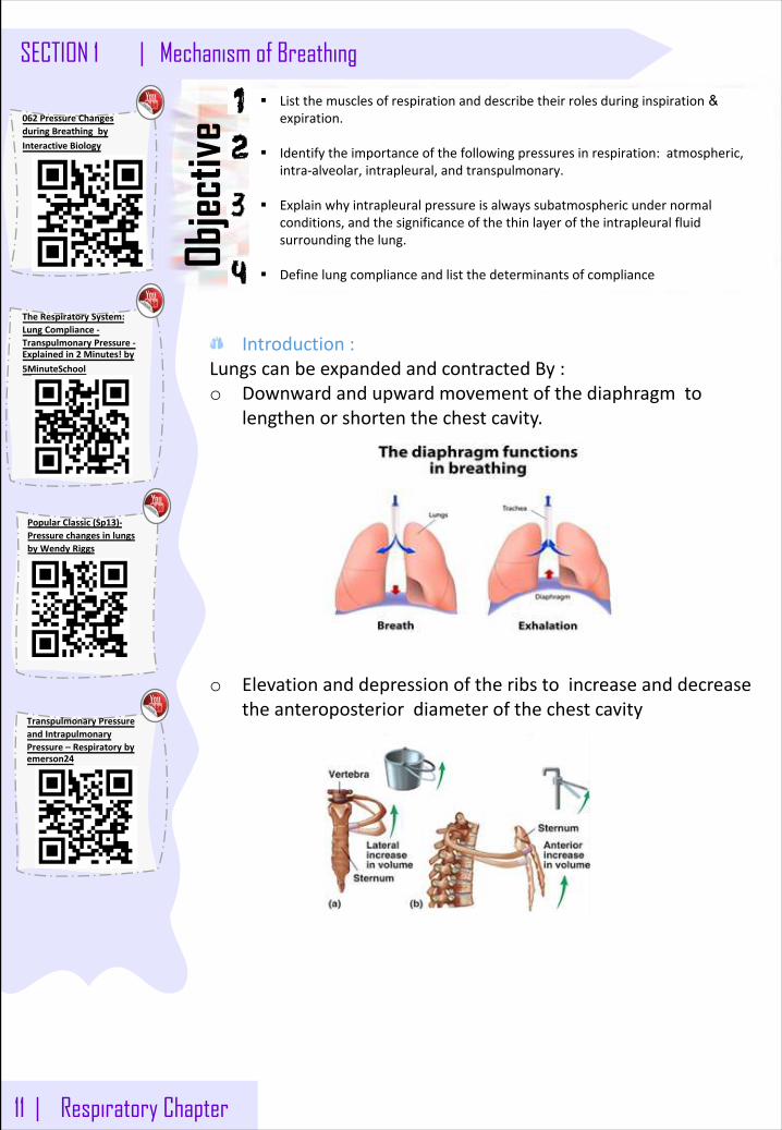
Introduction:LungscanbeexpandedandcontractedBy:o Downwardandupwardmovementofthediaphragmto
lengthenorshortenthechestcavity.
o Elevationanddepressionoftheribstoincreaseanddecreasetheanteroposteriordiameterofthechestcavity
062 PressureChangesduringBreathingbyInteractiveBiology
TheRespiratorySystem:LungCompliance-TranspulmonaryPressure-Explainedin2 Minutes!by5MinuteSchool
PopularClassic(Sp13)-PressurechangesinlungsbyWendyRiggs
TranspulmonaryPressureandIntrapulmonaryPressure– Respiratorybyemerson24
Respiratory Chapter | 12
Mechanism of Breathing | SECTION 1
Respiratory muscles:
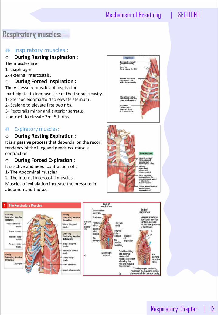
Inspiratorymuscles:o DuringRestingInspiration:Themusclesare1- diaphragm.2- externalintercostals.o DuringForcedinspiration:TheAccessorymusclesofinspirationparticipatetoincreasesizeofthethoraciccavity.1- Sternocleidomastoidtoelevatesternum.2- Scalenetoelevatefirsttwo ribs.3- Pectoralisminorandanteriorserratuscontracttoelevate3rd–5thribs.
Expiratorymuscles:o DuringRestingExpiration:Itisapassiveprocessthatdependsontherecoiltendencyofthelungandneedsnomusclecontractiono DuringForcedExpiration:Itisactiveandneedcontractionof:1- TheAbdominalmuscles.2- Theinternalintercostalmuscles.Musclesofexhalationincreasethepressureinabdomenandthorax.
13 | Respiratory Chapter
SECTION 1 | Mechanism of Breathing
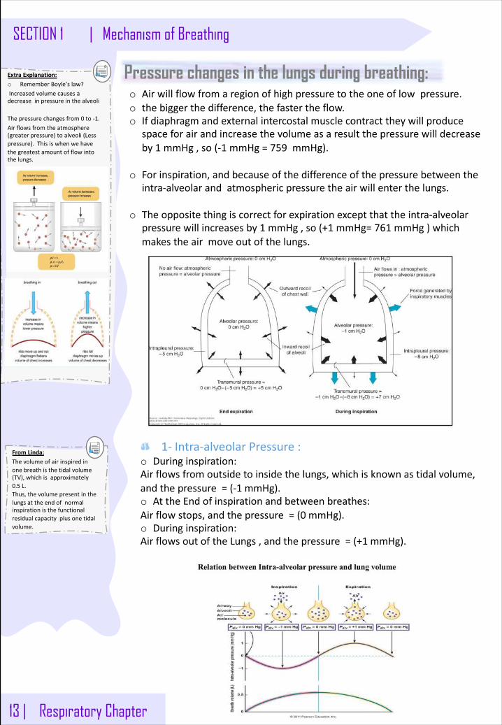
Pressure changes in the lungs during breathing:o Airwillflowfromaregionofhighpressuretotheoneoflowpressure.o thebiggerthedifference,thefastertheflow.o Ifdiaphragmandexternalintercostalmusclecontracttheywillproduce
spaceforairandincreasethevolumeasaresultthepressurewilldecreaseby1 mmHg,so(-1 mmHg=759 mmHg).
o Forinspiration,andbecauseofthedifferenceofthepressurebetweentheintra-alveolarandatmosphericpressuretheairwillenterthelungs.
o Theoppositethingiscorrectforexpirationexceptthattheintra-alveolarpressurewillincreasesby1 mmHg,so(+1 mmHg=761 mmHg)whichmakestheairmoveoutofthelungs.
ExtraExplanation:o RememberBoyle’slaw?Increasedvolumecausesadecreaseinpressureinthealveoli
Thepressurechangesfrom0 to-1.Airflowsfromtheatmosphere(greaterpressure)toalveoli(Lesspressure).Thisiswhenwehavethegreatestamountofflowintothelungs.
1- Intra-alveolarPressure:o Duringinspiration:Airflowsfromoutsidetoinsidethelungs,whichisknownastidalvolume,andthepressure=(-1 mmHg).o AttheEndofinspirationandbetweenbreathes:Airflowstops,andthepressure=(0 mmHg).o Duringinspiration:AirflowsoutoftheLungs,andthepressure=(+1 mmHg).
FromLinda:Thevolumeofairinspiredinonebreathisthetidalvolume(TV),whichisapproximately0.5 L.Thus,thevolumepresentinthelungsattheendofnormalinspirationisthefunctionalresidualcapacityplusonetidalvolume.
Respiratory Chapter | 14
Pressure changes in the lungs during breathing:2- Intrapleuralpressure(IPP)
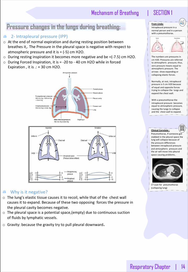
o Attheendofnormalexpirationandduringrestingpositionbetweenbreathesit,.ThePressureinthepleuralspaceisnegativewithrespecttoatmosphericpressureanditis=(-5)cmH2O.
o Duringrestinginspirationitbecomesmorenegativeandbe=(-7.5)cmH2O.o DuringForcedInspiration,itis=-20 to- 40 cmH2Owhileinforced
Expiration,itis.:+30 cmH2O.
FromLinda:Intrapleuralpressureinanormalpersonandinapersonwithapneumothorax.
ThenumbersarepressuresincmH20.Pressuresarereferredtoatmosphericpressure;thus,zeropressuremeansequaltoatmosphericpressure.Thearrowsshowexpandingorcollapsingelasticforces.
Normally,atrest,intrapleuralpressureis-5 cmH20 becauseofequalandoppositeforcestryingtocollapsethelungsandexpandthechestwall.
Withapneumothoraxtheintrapleuralpressurebecomesequaltoatmosphericpressure,causingthelungstocollapseandthechestwalltoexpand.
Whyisitnegative?o Thelung'selastictissuecausesittorecoil,whilethatofthechestwall
causesittoexpand.Becauseofthesetwoopposingforcesthepressureinthepleuralcavitybecomesnegative.
o Thepleuralspaceisapotentialspace,(empty)duetocontinuoussuctionoffluidsbylymphaticvessels.
o Gravity:becausethegravitytrytopullpleuraldownward.
ClinicalCorrelate:Pneumothorax,Ifsomeonegotstabbedinthepleuralspacethelungwillcollapsebecauseofthepressuredifferencesbetweenintrapleuralpressureandatmosphericpressureandtheairwillmoveintopleuralspacecausingproblems..
CTscanforpneumothorax(collapsinglung)
Mechanism of Breathing | SECTION 1
15 | Respiratory Chapter
SECTION 1 | Mechanism of Breathing
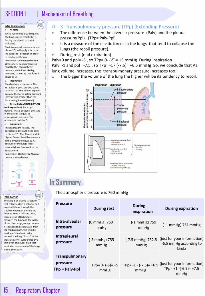
3- Transpulmonarypressure(TPp)(ExtendingPressure)o Thedifferencebetweenthealveolarpressure(Palv)andthepleural
pressure(Ppl).(TPp=Palv-Ppl).o Itisameasureoftheelasticforcesinthelungsthattendtocollapsethe
lungs(therecoilpressure).o Duringrest(endexpiration)Palv=0 andppl=-5 ,soTPp=0- (-5)=+5 mmHgDuringinspirationPalv=-1 andppl=-7.5 ,soTPp=-1 - (-7.5)=+6.5 mmHgSo,weconcludethatAslungvolumeincreases,thetranspulmonarypressureincreasestoo.o Thebiggerthevolumeofthelungthehigherwillbeitstendencytorecoil.
In Summary
PressureDuring rest During
inspirationDuring expiration
Intra-alveolarpressure
(0 mmHg)760mmHg
(-1 mmHg)759mmHg
(+1 mmHg)761mmHg
Intrapleuralpressure (-5 mmHg)755
mmHg(-7.5 mmHg)752.5
mmHg(justforyour information)-6.5 mmHgaccordingto
Linda
TranspulmonarypressureTPp= Palv-Ppl
TPp=0- (-5)=+5mmHg
TPp=-1 - (-7.5)=+6.5mmHg
(justforyour information)TPp=+1 -(-6.5)=+7.5
mmHg
The atmosphericpressureis760mmHg
ExtraExplanation:o Atrest: Whileyou’renotbreathing,yet.Thelung’srecoil(elasticity)isforcingthealveolitoshrink(collapse).Theintrapleuralpressure(about-5 cmH2O)willapplyaforceintheoppositedirectioninordertoreachequilibrium.Thealveoliisconnectedtotheatmosphere,soitspressureisequaltotheatmosphericpressure.(Wedon’tlikebignumbers,sowesaythatPatmisequalto0)o Inspiration:Thediaphragmcontracts.Theintrapleuralpressuredecreasesto-8 — -7.5.Thealveoliexpandbecausetheforceactingoutward(pressure)isgreaterthantheforceactinginward(recoil).o AttheENDofINSPIRATION(notexpiration),Airstopsflowing.That’sbecausepressureinthealveoliisequaltoatmosphericpressure.Thepressureisbackto0.o Expiration:Thediaphragmrelaxes.Theintrapleuralpressurerisesbackto-5 cmH2O.TheAlveolishrinks(Again,Boyle’slaw)thepressureinthealveoliincreasesto+1becauseofthelungsrecoil(elasticity).Airflowsouttotheatmosphere.Remember:Elasticity&Alveolarpressureateachstep.
FromGuytonThelungisanelasticstructurethatcollapseslikeaballoonandexpelsallitsairthroughthetracheawheneverthereisnoforcetokeepitinflated.Also,therearenoattachmentsbetweenthelungandthewallsofthechestcage,exceptwhereitissuspendedatitshilumfromthemediastinum,themiddlesectionofthechestcavity.Instead,thelung“floats”inthethoraciccavity,surroundedbyathinlayerofpleuralfluidthatlubricatesmovementofthelungswithinthecavity.
Respiratory Chapter | 16
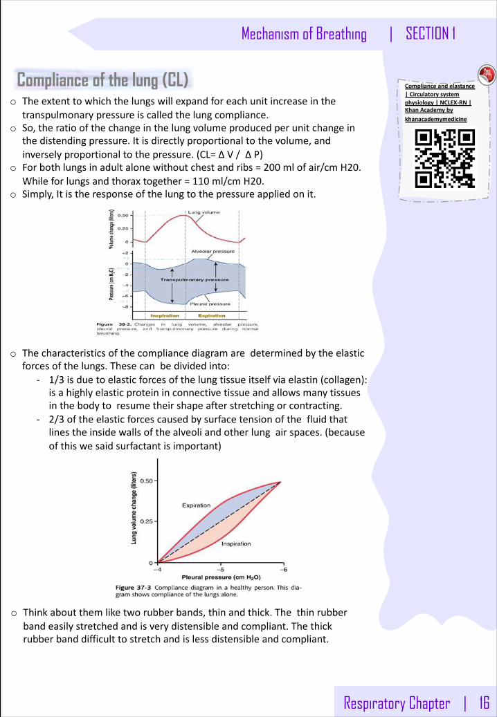
Compliance of the lung (CL)o Theextenttowhichthelungswillexpandforeachunitincreaseinthe
transpulmonarypressureiscalledthelungcompliance.o So,theratioofthechangeinthelungvolumeproducedperunitchangein
thedistendingpressure.Itisdirectlyproportionaltothevolume,andinverselyproportionaltothepressure.(CL=∆V/∆P)
o Forbothlungsinadultalonewithoutchestandribs=200 mlofair/cmH20.Whileforlungsandthoraxtogether=110 ml/cmH20.
o Simply,Itistheresponseofthelungtothepressureappliedonit.
o Thecharacteristicsofthecompliancediagramaredeterminedbytheelasticforcesofthelungs.Thesecanbedividedinto:
- 1/3 isduetoelasticforcesofthelungtissueitselfviaelastin(collagen):isahighlyelasticproteininconnectivetissueandallowsmanytissuesinthebodytoresumetheirshapeafterstretchingorcontracting.
- 2/3 oftheelasticforcescausedbysurfacetensionofthefluidthatlinestheinsidewallsofthealveoliandotherlungairspaces.(becauseofthiswesaidsurfactantisimportant)
o Thinkaboutthemliketworubberbands,thinandthick.Thethinrubberbandeasilystretchedandisverydistensibleandcompliant.Thethickrubberbanddifficulttostretchandislessdistensibleandcompliant.
Mechanism of Breathing | SECTION 1
Complianceandelastance|Circulatorysystemphysiology|NCLEX-RN|KhanAcademybykhanacademymedicine
17 | Respiratory Chapter
SECTION 1 | Mechanism of Breathing
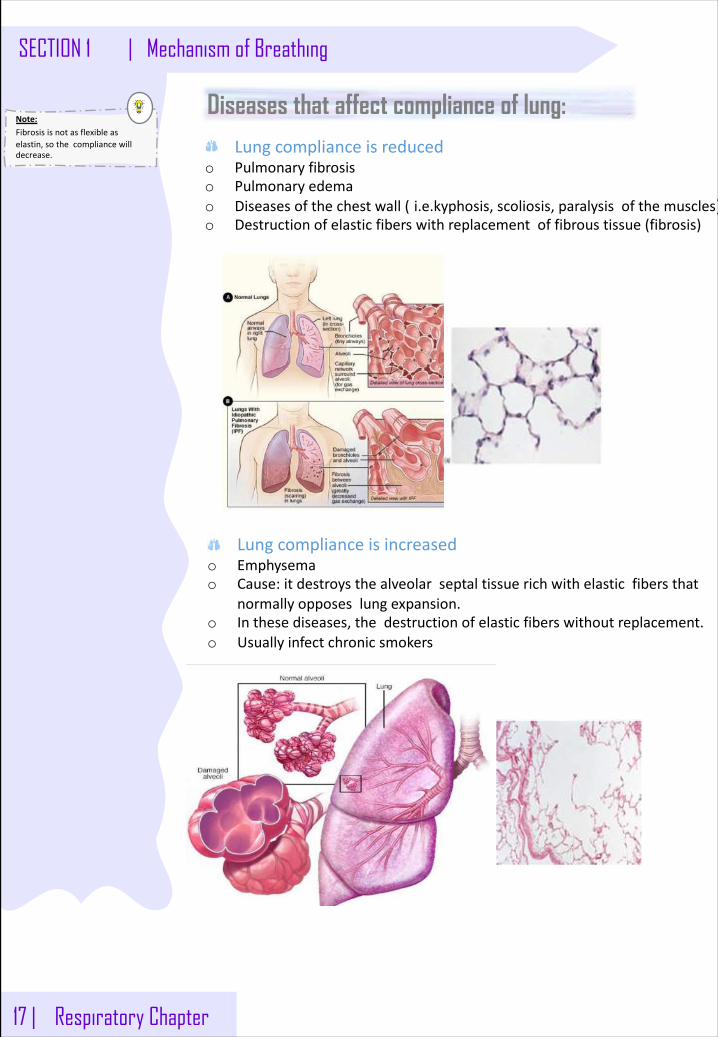
Diseases that affect compliance of lung:
Lungcomplianceisincreasedo Emphysemao Cause:itdestroysthealveolarseptaltissuerichwithelasticfibersthat
normallyopposeslungexpansion.o Inthesediseases,thedestructionofelasticfiberswithoutreplacement.o Usuallyinfectchronicsmokers
Lungcomplianceisreducedo Pulmonaryfibrosiso Pulmonaryedemao Diseasesofthechestwall( i.e.kyphosis,scoliosis,paralysisofthemuscles)o Destructionofelasticfiberswithreplacementoffibroustissue(fibrosis)
Note:Fibrosisisnotasflexibleaselastin,sothecompliancewilldecrease.
ForcesActingonTheLungSystem
Inrespiratoryphysiology,unitsofpressureareusuallygivenascmH2O.1 cmH2O=0.74 mmHg(1 mmHg=1.36 cmH2O)
1. Lungrecoilandintrapleuralpress• Understandinglungmechanicsinvolvesunderstandingthemainforcesactingontherespiratorysystem.• Lungrecoilrepresentstheinwardforcecreatedbytheelasticrecoilpropertiesofalveoli.
o Asthelungexpands,recoilincreases;asthelunggetssmaller,recoildecreases.o Recoil,asaforce,alwaysactstocollapsethelung.
• Chestwallrecoilrepresentstheoutwardforceofthechestwall.o FRCrepresentsthepointwherethisoutwardrecoilofthechestwalliscounterbalancedbythe
inwardrecoilofthelung.• Intrapleuralpressure(IPP)representsthepressureinsidethethinfilmoffluidbetweenthevisceral
pleura,whichisattachedtothelung,andtheparietalpleura,whichisattachedtothechestwall.o Theoutwardrecoilofthechestandinwardrecoilofthelungcreateanegative(sub
atmospheric)IPP.o IPPistheoutsidepressureforallstructuresinsidethechestwall.
2. Transmuralpressuregradient• Transmuralpressuregradient(PTM)representsthepressuregradientacrossanytubeorsphere.
• Calculatedasinsidepressureminusoutsidepressure• Ifpositive(insidegreaterthanoutside),itisanetforcepushingoutagainstthewallsofthe
structure• Ifnegative(outsidegreaterthaninside),itisanetforcepushinginagainstthewallsofthe
structure;dependinguponthestructuralcomponents,thetube/spherecancollapseifPTMisnegativeorzero
• AtFRC,IPPisnegative,andthusPTMispositive.Thispositiveout-wardforcepreventsalveolarcollapse(atelectasis).
• Fortheentirelung,PTMiscalledthetranspulmonarypressure(TPP).
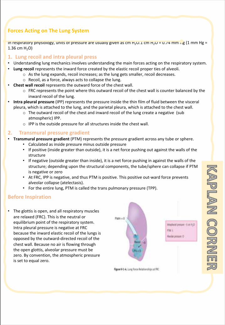
BeforeInspiration
• Theglottisisopen,andallrespiratorymusclesarerelaxed(FRC).Thisistheneutralorequilibriumpointoftherespiratorysystem.IntrapleuralpressureisnegativeatFRCbecausetheinwardelasticrecoilofthelungsisopposedbytheoutward-directedrecoilofthechestwall.Becausenoairisflowingthroughtheopenglottis,alveolarpressuremustbezero.Byconvention,theatmosphericpressureissettoequalzero.
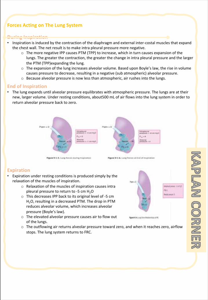
ForcesActingonTheLungSystem
DuringInspiration• Inspirationisinducedbythecontractionofthediaphragmandexternalinter-costalmusclesthatexpand
thechestwall.Thenetresultistomakeintrapleuralpressuremorenegative.o ThemorenegativeIPPcausesPTM(TPP)toincrease,whichinturncausesexpansionofthe
lungs.Thegreaterthecontraction,thegreaterthechangeinintrapleuralpressureandthelargerthePTM(TPP)expandingthelung.
o Theexpansionofthelungincreasesalveolarvolume.BaseduponBoyle’slaw,theriseinvolumecausespressuretodecrease,resultinginanegative(subatmospheric)alveolarpressure.
o Becausealveolarpressureisnowlessthanatmospheric,airrushesintothelungs.
EndofInspiration• Thelungexpandsuntilalveolarpressureequilibrateswithatmosphericpressure.Thelungsareattheir
new,largervolume.Underrestingconditions,about500 mLofairflowsintothelungsysteminordertoreturnalveolarpressurebacktozero.
Expiration• Expirationunderrestingconditionsisproducedsimplybythe
relaxationofthemusclesofinspiration.o Relaxationofthemusclesofinspirationcausesintra
pleuralpressuretoreturnto-5 cmH2Oo ThisdecreasesIPPbacktoitsoriginallevelof-5 cm
H2O,resultinginadecreasedPTM.ThedropinPTMreducesalveolarvolume,whichincreasesalveolarpressure(Boyle’slaw).
o Theelevatedalveolarpressurecausesairtoflowoutofthelungs.
o Theoutflowingairreturnsalveolarpressuretowardzero,andwhenitreacheszero,airflowstops.ThelungsystemreturnstoFRC.
ForcesActingonTheLungSystem
LungCompliance
v Lungcompliance0.6 liters/3 cmH2O=0.200 liters/cmH2O• Theprecedingcalculationsimplymeansthatforevery1 cmH2Osurroundingpressurechanges,
200 mLofairflowsinoroutoftherespiratorysystem.Itflowsintothesystemifsurroundingpressurebecomesmorenegative(e.g.,-5to-6 cmH2O)oroutofthesystemifsurroundingpressurebecomesmorepositive(e.g.,-5 to-4 cmH2O).
o Increasedcompliancemeansmoreairwillflowforagivenchangeinpressure.o Reducedcompliancemeanslessairwillflowforagivenchangeinpressure.o Intheprecedingcurve,althoughtheslopeischangingduringinflation,Itsvalueatany
pointisthelung’scompliance.Itistherelationshipbetweenthechangeinlungvolume(tidalvolume)andthechangeinintrapleuralorsurroundingpressure.
o Thesteepertheline,themorecompliantthelungs.Restfulbreathingworksonthesteepest,mostcompliantpartofthecurve.
o Withadeepinspiration,thelungmovestowardtheflatterpartofthecurve,andthusithasreducedcompliance.LungcomplianceislessatTLCcomparedtoFRC.Thefigurebelowshowspathologicstatesinwhichlungcompliancechanges.
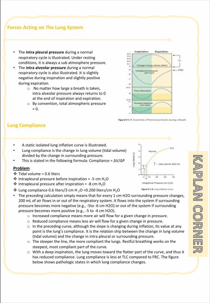
• Theintrapleuralpressureduringanormalrespiratorycycleisillustrated.Underrestingconditions,itisalwaysasubatmospherepressure.
• Theintraalveolarpressureduringanormalrespiratorycycleisalsoillustrated.Itisslightlynegativeduringinspirationandslightlypositiveduringexpiration.
o Nomatterhowlargeabreathistaken,intraalveolarpressurealwaysreturnsto0attheendofinspirationandexpiration.
o Byconvention,totalatmosphericpressure=0.
• Astaticisolatedlunginflationcurveisillustrated.• Lungcomplianceisthechangeinlungvolume(tidalvolume)
dividedbythechangeinsurroundingpressure.• Thisisstatedinthefollowingformula: Compliance=∆V/∆P
Problemv Tidalvolume=0.6 litersv Intrapleural pressurebeforeinspiration=-5 cmH2Ov Intrapleural pressureafterinspiration=-8 cmH2O
18 | Respiratory Chapter
SECTION 1 | Respiratory ventilation
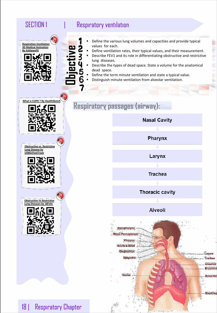
Respiratory passages (airway):
§ Definethevariouslungvolumesandcapacitiesandprovidetypicalvaluesforeach.
§ Defineventilationrates,theirtypicalvalues,andtheirmeasurement.§ DescribeFEV1 anditsroleindifferentiatingobstructiveandrestrictive
lungdiseases.§ Describethetypesofdeadspace.Stateavolumefortheanatomical
deadspace.§ Definethetermminuteventilationandstateatypicalvalue.§ Distinguishminuteventilationfromalveolarventilation.Ob
jectiv
e
ObstructiveVsRestrictiveLungDiseasesby18Fafo
Obstructivevs.RestrictiveLungDiseasebyUSMLEFastTrack
WhatisCOPD?ByHealthSketch
Respiration-Ventilation3DMedicalAnimationByAshlawnPE
Respiratory Chapter | 19
Respiratory ventilation | SECTION 1
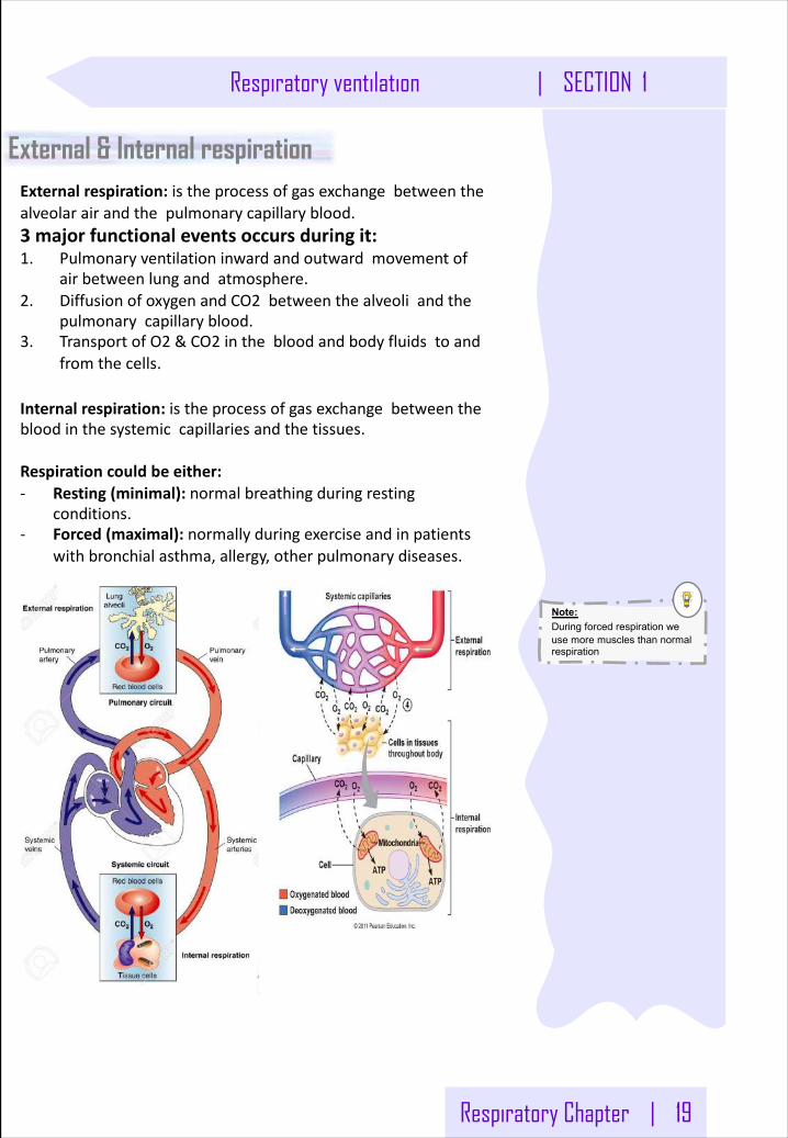
External & Internal respirationExternalrespiration:istheprocessofgasexchangebetweenthealveolarairandthepulmonarycapillaryblood.3 majorfunctionaleventsoccursduringit:1. Pulmonaryventilationinwardandoutwardmovementof
airbetweenlungandatmosphere.2. DiffusionofoxygenandCO2 betweenthealveoliandthe
pulmonarycapillaryblood.3. TransportofO2 &CO2 inthebloodandbodyfluidstoand
fromthecells.
Internalrespiration:istheprocessofgasexchangebetweenthebloodinthesystemiccapillariesandthetissues.
Respirationcouldbeeither:- Resting(minimal):normalbreathingduringresting
conditions.- Forced(maximal): normallyduringexerciseandinpatients
withbronchialasthma,allergy,otherpulmonarydiseases.
Note:During forced respiration we use more muscles than normal respiration
20 | Respiratory Chapter
SECTION 1 | Respiratory ventilation
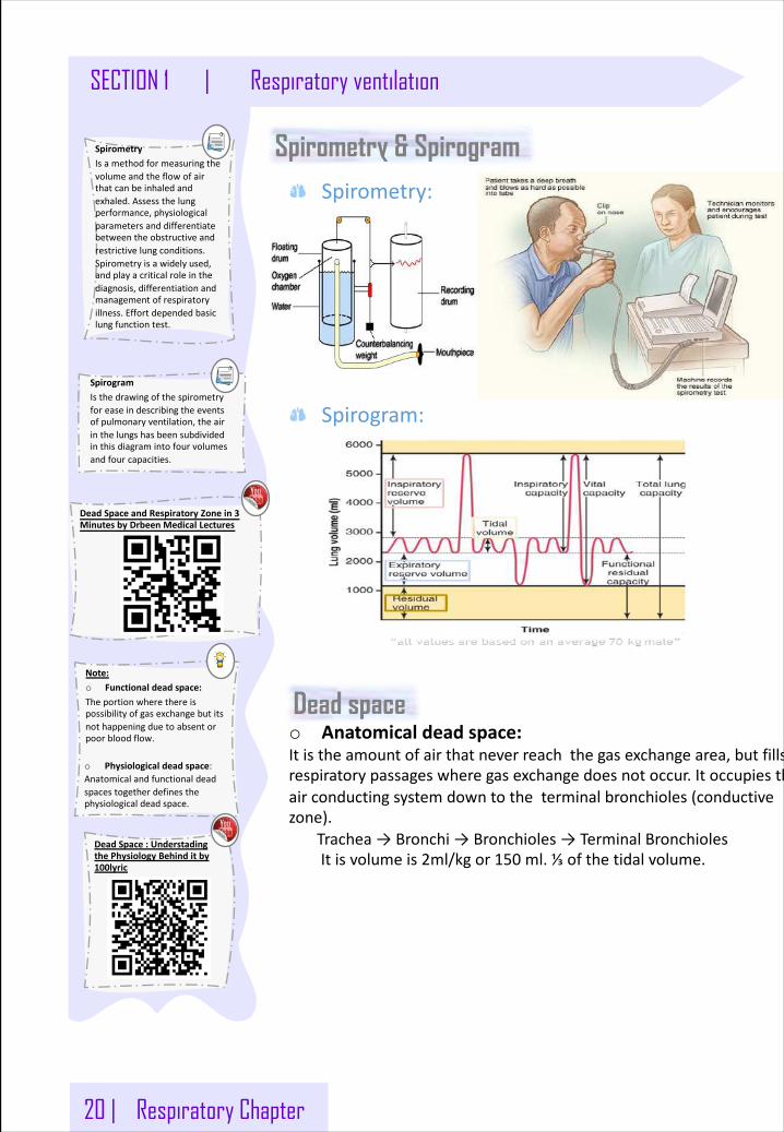
Spirometry & Spirogram Spirometry:
Spirogram:
SpirometryIsamethodformeasuringthevolumeandtheflowofairthatcanbeinhaledandexhaled.Assessthelungperformance,physiologicalparametersanddifferentiatebetweentheobstructiveandrestrictivelungconditions.Spirometryisawidelyused,andplayacriticalroleinthediagnosis,differentiationandmanagementofrespiratoryillness.Effortdependedbasiclungfunctiontest.
SpirogramIsthedrawingofthespirometryforeaseindescribingtheeventsofpulmonaryventilation,theairinthelungshasbeensubdividedinthisdiagramintofourvolumesandfourcapacities.
Note:o Functionaldeadspace:Theportionwherethereispossibilityofgasexchangebutitsnothappeningduetoabsentorpoorbloodflow.
o Physiologicaldeadspace:Anatomicalandfunctionaldeadspacestogetherdefinesthephysiologicaldeadspace.
Dead spaceo Anatomicaldeadspace:Itistheamountofairthatneverreachthegasexchangearea,butfillsrespiratorypassageswheregasexchangedoesnotoccur.Itoccupiestheairconductingsystemdowntotheterminalbronchioles(conductivezone).
Trachea→Bronchi→Bronchioles→TerminalBronchiolesItisvolumeis2ml/kgor150 ml.⅓ofthetidalvolume.
DeadSpace:UnderstadingthePhysiologyBehinditby100lyric
DeadSpaceandRespiratoryZonein3MinutesbyDrbeen MedicalLectures
Respiratory Chapter | 21
Respiratory ventilation | SECTION 1
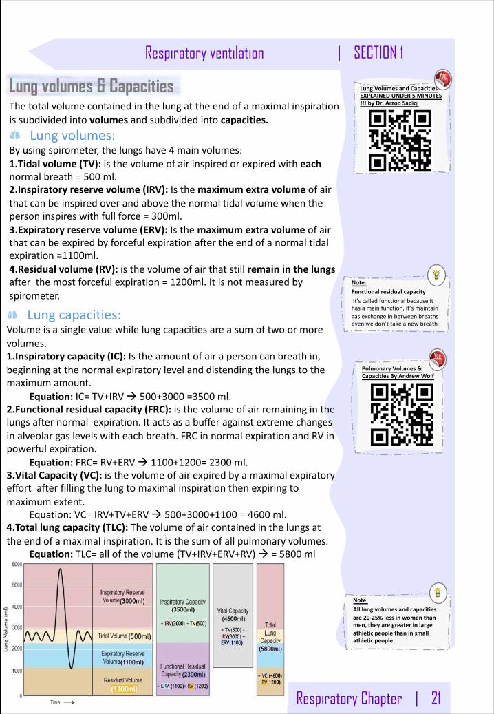
Lungcapacities:Volumeisasinglevaluewhilelungcapacitiesareasumoftwoormorevolumes.1.Inspiratorycapacity(IC):Istheamountofairapersoncanbreathin,beginningatthenormalexpiratorylevelanddistendingthelungstothemaximumamount.
Equation:IC=TV+IRVà 500+3000 =3500 ml.2.Functionalresidualcapacity(FRC):isthevolumeofairremaininginthelungsafternormalexpiration.Itactsasabufferagainstextremechangesinalveolargaslevelswitheachbreath.FRCinnormalexpirationandRVinpowerfulexpiration.
Equation:FRC=RV+ERVà 1100+1200=2300 ml.3.VitalCapacity(VC):isthevolumeofairexpiredbyamaximalexpiratoryeffortafterfillingthelungtomaximalinspirationthenexpiringtomaximumextent.
Equation:VC=IRV+TV+ERVà 500+3000+1100 =4600 ml.4.Totallungcapacity(TLC):Thevolumeofaircontainedinthelungsattheendofamaximalinspiration.Itisthesumofallpulmonaryvolumes.
Equation:TLC=allofthevolume(TV+IRV+ERV+RV)à =5800 ml
Note:Functionalresidualcapacityit’scalledfunctionalbecauseithasamainfunction,it’smaintaingasexchangeinbetweenbreathsevenwedon’ttakeanewbreath
Note:Alllungvolumesandcapacitiesare20-25%lessinwomenthanmen,theyaregreaterinlargeathleticpeoplethaninsmallathleticpeople.
Thetotalvolumecontainedinthelungattheendofamaximalinspirationissubdividedintovolumes andsubdividedintocapacities.
Lungvolumes:Byusingspirometer,thelungshave4 mainvolumes:1.Tidalvolume(TV):isthevolumeofairinspiredorexpiredwitheachnormalbreath=500 ml.2.Inspiratoryreservevolume(IRV):Isthemaximumextravolumeofairthatcanbeinspiredoverandabovethenormaltidalvolumewhenthepersoninspireswithfullforce=300ml.3.Expiratoryreservevolume(ERV):Isthemaximumextravolumeofairthatcanbeexpiredbyforcefulexpirationaftertheendofanormaltidalexpiration=1100ml.4.Residualvolume(RV):isthevolumeofairthatstillremaininthelungsafterthemostforcefulexpiration=1200ml.Itisnotmeasuredbyspirometer.
Lung volumes & Capacities LungVolumesandCapacitiesEXPLAINEDUNDER5 MINUTES!!!byDr.Arzoo Sadiqi
PulmonaryVolumes&CapacitiesByAndrewWolf
22 | Respiratory Chapter
SECTION 1 | Respiratory ventilationNote:WhydowechooseHe?BecauseHiisfromtheinnergases,whenit’senterourbodyitdoesn’tdiffusion,thatmeanswedon’tuseit!It’sonlyagasthattakesplacewithoutanyfunction.
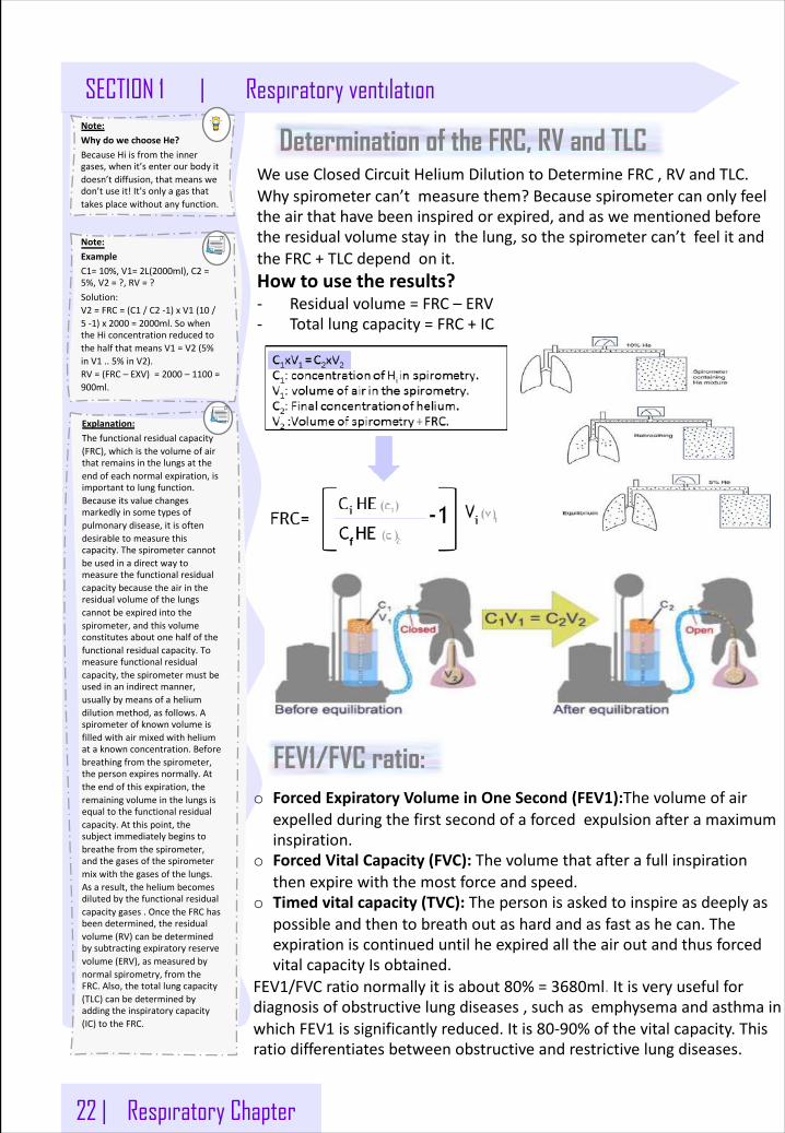
Determination of the FRC, RV and TLC WeuseClosedCircuitHeliumDilutiontoDetermineFRC,RVandTLC.Whyspirometercan’tmeasurethem?Becausespirometercanonlyfeeltheairthathavebeeninspiredorexpired,andaswementionedbeforetheresidualvolumestayinthelung,sothespirometercan’tfeelitandtheFRC+TLCdependonit.Howtousetheresults?- Residualvolume=FRC– ERV- Totallungcapacity=FRC+IC
Note:ExampleC1=10%,V1=2L(2000ml),C2 =5%,V2 =?,RV=?Solution:V2 =FRC=(C1 /C2 -1)xV1 (10 /5 -1)x2000 =2000ml.SowhentheHiconcentrationreducedtothehalfthatmeansV1 =V2 (5%inV1 ..5%inV2).RV=(FRC– EXV)=2000 – 1100 =900ml.
FEV1/FVC ratio:o ForcedExpiratoryVolumeinOneSecond(FEV1):Thevolumeofair
expelledduringthefirstsecondofaforcedexpulsionafteramaximuminspiration.
o ForcedVitalCapacity(FVC):Thevolumethatafterafullinspirationthenexpirewiththemostforceandspeed.
o Timedvitalcapacity(TVC):Thepersonisaskedtoinspireasdeeplyaspossibleandthentobreathoutashardandasfastashecan.TheexpirationiscontinueduntilheexpiredalltheairoutandthusforcedvitalcapacityIsobtained.
FEV1/FVCrationormallyitisabout80%=3680ml.Itisveryusefulfordiagnosisofobstructivelungdiseases,suchasemphysemaandasthmainwhichFEV1 issignificantlyreduced.Itis80-90%ofthevitalcapacity.Thisratiodifferentiatesbetweenobstructiveandrestrictivelungdiseases.
Explanation:Thefunctionalresidualcapacity(FRC),whichisthevolumeofairthatremainsinthelungsattheendofeachnormalexpiration,isimportanttolungfunction.Becauseitsvaluechangesmarkedlyinsometypesofpulmonarydisease,itisoftendesirabletomeasurethiscapacity.Thespirometercannotbeusedinadirectwaytomeasurethefunctionalresidualcapacitybecausetheairintheresidualvolumeofthelungscannotbeexpiredintothespirometer,andthisvolumeconstitutesaboutonehalfofthefunctionalresidualcapacity.Tomeasurefunctionalresidualcapacity,thespirometermustbeusedinanindirectmanner,usuallybymeansofaheliumdilutionmethod,asfollows.Aspirometerofknownvolumeisfilledwithairmixedwithheliumataknownconcentration.Beforebreathingfromthespirometer,thepersonexpiresnormally.Attheendofthisexpiration,theremainingvolumeinthelungsisequaltothefunctionalresidualcapacity.Atthispoint,thesubjectimmediatelybeginstobreathefromthespirometer,andthegasesofthespirometermixwiththegasesofthelungs.Asaresult,theheliumbecomesdilutedbythefunctionalresidualcapacitygases.OncetheFRChasbeendetermined,theresidualvolume(RV)canbedeterminedbysubtractingexpiratoryreservevolume(ERV),asmeasuredbynormalspirometry,fromtheFRC.Also,thetotallungcapacity(TLC)canbedeterminedbyaddingtheinspiratorycapacity(IC)totheFRC.
Respiratory Chapter | 23
Respiratory ventilation | SECTION 1
Note:Thenormalpersonandtherestrictivepersonallhavenormalratio.Howdowedifferentiatebetweenthem?Bythetotallungvolume.It’sdecreasedinrestrictiveperson.
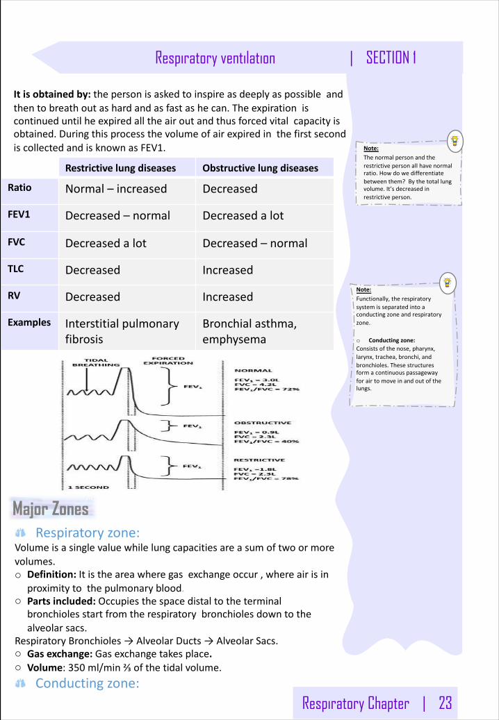
Itisobtainedby:thepersonisaskedtoinspireasdeeplyaspossibleandthentobreathoutashardandasfastashecan.Theexpirationiscontinueduntilheexpiredalltheairoutandthusforcedvitalcapacityisobtained.DuringthisprocessthevolumeofairexpiredinthefirstsecondiscollectedandisknownasFEV1.
Restrictivelungdiseases Obstructivelungdiseases
Ratio Normal– increased Decreased
FEV1 Decreased– normal Decreasedalot
FVC Decreasedalot Decreased– normal
TLC Decreased Increased
RV Decreased Increased
Examples Interstitialpulmonaryfibrosis
Bronchialasthma,emphysema
Major Zones
Note:Functionally,therespiratorysystemisseparatedintoaconductingzoneandrespiratoryzone.
o Conductingzone:Consistsofthenose,pharynx,larynx,trachea,bronchi,andbronchioles.Thesestructuresformacontinuouspassagewayforairtomoveinandoutofthelungs.
Respiratoryzone:Volumeisasinglevaluewhilelungcapacitiesareasumoftwoormorevolumes.o Definition:Itistheareawheregasexchangeoccur,whereairisin
proximitytothepulmonaryblood.o Partsincluded:Occupiesthespacedistaltotheterminal
bronchiolesstartfromtherespiratorybronchiolesdowntothealveolarsacs.
RespiratoryBronchioles→AlveolarDucts→AlveolarSacs.o Gasexchange:Gasexchangetakesplace.o Volume:350 ml/min⅔ofthetidalvolume.
Conductingzone:
24 | Respiratory Chapter
SECTION 1 | Respiratory ventilation
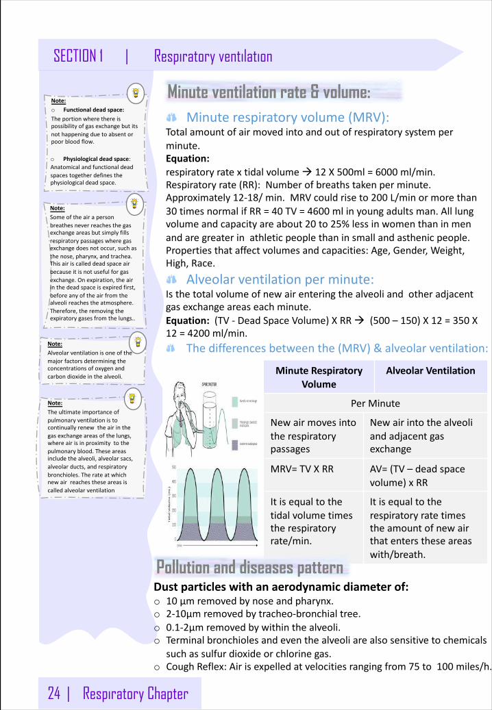
Minute ventilation rate & volume:Minuterespiratoryvolume(MRV):
Totalamountofairmovedintoandoutofrespiratorysystemperminute.Equation:respiratoryratextidalvolumeà 12 X500ml=6000 ml/min.Respiratoryrate(RR):Numberofbreathstakenperminute.Approximately12-18/min.MRVcouldriseto200 L/minormorethan30 timesnormalifRR=40 TV=4600 mlinyoungadultsman.Alllungvolumeandcapacityareabout20 to25%lessinwomenthaninmenandaregreaterinathleticpeoplethaninsmallandasthenicpeople.Propertiesthataffectvolumesandcapacities:Age,Gender,Weight,High,Race.
Alveolarventilationperminute:Isthetotalvolumeofnewairenteringthealveoliandotheradjacentgasexchangeareaseachminute.Equation:(TV- DeadSpaceVolume)XRRà (500 – 150)X12 =350 X12 =4200 ml/min.
Thedifferencesbetweenthe(MRV)&alveolarventilation:
Note:o Functionaldeadspace:Theportionwherethereispossibilityofgasexchangebutitsnothappeningduetoabsentorpoorbloodflow.
o Physiologicaldeadspace:Anatomicalandfunctionaldeadspacestogetherdefinesthephysiologicaldeadspace.
Note:Someoftheairapersonbreathesneverreachesthegasexchangeareasbutsimplyfillsrespiratorypassageswheregasexchangedoesnotoccur,suchasthenose,pharynx,andtrachea.Thisairiscalleddeadspaceairbecauseitisnotusefulforgasexchange.Onexpiration,theairinthedeadspaceisexpiredfirst,beforeanyoftheairfromthealveolireachestheatmosphere.Therefore,theremovingtheexpiratorygasesfromthelungs..
Note:Theultimateimportanceofpulmonaryventilationistocontinuallyrenewtheairinthegasexchangeareasofthelungs,whereairisinproximitytothepulmonaryblood.Theseareasincludethealveoli,alveolarsacs,alveolarducts,andrespiratorybronchioles.Therateatwhichnewairreachestheseareasiscalledalveolarventilation
Note:Alveolarventilationisoneofthemajorfactorsdeterminingtheconcentrationsofoxygenandcarbondioxideinthealveoli.
MinuteRespiratoryVolume
AlveolarVentilation
PerMinute
Newairmovesintotherespiratorypassages
Newairintothealveoliandadjacentgasexchange
MRV=TVXRR AV=(TV– deadspacevolume)xRR
Itisequaltothetidalvolumetimestherespiratoryrate/min.
Itisequaltotherespiratoryratetimestheamountofnewairthatenterstheseareaswith/breath.
Pollution and diseases patternDustparticleswithanaerodynamicdiameterof:o 10 μmremovedbynoseandpharynx.o 2-10μmremovedbytracheo-bronchialtree.o 0.1-2μmremovedbywithinthealveoli.o Terminalbronchiolesandeventhealveoliarealsosensitivetochemicals
suchassulfurdioxideorchlorinegas.o CoughReflex:Airisexpelledatvelocitiesrangingfrom75 to100 miles/h.
LungCompliance
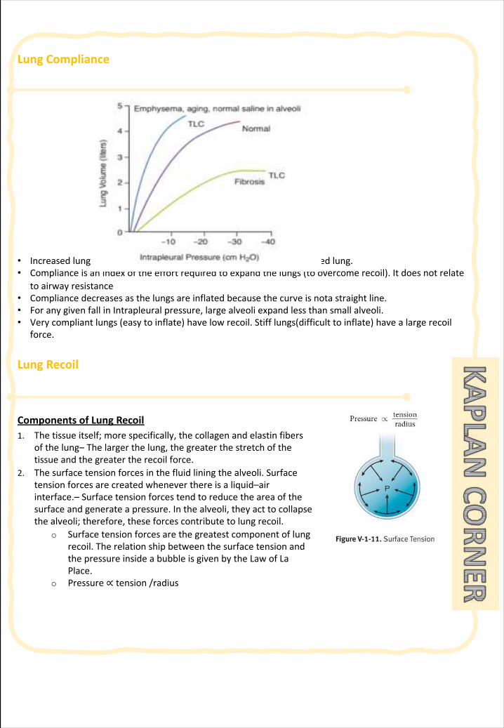
• Increasedlungcompliancealsooccurswithagingandwithasaline-filledlung.• Complianceisanindexoftheeffortrequiredtoexpandthelungs(toovercomerecoil).Itdoesnotrelate
toairwayresistance• Compliancedecreasesasthelungsareinflatedbecausethecurveisnotastraightline.• ForanygivenfallinIntrapleural pressure,largealveoliexpandlessthansmallalveoli.• Verycompliantlungs(easytoinflate)havelowrecoil.Stifflungs(difficulttoinflate)havealargerecoil
force.
LungRecoil
ComponentsofLungRecoil1. Thetissueitself;morespecifically,thecollagenandelastinfibers
ofthelung– Thelargerthelung,thegreaterthestretchofthetissueandthegreatertherecoilforce.
2. Thesurfacetensionforcesinthefluidliningthealveoli.Surfacetensionforcesarecreatedwheneverthereisaliquid–airinterface.– Surfacetensionforcestendtoreducetheareaofthesurfaceandgenerateapressure.Inthealveoli,theyacttocollapsethealveoli;therefore,theseforcescontributetolungrecoil.
o Surfacetensionforcesarethegreatestcomponentoflungrecoil.TherelationshipbetweenthesurfacetensionandthepressureinsideabubbleisgivenbytheLawofLaPlace.
o Pressure∝ tension/radius
LungRecoil
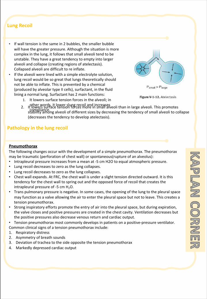
2. Itlowerssurfacetensionforcesmoreinsmallalveolithaninlargealveoli.Thispromotesstabilityamongalveoliofdifferentsizesbydecreasingthetendencyofsmallalveolitocollapse(decreasesthetendencytodevelopatelectasis).
Pathologyinthelungrecoil
• Ifwalltensionisthesamein2 bubbles,thesmallerbubblewillhavethegreaterpressure.Althoughthesituationismorecomplexinthelung,itfollowsthatsmallalveolitendtobeunstable.Theyhaveagreattendencytoemptyintolargeralveoliandcollapse(creatingregionsofatelectasis).Collapsedalveoliaredifficulttoreinflate.
• Ifthealveoliwerelinedwithasimpleelectrolytesolution,lungrecoilwouldbesogreatthatlungstheoreticallyshouldnotbeabletoinflate.Thisispreventedbyachemical(producedbyalveolartypeIIcells),surfactant,inthefluidlininganormallung.Surfactanthas2 mainfunctions:
1. Itlowerssurfacetensionforcesinthealveoli;inotherwords,itlowerslungrecoilandincreasescompliance.
PneumothoraxThefollowingchangesoccurwiththedevelopmentofasimplepneumothorax.Thepneumothoraxmaybetraumatic(perforationofchestwall)orspontaneous(ruptureofanalveolus):• Intrapleural pressureincreasesfromameanat-5 cmH2Otoequalatmosphericpressure.• Lungrecoildecreasestozeroasthelungcollapses.• Lungrecoildecreasestozeroasthelungcollapses.• Chestwallexpands.AtFRC,thechestwallisunderaslighttensiondirectedoutward.Itisthis
tendencyforthechestwalltospringoutandtheopposedforceofrecoilthatcreatestheintrapleural pressureof-5 cmH2O.
• Transpulmonarypressureisnegative.Insomecases,theopeningofthelungtothepleuralspacemayfunctionasavalveallowingtheairtoenterthepleuralspacebutnottoleave.Thiscreatesatensionpneumothorax.
• Stronginspiratoryeffortspromotetheentryofairintothepleuralspace,butduringexpiration,thevalveclosesandpositivepressuresarecreatedinthechestcavity.Ventilationdecreasesbutthepositivepressuresalsodecreasevenousreturnandcardiacoutput.
• Tensionpneumothoraxmostcommonlydevelopsinpatientsonapositive-pressureventilator.Commonclinicalsignsofatensionpneumothoraxinclude:1. Respiratorydistress2. Asymmetryofbreathsounds3. Deviationoftracheatothesideoppositethetensionpneumothorax4. Markedlydepressedcardiacoutput
Pathologyinthelungrecoil
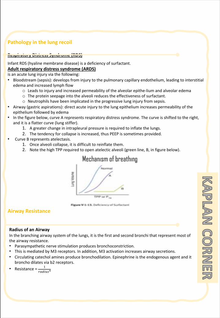
RespiratoryDistressSyndrome(RDS)
InfantRDS(hyalinemembranedisease)isadeficiencyofsurfactant.Adultrespiratorydistresssyndrome(ARDS)isanacutelunginjuryviathefollowing:• Bloodstream(sepsis):developsfrominjurytothepulmonarycapillaryendothelium,leadingtointerstitial
edemaandincreasedlymphflowo Leadstoinjuryandincreasedpermeabilityofthealveolarepithe-lium andalveolaredemao Theproteinseepageintothealveolireducestheeffectivenessofsurfactant.o Neutrophilshavebeenimplicatedintheprogressivelunginjuryfromsepsis.
• Airway(gastricaspirations):directacuteinjurytothelungepitheliumincreasespermeabilityoftheepitheliumfollowedbyedema
• Inthefigurebelow,curveArepresentsrespiratorydistresssyndrome.Thecurveisshiftedtotheright,anditisaflattercurve(lungstiffer).
1. Agreaterchangeinintrapleural pressureisrequiredtoinflatethelungs.2. Thetendencyforcollapseisincreased,thusPEEPissometimesprovided.
• CurveBrepresentsatelectasis.1. Oncealveolicollapse,itisdifficulttoreinflate them.2. NotethehighTPPrequiredtoopenatelectic alveoli(greenline,B,infigurebelow).
AirwayResistance
RadiusofanAirwayInthebranchingairwaysystemofthelungs,itisthefirstandsecondbronchithatrepresentmostoftheairwayresistance.• Parasympatheticnervestimulationproducesbronchoconstriction.• ThisismediatedbyM3 receptors.Inaddition,M3 activationincreasesairwaysecretions.• Circulatingcatecholaminesproducebronchodilation.Epinephrineistheendogenousagentandit
broncho dilatesviab2 receptors.• Resistance= "
#$%&'()
Mechanism of breathing
AirwayResistance
Ventilation
TotalVentilation• Totalventilationisalsoreferredtoasminutevolumeorminuteventilation.Itisthe totalvolumeofair
movedinorout(usuallythevolumeexpired)ofthelungsperminute.• Ve=VT×f (Ve:totalventilation.VT:tidalvolume f:respiratoryrate)• Normalrestingvalueswouldbe:VT=500 mL• f=15• 500 mL × 15/min = 7,500 mL/min
DeadSpaceRegionsoftherespiratorysystemthatcontainairbutarenotexchangingO2 andCO2 withbloodareconsidereddeadspace.
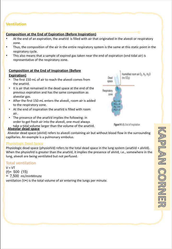
AnatomicDeadSpaceAirwayregionsthat,becauseofinherentstructure,arenotcapableofO2 andCO2 exchangewiththeblood.Anatomicdeadspace(anatVd)includesthecon-ductingzone,whichendsattheleveloftheterminalbronchioles.Significantgasexchange(O2 uptakeandCO2 removal)withthebloodoccursonlyinthealveoli.ThesizeoftheanatVd inmLisapproximatelyequaltoaperson’sweightinpounds.Thusa150-lbindividualhasananatomicdeadspaceof150 ml.
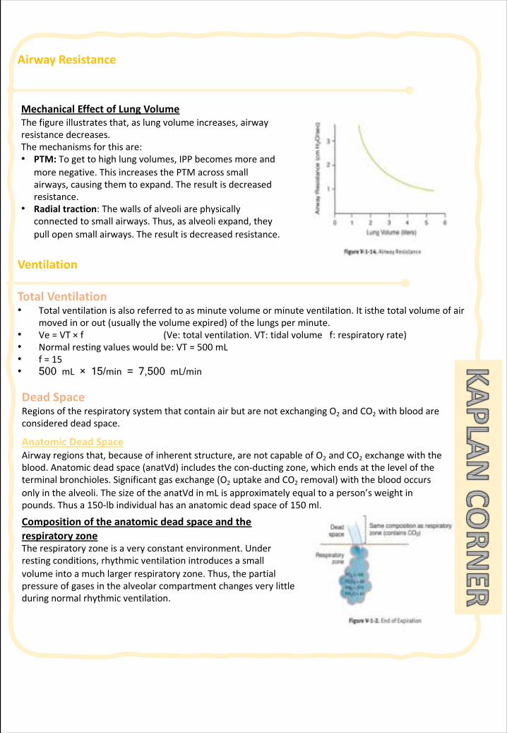
MechanicalEffectofLungVolumeThefigureillustratesthat,aslungvolumeincreases,airwayresistancedecreases.Themechanismsforthisare:• PTM:Togettohighlungvolumes,IPPbecomesmoreand
morenegative.ThisincreasesthePTMacrosssmallairways,causingthemtoexpand.Theresultisdecreasedresistance.
• Radialtraction:Thewallsofalveoliarephysicallyconnectedtosmallairways.Thus,asalveoliexpand,theypullopensmallairways.Theresultisdecreasedresistance.
CompositionoftheanatomicdeadspaceandtherespiratoryzoneTherespiratoryzoneisaveryconstantenvironment.Underrestingconditions,rhythmicventilationintroducesasmallvolumeintoamuchlargerrespiratoryzone.Thus,thepartialpressureofgasesinthealveolarcompartmentchangesverylittleduringnormalrhythmicventilation.
Ventilation
CompositionattheEndofExpiration(BeforeInspiration)• Attheendofanexpiration,theanatVd isfilledwithairthatoriginatedinthealveoliorrespiratory
zone.• Thus,thecompositionoftheairintheentirerespiratorysystemisthesameatthisstaticpointinthe
respiratorycycle.• Thisalsomeansthatasampleofexpiredgastakenneartheendofexpiration(endtidalair)is
representativeoftherespiratoryzone.
AlveolardeadspaceAlveolardeadspace(alvVd)referstoalveolicontainingairbutwithoutbloodflowinthesurroundingcapillaries.Anexampleisapulmonaryembolus.
PhysiologicDeadSpacePhysiologicdeadspace(physioIVd)referstothetotaldeadspaceinthelungsystem(anatVd +alvVd).WhenthephysiolVd isgreaterthantheanatVd,itimpliesthepresenceofalvVd,i.e.,somewhereinthelung,alveoliarebeingventilatedbutnotperfused.
TotalventilationV=VT (f)= 500 (15) = 7,500 mL/minMinute ventilation(V•)isthetotalvolumeofairenteringthelungsperminute.
CompositionattheEndofInspiration(BeforeExpiration)• Thefirst150 mLofairtoreachthealveolicomesfrom
theanatVd.• Itisairthatremainedinthedeadspaceattheendofthe
previousexpirationandhasthesamecompositionasalveolargas.
• Afterthefirst150 mLentersthealveoli,roomairisaddedtotherespiratoryzone.
• AttheendofinspirationtheanatVd isfilledwithroomair.
• ThepresenceoftheanatVd impliesthefollowing:inordertogetfreshairintothealveoli,onemustalwaystakeatidalvolumelargerthanthevolumeoftheanatVd.
Ventilation
AlveolarVentilationAlveolarventilationV•Arepresentstheroomairdeliveredtotherespiratoryzoneperbreath.• Thefirst150 mLofeachinspirationcomesfromtheanatomicdeadspaceanddoesnotcontributeto
alveolarventilation.• However,everyadditionalmLbeyond150 doescontributetoalveolarventilation.• V•A=(VT- VD)f= (500 mL - 150 mL) 15 = 5250 mL/min (V•A:alveolarventilation. VT:tidal
volume.VD:deadspace.f:respiratoryrate)• Thealveolarventilationperinspirationis350 mL. Thisequationimpliesthatthevolumeoffreshairthat
enterstherespiratoryzoneperminutedependsonthepatternofbreathing(howlargeaVTandtherateofbreathing).
IncreasesintheDepthofBreathing• Thereareequalincreasesintotalandalveolarventilationperbreath,sincedeadspace volumeis
constant.• Ifthedepthofbreathingincreasesfromadepthof500 mLtoadepthof700 mL,the increaseintotaland
alveolarventilationis200 mLperbreath.
IncreasesintheRateofBreathing• Thereisagreaterincreaseintotalventilationperminutethaninalveolarventilationperminute,
becausetheincreasedratecausesincreasedventilationofdeadspaceandalveoli.• Foreveryadditionalinspirationwithatidalvolumeof500 mL,totalventilationincreases500 mL,but
alveolarventilationonlyincreasesby350 mL(assumingdeadspaceis150 mL).
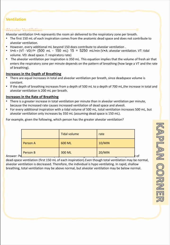
Forexample,giventhefollowing,whichpersonhasthegreateralveolarventilation?
Answer:PersonA.PersonBhasrapid,shallowbreathing.Thispersonhasalargecomponentofdead-spaceventilation(first150 mLofeachinspiration).Eventhoughtotalventilationmaybenormal,alveolarventilationisdecreased.Therefore,theindividualishypoventilating.Inrapid,shallowbreathing,totalventilationmaybeabovenormal,butalveolarventilationmaybebelownormal.
Tidalvolume rate
PersonA 600 ML 10/MIN
PersonB 300 ML 20/MIN
Ventilation
CardiovascularChangesWithVentilationInspirationWithinspiration,intrapleuralpressurebecomesmorenegative(decreases).ThisincreasesthePTMacrossthevasculature,causingthegreatveinsandrightatriumtoexpand.Thisexpansiondecreasesintravascularpressure,therebyincreasingthepressuregradientdrivingVRtotherightheart.• Systemicvenousreturnandrightventricularoutputareincreased.• Anincreaseintheoutputoftherightventricledelaysclosingofthepulmonicvalvesandtypicallyresults
inasplittingofthesecondheartsound.• Pulmonaryvesselsexpand,andthevolumeofbloodinthepulmonarycircuitincreases.Inaddition,
becausepulmonaryvascularresistance(PVR)islowestatFRC,itincreases.• Inturn,venousreturntotheleftheart,andtheoutputoftheleftventricleisdecreased,causing
decreasedsystemicarterialpressure(dropinsystolicmostprominent).• Inturn,venousreturntotheleftheart,andtheoutputoftheleftventricleisdecreased,causing
decreasedsystemicarterialpressure(dropinsystolicmostprominent).• Thisinspirationreducesvagaloutflowtotheheart(mechanismdebatable)resultinginaslightrisein
heartrate(respiratorysinusarrhythmia).Thisiswhypatientsareaskedtoholdtheirbreath,ifclinicallypossible,whenanEKGista
ExpirationExpirationisthereverseoftheprocessesabove.Intrapleural pressurebecomesmorepositive(increases),i.e.,returnstooriginalnegativevalue.PTMreturnstoitsoriginallevel,therebydecreasingthepressuregradientforVR.• Systemicvenousreturnandoutputoftherightventriclearedecreased.• Pulmonaryvesselsarecompressed,andthevolumeofbloodinthepulmonarycircuitdecreases.• Thereturnofbloodandoutputoftheleftventricleincreases,causingsystemicarterialpressureto
rise(primarilysystolic).• Vagaloutflowincreases(mechanismdebated),reducingHR(respiratorysinusarrhythmia).• AValsalvamaneuverisaforcedexpirationagainstaclosedglottis.Thisforcedexpirationcreatesa
positiveIPP(seelaterinthischapter),whichcompressesthegreatveinsinthechest.ThisinturnreducesVR.
Ventilation
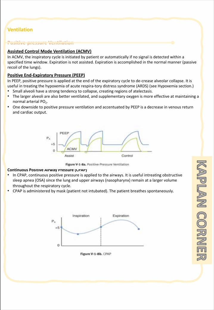
Positive-pressureVentilationAssistedControlModeVentilation(ACMV)InACMV,theinspiratorycycleisinitiatedbypatientorautomaticallyifnosignalisdetectedwithinaspecifiedtimewindow.Expirationisnotassisted.Expirationisaccomplishedinthenormalmanner(passiverecoilofthelungs).
PositiveEnd-ExpiratoryPressure(PEEP)InPEEP,positivepressureisappliedattheendoftheexpiratorycycletode-creasealveolarcollapse.Itisusefulintreatingthehypoxemiaofacuterespira-torydistresssyndrome(ARDS)(seeHypoxemiasection.)• Smallalveolihaveastrongtendencytocollapse,creatingregionsofatelectasis.• Thelargeralveoliarealsobetterventilated,andsupplementaryoxygenismoreeffectiveatmaintaininga
normalarterialPO2.• OnedownsidetopositivepressureventilationandaccentuatedbyPEEPisadecreaseinvenousreturn
andcardiacoutput.
ContinuousPositiveAirwayPressure(CPAP)• InCPAP,continuouspositivepressureisappliedtotheairways.Itisusefulintreating obstructive
sleepapnea(OSA)sincethelungandupperairways(nasopharynx)remainatalargervolumethroughouttherespiratorycycle.
• CPAPisadministeredbymask(patientnotintubated).Thepatientbreathesspontaneously.
25 | Respiratory Chapter
Note: IntroductionAfterventilationofthealveoliwithfreshairthenextstepistheprocesscalleddiffusionofoxygen(O2)fromthealveoliintothepulmonarybloodanddiffusionofcarbondioxide(CO2)inoppositedirection.PartialpressureofthegasisTherateofdiffusionofeachofthesegasesisdirectlyproportionaltothepressurecausedthisgasalone.Pressureiscausedbytheconstantimpactofkineticallymovingmoleculesagainstasurface.Howdoesgashaspressure?Gasesinformofmolecules,thesemoleculeshavekineticmovement,sothey’reinconstantmotion.ThismotioncauseImpactofgasmolecules,theforceofthiscollisionscollectedtogetherthenwillcalledpressure.Nodifferencesinpressures�Nogasesmovement� Nogasexchange.
SECTION 1 | Gas exchange and gas transfer
Gas exchange
§ Definepartialpressureofagas§ Understandthatthepressureexertedbyeachgasinamixtureofgasesis
independentofthepressureexertedbytheothergases(Dalton'sLaw)§ Understandthatgasesinaliquiddiffusefromhigherpartialpressuretolower
partialpressure(Henry’sLaw)§ Describethefactorsthatdeterminetheconcentrationofagasinaliquid.§ Describethecomponentsofthealveolar-capillarymembrane(i.e.,whatdoesa
moleculeofgaspassthrough).§ Knewthevariousfactorsdetermininggastransfer:-§ Surfacearea,thickness,partialpressuredifference,anddiffusioncoefficientofgas§ Statethepartialpressuresofoxygenandcarbondioxideintheatmosphere,
alveolargas,attheendofthepulmonarycapillary,insystemiccapillaries,andatthebeginningofapulmonarycapillary.
Objec
tive
Compositionofalveolarairanditsrelationtoatmosphericair:
o Thedryatmosphericairenterstherespiratorypassageishumidifiedbeforeitreaches thealveoli.
o Alveolarairispartiallyreplacedby atmosphericairwitheachbreath.
o O2 isconstantlyabsorbedfromthealveolar air.o CO2 constantlydiffusesfromthe pulmonarybloodintothe alveoli.
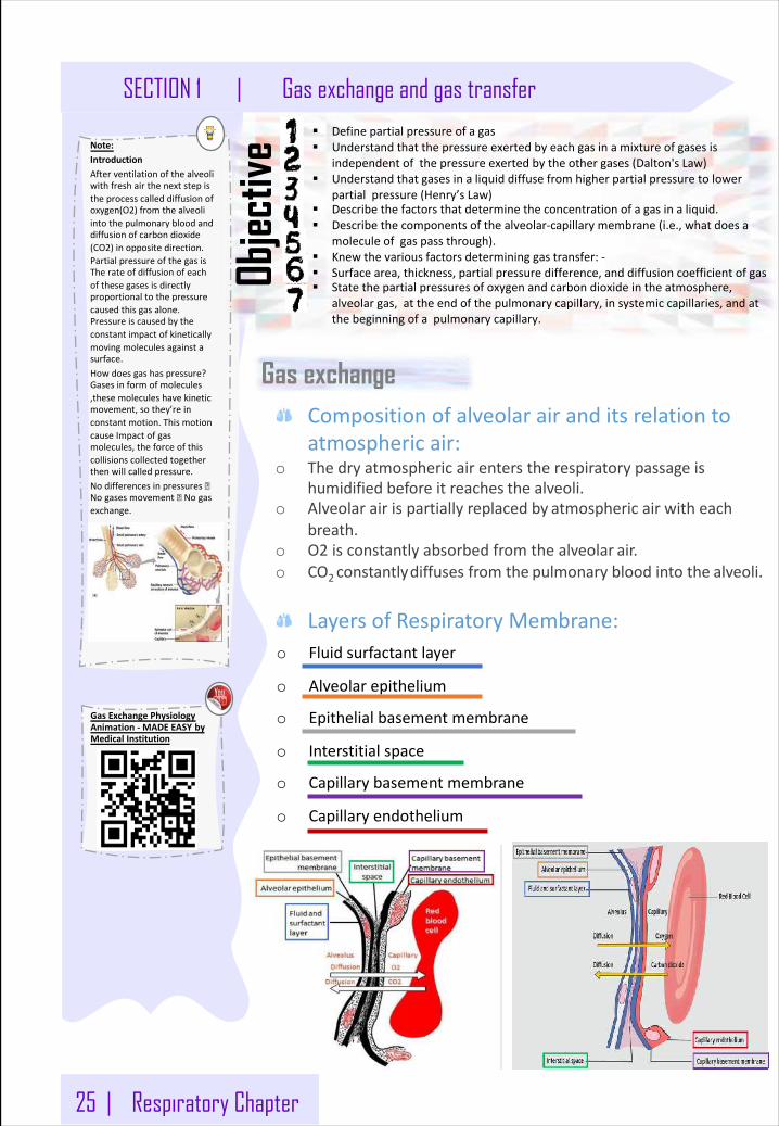
LayersofRespiratoryMembrane:o Fluidsurfactantlayer
o Alveolarepithelium
o Epithelialbasementmembrane
o Interstitialspace
o Capillarybasementmembrane
o Capillaryendothelium
GasExchangePhysiologyAnimation- MADEEASY byMedicalInstitution
Respiratory Chapter | 26
Gas exchange and gas transfer | SECTION 1
RespiratoryUnitAunitconsistingofarespiratorybronchiole,alveolarducts,atria,andalveoli.Thetotalnumberofalveoliinthehumanbodyisaround300million,witheachhavinganaveragediameterof0.2mm. Theextremelythinwalls ofthesealveoliformpartoftherespiratorymembrane,whosethicknessinverselyaffectstherateofgas diffusion.
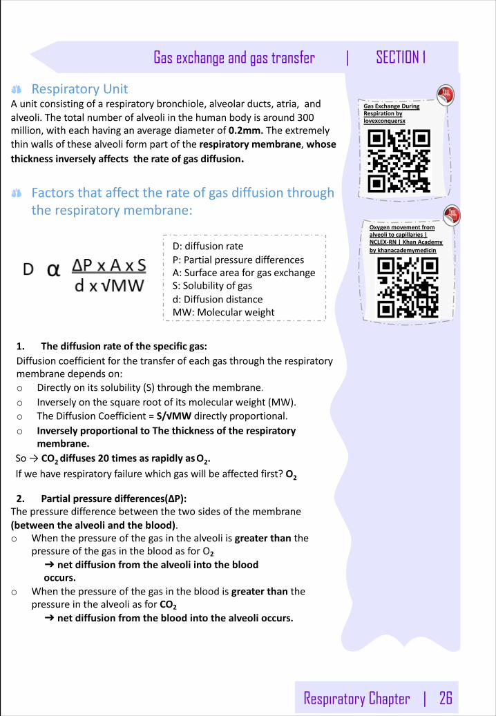
Factorsthataffecttherateofgasdiffusionthroughtherespiratorymembrane:
1. Thediffusionrateofthespecific gas:Diffusioncoefficientforthetransferofeachgasthroughtherespiratorymembranedepends on:o Directlyonitssolubility(S)throughthemembrane.o Inverselyonthesquarerootofitsmolecularweight (MW).o TheDiffusionCoefficient=S/√MWdirectly proportional.o InverselyproportionaltoThethicknessoftherespiratory
membrane.So→ CO2diffuses20 timesasrapidlyasO2.Ifwehaverespiratoryfailurewhichgaswillbeaffectedfirst?O2
2. Partialpressuredifferences(ΔP):Thepressuredifferencebetweenthetwosidesofthemembrane(betweenthealveoliandtheblood).o Whenthepressureofthegasinthealveoliisgreaterthanthe
pressureofthegasinthebloodasforO2
➔ netdiffusionfromthealveoliintothebloodoccurs.
o WhenthepressureofthegasinthebloodisgreaterthanthepressureinthealveoliasforCO2
➔ netdiffusionfromthebloodintothealveolioccurs.
D:diffusion rateP:Partialpressure differencesA:Surfaceareaforgas exchangeS:Solubilityof gasd:Diffusion distanceMW:Molecular weight
GasExchangeDuringRespirationbylovexconquersx
Oxygenmovementfromalveolitocapillaries|NCLEX-RN|KhanAcademybykhanacademymedicin
27 | Respiratory Chapter
Note:Solubility :Increasethesolubilityofgasàincreasethediffusionofit.CO2 is20 timessolublethanO2CO2 morediffusiblethanO2
SECTION 1 | Gas exchange and gas transfer
3. Surfaceareaofthemembrane(A):o Removalofanentirelungdecreasesthesurfaceareatohalf
normal.Range=50-100 m2o Inemphysemawithdissolutionofthealveolarwalldecreases
Surfaceareato5-foldsbecauseoflossofthealveolarwalls.o Increasesurfaceareaà IncreaseDiffusion.Sohowthesurface
areawillDecrease?Inalveoli:1. ByTrypsin.2. ByObstructionofsomebronchiolesorbronchibymucousor
tumor.Inpulmonarycapillaries:1. Bythrombusorbloodclot2. Lossofperfusion3. Lossofventilation
4. Solubility(S):
5. Diffusiondistance(d):Thethicknessoftherespiratorymembrane.- Increasinginthethicknessoftherespiratorymembranee.g.edema
➔ decreasestherateofdiffusion.- Thicknesswilldecreaseduringexercise,thereforetherateof
diffusionincreases.
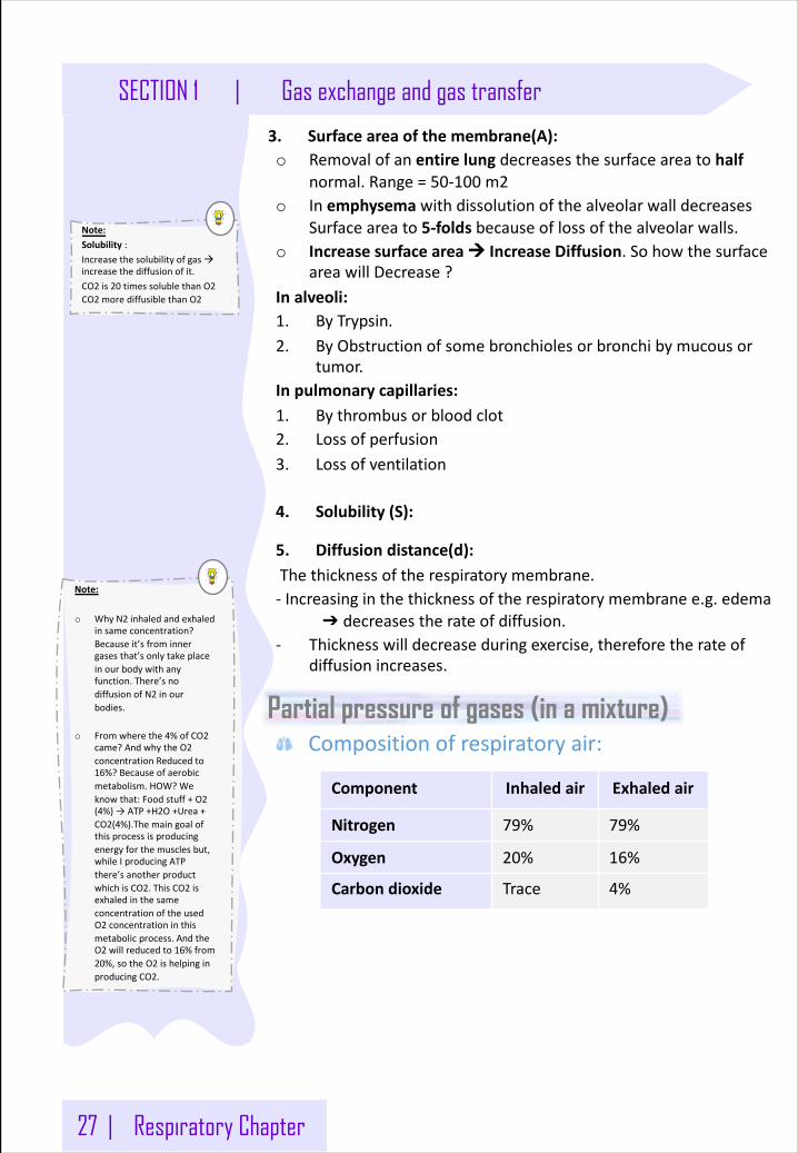
Partial pressure of gases (in a mixture)
Component Inhaledair Exhaledair
Nitrogen 79% 79%
Oxygen 20% 16%
Carbondioxide Trace 4%
Compositionofrespiratoryair:
Note: o WhyN2 inhaledandexhaled
insameconcentration?Becauseit’sfrominnergasesthat’sonlytakeplaceinourbodywithanyfunction.There’snodiffusionofN2 inourbodies.
o Fromwherethe4%ofCO2came?AndwhytheO2concentrationReducedto16%?Becauseofaerobicmetabolism.HOW?Weknowthat:Foodstuff+O2(4%)→ATP+H2O+Urea+CO2(4%).Themaingoalofthisprocessisproducingenergyforthemusclesbut,whileIproducingATPthere’sanotherproductwhichisCO2.ThisCO2 isexhaledinthesameconcentrationoftheusedO2 concentrationinthismetabolicprocess.AndtheO2 willreducedto16%from20%,sotheO2 ishelpinginproducingCO2.
Respiratory Chapter | 28
Gas exchange and gas transfer | SECTION 1
Partialpressureofgases(inamixture):o Inrespiratoryphysiology,thereisamixtureofgasesmainlyofO2,N2,
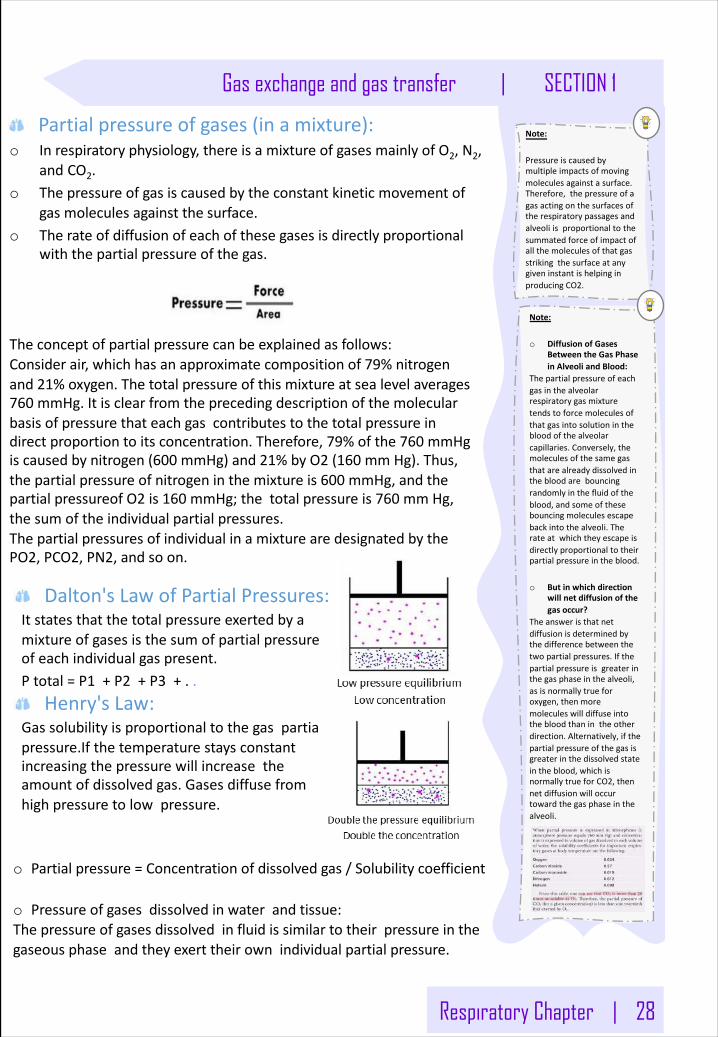
and CO2.o Thepressureofgasiscausedbytheconstantkineticmovementof
gasmoleculesagainstthe surface.o Therateofdiffusionofeachofthesegasesisdirectlyproportional
withthepartialpressureofthegas.
Theconceptofpartialpressurecanbeexplainedasfollows:Considerair,whichhasanapproximatecompositionof79%nitrogenand21%oxygen.Thetotalpressureofthismixtureatsealevelaverages760 mmHg.Itisclearfromtheprecedingdescriptionofthemolecularbasisofpressurethateachgascontributestothetotalpressureindirectproportiontoitsconcentration.Therefore,79%ofthe760 mmHgiscausedbynitrogen(600 mmHg)and21%byO2 (160 mmHg).Thus,thepartialpressureofnitrogeninthemixtureis600 mmHg,andthepartialpressureof O2 is160 mmHg;thetotalpressureis760 mmHg,thesumoftheindividualpartialpressures.ThepartialpressuresofindividualinamixturearedesignatedbythePO2,PCO2,PN2,andsoon.
Note: Pressureiscausedbymultipleimpactsofmovingmoleculesagainstasurface.Therefore,thepressureofagasactingonthesurfacesoftherespiratorypassagesandalveoliisproportionaltothesummatedforceofimpactofallthemoleculesofthatgasstrikingthesurfaceatanygiveninstantishelpinginproducingCO2.
Note: o DiffusionofGases
BetweentheGasPhaseinAlveoliandBlood:
Thepartialpressureofeachgasinthealveolarrespiratorygasmixturetendstoforcemoleculesofthatgasintosolutioninthebloodofthealveolarcapillaries.Conversely,themoleculesofthesamegasthatarealreadydissolvedinthebloodarebouncingrandomlyinthefluidoftheblood,andsomeofthesebouncingmoleculesescapebackintothealveoli.Therateatwhichtheyescapeisdirectlyproportionaltotheirpartialpressureintheblood.
o Butinwhichdirectionwillnetdiffusionofthegasoccur?
Theansweristhatnetdiffusionisdeterminedbythedifferencebetweenthetwopartialpressures.Ifthepartialpressureisgreaterinthegasphaseinthealveoli,asisnormallytrueforoxygen,thenmoremoleculeswilldiffuseintothebloodthanintheotherdirection.Alternatively,ifthepartialpressureofthegasisgreaterinthedissolvedstateintheblood,whichisnormallytrueforCO2,thennetdiffusionwilloccurtowardthegasphaseinthealveoli.
Dalton'sLawofPartialPressures:Itstatesthatthetotalpressureexertedbyamixtureofgasesisthesumofpartialpressureofeachindividualgaspresent.Ptotal=P1 +P2 +P3 +..
Henry'sLaw:Gassolubilityisproportionaltothegaspartialpressure.If thetemperaturestaysconstantincreasingthepressurewillincreasetheamountofdissolvedgas.Gasesdiffusefromhighpressuretolowpressure.
o Partialpressure=Concentrationofdissolvedgas/Solubilitycoefficient
o Pressureofgasesdissolvedinwaterandtissue:Thepressureofgasesdissolvedinfluidissimilartotheirpressureinthegaseousphaseandtheyexerttheirownindividualpartialpressure.
29 | Respiratory Chapter
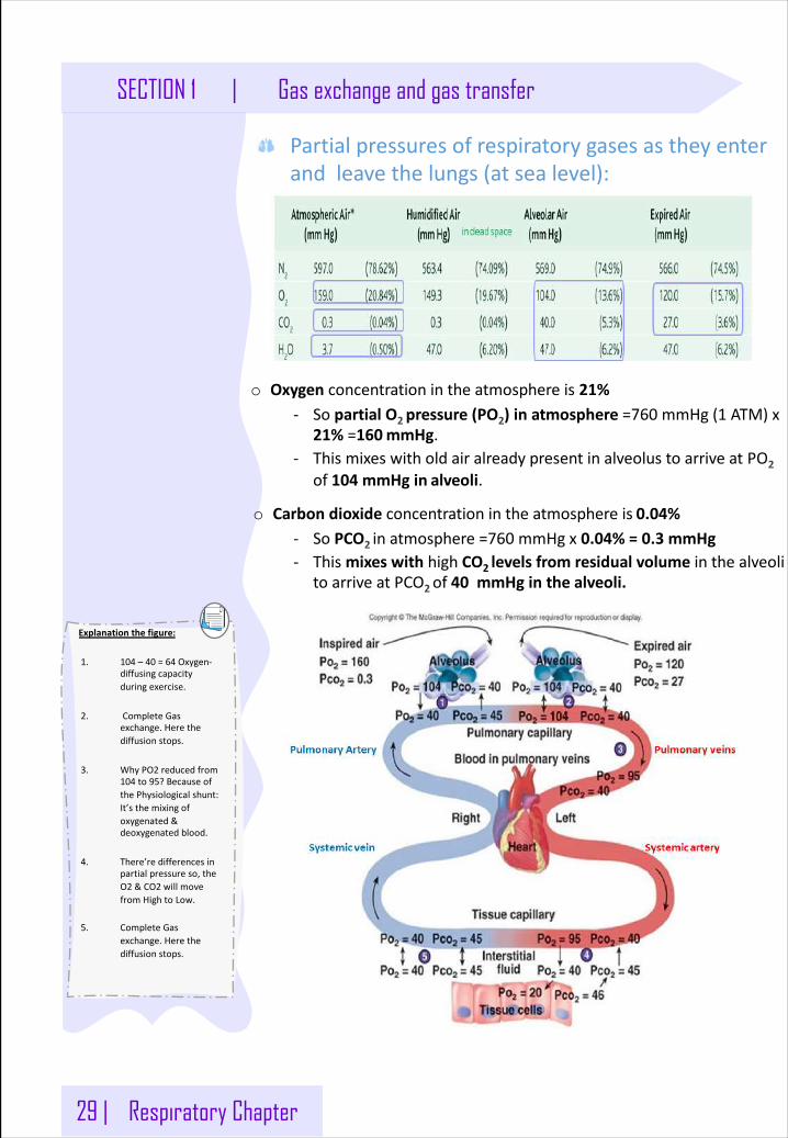
Explanationthefigure:
1. 104 – 40 =64 Oxygen-diffusingcapacityduringexercise.
2. CompleteGasexchange.Herethediffusionstops.
3. WhyPO2 reducedfrom104 to95?BecauseofthePhysiologicalshunt:It’sthemixingofoxygenated&deoxygenatedblood.
4. There’redifferencesinpartialpressureso,theO2 &CO2 willmovefromHightoLow.
5. CompleteGasexchange.Herethediffusionstops.
SECTION 1 | Gas exchange and gas transfer
Partialpressuresofrespiratorygasesastheyenterandleavethelungs(atsealevel):
o Oxygen concentrationintheatmosphereis 21%- SopartialO2 pressure(PO2)inatmosphere=760 mmHg(1 ATM)x
21%=160mmHg.- ThismixeswitholdairalreadypresentinalveolustoarriveatPO2
of104 mmHgin alveoli.
o Carbondioxideconcentrationintheatmosphereis 0.04%- SoPCO2 inatmosphere=760 mmHgx0.04%=0.3 mmHg- ThismixeswithhighCO2 levelsfromresidualvolume inthealveoli
toarriveatPCO2of40 mmHginthe alveoli.
Respiratory Chapter | 30
Gas exchange and gas transfer | SECTION 1
PO2 andPCO2 inair,lungsandtissue:
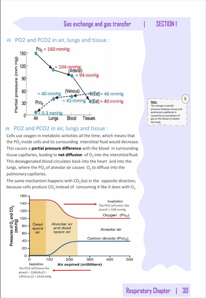
PO2 andPCO2 inair,lungsandtissue:Cellsuseoxygeninmetabolicactivitiesallthetime,whichmeansthatthePO2 insidecellsanditssurroundinginterstitialfluidwould decrease.Thiscausesapartialpressuredifferencewiththebloodinsurroundingtissuecapillaries,leadingtonetdiffusionofO2 intotheinterstitial fluid.Thisdeoxygenatedbloodcirculatesbackintotheheartandintothelungs,wherethePO2 ofalveolaraircausesO2 todiffuseintothepulmonarycapillaries.ThesamemechanismhappenswithCO2 butintheoppositedirection,becausecellsproduceCO2 insteadofconsumingitlikeitdoeswith O2
Note:Thechangeinpartialpressurebetweentissueandpulmonarycapillariesiscausedbyaccumulationofgasasthebloodcirculatesthebody.
31 | Respiratory Chapter
Note:Alllungvolumesandcapacitiesare20-25%lessinwomenthanmen,theyaregreaterinlargeathleticpeoplethaninsmallathleticpeople.
SECTION 1 | Gas exchange and gas transfer
o To summaries: PO2 PCO2
Alveoli 104 mmHg 40 mmHg
Pulmonarycapillaries 40 mmHg 45 mmHg
Tissuecapillaries 95 mmHg 40 mmHg
Interstitialfluid 40 mmHg 45 mmHg
Calls 23 mmHg 46 mmHg
Gasconcentrationsinthealveoli:
o Oxygen Atrestingcondition
Duringexercise
Ventilatorrate
4.2 L/min Mustincrease4 timestomaintainthealveolarPO2 atthenormalvalueof104mmHg(seeA)
Volumeenterthepulmonarycapillaries
250ml/min
1000 ml/min(x4 normalvolume)
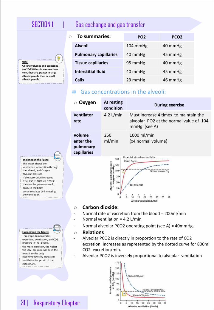
o Carbondioxide:- Normalrateofexcretionfromtheblood=200ml/min- Normalventilation=4.2 L/min- NormalalveolarPCO2 operatingpoint(seeA)=40mmHg.o Relations- AlveolarPCO2 isdirectlyinproportiontotherateofCO2
excretion.Increasesasrepresentedbythedottedcurvefor800mlCO2 excretion/min.
- AlveolarPCO2 isinverselyproportionaltoalveolarventilation
Explanationthefigure:Thisgraphshowstheventilation,absorptionthroughthealveoli,andOxygenalveolarpressure.iftheabsorptionincreasesfrom250 to1000 mlO2/min,thealveolarpressurewoulddrop.sothebodyaccommodatesbyincreasingtheventilation.
Explanationthefigure:Thisgraphdemonstratesexcretion,ventilation,andCO2pressureinthealveoli.themoreexcretion,thehighertheCO2 pressurewillbeinthealveoli.sothebodyaccommodatesbyincreasingventilationtogetridoftheexcessCO2.
GasExchangeandGasTransfer
Thenormallung
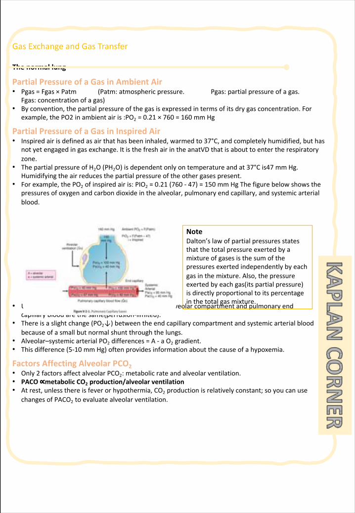
PartialPressureofaGasinAmbientAir• Pgas =Fgas × Patm (Patm:atmosphericpressure.Pgas:partialpressureofagas.
Fgas:concentrationofagas)• Byconvention,thepartialpressureofthegasisexpressedintermsofitsdrygasconcentration.For
example,thePO2 inambientairis:PO2 =0.21 × 760 =160 mmHg
PartialPressureofaGasinInspiredAir• Inspiredairisdefinedasairthathasbeeninhaled,warmedto37°C,andcompletelyhumidified,buthas
notyetengagedingasexchange.ItisthefreshairintheanatVD thatisabouttoentertherespiratoryzone.
• ThepartialpressureofH2O(PH2O) isdependentonlyontemperatureandat37°Cis47 mmHg.Humidifyingtheairreducesthepartialpressureoftheothergasespresent.
• Forexample,thePO2 ofinspiredairis:PIO2 =0.21 (760 - 47)=150 mmHgThefigurebelowshowsthepressuresofoxygenandcarbondioxideinthealveolar,pulmonaryendcapillary,andsystemicarterialblood.
• Undernormalconditions,thePO2 andPCO2 inthealveolarcompartmentandpulmonaryendcapillarybloodarethesame(perfusion-limited).
• Thereisaslightchange(PO2↓)betweentheendcapillarycompartmentandsystemicarterialbloodbecauseofasmallbutnormalshuntthroughthelungs.
• Alveolar–systemicarterialPO2 differences=A- aO2 gradient.• Thisdifference(5-10 mmHg)oftenprovidesinformationaboutthecauseofahypoxemia.
FactorsAffectingAlveolarPCO2• Only2 factorsaffectalveolarPCO2:metabolicrateandalveolarventilation.• PACO∝metabolicCO2 production/alveolarventilation• Atrest,unlessthereisfeverorhypothermia,CO2 productionisrelativelyconstant;soyoucanuse
changesofPACO2 toevaluatealveolarventilation.
NoteDalton’slawofpartialpressuresstatesthatthetotalpressureexertedbyamixtureofgasesisthesumofthepressuresexertedindependentlybyeachgasinthemixture.Also,thepressureexertedbyeachgas(itspartialpressure)isdirectlyproportionaltoitspercentageinthetotalgasmixture.
GasExchangeandGasTransfer
AlveolarVentilationThereisaninverserelationshipbetweenPACO2 andalveolarventilation.ThisisthemainfactoraffectingalveolarPCO2.Therefore,ifventilationincreases,PACO2 decreases;ifventilationdecreases,PACO2increases.
v HyperventilationDuringhyperventilation,thereisaninappropriatelyelevatedlevelofalveolarventilation,andPACO2 isdepressed.IfV•Aisdoubled,thenPACO2 isdecreasedbyhalf.Forexample,PACO2 =40 mmHg2 × V•A;PACO2 =20 mmHg
v HypoventilationDuringhypoventilation,thereisaninappropriatelydepressedlevelofalveolarventilation,andPACO2 iselevated.IfV•Aishalved,thenPACO2 isdoubled.Forexample,PACO2 =40 mmHg1/2 V•A;PACO2 = 80 mm Hg vMetabolicRateThereisadirectrelationshipbetweenalveolarPCO2 andbodymetabolism.ForPaCO2 toremainconstant,changesinbodymetabolismmustbematchedwithequivalentchangesinalveolarventilation.• IfV•Amatchesmetabolism,thenPACO2 remainsconstant.• Forexample,duringexercise,ifbodymetabolismdoubles,thenV•AmustdoubleifPaCO2 isto
remainconstant.• Ifbodytemperaturedecreasesandthereisnochangeinventilation,PaCO2 decreases,andthe
individualcanbeconsideredtobehyperventilating.
FactorsAffectingAlveolarPO2The alveolarairequationincludesallthefactorsthatcanaffectalveolarPO2.PAO2 =(patm - 47 )FiO2 - PACO2 /RQPracticalapplicationoftheequationincludesdifferentialdiagnosisofhypoxemiabyevaluatingthealveolararterial(A–a)gradientofoxygen.Thereare3 factorsthatcanaffectPAO2:1. Patm =atmosphericpressure,atsealevel760 mmHg
Anincreaseinatmosphericpressure(hyperbaricchamber)increasesalveolarPO2,andadecrease(highaltitude)decreasesalveolarPO2.
2. FiO2 =fractionalconcentrationofoxygen,roomair0.21AnincreaseininspiredoxygenconcentrationincreasesalveolarPO2.
3. PaCO2 =alveolarpressureofcarbondioxide,normally40 mmHgAnincreaseinalveolarPCO2 decreasesalveolarPO2,andadecreaseinalveolarPCO2 increasesalveolarPO2.Formostpurposes,youcanusearterialcarbondioxide(PaCO2)inthecalculation.
4. ThefourthvariableisRQ.RQrespiratoryexchangeratio=*+,
./01231 45/478 +, 908:2431 45/478
=normally0.8Forexample,apersonbreathingroomairatsealevelwouldhavePAO2 =(760 - 47)0.21 - 40/0.8 =100 mmHg.
GasExchangeandGasTransfer
EffectofPACO2 onPAO2PIO2 =PinspiredO2,i.e.,thePO2 intheconductingairwaysduringinspiration.BecausePaCO2 affectsalveolarPO2,hyperventilationandhypoventilationalsoaffectPaO2.
v Hyperventilation(e.g.,PaCO2 =20 mmHg)PaO2 =PiO2 - PaCO2 (assumeR=1)normal=150 - 40 =110 mmHghyperventilation=150 - 20 =130 mmHg
v Hypoventilation(e.g.,PaCO2 =80 mmHg)normal=150 - 40 =110 mmHghypoventilation=150 - 80 =70 mmHg
Alveolar–bloodGasTransfer:FickLawofDiffusionSimplediffusionistheprocessofgasexchangebetweenthealveolarcompartmentandpulmonarycapillaryblood.Thus,thosefactorsthataffecttherateofdiffusionalsoaffecttherateofexchangeofO2 andCO2acrossalveolarmembranes.(Anadditionalpointtorememberisthateachgasdiffusesindependently.)V•gas = A/T x Dx (P1-P2) (V•gas =rateofgasdiffusion)
StructuralFeaturesThatAffecttheRateofDiffusionThereare2 structuralfactorsand2 gasfactorsaffecttherateofdiffusion.1. A=surfaceareaforexchange,↓inemphysema,↑inexercise2. T=thicknessofthemembranesbetweenalveolargasandcapillaryblood,↑infibrosisand
manyotherrestrictivediseases
Astructuralprobleminthelungsisanysituationinwhichthereisalossofsur-faceareaand/oranincreaseinthethicknessofthemembranesystembetweenthealveolarairandthepulmonarycapillaryblood.Inallcases,therateofoxy-genandcarbondioxidediffusiondecreases.Thegreaterthestructuralproblem,thegreatertheeffectondiffusionrate.
FactorsSpecifictoEachGasPresentv D(diffusionconstant)=mainfactorissolubilityTheonlyclinicallysignificantfeatureofDissolubility.Themoresolublethegas,thefasteritdiffusesacrossthemembranes.CO2 isthemostsolublegaswithwhichwewillbedealing.ThegreatsolubilityofCO2 isthemainreasonwhyitdiffusesfasteracrossthealveolarmembranesthanO2.
GasExchangeandGasTransfer
v Gradientacrossthemembrane• (P1 - P2):Thisisthegaspartialpressuredifferenceacrossthealveolarmembrane.Thegreaterthepartial
pressuredifference,thegreatertherateofdiffusion.Underrestingconditions,whenbloodfirstentersthepulmonarycapillary,thegradientforO2 is: 100 - 40 = 60 mm Hg
• AnincreaseinthePO2 gradientacrossthelungmembraneshelpscompensateforastructuralproblem.IfsupplementalO2 isadministered,alveolarPO2 increases,becauseoftheelevatedgradient.However,supplementalO2 doesnotimprovetheabilityofthelungstoremoveCO2 fromblood.Thisincreasedgradienthelpsre-turntherateofO2 diffusiontowardnormal.Thegreaterthestructuralproblem,thegreaterthegradientnecessaryforanormalrateofO2 diffusion.
• ThegradientforCO2 is47 - 40 =7 mmHg.• EventhoughthegradientforCO2 islessthanforO2,CO2 stilldiffusesfasterbecauseofitsgreater
solubility.RecallQuestion:WhichofthefollowingfactorsincreasesalveolarPCO2,assumingnocompensation?A.Decreaseinatmosphericpressure(Patm)B.Increaseinfractionalconcentrationofoxygen(FiO2)C.DecreaseincomplianceofalveoliD.IncreaseinthicknessofthemembranesbetweenalveolargasandcapillarybloodE.IncreaseinbodytemperatureAnswer:E
DiffusingCapacityofTheLung
Thereare2 termsthatdescribethedynamicsofthetransferofindividualsub-stancesbetweentheinterstitium andthecapillary:1. Ifthesubstanceequilibratesbetweenthecapillaryandinterstitium,itissaidtobeina
perfusion-limitedsituation.2. Ifthesubstancedoesnotequilibratebetweenthecapillaryandinterstitium,itissaidtobeina
diffusion-limitedsituation.• Carbonmonoxideisauniquegasinthatittypicallydoesn’tequilibratebetweenthealveolarairand
thecapillaryblood.Thus,itisadiffusion-limitedgas.Thisistakenadvantageofclinically,andthemeasurementoftheuptakeofCOinmL/min/mmHgisreferredtoasthediffusingcapacityofthelung(DLCO).
• DLCOisanindexofthelung’sstructuralfeatures.
CarbonMonoxide:AGasThatIsAlwaysDiffusionLimited
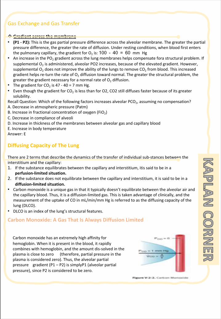
Carbonmonoxidehasanextremelyhighaffinityforhemoglobin.Whenitispresentintheblood,itrapidlycombineswithhemoglobin,andtheamountdis-solvedintheplasmaisclosetozero(therefore,partialpressureintheplasmaisconsideredzero).Thus,thealveolarpartialpressuregradient(P1 – P2)issimplyP1 (alveolarpartialpressure),sinceP2 isconsideredtobezero.
32 | Respiratory Chapter
SECTION 1 | Oxygen and carbon dioxide transport
Hemoglobin
§ Understandtheformsofoxygentransportintheblood,andtheimportanceofeach.
§ differentiatebetweenO2 capacity,O2 contentandO2 saturation.
§ Describetheoxygen-hemoglobindissociationcurve.§ definetheP50 anditssignificance
§ howDPG,temperature,H+ionsandPCO2 affectaffinityofO2 forhemoglobinandthephysiologicalimportanceoftheseeffects.
§ describethethreeformsofcarbondioxidethataretransportedintheblood,andthechlorideshift.
Objec
tive
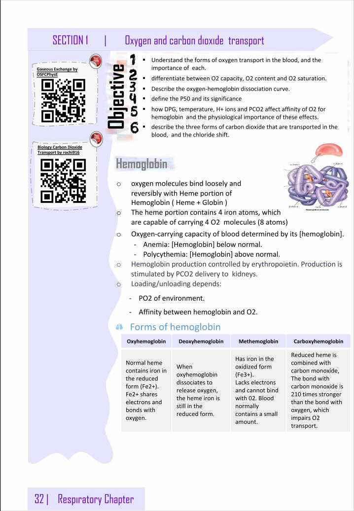
o oxygenmoleculesbindlooselyandreversiblywithHemeportionofHemoglobin(Heme+Globin)
o Thehemeportioncontains4 ironatoms,whicharecapableofcarrying4 O2 molecules(8 atoms)
Oxyhemoglobin Deoxyhemoglobin Methemoglobin Carboxyhemoglobin
Normalhemecontainsironinthereducedform(Fe2+).Fe2+shareselectronsandbondswithoxygen.
Whenoxyhemoglobindissociatestoreleaseoxygen,thehemeironisstillinthereducedform.
Hasironintheoxidizedform(Fe3+).Lackselectronsandcannotbindwith02.Bloodnormallycontainsasmallamount.
Reducedhemeiscombinedwithcarbonmonoxide,Thebondwithcarbonmonoxideis210 timesstrongerthanthebondwithoxygen,whichimpairsO2transport.
Formsofhemoglobin
o Oxygen-carryingcapacityofblooddeterminedbyits[hemoglobin].- Anemia:[Hemoglobin]below normal.- Polycythemia:[Hemoglobin]above normal.
o Hemoglobinproductioncontrolledbyerythropoietin.ProductionisstimulatedbyPCO2 deliverytokidneys.
o Loading/unloadingdepends:
- PO2 ofenvironment.
- AffinitybetweenhemoglobinandO2.
BiologyCarbonDioxideTransportbyrochi916
GaseousExchangebyOSFCPhysE
Respiratory Chapter | 33
Oxygen and carbon dioxide transport | SECTION 1
Explanationthefigure:Thisgraphdemonstratesexcretion,ventilation,andCO2pressureinthealveoli.themoreexcretion,thehighertheCO2 pressurewillbeinthealveoli.sothebodyaccommodatesbyincreasingventilationtogetridoftheexcessCO2.
Transport of O2o PO2 andtheconcentrationgradientplaysimportantfactorwhich
determineshowmuchoxygencombineswithHb whenthehaemoglobin (deoxygenatedHb)isconvertedtoHbO2,
o Mainfunctionofblood:Transportofrespiratorygasesbetweenthelungsandbodytissues.
- IfPO2 ishighAsinpulmonarycapillariesO2 bindstohemoglobinandviceversaresultingreaterHb saturation.
- IfPO2 islowAsinthetissuecapillariesHbreleasesO2resultinlowerHbsaturation
o FormsofOxygeninblood- 97%fromthelungstothetissuesiscarriedinchemical
combinationandgetrapidlydiffusedandbindstohemoglobin.
- 3%isphysicallybeingdissolvedinplasma
Hb+4O2 →Hb(O2)4
TransportofO2 byhemoglobin:o Hbcombineswithoxygenthecompoundformediscalled
oxyhemoglobin,anditdependsontheamountofHbpresentintheblood.
o Oxygencancombinelooselyandreversiblywithhemoglobin.Hb+O2 HbO2
o ThenormalamountofHb inyoungadultsisabout16 gm/dloftheblood.EachgramofHb canbindwith1.34 mlofO2 .Thus,16 x1.34 =21.44 mlofO2 /dl.
PartialPressureDifference:1. HighPartialPressureofO2 (Po2)inAlveoli2. LowPo2 inCapillaryo TransportO2:DiffusionDifference-VeryShort→O2 Diffusion-VeryRapid→O2DiffusesfromAlveoliIntoRBC→(AttachestoHemeMolecule)→CarriedToTissueso ConcentrationGradient- HighConcentrationofO2 inAlveoli- LowConcentrationofO2 inCapillaryO2
34 | Respiratory Chapter
SECTION 1 | Oxygen and carbon dioxide transport
Transportofoxygeninarterialblood:Whenblood
is100%saturatedwithO2
Whenbloodis97%
saturatedInvenous blood
Duringstrenuousexercise
NoteseachgramofHbcarry1.34 ml O2
97%of 100%saturation
Amountofoxygenreleasedfromthehemoglobintothetissuesis5mlO2 pereach100mlblood.
Duringstrenuousexercisetheoxygenuptakebythetissueincreases3 folds
5mlx3 folds=15mlO2 isgiven/100 mlblood
Calculations
HbxO2
15gx1.34
0.97 x 20
O2contentin97%saturation—oxygenreleasedtotissue=19.4 — 5
O2contentin97%saturation—oxygenreleasedtotissueduringstrenuousexercise=19.4 — 15
O2 content 20ml. 19.4ml. 14.4ml. 4.4 ml.
O2 capacity,contentandsaturation:O2 content
O2-bindingcapacity
Percent saturation Dissolved O2
AmountofO2 inblood(mlO2/100
mlblood)
MaximumamountofO2boundto
hemoglobin(mlO2/100 mlblood)measuredat100%
saturation
100 %ofhemegroupsboundtoO2
%saturationofHb =Oxygencontent X100Oxygencapacity
UnboundO2 inblood(mlO2 /100mlblood)
Transportofoxygeninthedissolvedstate:1. AtnormalarterialPO2=95 mmHg.
- 95 x0.003 (Thesolubility ofO2 inblood is 0.003 mL O2/100mL/mmHg(SOLUBILITYfactor)).
o 0.29 mlofoxygenisdissolvedineach100mlofblood.2. WhenthePO2 ofthebloodfallsto40 mmHgintissuecapillaries.
- 40 x0.003.o 0.12 ofoxygenremainsdissolved.➔ Calculation:fromhenry’slaw
o 0.17 mlofoxygenisnormallytransportedinthedissolvedstatetothetissuespereach100 mlofblood.
Partial pressure = Concentration of dissolved gas
Solubility coefficient
concentration of dissolved gas = partial pressure × solubility factor
Respiratory Chapter | 35
Oxygen and carbon dioxide transport | SECTION 1
CO2 transporto LargeamountofCO2 iscontinuouslyproducedinthebody.o Intherestingstate,4 mlCO2iscarriedtothelungper100 mlofblood.o CO2 iscarriedinthebloodin3 differentforms:1. 70%ofCO2 istransportedinBicarbonateform.2. 7%directlydissolvedinplasma3. 23%ofCO2 bindswithdeoxyhemoglobinintheRBC(globingpart)to
formcarbamino hemoglobin.Oncethebloodreachesthepulmonarycapillaries,theCO2 detachesfromthehemoglobinanddiffusesintothealveoli.
FactorsaffectingCO2diffusion:1. PartialPressureofCO2 (Pco2)-HigherInTissuesThanInCapillary2. ConcentrationGradient-CO2 HigherInTissuesThanInCapillary3. Distance-VeryShort
Bicarbonateform:1. CO2 isdiffusedfromthetissuetotheRBC.2. CO2 reactswithH2Ointhepresenceofcarbonicanhydrase(speeds
uptheprocess)toformcarbonicacid.3. Carbonicacidisthendissociatedintohydrogenions(bondwithHb
toformHHb)andbicarbonateions.4. Bicarbonateionsgoesintotheplasma,andchloride(Cl)ionstake
itsplacewithinthecelltomaintainnegativity.(chlorideshift)5. Bloodistransportedtothepulmonarycapillaries.Thenthe
bicarbonateionsswitchplaceswiththechlorideionspresentwithintheRBC.
6. ThebicarbonateionsreactwiththeH+ ionstoformCO2 andH2OwithintheRBC.
TransportofCO2 dissolvedinplasma:1. Littlecarbondioxideistransportedinthedissolvedstatetothelungs.2. PCO2 ofvenousbloodis45 mmHgandthePCO2 ofarterialbloodis40
mmHg.3. TheamountofCO2 dissolvedinthebloodat45 mmHgis2.7 ml/dl
(2.7%).4. TheamountofCO2 dissolvedat40 mmHgisabout2.4 ml5. Thedifferencebetween2.7 and2.4 isonly0.3 ml.6. About0.3 mlCO2 istransportedintheformofdissolvedCO2 byeach
100 mlofblood.7. Itisabout7 %ofallCO2 istransportedinthisform.
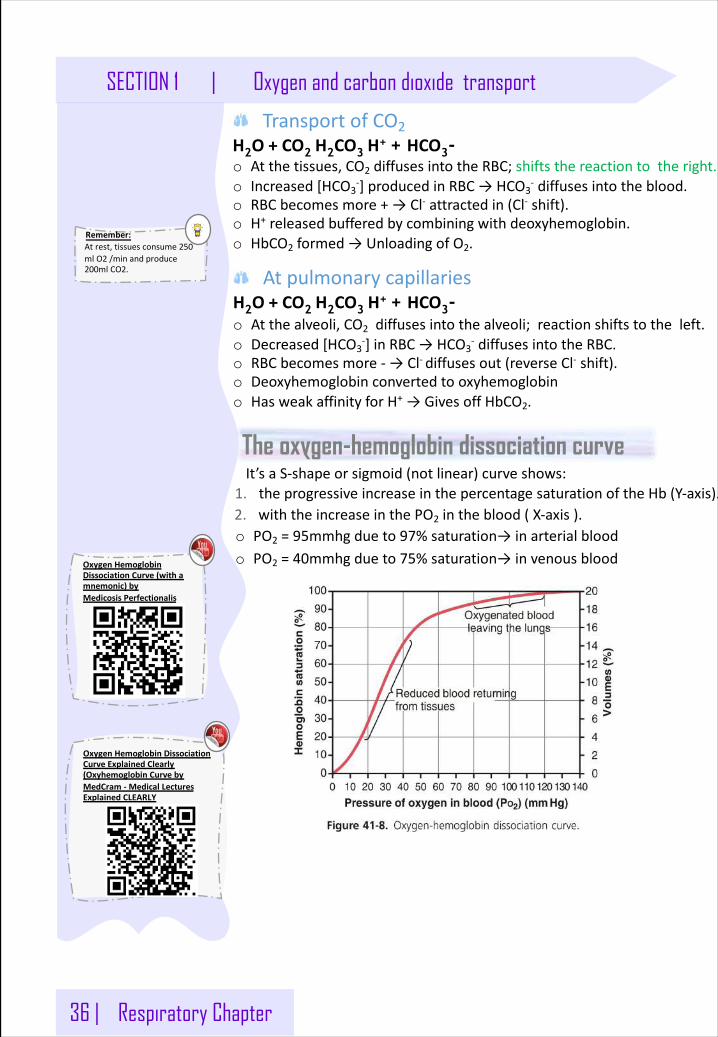
36 | Respiratory Chapter
SECTION 1 | Oxygen and carbon dioxide transport
Remember:Atrest,tissuesconsume250mlO2 /minandproduce200mlCO2.
TransportofCO2H2O+CO2 H2CO3 H+ + HCO3- o Atthetissues,CO2 diffusesintotheRBC;shiftsthereactiontotheright.o Increased[HCO3
-]producedinRBC→HCO3- diffusesintotheblood.
o RBCbecomesmore+→Cl- attractedin(Cl- shift).o H+ releasedbufferedbycombiningwithdeoxyhemoglobin.o HbCO2 formed→UnloadingofO2.
AtpulmonarycapillariesH2O+CO2 H2CO3 H+ + HCO3-o Atthealveoli,CO2 diffusesintothealveoli;reactionshiftstotheleft.o Decreased[HCO3
-]inRBC→HCO3- diffusesintotheRBC.
o RBCbecomesmore- →Cl- diffusesout(reverseCl- shift).o Deoxyhemoglobinconvertedtooxyhemoglobino HasweakaffinityforH+ →GivesoffHbCO2.
The oxygen-hemoglobin dissociation curveIt’saS-shapeorsigmoid(notlinear)curveshows:
1. theprogressiveincreaseinthepercentagesaturationoftheHb (Y-axis).2. withtheincreaseinthePO2 intheblood(X-axis).o PO2 =95mmhgdueto97%saturation→inarterialbloodo PO2 =40mmhgdueto75%saturation→invenousblood
OxygenHemoglobinDissociationCurveExplainedClearly(OxyhemoglobinCurvebyMedCram - MedicalLecturesExplainedCLEARLY
OxygenHemoglobinDissociationCurve(withamnemonic)byMedicosis Perfectionalis
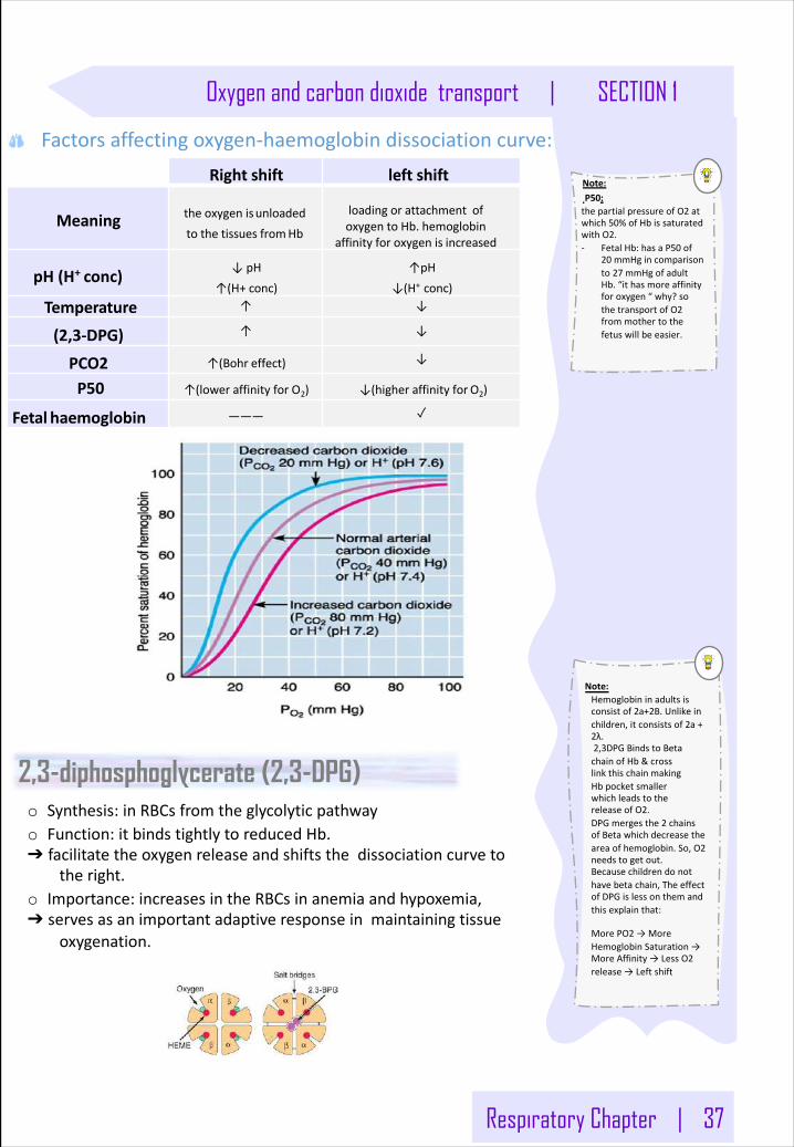
Respiratory Chapter | 37
Oxygen and carbon dioxide transport | SECTION 1
Factorsaffectingoxygen-haemoglobin dissociationcurve:Right shift left shift
Meaning theoxygenisunloadedtothetissuesfromHb
loadingorattachmentofoxygentoHb.hemoglobin
affinityforoxygenis increased
pH(H+ conc) ↓pH↑(H+conc)
↑pH↓(H+ conc)
Temperature ↑ ↓
(2,3-DPG) ↑ ↓
PCO2 ↑(Bohr effect) ↓
P50 ↑(lower affinity for O2) ↓(higheraffinityfor O2)
Fetal haemoglobin ——— ✓
Note:P50:thepartialpressureofO2 atwhich50%ofHb issaturatedwithO2.- FetalHb:hasaP50 of
20 mmHgincomparisonto27 mmHgofadultHb.“ithasmoreaffinityforoxygen“why?sothetransportofO2frommothertothefetuswillbeeasier.
2,3-diphosphoglycerate (2,3-DPG)o Synthesis:inRBCsfromtheglycolyticpathwayo Function:itbindstightlytoreducedHb.➔ facilitatetheoxygenreleaseandshiftsthedissociationcurveto
theright.o Importance:increasesintheRBCsinanemiaandhypoxemia,➔ servesasanimportantadaptiveresponseinmaintainingtissue
oxygenation.
Note:Hemoglobininadultsisconsistof2a+2B.Unlikeinchildren,itconsistsof2a+2λ.2,3DPGBindstoBetachainofHb &crosslinkthischainmakingHb pocketsmallerwhichleadstothereleaseofO2.DPGmergesthe2 chainsofBetawhichdecreasetheareaofhemoglobin.So,O2needstogetout.Becausechildrendonothavebetachain,TheeffectofDPGislessonthemandthisexplainthat:
MorePO2 →MoreHemoglobinSaturation→MoreAffinity→LessO2release→Leftshift
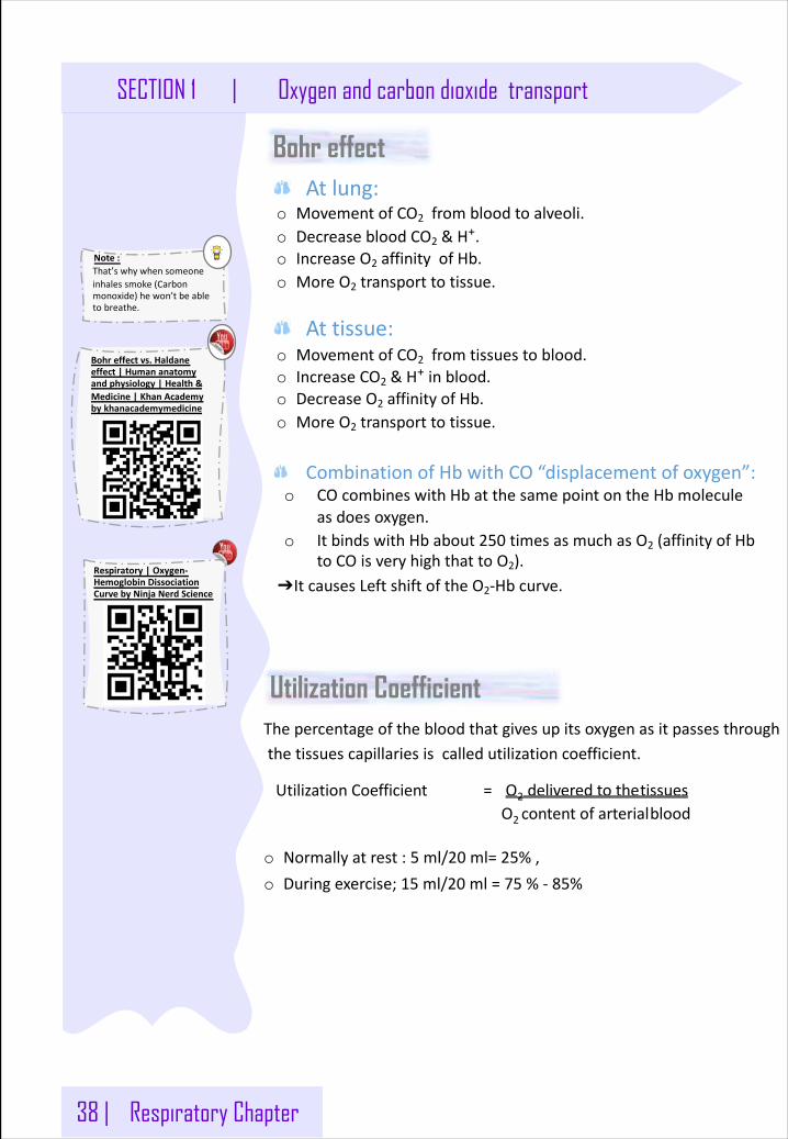
38 | Respiratory Chapter
SECTION 1 | Oxygen and carbon dioxide transport
Note:That’swhywhensomeoneinhalessmoke(Carbonmonoxide)hewon’tbeabletobreathe.
Bohr effectAtlung:
o MovementofCO2 frombloodtoalveoli.o DecreasebloodCO2 &H+.
o IncreaseO2 affinityofHb.o MoreO2 transporttotissue.
Attissue:o MovementofCO2 fromtissuestoblood.o IncreaseCO2 &H+ inblood.
o DecreaseO2 affinityofHb.o MoreO2 transporttotissue.
CombinationofHbwithCO“displacementofoxygen”:o COcombineswithHb atthesamepointontheHb molecule
asdoesoxygen.o ItbindswithHb about250 timesasmuchasO2 (affinityofHb
toCOisveryhighthattoO2).➔ItcausesLeftshiftoftheO2-Hbcurve.
Utilization CoefficientThepercentageofthebloodthatgivesupitsoxygenasitpassesthroughthetissuescapillariesiscalledutilizationcoefficient.
o Normallyatrest:5 ml/20 ml=25%,o Duringexercise;15 ml/20 ml=75 %- 85%
UtilizationCoefficient = O2 deliveredtothetissuesO2 contentofarterialblood
Respiratory|Oxygen-HemoglobinDissociationCurvebyNinjaNerdScience
Bohreffectvs.Haldaneeffect|Humananatomyandphysiology|Health&Medicine|KhanAcademybykhanacademymedicine
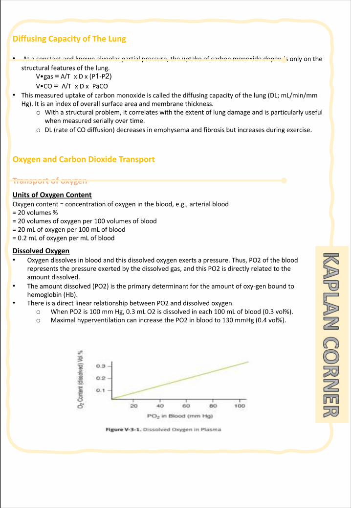
DiffusingCapacityofTheLung
• Ataconstantandknownalveolarpartialpressure,theuptakeofcarbonmonoxidedependsonlyonthestructuralfeaturesofthelung.
V•gas = A/T x Dx (P1-P2) V•CO = A/T x Dx PaCO
• Thismeasureduptakeofcarbonmonoxideiscalledthediffusingcapacityofthelung(DL;mL/min/mmHg).Itisanindexofoverallsurfaceareaandmembranethickness.
o Withastructuralproblem,itcorrelateswiththeextentoflungdamageandisparticularlyusefulwhenmeasuredseriallyovertime.
o DL(rateofCOdiffusion)decreasesinemphysemaandfibrosisbutincreasesduringexercise.
OxygenandCarbonDioxideTransport
TransportofoxygenUnitsofOxygenContentOxygencontent=concentrationofoxygenintheblood,e.g.,arterialblood=20 volumes%=20 volumesofoxygenper100 volumesofblood=20 mLofoxygenper100 mLofblood=0.2 mLofoxygenpermLofblood
DissolvedOxygen• Oxygendissolvesinbloodandthisdissolvedoxygenexertsapressure.Thus,PO2 oftheblood
representsthepressureexertedbythedissolvedgas,andthisPO2 isdirectlyrelatedtotheamountdissolved.
• Theamountdissolved(PO2)istheprimarydeterminantfortheamountofoxy-genboundtohemoglobin(Hb).
• ThereisadirectlinearrelationshipbetweenPO2 anddissolvedoxygen.o WhenPO2 is100 mmHg,0.3 mLO2 isdissolvedineach100 mLofblood(0.3 vol%).o MaximalhyperventilationcanincreasethePO2 inbloodto130 mmHg(0.4 vol%).
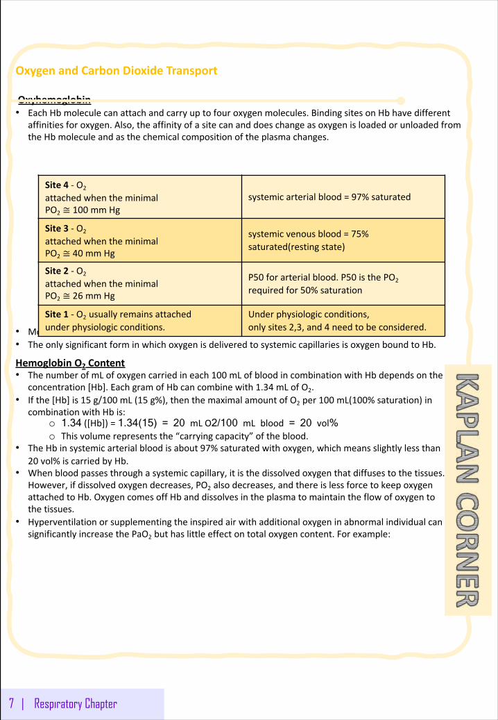
OxygenandCarbonDioxideTransport
Oxyhemoglobin• EachHbmoleculecanattachandcarryuptofouroxygenmolecules.BindingsitesonHbhavedifferent
affinitiesforoxygen.Also,theaffinityofasitecananddoeschangeasoxygenisloadedorunloadedfromtheHbmoleculeandasthechemicalcompositionoftheplasmachanges.
• MostoftheoxygeninsystemicarterialbloodisoxygenattachedtoHb.• TheonlysignificantforminwhichoxygenisdeliveredtosystemiccapillariesisoxygenboundtoHb.
HemoglobinO2 Content• ThenumberofmLofoxygencarriedineach100 mLofbloodincombinationwithHbdependsontheHb
concentration[Hb].EachgramofHbcancombinewith1.34 mLofO2.• Ifthe[Hb]is15 g/100 mL(15 g%),thenthemaximalamountofO2 per100 mL(100%saturation)in
combinationwithHbis:o 1.34 ([Hb])=1.34(15) = 20 mL O2/100 mL blood = 20 vol% o Thisvolumerepresentsthe“carryingcapacity”oftheblood.
• TheHbinsystemicarterialbloodisabout97%saturatedwithoxygen,whichmeansslightlylessthan20 vol%iscarriedbyHb.
• Whenbloodpassesthroughasystemiccapillary,itisthedissolvedoxygenthatdiffusestothetissues.However,ifdissolvedoxygendecreases,PO2 alsodecreases,andthereislessforcetokeepoxygenattachedtoHb.OxygencomesoffHbanddissolvesintheplasmatomaintaintheflowofoxygentothetissues.
• HyperventilationorsupplementingtheinspiredairwithadditionaloxygeninabnormalindividualcansignificantlyincreasethePaO2 buthaslittleeffectontotaloxygencontent.Forexample:
7 | Respiratory Chapter
Site4 - O2
attachedwhentheminimalPO2 ≅ 100 mmHg
systemicarterialblood=97%saturated
Site3 - O2
attachedwhentheminimalPO2 ≅ 40 mmHg
systemicvenousblood=75%saturated(restingstate)
Site2 - O2
attachedwhentheminimalPO2 ≅ 26 mmHg
P50 forarterialblood.P50 isthePO2
requiredfor50%saturation
Site1 - O2 usuallyremainsattachedunderphysiologicconditions.
Underphysiologicconditions,onlysites2,3,and4 needtobeconsidered.
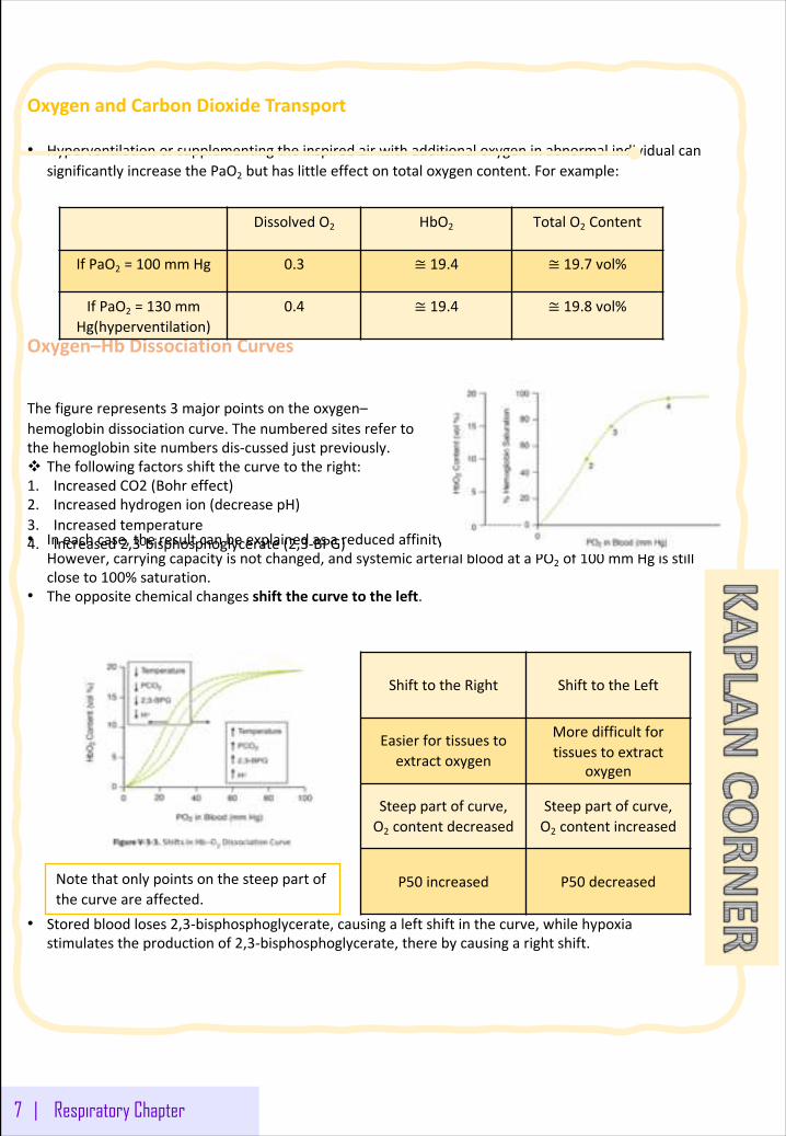
OxygenandCarbonDioxideTransport
• HyperventilationorsupplementingtheinspiredairwithadditionaloxygeninabnormalindividualcansignificantlyincreasethePaO2 buthaslittleeffectontotaloxygencontent.Forexample:
Oxygen–HbDissociationCurves
• Ineachcase,theresultcanbeexplainedasareducedaffinityoftheHbmoleculeforoxygen.However,carryingcapacityisnotchanged,andsystemicarterialbloodataPO2 of100 mmHgisstillcloseto100%saturation.
• Theoppositechemicalchangesshiftthecurvetotheleft.
• Storedbloodloses2,3-bisphosphoglycerate,causingaleftshiftinthecurve,whilehypoxiastimulatestheproductionof2,3-bisphosphoglycerate,therebycausingarightshift.
7 | Respiratory Chapter
DissolvedO2 HbO2 TotalO2 Content
IfPaO2 =100 mmHg 0.3 ≅ 19.4 ≅ 19.7 vol%
IfPaO2 =130 mmHg(hyperventilation)
0.4 ≅ 19.4 ≅ 19.8 vol%
Thefigurerepresents3 majorpointsontheoxygen–hemoglobindissociationcurve.Thenumberedsitesrefertothehemoglobinsitenumbersdis-cussedjustpreviously.v Thefollowingfactorsshiftthecurvetotheright:1. IncreasedCO2 (Bohreffect)2. Increasedhydrogenion(decreasepH)3. Increasedtemperature4. Increased2,3-bisphosphoglycerate(2,3-BPG)
Notethatonlypointsonthesteeppartofthecurveareaffected.
ShifttotheRight ShifttotheLeft
Easierfortissuestoextractoxygen
Moredifficultfortissuestoextract
oxygen
Steeppartofcurve,O2 contentdecreased
Steeppartofcurve,O2 contentincreased
P50 increased P50 decreased
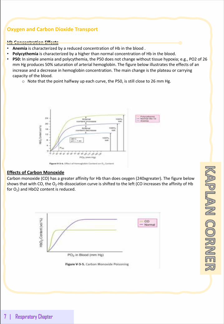
OxygenandCarbonDioxideTransport
HbConcentrationEffects• Anemia ischaracterizedbyareducedconcentrationofHbintheblood.• Polycythemia ischaracterizedbyahigherthannormalconcentrationofHbintheblood.• P50:Insimpleanemiaandpolycythemia,theP50 doesnotchangewithouttissuehypoxia;e.g.,PO2 of26
mmHgproduces50%saturationofarterialhemoglobin.Thefigurebelowillustratestheeffectsofanincreaseandadecreaseinhemoglobinconcentration.Themainchangeistheplateauorcarryingcapacityoftheblood.
o Notethatthepointhalfwayupeachcurve,theP50,isstillcloseto26 mmHg.
EffectsofCarbonMonoxideCarbonmonoxide(CO)hasagreateraffinityforHbthandoesoxygen(240xgreater).ThefigurebelowshowsthatwithCO,theO2-Hbdissociationcurveisshiftedtotheleft(COincreasestheaffinityofHbforO2)andHbO2 contentisreduced.
7 | Respiratory Chapter
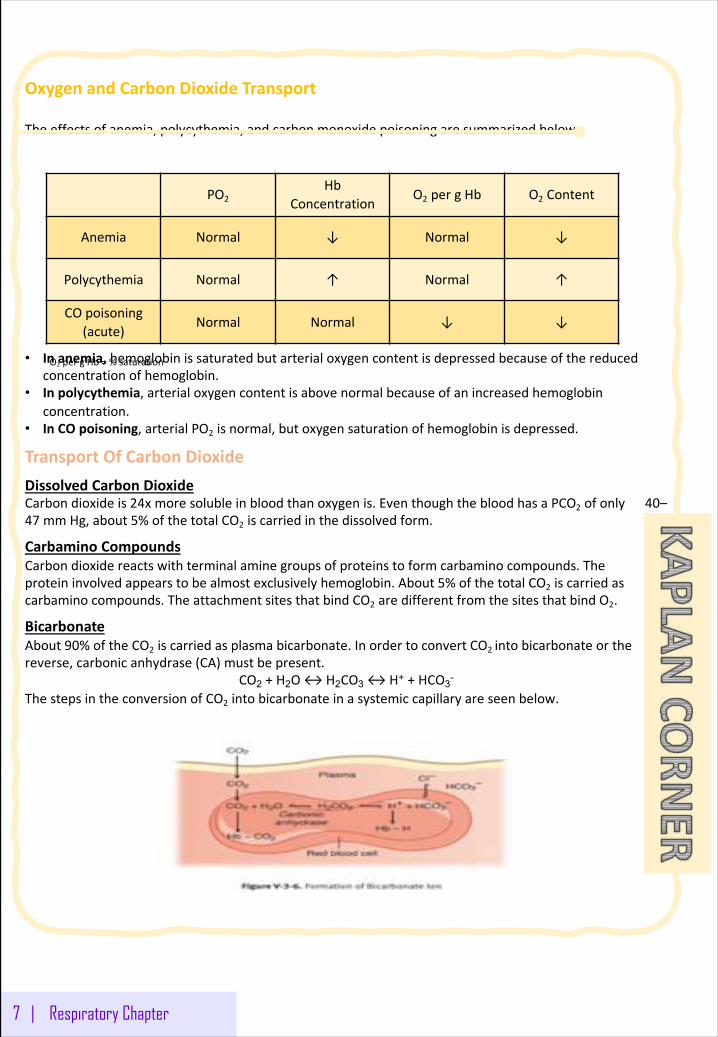
OxygenandCarbonDioxideTransport
Theeffectsofanemia,polycythemia,andcarbonmonoxidepoisoningaresummarizedbelow.
• Inanemia,hemoglobinissaturatedbutarterialoxygencontentisdepressedbecauseofthereducedconcentrationofhemoglobin.
• Inpolycythemia,arterialoxygencontentisabovenormalbecauseofanincreasedhemoglobinconcentration.
• InCOpoisoning,arterialPO2 isnormal,butoxygensaturationofhemoglobinisdepressed.
TransportOfCarbonDioxideDissolvedCarbonDioxideCarbondioxideis24xmoresolubleinbloodthanoxygenis.EventhoughthebloodhasaPCO2 ofonly40–47 mmHg,about5%ofthetotalCO2 iscarriedinthedissolvedform.
Carbamino CompoundsCarbondioxidereactswithterminalaminegroupsofproteinstoformcarbamino compounds.Theproteininvolvedappearstobealmostexclusivelyhemoglobin.About5%ofthetotalCO2 iscarriedascarbamino compounds.TheattachmentsitesthatbindCO2 aredifferentfromthesitesthatbindO2.
BicarbonateAbout90%oftheCO2 iscarriedasplasmabicarbonate.InordertoconvertCO2 intobicarbonateorthereverse,carbonicanhydrase(CA)mustbepresent.
CO2 +H2O↔H2CO3 ↔H+ +HCO3-
ThestepsintheconversionofCO2 intobicarbonateinasystemiccapillaryareseenbelow.
7 | Respiratory Chapter
PO2Hb
ConcentrationO2 pergHb O2 Content
Anemia Normal ↓ Normal ↓
Polycythemia Normal ↑ Normal ↑
COpoisoning(acute)
Normal Normal ↓ ↓
O2 pergHb=%saturation
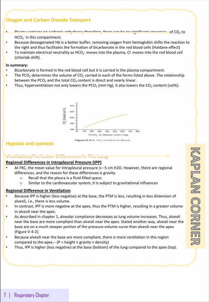
OxygenandCarbonDioxideTransport
• Plasmacontainsnocarbonicanhydrase;therefore,therecanbenosignificantconversionofCO2 toHCO3
- inthiscompartment.• BecausedeoxygenatedHbisabetterbuffer,removingoxygenfromhemoglobinshiftsthereactionto
therightandthusfacilitatestheformationofbicarbonateintheredbloodcells(Haldaneeffect)• TomaintainelectricalneutralityasHCO3
- movesintotheplasma,Cl- movesintotheredbloodcell(chlorideshift).
Insummary:• Bicarbonateisformedintheredbloodcellbutitiscarriedintheplasmacompartment.• ThePCO2 determinesthevolumeofCO2 carriedineachoftheformslistedabove.Therelationship
betweenthePCO2 andthetotalCO2 contentisdirectandnearlylinear.• Thus,hyperventilationnotonlylowersthePCO2 (mmHg),italsolowerstheCO2 content(vol%).
Hypoxiaandcyanosis
Ventilation/PerfusionDifferencesInTheLungRegionalDifferencesinIntrapleural Pressure(IPP)• AtFRC,themeanvalueforIntrapleural pressureis–5 cmH2O.However,thereareregional
differences,andthereasonforthesedifferencesisgravity.o Recallthatthepleuraisafluid-filledspace.o Similartothecardiovascularsystem,itissubjecttogravitationalinfluences
RegionalDifferenceinVentilation• BecauseIPPishigher(lessnegative)atthebase,thePTMisless,resultinginlessdistensionof
alveoli,i.e.,thereislessvolume.• Incontrast,IPPismorenegativeattheapex,thusthePTMishigher,resultinginagreatervolume
inalveolineartheapex.• Asdescribedinchapter1,alveolarcompliancedecreasesaslungvolumeincreases.Thus,alveoli
nearthebasearemorecompliantthanalveolineartheapex.Statedanotherway,alveolinearthebaseareonamuchsteeperportionofthepressure-volumecurvethanalveolineartheapex(FigureV-4-2).
• Becausealveolinearthebasearemorecompliant,thereismoreventilationinthisregioncomparedtotheapex.– (P=height× gravity× density)
• Thus,IPPishigher(lessnegative)atthebase(bottom)ofthelungcomparedtotheapex(top).
7 | Respiratory Chapter
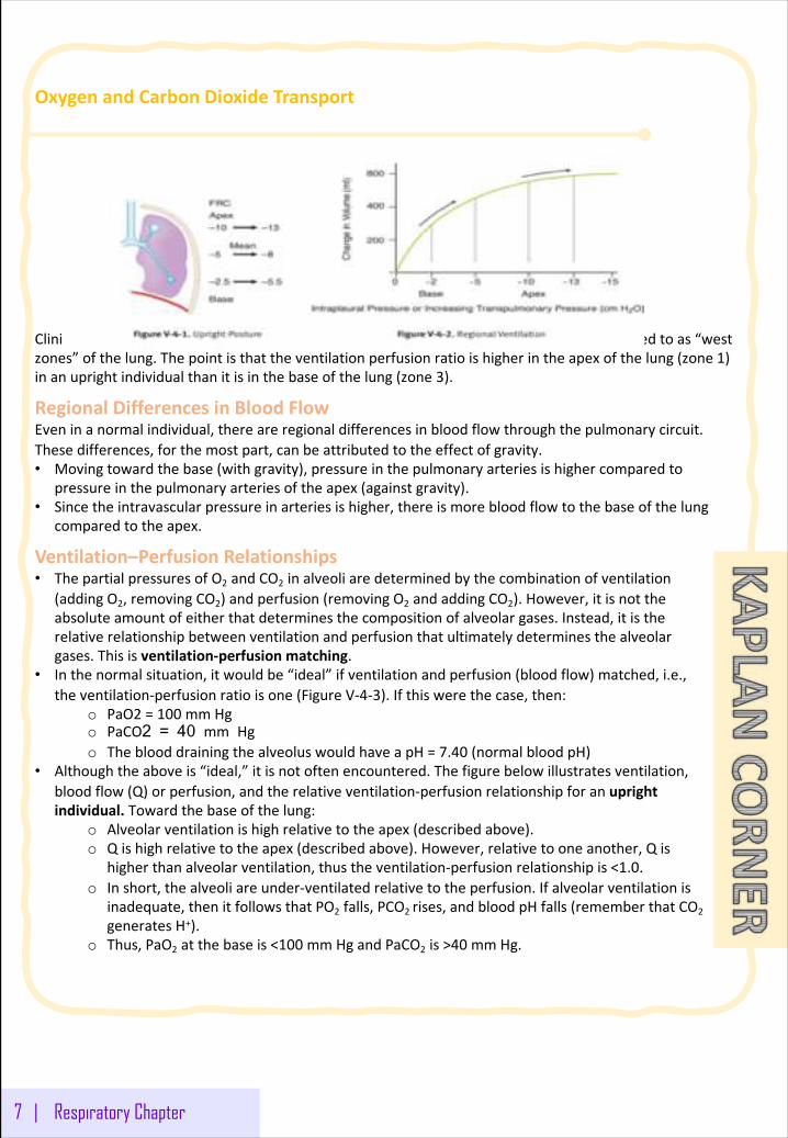
OxygenandCarbonDioxideTransport
ClinicalCorrelateTheregionaldifferenceofalveolarandarterialpressureinthelungisreferredtoas“westzones”ofthelung.Thepointisthattheventilationperfusionratioishigherintheapexofthelung(zone1)inanuprightindividualthanitisinthebaseofthelung(zone3).
RegionalDifferencesinBloodFlowEveninanormalindividual,thereareregionaldifferencesinbloodflowthroughthepulmonarycircuit.Thesedifferences,forthemostpart,canbeattributedtotheeffectofgravity.• Movingtowardthebase(withgravity),pressureinthepulmonaryarteriesishighercomparedto
pressureinthepulmonaryarteriesoftheapex(againstgravity).• Sincetheintravascularpressureinarteriesishigher,thereismorebloodflowtothebaseofthelung
comparedtotheapex.
Ventilation–PerfusionRelationships• ThepartialpressuresofO2 andCO2 inalveoliaredeterminedbythecombinationofventilation
(addingO2,removingCO2)andperfusion(removingO2 andaddingCO2).However,itisnottheabsoluteamountofeitherthatdeterminesthecompositionofalveolargases.Instead,itistherelativerelationshipbetweenventilationandperfusionthatultimatelydeterminesthealveolargases.Thisisventilation-perfusionmatching.
• Inthenormalsituation,itwouldbe“ideal”ifventilationandperfusion(bloodflow)matched,i.e.,theventilation-perfusionratioisone(FigureV-4-3).Ifthiswerethecase,then:
o PaO2 =100 mmHgo PaCO2 = 40 mm Hg o TheblooddrainingthealveoluswouldhaveapH=7.40 (normalbloodpH)
• Althoughtheaboveis“ideal,”itisnotoftenencountered.Thefigurebelowillustratesventilation,bloodflow(Q)orperfusion,andtherelativeventilation-perfusionrelationshipforanuprightindividual.Towardthebaseofthelung:
o Alveolarventilationishighrelativetotheapex(describedabove).o Qishighrelativetotheapex(describedabove).However,relativetooneanother,Qis
higherthanalveolarventilation,thustheventilation-perfusionrelationshipis<1.0.o Inshort,thealveoliareunder-ventilatedrelativetotheperfusion.Ifalveolarventilationis
inadequate,thenitfollowsthatPO2 falls,PCO2 rises,andbloodpHfalls(rememberthatCO2generatesH+).
o Thus,PaO2 atthebaseis<100 mmHgandPaCO2 is>40 mmHg.
7 | Respiratory Chapter
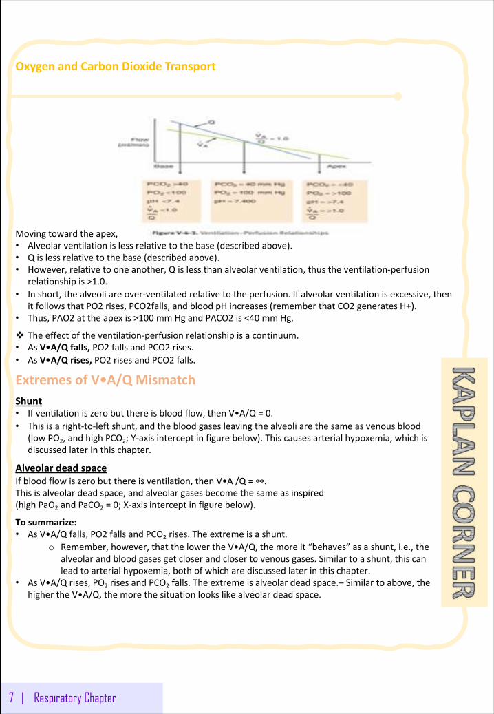
OxygenandCarbonDioxideTransport
Movingtowardtheapex,thesituationreverses:• Alveolarventilationislessrelativetothebase(describedabove).• Qislessrelativetothebase(describedabove).• However,relativetooneanother,Qislessthanalveolarventilation,thustheventilation-perfusion
relationshipis>1.0.• Inshort,thealveoliareover-ventilatedrelativetotheperfusion.Ifalveolarventilationisexcessive,then
itfollowsthatPO2 rises,PCO2falls,andbloodpHincreases(rememberthatCO2 generatesH+).• Thus,PAO2 attheapexis>100 mmHgandPACO2 is<40 mmHg.
v Theeffectoftheventilation-perfusionrelationshipisacontinuum.• AsV•A/Qfalls,PO2 fallsandPCO2 rises.• AsV•A/Qrises,PO2 risesandPCO2 falls.
ExtremesofV•A/QMismatchShunt• Ifventilationiszerobutthereisbloodflow,thenV•A/Q=0.• Thisisaright-to-leftshunt,andthebloodgasesleavingthealveoliarethesameasvenousblood
(lowPO2,andhighPCO2;Y-axisinterceptinfigurebelow).Thiscausesarterialhypoxemia,whichisdiscussedlaterinthischapter.
AlveolardeadspaceIfbloodflowiszerobutthereisventilation,thenV•A/Q=∞.Thisisalveolardeadspace,andalveolargasesbecomethesameasinspired(highPaO2 andPaCO2 =0;X-axisinterceptinfigurebelow).
Tosummarize:• AsV•A/Qfalls,PO2 fallsandPCO2 rises.Theextremeisashunt.
o Remember,however,thatthelowertheV•A/Q,themoreit“behaves”asashunt,i.e.,thealveolarandbloodgasesgetcloserandclosertovenousgases.Similartoashunt,thiscanleadtoarterialhypoxemia,bothofwhicharediscussedlaterinthischapter.
• AsV•A/Qrises,PO2 risesandPCO2 falls.Theextremeisalveolardeadspace.– Similartoabove,thehighertheV•A/Q,themorethesituationlookslikealveolardeadspace.
7 | Respiratory Chapter
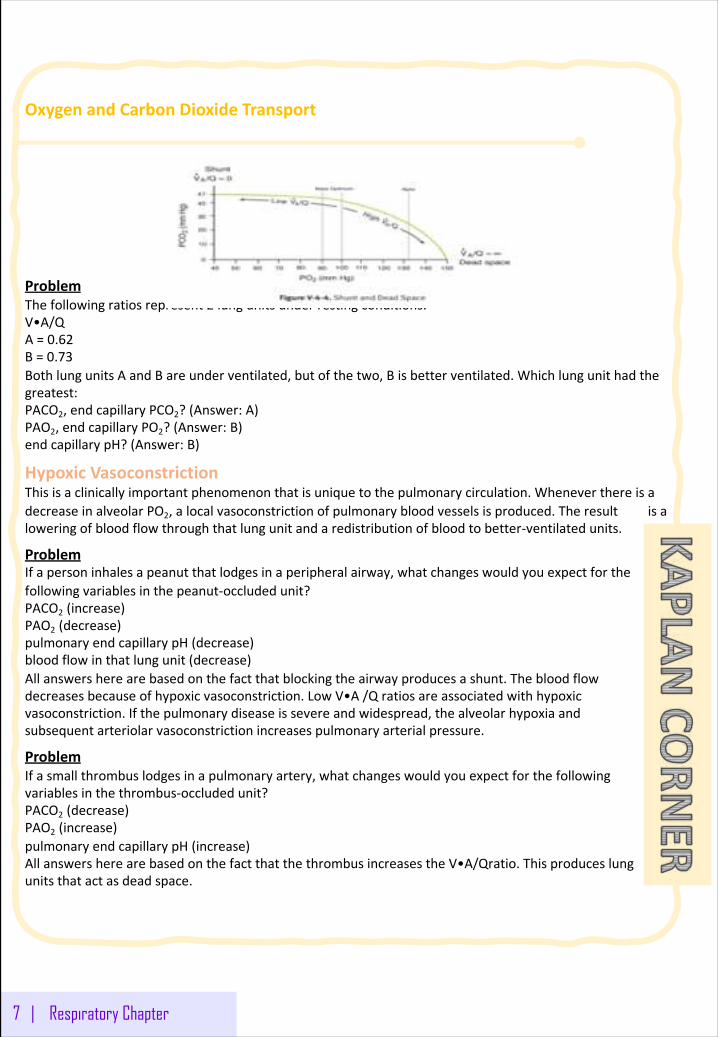
OxygenandCarbonDioxideTransport
ProblemThefollowingratiosrepresent2 lungunitsunderrestingconditions:V•A/QA=0.62B=0.73BothlungunitsAandBareunderventilated,butofthetwo,Bisbetterventilated.Whichlungunithadthegreatest:PACO2,endcapillaryPCO2?(Answer:A)PAO2,endcapillaryPO2?(Answer:B)endcapillarypH?(Answer:B)
HypoxicVasoconstrictionThisisaclinicallyimportantphenomenonthatisuniquetothepulmonarycirculation.WheneverthereisadecreaseinalveolarPO2,alocalvasoconstrictionofpulmonarybloodvesselsisproduced.Theresultisaloweringofbloodflowthroughthatlungunitandaredistributionofbloodtobetter-ventilatedunits.
ProblemIfapersoninhalesapeanutthatlodgesinaperipheralairway,whatchangeswouldyouexpectforthefollowingvariablesinthepeanut-occludedunit?PACO2 (increase)PAO2 (decrease)pulmonaryendcapillarypH(decrease)bloodflowinthatlungunit(decrease)Allanswersherearebasedonthefactthatblockingtheairwayproducesashunt.Thebloodflowdecreasesbecauseofhypoxicvasoconstriction.LowV•A/Qratiosareassociatedwithhypoxicvasoconstriction.Ifthepulmonarydiseaseissevereandwidespread,thealveolarhypoxiaandsubsequentarteriolarvasoconstrictionincreasespulmonaryarterialpressure.
ProblemIfasmallthrombuslodgesinapulmonaryartery,whatchangeswouldyouexpectforthefollowingvariablesinthethrombus-occludedunit?PACO2 (decrease)PAO2 (increase)pulmonaryendcapillarypH(increase)AllanswersherearebasedonthefactthatthethrombusincreasestheV•A/Qratio.Thisproduceslungunitsthatactasdeadspace.
7 | Respiratory Chapter
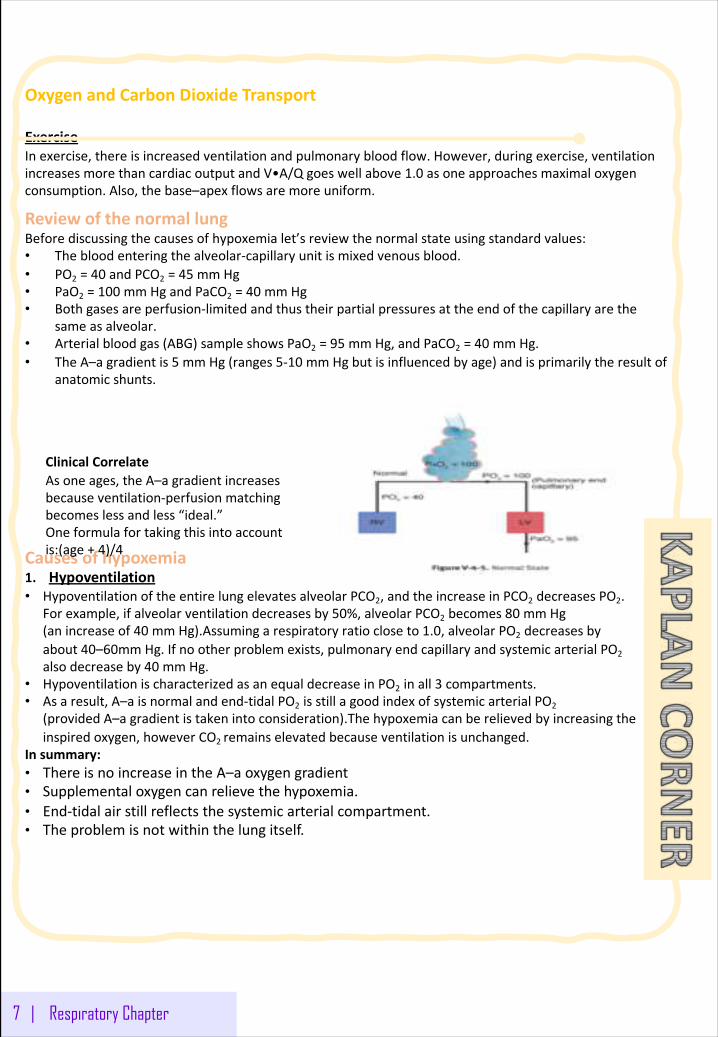
OxygenandCarbonDioxideTransport
ExerciseInexercise,thereisincreasedventilationandpulmonarybloodflow.However,duringexercise,ventilationincreasesmorethancardiacoutputandV•A/Qgoeswellabove1.0 asoneapproachesmaximaloxygenconsumption.Also,thebase–apexflowsaremoreuniform.
ReviewofthenormallungBeforediscussingthecausesofhypoxemialet’sreviewthenormalstateusingstandardvalues:• Thebloodenteringthealveolar-capillaryunitismixedvenousblood.• PO2 =40 andPCO2 =45 mmHg• PaO2 =100 mmHgandPaCO2 =40 mmHg• Bothgasesareperfusion-limitedandthustheirpartialpressuresattheendofthecapillaryarethe
sameasalveolar.• Arterialbloodgas(ABG)sampleshowsPaO2 =95 mmHg,andPaCO2 =40 mmHg.• TheA–agradientis5 mmHg(ranges5-10 mmHgbutisinfluencedbyage)andisprimarilytheresultof
anatomicshunts.
Causesofhypoxemia1. Hypoventilation• HypoventilationoftheentirelungelevatesalveolarPCO2,andtheincreaseinPCO2 decreasesPO2.
Forexample,ifalveolarventilationdecreasesby50%,alveolarPCO2 becomes80 mmHg(anincreaseof40 mmHg).Assumingarespiratoryratiocloseto1.0,alveolarPO2 decreasesbyabout40–60mmHg.Ifnootherproblemexists,pulmonaryendcapillaryandsystemicarterialPO2alsodecreaseby40 mmHg.
• HypoventilationischaracterizedasanequaldecreaseinPO2 inall3 compartments.• Asaresult,A–aisnormalandend-tidalPO2 isstillagoodindexofsystemicarterialPO2
(providedA–agradientistakenintoconsideration).Thehypoxemiacanberelievedbyincreasingtheinspiredoxygen,howeverCO2 remainselevatedbecauseventilationisunchanged.
Insummary:• ThereisnoincreaseintheA–aoxygengradient• Supplementaloxygencanrelievethehypoxemia.• End-tidalairstillreflectsthesystemicarterialcompartment.• Theproblemisnotwithinthelungitself.
7 | Respiratory Chapter
ClinicalCorrelateAsoneages,theA–agradientincreasesbecauseventilation-perfusionmatchingbecomeslessandless“ideal.”Oneformulafortakingthisintoaccountis:(age+4)/4
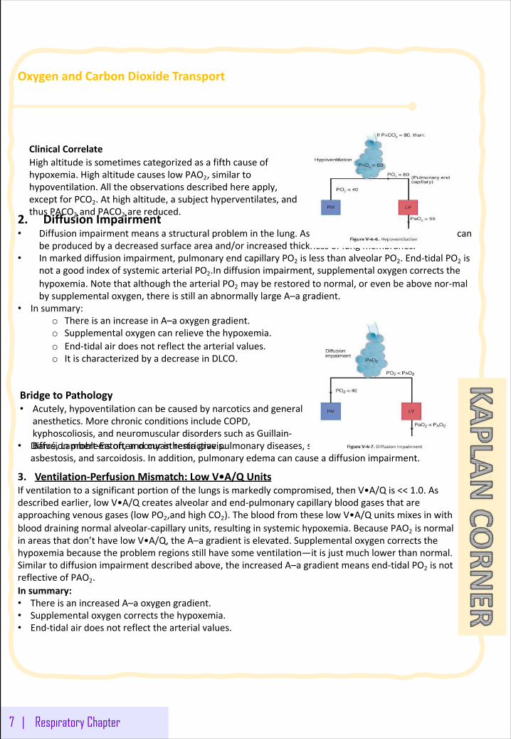
OxygenandCarbonDioxideTransport
2. DiffusionImpairment• Diffusionimpairmentmeansastructuralprobleminthelung.Asdescribedearlierinthisbook,thiscan
beproducedbyadecreasedsurfaceareaand/orincreasedthicknessoflungmembranes.• Inmarkeddiffusionimpairment,pulmonaryendcapillaryPO2 islessthanalveolarPO2.End-tidalPO2 is
notagoodindexofsystemicarterialPO2.Indiffusionimpairment,supplementaloxygencorrectsthehypoxemia.NotethatalthoughthearterialPO2 mayberestoredtonormal,orevenbeabovenor-malbysupplementaloxygen,thereisstillanabnormallylargeA–agradient.
• Insummary:o ThereisanincreaseinA–aoxygengradient.o Supplementaloxygencanrelievethehypoxemia.o End-tidalairdoesnotreflectthearterialvalues.o ItischaracterizedbyadecreaseinDLCO.
• Diffusionproblemsoftenoccurinrestrictivepulmonarydiseases,suchaspulmonaryfibrosis,asbestosis,andsarcoidosis.Inaddition,pulmonaryedemacancauseadiffusionimpairment.
3. Ventilation-PerfusionMismatch:LowV•A/QUnitsIfventilationtoasignificantportionofthelungsismarkedlycompromised,thenV•A/Qis<<1.0.Asdescribedearlier,lowV•A/Qcreatesalveolarandend-pulmonarycapillarybloodgasesthatareapproachingvenousgases(lowPO2,andhighCO2).ThebloodfromtheselowV•A/Qunitsmixesinwithblooddrainingnormalalveolar-capillaryunits,resultinginsystemichypoxemia.BecausePAO2 isnormalinareasthatdon’thavelowV•A/Q,theA–agradientiselevated.Supplementaloxygencorrectsthehypoxemiabecausetheproblemregionsstillhavesomeventilation—itisjustmuchlowerthannormal.Similartodiffusionimpairmentdescribedabove,theincreasedA–agradientmeansend-tidalPO2 isnotreflectiveofPAO2.Insummary:• ThereisanincreasedA–aoxygengradient.• Supplementaloxygencorrectsthehypoxemia.• End-tidalairdoesnotreflectthearterialvalues.
7 | Respiratory Chapter
ClinicalCorrelateHighaltitudeissometimescategorizedasafifthcauseofhypoxemia.HighaltitudecauseslowPAO2,similartohypoventilation.Alltheobservationsdescribedhereapply,exceptforPCO2.Athighaltitude,asubjecthyperventilates,andthusPACO2 andPACO2 arereduced.
BridgetoPathology• Acutely,hypoventilationcanbecausedbynarcoticsandgeneral
anesthetics.MorechronicconditionsincludeCOPD,kyphoscoliosis,andneuromusculardisorderssuchasGuillain-Barré,Lambert-Eaton,andmyastheniagravis.
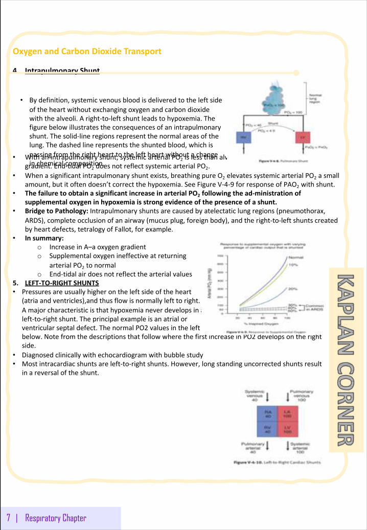
OxygenandCarbonDioxideTransport
4. IntrapulmonaryShunt
• Withanintrapulmonaryshunt,systemicarterialPO2 islessthanalveolar,resultinginanelevatedA–agradient.End-tidalPO2 doesnotreflectsystemicarterialPO2.
• Whenasignificantintrapulmonaryshuntexists,breathingpureO2 elevatessystemicarterialPO2 asmallamount,butitoftendoesn’tcorrectthehypoxemia.SeeFigureV-4-9 forresponseofPAO2 withshunt.
• ThefailuretoobtainasignificantincreaseinarterialPO2 followingthead-ministrationofsupplementaloxygeninhypoxemiaisstrongevidenceofthepresenceofashunt.
• BridgetoPathology: Intrapulmonaryshuntsarecausedbyatelectatic lungregions(pneumothorax,ARDS),completeocclusionofanairway(mucusplug,foreignbody),andtheright-to-leftshuntscreatedbyheartdefects,tetralogyofFallot,forexample.
• Insummary:o IncreaseinA–aoxygengradiento Supplementaloxygenineffectiveatreturning
arterialPO2 tonormalo End-tidalairdoesnotreflectthearterialvalues
5. LEFT-TO-RIGHTSHUNTS• Pressuresareusuallyhigherontheleftsideoftheheart
(atriaandventricles),andthusflowisnormallylefttoright.Amajorcharacteristicisthathypoxemianeverdevelopsinaleft-to-rightshunt.Theprincipalexampleisanatrialorventricularseptaldefect.ThenormalPO2 valuesintheleftandrightcompartmentscanbeseenbelow.NotefromthedescriptionsthatfollowwherethefirstincreaseinPO2 developsontherightside.
• Diagnosedclinicallywithechocardiogramwithbubblestudy• Mostintracardiac shuntsareleft-to-rightshunts.However,longstandinguncorrectedshuntsresult
inareversaloftheshunt.
7 | Respiratory Chapter
• Bydefinition,systemicvenousbloodisdeliveredtotheleftsideoftheheartwithoutexchangingoxygenandcarbondioxidewiththealveoli.Aright-to-leftshuntleadstohypoxemia.Thefigurebelowillustratestheconsequencesofanintrapulmonaryshunt.Thesolid-lineregionsrepresentthenormalareasofthelung.Thedashedlinerepresentstheshuntedblood,whichispassingfromtherighthearttotheleftheartwithoutachangeinchemicalcomposition.
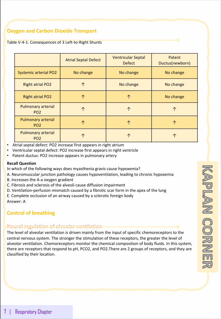
OxygenandCarbonDioxideTransport
TableV-4-1.Consequencesof3 Left-to-RightShunts
• Atrialseptaldefect:PO2 increasefirstappearsinrightatrium• Ventricularseptaldefect:PO2 increasefirstappearsinrightventricle• Patentductus:PO2 increaseappearsinpulmonaryartery
RecallQuestionInwhichofthefollowingwaysdoesmyastheniagraviscausehypoxemia?A.Neuromuscularjunctionpathologycauseshypoventilation,leadingtochronichypoxemiaB.IncreasestheA-aoxygengradientC.FibrosisandsclerosisofthealveolicausediffusionimpairmentD.Ventilation-perfusionmismatchcausedbyafibroticscarformintheapexofthelungE.CompleteocclusionofanairwaycausedbyascleroticforeignbodyAnswer:A
Controlofbreathing
NeuralregulationofalveolarventilationThelevelofalveolarventilationisdrivenmainlyfromtheinputofspecificchemoreceptorstothecentralnervoussystem.Thestrongerthestimulationofthesereceptors,thegreaterthelevelofalveolarventilation.Chemoreceptorsmonitorthechemicalcompositionofbodyfluids.Inthissystem,therearereceptorsthatrespondtopH,PCO2,andPO2.Thereare2 groupsofreceptors,andtheyareclassifiedbytheirlocation.
7 | Respiratory Chapter
AtrialSeptalDefect VentricularSeptalDefect
PatentDuctus(newborn)
SystemicarterialPO2 Nochange Nochange Nochange
RightatrialPO2 ↑ Nochange Nochange
RightatrialPO2 ↑ ↑ Nochange
PulmonaryarterialPO2
↑ ↑ ↑
PulmonaryarterialPO2
↑ ↑ ↑
PulmonaryarterialPO2
↑ ↑ ↑
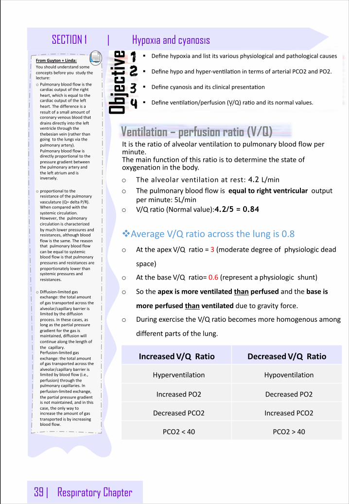
39 | Respiratory Chapter
SECTION 1 | Hypoxia and cyanosis
Ventilation – perfusion ratio (V/Q)
§ Definehypoxiaandlistitsvariousphysiologicalandpathologicalcauses
§ Definehypoandhyper-ven�la�onintermsofarterialPCO2andPO2.
§ Definecyanosisanditsclinicalpresenta�on
§ Defineven�la�on/perfusion(V͎/Q)ra�oanditsnormalvalues.Objec
tive
Itistheratioofalveolarventilationtopulmonarybloodflowperminute.Themainfunctionofthisratioistodeterminethestateofoxygenationinthebody.
o Thealveolarventilationatrest:4.2 L/mino Thepulmonarybloodflowisequaltorightventricularoutput
per minute:5L/mino V/Q ratio(Normal value):4.2/5 = 0.84
vAverageV/Qratioacrossthelung is 0.8o Atthe apex V/Qratio = 3 (moderatedegree of physiologic dead
space)
o Atthe base V/Qratio= 0.6 (represent a physiologic shunt)
o Sothe apex is more ventilated than perfused and the base is
more perfused than ventilated dueto gravity force.
o During exercise the V/Qratio becomes more homogenous among
differentparts of the lung.
FromGuyton+Linda:Youshouldunderstandsomeconceptsbeforeyoustudythelecture:o Pulmonarybloodflowisthecardiacoutputoftherightheart,whichisequaltothecardiacoutputoftheleftheart.Thedifferenceisaresultofasmallamountofcoronaryvenousbloodthatdrainsdirectlyintotheleftventriclethroughthethebesianvein(ratherthangoingtothelungsviathepulmonaryartery).Pulmonarybloodflowisdirectlyproportionaltothepressuregradientbetweenthepulmonaryarteryandtheleftatriumandisinversely.
o proportionaltotheresistanceofthepulmonaryvasculature(Q=deltaP/R).Whencomparedwiththesystemiccirculation.However,thepulmonarycirculationischaracterizedbymuchlowerpressuresandresistances,althoughbloodflowisthesame.Thereasonthatpulmonarybloodflowcanbeequaltosystemicbloodflowisthatpulmonarypressuresandresistancesareproportionatelylowerthansystemicpressuresandresistances.
o Diffusion-limitedgasexchange:thetotalamountofgastransportedacrossthealveolar/capillarybarrierislimitedbythediffusionprocess.Inthesecases,aslongasthepartialpressuregradientforthegasismaintained,diffusionwillcontinuealongthelengthofthecapillary.Perfusion-limitedgasexchange:thetotalamountofgastransportedacrossthealveolar/capillarybarrierislimitedbybloodflow(i.e.,perfusion)throughthepulmonarycapillaries.Inperfusion-limitedexchange,thepartialpressuregradientisnotmaintained,andinthiscase,theonlywaytoincreasetheamountofgastransportedisbyincreasingbloodflow.
IncreasedV/QRatio DecreasedV/QRatio
Hyperventilation Hypoventilation
Increased PO2 Decreased PO2
Decreased PCO2 Increased PCO2
PCO2 < 40 PCO2 > 40
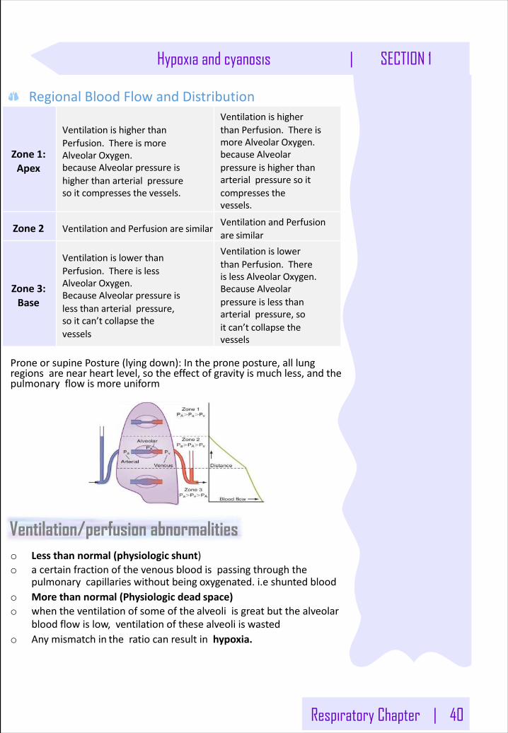
Respiratory Chapter | 40
Hypoxia and cyanosis | SECTION 1
RegionalBloodFlowandDistribution
Zone1:Apex
VentilationishigherthanPerfusion.ThereismoreAlveolar Oxygen.becauseAlveolarpressureishigherthanarterialpressuresoitcompressesthe vessels.
VentilationishigherthanPerfusion.ThereismoreAlveolar Oxygen.becauseAlveolarpressureishigherthanarterialpressuresoitcompressesthevessels.
Zone 2 VentilationandPerfusionare similarVentilationandPerfusionare similar
Zone3:Base
VentilationislowerthanPerfusion.ThereislessAlveolar Oxygen.BecauseAlveolarpressureislessthanarterialpressure,soitcan’tcollapsethevessels
VentilationislowerthanPerfusion.ThereislessAlveolar Oxygen.BecauseAlveolarpressureislessthanarterialpressure,soitcan’tcollapsethevessels
ProneorsupinePosture(lyingdown):Intheproneposture,alllungregionsarenearheartlevel,sotheeffectofgravityismuchless,andthepulmonaryflowismore uniform
Ventilation/perfusion abnormalitieso Lessthannormal(physiologic shunt)o acertainfractionofthevenousbloodispassingthroughthe
pulmonarycapillarieswithoutbeing oxygenated.i.e shunted bloodo Morethannormal(Physiologicdead space)o whentheventilationofsomeofthe alveoliisgreatbutthealveolar
bloodflowislow,ventilationofthesealveoliis wastedo Anymismatchin theratiocanresultinhypoxia.
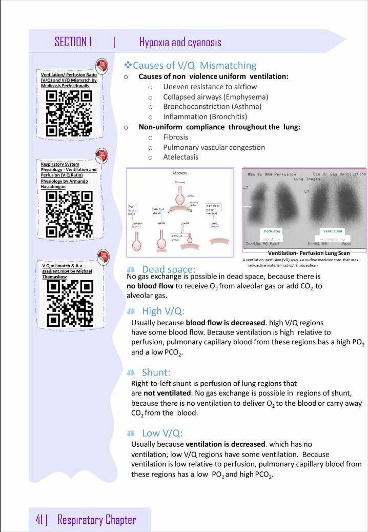
41 | Respiratory Chapter
SECTION 1 | Hypoxia and cyanosis
vCausesofV/QMismatchingo Causesofnonviolence uniformventilation:
o Unevenresistanceto airflowo Collapsedairways (Emphysema)o Bronchoconstriction (Asthma)o Inflammation (Bronchitis)
o Non-uniformcompliancethroughout thelung:o Fibrosiso Pulmonaryvascular congestiono Atelectasis
Ventilation- PerfusionLung ScanAventilation–perfusion(VQ)scanisanuclearmedicinescanthatuses
radioactivematerial (radiopharmaceutical)
Ventilation
examineairflow
Perfusion
bloodflow
highairNoblood
highbloodNoair(oxygen)
highO2 inalveoli
lowO2 inalveoli
Deadspace:Nogasexchangeispossible in deadspace,becausethere isnobloodflowtoreceive O2 fromalveolargasoraddCO2 toalveolar gas.
HighV/Q:Usuallybecausebloodflow is decreased.highV/Q regionshavesomeblood flow. Becauseventilationishighrelativetoperfusion, pulmonarycapillary blood fromtheseregionshasa high PO2andalow PCO2.
Shunt:Right-to-leftshunt is perfusionoflungregions thatarenotventilated.No gas exchangeispossibleinregionsofshunt,because thereisnoventilation to deliverO2 tothebloodor carryawayCO2 fromtheblood.
LowV/Q:Usuallybecause ventilation isdecreased.whichhas noventilation,lowV/Q regions havesomeventilation.Becauseventilationis low relativeto perfusion, pulmonarycapillary blood fromtheseregionshasalowPO2andhigh PCO2.
VQmismatch&A-agradient.mp4 byMichaelThomashow
RespiratorySystemPhysiology- VentilationandPerfusion(V:QRatio)PhysiologybyArmandoHasudungan
Ventilation/PerfusionRatio(V/Q)andV/QMismatchbyMedicosis Perfectionalis
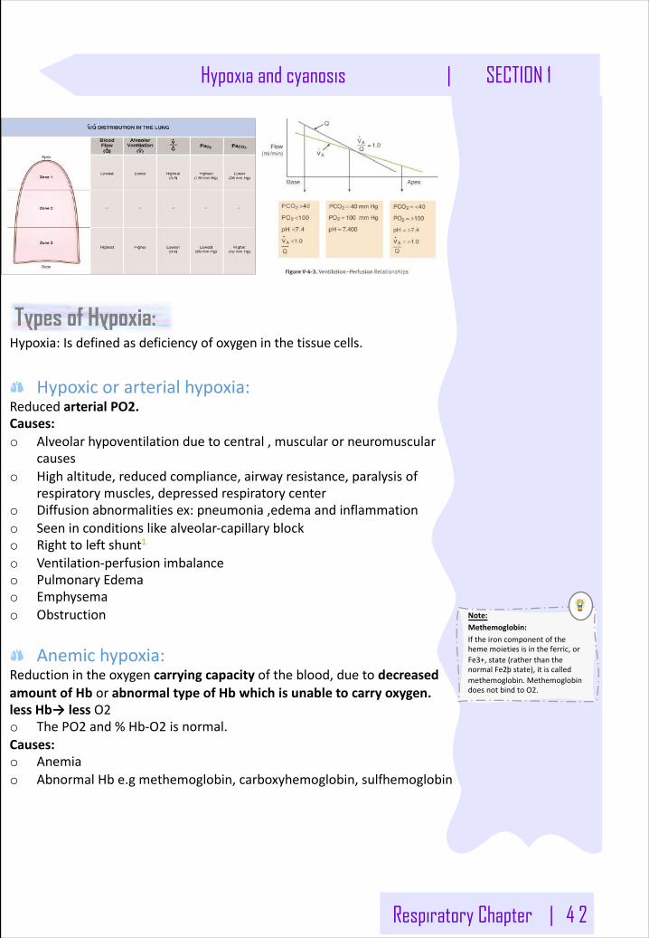
Respiratory Chapter | 4 2
Hypoxia and cyanosis | SECTION 1
Types of Hypoxia:Hypoxia:Isdefinedasdeficiencyofoxygeninthetissue cells.
Hypoxicorarterialhypoxia:ReducedarterialPO2.Causes:o Alveolarhypoventilationduetocentral,muscularorneuromuscular
causeso Highaltitude,reducedcompliance,airwayresistance,paralysisof
respiratorymuscles,depressedrespiratorycentero Diffusionabnormalitiesex:pneumonia,edemaandinflammationo Seeninconditionslikealveolar-capillaryblocko Righttoleftshunt1
o Ventilation-perfusionimbalanceo PulmonaryEdemao Emphysemao Obstruction
Anemichypoxia:Reductionintheoxygencarryingcapacityoftheblood,duetodecreasedamountofHb orabnormaltypeofHb whichisunabletocarryoxygen.lessHb→lessO2o ThePO2 and%Hb-O2 isnormal.Causes:o Anemiao AbnormalHbe.g methemoglobin,carboxyhemoglobin,sulfhemoglobin
Note:Methemoglobin:Iftheironcomponentofthehememoietiesisintheferric,orFe3+,state(ratherthanthenormalFe2þstate),itiscalledmethemoglobin.MethemoglobindoesnotbindtoO2.
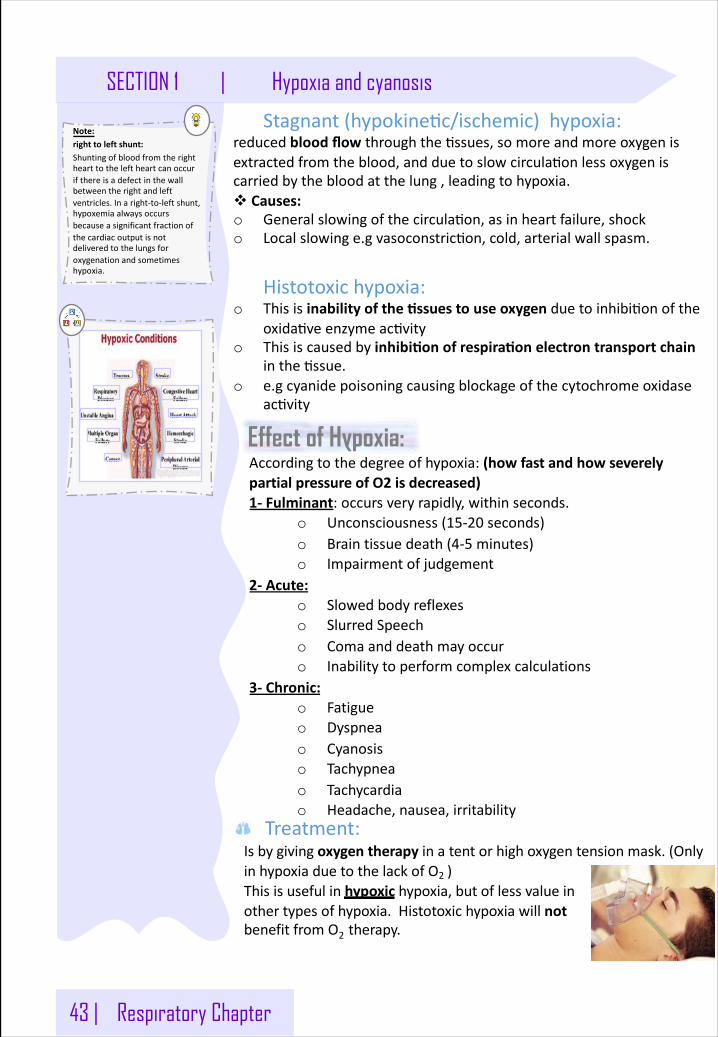
43 | Respiratory Chapter
Note:righttoleftshunt:Shuntingofbloodfromtherighthearttotheleftheartcanoccurifthereisadefectinthewallbetweentherightandleftventricles.Inaright-to-leftshunt,hypoxemiaalwaysoccursbecauseasignificantfractionofthecardiacoutputisnotdeliveredtothelungsforoxygenationandsometimeshypoxia.
SECTION 1 | Hypoxia and cyanosis
Stagnant(hypokine�c/ischemic)hypoxia:reducedbloodflowthroughthe�ssues,somoreandmoreoxygenisextractedfromtheblood,andduetoslowcircula�onlessoxygeniscarriedbythebloodatthelung,leadingtohypoxia.v Causes:o Generalslowingofthecircula�on,asinheartfailure,shocko Localslowinge.gvasoconstric�on,cold,arterialwallspasm.
Histotoxichypoxia:o Thisisinabilityofthetssuestouseoxygenduetoinhibi�onofthe
oxida�veenzymeac�vityo Thisiscausedbyinhibitonofrespiratonelectrontransportchain
inthe�ssue.o e.gcyanidepoisoningcausingblockageofthecytochromeoxidase
ac�vity
Effect of Hypoxia:Accordingtothedegreeofhypoxia: (howfastandhowseverelypartialpressureofO2 isdecreased) 1- Fulminant:occursveryrapidly,withinseconds.
o Unconsciousness(15-20 seconds)o Braintissuedeath(4-5 minutes)o Impairmentofjudgement
2- Acute:o Slowedbodyreflexeso SlurredSpeecho Comaanddeathmayoccuro Inabilitytoperformcomplexcalculations
3- Chronic:o Fatigueo Dyspneao Cyanosiso Tachypneao Tachycardiao Headache,nausea,irritability
Treatment:Isbygivingoxygentherapyinatentorhighoxygentensionmask.(OnlyinhypoxiaduetothelackofO2 )Thisisusefulinhypoxic hypoxia,butoflessvalueinothertypesofhypoxia.HistotoxichypoxiawillnotbenefitfromO2 therapy.
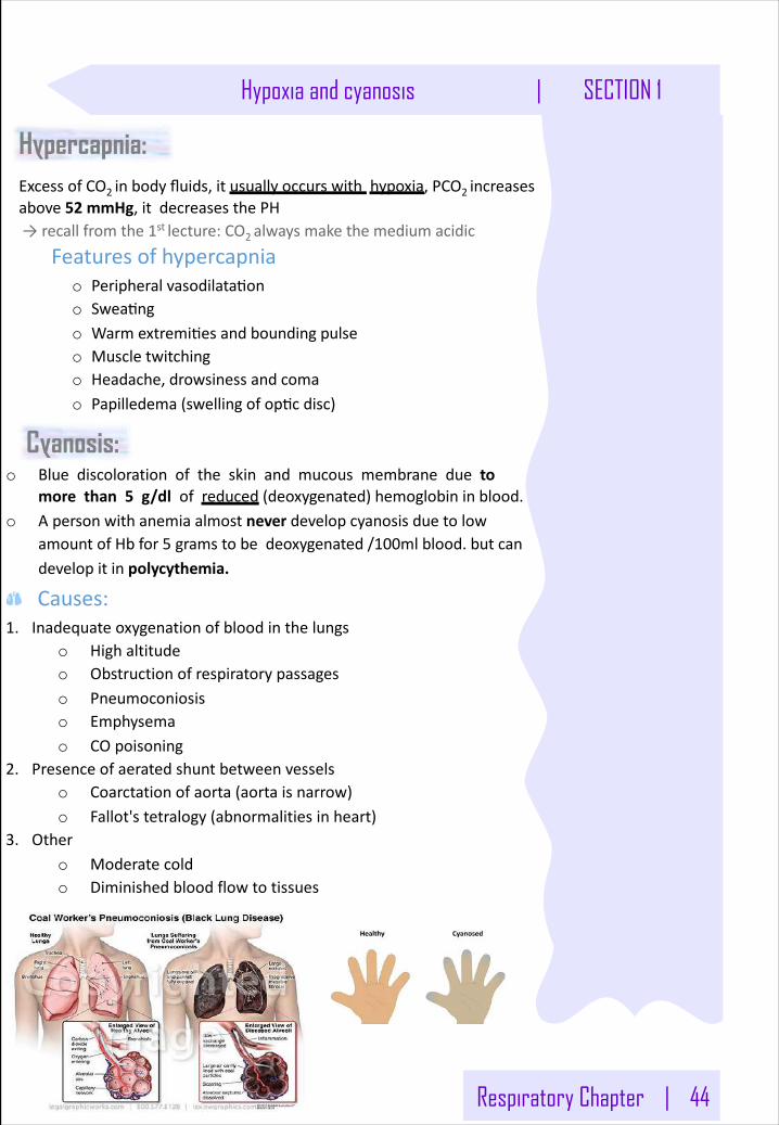
Respiratory Chapter | 44
Hypoxia and cyanosis | SECTION 1
Hypercapnia:ExcessofCO2inbodyfluids,itusuallyoccurswithhypoxia,PCO2increasesabove52mmHg,itdecreasesthePH→recallfromthe1stlecture:CO2alwaysmakethemediumacidic
Featuresofhypercapniao Peripheralvasodilata�ono Swea�ngo Warmextremi�esandboundingpulseo Muscletwitchingo Headache,drowsinessandcomao Papilledema(swellingofop�cdisc)
Cyanosis:o Blue discoloration of the skin and mucous membrane due to
more than 5 g/dl of reduced (deoxygenated)hemoglobinin blood.o Apersonwithanemiaalmostnever developcyanosisduetolow
amountofHb for5 gramstobedeoxygenated/100mlblood.butcandevelopitin polycythemia.
Causes:1. Inadequateoxygenationofbloodinthelungs
o Highaltitudeo Obstructionofrespiratorypassageso Pneumoconiosiso Emphysemao COpoisoning
2. Presenceofaeratedshuntbetweenvesselso Coarctationofaorta(aortaisnarrow)o Fallot'stetralogy(abnormalitiesinheart)
3. Othero Moderatecoldo Diminishedbloodflowtotissues
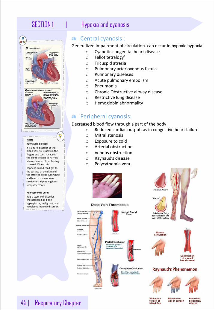
45 | Respiratory Chapter
Note:Raynaud'sdiseaseItisararedisorderofthebloodvessels,usuallyinthefingersandtoes.Itcausesthebloodvesselstonarrowwhenyouarecoldorfeelingstressed.Whenthishappens,bloodcan'tgettothesurfaceoftheskinandtheaffectedareasturnwhiteandblue.Itmayrequirecervicodorsalpreganglionicsympathectomy
Polycythemiavera:Itisastemcelldisordercharacterizedasapanhyperplastic,malignant,andneoplasticmarrowdisorder.
SECTION 1 | Hypoxia and cyanosis
Centralcyanosis:Generalizedimpairmentofcirculation.canoccurinhypoxichypoxia.
o Cyanoticcongenitalheart-diseaseo Fallottetralogy1
o Tricuspidatresiao Pulmonaryarteriovenousfistulao Pulmonarydiseaseso Acutepulmonaryembolismo Pneumoniao ChronicObstructiveairwaydiseaseo Restrictivelungdiseaseo Hemoglobinabnormality
Peripheralcyanosis:Decreasedbloodflowthroughapartofthebody
o Reducedcardiacoutput,asincongestiveheartfailureo Mitralstenosiso Exposuretocoldo Arterialobstructiono Venousobstructiono Raynaud’sdiseaseo Polycythemiavera
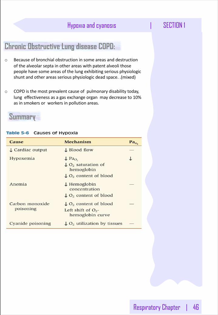
Respiratory Chapter | 46
Hypoxia and cyanosis | SECTION 1
Chronic Obstructive Lung disease COPD:
o Becauseofbronchialobstructioninsomeareasanddestructionofthealveolarseptainotherareaswithpatentalveolithosepeoplehavesomeareasofthelungexhibitingseriousphysiologicshuntandotherareasseriousphysiologicdeadspace..(mixed)
o COPDisthemostprevalentcauseofpulmonarydisabilitytoday,lungeffectivenessasagasexchangeorganmaydecreaseto10%asinsmokersorworkersinpollution areas.
Summary
47 | Respiratory Chapter
SECTION 1 | Control of breathing
The overall processes of External Respiration:
§ Understandtheroleofthemedullaoblongataindeterminingthebasicpa�ernofrespiratoryac�vity.
§ Listsomefactorsthatcanmodifythebasicbreathingpa�ernlikee.g.a-TheHering-Breuerreflexes,b-Theproprioceptorreflexes,c-Theprotec�vereflexes,liketheirritant,andtheJ-receptors.
§ UnderstandtherespiratoryconsequencesofchangingPO2,PCO2,andPH.
§ Describetheloca�onsandrolesoftheperipheralandcentralchemoreceptors.
§ Compareandcontrastmetabolicandrespiratoryacidosisandmetabolicandrespiratoryalkalosis.
Objec
tive
Allthisprocessisregulatedbytherespiratory center
Controls of rate and depth of respiration:ArterialPO2:
WhenPO2 isVERYlow(Hypoxia),ventilationincreases.(itwillstimulaterespiration,butit’snotamajorplayer)“Lesssensitive,onlymajorchangesinPO2 willcauseincreaseventilation”
ArterialPCO2:ThemostimportantregulatorofventilationisPCO(verystrongstimulus),SmallincreasesinPCO2,greatlyincreasesventilation.(evenaslightincreaseinco2 meansthatthere’saproblem)➔ RecallCO2 20 timemoresolublethanO2.
ArterialpH:Ashydrogenionsincrease(acidosis),alveolarventilationincreases.
o ConcentrationofH+↑>(acidosis)o ConcentrationofH+↓>(alkalosis)o CO2 + H2O ⇌ H2CO3 ⇌ H++HCO3-
Acid/basemetabolisminthebodyisregulatedbythischemicalequation.
ControlOfRespiration(regulationofbreathing)byArmandoHasudungan
Centralchemoreceptors|Respiratorysystemphysiology|NCLEX-RN|KhanAcademy by khanacademymedicine
Peripheralchemoreceptors|Respiratorysystemphysiology|NCLEX-RN|KhanAcademys by khanacademymedicine
ControlofVentlaton byCalit2ube
Therespiratorycenter|Respiratorysystemphysiology|NCLEX-RN|KhanAcademybykhanacademymedicine
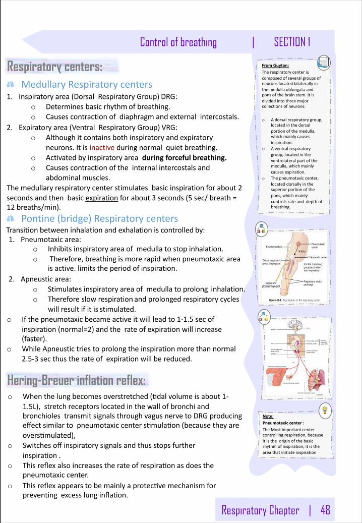
Respiratory Chapter | 48
Control of breathing | SECTION 1
Respiratory centers:MedullaryRespiratorycenters
1. Inspiratory area(DorsalRespiratoryGroup) DRG:o Determinesbasicrhythm ofbreathing.o Causescontractionofdiaphragmandexternalintercostals.
2. Expiratory area(VentralRespiratoryGroup) VRG:o Althoughitcontains both inspiratoryandexpiratory
neurons. Itisinactiveduringnormalquiet breathing.o Activatedbyinspiratoryarea duringforceful breathing.o Causescontractionoftheinternalintercostalsand
abdominal muscles.Themedullaryrespiratorycenterstimulatesbasicinspirationforabout2secondsandthenbasicexpiration forabout3 seconds (5 sec/breath=12 breaths/min).
Pontine(bridge)RespiratorycentersTransitionbetweeninhalationandexhalationiscontrolled by:1. Pneumotaxicarea:
o Inhibitsinspiratoryareaofmedullatostopinhalation.o Therefore,breathingismorerapidwhenpneumotaxicarea
isactive.limitstheperiodof inspiration.2. Apneusticarea:
o Stimulatesinspiratoryareaofmedullatoprolong inhalation.o Thereforeslowrespiration and prolongedrespiratorycycles
willresultifitis stimulated.o Ifthepneumotaxicbecameactiveitwillleadto1-1.5 secof
inspiration(normal=2)andtherateofexpirationwillincrease(faster).
o WhileApneustictriestoprolongtheinspirationmorethannormal2.5-3 secthustherateofexpirationwillbe reduced.
FromGuyton:Therespiratorycenteriscomposedofseveralgroupsofneuronslocatedbilaterallyinthemedullaoblongataandponsofthebrainstem.Itisdividedintothreemajorcollectionsofneurons:
o Adorsalrespiratorygroup,locatedinthedorsalportionofthemedulla,whichmainlycausesinspiration.
o Aventralrespiratorygroup,locatedintheventrolateralpartofthemedulla,whichmainlycausesexpiration.
o Thepneumotaxiccenter,locateddorsallyinthesuperiorportionofthepons,whichmainlycontrolsrateanddepthofbreathing.
Hering-Breuer inflation reflex:o Whenthelungbecomesoverstretched(�dalvolumeisabout1-
1.5L),stretchreceptorslocatedinthewallofbronchiandbronchiolestransmitsignalsthroughvagusnervetoDRGproducingeffectsimilartopneumotaxiccenters�mula�on(becausetheyareovers�mulated),
o Switchesoffinspiratorysignalsandthusstopsfurtherinspira�on.
o Thisreflexalsoincreasestherateofrespira�onasdoesthepneumotaxiccenter.
o Thisreflexappearstobemainlyaprotec�vemechanismforpreven�ngexcesslunginfla�on.
Note:Pneumotaxiccenter:TheMostimportantcentercontrollingrespiration,becauseitistheoriginofthebasicrhythmofinspiration,itistheareathatinitiateinspiration
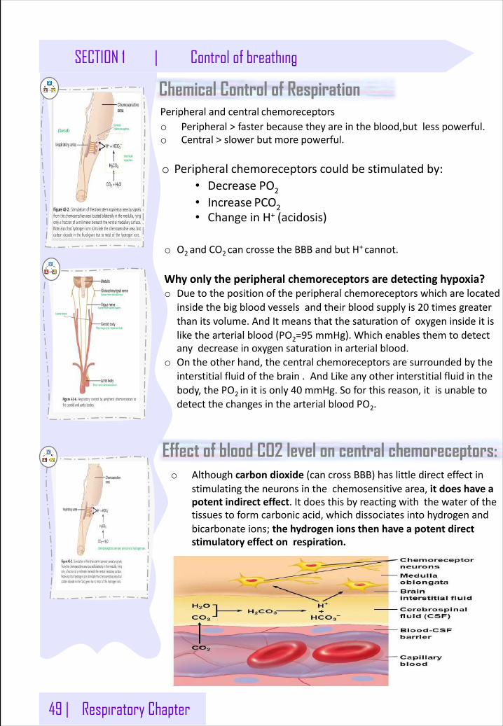
49 | Respiratory Chapter
SECTION 1 | Control of breathing
Chemical Control of RespirationPeripheralandcentral chemoreceptorso Peripheral>fasterbecausetheyareintheblood,but less powerful.o Central>slowerbutmore powerful.
o Peripheralchemoreceptorscouldbestimulatedby:
• Decrease PO2• Increase PCO2• ChangeinH+ (acidosis)
o O2andCO2 cancrosse theBBBandbutH+ cannot.
Whyonlytheperipheralchemoreceptorsaredetecting hypoxia?o Duetothepositionoftheperipheralchemoreceptorswhicharelocated
insidethebigbloodvesselsandtheirbloodsupplyis20 timesgreaterthanitsvolume.AndItmeansthatthesaturationofoxygeninsideitislikethearterialblood(PO2=95 mmHg).Whichenablesthemtodetectanydecreaseinoxygensaturationinarterial blood.
o Ontheotherhand,thecentralchemoreceptorsaresurroundedbytheinterstitialfluidofthebrain.AndLikeanyotherinterstitialfluidinthebody,thePO2 initisonly40 mmHg.Soforthisreason,itisunabletodetectthechangesinthearterialblood PO2.
Effect of blood CO2 level on central chemoreceptors:o Althoughcarbondioxide (cancrossBBB)haslittledirecteffectin
stimulatingtheneuronsinthechemosensitive area,itdoeshaveapotentindirecteffect.Itdoesthisbyreactingwiththewaterofthetissuestoformcarbonicacid,whichdissociatesintohydrogenandbicarbonateions; thehydrogenionsthenhaveapotentdirectstimulatoryeffectonrespiration.
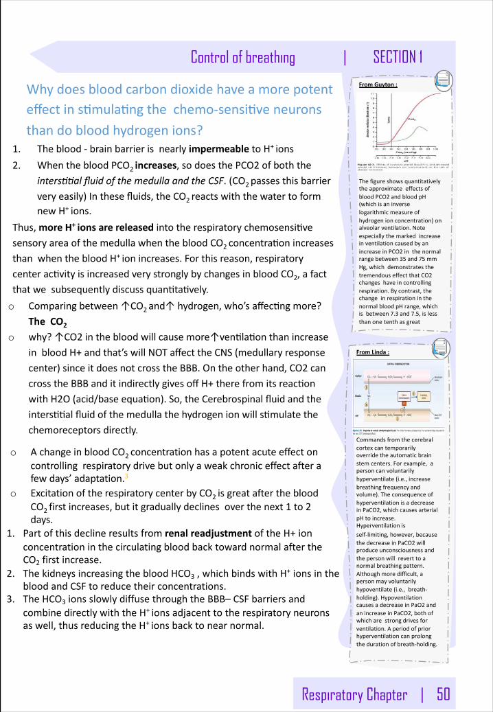
Respiratory Chapter | 50
Control of breathing | SECTION 1FromGuyton:
ThefigureshowsquantitativelytheapproximateeffectsofbloodPCO2 andbloodpH(whichisaninverselogarithmicmeasureofhydrogenionconcentration)onalveolarventilation.NoteespeciallythemarkedincreaseinventilationcausedbyanincreaseinPCO2 inthenormalrangebetween35 and75 mmHg,whichdemonstratesthetremendouseffectthatCO2changeshaveincontrollingrespiration.Bycontrast,thechangeinrespirationinthenormalbloodpHrange,whichisbetween7.3 and7.5,islessthanonetenthasgreat
Whydoesbloodcarbondioxidehaveamorepotenteffectins�mula�ngthechemo-sensi�veneuronsthandobloodhydrogenions?
1. Theblood-brainbarrierisnearlyimpermeabletoH+ions2. WhenthebloodPCO2increases,sodoesthePCO2ofboththe
intersUUalfluidofthemedullaandtheCSF.(CO2passesthisbarrierveryeasily)Inthesefluids,theCO2reactswiththewatertoformnewH+ions.
Thus,moreH+ionsarereleasedintotherespiratorychemosensi�vesensoryareaofthemedullawhenthebloodCO2concentra�onincreasesthanwhenthebloodH+ionincreases.Forthisreason,respiratorycenterac�vityisincreasedverystronglybychangesinbloodCO2,afactthatwesubsequentlydiscussquan�ta�vely.o Comparingbetween↑CO2and↑hydrogen,who’saffec�ngmore?
TheCO2
o why?↑CO2inthebloodwillcausemore↑ven�la�onthanincreaseinbloodH+andthat’swillNOTaffecttheCNS(medullaryresponsecenter)sinceitdoesnotcrosstheBBB.Ontheotherhand,CO2cancrosstheBBBanditindirectlygivesoffH+therefromitsreac�onwithH2O(acid/baseequa�on).So,theCerebrospinalfluidandtheinters��alfluidofthemedullathehydrogenionwills�mulatethechemoreceptorsdirectly.
o AchangeinbloodCO2 concentrationhasapotentacuteeffectoncontrollingrespiratorydrivebutonlyaweakchroniceffectafterafewdays’adaptation.3
o ExcitationoftherespiratorycenterbyCO2 isgreatafterthebloodCO2 firstincreases,butitgraduallydeclines overthenext1 to2days.
1. PartofthisdeclineresultsfromrenalreadjustmentoftheH+ionconcentrationinthecirculatingbloodbacktowardnormalaftertheCO2 firstincrease.
2. ThekidneysincreasingthebloodHCO3 ,whichbindswithH+ ionsinthebloodandCSFtoreducetheirconcentrations.
3. TheHCO3 ionsslowlydiffusethroughtheBBB– CSFbarriersandcombinedirectlywiththeH+ionsadjacenttotherespiratoryneuronsaswell,thusreducingtheH+ionsbacktonearnormal.
FromLinda:
Commandsfromthecerebralcortexcantemporarilyoverridetheautomaticbrainstemcenters.Forexample,apersoncanvoluntarilyhyperventilate(i.e.,increasebreathingfrequencyandvolume).TheconsequenceofhyperventilationisadecreaseinPaCO2,whichcausesarterialpHtoincrease.Hyperventilationisself-limiting,however,becausethedecreaseinPaCO2 willproduceunconsciousnessandthepersonwillreverttoanormalbreathingpattern.Althoughmoredifficult,apersonmayvoluntarilyhypoventilate (i.e.,breath-holding).HypoventilationcausesadecreaseinPaO2 andanincreaseinPaCO2,bothofwhicharestrongdrivesforventilation.Aperiodofpriorhyperventilationcanprolongthedurationofbreath-holding.
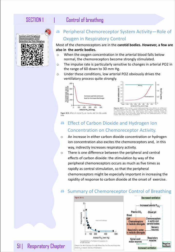
51 | Respiratory Chapter
SECTION 1 | Control of breathing
PeripheralChemoreceptorSystemActivity—RoleofOxygeninRespiratoryControl
Mostofthechemoreceptorsareinthecarotidbodies.However,afewarealsointheaortic bodies.o Whentheoxygenconcentrationinthearterialbloodfallsbelow
normal,thechemoreceptorsbecomestronglystimulated.o TheimpulserateisparticularlysensitivetochangesinarterialPO2 in
therangeof60 downto30 mmHg.o Undertheseconditions,lowarterialPO2 obviouslydrivesthe
ventilatoryprocessquitestrongly.
Increasepartialpressureleadtoincreasedischarge
EffectofCarbonDioxideandHydrogenIonConcentrationonChemoreceptorActivity.
o Anincreaseineithercarbondioxideconcentrationorhydrogenionconcentrationalsoexcitesthechemoreceptorsand,inthisway,indirectlyincreasesrespiratory activity.
o Thereisonedifferencebetweentheperipheralandcentraleffectsofcarbondioxide:thestimulationbywayoftheperipheralchemoreceptorsoccursasmuchasfivetimesasrapidlyascentralstimulation,sothattheperipheralchemoreceptorsmightbeespeciallyimportantinincreasingtherapidityofresponsetocarbondioxideattheonsetofexercise.
SummaryofChemoreceptorControlofBreathing
CentralandPeripheralChemoreceptors(CO2DetectonbyPeteMeighan
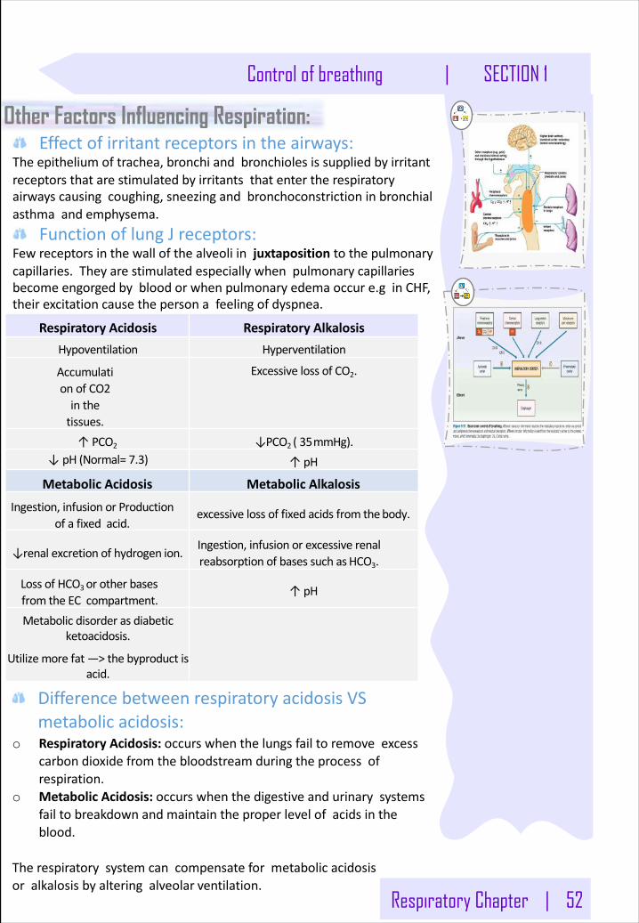
Respiratory Chapter | 52
Control of breathing | SECTION 1
Other Factors Influencing Respiration:Effectofirritantreceptorsintheairways:
Theepitheliumoftrachea,bronchiandbronchiolesissuppliedbyirritantreceptorsthatarestimulatedbyirritantsthatentertherespiratoryairwayscausingcoughing,sneezingandbronchoconstrictioninbronchialasthmaand emphysema.
FunctionoflungJreceptors:Fewreceptorsinthewallofthealveoliinjuxtaposition tothepulmonarycapillaries.Theyarestimulatedespeciallywhenpulmonarycapillariesbecomeengorgedbybloodorwhenpulmonaryedemaoccure.g inCHF,theirexcitationcausethepersonafeelingof dyspnea.
DifferencebetweenrespiratoryacidosisVSmetabolicacidosis:
o RespiratoryAcidosis:occurswhenthelungsfailtoremoveexcesscarbondioxidefromthebloodstreamduringtheprocessofrespiration.
o MetabolicAcidosis:occurswhenthedigestiveandurinarysystemsfailtobreakdownandmaintaintheproperlevelofacidsintheblood.
Therespiratorysystemcancompensateformetabolicacidosisoralkalosisbyalteringalveolar ventilation.
Respiratory Acidosis Respiratory AlkalosisHypoventilation Hyperventilation
AccumulationofCO2inthetissues.
Excessivelossof CO2.
↑ PCO2 ↓PCO2 (35mmHg).↓ pH (Normal= 7.3) ↑ pH
Metabolic Acidosis Metabolic Alkalosis
Ingestion,infusionorProductionofafixedacid.
excessivelossoffixedacidsfromthebody.
↓renalexcretionofhydrogen ion.Ingestion,infusionorexcessiverenalreabsorptionofbasessuchasHCO3.
LossofHCO3orotherbasesfromtheECcompartment.
↑ pH
Metabolicdisorderasdiabeticketoacidosis.
Utilizemorefat—>thebyproductisacid.
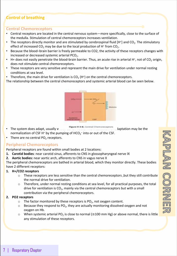
Controlofbreathing
CentralChemoreceptors• Centralreceptorsarelocatedinthecentralnervoussystem—morespecifically,closetothesurfaceof
themedulla.Stimulationofcentralchemoreceptorsincreasesventilation.• Thereceptorsdirectlymonitorandarestimulatedbycerebrospinalfluid[H+]andCO2.Thestimulatory
effectofincreasedCO2 maybeduetothelocalproductionofH+ fromCO2.• Becausetheblood–brainbarrierisfreelypermeabletoCO2,theactivityofthesereceptorschangeswith
increasedordecreasedsystemicarterialPCO2.• H+doesnoteasilypenetratetheblood-brainbarrier.Thus,anacuteriseinarterialH+,notofCO2 origin,
doesnotstimulatecentralchemoreceptors.• Thesereceptorsareverysensitiveandrepresentthemaindriveforventilationundernormalresting
conditionsatsealevel.• Therefore,themaindriveforventilationisCO2 (H+)onthecentralchemoreceptors.Therelationshipbetweenthecentralchemoreceptorsandsystemicarterialbloodcanbeseenbelow.
• Thesystemdoesadapt,usuallywithin12–24 hours.ThemechanismofadaptationmaybethenormalizationofCSFH+ bythepumpingofHCO3
– intooroutoftheCSF.• TherearenocentralPO2 receptors.
PeripheralChemoreceptorsPeripheralreceptorsarefoundwithinsmallbodiesat2 locations:1. Carotidbodies:nearcarotidsinus,afferentstoCNSinglossopharyngealnerveIX2. Aorticbodies:nearaorticarch,afferentstoCNSinvagusnerveXTheperipheralchemoreceptorsarebathedinarterialblood,whichtheymonitordirectly.Thesebodieshave2 differentreceptors:1. H+/CO2 receptors
o Thesereceptorsarelesssensitivethanthecentralchemoreceptors,buttheystillcontributetothenormaldriveforventilation.
o Therefore,undernormalrestingconditionsatsealevel,forallpracticalpurposes,thetotaldriveforventilationisCO2,mainlyviathecentralchemoreceptorsbutwithasmallcontributionviatheperipheralchemoreceptors.
2. PO2 receptorso ThefactormonitoredbythesereceptorsisPO2,notoxygencontent.o BecausetheyrespondtoPO2,theyareactuallymonitoringdissolvedoxygenandnot
oxygenonHb.o WhensystemicarterialPO2 isclosetonormal(≅100 mmHg)orabovenormal,thereislittleif
anystimulationofthesereceptors.
7 | Respiratory Chapter
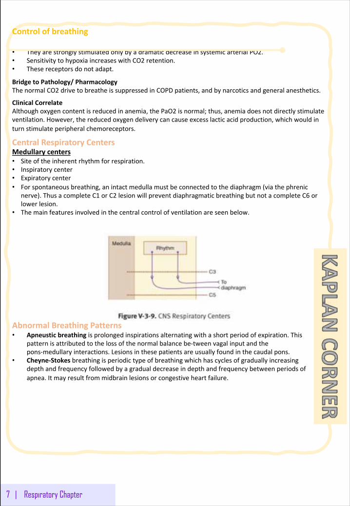
Controlofbreathing
• TheyarestronglystimulatedonlybyadramaticdecreaseinsystemicarterialPO2.• SensitivitytohypoxiaincreaseswithCO2 retention.• Thesereceptorsdonotadapt.
BridgetoPathology/PharmacologyThenormalCO2 drivetobreatheissuppressedinCOPDpatients,andbynarcoticsandgeneralanesthetics.
ClinicalCorrelateAlthoughoxygencontentisreducedinanemia,thePaO2 isnormal;thus,anemiadoesnotdirectlystimulateventilation.However,thereducedoxygendeliverycancauseexcesslacticacidproduction,whichwouldinturnstimulateperipheralchemoreceptors.
CentralRespiratoryCentersMedullarycenters• Siteoftheinherentrhythmforrespiration.• Inspiratorycenter• Expiratorycenter• Forspontaneousbreathing,anintactmedullamustbeconnectedtothediaphragm(viathephrenic
nerve).ThusacompleteC1 orC2 lesionwillpreventdiaphragmaticbreathingbutnotacompleteC6 orlowerlesion.
• Themainfeaturesinvolvedinthecentralcontrolofventilationareseenbelow.
AbnormalBreathingPatterns• Apneustic breathingisprolongedinspirationsalternatingwithashortperiodofexpiration.This
patternisattributedtothelossofthenormalbalancebe-tweenvagalinputandthepons-medullaryinteractions.Lesionsinthesepatientsareusuallyfoundinthecaudalpons.
• Cheyne-Stokes breathingisperiodictypeofbreathingwhichhascyclesofgraduallyincreasingdepthandfrequencyfollowedbyagradualdecreaseindepthandfrequencybetweenperiodsofapnea.Itmayresultfrommidbrainlesionsorcongestiveheartfailure.
7 | Respiratory Chapter
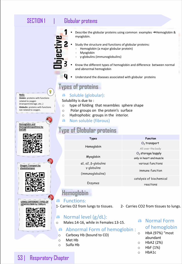
53 | Respiratory Chapter
SECTION 1 | Globular proteins
Types of proteins
§ Describetheglobularproteinsusingcommonexamples➔Hemoglobin&myoglobin.
§ Studythestructureandfunctionsofglobularproteins:- Hemoglobin(amajorglobularprotein)- Myoglobin- γ-globulins(immunoglobulins)
§ Knowthedifferenttypesofhemoglobinanddifferencebetweennormalandabnormalhemoglobin
§ Understandthediseasesassociatedwithglobularproteins
Objec
tive
Soluble(globular):Solubilityisdueto:o typeoffoldingthatresemblessphereshapeo Polargroupsontheprotein’ssurfaceo Hydrophobicgroupsintheinterior.
Nonsoluble(fibrous)
Types Function
HemoglobinO2 transport
All over the body
Myoglobin ystorage/suppl2O
only in heart and muscle
α1, α2, β-globulins various functionsγ-globulins
(immunoglobulins)immune function
Enzymescatalysis of biochemical
reactions
HemoglobinFunctions:
1- CarriesO2 fromlungstotissues.2- CarriesCO2 fromtissuestolungs.
Normallevel(g/dL):o Males:14-16,whileinFemales:13-15.
Note:Globin:proteinswithfunctionsrelatedtooxygen(transport/storage..etc..)Globulin:proteinswithfunctionsnotrelatedtooxygen.
Type of Globular proteins
AbnormalFormofhemoglobin:o CarboxyHb(boundtoCO)o MetHbo SulfaHb
NormalFormofhemoglobin
o HbA (97%)“mostabundant
o HbA2 (2%)o HbF (1%)o HbA1c
HemoglobinandHemoglobinopathies)byDaf189
OxygenTransportbysheekshampoo
USMLEDIFFERENTTYPESOFHEMOGLOBINby100lyric
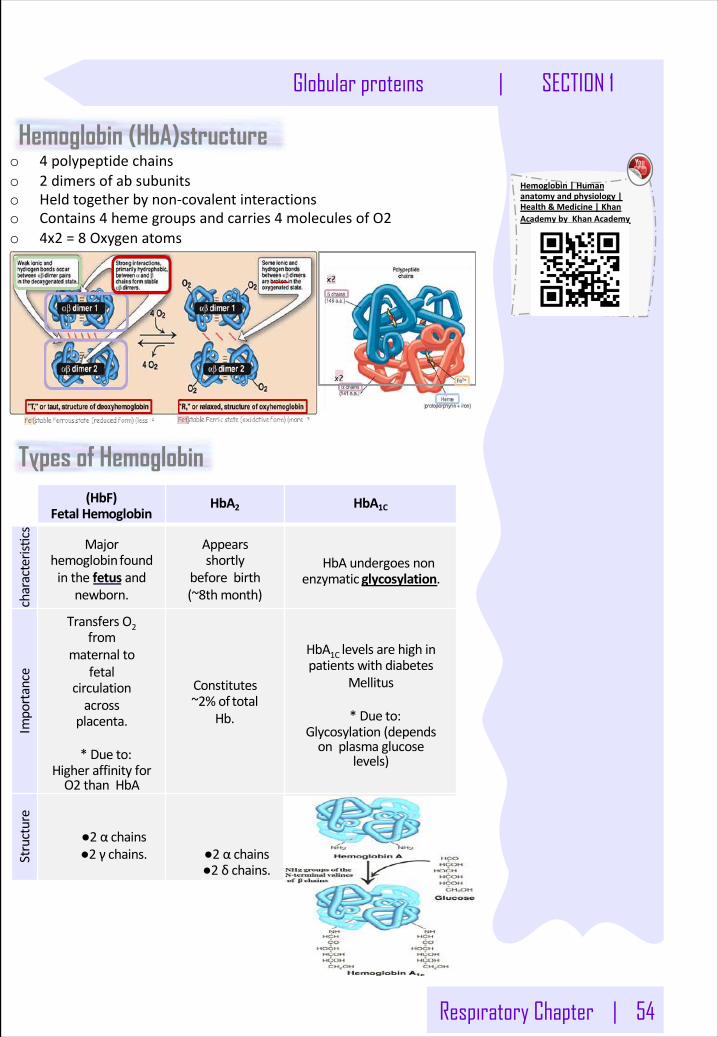
Respiratory Chapter | 54
Globular proteins | SECTION 1
Hemoglobin (HbA)structure
(HbF)Fetal Hemoglobin
HbA2 HbA1C
characteris�cs
Majorhemoglobinfoundinthefetus and
newborn.
Appearsshortly
beforebirth(~8thmonth)
HbAundergoesnonenzymatic glycosylation.
Importance
TransfersO2from
maternaltofetal
circulationacross
placenta.
*Due to:HigheraffinityforO2 thanHbA
Constitutes~2%of total
Hb.
HbA1Clevelsarehighinpatientswithdiabetes
Mellitus
*Due to:Glycosylation(dependsonplasmaglucose
levels)
Structure
●2 α chains●2 γ chains. ●2 α chains
●2 δ chains.
o 4 polypeptidechainso 2 dimersofabsubunitso Heldtogetherbynon-covalentinteractionso Contains4 hemegroupsandcarries4 moleculesofO2o 4x2 =8 Oxygenatoms
Types of Hemoglobin
Hemoglobin|Humananatomyandphysiology|Health&Medicine|KhanAcademybyKhanAcademy
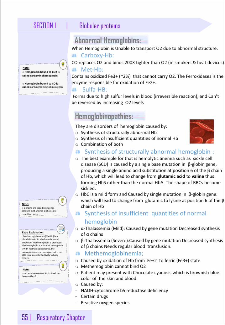
55 | Respiratory Chapter
SECTION 1 | Globular proteins
Hemoglobinopathies:Theyaredisordersofhemoglobincausedby:o SynthesisofstructurallyabnormalHbo SynthesisofinsufficientquantitiesofnormalHbo Combinationofboth
Synthesisofstructurallyabnormalhemoglobin :o Thebestexampleforthatishemolyticanemiasuchassicklecell
disease(SCD)iscausedbyasinglebasemutationinβ-globingene,producingasingleaminoacidsubstitutionatposition6 oftheβchainofHb,whichwillleadtochangefromglutamicacidtovaline thusformingHbSratherthanthenormalHbA.TheshapeofRBCsbecomesickled.
o HbC isamildformandCausedbysinglemutationinβ-globingene.whichwillleadtochangefrom glutamictolysineatposition6 oftheβchainofHbSynthesisofinsufficientquantitiesofnormalhemoglobin
o α-Thalassemia(Mild):CausedbygenemutationDecreasedsynthesisofαchains
o β-Thalassemia(Severe):CausedbygenemutationDecreasedsynthesisofβchainsNeedsregularbloodtransfusion.Methemoglobinemia;
o CausedbyoxidationofHbfromFe+2 toferric(Fe3+)stateo MethemoglobincannotbindO2o PatientmaypresentwithChocolatecyanosiswhichisbrownish-blue
coloroftheskinandblood.o Causedby:- NADH-cytochromeb5 reductasedeficiency- Certaindrugs- Reactiveoxygenspecies
WhenHemoglobinisUnabletotransportO2 duetoabnormalstructure.Carboxy-Hb:
COreplacesO2 andbinds200XtighterthanO2 (insmokers&heatdevices)Met-Hb:
ContainsoxidizedFe3+(~2%)thatcannotcarryO2.TheFerroxidasesistheenzymeresponsibleforoxidationofFe2+.
Sulfa-HB:Formsduetohighsulfurlevelsinblood(irreversiblereaction),andCan’tbereversedbyincreasingO2 levels
Abnormal Hemoglobins:
Note:o HemoglobinboundtoCO2 iscalledcarbaminohemoglobin.
o HemoglobinboundtoCOiscalledcarboxyhemoglobin oxygen
Note:o αchainsarecodedby2 genesabsencemildanemiaβchainsarecodedby1 gene
ExtraExplanation:oMethemoglobinemia(MetHb)isablooddisorderinwhichanabnormalamountofmethemoglobinisproduced.Methemoglobinisaformofhemoglobin.oWithmethemoglobinemia,thehemoglobincancarryoxygen,butisnotabletoreleaseiteffectivelytobodytissues.
Note:o Anenzymeconvertferric(Fe+3 )toferrous(Fe+2 )
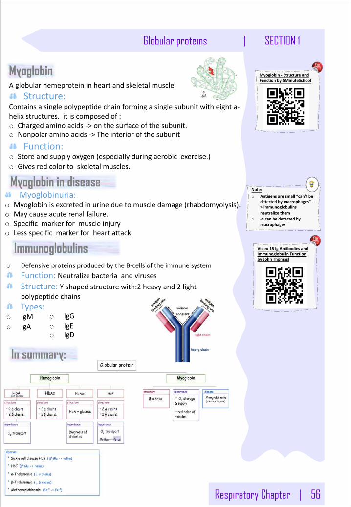
Respiratory Chapter | 56
Globular proteins | SECTION 1
Note:o Antigensaresmall“can’tbe
detectedbymacrophages”->immunoglobulinsneutralizethem
o ->canbedetectedbymacrophages
Myoglobin in diseaseMyoglobinuria:
o Myoglobinisexcretedinurineduetomuscledamage(rhabdomyolysis).o Maycauseacuterenalfailure.o Specificmarkerformuscleinjuryo Lessspecificmarkerforheartattack
Immunoglobulins
In summary:
MyoglobinAglobularhemeproteininheartandskeletalmuscle
Structure: Containsasinglepolypeptidechainformingasinglesubunitwitheighta-helixstructures.itiscomposedof: o Chargedaminoacids ->onthesurfaceofthesubunit.o Nonpolaraminoacids->Theinteriorofthesubunit
Function: o Storeandsupplyoxygen(especiallyduringaerobicexercise.)o Givesredcolortoskeletalmuscles.
o DefensiveproteinsproducedbytheB-cellsoftheimmunesystemFunction:NeutralizebacteriaandvirusesStructure:Y-shapedstructurewith:2 heavyand2 lightpolypeptidechainsTypes:
o IgMo IgA
o IgGo IgEo IgD
Myoglobin- StructureandFunctionby5MinuteSchool
Video15 IgAntibodiesandImmunoglobulinFunctionbyJohnThomasl
57 | Respiratory Chapter
SECTION 1 | Globular proteins
o Aminoacidchainsfoldintoshapesthatresemblespheresarecalled
globularproteins.
o Fibrousproteinsaremainlyinsoluble,whileglobularproteinsaresoluble
structuralproteins.Hb,Myoglobin,globulinesandenzymesare
examplesofglobularproteins.
o Functionally,HbisforO2 andCO2 transport.
o HbA,HbA2 andHbFareexamplesofnormalHb,inwhichthetetrameric
structureiscomposedof2αconstantsubunitswith2 changeableβ
subunitsaccordingtoHbtype.
o HbA1CisaHbAwhichundergoesnon-enzymaticglycosylation,
dependingonplasmaglucoselevels.
o Carboxy-Hb,Met-HbandSulf-HbareexamplesofabnormalHb,inwhich
O2 moleculesarenottransportedduetoabnormalHbstructure.
o DisordersofHbcausedbysynthesisofstructurallyabnormalHband/or
insufficientquantitiesofnormalHb.
o Sicklecell(HbS)andHbCdiseasesarecausedbyasinglemutationinβ-
globingene.
o Glu6 inHbSisreplacedbyVal,whileitisreplacedbyLysinHbC.
o MethemoglobinemiaiscausedbyoxidationofHb,inhibitingO2 binding
leadingtochocolatecyanosis.
o Thalassemiaiscausedbyadefectinsynthesisofeitherα- orβ-globulin
chain,asaresultofgenemutation.
o α-Thalassemiacauseslesssevereanemiathanβ-Thalassemia.
o Myoglobinisaglobularhemeprotein,whichstoresandsuppliesO2 to
theheartandmuscleonly
o Hbiscomposedof4 chains(subunits),whileMyoglobiniscomposedofa
singlechain.
o Myoglobinuriaisaspecificmarkerformuscleinjuryandmaycauseacute
renalfailure.
o ImmunoglobulinsaredefensiveproteinsproducedbytheB-cells.
o Immunoglobulinsconsistof5 types:IgA,IgD,IgE,IgGandIgM
SECTION 2 :
UPPER RESPIRATORY TRACT
58 Histology:The upper respiratory tract.
ANATOMY:Nose, nasal cavity, paranasal sinuses and pharynx
MICROBIOLOGY:Bacteria causing upper respiratory tract infection
MICROBIOLOGY:Viruses Causing Respiratory Infections I
PHARMACOLOGY:Rhinitis and cough
MICROBIOLOGY:Viruses Causing Respiratory Infections II
62
69
78
86
91
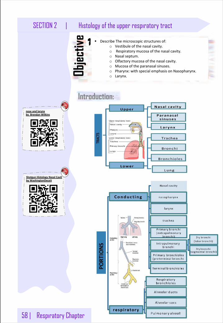
58 | Respiratory Chapter
SECTION 2 | Histology of the upper respiratory tract
Introduction:
§ DescribeThemicroscopicstructuresof:o Vestibuleofthenasalcavity.o Respiratorymucosaofthenasalcavity.o Nasalseptum.o Olfactorymucosaofthenasalcavity.o Mucosaoftheparanasalsinuses.o Pharynx:withspecialemphasisonNasopharynx.o Larynx.Ob
jectiv
e
noseandlarynxbyBrendanWilkins
ShotgunHistologyNasalCavitbyWashingtonDeceit
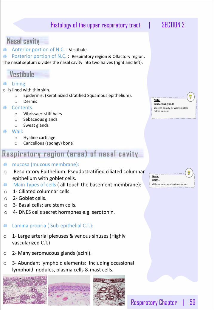
Respiratory Chapter | 59
Histology of the upper respiratory tract | SECTION 2
Respiratory region (area) of nasal cavity
Vestibule
Nasal cavity AnteriorportionofN.C.:Vestibule.PosteriorportionofN.C.:Respiratoryregion&Olfactoryregion.
Thenasalseptumdividesthenasalcavityintotwohalves(rightandleft).
mucosa(mucousmembrane):o RespiratoryEpithelium:Pseudostratifiedciliatedcolumnar
epitheliumwithgobletcells.MainTypesofcells(alltouchthebasementmembrane):
o 1- Ciliatedcolumnarcells.o 2- Gobletcells.o 3- Basalcells:arestemcells.o 4- DNEScellssecrethormonese.g.serotonin.
Laminapropria(Sub-epithelialC.T.):
o 1- Largearterialplexuses&venoussinuses(HighlyvascularizedC.T.)
o 2- Manyseromucousglands(acini).
o 3- Abundantlymphoidelements:Includingoccasionallymphoidnodules,plasmacells&mastcells.
Note:Sebaceousglandssecreteanoilyorwaxymattercalledsebum
Note:DNES=diffuseneuroendocrinesystem.
Lining:o islinedwiththinskin.
o Epidermis:(KeratinizedstratifiedSquamousepithelium).o DermisContents:o Vibrissae:stiffhairso Sebaceousglandso SweatglandsWall:o Hyalinecartilageo Cancellous(spongy)bone
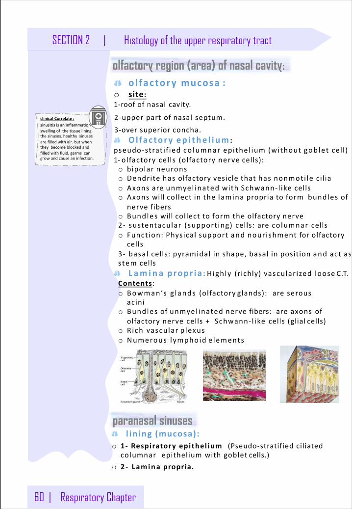
60 | Respiratory Chapter
SECTION 2 | Histology of the upper respiratory tract
olfactory region (area) of nasal cavity:o l fac tory mucosa :
o site:1-roofofnasalcavity.
2-upperpartofnasalseptum.
3-oversuperior concha.Ol fac tory ep i t he l i um :
pseudo-strat i f ied co lumnar epi the l ium (without gob let cell)1- olfactorycel ls (olfactorynerve cells):o bipolar neuronso Dendrite has olfactory vesicle that has nonmoti le cil iao Axons are unmye l inated with Schwann- l ike cellso Axons will collect in the lamina propria to form bundles of
nerve fiberso Bundles will collect to form the olfactory nerve2- sustentacular (support ing) cells:areco lumnar cellso Funct ion: Physical support and nourishment for olfactory
cells3- basal cells: pyramidal in shape, basal in position and act asstem cells
L am i n a propr i a :H igh ly (richly)vascular ized loose C.T.Contents :o Bowman’s g l ands (olfactory glands): areserous
acinio Bundles of unmye l inated nerve fibers:areaxonsof
olfactorynervecells+Schwann- l ike cells(glial cells)o Rich vascular p lexuso Numerous l ympho id e lements
l in ing (mucosa):o 1- Respiratoryepi thel ium (Pseudo-stratifiedciliated
columnar epitheliumwithgoblet cells.)o 2 - Lamina propria.
clinicalCorrelate:sinusitisisaninflammationorswellingofthetissueliningthesinuses.healthysinusesarefilledwithair.butwhentheybecomeblockedandfilledwithfluid,germscangrowandcauseaninfection.
paranasal sinuses
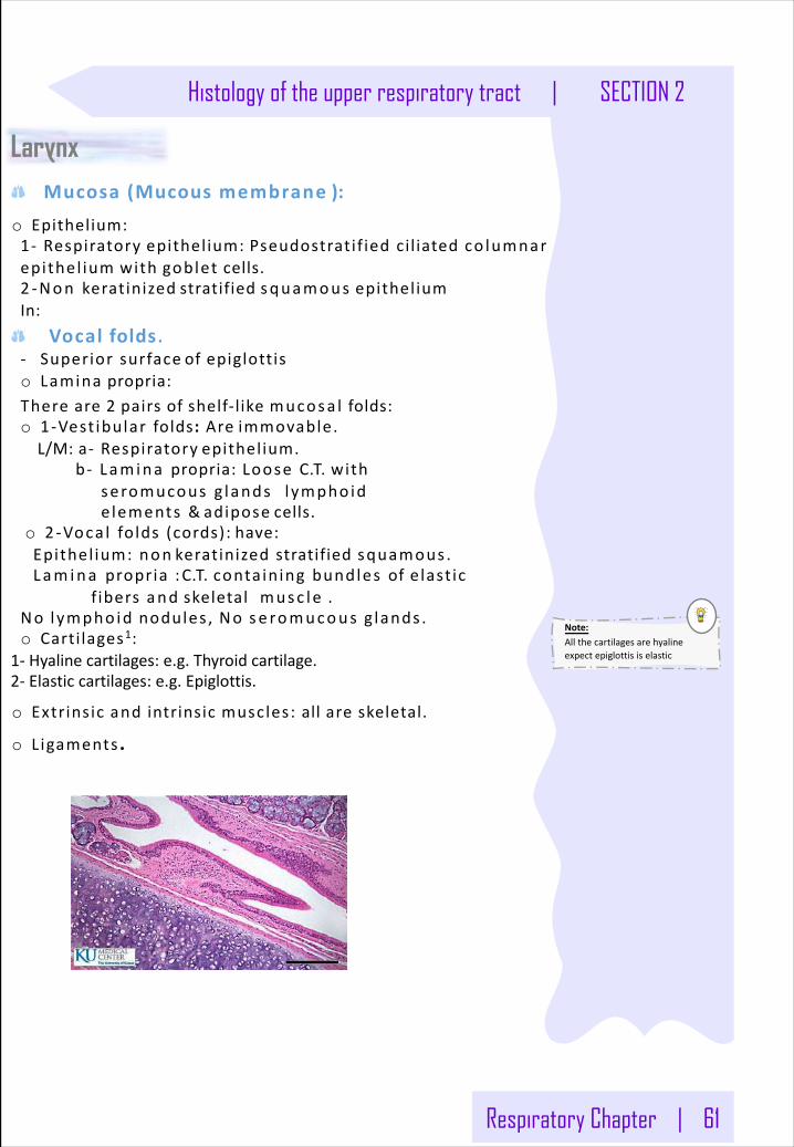
Respiratory Chapter | 61
Histology of the upper respiratory tract | SECTION 2
Larynx Mucosa(Mucousmembrane ):
o Epithelium:1- Respiratory epithel ium: Pseudostratif ied cil iatedco lumnar epithel ium with goblet cells.2 -Non keratinized stratified squamous epithel iumIn:
Vocal folds.- Superiorsurface ofepiglottiso Lamina propria:There are 2 pairs of shelf-like mucosa l folds:o 1-Vest ibular folds:Are immovable.L/M:a- Respiratory epithel ium.
b- Lam ina propria:Loose C.T.withseromucous g lands l ympho id e lements &adipose cells.
o 2-Voca l fo lds (cords): have:Epithel ium:non keratinizedstratifiedsquamous .Lam ina propria :C.T.conta in ing bundles ofe last ic
f ibers and skeletal musc le .No lympho id nodules , No se romucous g lands .o Carti lages1:
1- Hyalinecartilages:e.g.Thyroidcartilage.2- Elasticcartilages:e.g.Epiglottis.
o Extrinsic and intrinsic muscles: all are skeletal.
o Ligaments .
Note:Allthecartilagesarehyalineexpectepiglottisiselastic
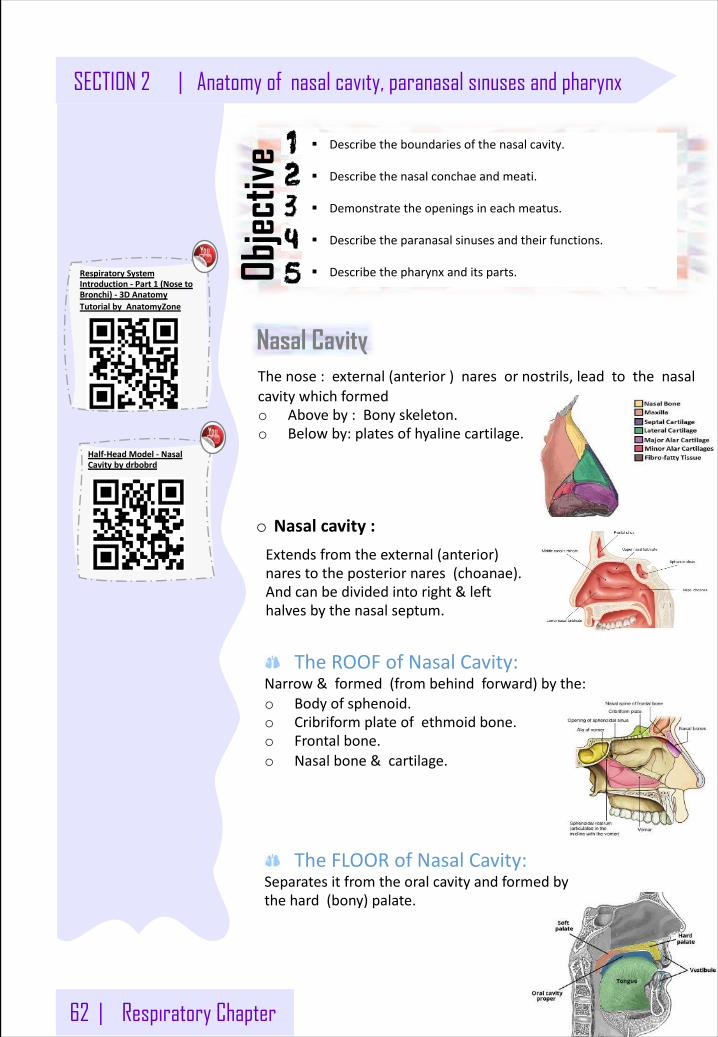
62 | Respiratory Chapter
SECTION 2 | Anatomy of nasal cavity, paranasal sinuses and pharynx
Nasal Cavity
§ Describetheboundariesofthenasalcavity.
§ Describethenasalconchaeandmeati.
§ Demonstratetheopeningsineachmeatus.
§ Describetheparanasalsinusesandtheirfunctions.
§ Describethepharynxanditsparts.Objec
tive
Thenose:external(anterior)naresornostrils,leadtothenasalcavitywhichformedo Aboveby:Bonyskeleton.o Belowby:platesofhyalinecartilage.
o Nasalcavity:Extendsfromtheexternal(anterior)narestotheposteriornares(choanae).Andcanbedividedintoright&lefthalvesbythenasalseptum.
TheROOFofNasalCavity: Narrow&formed(frombehindforward)bythe:o Bodyofsphenoid.o Cribriformplateofethmoidbone.o Frontalbone.o Nasalbone&cartilage.
TheFLOORofNasalCavity: Separatesitfromtheoralcavityandformedbythehard(bony)palate.
RespiratorySystemIntroduction- Part1 (NosetoBronchi)- 3DAnatomyTutorialbyAnatomyZone
Half-HeadModel- NasalCavitybydrbobrd
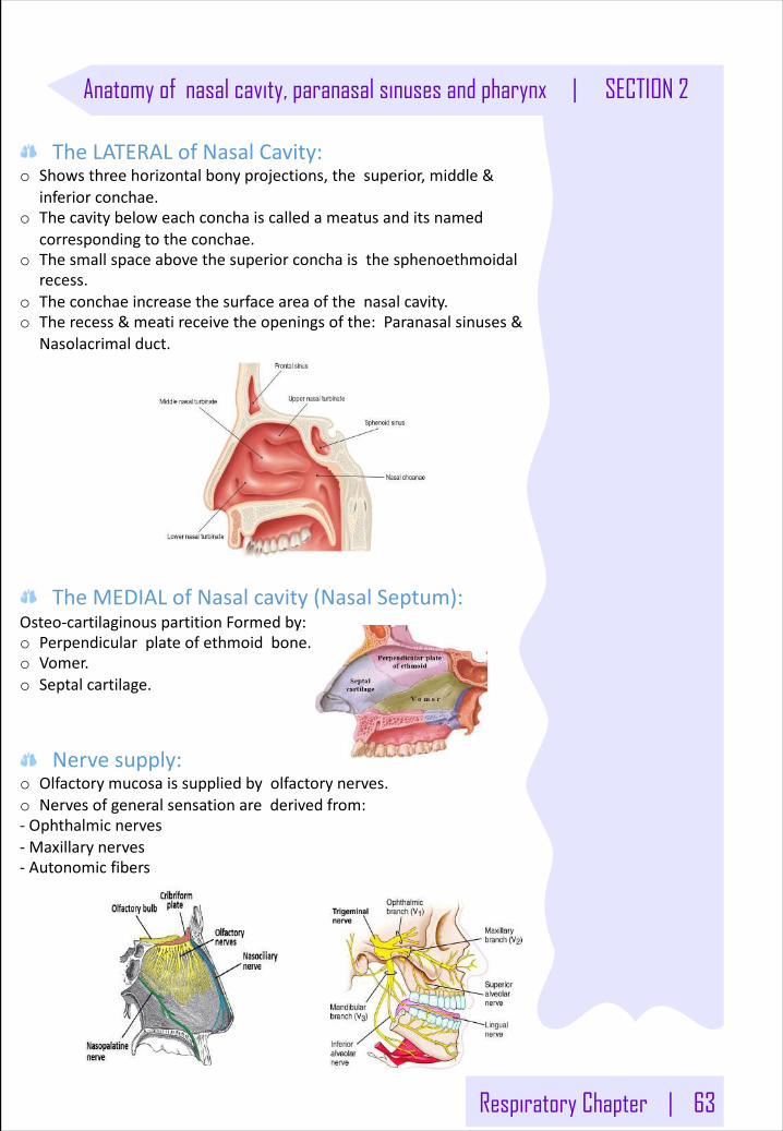
Respiratory Chapter | 63
Anatomy of nasal cavity, paranasal sinuses and pharynx | SECTION 2
TheLATERALofNasalCavity:o Showsthreehorizontalbonyprojections,thesuperior,middle&
inferiorconchae.o Thecavitybeloweachconchaiscalledameatusanditsnamed
correspondingtotheconchae.o Thesmallspaceabovethesuperiorconchaisthesphenoethmoidal
recess.o Theconchaeincreasethesurfaceareaofthenasalcavity.o Therecess&meatireceivetheopeningsofthe:Paranasalsinuses&
Nasolacrimalduct.
TheMEDIALofNasalcavity(NasalSeptum):Osteo-cartilaginouspartitionFormedby:o Perpendicularplateofethmoidbone.o Vomer.o Septalcartilage.
Nervesupply:o Olfactorymucosaissuppliedbyolfactorynerves.o Nervesofgeneralsensationarederivedfrom:- Ophthalmicnerves- Maxillarynerves- Autonomicfibers
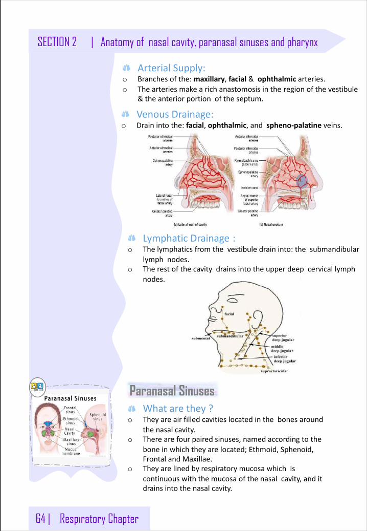
64 | Respiratory Chapter
SECTION 2 | Anatomy of nasal cavity, paranasal sinuses and pharynx
ArterialSupply:o Branchesofthe:maxillary,facial &ophthalmic arteries.o Thearteriesmakearichanastomosisinthe regionofthevestibule
&theanteriorportionoftheseptum.
VenousDrainage:o Drainintothe:facial,ophthalmic,andspheno-palatineveins.
LymphaticDrainage :o Thelymphaticsfromthevestibuledraininto:thesubmandibular
lymphnodes.o Therestofthecavitydrainsintotheupperdeepcervicallymph
nodes.
Paranasal SinusesWhatarethey?
o Theyareairfilledcavitieslocatedinthebonesaroundthenasalcavity.
o Therearefourpairedsinuses,namedaccordingtotheboneinwhichtheyarelocated;Ethmoid,Sphenoid,FrontalandMaxillae.
o Theyarelinedbyrespiratorymucosawhichiscontinuouswiththemucosaofthenasalcavity,anditdrainsintothenasalcavity.
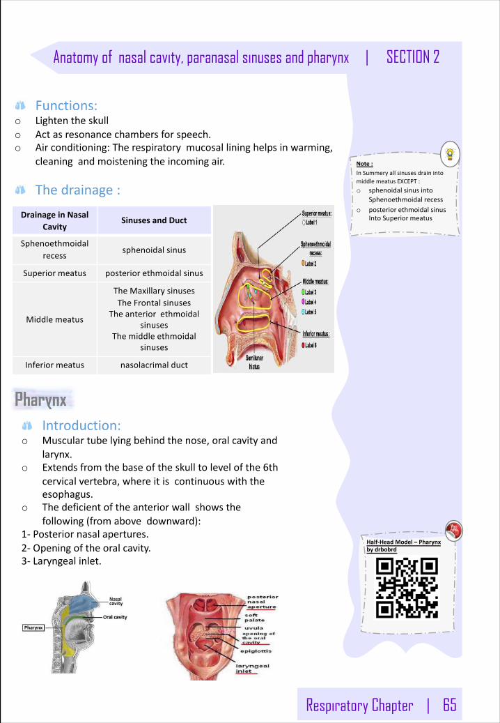
Respiratory Chapter | 65
Anatomy of nasal cavity, paranasal sinuses and pharynx | SECTION 2
Half-HeadModel– Pharynxbydrbobrd
DrainageinNasalCavity
SinusesandDuct
Sphenoethmoidalrecess sphenoidalsinus
Superiormeatus posteriorethmoidalsinus
Middlemeatus
TheMaxillarysinusesTheFrontalsinuses
Theanteriorethmoidalsinuses
Themiddleethmoidalsinuses
Inferiormeatus nasolacrimalduct
Functions:o Lightentheskullo Actasresonancechambersforspeech.o Airconditioning:Therespiratorymucosallininghelpsinwarming,
cleaningandmoisteningtheincomingair.
Thedrainage:
Note:InSummeryallsinusesdrainintomiddlemeatusEXCEPT:o sphenoidalsinusinto
Sphenoethmoidalrecesso posteriorethmoidalsinus
IntoSuperiormeatus
PharynxIntroduction:
o Musculartubelyingbehindthenose,oralcavityandlarynx.
o Extendsfromthebaseoftheskulltolevelofthe6thcervicalvertebra,whereitiscontinuouswiththeesophagus.
o Thedeficientoftheanteriorwallshowsthefollowing(fromabovedownward):
1- Posteriornasalapertures.2- Openingoftheoralcavity.3- Laryngealinlet.
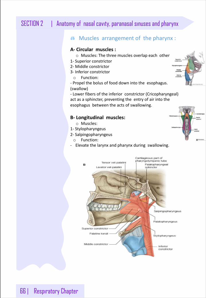
66 | Respiratory Chapter
SECTION 2 | Anatomy of nasal cavity, paranasal sinuses and pharynx
Musclesarrangementofthepharynx:
A- Circularmuscles:o Muscles:Thethreemusclesoverlapeachother
1- Superiorconstrictor2- Middleconstrictor3- Inferiorconstrictoro Function:
- Propelthebolusoffooddownintotheesophagus.(swallow)- Lowerfibersoftheinferiorconstrictor(Cricopharyngeal)actasasphincter,preventingtheentryofairintotheesophagusbetweentheactsofswallowing.
B- Longitudinalmuscles:o Muscles:
1- Stylopharyngeus2- Salpingopharyngeuso Function:
- Elevatethelarynxandpharynxduringswallowing.
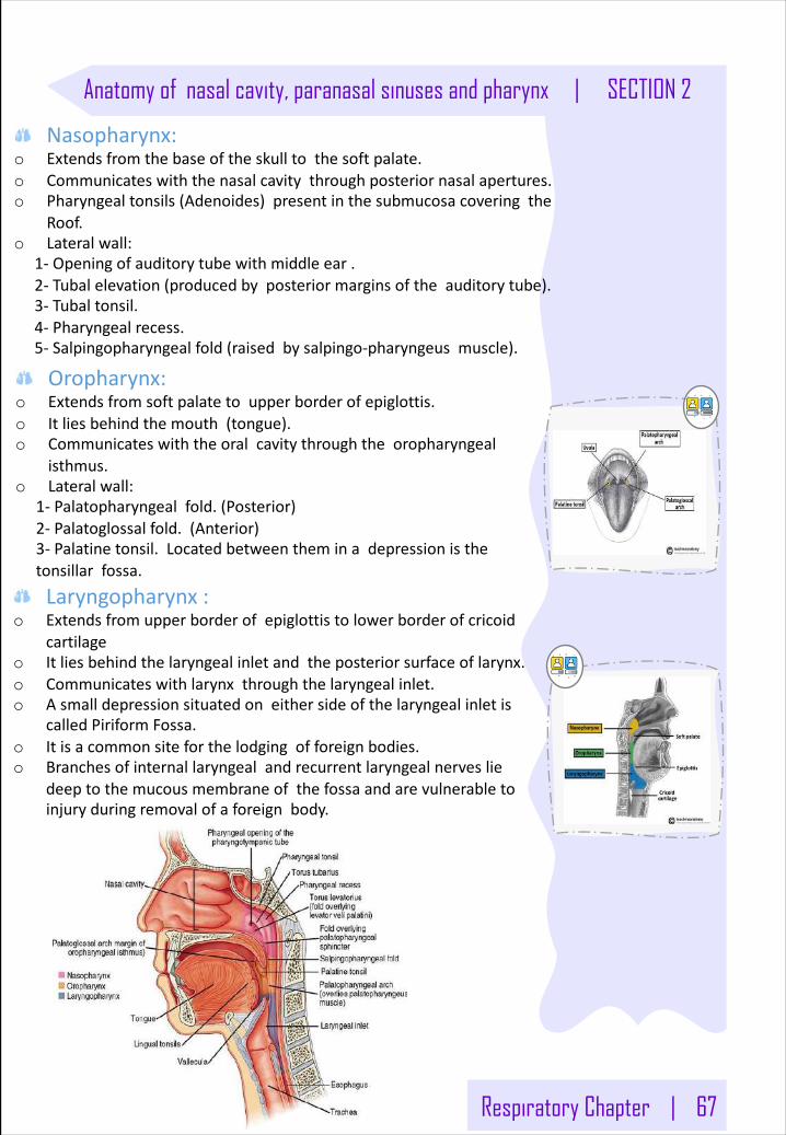
Respiratory Chapter | 67
Anatomy of nasal cavity, paranasal sinuses and pharynx | SECTION 2
Oropharynx:o Extendsfromsoftpalatetoupperborderofepiglottis.o Itliesbehindthemouth(tongue).o Communicateswiththeoralcavitythroughtheoropharyngeal
isthmus.o Lateralwall:
1- Palatopharyngealfold.(Posterior)2- Palatoglossalfold.(Anterior)3- Palatinetonsil.Locatedbetweentheminadepressionisthetonsillarfossa.
Laryngopharynx:o Extendsfromupperborderofepiglottistolowerborderofcricoid
cartilageo Itliesbehindthelaryngealinletandtheposteriorsurfaceoflarynx.o Communicateswithlarynxthroughthelaryngealinlet.o Asmalldepressionsituatedoneithersideofthelaryngealinletis
calledPiriformFossa.o Itisacommonsiteforthelodgingofforeignbodies.o Branchesofinternallaryngealandrecurrentlaryngealnerveslie
deeptothemucousmembraneofthefossaandarevulnerabletoinjuryduringremovalofaforeignbody.
Nasopharynx:o Extendsfromthebaseoftheskulltothesoftpalate.o Communicateswiththenasalcavitythroughposteriornasalapertures.o Pharyngealtonsils(Adenoides)presentinthesubmucosacoveringthe
Roof.o Lateralwall:
1- Openingofauditorytubewithmiddleear.2- Tubalelevation(producedbyposteriormarginsoftheauditorytube).3- Tubaltonsil.4- Pharyngealrecess.5- Salpingopharyngealfold(raisedbysalpingo-pharyngeusmuscle).
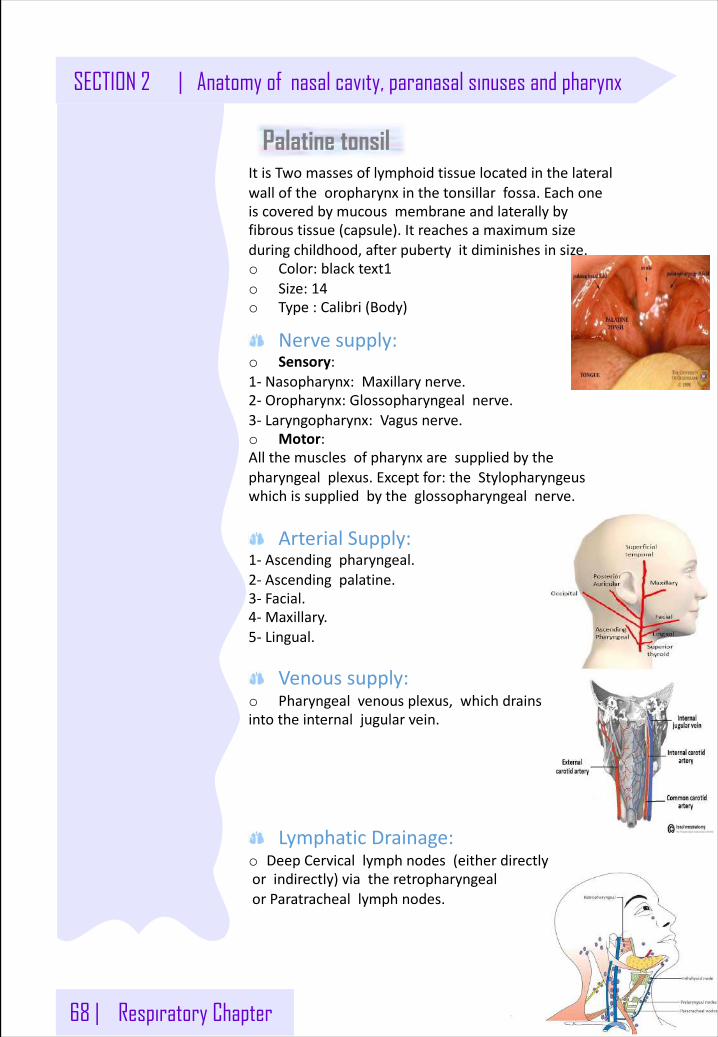
68 | Respiratory Chapter
SECTION 2 | Anatomy of nasal cavity, paranasal sinuses and pharynx
Palatine tonsil
Nervesupply:o Sensory:1- Nasopharynx:Maxillarynerve.2- Oropharynx:Glossopharyngealnerve.3- Laryngopharynx:Vagusnerve.o Motor:Allthemusclesofpharynxaresuppliedbythepharyngealplexus.Exceptfor:theStylopharyngeuswhichissuppliedbytheglossopharyngealnerve.
ArterialSupply:1- Ascendingpharyngeal.2- Ascendingpalatine.3- Facial.4- Maxillary.5- Lingual.
Venoussupply:o Pharyngealvenousplexus,whichdrainsintotheinternaljugularvein.
LymphaticDrainage:o DeepCervicallymphnodes(eitherdirectlyorindirectly)viatheretropharyngealorParatracheallymphnodes.
ItisTwomassesoflymphoidtissuelocatedinthelateralwalloftheoropharynxinthetonsillarfossa.Eachoneiscoveredbymucousmembraneandlaterallybyfibroustissue(capsule).Itreachesamaximumsizeduringchildhood,afterpubertyitdiminishesinsize.o Color:blacktext1o Size:14o Type:Calibri(Body)
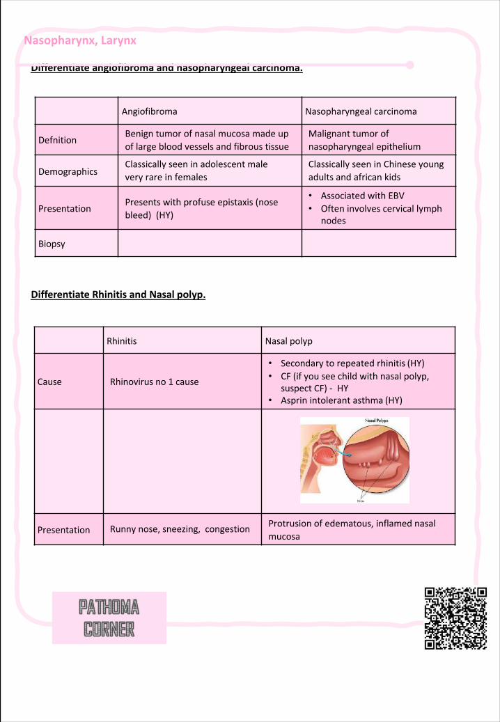
Nasopharynx,Larynx
Differentiateangiofibroma andnasopharyngeal carcinoma.
DifferentiateRhinitisandNasal polyp.
Angiofibroma Nasopharyngeal carcinoma
DefnitionBenigntumorofnasalmucosamadeupoflargebloodvesselsandfibroustissue
Malignanttumorofnasopharyngealepithelium
DemographicsClassicallyseeninadolescentmaleveryrareinfemales
ClassicallyseeninChineseyoungadultsandafrican kids
PresentationPresentswithprofuseepistaxis(nosebleed) (HY)
• AssociatedwithEBV• Ofteninvolvescervicallymph
nodes
Biopsy
Rhinitis Nasalpolyp
Cause Rhinovirusno1 cause
• Secondarytorepeatedrhinitis (HY)• CF(ifyouseechildwithnasalpolyp,
suspectCF)- HY• Asprin intolerantasthma (HY)
Presentation Runnynose,sneezing,congestion Protrusionofedematous,inflamednasalmucosa
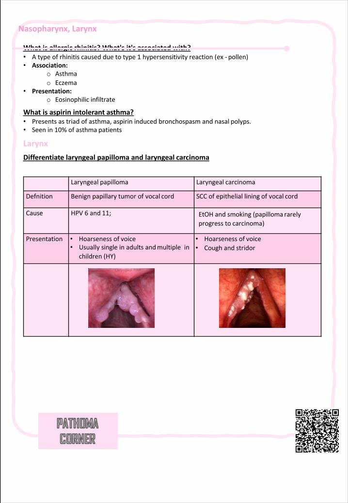
Nasopharynx,Larynx
Whatisallergicrhinitis?What'sit'sassociated with?• Atypeofrhinitiscausedduetotype1 hypersensitivityreaction(ex- pollen)• Association:
o Asthmao Eczema
• Presentation:o Eosinophilic infiltrate
Whatisaspirinintolerant asthma?• Presentsastriadofasthma,aspirininducedbronchospasmandnasal polyps.• Seenin10%ofasthma patients
LarynxDifferentiatelaryngealpapillomaandlaryngeal carcinoma
Laryngeal papilloma Laryngeal carcinoma
Defnition Benignpapillarytumorofvocal cord SCCofepithelialliningofvocal cord
Cause HPV6 and 11; EtOHandsmoking(papilloma rarelyprogressto carcinoma)
Presentation • Hoarsenessof voice• Usuallysingleinadultsandmultiplein
children (HY)
• Hoarsenessof voice• Coughand stridor

Nasopharynx,Larynx
Differentiateacuteepiglottitisandlaryngotracheobronchitis (croup).
Whatisvocalcordnodule(singer'snodule)?What'sits cause?• Noduleontruevocal cord• Causedduetoexcessiveuseofvocalcord;usuallybilateral(wearandtear issue)• Composedondegenerativemyxoid connective tissue• Treatwith rest
Fig:vocalcordnodule(usuallybilateralandseenontruevocal cord)
Acute epiglottitis laryngotracheobronchitis (croup)
Cause H.Influenzaetypebmostcommoncause (inimmunizedornon-immunized kids.
Parainfluenzavirusmostcommoncause
Presentation Toomuchdrooling,dysphagia,sorethroat,fever,muffledvoice,inspiratory stridor
Barkingcoughand inspiratorystridor
Riskofactueairwayobstruction(medicalemergency)
X-ray Thumbsignon X-ray Steeplesignon X-ray
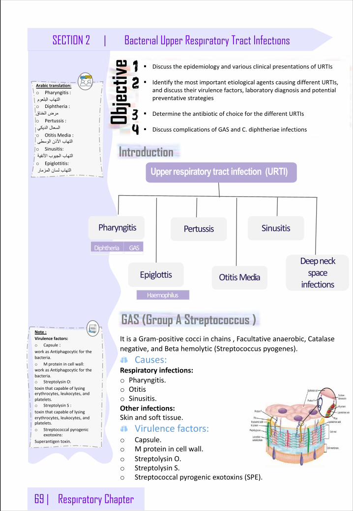
69 | Respiratory Chapter
SECTION 2 | Bacterial Upper Respiratory Tract Infections
Introduction
§ DiscusstheepidemiologyandvariousclinicalpresentationsofURTIs
§ IdentifythemostimportantetiologicalagentscausingdifferentURTIs,anddiscusstheirvirulencefactors,laboratorydiagnosisandpotentialpreventativestrategies
§ DeterminetheantibioticofchoiceforthedifferentURTIs
§ DiscusscomplicationsofGASandC.diphtheriaeinfectionsOb
jectiv
e
ItisaGram-positivecocciinchains,Facultativeanaerobic,Catalasenegative,andBetahemolytic(Streptococcuspyogenes).
Causes:Respiratoryinfections:o Pharyngitis.o Otitiso Sinusitis.Otherinfections:Skinandsofttissue.
Virulencefactors:o Capsule.o Mproteinincellwall.o StreptolysinO.o StreptolysinS.o Streptococcalpyrogenicexotoxins(SPE).
Upper respiratory tract infection (URTI)
Pharyngitis
Epiglottis
Pertussis
OtitisMedia
Sinusitis
Deepneckspace
infections
GASDiphtheria
Haemophilus
GAS (Group A Streptococcus )Note:Virulencefactors:o Capsule :workasAntiphagocyticforthebacteria.o Mproteinincellwall:workasAntiphagocyticforthebacteria.o StreptolysinO:toxinthatcapableoflysingerythrocytes,leukocytes,andplatelets.o StreptolysinS:toxinthatcapableoflysingerythrocytes,leukocytes,andplatelets.o Streptococcalpyrogenic
exotoxins:Superantigentoxin.
Arabictranslation:o Pharyngitis:
موعلبلا باھتلاo Diphtheria:
قانخلا ضرمo Pertussis:
يكیدلا لاعسلاo OtitisMedia:
ىطسولا نذألا باھتلاo Sinusitis:
ةیفنألا بویجلا باھتلاo Epiglottitis:
رامزملا ناسل باھتلا
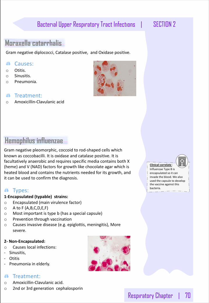
Respiratory Chapter | 70
Bacterial Upper Respiratory Tract Infections | SECTION 2
Gramnegativepleomorphic,coccoidtorod-shapedcellswhichknownascoccobacilli.Itisoxidaseandcatalasepositive.ItisfacultativelyanaerobicandrequiresspecificmediacontainsbothX(heme)andV(NAD)factorsforgrowthlikechocolateagarwhichisheatedbloodandcontainsthenutrientsneededforitsgrowth,anditcanbeusedtoconfirmthediagnosis.
Types:1-Encapsulated(typable)strains:o Encapsulated(mainvirulencefactor)o AtoF(A,B,C,D,E,F)o Mostimportantistypeb(hasaspecialcapsule)o Preventionthroughvaccinationo Causesinvasivedisease(e.g.epiglottis,meningitis),More
severe.
2- Non-Encapsulated:o Causeslocalinfections:- Sinusitis,- Otitis- Pneumoniainelderly.
Treatment:o Amoxicillin-Clavulanicacid.o 2ndor3rdgenerationcephalosporin
Gramnegativediplococci,Catalasepositive,andOxidasepositive.
Causes:o Otitis.o Sinusitis.o Pneumonia.
Treatment:
o Amoxicillin-Clavulanicacid
Moraxella catarrhalis
Hemophilus influenzae
Clinicalcorrelate:InfluenzaeTypeBisencapsulatedsoitcaninvadetheblood.Wealsousedthecapsuletodevelopthevaccineagainstthisbacteria.
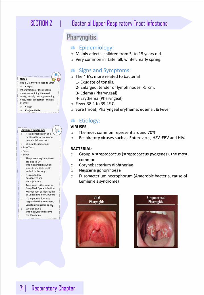
71 | Respiratory Chapter
SECTION 2 | Bacterial Upper Respiratory Tract Infections
Epidemiology:o Mainlyaffectschildrenfrom5 to15 yearsold.o VerycommoninLatefall,winter,earlyspring.
SignsandSymptoms:o The4 E’s:morerelatedtobacterial
1- Exudateoftonsils.2- Enlarged,tenderoflymphnodes>1 cm.3- Edema(Pharyngeal)4- Erythema(Pharyngeal)
o Fever38.4 to39.4ºC.o Sorethroat,Pharyngealerythema,edema,&Fever
Etiology:VIRUSES:o Themostcommon representaround70%.o RespiratoryvirusessuchasEnterovirus,HSV,EBVandHIV.
BACTERIAL:o GroupAstreptococcus(streptococcuspyogenes),themost
common o Corynebacteriumdiphtheriae o Neisseriagonorrhoeaeo Fusobacteriumnecrophorum(Anaerobicbacteria,causeof
Lemierre'ssyndrome)
Pharyngitis
Note:The3 C’s,morerelatedtoviralo Coryza: Inflammationofthemucousmembranesliningthenasalcavity,usuallycausingarunningnose,nasalcongestionandlossofsmell.o Cougho Conjunctivitis
Lemierre'sSyndrome:o Itisacomplicationofa
peritonsillarabscessorapostdentalinfection.
o ClinicalPresentation:- SoreThroat- Fever- Shocko Thepresentingsymptoms
areduetoIJVthrombophlebitiswhichleadstomultiplesepticemboliinthelung.
o ItiscausedbyFusobacteriumNecrophorum
o TreatmentisthesameasDeepNeckSpaceinfectionMeropenemorPiperacillinorClindamycinfor2 weeks
o Ifthepatientdoesnotrespondtothetreatment,venotomymustbedone.
o Wealsogiveathrombolytictodissolvethethrombus
Respiratory Chapter | 72
Bacterial Upper Respiratory Tract Infections | SECTION 2
GAS Pharyngitis (Group A Streptococcus)
Diagnosis:Throatswab:o RapidBacterialantigendetection.ImpinER.o Cultureonbloodagar.
Antistreptolysin O.
Treatment:o Drugofchoice:Penicillinfor10 days.IncaseofAllergyto
penicillin,weuse:Clindamycinormacrolide(e.g.Clarithromycin).o Clarithromycinisanewtypewhichhasfewersideeffects,better
penetration,&longerhalf-life.
Complications:
SUPPURATIVE:Thereisformationofpusanditoccursrightawayaftertheinfection.e.g.peritonsillarabscessandparapharyngealspaceabscess.
NON-SUPPURATIVE:Itoccurs1-6 weeksafteracuteS.pyogenesinfection.
o Rheumaticfever:- Whenithappens?afterrespiratorytractinfection.- Whatdoesitdotothebody?mainlycauseinflammationofheart(pancarditis),andinflammationofjoints,bloodvessels,andsubcutaneoustissue.- Howithappens?resultsfromcrossreactivityofanti-MproteinAbandthehumanhearttissue.
o AcuteGlomerulonephritis:- Whenithappens?afterinfectionoftheskinorrespiratorytract.- WhataretheSymptoms?Edema,hypertension,hematuria,andproteinuria.- Whyithappens?InitiatedbyAg-Abcomplexesontheglomerularbasementmembrane.
73 | Respiratory Chapter
SECTION 2 | Bacterial Upper Respiratory Tract Infections
DiphtheriaEpidemiology:
o Oneofthemostcommoncausesofdeathinunvaccinatedchildren1-5 years.
o FoundmostinNon-developingcountries.o Toxinmediateddisease
Pathogenesis:o Rapidprogression,tightlyadheringgraymembraneinthethroat.
Etiology:o PharyngitiscausedbyCorynebacteriumdiphtheriae(agram-positive
bacilli,Aerobic,non-sporeforming).o Virulence:Diphtheriatoxin:It’satoxinproducedbyC.diphtheriae,whichinhibittheproteinsynthesisofthecellandcausecelldeath,targets:heart/nerves/epithelium.
SignsandSymptoms:o MainlypresentsasURTI.,oneofitscharacteristicisformationof
pseudomembranesinthethroat.
Diagnosis:o Throatswab.o Cultureonspecialmediacontainingtellurite(e.g.Tinsdalemedia).o ELEK’sTestforconfirmationoftoxinproduction.
Complications:o Myocarditisalsoknownasinflammatorycardiomyopathy,is
inflammationoftheheartmuscle.o Neuritisitisinflammationofanerveorthegeneralinflammation
oftheperipheralnervoussystem.Symptomsdependonthenervesinvolved.
Treatment:o WegivebothAntitoxinandantibiotic.Penicillincanbegivenor
erythromycinifthechildisallergictopenicillin.
Prevention:o Vaccinationwithdiphtheriatoxoid.
Note:Diphtheriaispresentasnormalflorainourbodies,butitisnon-toxigenic.Whenitgetsinfectedwithabacteriophage(whichisavirus),ittransmitsatoxinandbecomestoxigenic.Superantigentoxin.
o Note:o Thepatientwill
presentwithasorethroat,difficultyinbreathingandswallowinganddroolingofthesaliva.
o Themembranecangetbiggerandbiggeruntilitobstructstheairwayandwillcausesuffocationanddeathwhichiswhytheycallthisdisease.
o Note:ELEK’STest:o Ateststripoffilter
papercontainingdiphtheriaantitoxinisplacedinthecenteroftheagarplate.Strainstobetested(patient’sisolate),knownpositiveandnegativetoxigenicstrainsarealsostreakedontheagar’ssurfaceinalineacrosstheplateandatarightangletotheantitoxinpaperstrip.
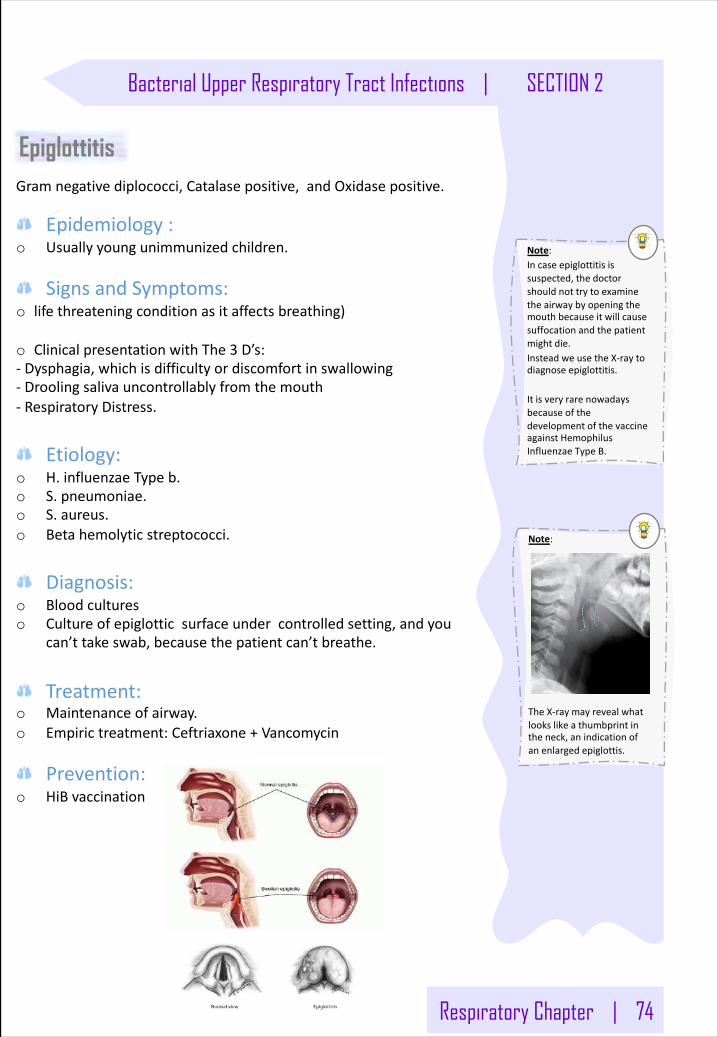
Respiratory Chapter | 74
Bacterial Upper Respiratory Tract Infections | SECTION 2
Gramnegativediplococci,Catalasepositive,andOxidasepositive.
Epidemiology:o Usuallyyoungunimmunizedchildren.
SignsandSymptoms:o lifethreateningconditionasitaffectsbreathing)
o ClinicalpresentationwithThe3 D’s:- Dysphagia,whichisdifficultyordiscomfortinswallowing- Droolingsalivauncontrollablyfromthemouth- RespiratoryDistress.
Etiology:o H.influenzaeTypeb.o S.pneumoniae.o S.aureus.o Betahemolyticstreptococci.
Diagnosis:o Bloodcultureso Cultureofepiglotticsurfaceundercontrolledsetting,andyou
can’ttakeswab,becausethepatientcan’tbreathe.
Treatment:
o Maintenanceofairway.o Empirictreatment:Ceftriaxone+Vancomycin
Prevention:o HiBvaccination
Epiglottitis
Note:Incaseepiglottitisissuspected,thedoctorshouldnottrytoexaminetheairwaybyopeningthemouthbecauseitwillcausesuffocationandthepatientmightdie.InsteadweusetheX-raytodiagnoseepiglottitis.
ItisveryrarenowadaysbecauseofthedevelopmentofthevaccineagainstHemophilusInfluenzaeTypeB.
Note:
TheX-ray mayrevealwhatlookslikeathumbprintintheneck,anindicationofanenlarged epiglottis.
75 | Respiratory Chapter
SECTION 2 | Bacterial Upper Respiratory Tract Infections
ExtraExplanation: Pathogenesis:o Thebacteriaattachtothe
ciliaoftherespiratoryepithelialcells.
o Itproducetoxins(Pertussistoxin)thatparalyzetheciliawhichcauseinflammationoftherespiratorytractandinterfereswiththeclearingofthepulmonarysecretions.
o ItalsoproduceFilamentoushemagglutinin(FHA)andPertactin(PRN),theseproductsareresponsiblefortheclinicalfeatures.
o CausesLeukocytosisandLymphocytosis,andItistheonlybacterialinfectionthatcauseslymphocytosis.
Epidemiology:o Mainlyininfantsandchildren(mostsevere&deadly).o Adultscangetinfectedalso.
Etiology:
o Bordetellapertussis(GNB).
Thecourseofthedisease:o Incubationperiod:From1 to3 weeks,withNosymptoms.o CatarrhalStage:From1 to2 weeks,withmildoccasionalcoughandrunnynose.o ParoxysmalStage:From2 to4 weeks,withsevere&rapidcough,vomitinganditisdangerous.o ConvalescentStage:From1 to2 weeks,withGradualrecovery,Thecoughbeingtocalm.
Virulence:o Pertussistoxino Filamentoushemagglutinino Pertactin
Diagnosis:o Nasopharyngealswabs,whichusuallyareusedforthediagnosisof
viralinfectionso Specialmedianeeded:
- Charcoalblood(Regan-Lowe)- Bordetellaselectivemedia(BordetGengou)
Treatment:
o Preventedbyvaccination.o Erythromycin.
Pertussis (Whooping cough):
Note: o Pertussisininfantsless
than6 monthswillpresentwithcyanosisbecausethesestoxinswillproduceathickmucusthatwillblocktheirtinytracheaandpreventairfromflowingin.
o Inolderchildrenthediseasewillpresentwithwhoopingcough.Inadults,thepatientswillhaveachroniccough.
Note: o Erythromycinisalsogiven
asaprophylactictreatmenttothepeoplewhowereincontactwithaninfectedindividualuntiltheantibodiesagainstthebacteriaareproducedasaresponsetothevaccinetopreventfurtherspreadoftheinfection.
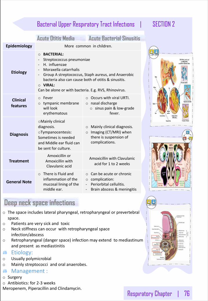
Respiratory Chapter | 76
Bacterial Upper Respiratory Tract Infections | SECTION 2
o Thespaceincludeslateralpharyngeal,retropharyngealorprevertebralspace.
o Patientsareverysickandtoxico Neckstiffnesscanoccurwithretropharyngealspace
infection/abscesso Retropharyngeal(dangerspace)infectionmayextendtomediastinum
andpresentasmediastinitis Etiology:
o Usuallypolymicrobialo Mainlystreptococciandoralanaerobes.
Management:o Surgeryo Antibiotics:for2-3 weeksMeropenem,PiperacillinandClindamycin.
Epidemiology Morecommoninchildren.
Etiology
o BACTERIAL:- Streptococcuspneumoniae- H.influenzae- Moraxellacatarrhalis- GroupAstreptococcus,Staphaureus,andAnaerobic
bacteriaalsocancausebothofotitis&sinusitis.o VIRAL:Canbealoneorwithbacteria.E.g.RVS,Rhinovirus.
Clinicalfeatures
o Fevero tympanicmembrane
willlookerythematous
o OccurswithviralURTI.o nasaldischargeo sinuspain&low-grade
fever.
Diagnosis
oMainlyclinicaldiagnosis.oTympanocentesis:SometimesisneededandMiddleearfluidcanbesentforculture.
o Mainlyclinicaldiagnosis.o Imaging(CT/MRI)when
thereissuspensionofcomplications.
TreatmentAmoxicillinorAmoxicillinwithClavulanicacid
AmoxicillinwithClavulanicacidfor1 to2 weeks
GeneralNote
o ThereisFluidandinflammationofthemucosalliningofthemiddleear.
o Canbeacuteorchronic o complication:- Periorbitalcellulitis.- Brainabscess&meningitis
Deep neck space infections
Acute Otitis Media Acute Bacterial Sinusitis
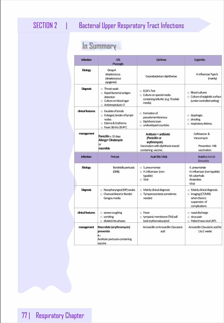
77 | Respiratory Chapter
SECTION 2 | Bacterial Upper Respiratory Tract Infections
In SummaryInfection GAS
PharyngitisDiphtheria Epiglottitis
Etiology GroupAstreptococcus(streptococcuspyogenes)
Corynebacteriumdiphtheriae H.influenzaeTypeb(mainly)
Diagnosis o Throatswabo RapidBacterialantigen
detectiono Cultureonbloodagaro AntistreptolysinO
o ELEK’sTesto Cultureonspecialmedia
containingtellurite(e.g.Tinsdalemedia)
o Bloodcultureso Cultureofepiglotticsurface
(undercontrolledsetting)
clinicalfeatures o Exudateoftonsilso Enlarged,tenderoflymph
nodeso Edema&Erythemao Fever38.4to39.4ºC
o Formationofpseudomembranous
o Diphtheriatoxino undevelopedcountries
o dysphagia.o drooling.o respiratorydistress.
managementPenicillin x10daysAllergy= Clindamycinormacrolide
Antitoxin + antibiotic (Penicillin orerythromycin)
Vaccinationwithdiphtheriatoxoidcontainingvaccine.
Ceftriaxone&Vancomycin
Prevention:HiBvaccination
Infection Pertussis AcuteOtitisMedia AcuteBacterialSinusitis
Etiology Bordetellapertussis(GNB).
o S.pneumoniaeo H.influenzae(non-
typable)o Viral
-S. pneumoniae-H.influenzae (non-typable)-M.catarrhalis-Anaerobes-Viral
Diagnosis o Nasopharyngeal(NP)swabso CharcoalbloodorBordet-
Gengoumedia
o Mainlyclinicaldiagnosiso Tympanocentesissometimes
needed
o Mainlyclinicaldiagnosis.o Imaging(CT/MRI)
whenthereissuspensionofcomplications
clinicalfeatures o severecoughingo vomitingo dividedintophases
o Fevero tympanicmembrane(TM)will
lookerythematous(red
o nasaldischargeo sinuspaino PatienthaveviralURTI.
management Macrolide (erythromycin)prevention :Acellularpertussis-containingvaccine
AmoxicillinorAmoxicillinClavulanicacid
AmoxicillinClavulanicacidfor1to2 weeks
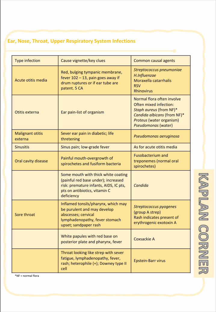
Ear,Nose,Throat,UpperRespiratorySystemInfections
Typeinfection Causevignette/key clues Commoncausalagents
Acuteotitismedia
Red,bulgingtympanicmembrane,fever102 – 13,paingoesawayifdrumrupturesorifeartubearepatent.5 CA
StreptococcuspneumoniaeH.InfluenzaeMoraxellacatarrhalisRSVRhinovirus
Otitisexterna Earpain-listoforganism
NormalfloraofteninvolveOften mixedinfection:Staphaureus(fromNF)*Candidaalbicans (fromNF)*Proteus (waterorganism)Pseudomonas (water)
Malignantotitisexterna
Severearpain indiabetic;lifethretening
Pseudomonasaeruginosa
Sinusitis Sinuspain; low-gradefever Asforacuteotitismedia
OralcavitydiseasePainfulmouth-overgrowthofspirochetesandfusiform bacteria
Fusobacterium andtreponemes (normaloralspirochetes)
Somemouthwiththickwhitecoating(painfulredbaseunder); increasedrisk:prematureinfants,AIDS,ICpts,ptsonantibiotics,vitaminCdeficiency
Candida
Sorethroat
Inflamedtonsils/pharynx, whichmaybepurulentandmaydevelopabscesses;cervicallymphadenopathy,feverstomachupset;sandpaperrash
Streptococcuspyogenes(groupA strep)RashindicatespresentoferythrogenicexotoxinA
White papuleswithredbaseonposteriorplateandpharynx,fever
CoxsackieA
Throatlookinglikestrepwithseverfatigue, lymphadenopathy,fever,rash;heterophile (+);DowneytypeIIcell
Epstein-Barr virus
*NF=normalflora
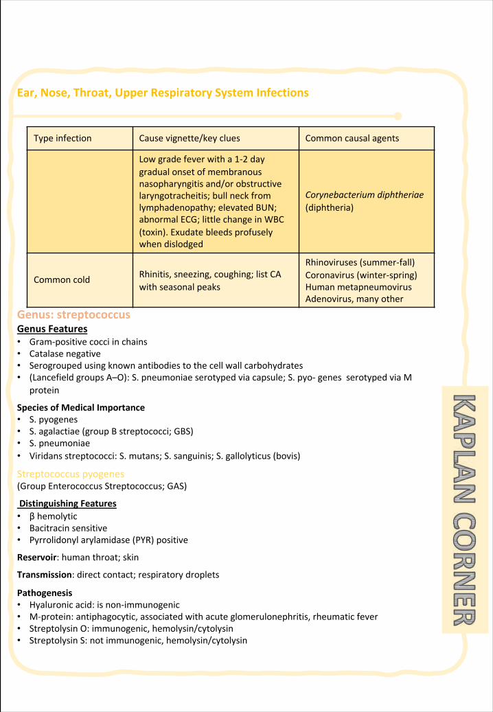
Ear,Nose,Throat,UpperRespiratorySystemInfections
Genus:streptococcusGenusFeatures• Gram-positivecocciinchains• Catalasenegative• Serogroupedusingknownantibodiestothecellwallcarbohydrates• (LancefieldgroupsA–O):S.pneumoniaeserotypedviacapsule;S.pyo-genesserotypedviaM
protein
SpeciesofMedicalImportance• S.pyogenes• S.agalactiae(groupBstreptococci;GBS)• S.pneumoniae• Viridansstreptococci:S.mutans;S.sanguinis;S.gallolyticus(bovis)
Streptococcuspyogenes(GroupEnterococcusStreptococcus;GAS)
DistinguishingFeatures• βhemolytic• Bacitracinsensitive• Pyrrolidonylarylamidase(PYR)positive
Reservoir:humanthroat;skin
Transmission:directcontact;respiratorydroplets
Pathogenesis• Hyaluronicacid:isnon-immunogenic• M-protein:antiphagocytic,associatedwithacuteglomerulonephritis,rheumaticfever• StreptolysinO:immunogenic,hemolysin/cytolysin• StreptolysinS:notimmunogenic,hemolysin/cytolysin
Typeinfection Causevignette/key clues Commoncausalagents
Lowgradefeverwitha1-2 daygradualonsetofmembranousnasopharyngitis and/orobstructivelaryngotracheitis; bullneckfromlymphadenopathy;elevatedBUN;abnormalECG;littlechangeinWBC(toxin).Exudatebleedsprofuselywhendislodged
Corynebacteriumdiphtheriae(diphtheria)
Commoncold Rhinitis,sneezing,coughing;listCAwithseasonalpeaks
Rhinoviruses(summer-fall)Coronavirus(winter-spring)HumanmetapneumovirusAdenovirus,manyother
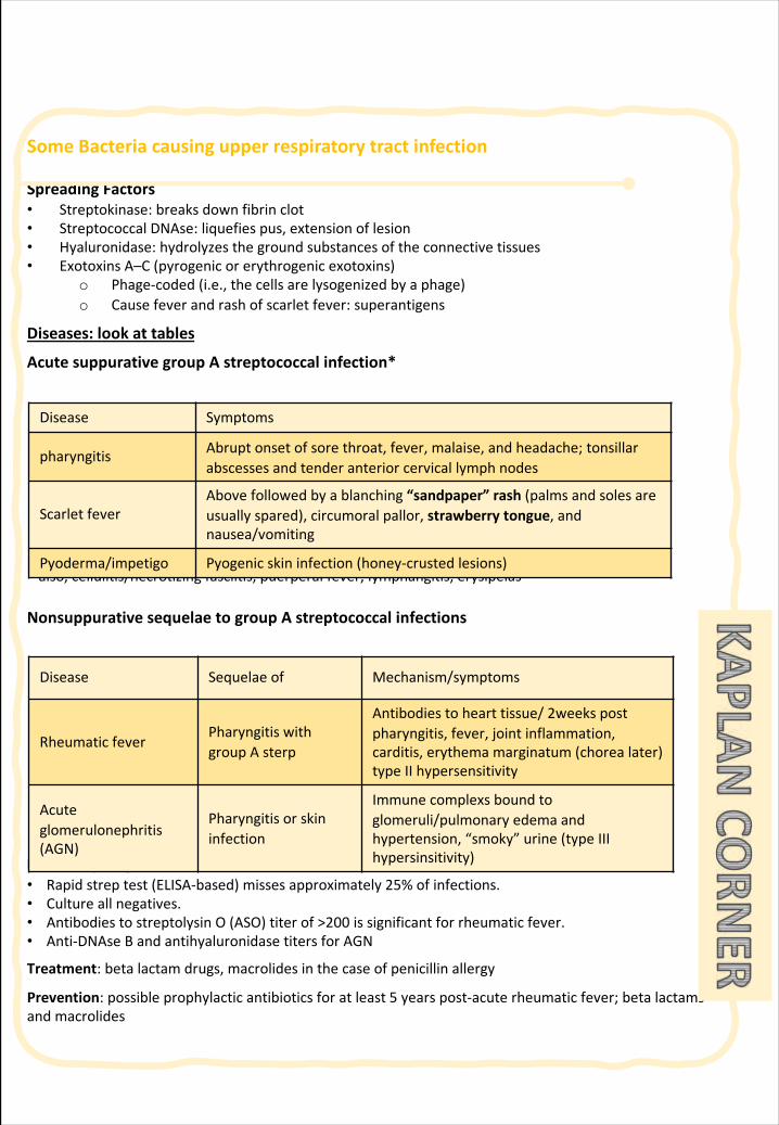
SomeBacteriacausingupperrespiratorytractinfection
SpreadingFactors• Streptokinase:breaksdownfibrinclot• StreptococcalDNAse:liquefiespus,extensionoflesion• Hyaluronidase:hydrolyzesthegroundsubstancesoftheconnectivetissues• ExotoxinsA–C(pyrogenicorerythrogenicexotoxins)
o Phage-coded(i.e.,thecellsarelysogenizedbyaphage)o Causefeverandrashofscarletfever:superantigens
Diseases:lookattables
Acutesuppurative groupAstreptococcalinfection*
*also,cellulitis/necrotizingfasciitis,puerperalfever,lymphangitis,erysipelas
Nonsuppurative sequelaetogroupAstreptococcalinfections
LaboratoryDiagnosis• Rapidstreptest(ELISA-based)missesapproximately25%ofinfections.• Cultureallnegatives.• AntibodiestostreptolysinO(ASO)titerof>200issignificantforrheumaticfever.• Anti-DNAseBandantihyaluronidasetitersforAGN
Treatment:betalactamdrugs,macrolidesinthecaseofpenicillinallergy
Prevention:possibleprophylacticantibioticsforatleast5yearspost-acuterheumaticfever;betalactamsandmacrolides
Disease Symptoms
pharyngitis Abrupt onsetofsorethroat,fever,malaise,andheadache;tonsillarabscessesandtenderanteriorcervicallymphnodes
Scarlet feverAbovefollowedbyablanching“sandpaper”rash(palmsandsolesareusuallyspared),circumoral pallor,strawberrytongue,andnausea/vomiting
Pyoderma/impetigo Pyogenic skininfection(honey-crustedlesions)
Disease Sequelaeof Mechanism/symptoms
RheumaticfeverPharyngitiswithgroupAsterp
Antibodiestohearttissue/2weekspostpharyngitis,fever,jointinflammation,carditis,erythemamarginatum(chorealater)typeIIhypersensitivity
Acuteglomerulonephritis(AGN)
Pharyngitisorskininfection
Immunecomplexs boundtoglomeruli/pulmonaryedemaandhypertension,“smoky”urine(typeIIIhypersinsitivity)
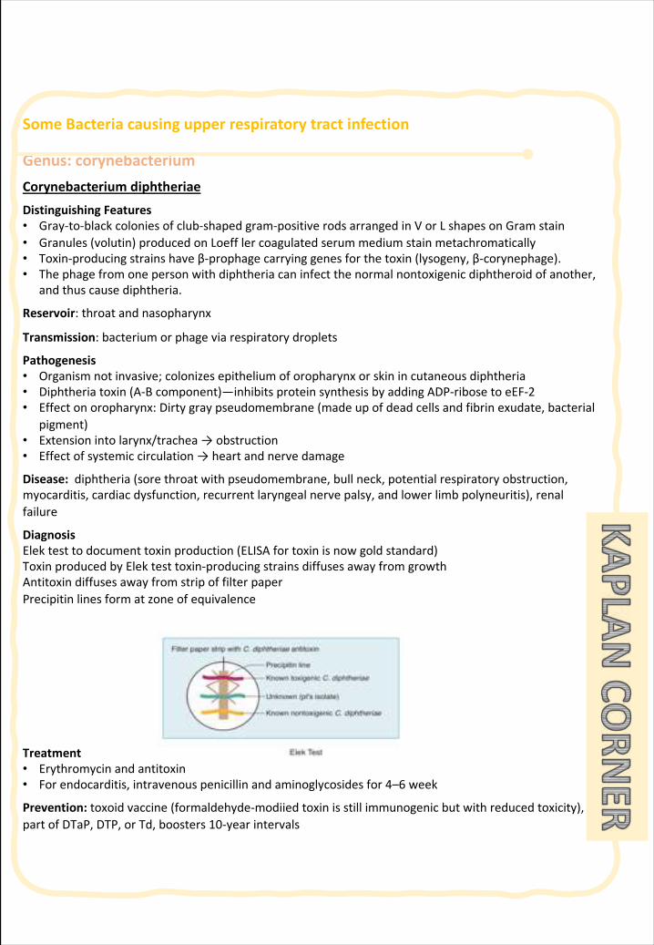
SomeBacteriacausingupperrespiratorytractinfection
Genus:corynebacteriumCorynebacteriumdiphtheriae
DistinguishingFeatures• Gray-to-blackcoloniesofclub-shapedgram-positiverodsarrangedinVorLshapesonGramstain• Granules(volutin)producedonLoefflercoagulatedserummediumstainmetachromatically• Toxin-producingstrainshaveβ-prophagecarryinggenesforthetoxin(lysogeny,β-corynephage).• Thephagefromonepersonwithdiphtheriacaninfectthenormalnontoxigenicdiphtheroidofanother,
andthuscausediphtheria.
Reservoir:throatandnasopharynx
Transmission:bacteriumorphageviarespiratorydroplets
Pathogenesis• Organismnotinvasive;colonizesepitheliumoforopharynxorskinincutaneousdiphtheria• Diphtheriatoxin(A-Bcomponent)—inhibitsproteinsynthesisbyaddingADP-ribosetoeEF-2• Effectonoropharynx:Dirtygraypseudomembrane(madeupofdeadcellsandfibrinexudate,bacterial
pigment)• Extensionintolarynx/trachea→obstruction• Effectofsystemiccirculation→heartandnervedamage
Disease:diphtheria(sorethroatwithpseudomembrane,bullneck,potentialrespiratoryobstruction,myocarditis,cardiacdysfunction,recurrentlaryngealnervepalsy,andlowerlimbpolyneuritis),renalfailure
DiagnosisElektesttodocumenttoxinproduction(ELISAfortoxinisnowgoldstandard)ToxinproducedbyElektesttoxin-producingstrainsdiffusesawayfromgrowthAntitoxindiffusesawayfromstripoffilterpaperPrecipitinlinesformatzoneofequivalence
Treatment• Erythromycinandantitoxin• Forendocarditis,intravenouspenicillinandaminoglycosidesfor4–6week
Prevention:toxoidvaccine(formaldehyde-modiiedtoxinisstillimmunogenicbutwithreducedtoxicity),partofDTaP,DTP,orTd,boosters10-yearintervals
SomeBacteriacausingupperrespiratorytractinfection
Genus:haemophilusHaemophilus influenzae
DistinguishingFeatures• Encapsulated,gram-negativerod;95%ofinvasivediseasecausedbycapsulartypeb• RequiresgrowthfactorsX(hemin)andV(NAD)forgrowthonnutrientorbloodagar(BA)• GrowsnearS.aureusonBA=“satellite”phenomenon• ChocolateagarprovidesbothXandVfactors
Reservoir:humannasopharynxTransmission:respiratorydroplets,sharedtoys
Pathogenesis• Polysaccharidecapsule(typebcapsuleispolyribitolphosphate)mostimportantvirulencefactor• Capsuleimportantindiagnosis;antigenscreenonCSF(e.g.,latexparticleagglutination);serotypeall
isolatesbyquellung.• IgAproteaseisamucosalcolonizingfactor.
Diseases• Meningitis
o Epidemicinunvaccinatedchildrenages3monthsto2years–Aftermaternalantibodyhaswanedandbeforeimmuneresponseofchildisadequate–Upto1990,H.influenzaewasmostcommoncauseofmeningitisage1–5(mainly<2);isstillaproblemifchildage<2andnotvaccinated
• Otitismedia:usuallynontypeablestrains• Bronchitis:exacerbationsofacutebronchitisinsmokerswithCOPD• Pneumonia:1–24months;rareinvaccinatedchildren;smokers• Epiglottitis:rareinvaccinatedchildren;seeninunvaccinatedtoddlers;H.influenzaewasmajor
causalagent
Diagnosis:bloodorCSFcultureonchocolateagar;PCR;antigendetectionofcapsule(latexparticleagglutination)
Treatment:cefotaximeorcetriaxoneforempiricaltherapyofmeningitis;checknasalcarriagebeforereleasing;userifampinifstillcolonized
Prevention• Conjugatecapsularpolysaccharide-proteinvaccine• Vaccinationeffectivetopreventtypebdisease
o Polyribitolcapsuleconjugatedtoprotein:(diphtheriatoxoidorN.meningitidisoutermembraneproteins),makingitaT-celldependentvaccine–Vaccine:2,4,6months;booster15months;95%effective
• Rifampinreducesoropharynxcolonizationandpreventsmeningitisinunvaccinated,closecontactsage<2years
SomeBacteriacausingupperrespiratorytractinfection
Moraxellacatarrhalis
DistinguishingFeatures• Gram-negativediplococcus• CloserelativeofNeisseriaReservoir:normalupperrespiratorytractlora
Transmission:respiratorydropletsPathogenesis:endotoxinmayplayroleindisease
Disease(s):otitismedia;sinusitis;bronchitisandbronchopneumoniainelderlypatientswithCOPD
Treatment:amoxicillinandclavulanate,second-orthird-generationcepha-losporinorTMP-SMX;drugresistanceisaproblem(moststrainsproduceaβ-lactamase)
Genus:bordetellaGenusFeatures• Gram-negativesmallrods• Strictaerobes
SpeciesofMedicalImportance:Bordetellapertussis
Bordetellapertussis
DistinguishingFeatures:smallgram-negative,aerobicrods;encapsulatedorganism
Reservoir:human(vaccinated)
Transmission:respiratorydroplets
Pathogenesis• B.pertussisismucosalsurfacepathogen• Attachmenttonasopharyngealciliatedepithelialcellsisviafilamentoushemagglutinin;pertussis
toxin(onoutermembrane)aidsinattachment• Toxinsdamagerespiratoryepithelium.
o Adenylatecyclasetoxin:impairsleukocytechemotaxis→inhibitsphagocytosisandcauseslocaledema
o Trachealcytotoxin:interfereswithciliaryaction;killsciliatedcellso Endotoxino Pertussistoxin(AandBcomponent,OMproteintoxin):ADPribosylationofGi(inhibiting
negativeregulatorofadenylatecyclase)interfereswithtransferofsignalsfromcellsurfacetointracellularmediatorsystem:lymphocytosis;islet-activationleadingtohypoglycemia;blockingofimmuneeffectorcells(decreasedchemotaxis);increasedhistaminesensitivity
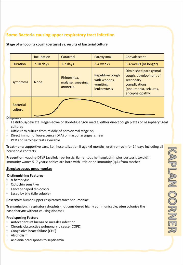
SomeBacteriacausingupperrespiratorytractinfection
Stageofwhoopingcough(pertusis)vs.resultsofbacterialculture
Diagnosis• Fastidious/delicate:Regan-LoweorBordet-Gengoumedia;eitherdirectcoughplatesornasopharyngeal
cultures• Difficulttoculturefrommiddleofparoxysmalstageon• Directimmunofluorescence(DFA)onnasopharyngealsmear• PCRandserologictestsavailable
Treatment:supportivecare,i.e.,hospitalizationifage<6months;erythromycinfor14daysincludingallhouseholdcontacts
Prevention:vaccineDTaP(acellularpertussis:ilamentoushemagglutininpluspertussistoxoid);immunitywanes5–7years;babiesarebornwithlittleornoimmunity(IgA)frommother
Streptococcuspneumoniae
DistinguishingFeatures• αhemolytic• Optochinsensitive• Lancet-shapeddiplococci• Lysedbybile(bilesoluble)
Reservoir:humanupperrespiratorytractpneumoniae
Transmission:respiratorydroplets(notconsideredhighlycommunicable;otencolonizethenasopharynxwithoutcausingdisease)
PredisposingFactors• Antecedentinfluenzaormeaslesinfection• Chronicobstructivepulmonarydisease(COPD)• Congestiveheartfailure(CHF)• Alcoholism• Aspleniapredisposestosepticemia
Incubation Catarrhal Paroxysmal Convalescent
Duration 7-10 days 1-2 days 2-4 weeks 3-4 weeks(orlonger)
symptoms NoneRhinorrhea,malaise, sneezing,anorexia
Repetitivecoughwithwhoops,vomiting,leukocytosis
Diminishedparoxysmalcough,developmentofsecondarycomplications(pneumonia,seizures,encephalopathy
Bacterialculture
SomeBacteriacausingupperrespiratorytractinfection
Pathogenesis• Polysaccharidecapsuleisthemajorvirulencefactor• IgAprotease• Teichoicacid• Teichoicacid• PneumolysinO:hemolysin/cytolysin:damagesrespiratoryepithelium;inhibitsleukocyterespiratory
burstandinhibitsclassicalcomplementfixation
Diseases• Typicalpneumonia:mostcommoncause(especiallyindecade6oflife);shakingchills,highfever,lobar
consolidation,blood-tinged,“rusty”sputum• Adultmeningitis:mostcommoncause;peptidoglycanandteichoicacidsarehighlyinflammatoryin
CNS;CSFrevealshighWBCs(neu-trophils)andlowglucose,highprotein• Otitismediaandsinusitisinchildrenmostcommoncause
LaboratoryDiagnosis• GramstainandcultureofCSForsputum• Quellungreaction:positive(swellingofthecapsulewiththeadditionoftype-specificantiserum,no
longerusedbutstilltested!)• Latexparticleagglutination:testforcapsularantigeninCSF• Urinaryantigentest
Treatment:betalactamsforbacterialpneumonia;cetriaxoneorcefotaximeforadultmeningitis(addvancomycinifpenicillin-resistantS.pneumoniaehasbeenreportedincommunity);amoxicillinforotitismediaandsinusitisinchildren(erythromycinincasesofallergy)
Prevention• Antibodytocapsule(>80capsularserotypes)providestype-specificimmunity• Vaccine
o Pediatric(PCV,pneumococcalconjugatevaccine):13ofmostcommonserotypes;conjugatedtodiphtheriatoxoid;preventsinvasivedisease
o Adult(PPV,pneumococcalpolysaccharidevaccine):23ofmostcommoncapsularserotypes;recommendedforalladultsage≥65plusat-riskindividuals
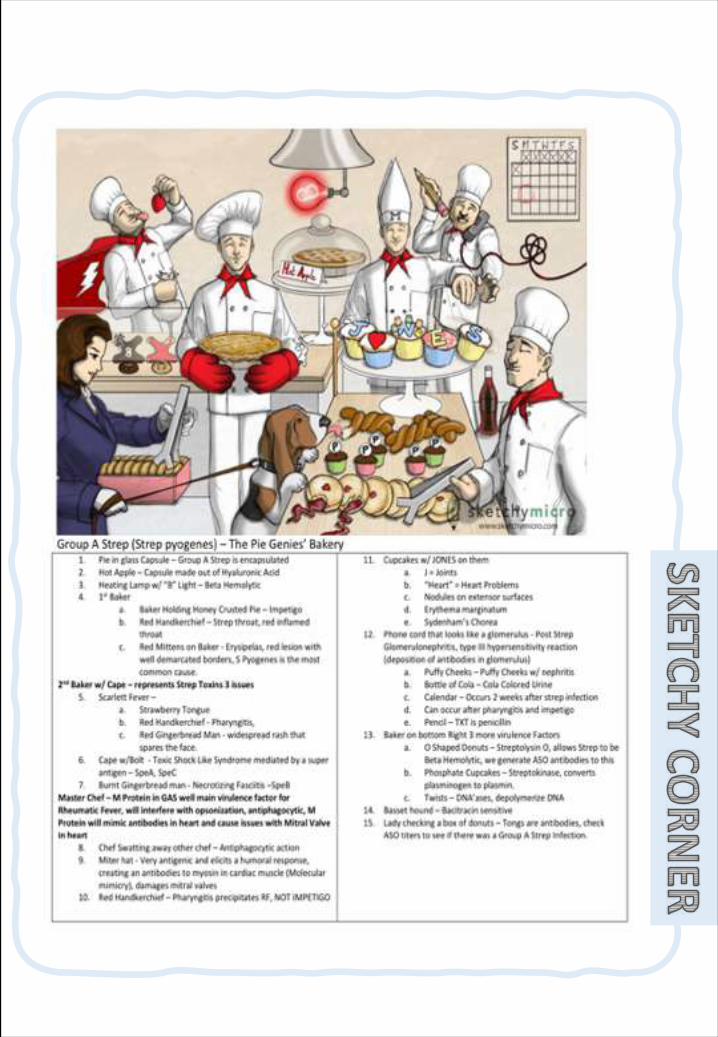
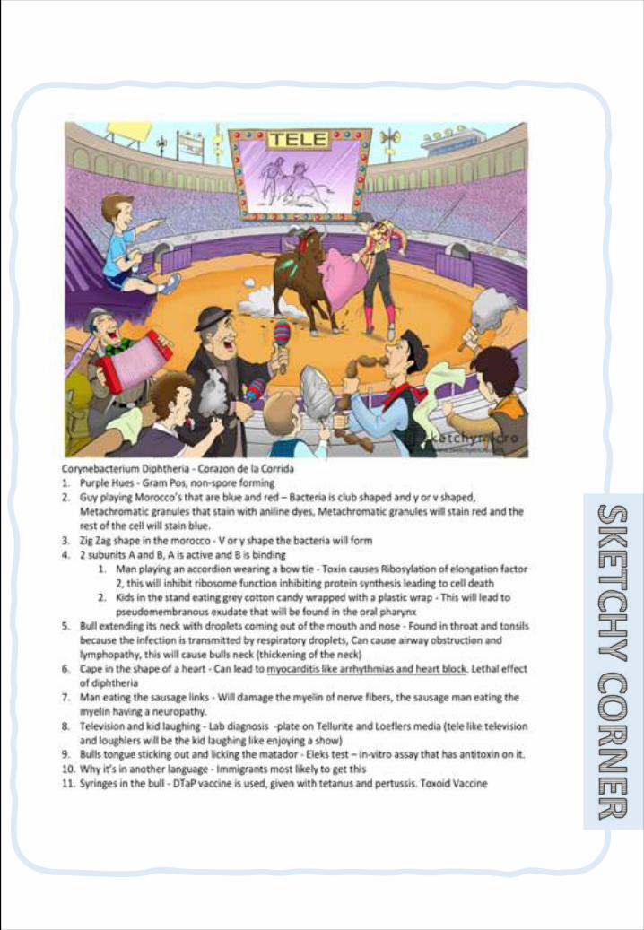
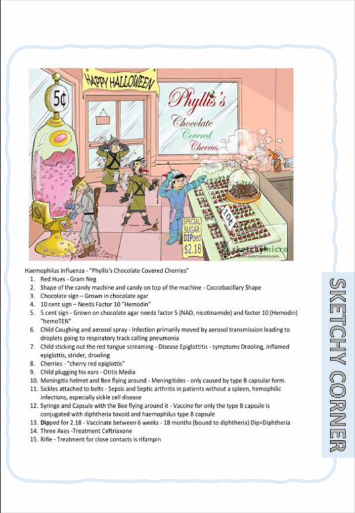
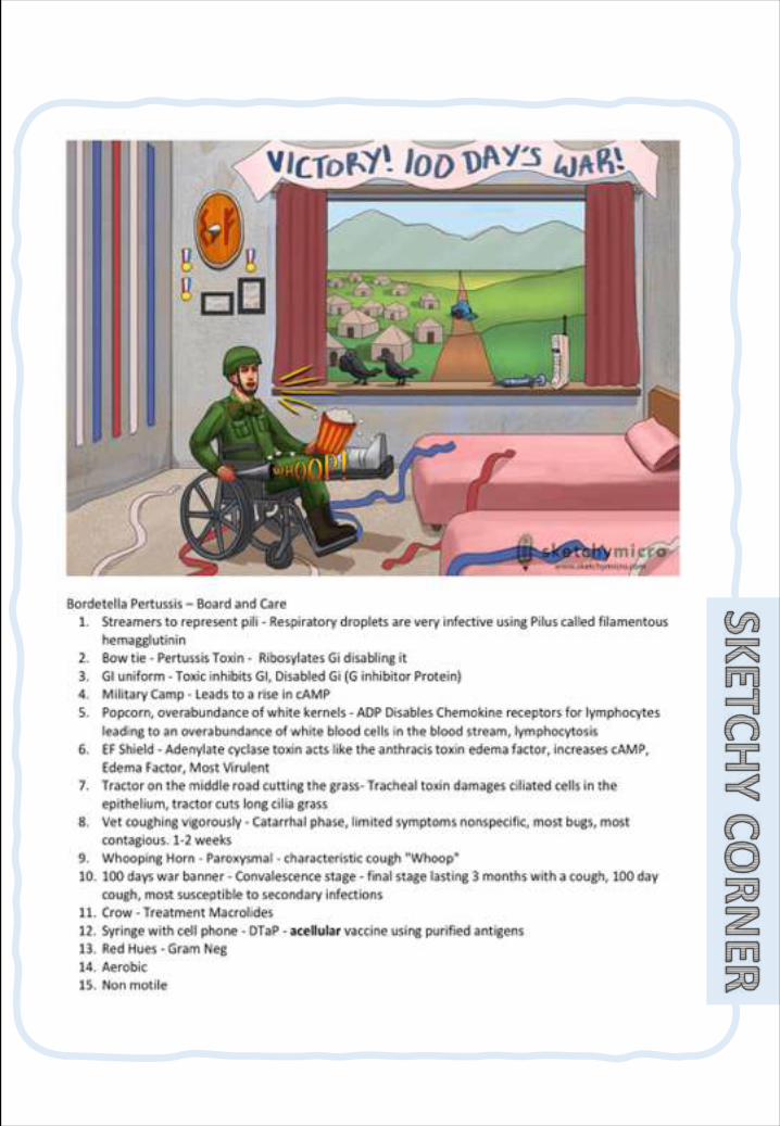
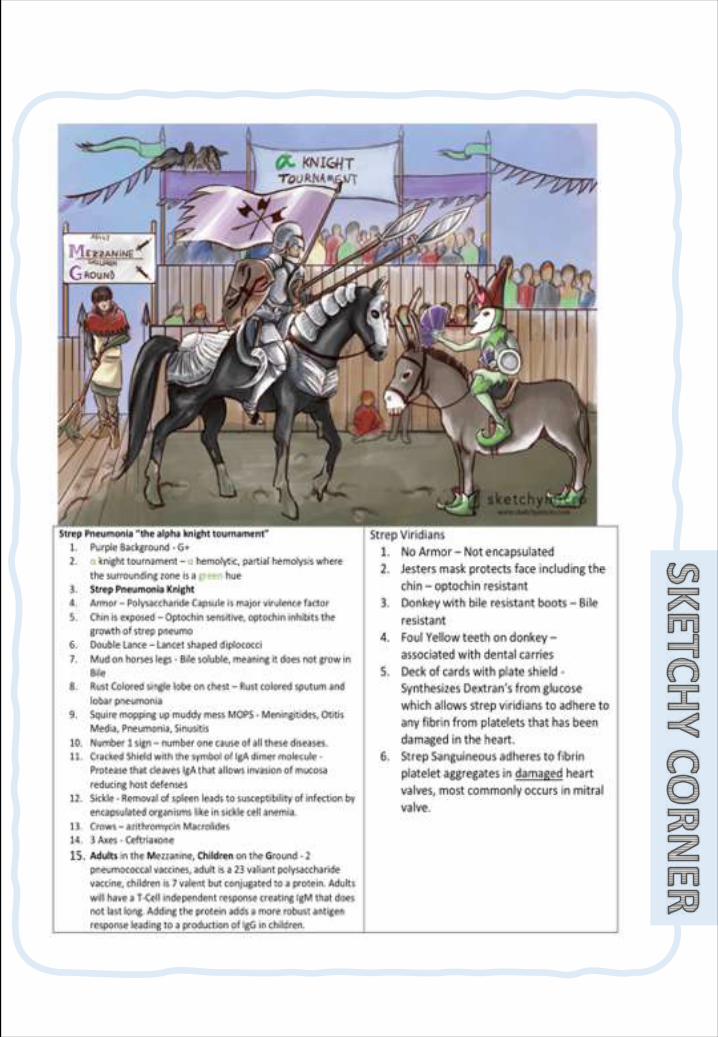

79 | Respiratory Chapter
SECTION 2 | Viruses Causing Respiratory Infections I
Objec
tive
Respiratory Tract Infections:
Note: (1)Ifitgetstothelowerrespiratorytractitbecomesevere.
§ Acquirethebasicknowledgeaboutstructureandclassificationofthesevirusesinfectingtherespiratorysystem.
Including(Influenza viruses,Parainfluenza viruses,Respiratorysyncytialvirus(RSV)andhumanmetapneumovirus(HMPV),Measles virus,andMumps)
§ Describetheirepidemiologyandpathogenesis
§ IdentifytherespiratoryinfectionsandtheclinicalfeaturesofURTIandLRTI.
§ Describetheirepidemiologyandpathogenesis
§ Knowthelaboratorydiagnosis,andtreatmentoftheseinfections.
§ Recognizethemethodsforprevention.
Note: (2)Lowerrespiratorytractinfections
Introduction:o Theyarethecommonestofhumaninfectionsandcausealarge
amountofmorbidityandlossoftimeatwork(sickleave).Theyarecommoninbothchildrenandadults.
o Mostlycausedbyviruses.Mostlyareself-limitingdisease,whichmeanthediseasetendstogoawayonitsown,withouttreatment.
o Mostlyaremildandconfinedtotheupperrespiratorytract(URT).1o URT-infectionmayspreadtootherorganscausingmoresevere
infectionanddeath.
ClinicalManifestations(symptoms):o Commoncold(rhinitis).o Pharyngitis.o Tonsillitis.o Sinusitis&otitismedia.o Croup(acutelaryngotracheobronchitis).o Acutebronchitis,AcutebronchiolitisandViralpneumonia.2
Commonrespiratoryviruses::
Nameofthevirus Family Disease1)Influenzavirus Orthomyxoviridae URTIandLRTI
2)Parainfluenzavirus Paramyxoviridae LRTI3)Respiratorysyncytialvirus
4 Rhinovirus Picornaviridae URTI
5 Coronavirus Coronaviridae URTIandLRTI
6 Adenovirus Adenoviridae URTIandeyeinfections
7 Humanmetapneumovirus Paramyxoviridae LRTI
ArabicTranslation: Croup= قانخلا ضرم
ExtraExplanation: RNAsenseinviruses
o Positivesense(+vestrand):(5'to3')viralRNAsignifiesthataparticularviralRNAsequencemaybedirectlytranslatedintothedesiredviralproteins.
o Negativesense(-vestrand):ThisRNA(3'to5')cannotbetranslatedintoproteindirectly.Instead,itmustfirstbetranscribedintoapositivesenseRNAthatactsasanmRNA.Someviruses(Influenza,forexample)havenegativesensegenomesandsomustcarryanRNApolymeraseinsidethevirion.

Respiratory Chapter | 80
Viruses Causing Respiratory Infections I | SECTION 2
Note: o (3)Glycoproteinfoundon
thesurfaceofinfluenzaviruses.(More)
o (4)Enzymesthathydrolyzeglycosidebonds,leadingtothedegradationneuraminicacids.
1: Influenza virus : Structural features:
o 8 helicalSegmentedgenome (NegativepolarityssRNA)o Envelopedviruswith2projectingglycoproteinspikes :-- Haemagglutinin(H) - Neuraminidase (N)
Influenzaviralproteins:
Haemagglutinin(H) 3 Neuraminidase(N) 4
Attachmenttothehostcellsurfacereceptors.
NoAttachment=Noinfection
Responsibleforreleaseoftheprogenyviralparticlesfromtheinfectedcell.
AntibodiestotheHAisresponsibleforimmunity.our immunesystemuseitasanantigen.
___
16 haemagglutininantigenictype,(H1 – H16). 9 neuraminidaseantigenictype,(N1 – N9).
HumanassociatedHantigenictypeareH1,H2,H3OtherHforanimals,thoughitcaninfecthuman(*).
HumanassociatedNantigenictypeareN1,N2.OtherNforanimals,thoughitcaninfecthuman1
Note: Thisvirusishighlysusceptibletomutationsandrearrangementswithintheinfectedhost.That’swhyit’shardtomakeaspecificdrugforit.
Epidemiology:o Seasonal,spreadsmostlyinwinter.o Highlysusceptibletomutationsandrearrangeablewithintheinfectedhost.o Pastantigenicshifts:- 1918→H1N1 “SpanishInfluenza”→2040milliondeaths.- 1957→H2N2 “AsianFlu”→12milliondeaths.- 1968→H3N2 “HongKongFlu”→700,000 deaths.- 1977→H1N1Reemergence→notpandemic.
Typesofinfluenzavirus:InfluenzaVirusTypes TypeA TypeB TypeC
Infect HumanandAnimal. Humanonly.
Humanonly.
Cause Human:Epidemic&Pandemic.
Animal:Epizootic.Outbreak. Mildillness.
Antigenic
changes
- Antigenicdrift
- Antigenicshift=Reassortment=
rearrangement
Antigenicdriftonly. ____
-Antigenicdrift→Minorchangeingenetic material.-Antigenicshift→Majorchangeingenetic material.
Note: Ifthereisonlyoneofthehumanassociatedantigenicinavirus,eitherHorN,thisviruscaninfectahuman.
Orthomyxoviridae Family- Influenzavirus,AvianfluandSwineflu-
ExtraExplanation:
Antigenicdrift(minorchange)o Amechanismforvariation
invirusesthatinvolvestheaccumulationofmutationswithinthegenesthatcodeforantibodybinding sites.Thisresultsinanewstrainofvirusparticles.whichcannotbeinhibitedaseffectivelybytheoriginalantibodies.
Antigenicshift(majorchange)o Theprocessbywhichtwo
ormoredifferentstrainsofavirus,orstrainsoftwoormoredifferentviruses,combinetoformanewsubtypehavingamixtureofthesurfaceantigensofthetwoormoreoriginalstrains.
81 | Respiratory Chapter
SECTION 2 | Viruses Causing Respiratory Infections I
Pathogenesis:Infectstheepithelialcellsofthenose,throat,bronchiandoccasionallythelungs.Accordingtothehost’simmunity,itcaneitherbelocalizedasURTIorspreadtotheLRTandViremiaandfeverusuallyoccur.
Transmission:InhalationofinfectiousaerosoldropletsIncubationperiod:1-4 daysSymptoms:Fever,malaise,headache,cough,chills,sorethroat,generalizedpain.Prognosis:Usuallyself-limitingdisease.Complications:
o Primaryinfluenzapneumonia.5o Secondarybacterialpneumonia.6o Reye’ssyndrome.7
Labdiagnosis:DirectdetectionofinfluenzaAorBvirusfrom:-NasopharyngealAspirate(NPA),Sputum,respiratorysecretion,&Nasopharyngealswab.o RoutinetestingbydirectdetectionofInfluenzaAorBvirusfrom:- Sputum.- Nasopharyngealswabaspirate(NPA).- Respiratorysecretionbydirectimmunofluorescentassay(IFA).o Otherdetectionmethods:- Tissueculture.- PCR.8
Treatment:o Amantadine:forInfluenzaAvirusonly.o Rimantadine,Oseltamivir(Tamiflu)orZanamivir(Relenza):ForbothInfluenzaA&Bviruses&Canbeusedastreatmentofprophylaxis.
Prevention:1- Theflushotvaccine,whichisInactivated“Killedvaccine.ItisGiventopeopleolderthan6-months,eitherhealthyorthosewithchronicmedicalconditions.
2- Thenasalsprayfluvaccine“Flumist"whichisLiveattenuated.itisApprovedforhealthypeoplebetween5-49 years.
BothcontaintwostrainsofcurrentcirculationofInfluenzaA&Bviruses.(affectbothA&B).ThevaccineshouldbegiveninOct&Novbeforetheinfluenzaseasonbegins.
Note: o (5)IfvirusentersthelungdirectlywithoutinfectURTfirst.
o (6)Bacteriaentersthelungsinresponsetoviralinfection
o (7)fattydegenerationofCNSandLiver,IfyougiveAspirintochildwithviralinfection.
o (8)Polymerasechainreaction:arapidtechniqueforinvitroamplificationofspecificDNAorRNAsequences,allowingsmallquantitiesofshortsequencestobeanalyzedwithoutcloning.WewilldothiswhentheIFAshows(applegreenfluorescence)whichmeanstypeAsowewanttodeterminewhichstrainofinfluenza
Note: o Killedvaccine:GrowninculturethenkilledbyHeatorformaldehyde.
o Liveattenuated:Containslivingorganisms
Note: o aerosoldroplets:Accumulationofsolidparticlesorliquiddropletsinairoranothergas.
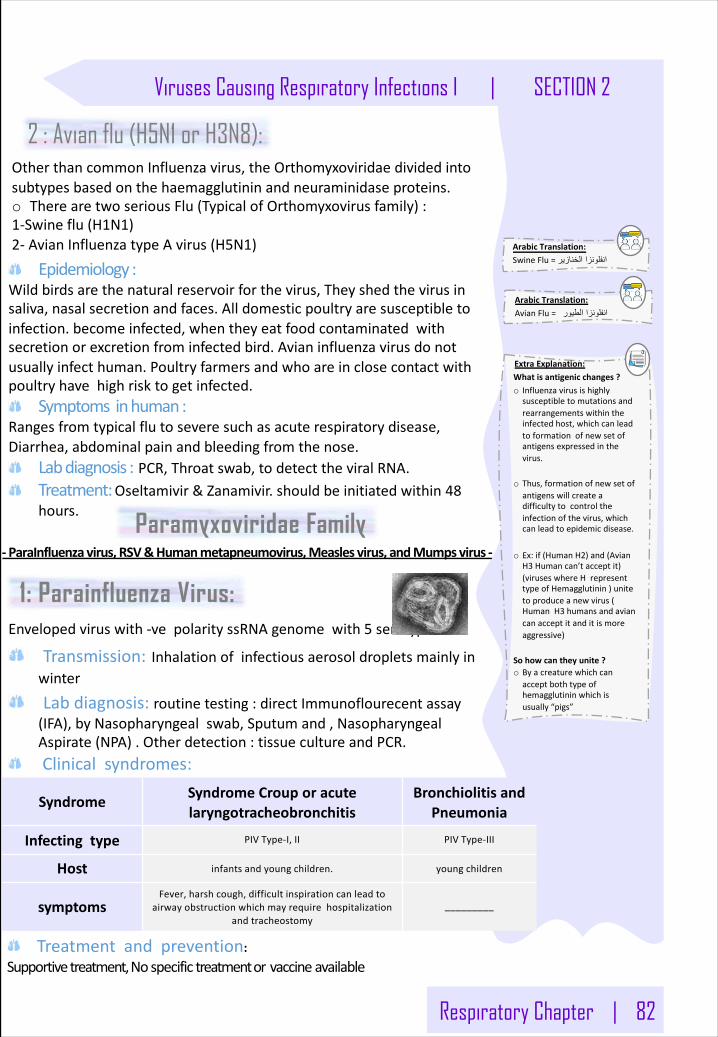
Respiratory Chapter | 82
Viruses Causing Respiratory Infections I | SECTION 2
2 : Avian flu (H5N1 or H3N8): OtherthancommonInfluenzavirus,theOrthomyxoviridaedividedintosubtypesbasedonthehaemagglutininandneuraminidaseproteins.o TherearetwoseriousFlu(TypicalofOrthomyxovirusfamily):1-Swineflu(H1N1)2- AvianInfluenzatypeAvirus(H5N1)
Epidemiology:Wildbirdsarethenaturalreservoirforthevirus,Theyshedthevirusinsaliva,nasalsecretionandfaces.Alldomesticpoultryaresusceptibletoinfection.becomeinfected,whentheyeatfoodcontaminatedwithsecretionorexcretionfrominfectedbird.Avianinfluenzavirusdonotusuallyinfecthuman.Poultryfarmersandwhoareinclosecontactwithpoultryhavehighrisktogetinfected.
Symptomsinhuman:Rangesfromtypicalflutoseveresuchasacuterespiratorydisease,Diarrhea,abdominalpainandbleedingfromthenose.
Labdiagnosis:PCR,Throatswab,todetecttheviralRNA.Treatment:Oseltamivir&Zanamivir.shouldbeinitiatedwithin48hours.
ExtraExplanation: Whatisantigenicchanges?o Influenzavirusishighlysusceptibletomutationsandrearrangementswithintheinfectedhost,whichcanleadtoformationofnewsetofantigensexpressedinthevirus.
o Thus,formationofnewsetofantigenswillcreateadifficultytocontroltheinfectionofthevirus,whichcanleadtoepidemicdisease.
o Ex:if(HumanH2)and(AvianH3 Humancan’tacceptit)(viruseswhereHrepresenttypeofHemagglutinin)unitetoproduceanewvirus(HumanH3 humansandaviancanacceptitanditismoreaggressive)
Sohowcantheyunite?o Byacreaturewhichcanacceptbothtypeofhemagglutininwhichisusually“pigs”
ArabicTranslation: SwineFlu= ریزانخلا ازنولفنا
ArabicTranslation: AvianFlu= رویطلا ازنولفنا
Paramyxoviridae Family
Envelopedviruswith-vepolarityssRNAgenomewith5 serotypes.Transmission: Inhalationofinfectiousaerosoldropletsmainlyinwinter
Labdiagnosis:routinetesting:directImmunoflourecent assay(IFA),byNasopharyngealswab,Sputumand,NasopharyngealAspirate(NPA).Otherdetection:tissuecultureandPCR.Clinicalsyndromes:
Syndrome SyndromeCrouporacutelaryngotracheobronchitis
BronchiolitisandPneumonia
Infectingtype PIVType-I,II PIVType-III
Host infantsandyoungchildren. youngchildren
symptomsFever,harshcough,difficultinspirationcanleadto
airwayobstructionwhichmayrequirehospitalizationandtracheostomy
_________
Treatmentandprevention:Supportivetreatment,Nospecifictreatmentorvaccine available
- ParaInfluenzavirus,RSV&Humanmetapneumovirus,Measlesvirus,andMumpsvirus-
1: Parainfluenza Virus:
83 | Respiratory Chapter
SECTION 2 | Viruses Causing Respiratory Infections I
2- Respiratory Syncytial Virus (RSV) and Human metapneumovirus1
Structuralfeatures:Envelopedviruswith(-vepolarityssRNA).
Transmission:o Inhalationofinfectiousaerosoldropletsmainlyinwinter.o RSVvirusisverycontagiouswith36 daysasIncubationperiodso TheimportanceofRSVliesinitstendencytoinvadetheLRTofinfant
Clinicalsyndromes::o 1)Bronchiolitis:Life-threateningdiseaseininfantsespeciallyunder6monthoflife.Withrespiratorydistressandcyanosis,itcanleadtoachroniclungdiseaselaterinlifeorbefatal.o 2)Pneumonia:Canalsobefatalininfants.
Labdiagnosis::o Directdetectionofthevirus:Fromsputum,nasopharyngealswab,aspirate(NPA)orrespiratorysecretionbydirectimmunofluorescentassay(IFA)andELISA.o Otherdetectionmethods:Isolatedofvirusbycellculturefrom(NPA)withmultinucleatedgiantcellorsyncytiaascytopathiceffect(C.P.E)orPCR.
Treatmentandprevention:o Ribavirin administeredbyinhalationforinfantswithseverecondition.o Infantswillbehypoxicandneedhospitalizationforoxygeninhalation
andshouldbeisolatedthanotherinfants.o Novaccineavailable,butpassiveimmunizationimmunoglobulincanbe
givenforinfectedprematureinfants.
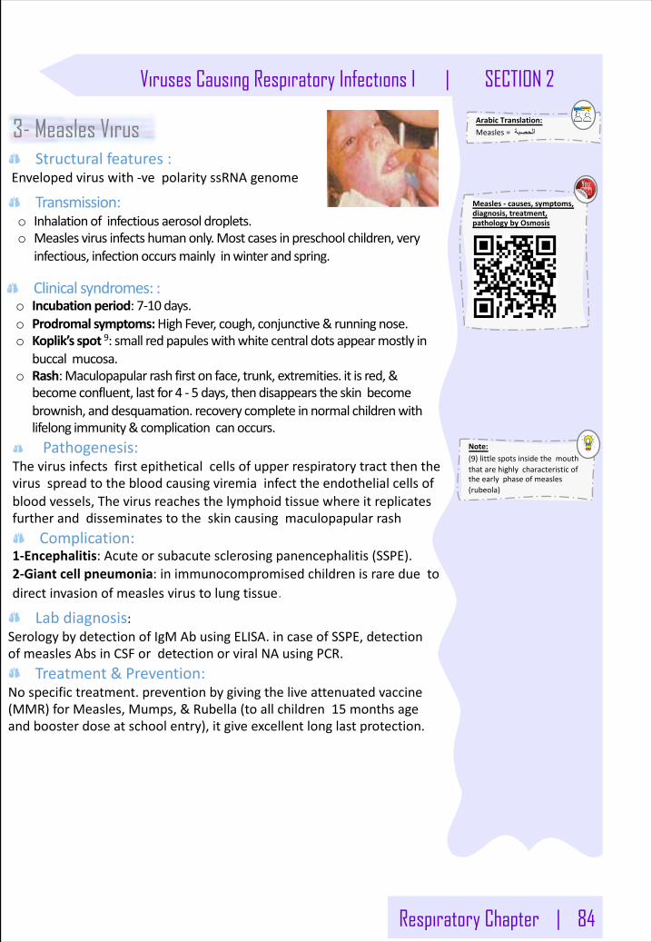
Respiratory Chapter | 84
Viruses Causing Respiratory Infections I | SECTION 2
Note: (9)littlespotsinsidethemouththatarehighlycharacteristicoftheearlyphaseofmeasles(rubeola)
Clinicalsyndromes::o Incubationperiod:7-10 days.o Prodromalsymptoms:High Fever,cough,conjunctive&runningnose.o Koplik’s spot9:smallredpapuleswithwhitecentraldotsappearmostlyin
buccalmucosa.o Rash:Maculopapularrashfirstonface,trunk,extremities.itisred,&
becomeconfluent,lastfor4 - 5 days,thendisappearstheskinbecomebrownish,anddesquamation.recoverycompleteinnormalchildrenwithlifelongimmunity&complicationcanoccurs.Pathogenesis:
Thevirusinfectsfirstepitheticalcellsofupperrespiratorytractthenthevirusspreadtothebloodcausingviremiainfecttheendothelialcellsofbloodvessels,Thevirusreachesthelymphoidtissuewhereitreplicatesfurtheranddisseminatestotheskincausingmaculopapularrash
Complication:1-Encephalitis:Acuteorsubacutesclerosingpanencephalitis(SSPE).2-Giantcellpneumonia:inimmunocompromisedchildrenisrareduetodirectinvasionofmeaslesvirustolungtissue.
Labdiagnosis:SerologybydetectionofIgMAbusingELISA.incaseofSSPE,detectionofmeaslesAbsinCSFordetectionorviralNAusingPCR.
Treatment&Prevention:Nospecifictreatment.preventionbygivingtheliveattenuatedvaccine(MMR)forMeasles,Mumps,&Rubella(toallchildren15 monthsageandboosterdoseatschoolentry),itgiveexcellentlonglastprotection.
3- Measles VirusStructuralfeatures:
Envelopedviruswith-vepolarityssRNA genome
Transmission:o Inhalationofinfectiousaerosoldroplets.o Measlesvirusinfectshumanonly.Mostcasesinpreschoolchildren,very
infectious,infectionoccursmainlyinwinterandspring.
ArabicTranslation: Measles= ةبصحلا
Measles- causes,symptoms,diagnosis,treatment,pathologybyOsmosis
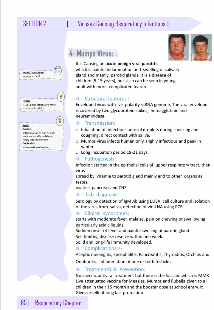
85 | Respiratory Chapter
SECTION 2 | Viruses Causing Respiratory Infections I
4- Mumps Virus:ItisCausinganacutebenignviralparotitiswhichispainfulinflammationandswellingofsalivaryglandandmainlyparotidglands.itisadiseaseofchildren(5-15 years),butalsocanbeseeninyoungadultwithmorecomplicatedfeature.
Structuralfeatures:Envelopedviruswith-vepolarityssRNA genome,Theviralenvelopeiscoveredbytwoglycoproteinspikes,hemagglutininandneuraminidase.
Transmission:o Inhalationofinfectiousaerosoldropletsduringsneezingand
coughing,directcontactwithsaliva.o Mumpsvirusinfectshumanonly,Highlyinfectiousandpeakin
wintero Longincubationperiod18-21 days.
Pathogenesis:Infectionstartedintheepithelialcellsofupperrespiratorytract,thenvirus spreadbyviremiatoparotidglandmainlyandtootherorgansas:testes,ovaries,pancreasandCNS.
Labdiagnosis:SerologybydetectionofIgMAbusingELISA,cellcultureandisolationofthevirusfromsaliva,detectionofviralNAusingPCR.
Clinicalsyndromes:startswithmoderatefever,malaise,painonchewingorswallowing,particularlyacidicliquids.Suddenonsetoffeverandpainfulswellingofparotidgland.Self-limitingdiseaseresolvewithinoneweek.Solidandlong-lifeimmunitydeveloped.
Complications: 10
Asepticmeningitis,Encephalitis,Pancreatitis,Thyroiditis,OrchitisandOophoritis.inflammationofoneorbothtesticles.
Treatment&&Prevention:NospecificantiviraltreatmentbutthereistheVaccinewhichisMMRLiveattenuatedvaccineforMeasles,MumpsandRubellagiventoallchildrenintheir15 monthandtheboosterdoseatschoolentry.ItGivesexcellentlonglastprotection
Note: (10)Complicationsaremorecommoninadults
Note: Orchitis:inflammationofoneorbothtesticles.usuallyunilateral,rarelyleadstosterility.Oophoritis:inflammationofovaries
ArabicTranslation: Mumps= فاكنلا
86 | Respiratory Chapter
SECTION 2 | Viruses Causing Respiratory Infections II
Objec
tive § Acquirethebasicknowledgeaboutstructureandclassificationoftheseviruses
infectingtherespiratorysystem.Including(Coronavirus(SARS &MiddleEastRespiratorySyndrome- CoronavirusCOV(MERSCoV),Rhinovirus,Enteroviruses,Adenovirus,andEBV)
§ Describetheirepidemiologyandpathogenesis
§ IdentifytheTargetgroupandmodesoftransmissionandClinicalmanifestations..
§ Describetheirepidemiologyandpathogenesis
§ Knowthelaboratorydiagnosis,andtreatmentoftheseinfections.
§ Recognizethemethodsforprevention.
Coronaviridae Family- SARSCoVandMERSCoV-
Structuralfeatures:Envelopedviruswith+vepolarityss-RNAgenome.
Transmission:Inhalationofinfectiousaerosoldroplets.
Clinicalfeatures:Itisthesecondcauseofcommoncold1.ItCausezoonoticdiseasesandcaninfectshumansandanimals
Treatment:Nospecificantiviraltreatment.Forseverecases,currenttreatmentincludescaretosupportvitalorganfunctions.
SevereformsofCoronavirus:o 1)SevereAcuteRespiratorySyndrome(SARS)o 2)MiddleEastRespiratorySyndrome(MERS)
1- Severe Acute Respiratory Syndrome (SARS): Inwinterof2002,anewrespiratorydiseaseknownas(SARS)emergedinChinaafteranewmutationofcoronavirus.Theanimalreservoirmaybecatsorbats.Thenthediseasespreadworldwideduetotravelling.ItisAssociatedwithhighmortalityduetorespiratoryfailure.
Symptoms:SARSstartswithhighfeverfollowedbycoughwithdifficultyinbreathing(atypicalpneumonia).
Respiratory Chapter | 87
Viruses Causing Respiratory Infections II | SECTION 2
2- Middle East Respiratory Syndrome (MERS):InSeptember2012,acaseofnovel(New)coronavirusinfectionwasreportedinvolvingamaninSaudiArabiawhowasadmittedtoahospitalwithpneumoniaandacutekidneyfailure.ThisvirushasbeennamedasMiddleEastRespiratorySyndrome- CoronaVirus(MERS-CoV),viruscloselyrelatedtoseveralbatcoronaviruses.
ArabicTranslation: MERS= طسوألا قرشلا ةمزالتم
:Epidemiology .انوروك سوریفل ةیسفنتلاMERS-CoV infectedseveralhumancells,includinglowerbutnotupperrespiratory,kidney,intestinal,andlivercells.Sofar,allthecaseshavebeenlinkedtocountriesinandneartheArabianPeninsula.Highlyinfectious,Peakinwinter,withincubationperiod(2-14 days).
Transmission:closecontactwithillpeople,it’snotepidemicorpandemic.,closecontactwithinfectedanimals
Prevention:Peopleshouldprotectthemselvesfromrespiratoryillnessesbytakingeverydaypreventiveactions:
o Washhandsoftenwithwaterandsoaporuseanalcohol-basedhandsanitizer.
o Covernoseandmouthwithatissuewhencoughorsneeze.o Avoidtouchingeyes,noseandmouthwithunwashedhands.o Avoidpersonalcontactwithsickpeopleo Cleananddisinfectfrequentlytouchedsurfacessuchastoysand
doorknobs.Riskgroup:
o Individualswithweakenedimmunesystemso Peoplewithpre-existingmedicalconditions(orcomorbidities)suchas
diabetes,cancer,andchroniclung,heart,andkidneydiseasesClinicalfeatures:
o Somepeoplealsohadgastrointestinalsymptomsincludingdiarrheaandnausea/vomiting.Someinfectedpeoplehadmildsymptoms(suchascoldlikesymptoms)ornosymptomsatallandtheyrecoveredcompletely.
o MostpeoplewithconfirmedMERSCoVinfectiondevelopedsevereacuterespiratoryillness.
o Theyhadfever,cough,andshortnessofbreath.Complication:
Severecomplicationsincludepneumoniaandkidneyfailure.About30%ofinfectedpeopledied(speciallythosewhohadincludedintheriskgroup)
Diagnosis:o Detectionoftheviralnucleicacid(NA)byPCR(PolymeraseChainReaction).o Othermethods:IsolationofthevirusfromNasopharyngealaspiration
(NPA)bycellculture.
88 | Respiratory Chapter
SECTION 2 | Viruses Causing Respiratory Infections II
Picornaviridae Family- RhinovirusandCoxsackieviruses-
1- Rhinovirus :Structuralfeatures:
Nonenvelopedviruswith(+vepolarityssRNA)genome,morethan100serotypesavailable.
Transmission:Inhalationofinfectiousaerosoldroplets.
Clinicalfeatures:o Rhinovirusesarethe1stcauseofcommoncold,responsibleof60%of
allcases.o Themainsymptomsofcommoncoldaresneezing,clearwaterynasal
dischargewithmildsorethroat,andcough.
LabDiagnosis:DetectionforviralNAbyusingPCRanddirectimmunofluorescenceassay.
Treatmentandprevention:Usuallyself-limitingdisease,Nospecifictreatment,andNovaccineavailable.
2- Coxsackieviruses :Structuralfeatures:
Non-envelopedviruswith+polarityssRNA genomeCoxsackievirusesgroupA&B,Echovirus,Enteroviruses.
Transmission:Inhalationofinfectiousaerosoldroplets.
Clinicalfeatures:o Coxsackievirusescauseherpanginaandpharyngitiso Echovirus&otherEnterovirusescauserespiratorysymptoms.
LabDiagnosis:routinetestingbydetectionoftheviralNAfromNPAusingPCR.
Treatmentandprevention:Usuallyself-limitingdisease,nospecifictreatment,andnovaccineavailable.
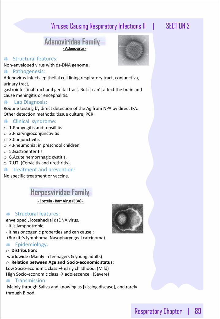
Respiratory Chapter | 89
Viruses Causing Respiratory Infections II | SECTION 2
Adenoviridae Family- Adenovirus-
Structuralfeatures:Non-envelopedviruswithds-DNAgenome.
Pathogenesis:Adenovirusinfectsepithelialcellliningrespiratorytract,conjunctiva,urinarytract,gastrointestinaltractandgenitaltract.Butitcan’taffectthebrainandcausemeningitisorencephalitis.
LabDiagnosis:RoutinetestingbydirectdetectionoftheAgfromNPAbydirectIFA.Otherdetectionmethods:tissueculture,PCR.
Clinicalsyndrome:o 1.Phrayngitisandtonsillitiso 2.Pharyngioconjunctivitiso 3.Conjunctivitiso 4.Pneumonia:inpreschoolchildren.o 5.Gastroenteritiso 6.Acutehemorrhagiccystitis.o 7.UTI(Cervicitisandurethritis).
Treatmentandprevention:Nospecifictreatmentorvaccine.
Herpesviridae Family- Epstein- BarrVirus(EBV)-
Structuralfeatures:enveloped,icosahedraldsDNAvirus.- Itislymphotropic.- Ithasoncogenicpropertiesandcancause:(Burkitt’slymphoma.Nasopharyngealcarcinoma).
Epidemiology:o Distribution:worldwide(Mainlyinteenagers&youngadults)o RelationbetweenAgeandSocio-economicstatus:LowSocio-economicclass→earlychildhood.(Mild)HighSocio-economicclass→adolescence.(Severe)
Transmission:MainlythroughSalivaandknowingas[kissingdisease],andrarelythroughBlood.
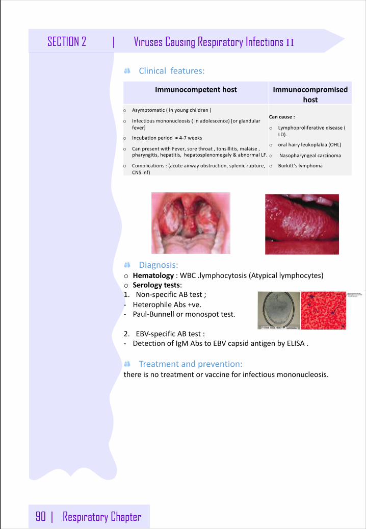
90 | Respiratory Chapter
SECTION 2 | Viruses Causing Respiratory Infections II
Clinicalfeatures:
Diagnosis:o Hematology :WBC.lymphocytosis(Atypicallymphocytes)o Serology tests:1. Non-specificABtest;- HeterophileAbs+ve.- Paul-Bunnellormonospottest.
2. EBV-specificABtest:- DetectionofIgMAbstoEBVcapsidantigenbyELISA.
Treatmentandprevention:thereisnotreatmentorvaccineforinfectiousmononucleosis.
Immunocompetenthost Immunocompromisedhost
o Asymptomatic(inyoungchildren)
o Infectiousmononucleosis(inadolescence)[orglandularfever]
o Incubationperiod=4-7 weeks
o CanpresentwithFever,sorethroat,tonsillitis,malaise,pharyngitis,hepatitis,hepatosplenomegaly&abnormalLF.
o Complications:(acuteairwayobstruction,splenicrupture,CNSinf)
Cancause:
o Lymphoproliferativedisease(LD).
o oralhairyleukoplakia(OHL)
o Nasopharyngealcarcinoma
o Burkitt’slymphoma
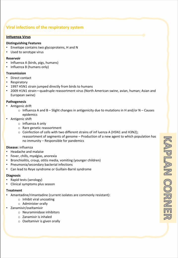
Viralinfectionsoftherespiratorysystem
InfluenzaVirus
DistinguishingFeatures• Envelopecontainstwoglycoproteins,HandN• Usedtoserotypevirus
Reservoir• InfluenzaA(birds,pigs,humans)• InfluenzaB(humansonly)
Transmission• Directcontact• Respiratory• 1997 H5N1 strainjumpeddirectlyfrombirdstohumans• 2009 H1N1 strain—quadruplereassortment virus(NorthAmericanswine,avian,human;Asianand
Europeanswine)
Pathogenesis• Antigenicdrift
o InfluenzaAandB– SlightchangesinantigenicityduetomutationsinHand/orN– Causesepidemics
• Antigenicshifto InfluenzaAonlyo Raregeneticreassortmento Coinfectionofcellswithtwodifferentstrainsofinf luenza A(H5N1 andH3N2);
reassortment ofsegmentsofgenome– Productionofanewagenttowhichpopulationhasnoimmunity– Responsibleforpandemics
Disease: influenza• Headacheandmalaise• Fever,chills,myalgias,anorexia• Bronchiolitis,croup,otitismedia,vomiting(youngerchildren)• Pneumonia/secondarybacterialinfections• CanleadtoReyesyndromeorGuillain-Barré syndrome
Diagnosis• Rapidtests(serology)• Clinicalsymptomsplusseason
Treatment• Amantadine/rimantadine (currentisolatesarecommonlyresistant):
o Inhibitviraluncoatingo Administerorally
• Zanamivir/oseltamiviro Neuraminidaseinhibitorso Zanamivir isinhaledo Oseltamivirisgivenorally
Viralinfectionsoftherespiratorysystem
Prevention• Killedvaccine
o TwostrainsofinfluenzaA(H3N2,H1N1,forexample)andonestrainofinfluenzaBareincorporatedintothevaccine
• Live,attenuatedvaccineo Intranasaladministrationo Similarcompositiono Nolongerrecommended
AdenoviridaeVirusCharacteristics• dsDNA,nonenveloped• Hexons,pentons,andfibers
VirusesofMedicalImportance• Adenovirus• Over50serotypes• SubgroupsA–F
Adenovirus
Reservoir:ubiquitousinhumansandanimals
Transmission:respiratory,fecal-oral,directcontact
Pathogenesis• Pentonfibersactashemagglutinin• Purifiedpentonfibersaretoxictocells• Lytic,latent,ortransforming:virusislyticinpermissivecellsandcanbechronicoroncogenicin
nonpermissivehosts;theadenovirusesarestandardexampleofpermissivehost(wherevirusisproduced)andnonpermissivehost(wherevirusisnotproducedbuttransformed)
Disease• Acuterespiratorydisease(ARD)andpneumonia:springandwinterpeakincidence;children,
youngmilitaryrecruits,collegestudentsserotypes4and7;cough,conjunctivitis,fever,pharyngitis,hoarseness
• Pharyngoconjunctivitis:swimmingpoolconjunctivitis,pinkeye;fever,sorethroat,coryza,redeyes;nonpurulent
• Acutehemorrhagiccystitis:mostlyboysage5–15;dysuria,hematuria• Gastroenteritis:daycare(notascommonasrotavirus);serotypes40and41• Myocarditis• Transplantpatients
Diagnosis:serology;ELISA
Treatment:supportivecareforotherwisehealthypatients;cidofovirandalphaglobulinsforimmunocompromisedorseverelydiseased
Prevention:live,nonattenuatedvaccine
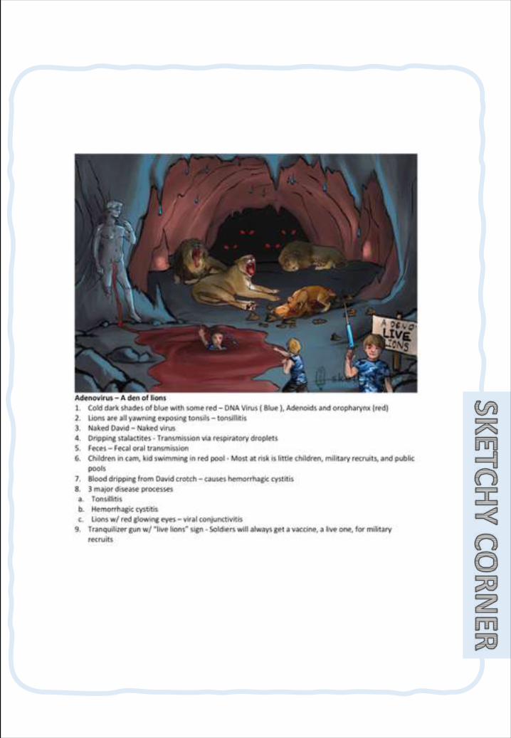
Viralinfectionsoftherespiratorysystem
CoronaviridaeFamilyCharacteristics• Enveloped,helical• Positive-sensessRNA• Hemagglutininmoleculesmakeuppeplomersonvirussurface,whichgiveshapelikesunwithcorona
VirusesofMedicalImportance• Coronavirus• Severeacuterespiratorysyndromecoronavirus(SARS-CoV)
Coronavirus• Secondmostcommoncauseofthecommoncold• Winter/springpeakincidenceSARS-CoV
Reservoir:birdsandsmallmammals(civetcats)
Transmission:respiratorydroplets;virusalsofoundinurine,sweat,andfeces;originalcaseisthoughttohavejumpedfromanimaltohuman
Disease:severeacuterespiratorysyndrome(SARS)• TraveltoFarEastorToronto• Clinicalcasedefinitionincludesfeverof>38.0C(100.4F),flu-likeillness,drycough,dyspnea,and
progressivehypoxia• Chestx-raymayshowpatchydistributionoffocalinterstitialinfiltrates
Diagnosis• Includesclinicalpresentationandpriorhistoryoftraveltoendemicareaoranassociationwith
someonewhorecentlytraveledtoendemicarea• Labtests:detectionofantibodiestoSARS-CoV,RT-PCR,andisolationofthevirusinculture
Treatment:supportive;ribavirinandinterferonarepromising
MERS-CoV (MiddleEasternRespiratorySyndrome)
Reservoir:batsandcamels
Diseaseandtransmission:similartoSARS
MeaslesVirus
DistinguishingCharacteristics: singleserotype;H-glycoproteinandfusionprotein;noneuraminidase
Reservoir:humanrespiratorytract
Transmission:respiratoryroute
Pathogenesis:abilitytocausecell:cellfusion→giantcells;viruscanescapeimmunedetection
Viralinfectionsoftherespiratorysystem
Disease• Measles:presentationgenerally3 Cs(cough,coryza,andconjunctivitis)withphotophobia;Koplik spots
→maculopapularrashfromearsdown→giantcellpneumonia(Warthin-Finkeldey cells)• Subacutesclerosing panencephalitis:rarelatecomplication(meantime7–10 years);mutantmeasles
viruspersistsinbrain,actsasslowvirus;chronicCNSdegeneration
Diagnosis:serology
Treatment:supportive,ribavirin(experimental)
Prevention:live,attenuatedvaccine,MMRandmeningoencephalitisDiagnosis:clinical;serology;ELISA,IFA,hemagglutination inhibition
MumpsVirus
DistinguishingCharacteristics: negative-sensessRNA;helical;enveloped;singleHNglycoprotein,alsoFprotein;singleserotype
Reservoir:humanrespiratorytract
Transmission:persontopersonviarespiratorydroplets
Pathogenesis:lyticinfectionofepithelialcellsofupperrespiratorytractandparotidglands→spreadthroughoutbody
Disease:mumps• Asymptomatictobilateralparotitis withfever,headache,andmalaise• Complicationsincludepancreatitis,orchitis (leadstosterilityinmales)
Treatment: supportive
Prevention: live,attenuatedvaccine,MMR
Epstein-BarrVirus(EBV)
Reservoir:humansEBV
Transmission:saliva,90%ofadultpopulationisseropositive
Pathogenesis• Virusinfectsnasopharyngealepithelialcells,salivaryandlymphoidtissues→latentinfectionof
Bcells(EBVbindstoCD21andactsasaB-cellmitogen)→resultsinproductionofatypicalreactiveTcells(Downeycells),whichmayconstituteupto70%ofWBCcount
• Heterophileantibodiesareproduced(duetoBcellmitogenesis)
Diseases
• Heterophile-positivemononucleosis,“kissingdisease”:fatigue,fever,sorethroat,lymphadenopathy,splenomegaly;latencyinBcells
• Lymphoproliferativedisease:occursinimmunocompromisedpatients;Tcellscan’tcontrolB-cellgrowth
• Hairyoralleukoplakia:hyperproliferationoflingualepithelialcells;occursinAIDSpatients
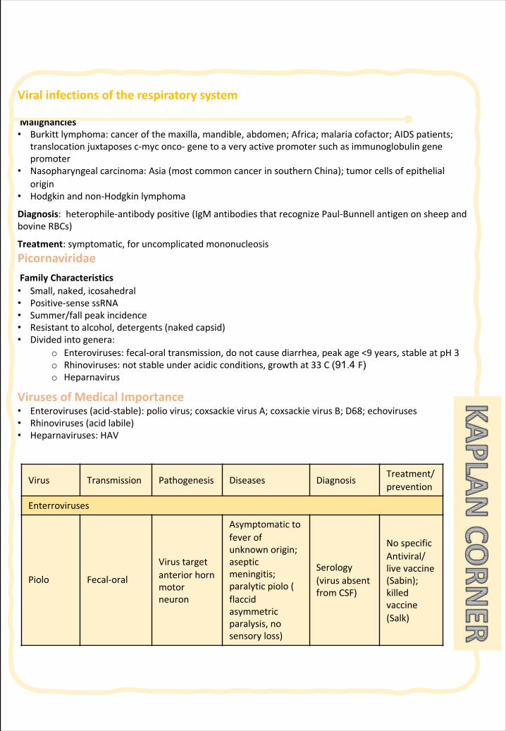
Viralinfectionsoftherespiratorysystem
Malignancies• Burkittlymphoma:cancerofthemaxilla,mandible,abdomen;Africa;malariacofactor;AIDSpatients;
translocationjuxtaposesc-myconco-genetoaveryactivepromotersuchasimmunoglobulingenepromoter
• Nasopharyngealcarcinoma:Asia(mostcommoncancerinsouthernChina);tumorcellsofepithelialorigin
• Hodgkinandnon-Hodgkinlymphoma
Diagnosis:heterophile-antibodypositive(IgMantibodiesthatrecognizePaul-BunnellantigenonsheepandbovineRBCs)
Treatment:symptomatic,foruncomplicatedmononucleosisPicornaviridaeFamilyCharacteristics• Small,naked,icosahedral• Positive-sensessRNA• Summer/fallpeakincidence• Resistanttoalcohol,detergents(nakedcapsid)• Dividedintogenera:
o Enteroviruses:fecal-oraltransmission,donotcausediarrhea,peakage<9years,stableatpH3o Rhinoviruses:notstableunderacidicconditions,growthat33C(91.4 F) o Heparnavirus
VirusesofMedicalImportance• Enteroviruses(acid-stable):poliovirus;coxsackievirusA;coxsackievirusB;D68;echoviruses• Rhinoviruses(acidlabile)• Heparnaviruses:HAV
Virus Transmission Pathogenesis Diseases DiagnosisTreatment/prevention
Enterroviruses
Piolo Fecal-oral
Virustargetanteriorhornmotorneuron
Asymptomatictofeverofunknownorigin;asepticmeningitis;paralyticpiolo (flaccidasymmetricparalysis,nosensoryloss)
Serology(virusabsentfromCSF)
NospecificAntiviral/livevaccine(Sabin);killedvaccine(Salk)
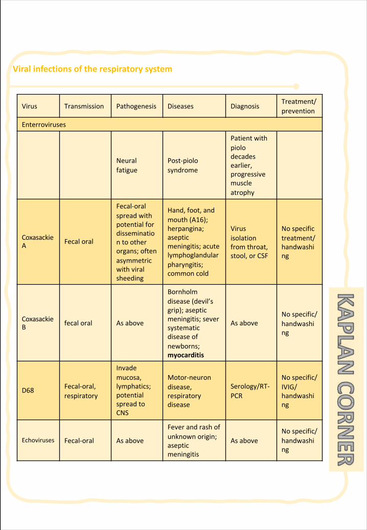
Viralinfectionsoftherespiratorysystem
Virus Transmission Pathogenesis Diseases DiagnosisTreatment/prevention
Enterroviruses
Neuralfatigue
Post-piolosyndrome
Patientwithpiolodecadesearlier,progressivemuscleatrophy
CoxasackieA Fecaloral
Fecal-oralspreadwithpotentialfordisseminationtootherorgans;oftenasymmetricwithviralsheeding
Hand,foot,andmouth(A16);herpangina;asepticmeningitis;acutelymphoglandularpharyngitis;commoncold
Virusisolationfromthroat,stool,orCSF
Nospecifictreatment/handwashing
CoxasackieB fecaloral Asabove
Bornholmdisease (devil’sgrip);asepticmeningitis;seversystematicdiseaseofnewborns;myocarditis
AsaboveNospecific/handwashing
D68 Fecal-oral,respiratory
Invademucosa,lymphatics;potentialspreadtoCNS
Motor-neurondisease,respiratorydisease
Serology/RT-PCR
Nospecific/IVIG/handwashing
Echoviruses Fecal-oral Asabove
Feverandrashofunknown origin;asepticmeningitis
AsaboveNospecific/handwashing
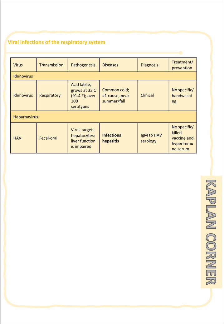
Viralinfectionsoftherespiratorysystem
Virus Transmission Pathogenesis Diseases DiagnosisTreatment/prevention
Rhinovirus
Rhinovirus Respiratory
Acidlablie;grows at33 C(91.4 F);over100serotypes
Commoncold;#1 cause,peaksummer/fall
ClinicalNospecific/handwashing
Heparnavirus
HAV Fecal-oral
Virustargetshepatocytes;liverfunctionisimpaired
Infectioushepatitis
IgMtoHAVserology
Nospecific/killedvaccineandhyperimmuneserum
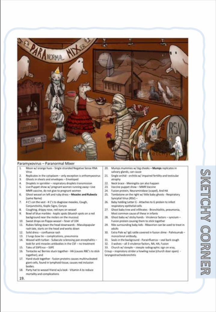
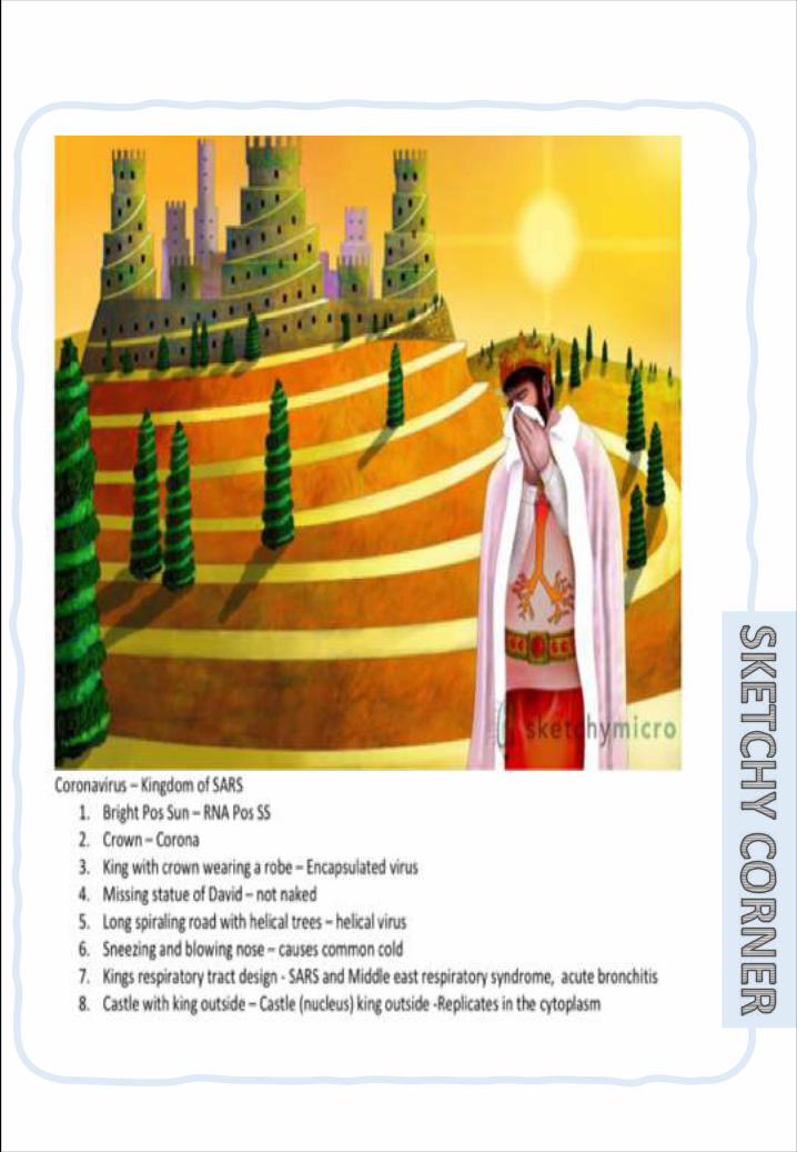
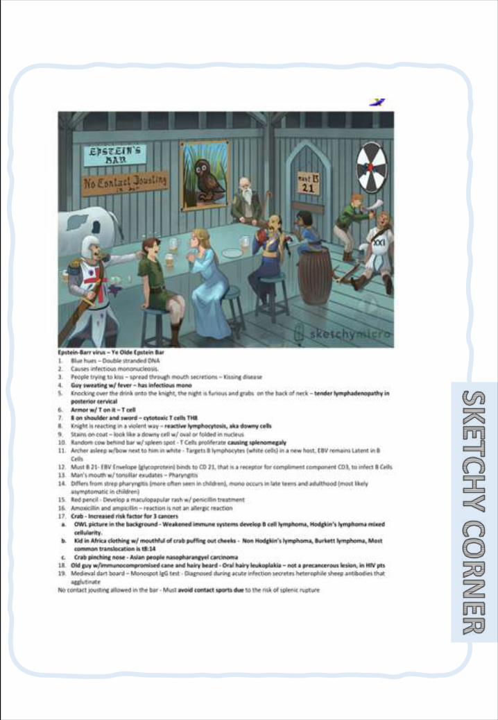
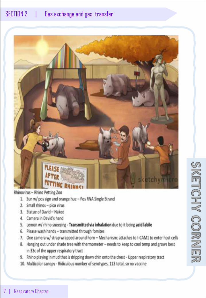
7 | Respiratory Chapter
SECTION 2 | Gas exchange and gas transfer
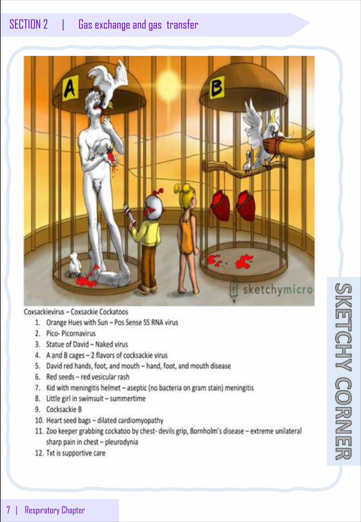
7 | Respiratory Chapter
SECTION 2 | Gas exchange and gas transfer
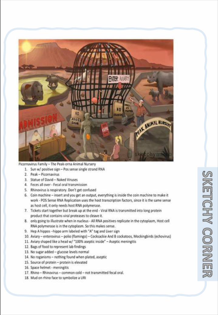
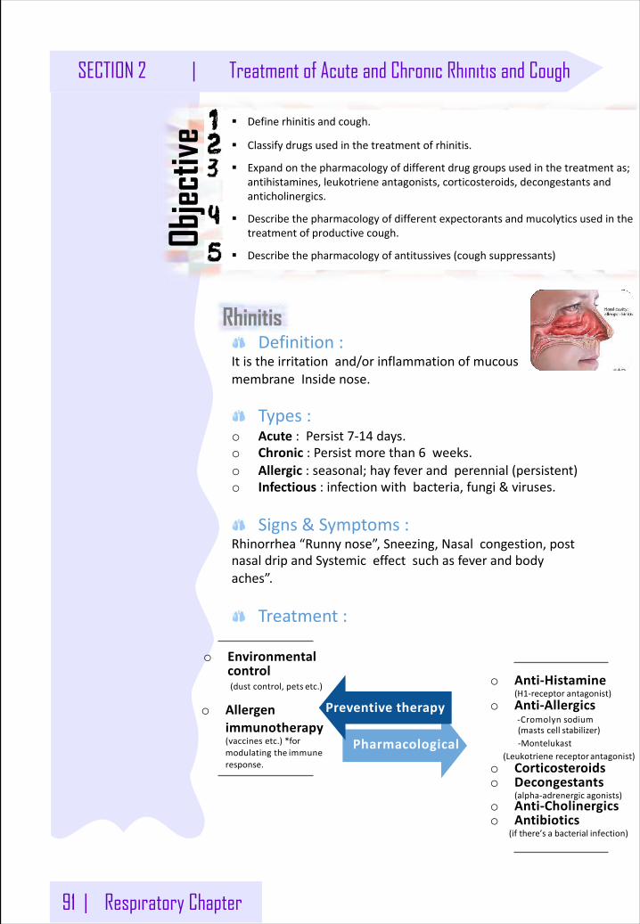
91 | Respiratory Chapter
SECTION 2 | Treatment of Acute and Chronic Rhinitis and Cough
Definition:Itistheirritationand/orinflammationofmucousmembraneInsidenose.
Types:o Acute :Persist7-14 days.o Chronic :Persistmorethan6 weeks.o Allergic :seasonal;hayfeverandperennial(persistent)o Infectious :infectionwithbacteria,fungi&viruses.
Signs&Symptoms: Rhinorrhea“Runnynose”,Sneezing,Nasalcongestion,postnasaldripandSystemiceffectsuchasfeverandbodyaches”.
Treatment:
Rhinitis
§ Definerhinitisandcough.
§ Classifydrugsusedinthetreatmentofrhinitis.
§ Expandonthepharmacologyofdifferentdruggroupsusedinthetreatmentas;antihistamines,leukotrieneantagonists,corticosteroids,decongestantsandanticholinergics.
§ Describethepharmacologyofdifferentexpectorantsandmucolyticsusedinthetreatmentofproductivecough.
§ Describethepharmacologyofantitussives(coughsuppressants)
Objec
tive
o Environmentalcontrol(dustcontrol,pets etc.)
o Allergenimmunotherapy(vaccinesetc.)*formodulatingthe immuneresponse.
Preventive therapy
Pharmacological
o Anti-Histamine(H1-receptor antagonist)
o Anti-Allergics-Cromolyn sodium(mastscell stabilizer)-Montelukast
(Leukotrienereceptorantagonist)o Corticosteroidso Decongestants
(alpha-adrenergic agonists)o Anti-Cholinergicso Antibiotics
(ifthere’sabacterial infection)
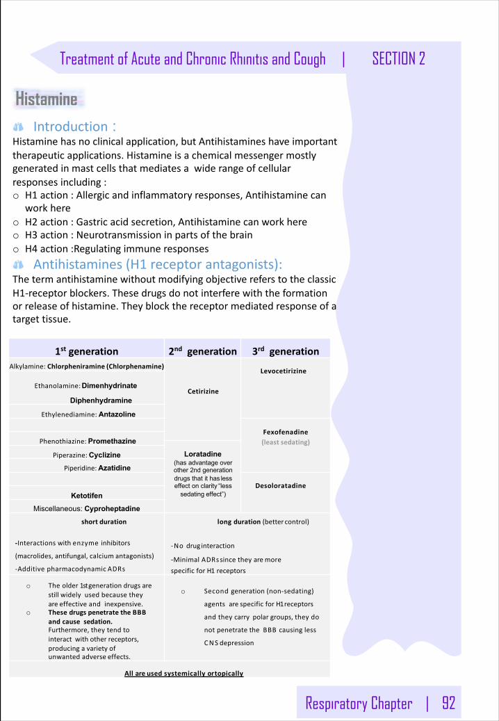
Respiratory Chapter | 92
Treatment of Acute and Chronic Rhinitis and Cough | SECTION 2
Introduction : Histaminehasnoclinicalapplication,butAntihistamineshaveimportanttherapeuticapplications.Histamineisachemicalmessengermostlygeneratedinmastcellsthatmediatesawiderangeofcellularresponsesincluding:o H1 action:Allergicandinflammatoryresponses,Antihistaminecan
workhereo H2 action:Gastricacidsecretion,Antihistaminecanworkhereo H3 action:Neurotransmissioninpartsofthebraino H4 action:Regulatingimmuneresponses
Antihistamines(H1 receptorantagonists):ThetermantihistaminewithoutmodifyingobjectivereferstotheclassicH1-receptorblockers.Thesedrugsdonotinterferewiththeformationorreleaseofhistamine.Theyblockthereceptormediatedresponseofatargettissue.
1st generation 2nd generation 3rd generationAlkylamine:Chlorpheniramine (Chlorphenamine)
Cetirizine
Levocetirizine
Ethanolamine:Dimenhydrinate
Diphenhydramine
Ethylenediamine: Antazoline
Fexofenadine(least sedating)Phenothiazine: Promethazine
Loratadine(has advantage over other 2nd generation drugs that it has less effect on clarity “less
sedating effect”)
Piperazine:CyclizinePiperidine:Azatidine
DesoloratadineKetotifen
Miscellaneous: Cyproheptadineshort duration
-Interactionswithenzymeinhibitors
(macrolides,antifungal,calcium antagonists)
-Additivepharmacodynamic ADRs
longduration(better control)
-Nodrug interaction
-MinimalADRssincetheyaremorespecificforH1 receptors
o Theolder1stgenerationdrugsarestillwidelyusedbecausetheyareeffectiveandinexpensive.
o ThesedrugspenetratetheBBBandcausesedation.Furthermore,theytendtointeractwithotherreceptors,producingavarietyofunwantedadverse effects.
o Secondgeneration(non-sedating)
agentsarespecificforH1receptors
andtheycarrypolargroups,theydo
notpenetratetheBBBcausingless
CNS depression
Allareusedsystemicallyortopically
Histamine
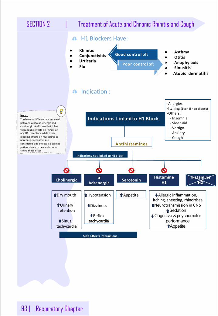
93 | Respiratory Chapter
SECTION 2 | Treatment of Acute and Chronic Rhinitis and Cough
H1 BlockersHave:
Indication:
IndicationsLinkedtoH1 Block
Antihistamines
CholinergicHistamineα
AdrenergicSerotonin
HistamineH1 H2
Dry mouth
Urinaryretention
Sinustachycardia
Hypotension
Dizziness
Reflextachycardia
Appetite Allergicinflammation,itching,sneezing, rhinorrheaNeurotransmissioninCNS
SedationCognitive & psychomotor
performanceAppetite
-Allergies-Itching(Evenifnon allergic)-Others:- Insomnia- Sleep aid- Vertigo- Anxiety- Cough
IndicationsnotlinkedtoH1block
SideEffects Interactions
● Rhinitis● Conjunctivitis● Urticaria● Flu
● Asthma● Otitis● Anaphylaxis● Sinusitis● Atopicdermatitis
Goodcontrol of:
Poorcontrol of:
Note:YouhavetodifferentiateverywellbetweenAlpha-adrenergicandcholinergic.AndknowthatithastherapeuticeffectsonrhinitisoranyH1 receptors,whileotherblockingeffectsonmuscarinicoradrenergicreceptorsareconsideredsideeffects.Socardiacpatientshavetobecarefulwhentakingthesedrugs
Respiratory Chapter | 94
Treatment of Acute and Chronic Rhinitis and Cough | SECTION 2
Drugs: antihistamine1- AntihistamineDrugs (FIRSTLINE)
Actio
ns
● TheactionofalltheH1receptorblockerisqualitatively similar●Theyaremuchmoreeffectiveinpreventingsymptoms thanreversingthemoncetheyhaveoccurred●Mostofthesedrugshaveadditionaleffects(especially1stgeneration)unrelatedtotheirblockingH1 receptors,whichprobablyreflectbindingofH1 antagonists to:○ Cholinergic○ Adrenergic○ Serotonin receptors
Therap
euticU
ses
1.Allergicrhinitis:relievesrhinorrhea,sneezing,anditchingofeyesandnasal mucosa2. Motionsickness,sleeping&anxiety.3. Nauseaandvomiting: promethazine4.Commoncold:driesoutthenasalmucosa.Oftencombinedwithnasaldecongestantand analgesics5. Allergicdermatoses:cancontrolitchingassociatedwithinsectbites.
Pharmacokinetics ● H1receptorblockersarewellabsorbedafteroral administration
● Maximumserumlevelsoccurringat1-2hours● Averageplasmahalflifeis4to6 hours●havehighbioavailabilityanddistributedtoalltissuesincludingCNS● MetabolizedbythehepaticcytochromeP450 system●Excretionoccurviakidneyexceptfexofenadineexcretedinfecesunchanged
ADRs
● Sedation● tinnitus● fatigue● dizziness● blurred vision● drymouth.Thesereactionsweremoreevidentin1st generation.
Drug
interaction
● CNS depressants● cholinesterase inhibitors
Over-do
se ThemostcommonanddangerouseffectsofacutepoisoningarethoseonCNS;includinghallucinations,excitement,ataxiaandconvulsions
Note:Ataxia:abnormalgait
Antihistamines- LearnwithVisualMnemonics!byVLStudios
AntihistaminePharmacology byNerdyNutter
95 | Respiratory Chapter
SECTION 2 | Treatment of Acute and Chronic Rhinitis and Cough
2- Anti-allergics
Type Mastcellstabilizers
Leukotrienereceptorantagonists
Exam
ple
Cromolyn(anothernameforcromoglycate)andNedocromyl
MontelukastM.O.A
Histaminerelease(mastcellstabilizersbyinhibitingCl channels),i.e.canactonlyasa prophylactic; itdoesnotantagonizereleaseshistamine.
Blockleukotriene action
Uses
o Usedinchildrenforprophylaxisofperennialallergic rhinitis
shouldbegivenondailybasisandneverstopabruptly evenifthechildisshowinganimprovement
forprophylaxisoflowerrespiratory tractallergies(e.g.perennialallergen,exercise or aspirininducedasthma)morethanonupperrespiratorytractallergies(e.g. chronic rhinosinusitis)
ADRs Caninducecough,wheezes,
headache,rash, ...etc. Asin asthma
Drugs: Anti-allergic
Note:Wecan’tuseantihistamineinasthmabecausethechemicalmediatorisleukotrienenothistamine
3- Corticosteroids(forseverecasesofrhinitisandasthma)
Exam
ple
Beclomethasone,budesonideandfluticasone
M.O.A
IthasAnti-inflammatoryeffectbyblockingphospholipaseA2andarachidonicacidsynthesiswhichleadtoinhibitprostaglandins&leukotrienessynthesisthat'swhycorticosteroidsareimportantinasthma?inhibitsthesynthesisofleukotrienes.
Uses
o Administeredtopically(inhaled)assteroidsprayo Givenifsevereintermittentormoderatepersistentsymptoms
Localcorticosteroidsarepreferablyusedmorethansystemicones.
o Why?Toreducethesideeffects.
ADRs Nasalirritation,fungalinfection,hoarsenessofvoice
Drugs: Corticosteroids
Note:EffectivenessofdifferentdruggroupsincontrollingsymptomsofRhinitis:
Respiratory Chapter | 96
Treatment of Acute and Chronic Rhinitis and Cough | SECTION 2
4- Decongestants
Type Systemic Topical
Exam
ple
Pseudoephedrine
1-Phenylethylamines:
oPhenylephrine
oMethoxamine
2-Imidazoline:
o Naphazoline
oOxymetazolineHCL
oXylometazolineHCL
M.O.A 𝛂-adrenergicagonists.Theymakevasoconstrictionofblood
vesselsinnasalmucosa&reducetherhinorrhea.
Uses
Treatmentofnasalstuffiness
ADRs
nervous,insomnia,tremors,palpitations,andhypertension.
CancauseReboundnasalstuffiness
(repeatedadministration10 days-2 weeks)
C.I
hypertension,heartfailure,anginapectoris,hyperthyroidismandglaucoma.
5- Anticholinergics
Exam
ple
Ipratropium
Uses
o Nasaldropstocontrolrhinorrhea(excessnasalsecretions&discharge),soveryeffectiveinvasomotorrhinitis(wateryhyper-secretion).
o Bronchodilatorinasthma.
ADRs Minimalsystemicsideeffects(wheezing,bladderpain,cough
producingmucus).
Drugs: Anticholinergics
Note:EffectivenessofdifferentdruggroupsincontrollingsymptomsofRhinitis:
Drugs: Decongestants
Note:Pseudoephedrinehasmanysideeffectsbecauseoftheephedrinewhichisasympathomimetic
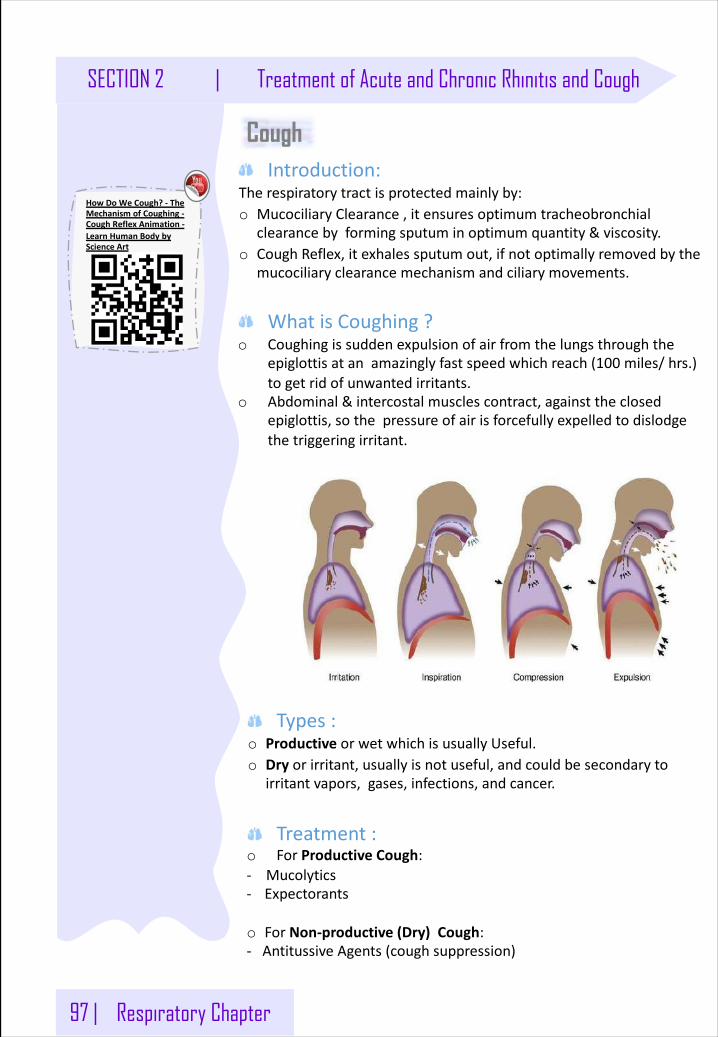
97 | Respiratory Chapter
SECTION 2 | Treatment of Acute and Chronic Rhinitis and Cough
Cough Introduction:
Therespiratorytractisprotectedmainlyby:o MucociliaryClearance,itensuresoptimumtracheobronchial
clearancebyformingsputuminoptimumquantity&viscosity.o CoughReflex,itexhalessputumout,ifnotoptimallyremovedbythe
mucociliaryclearancemechanismandciliarymovements.
WhatisCoughing?o Coughingissuddenexpulsionofairfromthelungsthroughthe
epiglottisatanamazinglyfastspeedwhichreach(100 miles/hrs.)togetridofunwantedirritants.
o Abdominal&intercostalmusclescontract,againsttheclosedepiglottis,sothepressureofairisforcefullyexpelledtodislodgethetriggeringirritant.
Types:o Productive orwetwhichisusuallyUseful.o Dry orirritant,usuallyisnotuseful,andcouldbesecondaryto
irritantvapors,gases,infections,andcancer.
Treatment:o ForProductiveCough:- Mucolytics - Expectorants
o ForNon-productive(Dry)Cough:- AntitussiveAgents(coughsuppression)
HowDoWeCough?- TheMechanismofCoughing-CoughReflexAnimation-LearnHumanBodybyScienceArt
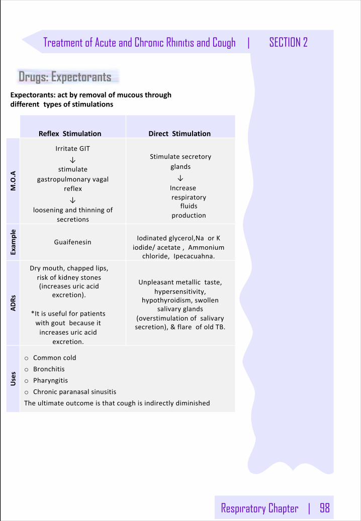
Respiratory Chapter | 98
Treatment of Acute and Chronic Rhinitis and Cough | SECTION 2
ReflexStimulation DirectStimulation
M.O.A
IrritateGIT↓
stimulategastropulmonaryvagal
reflex↓
looseningandthinningofsecretions
Stimulatesecretoryglands↓
Increaserespiratory
fluidsproduction
Exam
ple
Guaifenesin Iodinatedglycerol,NaorKiodide/acetate,Ammonium
chloride,Ipecacuahna.
ADRs
Drymouth,chappedlips,riskofkidneystones(increasesuricacid
excretion).
*Itisusefulforpatientswithgoutbecauseitincreasesuricacid
excretion.
Unpleasantmetallictaste,hypersensitivity,
hypothyroidism,swollensalivaryglands
(overstimulationofsalivarysecretion),&flareofoldTB.
Uses
o Commoncoldo Bronchitiso Pharyngitiso ChronicparanasalsinusitisTheultimateoutcomeisthatcoughisindirectlydiminished
Expectorants:actbyremovalofmucousthrough different typesofstimulations
Drugs: Expectorants

99 | Respiratory Chapter
SECTION 2 | Treatment of Acute and Chronic Rhinitis and Cough
Drugs: Mucolytics
N-AcetylCysteine
Bromhexine&Ambroxol
(AmbroxolisametaboliteofBromhexine)
PulmozymeDornaseAlphaor rhDNAase
Overview Afreeradical
scavengerusedinacetaminophenoverdose
M.O.A
IncreaseBreakdownofS-S bondsinglycoprotein inmucous→whichleadto lessviscid mucous.
o IncreaseSynthesizeofserousmucus
o Increaseactivateciliary clearance
CleavageofextracellularbacterialDNA,thatcontributestoviscosityofsputumincase ofbacterialinfection only
ADRs
Bronchospasm,stomatitis,rhinorrhea,rash, nausea & vomiting
Rhinorrhea,lacrymation,gastricirritations,hypersensitivity
Voicechanges,pharyngitis,laryngitis, rhinitis,chestpain, fever,rash
Uses
MostmucolyticsareeffectiveasadjuvanttherapyinCOPD,asthma,bronchitis(whenthereisexcessive,thickmucus).Inbronchiectasis,pneumonia&TBtheyareofpartialbenefitandhardlyanybenefitincysticfibrosis&severeinfections→giverhDNAase
Ithelpsto:o IncreaseImmune
defense.o Decrease
antibioticsusage.
o Decreasepaininacutesorethroat
Arecombinanthuman-deoxyribonuclease-1enzymewhichisgeneticallyengineeredthatisnebulized+Fullbenefitappearswithin3-7 days
Usedtodissolveorbreakdownmucusintherespiratorytract→mucusislessviscous→coughedupeasily.
OtherMucolytics:o HypertonicSaline&NaHCO3 :WorkbyDecreasingViscoelasticitybyIncreasingWaterContent.
o Steaminhalation:WorkbyDecreasingAdhesiveness.
3 MucolyticsbyTrevorTessier
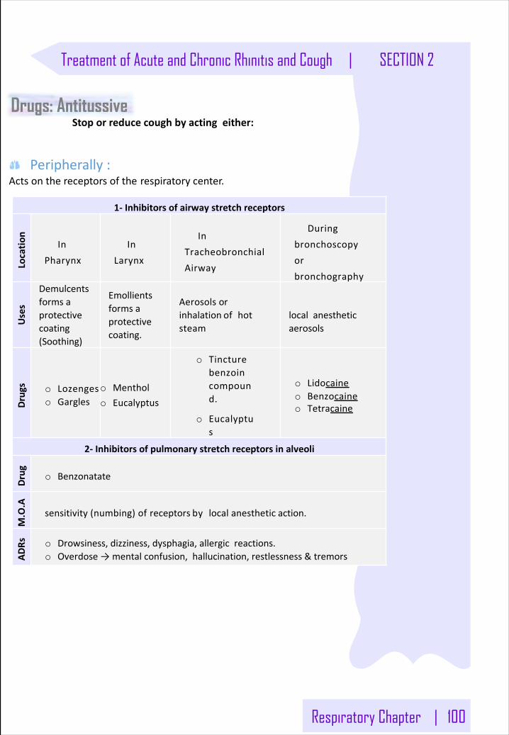
Respiratory Chapter | 100
Treatment of Acute and Chronic Rhinitis and Cough | SECTION 2
1- Inhibitorsofairwaystretchreceptors
Locatio
n
InPharynx
InLarynx
InTracheobronchialAirway
Duringbronchoscopyorbronchography
Uses
Demulcentsformsaprotectivecoating(Soothing)
Emollientsformsaprotectivecoating.
Aerosolsorinhalation ofhotsteam
localanestheticaerosols
Drugs o Lozenges
o Gargleso Mentholo Eucalyptus
o Tincturebenzoincompound.
o Eucalyptus
o Lidocaineo Benzocaineo Tetracaine
2- Inhibitorsofpulmonarystretchreceptorsinalveoli
Drug o Benzonatate
M.O.A
sensitivity(numbing)ofreceptors bylocalanesthetic action.
ADRs o Drowsiness,dizziness,dysphagia,allergicreactions.
o Overdose→mentalconfusion,hallucination,restlessness&tremors
Peripherally:Actsonthereceptorsofthe respiratorycenter.
Drugs: AntitussiveStoporreducecoughbyactingeither:
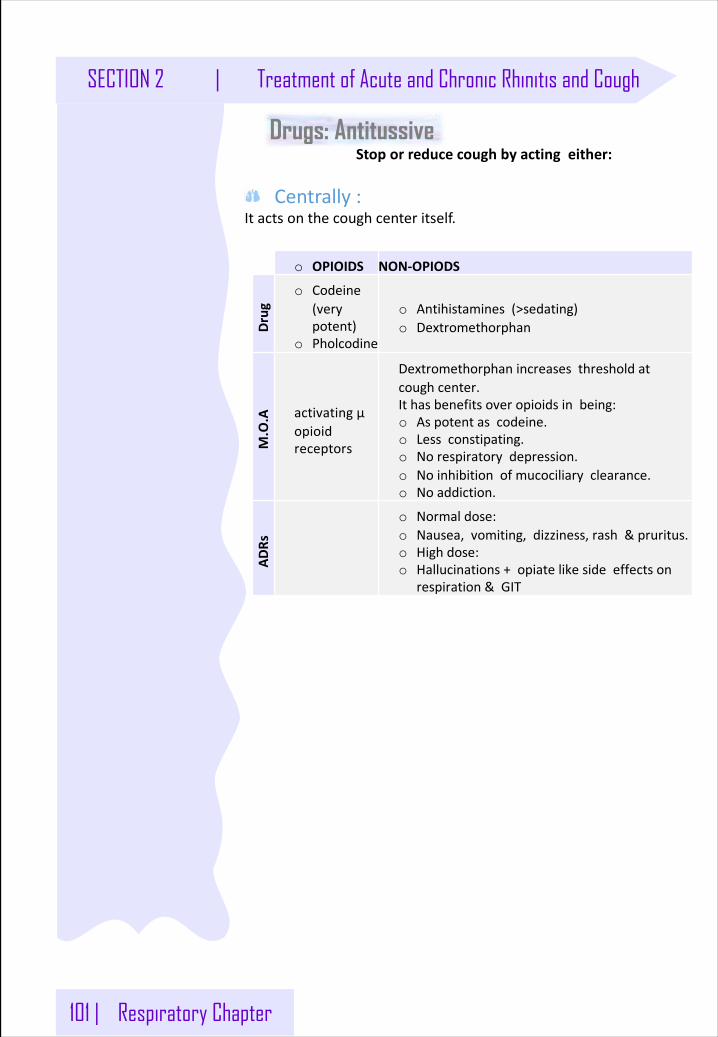
101 | Respiratory Chapter
SECTION 2 | Treatment of Acute and Chronic Rhinitis and Cough
Drugs: AntitussiveStoporreducecoughbyactingeither:
Centrally:Itactsonthecoughcenteritself.
o OPIOIDS NON-OPIODSDrug
o Codeine(verypotent)
o Pholcodine
o Antihistamines(>sedating)o Dextromethorphan
M.O.A activatingµ
opioidreceptors
Dextromethorphanincreasesthresholdatcoughcenter.Ithasbenefitsoveropioidsinbeing:o Aspotentascodeine.o Lessconstipating.o Norespiratorydepression.o Noinhibitionofmucociliaryclearance.o Noaddiction.
ADRs
o Normaldose:o Nausea,vomiting,dizziness,rash&pruritus.o Highdose:o Hallucinations+opiatelikesideeffectson
respiration&GIT
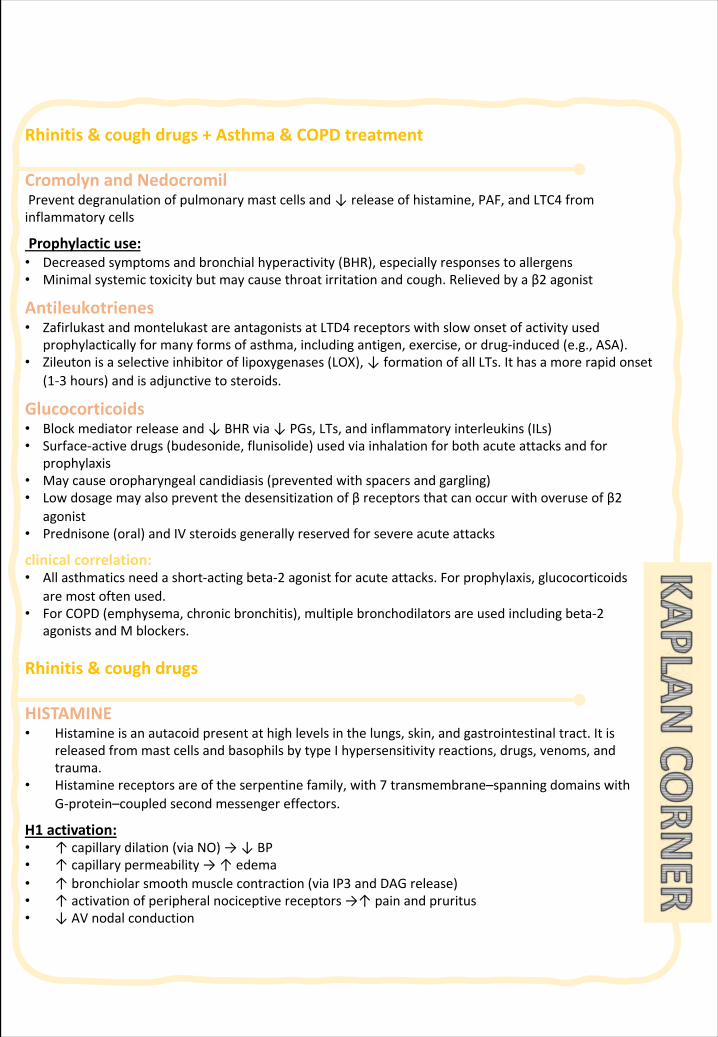
Rhinitis&coughdrugs+Asthma&COPDtreatment
Cromolyn andNedocromilPreventdegranulationofpulmonarymastcellsand↓releaseofhistamine,PAF,andLTC4 frominflammatorycells
Prophylacticuse:• Decreasedsymptomsandbronchialhyperactivity(BHR),especiallyresponsestoallergens• Minimalsystemictoxicitybutmaycausethroatirritationand cough.Relievedbyaβ2 agonist
Antileukotrienes• Zafirlukast andmontelukast areantagonistsatLTD4 receptorswithslowonsetofactivityused
prophylacticallyformanyformsofasthma,includingantigen,exercise,ordrug-induced(e.g.,ASA).• Zileuton isaselectiveinhibitoroflipoxygenases(LOX),↓formationofallLTs.Ithasamorerapidonset
(1-3 hours)andisadjunctivetosteroids.
Glucocorticoids• Blockmediatorreleaseand↓BHRvia↓PGs,LTs,andinflammatoryinterleukins(ILs)• Surface-activedrugs(budesonide,flunisolide)usedviainhalationforbothacuteattacksandfor
prophylaxis• Maycauseoropharyngealcandidiasis(preventedwithspacersandgargling)• Lowdosagemayalsopreventthedesensitizationofβreceptorsthatcanoccurwithoveruseofβ2
agonist• Prednisone(oral)andIVsteroidsgenerallyreservedforsevereacuteattacks
clinicalcorrelation:• Allasthmaticsneedashort-actingbeta-2 agonistforacuteattacks.Forprophylaxis,glucocorticoids
aremostoftenused.• ForCOPD(emphysema,chronicbronchitis),multiplebronchodilatorsareusedincludingbeta-2
agonistsandMblockers.
Rhinitis&coughdrugs
HISTAMINE• Histamineisanautacoidpresentathighlevelsinthelungs,skin,andgastrointestinaltract.Itis
releasedfrommastcellsandbasophilsbytypeIhypersensitivityreactions,drugs,venoms,andtrauma.
• Histaminereceptorsareoftheserpentinefamily,with7 transmembrane–spanningdomainswithG-protein–coupledsecondmessengereffectors.
H1 activation:• ↑capillarydilation(viaNO)→↓BP• ↑capillarypermeability→↑edema• ↑bronchiolarsmoothmusclecontraction(viaIP3 andDAGrelease)• ↑activationofperipheralnociceptivereceptors→↑painandpruritus• ↓AVnodalconduction
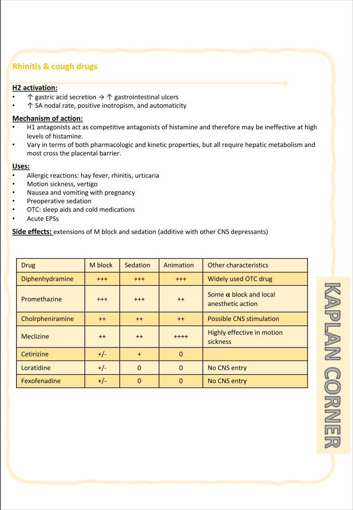
Rhinitis&coughdrugs
H2 activation:• ↑gastricacidsecretion→↑gastrointestinalulcers• ↑SAnodalrate,positiveinotropism,andautomaticity
Mechanismofaction:• H1 antagonistsactascompetitiveantagonistsofhistamineandthereforemaybeineffectiveathigh
levelsofhistamine.• Varyintermsofbothpharmacologicandkineticproperties,butallrequirehepaticmetabolismand
mostcrosstheplacentalbarrier.
Uses:• Allergicreactions:hayfever,rhinitis,urticaria• Motionsickness,vertigo• Nauseaandvomitingwithpregnancy• Preoperativesedation• OTC:sleepaidsandcoldmedications• AcuteEPSs
Sideeffects:extensionsofMblockandsedation(additivewithotherCNSdepressants)
Drug Mblock Sedation Animation Othercharacteristics
Diphenhydramine +++ +++ +++ WidelyusedOTCdrug
Promethazine +++ +++ ++Some𝛂 blockandlocalanesthetic action
Cholrpheniramine ++ ++ ++ PossibleCNSstimulation
Meclizine ++ ++ ++++ Highlyeffectivein motionsickness
Cetirizine +/- + 0
Loratidine +/- 0 0 NoCNSentry
Fexofenadine +/- 0 0 NoCNSentry

79 | Respiratory Chapter
SECTION 2 | Viruses Causing Respiratory Infections I
Objec
tive
Respiratory Tract Infections:
Note: (1)Ifitgetstothelowerrespiratorytractitbecomesevere.
§ Acquirethebasicknowledgeaboutstructureandclassificationofthesevirusesinfectingtherespiratorysystem.
Including(Influenza viruses,Parainfluenza viruses,Respiratorysyncytialvirus(RSV)andhumanmetapneumovirus(HMPV),Measles virus,andMumps)
§ Describetheirepidemiologyandpathogenesis
§ IdentifytherespiratoryinfectionsandtheclinicalfeaturesofURTIandLRTI.
§ Describetheirepidemiologyandpathogenesis
§ Knowthelaboratorydiagnosis,andtreatmentoftheseinfections.
§ Recognizethemethodsforprevention.
Note: (2)Lowerrespiratorytractinfections
Introduction:o Theyarethecommonestofhumaninfectionsandcausealarge
amountofmorbidityandlossoftimeatwork(sickleave).Theyarecommoninbothchildrenandadults.
o Mostlycausedbyviruses.Mostlyareself-limitingdisease,whichmeanthediseasetendstogoawayonitsown,withouttreatment.
o Mostlyaremildandconfinedtotheupperrespiratorytract(URT).1o URT-infectionmayspreadtootherorganscausingmoresevere
infectionanddeath.
ClinicalManifestations(symptoms):o Commoncold(rhinitis).o Pharyngitis.o Tonsillitis.o Sinusitis&otitismedia.o Croup(acutelaryngotracheobronchitis).o Acutebronchitis,AcutebronchiolitisandViralpneumonia.2
Commonrespiratoryviruses::
Nameofthevirus Family Disease1)Influenzavirus Orthomyxoviridae URTIandLRTI
2)Parainfluenzavirus Paramyxoviridae LRTI3)Respiratorysyncytialvirus
4 Rhinovirus Picornaviridae URTI
5 Coronavirus Coronaviridae URTIandLRTI
6 Adenovirus Adenoviridae URTIandeyeinfections
7 Humanmetapneumovirus Paramyxoviridae LRTI
ArabicTranslation: Croup= قانخلا ضرم
ExtraExplanation: RNAsenseinviruses
o Positivesense(+vestrand):(5'to3')viralRNAsignifiesthataparticularviralRNAsequencemaybedirectlytranslatedintothedesiredviralproteins.
o Negativesense(-vestrand):ThisRNA(3'to5')cannotbetranslatedintoproteindirectly.Instead,itmustfirstbetranscribedintoapositivesenseRNAthatactsasanmRNA.Someviruses(Influenza,forexample)havenegativesensegenomesandsomustcarryanRNApolymeraseinsidethevirion.
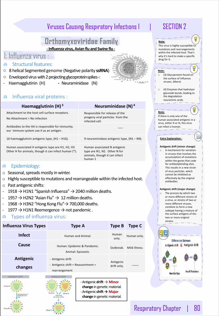
Respiratory Chapter | 80
Viruses Causing Respiratory Infections I | SECTION 2
Note: o (3)Glycoproteinfoundon
thesurfaceofinfluenzaviruses.(More)
o (4)Enzymesthathydrolyzeglycosidebonds,leadingtothedegradationneuraminicacids.
1: Influenza virus : Structural features:
o 8 helicalSegmentedgenome (NegativepolarityssRNA)o Envelopedviruswith2projectingglycoproteinspikes :-- Haemagglutinin(H) - Neuraminidase (N)
Influenzaviralproteins:
Haemagglutinin(H) 3 Neuraminidase(N) 4
Attachmenttothehostcellsurfacereceptors.
NoAttachment=Noinfection
Responsibleforreleaseoftheprogenyviralparticlesfromtheinfectedcell.
AntibodiestotheHAisresponsibleforimmunity.our immunesystemuseitasanantigen.
___
16 haemagglutininantigenictype,(H1 – H16). 9 neuraminidaseantigenictype,(N1 – N9).
HumanassociatedHantigenictypeareH1,H2,H3OtherHforanimals,thoughitcaninfecthuman(*).
HumanassociatedNantigenictypeareN1,N2.OtherNforanimals,thoughitcaninfecthuman1
Note: Thisvirusishighlysusceptibletomutationsandrearrangementswithintheinfectedhost.That’swhyit’shardtomakeaspecificdrugforit.
Epidemiology:o Seasonal,spreadsmostlyinwinter.o Highlysusceptibletomutationsandrearrangeablewithintheinfectedhost.o Pastantigenicshifts:- 1918→H1N1 “SpanishInfluenza”→2040milliondeaths.- 1957→H2N2 “AsianFlu”→12milliondeaths.- 1968→H3N2 “HongKongFlu”→700,000 deaths.- 1977→H1N1Reemergence→notpandemic.
Typesofinfluenzavirus:InfluenzaVirusTypes TypeA TypeB TypeC
Infect HumanandAnimal. Humanonly.
Humanonly.
Cause Human:Epidemic&Pandemic.
Animal:Epizootic.Outbreak. Mildillness.
Antigenic
changes
- Antigenicdrift
- Antigenicshift=Reassortment=
rearrangement
Antigenicdriftonly. ____
-Antigenicdrift→Minorchangeingenetic material.-Antigenicshift→Majorchangeingenetic material.
Note: Ifthereisonlyoneofthehumanassociatedantigenicinavirus,eitherHorN,thisviruscaninfectahuman.
Orthomyxoviridae Family- Influenzavirus,AvianfluandSwineflu-
ExtraExplanation:
Antigenicdrift(minorchange)o Amechanismforvariation
invirusesthatinvolvestheaccumulationofmutationswithinthegenesthatcodeforantibodybinding sites.Thisresultsinanewstrainofvirusparticles.whichcannotbeinhibitedaseffectivelybytheoriginalantibodies.
Antigenicshift(majorchange)o Theprocessbywhichtwo
ormoredifferentstrainsofavirus,orstrainsoftwoormoredifferentviruses,combinetoformanewsubtypehavingamixtureofthesurfaceantigensofthetwoormoreoriginalstrains.
102 EMBRYOLOGY:Development of respiratory system
HISTOLOGY : The lower respiratory tract.
ANATOMY: Larynx, trachea and bronchi
PATHOLOGY:Introduction to COPD
PATHOLOGY: Bronchial asthma
IMMUNOLOGY :Bronchial asthma
PHARMACOLOGY: Asthma and COPD treatment
PHYSIOLOGY:Lung function in health and disease
ANATOMY: Lung and pleura
SECTION 3 :
LOWER RESPIRATORY TRACT
110116127134138
PATHOLOGY:Restrictive lung disease
154164179185
PATHOLOGY: Lobar AND bronchopneumonia
SECTION 3 :
LOWER RESPIRATORY TRACT
PHARMACOLOGY: Drugs used in TB212
216
MICROBIOLOGY: Community Acquired pneumonia
230 MICROBIOLOGY: Hospital Acquired pneumonia
PATHOLGY:MICROBIOLOGY:IMMUNOLOGY:Tuberculosis (TB)
196 PHARMACOLOGY: Respiratory tract infections
MICROBIOLOGY: Respiratory fungal infection and aspergillosis
243
ANATOMY: Mediastinum
ANATOMY: Radiological anatomy of the thorax.236
250
272277
PATHOLOGY: Lung Tumors258
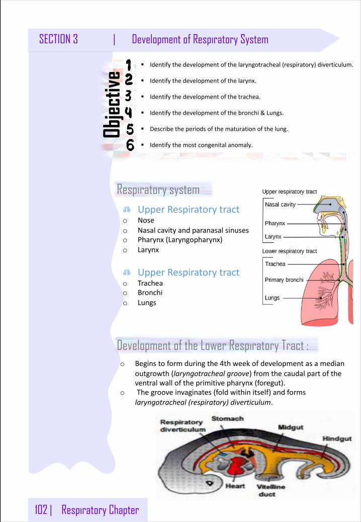
102 | Respiratory Chapter
SECTION 3 | Development of Respiratory System
Respiratory system
§ Identifythedevelopmentofthelaryngotracheal(respiratory)diverticulum.
§ Identifythedevelopmentofthelarynx.
§ Identifythedevelopmentofthetrachea.
§ Identifythedevelopmentofthebronchi&Lungs.
§ Describetheperiodsofthematurationofthelung.
§ Identifythemostcongenitalanomaly.
Objec
tive
UpperRespiratorytracto Noseo Nasalcavityandparanasalsinuseso Pharynx(Laryngopharynx)o Larynx
UpperRespiratorytracto Tracheao Bronchio Lungs
Development of the Lower Respiratory Tract :o Beginstoformduringthe4thweekofdevelopmentasamedian
outgrowth(laryngotrachealgroove)fromthecaudalpartoftheventralwalloftheprimitivepharynx(foregut).
o Thegrooveinvaginates(foldwithinitself)andformslaryngotracheal(respiratory)diverticulum.
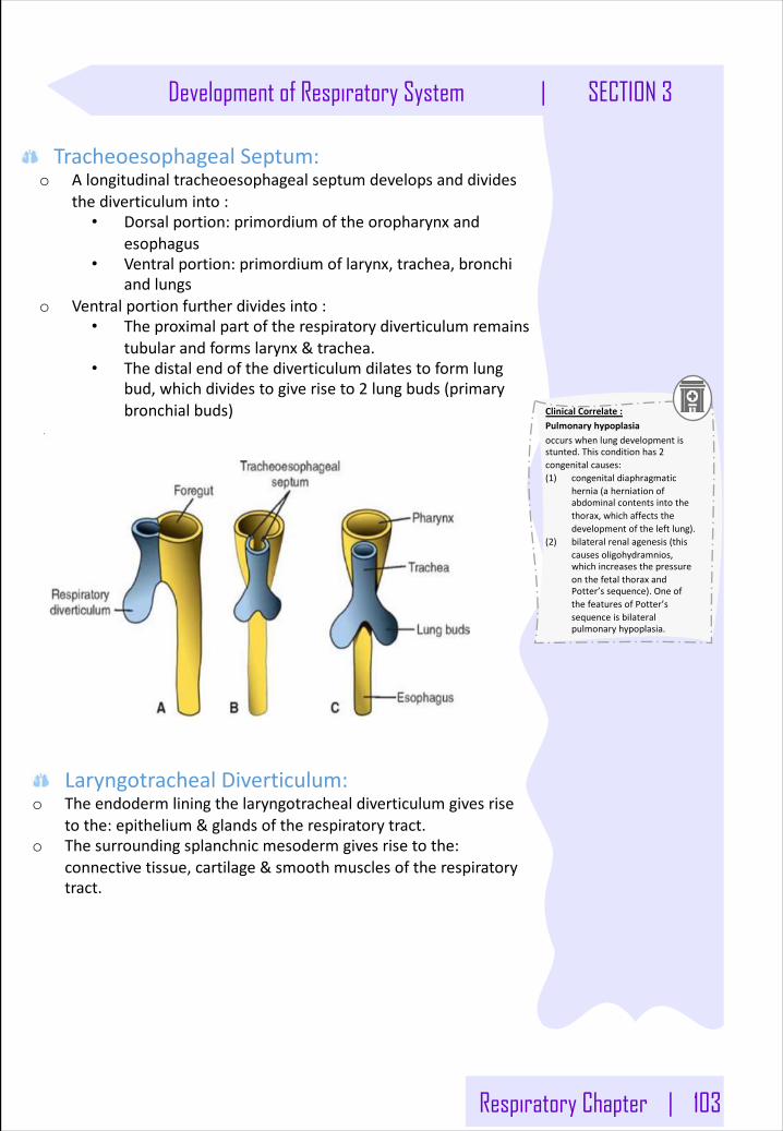
Respiratory Chapter | 103
Development of Respiratory System | SECTION 3
TracheoesophagealSeptum:o Alongitudinaltracheoesophagealseptumdevelopsanddivides
thediverticuluminto:• Dorsalportion:primordiumoftheoropharynxand
esophagus• Ventralportion:primordiumoflarynx,trachea,bronchi
andlungso Ventralportionfurtherdividesinto:
• Theproximalpartoftherespiratorydiverticulumremainstubularandformslarynx&trachea.
• Thedistalendofthediverticulumdilatestoformlungbud,whichdividestogiveriseto2 lungbuds(primarybronchialbuds)
LaryngotrachealDiverticulum:o Theendodermliningthelaryngotrachealdiverticulumgivesrise
tothe:epithelium&glandsoftherespiratorytract.o Thesurroundingsplanchnicmesodermgivesrisetothe:
connectivetissue,cartilage&smoothmusclesoftherespiratorytract.
ClinicalCorrelate: Pulmonaryhypoplasiaoccurswhenlungdevelopmentisstunted.Thisconditionhas2congenitalcauses:(1) congenitaldiaphragmatic
hernia(aherniationofabdominalcontentsintothethorax,whichaffectsthedevelopmentoftheleftlung).
(2) bilateralrenalagenesis(thiscausesoligohydramnios,whichincreasesthepressureonthefetalthoraxandPotter’ssequence).OneofthefeaturesofPotter’ssequenceisbilateralpulmonaryhypoplasia.
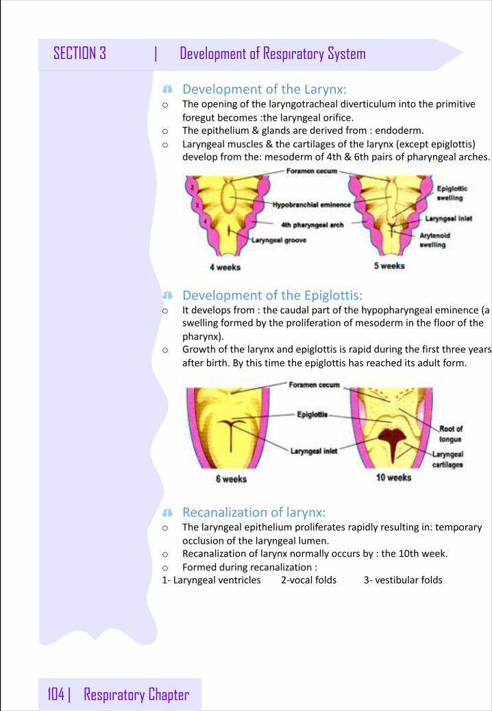
104 | Respiratory Chapter
SECTION 3 | Development of Respiratory System
DevelopmentoftheLarynx:o Theopeningofthelaryngotrachealdiverticulumintotheprimitive
foregutbecomes:thelaryngealorifice.o Theepithelium&glandsarederivedfrom:endoderm.o Laryngealmuscles&thecartilagesofthelarynx(exceptepiglottis)
developfromthe:mesodermof4th&6thpairsofpharyngealarches.
DevelopmentoftheEpiglottis:o Itdevelopsfrom:thecaudalpartofthehypopharyngealeminence(a
swellingformedbytheproliferationofmesoderminthefloorofthepharynx).
o Growthofthelarynxandepiglottisisrapidduringthefirstthreeyearsafterbirth.Bythistimetheepiglottishasreacheditsadultform.
Recanalizationoflarynx:o Thelaryngealepitheliumproliferatesrapidlyresultingin:temporary
occlusionofthelaryngeallumen.o Recanalizationoflarynxnormallyoccursby:the10thweek.o Formedduringrecanalization: 1- Laryngealventricles2-vocalfolds 3- vestibularfolds
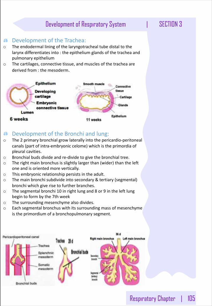
Respiratory Chapter | 105
Development of Respiratory System | SECTION 3
DevelopmentoftheTrachea:o Theendodermalliningofthelaryngotrachealtubedistaltothe
larynxdifferentiatesinto:theepitheliumglandsofthetracheaandpulmonaryepithelium
o Thecartilages,connectivetissue,andmusclesofthetracheaarederivedfrom:themesoderm.
DevelopmentoftheBronchiandlung:o The2 primarybronchialgrowlaterallyintothepericardio-peritoneal
canals(partofintra-embryoniccelome)whichistheprimordiaofpleuralcavities.
o Bronchialbudsdivideandre-dividetogivethebronchialtree.o Therightmainbronchusisslightlylargerthan(wider)thantheleft
oneandisorientedmorevertically.o Thisembryonicrelationshippersistsintheadult.o Themainbronchisubdivideintosecondary&tertiary(segmental)
bronchiwhichgiverisetofurtherbranches.o Thesegmentalbronchi10 inrightlungand8 or9 intheleftlung
begintoformbythe7thweeko Thesurroundingmesenchymealsodivides.o Eachsegmentalbronchuswithitssurroundingmassofmesenchyme
istheprimordiumofabronchopulmonarysegment.
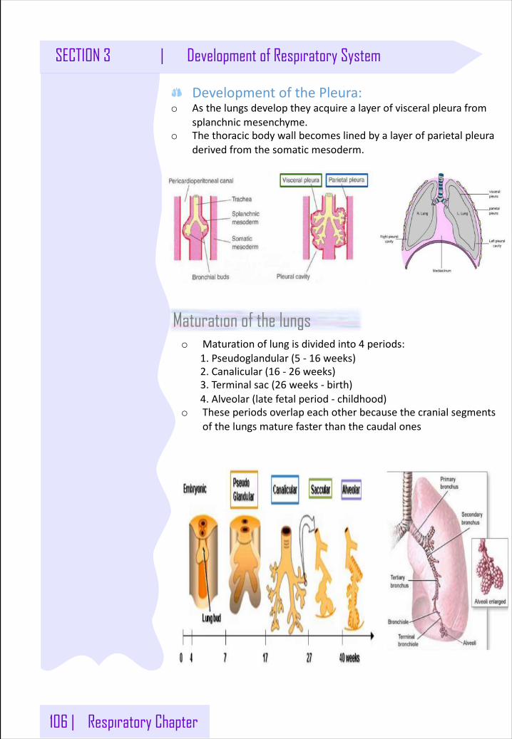
106 | Respiratory Chapter
SECTION 3 | Development of Respiratory System
DevelopmentofthePleura:o Asthelungsdeveloptheyacquirealayerofvisceralpleurafrom
splanchnicmesenchyme.o Thethoracicbodywallbecomeslinedbyalayerofparietalpleura
derivedfromthesomaticmesoderm.
o Maturationoflungisdividedinto4 periods:1.Pseudoglandular (5 - 16 weeks)2.Canalicular(16 - 26 weeks)3.Terminalsac(26 weeks- birth)4.Alveolar(latefetal period- childhood)
o Theseperiodsoverlapeachotherbecausethecranialsegmentsofthelungsmaturefasterthanthecaudalones
Maturation of the lungs
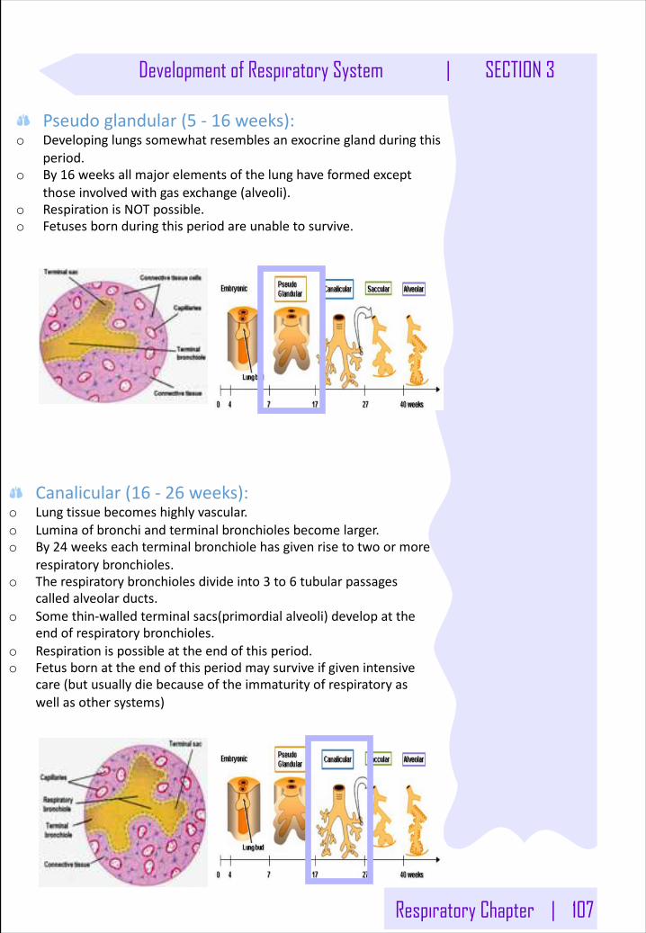
Respiratory Chapter | 107
Development of Respiratory System | SECTION 3
Pseudoglandular(5 - 16 weeks):o Developinglungssomewhatresemblesanexocrineglandduringthis
period.o By16 weeksallmajorelementsofthelunghaveformedexcept
thoseinvolvedwithgasexchange(alveoli).o RespirationisNOTpossible.o Fetuses bornduringthisperiodareunabletosurvive.
Canalicular(16 - 26 weeks):o Lungtissuebecomeshighlyvascular.o Luminaofbronchiandterminalbronchiolesbecomelarger.o By24 weekseachterminalbronchiolehasgivenrisetotwoormore
respiratorybronchioles.o Therespiratorybronchiolesdivideinto3 to6 tubularpassages
calledalveolarducts.o Somethin-walledterminalsacs(primordialalveoli)developatthe
endofrespiratorybronchioles.o Respirationispossibleattheendofthisperiod.o Fetus bornattheendofthisperiodmaysurviveifgivenintensive
care(butusuallydiebecauseoftheimmaturityofrespiratoryaswellasothersystems)
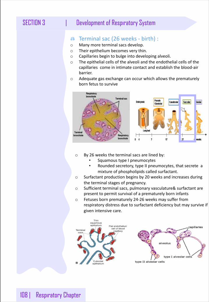
108 | Respiratory Chapter
SECTION 3 | Development of Respiratory System
Terminalsac(26 weeks- birth):o Manymoreterminalsacsdevelop.o Theirepitheliumbecomesverythin.o Capillariesbegintobulgeintodevelopingalveoli.o Theepithelialcellsofthealveoliandtheendothelialcellsofthe
capillariescomeinintimatecontactandestablishtheblood-airbarrier.
o Adequategasexchangecanoccurwhichallowstheprematurelybornfetus tosurvive
o By26 weekstheterminalsacsarelinedby:• SquamoustypeIpneumocytes• Roundedsecretory,typeIIpneumocytes,thatsecretea
mixtureofphospholipidscalledsurfactant.o Surfactantproductionbeginsby20 weeksandincreasesduring
theterminalstagesofpregnancy.o Sufficientterminalsacs,pulmonaryvasculature&surfactantare
presenttopermitsurvivalofaprematurelyborninfantso Fetuses bornprematurely24-26 weeksmaysufferfrom
respiratorydistressduetosurfactantdeficiencybutmaysurviveifgivenintensivecare.
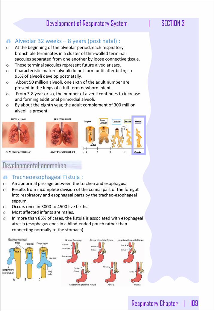
Respiratory Chapter | 109
Development of Respiratory System | SECTION 3
Alveolar32 weeks– 8 years(postnatal):o Atthebeginningofthealveolarperiod,eachrespiratory
bronchioleterminatesinaclusterofthin-walledterminalsacculesseparatedfromoneanotherbylooseconnectivetissue.
o Theseterminalsacculesrepresentfuturealveolarsacs.o Characteristicmaturealveolidonotformuntilafterbirth;so
95%ofalveolideveloppostnatally.o About50 millionalveoli,onesixthoftheadultnumberare
presentinthelungsofafull-termnewborn infant.o From3-8 yearorso,thenumberofalveolicontinuestoincrease
andformingadditionalprimordialalveoli.o Byabouttheeighthyear,theadultcomplementof300 million
alveoliispresent.
TracheoesophagealFistula:o Anabnormalpassagebetweenthetracheaandesophagus.o Resultsfromincompletedivisionofthecranialpartoftheforegut
intorespiratoryandesophageal partsbythetracheo-esophagealseptum.
o Occursoncein3000 to4500 livebirths.o Mostaffectedinfantsaremales.o Inmorethan85%ofcases,thefistulaisassociatedwithesophageal
atresia(esophagusendsinablind-endedpouchratherthanconnectingnormallytothestomach)
Developmental anomalies
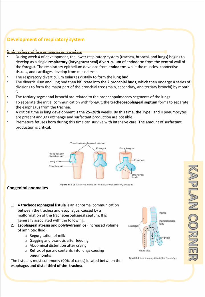
Developmentofrespiratorysystem Embryologyoflowerrespiratorysystem• Duringweek4 ofdevelopment,thelowerrespiratorysystem(trachea,bronchi,andlungs)beginsto
developasasinglerespiratory(laryngotracheal)diverticulumofendodermfromtheventralwalloftheforegut.Therespiratoryepitheliumdevelopsfromendoderm whilethemuscles,connectivetissues,andcartilagesdevelopfrommesoderm.
• Therespiratorydiverticulumenlargesdistallytoformthelungbud.• Thediverticulumandlungbudthenbifurcateintothe2 bronchialbuds,whichthenundergoaseriesof
divisionstoformthemajorpartofthebronchialtree(main,secondary,andtertiarybronchi)bymonth6.
• Thetertiarysegmentalbronchiarerelatedtothebronchopulmonarysegmentsofthelungs.• Toseparatetheinitialcommunicationwithforegut,thetracheoesophageal septum formstoseparate
theesophagusfromthetrachea.• Acriticaltimeinlungdevelopmentisthe25–28th weeks.Bythistime,theTypeIandIIpneumocytes
arepresentandgasexchangeandsurfactantproductionarepossible. • Prematurefetusesbornduringthistimecansurvivewithintensivecare.Theamountofsurfactant
productioniscritical.
Congenitalanomalies
1. Atracheoesophagealfistulaisanabnormalcommunicationbetweenthetracheaandesophaguscausedbyamalformationofthetracheoesophagealseptum.Itisgenerallyassociatedwiththefollowing:
2. Esophagealatresiaandpolyhydramnios (increasedvolumeofamnioticfluid)
o Regurgitationofmilko Gaggingandcyanosisafterfeedingo Abdominaldistentionaftercryingo Reflux ofgastriccontentsintolungscausing
pneumonitisThefistulaismostcommonly(90%ofcases)locatedbetweentheesophagusanddistalthirdofthetrachea.
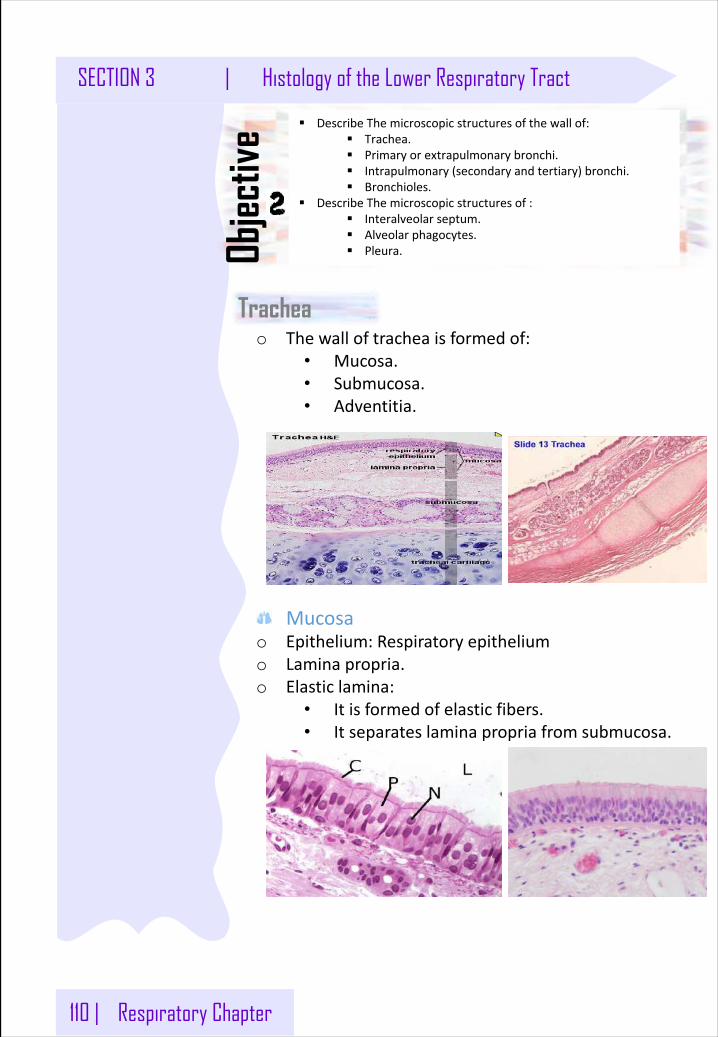
110 | Respiratory Chapter
SECTION 3 | Histology of the Lower Respiratory Tract
Trachea
§ DescribeThemicroscopicstructuresofthewallof:§ Trachea.§ Primaryorextrapulmonarybronchi.§ Intrapulmonary(secondaryandtertiary)bronchi.§ Bronchioles.
§ DescribeThemicroscopicstructuresof:§ Interalveolarseptum.§ Alveolarphagocytes.§ Pleura.Ob
jectiv
e
o Thewalloftracheaisformedof:• Mucosa.• Submucosa.• Adventitia.
Mucosao Epithelium:Respiratoryepitheliumo Laminapropria.o Elasticlamina:
• Itisformedofelasticfibers.• Itseparateslaminapropriafromsubmucosa.
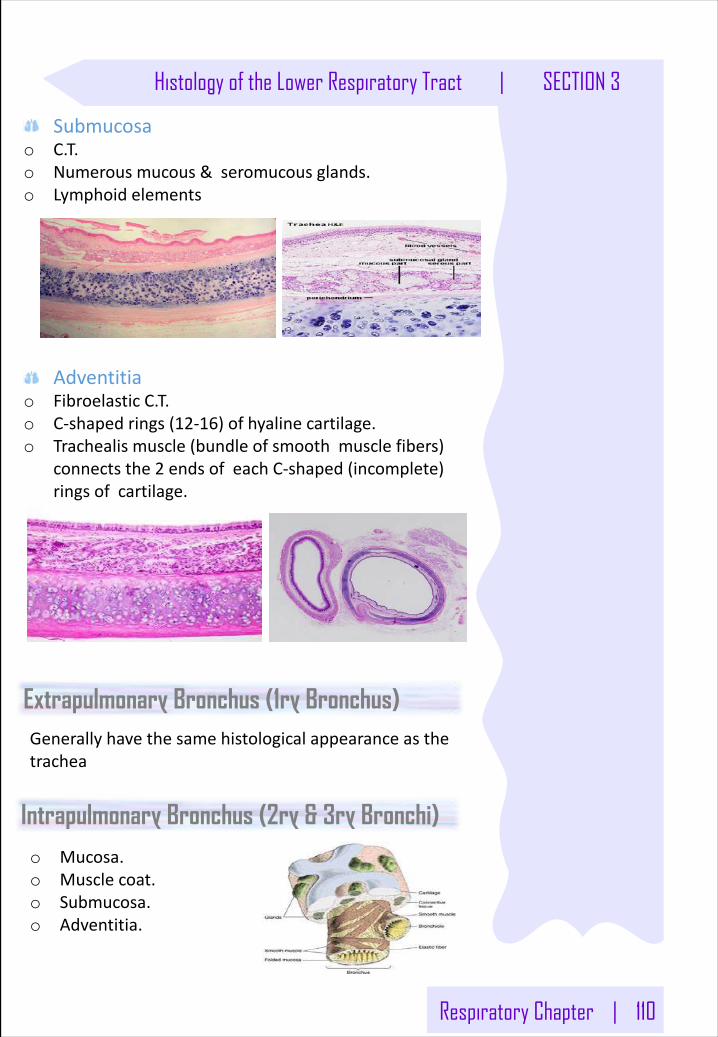
Respiratory Chapter | 110
Histology of the Lower Respiratory Tract | SECTION 3
Extrapulmonary Bronchus (1ry Bronchus)Generallyhavethesamehistologicalappearanceasthetrachea
Submucosao C.T.o Numerous mucous &seromucous glands.o Lymphoid elements
Adventitiao FibroelasticC.T.o C-shapedrings(12-16)ofhyalinecartilage.o Trachealismuscle(bundleofsmoothmusclefibers)
connectsthe2 endsofeachC-shaped(incomplete)ringsofcartilage.
Intrapulmonary Bronchus (2ry & 3ry Bronchi)o Mucosa.o Musclecoat.o Submucosa.o Adventitia.
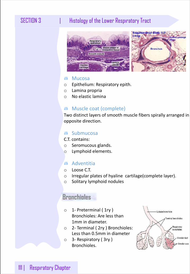
111 | Respiratory Chapter
SECTION 3 | Histology of the Lower Respiratory Tract
Mucosao Epithelium:Respiratoryepith.o Laminapropriao Noelasticlamina
Musclecoat(complete)Twodistinctlayersofsmoothmusclefibers spirallyarrangedinoppositedirection.
SubmucosaC.T.contains:o Seromucous glands.o Lymphoid elements.
Adventitiao LooseC.T.o Irregularplatesofhyalinecartilage(completelayer).o Solitarylymphoidnodules
Bronchioles
o 1- Preterminal(1ry)Bronchioles:Arelessthan1mmindiameter.
o 2- Terminal(2ry)Bronchioles:Lessthan0.5mmindiameter
o 3- Respiratory(3ry)Bronchioles.
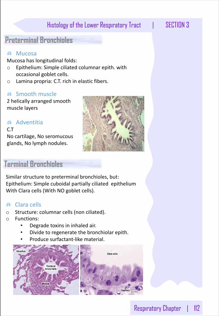
Respiratory Chapter | 112
Histology of the Lower Respiratory Tract | SECTION 3
MucosaMucosahaslongitudinalfolds:o Epithelium:Simpleciliatedcolumnarepith.with
occasionalgobletcells.o Laminapropria:C.T.richinelasticfibers.
Preterminal Bronchioles
Similarstructuretopreterminalbronchioles,but:Epithelium:SimplecuboidalpartiallyciliatedepitheliumWithClaracells(WithNOgobletcells).
Claracellso Structure:columnarcells(nonciliated).o Functions:
• Degradetoxinsininhaledair.• Dividetoregeneratethebronchiolarepith.• Producesurfactant-likematerial.
Terminal Bronchioles
Smoothmuscle2 helicallyarrangedsmoothmusclelayers
AdventitiaC.TNocartilage,Noseromucousglands,Nolymphnodules.
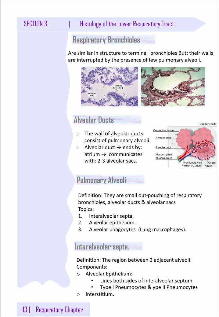
113 | Respiratory Chapter
SECTION 3 | Histology of the Lower Respiratory Tract
Respiratory Bronchioles
AresimilarinstructuretoterminalbronchiolesBut:theirwallsareinterruptedbythepresenceoffewpulmonaryalveoli.
Alveolar Ducts
o Thewallofalveolarductsconsistofpulmonaryalveoli.
o Alveolarduct→endsby:atrium→communicateswith:2-3 alveolarsacs.
Pulmonary Alveoli
Definition:Theyaresmallout-pouchingofrespiratorybronchioles,alveolarducts&alveolarsacsTopics:1. Interalveolarsepta.2. Alveolarepithelium.3. Alveolarphagocytes(Lungmacrophages).
Interalveolar septa.
Definition:Theregionbetween2 adjacentalveoli.Components:o AlveolarEpithelium:
• Linesbothsidesofinteralveolarseptum• TypeIPneumocytes&ype IIPneumocytes
o Interstitium.
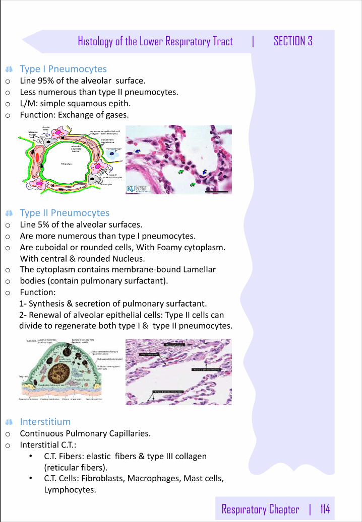
Respiratory Chapter | 114
Histology of the Lower Respiratory Tract | SECTION 3
TypeIPneumocyteso Line95%ofthealveolarsurface.o LessnumerousthantypeIIpneumocytes.o L/M:simplesquamousepith.o Function:Exchangeofgases.
TypeIIPneumocyteso Line5%ofthealveolarsurfaces.o AremorenumerousthantypeIpneumocytes.o Arecuboidalorroundedcells,WithFoamycytoplasm.
Withcentral&roundedNucleus.o Thecytoplasmcontainsmembrane-boundLamellaro bodies(containpulmonarysurfactant).o Function:
1- Synthesis&secretionofpulmonarysurfactant.2- Renewalofalveolarepithelialcells:TypeIIcellscandividetoregeneratebothtypeI&typeIIpneumocytes.
Interstitiumo ContinuousPulmonaryCapillaries.o InterstitialC.T.:
• C.T.Fibers:elasticfibers &typeIIIcollagen(reticularfibers).
• C.T.Cells:Fibroblasts,Macrophages,Mastcells,Lymphocytes.
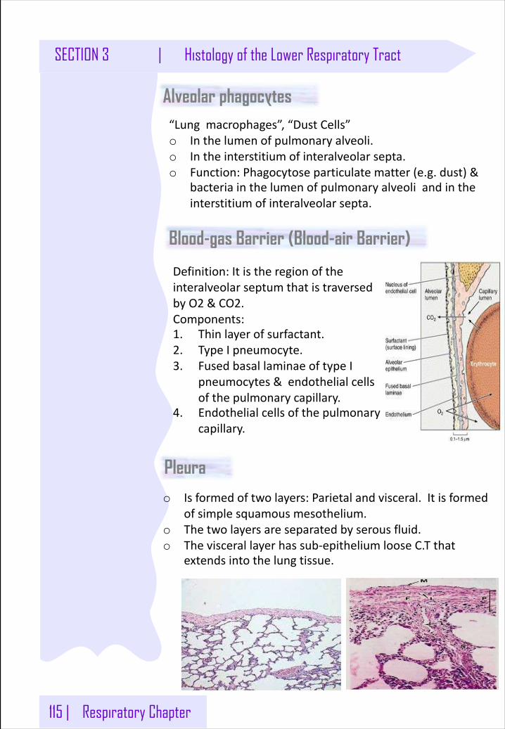
115 | Respiratory Chapter
SECTION 3 | Histology of the Lower Respiratory Tract
Alveolar phagocytes “Lungmacrophages”,“DustCells”o Inthelumenofpulmonaryalveoli.o Intheinterstitium ofinteralveolarsepta.o Function:Phagocytoseparticulatematter(e.g.dust)&
bacteriainthelumenofpulmonaryalveoliandintheinterstitium ofinteralveolarsepta.
Blood-gas Barrier (Blood-air Barrier)
Definition:ItistheregionoftheinteralveolarseptumthatistraversedbyO2 &CO2.Components:1. Thinlayerofsurfactant.2. TypeIpneumocyte.3. FusedbasallaminaeoftypeI
pneumocytes&endothelialcellsofthepulmonarycapillary.
4. Endothelialcellsofthepulmonarycapillary.
Pleura
o Isformedoftwolayers:Parietalandvisceral.Itisformedofsimplesquamousmesothelium.
o Thetwolayersareseparatedbyserousfluid.o Theviscerallayerhassub-epitheliumlooseC.Tthat
extendsintothelungtissue.
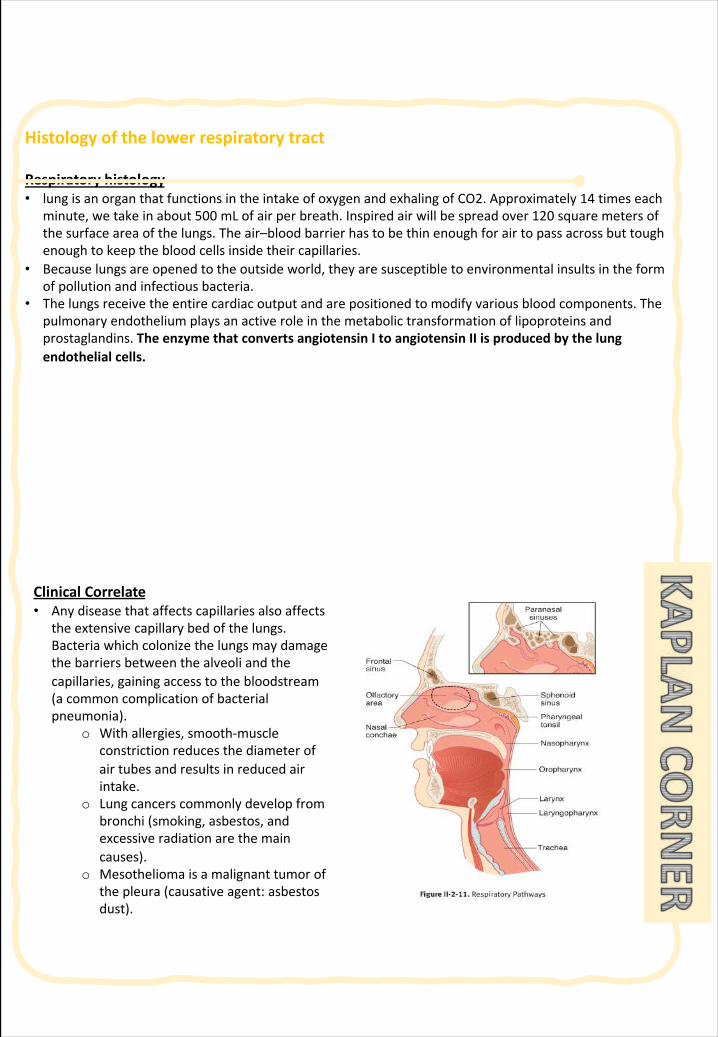
Histologyofthelowerrespiratorytract
Respiratoryhistology• lungisanorganthatfunctionsintheintakeofoxygenandexhalingofCO2.Approximately14 timeseach
minute,wetakeinabout500 mLofairperbreath.Inspiredairwillbespreadover120 squaremetersofthesurfaceareaofthelungs.Theair–bloodbarrierhastobethinenoughforairtopassacrossbuttoughenoughtokeepthebloodcellsinsidetheircapillaries.
• Becauselungsareopenedtotheoutsideworld,theyaresusceptibletoenvironmentalinsultsintheformofpollutionandinfectiousbacteria.
• Thelungsreceivetheentirecardiacoutputandarepositionedtomodifyvariousbloodcomponents.Thepulmonaryendotheliumplaysanactiveroleinthemetabolictransformationoflipoproteinsandprostaglandins.TheenzymethatconvertsangiotensinItoangiotensinIIisproducedbythelungendothelialcells.
ClinicalCorrelate • Anydiseasethataffectscapillariesalsoaffects
theextensivecapillarybedofthelungs.Bacteriawhichcolonizethelungsmaydamagethebarriersbetweenthealveoliandthecapillaries,gainingaccesstothebloodstream(acommoncomplicationofbacterialpneumonia).
o Withallergies,smooth-muscleconstrictionreducesthediameterofairtubesandresultsinreducedairintake.
o Lungcancerscommonlydevelopfrombronchi(smoking,asbestos,andexcessiveradiationarethemaincauses).
o Mesotheliomaisamalignanttumorofthepleura(causativeagent:asbestosdust).
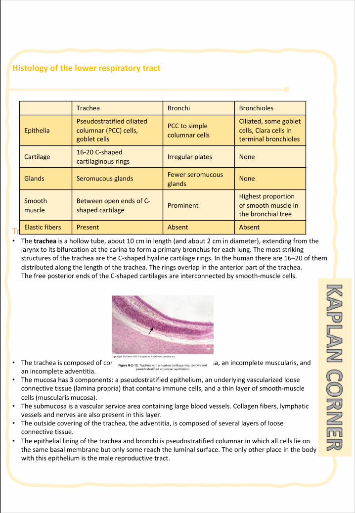
Histologyofthelowerrespiratorytract
Trachea• Thetrachea isahollowtube,about10 cminlength(andabout2 cmindiameter),extendingfromthe
larynxtoitsbifurcationatthecarinatoformaprimarybronchusforeachlung.ThemoststrikingstructuresofthetracheaaretheC-shapedhyalinecartilagerings.Inthehumanthereare16–20 ofthemdistributedalongthelengthofthetrachea.Theringsoverlapintheanteriorpartofthetrachea.ThefreeposteriorendsoftheC-shapedcartilagesareinterconnectedbysmooth-musclecells.
• Thetracheaiscomposedofconcentricringsofmucosa,submucosa,anincompletemuscularis,andanincompleteadventitia.
• Themucosahas3 components:apseudostratifiedepithelium,anunderlyingvascularizedlooseconnectivetissue(laminapropria)thatcontainsimmunecells,andathinlayerofsmooth-musclecells(muscularis mucosa).
• Thesubmucosaisavascularserviceareacontaininglargebloodvessels.Collagenfibers,lymphaticvesselsandnervesarealsopresentinthislayer.
• Theoutsidecoveringofthetrachea,theadventitia,iscomposedofseverallayersoflooseconnectivetissue.
• Theepithelialliningofthetracheaandbronchiispseudostratifiedcolumnarinwhichallcellslieonthesamebasalmembranebutonlysomereachtheluminalsurface.Theonlyotherplaceinthebodywiththisepitheliumisthemalereproductivetract.
Trachea Bronchi Bronchioles
EpitheliaPseudostratifiedciliatedcolumnar(PCC)cells,gobletcells
PCCtosimplecolumnar cells
Ciliated,somegobletcells,Claracellsinterminalbronchioles
Cartilage16-20 C-shapedcartilaginousrings
Irregularplates None
Glands Seromucous glands Fewerseromucousglands
None
Smoothmuscle
BetweenopenendsofC-shapedcartilage
ProminentHighestproportionofsmoothmuscleinthebronchialtree
Elasticfibers Present Absent Absent
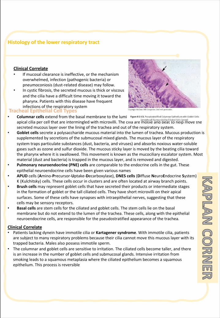
Histologyofthelowerrespiratorytract
TrachealEpithelialCellTypes• Columnarcells extendfromthebasalmembranetotheluminalsurface.Thesecellscontain200–300
apicalciliapercellthatareintermingledwithmicrovilli.Theciliaaremotileandbeattohelpmovethesecretedmucouslayerovertheliningofthetracheaandoutoftherespiratorysystem.
• Gobletcellssecreteapolysaccharidemucousmaterialintothelumenoftrachea.Mucousproductionissupplementedbysecretionsofthesubmucosalmixedglands.Themucouslayeroftherespiratorysystemtrapsparticulatesubstances(dust,bacteria,andviruses)andabsorbsnoxiouswater-solublegasessuchasozoneandsulfurdioxide.Themucousstickylayerismovedbythebeatingciliatowardthepharynxwhereitisswallowed.Thismovementisknownasthemucociliary escalatorsystem.Mostmaterial(dustandbacteria)istrappedinthemucouslayer,andisremovedanddigested.
• Pulmonaryneuroendocrine(PNE)cells arecomparabletotheendocrinecellsinthegut.Theseepithelialneuroendocrinecellshavebeengivenvariousnames
• APUDcells(Amino-Precursor-Uptake-Decarboxylase),DNES cells (Diffuse NeuroEndocrine System)andK(Kulchitsky)cells.Thesecellsoccurinclustersandareoftenlocatedatairwaybranchpoints.
• Brushcellsmayrepresentgobletcellsthathavesecretedtheirproductsorintermediatestagesintheformationofgobletorthetallciliatedcells.Theyhaveshortmicrovilliontheirapicalsurfaces.Someofthesecellshavesynapseswithintraepithelialnerves,suggestingthatthesecellsmaybesensoryreceptors.
• Basalcellsarestemcellsfortheciliatedandgobletcells.Thestemcellslieonthebasalmembranebutdonotextendtothelumenofthetrachea.Thesecells,alongwiththeepithelialneuroendocrinecells,areresponsibleforthepseudostratifiedappearanceofthetrachea.
ClinicalCorrelate • PatientslackingdyneinhaveimmotileciliaorKartagenersyndrome.Withimmotilecilia,patients
aresubjecttomanyrespiratoryproblemsbecausetheirciliacannotmovethismucouslayerwithitstrappedbacteria.Malesalsopossessimmotilesperm.
• Thecolumnarandgobletcellsaresensitivetoirritation.Theciliatedcellsbecometaller,andthereisanincreaseinthenumberofgobletcellsandsubmucosalglands.Intensiveirritationfromsmokingleadstoasquamousmetaplasiawheretheciliatedepitheliumbecomesasquamousepithelium.Thisprocessisreversible
ClinicalCorrelate • Ifmucosalclearanceisineffective,orthemechanism
overwhelmed,infection(pathogenicbacteria)orpneumoconiosis(dust-relateddisease)mayfollow.
• Incysticfibrosis,thesecretedmucousisthickorviscousandtheciliahaveadifficulttimemovingittowardthepharynx.Patientswiththisdiseasehavefrequentinfectionsoftherespiratorysystem
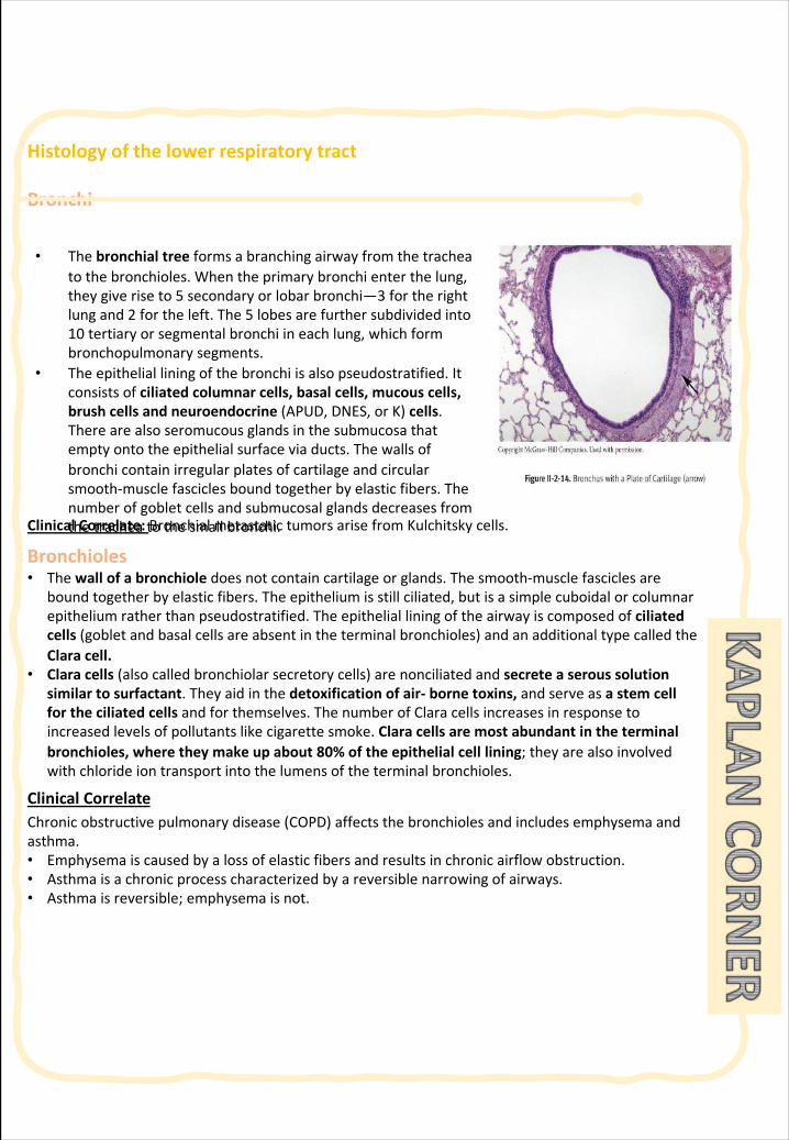
Histologyofthelowerrespiratorytract
Bronchi
ClinicalCorrelate:BronchialmetastatictumorsarisefromKulchitsky cells.
Bronchioles• Thewallofabronchioledoesnotcontaincartilageorglands.Thesmooth-musclefasciclesare
boundtogetherbyelasticfibers.Theepitheliumisstillciliated,butisasimplecuboidalorcolumnarepitheliumratherthanpseudostratified.Theepithelialliningoftheairwayiscomposedofciliatedcells(gobletandbasalcellsareabsentintheterminalbronchioles)andanadditionaltypecalledtheClaracell.
• Claracells(alsocalledbronchiolarsecretorycells)arenonciliated andsecreteaseroussolutionsimilartosurfactant.Theyaidinthedetoxificationofair- bornetoxins,andserveasastemcellfortheciliatedcells andforthemselves.ThenumberofClaracellsincreasesinresponsetoincreasedlevelsofpollutantslikecigarettesmoke.Claracellsaremostabundantintheterminalbronchioles,wheretheymakeupabout80%oftheepithelialcelllining;theyarealsoinvolvedwithchlorideiontransportintothelumensoftheterminalbronchioles.
ClinicalCorrelate Chronicobstructivepulmonarydisease(COPD)affectsthebronchiolesandincludesemphysemaandasthma.• Emphysemaiscausedbyalossofelasticfibersandresultsinchronicairflowobstruction.• Asthmaisachronicprocesscharacterizedbyareversiblenarrowingofairways.• Asthmaisreversible;emphysemaisnot.
• Thebronchialtreeformsabranchingairwayfromthetracheatothebronchioles.Whentheprimarybronchienterthelung,theygiveriseto5 secondaryorlobarbronchi—3 fortherightlungand2 fortheleft.The5 lobesarefurthersubdividedinto10 tertiaryorsegmentalbronchiineachlung,whichformbronchopulmonarysegments.
• Theepithelialliningofthebronchiisalsopseudostratified.Itconsistsofciliatedcolumnarcells,basalcells,mucouscells,brushcellsandneuroendocrine(APUD,DNES,orK)cells.Therearealsoseromucous glandsinthesubmucosathatemptyontotheepithelialsurfaceviaducts.Thewallsofbronchicontainirregularplatesofcartilageandcircularsmooth-musclefasciclesboundtogetherbyelasticfibers.Thenumberofgobletcellsandsubmucosalglandsdecreasesfromthetracheatothesmallbronchi.
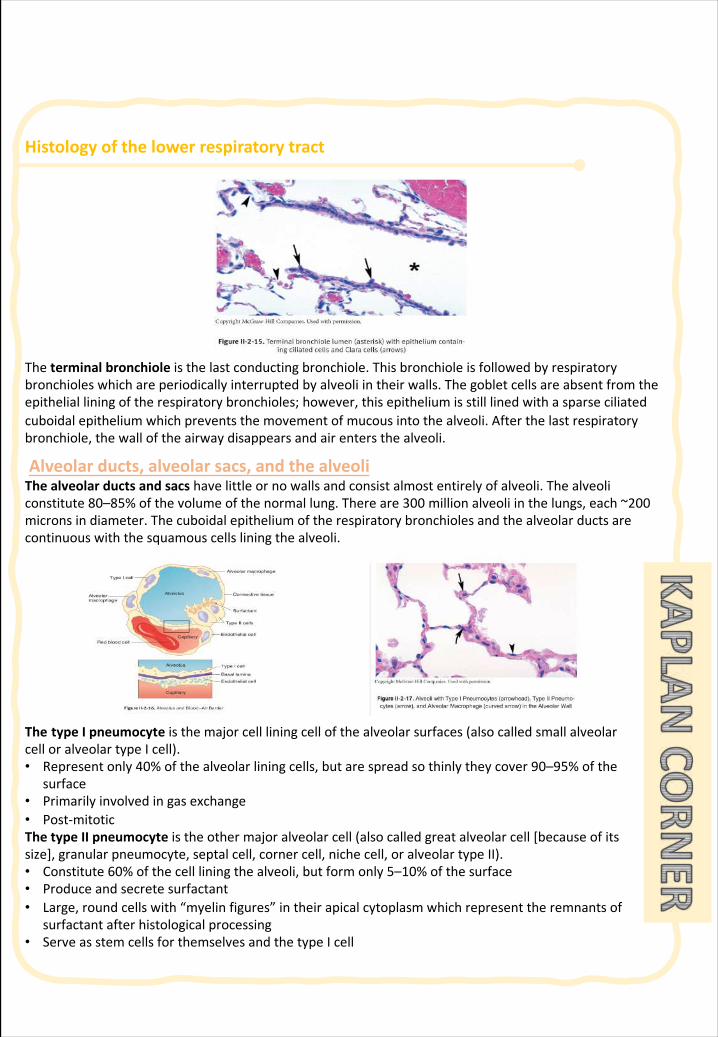
Histologyofthelowerrespiratorytract
Theterminalbronchioleisthelastconductingbronchiole.Thisbronchioleisfollowedbyrespiratorybronchioleswhichareperiodicallyinterruptedbyalveoliintheirwalls.Thegobletcellsareabsentfromtheepithelialliningoftherespiratorybronchioles;however,thisepitheliumisstilllinedwithasparseciliatedcuboidalepitheliumwhichpreventsthemovementofmucousintothealveoli.Afterthelastrespiratorybronchiole,thewalloftheairwaydisappearsandairentersthealveoli.
Alveolarducts,alveolarsacs,andthealveoliThealveolarductsandsacs havelittleornowallsandconsistalmostentirelyofalveoli.Thealveoliconstitute80–85%ofthevolumeofthenormallung.Thereare300 millionalveoliinthelungs,each~200micronsindiameter.Thecuboidalepitheliumoftherespiratorybronchiolesandthealveolarductsarecontinuouswiththesquamouscellsliningthealveoli.
The typeIpneumocyte isthemajorcellliningcellofthealveolarsurfaces(alsocalledsmallalveolarcelloralveolartypeIcell).• Representonly40%ofthealveolarliningcells,butarespreadsothinlytheycover90–95%ofthe
surface• Primarilyinvolvedingasexchange• Post-mitoticThetypeIIpneumocyte istheothermajoralveolarcell(alsocalledgreatalveolarcell[becauseofitssize],granularpneumocyte,septalcell,cornercell,nichecell,oralveolartypeII).• Constitute60%ofthecellliningthealveoli,butformonly5–10%ofthesurface• Produceandsecretesurfactant• Large,roundcellswith“myelinfigures”intheirapicalcytoplasmwhichrepresenttheremnantsof
surfactantafterhistologicalprocessing• ServeasstemcellsforthemselvesandthetypeIcell
Histologyofthelowerrespiratorytract
Surfactant• Surfactant isessentialtomaintainthenormalrespiratorymechanicsofthealveoli.Productionof
surfactantinthefetusisessentialforthesurvivaloftheneonateasittakesitsfirstbreath.Surfactantiscomposedofamixtureofphospholipidsandsurfactantproteinswhosefunctionistoaidinthespreadingofthesurfactantatthealveolarair–waterinterface.Thephospholipidsactasadetergentwhichlowersthesurfacetensionofthealveoliandpreventsalveolarcollapseduringexpiration.
• MostsurfactantisrecycledbacktoTypeIIcellsforreutilization;someofitundergoesphagocytosisbymacrophages.
ClinicalCorrelate • Corticosteroidsinducethefetalsynthesisofsurfactant.Highinsulinlevelsindiabeticmothers
antagonizetheeffectsofcorticosteroids.• Infantsofdiabeticmothershaveahigherincidenceofrespiratorydistresssyndrome.
AlveolarWall• Inthealveolarwall underthealveolarepitheliumisarichnetworkofcapillariesarisingfrom
pulmonaryarteries.Thealveolarwallcontainsavarietyofcellsandextracellularfibers.Thecellsincludefibroblasts,macrophages,myofibroblasts,smooth-musclecells,andoccasionalmastcells.TypeIandIIcollagens,aswellaselasticfibers,areinthesepta.TypeIcollagenispresentprimarilyinthewallsofthebronchiandbronchioles.Twentypercentofthemassofthelungconsistsofcollagenandelasticfibers.Elasticfibersareresponsibleforthestretchingandrecoilingactivitiesofthealveoliduringrespiration.Thesemicroscopicelementsareresponsiblefortherecoilofthelungsduringexpiration.
• Gasexchangeoccursbetweencapillarybloodandalveolarairacrosstheblood– gasbarrier.Thisbarrierconsistsofsurfactant,thesquamousTypeIpneumocytes,asharedbasallamina,andcapillaryendothelium.Thedistancebetweenthelumenofthecapillaryandthelumenofthealveoluscanbeasthinas0.1 microns.ThereareopeningsinthewallofmostalveolithatformtheporesofKohn.Theseporesarethoughttobeimportantincollateralventilation.Thediameterofthesealveolarporescanbeaslargeas10 to15 microns.
AlveolarMacrophages• Thealveolarmacrophagesarederivedfrommonocytesthatexitthebloodvesselsinthelungs.
Theresidentalveolarmacrophagescanundergolimitedmitosestoformadditionalmacrophages.Thesecellscanresideintheinteralveolar septaaswellasinthealveoli.AlveolarmacrophagesthatpatrolthealveolarsurfacesmaypassthroughtheporesofKohn.
• Thereare~1–3 macrophagesperalveolus.Alveolarmacrophagesvaryinsize,15–40 micronsindiameter.Thesemacrophagesrepresentthelastdefensemechanismofthelung.Macrophagescanpassoutofthealveolitothebronchiolesandenterthelymphaticsorbecometrappedinthemovingmucouslayerandpropelledtowardthepharynxtobeswallowedanddigested.
ClinicalCorrelate:Alveolarmacrophageshaveseveralothernames:dustcellsbecausetheyhavephagocytoseddustorcigaretteparticles,andheartfailurecellsbecausetheyhavephagocytosedbloodcellsthathaveescapedintothealveolarspaceduringcongestiveheartfailure.
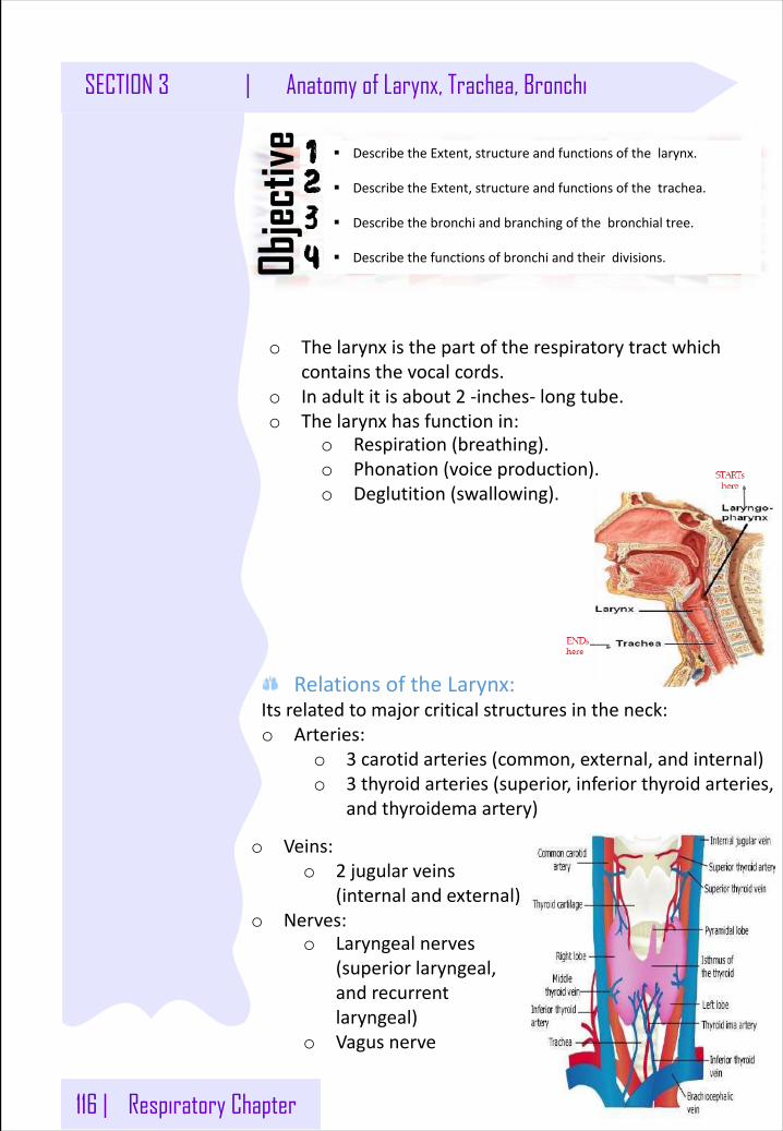
116 | Respiratory Chapter
SECTION 3 | Anatomy of Larynx, Trachea, Bronchi
§ DescribetheExtent,structureandfunctionsofthelarynx.
§ DescribetheExtent,structureandfunctionsofthetrachea.
§ Describethebronchiandbranchingofthebronchialtree.
§ Describethefunctionsofbronchiandtheirdivisions.Objec
tive
o Thelarynxisthepartoftherespiratorytractwhichcontainsthevocalcords.
o Inadultitisabout2 -inches- longtube.o Thelarynxhasfunctionin:
o Respiration(breathing).o Phonation(voiceproduction).o Deglutition(swallowing).
RelationsoftheLarynx:Itsrelatedtomajorcriticalstructuresintheneck:o Arteries:
o 3 carotidarteries(common,external,andinternal)o 3 thyroidarteries(superior,inferiorthyroidarteries,
andthyroidema artery)
o Veins:o 2 jugularveins
(internalandexternal)o Nerves:
o Laryngealnerves(superiorlaryngeal,andrecurrentlaryngeal)
o Vagusnerve
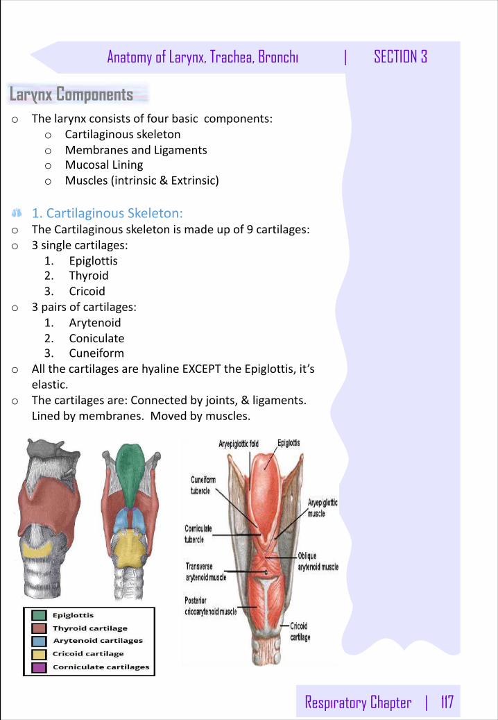
Respiratory Chapter | 117
Anatomy of Larynx, Trachea, Bronchi | SECTION 3
o Thelarynxconsistsoffourbasiccomponents:o Cartilaginousskeletono MembranesandLigamentso MucosalLiningo Muscles(intrinsic&Extrinsic)
1.CartilaginousSkeleton:o TheCartilaginousskeletonismadeupof9 cartilages:o 3 singlecartilages:
1. Epiglottis2. Thyroid3. Cricoid
o 3 pairsofcartilages:1. Arytenoid2. Coniculate3. Cuneiform
o AllthecartilagesarehyalineEXCEPTtheEpiglottis,it’selastic.
o Thecartilagesare:Connectedbyjoints,&ligaments.Linedbymembranes.Movedbymuscles.
Larynx Components
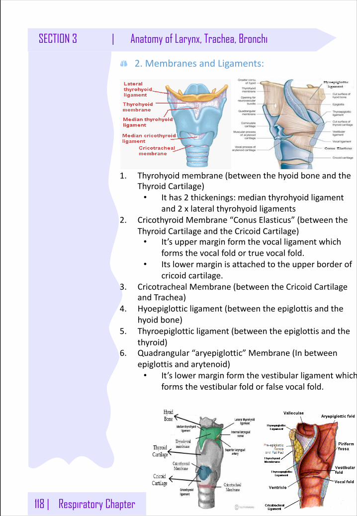
118 | Respiratory Chapter
SECTION 3 | Anatomy of Larynx, Trachea, Bronchi
2.MembranesandLigaments:
1. Thyrohyoidmembrane(betweenthehyoidboneandtheThyroidCartilage)
• Ithas2 thickenings:medianthyrohyoidligamentand2 xlateralthyrohyoidligaments
2. CricothyroidMembrane“ConusElasticus”(betweentheThyroidCartilageandtheCricoidCartilage)
• It’suppermarginformthevocalligamentwhichformsthevocalfoldortruevocalfold.
• Itslowermarginisattachedtotheupperborderofcricoidcartilage.
3. Cricotracheal Membrane(betweentheCricoidCartilageandTrachea)
4. Hyoepiglottic ligament(betweentheepiglottisandthehyoidbone)
5. Thyroepiglottic ligament(betweentheepiglottisandthethyroid)
6. Quadrangular“aryepiglottic”Membrane(Inbetweenepiglottisandarytenoid)
• It’slowermarginformthevestibularligamentwhichformsthevestibularfoldorfalsevocalfold.
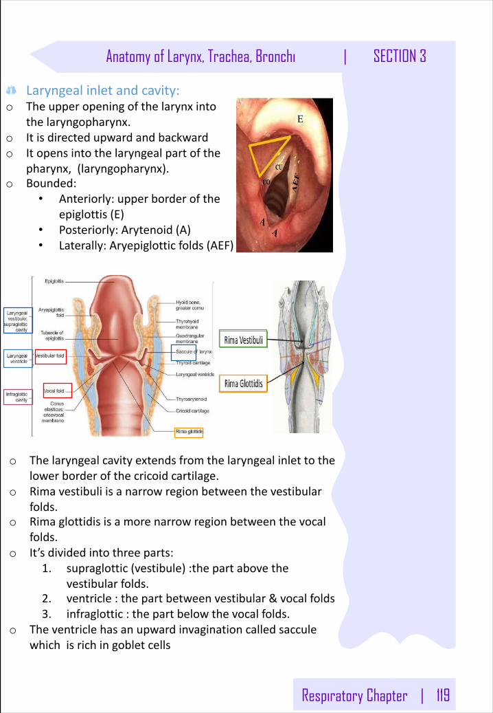
Respiratory Chapter | 119
Anatomy of Larynx, Trachea, Bronchi | SECTION 3
Laryngealinletandcavity:o Theupperopeningofthelarynxinto
thelaryngopharynx.o Itisdirectedupwardandbackwardo Itopensintothelaryngealpartofthe
pharynx,(laryngopharynx).o Bounded:
• Anteriorly:upperborderoftheepiglottis(E)
• Posteriorly:Arytenoid(A)• Laterally:Aryepiglotticfolds(AEF)
E
o Thelaryngealcavityextendsfromthelaryngealinlettothelowerborderofthecricoidcartilage.
o Rimavestibuliisanarrowregionbetweenthevestibularfolds.
o Rimaglottidisisamorenarrowregionbetweenthevocalfolds.
o It’sdividedintothreeparts:1. supraglottic(vestibule):thepartabovethe
vestibularfolds.2. ventricle:thepartbetweenvestibular&vocalfolds3. infraglottic :thepartbelowthevocalfolds.
o Theventriclehasanupwardinvaginationcalledsacculewhichisrichingobletcells
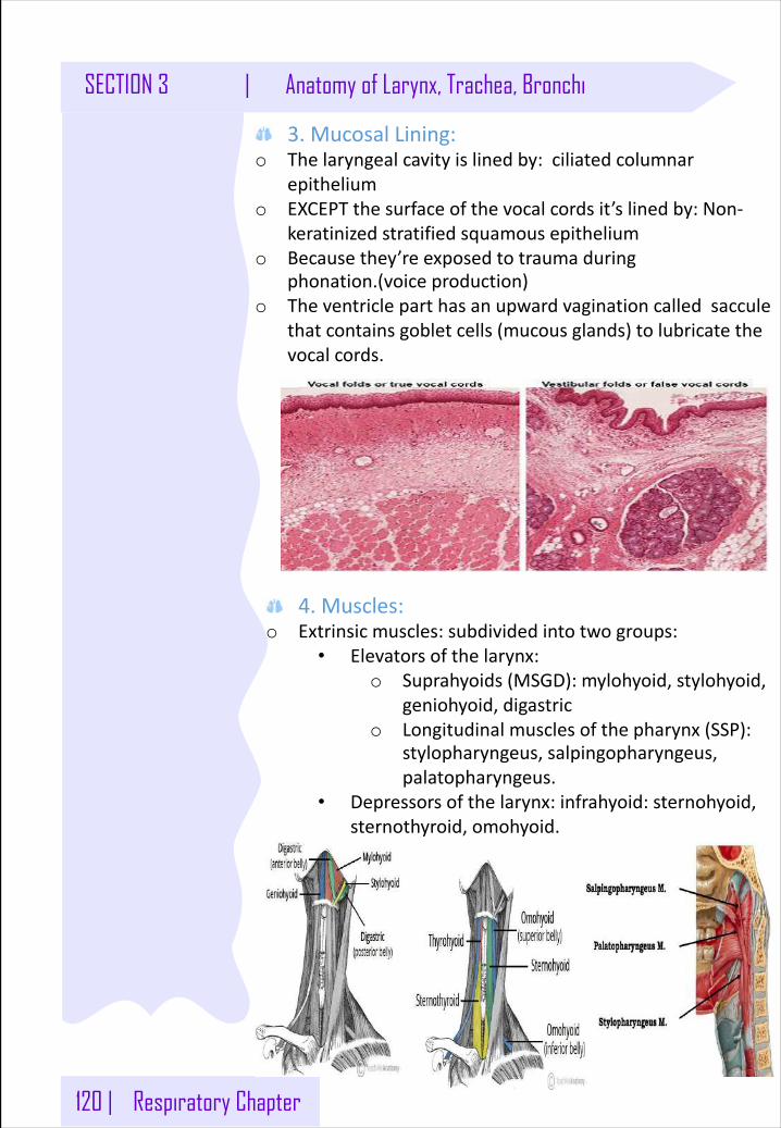
120 | Respiratory Chapter
SECTION 3 | Anatomy of Larynx, Trachea, Bronchi
3.MucosalLining:o Thelaryngealcavityislinedby:ciliatedcolumnar
epitheliumo EXCEPTthesurfaceofthevocalcordsit’slinedby:Non-
keratinizedstratifiedsquamousepitheliumo Becausethey’reexposedtotraumaduring
phonation.(voiceproduction)o Theventricleparthasanupwardvagination calledsaccule
thatcontainsgobletcells(mucousglands)tolubricatethevocalcords.
4.Muscles:o Extrinsicmuscles:subdividedintotwogroups:
• Elevatorsofthelarynx:o Suprahyoids(MSGD):mylohyoid,stylohyoid,
geniohyoid,digastrico Longitudinalmusclesofthepharynx(SSP):
stylopharyngeus,salpingopharyngeus,palatopharyngeus.
• Depressorsofthelarynx:infrahyoid:sternohyoid,sternothyroid,omohyoid.
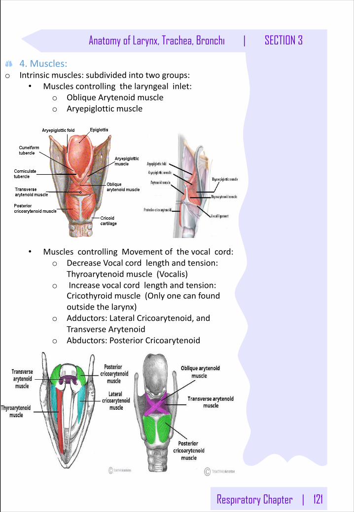
Respiratory Chapter | 121
Anatomy of Larynx, Trachea, Bronchi | SECTION 3
4.Muscles:o Intrinsicmuscles:subdividedintotwogroups:
• Musclescontrollingthelaryngealinlet:o ObliqueArytenoidmuscleo Aryepiglotticmuscle
• MusclescontrollingMovementofthevocalcord:o DecreaseVocalcordlengthandtension:
Thyroarytenoidmuscle(Vocalis)o Increasevocalcordlengthandtension:
Cricothyroidmuscle(Onlyonecanfoundoutsidethelarynx)
o Adductors:LateralCricoarytenoid,andTransverseArytenoid
o Abductors:PosteriorCricoarytenoid
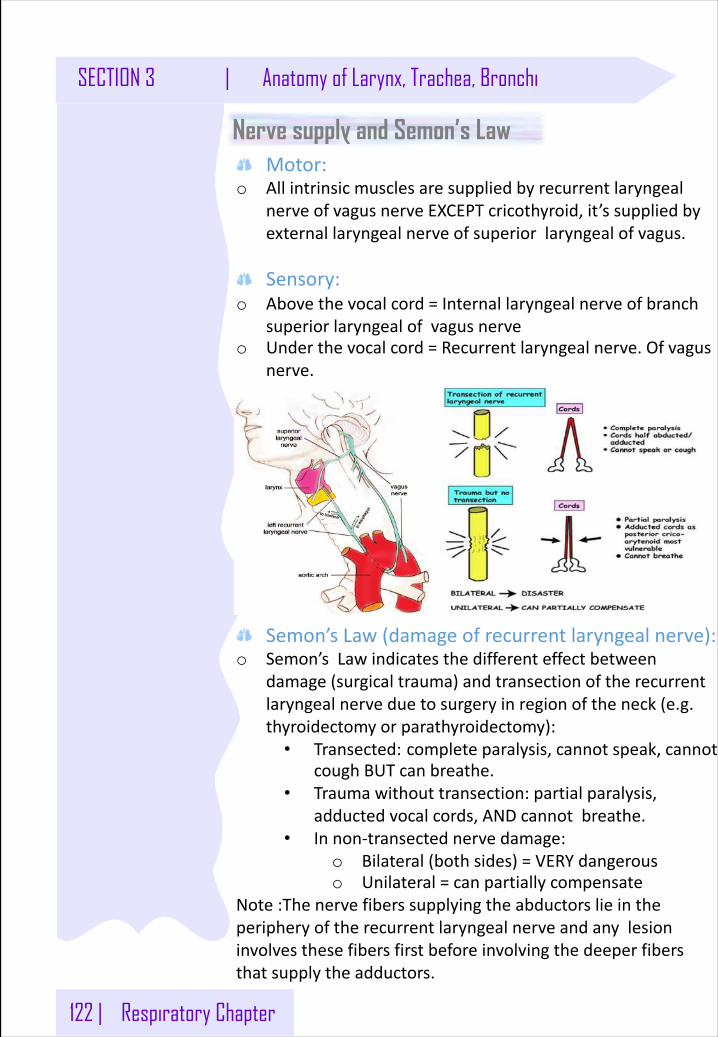
122 | Respiratory Chapter
SECTION 3 | Anatomy of Larynx, Trachea, Bronchi
Motor:o Allintrinsicmusclesaresuppliedbyrecurrentlaryngeal
nerveofvagusnerveEXCEPTcricothyroid,it’ssuppliedbyexternallaryngealnerveofsuperiorlaryngealofvagus.
Sensory:o Abovethevocalcord=Internallaryngealnerveofbranch
superiorlaryngealofvagusnerveo Underthevocalcord=Recurrentlaryngealnerve.Ofvagus
nerve.
Nerve supply and Semon’s Law
Semon’s Law(damageofrecurrentlaryngealnerve):o Semon’s Lawindicatesthedifferenteffectbetween
damage(surgicaltrauma)andtransectionoftherecurrentlaryngealnerveduetosurgeryinregionoftheneck(e.g.thyroidectomyorparathyroidectomy):
• Transected: completeparalysis,cannotspeak,cannotcoughBUTcanbreathe.
• Traumawithouttransection:partialparalysis,adductedvocalcords,ANDcannotbreathe.
• Innon-transected nervedamage:o Bilateral(bothsides)=VERYdangerouso Unilateral=canpartiallycompensate
Note:Thenervefibers supplyingtheabductorslieintheperipheryoftherecurrentlaryngealnerveandanylesioninvolvesthesefibers firstbeforeinvolvingthedeeperfibersthatsupplytheadductors.
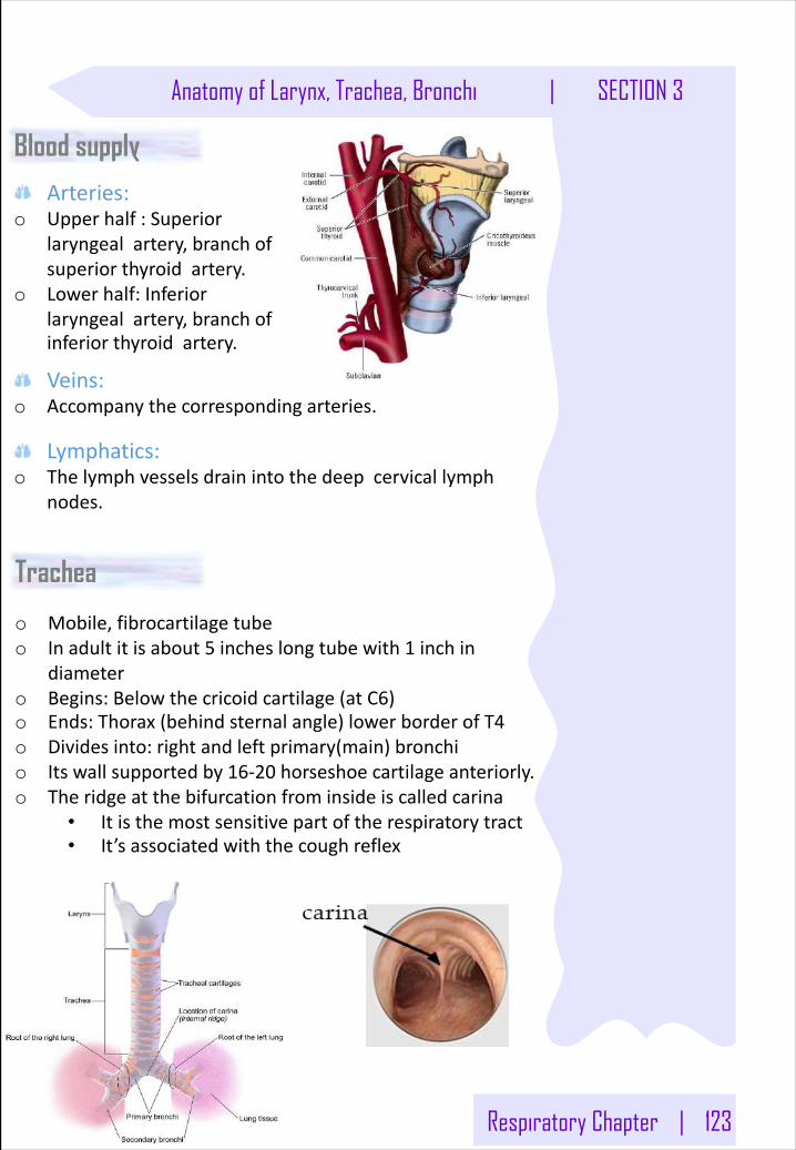
Respiratory Chapter | 123
Anatomy of Larynx, Trachea, Bronchi | SECTION 3
Arteries:o Upperhalf:Superior
laryngealartery,branchofsuperiorthyroidartery.
o Lowerhalf:Inferiorlaryngealartery,branchofinferiorthyroidartery.
Blood supply
Veins:o Accompanythecorrespondingarteries.
Lymphatics:o Thelymphvesselsdrainintothedeepcervicallymph
nodes.
o Mobile,fibrocartilagetubeo Inadultitisabout5 incheslongtubewith1 inchin
diametero Begins:Belowthecricoidcartilage(atC6)o Ends:Thorax(behindsternalangle)lowerborderofT4o Dividesinto:rightandleftprimary(main)bronchio Itswallsupportedby16-20 horseshoecartilageanteriorly.o Theridgeatthebifurcationfrominsideiscalledcarina
• Itisthemostsensitivepartoftherespiratorytract• It’sassociatedwiththecoughreflex
Trachea
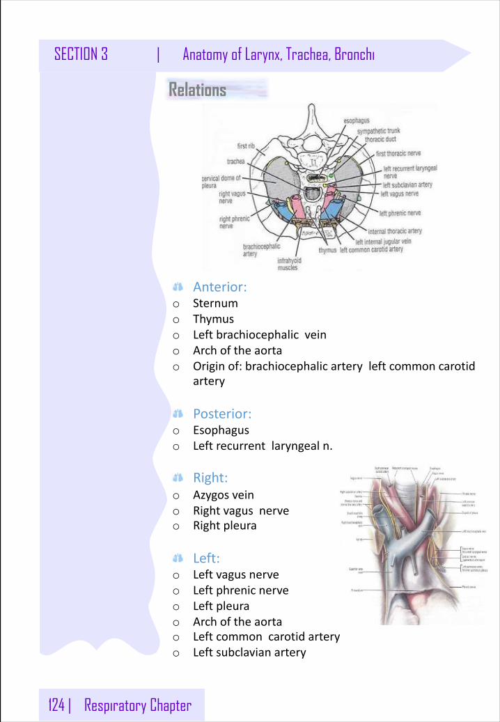
124 | Respiratory Chapter
SECTION 3 | Anatomy of Larynx, Trachea, Bronchi
Anterior:o Sternumo Thymuso Leftbrachiocephalicveino Archoftheaortao Originof:brachiocephalicarteryleftcommoncarotid
artery
Posterior:o Esophaguso Leftrecurrentlaryngealn.
Right:o Azygosveino Rightvagusnerveo Rightpleura
Left:o Leftvagusnerveo Leftphrenicnerveo Leftpleurao Archoftheaortao Leftcommoncarotidarteryo Leftsubclavianartery
Relations
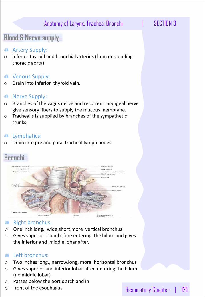
Respiratory Chapter | 125
Anatomy of Larynx, Trachea, Bronchi | SECTION 3
ArterySupply:o Inferiorthyroidandbronchialarteries(fromdescending
thoracicaorta)
VenousSupply:o Drainintoinferiorthyroidvein.
NerveSupply:o Branchesofthevagusnerveandrecurrentlaryngealnerve
givesensoryfibers tosupplythemucousmembrane.o Trachealisissuppliedbybranchesofthesympathetic
trunks.
Lymphatics:o Drainintopreandparatracheallymphnodes
Blood & Nerve supply
Bronchi
Rightbronchus:o Oneinchlong.,wide,short,more verticalbronchuso Givessuperiorlobarbeforeenteringthehilumandgives
theinferiorandmiddlelobarafter.
Leftbronchus:o Twoincheslong.,narrow,long,morehorizontalbronchuso Givessuperiorandinferiorlobarafterenteringthehilum.
(nomiddlelobar)o Passesbelowtheaorticarchandino frontoftheesophagus.
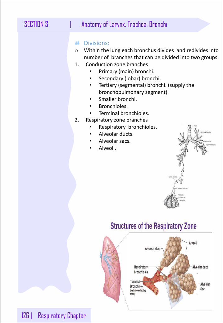
126 | Respiratory Chapter
SECTION 3 | Anatomy of Larynx, Trachea, Bronchi
Divisions:o Withinthelungeachbronchusdividesandredivides into
numberofbranchesthatcanbedividedintotwogroups:1. Conductionzonebranches
• Primary(main)bronchi.• Secondary(lobar)bronchi.• Tertiary(segmental)bronchi.(supplythe
bronchopulmonarysegment).• Smallerbronchi.• Bronchioles.• Terminalbronchioles.
2. Respiratoryzonebranches• Respiratorybronchioles.• Alveolarducts.• Alveolarsacs.• Alveoli.
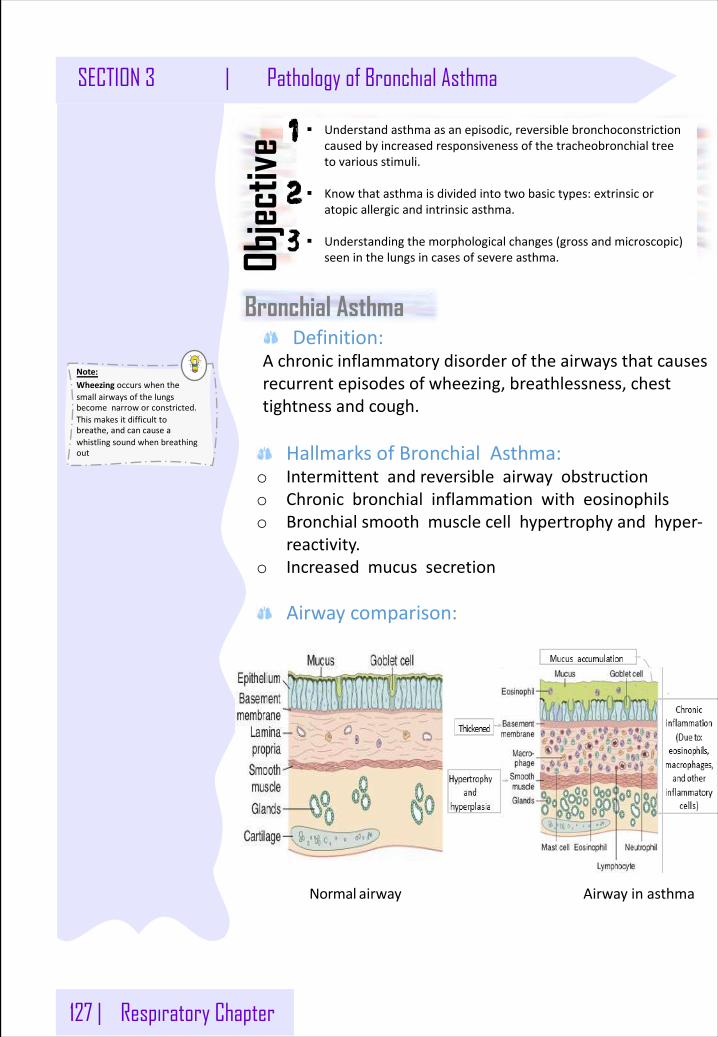
127 | Respiratory Chapter
SECTION 3 | Pathology of Bronchial Asthma
Note: Wheezing occurswhenthesmallairwaysofthelungsbecomenarroworconstricted.Thismakesitdifficulttobreathe,andcancauseawhistlingsoundwhenbreathingout
Bronchial Asthma
§ Understandasthmaasanepisodic,reversiblebronchoconstrictioncausedbyincreasedresponsivenessofthetracheobronchialtreetovariousstimuli.
§ Knowthatasthmaisdividedintotwobasictypes:extrinsicoratopicallergicandintrinsicasthma.
§ Understandingthemorphologicalchanges(grossandmicroscopic)seeninthelungsincasesofsevereasthma.Ob
jectiv
e
Definition:Achronicinflammatorydisorderoftheairwaysthatcausesrecurrentepisodesofwheezing,breathlessness,chesttightnessandcough.
HallmarksofBronchialAsthma:o Intermittentandreversibleairwayobstructiono Chronicbronchialinflammationwitheosinophilso Bronchialsmoothmusclecellhypertrophyandhyper-
reactivity.o Increasedmucussecretion
Airwaycomparison:
Normal airway Airwayinasthma
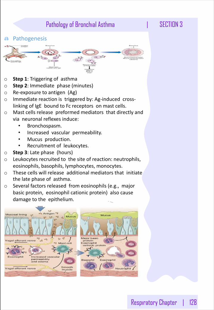
Respiratory Chapter | 128
Pathology of Bronchial Asthma | SECTION 3
Pathogenesis
o Step1:Triggeringofasthmao Step2:Immediatephase(minutes)o Re-exposuretoantigen(Ag)o Immediatereactionistriggeredby:Ag-inducedcross-
linkingofIgE boundtoFcreceptorsonmastcells.o Mastcellsreleasepreformedmediatorsthatdirectlyand
vianeuronalreflexesinduce:• Bronchospasm.• Increasedvascularpermeability.• Mucusproduction.• Recruitmentofleukocytes.
o Step3:Latephase(hours)o Leukocytesrecruitedtothesiteofreaction:neutrophils,
eosinophils,basophils,lymphocytes,monocytes.o Thesecellswillreleaseadditionalmediatorsthatinitiate
thelatephaseofasthma.o Severalfactorsreleasedfromeosinophils(e.g.,major
basicprotein,eosinophilcationicprotein)alsocausedamagetotheepithelium.
129 | Respiratory Chapter
SECTION 3 | Pathology of Bronchial Asthma
CytokinesproducedbyType2 helperT(TH2)cells:
Function
IL-4 StimulateIgE production.
IL-5
Activateseosinophilswhichplayamajorpartinthepathogenesisofasthma.Becausetheyproducebasicproteins,whichcauseasthma,damagebronchialwall,inciteinflammation,andinducecontractionofbronchialsmoothmuscles.
IL-9 Damagesthebronchialepitheliumdirectly.
IL-13 StimulatesIgE andmucusproduction.
Repeatedboutsofinflammationleadtostructuralchangesinthebronchialwall(Airwayremodeling):o Hypertrophyofbronchialsmoothmuscle.o Hypertrophyofmucusglands.o Increasedvascularityanddepositionof
subepithelialcollagen.
Types of Bronchial Asthma
1- Extrinsicasthma(Atopic):o Bronchospasmisinducedbyinhaledantigenso Atopic/allergicAsthma70%o Type1 hypersensitivityreaction*mediatedbyIgE,
inducedbyexposuretoextrinsicantigen/allergense.g.food,pollen,dust,etc,
o Familyhistory:positiveSkintest:positive
2- Intrinsicasthma(Non-Atopic):o Adiseaseinwhichthebronchialhyper- reactivityis
inducedbynon-immunemechanismso Non-atopic/NotallergicAsthma30%o Initiatedbydiverse,non-immunemechanisms.o Familyhistory:uncommonSkintest:Negative
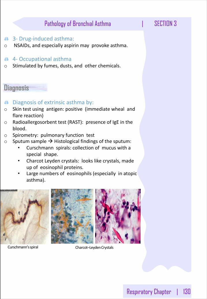
Respiratory Chapter | 130
Pathology of Bronchial Asthma | SECTION 3
3- Drug-inducedasthma:o NSAIDs,andespeciallyaspirinmayprovokeasthma.
4- Occupationalasthmao Stimulatedbyfumes,dusts,andotherchemicals.
Diagnosis
Charcot–LeydenCrystalsCurschmann’sspiral
Diagnosisofextrinsicasthmaby:o Skintestusingantigen:positive(immediatewhealand
flarereaction)o Radioallergosorbent test(RAST):presenceofIgE inthe
blood.o Spirometry:pulmonaryfunctiontesto Sputumsampleà Histologicalfindingsofthesputum:
• Curschmann spirals:collectionofmucuswithaspecialshape.
• CharcotLeydencrystals:lookslikecrystals,madeupofeosinophilproteins.
• Largenumbersofeosinophils(especiallyinatopicasthma).
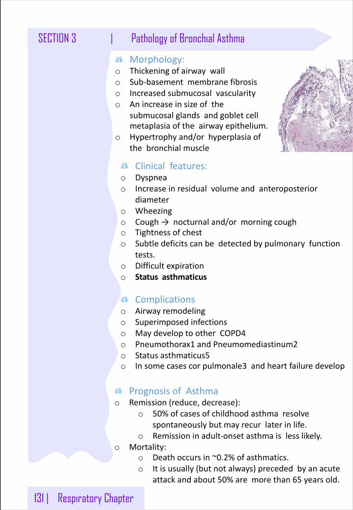
131 | Respiratory Chapter
SECTION 3 | Pathology of Bronchial Asthma
Morphology:o Thickeningofairwaywallo Sub-basementmembranefibrosiso Increasedsubmucosalvascularityo Anincreaseinsizeofthe
submucosalglandsandgobletcellmetaplasiaoftheairwayepithelium.
o Hypertrophyand/orhyperplasiaofthebronchialmuscle
Clinicalfeatures:o Dyspneao Increaseinresidualvolumeandanteroposterior
diametero Wheezingo Cough→nocturnaland/ormorningcougho Tightnessofchesto Subtledeficitscanbedetectedbypulmonaryfunction
tests.o Difficultexpirationo Statusasthmaticus
Complicationso Airwayremodelingo Superimposedinfectionso MaydeveloptootherCOPD4o Pneumothorax1 andPneumomediastinum2o Statusasthmaticus5o Insomecasescor pulmonale3 andheartfailuredevelop
PrognosisofAsthmao Remission(reduce,decrease):
o 50%ofcasesofchildhoodasthmaresolvespontaneouslybutmayrecurlaterinlife.
o Remissioninadult-onsetasthmaislesslikely.o Mortality:
o Deathoccursin~0.2%ofasthmatics.o Itisusually(butnotalways)precededbyanacute
attackandabout50%aremorethan65 yearsold.
Respiratory Chapter | 132
Pathology of Bronchial Asthma | SECTION 3
PreventionofAsthmao Controloffactorscontributingtoasthmaseverity:
o Exposuretoirritantsorallergenshasbeenshowntoincreaseasthmasymptomsandcauseexacerbations.
o Skintest:o Resultsshouldbeusedtoassesssensitivityto
commonindoorAllergens.o Allpatientswithasthmashouldbeadvisedto
avoidexposuretoallergenstowhichtheyaresensitive.
In SUMMARY:Definition
Inflammatorydisorderoftheairway,whichischaracterizedbyincreasedresponsivenessofthebronchialmucosaandbronchialwalltovariousstimuli,it’salsocharacterizedbyepisodicattacksandit’sreversible,whichmeansthatitcanbereversedbyavoidingthestimuli.
Signsandsymptomso Dyspnea.”Most serious”o Wheezing.o Cough.”Not amajorsymptom”
Types:o Extrinsic(atopic):commoninchildren.o Intrinsic(non-atopic):
• Usuallyhappensinadults.• Thesepatientsdon’thavehistoryof
hypersensitivity.• Usuallycomeafterexercise(exerciseinduced
intrinsicasthma):Exercisecausesdehydrationwhichwillincreasetheosmolalityofthesputumwhichisassociatedwithsecretionofcertainchemicalmediators(especiallyLeukotrienesC4,D4,E4 whichwillcausebronchospasm).
• Aspirin(blockPGs—>enhanceleukotrienesC4,• D4,E4—>bronchoconstriction)
133 | Respiratory Chapter
SECTION 3 | Pathology of Bronchial Asthma
In SUMMARY: Pathogenesis
1. Entryofantigen,thenitisengulfedbyAPCsandpresentedtoT-lymphocytes,especiallyCD4.
2. ActivatestheCD4 T-lymphocytesandtransformthemintoTH2 cells.
3. TH2 cellsstartsecretingthesecytokines:• IL-4:willstimulatetheB-lymphocytestosecrete
IgE(hasamainroleinhypersensitivitytype1),thisIgE willbindtocertainreceptorsonmastcells,thentheantigenwillcomeandbindtotheantibody,thenthemastcellwillreleasethegranules(whichcontainhistamine,serotoninandotherchemicalmediators)whichwillcausevasodilationandedema.
• IL-5:CanbesecretedbyT&Blymphocytes,itwillfacilitatetherecruitmentofeosinophils,whichwillalsoreleasetheirgranules(hasmajorbasicproteins)whichwillcausedamagetothebronchialepithelium,thenthedamagedepitheliumwillsecreteeotaxin whichwillrecruitmoreeosinophils.
• IL-9:Actdirectlyonthebronchialepitheliumandwillcausedamagetoit.
• IL-13:SameactionasIL-4.
Pathologicalchangesinasthma:o Hyperplasia/Hypertrophyandspasm(duetostimulation
andirritationofthenervesendingsspeciallynervesfromthevagusnerve)
o Excessivemucussecretionwithaccumulationofeosinophils.
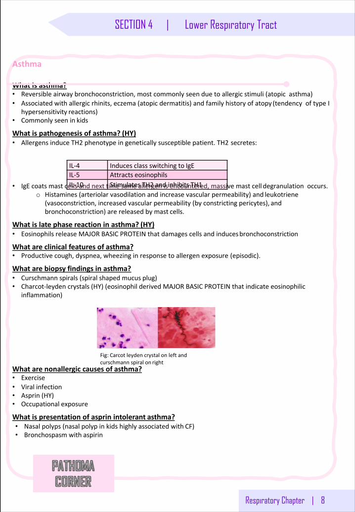
Respiratory Chapter | 8
SECTION 4 | Lower Respiratory Tract
Asthma
Whatis asthma?• Reversibleairwaybronchoconstriction,mostcommonlyseenduetoallergicstimuli(atopicasthma)• Associatedwithallergicrhinits,eczema(atopicdermatitis)andfamilyhistoryofatopy (tendencyoftypeI
hypersensitivity reactions)• Commonlyseenin kids
Whatispathogenesisofasthma? (HY)• AllergensinduceTH2 phenotypeingeneticallysusceptiblepatient.TH2 secretes:
• IgE coatsmastcellsandnexttimesameallergenisencountered,massivemastcell degranulationoccurs.o Histamines(arteriolarvasodilationandincreasevascularpermeability)and leukotriene
(vasoconstriction,increasedvascularpermeability(byconstrictingpericytes), andbronchoconstriction)arereleasedbymast cells.
Whatislatephasereactioninasthma? (HY)• EosinophilsreleaseMAJORBASICPROTEINthatdamagescellsandinducesbronchoconstriction
Whatareclinicalfeaturesof asthma?• Productivecough,dyspnea,wheezinginresponsetoallergenexposure (episodic).
Whatarebiopsyfindingsin asthma?• Curschmann spirals(spiralshapedmucus plug)• Charcot-leyden crystals(HY)(eosinophilderivedMAJORBASICPROTEINthatindicateeosinophilic
inflammation)
Whatarenonallergic causesof asthma?• Exercise• Viral infection• Asprin (HY)• Occupational exposure
Whatispresentationofasprin intolerant asthma?• Nasalpolyps(nasalpolypinkidshighlyassociatedwith CF)• Bronchospasmwith aspirin
IL-4 Inducesclassswitchingto IgEIL-5 Attracts eosinophilsIL-10 StimulatesTH2 andinhibits TH1
Fig:Carcot leyden crystalonleftandcurschmann spiralon right
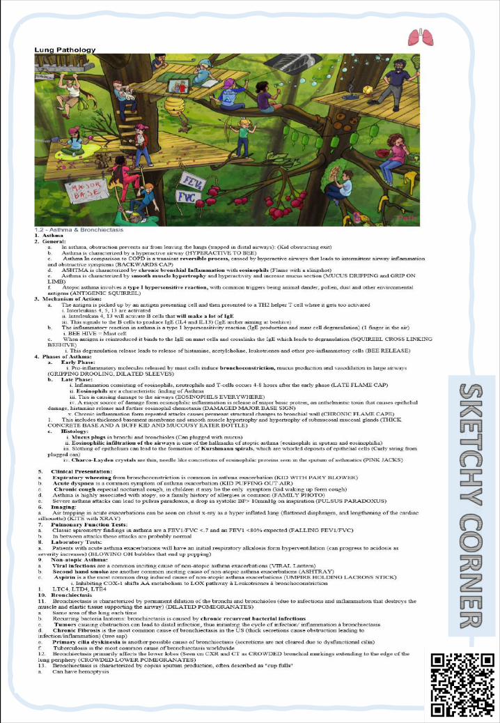
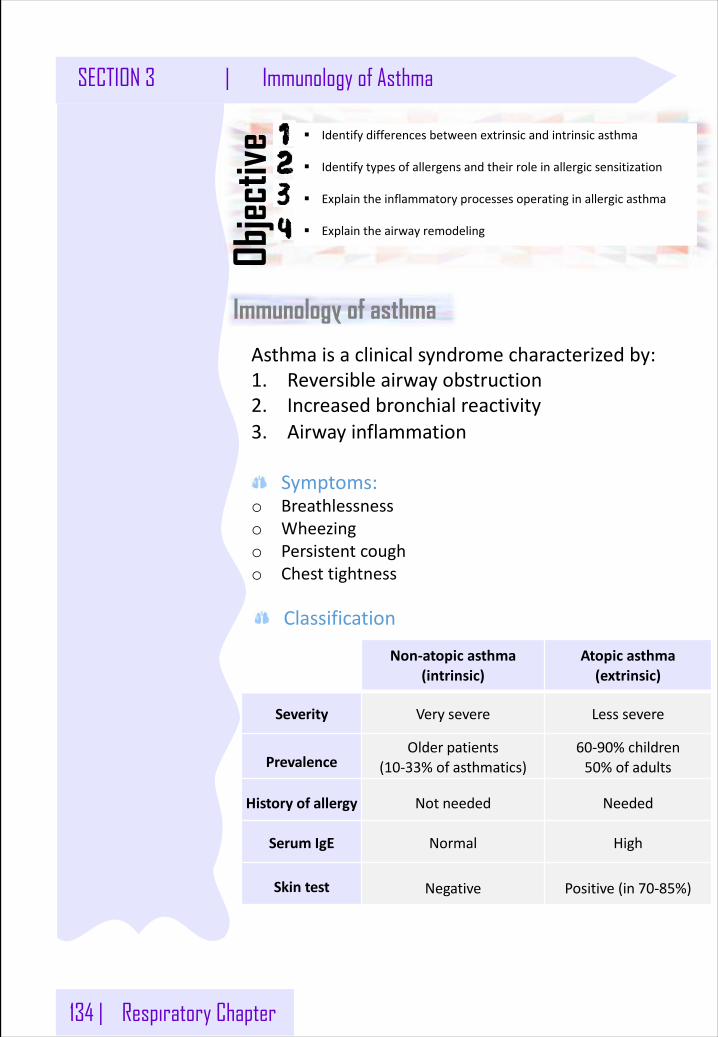
134 | Respiratory Chapter
SECTION 3 | Immunology of Asthma
Immunology of asthma
§ Identifydifferencesbetweenextrinsicandintrinsicasthma
§ Identifytypesofallergensandtheirroleinallergicsensitization
§ Explaintheinflammatoryprocessesoperatinginallergicasthma
§ Explaintheairwayremodeling
Objec
tive
Asthmaisaclinicalsyndromecharacterizedby:1. Reversibleairwayobstruction2. Increasedbronchialreactivity3. Airwayinflammation
Symptoms:o Breathlessnesso Wheezingo Persistentcougho Chesttightness
Non-atopicasthma(intrinsic)
Atopicasthma(extrinsic)
Severity Very severe Less severe
PrevalenceOlder patients
(10-33%of asthmatics)60-90% children50%of adults
Historyof allergy Not needed Needed
Serum IgE Normal High
Skin test Negative Positive(in 70-85%)
Classification
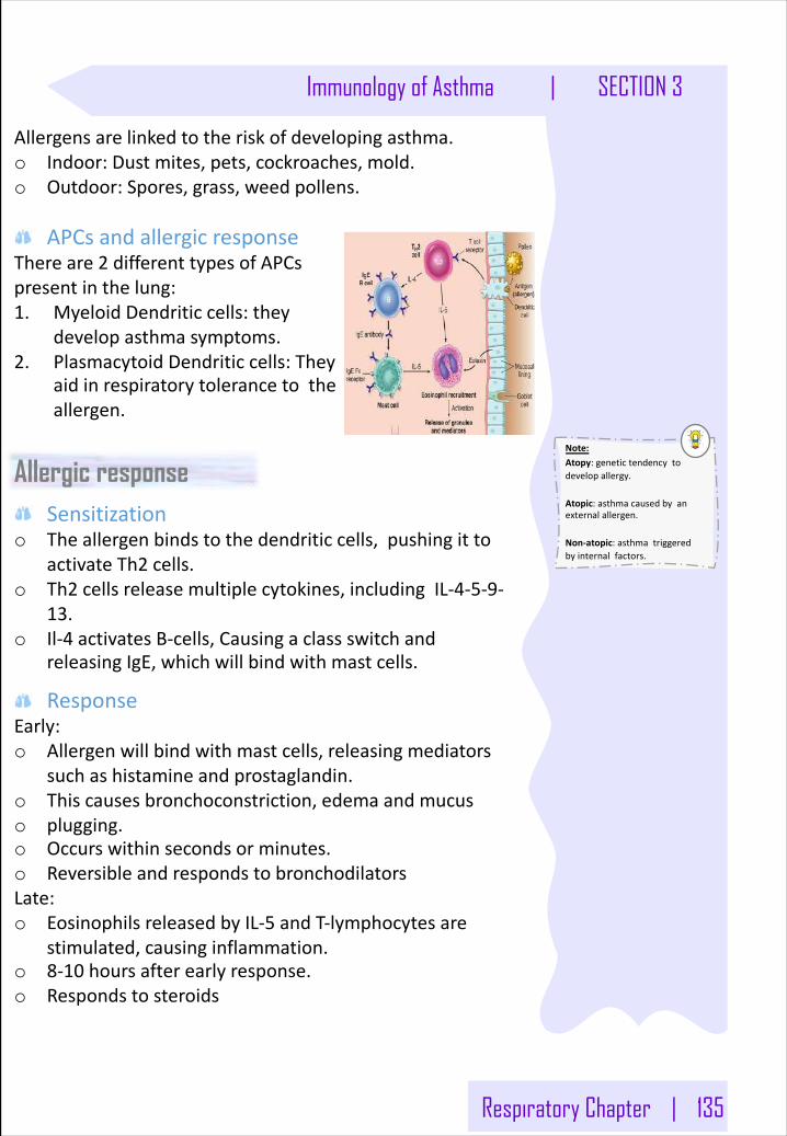
Respiratory Chapter | 135
Immunology of Asthma | SECTION 3
Allergensarelinkedtotheriskofdevelopingasthma.o Indoor:Dustmites,pets,cockroaches,mold.o Outdoor:Spores,grass,weedpollens.
APCsandallergicresponseThereare2 differenttypesofAPCspresentinthelung:1. MyeloidDendriticcells:they
developasthmasymptoms.2. PlasmacytoidDendriticcells:They
aidinrespiratorytolerancetotheallergen.
Allergic responseSensitization
o Theallergenbindstothedendriticcells,pushingittoactivateTh2 cells.
o Th2 cellsreleasemultiplecytokines,includingIL-4-5-9-13.
o Il-4 activatesB-cells,CausingaclassswitchandreleasingIgE,whichwillbindwithmastcells.
ResponseEarly:o Allergenwillbindwithmastcells,releasingmediators
suchashistamineandprostaglandin.o Thiscausesbronchoconstriction,edema andmucuso plugging.o Occurswithinsecondsorminutes.o ReversibleandrespondstobronchodilatorsLate:o EosinophilsreleasedbyIL-5 andT-lymphocytesare
stimulated,causinginflammation.o 8-10 hoursafterearlyresponse.o Respondstosteroids
Note: Atopy:genetictendencytodevelopallergy.
Atopic:asthmacausedbyanexternalallergen.
Non-atopic:asthmatriggeredbyinternalfactors.
136 | Respiratory Chapter
SECTION 3 | Immunology of Asthma
TheRoleofCells&CytokinesinAllergicAsthmaTh2 cellsduringallergicasthmasecrete:Interleukin(4,5,9 &13)Thiscauses:o ProductionofIgEo Eosinophilsattractiono Airwayinflammationo Increaseinbronchialreactivity
Theinflammatorycellsinteractwith:o Nervoussystemo Airwayepitheliumo Bronchialmuscles
CytokinesIL-4:o RegulatesisotypeswitchingtoIgE inBcells.o InducesMHCII(inantigenpresentingcells)o Inducesadhesionmolecules.o Activatesmastcellsandeosinophils.
IL-5o Increaseseosinophilsproduction.o Releaseeosinophilsfrombonemarrow.
IL-13o Inducesinflammation.o Stimulatemucushypersecretion.o Inducessubepithelialfibrosis.
CellsEosinophils:o Initiateasthmaticsymptomsbycausingtissuedamage.o IL-10 caninhibiteosinophils’production
RegulatoryT-cellso Suppressasthmaticsymptoms.o Someasthmaticslackthisfunction.
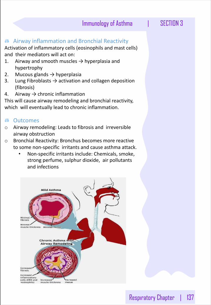
Respiratory Chapter | 137
Immunology of Asthma | SECTION 3
AirwayinflammationandBronchialReactivityActivationofinflammatorycells(eosinophilsandmastcells)andtheirmediatorswillacton:1. Airwayandsmoothmuscles→hyperplasiaand
hypertrophy2. Mucousglands→hyperplasia3. LungFibroblasts→activationandcollagendeposition
(fibrosis)4. Airway→chronicinflammationThiswillcauseairwayremodelingandbronchialreactivity,whichwilleventuallyleadtochronicinflammation.
Outcomeso Airwayremodeling:Leadstofibrosisandirreversible
airwayobstructiono BronchialReactivity:Bronchusbecomesmorereactive
tosomenon-specificirritantsandcauseasthmaattack.• Non-specificirritantsinclude:Chemicals,smoke,
strongperfume,sulphur dioxide,airpollutantsandinfections
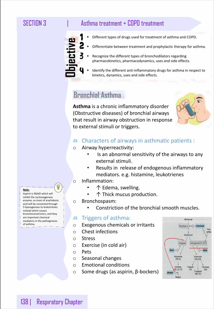
138 | Respiratory Chapter
SECTION 3 | Asthma treatment + COPD treatment
Note: AspirinisNSAIDwhichwillinhibitthecyclooxygenaseenzyme,somostofarachidonicacidwillbeconvertedthrough5-lipoxygenasetoleukotrienesinsteadwhichcausesbronchoconstrictors,andtheyareimportantchemicalmediatorsinthepathogenesisofasthma.
Bronchial Asthma
§ DifferenttypesofdrugsusedfortreatmentofasthmaandCOPD.
§ Differentiatebetweentreatmentandprophylactictherapyforasthma.
§ Recognizethedifferenttypesofbronchodilatorsregardingpharmacokinetics,pharmacodynamics,usesandsideeffects.
§ Identifythedifferentanti-inflammatorydrugsforasthmainrespecttokinetics,dynamics,usesandsideeffects.Ob
jectiv
e
Asthma isachronicinflammatorydisorder(Obstructivediseases)ofbronchialairwaysthatresultinairwayobstructioninresponsetoexternalstimuliortriggers.
Charactersofairwaysinasthmaticpatients:o Airwayhyperreactivity:
• Isanabnormalsensitivityoftheairwaystoanyexternalstimuli.
• Resultsinreleaseofendogenousinflammatorymediators.e.g.histamine,leukotrienes
o Inflammation:• ↑Edema,swelling.• ↑Thickmucusproduction.
o Bronchospasm:• Constrictionofthebronchialsmoothmuscles.
Triggersofasthma:o Exogenouschemicalsorirritantso Chestinfectionso Stresso Exercise(incoldair)o Petso Seasonalchangeso Emotionalconditionso Somedrugs(asaspirin,β-bockers)
Respiratory Chapter | 139
Asthma treatment + COPD treatment | SECTION 3
SymptomsofasthmaAsthmaproducesrecurrentepisodicattackof:o Acutebronchoconstrictiono Shortnessofbreatho Chesttightnesso Wheezingo Rapidrespirationo CoughSymptomscanhappeneachtimetheairwaysareirritatedbyinhaledirritantsorallergens.
Antiasthmaticdrugs
Supplyo Parasympatheticsupply:
• M3 receptorsinsmoothmusclesandglands• Action:Bronchoconstriction+Increasemucus
secretion.o Sympatheticsupply:
• B2 receptorsinsmoothmusclesandglands.• Action:Bronchodilation+Decreasemucus
secretion.
Bronchodilators
ProphylacticBronchodilators
Itisaquickreliefà TreatacuteepisodicattackofAsthma.- Antimuscarinics- Shortactingß2-agonists- Xanthinepreparations
Itisforcontrol (Anti-inflammatory)à Reducethefrequencyofattacksandnocturnalnightawakenings.- Corticosteroids- Mastcellstabilizers- Leukotrienesantagonists- Anti-IgE monoclonalantibody- Longactingß2-agonist
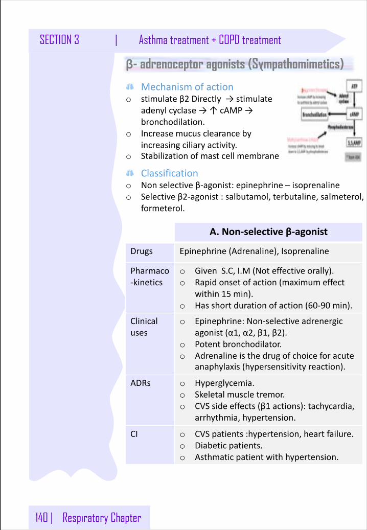
140 | Respiratory Chapter
SECTION 3 | Asthma treatment + COPD treatment
β- adrenoceptor agonists (Sympathomimetics)
Mechanismofactiono stimulateβ2 Directly→stimulate
adenylcyclase→↑cAMP→bronchodilation.
o Increasemucusclearancebyincreasingciliaryactivity.
o Stabilizationofmastcellmembrane
Classificationo Nonselectiveβ-agonist:epinephrine– isoprenalineo Selectiveβ2-agonist:salbutamol,terbutaline,salmeterol,
formeterol.
A.Non-selectiveβ-agonist
Drugs Epinephrine(Adrenaline),Isoprenaline
Pharmaco-kinetics
o GivenS.C,I.M(Noteffectiveorally).o Rapidonsetofaction(maximumeffect
within15 min).o Hasshortdurationofaction(60-90 min).
Clinicaluses
o Epinephrine:Non-selectiveadrenergicagonist(α1,α2,β1,β2).
o Potentbronchodilator.o Adrenalineisthedrugofchoiceforacute
anaphylaxis(hypersensitivityreaction).
ADRs o Hyperglycemia.o Skeletalmuscletremor.o CVSsideeffects(β1 actions): tachycardia,
arrhythmia,hypertension.
CI o CVSpatients:hypertension,heartfailure.o Diabeticpatients.o Asthmaticpatientwithhypertension.
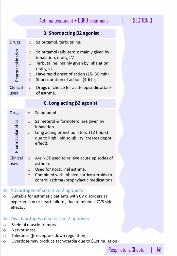
Respiratory Chapter | 141
Asthma treatment + COPD treatment | SECTION 3
Advantagesofselective2 agonistso SuitableforasthmaticpatientswithCVdisordersas
hypertensionorheartfailure,duetominimalCVSsideeffects.
Disadvantagesofselective2 agonistso Skeletalmuscletremors.o Nervousnesso Tolerance(β-receptorsdownregulation).o Overdosemayproducetachycardiaduetoβ1stimulation.
B.Shortactingβ2 agonist
Drugs o Salbutamol,terbutaline.
Pharmacokinetics
o Salbutamol(albuterol):mainlygivenbyinhalation,orally,I.V
o Terbutaline:mainlygivenbyinhalation,orally,s.c.
o Haverapidonsetofaction(15- 30 min)o Shortdurationofaction(4-6 hr).
Clinicaluses
o Drugsofchoiceforacuteepisodicattackofasthma.
C.Longactingβ2 agonist
Drugs o Salbutamol
Pharmacokinetics o Salmeterol &formoterol aregivenby
inhalation.o Longactingbronchodilators(12 hours)
duetohighlipidsolubility(createsdepoteffect).
Clinicaluses
o AreNOTusedtorelieveacuteepisodesofasthma.
o Usedfornocturnalasthma.o Combinedwithinhaledcorticosteroidsto
controlasthma(prophylacticmedication)
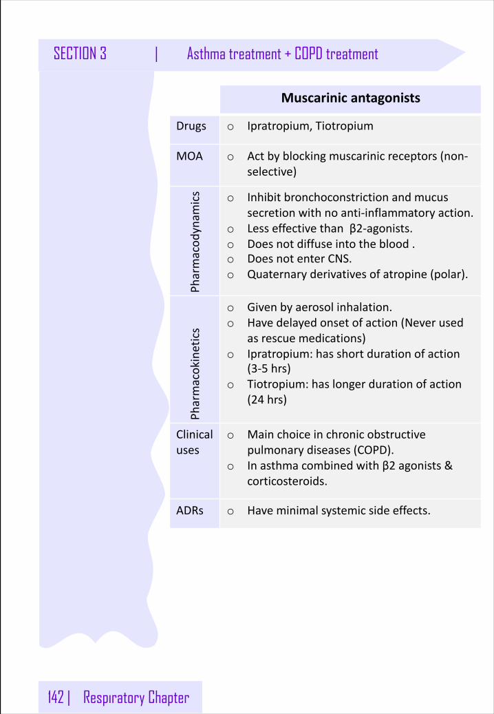
142 | Respiratory Chapter
SECTION 3 | Asthma treatment + COPD treatment
Muscarinicantagonists
Drugs o Ipratropium,Tiotropium
MOA o Actbyblockingmuscarinicreceptors(non-selective)
Pharmacod
ynam
ics o Inhibitbronchoconstrictionandmucus
secretionwithnoanti-inflammatoryaction.o Lesseffectivethanβ2-agonists.o Doesnotdiffuseintotheblood.o DoesnotenterCNS.o Quaternaryderivativesofatropine(polar).
Pharmacokinetics
o Givenbyaerosolinhalation.o Havedelayedonsetofaction(Neverused
asrescuemedications)o Ipratropium:hasshortdurationofaction
(3-5 hrs)o Tiotropium:haslongerdurationofaction
(24 hrs)
Clinicaluses
o Mainchoiceinchronicobstructivepulmonary diseases(COPD).
o Inasthmacombinedwithβ2 agonists&corticosteroids.
ADRs o Haveminimalsystemicsideeffects.
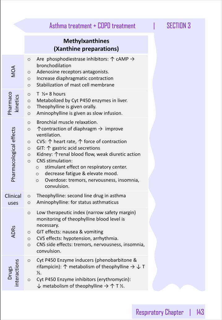
Respiratory Chapter | 143
Asthma treatment + COPD treatment | SECTION 3
Methylxanthines(Xanthinepreparations)
MOA
o Arephosphodiestrase inhibitors:↑cAMP →bronchodilation
o Adenosinereceptorsantagonists.o Increasediaphragmaticcontractiono Stabilizationofmastcellmembrane
Pharmaco
kine
tics o T½=8 hours
o MetabolizedbyCyt P450 enzymesinliver.o Theophyllineisgivenorally.o Aminophyllineisgivenasslowinfusion.
Pharmacologicaleffe
cts
o Bronchialmusclerelaxation.o ↑contractionofdiaphragm→improve
ventilation.o CVS:↑heartrate,↑forceofcontractiono GIT:↑gastricacidsecretionso Kidney:↑renalbloodflow,weakdiureticactiono CNSstimulation:
o stimulanteffectonrespiratorycenter.o decreasefatigue&elevatemood.o Overdose:tremors,nervousness,insomnia,
convulsion.
Clinicaluses
o Theophylline:secondlinedruginasthmao Aminophylline:forstatusasthmaticus
ADRs
o Lowtherapeuticindex(narrowsafetymargin)monitoringoftheophyllinebloodlevelisnecessary.
o GITeffects:nausea&vomitingo CVSeffects:hypotension,arrhythmia.o CNSsideeffects:tremors,nervousness,insomnia,
convulsion.
Drugs
interactions o Cyt P450 Enzymeinducers(phenobarbitone &
rifampicin):↑metabolismoftheophylline→↓T½.
o Cyt P450 Enzymeinhibitors(erythromycin):↓metabolismoftheophylline→↑T½.
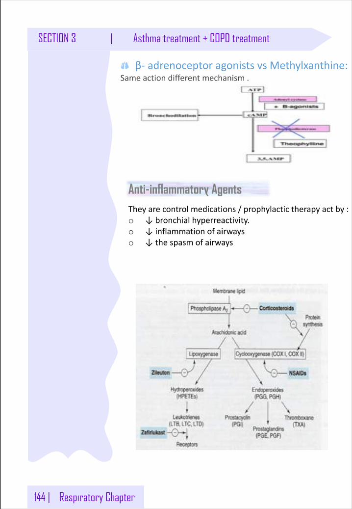
144 | Respiratory Chapter
SECTION 3 | Asthma treatment + COPD treatment
Anti-inflammatory Agents
β- adrenoceptoragonistsvsMethylxanthine:Sameactiondifferentmechanism.
Theyarecontrolmedications/prophylactictherapyactby:o ↓bronchialhyperreactivity.o ↓inflammationofairwayso ↓thespasmofairways
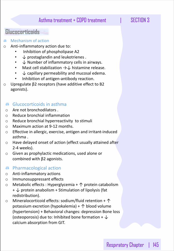
Respiratory Chapter | 145
Asthma treatment + COPD treatment | SECTION 3
Glucocorticoidsinasthmao Arenotbronchodilators.o Reducebronchialinflammationo Reducebronchialhyperreactivitytostimulio Maximumactionat9-12 months.o Effectiveinallergic,exercise,antigenandirritant-induced
asthma.o Havedelayedonsetofaction(effectusuallyattainedafter
2-4 weeks).o Givenasprophylacticmedications,usedaloneor
combinedwithβ2 agonists.
Pharmacologicalactiono Anti-inflammatoryactionso Immunosuppressanteffectso Metaboliceffects:Hyperglycemia +↑proteincatabolism
+↓proteinanabolism+Stimulationoflipolysis(fatredistribution).
o Mineralocorticoideffects:sodium/fluidretention+↑potassiumexcretion(hypokalemia)+↑bloodvolume(hypertension)+Behavioral changes:depressionBoneloss(osteoporosis)dueto:Inhibitedboneformation+↓calciumabsorptionfromGIT.
GlucocorticoidsMechanismofaction
o Anti-inflammatoryactiondueto:• InhibitionofphospholipaseA2• ↓prostaglandinandleukotrienes.• ↓Numberofinflammatorycellsinairways.• Mastcellstabilization→↓histaminerelease.• ↓capillarypermeabilityandmucosaledema.• Inhibitionofantigen-antibodyreaction.
o Upregulateβ2 receptors(haveadditiveeffecttoB2agonists).
146 | Respiratory Chapter
SECTION 3 | Asthma treatment + COPD treatment
Administrationo Inhalation:Givenbyinhalation(metered-doseinhaler).
Havefirstpassmetabolismthereforelesssideeffectssoit’sthebestchoiceinprophylaxisofasthma.e.g.Budesonide&Fluticasone,beclometasone.
o Orally:Prednisone,methylprednisolone.o Injection:Hydrocortisone,dexamethasone.
ClinicalUsesofglucocorticoidso Treatmentofinflammatorydisorders(asthma,
rheumatoidarthritis).o Treatmentofautoimmunedisorders(ulcerativecolitis,
psoriasisandafterorganorbonemarrowtransplantationasimmunosuppressants.
o Antiemeticsincancerchemotherapyo Systemiccorticosteroidsarereservedfor:Status
asthmaticus(i.v.)
Sideeffectso Duetosystemiccorticosteroids:1- FluidRetention,Hypertension,Weightgain,Hyperglycemia2-Growthretardationinchildren3-Adrenalsuppression4-Cataract5-Osteoporosis6-Susceptibilitytoinfections7-Fatdistribution8-Psychosiso Inhalationhasveryfewsideeffectsbutcancausethe
following:• Oropharyngealcandidiasis(thrush).• Dysphonia(voicehoarseness)
WithdrawalofsystemiccorticosteroidsAbruptstopofcorticosteroidsshouldbeavoidedanddoseshouldbetapered(adrenalinsufficiencysyndrome).Administrationandwithdrawalshouldbegradual.
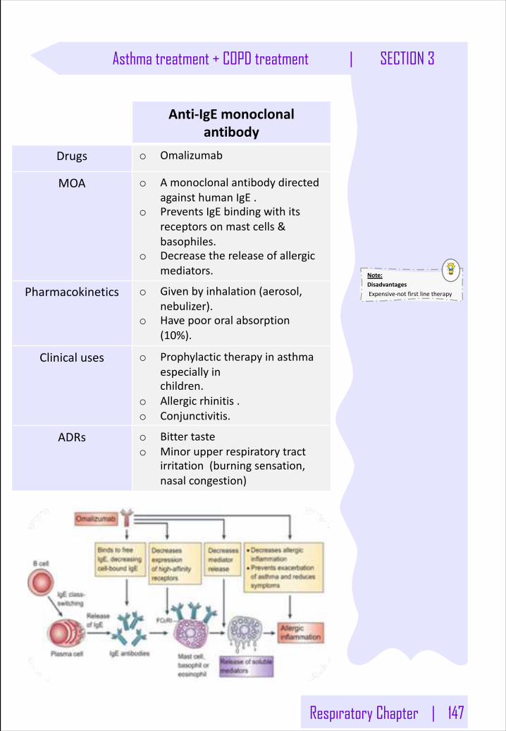
Respiratory Chapter | 147
Asthma treatment + COPD treatment | SECTION 3
Anti-IgE monoclonalantibody
Drugs o Omalizumab
MOA o AmonoclonalantibodydirectedagainsthumanIgE .
o PreventsIgE bindingwithitsreceptorsonmast cells&basophiles.
o Decreasethereleaseofallergicmediators.
Pharmacokinetics o Givenbyinhalation(aerosol,nebulizer).
o Havepoororalabsorption(10%).
Clinicaluses o Prophylactictherapyinasthmaespeciallyinchildren.
o Allergicrhinitis.o Conjunctivitis.
ADRs o Bittertasteo Minorupperrespiratorytract
irritation(burningsensation,nasalcongestion)
Note: DisadvantagesExpensive-notfirstlinetherapy
148 | Respiratory Chapter
SECTION 3 | Asthma treatment + COPD treatment
Mastcellstabilizers
Drugs o Cromoglycate (alsocalledcromolyn),Nedocromil
MOA
o Actbystabilizationofmastcellmembrane.
o Theyarenotbronchodilators,sotheyarenoteffectiveinacuteattackofasthma.
o Prophylacticanti-inflammatorydrugs.
o Reducebronchialhyperreactivity.o Effectiveinexercise,antigenand
irritant-inducedasthma.o Childrenrespondbetterthanadults.
Pharmacokineticso Givenbyinhalation(aerosol,
nebulizer).o Havepoororalabsorption(10%).
Clinicaluses
o Prophylactictherapyinasthmaespeciallyinchildren.
o Allergicrhinitis.o Conjunctivitis.
ADRs
o Bittertasteo Minorupperrespiratorytract
irritation(burningsensation,nasalcongestion)
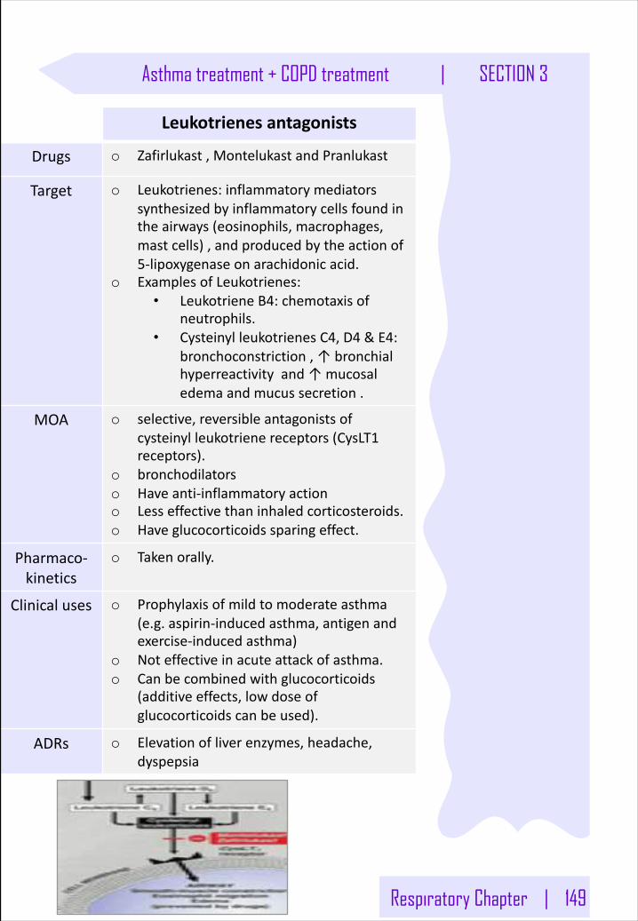
Respiratory Chapter | 149
Asthma treatment + COPD treatment | SECTION 3
Leukotrienesantagonists
Drugs o Zafirlukast ,Montelukast andPranlukast
Target o Leukotrienes:inflammatorymediatorssynthesizedbyinflammatorycellsfoundintheairways(eosinophils, macrophages,mastcells),andproducedbytheactionof5-lipoxygenaseonarachidonic acid.
o ExamplesofLeukotrienes:• LeukotrieneB4:chemotaxisof
neutrophils.• Cysteinyl leukotrienesC4,D4 &E4:
bronchoconstriction,↑bronchialhyperreactivity and↑mucosaledema andmucussecretion.
MOA o selective,reversibleantagonistsofcysteinyl leukotrienereceptors(CysLT1receptors).
o bronchodilatorso Haveanti-inflammatoryactiono Lesseffectivethaninhaledcorticosteroids.o Haveglucocorticoidssparingeffect.
Pharmaco-kinetics
o Takenorally.
Clinicaluses o Prophylaxisofmildtomoderateasthma(e.g.aspirin-inducedasthma,antigenandexercise-inducedasthma)
o Noteffectiveinacuteattackofasthma.o Canbecombinedwithglucocorticoids
(additiveeffects,lowdoseofglucocorticoidscanbeused).
ADRs o Elevationofliverenzymes,headache,dyspepsia
150 | Respiratory Chapter
SECTION 3 | Asthma treatment + COPD treatment
Chronic Obstructive Pulmonary Disease (COPD)o achronicirreversibleairflowobstruction,lungdamageand
inflammationoftheairsacs(alveoli).o characterizedbychronicbronchitisandemphysema
(destructionofwallsofalveoli).o Smokingisahighriskfactorbutairpollutionandgenetic
factorscancontribute..
Treatment:1.Inhaledbronchodilators
o Inhaledantimuscarinics:• Ipratropium&tiotropium.• Superiortoβ2 agonistsinCOPD
o β2 agonists,beusedeitheraloneorcombined:o salbutamol+ipratropium(shortacting)o salmeterol +Tiotropium (longacting-lessdose
frequency).
2.Oxygentherapy
3.Lungtransplantation
4.Inhaledglucocorticoids
5.Antibioticsspecificallymacrolidessuchas
azithromycintoreducethenumberofexacerbations.
InhaledbronchodilatorsinCOPDo Inhaledantimuscarinics:
• Ipratropium&tiotropium.• Superiortoβ2 agonistsinCOPD
o β2 agonists,beusedeitheraloneorcombined:o salbutamol+ipratropium(shortacting)o salmeterol+Tiotropium(longacting-lessdose
frequency).
Respiratory Chapter | 151
Asthma treatment + COPD treatment | SECTION 3
A.Muscarinicantagonists
Drugs o Ipratropium,Tiotropium
MOA o Actbyblockingmuscarinicreceptors(non-selective)
Pharmacod
ynam
ics o Inhibitbronchoconstrictionandmucus
secretionwithnoanti-inflammatoryaction.
o Lesseffectivethanβ2-agonists.o Doesnotdiffuseintotheblood.o DoesnotenterCNS.o Quaternaryderivativesofatropine
(polar).
Pharmacokinetics o Givenbyaerosolinhalation.
o Havedelayedonsetofaction(Neverusedasrescuemedications)
o Ipratropium:hasshortdurationofaction(3-5 hrs)
o Tiotropium:haslongerdurationofaction(24 hrs)
Clinicaluses
o Mainchoiceinchronicobstructivepulmonary diseases(COPD).
o Inasthmacombinedwithβ2 agonists&corticosteroids.
ADRs o Haveminimalsystemicsideeffects.
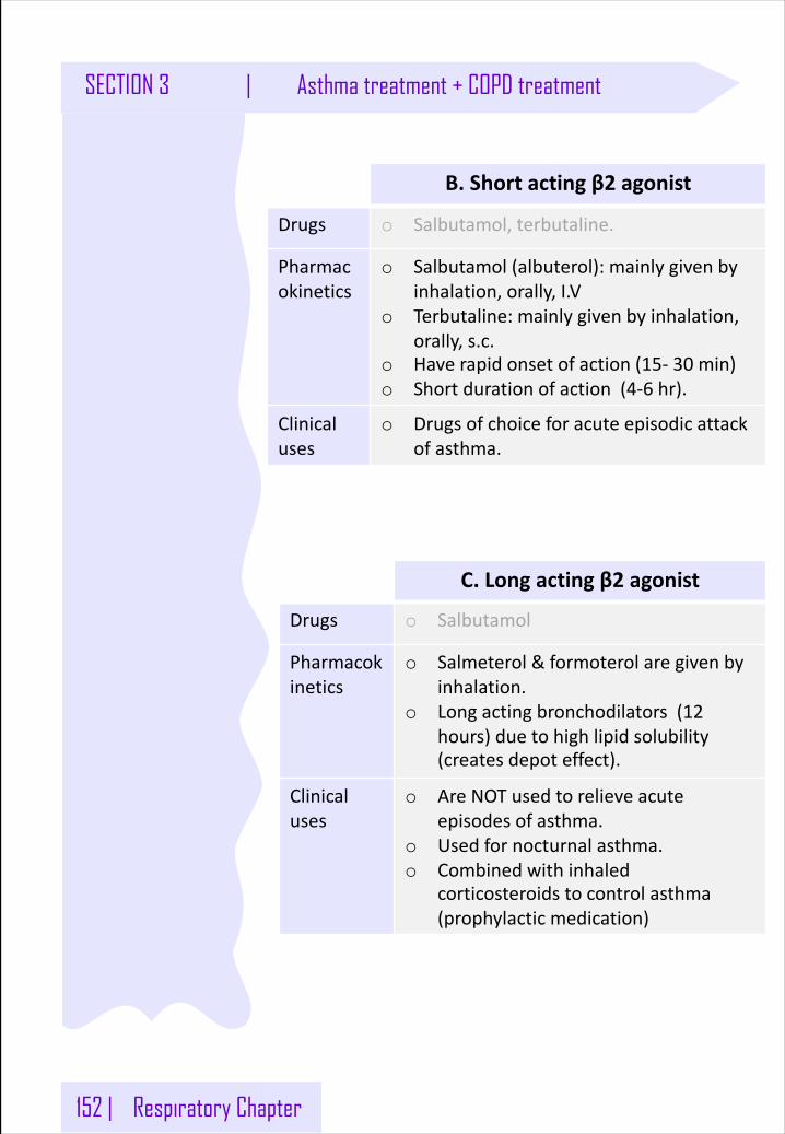
152 | Respiratory Chapter
SECTION 3 | Asthma treatment + COPD treatment
B.Shortactingβ2 agonist
Drugs o Salbutamol,terbutaline.
Pharmacokinetics
o Salbutamol(albuterol):mainlygivenbyinhalation,orally,I.V
o Terbutaline:mainlygivenbyinhalation,orally,s.c.
o Haverapidonsetofaction(15- 30 min)o Shortdurationofaction(4-6 hr).
Clinicaluses
o Drugsofchoiceforacuteepisodicattackofasthma.
C.Longactingβ2 agonist
Drugs o Salbutamol
Pharmacokinetics
o Salmeterol &formoterol aregivenbyinhalation.
o Longactingbronchodilators(12hours)duetohighlipidsolubility(createsdepoteffect).
Clinicaluses
o AreNOTusedtorelieveacuteepisodesofasthma.
o Usedfornocturnalasthma.o Combinedwithinhaled
corticosteroidstocontrolasthma(prophylacticmedication)
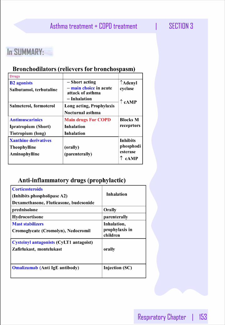
Respiratory Chapter | 153
Asthma treatment + COPD treatment | SECTION 3
In SUMMARY:
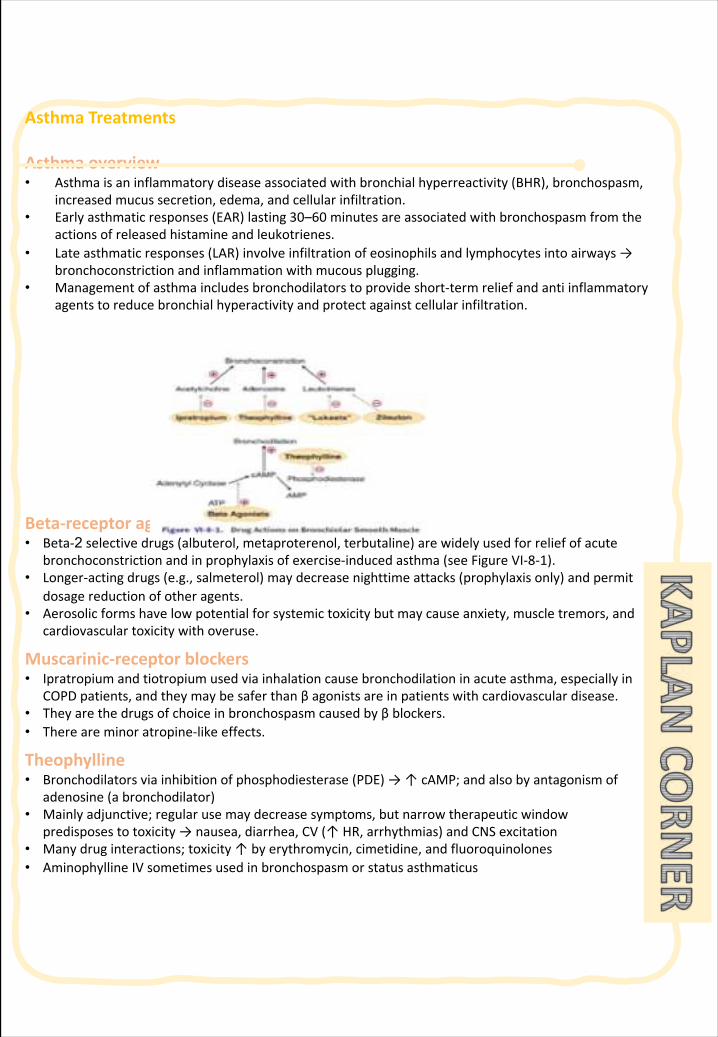
AsthmaTreatments
Asthmaoverview• Asthmaisaninflammatorydiseaseassociatedwithbronchialhyperreactivity (BHR),bronchospasm,
increasedmucussecretion,edema,andcellularinfiltration.• Earlyasthmaticresponses(EAR)lasting30–60 minutesareassociatedwithbronchospasmfromthe
actionsofreleasedhistamineandleukotrienes.• Lateasthmaticresponses(LAR)involveinfiltrationofeosinophilsandlymphocytesintoairways→
bronchoconstrictionandinflammationwithmucousplugging.• Managementofasthmaincludesbronchodilatorstoprovideshort-termreliefandantiinflammatory
agentstoreducebronchialhyperactivityandprotectagainstcellularinfiltration.
Beta-receptoragonists• Beta-2 selectivedrugs(albuterol,metaproterenol,terbutaline)arewidelyusedforreliefofacute
bronchoconstrictionandinprophylaxisofexercise-inducedasthma (seeFigureVI-8-1).• Longer-actingdrugs(e.g.,salmeterol)maydecreasenighttimeattacks(prophylaxisonly)andpermit
dosagereductionofotheragents.• Aerosolicformshavelowpotentialforsystemictoxicitybutmaycauseanxiety,muscletremors,and
cardiovasculartoxicitywithoveruse.
Muscarinic-receptorblockers• Ipratropiumandtiotropiumusedviainhalationcausebronchodilationinacuteasthma,especiallyin
COPDpatients,andtheymaybesaferthanβagonistsareinpatientswithcardiovasculardisease.• Theyarethedrugsofchoiceinbronchospasmcausedbyβblockers.• Thereareminoratropine-likeeffects.
Theophylline• Bronchodilatorsviainhibitionofphosphodiesterase(PDE)→↑cAMP;andalsobyantagonismof
adenosine(abronchodilator)• Mainlyadjunctive;regularusemaydecreasesymptoms,butnarrowtherapeuticwindow
predisposestotoxicity→nausea,diarrhea,CV(↑HR,arrhythmias)andCNSexcitation• Manydruginteractions;toxicity↑byerythromycin,cimetidine,andfluoroquinolones• AminophyllineIVsometimesusedinbronchospasmorstatusasthmaticus
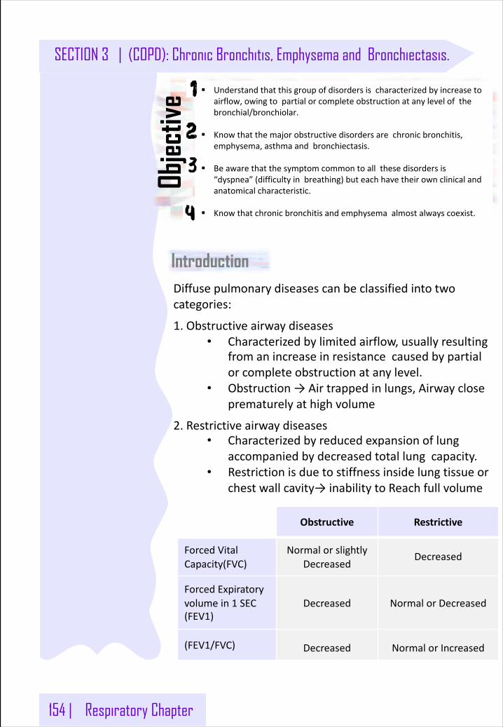
154 | Respiratory Chapter
SECTION 3 | (COPD): Chronic Bronchitis, Emphysema and Bronchiectasis.
Introduction
§ Understandthatthisgroupofdisordersischaracterizedbyincreasetoairflow,owingtopartialorcompleteobstructionatanylevelofthebronchial/bronchiolar.
§ Knowthatthemajorobstructivedisordersarechronicbronchitis,emphysema,asthmaandbronchiectasis.
§ Beawarethatthesymptomcommontoallthesedisordersis“dyspnea”(difficultyinbreathing)buteachhavetheirownclinicalandanatomicalcharacteristic.
§ Knowthatchronicbronchitisandemphysemaalmostalwayscoexist.
Diffusepulmonarydiseasescanbeclassifiedintotwocategories:
1.Obstructiveairwaydiseases• Characterizedbylimitedairflow,usuallyresulting
fromanincreaseinresistancecausedbypartialorcompleteobstructionatanylevel.
• Obstruction→Airtrappedinlungs,Airwaycloseprematurelyathighvolume
2.Restrictiveairwaydiseases• Characterizedbyreducedexpansionoflung
accompaniedbydecreasedtotallungcapacity.• Restrictionisduetostiffnessinsidelungtissueor
chestwallcavity→inabilitytoReachfullvolume
Obstructive Restrictive
ForcedVitalCapacity(FVC)
NormalorslightlyDecreased
Decreased
ForcedExpiratoryvolumein1 SEC(FEV1)
Decreased NormalorDecreased
(FEV1/FVC) Decreased NormalorIncreased
Objec
tive
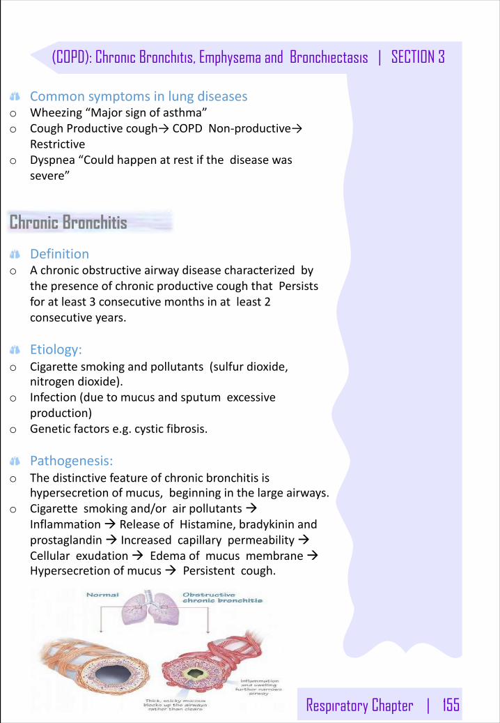
Respiratory Chapter | 155
(COPD): Chronic Bronchitis, Emphysema and Bronchiectasis | SECTION 3
Commonsymptomsinlungdiseaseso Wheezing“Majorsignofasthma”o CoughProductivecough→COPDNon-productive→
Restrictiveo Dyspnea“Couldhappenatrestifthediseasewas
severe”
Chronic Bronchitis
Definitiono Achronicobstructiveairwaydiseasecharacterizedby
thepresenceofchronicproductivecoughthatPersistsforatleast3 consecutivemonthsinatleast2consecutiveyears.
Etiology:o Cigarettesmokingandpollutants(sulfur dioxide,
nitrogendioxide).o Infection(duetomucusandsputumexcessive
production)o Geneticfactorse.g.cysticfibrosis.
Pathogenesis:o Thedistinctivefeatureofchronicbronchitisis
hypersecretionofmucus,beginninginthelargeairways.o Cigarettesmokingand/orairpollutantsà
Inflammationà ReleaseofHistamine,bradykininandprostaglandinà IncreasedcapillarypermeabilityàCellularexudationà EdemaofmucusmembraneàHypersecretionofmucusà Persistentcough.
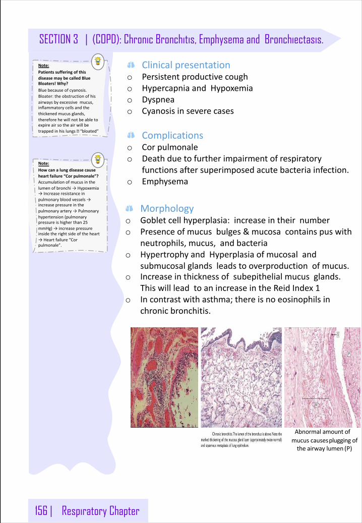
156 | Respiratory Chapter
SECTION 3 | (COPD): Chronic Bronchitis, Emphysema and Bronchiectasis.
Clinicalpresentationo Persistentproductivecougho HypercapniaandHypoxemiao Dyspneao Cyanosisinseverecases
Complicationso Corpulmonaleo Deathduetofurtherimpairmentofrespiratory
functionsaftersuperimposedacutebacteriainfection.o Emphysema
Morphologyo Gobletcellhyperplasia:increaseintheirnumbero Presenceofmucusbulges&mucosacontainspuswith
neutrophils,mucus,andbacteriao HypertrophyandHyperplasiaofmucosaland
submucosalglandsleadstooverproductionofmucus.o Increaseinthicknessofsubepithelialmucusglands.
ThiswillleadtoanincreaseintheReidIndex1o Incontrastwithasthma;thereisnoeosinophilsin
chronicbronchitis.
Abnormalamountofmucuscausespluggingoftheairwaylumen (P)
Note: PatientssufferingofthisdiseasemaybecalledBlueBloaters!Why?Bluebecauseofcyanosis.Bloater:theobstructionofhisairwaysbyexcessivemucus,inflammatorycellsandthethickenedmucusglands,thereforehewillnotbeabletoexpireairsotheairwillbetrappedinhislungs� “bloated”
Note: Howcanalungdiseasecauseheartfailure“Corpulmonale”?Accumulationofmucusinthelumenofbronchi→Hypoxemia→Increaseresistanceinpulmonarybloodvessels→increasepressureinthepulmonaryartery→Pulmonaryhypertension(pulmonarypressureishigherthan25mmHg)→increasepressureinsidetherightsideoftheheart→Heartfailure“Corpulmonale”.
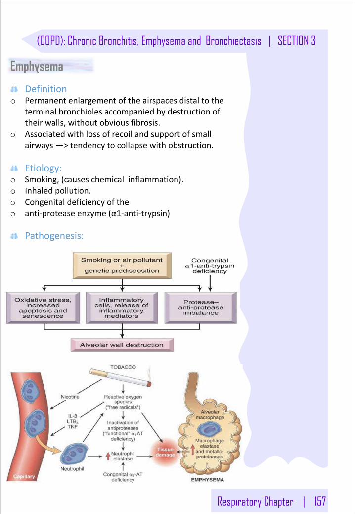
Respiratory Chapter | 157
(COPD): Chronic Bronchitis, Emphysema and Bronchiectasis | SECTION 3
Emphysema
Definitiono Permanentenlargementoftheairspacesdistaltothe
terminalbronchiolesaccompaniedbydestructionoftheirwalls,withoutobviousfibrosis.
o Associatedwithlossofrecoilandsupportofsmallairways—>tendencytocollapsewithobstruction.
Etiology:o Smoking,(causeschemicalinflammation).o Inhaledpollution.o Congenitaldeficiencyoftheo anti-proteaseenzyme(α1-anti-trypsin)
Pathogenesis:
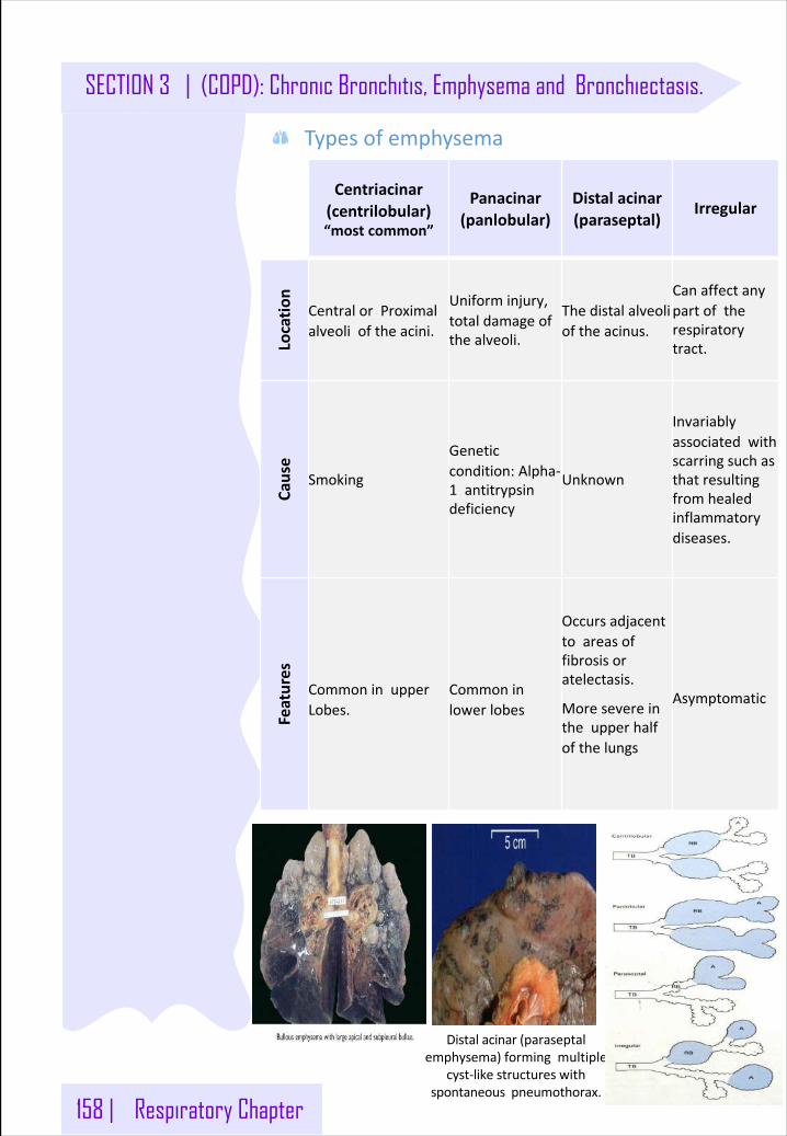
158 | Respiratory Chapter
SECTION 3 | (COPD): Chronic Bronchitis, Emphysema and Bronchiectasis.
Typesofemphysema
Distalacinar(paraseptalemphysema)formingmultiple
cyst-likestructureswithspontaneouspneumothorax.
Centriacinar(centrilobular)“mostcommon”
Panacinar(panlobular)
Distalacinar(paraseptal)
Irregular
Locatio
nCentralorProximalalveolioftheacini.
Uniforminjury,totaldamageofthealveoli.
Thedistalalveolioftheacinus.
Canaffectanypartoftherespiratorytract.
Cause
Smoking
Geneticcondition:Alpha-1 antitrypsindeficiency
Unknown
Invariablyassociatedwithscarringsuchasthatresultingfromhealedinflammatorydiseases.
Features
CommoninupperLobes.
Commoninlowerlobes
Occursadjacenttoareasoffibrosisoratelectasis.
Moresevereintheupperhalfofthelungs
Asymptomatic
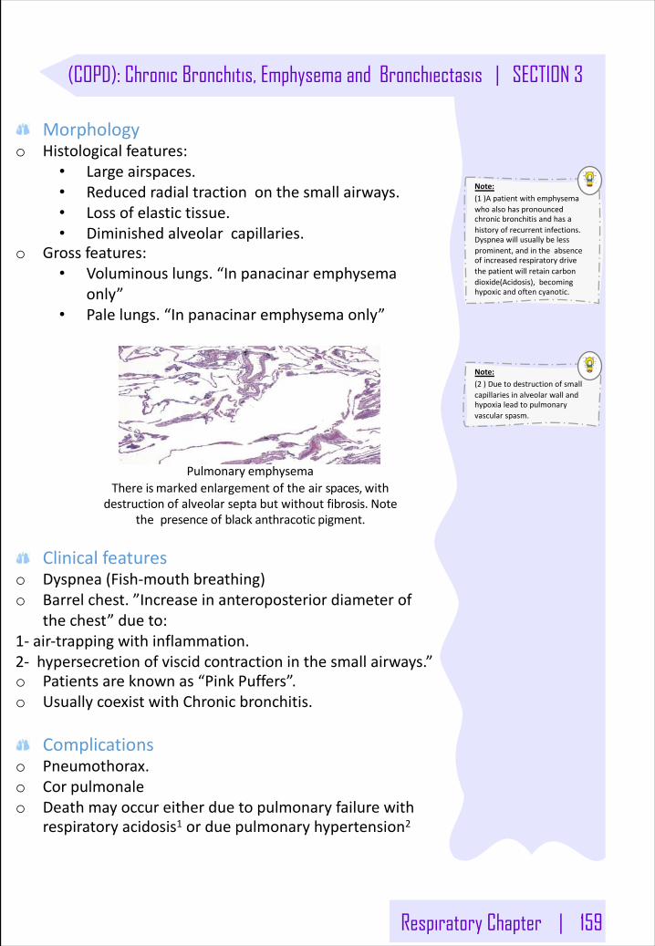
Respiratory Chapter | 159
(COPD): Chronic Bronchitis, Emphysema and Bronchiectasis | SECTION 3
Morphologyo Histologicalfeatures:
• Largeairspaces.• Reducedradialtractiononthesmallairways.• Lossofelastictissue.• Diminishedalveolarcapillaries.
o Grossfeatures:• Voluminouslungs.“Inpanacinar emphysema
only”• Palelungs.“Inpanacinar emphysemaonly”
Pulmonary emphysemaThereismarkedenlargementoftheairspaces,with
destructionofalveolarseptabutwithoutfibrosis.Notethepresenceofblackanthracotic pigment.
Clinicalfeatureso Dyspnea(Fish-mouthbreathing)o Barrelchest.”Increaseinanteroposteriordiameterof
thechest”dueto:1- air-trappingwithinflammation.2- hypersecretionofviscidcontractioninthesmallairways.”o Patientsareknownas“PinkPuffers”.o UsuallycoexistwithChronicbronchitis.
Complicationso Pneumothorax.o Corpulmonaleo Deathmayoccureitherduetopulmonaryfailurewith
respiratoryacidosis1 orduepulmonaryhypertension2
Note: (1 )Apatientwithemphysemawhoalsohaspronouncedchronicbronchitisandhasahistoryofrecurrentinfections.Dyspneawillusuallybelessprominent,andintheabsenceofincreasedrespiratorydrivethepatientwillretaincarbondioxide(Acidosis),becominghypoxicandoftencyanotic.
Note: (2 )Duetodestructionofsmallcapillariesinalveolarwallandhypoxialeadtopulmonaryvascularspasm.
160 | Respiratory Chapter
SECTION 3 | (COPD): Chronic Bronchitis, Emphysema and Bronchiectasis.
Bronchiectasis
Definitiono PermanentDilationoftheBronchiandtheBronchioles
causedbydestructionofthesmoothmuscleandthesupportingElastictissue.
Etiology:o Bronchialobstruction:
o Localized:Tumors,Foreign bodiesormucousimpaction.
o Systemic:BronchialAsthmaandChronicbronchitiso CongenitalorHereditary:
o Congenitalbronchiectasiso CysticFibrosiso PrimaryCiliaryDyskinesiao Intralobar sequestrationoflungo Immunodeficiency
o SuppurativePneumoniao Klebsiellaspp.o Staphylococcusaureus
Pathogenesis:TwointertwinedprocessesthatcontributetoBronchiectasisObstructionandChronicInfection:o Step1:AforeignBodyentersthebodyleadingto
obstruction.o Step2:Impairedmucociliary clearance,mucusstasisand
accumulationwhichinturnfurthermakestheairwayssusceptibletomicrobialcolonization.
o Step3:thepersistenceofthepathologywithsuperaddedinfectionleadstoa“viciouscircle”ofinflammationandtissuedamage.
o Step4:inflammatorydamagetothebronchiwhichwillleadtoirreversibledilationandlossofelasticityofthealveolarwallleadingtobronchiectasis.
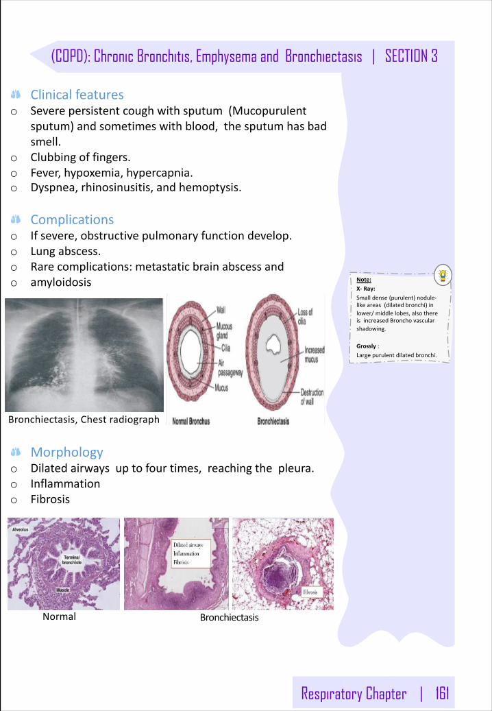
Respiratory Chapter | 161
(COPD): Chronic Bronchitis, Emphysema and Bronchiectasis | SECTION 3
Clinicalfeatureso Severepersistentcoughwithsputum(Mucopurulent
sputum)andsometimeswithblood,thesputumhasbadsmell.
o Clubbingoffingers.o Fever,hypoxemia,hypercapnia.o Dyspnea,rhinosinusitis,andhemoptysis.
Complicationso Ifsevere,obstructivepulmonaryfunctiondevelop.o Lungabscess.o Rarecomplications:metastaticbrainabscessando amyloidosis
Morphologyo Dilatedairwaysuptofourtimes,reachingthepleura.o Inflammationo Fibrosis
Normal Bronchiectasis
Bronchiectasis,Chest radiograph
Note: X- Ray:Smalldense(purulent)nodule-likeareas(dilatedbronchi)inlower/middlelobes,alsothereisincreasedBronchovascularshadowing.
Grossly :Largepurulentdilatedbronchi.
162 | Respiratory Chapter
SECTION 3 | (COPD): Chronic Bronchitis, Emphysema and Bronchiectasis.
Kartagener Syndrome(ImmotileCiliasyndromeorCiliaryDyskinesia)
Definitiono AutosomalRecessivediseasecharacterizedbythe
absenceofouterandinnerDyneinArmscausingimmotilecilia.Characteristics:
ItcausesamalfunctionintheciliathereforelossofdefenseintheUpperrespiratorytrac,staphylococcusaureus
Diagnosis:o GeneticStudy.o ElectronMicroscope.
Complicationo Recurrentrespiratorytractinfections,e.g.Sinusitiso InfertilityinMales.o Deafness(Can’thear).
Cystic Fibrosis(Mucoviscidosis)
DefinitionAninheritedDiseasecausingthicky,stickymucoussecretiontobuildupinthelunganddigestivetractcausingBronchiectasis.
Clinicalcorrelate: Usuallythesespatientspresentwith:1-Pancreatitis.2-Malabsorption.3-Upperrespiratorytractinfections.4-Bronchiectasis
Diagnosis:Bysodiumtest(Sweattest):usuallytheyhavelowsodiuminsweat.
In SUMMARY: ChronicBronchitis
o Definition (clinical):Persistentchronicproductivecoughforaperiodof3 monthsover2 consecutiveyears.
o Etiology:Almostallpatientsaresmokers.”Usuallycoexistwithemphysema”
o Symptoms:dyspnea,productivecough,wheezing“sometimesnotalways”duetoobstructionofthelumenofthebronchibyexcessivemucusproduction.
o Histologicalpresentations:• Mucussecretingbronchialglandsbecome
hypertrophic/hyperplasticthereforetherethicknesswillincreaseandwilloccupyalotofspaceinthebronchialwall.
• Congestedbloodvessels.”when thereisinflammationthereisvasodilation→increasedvascularpermeability”
• Submucosaledema.
Respiratory Chapter | 163
(COPD): Chronic Bronchitis, Emphysema and Bronchiectasis | SECTION 3
Emphysemao Definition (Pathological):Chronicobstructiveairwaydisease
characterizedbyabnormaldilationanddestructionoftheairspacesdistaltotheterminalbronchioles(whichincludestheRespiratorybronchioles,alveolarductandalveoli).
o Etiology:Smoking.”usuallycoexistwithchronicbronchitis”o Symptoms:
• Dyspnea• Productivecough• IfadvancedthepatientwillhaveHoneycomblungappearanceand
barrelchest”duetoincreaseintheanteroposteriordiameterofthethoraciccavity”,
• “Pinkpuffer.”:Pinkbecausehehasnocyanosisandpufferbecauseheblowsairout”
o Types:- Centriacinar:
• Onlytherespiratorybronchiolesaredilated• Commoninsmokers.
- Panacinar:• Respiratorybronchioles,alveolarductandaciniaredilated.• Commoninpatientswith𝛂1-antitrypsindeficiency.
- Distalacinar:• Dilatationofthedistalpartoftheacini.• Causesbullae”ifthebullaeruptureitwillcause
pneumothorax”• Commoninnon-smokersandyoungpeople.
- Irregular• Canaffectanypart.• Usuallyininflammatoryconditions“patientswithprevious
pneumonia,oldTB”
Bronchiectasiso Definition:Chronicobstructiveairwaydiseasecharacterizedbyabnormalandpermanentdilatationofbronchiandbronchiolesassociatedwithinflammationandfibrosisandpusformation.o Etiology:Bronchialobstructionorduetocongenitalabnormalities.o Symptoms:dyspnea,productivecough(purulent,copioussputumwithbadsmellduetoanaerobes.)
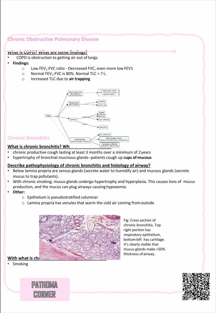
ChronicObstructivePulmonary Disease
WhatisCOPD?Whataresome findings?• COPDisobstructiontogettingairoutof lungs.• Findings:
o LowFEV1:FVCratio- DecreasedFVC,evenmorelow FEV1o NormalFEV1:FVCis80%.NormalTLC=7 L.o IncreasedTLCduetoair trapping
Chronic bronchitisWhatischronicbronchitis?Whatisthemechanism?• chronicproductivecoughlastingatleast3 monthsoveraminimumof2years• hypertrophyofbronchialmucinousglands--patientscoughupcupsofmucous
Describepathophysiologyofchronicbronchitisandhistologyof airway?• Belowlaminapropria areserousglands(secretewatertohumidifyair)andmucous glands (secrete
mucustotrap pollutants).• Withchronicsmoking,mucusglandsundergohypertrophyandhyperplasia.Thiscausestonsofmucus
production,andthemucuscanplugairwayscausing hypoxemia.• Other:
o Epitheliumispseudostratified columnaro Laminapropria hasvenules thatwarmthecoldaircomingfromoutside
Withwhatischronicbronchitishighly associated?• Smoking
Fig:Crosssectionofchronicbronchitis.Toprightportionhasrespiratoryepithelium,bottomlefthascartilage.It'sclearlyvisiblethatmucusglandsmake>50%thicknessof airway.

ChronicObstructivePulmonary Disease
Whataretheclinicalfeaturesofchronic bronchitis?• productivecoughduetoexcessivemucous production,• cyanosis('bluebloaters')- mucusplugstrap CO2;• increasedriskofinfection(anytimeyouplugatube,itincreasesriskofinfectionbehindthe block)• ReidindexIncreasesto>50 from <40%;• Cor pulmonale (pulm HTN)- duetogloballylowPAO2 inlungs(lowPAO2 inducesvasoconstriction)
WhatisReid index?• It'stheratioofthicknessofairwaymucousglandtototalthicknessofairways.Normally,it's <40%.
EmphysemaWhatis emphysema?
Whatispathophysiologyof emphysema?• Imbalancebetweenproteaseandantiprotease.Inflammationinduceshighproteaseactivity.So,smoking
leadstoinflammationwhichleadsto emphysema.
Whatis acinus?• Functionalunitoflung(aterminalbroncheole andalveoliassociatedwith it)
WhydoesA1ATdeficiencycause cirrhosis?• A1ATdeficiencyisduetomisfolding ofmutatedprotein,mutantA1ATaccumulatesinERofhepatocytes
whichresultsinliver damage.• A1ATisdoesn'tgotobloodandlungbecauseit'snotexportedby liver.
WhatdoesliverbiopsyinA1ATdeficiencyshow? (HY)Pink-purple,PAS-positive(astain)globulesinhepatocytes.Note- mucinandtropheryma whipplei arealsoPAS +ve
Fig:Histologyofemphysemashowingmultipleairsacscombiningtomakeone.
• Destructionofalveolarairsacandmultiplesacscombinetobecomeone.
• Duetolossofelasticityofairsacs,lungbecomeslikeashoppingbag,noteffectivelyexpellingairout.
• Also,elasticityofairsacsattachedtobronchioleskeepthebronchioleopenduring expiration.Whenthesacsaregone,thenbronchiolescollapseduring expiration.
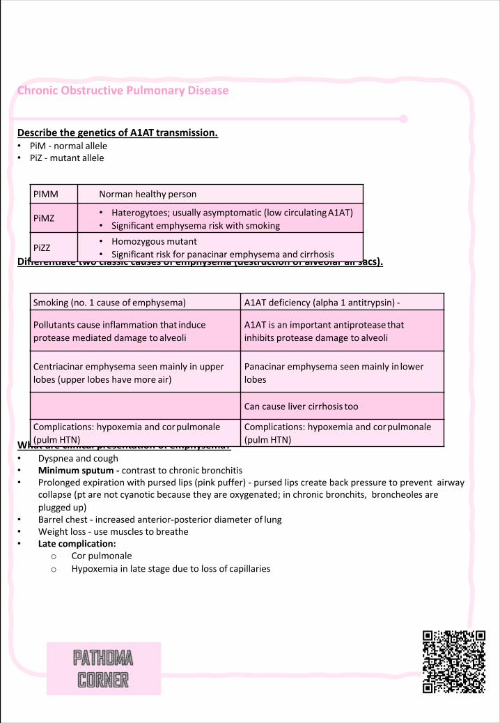
ChronicObstructivePulmonary Disease
DescribethegeneticsofA1AT transmission.• PiM - normal allele• PiZ - mutant allele
Differentiatetwoclassiccausesofemphysema(destructionofalveolarair sacs).
Whatareclinicalpresentationof emphysema?• Dyspneaand cough• Minimumsputum- contrasttochronic bronchitis• Prolongedexpirationwithpursedlips(pinkpuffer)- pursedlipscreatebackpressuretopreventairway
collapse(pt arenotcyanoticbecausetheyareoxygenated;inchronicbronchits,broncheoles areplugged up)
• Barrelchest- increasedanterior-posteriordiameterof lung• Weightloss- usemusclesto breathe• Late complication:
o Cor pulmonaleo Hypoxemiainlatestageduetolossof capillaries
PIMM Normanhealthy person
PiMZ • Haterogytoes;usuallyasymptomatic(lowcirculatingA1AT)• Significantemphysemariskwith smoking
PiZZ• Homozygousmutant• Significantriskforpanacinaremphysemaand cirrhosis
Smoking(no.1 causeof emphysema) A1ATdeficiency(alpha1 antitrypsin) -
Pollutantscauseinflammationthat induceproteasemediateddamageto alveoli
A1ATisanimportantantiproteasethatinhibitsproteasedamageto alveoli
Centriacinaremphysemaseenmainlyinupperlobes(upperlobeshavemore air)
Panacinaremphysemaseenmainlyin lowerlobes
Cancauselivercirrhosis too
Complications:hypoxemiaandcorpulmonale(pulm HTN)
Complications:hypoxemiaandcorpulmonale(pulm HTN)
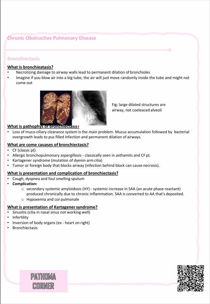
ChronicObstructivePulmonary Disease
BronchiectasisWhatis bronchieatasis?• Necrotizingdamagetoairwaywallsleadtopermanentdilationof bronchioles• Imagineifyoublowairintoabigtube,theairwilljustmoverandomlyinsidethetubeandmightnot
come out
Whatispathophys of bronchiectasis?• Lossofmuco-ciliaryclearancesystemisthemainproblem.Mucusaccumulationfollowedbybacterial
overgrowthleadstopusfilledinfectionandpermanentdilationof airways.
Whatarecomecauases of bronchiectasis?• CF(classic pt)• Allergicbronchopulmonaryaspergillosis- classicallyseeninasthamits andCF pt.• Kartagenersyndrome(mutationofdyenin arm cilia)• Tumororforeignbodythatblocksairway(infectionbehindblockcancause necrosis).
Whatispresentationandcomplicationof bronchiectasis?• Cough,dyspneaandfoulsmelling sputum• Complication:
o secondarysystemicamyloidosis(HY)- systemicincreaseinSAA(anacutephasereactant)producedchronicallyduetochronicinflammation.SAAisconvertedtoAAthat's deposited.
o Hypoxemiaandcor pulmonale
WhatispresentationofKartagener syndrome?• Sinusitis(ciliainnasalsinusnotworking well)• Infertility• Inversionofbodyorgans(ex- hearton right)• Bronchiectasis
Fig:largedilatedstructuresareairway,notcoeleaced alveoli
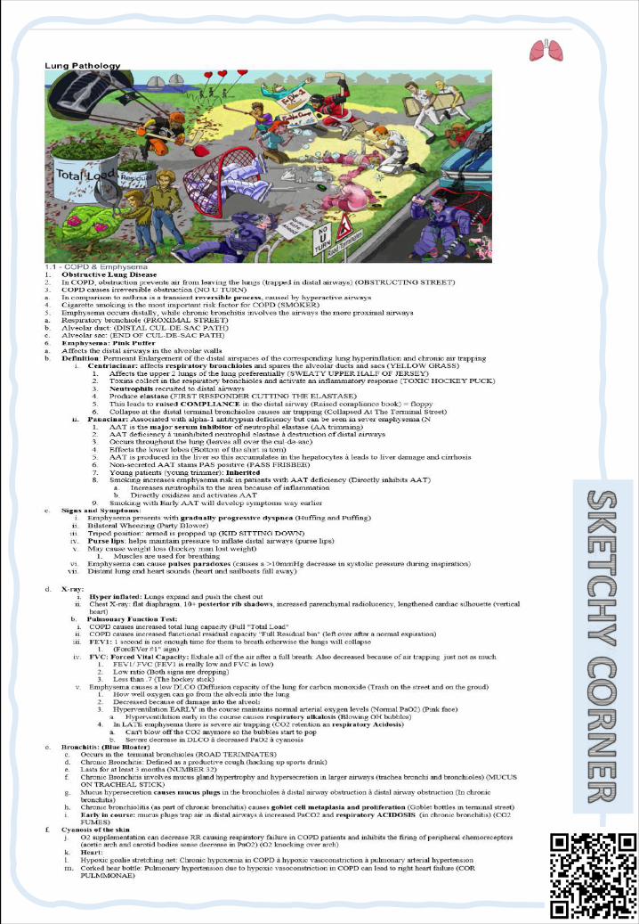
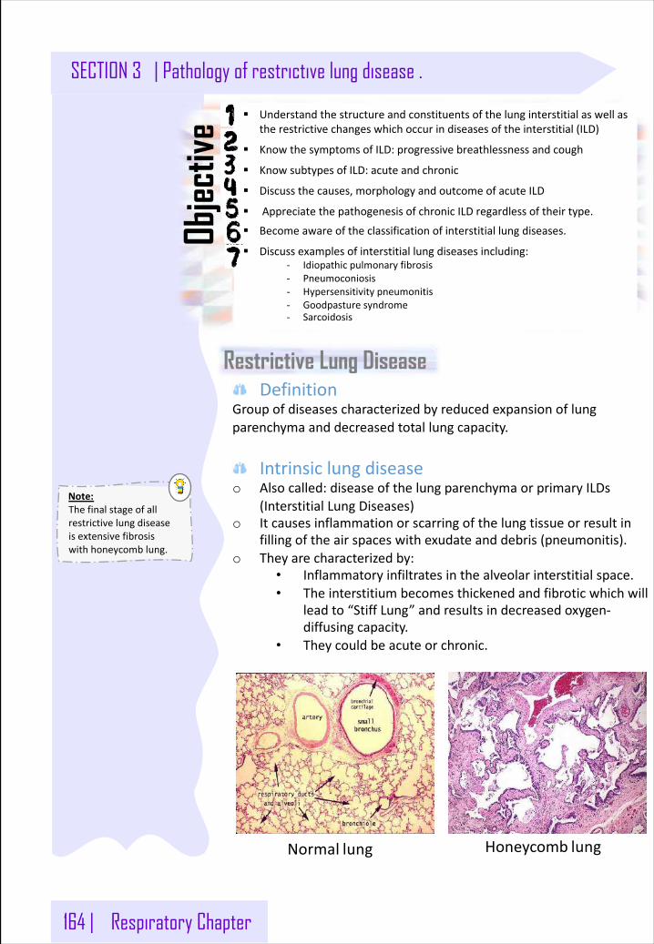
164 | Respiratory Chapter
SECTION 3 | Pathology of restrictive lung disease .
Restrictive Lung Disease
§ Understandthestructureandconstituentsofthelunginterstitialaswellastherestrictivechangeswhichoccurindiseasesoftheinterstitial(ILD)
§ KnowthesymptomsofILD:progressivebreathlessnessandcough
§ KnowsubtypesofILD:acuteandchronic
§ Discussthecauses,morphologyandoutcomeofacuteILD
§ AppreciatethepathogenesisofchronicILDregardlessoftheirtype.
§ Becomeawareoftheclassificationofinterstitiallungdiseases.
§ Discussexamplesofinterstitiallungdiseasesincluding:- Idiopathicpulmonaryfibrosis- Pneumoconiosis- Hypersensitivitypneumonitis- Goodpasturesyndrome- Sarcoidosis
Objec
tive
DefinitionGroupofdiseasescharacterizedbyreducedexpansionoflungparenchymaanddecreasedtotallungcapacity.
Intrinsiclungdiseaseo Alsocalled:diseaseofthelungparenchymaorprimaryILDs
(InterstitialLungDiseases)o Itcausesinflammationorscarringofthelungtissueorresultin
fillingoftheairspaceswithexudateanddebris(pneumonitis).o Theyarecharacterizedby:
• Inflammatoryinfiltratesinthealveolarinterstitialspace.• Theinterstitium becomesthickenedandfibroticwhichwill
leadto“StiffLung”andresultsindecreasedoxygen-diffusingcapacity.
• Theycouldbeacuteorchronic.
Honeycomb lungNormal lung
Note:Thefinalstageofallrestrictivelungdiseaseisextensivefibrosiswithhoneycomblung.
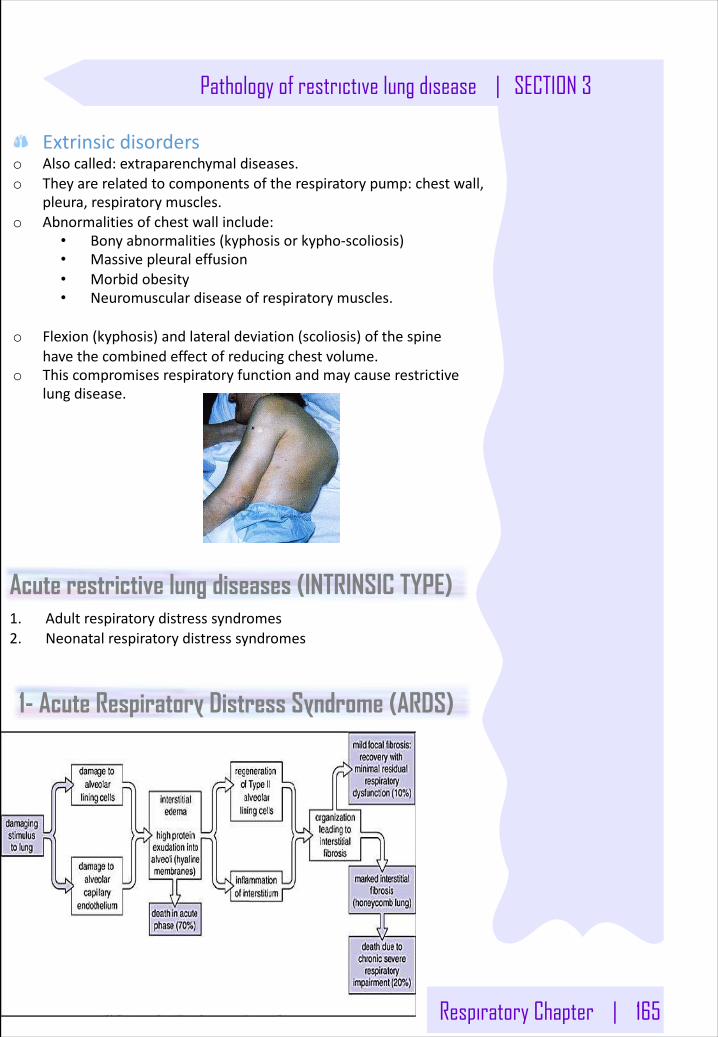
Respiratory Chapter | 165
Pathology of restrictive lung disease | SECTION 3
1- Acute Respiratory Distress Syndrome (ARDS)
Extrinsicdisorderso Alsocalled:extraparenchymal diseases.o Theyarerelatedtocomponentsoftherespiratorypump:chestwall,
pleura,respiratorymuscles.o Abnormalitiesofchestwallinclude:
• Bonyabnormalities(kyphosisorkypho-scoliosis)• Massivepleuraleffusion• Morbidobesity• Neuromusculardiseaseofrespiratorymuscles.
o Flexion(kyphosis)andlateraldeviation(scoliosis)ofthespinehavethecombinedeffectofreducingchestvolume.
o Thiscompromisesrespiratoryfunctionandmaycauserestrictivelungdisease.
Acute restrictive lung diseases (INTRINSIC TYPE)1. Adultrespiratorydistresssyndromes2. Neonatalrespiratorydistresssyndromes
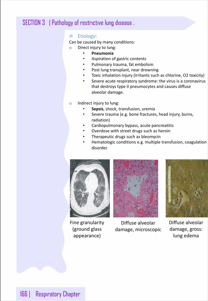
166 | Respiratory Chapter
SECTION 3 | Pathology of restrictive lung disease .
Etiology:Canbecausedbymanyconditions:o Directinjurytolung:
• Pneumonia• Aspirationofgastriccontents• Pulmonarytrauma,fatembolism• Postlungtransplant,neardrowning• Toxicinhalationinjury(irritantssuchaschlorine,O2 toxicity)• Severeacuterespiratorysyndrome:thevirusisacoronavirus
thatdestroystypeIIpneumocytesandcausesdiffusealveolardamage.
o Indirectinjurytolung:• Sepsis,shock,transfusion,uremia• Severetrauma(e.g.bonefractures,headinjury,burns,
radiation)• Cardiopulmonarybypass,acutepancreatitis• Overdosewithstreetdrugssuchasheroin• Therapeuticdrugssuchasbleomycin• Hematologicconditionse.g.multipletransfusion,coagulation
disorder.
Diffusealveolardamage,microscopic
Diffusealveolardamage,gross:lungedema
Finegranularity(groundglassappearance)

Respiratory Chapter | 167
Pathology of restrictive lung disease | SECTION 3
2- Neonatal Respiratory Distress Syndromes (NRDS)o Itisthemostcommoncauseofrespiratoryfailureinthenew-born
andisthemostcommoncauseofdeathinprematureinfants.
Etiologyo Inabilityoftheimmaturelungtosynthesizesufficientsurfactant*o ItisthesameasARDSexceptthatitiso causedbyadeficiencyofpulmonarysurfactantsinnew-borns,most
oftenasaresultsofimmaturity.
o Pathogenesis
168 | Respiratory Chapter
SECTION 3 | Pathology of restrictive lung disease .
Chronic restrictive Lung disease (INTRINSIC TYPE)
DefinitionHeterogenousgroupofdisorderscharacterizedbybilateraloftenpatchypulmonaryfibrosismainlyaffectingthewallsofalveoli.
MajorCategoriesTheyarecategorizedbasedonclinicopathologicfeaturesandcharacteristichistology.
Idiopathicfibrosing:Usualinterstitialpneumonia(idiopathicpulmonaryfibrosis)
Occupational:Pneumoconiosiso Anthracosisandcoalworker'spneumoconiosis,o Silicosiso Berylliosiso Asbestosis
Immunediseaseso Sarcoidosiso Goodpasturesyndromeo Hypersensitivitypneumonitis(extrinsicallergicalveolitis)o Systemiclupuserythematosuso Systemicsclerosis(scleroderma)o Wegenergranulomatosis
Drugo Chemotherapy,methotrexate,bleomycintoxicity
Smokingrelatedo Eosinophilicgranulomao Desquamativeinterstitialpneumoniao Respiratorybronchiolitis-associatedinterstitiallungdisease
RadiationReactionsOccurafterradiationwithdiffusealveolardamage,severeatypiaofhyperplastictypeIIcellsandfibroblasts
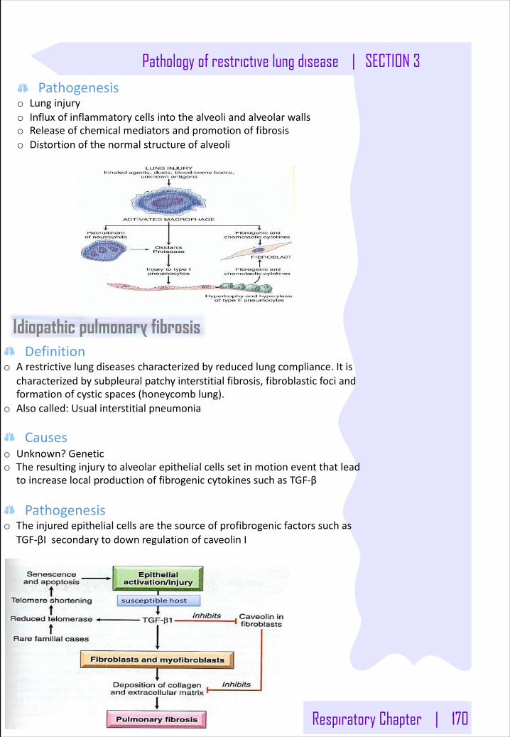
Respiratory Chapter | 170
Pathology of restrictive lung disease | SECTION 3
Pathogenesiso Lunginjuryo Influxofinflammatorycellsintothealveoliandalveolarwallso Releaseofchemicalmediatorsandpromotionoffibrosiso Distortionofthenormalstructureofalveoli
Idiopathic pulmonary fibrosisDefinition
o Arestrictivelungdiseasescharacterizedbyreducedlungcompliance.Itischaracterizedbysubpleuralpatchyinterstitialfibrosis,fibroblasticfociandformationofcysticspaces(honeycomblung).
o Alsocalled:Usualinterstitialpneumonia
Causeso Unknown?Genetico Theresultinginjurytoalveolarepithelialcellssetinmotioneventthatlead
toincreaselocalproductionoffibrogeniccytokinessuchasTGF-β
Pathogenesiso Theinjuredepithelialcellsarethesourceofprofibrogenicfactorssuchas
TGF-βIsecondarytodownregulationofcaveolinI
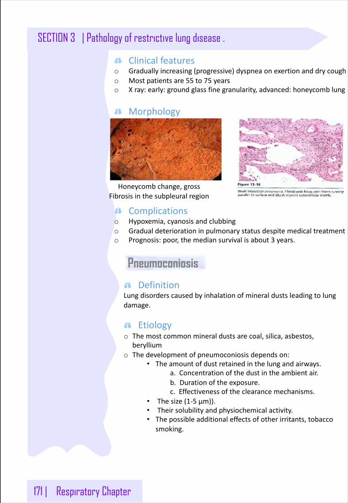
171 | Respiratory Chapter
SECTION 3 | Pathology of restrictive lung disease .
Clinicalfeatureso Graduallyincreasing(progressive) dyspnea onexertion anddrycougho Mostpatientsare55 to75 yearso Xray:early:groundglassfinegranularity,advanced:honeycomblung
Morphology
Complicationso Hypoxemia, cyanosisandclubbingo Gradualdeteriorationinpulmonarystatusdespitemedicaltreatmento Prognosis:poor,themediansurvivalisabout3 years.
Honeycombchange,grossFibrosisinthesubpleuralregion
Pneumoconiosis
DefinitionLungdisorderscausedbyinhalationofmineraldustsleadingtolungdamage.
Etiologyo Themostcommonmineraldustsarecoal,silica,asbestos,
berylliumo Thedevelopmentofpneumoconiosisdependson:
• Theamountofdustretainedinthelungandairways.a.Concentrationofthedustintheambientair.b.Durationoftheexposure.c.Effectivenessoftheclearancemechanisms.
• Thesize(1-5 μm)).• Theirsolubilityandphysiochemicalactivity.• Thepossibleadditionaleffectsofotherirritants,tobacco
smoking.
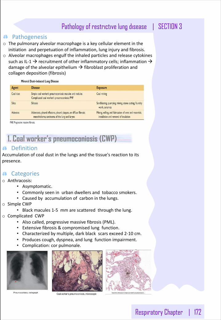
Respiratory Chapter | 172
Pathology of restrictive lung disease | SECTION 3Pathogenesis
o Thepulmonaryalveolarmacrophageisakeycellularelementintheinitiationandperpetuationofinflammation,lunginjuryandfibrosis.
o AlveolarmacrophagesengulftheinhaledparticlesandreleasecytokinessuchasIL-1à recruitmentofotherinflammatorycells;inflammationàdamageofthealveolarepitheliumà fibroblastproliferationandcollagendeposition(fibrosis)
1. Coal worker’s pneumoconiosis (CWP)Definition
Accumulationofcoaldustinthelungsandthetissue'sreactiontoitspresence.
Categorieso Anthracosis:
• Asymptomatic.• Commonlyseeninurbandwellersandtobaccosmokers.• Causedbyaccumulationofcarboninthelungs.
o SimpleCWP• Blackmacules1-5 mmarescatteredthroughthelung.
o ComplicatedCWP• Alsocalled,progressivemassivefibrosis(PML).• Extensivefibrosis&compromisedlungfunction.• Characterizedbymultiple,darkblackscarsexceed2-10 cm.• Producescough,dyspnea,andlungfunctionimpairment.• Complication:cor pulmonale.
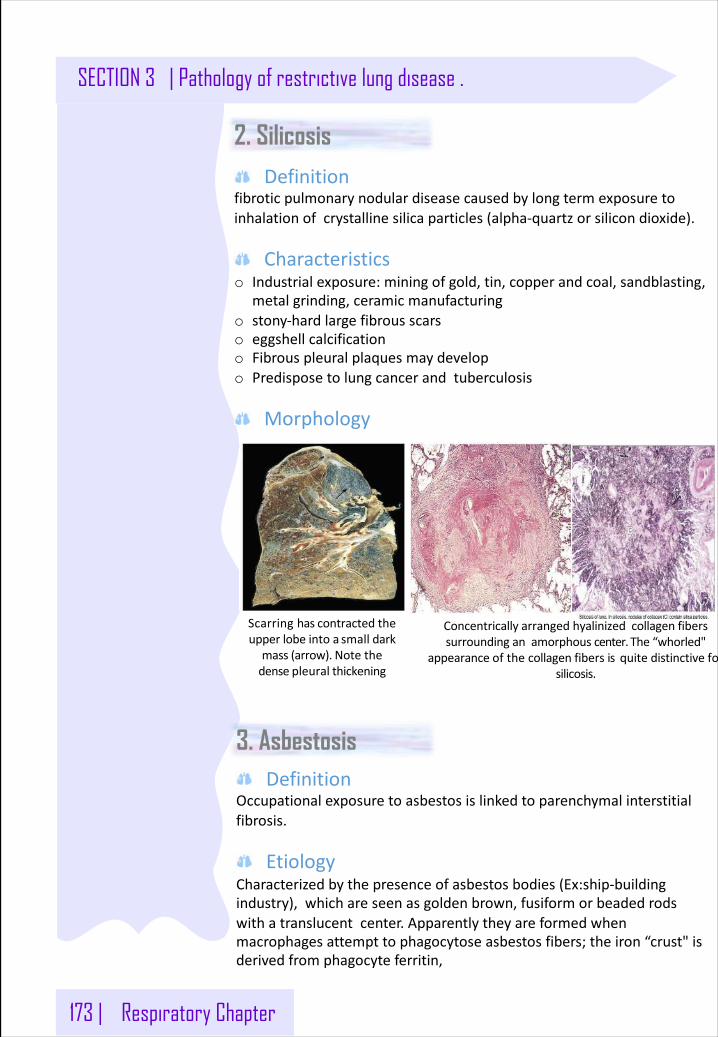
173 | Respiratory Chapter
SECTION 3 | Pathology of restrictive lung disease .
2. SilicosisDefinition
fibroticpulmonarynodulardiseasecausedbylongtermexposuretoinhalationofcrystallinesilicaparticles(alpha-quartzorsilicondioxide).
Characteristicso Industrialexposure:miningofgold,tin,copperandcoal,sandblasting,
metalgrinding,ceramicmanufacturingo stony-hardlargefibrousscarso eggshellcalcificationo Fibrouspleuralplaquesmaydevelopo Predisposetolungcancerandtuberculosis
Morphology
Scarringhascontractedtheupperlobeinto a small darkmass(arrow). Notethedensepleural thickening
Concentrically arrangedhyalinizedcollagenfiberssurroundinganamorphouscenter. The“whorled"
appearanceofthecollagenfibers isquite distinctiveforsilicosis.
3. AsbestosisDefinition
Occupationalexposuretoasbestosislinkedtoparenchymalinterstitialfibrosis.
EtiologyCharacterizedbythepresenceofasbestosbodies(Ex:ship-buildingindustry),whichareseenasgoldenbrown,fusiformorbeadedrodswithatranslucentcenter.Apparentlytheyareformedwhenmacrophagesattempttophagocytoseasbestosfibers;theiron“crust"isderivedfromphagocyteferritin,
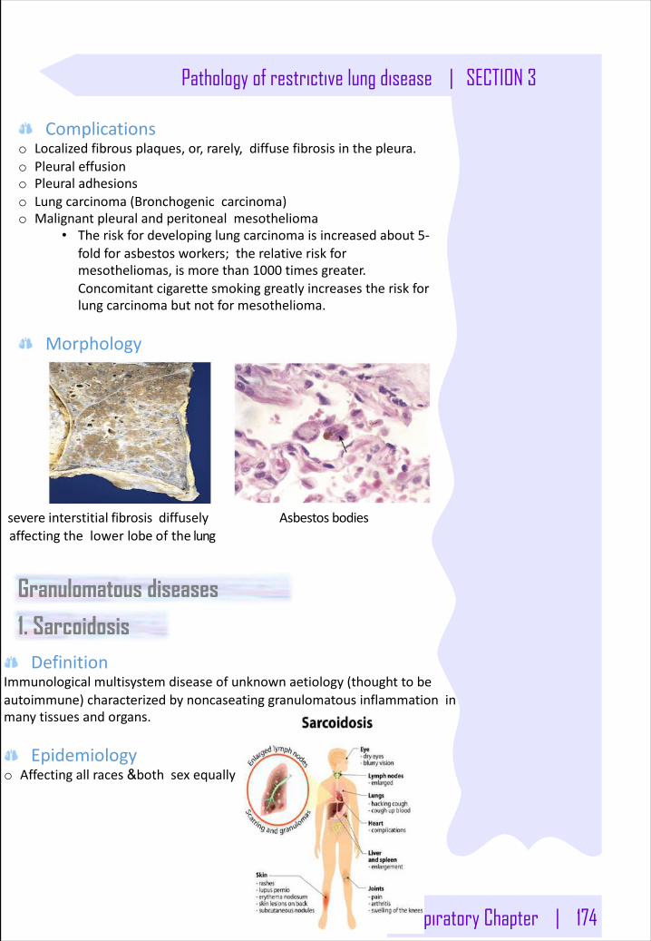
Respiratory Chapter | 174
Pathology of restrictive lung disease | SECTION 3
Complicationso Localizedfibrousplaques,or,rarely,diffusefibrosisinthepleura.o Pleuraleffusiono Pleuraladhesionso Lungcarcinoma(Bronchogeniccarcinoma)o Malignantpleuralandperitonealmesothelioma
• Theriskfordevelopinglungcarcinomaisincreasedabout5-foldforasbestosworkers;therelativeriskformesotheliomas,ismorethan1000 timesgreater.Concomitantcigarettesmokinggreatlyincreasestheriskforlungcarcinomabutnotformesothelioma.
Morphology
severeinterstitial fibrosisdiffuselyaffectingthelowerlobeofthe lung
Asbestosbodies
DefinitionImmunologicalmultisystemdiseaseofunknownaetiology(thoughttobeautoimmune)characterizedbynoncaseatinggranulomatousinflammationinmanytissuesandorgans.
Epidemiologyo Affectingallraces&bothsexequally
Granulomatous diseases
1. Sarcoidosis
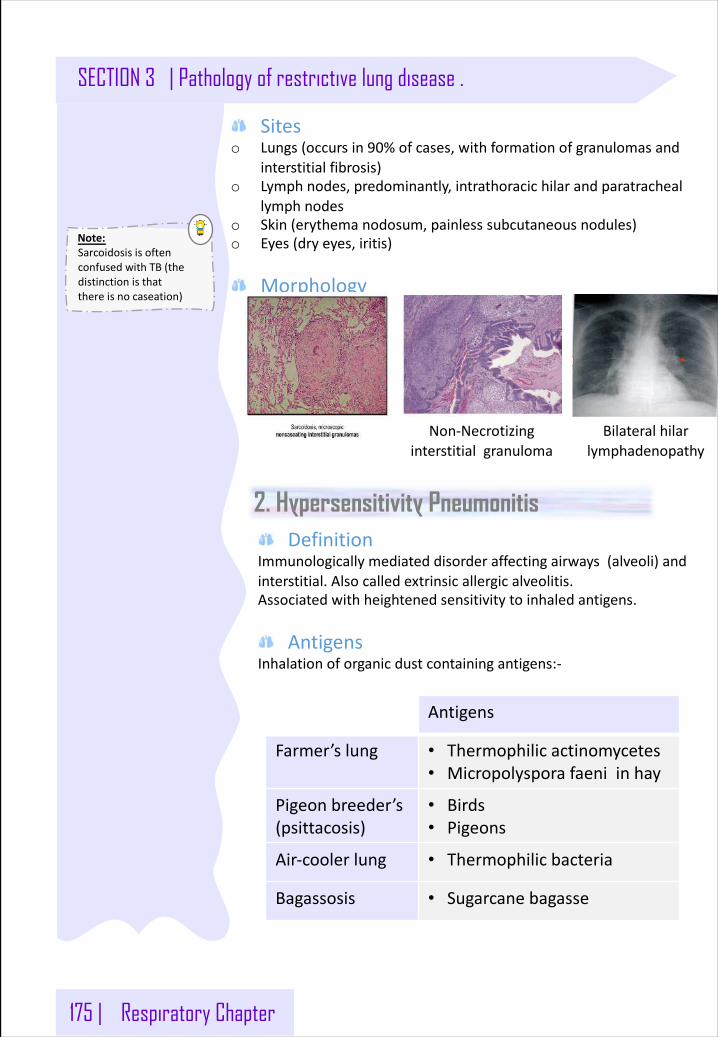
175 | Respiratory Chapter
SECTION 3 | Pathology of restrictive lung disease .
Siteso Lungs(occursin90%ofcases,withformationofgranulomasand
interstitialfibrosis)o Lymphnodes,predominantly,intrathoracichilarandparatracheal
lymphnodeso Skin(erythemanodosum,painlesssubcutaneousnodules)o Eyes(dryeyes,iritis)
Morphology
Non-Necrotizinginterstitialgranuloma
Bilateralhilarlymphadenopathy
2. Hypersensitivity PneumonitisDefinition
Immunologicallymediateddisorderaffectingairways(alveoli)andinterstitial.Alsocalledextrinsicallergicalveolitis.Associatedwithheightenedsensitivitytoinhaledantigens.
AntigensInhalationoforganicdustcontainingantigens:-
Antigens
Farmer’slung • Thermophilicactinomycetes• Micropolysporafaeniinhay
Pigeonbreeder’s(psittacosis)
• Birds• Pigeons
Air-coolerlung • Thermophilicbacteria
Bagassosis • Sugarcanebagasse
Note:SarcoidosisisoftenconfusedwithTB(thedistinctionisthatthereisnocaseation)
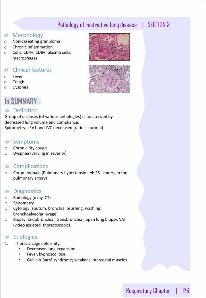
Respiratory Chapter | 176
Pathology of restrictive lung disease | SECTION 3Morphology
o Non-caseatinggranulomao Chronicinflammationo Cells:CD4+,CD8+,plasmacells,
macrophages
Clinicalfeatureso Fevero Cougho Dyspnea
In SUMMARYDefinition
Groupofdiseases(ofvariousaetiologies)characterizedbydecreasedlungvolumeandcompliance.Spirometry:LEV1 andLVCdecreased(ratioisnormal)
Symptomso Chronicdrycougho Dyspnea(varyinginseverity)
Complicationso Corpulmonale(Pulmonaryhypertensionà 25+mmHginthe
pulmonaryartery)
Diagnosticso Radiology(x-ray,CT)o Spirometryo Cytology(sputum,bronchialbrushing,washing,
bronchoalveolarlavage)o Biopsy:Endobronchial,transbronchial,openlungbiopsy,VAT
(videoassistedthoracoscopic)
Etiologies1. Thoraciccagedeformity:
• Decreasedlungexpansion• Fever,kyphoscoliosis• Guillain-Barrésyndrome;weakensintercostalmuscles
177 | Respiratory Chapter
SECTION 3 | Pathology of restrictive lung disease .
2. Idiopathicpulmonaryfibrosis:• Familial.• Affectingtheinterstitialofthelung/alveolarwall.• Donotaffecttheairspacesthemselves,butthetissuesaround
them.• Honeycombedlungduetoentrappedair(anthracosisà low
po2 highpco2)• Affectslowerpartofthelung• Bilateralperipheralreticulationà fibrosisshrinksthelung
(trappedairà dilatedalveoli)• Temporalheterogeneityfibroticdistribution• Histological:Bluestainindicatingprevalentconnectivetissue
(Masson'strichromestain)• Associatedwithusualinterstitialpneumonia• Pathogenesis:o Injuryaffectingmacrophagesleadingtocytokinerelease.o MUCB4 genemutation(chromosome9)mutationà higher
tendencytodevelopfibrosiso Shortertelomeres(Reducedgenesthatencodefortelomerase)
à shortercelllife(typeIpneumocytes)à senescence+apoptosisà whentheydietheysecrete:
• TGF-b1à fibrogenicà stimulatingfibroblastsandmyofibroblastsàcollagen
• LowCaveolin(inhibitsTGF-b1à therewillbenothingtocounteractit)o Treatment:
• Perfinidone(TGL-b1 antagonists)• Nentedanib (tyrosinekinaseantagonist)
3. RDS(Adult/Neonatal),DAD(diffusealveolardamage),HMPD(hyalinemembranepulmonarydisease):
• Veryseveredyspnoeaandhypoxia• Verysevereroadtrafficaccident,majorsurgery,aspirationof
gastriccontent,Csection,severeacutepancreatitis,hypovolemicshock,septicaemia
• 70%die• Effect:
o Edemao Inthelungà atelectasisà collapseo The50%whosurviveendupwithchronicpulmonaryfibrosiso Morphology:CTscanàWhitelungsyndromeduetofibrin
anddebrisà formahyalinemembranearoundthealveoli• RiskfactorsofNRDS(surfactantdeficiency):
o Prematureneonates(<36 weeks)o Multiplepregnancieso Maternaldiabeteso C-sectiono Amnioticfluidaspiration
Respiratory Chapter | 178
Pathology of restrictive lung disease | SECTION 3
4. Atypicalpneumonia• Couldleadtointerstitialpneumonitis• UsuallycausedbyInfluenzavirus• Oedemaintheinterstitialandchronicinflammatoryinfiltration• Inflammatorycellsarelymphocytes(viralinfection),andnot
neutrophils
5. Drugaddiction• Heroin• Amiodarone(antiarrhythmic)
6. Pneumoconiosis• Causedbyinhalingmineraldust1-5mmindiameter• Coalà coalworker'spneumoconiosis• Silicaà Silicosis(mostcommon)(silicainsandcontainsquartz,
whichisfibrogenic)• Buildingindustry• Concentricfibrosis• HigherriskofTBforunknownreasons• Asbestosà Asbestosis• Carcinogenic(mesothelioma)• Ship-buildingindustry• Asbestosfibers causesbleeding›formshemosiderin(prussian
bluestainshowsferruginousbodies)
7. Sarcoidosis:• Idiopathicbutnowthoughttobeautoimmune.• SymptomsincludeUveitis,arthritis,drynessofmouth,lackof
lacrimation,etc• OftenconfusedwithTB(thedistinctionisthatthereisno
caseation)
8. Hypersensitivitypneumonitis(extrinsicallergicalveolitis)• Sensitivitytoinhaledorganicmaterial• Illdefinedgranulomas(poorgranulomas)• Especiallyinupperlobes• Causes:
o Pigeonso Desertcoolero Incenseo Birdso Farmer'slungàmicrosporumà extrinsicallergic
alveolitis
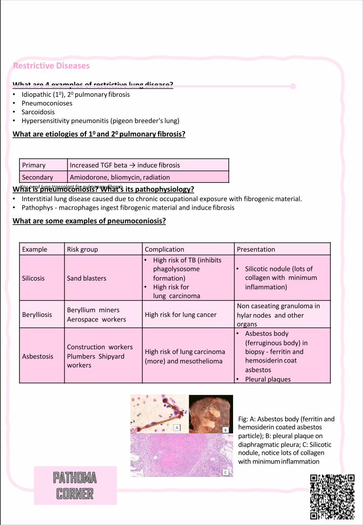
Restrictive Diseases
Whatare4 examplesofrestrictivelung disease?• Idiopathic(10),20 pulmonary fibrosis• Pneumoconioses• Sarcoidosis• Hypersensitivitypneumonitis(pigeonbreeder's lung)
Whatareetiologiesof10 and20 pulmonary fibrosis?
Whatispneumoconiosis?What'sits pathophysiology?• Interstitiallungdiseasecausedduetochronicoccupationalexposurewithfibrogenicmaterial.• Pathophys - macrophagesingestfibrogenic materialandinduce fibrosis
Whataresomeexamplesof pneumoconiosis?
Primary IncreasedTGFbeta→ induce fibrosis
Secondary Amiodorone,bliomycin, radiationYouneedlungtransplantforpulmonary fibrosis
Example Risk group Complication Presentation
Silicosis Sand blasters
• HighriskofTB(inhibitsphagolysosomeformation)
• Highriskforlungcarcinoma
• Silicoticnodule(lotsofcollagenwithminimuminflammation)
BerylliosisBerylliumminersAerospaceworkers
Highriskforlung cancerNoncaseatinggranulomainhylar nodesandotherorgans
AsbestosisConstructionworkersPlumbersShipyardworkers
Highriskoflung carcinoma(more)andmesothelioma
• Asbestosbody(ferruginousbody)inbiopsy- ferritinandhemosiderin coatasbestos
• Pleural plaques
Fig:A:Asbestosbody(ferritinandhemosiderincoatedasbestosparticle);B:pleuralplaqueondiaphragmaticpleura;C:Silicoticnodule,noticelotsofcollagenwithminimuminflammation
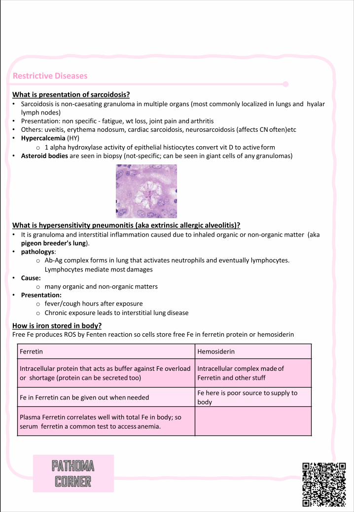
Restrictive Diseases
Whatispresentationof sarcoidosis?• Sarcoidosisisnon-caesating granulomainmultipleorgans(mostcommonlylocalizedinlungsandhyalar
lymph nodes)• Presentation:nonspecific- fatigue,wt loss,jointpainand arthritis• Others:uveitis,erythemanodosum,cardiacsarcoidosis,neurosarcoidosis (affectsCNoften)etc• Hypercalcemia(HY)
o 1 alphahydroxylaseactivityofepithelialhistiocytes convertvit Dtoactive form• Asteroidbodiesareseeninbiopsy(not-specific;canbeseeningiantcellsofany granulomas)
Whatishypersensitivitypneumonitis(akaextrinsicallergic alveolitis)?• Itisgranulomaandinterstitialinflammationcausedduetoinhaledorganicornon-organicmatter(aka
pigeonbreeder's lung).• pathologys:
o Ab-Agcomplexformsinlungthatactivatesneutrophilsandeventuallylymphocytes.Lymphocytesmediatemost damages
• Cause:o manyorganicandnon-organic matters
• Presentation:o fever/coughhoursafter exposureo Chronicexposureleadstointerstitiallung disease
Howisironstoredin body?FreeFeproducesROSbyFenten reactionsocellsstorefreeFeinferretin proteinor hemosiderin
Ferretin Hemosiderin
IntracellularproteinthatactsasbufferagainstFeoverloadorshortage(proteincanbesecreted too)
IntracellularcomplexmadeofFerretinandother stuff
FeinFerretincanbegivenoutwhenneededFehereispoorsourceto supply tobody
PlasmaFerretincorrelateswellwithtotalFeinbody;soserumferretinacommontesttoaccess anemia.
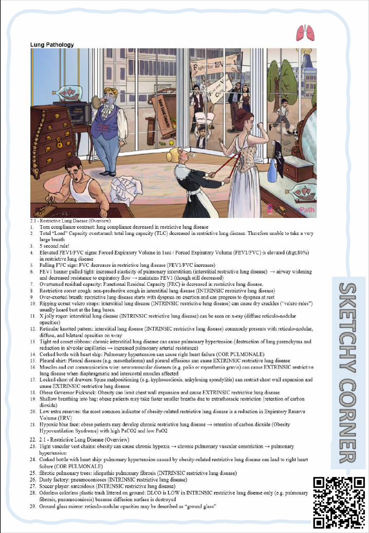
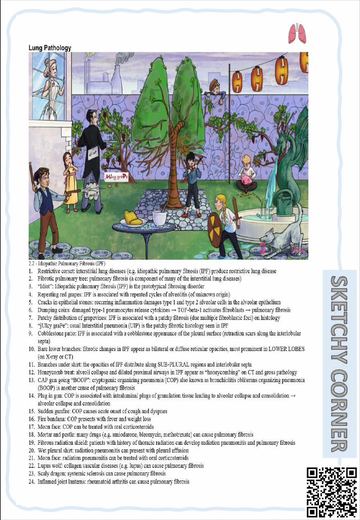
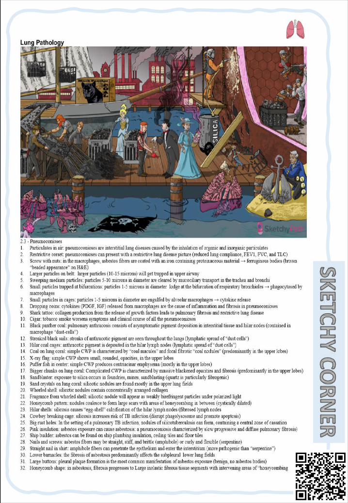
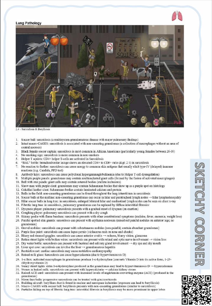
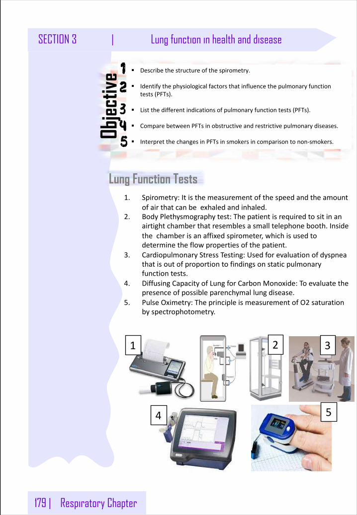
179 | Respiratory Chapter
SECTION 3 | Lung function in health and disease
Lung Function Tests
§ Describethestructureofthespirometry.
§ Identifythephysiologicalfactorsthatinfluencethepulmonaryfunctiontests(PFTs).
§ Listthedifferentindicationsofpulmonaryfunctiontests(PFTs).
§ ComparebetweenPFTsinobstructiveandrestrictivepulmonarydiseases.
§ InterpretthechangesinPFTsinsmokersincomparisontonon-smokers.
Objec
tive
1. Spirometry:Itisthemeasurementofthespeedandtheamountofairthatcanbeexhaledandinhaled.
2. BodyPlethysmographytest:Thepatientisrequiredtositinanairtightchamberthatresemblesasmalltelephonebooth.Insidethechamberisanaffixedspirometer,whichisusedtodeterminetheflowpropertiesofthepatient.
3. CardiopulmonaryStressTesting:Usedforevaluationofdyspneathatisoutofproportiontofindingsonstaticpulmonaryfunctiontests.
4. DiffusingCapacityofLungforCarbonMonoxide:Toevaluatethepresenceofpossibleparenchymallungdisease.
5. PulseOximetry:TheprincipleismeasurementofO2 saturationbyspectrophotometry.
1 2 3
4 5
Respiratory Chapter | 180
Lung function in health and disease | SECTION 3
SpirometrySpirometryisamethodtorecordvolumemovementofairintoandoutofthelungs.Spirometryisasimplemostcommonlyusedtestto:o Assessthelungperformance.o Measurethephysiologicalparameters:Lungvolumes,Capacities,
Flowrate.o Playacriticalroleinthediagnosis,differentiationandmanagement
ofrespiratorydiseases.o Differentiatebetweentheobstructiveandrestrictivelung
conditions.
Physiologicalconditionsaffectinglungfunctions:o Ageo Gendero Heighto Eighto Ethnicgroupo Pregnancy
GeneralIndicationsofSpirometry:o Symptoms:
• Dyspnea• Cough• Sputumproduction• Chestpain
o Signs:• Cyanosis• Clubbing• Chestdeformity• Diminishedchestexpansion• Hyperinflation• Diminishedbreathsounds• Prolongationofexpiratoryphase&crackles
o Arterialbloodgasanalysisshows: Hypoxemia,hypercapniao AbnormalchestXRay.
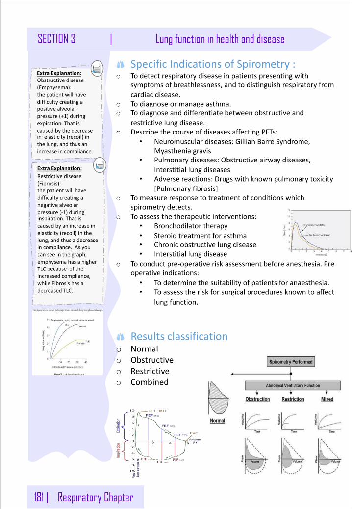
181 | Respiratory Chapter
SECTION 3 | Lung function in health and disease
SpecificIndicationsofSpirometry:o Todetectrespiratorydiseaseinpatientspresentingwith
symptomsofbreathlessness,andtodistinguishrespiratoryfromcardiacdisease.
o Todiagnoseormanageasthma.o Todiagnoseanddifferentiatebetweenobstructiveand
restrictivelungdisease.o DescribethecourseofdiseasesaffectingPFTs:
• Neuromusculardiseases:GillianBarreSyndrome,Myastheniagravis
• Pulmonarydiseases:Obstructiveairwaydiseases,Interstitiallungdiseases
• Adversereactions:Drugswithknownpulmonarytoxicity[Pulmonaryfibrosis]
o Tomeasureresponsetotreatmentofconditionswhichspirometrydetects.
o Toassessthetherapeuticinterventions:• Bronchodilatortherapy• Steroidtreatmentforasthma• Chronicobstructivelungdisease• Interstitiallungdisease
o Toconductpre-operativeriskassessmentbeforeanesthesia.Preoperativeindications:
• Todeterminethesuitabilityofpatientsforanaesthesia.• Toassesstheriskforsurgicalproceduresknowntoaffect
lung function.
Resultsclassificationo Normalo Obstructiveo Restrictiveo Combined
ExtraExplanation:Obstructivedisease(Emphysema):thepatientwillhavedifficultycreatingapositivealveolarpressure(+1)duringexpiration.Thatiscausedbythedecreaseinelasticity(recoil)inthelung,andthusanincreaseincompliance.
ExtraExplanation:Restrictivedisease(Fibrosis):thepatientwillhavedifficultycreatinganegativealveolarpressure(-1)duringinspiration.Thatiscausedbyanincreaseinelasticity(recoil)inthelung,andthusadecreaseincompliance.Asyoucanseeinthegraph,emphysemahasahigherTLCbecauseoftheincreasedcompliance,whileFibrosishasadecreasedTLC.
Respiratory Chapter | 182
Lung function in health and disease | SECTION 3
SpirometrySpirometryisamethodtorecordvolumemovementofairintoandoutofthelungs.Spirometryisasimplemostcommonlyusedtestto:o Assessthelungperformance.o Measurethephysiologicalparameters:Lungvolumes,
Capacities,Flowrate.o Playacriticalroleinthediagnosis,differentiationand
managementofrespiratorydiseases.o Differentiatebetweentheobstructiveandrestrictivelung
conditions.
Physiologicalconditionsaffectinglungfunctions:
o Ageo Gendero Heighto Eighto Ethnicgroupo Pregnancy
GeneralIndicationsofSpirometry:o Symptoms:
• Dyspnea• Cough• Sputumproduction• Chestpain
o Signs:• Cyanosis• Clubbing• Chestdeformity• Diminishedchestexpansion• Hyperinflation• Diminishedbreathsounds• Prolongationofexpiratoryphase&crackles
o Arterialbloodgasanalysisshows: Hypoxemia,hypercapniao AbnormalchestXRay.
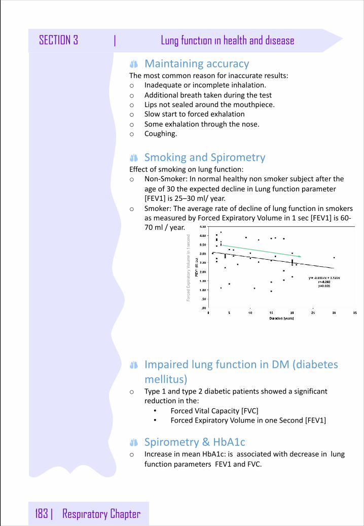
183 | Respiratory Chapter
SECTION 3 | Lung function in health and disease
MaintainingaccuracyThemostcommonreasonforinaccurateresults:o Inadequateorincompleteinhalation.o Additionalbreathtakenduringthetesto Lipsnotsealedaroundthemouthpiece.o Slowstarttoforcedexhalationo Someexhalationthroughthenose.o Coughing.
SmokingandSpirometryEffectofsmokingonlungfunction:o Non-Smoker:Innormalhealthynonsmokersubjectafterthe
ageof30 theexpecteddeclineinLungfunctionparameter[FEV1]is25–30 ml/year.
o Smoker:TheaveragerateofdeclineoflungfunctioninsmokersasmeasuredbyForcedExpiratoryVolumein1 sec[FEV1]is60-70 ml/year.
ImpairedlungfunctioninDM(diabetesmellitus)
o Type1 andtype2 diabeticpatientsshowedasignificantreductioninthe:
• ForcedVitalCapacity[FVC]• ForcedExpiratoryVolumeinoneSecond[FEV1]
Spirometry&HbA1co IncreaseinmeanHbA1c:isassociatedwithdecreaseinlung
functionparametersFEV1 andFVC.
Forc
ed E
xpir
ator
y Vo
lum
e in
1se
cond
Respiratory Chapter | 184
Lung function in health and disease | SECTION 3
DiagnosisofCOPDo Symptoms:cough,sputum,dyspneao Exposuretoriskfactors:tobacco,occupation,
indoor/outdoorpollution.è Inthepresentofthese,wecanuseSpirometryto
confirmthediagnose
SpirometryandCemENT INDUSTRY:Lungfunctionparameters:o FVCo FEV1o FEF25-75%o PEFweresignificantlydecreasedincementmillworkers
comparedtotheirmatchedcontrols.
SpirometryandWELDINGINDUSTRy :Lungfunctionparameterso FVCo FEV1o PEFweresignificantlyimpairedinweldingworkers
comparedtotheirmatchedcontrols.
SpirometryandOILSPILLY:Lungfunctionparameterso FVCo FEV1o FEF25-75%wereimpairedinsubjectsexposedtocrude
oilspillinseawater.
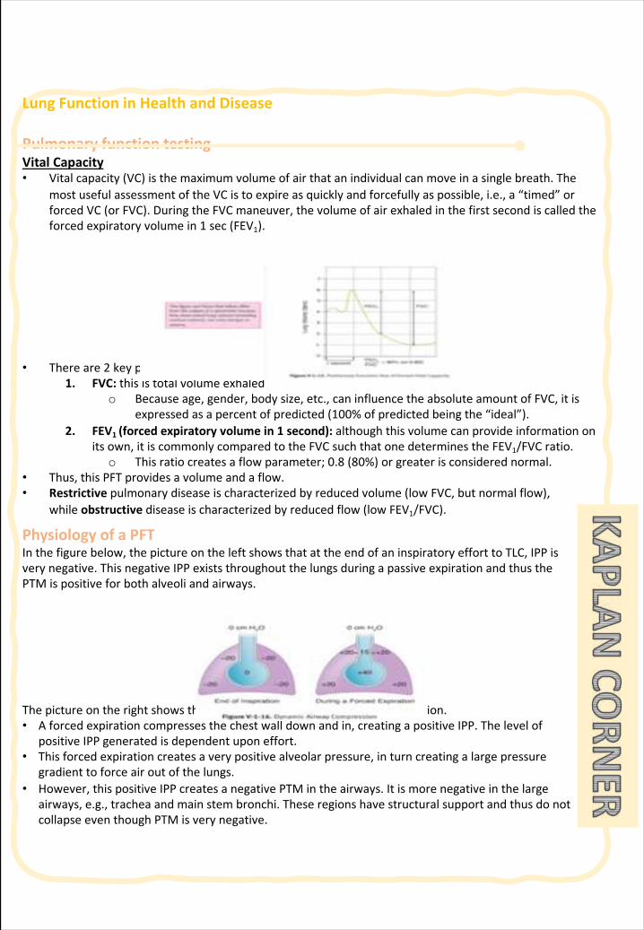
LungFunctioninHealthandDisease
PulmonaryfunctiontestingVitalCapacity• Vitalcapacity(VC)isthemaximumvolumeofairthatanindividualcanmoveinasinglebreath.The
mostusefulassessmentoftheVCistoexpireasquicklyandforcefullyaspossible,i.e.,a“timed”orforcedVC(orFVC).DuringtheFVCmaneuver,thevolumeofairexhaledinthefirstsecondiscalledtheforcedexpiratoryvolumein1 sec(FEV1).
• Thereare2 keypiecesofdatafromaPFTinvolvingthemeasurementofFVC:1. FVC:thisistotalvolumeexhaled
o Becauseage,gender,bodysize,etc.,caninfluencetheabsoluteamountofFVC,itisexpressedasapercentofpredicted(100%ofpredictedbeingthe“ideal”).
2. FEV1 (forcedexpiratoryvolumein1 second):althoughthisvolumecanprovideinformationonitsown,itiscommonlycomparedtotheFVCsuchthatonedeterminestheFEV1/FVCratio.
o Thisratiocreatesaflowparameter;0.8 (80%)orgreaterisconsiderednormal.• Thus,thisPFTprovidesavolumeandaflow.• Restrictive pulmonarydiseaseischaracterizedbyreducedvolume(lowFVC,butnormalflow),
whileobstructive diseaseischaracterizedbyreducedflow(lowFEV1/FVC).
PhysiologyofaPFTInthefigurebelow,thepictureontheleftshowsthatattheendofaninspiratoryefforttoTLC,IPPisverynegative.ThisnegativeIPPexiststhroughoutthelungsduringapassiveexpirationandthusthePTMispositiveforbothalveoliandairways.
Thepictureontherightshowsthesituationduringamaximalforcedexpiration.• Aforcedexpirationcompressesthechestwalldownandin,creatingapositiveIPP.Thelevelof
positiveIPPgeneratedisdependentuponeffort.• Thisforcedexpirationcreatesaverypositivealveolarpressure,inturncreatingalargepressure
gradienttoforceairoutofthelungs.• However,thispositiveIPPcreatesanegativePTMintheairways.Itismorenegativeinthelarge
airways,e.g.,tracheaandmainstembronchi.TheseregionshavestructuralsupportandthusdonotcollapseeventhoughPTMisverynegative.
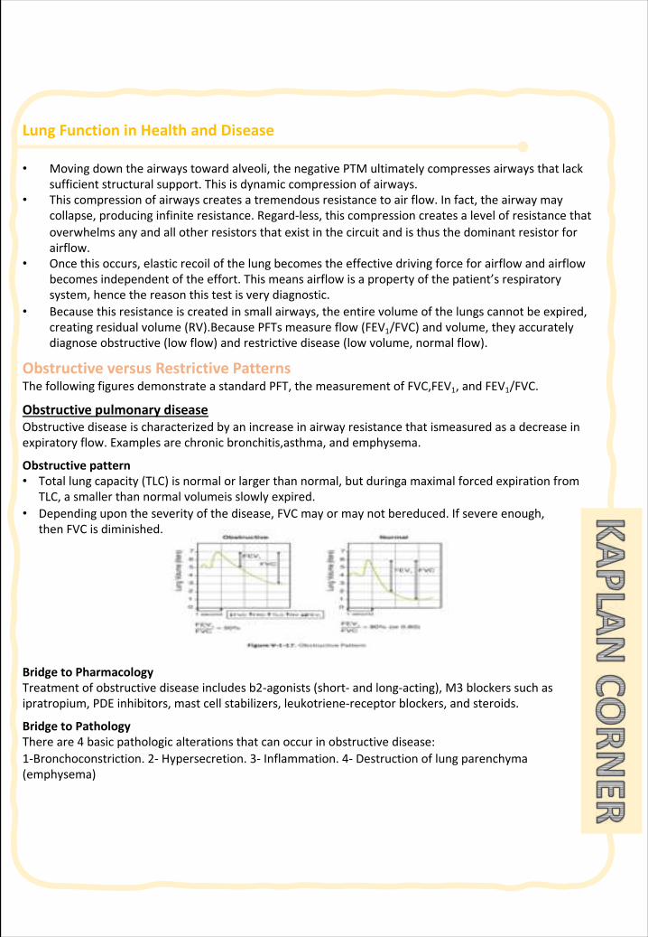
LungFunctioninHealthandDisease
• Movingdowntheairwaystowardalveoli,thenegativePTMultimatelycompressesairwaysthatlacksufficientstructuralsupport.Thisisdynamiccompressionofairways.
• Thiscompressionofairwayscreatesatremendousresistancetoairflow.Infact,theairwaymaycollapse,producinginfiniteresistance.Regard-less,thiscompressioncreatesalevelofresistancethatoverwhelmsanyandallotherresistorsthatexistinthecircuitandisthusthedominantresistorforairflow.
• Oncethisoccurs,elasticrecoilofthelungbecomestheeffectivedrivingforceforairflowandairflowbecomesindependentoftheeffort.Thismeansairflowisapropertyofthepatient’srespiratorysystem,hencethereasonthistestisverydiagnostic.
• Becausethisresistanceiscreatedinsmallairways,theentirevolumeofthelungscannotbeexpired,creatingresidualvolume(RV).BecausePFTsmeasureflow(FEV1/FVC)andvolume,theyaccuratelydiagnoseobstructive(lowflow)andrestrictivedisease(lowvolume,normalflow).
ObstructiveversusRestrictivePatternsThefollowingfiguresdemonstrateastandardPFT,themeasurementofFVC,FEV1,andFEV1/FVC.
ObstructivepulmonarydiseaseObstructivediseaseischaracterizedbyanincreaseinairwayresistancethatismeasured asadecreaseinexpiratoryflow.Examplesarechronicbronchitis,asthma,andemphysema.
Obstructivepattern• Totallungcapacity(TLC)isnormalorlargerthannormal,butduringa maximalforcedexpirationfrom
TLC,asmallerthannormalvolumeis slowlyexpired.• Dependingupontheseverityofthedisease,FVCmayormaynotbereduced.Ifsevereenough,
thenFVCisdiminished.
BridgetoPharmacologyTreatmentofobstructivediseaseincludesb2-agonists(short- andlong-acting),M3 blockerssuchasipratropium,PDEinhibitors,mastcellstabilizers,leukotriene-receptorblockers,andsteroids.
BridgetoPathologyThereare4 basicpathologicalterationsthatcanoccurinobstructivedisease:1-Bronchoconstriction.2- Hypersecretion.3- Inflammation.4- Destructionoflungparenchyma(emphysema)

LungFunctioninHealthandDisease
RestrictivepulmonarydiseaseRestrictivepulmonarydiseaseischaracterizedbyanincreaseinelasticrecoil—adecreaseinlungcompliance—whichismeasuredasadecreaseinalllungvolumes.Reducedvitalcapacitywithlowlungvolumesaretheindicatorsofrestrictivepulmonarydiseases.ExamplesareARDSandinterstitiallungdis-easessuchassarcoidosisandidiopathicpulmonaryfibrosis(IPF).
Restrictivepattern• TLCissmallerthannormal,butduringamaximalforcedexpirationfromTLC,thesmallervolumeis
expiredquicklyandmorecompletelythaninanormalpattern.• Therefore,eventhoughFEV1 isalsoreduced,theFEV1/FVCisoftenincreased.• However,thecriticaldistinctionislowFVCwithlowFRCandRV.
• FVCisalwaysdecreasedwhenpulmonaryfunctionissignificantlycompro-mised.A decreaseinFEV1/FVCratioisevidenceofanobstructivepattern.Anormalor increasedFEV1/FVCratioisevidenceofarestrictivepattern,butalowTLCis diagnosticofrestrictivelungdisease.
Flow–VolumeLoopsTheinstantaneousrelationshipbetweenflow(liters/sec)andlungvolumeisusefulindeterminingwhetherobstructiveorrestrictivelungdiseaseispresent.Intheloopshownbelow,expirationstartsattotallungcapacityandcontinuestoresidualvolume.ThewidthoftheloopistheFVC.
Variable ObstructivePattern(e.g.,Emphysema)
RestrictivePattern(e.g.,Fibrosis)
TLC ↑ ↓↓
FEV1 ↓↓ ↓
FVC ↓ ↓↓
FEV1/FVC ↓ ↑ornormal
Peakflow ↓ ↓
FRC ↑ ↓
RV ↑ ↓
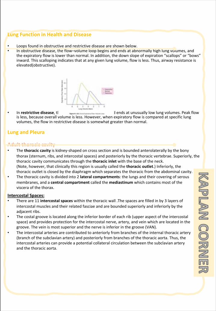
LungFunctioninHealthandDisease
• Loopsfoundinobstructiveandrestrictivediseaseareshownbelow.• Inobstructivedisease,theflow–volumeloopbeginsandendsatabnormallyhighlungvolumes,and
theexpiratoryflowislowerthannormal.Inaddition,thedownslopeofexpiration“scallops”or“bows”inward.Thisscallopingindicatesthatatanygivenlungvolume,flowisless.Thus,airwayresistanceiselevated(obstructive).
• Inrestrictivedisease,theflow–volumeloopbeginsandendsatunusuallylowlungvolumes.Peakflowisless,becauseoverallvolumeisless.However,whenexpiratoryflowiscomparedatspecificlungvolumes,theflowinrestrictivediseaseissomewhatgreaterthannormal.
LungandPleura
Adultthoraciccavity• Thethoraciccavityiskidney-shapedoncrosssectionandisboundedanterolaterally bythebony
thorax(sternum,ribs,andintercostalspaces)andposteriorlybythethoracicvertebrae.Superiorly,thethoraciccavitycommunicatesthroughthethoracicinletwiththebaseoftheneck.(Note,however,thatclinicallythisregionisusuallycalledthethoracicoutlet.)Inferiorly,thethoracicoutletisclosedbythediaphragmwhichseparatesthethoracicfromtheabdominalcavity.
• Thethoraciccavityisdividedinto2 lateralcompartments:thelungsandtheircoveringofserousmembranes,andacentralcompartmentcalledthemediastinum whichcontainsmostofthevisceraofthethorax.
IntercostalSpaces:• Thereare11 intercostalspaceswithinthethoracicwall.Thespacesarefilledinby3 layersof
intercostalmusclesandtheirrelatedfasciaeandareboundedsuperiorlyandinferiorlybytheadjacentribs.
• Thecostalgrooveislocatedalongtheinferiorborderofeachrib(upperaspectoftheintercostalspace)andprovidesprotectionfortheintercostalnerve,artery,andveinwhicharelocatedinthegroove.Theveinismostsuperiorandthenerveisinferiorinthegroove(VAN).
• Theintercostalarteriesarecontributedtoanteriorlyfrombranchesoftheinternalthoracicartery(branchofthesubclavianartery)andposteriorlyfrombranchesofthethoracicaorta.Thus,theintercostalarteriescanprovideapotentialcollateralcirculationbetweenthesubclavianarteryandthethoracicaorta.
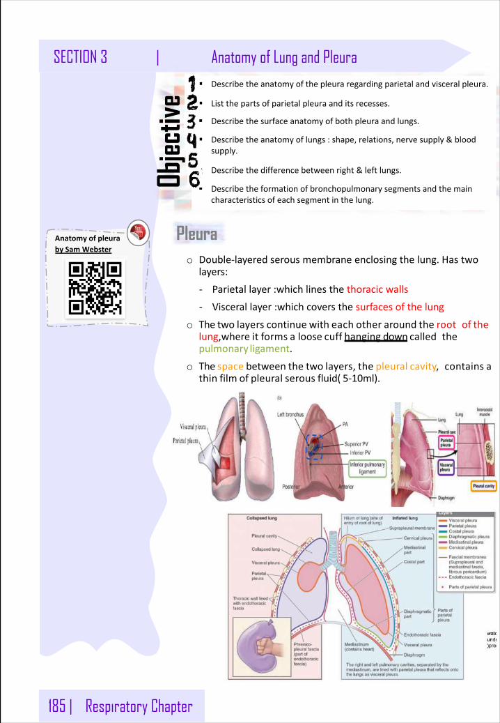
185 | Respiratory Chapter
SECTION 3 | Anatomy of Lung and Pleura
o Double-layeredserousmembraneenclosingthelung.Hastwolayers:
- Parietallayer : whichlinesthethoracicwalls
- Viscerallayer : whichcoversthesurfacesofthelung
o The two layers continue with each other around the rootof thelung,where it forms a loose cuff hanging down calledthepulmonary ligament.
o The space between the two layers, the pleural cavity,contains athin film of pleural serous fluid( 5-10ml).
Objec
tive
§ Describetheanatomyofthepleuraregardingparietalandvisceralpleura.
§ Listthepartsofparietalpleuraanditsrecesses.
§ Describethesurfaceanatomyofbothpleuraandlungs.
§ Describetheanatomyoflungs:shape,relations,nervesupply&bloodsupply.
§ Describethedifferencebetweenright&leftlungs.
§ Describetheformationofbronchopulmonarysegmentsandthemaincharacteristicsofeachsegmentinthelung.
PleuraAnatomyofpleurabySamWebster
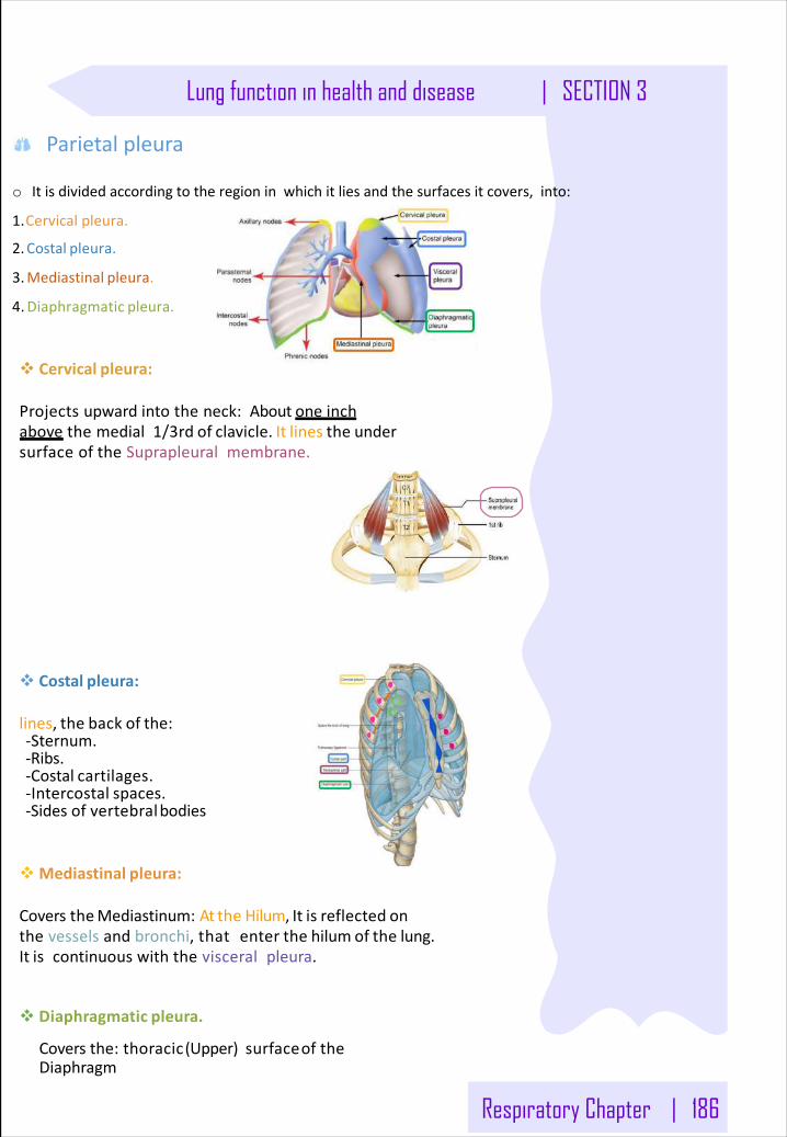
Respiratory Chapter | 186
Lung function in health and disease | SECTION 3
o Itisdividedaccordingtotheregioninwhichitliesandthesurfacesitcovers,into:
1.Cervicalpleura.
2.Costal pleura.
3.Mediastinal pleura.
4. Diaphragmatic pleura.
v Cervical pleura:
Projectsupwardintotheneck:Aboutoneinchabove themedial1/3rd of clavicle. It lines the undersurfaceoftheSuprapleural membrane.
v Costal pleura: lines, the back of the:-Sternum.-Ribs.-Costal cartilages.-Intercostal spaces.-Sidesofvertebralbodies
vMediastinal pleura: CoverstheMediastinum: At the Hilum, It is reflected onthevesselsandbronchi,thatenter the hilum of the lung.It iscontinuouswiththevisceralpleura.
v Diaphragmatic pleura.
Coversthe:thoracic(Upper)surfaceoftheDiaphragm
Parietalpleura
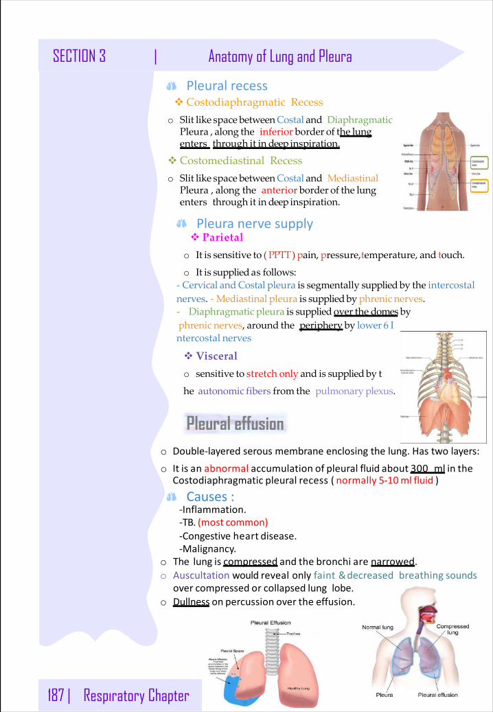
187 | Respiratory Chapter
SECTION 3 | Anatomy of Lung and Pleura
v Parietalo It is sensitive to ( PPTT) pain, pressure,temperature, and touch.
o It is supplied as follows:- Cervical and Costal pleura is segmentally supplied by the intercostalnerves. - Mediastinal pleura is supplied by phrenic nerves.- Diaphragmatic pleura is supplied over the domes byphrenic nerves, around the periphery by lower 6 Intercostal nerves
v Visceralo sensitive to stretch only and is supplied by t
he autonomic fibers from the pulmonary plexus.
v Costodiaphragmatic Recesso Slit like space between Costal and Diaphragmatic
Pleura , along the inferior border of the lungenters through it in deep inspiration.
v Costomediastinal Recesso Slit like space between Costal and Mediastinal
Pleura , along the anterior border of the lungenters through it in deep inspiration.
Pleuralrecess
Pleuranervesupply
o Double-layeredserousmembraneenclosingthelung.Hastwolayers:
o It is an abnormal accumulation of pleural fluid about 300 ml in theCostodiaphragmatic pleural recess ( normally 5-10ml fluid )
-Inflammation.-TB.(most common)-Congestiveheart disease.-Malignancy.
o The lung is compressed and the bronchi are narrowed.o Auscultationwouldrevealonlyfaint&decreasedbreathing sounds
over compressed or collapsed lunglobe.o Dullness on percussion over the effusion.
Pleural effusion
Causes:
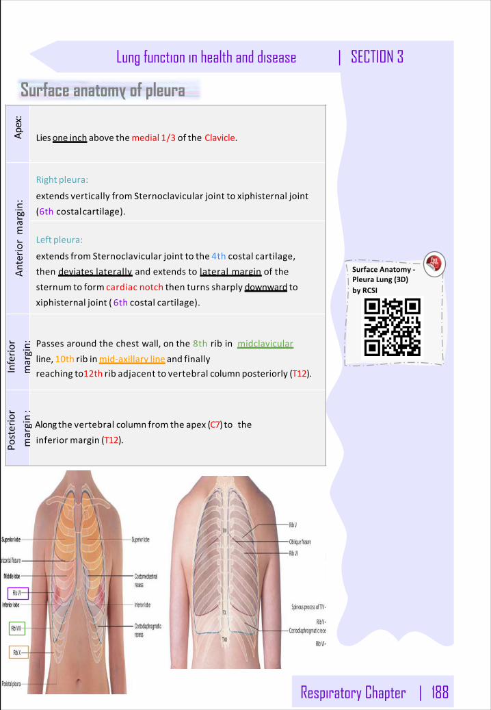
Respiratory Chapter | 188
Lung function in health and disease | SECTION 3
Apex:
Lies one inch above themedial 1/3 of the Clavicle.
Anterio
rmargin:
Right pleura:
extends vertically from Sternoclavicular joint to xiphisternal joint(6thcostalcartilage).
Left pleura:
extends from Sternoclavicular joint to the 4th costal cartilage,thendeviateslaterally andextendstolateralmargin ofthesternum to form cardiac notch then turns sharply downward toxiphisternal joint ( 6th costal cartilage).
Inferio
rmargin: Passesaroundthechestwall,onthe8thribinmidclavicular
line, 10th rib inmid-axillary line and finallyreaching to12th rib adjacent to vertebral column posteriorly (T12).
Posterior
margin:
Along the vertebral column from the apex (C7) totheinferior margin (T12).
Surface anatomy of pleura
SurfaceAnatomy-PleuraLung(3D)byRCSI
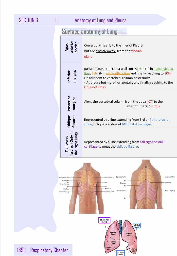
189 | Respiratory Chapter
SECTION 3 | Anatomy of Lung and Pleura
Apex,
anterio
rborder Correspond nearly to the lines of Pleura
but are slightly away fromthemedianplane
Inferio
rmargin:
passes around the chest wall , on the 6th rib in midclavicularline , 8th rib in mid-axillary line and finally reaching to 10thrib adjacent to vertebral column posteriorly.- As pleura butmore horizontally and finally reaching to the(T10)not(T12)
Posterior
margin:
Along the vertebral column from the apex ( C7) to theinferiormargin(T10)
Oblique
fissure:
Represented by a line extending from 3rd or 4th thoracicspine, obliquely ending at 6th costal cartilage.
Transverse
fissure:(Onlyin
therightlun
g)
Represented by a line extending from 4th right costalcartilage tomeet the oblique fissure.
Surface anatomy of Lung
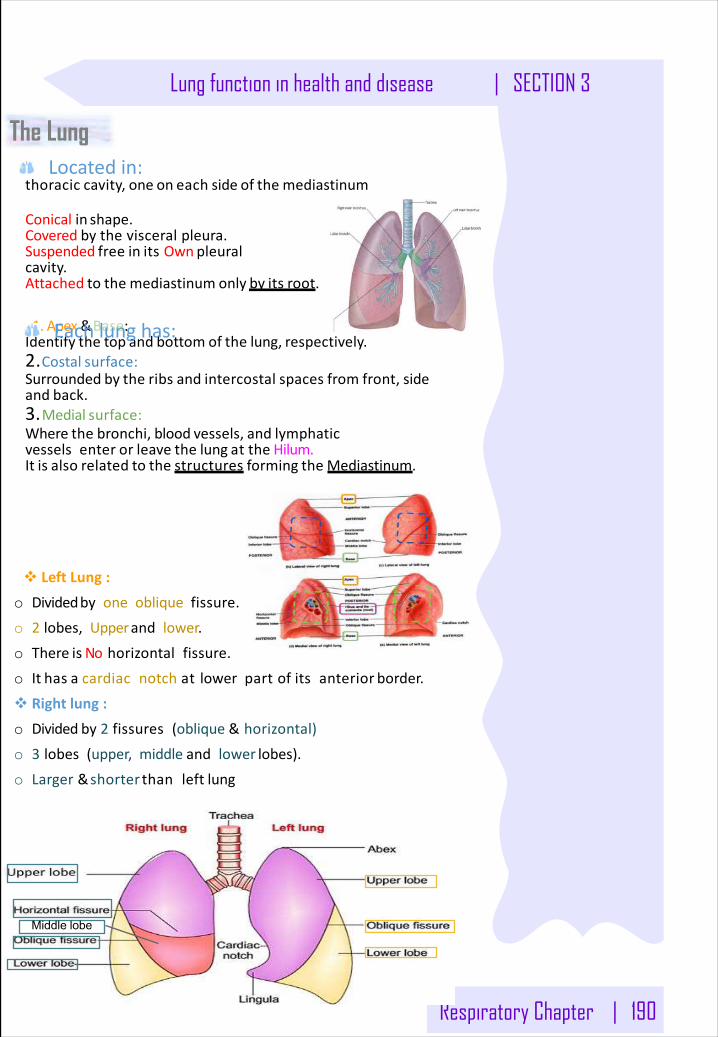
Respiratory Chapter | 190
Lung function in health and disease | SECTION 3
The Lung
thoracic cavity, one on each side of the mediastinum
Conicalin shape.Coveredbythevisceralpleura.Suspended free in its Own pleuralcavity.Attached to the mediastinum only by its root.
1.Apex&Base:Identify the top and bottom of the lung, respectively.2.Costal surface:Surrounded by the ribs and intercostal spaces from front, sideand back.3.Medial surface:Where the bronchi, blood vessels, and lymphaticvesselsenter or leave the lung at the Hilum.It is also related to the structures forming the Mediastinum.
Middle lobe
v LeftLung:
o Dividedby oneobliquefissure.
o 2 lobes, Upperand lower.
o There is No horizontalfissure.
o It has a cardiacnotchatlowerpartofitsanterior border.
v Rightlung: o Dividedby2 fissures(oblique&horizontal)
o 3 lobes(upper, middleand lower lobes).
o Larger&shorter thanleft lung
Locatedin:
Eachlunghas:
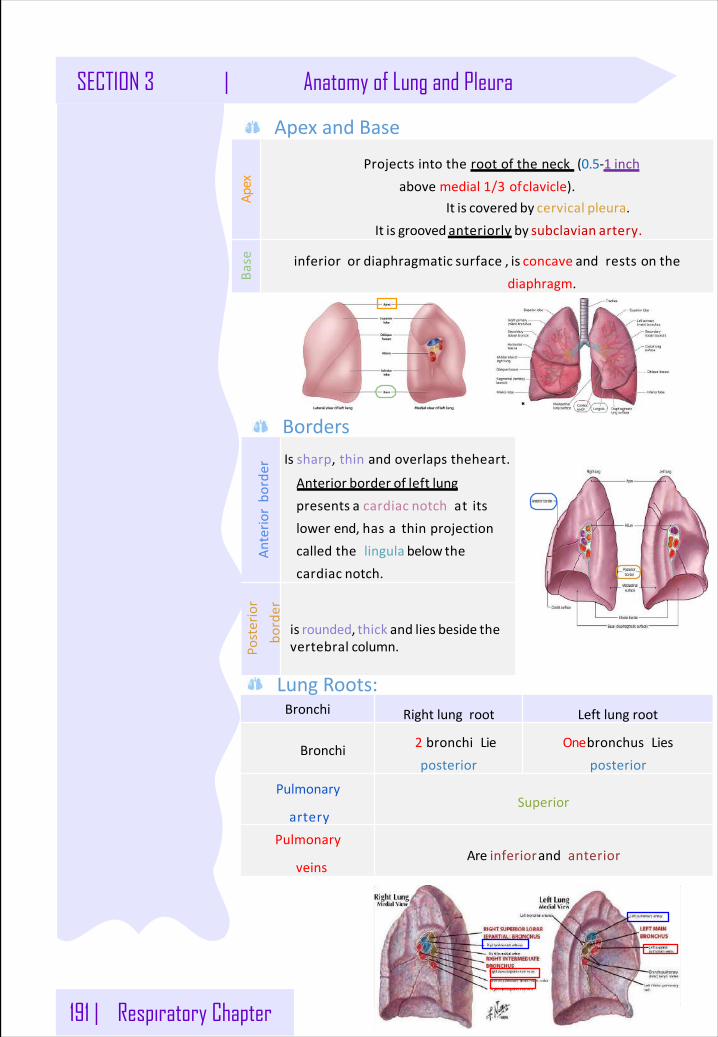
191 | Respiratory Chapter
SECTION 3 | Anatomy of Lung and Pleura
Anterio
rbo
rder
Is sharp,thinandoverlapstheheart.
Anterior border of left lungpresents a cardiac notch atitslowerend,hasathinprojectioncalledthelingula below thecardiac notch.
Posterior
border
is rounded, thick and lies beside thevertebral column.
Apex
Projectsintotherootoftheneck (0.5-1 inchabovemedial1/3 ofclavicle).
It is covered by cervical pleura.It is grooved anteriorly by subclavian artery.
Base inferior or diaphragmatic surface , is concave andrestsonthe
diaphragm.
Borders
ApexandBase
Bronchi Rightlungroot Leftlungroot
Bronchi 2 bronchiLieposterior
OnebronchusLiesposterior
Pulmonary
arterySuperior
Pulmonary
veinsAreinferiorandanterior
LungRoots:
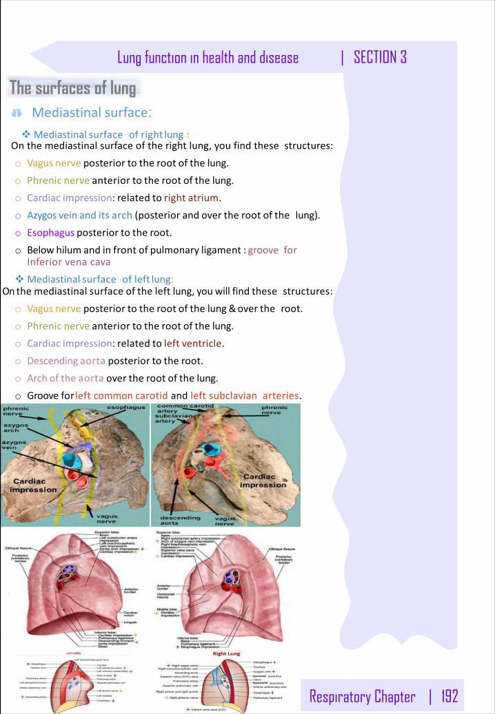
Respiratory Chapter | 192
Lung function in health and disease | SECTION 3
The surfaces of lungMediastinalsurface :
vMediastinal surfaceofright lung :Onthemediastinalsurfaceoftherightlung,youfindthesestructures:
o Vagus nerve posterior to the root of the lung.
o Phrenic nerve anterior to the root of the lung.
o Cardiac impression: related to right atrium.
o Azygos vein and its arch (posterior and over the root of thelung).
o Esophagus posterior to the root.
o Below hilum and in front of pulmonary ligament : grooveforInferiorvenacava
vMediastinal surfaceofleft lung:On the mediastinal surface of the left lung, youwill find thesestructures:
o Vagus nerve posterior to the root of the lung &over theroot.
o Phrenic nerve anterior to the root of the lung.
o Cardiac impression: related to left ventricle.
o Descending aorta posterior to the root.
o Arch of the aorta over the root of the lung.
o Grooveforleftcommoncarotidandleftsubclavianarteries.
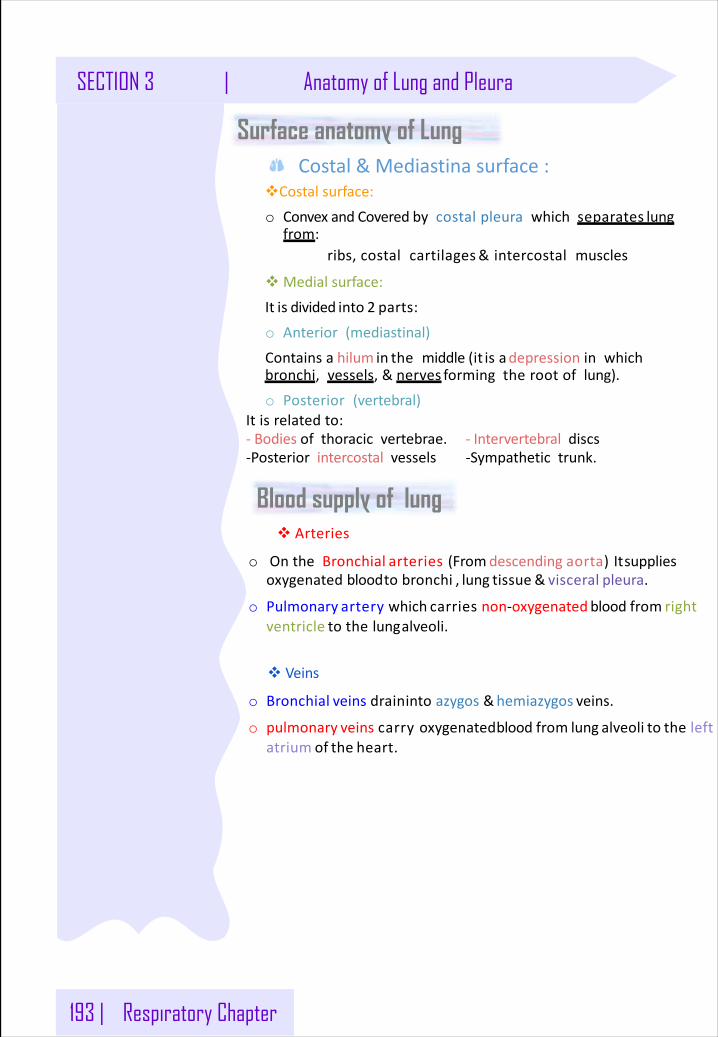
193 | Respiratory Chapter
SECTION 3 | Anatomy of Lung and Pleura
Surface anatomy of Lung Costal&Mediastinasurface:
vCostalsurface:
o ConvexandCoveredbycostalpleurawhichseparates lungfrom:
ribs,costalcartilages& intercostalmuscles
vMedialsurface: It is divided into 2 parts:
o Anterior(mediastinal)
Contains a hilum in themiddle(it is adepressioninwhichbronchi,vessels,&nerves forming therootoflung).
o Posterior(vertebral)Itisrelatedto:- Bodiesofthoracicvertebrae.- Intervertebral discs-Posteriorintercostal vessels-Sympathetictrunk.
v Arteries
o Onthe Bronchial arteries(Fromdescending aorta)Itsuppliesoxygenatedbloodto bronchi , lung tissue & visceral pleura.
o Pulmonary artery which carries non-oxygenated blood from rightventricle to thelungalveoli.
v Veins
o Bronchial veins drainintoazygos&hemiazygos veins.
o pulmonary veins carryoxygenatedblood from lung alveoli to the leftatrium of the heart.
Blood supply of lung
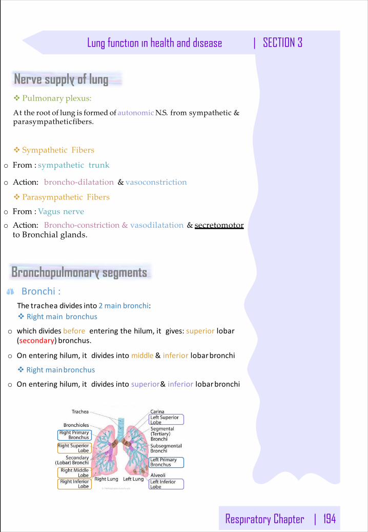
Respiratory Chapter | 194
Lung function in health and disease | SECTION 3
Nerve supply of lung
The trachea divides into 2 main bronchi:v Rightmainbronchus
o whichdividesbefore enteringthehilum,itgives:superiorlobar(secondary) bronchus.
o Onenteringhilum,itdividesintomiddle& inferiorlobarbronchi
v Rightmainbronchus
o Onenteringhilum,itdividesintosuperior& inferiorlobarbronchi
v Pulmonary plexus:
At the root of lung is formed of autonomic N.S. from sympathetic & parasympatheticfibers.
v Sympathetic Fibers
o From : sympathetic trunk
o Action: broncho-dilatation & vasoconstriction
v Parasympathetic Fibers
o From : Vagus nerveo Action: Broncho-constriction & vasodilatation & secretomotor
to Bronchial glands.
Bronchopulmonary segmentsBronchi:
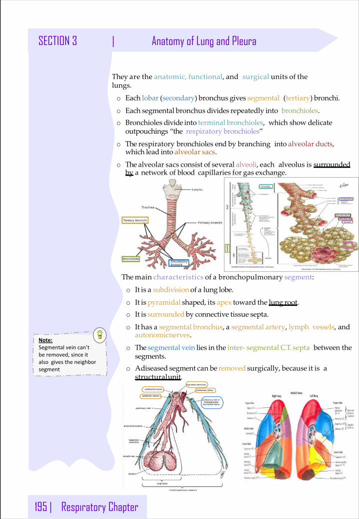
195 | Respiratory Chapter
SECTION 3 | Anatomy of Lung and Pleura
They are the anatomic, functional, and surgical units of thelungs.
o Each lobar (secondary) bronchus gives segmental (tertiary) bronchi.
o Each segmental bronchus divides repeatedly into bronchioles.o Bronchioles divide into terminal bronchioles, which show delicate
outpouchings “the respiratory bronchioles”
o The respiratory bronchioles end by branching into alveolar ducts,which lead into alveolar sacs.
o The alveolar sacs consist of several alveoli, each alveolus is surrounded by a network of blood capillaries for gas exchange.
The main characteristics of a bronchopulmonary segment:
o It is a subdivision of a lung lobe.
o It is pyramidal shaped, its apex toward the lung root.o It is surrounded by connective tissue septa.
o It has a segmental bronchus, a segmental artery, lymph vessels, and autonomicnerves.
o The segmental vein lies in the inter- segmental C.T.septa between thesegments.
o Adiseased segment can be removed surgically, because it is a structuralunit.
Note:Segmentalveincan’tberemoved,sinceitalsogivestheneighborsegment
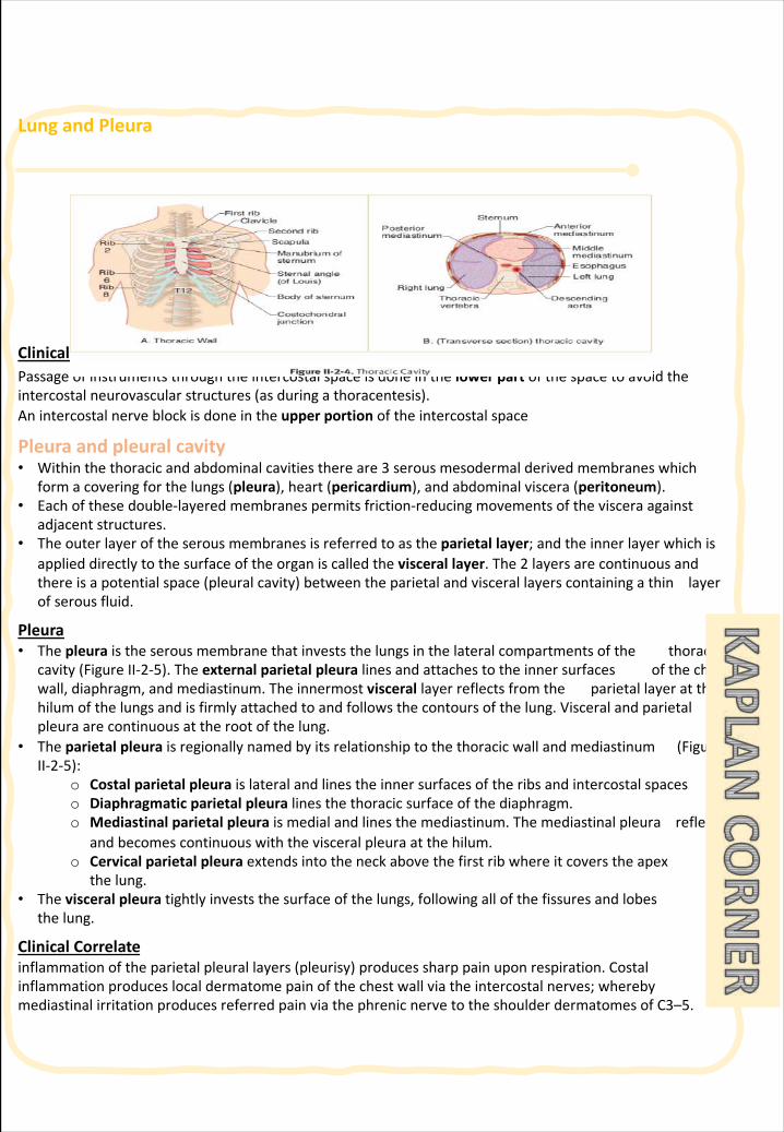
LungandPleura
ClinicalCorrelatePassageofinstrumentsthroughtheintercostalspaceisdoneinthelowerpartofthespacetoavoidtheintercostalneurovascularstructures(asduringathoracentesis). Anintercostalnerveblockisdoneintheupperportionoftheintercostalspace
Pleuraandpleuralcavity• Withinthethoracicandabdominalcavitiesthereare3 serousmesodermalderivedmembraneswhich
formacoveringforthelungs(pleura),heart(pericardium),andabdominalviscera(peritoneum).• Eachofthesedouble-layeredmembranespermitsfriction-reducingmovementsofthevisceraagainst
adjacentstructures.• Theouterlayeroftheserousmembranesisreferredtoastheparietallayer;andtheinnerlayerwhichis
applieddirectlytothesurfaceoftheorganiscalledtheviscerallayer.The2 layersarecontinuousandthereisapotentialspace(pleuralcavity)betweentheparietalandviscerallayerscontainingathinlayerofserousfluid.
Pleura• Thepleura istheserousmembranethatinveststhelungsinthelateralcompartmentsofthethoracic
cavity(FigureII-2-5).Theexternalparietalpleuralinesandattachestotheinnersurfacesofthechestwall,diaphragm,andmediastinum.Theinnermostvisceral layerreflectsfromtheparietallayeratthehilumofthelungsandisfirmlyattachedtoandfollowsthecontoursofthelung.Visceralandparietalpleuraarecontinuousattherootofthelung.
• Theparietalpleuraisregionallynamedbyitsrelationshiptothethoracicwallandmediastinum(FigureII-2-5):
o Costalparietalpleuraislateralandlinestheinnersurfacesoftheribsandintercostalspaceso Diaphragmaticparietalpleuralinesthethoracicsurfaceofthediaphragm.o Mediastinalparietalpleuraismedialandlinesthemediastinum.Themediastinalpleurareflects
andbecomescontinuouswiththevisceralpleuraatthehilum.o Cervicalparietalpleuraextendsintotheneckabovethefirstribwhereitcoverstheapexof
thelung.• Thevisceralpleuratightlyinveststhesurfaceofthelungs,followingallofthefissuresandlobesof
thelung.
ClinicalCorrelateinflammationoftheparietalpleurallayers(pleurisy)producessharppainuponrespiration.Costalinflammationproduceslocaldermatomepainofthechestwallviatheintercostalnerves;wherebymediastinalirritationproducesreferredpainviathephrenicnervetotheshoulderdermatomesofC3–5.
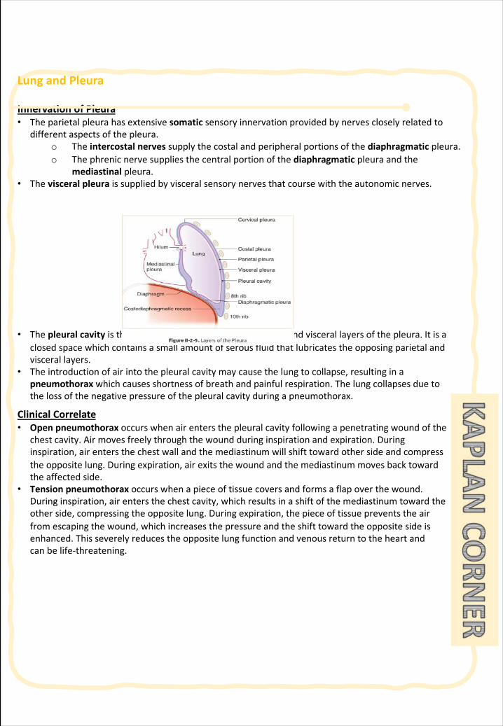
LungandPleura
InnervationofPleura• Theparietalpleurahasextensivesomatic sensoryinnervationprovidedbynervescloselyrelatedto
differentaspectsofthepleura.o Theintercostalnervessupplythecostalandperipheralportionsofthediaphragmatic pleura.o Thephrenicnervesuppliesthecentralportionofthediaphragmatic pleuraandthe
mediastinal pleura.• Thevisceralpleuraissuppliedbyvisceralsensorynervesthatcoursewiththeautonomicnerves.
• Thepleuralcavityisthe potentialspacebetweentheparietalandviscerallayersofthepleura. Itisaclosedspacewhichcontainsasmallamountofserousfluidthatlubricatestheopposingparietalandviscerallayers.
• The introductionofairintothepleuralcavitymaycausethelungtocollapse,resultinginapneumothorax whichcausesshortnessofbreathandpainfulrespiration. Thelungcollapsesduetothelossofthenegativepressureofthepleuralcavityduringapneumothorax.
ClinicalCorrelate• Openpneumothorax occurswhenairentersthepleuralcavityfollowingapenetratingwoundofthe
chestcavity.Airmovesfreelythroughthewoundduringinspirationandexpiration.Duringinspiration,airentersthechestwallandthemediastinumwillshifttowardothersideandcompresstheoppositelung.Duringexpiration,airexitsthewoundandthemediastinummovesbacktowardtheaffectedside.
• Tensionpneumothoraxoccurswhenapieceoftissuecoversandformsaflapoverthewound.Duringinspiration,airentersthechestcavity,whichresultsinashiftofthemediastinumtowardtheotherside,compressingtheoppositelung.Duringexpiration,thepieceoftissuepreventstheairfromescapingthewound,whichincreasesthepressureandtheshifttowardtheoppositesideisenhanced.Thisseverelyreducestheoppositelungfunctionandvenousreturntotheheartandcanbelife-threatening.
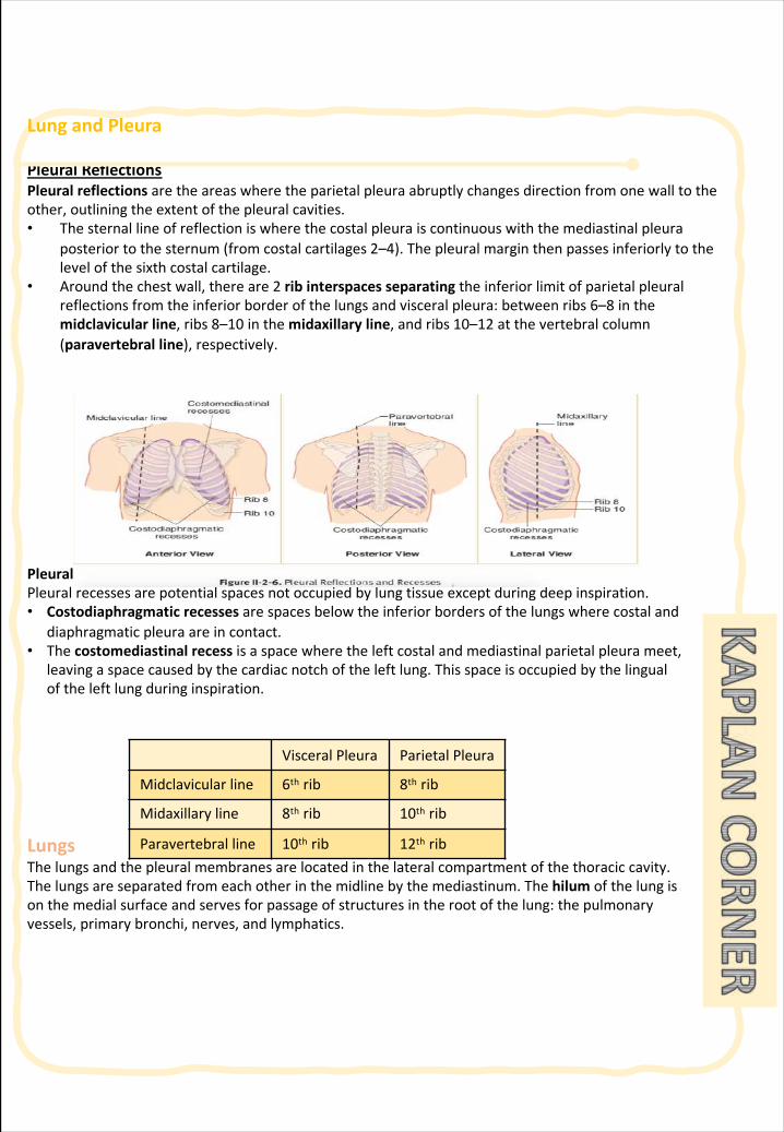
LungandPleura
PleuralReflectionsPleuralreflectionsaretheareaswheretheparietalpleuraabruptlychangesdirectionfromonewalltotheother,outliningtheextentofthepleuralcavities.• Thesternallineofreflectioniswherethecostalpleuraiscontinuouswiththemediastinalpleura
posteriortothesternum(fromcostalcartilages2–4).Thepleuralmarginthenpassesinferiorlytothelevelofthesixthcostalcartilage.
• Aroundthechestwall,thereare2 ribinterspacesseparatingtheinferiorlimitofparietalpleuralreflectionsfromtheinferiorborderofthelungsandvisceralpleura:betweenribs6–8 inthemidclavicular line,ribs8–10 inthemidaxillary line,andribs10–12 atthevertebralcolumn(paravertebralline),respectively.
PleuralRecessesPleuralrecessesarepotentialspacesnotoccupiedbylungtissueexceptduringdeepinspiration. • Costodiaphragmatic recesses arespacesbelowtheinferiorbordersofthelungswherecostaland
diaphragmaticpleuraareincontact.• Thecostomediastinal recess isaspacewheretheleftcostalandmediastinalparietalpleurameet,
leavingaspacecausedbythecardiacnotchoftheleftlung.Thisspaceisoccupiedbythelingualoftheleftlungduringinspiration.
LungsThelungsandthepleuralmembranesarelocatedinthelateralcompartmentofthethoraciccavity.Thelungsareseparatedfromeachotherinthemidlinebythemediastinum.Thehilum ofthelungisonthemedialsurfaceandservesforpassageofstructuresintherootofthelung:thepulmonaryvessels,primarybronchi,nerves,andlymphatics.
VisceralPleura ParietalPleura
Midclavicularline 6th rib 8th rib
Midaxillaryline 8th rib 10th rib
Paravertebralline 10th rib 12th rib
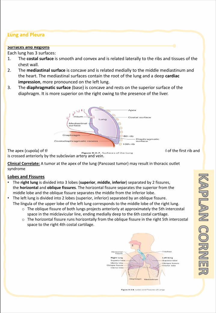
LungandPleura
SurfacesandRegionsEachlunghas3 surfaces:1. Thecostalsurfaceissmoothandconvexandisrelatedlaterallytotheribsandtissuesofthe
chestwall.2. Themediastinalsurfaceisconcaveandisrelatedmediallytothemiddlemediastinumand
theheart.Themediastinalsurfacescontaintherootofthelungandadeepcardiacimpression,morepronouncedontheleftlung.
3. Thediaphragmaticsurface(base)isconcaveandrestsonthesuperiorsurfaceofthediaphragm.Itismoresuperiorontherightowingtothepresenceoftheliver.
Theapex(cupola)ofthelungprojectssuperiorlyintotherootoftheneckabovethelevelofthefirstribandiscrossedanteriorlybythesubclavianarteryandvein.
ClinicalCorrelate: Atumorattheapexofthelung(Pancoasttumor)mayresultinthoracicoutletsyndrome
LobesandFissures• Therightlungisdividedinto3 lobes(superior,middle,inferior)separatedby2 fissures,
thehorizontal andoblique fissures.Thehorizontalfissureseparatesthesuperiorfromthemiddlelobeandtheobliquefissureseparatesthemiddlefromtheinferiorlobe.
• Theleftlungisdividedinto2 lobes(superior,inferior)separatedbyanobliquefissure.Thelingula oftheupperlobeoftheleftlungcorrespondstothemiddlelobeoftherightlung.
o Theobliquefissureofbothlungsprojectsanteriorlyatapproximatelythe5thintercostalspaceinthemidclavicularline,endingmediallydeeptothe6thcostalcartilage.
o Thehorizontalfissurerunshorizontallyfromtheobliquefissureintheright5thintercostalspacetotheright4thcostalcartilage.

LungandPleura
ClinicalCorrelate• Thesuperiorlobeoftherightlung projectsanteriorlyonthechestwallabovethe4thribandthe
middlelobeprojectsanteriorlybelow the4thrib.• Asmallportionoftheinferiorlobeofbothlungsprojectsbelow the6thribanteriorlybutprimarily
projectstotheposteriorchestwall.• Tolistentobreathsoundsofthesuperiorlobesoftherightandleftlungs,thestethoscopeisplacedon
thesuperiorareaoftheanteriorchestwall(abovethe4thrib fortherightlung).• Forbreathsoundsfromthemiddlelobeoftherightlung,thestethoscopeisplacedontheanterior
chestwallinferiortothe4thribandmediallytowardthesternum.• Fortheinferiorlobesofbothlungs,breathsoundsareprimarilyheardontheposterior chestwall.• Aspirationofaforeignbodywillmoreoftenentertherightprimarybronchus,whichisshorter,wider,
andmoreverticalthantheleftprimarybronchus.Whentheindividualisvertical,theforeignbodyusuallyfallsintotheposteriorbasalsegmentoftherightinferiorlobe.
LymphaticSystem • Thelymphaticsystem consistsofanextensivenetworkoflymphcapillaries,vessels,andnodesthatdrain
extracellularfluidfrommostofthebodytissuesandorgans. Thelymphflowwillreturntothebloodvenoussystemby2 majorlymphaticvessels, therightlymphaticductandthethoracicductontheleft(FigureII-2-10A). These2 vesselsdrainintothejunctionoftheinternaljugularandthesubclavianveinsontheirrespectivesides
o Thethoracicduct carriesalllymphaticdrainagefromthebodybelowthediaphragmandontheleftsideofthetrunkandheadabovethediaphragm(FigureII-2-10B).
o Therightlymphaticduct drainslymphflowfromtherightheadandneckandtherightsideofthetrunkabovethediaphragm(FigureII2-10B).
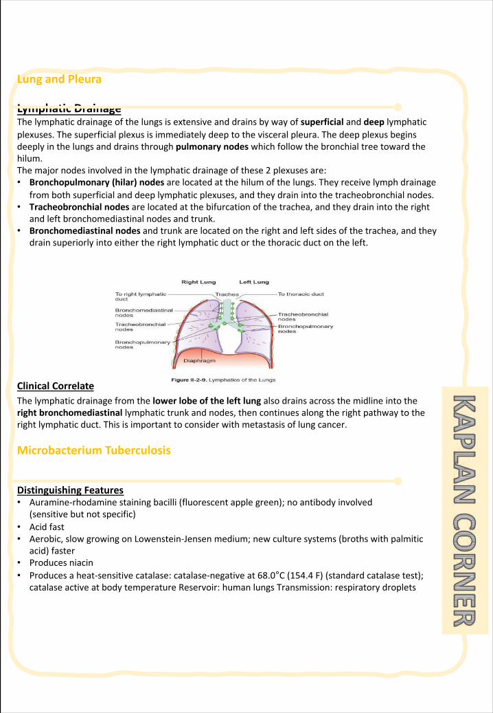
LungandPleura
LymphaticDrainage Thelymphaticdrainageofthelungsis extensiveanddrainsbywayofsuperficial anddeep lymphaticplexuses. Thesuperficialplexusisimmediatelydeeptothevisceralpleura. Thedeepplexusbeginsdeeplyinthelungsanddrainsthroughpulmonarynodeswhichfollowthebronchialtreetowardthehilum.The majornodesinvolvedinthelymphaticdrainageofthese2 plexusesare:• Bronchopulmonary(hilar)nodesarelocatedatthehilumofthelungs.Theyreceivelymphdrainage
frombothsuperficialanddeeplymphaticplexuses,andtheydrainintothetracheobronchialnodes.• Tracheobronchialnodesarelocatedatthebifurcationofthetrachea,andtheydrainintotheright
andleftbronchomediastinal nodesandtrunk.• Bronchomediastinal nodesandtrunkarelocatedontherightandleftsidesofthetrachea,andthey
drainsuperiorlyintoeithertherightlymphaticductorthethoracicductontheleft.
ClinicalCorrelateThelymphaticdrainagefromthelowerlobeoftheleftlungalsodrainsacrossthemidlineintotherightbronchomediastinal lymphatictrunkandnodes,thencontinuesalongtherightpathwaytotherightlymphaticduct.Thisisimportanttoconsiderwithmetastasisoflungcancer.
Microbacterium Tuberculosis
DistinguishingFeatures• Auramine-rhodaminestainingbacilli(fluorescentapplegreen);noantibodyinvolved
(sensitivebutnotspecific)• Acidfast• Aerobic,slowgrowingonLowenstein-Jensenmedium;newculture systems(brothswithpalmitic
acid)faster• Producesniacin• Producesaheat-sensitivecatalase:catalase-negativeat68.0°C(154.4 F)(standardcatalasetest);
catalaseactiveatbodytemperatureReservoir:humanlungsTransmission:respiratorydroplets
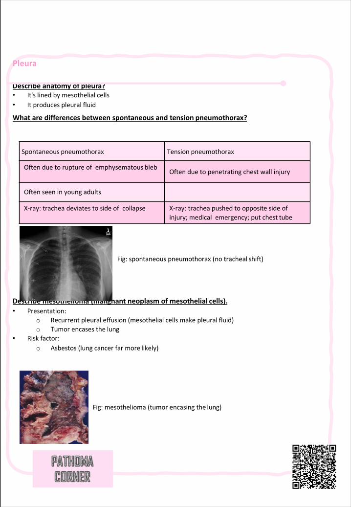
Pleura
Describeanatomyof pleura?• It'slinedbymesothelial cells• Itproducespleural fluid
Whataredifferencesbetweenspontaneousandtension pneumothorax?
Describemesothelioma(malignantneoplasmofmesothelial cells).• Presentation:
o Recurrentpleuraleffusion(mesothelialcellsmakepleural fluid)o Tumorencasesthe lung
• Risk factor:o Asbestos(lungcancerfarmore likely)
Spontaneous pneumothorax Tension pneumothorax
Oftenduetoruptureofemphysematous blebOftenduetopenetratingchestwallinjury
Oftenseeninyoungadults
X-ray: tracheadeviatestosideofcollapse X-ray: tracheapushedtooppositesideofinjury;medicalemergency;putchest tube
Fig:spontaneouspneumothorax(notracheal shift)
Fig:mesothelioma(tumorencasingthe lung)
196 | Respiratory Chapter
SECTION 3 | PATHOLOGY , MICOBIOLOGY AND IMMUNOLOGY of TB
§ Definetuberculosis&Knowtheepidemiologyoftuberculosis(TB).
§ ListthediseasescausedbyMycobacteria&conditionsassociatedwithincreasedriskofTuberculosisandfactorspredisposingtoextensionoftheinfection
§ RecognizethemorphologyofMycobacteriaanditsspecialstain(theZiehl-Neelsen)aswellasthemorphologyofgranulomasinTB(tubercles).
§ InregardstoMycobacteriallunginfection:Compareandcontrastthefollowinginrelationtotheirgrossandhistologiclungpathology:1.Primarytuberculosis(includeadefinitionoftheGhon complex).2.Secondaryorreactivationtuberculosis.3.Miliary tuberculosis.
§ Listorgansotherthanlungthatarecommonlyaffectedbytuberculosis.
§ Knowthebasisanduseoftuberculinskin(Mantoux)test.
§ Listthecommonclinicalpresentationoftuberculosis&Listthecomplicationandprognosisoftuberculosis.
Microbiology:
§ Definetuberculosisasachronicdiseasemainlyaffectingtherespiratorysystem,ANDRecognizeroughlytheepidemiologyoftuberculosisworldwideandinthekingdomofSaudiArabia
§ Understandthemethodsoftransmissionoftuberculosisandthepeopleatrisk,ANDUnderstandthepathogenesisoftuberculosis.
§ Knowthecausativeagentsandtheircharacteristicandclassificationandmethodsofdetection.
§ Differentiatebetweenprimaryandsecondarytuberculosisandtheclinicalfeaturesofeach.
§ Understandanddescribeandexplainthemethodsoftuberculintest,tuberculinskintest(TST)anditsdifferentresults,ANDKnowtheradiologicalandlaboratorydiagnosticmethods.
§ Knowthechemotherapeuticandothermethodsofmanagementoftuberculosiscases.
§ Describethemethodsofpreventionandcontroloftuberculosis.
IMMUNOLOGY:
§ ToknowhowM.tuberculosisinfectioniscontractedanditsinitialencounterwiththeimmunesystem
§ TounderstanddelayedtypeofhypersensitivityreactionagainstM.tuberculosis
§ ToidentifypossibleoutcomesoftheinfectionwithM.tuberculosisinimmunocompetentandimmuno-compromisedhosts.
§ Toknowthebasisofinterferongammareleaseassayanditspotentialtodetectlatenttuberculosis.
§ ToidentifythebasisoftuberculintestanditsimportanceingaugingimmunityagainstM.tuberculosis
Objec
tive
Objec
tive
Objec
tive
TBpathogenesis|Infectiousdiseases|NCLEX-RN|KhanAcademybykhanacademymedicine
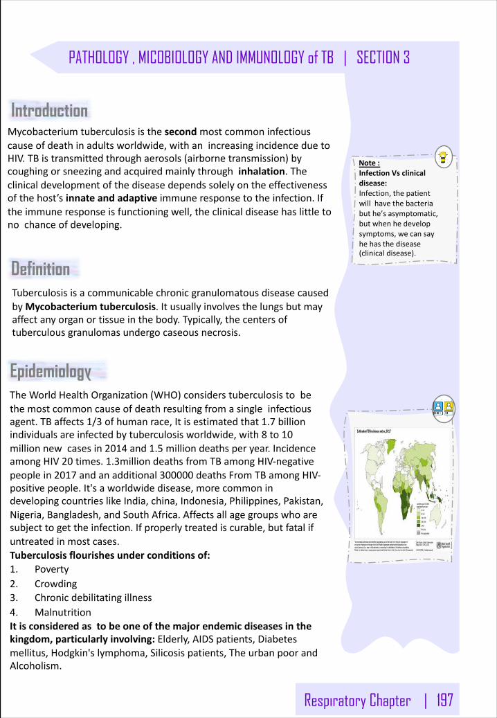
Respiratory Chapter | 197
PATHOLOGY , MICOBIOLOGY AND IMMUNOLOGY of TB | SECTION 3
Definition TuberculosisisacommunicablechronicgranulomatousdiseasecausedbyMycobacteriumtuberculosis.Itusuallyinvolvesthelungsbutmayaffectanyorganortissueinthebody.Typically,thecentersoftuberculousgranulomasundergocaseous necrosis.
Introduction Mycobacteriumtuberculosisisthesecondmostcommoninfectiouscauseofdeathinadultsworldwide,withanincreasingincidenceduetoHIV.TBistransmittedthroughaerosols(airbornetransmission)bycoughingorsneezingandacquiredmainlythroughinhalation.Theclinicaldevelopmentofthediseasedependssolelyontheeffectivenessofthehost’sinnateandadaptiveimmuneresponsetotheinfection.Iftheimmuneresponseisfunctioningwell,theclinicaldiseasehaslittletonochanceofdeveloping.
Note:InfectionVsclinicaldisease:Infection,thepatientwillhavethebacteriabuthe’sasymptomatic,butwhenhedevelopsymptoms,wecansayhehasthedisease(clinicaldisease).
Epidemiology TheWorldHealthOrganization(WHO)considerstuberculosistobethemostcommoncauseofdeathresultingfromasingleinfectiousagent.TBaffects1/3 ofhumanrace,Itisestimatedthat1.7 billionindividualsareinfectedbytuberculosisworldwide,with8 to10millionnewcasesin2014 and1.5 milliondeathsperyear.IncidenceamongHIV20 times.1.3milliondeathsfromTBamongHIV-negativepeoplein2017 andanadditional300000 deathsFromTBamongHIV-positivepeople.It'saworldwidedisease,morecommonindevelopingcountrieslikeIndia,china,Indonesia,Philippines,Pakistan,Nigeria,Bangladesh,andSouthAfrica.Affectsallagegroupswhoaresubjecttogettheinfection.Ifproperlytreatediscurable,butfatalifuntreatedinmostcases.Tuberculosisflourishesunderconditionsof:1. Poverty2. Crowding3. Chronicdebilitatingillness4. MalnutritionItisconsideredastobeoneofthemajorendemicdiseasesinthekingdom,particularlyinvolving:Elderly,AIDSpatients,Diabetesmellitus,Hodgkin'slymphoma,Silicosispatients,TheurbanpoorandAlcoholism.
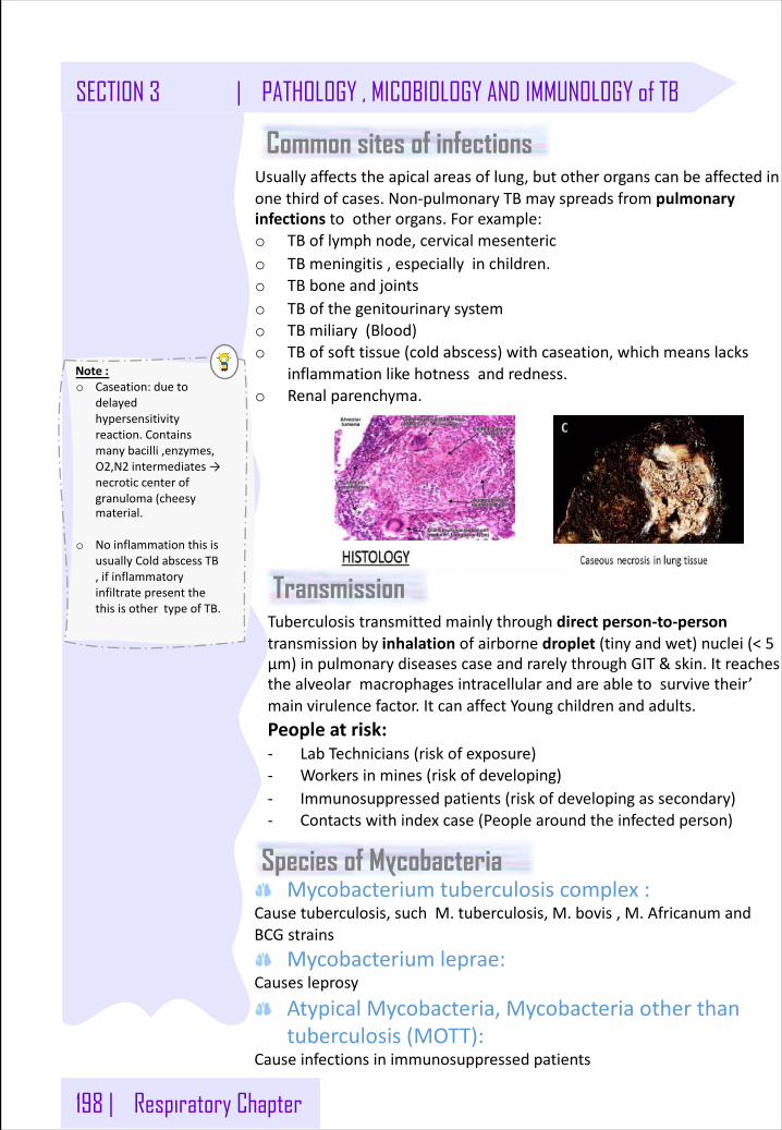
198 | Respiratory Chapter
SECTION 3 | PATHOLOGY , MICOBIOLOGY AND IMMUNOLOGY of TB
Common sites of infections Usuallyaffectstheapicalareasoflung,butotherorganscanbeaffectedinonethirdofcases.Non-pulmonaryTBmayspreadsfrompulmonaryinfectionstootherorgans.Forexample:o TBoflymphnode,cervicalmesenterico TBmeningitis,especiallyinchildren.o TBboneandjointso TBofthegenitourinarysystemo TBmiliary(Blood)o TBofsofttissue(coldabscess)withcaseation,whichmeanslacks
inflammationlikehotnessandredness.o Renalparenchyma.
Tuberculosistransmittedmainlythroughdirectperson-to-persontransmissionbyinhalation ofairbornedroplet (tinyandwet)nuclei(<5µm)inpulmonarydiseasescaseandrarelythroughGIT&skin.Itreachesthealveolarmacrophagesintracellularandareabletosurvivetheir’mainvirulencefactor.ItcanaffectYoungchildrenandadults.Peopleatrisk:- LabTechnicians(riskofexposure)- Workersinmines(riskofdeveloping)- Immunosuppressedpatients(riskofdevelopingassecondary)- Contactswithindexcase(Peoplearoundtheinfectedperson)
Note:o Caseation:dueto
delayedhypersensitivityreaction.Containsmanybacilli,enzymes,O2,N2 intermediates→necroticcenterofgranuloma(cheesymaterial.
o NoinflammationthisisusuallyColdabscessTB,ifinflammatoryinfiltratepresentthethisisothertypeofTB.
Species of MycobacteriaMycobacteriumtuberculosiscomplex:
Causetuberculosis,suchM.tuberculosis,M.bovis,M.AfricanumandBCGstrains
Mycobacteriumleprae:Causesleprosy
AtypicalMycobacteria,Mycobacteriaotherthantuberculosis(MOTT):
Causeinfectionsinimmunosuppressedpatients
Transmission
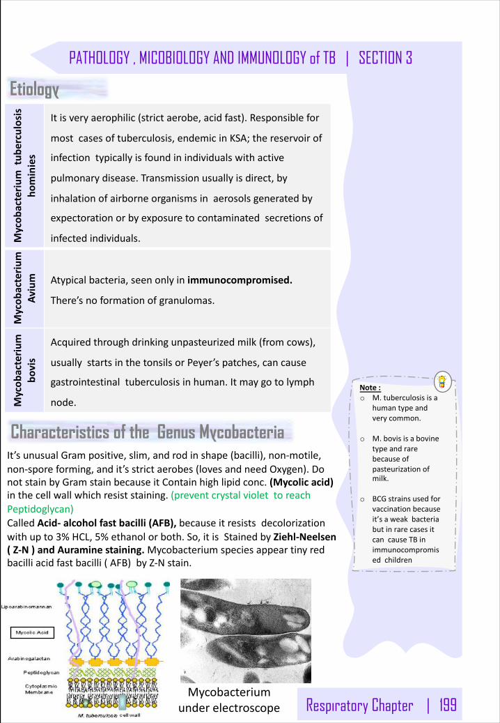
Respiratory Chapter | 199
PATHOLOGY , MICOBIOLOGY AND IMMUNOLOGY of TB | SECTION 3
Etiology
Mycob
acteriu
mtub
erculosis
hominies
Itisveryaerophilic(strictaerobe,acidfast).Responsiblefor
mostcasesoftuberculosis,endemicinKSA;thereservoirof
infectiontypicallyisfoundinindividualswithactive
pulmonarydisease.Transmissionusuallyisdirect,by
inhalationofairborneorganismsinaerosolsgeneratedby
expectorationorbyexposuretocontaminatedsecretionsof
infectedindividuals.
Mycob
acteriu
m
Avium Atypicalbacteria,seenonlyinimmunocompromised.
There’snoformationofgranulomas.
Mycob
acteriu
m
bovis
Acquiredthroughdrinkingunpasteurizedmilk(fromcows),
usuallystartsinthetonsilsorPeyer’spatches,cancause
gastrointestinaltuberculosisinhuman.Itmaygotolymph
node.
Characteristics of the Genus Mycobacteria
Mycobacteriumunderelectroscope
It’sunusualGrampositive,slim,androdinshape(bacilli),non-motile,non-sporeforming,andit’sstrictaerobes(lovesandneedOxygen).DonotstainbyGramstainbecauseitContainhighlipidconc.(Mycolicacid)inthecellwallwhichresiststaining.(preventcrystalviolettoreachPeptidoglycan)CalledAcid- alcoholfastbacilli(AFB),becauseitresistsdecolorizationwithupto3%HCL,5%ethanolorboth.So,itisStainedbyZiehl-Neelsen(Z-N)andAuraminestaining.Mycobacteriumspeciesappeartinyredbacilliacidfastbacilli(AFB)byZ-Nstain.
Note:o M.tuberculosisisa
humantypeandverycommon.
o M.bovisisabovinetypeandrarebecauseofpasteurizationofmilk.
o BCGstrainsusedforvaccinationbecauseit’saweakbacteriabutinrarecasesitcancauseTBinimmunocompromisedchildren
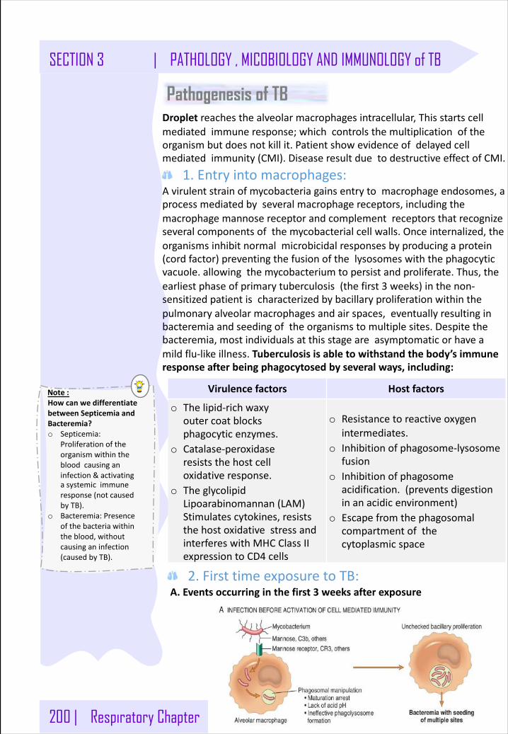
200 | Respiratory Chapter
SECTION 3 | PATHOLOGY , MICOBIOLOGY AND IMMUNOLOGY of TB
Pathogenesis of TB
Virulencefactors Hostfactors
o Thelipid-richwaxyoutercoatblocksphagocyticenzymes.
o Catalase-peroxidaseresiststhehostcelloxidativeresponse.
o TheglycolipidLipoarabinomannan(LAM)Stimulatescytokines,resiststhehostoxidativestressandinterfereswithMHCClassIIexpressiontoCD4 cells
o Resistancetoreactiveoxygenintermediates.
o Inhibitionofphagosome-lysosomefusion
o Inhibitionofphagosomeacidification.(preventsdigestioninanacidicenvironment)
o Escapefromthephagosomalcompartmentofthecytoplasmicspace
Droplet reachesthealveolarmacrophagesintracellular,Thisstartscellmediatedimmuneresponse;whichcontrolsthemultiplicationoftheorganismbutdoesnotkillit.Patientshowevidenceofdelayedcellmediatedimmunity(CMI).DiseaseresultduetodestructiveeffectofCMI.
1.Entryintomacrophages:Avirulentstrainofmycobacteriagainsentrytomacrophageendosomes,aprocessmediatedbyseveralmacrophagereceptors,includingthemacrophagemannosereceptorandcomplementreceptorsthatrecognizeseveralcomponentsofthemycobacterialcellwalls.Onceinternalized,theorganismsinhibitnormalmicrobicidalresponsesbyproducingaprotein(cordfactor)preventingthefusionofthelysosomeswiththephagocyticvacuole.allowingthemycobacteriumtopersistandproliferate.Thus,theearliestphaseofprimarytuberculosis(thefirst3 weeks)inthenon-sensitizedpatientischaracterizedbybacillaryproliferationwithinthepulmonaryalveolarmacrophagesandairspaces,eventuallyresultinginbacteremiaandseedingoftheorganismstomultiplesites.Despitethebacteremia,mostindividualsatthisstageareasymptomaticorhaveamildflu-likeillness.Tuberculosisisabletowithstandthebody’simmuneresponseafterbeingphagocytosedbyseveralways,including:
2.FirsttimeexposuretoTB:A.Eventsoccurringinthefirst3 weeksafterexposure
Note:HowcanwedifferentiatebetweenSepticemiaandBacteremia?o Septicemia:
Proliferationoftheorganismwithinthebloodcausinganinfection&activatingasystemicimmuneresponse(notcausedbyTB).
o Bacteremia:Presenceofthebacteriawithintheblood,withoutcausinganinfection(causedbyTB).
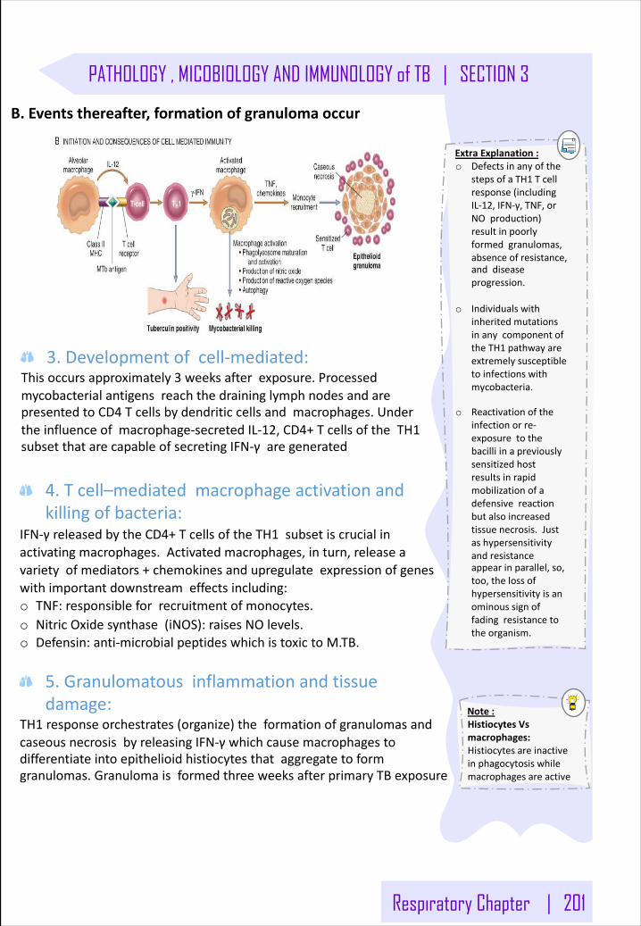
Respiratory Chapter | 201
PATHOLOGY , MICOBIOLOGY AND IMMUNOLOGY of TB | SECTION 3
3.Developmentofcell-mediated:Thisoccursapproximately3 weeksafterexposure.ProcessedmycobacterialantigensreachthedraininglymphnodesandarepresentedtoCD4 Tcellsbydendriticcellsandmacrophages.Undertheinfluenceofmacrophage-secretedIL-12,CD4+TcellsoftheTH1subsetthatarecapableofsecretingIFN-γaregenerated
B.Eventsthereafter,formationofgranulomaoccur
4.Tcell–mediatedmacrophageactivationandkillingofbacteria:
IFN-γreleasedbytheCD4+TcellsoftheTH1 subsetiscrucialinactivatingmacrophages.Activatedmacrophages,inturn,releaseavarietyofmediators+chemokinesandupregulateexpressionofgeneswithimportantdownstreameffectsincluding:o TNF:responsibleforrecruitmentofmonocytes.o NitricOxidesynthase(iNOS):raisesNOlevels.o Defensin:anti-microbialpeptideswhichistoxictoM.TB.
5.Granulomatousinflammationandtissuedamage:
TH1 responseorchestrates(organize)theformationofgranulomasandcaseousnecrosisbyreleasingIFN-γwhichcausemacrophagestodifferentiateintoepithelioidhistiocytesthataggregatetoformgranulomas.GranulomaisformedthreeweeksafterprimaryTBexposure
Note:HistiocytesVsmacrophages:Histiocytesareinactiveinphagocytosiswhilemacrophagesareactive
ExtraExplanation:o Defectsinanyofthe
stepsofaTH1 Tcellresponse(includingIL-12,IFN-γ,TNF,orNOproduction)resultinpoorlyformedgranulomas,absenceofresistance,anddiseaseprogression.
o IndividualswithinheritedmutationsinanycomponentoftheTH1 pathwayareextremelysusceptibletoinfectionswithmycobacteria.
o Reactivationoftheinfectionorre-exposuretothebacilliinapreviouslysensitizedhostresultsinrapidmobilizationofadefensivereactionbutalsoincreasedtissuenecrosis.Justashypersensitivityandresistanceappearinparallel,so,too,thelossofhypersensitivityisanominoussignoffadingresistancetotheorganism.
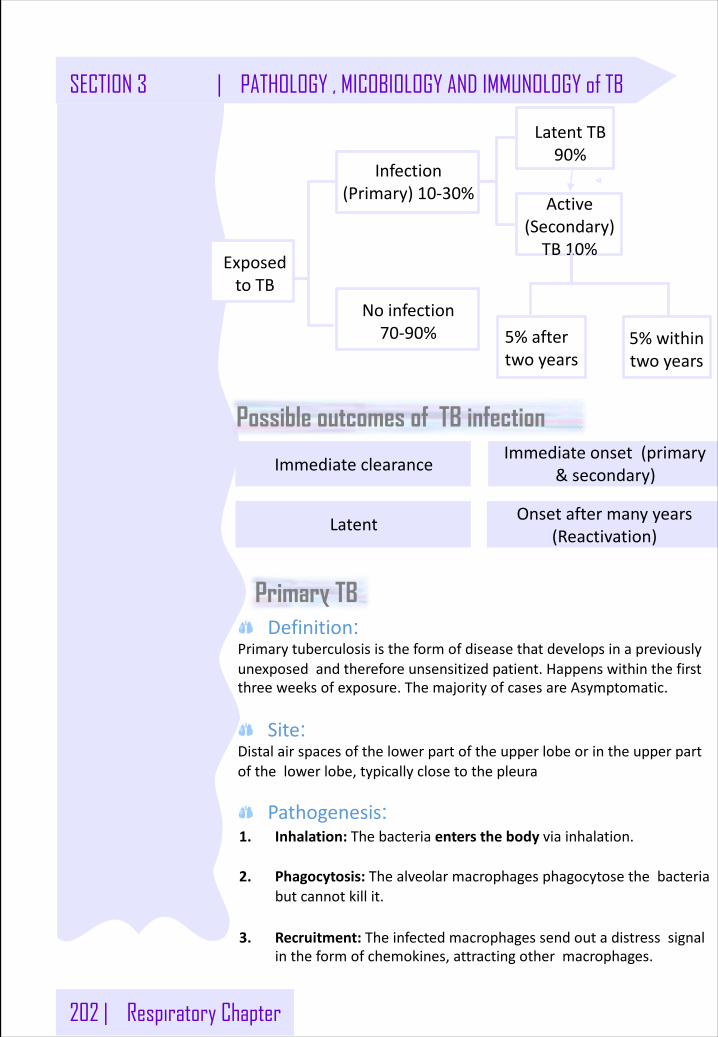
202 | Respiratory Chapter
SECTION 3 | PATHOLOGY , MICOBIOLOGY AND IMMUNOLOGY of TB
Possible outcomes of TB infection
ExposedtoTB
Infection(Primary)10-30%
5%withintwoyears
Noinfection70-90%
LatentTB90%
Active(Secondary)TB10%
5%aftertwoyears
ImmediateclearanceImmediateonset(primary
&secondary)
Latent Onsetaftermanyyears(Reactivation)
Primary TBDefinition:
Primarytuberculosisistheformofdiseasethatdevelopsinapreviouslyunexposedandthereforeunsensitizedpatient.Happenswithinthefirstthreeweeksofexposure.ThemajorityofcasesareAsymptomatic.
Site: Distalairspacesofthelowerpartoftheupperlobeorintheupperpartofthelowerlobe,typicallyclosetothepleura
Pathogenesis: 1. Inhalation:Thebacteriaentersthebodyviainhalation.
2. Phagocytosis:Thealveolarmacrophagesphagocytosethebacteriabutcannotkillit.
3. Recruitment:Theinfectedmacrophagessendoutadistresssignalintheformofchemokines,attractingothermacrophages.
Respiratory Chapter | 203
PATHOLOGY , MICOBIOLOGY AND IMMUNOLOGY of TB | SECTION 3
4. Ghon’sfocus:Thenewlyrecruitedmacrophagessurroundthebacteria,thiseventuallyformsanodulargranulomacalledatubercle.ThiswholestructureisknownasaGhon’sfocus.
5. Ghon’scomplex:Ifthereplicationisn’tcontrolled,itspreadstothedraininglymphnodes,formingaGhon’scomplex.
6. Ranke’scomplex:Insomecases,thetuberclesbecomefibroticandheal,formingaRanke’scomplex.Thistypeoffibrosisnevergoesaway.
2-6 weeksaftertheinfection,thebacillitriggeraCellMediatedImmunityresponse.Thisleadsto:
1. Th1 cells:Weeksaftertheinfection,theCMIresponsecausesTh1 cellstorelease:A. IFN-ɣ:activatesmoremacrophagesandenhancesits
abilitytokillphagocytosedbacilli.B. TNF:induceslocalinflammationandactivatesmore
macrophages.
2. CMIresponse:IftheCMIresponseisnoteffective,thelunggetsdestroyedby:A. NitrogenintermediatesB. TNF-ɑC. ReactiveoxygenD. Contentsofcytotoxiccells(Perforin,granzymes)
3.Outcome:Thedestructivesubstancesleadtocaseous necrosis,amajorcharacteristicofTB.Eventually,thecaseatinglesionsstarttoerode,spreadingtotheairwaysandbecominginfectious.Ifleftuntreated,thediseasecanbecomechronicorevenleadtodeath(80%ofcases).
4.ChronicDisease:Itischaracterizedbyepisodesofhealingbyfibroticchangesaroundthelesionandtissuebreakdown.Recoveryispossible(20%)atthisstage,butcompleteeradicationofthebacilliisrare.
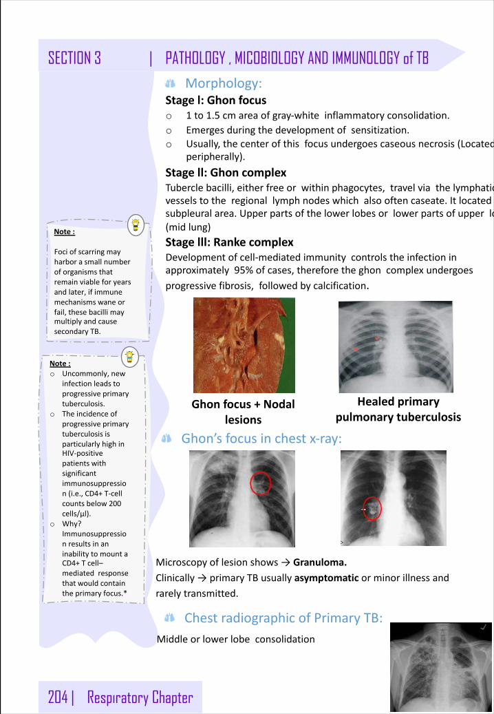
204 | Respiratory Chapter
SECTION 3 | PATHOLOGY , MICOBIOLOGY AND IMMUNOLOGY of TB
Note:
Fociofscarringmayharborasmallnumberoforganismsthatremainviableforyearsandlater,ifimmunemechanismswaneorfail,thesebacillimaymultiplyandcausesecondaryTB.
Ghonfocus+Nodallesions
Healedprimarypulmonarytuberculosis
Microscopyoflesionshows→Granuloma.Clinically→primaryTBusuallyasymptomatic orminorillnessandrarelytransmitted.
Middleorlowerlobeconsolidation
Morphology:Stagel:Ghonfocuso 1 to1.5 cmareaofgray-whiteinflammatoryconsolidation.o Emergesduringthedevelopmentofsensitization.o Usually,thecenterofthisfocusundergoescaseousnecrosis(Located
peripherally).Stagell:GhoncomplexTuberclebacilli,eitherfreeorwithinphagocytes,travelviathelymphaticvesselstotheregionallymphnodeswhichalsooftencaseate.Itlocatedsubpleuralarea.Upperpartsofthelowerlobesorlowerpartsofupperlobes(midlung)Stagelll:RankecomplexDevelopmentofcell-mediatedimmunitycontrolstheinfectioninapproximately95%ofcases,thereforetheghon complexundergoesprogressivefibrosis,followedbycalcification.
Ghon’sfocusinchestx-ray:
ChestradiographicofPrimaryTB:
Note:o Uncommonly,new
infectionleadstoprogressiveprimarytuberculosis.
o TheincidenceofprogressiveprimarytuberculosisisparticularlyhighinHIV-positivepatientswithsignificantimmunosuppression(i.e.,CD4+T-cellcountsbelow200cells/μl).
o Why?ImmunosuppressionresultsinaninabilitytomountaCD4+Tcell–mediatedresponsethatwouldcontaintheprimaryfocus.*
Respiratory Chapter | 205
PATHOLOGY , MICOBIOLOGY AND IMMUNOLOGY of TB | SECTION 3
Definition:Secondarytuberculosisisthepatternofdiseasethatarisesinapreviouslysensitizedhost,itarisesduetoreactivationofdormantprimarylesionsorduetoreinfection.ItformsCavitaryfociofcaseousnecrosis:Theriskofspreadofinfectiontonon-infectedpersonsfromindividualswithcavitarytuberculosisisveryhigh.Why?becausethepatientnowcoughssputumthatcontainsbacilli,thereforepatientshouldbeisolatedfor10-14 daysfromstartingtreatment.
Site:Classicallylocalizedtotheapexofoneorbothupperlobes.Thereasonisobscurebutmayrelatetohighoxygentensionintheapices.TheregionallymphnodesarelessprominentlyinvolvedearlyinthediseasethantheyareinprimaryTB.
Complication:ItmayprogresstoMiliaryTuberculosis,whichcanrupturethemacrophagesandescapeintothebloodstreamvialymphaticvessels.Thewordmiliaryisderivedfromtheresemblanceofthesefocitomilletseeds.Itcangoanywhere&symptomsdependonthelocation,E.g.Liver,bonemarrow,meningesfallopiantubesandepididymis.
Secondary TB
Miliary TBMiliary TB (disseminated TB) can occur if the primary infection is notproperly contained. This develops when the TB bacilli spreadsthroughout the lung and/or to other organs through hematogenouslymphatic spread. Its most common presentation ismeningeal TB.
PulmonaryMTB:Occurswhenorganismsreachthebloodstreamthroughlymphaticvesselsandthenrecirculatetothelungviathepulmonaryarteries.Thelesionsappearassmall(2-mm)fociofyellow-whiteconsolidationscatteredthroughthelungparenchyma
SystemicMTB:o Ensueswhentheorganismsdisseminatehematogenouslythroughout
thebody.o Systemicmiliarytuberculosisismostprominentintheliver,bone
marrow,spleen,adrenalglands,meninges,kidneys,fallopiantubes,andepididymis.
o Multiplesmallyellownodularlesionsinseveralorgans.Almosteveryorganinthebodymaybeseeded.Lesionsresemblethoseinthelungs
206 | Respiratory Chapter
SECTION 3 | PATHOLOGY , MICOBIOLOGY AND IMMUNOLOGY of TB
Note:TheimmuneresponseandAnti-MTBdrugsaredirectedtowardsthegrowing bacilli,thereforemakingthenon-replicating bacilliinlatentTBsomewhatinvisibletothebody(resistant).
Isolated-organTB(ExtrapulmonaryTB):Mayappearinanyoneoftheorgansortissuesseededhematogenously.Organstypicallyinvolvedinclude:o Lymphnodes(tuberculouslymphadenitis):arethemostfrequent
formofextrapulmonarytuberculosisesp.inthecervicalregion“Scrofula”.
o Pleurawithpleuraleffusion(exudate).o Liver,spleen,kidneysandAdrenalsglands.o Fallopiantube(Tuberculoussalpingitis)andendometrium.o Epididymisandprostate.o Meninges(tuberculousmeningitis).o BonemarrowandVertebrae(Pott'sdisease).o Intestinaltuberculosis.
Latent TB
Reactivation
Pathogenesis:1. PresentationofantigensbyAPCsinthelymphnodes.Delayed-type
hypersensitivity(TypeIV).
2. ActivationofCD4+(Th1)lymphocytes.Thisphasecoincideswithhighrateofreplicationofbacilli.
3. LowinductionofCD8+lymphocytes.CD8+lymphocytesrecognizetheantigenandproduceIFN-γ,leadingtomacrophageactivation.
4. InductionofhighnumberofCD8 +IncreasedproductionofIFN-γandcytotoxicactivity.Thisphasecoincideswithbacterialgrowthstabilization.
5. Bacterialloadremainsconstantandinfectioniskeptinadormantstate.
Thedormantbacteriathatwerestoppedduringprimaryinfectioncanstartproliferatingagain(5-10%ofcases).Ittendstobelocalized withmuchlesscaseationandlittlelymphnodeinvolvement.Itusuallyonlyaffectsthelungapices.Disseminationhereisusuallyuncommon.
Factorscontributingtoreactivation:
ChestradiographicInreactivationofTB:Classicallyfibro-cavitaryapicaldisease
o Immunosuppression o End-stagerenaldisease o Diabetes
o MalignantLymphoma
o Corticosteroids o Aging
o HIV/AIDS o AntiTNF-α drugs
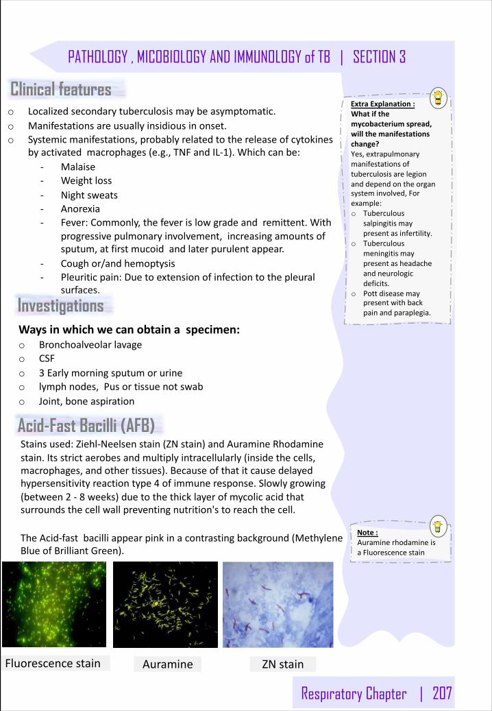
Respiratory Chapter | 207
PATHOLOGY , MICOBIOLOGY AND IMMUNOLOGY of TB | SECTION 3
Note:AuraminerhodamineisaFluorescencestain
ExtraExplanation:Whatifthemycobacteriumspread,willthemanifestationschange?Yes,extrapulmonarymanifestationsoftuberculosisarelegionanddependontheorgansysteminvolved,Forexample:o Tuberculous
salpingitismaypresentasinfertility.
o Tuberculousmeningitismaypresentasheadacheandneurologicdeficits.
o Pottdiseasemaypresentwithbackpainandparaplegia.
Clinical features o Localizedsecondarytuberculosismaybeasymptomatic.o Manifestationsareusuallyinsidiousinonset.o Systemicmanifestations,probablyrelatedtothereleaseofcytokines
byactivatedmacrophages(e.g.,TNFandIL-1).Whichcanbe:- Malaise- Weightloss- Nightsweats- Anorexia- Fever:Commonly,thefeverislowgradeandremittent.With
progressivepulmonaryinvolvement,increasingamountsofsputum,atfirstmucoidandlaterpurulentappear.
- Coughor/andhemoptysis- Pleuriticpain:Duetoextensionofinfectiontothepleural
surfaces.
Investigations Waysinwhichwecanobtainaspecimen:o Bronchoalveolarlavageo CSFo 3 Earlymorningsputumorurineo lymphnodes,Pusortissuenotswabo Joint,boneaspiration
ZNstain
Acid-Fast Bacilli (AFB)Stainsused:Ziehl-Neelsen stain(ZNstain)andAuramineRhodaminestain.Itsstrictaerobesandmultiplyintracellularly(insidethecells,macrophages,andothertissues).Becauseofthatitcausedelayedhypersensitivityreactiontype4 ofimmuneresponse.Slowlygrowing(between2 - 8 weeks)duetothethicklayerofmycolicacidthatsurroundsthecellwallpreventingnutrition'storeachthecell.
TheAcid-fastbacilliappearpinkinacontrastingbackground(MethyleneBlueofBrilliantGreen).
Fluorescence stain Auramine
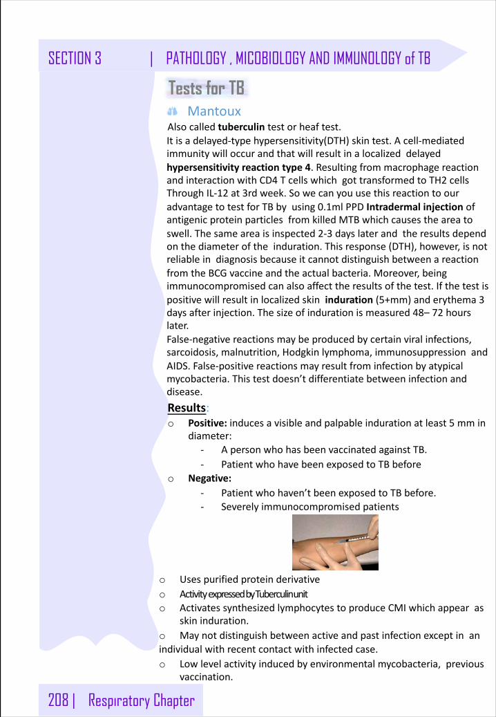
208 | Respiratory Chapter
SECTION 3 | PATHOLOGY , MICOBIOLOGY AND IMMUNOLOGY of TB
MantouxAlsocalledtuberculin testorheaf test.Itisadelayed-typehypersensitivity(DTH)skintest.Acell-mediatedimmunitywilloccurandthatwillresultinalocalizeddelayedhypersensitivityreactiontype4.ResultingfrommacrophagereactionandinteractionwithCD4 TcellswhichgottransformedtoTH2 cellsThroughIL-12 at3rdweek.SowecanyouusethisreactiontoouradvantagetotestforTBbyusing0.1mlPPDIntradermalinjectionofantigenicproteinparticlesfromkilledMTBwhichcausestheareatoswell.Thesameareaisinspected2-3 dayslaterandtheresultsdependonthediameteroftheinduration.Thisresponse(DTH),however,isnotreliableindiagnosisbecauseitcannotdistinguishbetweenareactionfromtheBCGvaccineandtheactualbacteria.Moreover,beingimmunocompromisedcanalsoaffecttheresultsofthetest.Ifthetestispositivewillresultinlocalizedskininduration (5+mm)anderythema3daysafterinjection.Thesizeofindurationismeasured48– 72 hourslater.False-negativereactionsmaybeproducedbycertainviralinfections,sarcoidosis,malnutrition,Hodgkinlymphoma,immunosuppressionandAIDS.False-positivereactionsmayresultfrominfectionbyatypicalmycobacteria.Thistestdoesn’tdifferentiatebetweeninfectionanddisease.Results:o Positive:inducesavisibleandpalpableindurationatleast5 mmin
diameter:- ApersonwhohasbeenvaccinatedagainstTB.- PatientwhohavebeenexposedtoTBbefore
o Negative:- Patientwhohaven’tbeenexposedtoTBbefore.- Severelyimmunocompromisedpatients
Tests for TB
o Usespurifiedproteinderivativeo ActivityexpressedbyTuberculinunito ActivatessynthesizedlymphocytestoproduceCMIwhichappearas
skininduration.o Maynotdistinguishbetweenactiveandpastinfectionexceptinanindividualwithrecentcontactwithinfectedcase.o Lowlevelactivityinducedbyenvironmentalmycobacteria,previous
vaccination.
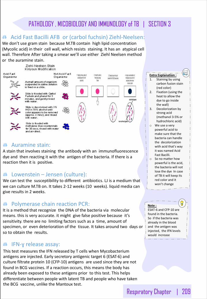
Respiratory Chapter | 209
PATHOLOGY , MICOBIOLOGY AND IMMUNOLOGY of TB | SECTION 3
ExtraExplanation:1. Stainingbyusing
carbonfusionstain(redcolor)
2. Fixation(usingtheheattoallowthedyetogoinsidethewall)
3. Decolorizationbystrongacid(methanol3-5%orhydrochloricacid)
o Weuseaverypowerfulacidtomakesurethatthebacteriacanhandlethedecolorizationwithacidthat’swayitwasnamedAcidFastBacilli.
o Sonomatterhowpowerfulistheacid,thebacteriawillnotlosethedyeIncaseofTBitwillkeepitsredcoloranditwon’tchange
AcidFastBacilliAFBor(carbol fuchsin)Ziehl-Neelsen:Wedon’tusegramstainbecauseM.TBcontainhighlipidconcentration(Mycolicacid)intheircellwall,whichresistsstaining.Ithasanatypicalcellwall.ThereforeAftertakingasmearwe’lluseeitherZiehl Neelsen methodortheauraminestain.
Auraminestain:Astainthatinvolvesstainingtheantibodywithanimmunofluorescencedyeandthenreactingitwiththeantigenofthebacteria.Ifthereisareactionthenitispositive.
Lowenstein– Jensen(culture):Wecantestthesusceptibilitytodifferentantibiotics.LJisamediumthatwecancultureM.TBon.Ittakes2-12 weeks(10 weeks).liquidmediacangiveresultsin2 weeks.
PolymerasechainreactionPCR:ItisamethodthatrecognizetheDNAofthebacteriaviamolecularmeans.thisisveryaccurate.itmightgivefalsepositivebecauseit’ssensitivity.therearenolimitingfactorssuchasatime,amountofspecimen,orevendeteriorationofthetissue.Ittakesaroundtwodaysorsotoobtaintheresults.
IFN-ɣreleaseassay:ThistestmeasurestheIFNreleasedbyTcellswhenMycobacteriumantigensareinjected.Earlysecretoryantigenictarget6 (ESAT-6)andculturefiltrateprotein10 (CFP-10)antigensareusedsincetheyarenotfoundinBCGvaccines.Ifareactionoccurs,thismeansthebodyhasalreadybeenexposedtotheseantigenspriortothistest.ThishelpsdifferentiatebetweenpeoplewithlatentTBandpeoplewhohavetakentheBCGvaccine,unliketheMantouxtest.
Note:ESAT-6 andCFP-10 arefoundinthebacteria.Soifthebacteriawasalreadyinthebloodandtheantigenwasinjected,theIFNlevelswouldincrease
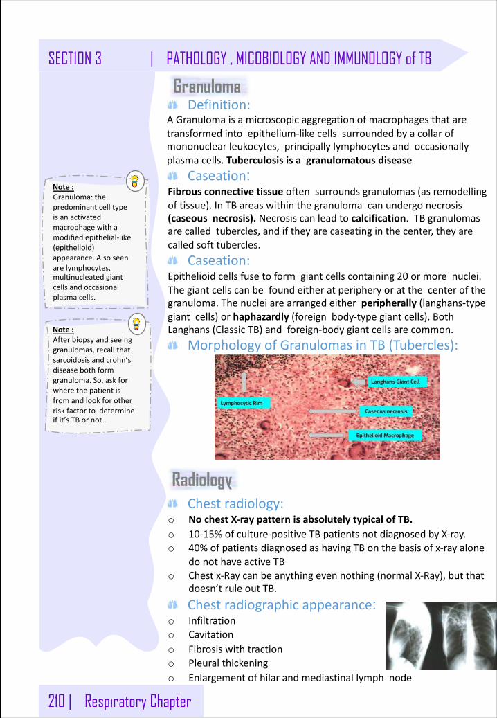
210 | Respiratory Chapter
SECTION 3 | PATHOLOGY , MICOBIOLOGY AND IMMUNOLOGY of TB
Chestradiology:o NochestX-raypatternisabsolutelytypicalofTB.o 10-15%ofculture-positiveTBpatientsnotdiagnosedbyX-ray.o 40%ofpatientsdiagnosedashavingTBonthebasisofx-rayalone
donothaveactiveTBo Chestx-Raycanbeanythingevennothing(normalX-Ray),butthat
doesn’truleoutTB.
Chestradiographicappearance: o Infiltrationo Cavitationo Fibrosiswithtractiono Pleuralthickeningo Enlargementofhilarandmediastinallymphnode
Note:Granuloma:thepredominantcelltypeisanactivatedmacrophagewithamodifiedepithelial-like(epithelioid)appearance.Alsoseenarelymphocytes,multinucleatedgiantcellsandoccasionalplasmacells.
Definition:AGranulomaisamicroscopicaggregationofmacrophagesthataretransformedintoepithelium-likecellssurroundedbyacollarofmononuclearleukocytes,principallylymphocytesandoccasionallyplasmacells.Tuberculosisisagranulomatousdisease
Caseation: Fibrousconnectivetissueoftensurroundsgranulomas(asremodellingoftissue).InTBareaswithinthegranulomacanundergonecrosis(caseousnecrosis).Necrosiscanleadtocalcification.TBgranulomasarecalledtubercles,andiftheyarecaseatinginthecenter,theyarecalledsofttubercles.
Caseation:Epithelioidcellsfusetoformgiantcellscontaining20 ormorenuclei.Thegiantcellscanbefoundeitheratperipheryoratthecenterofthegranuloma.Thenucleiarearrangedeitherperipherally (langhans-typegiantcells)orhaphazardly (foreignbody-typegiantcells).BothLanghans (ClassicTB)andforeign-bodygiantcellsarecommon.
MorphologyofGranulomasinTB(Tubercles):
Granuloma
Radiology
Note:Afterbiopsyandseeinggranulomas,recallthatsarcoidosisandcrohn’sdiseasebothformgranuloma.So,askforwherethepatientisfromandlookforotherriskfactortodetermineifit’sTBornot.
Respiratory Chapter | 211
PATHOLOGY , MICOBIOLOGY AND IMMUNOLOGY of TB | SECTION 3
SummaryTuberculosis
Gen
eral
considerations
Tuberculosisoccursworldwide,withgreatestfrequencyindisadvantagedgroups. Inthepulmonaryform,itisspreadbyinhalationofdroplets containingtheorganismMycobacteriumtuberculosis(alsoreferredtoasthetuberclebacillus).
Type
softube
rculosis PrimaryTB:
It’stheinitialinfection,characterizedbytheGhoncomplex,thecombinationofaperipheralsubpleuralparenchymallesionandinvolvedhilarlymphnodes. Primarytuberculosisismostoftenasymptomatic.Itusuallydoes notprogresstoclinicallyevidentdisease.SecondaryTB:UsuallyresultsfromactivationofapriorGhoncomplex,withspreadtoanewpulmonaryorextrapulmonarysite.
Patholog
iccha
nges
A. Localizedlesions:usuallyintheapicalorposteriorsegmentsoftheupperlobes.Involvementofhilarlymphnodesisalsocommon.
B. Tubercleformation:Thelesionsfrequentlycoalesceandrupture intothebronchi.Thecaseous contentsmayliquefyandbeexpelled,resultingincavitary lesions.Cavitationisacharacteristicofsecondary,butnotprimary,tuberculosis;caseation(amanifestationofpartialimmunity)isseeninboth.
C. Scarringandcalcification.
Spread
ofdisease Secondarytuberculosismaybecomplicatedbylymphaticand
hematogenousspread,resultinginmiliarytuberculosis,whichisseedingofdistalorganswithinnumerablesmallmilletseed-likelesions. Hematogenous spreadmayalsoresultinlargerlesions,whichmayinvolvealmostanyorgan. Organstypicallyinvolvedinclude:Meninges,fallopiantube”Tuberculoussalpingitis”,vertebrae”Pott disease”,Lymphadenitisinthecervicalregion“Scrofula”.
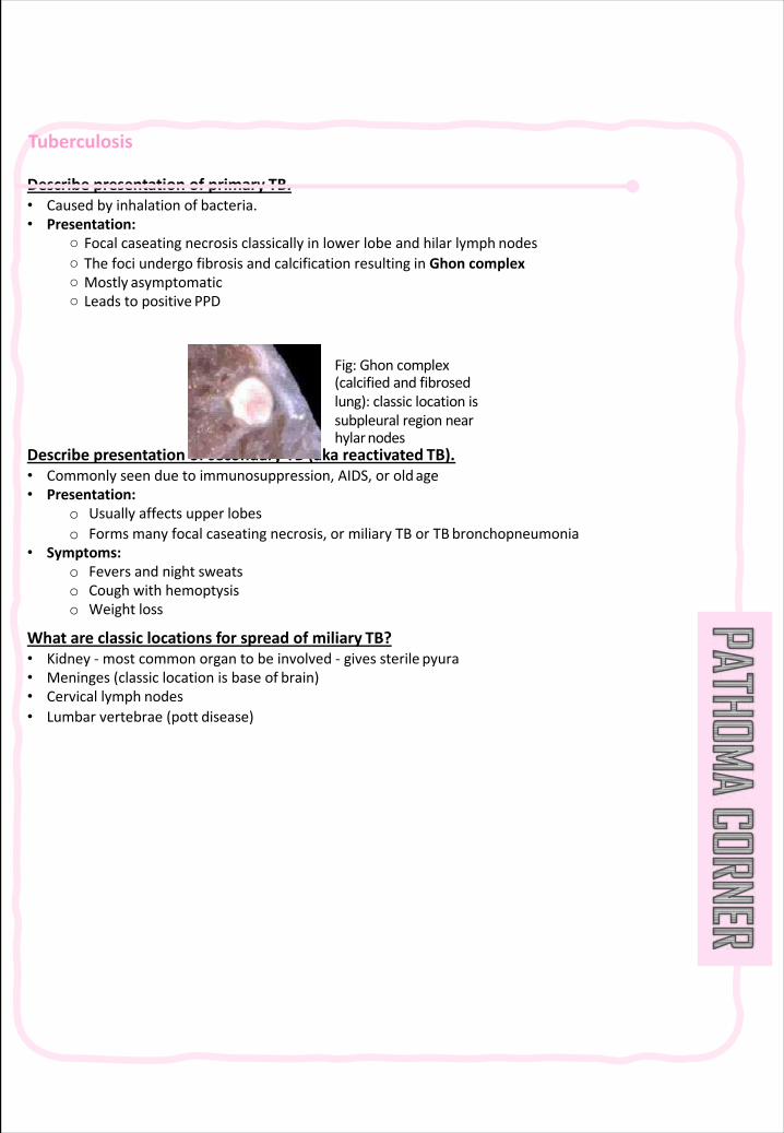
Tuberculosis
Describepresentationofprimary TB.• Causedbyinhalationof bacteria.• Presentation:
○ Focalcaseating necrosisclassicallyinlowerlobeandhilarlymph nodes○ ThefociundergofibrosisandcalcificationresultinginGhon complex○ Mostly asymptomatic○ Leadstopositive PPD
DescribepresentationofsecondaryTB(akareactivated TB).• Commonlyseenduetoimmunosuppression,AIDS,oroldage• Presentation:
o Usuallyaffectsupper lobeso Formsmanyfocalcaseating necrosis,ormiliary TBorTB bronchopneumonia
• Symptoms:o Feversandnight sweatso Coughwith hemoptysiso Weight loss
Whatareclassiclocationsforspreadofmiliary TB?• Kidney- mostcommonorgantobeinvolved- givessterile pyura• Meninges(classiclocationisbaseof brain)• Cervicallymph nodes• Lumbarvertebrae(pott disease)
Fig:Ghon complex(calcifiedandfibrosedlung):classiclocationissubpleural regionnearhylarnodes
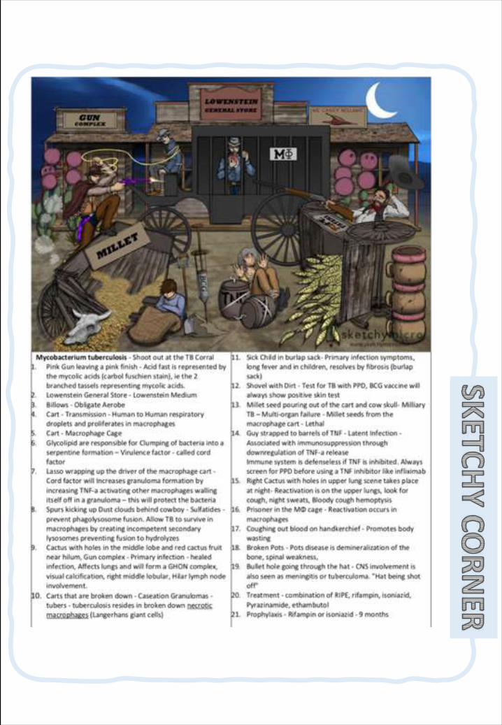
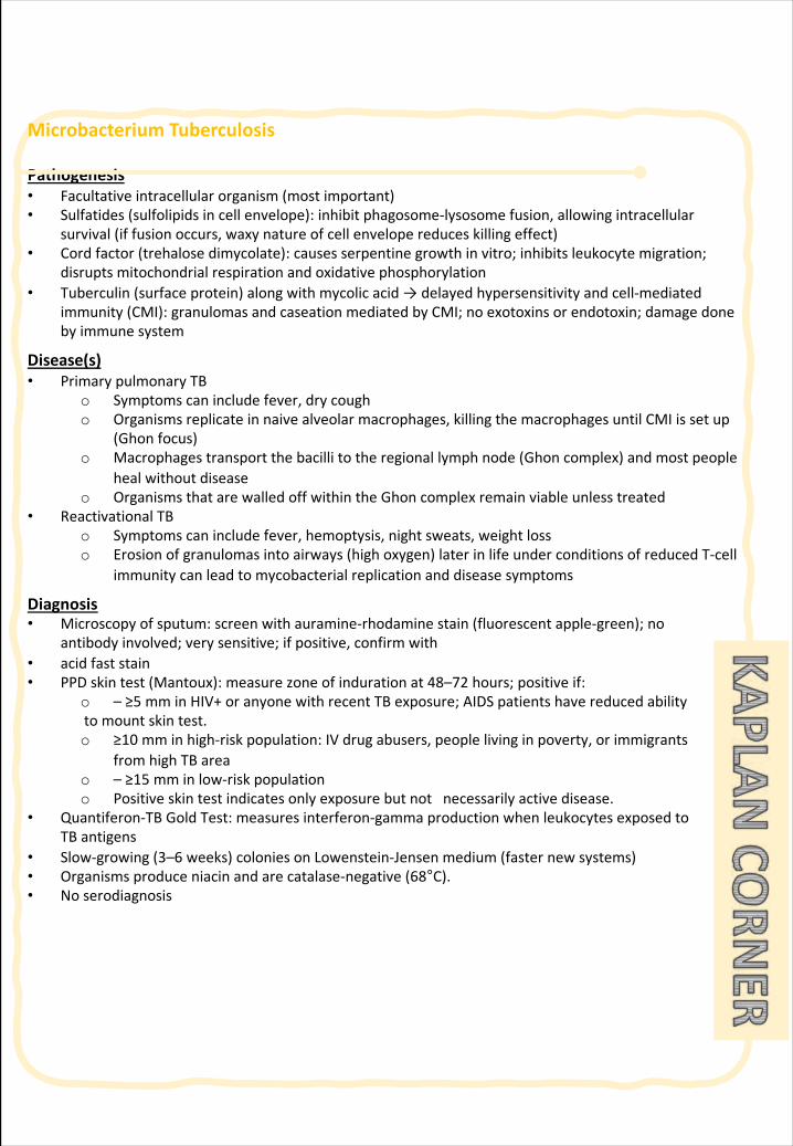
MicrobacteriumTuberculosis
Pathogenesis• Facultativeintracellularorganism(mostimportant)• Sulfatides (sulfolipids incellenvelope):inhibitphagosome-lysosomefusion,allowingintracellular
survival(iffusionoccurs,waxynatureofcellenvelopereduceskillingeffect)• Cordfactor(trehalose dimycolate):causesserpentinegrowthinvitro;inhibitsleukocytemigration;
disruptsmitochondrialrespirationandoxidativephosphorylation• Tuberculin(surfaceprotein)alongwithmycolicacid→delayedhypersensitivityandcell-mediated
immunity(CMI):granulomasandcaseationmediatedbyCMI;noexotoxinsorendotoxin;damagedonebyimmunesystem
Disease(s)• PrimarypulmonaryTB
o Symptomscanincludefever,drycougho Organismsreplicateinnaivealveolarmacrophages,killingthemacrophagesuntilCMIissetup
(Ghon focus)o Macrophagestransportthebacillitotheregionallymphnode(Ghon complex)andmostpeople
healwithoutdiseaseo OrganismsthatarewalledoffwithintheGhon complexremainviableunlesstreated
• Reactivational TBo Symptomscanincludefever,hemoptysis,nightsweats,weightlosso Erosionofgranulomasintoairways(highoxygen)laterinlifeunderconditionsofreducedT-cell
immunitycanleadtomycobacterialreplicationanddiseasesymptoms
Diagnosis• Microscopyofsputum:screenwithauramine-rhodaminestain(fluorescentapple-green);no
antibodyinvolved;verysensitive;ifpositive,confirmwith• acidfaststain• PPDskintest(Mantoux):measurezoneofindurationat48–72 hours;positiveif:
o – ≥5 mminHIV+oranyonewithrecentTBexposure;AIDSpatientshavereducedabilitytomountskintest.o ≥10 mminhigh-riskpopulation:IVdrugabusers,peoplelivinginpoverty,orimmigrants
fromhighTBareao – ≥15 mminlow-riskpopulationo Positiveskintestindicatesonlyexposurebutnotnecessarilyactivedisease.
• Quantiferon-TBGoldTest:measuresinterferon-gammaproductionwhenleukocytesexposedtoTBantigens
• Slow-growing(3–6 weeks)coloniesonLowenstein-Jensenmedium(fasternewsystems)• Organismsproduceniacinandarecatalase-negative(68°C).• Noserodiagnosis
MicrobacteriumTuberculosis
Treatment• Multipledrugscriticaltotreatinfection• Standardobservedshort-termtherapyforuncomplicatedpulmonaryTB(ratewhereacquired
resistance<4%):o First2 months:rifampin+isoniazid+pyrazinamide+ethambutol(RIPE)o Next4 months:rifampinandisoniazid
• Streptomycinaddedforpossibledrug-resistantcasesuntilsusceptibilitytestsareback(ifareaacquiredhas>4%drug-resistantmycobacteria)
• ForMDRTB,use3−5 previouslyunuseddrugs:aminoglycosides,fluoroquinolones,thioamide,cycloserine,bedaquiline
Prevention• Isoniazidtakenfor9 monthscanpreventTBinpersonswithinfectionbutnoclinicalsymptoms• Bacille Calmette-Guérin (BCG)vaccinecontaininglive,attenuatedorganismsmaypreventdisseminated
disease;notusedinU.S• UVlightorHEPAfilterusedtotreatpotentiallycontaminatedair
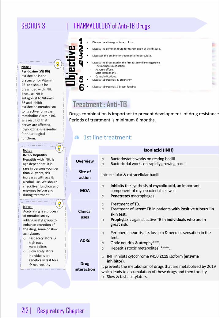
212 | Respiratory Chapter
SECTION 3 | PHARMACOLOGY of Anti-TB Drugs
§ Discusstheetiologyoftuberculosis.
§ Discussthecommonroutefortransmissionofthedisease.
§ Discussestheoutlinefortreatmentoftuberculosis.
§ Discussthedrugsusedinthefirst&secondlineRegarding:- Themechanismofaction.- Adverseeffects.- Druginteractions.- Contraindications.
§ Discusstuberculosis &pregnancy.
§ Discusstuberculosis&breastfeedingObjec
tive
1stlinetreatment:
Treatment : Anti-TB
Isoniazid(INH)
Overview o Bacteriostaticworksonrestingbacillio Bactericidalworksonrapidlygrowingbacilli
Siteofaction
Intracellular&extracellularbacilli
MOAo Inhibits thesynthesisofmycolicacid,animportant
componentofmycobacterialcellwall.o Penetratesmacrophages.
Clinicaluses
o TreatmentofTB.o TreatmentofLatentTBinpatientswithPositivetuberculin
skintest.o Prophylaxis againstactiveTBinindividualswhoarein
greatrisk.
ADRs
o Peripheralneuritis,i.e.losspin&needlessensationinthefeet.
o Opticneuritis&atrophy***.o Hepatitis(toxicmetabolites) ****.
Druginteraction
o INHinhibitscytochromeP450 2C19 isoform(enzymeinhibitor).
Itpreventsthemetabolismofdrugsthataremetabolizedby2C19whichleadstoaccumulationofthesedrugsandthentoxicityo Slow&fastacetylators.
Drugscombinationisimportanttopreventdevelopment ofdrugresistance.Periodsoftreatmentisminimum6 months.
Note:Acetylatingisaprocessofmetabolismbyaddingacetylgrouptoenhanceexcretionofthedrug,someorslowacetylatorso Fastacetylators→
hightoxicmetabolites
o Slowacetylatorsindividualsaregeneticallyfasttors→neuropathy
Note:Pyridoxine(VitB6)pyridoxineistheprecursorforVitaminB6 andshouldbeprescribedwithINH.BecauseINHisantagonisttoVitaminB6 andinhibitpyridoxinemetabolismtoitsactiveformthemetaboliteVitaminB6.asaresultofthatnervesareaffected.(pyridoxine)isessentialforneurologicalfunctions,
Note:INH&HepatitisHepatitiswithINH,isagedependent;itisrareinpersonsyoungerthan20 years,riskincreaseswithage&alcoholuse.Weshouldcheckliverfunctionandenzymesbeforeandduringtreatment.
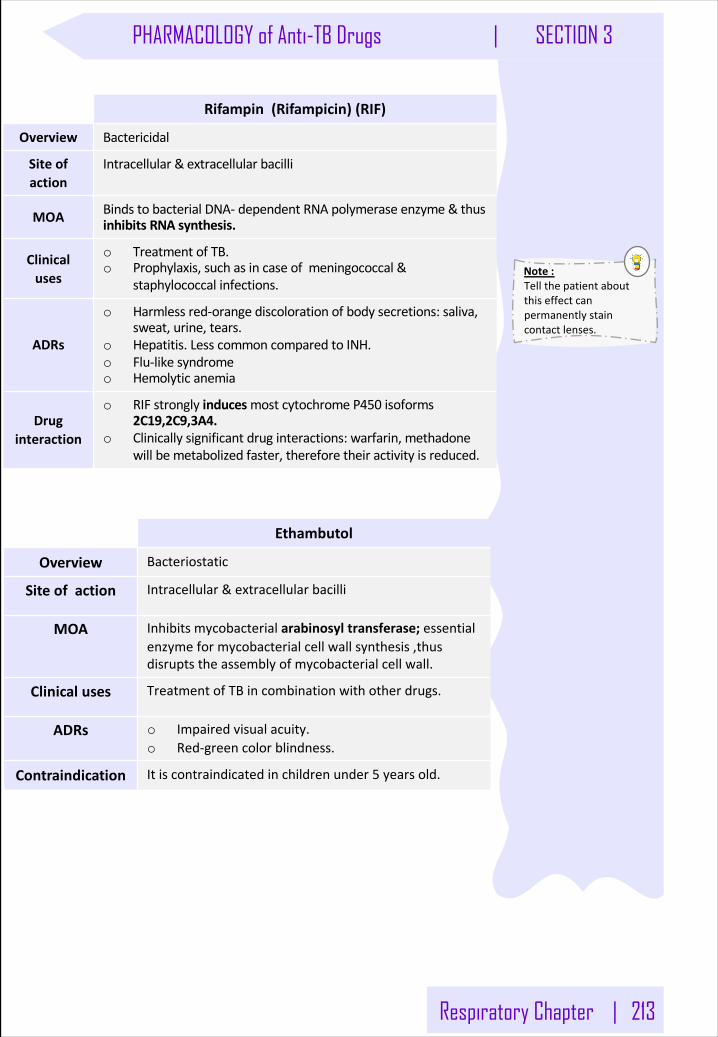
Respiratory Chapter | 213
PHARMACOLOGY of Anti-TB Drugs | SECTION 3
Rifampin(Rifampicin)(RIF)
Overview Bactericidal
Siteofaction
Intracellular&extracellularbacilli
MOA BindstobacterialDNA- dependentRNApolymeraseenzyme&thusinhibitsRNAsynthesis.
Clinicaluses
o TreatmentofTB.o Prophylaxis,suchasincaseofmeningococcal&
staphylococcalinfections.
ADRs
o Harmlessred-orangediscolorationofbodysecretions:saliva,sweat,urine,tears.
o Hepatitis. LesscommoncomparedtoINH.o Flu-likesyndromeo Hemolyticanemia
Druginteraction
o RIFstronglyinducesmostcytochromeP450 isoforms2C19,2C9,3A4.
o Clinicallysignificantdruginteractions:warfarin,methadonewillbemetabolizedfaster, thereforetheiractivityisreduced.
Note:Tellthepatientaboutthiseffectcanpermanentlystaincontactlenses.
Ethambutol
Overview Bacteriostatic
Siteofaction Intracellular&extracellularbacilli
MOA Inhibitsmycobacterialarabinosyl transferase;essentialenzymeformycobacterialcellwallsynthesis,thusdisruptstheassemblyofmycobacterialcellwall.
Clinicaluses TreatmentofTBincombinationwithotherdrugs.
ADRs o Impairedvisualacuity.o Red-greencolorblindness.
Contraindication Itiscontraindicatedinchildrenunder5 yearsold.
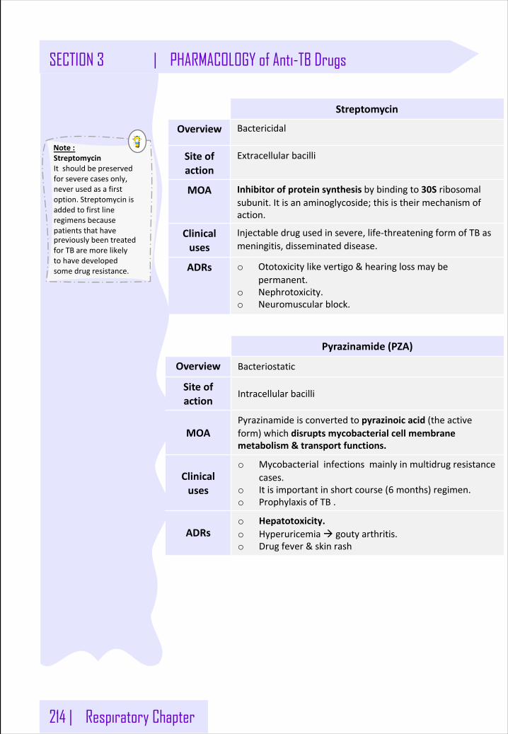
214 | Respiratory Chapter
Streptomycin
Overview Bactericidal
Siteofaction
Extracellularbacilli
MOA Inhibitorofproteinsynthesisbybindingto30S ribosomalsubunit. Itisanaminoglycoside;thisistheirmechanismofaction.
Clinicaluses
Injectabledrugusedinsevere,life-threateningformofTB asmeningitis,disseminateddisease.
ADRs o Ototoxicitylike vertigo&hearingloss maybepermanent.
o Nephrotoxicity.o Neuromuscularblock.
Pyrazinamide(PZA)
Overview Bacteriostatic
Siteofaction
Intracellularbacilli
MOAPyrazinamideisconvertedtopyrazinoic acid(theactiveform)whichdisruptsmycobacterialcellmembranemetabolism&transportfunctions.
Clinicaluses
o Mycobacterialinfectionsmainlyinmultidrugresistancecases.
o Itisimportantinshortcourse(6 months)regimen.o ProphylaxisofTB.
ADRso Hepatotoxicity.o Hyperuricemiaà goutyarthritis.o Drugfever&skinrash
Note:StreptomycinItshouldbepreservedforseverecasesonly,neverusedasafirstoption.StreptomycinisaddedtofirstlineregimensbecausepatientsthathavepreviouslybeentreatedforTBaremorelikelytohavedevelopedsomedrugresistance.
SECTION 3 | PHARMACOLOGY of Anti-TB Drugs
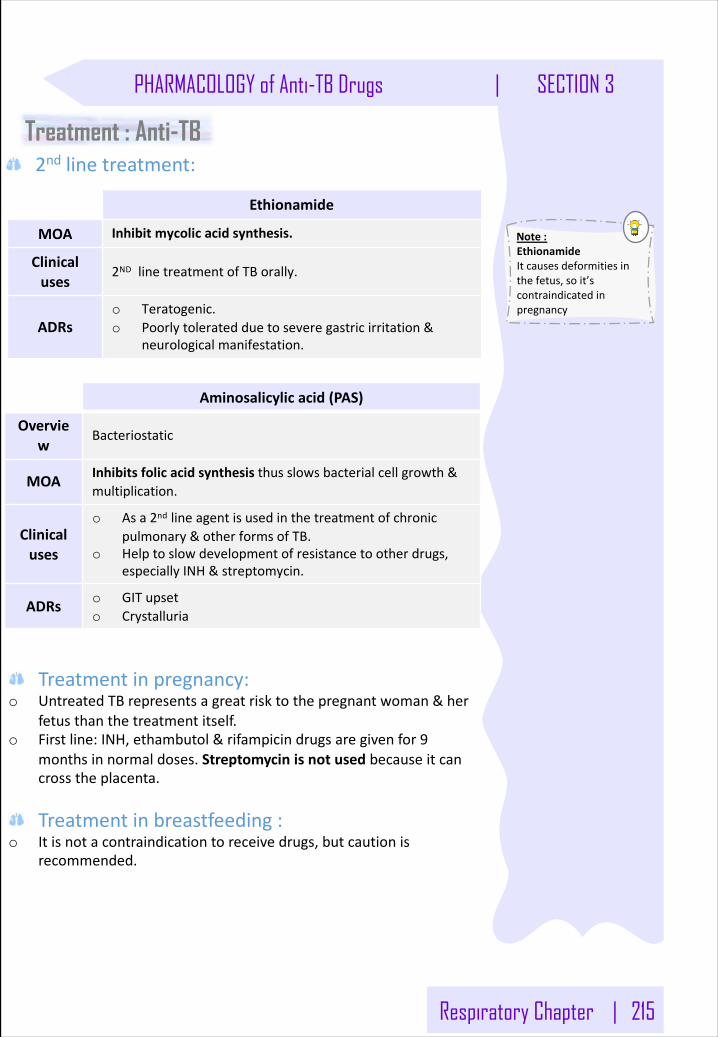
Respiratory Chapter | 215
2nd linetreatment:Treatment : Anti-TB
Note:EthionamideItcausesdeformitiesinthefetus,soit’scontraindicatedinpregnancy
Ethionamide
MOA Inhibitmycolicacidsynthesis.
Clinicaluses
2ND linetreatmentofTBorally.
ADRso Teratogenic.o Poorlytoleratedduetoseveregastricirritation&
neurologicalmanifestation.
Aminosalicylicacid(PAS)
Overview
Bacteriostatic
MOA Inhibitsfolicacidsynthesisthusslowsbacterialcellgrowth&multiplication.
Clinicaluses
o Asa2nd lineagentisusedinthe treatmentofchronicpulmonary& otherformsofTB.
o Helptoslowdevelopmentof resistancetootherdrugs,especiallyINH&streptomycin.
ADRs o GITupseto Crystalluria
Treatmentinpregnancy:o UntreatedTBrepresentsagreatrisktothepregnantwoman&her
fetusthanthetreatmentitself.o Firstline:INH,ethambutol&rifampicindrugsaregivenfor9
monthsinnormaldoses.Streptomycinisnotusedbecauseitcancrosstheplacenta.
Treatmentinbreastfeeding:o Itisnotacontraindicationtoreceivedrugs,butcautionis
recommended.
PHARMACOLOGY of Anti-TB Drugs | SECTION 3
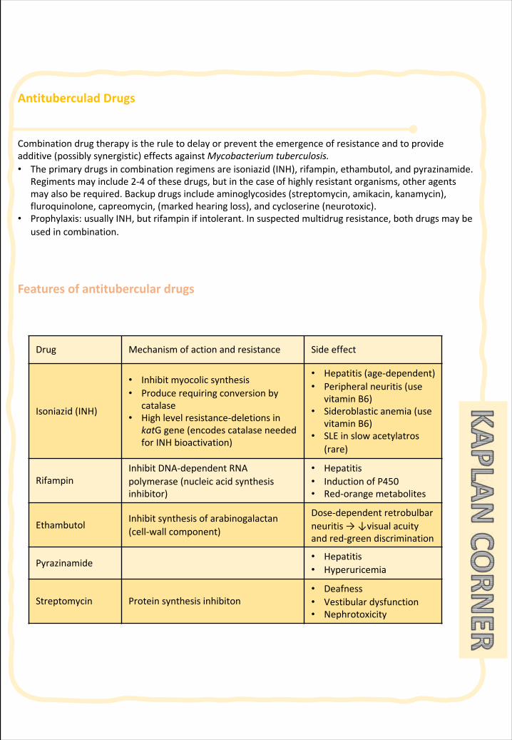
Antituberculad Drugs
Combinationdrugtherapyistheruletodelayorpreventtheemergenceofresistanceandtoprovideadditive(possiblysynergistic)effectsagainstMycobacteriumtuberculosis.• Theprimarydrugsincombinationregimensareisoniazid(INH),rifampin,ethambutol,andpyrazinamide.
Regimentsmayinclude2-4 ofthesedrugs,butinthecaseofhighlyresistantorganisms,otheragentsmayalsoberequired.Backupdrugsincludeaminoglycosides(streptomycin,amikacin,kanamycin),fluroquinolone,capreomycin,(markedhearingloss),andcycloserine (neurotoxic).
• Prophylaxis:usuallyINH,butrifampinifintolerant.Insuspectedmultidrugresistance,bothdrugsmaybeusedincombination.
Featuresofantituberculardrugs
Drug Mechanismofactionandresistance Sideeffect
Isoniazid(INH)
• Inhibitmyocolic synthesis• Produce requiringconversionby
catalase• Highlevelresistance-deletionsin
katG gene(encodescatalaseneededforINHbioactivation)
• Hepatitis (age-dependent)• Peripheralneuritis(use
vitaminB6)• Sideroblastic anemia(use
vitaminB6)• SLEinslowacetylatros
(rare)
RifampinInhibitDNA-dependentRNApolymerase(nucleicacidsynthesisinhibitor)
• Hepatitis• InductionofP450• Red-orangemetabolites
EthambutolInhibitsynthesisofarabinogalactan(cell-wallcomponent)
Dose-dependent retrobulbarneuritis→↓visualacuityandred-greendiscrimination
Pyrazinamide• Hepatitis• Hyperuricemia
Streptomycin Proteinsynthesisinhibiton• Deafness• Vestibulardysfunction• Nephrotoxicity
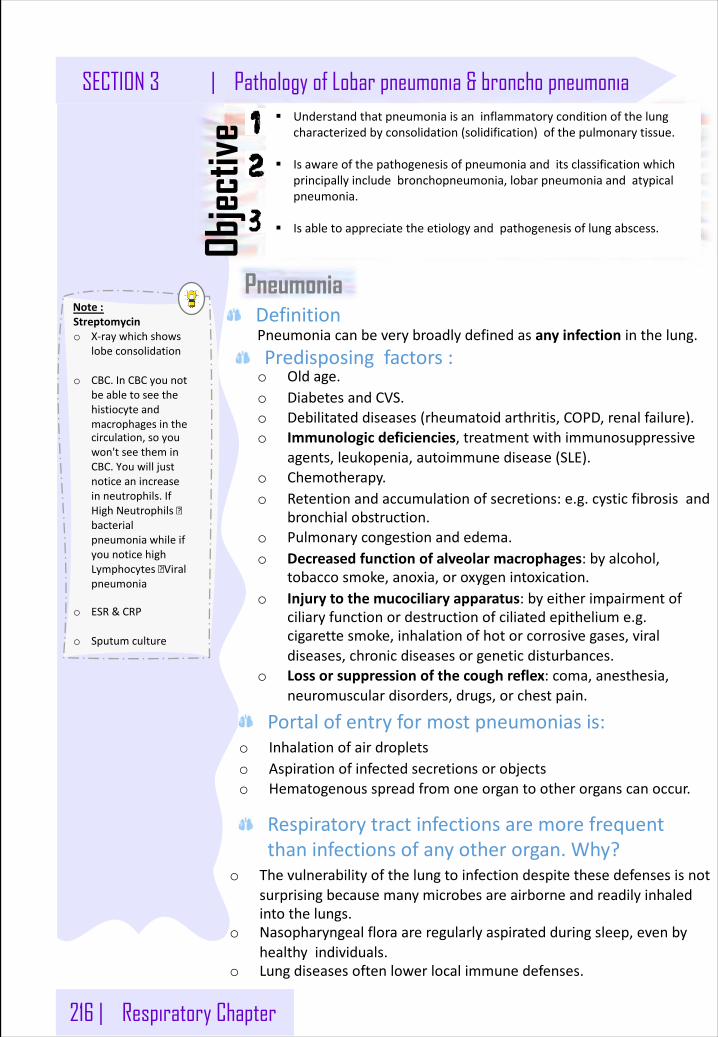
216 | Respiratory Chapter
Note:Streptomycino X-raywhichshows
lobeconsolidation
o CBC.InCBCyounotbeabletoseethehistiocyteandmacrophagesinthecirculation,soyouwon'tseetheminCBC.Youwilljustnoticeanincreaseinneutrophils.IfHighNeutrophils�bacterialpneumoniawhileifyounoticehighLymphocytes�Viralpneumonia
o ESR&CRP
o Sputumculture
SECTION 3 | Pathology of Lobar pneumonia & broncho pneumonia
Pneumonia
§ Understandthatpneumoniaisaninflammatoryconditionofthelungcharacterizedbyconsolidation(solidification)ofthepulmonarytissue.
§ Isawareofthepathogenesisofpneumoniaanditsclassificationwhichprincipallyincludebronchopneumonia,lobarpneumoniaandatypicalpneumonia.
§ Isabletoappreciatetheetiologyandpathogenesisoflungabscess.Objec
tive
Pneumoniacanbeverybroadlydefinedasanyinfectioninthelung.
o Oldage.o DiabetesandCVS.o Debilitateddiseases(rheumatoidarthritis,COPD,renalfailure).o Immunologicdeficiencies,treatmentwithimmunosuppressive
agents,leukopenia,autoimmunedisease(SLE).o Chemotherapy.o Retentionandaccumulationofsecretions:e.g.cysticfibrosisand
bronchialobstruction.o Pulmonarycongestionandedema.o Decreasedfunctionofalveolarmacrophages:byalcohol,
tobaccosmoke,anoxia,oroxygenintoxication.o Injurytothemucociliary apparatus:byeitherimpairmentof
ciliaryfunctionordestructionofciliatedepitheliume.g.cigarettesmoke,inhalationofhotorcorrosivegases,viraldiseases,chronicdiseasesorgeneticdisturbances.
o Lossorsuppressionofthecoughreflex:coma,anesthesia,neuromusculardisorders,drugs,orchestpain.
Definition
Predisposingfactors:
o Inhalationofairdropletso Aspirationofinfectedsecretionsorobjectso Hematogenousspreadfromoneorgantootherorganscanoccur.
o Thevulnerabilityofthelungtoinfectiondespitethesedefensesisnotsurprisingbecausemanymicrobesareairborneandreadilyinhaledintothelungs.
o Nasopharyngealfloraareregularlyaspiratedduringsleep,evenbyhealthyindividuals.
o Lungdiseasesoftenlowerlocalimmunedefenses.
Portalofentryformostpneumoniasis:
Respiratorytractinfectionsaremorefrequentthaninfectionsofanyotherorgan.Why?
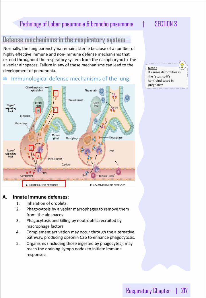
Respiratory Chapter | 217
Note:Itcausesdeformitiesinthefetus,soit’scontraindicatedinpregnancy
Pathology of Lobar pneumonia & broncho pneumonia | SECTION 3
Normally,thelungparenchymaremainssterilebecauseofanumberofhighlyeffectiveimmuneand non-immunedefensemechanismsthatextendthroughouttherespiratorysystemfromthenasopharynxtothealveolarairspaces.Failureinanyofthesemechanismscanleadtothedevelopmentofpneumonia.
Defense mechanisms in the respiratory system
A. Innateimmunedefenses:1. Inhalationofdroplets.2. Phagocytosisbyalveolarmacrophagestoremovethem
fromtheairspaces.3. Phagocytosisandkillingbyneutrophilsrecruitedby
macrophagefactors.4. Complementactivationmayoccurthroughthealternative
pathway,producingopsonin C3btoenhancephagocytosis.5. Organisms(includingthoseingestedbyphagocytes),may
reachthedraininglymphnodestoinitiateimmuneresponses.
Immunologicaldefensemechanismsofthelung:
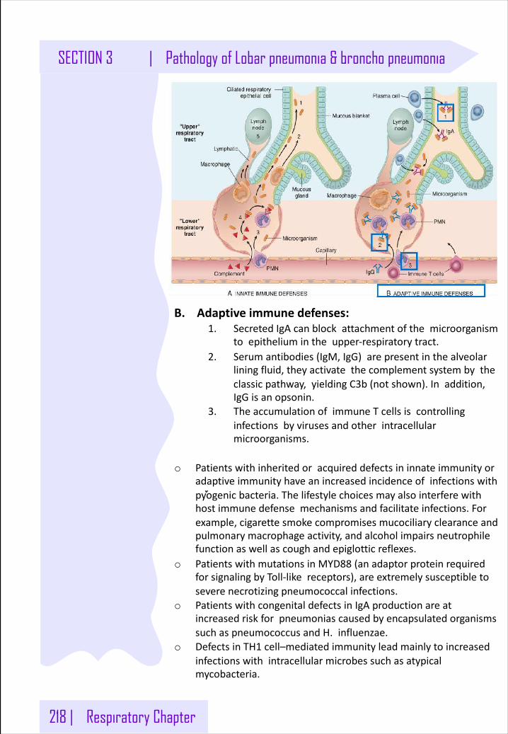
218 | Respiratory Chapter
SECTION 3 | Pathology of Lobar pneumonia & broncho pneumonia
B.Adaptiveimmunedefenses:1. SecretedIgAcanblockattachmentofthemicroorganism
toepitheliumintheupper-respiratorytract.2. Serumantibodies(IgM,IgG)arepresentinthealveolar
liningfluid,theyactivatethecomplementsystembytheclassicpathway,yieldingC3b(notshown).Inaddition,IgGisanopsonin.
3. TheaccumulationofimmuneTcellsiscontrollinginfectionsbyvirusesandotherintracellularmicroorganisms.
o Patientswithinheritedoracquireddefectsininnateimmunityoradaptiveimmunityhaveanincreasedincidenceofinfectionswithpyogenicbacteria.Thelifestylechoicesmayalsointerferewithhostimmunedefensemechanismsandfacilitateinfections.Forexample,cigarettesmokecompromisesmucociliary clearanceandpulmonarymacrophageactivity,andalcoholimpairsneutrophilefunctionaswellascoughandepiglottic reflexes.
o PatientswithmutationsinMYD88 (anadaptorproteinrequiredforsignalingbyToll-likereceptors),areextremelysusceptibletoseverenecrotizingpneumococcalinfections.
o PatientswithcongenitaldefectsinIgAproductionareatincreasedriskforpneumoniascausedbyencapsulatedorganismssuchaspneumococcusandH.influenzae.
o DefectsinTH1 cell–mediatedimmunityleadmainlytoincreasedinfectionswithintracellularmicrobessuchasatypicalmycobacteria.
Respiratory Chapter | 219
Note:Bacterialpneumoniasareclassifiedaccordingtothespecificetiologicagentor,ifnopathogencanbeisolated,bytheclinicalsettinginwhichtheinfectionoccurs.
Pathology of Lobar pneumonia & broncho pneumonia | SECTION 3
Pneumonia classificationBasedontheetiology:
o Streptococcuspneumoniae(Pneumococcal)
o Klebsiellapneumoniae:
inchronicalcoholicpeopleandwhoaredebilitated.
o Legionellapneumonia:
Especiallyinimmunocompromisedposttransplant.thebacterialoves
watertanksoranywetthings.
o Haemophilus influenzae:
isthemostcommonbacterialcauseofacuteexacerbationsofCOPD.
o Moraxellacatarrhalisorganisms:
Itisthesecondmostcommonbacterialcauseofacuteexacerbationof
COPDinadults.
o Staphylococcalspecies.
o streptococcuspyogenes.
Clinically:o CommunityAcquiredacutePneumonia.
o CommunityAcquiredAtypicalPneumonia.
o HospitalAcquired(Nosocomial)Pneumonia.
o AspirationPneumonia
o ChronicPneumonia
o NecrotizingPneumoniaandLungAbscess
o Opportunisticpneumonias(PneumoniaintheImmunocompromised
Host)
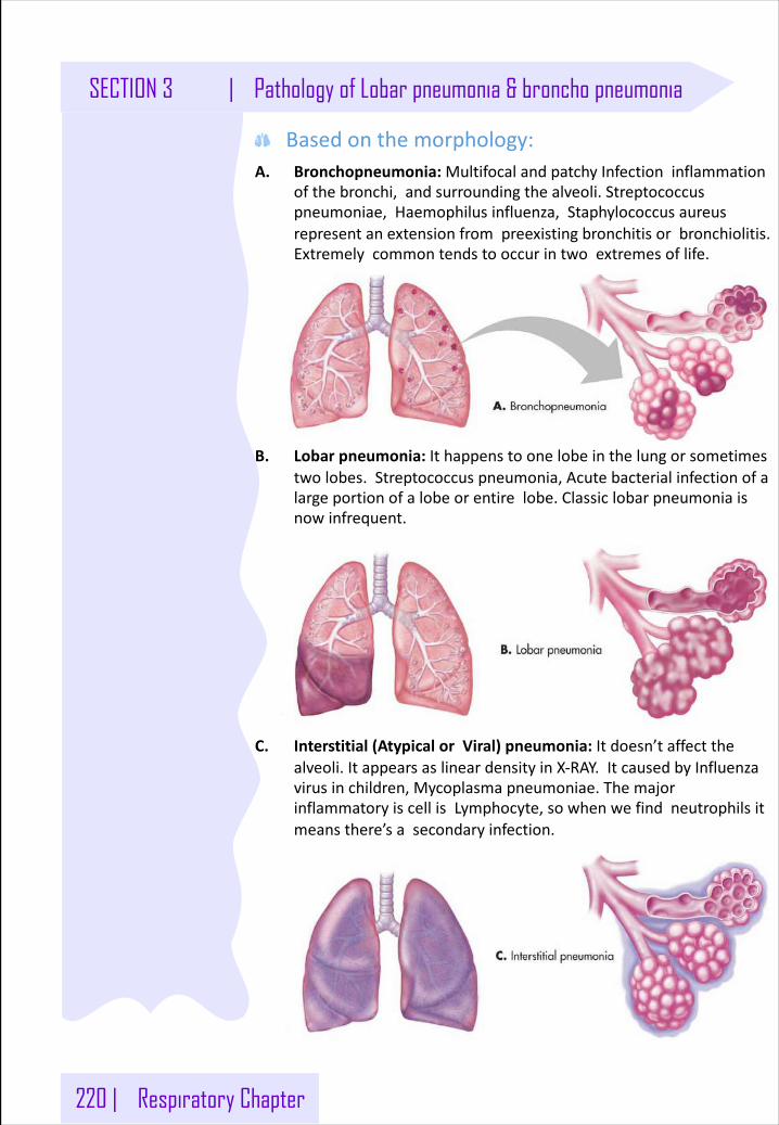
220 | Respiratory Chapter
SECTION 3 | Pathology of Lobar pneumonia & broncho pneumonia
A. Bronchopneumonia:MultifocalandpatchyInfectioninflammationofthebronchi,andsurroundingthealveoli.Streptococcuspneumoniae,Haemophilus influenza,Staphylococcusaureusrepresentanextensionfrompreexistingbronchitisorbronchiolitis.Extremelycommontendstooccurintwoextremesoflife.
B. Lobarpneumonia:Ithappenstoonelobeinthelungorsometimestwolobes.Streptococcuspneumonia,Acutebacterialinfectionofalargeportionofalobeorentirelobe.Classiclobarpneumoniaisnowinfrequent.
C. Interstitial(AtypicalorViral)pneumonia:Itdoesn’taffectthealveoli.ItappearsaslineardensityinX-RAY.ItcausedbyInfluenzavirusinchildren,Mycoplasmapneumoniae.ThemajorinflammatoryiscellisLymphocyte,sowhenwefindneutrophilsitmeansthere’sasecondaryinfection.
Basedonthemorphology:
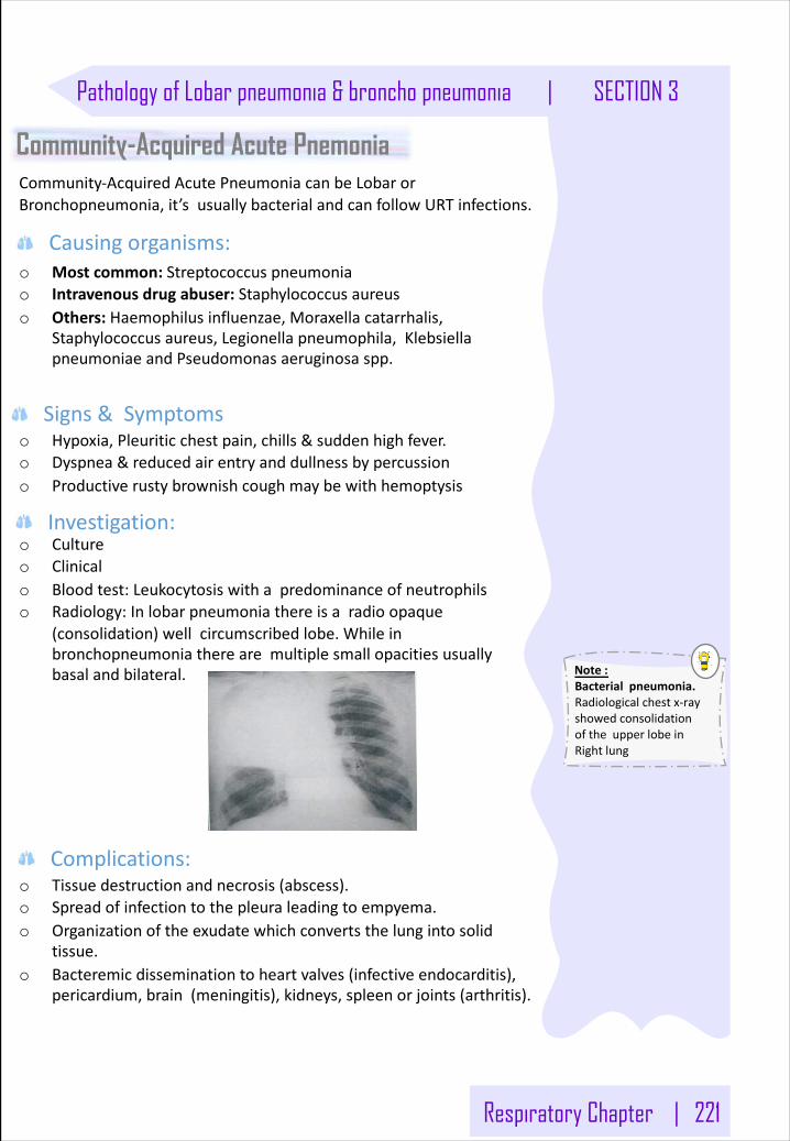
Respiratory Chapter | 221
Note:Bacterialpneumonia.Radiologicalchestx-rayshowedconsolidationoftheupperlobeinRightlung
Pathology of Lobar pneumonia & broncho pneumonia | SECTION 3
Community-Acquired Acute PnemoniaCommunity-AcquiredAcutePneumoniacanbeLobarorBronchopneumonia,it’susuallybacterialandcanfollowURTinfections.
o Mostcommon:Streptococcuspneumoniao Intravenousdrugabuser:Staphylococcusaureuso Others:Haemophilus influenzae,Moraxellacatarrhalis,
Staphylococcusaureus,Legionellapneumophila,Klebsiellapneumoniae andPseudomonasaeruginosaspp.
o Hypoxia,Pleuriticchestpain,chills&suddenhighfever.o Dyspnea&reducedairentryanddullnessbypercussiono Productiverustybrownishcoughmaybewithhemoptysis
o Cultureo Clinicalo Bloodtest:Leukocytosiswithapredominanceofneutrophilso Radiology:Inlobarpneumoniathereisaradioopaque
(consolidation)wellcircumscribedlobe.Whileinbronchopneumoniatherearemultiplesmallopacitiesusuallybasalandbilateral.
o Tissuedestructionandnecrosis(abscess).o Spreadofinfectiontothepleuraleadingtoempyema.o Organizationoftheexudatewhichconvertsthelungintosolid
tissue.o Bacteremic disseminationtoheartvalves(infectiveendocarditis),
pericardium,brain(meningitis),kidneys,spleenorjoints(arthritis).
Causingorganisms:
Signs&Symptoms
Investigation:
Complications:
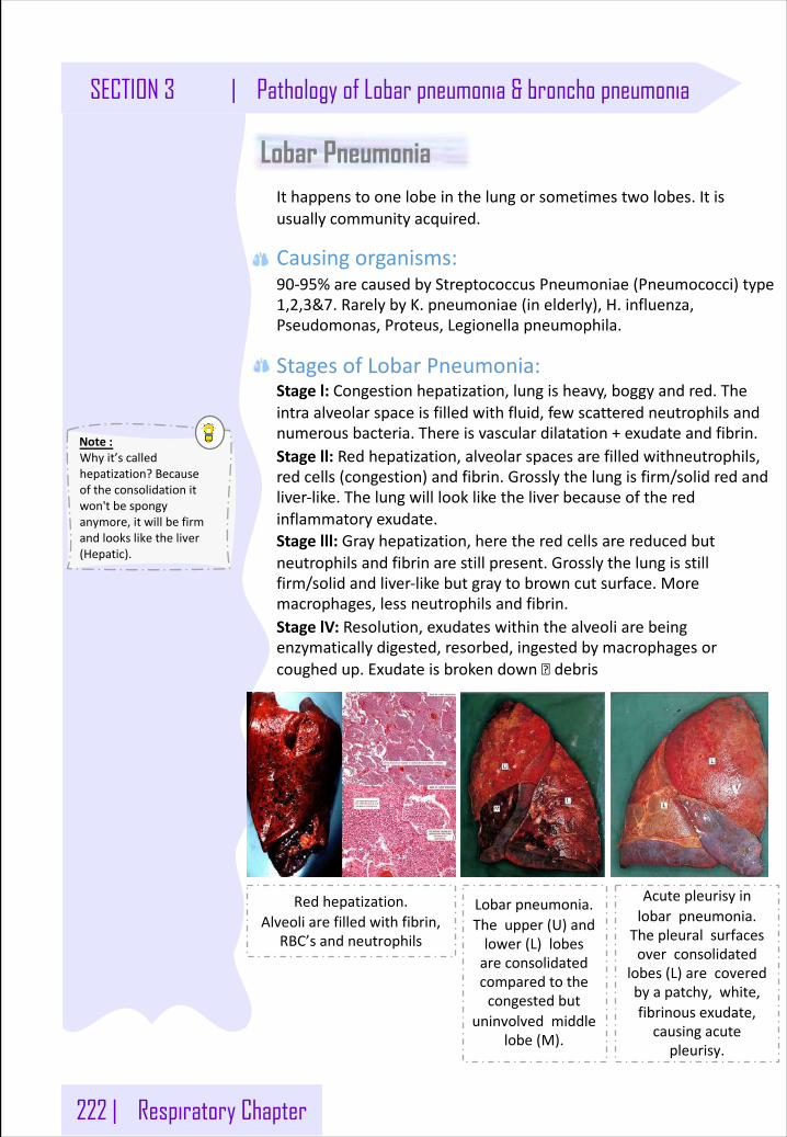
222 | Respiratory Chapter
Note:Whyit’scalledhepatization?Becauseoftheconsolidationitwon'tbespongyanymore,itwillbefirmandlooksliketheliver(Hepatic).
SECTION 3 | Pathology of Lobar pneumonia & broncho pneumonia
Lobar PneumoniaIthappenstoonelobeinthelungorsometimestwolobes.Itisusuallycommunityacquired.
Causingorganisms:90-95%arecausedbyStreptococcusPneumoniae (Pneumococci)type1,2,3&7.RarelybyK.pneumoniae (inelderly),H.influenza,Pseudomonas,Proteus,Legionellapneumophila.
StagesofLobarPneumonia:Stagel:Congestionhepatization,lungisheavy,boggyandred.Theintraalveolarspaceisfilledwithfluid,fewscatteredneutrophilsandnumerousbacteria.Thereisvasculardilatation+exudateandfibrin.Stagell:Redhepatization,alveolarspacesarefilledwithneutrophils,redcells(congestion)andfibrin.Grosslythelungisfirm/solidredandliver-like.Thelungwilllookliketheliverbecauseoftheredinflammatoryexudate.Stagelll:Grayhepatization,heretheredcellsarereducedbutneutrophilsandfibrinarestillpresent.Grosslythelungisstillfirm/solidandliver-likebutgraytobrowncutsurface.Moremacrophages,lessneutrophilsandfibrin.StagelV:Resolution,exudateswithinthealveoliarebeingenzymaticallydigested,resorbed,ingestedbymacrophagesorcoughedup.Exudateisbrokendown� debris
Redhepatization.Alveoliarefilledwithfibrin,
RBC’sandneutrophils
Lobarpneumonia.Theupper(U)andlower(L)lobesareconsolidatedcomparedtothecongestedbut
uninvolvedmiddlelobe(M).
Acutepleurisyinlobarpneumonia.Thepleuralsurfacesoverconsolidated
lobes(L)arecoveredbyapatchy,white,fibrinousexudate,causingacute
pleurisy.
.
.
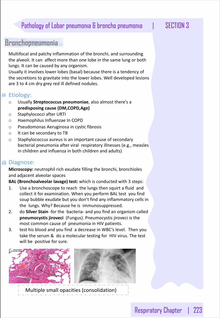
Respiratory Chapter | 223
Pathology of Lobar pneumonia & broncho pneumonia | SECTION 3
.
BronchopneumoniaMultifocalandpatchyinflammationofthebronchi,andsurroundingthealveoli.Itcanaffectmorethanonelobeinthesamelungorbothlungs.Itcanbecausedbyanyorganism.Usuallyitinvolveslowerlobes(basal)becausethereisatendencyofthesecretionstogravitateintothelowerlobes.Welldevelopedlesionsare3 to4 cmdrygreyredilldefinednodules.
Etiology:o UsuallyStreptococcuspneumoniae,alsoalmostthere’sa
predisposingcause(DM,COPD,Age)o StaphylococciafterURTIo Haemophilus Influenzae InCOPDo PseudomonasAeruginosaincysticfibrosiso ItcanbesecondarytoTBo Staphylococcusaureusisanimportantcauseofsecondary
bacterialpneumoniaafterviralrespiratoryillnesses(e.g.,measlesinchildrenandinfluenzainbothchildrenandadults)
Diagnose:Microscopy:neutrophilrichexudatefillingthebronchi,bronchiolesandadjacentalveolarspacesBAL(Bronchoalveolarlavage)test:whichisconductedwith3 steps:1. Useabronchoscopetoreachthelungsthensquirtafluidand
collectitforexamination.WhenyouperformBALtestyoufindsoupbubbleexudatebutyoudon’tfindanyinflammatorycellsinthelungs.Why?Becauseheisimmunosuppressed.
2. doSilverStain-forthebacteria- andyoufindanorganismcalledpneumocystisjiroveci (Fungus).Pneumocystisjiroveci isthemostcommoncauseofpneumoniainHIVpatients.
3. testhisbloodandyoufindadecreaseinWBC’slevel.Thenyoutaketheserum&doamoleculartestingforHIVvirus.Thetestwillbepositiveforsure.
Multiplesmallopacities(consolidation)
.
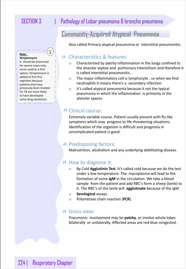
224 | Respiratory Chapter
Note:StreptomycinItshouldbepreservedforseverecasesonly,neverusedasafirstoption.StreptomycinisaddedtofirstlineregimensbecausepatientsthathavepreviouslybeentreatedforTBaremorelikelytohavedevelopedsomedrugresistance.
SECTION 3 | Pathology of Lobar pneumonia & broncho pneumonia
.
Community-Acquired Atypical PneumoniaAlsocalledPrimaryatypicalpneumoniaorinterstitialpneumonitis.
Characteristics&features:o Characterizedbypatchyinflammationinthelungsconfinedto
thealveolarseptae andpulmonaryinterstitium andthereforeitiscalledinterstitialpneumonitis..
o Themajorinflammatorycellislymphocyte,sowhenwefindneutrophilsitmeansthere'sasecondaryinfection.
o It’scalledatypicalpneumoniabecauseitnotthetypicalpneumoniainwhichtheinflammationisprimarilyinthealveolarspaces.
Clinicalcourse:Extremelyvariablecourse.Patientusuallypresentwithflulikesymptomswhichmayprogresstolifethreateningsituations.Identificationoftheorganismisdifficultandprognosisinuncomplicatedpatientisgood.
Predisposingfactors:Malnutrition,alcoholismandanyunderlyingdebilitatingdisease.
Howtodiagnoseit:o ByColdAgglutininTest.It’scalledcoldbecausewedothetest
underalowtemperature.ThemycoplasmawillleadtotheformationofsomeIgM inthecirculation.WetakeabloodsamplefromthepatientandaddRBC’sformasheep(lamb)toit.TheRBC’softhelambwillagglutinate becauseoftheIgM.
o Serological assays.o Polymerasechainreaction(PCR).
Grossview:Pneumonicinvolvementmaybepatchy,orinvolvewholelobesbilaterallyorunilaterally.Affectedareasarered-bluecongested.
.
.
.
.
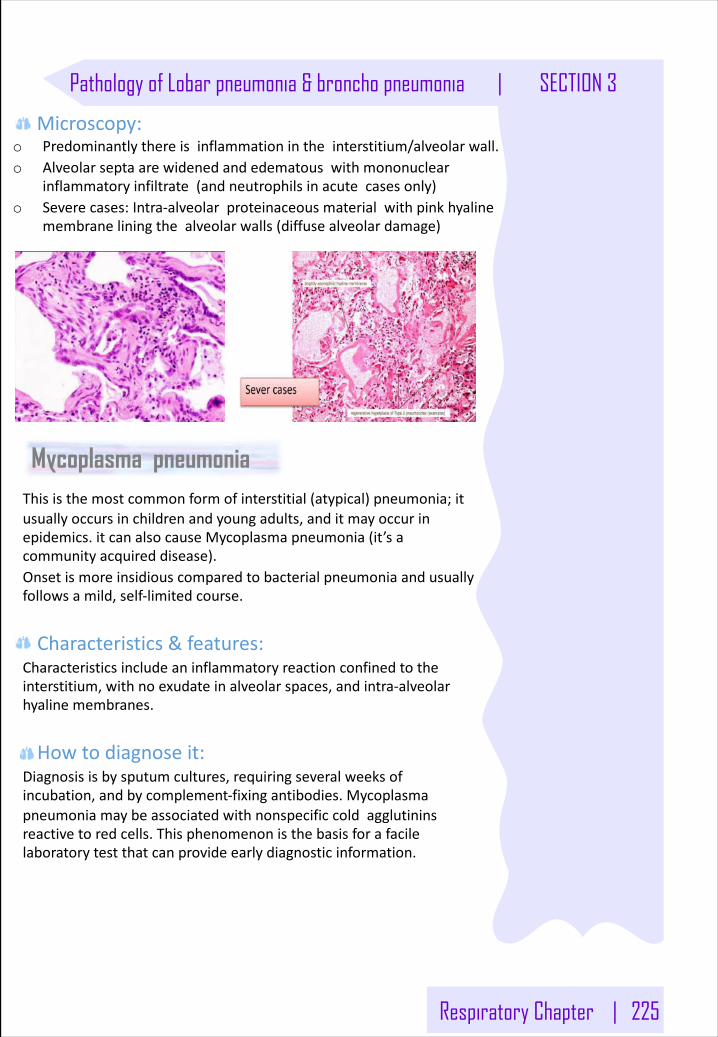
Respiratory Chapter | 225
Pathology of Lobar pneumonia & broncho pneumonia | SECTION 3
.
Microscopy:o Predominantlythereisinflammationintheinterstitium/alveolarwall.o Alveolarseptaarewidenedandedematouswithmononuclear
inflammatoryinfiltrate(andneutrophilsinacutecasesonly)o Severecases:Intra-alveolarproteinaceous materialwithpinkhyaline
membraneliningthealveolarwalls(diffusealveolardamage)
Mycoplasma pneumoniaThisisthemostcommonformofinterstitial(atypical)pneumonia;itusuallyoccursinchildrenandyoungadults,anditmayoccurinepidemics.itcanalsocauseMycoplasmapneumonia(it’sacommunityacquireddisease).Onsetismoreinsidiouscomparedtobacterialpneumoniaandusuallyfollowsamild,self-limitedcourse.
Characteristics&features:Characteristicsincludeaninflammatoryreactionconfinedtotheinterstitium,withnoexudateinalveolarspaces,andintra-alveolarhyalinemembranes.
Howtodiagnoseit:Diagnosisisbysputumcultures,requiringseveralweeksofincubation,andbycomplement-fixingantibodies.Mycoplasmapneumoniamaybeassociatedwithnonspecificcoldagglutininsreactivetoredcells.Thisphenomenonisthebasisforafacilelaboratorytestthatcanprovideearlydiagnosticinformation.
.
.
226 | Respiratory Chapter
SECTION 3 | Pathology of Lobar pneumonia & broncho pneumonia
Viral pneumoniasViralpneumoniasarethemostcommontypesofpneumoniainchildhood.Theyarecausemostcommonlyby:o Influenzavirus(children)o InfluenzaAandB(adults)o Adenoviruseso Rhinoviruso Respiratorysyncytialviruso SARSvirus.Mayalsoariseafterchildhoodexanthems (viraleruptions)suchasrubeola (measles)orvaricella(chickenpox);themeaslesvirusproducesgiantcellpneumonia,markedbynumerousgiantcellsandoftencomplicatedbytracheobronchitis.
Coxiella burnettiQfeveristhemostcommonrickettsial pneumonia.ItiscausedbyCoxiellaburnetti.Itmayinfectpersonsworkingwithinfectedcattleorsheep,whoinhaledustparticlescontainingtheorganism,orthosewhodrinkunpasteurizedmilkfrominfectedanimals.
ChlamydiaCausesOrnithosis (psittacosis),whichistransmittedbyinhalationofdriedexcretaofinfectedbirds.
Nosocomial pneumonia
Patientstatus Causes
- Severeunderlyingconditions, e.g.Immunosuppression.
- Prolongedantibiotictherapy.- Intravascularcatheter.- Patientswithmechanicalventilator.
Gram-negativeorganismslikeKlebsiella,PseudomonasaeruginosaandE.coli.
(HospitalacquiredPneumonia)
Respiratory Chapter | 227
Pathology of Lobar pneumonia & broncho pneumonia | SECTION 3
Aspiration pneumonia
Patientstatus Causes
- Debilitatedpatients- Comatose- Alcoholic- Thosewhoaspirated
gastriccontents.
Chemicalinjuryduegastricacidandbacterialinfection(anaerobicbacteriaadmixedwithaerobicbacteria,e.g.Bacteroides,FusobacteriumandPeptococcus).
Anecrotizingpneumoniawithfulminantclinicalcourse,commoncomplication(abscess)andfrequentcauseofdeath.
Chronic pneumoniaPatientstatus Causes
OftenalocalizedlesioninImmunocompetentperson,withorwithoutregionallymphnodeinvolvement. Intheimmunocompromised,thereisusuallysystemicdisseminationofthecausativeorganism,accompaniedbywidespreaddisease.
Thereistypicallygranulomatousinflammation, whichmaybeduetobacteria(M.Tuberculosis)orFungi(Histoplasmacapsulatum,coccidioidesimmitis,blastomyces.Tuberculosis isthemostimportantentitywithinthespectrumofchronicpneumonias.
Opportunistic pneumonia
Patientstatus Causes
Immunosuppressedpatients(AIDS,cancerpatientsandtransplantrecipients).
Cytomegalovirus,Pneumocystis jiroveci(carinii), Mycobacteriumavium-intracellulare,Invasiveaspergillosis,Invasivecandidiasisand"Usual"bacterial,viral,andfungalorganisms.
Effectivemethodsofdiagnosisare:identifytheorganisminbronchoalveolarlavagefluidsorinatransbronchialbiopsyspecimen.immunofluorescenceantibodykitsandPCR-basedassays
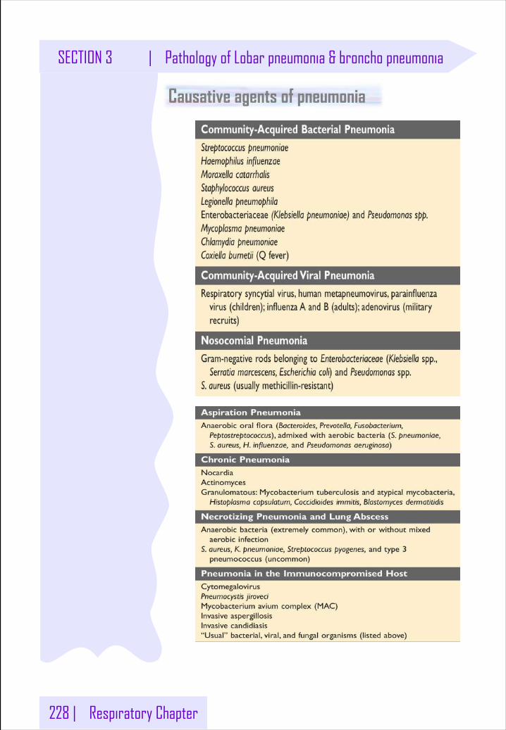
228 | Respiratory Chapter
SECTION 3 | Pathology of Lobar pneumonia & broncho pneumonia
Causative agents of pneumonia
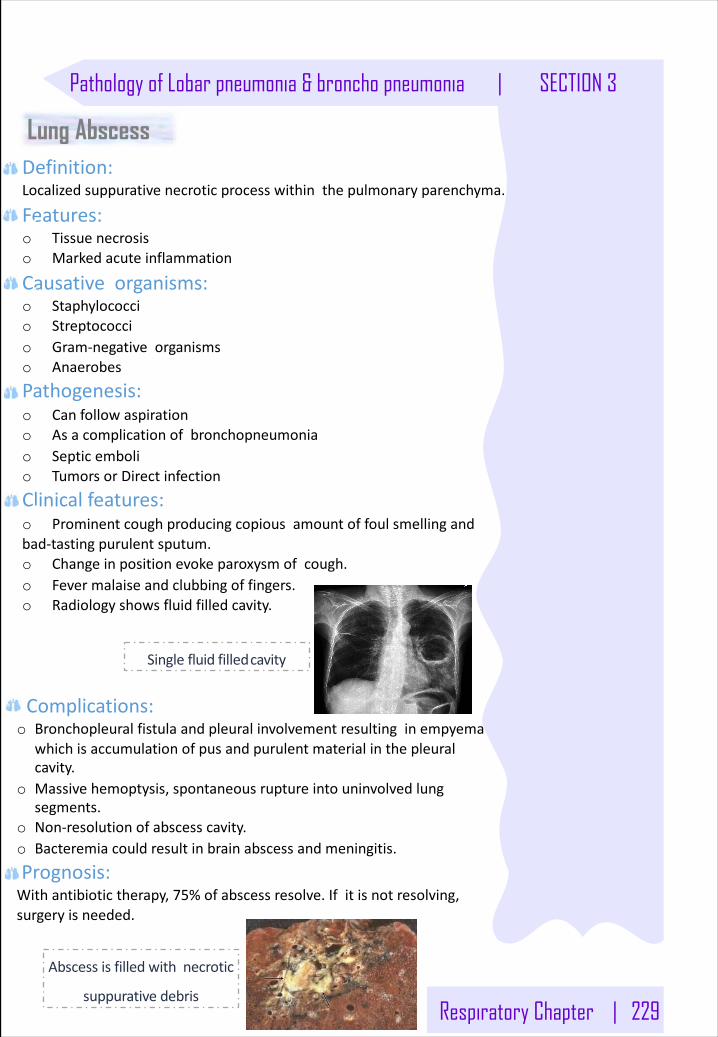
Respiratory Chapter | 229
Pathology of Lobar pneumonia & broncho pneumonia | SECTION 3
Singlefluidfilledcavity
Lung AbscessDefinition:Localizedsuppurativenecroticprocesswithinthepulmonaryparenchyma.
Features:o Tissuenecrosiso Markedacuteinflammation
Causativeorganisms:o Staphylococcio Streptococcio Gram-negativeorganismso Anaerobes
Pathogenesis:o Canfollowaspirationo Asacomplicationofbronchopneumoniao Septicembolio TumorsorDirectinfection
Clinicalfeatures:o Prominentcoughproducingcopiousamountoffoulsmellingandbad-tastingpurulentsputum.o Changeinpositionevokeparoxysmofcough.o Fevermalaiseandclubbingoffingers.o Radiologyshowsfluidfilledcavity.
Complications:o Bronchopleuralfistulaandpleuralinvolvementresultinginempyema
whichisaccumulationofpusandpurulentmaterialinthepleuralcavity.
o Massivehemoptysis,spontaneousruptureintouninvolvedlungsegments.
o Non-resolutionofabscesscavity.o Bacteremiacouldresultinbrainabscessandmeningitis.
Prognosis:Withantibiotictherapy,75%ofabscessresolve.Ifitisnotresolving,surgeryisneeded.
Abscessisfilledwithnecrotic
suppurativedebris
.
.
.
.
.
.
.
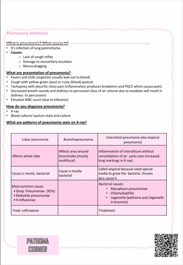
Pulmonary Infections
Whatispneumonia?Whatcauses it?• It'sinfectionoflung parenchyma.• Causes:
o Lackofcough reflexo Damagetomucociliary escalatoro Mucus plugging
Whatarepresentationof pneumonia?• Feversandchills(organismusuallyleakoutto blood)• Coughwithyellow-green(pus)orrusty(blood) sputum• Tachypneawithpleuriticchestpain(inflammationproducesbradykininandPGE2 whichcauses pain)• Decreasedbreathsoundsanddullnesstopercussion(lossofairvolumeduetoexudateswillresultin
dullnessto percussion)• ElevatedWBCcount(dueto infection)
Howdoyoudiagnose pneumonia?• X-ray• Bloodculture/sputumstainand culture
Whatarepatternsofpneumoniaseenon X-ray?
Lobar pneumonia Bronchopneumonia Interstitialpneumonia(akaatypicalpneumonia)
AffectswholelobeAffects areaaroundbronchioles (mostlymultifocal)
Inflammation of interstitiumwithoutconsolidation of airsacks(seeincreasedlungmarkingsin X-ray)
CauseismostlybacterialCauseismostlybacterial
Calledatypicalbecauseneedspecialmediatogrowthebacteria.Virusesalsocause it
Mostcommoncause:• Strep. Pneumoniae(95%)• Klebsiellapneumoniae•H influenziae
Bacterial causes:• Mycoplasm pneumoniae• Chlamydophilla• Legionella(pathomaputslegionella
in broncho)
Treat: ceftriaxone Treatment:
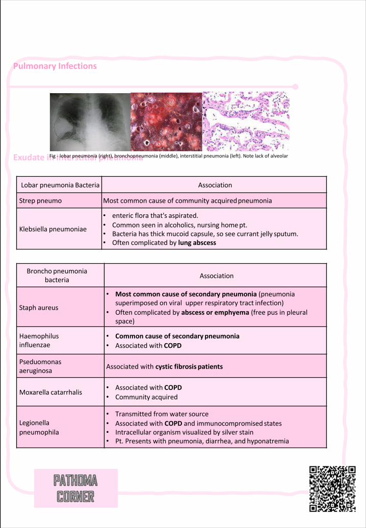
Pulmonary Infections
Exudateininterstitial pneumoniaFig- lobarpneumonia(right),bronchopneumonia(middle),interstitialpneumonia(left).Notelackof alveolar
Lobarpneumonia Bacteria Association
Strep pneumo Mostcommoncauseofcommunityacquiredpneumonia
Klebsiella pneumoniae
• entericflorathat's aspirated.• Commonseeninalcoholics,nursinghomept.• Bacteriahasthickmucoidcapsule,soseecurrantjelly sputum.• Oftencomplicatedbylung abscess
Broncho pneumoniabacteria Association
Staph aureus
• Mostcommoncauseofsecondarypneumonia(pneumoniasuperimposedonviralupperrespiratorytract infection)
• Oftencomplicatedbyabscessoremphyema(freepusinpleuralspace)
Haemophilusinfluenzae
• Commoncauseofsecondary pneumonia• Associatedwith COPD
Pseduomonasaeruginosa Associatedwithcysticfibrosis patients
Moxarella catarrhalis• Associatedwith COPD• Community acquired
Legionellapneumophila
• Transmittedfromwater source• AssociatedwithCOPDandimmunocompromised states• Intracellularorganismvisualizedbysilver stain• Pt.Presentswithpneumonia,diarrhea,and hyponatremia
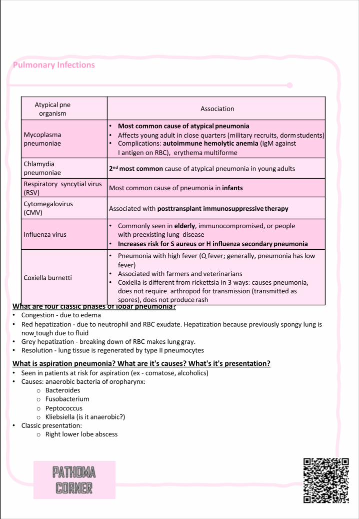
Pulmonary Infections
Whatarefourclassicphasesoflobar pneumonia?• Congestion- dueto edema• Redhepatization - duetoneutrophilandRBCexudate.Hepatization becausepreviouslyspongylungis
now toughdueto fluid• Greyhepatization - breakingdownofRBCmakeslung gray.• Resolution- lungtissueisregeneratedbytypeII pneumocytes
Whatisaspirationpneumonia?Whatareit'scauses?What'sit's presentation?• Seeninpatientsatriskforaspiration(ex- comatose, alcoholics)• Causes:anaerobicbacteriaof oropharynx:
o Bacteroideso Fusobacteriumo Peptococcuso Kliebsiella (isit anaerobic?)
• Classic presentation:o Rightlowerlobe abscess
Atypical pneorganism
Association
Mycoplasmapneumoniae
• Mostcommoncauseofatypical pneumonia• Affectsyoungadultinclosequarters(militaryrecruits,dormstudents)• Complications:autoimmunehemolyticanemia (IgMagainst
IantigenonRBC),erythemamultiformeChlamydiapneumoniae 2ndmostcommoncauseofatypicalpneumoniainyoung adults
Respiratorysyncytialvirus(RSV)
Mostcommoncauseofpneumoniain infants
Cytomegalovirus(CMV) Associatedwithposttransplantimmunosuppressive therapy
Influenza virus• Commonlyseeninelderly,immunocompromised,orpeople
withpreexistinglungdisease• IncreasesriskforSaureusorHinfluenzasecondary pneumonia
Coxiella burnetti
• Pneumoniawithhighfever(Qfever;generally,pneumoniahaslowfever)
• Associatedwithfarmersand veterinarians• Coxiellaisdifferentfromrickettsiain3 ways:causespneumonia,
doesnotrequirearthropodfortransmission(transmittedasspores),doesnotproducerash
230 | Respiratory Chapter
SECTION 3 | Hospital Acquired pneumonia
Pneumonia
§ Definetheterms,pneumonia,communityacquiredpneumonia,healthcareassociatedpneumonia(HCAP)andventilatorassociatedpneumonia(VAP).
§ Describethepathogenesisofthehealthcareassociatedpneumonia(hospitalassociatedpneumonia)andVAP.
§ ClassifyHCAPaccordingtothetimeofonset&Namethedifferentcausativebacterialagents
§ ClassifyanddescribetypesofVAP.
§ RecognizethewaysbywhichVAPisprevented.
§ Describethedifferentchemotherapeuticantimicrobialagentsusedforthetreatmentofhealthcareassociatedpneumonia.
§ Evaluateresponsetotreatmentandrecognizereasonsforfailureoftreatment
Objec
tive
Definition:infectionofthepulmonaryparenchyma.Types:o Communityacquiredpneumonia:Acquiredinthecommunity.Theorganismscausingitusuallysusceptibletoantibiotics..Example:streptococcuspneumonia.o Healthcareassociatedpneumonia(Nosocomialpneumonia):
Acquiredatleast48 hours(andnotincubating)afteradmissiontohealthcareinstitutions.Theorganismscausingitusuallyresistanttoantibiotics.Example:PseudomonasAeruginosa.Ifthesymptomsoccurbefore48 hours,thentheinfectionisacquiredfromthecommunitynotthehospital.
- HospitalAcquiredPneumonia(HAP)- VentilatorAssociatedPneumonia(VAP):inpatientswith
assistedrespiration(mechanicalventilation)foraperiodatleast48 hours.
EpidemiologyofNosocomialPneumonia:o Nosocomialpneumoniaisthe2nd mostcommonhospital-
acquiredinfectionsafterurinarytractinfectiono Nosocomialpneumoniaistheleadingcauseofdeathfrom
hospital-acquiredinfections.o TheincidenceofnosocomialpneumoniaishighestinICU
patients.o Theincidenceofnosocomialpneumoniainventilatedpatientsis
10-foldhigher thannon-ventilatedpatients.o Thereportedcrudemortality forHAPis30%togreaterthan70%.
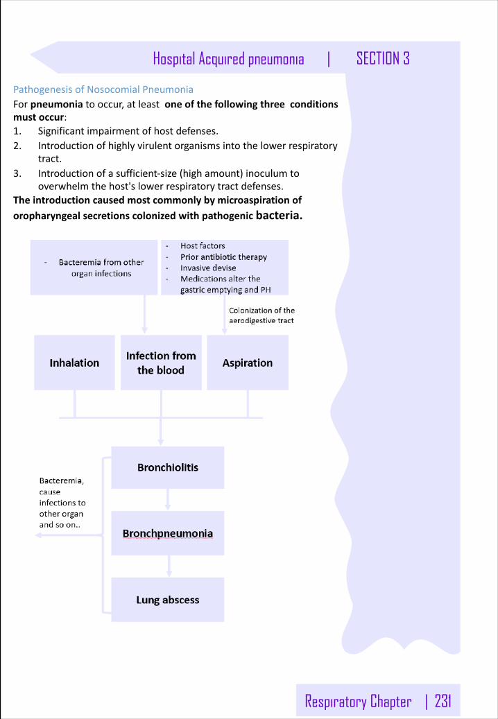
Respiratory Chapter | 231
Hospital Acquired pneumonia | SECTION 3
PathogenesisofNosocomialPneumoniaForpneumonia tooccur,atleastoneofthefollowingthreeconditionsmustoccur:1. Significantimpairmentofhostdefenses.2. Introductionofhighlyvirulentorganismsintothelowerrespiratory
tract.3. Introductionofasufficient-size(highamount)inoculumto
overwhelmthehost'slowerrespiratorytractdefenses.Theintroductioncausedmostcommonlybymicroaspiration oforopharyngealsecretionscolonizedwithpathogenicbacteria.
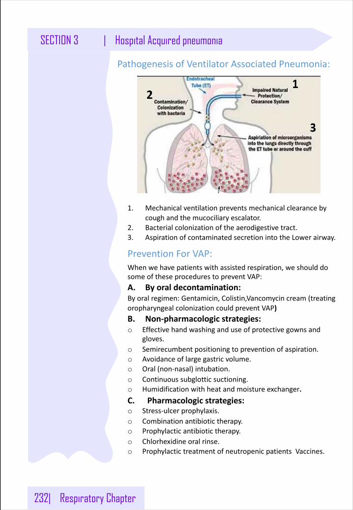
232| Respiratory Chapter
SECTION 3 | Hospital Acquired pneumonia
PathogenesisofVentilatorAssociatedPneumonia:
1. Mechanicalventilationpreventsmechanicalclearancebycoughandthemucociliary escalator.
2. Bacterialcolonizationoftheaerodigestivetract.3. AspirationofcontaminatedsecretionintotheLowerairway.
PreventionForVAP:Whenwehavepatientswithassistedrespiration,weshoulddosomeoftheseprocedurestopreventVAP:A. Byoraldecontamination:Byoralregimen:Gentamicin,Colistin,Vancomycin cream(treatingoropharyngealcolonizationcouldpreventVAP)B. Non-pharmacologicstrategies:o Effectivehandwashinganduseofprotectivegownsand
gloves.o Semirecumbent positioningtopreventionofaspiration.o Avoidanceoflargegastricvolume.o Oral(non-nasal)intubation.o Continuoussubglotticsuctioning.o Humidificationwithheatandmoistureexchanger.C. Pharmacologicstrategies:o Stress-ulcerprophylaxis.o Combinationantibiotictherapy.o Prophylacticantibiotictherapy.o Chlorhexidineoralrinse.o ProphylactictreatmentofneutropenicpatientsVaccines.
Respiratory Chapter | 233
Hospital Acquired pneumonia | SECTION 3
Classificationofnosocomialpneumonia:
ByClassifyingPneumoniaaccordingtotheonset,wecanidentifythegroupoforganismscausingit.
A. Early-onsetnosocomialpneumonia:Occursduringthefirst4daysofadmission.- S.pneumonia- MethicillinsensitiveS.Aureus- H.Influenza- Anaerobes
B. Late-onsetnosocomialpneumonia:occursmorethan4 daysofadmission.- Gramnegativeorganismslike:Pseudomonasaeruginosa,
Acinetobacter.- Enterobacteriaceae like:Klebsiella,Enterobacter,serratia.- MethicillinresistanceS.Aureus
InthecaseofVAP,theClassificationis:ThesamePrincipleappliedtoVAP,butwestartcountingthedaysofTheonsetofthediseasefromthetrachealintubation,notfromtheadmissiontothehospital.
A. Early-onsetVAP:within48-72 hoursaftertrachealintubation.
B. Late-onsetVAP:after72 hoursaftertrachealintubation.
234 | Respiratory Chapter
SECTION 3 | Hospital Acquired pneumonia
CausativeAgents:
A. EntericGramnegativebacilli:Mostfrequentlyinpatients:
o Withlate-onsetdiseaseo withseriousunderlyingdiseaseoftenalreadyonbroad-
spectrumantibiotics.Prioruseofbroad-spectrumantibioticsandanimmunocompromisedstatemakeresistantGram-negativeorganismsmorelikely.
B.S.aureus:Mostfrequentlyinpatients:
o Ventilatedpatientsafterheadtrauma,neurosurgeryandwoundinfection.
o Receivedpriorantibiotics.o ProlongedcareinICU.
SpeciallymethicillinresistantS.aureus iscommonlyinpatientswho:o Receivedcorticosteroids.o Undergonemechanicalventilation>5 days.o Presentedwithchroniclungdisease.
C.Pseudomonasaeruginosa,AcinetobacterMostfrequentlyinpatients:
o Withlate-onsetpneumonia.o Inventilatedpatients.
ThefrequencyofICU-acquiredP.aeruginosacarriageorcolonization/infectionwas23.4%at7 daysand57.8%at14 days.
D. Anaerobes:Mostfrequentlyinpatients:
o Predisposedtoaspiration..o Anaerobesoccurredmoreoftenwithoropharyngeal
intubationthannasopharyngealintubation.
Respiratory Chapter | 235
Hospital Acquired pneumonia | SECTION 3
TreatmentofNosocomialPneumonia:
Mostinitialtherapyisempiric,becausethepathogenisnotidentifiedorresultsarenotavailablewhenantimicrobialdecisionsaremadeinmostpatients,soinitiallybetreatedwithabroad- spectrumantibioticregimenaimedatcoveringalllikelybacterialpathogens.Thisregimenshouldsubsequentlybenarrowed,accordingtotheresultofculture.
AmericanThoracicSocietyhasdividedpatientsintothreegroups,eachwithasetofprobablepathogens:1. MildtomoderateHAPwithno riskfactor2. MildtomoderateHAPwith riskfactor3. A.severeHAP,early-onsetwithno riskfactor3.b.severeHAP,late-onsetorwith riskfactor
MildtomoderateHAP,monotherapyhasbeenshowntobeeffective.SevereHAPinwhichinfectionwithresistantorganismsislikely,combination therapyprobablyshouldbeinstituteduntilcultureresultareavailable.
- Vancomycin+Linezolid(betterduetolessnephrotoxicity)forPatientswithS.aureusinfection.
- Combination ofAntipseudomonaldrugs.ThereiscontroversyinthecombinationofAntipseudomonaldrugs:
- AntipseudomonalBeta-lactamwithanAminoglycoside.Synergybutpotentialnephrotoxicity.
- AntipseudomonalBeta-lactamwithaFluoroquinolone.Nosynergybutreducednephrotoxicity.
Responsetothetherapy:Ifnoclinicalresponseisnotedordeteriorationoccurs,weneedtoconsider:
Infectiouscauses: Noninfectiousevents:o Resistantpathogeno Resistantpathogeno Superinfectiono Unusualpathogenso Lungabscesso Extrapulmonary infection
o Heart:congestiveheartfailure.o Lung:fibroproliferative acute
respiratorydistresssyndrome(ARDS),pulmonaryemboli,Atelectesis.
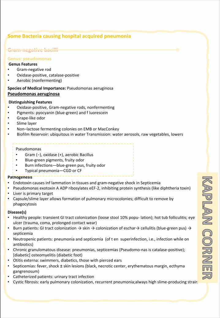
SomeBacteriacausinghospitalacquiredpneumonia
Gram-negativebacilliGenus:pseudomonasGenusFeatures• Gram-negativerod• Oxidase-positive,catalase-positive• Aerobic(nonfermenting)
SpeciesofMedicalImportance:PseudomonasaeruginosaPseudomonasaeruginosa
DistinguishingFeatures• Oxidase-positive,Gram-negativerods,nonfermenting• Pigments:pyocyanin(blue-green)andfluorescein• Grape-likeodor• Slimelayer• Non–lactosefermentingcoloniesonEMBorMacConkey• BiofilmReservoir:ubiquitousinwaterTransmission:wateraerosols,rawvegetables,lowers
Pathogenesis• Endotoxincausesinflammationintissuesandgram-negativeshockinSepticemia• PseudomonasexotoxinAADPribosylateseEF-2,inhibitingproteinsynthesis(likediphtheriatoxin)• Liverisprimarytarget• Capsule/slimelayerallowsformationofpulmonarymicrocolonies;difficulttoremoveby
phagocytosis
Disease(s)• Healthypeople:transientGItractcolonization(loosestool10%popu-lation);hottubfolliculitis;eye
ulcer(trauma,coma,prolongedcontactwear)• Burnpatients:GItractcolonization→skin→colonizationofeschar→cellulitis(blue-greenpus)→
septicemia• Neutropenicpatients:pneumoniaandsepticemia(oftensuperinfection,i.e.,infectionwhileon
antibiotics)• Chronicgranulomatousdisease:pneumonias,septicemias(Pseudomo-nasiscatalase-positive);
[diabetic]osteomyelitis(diabeticfoot)• Otitisexterna:swimmers,diabetics,thosewithpiercedears• Septicemias:fever,shock±skinlesions(black,necroticcenter,erythematousmargin,ecthyma
gangrenosum)• Catheterizedpatients:urinarytractinfection• Cysticfibrosis:earlypulmonarycolonization,recurrentpneumonia;alwayshighslime-producingstrain
Pseudomonas• Gram(−),oxidase(+),aerobicBacillus• Blue-greenpigments,fruityodor• Burninfections—blue-greenpus,fruityodor• Typicalpneumonia—CGDorCF
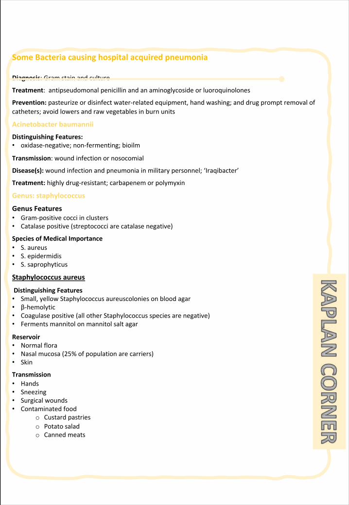
SomeBacteriacausinghospitalacquiredpneumonia
Diagnosis:Gramstainandculture
Treatment:antipseudomonalpenicillinandanaminoglycosideorluoroquinolones
Prevention:pasteurizeordisinfectwater-relatedequipment,handwashing;anddrugpromptremovalofcatheters;avoidlowersandrawvegetablesinburnunits
Acinetobacterbaumannii
DistinguishingFeatures:• oxidase-negative;non-fermenting;bioilm
Transmission:woundinfectionornosocomial
Disease(s):woundinfectionandpneumoniainmilitarypersonnel;‘Iraqibacter’
Treatment:highlydrug-resistant;carbapenemorpolymyxin
Genus:staphylococcus
GenusFeatures• Gram-positivecocciinclusters• Catalasepositive(streptococciarecatalasenegative)
SpeciesofMedicalImportance• S.aureus• S.epidermidis• S.saprophyticus
Staphylococcusaureus
DistinguishingFeatures• Small,yellowStaphylococcusaureuscoloniesonbloodagar• β-hemolytic• Coagulasepositive(allotherStaphylococcusspeciesarenegative)• Fermentsmannitolonmannitolsaltagar
Reservoir• Normalflora• Nasalmucosa(25%ofpopulationarecarriers)• Skin
Transmission• Hands• Sneezing• Surgicalwounds• Contaminatedfood
o Custardpastrieso Potatosalado Cannedmeats
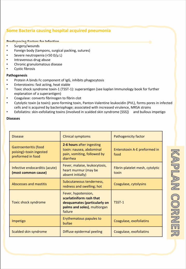
SomeBacteriacausinghospitalacquiredpneumonia
PredisposingFactorsforInfection• Surgery/wounds• Foreignbody(tampons,surgicalpacking,sutures)• Severeneutropenia(<500/µL)• Intravenousdrugabuse• Chronicgranulomatousdisease• Cysticfibrosis
Pathogenesis• ProteinAbindsFccomponentofIgG,inhibitsphagocytosis• Enterotoxins:fastacting,heatstable• Toxicshocksyndrometoxin-1(TSST-1):superantigen(seekaplanImmunologybookforfurther
explanationofasuperantigen)• Coagulase:convertsfibrinogentofibrinclot• Cytolytictoxin(αtoxin):pore-formingtoxin,Panton-Valentineleukocidin(PVL),formsporesininfected
cellsandisacquiredbybacteriophage;associatedwithincreasedvirulence,MRSAstrains• Exfoliatins:skin-exfoliatingtoxins(involvedinscaldedskinsyndrome[SSS])andbullousimpetigo
Diseases
Disease Clinicalsymptoms Pathogenicityfactor
Gastroenteritis (foodpoising)–toxiningestedpreformedinfood
2-6 hours afteringestingtoxin:nausea,abdominalpain,vomiting,followedbydiarrhea
EnterotoxinA-Epreformedinfood
Infectiveendocarditis(acute)(mostcommoncause)
Fever,malaise, leukocytosis,heartmurmur(maybeabsentinitially)
Fibrin-platelet mesh,cytolytictoxin
AbscessesandmastitisSubcutaneoustenderness,rednessandswelling;hot
Coagulase, cytolysins
Toxicshocksyndrome
Fever,hypotension,scarlatiniform rashthatdesquamates(particularlyonpalmsandsoles),multiorganfailure
TSST-1
ImpetigoErythematouspapulestobullae
Coagulase,exofoliatins
Scaldedskinsyndrome Diffuseepidermal peeling Coagulase,exofoliatins
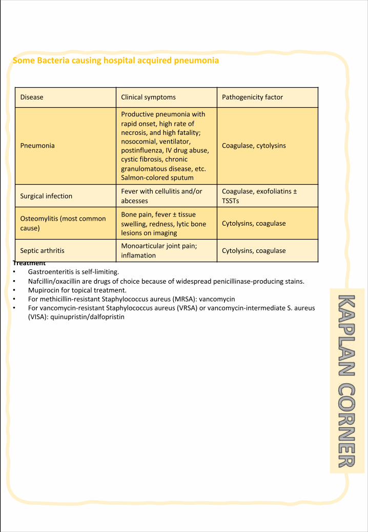
SomeBacteriacausinghospitalacquiredpneumonia
Treatment• Gastroenteritisisself-limiting.• Nafcillin/oxacillinaredrugsofchoicebecauseofwidespreadpenicillinase-producingstains.• Mupirocinfortopicaltreatment.• Formethicillin-resistantStaphylococcusaureus(MRSA):vancomycin• Forvancomycin-resistantStaphylococcusaureus(VRSA)orvancomycin-intermediateS.aureus
(VISA):quinupristin/dalfopristin
Disease Clinicalsymptoms Pathogenicityfactor
Pneumonia
Productivepneumoniawithrapidonset,highrateofnecrosis, andhighfatality;nosocomial,ventilator,postinfluenza,IVdrugabuse,cysticfibrosis,chronicgranulomatousdisease,etc.Salmon-coloredsputum
Coagulase, cytolysins
SurgicalinfectionFeverwithcellulitisand/orabcesses
Coagulase, exofoliatins ±TSSTs
Osteomylitis (mostcommoncause)
Bonepain,fever± tissueswelling,redness,lyticbonelesionsonimaging
Cytolysins,coagulase
SepticarthritisMonoarticular jointpain;inflamation
Cytolysins,coagulase
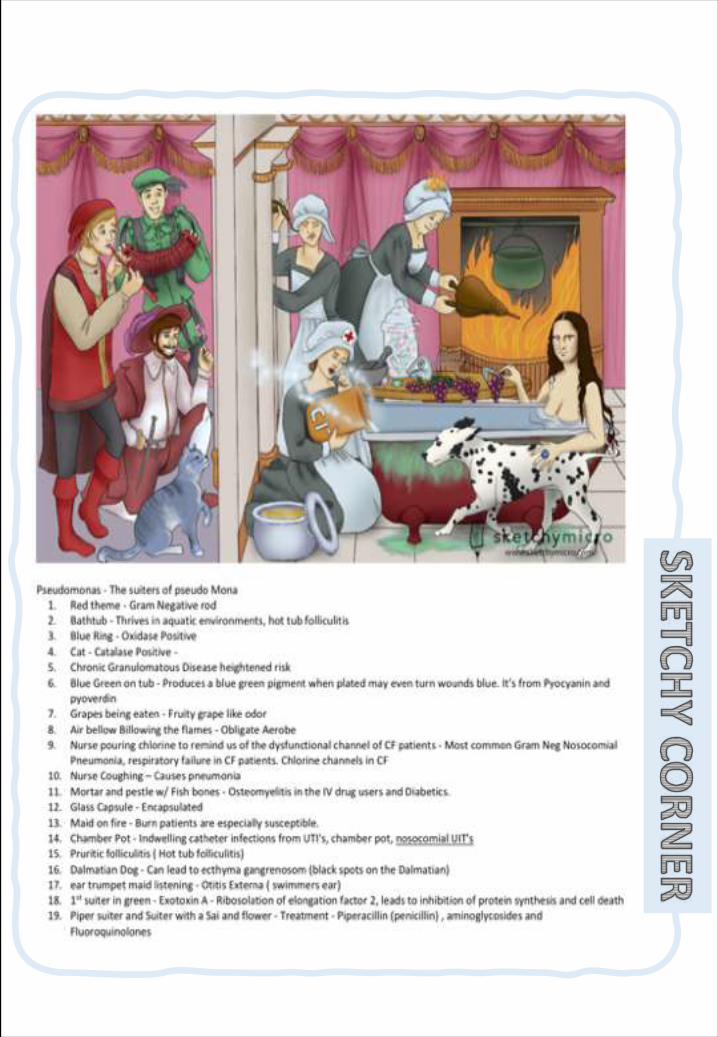
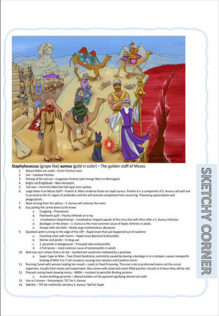
236 | Respiratory Chapter
Note:Aspirationmeansinhalingthesecretionofnasopharynxtothelungdirectlywhichisabnormal.thepersonmighthaveaspirationwhentheylosetheirconsciouslikealcoholicandintubation.
SECTION 3 | Community Acquired pneumonia
§ DiscusstheepidemiologyandpathophysiologyofpneumoniaandCAP
§ Explainthedifferentclassificationsofpneumonia
§ RecognizeclinicalpresentationsassociatedwithCAP
§ DiscussthediagnosisandtreatmentofCAP
§ IdentifycommonetiologicalagentscausingCAPanddiscusstheirlaboratoryworkup
§ DiscussvirulencefactorsandpreventionofStreptococcuspneumoniae
Objec
tive
PneumoniaDefinition:It’sisaninfectionthatleadstoinflammationoftheparenchyma ofthelung(thealveoli)à consolidationandexudation.
Epidemiology:OveralltherateofCAP5-6 casesper1000 personsperyear.Mortality23%– High,especiallyinoldpeople.Almost1 millionannualepisodesofCAPinadults>65 yearsintheUS.
Riskfactors:o Age<2yrsand>65yrso Alcoholism and Smokingo AsthmaandCOPDo Aspirationo Dementiao Priorinfluenzao ImmunosuppressionandHIVo Institutionalizationo Recenthotel:Legionellao Travel,pets,occupationalexposures- birdsowner(C.psittaci)
Courseofdisease:Itmaypresentas:o Acute,fulminantclinicaldiseaseà verysevere.o Chronicdiseasewithamoreprolongedcourse.
Etiologicalagents:
Note:Histology:o Fibrinopurulent
alveolarexudateisseeninacutebacterialpneumonias.
o Mononuclearinterstitialinfiltratesinviralandotheratypicalpneumonias.
o Granulomasandcavitationseeninchronicpneumonias.
o Twofactorsinvolvedintheformationofpneumonia:1. Pathogens2. Hostdefenses
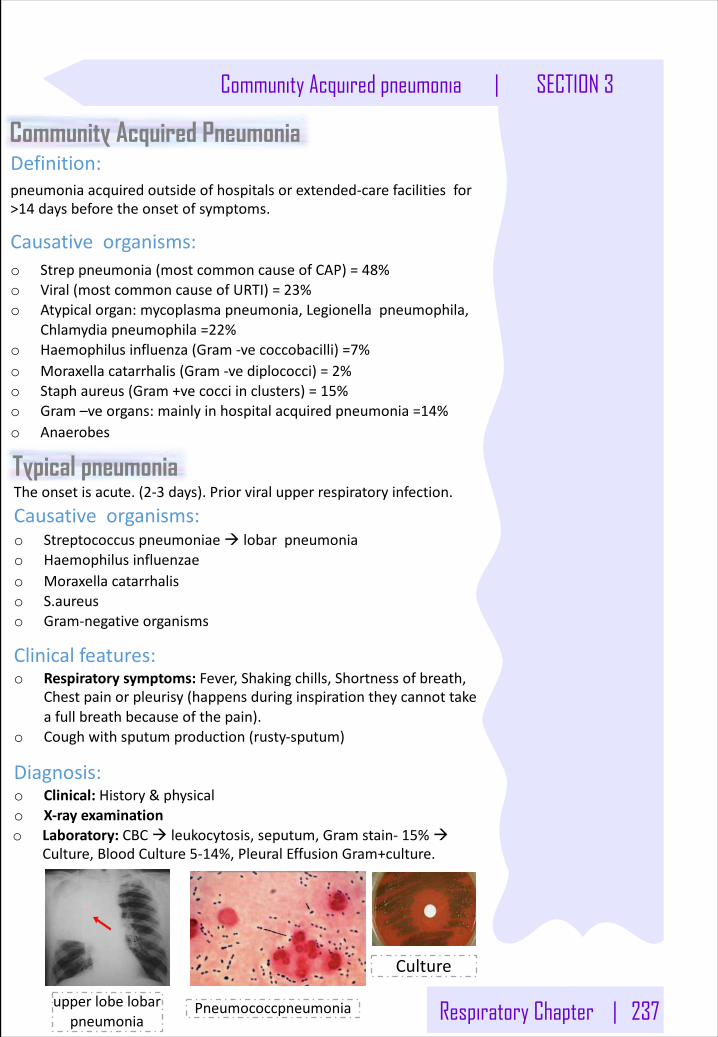
Respiratory Chapter | 237
Community Acquired pneumonia | SECTION 3
Community Acquired Pneumonia Definition:pneumoniaacquiredoutsideofhospitalsorextended-carefacilitiesfor>14 daysbeforetheonsetofsymptoms.
Causativeorganisms:o Streppneumonia(mostcommoncauseofCAP)=48%o Viral(mostcommoncauseofURTI)=23%o Atypicalorgan:mycoplasmapneumonia,Legionellapneumophila,
Chlamydiapneumophila =22%o Haemophilus influenza(Gram-vecoccobacilli)=7%o Moraxellacatarrhalis(Gram-vediplococci)=2%o Staphaureus(Gram+vecocciinclusters)=15%o Gram–veorgans:mainlyinhospitalacquiredpneumonia=14%o Anaerobes
Pneumococcpneumonia
Typical pneumoniaTheonsetisacute.(2-3 days).Priorviralupperrespiratoryinfection.
Causativeorganisms:o Streptococcuspneumoniaeà lobarpneumoniao Haemophilus influenzaeo Moraxellacatarrhaliso S.aureuso Gram-negativeorganisms
Clinicalfeatures:o Respiratorysymptoms:Fever,Shakingchills,Shortnessofbreath,
Chestpainorpleurisy(happensduringinspirationtheycannottakeafullbreathbecauseofthepain).
o Coughwithsputumproduction(rusty-sputum)
Diagnosis:o Clinical:History&physicalo X-rayexaminationo Laboratory:CBCà leukocytosis,seputum,Gramstain- 15%à
Culture,BloodCulture5-14%,PleuralEffusionGram+culture.
upperlobelobarpneumonia
Culture
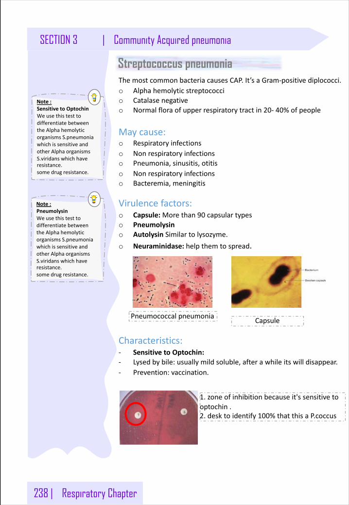
238 | Respiratory Chapter
Note:SensitivetoOptochinWeusethistesttodifferentiatebetweentheAlphahemolyticorganismsS.pneumoniawhichissensitiveandotherAlphaorganismsS.viridans whichhaveresistance.somedrugresistance.
SECTION 3 | Community Acquired pneumonia
1.zoneofinhibitionbecauseit'ssensitivetooptochin .2.desktoidentify100%thatthisaP.coccus
Streptococcus pneumoniaThemostcommonbacteriacausesCAP.It’saGram-positivediplococci.o Alphahemolyticstreptococcio Catalasenegativeo Normalfloraofupperrespiratorytractin20- 40%ofpeople
Maycause:o Respiratoryinfectionso Nonrespiratoryinfectionso Pneumonia,sinusitis,otitiso Nonrespiratoryinfectionso Bacteremia,meningitis
Virulencefactors:o Capsule:Morethan90 capsulartypeso Pneumolysino Autolysin Similartolysozyme.o Neuraminidase:helpthemtospread.
Characteristics:- SensitivetoOptochin:- Lysedbybile:usuallymildsoluble,afterawhileitswilldisappear.- Prevention:vaccination.
Pneumococcalpneumonia Capsule
Note:PneumolysinWeusethistesttodifferentiatebetweentheAlphahemolyticorganismsS.pneumoniawhichissensitiveandotherAlphaorganismsS.viridans whichhaveresistance.somedrugresistance.
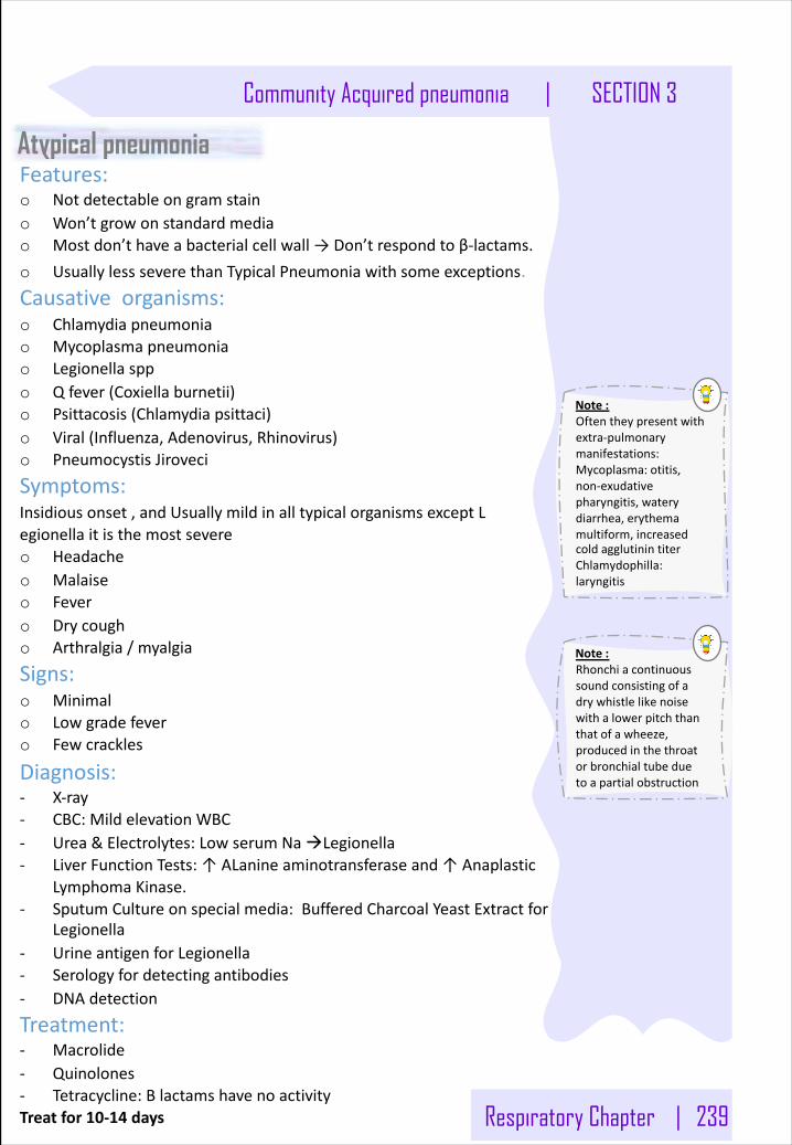
Respiratory Chapter | 239
Note:Rhonchiacontinuoussoundconsistingofadrywhistlelikenoisewithalowerpitchthanthatofawheeze,producedinthethroatorbronchialtubeduetoapartialobstruction
Community Acquired pneumonia | SECTION 3
Atypical pneumoniaFeatures:o Notdetectableongramstaino Won’tgrowonstandardmediao Mostdon’thaveabacterialcellwall→Don’trespondtoβ-lactams.o UsuallylessseverethanTypicalPneumoniawithsomeexceptions.Causativeorganisms:o Chlamydiapneumoniao Mycoplasmapneumoniao Legionellasppo Qfever(Coxiellaburnetii)o Psittacosis(Chlamydiapsittaci)o Viral(Influenza,Adenovirus,Rhinovirus)o PneumocystisJiroveci
Symptoms:Insidiousonset,andUsuallymildinalltypicalorganismsexceptLegionella itisthemostsevereo Headacheo Malaiseo Fevero Drycougho Arthralgia/myalgia
Signs:o Minimalo Lowgradefevero Fewcrackles
Diagnosis:- X-ray- CBC:MildelevationWBC- Urea&Electrolytes:LowserumNaàLegionella- LiverFunctionTests:↑ALanine aminotransferaseand↑Anaplastic
LymphomaKinase.- SputumCultureonspecialmedia:BufferedCharcoalYeastExtractfor
Legionella- UrineantigenforLegionella- Serologyfordetectingantibodies- DNAdetection
Treatment:- Macrolide- Quinolones- Tetracycline:BlactamshavenoactivityTreatfor10-14 days
Note:Oftentheypresentwithextra-pulmonarymanifestations:Mycoplasma:otitis,non-exudativepharyngitis,waterydiarrhea,erythemamultiform,increasedcoldagglutinintiterChlamydophilla:laryngitis
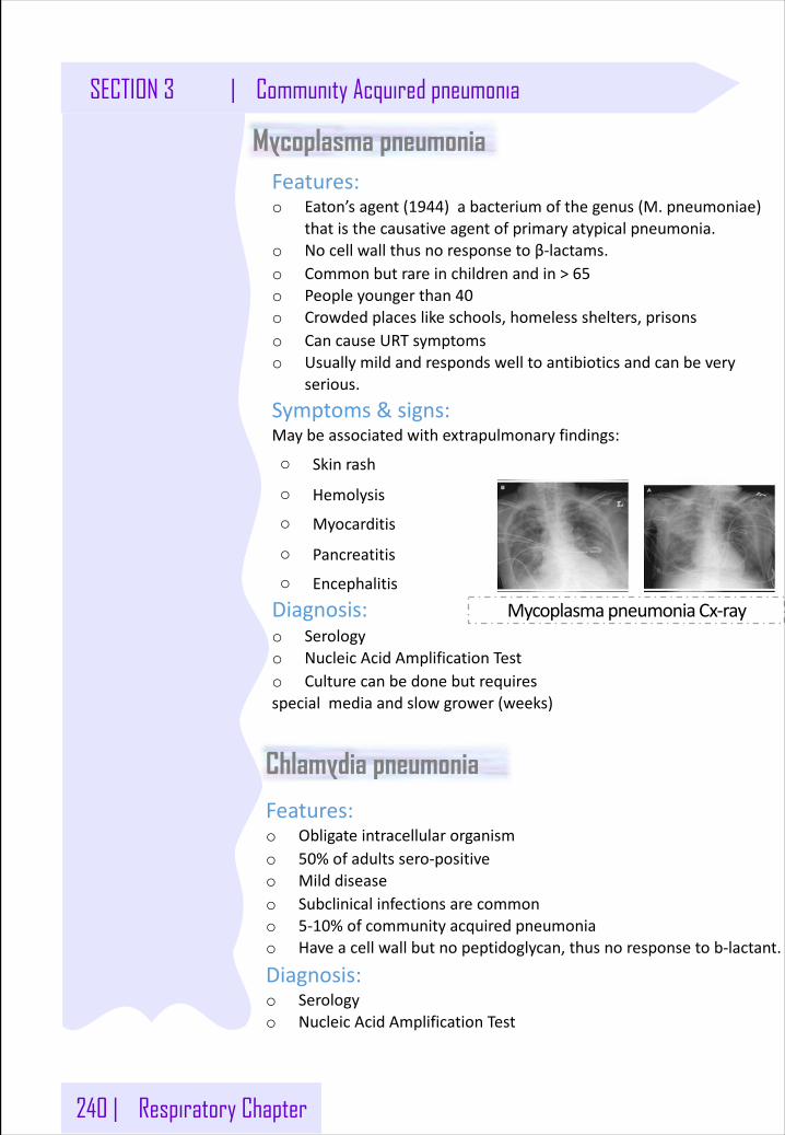
Features:o Eaton’sagent(1944)abacteriumofthegenus(M.pneumoniae)
thatisthecausativeagentofprimaryatypicalpneumonia.o Nocellwallthusnoresponsetoβ-lactams.o Commonbutrareinchildrenandin>65o Peopleyoungerthan40o Crowdedplaceslikeschools,homelessshelters,prisonso CancauseURTsymptomso Usuallymildandrespondswelltoantibioticsandcanbevery
serious.
Symptoms&signs:Maybeassociatedwithextrapulmonary findings:
o Skinrash
o Hemolysis
o Myocarditis
o Pancreatitis
o Encephalitis
Diagnosis:o Serologyo NucleicAcidAmplificationTesto Culturecanbedonebutrequiresspecialmediaandslowgrower(weeks)
240 | Respiratory Chapter
SECTION 3 | Community Acquired pneumonia
MycoplasmapneumoniaCx-ray
Mycoplasma pneumonia
Chlamydia pneumonia
Features:o Obligateintracellularorganismo 50%ofadultssero-positiveo Milddiseaseo Subclinicalinfectionsarecommono 5-10%ofcommunityacquiredpneumoniao Haveacellwallbutnopeptidoglycan,thusnoresponsetob-lactant.
Diagnosis:o Serologyo NucleicAcidAmplificationTest
Respiratory Chapter | 241
Note:Thisatypicalbacteriumcommonlycausespharyngitis,bronchitis,coronaryarterydiseaseandatypicalpneumoniainadditiontoseveralotherpossiblediseaseswithothersubtypes.Somainlythemostimportantare(atypicalpneumonia,urethritisandtrachomatis).
Community Acquired pneumonia | SECTION 3
PsittacosisFeatures:o ThecauseisChlamydiapsittaci.
o Whoaffectedarebirdowners,petshopemployees,vetsParrots,pigeonsandpoultry.
o Birdsoftenasymptomatic
Q fever (Coxiella burnetii)Features:o Pneumoniaisacuteformofinfectiono Exposuretofarmanimalsmainlysheepo Spreadbyinhalationofinfectedanimalbirthproducts
Diagnosis:o Serology
Legionella pneumophilaFeatures:o TheMostsevereone.CanbeverysevereandleadtoICUadmission.
o CancauseLegionnairesdisease.
o Seriousoutbreakslinkedtoexposuretocoolingtowers.Peopleusuallygetinfectedfromtheairconditioningsystemofhotels
o It’sawaterbornebacteria,andusuallytargettheimmunosuppressedpatient
Symptoms&signs:Cancause:
o Hyponatraemia:<130 mMol andWBC<15,000
o Bradycardia
o AbnormalLiverFunctionTests
o RaisedCreatine PhosphoKinase
o AcuteRenalfailure
Diagnosis:o Specimen:sputum
o Cultureonspecializedmedia(BCYE)o DirectFluorescentAntibodyo NucleicAcidAmplificationTest
o Urineantigentesting
242 | Respiratory Chapter
SECTION 3 | Community Acquired pneumonia
LegionellapresentsonX-Rayas:interstitialpneumoniabutcanpresentaslobaroranyothertype
Pontiac feverFeatures:o Nonpneumonic
o Influenzalikeillness
o Selflimiting
o RelatedtoexposuretoenvironmentalaerosolscontainingLegionella(potentiallyreactiontobacterialendotoxins).
Antibiotic Treatment of CAP
Factorstoconsiderinselectionofantibiotic:o Comorbidities.
o Previousantibioticexposureinlast3 months
o Severity.OutpatientmanagementvsrequiringinpatientadmissionvsrequiringICU.
SomeBacteriacausinghospitalacquiredpneumonia
Streptococcuspneumoniae
DistinguishingFeatures• αhemolytic• Optochinsensitive• Lancet-shapeddiplococci• Lysedbybile(bilesoluble)
Reservoir:humanupperrespiratorytractpneumoniae
Transmission:respiratorydroplets(notconsideredhighlycommunicable;otencolonizethenasopharynxwithoutcausingdisease)
PredisposingFactors• Antecedentinfluenzaormeaslesinfection• Chronicobstructivepulmonarydisease(COPD)• Congestiveheartfailure(CHF)• Alcoholism• Aspleniapredisposestosepticemia
Pathogenesis• Polysaccharidecapsuleisthemajorvirulencefactor• IgAprotease• Teichoicacid• PneumolysinO:hemolysin/cytolysin:damagesrespiratoryepithelium;inhibitsleukocyterespiratory
burstandinhibitsclassicalcomplementfixation
Diseases• Typicalpneumonia:mostcommoncause(especiallyindecade6oflife);shakingchills,highfever,
lobarconsolidation,blood-tinged,“rusty”sputum• Adultmeningitis:mostcommoncause;peptidoglycanandteichoicacidsarehighlyinflammatory
inCNS;CSFrevealshighWBCs(neu-trophils)andlowglucose,highprotein• Otitismediaandsinusitisinchildrenmostcommoncause
LaboratoryDiagnosis• GramstainandcultureofCSForsputum• Quellungreaction:positive(swellingofthecapsulewiththeadditionoftype-specificantiserum,
nolongerusedbutstilltested!)• Latexparticleagglutination:testforcapsularantigeninCSF• Urinaryantigentest
Treatment:betalactamsforbacterialpneumonia;cetriaxoneorcefotaximeforadultmeningitis(addvancomycinifpenicillin-resistantS.pneumoniaehasbeenreportedincommunity);amoxicillinforotitismediaandsinusitisinchildren(erythromycinincasesofallergy)

SomeBacteriacausinghospitalacquiredpneumonia
Prevention• Antibodytocapsule(>80capsularserotypes)providestype-specificimmunity• Vaccine
o Pediatric(PCV,pneumococcalconjugatevaccine):13ofmostcommonserotypes;conjugatedtodiphtheriatoxoid;preventsinvasivedisease
o Adult(PPV,pneumococcalpolysaccharidevaccine):23ofmostcommoncapsularserotypes;recommendedforalladultsage≥65plusat-riskindividuals
Mycoplasmapneumoniae
DistinguishingFeatures• Extracellular,tiny,flexible• Nocellwall;notseenonGram-stainedsmear• Membranewithcholesterolbutdoesnotsynthesizecholesterol• RequirescholesterolforinvitrocultureReservoir:humanrespiratorytractTransmission:respiratory
droplets;closecontact:families,militaryrecruits,medicalschoolclasses,collegedorms
Pathogenesis• Surfaceparasite:notinvasive• AttachestorespiratoryepitheliumviaP1protein• Inhibitsciliaryaction• Produceshydrogenperoxide,superoxideradicals,andcytolyticenzymes,whichdamagethe
respiratoryepithelium,leadingtonecrosisandabad,hackingcough(walkingpneumonia)• M.pneumoniaefunctionsassuperantigen,elicitsproductionofIL-1,IL-6,andTNF-α
Disease:walkingpneumonia• Pharyngitis• Maydevelopintoatypicalpneumoniawithpersistenthack(littlesputumproduced)• Mostcommonatypicalpneumonia(alongwithviruses)inyoungadults
Diagnosis• Primarilyclinicaldiagnosis;PCR/nucleicacidprobes• ELISAandimmunofluorescencesensitiveandspecific• Fried-egg-shapedcoloniesonsterol-containingmedia,10days• Positivecoldagglutinins(autoantibodytoRBCs)testisnonspecificandispositiveinonly65%of
cases
Treatment:erythromycin,azithromycin,clarithromycin;nocephalosporinorpenicillin
Prevention:none
SomeBacteriacausinghospitalacquiredpneumonia
Coxiella burnetii
DistinguishingFeatures:• obligateintracellular,spore-likecharacteristics
Transmission:inhalationfromdriedplacentalmaterial;zoonosis(sheepandgoats);possiblebioterrorismagent
Pathogenesis:obligateintracellular,liveinsidephagolysosomes
Disease(s):Qfever:atypicalpneumonia,hepatitis,orendocarditis
Diagnosis:serologicdetectionofPhaseIILPSantigen(foracutedisease)andPhaseIandPhaseIILPSantigens(forchronicdisease)
Treatment:doxycycline
Family:chlamydiaceaeFamilyFeatures• Obligateintracellularbacteria• Elementarybody/reticulatebody• NotseenonGramstain• CannotmakeATP• Cellwalllacksmuramicacid
GeneraofMedicalImportance• Chlamydiatrachomatis• Chlamydophilapneumoniae• Chlamydophilapsittaci
Chlamydiatrachomatis
DistinguishingFeatures• Obligateintracellularbacterium;cannotmakeATP• Foundincellsasmetabolicallyactive,replicatingreticulatebodies• Infectiveform:inactive,extracellularelementarybody• NotseenonGramstain;peptidoglycanlayerlacksmuramicacid
Reservoir:humangenitaltractandeyes
Transmission:sexualcontactandatbirth;trachomaistransmittedbyhand-to-eyecontactandlies.
Pathogenesis:infectionofnonciliatedcolumnarorcuboidalepithelialcellsofmucosalsurfacesleadstogranulomatousresponseanddamage
SomeBacteriacausinghospitalacquiredpneumonia
Diseases• STDsinU.S.
o SerotypesD-K(mostcommonbacterialSTDinU.S.,thoughoverallherpesandHPVaremorecommoninprevalence)
o Nongonococcalurethritis,cervicitis,PID,andmajorportionofinfertility(noresistancetoreinfection)
o Inclusionconjunctivitisinadults(withNGUandreactivearthritis)o Inclusionconjunctivitisand/orpneumoniainneonates/infants(staccatocough)with
eosinophilicinfiltrate• Lymphogranulomavenereum
o SerotypesL1,2,3(prevalentinAfrica,Asia,SouthAmerica);painlessulceratsiteofcontact;swollenlymphnodes(buboes)aroundinguinalligament(groovesign);tertiaryincludesulcers,fistulas,genitalelephantiasis
• Trachomao Leadingcauseofpreventableinfectiousblindness:serotypesA,B,Ba,andCo Follicularconjunctivitisleadingtoconjunctivalscarring,andinturnedeyelashesleadingto
cornealscarringandblindness
Diagnosis• NAAT;DNAprobesinU.S.(rRNA)andPCR• CytoplasmicinclusionsseenonGiemsa-,iodine-,orfluorescent-antibody-stainedsmearorscrapings• Cannotbeculturedoninertmedia• Isculturedintissueculturesorembryonatedeggs• Serodiagnosis:DFA,ELISA
Treatment:azithromycinordoxycycline
Prevention:erythromycinforinfectedmotherstopreventneonataldisease;systemicerythromycinforneonatalconjunctivitistopreventpneumonia
7 | Respiratory Chapter
SomeBacteriacausinghospitalacquiredpneumonia
Genus:legionellaLegionellapneumophila
Distinguishing Features• Stain poorly with standard Gram stain; gram-negative• Fastidious requiring increased iron and cysteine for laboratory culture (BCYE, buffered
charcoal, yeast extract)• Facultative intracellular
• Reservoir: rivers/streams/amebae; air-conditioning water cooling tanks
• Transmission: aerosols from contaminated air-conditioning; no human-to-human transmission
Predisposing Factors: smokers age >55 with high alcohol intake; immunosuppressed patients such as renal transplant patients
Pathogenesis: facultative intracellular pathogen; endotoxin
Disease(s)• Legionnairesdisease(“atypicalpneumonia”):associatedwithair-conditioningsystems(nowroutinely
decontaminated);pneumonia;hyponatremia;mentalconfusion;diarrhea(noLegionellainGItract)• Pontiacfever:pneumonitis;nofatalities
Diagnosis• Urinaryantigentest(serogroup1)• DFA(directfluorescentantibody)onbiopsy,(+)byDieterlesilverstain• Fourfoldincreaseinantibody
Treatment:luoroquinolone(levoloxacin)ormacrolide(azithromycin)withrifampin(immunocompromisedpatients);drugmustpenetratehumancells.
Prevention:routinedecontaminationofair-conditionercoolingtanks
SECTION 4 | Lower Respiratory Tract
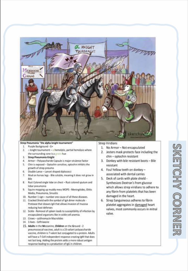
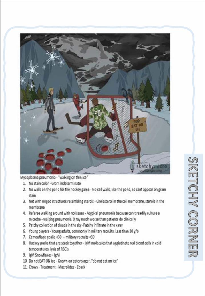
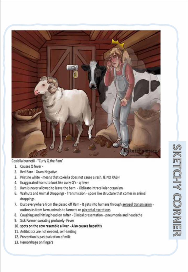
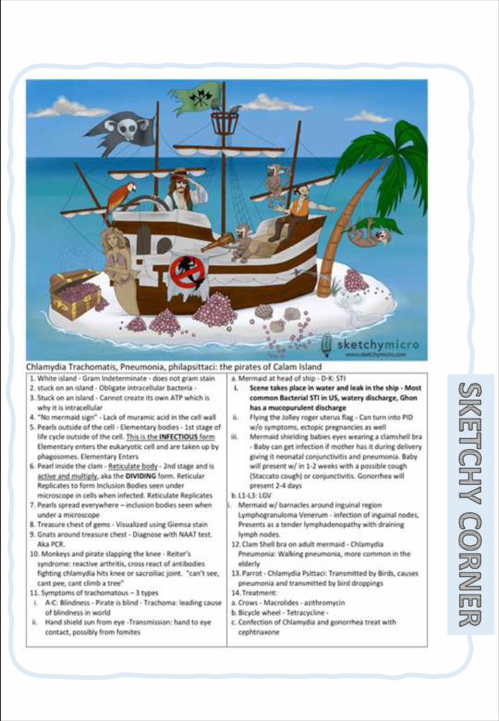
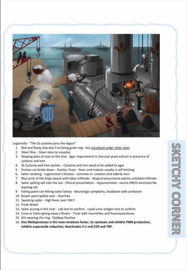
243 | Respiratory Chapter
SECTION 3 | Pharmacology of Respiratory tract infections
Classification of Respiratory Tract Infections
§ Thetypesofrespiratorytractinfections(RTI).
§ TheantibioticsthatarecommonlyusedtotreatRTI&theirsideeffects.
§ Understandthemechanismofaction&pharmacokineticsofindividualdrugs.Objec
tive
UpperRespiratoryTractInfections:
Viruses:MostURTIsareofviraletiology.ShouldNOTbetreatedwithantibiotics.Restandplentyoffluids,overthecountercoldandpainrelievers.
Bacteria:MainlygroupAstreptococcusandH.Influenza.ShouldtreatedbyAntibiotics,typedependson:
- Typeofbacteria.- Sensitivitytest.
LowerRespiratoryTractInfectionsCostlyandmoredifficulttotreat.
Bronchitis:inflammationofmajorbronchi&trachea.CouldbeAcute,ChronicorAcuteexacerbationofchronicbronchitis.ThecausesofcouldbeVirusesorbacteria:H.Influenza,Streptococcuspneumonia&Moraxellacatarrhalis.
Pneumonia:seriousinfectionofbronchioles&alveoli.ItdividedintoCommunity-acquired&Hospital-acquired.ThecausescouldbeBacteria:S.pneumoniae(66%)&H.Influenza(20%).
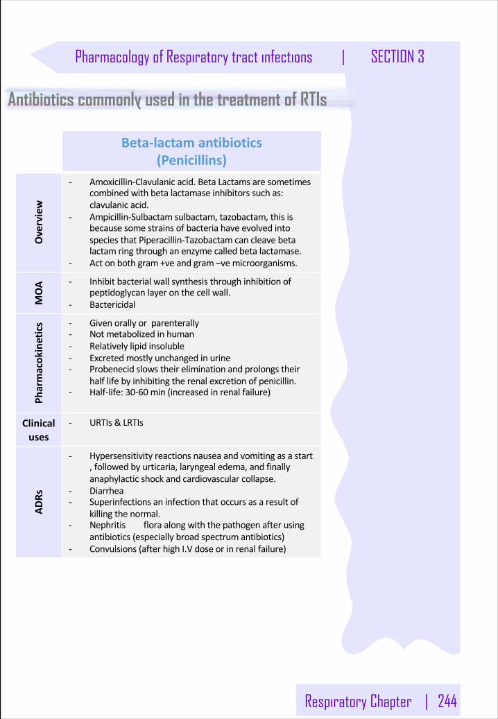
Respiratory Chapter | 244
Pharmacology of Respiratory tract infections | SECTION 3
Antibiotics commonly used in the treatment of RTIs
Beta-lactamantibiotics(Penicillins)
Overview
- Amoxicillin-Clavulanicacid.BetaLactamsaresometimescombinedwith betalactamaseinhibitorssuchas:clavulanicacid.
- Ampicillin-Sulbactam sulbactam,tazobactam,thisisbecausesome strainsofbacteriahaveevolvedintospeciesthat Piperacillin-Tazobactam cancleavebetalactamringthroughanenzyme calledbetalactamase.
- Actonbothgram+veandgram–ve microorganisms.
MOA - Inhibitbacterialwallsynthesisthroughinhibitionof
peptidoglycanlayeronthecellwall.- Bactericidal
Pharmacok
inetics - Givenorallyorparenterally
- Notmetabolizedinhuman- Relativelylipidinsoluble- Excretedmostlyunchangedinurine- Probenecidslowstheireliminationandprolongstheir
halflifebyinhibitingtherenalexcretionofpenicillin.- Half-life:30-60 min(increasedinrenalfailure)
Clinicaluses
- URTIs &LRTIs
ADRs
- Hypersensitivityreactionsnauseaandvomitingasastart,followedbyurticaria,laryngealedema,andfinallyanaphylacticshockandcardiovascularcollapse.
- Diarrhea- Superinfectionsaninfectionthatoccursasaresultof
killingthenormal.- Nephritis floraalongwiththepathogenafterusing
antibiotics (especiallybroadspectrumantibiotics)- Convulsions(afterhighI.Vdoseorinrenalfailure)
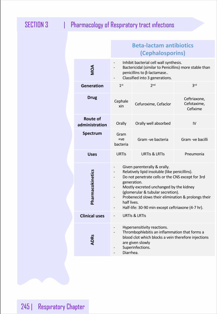
245 | Respiratory Chapter
SECTION 3 | Pharmacology of Respiratory tract infections
Beta-lactamantibiotics(Cephalosporins)
MOA
- Inhibitbacterialcellwallsynthesis.- Bactericidal(similartoPenicillins)morestablethan
penicillins toβ-lactamase..- Classifiedinto3 generations.
Generation 1st 2nd 3rd
DrugCephalexin Cefuroxime,Cefaclor
Ceftriaxone,Cefotaxime,Cefixime
Routeofadministration Orally Orallywellabsorbed IV
Spectrum Gram+ve
bacteriaGram-vebacteria Gram-vebacilli
Uses URTIs URTIs&LRTIs Pneumonia
Pharmacok
inetics
- Givenparenterally&orally.- Relativelylipidinsoluble(likepenicillins).- DonotpenetratecellsortheCNSexceptfor3rd
generation.- Mostlyexcretedunchangedbythekidney
(glomerular&tubularsecretion).- Probenecidslowstheirelimination&prolongstheir
halflives.- Half-life:30-90 minexceptceftriaxone(4-7 hr).
Clinicaluses - URTIs &LRTIs
ADRs
- Hypersensitivityreactions.- Thrombophlebitisaninflammationthatformsa
bloodclotwhichblocksaveinthereforeinjectionsaregivenslowly
- Superinfections.- Diarrhea.
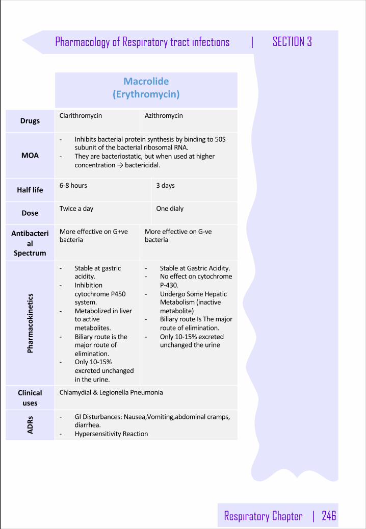
Respiratory Chapter | 246
Pharmacology of Respiratory tract infections | SECTION 3
Macrolide(Erythromycin)
DrugsClarithromycin Azithromycin
MOA
- Inhibitsbacterialproteinsynthesisbybindingto50SsubunitofthebacterialribosomalRNA.
- Theyarebacteriostatic,butwhenusedathigherconcentration→bactericidal.
Halflife 6-8 hours 3 days
Dose Twice aday One dialy
Antibacterial
Spectrum
MoreeffectiveonG+vebacteria
MoreeffectiveonG-vebacteria
Pharmacok
inetics
- Stableatgastricacidity.
- InhibitioncytochromeP450system.
- Metabolizedinlivertoactivemetabolites.
- Biliaryrouteisthemajorrouteofelimination.
- Only10-15%excretedunchangedintheurine.
- StableatGastricAcidity.- Noeffectoncytochrome
P-430.- UndergoSomeHepatic
Metabolism(inactivemetabolite)
- BiliaryrouteIsThemajorrouteofelimination.
- Only10-15%excretedunchangedtheurine
Clinicaluses
Chlamydial&LegionellaPneumonia
ADRs - GIDisturbances: Nausea,Vomiting,abdominal cramps,
diarrhea.- HypersensitivityReaction
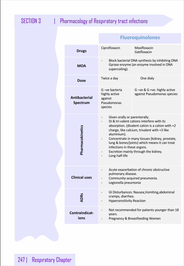
247 | Respiratory Chapter
SECTION 3 | Pharmacology of Respiratory tract infections
Fluoroquinolones
DrugsCiprofloxacin Moxifloxacin
Gatifloxacin
MOA- BlockbacterialDNAsynthesisbyinhibitingDNA
Gyraseenzyme(anenzymeinvolvedinDNAsupercoiling).
Dose Twice aday One dialy
AntibacterialSpectrum
G–vebacteriahighlyactiveagainstPseudomonasspecies
G–ve&G+ve. highlyactiveagainstPseudomonasspecies
Pharmacok
inetics
- Givenorallyorparenterally.- Di&tri-valent cationsinterferewithits
absorption.(divalentcationisacationwith+2charge,likecalcium,trivalentwith+3 likealuminium).
- Concentrateinmanytissues(kidney,prostate,lung&bones/joints)whichmeansitcantreatinfectionsintheseorgans.
- Excretionmainlythroughthekidney.- Longhalf-life
Clinicaluses
- Acuteexacerbationofchronicobstructivepulmonarydisease.
- Community-acquiredpneumonia.- Legionellapneumonia
ADRs
- GIDisturbances: Nausea,Vomiting,abdominalcramps,diarrhea.
- HypersensitivityReaction
Contraindicat-ions
- Notrecommendedforpatientsyoungerthan18years.
- Pregnancy &BreastfeedingWomen
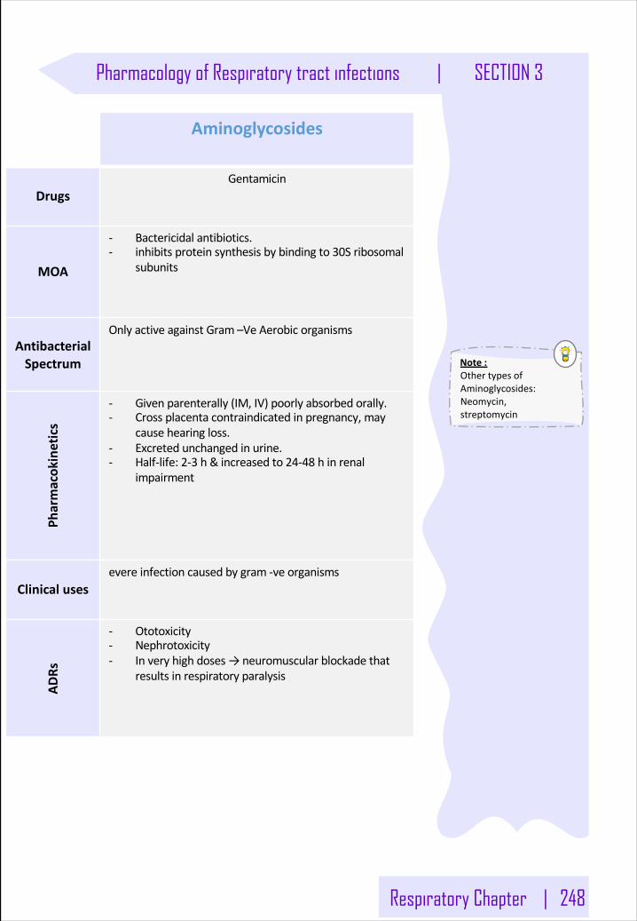
Respiratory Chapter | 248
Pharmacology of Respiratory tract infections | SECTION 3
Note:OthertypesofAminoglycosides:Neomycin,streptomycin
Aminoglycosides
DrugsGentamicin
MOA
- Bactericidalantibiotics.- inhibitsproteinsynthesisbybindingto30Sribosomal
subunits
AntibacterialSpectrum
OnlyactiveagainstGram–VeAerobicorganisms
Pharmacok
inetics
- Givenparenterally(IM,IV)poorlyabsorbedorally.- Crossplacentacontraindicatedinpregnancy,may
causehearingloss.- Excretedunchangedinurine.- Half-life:2-3 h&increasedto24-48 hinrenal
impairment
Clinicalusesevereinfectioncausedbygram-veorganisms
ADRs
- Ototoxicity- Nephrotoxicity- Inveryhighdoses→neuromuscularblockadethat
resultsinrespiratoryparalysis
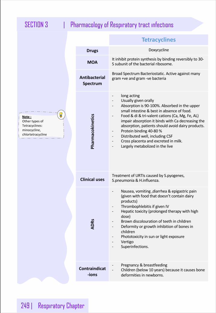
249 | Respiratory Chapter
Note:OthertypesofTetracyclines:minocycline,chlortetracycline
SECTION 3 | Pharmacology of Respiratory tract infections
Tetracyclines
Drugs Doxycycline
MOAItinhibitproteinsynthesisbybindingreversiblyto30-Ssubunitofthebacterialribosome.
AntibacterialSpectrum
BroadSpectrumBacteriostatic. Activeagainstmanygram+veandgram-vebacteria
Pharmacok
inetics
- longacting- Usuallygivenorally- Absorptionis90-100%. Absorbedintheupper
small intestine&bestinabsenceoffood.- Food&di&tri-valent cations(Ca,Mg,Fe,AL)
impairabsorptionitbindswithCadecreasingtheabsorption,patientsshouldavoiddairyproducts.
- Proteinbinding40-80 %- Distributedwell,includingCSF- Crossplacentaandexcretedinmilk.- Largelymetabolizedinthelive
ClinicalusesTreatmentofURTIscausedbyS.pyogenes,S.pneumonia &H.influenza.
ADRs
- Nausea,vomiting,diarrhea&epigastricpain(givenwithfoodthatdoesn’tcontaindairyproducts)
- Thrombophlebitisif givenIV- Hepatictoxicity(prolongedtherapywithhigh
dose)- Browndiscolouration ofteethinchildren- Deformityorgrowthinhibitionofbonesin
children- Phototoxicity insunorlightexposure- Vertigo- Superinfections.
Contraindicat-ions
- Pregnancy &breastfeeding- Children(below10 years) becauseitcausesbone
deformitiesinnewborns.
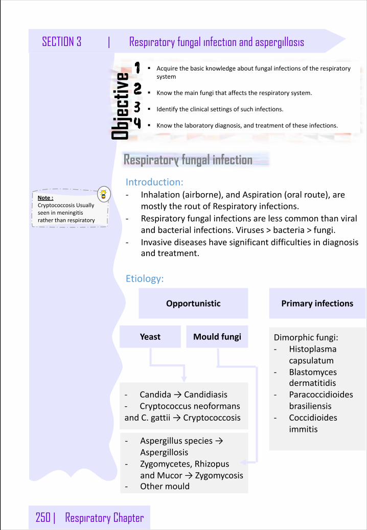
250 | Respiratory Chapter
Note:CryptococcosisUsuallyseeninmeningitisratherthanrespiratory
SECTION 3 | Respiratory fungal infection and aspergillosis
§ Acquirethebasicknowledgeaboutfungalinfectionsoftherespiratorysystem
§ Knowthemainfungithataffectstherespiratorysystem.
§ Identifytheclinicalsettingsofsuchinfections.
§ Knowthelaboratorydiagnosis,andtreatmentoftheseinfections.Objec
tive
Respiratory fungal infection
Introduction:- Inhalation(airborne),andAspiration(oralroute),are
mostlytheroutofRespiratoryinfections.- Respiratoryfungalinfectionsarelesscommonthanviral
andbacterialinfections.Viruses>bacteria>fungi.- Invasivediseaseshavesignificantdifficultiesindiagnosis
andtreatment.
Etiology:
PrimaryinfectionsOpportunistic
Mould fungiYeast
- Candida→Candidiasis- CryptococcusneoformansandC.gattii →Cryptococcosis
- Aspergillusspecies→Aspergillosis
- Zygomycetes,RhizopusandMucor →Zygomycosis
- Othermould
Dimorphicfungi:- Histoplasma
capsulatum- Blastomyces
dermatitidis- Paracoccidioides
brasiliensis- Coccidioides
immitis
Respiratory Chapter | 251
Respiratory fungal infection and aspergillosis | SECTION 3
Note:Aspergillosis includeMycotoxicosis,Colonization(withoutinvasionandextension)inpreformedcavities,Invasivediseaseoflungs,Systemicanddisseminateddisease,Allergy.
Primary Systemic Mycoses
Definition:Infectionsoftherespiratorysystem.
Epidemiology:CommoninNorthAmericatoalesserextentinSouthAmerica.NotcommoninotherpartsoftheWorld.
Transmission:Inhalation.
Wherecanweseeit?Dissemination.Itcanspreadtomorethanorgan.seeninimmunocompromisedhosts.
Etiology:Innaturefoundinsoilofrestrictedhabitats.Primarypathogens.Theyarehighlyinfectious.Ifyouinhaledjustfewofityouwillgetinfectedunlikeothers.Itinclude:- Histoplasmosis- Blastomycosis- Coccidioidomycosis- Paracoccidioidomycosis
Aspergillosis
Definition:AspergillosisisaspectrumofdiseasesofhumansandanimalscausedbymembersofthegenusAspergillus(mould fungi).
Etiology:Aspergillusspecies,commonspeciesare:- A.fumigatuàmostvirulent- A.flavusàmostcommoninRiyadh- A.niger- A.terreusand- A.nidulans
252 | Respiratory Chapter
Note:SputumisNOTthebestbecauseit’sfullwithnormalflorawhileBronchoAlveolarLavageSecondisthebestmethodandlungbiopsyisthetestforchoicebecauseit’ssterile+youcancheckifthediseaseisinvasiveornot.
SECTION 3 | Respiratory fungal infection and aspergillosis
Riskfactor:
- Bonemarrow&OrgantransplantationBecausewegavethemimmunosuppressants todecreasetheirimmunity- Cancer:Leukemia,lymphoma- AIDS- Drugs:Cytotoxicdrugs,steroids- Diabetes
Diagnosis:
Specimen:
- Respiratoryspecimens:Sputum,BronchoAlveolarLavageSecond,Lungbiopsy.
- Othersamples:Blood
LabInvestigations:
- DirectMicroscopy:GiemsaStain,Grocott MethenamineSilverStain.Willshowfungalseptatehyphae.
- CultureonSDA.
Serology:
- TestforAntibody.
- ELISAtestforgalactomannanAntigen.specificforAspergillus.
PCR:DetectionofAspergillusDNAinclinicalsamples.
Treatment:Antifungal:Voriconazoleà thedrugofchoice.Alternativetherapy:AmphotericinB,Itraconazole,Caspofungin+surgerytoremoveaspergilloma.
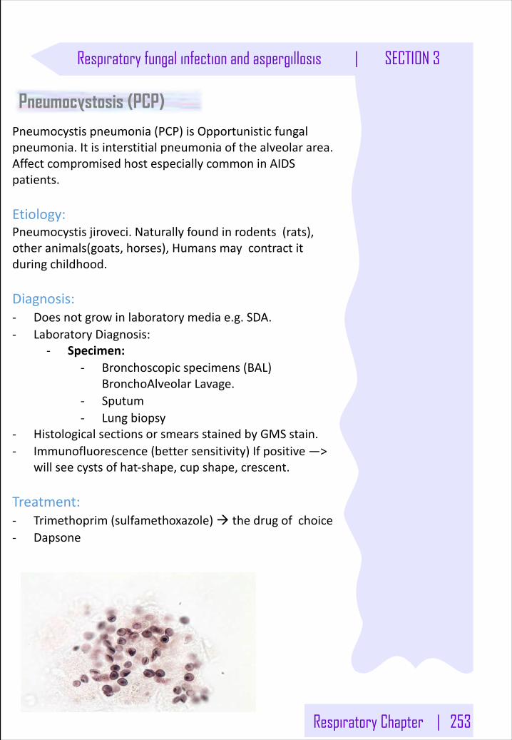
Respiratory Chapter | 253
Respiratory fungal infection and aspergillosis | SECTION 3
Pneumocystosis (PCP)Pneumocystispneumonia(PCP)isOpportunisticfungalpneumonia.Itisinterstitialpneumoniaofthealveolararea.AffectcompromisedhostespeciallycommoninAIDSpatients.
Etiology:Pneumocystisjiroveci.Naturallyfoundinrodents(rats),otheranimals(goats,horses),Humansmaycontractitduringchildhood.
Diagnosis:- Doesnotgrowinlaboratorymediae.g.SDA.- LaboratoryDiagnosis:
- Specimen:- Bronchoscopic specimens(BAL)
BronchoAlveolarLavage.- Sputum- Lungbiopsy
- HistologicalsectionsorsmearsstainedbyGMSstain.- Immunofluorescence(bettersensitivity)Ifpositive—>
willseecystsofhat-shape,cupshape,crescent.
Treatment:- Trimethoprim(sulfamethoxazole)à thedrugofchoice- Dapsone
254 | Respiratory Chapter
Note:InvasivepulmonaryAspergillosisInimmunocompromisedpatient
SECTION 3 | Respiratory fungal infection and aspergillosis
Classification of Aspergillosis
InvasivepulmonaryAspergillosis
Signs&symptom
- Cough- Hemoptysis- Fever- Leukocytosis
Diagnosis Radiology:willshowlesionswithhalosign.
ChronicAspergillosis(ColonizingAspergillosis)
Types - Aspergillomaoflung- Maxillary(sinus)aspergilloma
Causes Aspergilloma,whichisalsoknownas(Aspergillusfungusball).
Signs&symptom
- Drycough- Hemoptysis- Variablefever
Diagnosis Radiology:willshowmassinthelung.radiolucentcrescentairsurroundthemass.
Allergic
Types- Allergicbronchopulmonary- Aspergillosis(ABPA)- AllergicAspergillussinusitis
Signs&symptom
- SymptomsofAsthma- Bronchialobstruction- Eosinophilia- Wheezing+/-
Diagnosis- SkintestreactivitytoAspergillus- SerumantibodiestoAspergillus.- SerumIgE>1000 ng/m
Persistencewithoutdisease
Signs&symptom Colonisation oftheairwaysornose/sinuses.
Note:SerumantibodiestoAspergillusisusedtodifferentiatebetweenasthmaandallergicaspergillosis.
Note:AllergicAspergillosisInhealthypatientusuallycommoninKSA
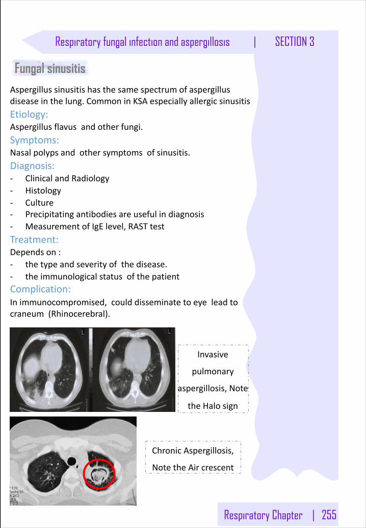
Respiratory Chapter | 255
Respiratory fungal infection and aspergillosis | SECTION 3
Fungal sinusitisAspergillussinusitishasthesamespectrumofaspergillusdiseaseinthelung.CommoninKSAespeciallyallergicsinusitisEtiology:Aspergillusflavusandotherfungi.Symptoms:Nasalpolypsandothersymptomsofsinusitis.Diagnosis:- ClinicalandRadiology- Histology- Culture- Precipitatingantibodiesareusefulindiagnosis- MeasurementofIgE level,RASTtestTreatment:Dependson:- thetypeandseverityofthedisease.- theimmunologicalstatusofthepatientComplication:Inimmunocompromised,coulddisseminatetoeyeleadtocraneum (Rhinocerebral).
ChronicAspergillosis,
NotetheAircrescent
Invasive
pulmonary
aspergillosis,Note
theHalosign
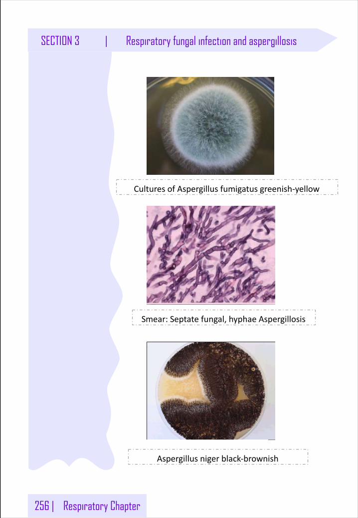
256 | Respiratory Chapter
SECTION 3 | Respiratory fungal infection and aspergillosis
Aspergillusniger black-brownish
Smear:Septatefungal,hyphaeAspergillosis
CulturesofAspergillusfumigatusgreenish-yellow
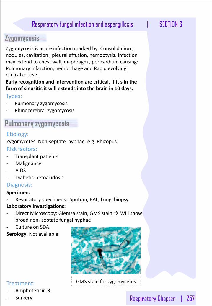
Respiratory Chapter | 257
Respiratory fungal infection and aspergillosis | SECTION 3
ZygomycosisZygomycosisisacuteinfectionmarkedby:Consolidation,nodules,cavitation,pleuraleffusion,hemoptysis.Infectionmayextendtochestwall,diaphragm,pericardiumcausing:Pulmonaryinfarction,hemorrhageandRapidevolvingclinicalcourse.Earlyrecognitionandinterventionarecritical.Ifit’sintheformofsinusitisitwillextendsintothebrainin10 days.Types:- Pulmonaryzygomycosis- Rhinocerebral zygomycosis
Etiology:Zygomycetes:Non-septatehyphae.e.g.RhizopusRiskfactors:- Transplantpatients- Malignancy- AIDS- DiabeticketoacidosisDiagnosis:Specimen:- Respiratoryspecimens:Sputum,BAL,Lungbiopsy.LaboratoryInvestigations:- DirectMicroscopy:Giemsastain,GMSstainàWillshow
broadnon- septatefungalhyphae- CultureonSDA.Serology:Notavailable
Treatment:- AmphotericinB- Surgery
Pulmonary zygomycosis
GMSstainforzygomycetes
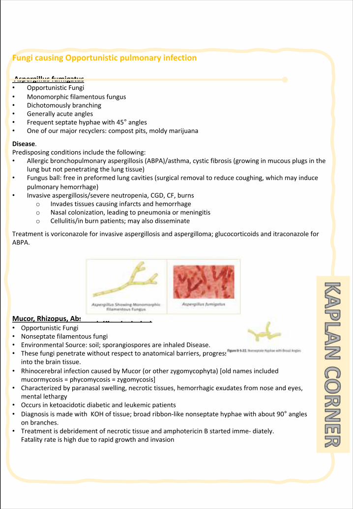
FungicausingOpportunisticpulmonaryinfection
Aspergillusfumigatus• OpportunisticFungi• Monomorphicfilamentousfungus• Dichotomouslybranching• Generallyacuteangles• Frequentseptatehyphaewith45° angles• Oneofourmajorrecyclers:compostpits,moldymarijuana
Disease.Predisposingconditionsincludethefollowing:• Allergicbronchopulmonaryaspergillosis(ABPA)/asthma,cystic fibrosis(growinginmucousplugsinthe
lungbutnotpenetratingthelungtissue)• Fungusball:freeinpreformedlungcavities(surgicalremovaltoreducecoughing,whichmayinduce
pulmonaryhemorrhage)• Invasiveaspergillosis/severeneutropenia,CGD,CF,burns
o Invadestissuescausinginfarctsandhemorrhageo Nasalcolonization,leadingtopneumoniaormeningitiso Cellulitis/inburnpatients;mayalsodisseminate
Treatmentisvoriconazole forinvasiveaspergillosisandaspergilloma;glucocorticoidsanditraconazole forABPA.
Mucor,Rhizopus,Absidia (Zygomycophyta)• OpportunisticFungi• Nonseptate filamentousfungi• EnvironmentalSource:soil;sporangiospores areinhaledDisease.• Thesefungipenetratewithoutrespecttoanatomicalbarriers,progressingrapidlyfromsinuses
intothebraintissue.• Rhinocerebral infectioncausedbyMucor (orotherzygomycophyta)[oldnamesincluded
mucormycosis =phycomycosis =zygomycosis]• Characterizedbyparanasalswelling,necrotictissues,hemorrhagicexudatesfromnoseandeyes,
mentallethargy• Occursinketoacidotic diabeticandleukemicpatients• DiagnosisismadewithKOHoftissue;broadribbon-likenonseptate hyphaewithabout90° angles
onbranches.• TreatmentisdebridementofnecrotictissueandamphotericinBstartedimme- diately.
Fatalityrateishighduetorapidgrowthandinvasion
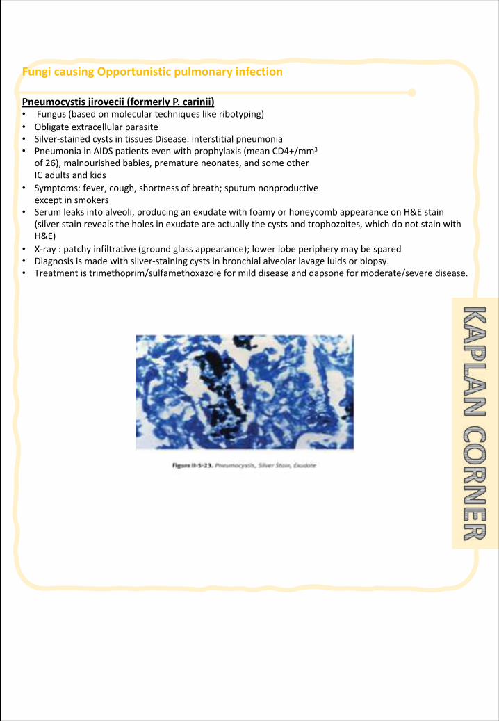
FungicausingOpportunisticpulmonaryinfection
Pneumocystisjirovecii (formerlyP.carinii)• Fungus(basedonmoleculartechniqueslikeribotyping)• Obligateextracellularparasite• Silver-stainedcystsintissuesDisease:interstitialpneumonia• PneumoniainAIDSpatientsevenwithprophylaxis(meanCD4+/mm3
of26),malnourishedbabies,prematureneonates,andsomeotherICadultsandkids
• Symptoms:fever,cough,shortnessofbreath;sputumnonproductiveexceptinsmokers
• Serumleaksintoalveoli,producinganexudatewithfoamy or honeycombappearanceonH&Estain(silverstainrevealstheholesinexudateareactuallythecystsandtrophozoites,whichdonotstainwithH&E)
• X-ray:patchyinfiltrative(groundglassappearance);lowerlobeperipherymaybespared• Diagnosisismadewithsilver-stainingcystsinbronchialalveolarlavageluids orbiopsy.• Treatmentistrimethoprim/sulfamethoxazoleformilddiseaseanddapsone for moderate/severedisease.
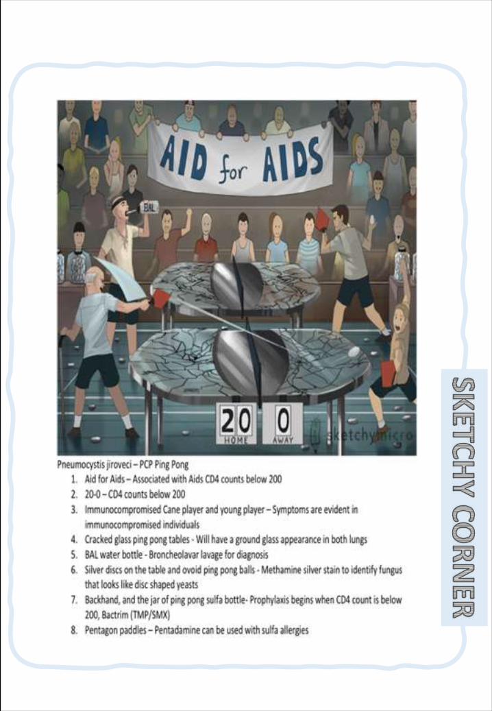
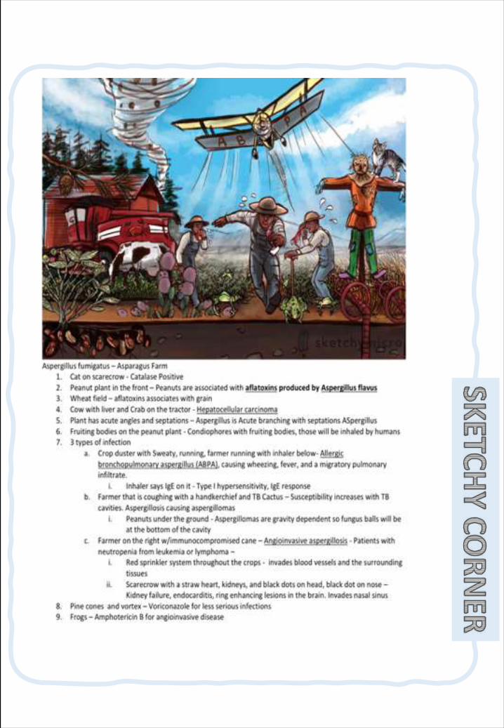
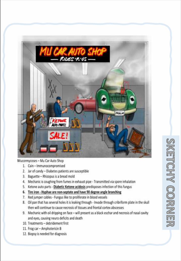
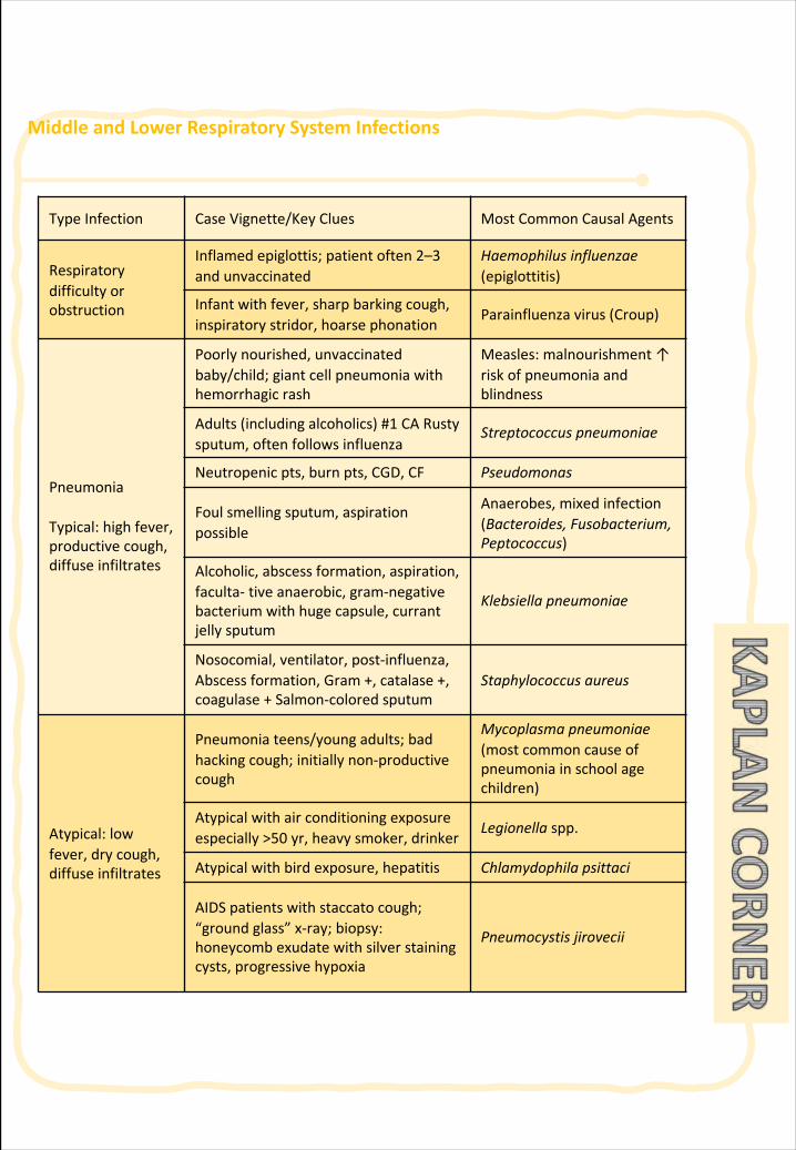
MiddleandLowerRespiratorySystemInfections
TypeInfection CaseVignette/KeyClues MostCommonCausalAgents
Respiratorydifficultyorobstruction
Inflamedepiglottis;patientoften2–3andunvaccinated
Haemophilus influenzae(epiglottitis)
Infantwithfever,sharpbarkingcough,inspiratorystridor,hoarsephonation
Parainfluenzavirus(Croup)
Pneumonia
Typical:highfever,productivecough,diffuseinfiltrates
Poorlynourished,unvaccinatedbaby/child;giantcellpneumoniawithhemorrhagicrash
Measles:malnourishment↑riskofpneumoniaandblindness
Adults(includingalcoholics)#1 CARustysputum,oftenfollowsinfluenza
Streptococcuspneumoniae
Neutropenicpts,burnpts,CGD,CF Pseudomonas
Foulsmellingsputum,aspirationpossible
Anaerobes,mixedinfection(Bacteroides,Fusobacterium,Peptococcus)
Alcoholic,abscessformation,aspiration,faculta- tive anaerobic,gram-negativebacteriumwithhugecapsule,currantjellysputum
Klebsiella pneumoniae
Nosocomial,ventilator,post-influenza,Abscessformation, Gram+,catalase+,coagulase+ Salmon-coloredsputum
Staphylococcusaureus
Atypical:lowfever,drycough,diffuseinfiltrates
Pneumoniateens/youngadults;badhackingcough;initiallynon-productivecough
Mycoplasmapneumoniae(mostcommoncauseofpneumoniainschoolagechildren)
Atypicalwithairconditioningexposureespecially>50 yr,heavysmoker,drinker
Legionellaspp.
Atypicalwithbirdexposure,hepatitis Chlamydophila psittaci
AIDSpatientswithstaccatocough;“groundglass”x-ray;biopsy:honeycombexudatewithsilverstainingcysts,progressivehypoxia
Pneumocystisjirovecii
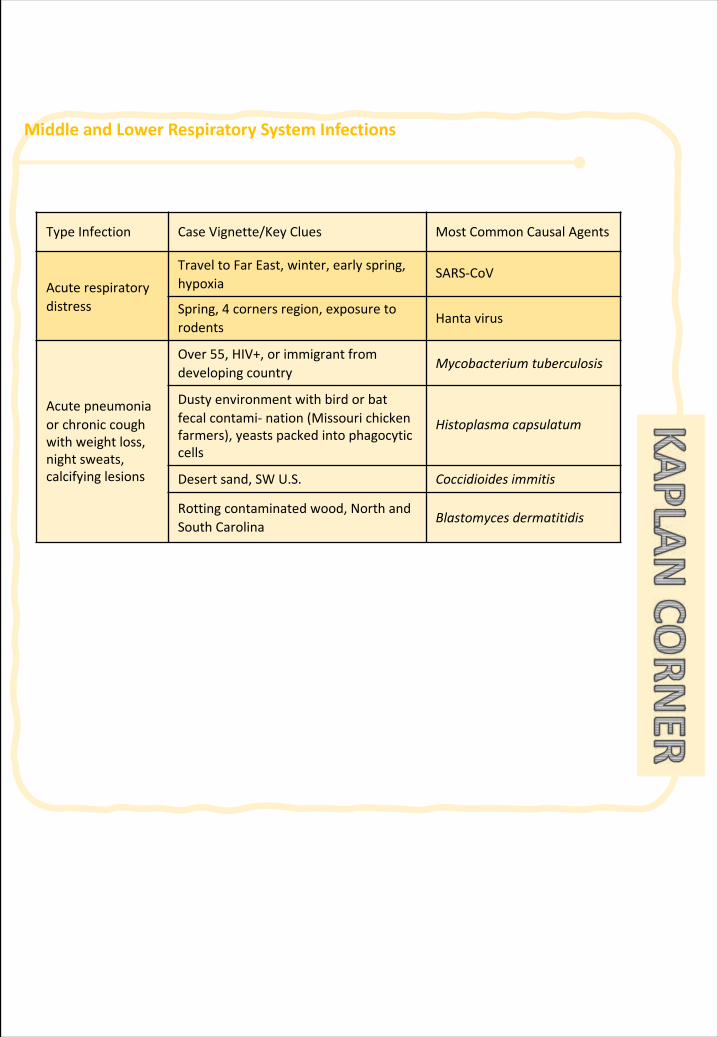
MiddleandLowerRespiratorySystemInfections
TypeInfection CaseVignette/KeyClues MostCommonCausalAgents
Acuterespiratorydistress
TraveltoFarEast,winter,earlyspring,hypoxia
SARS-CoV
Spring,4 cornersregion,exposuretorodents
Hantavirus
Acutepneumoniaorchroniccoughwithweightloss,nightsweats,calcifyinglesions
Over55,HIV+,orimmigrantfromdevelopingcountry
Mycobacteriumtuberculosis
Dustyenvironmentwithbirdorbatfecalcontami- nation(Missourichickenfarmers),yeastspackedintophagocyticcells
Histoplasmacapsulatum
Desertsand,SWU.S. Coccidioides immitis
Rottingcontaminatedwood,NorthandSouthCarolina
Blastomyces dermatitidis
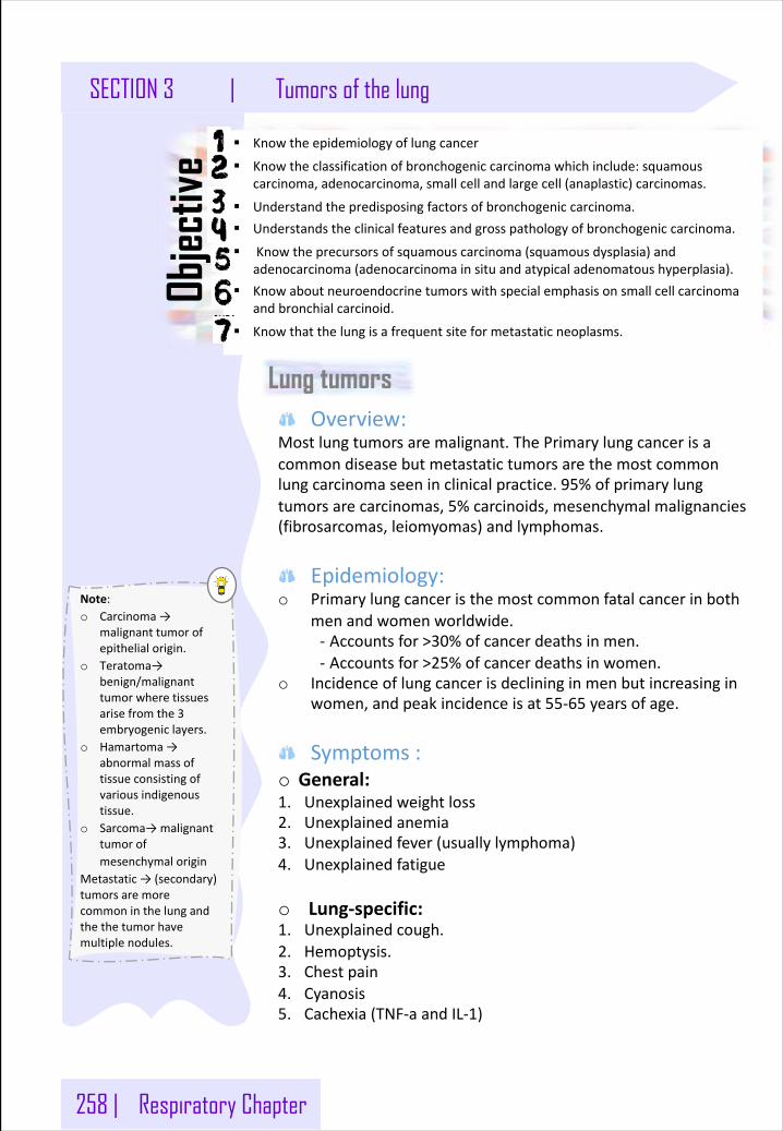
258 | Respiratory Chapter
SECTION 3 | Tumors of the lung
Lung tumors
§ Knowtheepidemiologyoflungcancer
§ Knowtheclassificationofbronchogeniccarcinomawhichinclude:squamouscarcinoma,adenocarcinoma,smallcellandlargecell(anaplastic)carcinomas.
§ Understandthepredisposingfactorsofbronchogeniccarcinoma.§ Understandstheclinicalfeaturesandgrosspathologyofbronchogeniccarcinoma.
§ Knowtheprecursorsofsquamouscarcinoma(squamousdysplasia)andadenocarcinoma(adenocarcinomainsituandatypicaladenomatoushyperplasia).
§ Knowaboutneuroendocrinetumorswithspecialemphasisonsmallcellcarcinomaandbronchialcarcinoid.
§ Knowthatthelungisafrequentsiteformetastaticneoplasms.
Objec
tive
Overview:Mostlungtumorsaremalignant.ThePrimarylungcancerisacommondiseasebutmetastatictumorsarethemostcommonlungcarcinomaseeninclinicalpractice.95%ofprimarylungtumorsarecarcinomas,5%carcinoids,mesenchymalmalignancies(fibrosarcomas,leiomyomas)andlymphomas.
Epidemiology:o Primarylungcanceristhemostcommonfatalcancerinboth
menandwomenworldwide.- Accountsfor>30%ofcancerdeathsinmen.- Accountsfor>25%ofcancerdeathsinwomen.
o Incidenceoflungcancerisdeclininginmenbutincreasinginwomen,andpeakincidenceisat55-65 yearsofage.
Symptoms:o General:1. Unexplainedweightloss2. Unexplainedanemia3. Unexplainedfever(usuallylymphoma)4. Unexplainedfatigue
o Lung-specific:1. Unexplainedcough.2. Hemoptysis.3. Chestpain4. Cyanosis5. Cachexia(TNF-aandIL-1)
Note:o Carcinoma→
malignanttumorofepithelialorigin.
o Teratoma→benign/malignanttumorwheretissuesarisefromthe3embryogeniclayers.
o Hamartoma→abnormalmassoftissueconsistingofvariousindigenoustissue.
o Sarcoma→malignanttumorofmesenchymalorigin
Metastatic→(secondary)tumorsaremorecommoninthelungandthethe tumorhavemultiplenodules.
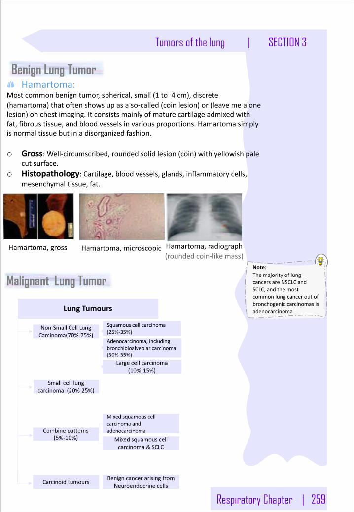
Respiratory Chapter | 259
Tumors of the lung | SECTION 3
Benign Lung Tumor Hamartoma:
Mostcommonbenigntumor,spherical,small(1 to4 cm),discrete(hamartoma)thatoftenshowsupasaso-called(coinlesion)or(leavemealonelesion)onchestimaging.Itconsistsmainlyofmaturecartilageadmixedwithfat,fibroustissue,andbloodvesselsinvariousproportions.Hamartomasimplyisnormaltissuebutinadisorganizedfashion.
o Gross:Well-circumscribed,roundedsolidlesion(coin)withyellowishpalecutsurface.
o Histopathology:Cartilage,bloodvessels,glands,inflammatorycells,mesenchymaltissue,fat.
Hamartoma,gross Hamartoma,microscopic Hamartoma,radiograph(roundedcoin-likemass)
Malignant Lung Tumor Note:ThemajorityoflungcancersareNSCLCandSCLC,andthemostcommonlungcanceroutofbronchogeniccarcinomasisadenocarcinoma
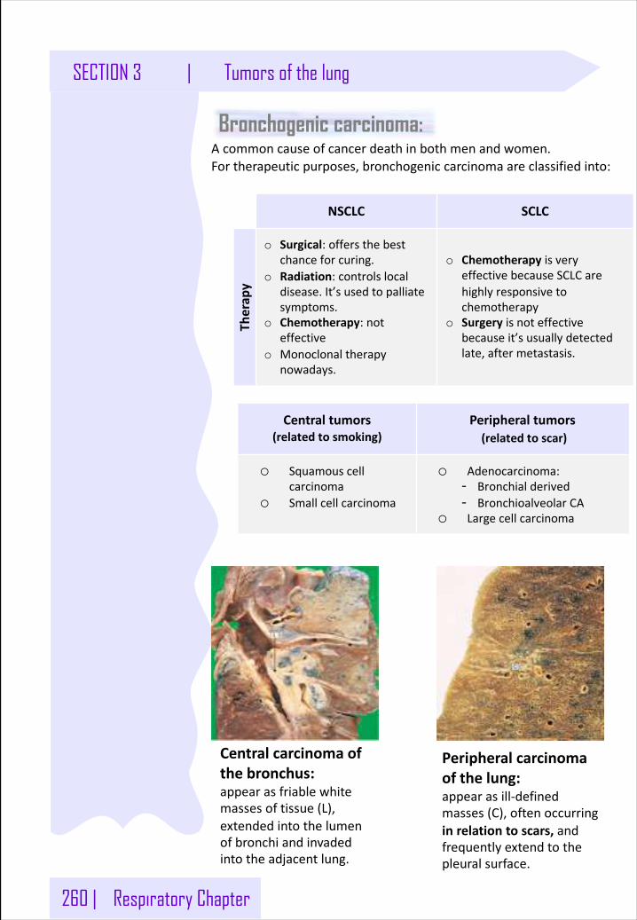
260 | Respiratory Chapter
SECTION 3 | Tumors of the lung
Bronchogenic carcinoma:
Centralcarcinomaofthebronchus:appearasfriablewhitemassesoftissue(L),extendedintothelumenofbronchiandinvadedintotheadjacentlung.
Acommoncauseofcancerdeathinbothmenandwomen.Fortherapeuticpurposes,bronchogeniccarcinomaareclassifiedinto:
NSCLC SCLC
Therap
yo Surgical:offersthebest
chanceforcuring.o Radiation:controlslocal
disease.It’susedtopalliatesymptoms.
o Chemotherapy:noteffective
o Monoclonaltherapynowadays.
o Chemotherapy isveryeffectivebecauseSCLCarehighlyresponsivetochemotherapy
o Surgery isnoteffectivebecauseit’susuallydetectedlate,aftermetastasis.
Centraltumors(relatedtosmoking)
Peripheraltumors(relatedtoscar)
o Squamouscellcarcinoma
o Smallcellcarcinoma
o Adenocarcinoma:- Bronchialderived- BronchioalveolarCA
o Largecellcarcinoma
Peripheralcarcinomaofthelung:appearasill-definedmasses(C),oftenoccurringinrelationtoscars,andfrequentlyextendtothepleuralsurface.
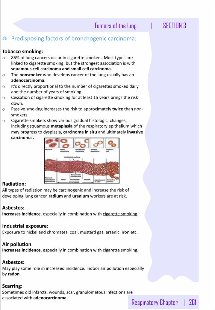
Respiratory Chapter | 261
Tumors of the lung | SECTION 3
Predisposingfactorsofbronchogeniccarcinoma:
Tobaccosmoking:o 85%oflungcancersoccurincigarettesmokers.Mosttypesare
linkedtocigarettesmoking,butthestrongestassociationiswithsquamouscellcarcinomaandsmallcellcarcinoma.
o Thenonsmoker whodevelopscancerofthelungusuallyhasanadenocarcinoma.
o It’sdirectlyproportionaltothenumberofcigarettessmokeddailyandthenumberofyearsofsmoking.
o Cessationofcigarettesmokingforatleast15 yearsbringstheriskdown.
o Passivesmokingincreasestherisktoapproximatelytwice thannon-smokers.
o Cigarettesmokersshowvariousgradualhistologicchanges,includingsquamousmetaplasia oftherespiratoryepitheliumwhichmayprogresstodysplasia,carcinomainsituandultimatelyinvasivecarcinoma.
Radiation:Alltypesofradiationmaybecarcinogenicandincreasetheriskofdevelopinglungcancer.radium anduranium workersareatrisk.
Asbestos:Increasesincidence,especiallyincombinationwithcigarettesmoking.
Industrialexposure:Exposuretonickelandchromates,coal,mustardgas,arsenic,ironetc.
AirpollutionIncreasesincidence,especiallyincombinationwithcigarettesmoking.
Asbestos:Mayplaysomeroleinincreasedincidence.Indoorairpollutionespeciallybyradon.
Scarring:Sometimesoldinfarcts,wounds,scar,granulomatousinfectionsareassociatedwithadenocarcinoma.
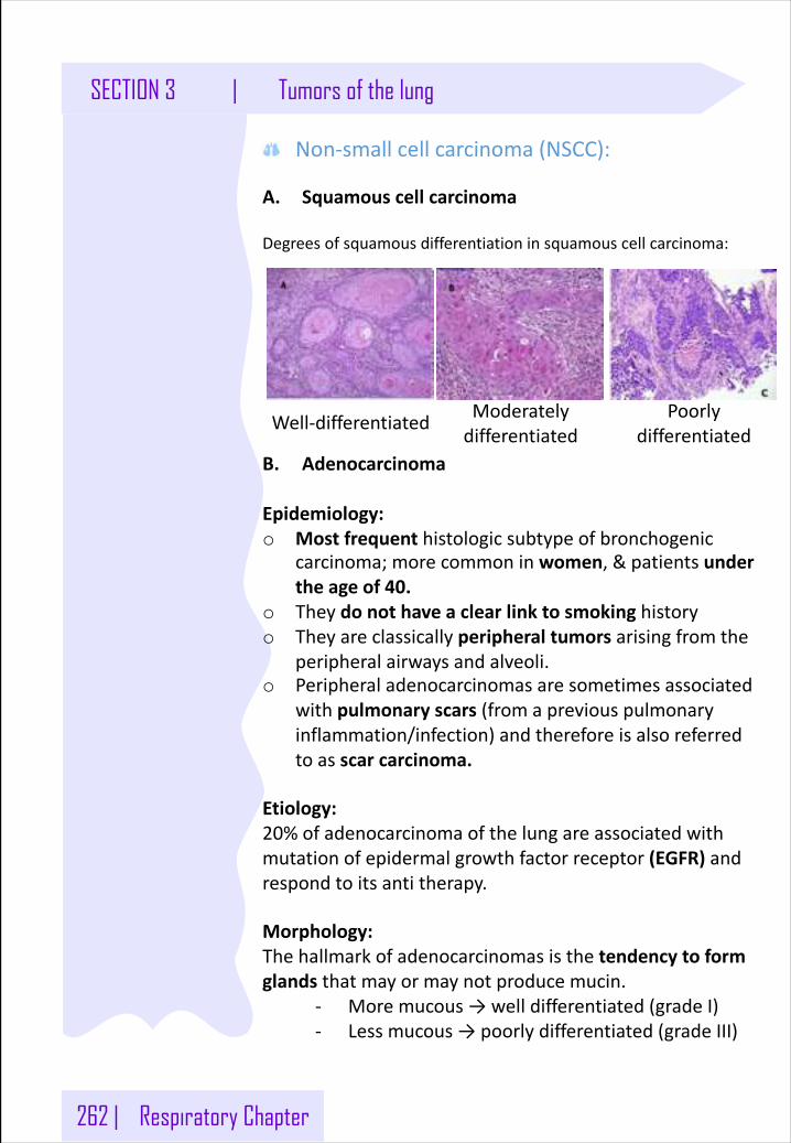
262 | Respiratory Chapter
SECTION 3 | Tumors of the lung
Well-differentiated
Non-smallcellcarcinoma(NSCC):
A. Squamouscellcarcinoma
Degreesofsquamousdifferentiationinsquamouscellcarcinoma:
B. Adenocarcinoma
Epidemiology:o Mostfrequenthistologicsubtypeofbronchogenic
carcinoma;morecommoninwomen,&patientsundertheageof40.
o Theydonothaveaclearlinktosmokinghistoryo Theyareclassicallyperipheraltumorsarisingfromthe
peripheralairwaysandalveoli.o Peripheraladenocarcinomasaresometimesassociated
withpulmonaryscars(fromapreviouspulmonaryinflammation/infection)andthereforeisalsoreferredtoasscarcarcinoma.
Etiology:20%ofadenocarcinomaofthelungareassociatedwithmutationofepidermalgrowthfactorreceptor(EGFR)andrespondtoitsantitherapy.
Morphology:Thehallmarkofadenocarcinomasisthetendencytoformglandsthatmayormaynotproducemucin.
- Moremucous→welldifferentiated(gradeI)- Lessmucous→poorlydifferentiated(gradeIII)
Moderatelydifferentiated
Poorlydifferentiated
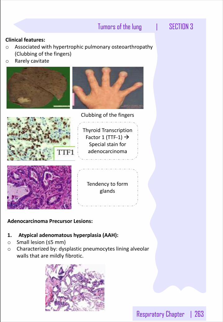
Respiratory Chapter | 263
Tumors of the lung | SECTION 3
Clinicalfeatures:o Associatedwithhypertrophicpulmonaryosteoarthropathy
(Clubbingofthefingers)o Rarelycavitate
Tendencytoformglands
Clubbingofthefingers
ThyroidTranscriptionFactor1 (TTF-1)àSpecialstainforadenocarcinoma
AdenocarcinomaPrecursorLesions:
1. Atypicaladenomatoushyperplasia(AAH):o Smalllesion(≤5 mm)o Characterizedby:dysplasticpneumocytes liningalveolar
wallsthataremildlyfibrotic.
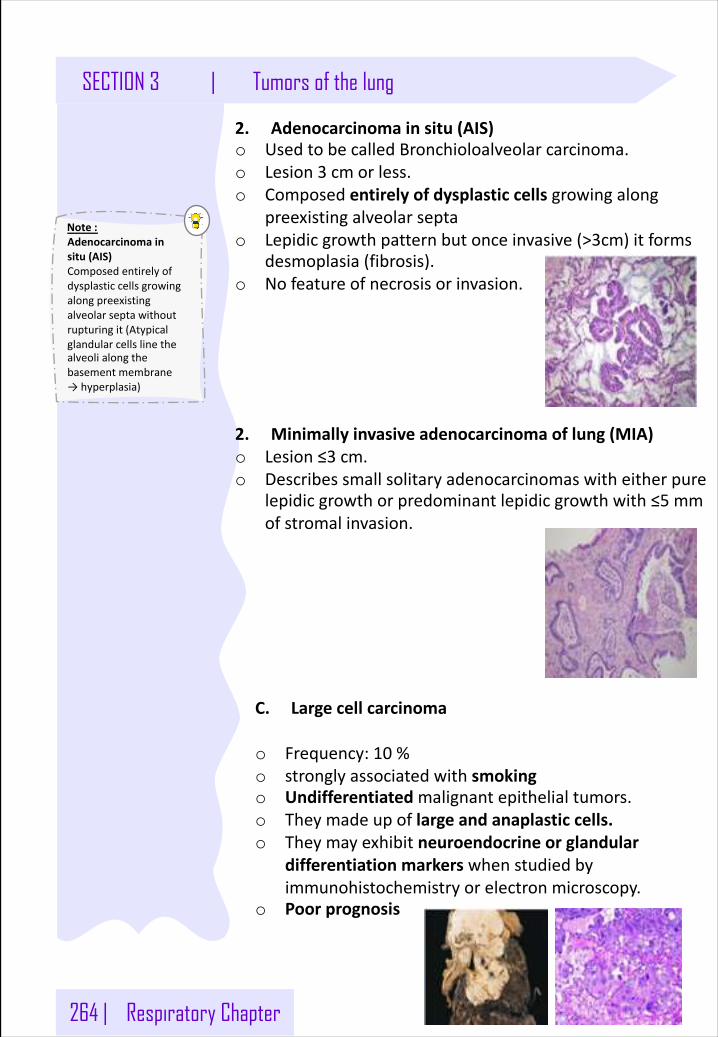
264 | Respiratory Chapter
Note:Adenocarcinomainsitu(AIS)Composedentirelyofdysplasticcellsgrowingalongpreexistingalveolarseptawithoutrupturingit(Atypicalglandularcellslinethealveolialongthebasementmembrane→hyperplasia)
SECTION 3 | Tumors of the lung
C. Largecellcarcinoma
o Frequency:10 %o stronglyassociatedwithsmokingo Undifferentiatedmalignantepithelialtumors.o Theymadeupoflargeandanaplasticcells.o Theymayexhibitneuroendocrineorglandular
differentiationmarkers whenstudiedbyimmunohistochemistryorelectronmicroscopy.
o Poorprognosis
2. Adenocarcinomainsitu(AIS)o UsedtobecalledBronchioloalveolar carcinoma.o Lesion3 cmorless.o Composedentirelyofdysplasticcellsgrowingalong
preexistingalveolarseptao Lepidicgrowthpatternbutonceinvasive(>3cm)itforms
desmoplasia(fibrosis).o Nofeatureofnecrosisorinvasion.
2. Minimallyinvasiveadenocarcinomaoflung(MIA)o Lesion≤3 cm.o Describessmallsolitaryadenocarcinomaswitheitherpure
lepidic growthorpredominantlepidic growthwith≤5 mmofstromalinvasion.
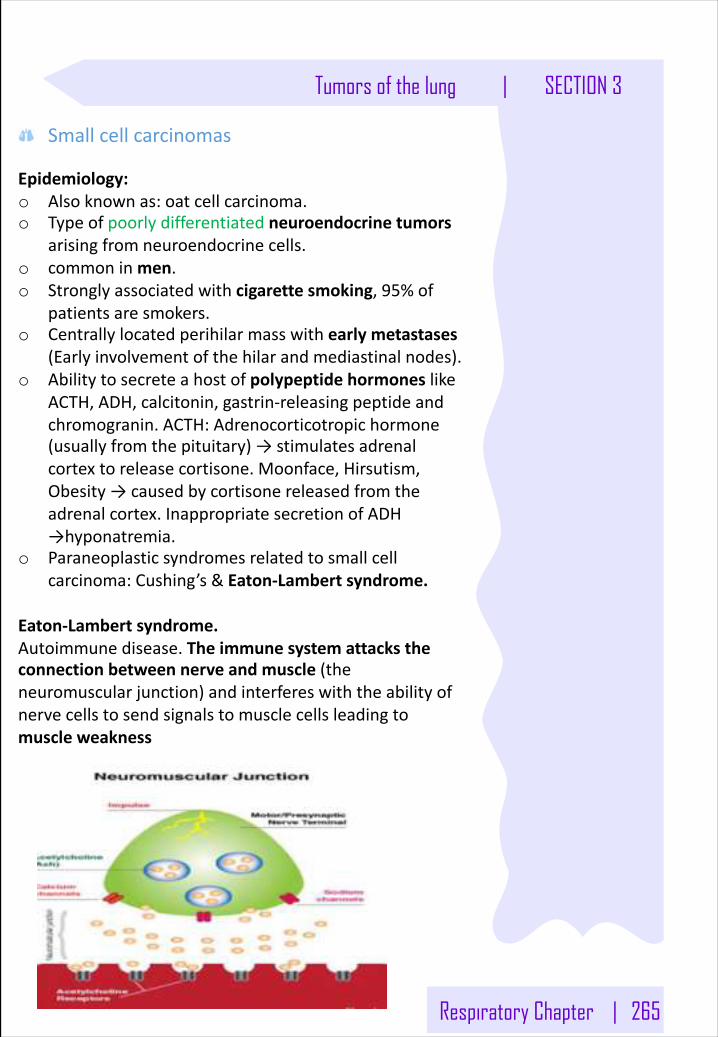
Respiratory Chapter | 265
Tumors of the lung | SECTION 3
Smallcellcarcinomas
Epidemiology:o Alsoknownas:oatcellcarcinoma.o Typeofpoorlydifferentiatedneuroendocrinetumors
arisingfromneuroendocrinecells.o commoninmen.o Stronglyassociatedwithcigarette smoking,95%of
patientsaresmokers.o Centrallylocatedperihilar masswithearlymetastases
(Earlyinvolvementofthehilarandmediastinalnodes).o Abilitytosecreteahostofpolypeptidehormones like
ACTH,ADH,calcitonin,gastrin-releasingpeptideandchromogranin.ACTH:Adrenocorticotropichormone(usuallyfromthepituitary)→stimulatesadrenalcortextoreleasecortisone.Moonface,Hirsutism,Obesity→causedbycortisonereleasedfromtheadrenalcortex.InappropriatesecretionofADH→hyponatremia.
o Paraneoplasticsyndromesrelatedtosmallcellcarcinoma:Cushing’s&Eaton-Lambertsyndrome.
Eaton-Lambertsyndrome.Autoimmunedisease.Theimmunesystemattackstheconnectionbetweennerveandmuscle (theneuromuscularjunction)andinterfereswiththeabilityofnervecellstosendsignalstomusclecellsleadingtomuscleweakness
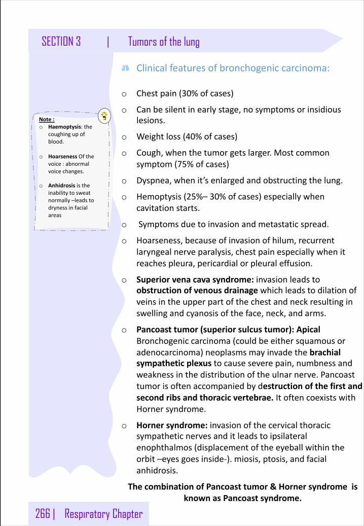
266 | Respiratory Chapter
Note:o Haemoptysis:the
coughingupofblood.
o Hoarseness Ofthevoice:abnormalvoicechanges.
o Anhidrosis istheinabilitytosweatnormally–leadstodrynessinfacialareas
SECTION 3 | Tumors of the lung
Clinicalfeaturesofbronchogeniccarcinoma:
o Chestpain(30%ofcases)
o Canbesilentinearlystage,nosymptomsorinsidiouslesions.
o Weightloss(40%ofcases)
o Cough,whenthetumorgetslarger.Mostcommonsymptom(75%ofcases)
o Dyspnea,whenit’senlargedandobstructingthelung.
o Hemoptysis(25%– 30%ofcases)especiallywhencavitationstarts.
o Symptomsduetoinvasionandmetastaticspread.
o Hoarseness,becauseofinvasionofhilum,recurrentlaryngealnerveparalysis,chestpainespeciallywhenitreachespleura,pericardialorpleuraleffusion.
o Superiorvenacavasyndrome:invasionleadstoobstructionofvenousdrainagewhichleadstodilationofveinsintheupperpartofthechestandneckresultinginswellingandcyanosisoftheface,neck,andarms.
o Pancoasttumor(superiorsulcustumor):ApicalBronchogeniccarcinoma(couldbeeithersquamousoradenocarcinoma)neoplasmsmayinvadethebrachialsympatheticplexustocauseseverepain,numbnessandweaknessinthedistributionoftheulnarnerve.Pancoasttumorisoftenaccompaniedbydestructionofthefirstandsecondribsandthoracicvertebrae. ItoftencoexistswithHornersyndrome.
o Hornersyndrome:invasionofthecervicalthoracicsympatheticnervesanditleadstoipsilateralenophthalmos(displacementoftheeyeballwithintheorbit–eyesgoesinside-).miosis,ptosis,andfacialanhidrosis.
ThecombinationofPancoasttumor&HornersyndromeisknownasPancoastsyndrome.
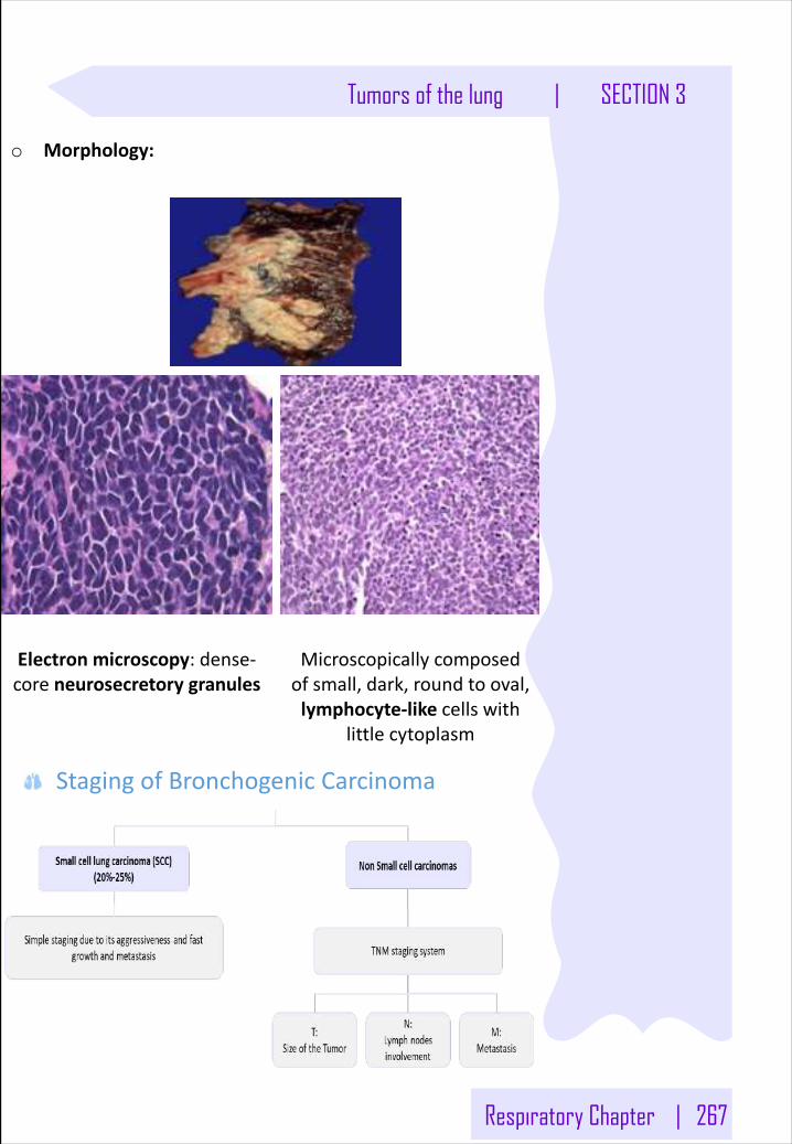
Respiratory Chapter | 267
Tumors of the lung | SECTION 3
o Morphology:
Electron microscopy:dense-coreneurosecretorygranules
Microscopicallycomposedofsmall,dark,roundtooval,lymphocyte-like cellswith
littlecytoplasm
StagingofBronchogenicCarcinoma
268 | Respiratory Chapter
Note:IfwechecktheX-Rayandfoundamassinthelung.Andtheabdominalscanshowsbilateraladrenalglandenlargement.Thenitisbronchogeniccarcinomawithmetastasistoadrenals.
SECTION 3 | Tumors of the lung
Complicationsofbronchogeniccarcinomao Bronchiectasiso Obstructivepneumoniao Paraneoplasticsyndromeo Pleuraleffusion,bloodyo Hoarsenessfromrecurrentlaryngealnerveparalysis
Spreadofbronchogeniccarcinomao Lymphaticspread
o Successivechainsofnodes(scalenenodes).o Involvementofthesupraclavicularnode(Virchow’s
node).o Extendintothepericardialorpleuralspacesà Infiltrate
thesuperiorvenacava.o Atumormayextenddirectlyintotheesophagus,
producingobstruction,sometimescomplicatedbyafistula.
o Phrenicnerveinvasionusuallycausesdiaphragmaticparalysis.
o Mayinvadethebrachialorcervicalsympatheticplexus.o Distantmetastasistoliver(30-50%),adrenals(>50%),
brain(20%)andbone(20%).
Treatment:o Chemotherapyresponsiveo Leastlikelyformtobecuredbysurgery;usuallyalready
metastaticatdiagnosis
Prognosis:o Highlymalignantandaggressivetumor,poorprognosis,
rarelyresectable.o Histologicaltypesandthestageoflungcancerdetermine
theoutcome.o Survivalisbetterforearlystagedisease,exceptforsmall
cellcarcinoma (veryearlymetastases)o Nonsmallcellcancersfarebetterthansmallcell
carcinomao Overallcombined5-yearsurvivalrateis~15%
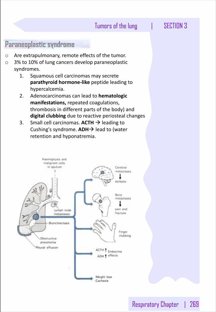
Respiratory Chapter | 269
Tumors of the lung | SECTION 3
Paraneoplastic syndromeo Areextrapulmonary,remoteeffectsofthetumor.o 3%to10%oflungcancersdevelopparaneoplastic
syndromes.1. Squamouscellcarcinomasmaysecrete
parathyroidhormone-like peptideleadingtohypercalcemia.
2. Adenocarcinomascanleadtohematologicmanifestations,repeatedcoagulations,thrombosisindifferentpartsofthebody)anddigitalclubbingduetoreactiveperiostealchanges
3. Smallcellcarcinomas.ACTHà leadingtoCushing'ssyndrome.ADHà leadto(waterretentionandhyponatremia.
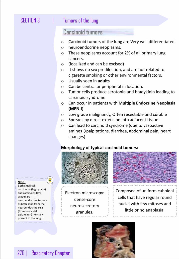
270 | Respiratory Chapter
Note:Bothsmallcellcarcinoma(highgrade)andcarcinoids,(lowgrade)areneuroendocrinetumorsasbotharisefromtheneuroendocrinecells(frombronchialepithelium)normallypresentinthelung.
SECTION 3 | Tumors of the lung
Carcinoid tumorso CarcinoidtumorsofthelungareVerywelldifferentiatedo neuroendocrineneoplasms.o Theseneoplasmsaccountfor2%ofallprimarylung
cancers.o (localizedandcanbeexcised)o Itshowsnosexpredilection,andarenotrelatedto
cigarettesmokingorotherenvironmentalfactors.o Usuallyseeninadultso Canbecentralorperipheralinlocation.o Tumorcellsproduceserotoninandbradykininleadingto
carcinoidsyndromeo CanoccurinpatientswithMultipleEndocrineNeoplasia
(MEN-I)o Lowgrademalignancy,Oftenresectable andcurableo Spreadsbydirectextensionintoadjacenttissueo Canleadtocarcinoidsyndrome(duetovasoactive
amines→palpitations,diarrhea,abdominalpain,heartchanges)
Morphologyoftypicalcarcinoidtumors:
Electronmicroscopy:dense-core
neurosecretorygranules.
Composedofuniformcuboidalcellsthathaveregularroundnucleiwithfewmitosesand
littleornoanaplasia.
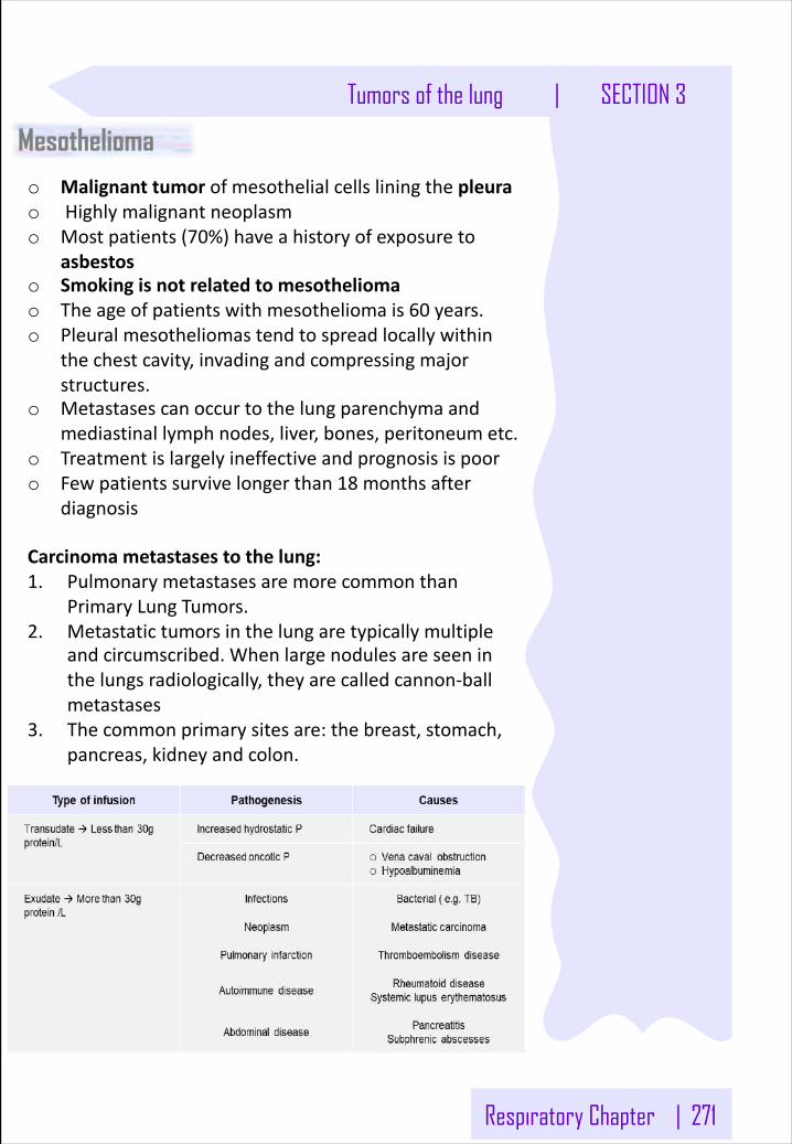
Respiratory Chapter | 271
Tumors of the lung | SECTION 3
Mesotheliomao Malignanttumorofmesothelialcellsliningthepleurao Highlymalignantneoplasmo Mostpatients(70%)haveahistoryofexposureto
asbestoso Smokingisnotrelatedtomesotheliomao Theageofpatientswithmesotheliomais60 years.o Pleuralmesotheliomastendtospreadlocallywithin
thechestcavity,invadingandcompressingmajorstructures.
o Metastasescanoccurtothelungparenchymaandmediastinallymphnodes,liver,bones,peritoneumetc.
o Treatmentislargelyineffectiveandprognosisispooro Fewpatientssurvivelongerthan18 monthsafter
diagnosis
Carcinomametastasestothelung:1. Pulmonarymetastasesaremorecommonthan
PrimaryLungTumors.2. Metastatictumorsinthelungaretypicallymultiple
andcircumscribed.Whenlargenodulesareseeninthelungsradiologically,theyarecalledcannon-ballmetastases
3. Thecommonprimarysitesare:thebreast,stomach,pancreas,kidneyandcolon.
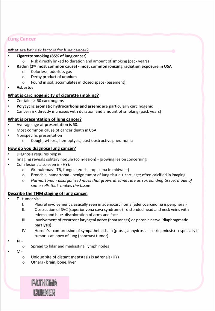
Lung Cancer
Whatarekeyriskfactorsforlung cancer?• Cigarettesmoking(85%oflung cancer)
o Riskdirectlylinkedtodurationandamountofsmoking(pack years)• Radon (2nd most common cause)- most common ionizing radiation exposure in USA
o Colorless,odorless gaso Decayproductof uraniumo Foundinsoil,accumulatesinclosedspace (basement)
• Asbestos
Whatiscarcinogenicityofcigarette smoking?• Contains>60 carcinogens• Polycyclicaromatichydrocarbonsandarsenicareparticularly carcinogenic• Cancer risk directly increases with duration and amount of smoking (pack years)
Whatispresentationoflung cancer?• Averageageatpresentationis 60.• MostcommoncauseofcancerdeathinUSA• Nonspecificpresentation
o Cough,wt loss,hemoptysis,postobstructive pneumonia
Howdoyoudiagnoselung cancer?• Diagnosisrequires biopsy• Imagingrevealssolitarynodule(coin-lesion)- growinglesion concerning• Coinlesionsalsoseenin (HY):
o Granulomas- TB,fungus(ex- histoplasma in midwest)o Bronchialhamartoma- benigntumoroflungtissue+cartilage;oftencalcifiedin imagingo Harmartoma - disorganizedmassthatgrowsatsamerateassurroundingtissue;madeof
samecellsthatmakesthe tissue
DescribetheTNMstagingoflung cancer.• T- tumor size
I. Pleuralinvolvementclassicallyseeninadenocarcinoma(adenocarcinomais peripheral)II. ObstructionofSVC(superiorvenacavasyndrome)- distendedheadandneckveinswith
edemaandbluediscolorationofarmsand faceIII. Involvementofrecurrentlaryngealnerve(hoarseness)orphrenicnerve(diaphragmatic
paralysis)IV. Horner's- compressionofsympatheticchain(ptosis,anhydrosis - inskin,miosis)- especiallyif
tumorisatapexoflung(pancoast tumor)• N–
o Spreadtohilarandmediastinallymph nodes• M-
o Uniquesiteofdistantmetastasisisadrenals (HY)o Others- brain,bone, liver
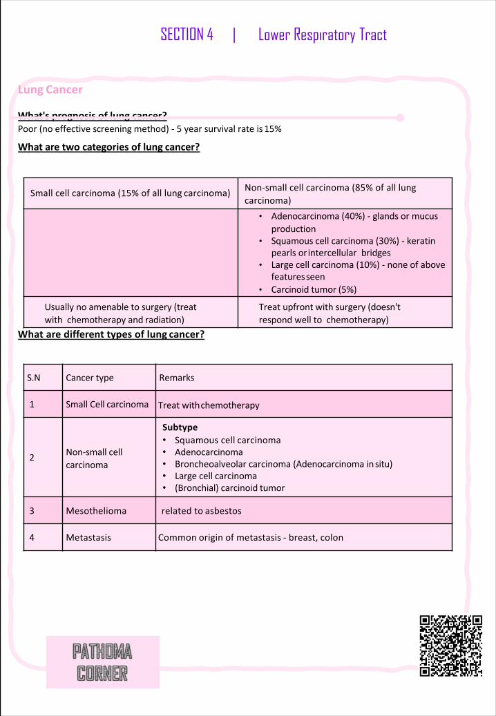
Lung Cancer
What'sprognosisoflung cancer?Poor(noeffectivescreeningmethod)- 5 yearsurvivalrateis 15%
Whataretwocategoriesoflung cancer?
Whataredifferenttypesoflung cancer?
Smallcellcarcinoma(15%ofalllung carcinoma) Non-smallcellcarcinoma(85%ofalllungcarcinoma)
• Adenocarcinoma(40%)- glandsormucusproduction
• Squamouscellcarcinoma(30%)- keratinpearlsor intercellularbridges
• Largecellcarcinoma(10%)- noneofabovefeaturesseen
• Carcinoidtumor (5%)
Usuallynoamenabletosurgery(treatwithchemotherapyand radiation)
Treatupfrontwithsurgery(doesn'trespondwelltochemotherapy)
S.N Cancer type Remarks
1 SmallCell carcinoma Treatwithchemotherapy
2 Non-smallcellcarcinoma
Subtype• Squamouscell carcinoma• Adenocarcinoma• Broncheoalveolar carcinoma(Adenocarcinomainsitu)• Largecell carcinoma• (Bronchial)carcinoid tumor
3 Mesothelioma relatedto asbestos
4 Metastasis Common origin of metastasis - breast, colon
SECTION 4 | Lower Respiratory Tract
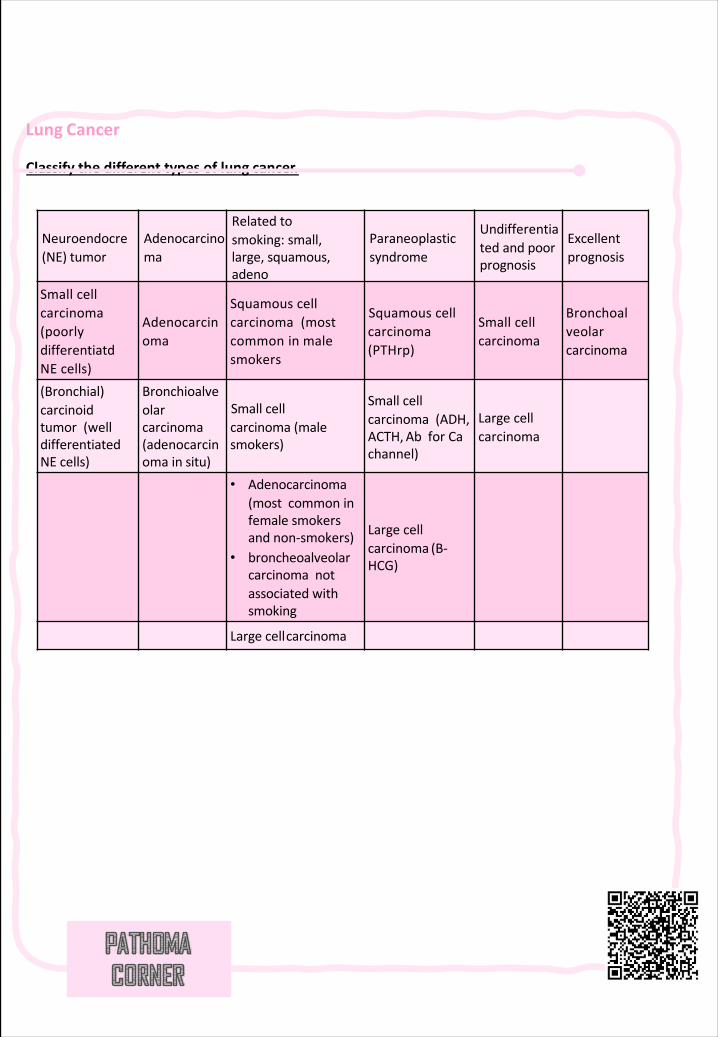
Lung Cancer
Classifythedifferenttypesoflung cancer.
Neuroendocre(NE) tumor
Adenocarcinoma
Relatedtosmoking: small,large,squamous,adeno
Paraneoplasticsyndrome
Undifferentiatedandpoorprognosis
Excellentprognosis
Smallcellcarcinoma(poorlydifferentiatdNE cells)
Adenocarcinoma
Squamouscellcarcinoma(mostcommoninmalesmokers
Squamouscellcarcinoma(PTHrp)
Smallcellcarcinoma
Bronchoalveolarcarcinoma
(Bronchial)carcinoidtumor(welldifferentiatedNEcells)
Bronchioalveolarcarcinoma(adenocarcinomain situ)
Smallcellcarcinoma(malesmokers)
Smallcellcarcinoma(ADH,ACTH,AbforCachannel)
Largecellcarcinoma
• Adenocarcinoma(mostcommoninfemalesmokersandnon-smokers)
• broncheoalveolarcarcinomanotassociatedwithsmoking
Largecellcarcinoma (B-HCG)
Largecellcarcinoma
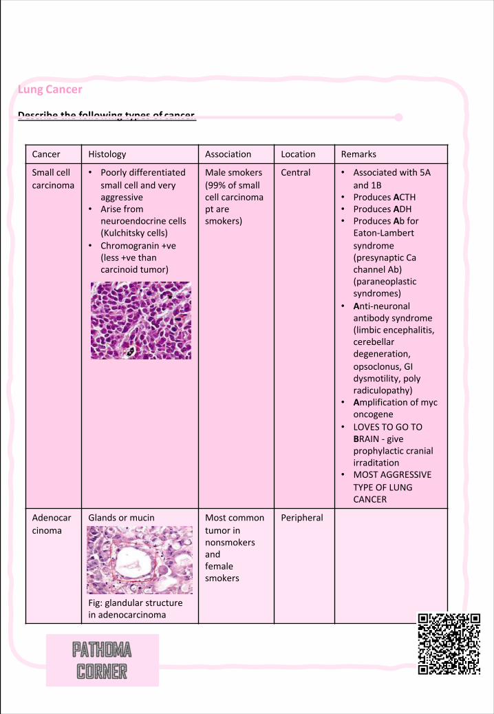
Lung Cancer
Describethefollowingtypesof cancer.
Cancer Histology Association Location Remarks
Smallcellcarcinoma
• Poorlydifferentiatedsmallcellandveryaggressive
• Arisefromneuroendocrinecells(Kulchitsky cells)
• Chromogranin+ve(less+vethancarcinoidtumor)
Malesmokers(99%ofsmallcellcarcinomapt aresmokers)
Central • Associatedwith5Aand1B
• ProducesACTH• ProducesADH• ProducesAbfor
Eaton-Lambertsyndrome(presynapticCachannelAb)(paraneoplasticsyndromes)
• Anti-neuronalantibodysyndrome(limbicencephalitis,cerebellardegeneration,opsoclonus,GIdysmotility,polyradiculopathy)
• Amplificationofmyconcogene
• LOVESTOGOTOBRAIN- giveprophylacticcranialirraditation
• MOSTAGGRESSIVETYPEOFLUNGCANCER
Adenocarcinoma
Glandsormucin
Fig:glandularstructureinadenocarcinoma
Mostcommontumorinnonsmokersandfemalesmokers
Peripheral
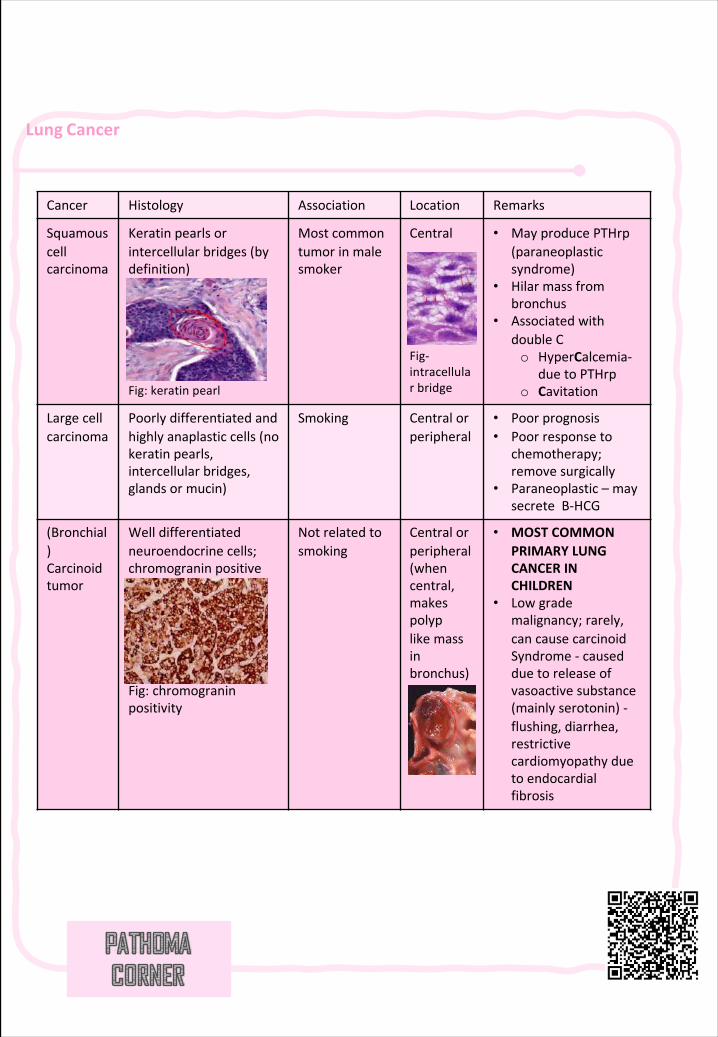
Lung Cancer
Cancer Histology Association Location Remarks
Squamouscellcarcinoma
Keratinpearlsorintercellularbridges(bydefinition)
Fig:keratinpearl
Mostcommontumorinmalesmoker
Central
Fig-intracellularbridge
• MayproducePTHrp(paraneoplasticsyndrome)
• Hilarmassfrombronchus
• AssociatedwithdoubleCo HyperCalcemia-
duetoPTHrpo Cavitation
Largecellcarcinoma
Poorlydifferentiatedandhighlyanaplasticcells(nokeratinpearls,intercellularbridges,glandsormucin)
Smoking Centralorperipheral
• Poorprognosis• Poorresponseto
chemotherapy;remove surgically
• Paraneoplastic– maysecrete B-HCG
(Bronchial)Carcinoidtumor
Welldifferentiatedneuroendocrinecells;chromograninpositive
Fig:chromograninpositivity
Notrelatedtosmoking
Centralorperipheral(whencentral,makespolyplikemassinbronchus)
• MOSTCOMMONPRIMARYLUNGCANCERINCHILDREN
• Lowgrademalignancy;rarely,cancausecarcinoidSyndrome- causedduetoreleaseofvasoactive substance(mainlyserotonin)-flushing,diarrhea,restrictivecardiomyopathyduetoendocardialfibrosis
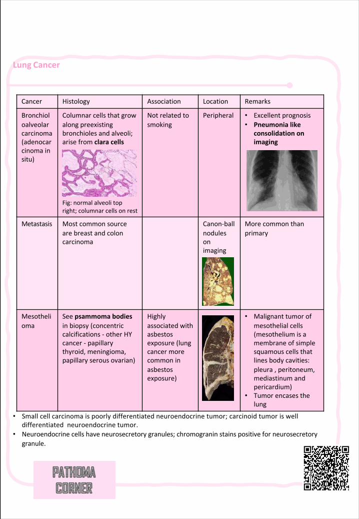
Lung Cancer
• Smallcellcarcinomaispoorlydifferentiatedneuroendocrinetumor;carcinoidtumoriswelldifferentiatedneuroendocrine tumor.
• Neuroendocrinecellshaveneurosecretorygranules;chromograninstainspositiveforneurosecretorygranule.
Cancer Histology Association Location Remarks
Bronchioloalveolarcarcinoma(adenocarcinoma insitu)
Columnarcellsthatgrowalongpreexistingbronchiolesandalveoli;arisefromclara cells
Fig:normalalveolitopright;columnarcellsonrest
Notrelatedtosmoking
Peripheral • Excellentprognosis• Pneumonialike
consolidation onimaging
Metastasis Mostcommonsourcearebreastandcoloncarcinoma
Canon-ballnodulesonimaging
Morecommonthanprimary
Mesothelioma
Seepsammoma bodiesinbiopsy(concentriccalcifications- otherHYcancer- papillarythyroid,meningioma,papillaryserousovarian)
Highlyassociatedwithasbestosexposure(lungcancermorecommoninasbestosexposure)
• Malignanttumorofmesothelialcells(mesotheliumisamembraneofsimplesquamouscellsthatlinesbodycavities:pleura,peritoneum,mediastinumandpericardium)
• Tumorencasesthelung
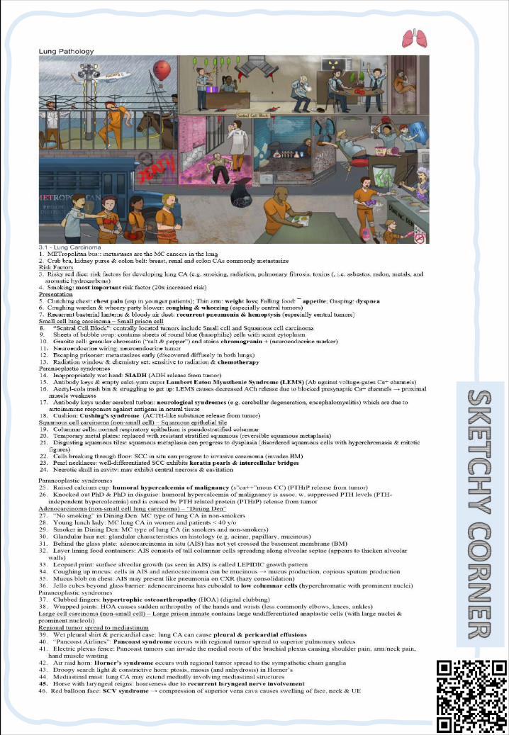
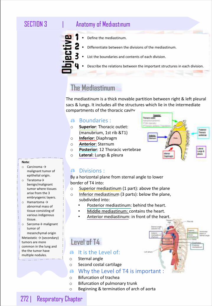
272 | Respiratory Chapter
SECTION 3 | Anatomy of Mediastinum
Note:o Carcinoma→
malignanttumorofepithelialorigin.
o Teratoma→benign/malignanttumorwheretissuesarisefromthe3embryogeniclayers.
o Hamartoma→abnormalmassoftissueconsistingofvariousindigenoustissue.
o Sarcoma→malignanttumorofmesenchymalorigin
Metastatic→(secondary)tumorsaremorecommoninthelungandthethe tumorhavemultiplenodules.
§ Definethemediastinum.
§ Differentiatebetweenthedivisionsofthemediastinum.
§ Listtheboundariesandcontentsofeachdivision.
§ Describetherelationsbetweentheimportantstructuresineachdivision.Objec
tive
Themediastinumisathickmovablepartitionbetweenright&leftpleuralsacs&lungs.Itincludesallthestructureswhichlieintheintermediatecompartmentsofthethoraciccavity
The Mediastinum
Boundaries:o Superior:Thoracicoutlet:
(manubrium,1strib&T1)o Inferior:Diaphragmo Anterior:Sternumo Posterior:12 Thoracicvertebraeo Lateral:Lungs&pleura
Divisions:ByahorizontalplanefromsternalangletolowerborderofT4 into:o Superiormediastinum(1 part):abovetheplaneo Inferiormediastinum(3 parts):belowtheplane,
subdividedinto:• Posteriormediastinum:behindtheheart.• Middlemediastinum:containstheheart.• Anteriormediastinum:infrontoftheheart.
ItistheLevelof:o Sternalangleo Secondcostalcartilage
WhytheLevelofT4 isimportant:o Bifurcationoftracheao Bifurcationofpulmonarytrunko Beginning&terminationofarchofaorta
Level of T4
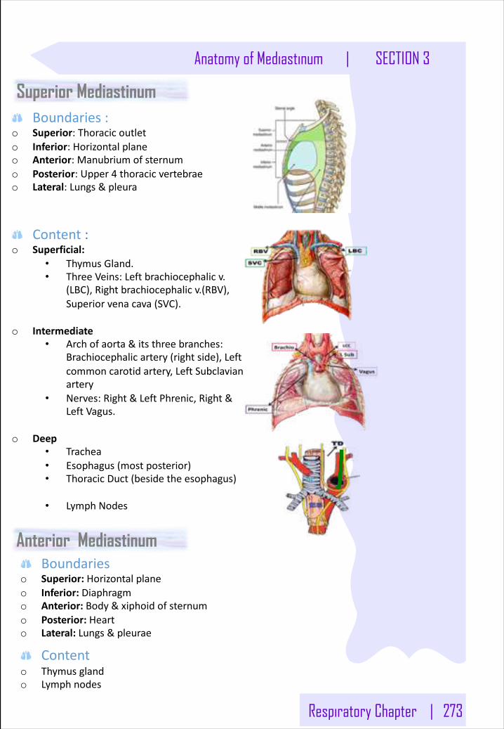
Respiratory Chapter | 273
Anatomy of Mediastinum | SECTION 3
Superior Mediastinum Boundaries:
o Superior:Thoracicoutleto Inferior:Horizontalplaneo Anterior:Manubriumofsternumo Posterior:Upper4 thoracicvertebraeo Lateral:Lungs&pleura
Content :o Superficial:
• ThymusGland.• ThreeVeins:Leftbrachiocephalicv.
(LBC),Rightbrachiocephalicv.(RBV),Superiorvenacava(SVC).
o Intermediate• Archofaorta&itsthreebranches:
Brachiocephalicartery(rightside),Leftcommoncarotidartery,LeftSubclavianartery
• Nerves:Right&LeftPhrenic,Right&LeftVagus.
o Deep• Trachea• Esophagus(mostposterior)• ThoracicDuct(besidetheesophagus)
• LymphNodes
Anterior MediastinumBoundaries
o Superior:Horizontalplaneo Inferior:Diaphragmo Anterior:Body&xiphoidofsternumo Posterior:Hearto Lateral:Lungs&pleurae
Contento Thymusgland o Lymphnodes
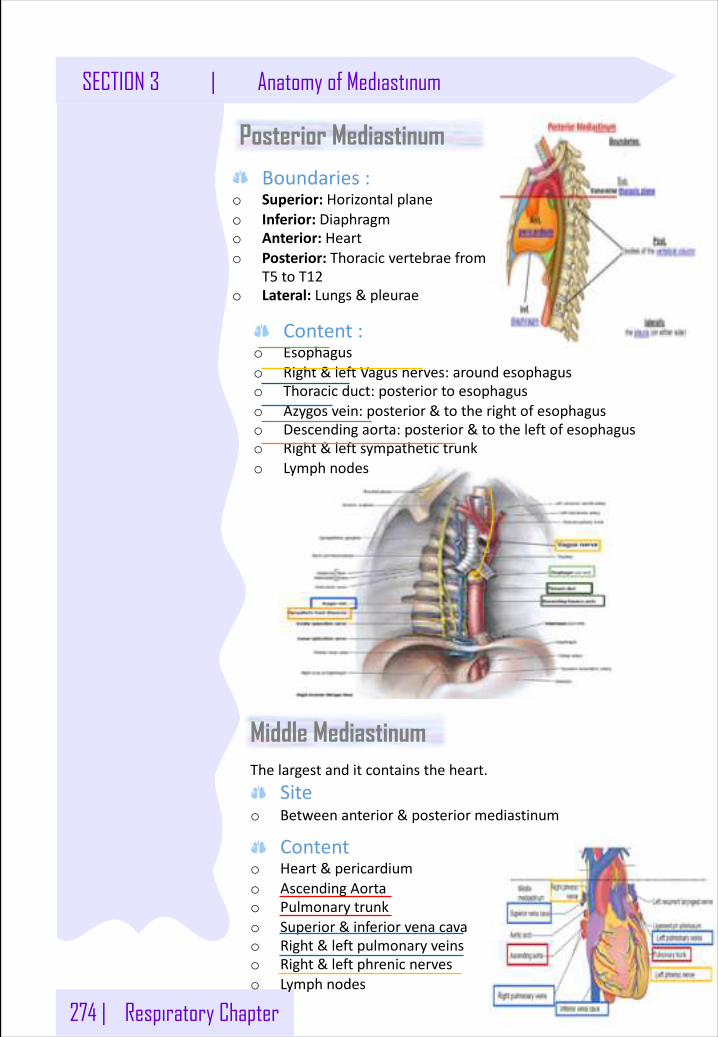
274 | Respiratory Chapter
SECTION 3 | Anatomy of Mediastinum
Posterior Mediastinum
Boundaries:o Superior:Horizontalplaneo Inferior:Diaphragmo Anterior:Hearto Posterior:Thoracicvertebraefrom
T5 toT12o Lateral:Lungs&pleurae
Content:o Esophaguso Right&leftVagusnerves:aroundesophaguso Thoracicduct:posteriortoesophaguso Azygosvein:posterior&totherightofesophaguso Descendingaorta:posterior&totheleftofesophaguso Right&leftsympathetictrunko Lymphnodes
Middle MediastinumThelargestanditcontainstheheart.
Siteo Betweenanterior&posteriormediastinum
Contento Heart&pericardiumo AscendingAortao Pulmonarytrunko Superior&inferiorvenacavao Right&leftpulmonaryveinso Right&leftphrenicnerveso Lymphnodes
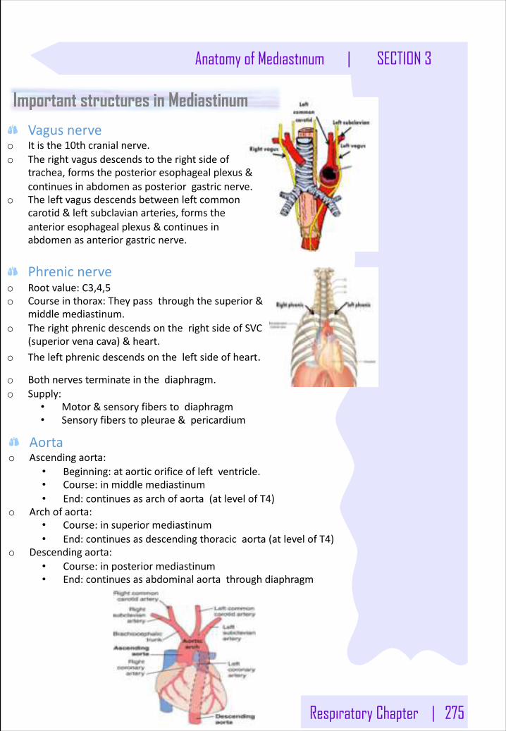
Respiratory Chapter | 275
Anatomy of Mediastinum | SECTION 3
Important structures in Mediastinum
Vagusnerveo Itisthe10thcranialnerve.o Therightvagusdescendstotherightsideof
trachea,formstheposterioresophagealplexus&continuesinabdomenasposteriorgastricnerve.
o Theleftvagusdescendsbetweenleftcommoncarotid&leftsubclavianarteries,formstheanterioresophagealplexus&continuesinabdomenasanteriorgastricnerve.
Phrenicnerveo Rootvalue:C3,4,5o Courseinthorax:Theypassthroughthesuperior&
middlemediastinum.o TherightphrenicdescendsontherightsideofSVC
(superiorvenacava)&heart.o Theleftphrenicdescendsontheleftsideofheart.
o Bothnervesterminateinthediaphragm.o Supply:
• Motor&sensoryfiberstodiaphragm• Sensoryfiberstopleurae&pericardium
Aortao Ascendingaorta:
• Beginning:ataorticorificeofleftventricle.• Course:inmiddlemediastinum• End:continuesasarchofaorta(atlevelofT4)
o Archofaorta:• Course:insuperiormediastinum• End:continuesasdescendingthoracicaorta(atlevelofT4)
o Descendingaorta:• Course:inposteriormediastinum• End:continuesasabdominalaortathroughdiaphragm
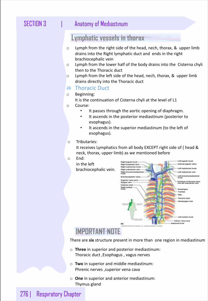
276 | Respiratory Chapter
SECTION 3 | Anatomy of Mediastinum
Therearesixstructurepresentinmorethanoneregioninmediastinum
o Threeinsuperiorandposteriormediastinum:Thoracicduct,Esophagus,vagusnerves
o Twoinsuperiorandmiddlemediastinum:Phrenicnerves,superiorvenacava
o Oneinsuperiorandanteriormediastinum:Thymusgland
IMPORTANT NOTE
Lymphatic vessels in thoraxo Lymphfromtherightsideofthehead,nech,thorax,&upperlimb
drainsintotheRightlymphaticductandendsintherightbrachiocephalicvein
o LymphfromthelowerhalfofthebodydrainsintotheCisternachylithentotheThoracicduct
o Lymphfromtheleftsideofthehead,nech,thorax,&upperlimbdrainsdirectlyintotheThoracicductThoracicDuct
o Beginning:ItisthecontinuationofCisternachyliatthelevelofL1
o Course:• Itpassesthroughtheaorticopeningofdiaphragm.• Itascendsintheposteriormediastinum(posteriorto
esophagus).• Itascendsinthesuperiormediastinum(totheleftof
esophagus).
o Tributaries:ItreceivesLymphaticsfromallbodyEXCEPTrightsideof(head&neck,thorax,upperlimb)aswementionedbefore
o End:intheleftbrachiocephalicvein.
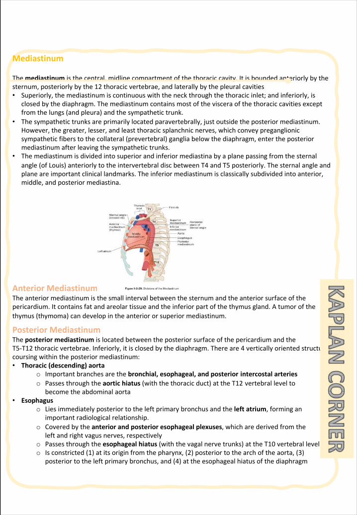
Mediastinum
Themediastinum isthecentral,midlinecompartmentofthethoraciccavity.Itisboundedanteriorlybythesternum,posteriorlybythe12 thoracicvertebrae,andlaterallybythepleuralcavities • Superiorly,themediastinumiscontinuouswiththeneckthroughthethoracicinlet;andinferiorly,is
closedbythediaphragm.Themediastinumcontainsmostofthevisceraofthethoraciccavitiesexceptfromthelungs(andpleura)andthesympathetictrunk.
• Thesympathetictrunksareprimarilylocatedparavertebrally,justoutsidetheposteriormediastinum.However,thegreater,lesser,andleastthoracicsplanchnicnerves,whichconveypreganglionicsympatheticfiberstothecollateral(prevertebral)gangliabelowthediaphragm,entertheposteriormediastinumafterleavingthesympathetictrunks.
• Themediastinumisdividedintosuperiorandinferiormediastinabyaplanepassingfromthesternalangle(ofLouis)anteriorlytotheintervertebraldiscbetweenT4 andT5 posteriorly.Thesternalangleandplaneareimportantclinicallandmarks.Theinferiormediastinumisclassicallysubdividedintoanterior,middle,andposteriormediastina.
AnteriorMediastinumTheanteriormediastinumisthesmallintervalbetweenthesternumandtheanteriorsurfaceofthepericardium.Itcontainsfatandareolartissueandtheinferiorpartofthethymusgland.Atumorofthethymus(thymoma)candevelopintheanteriororsuperiormediastinum.
PosteriorMediastinumTheposteriormediastinumislocatedbetweentheposteriorsurfaceofthepericardiumandtheT5-T12 thoracicvertebrae.Inferiorly,itisclosedbythediaphragm.Thereare4 verticallyorientedstructurescoursingwithintheposteriormediastinum: • Thoracic(descending)aorta
o Importantbranchesarethebronchial,esophageal,andposterior intercostalarterieso Passesthroughtheaortichiatus(withthethoracicduct)attheT12 vertebrallevelto
becometheabdominalaorta • Esophagus
o Liesimmediatelyposteriortotheleftprimarybronchusandtheleft atrium,forminganimportantradiologicalrelationship.
o Coveredbytheanteriorandposterioresophagealplexuses,whicharederivedfromtheleftandrightvagusnerves,respectively
o Passesthroughtheesophagealhiatus(withthevagalnervetrunks)attheT10 vertebrallevelo Isconstricted(1)atitsoriginfromthepharynx,(2)posteriortothearchoftheaorta,(3)
posteriortotheleftprimarybronchus,and(4)attheesophagealhiatusofthediaphragm
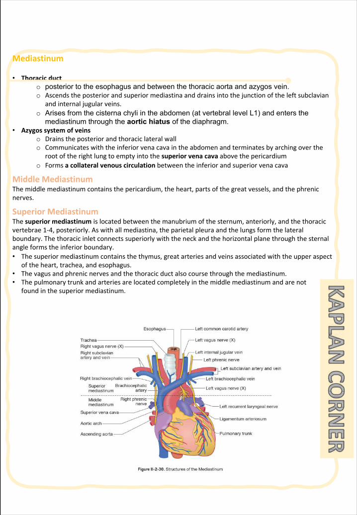
Mediastinum
• Thoracicducto posterior to the esophagus and between the thoracic aorta and azygos vein.o Ascendstheposteriorandsuperiormediastinaanddrainsintothejunctionoftheleftsubclavian
andinternaljugularveins.o Arises from the cisterna chyli in the abdomen (at vertebral level L1) and enters the
mediastinum through the aortic hiatus of the diaphragm.• Azygossystemofveins
o Drainstheposteriorandthoraciclateralwallo Communicateswiththeinferiorvenacavaintheabdomenandterminatesbyarchingoverthe
rootoftherightlungtoemptyintothesuperiorvenacavaabovethepericardiumo Formsacollateralvenouscirculationbetweentheinferiorandsuperiorvenacava
MiddleMediastinumThemiddlemediastinumcontainsthepericardium,theheart,partsofthegreatvessels,andthephrenicnerves.
SuperiorMediastinumThesuperiormediastinumislocatedbetweenthemanubriumofthesternum,anteriorly,andthethoracicvertebrae1-4,posteriorly.Aswithallmediastina,theparietalpleuraandthelungsformthelateralboundary.Thethoracicinletconnectssuperiorlywiththeneckandthehorizontalplanethroughthesternalangleformstheinferiorboundary.• Thesuperiormediastinumcontainsthethymus,greatarteriesandveinsassociatedwiththeupperaspect
oftheheart,trachea,andesophagus.• Thevagusandphrenicnervesandthethoracicductalsocoursethroughthemediastinum.• Thepulmonarytrunkandarteriesarelocatedcompletelyinthemiddlemediastinumandarenot
foundinthesuperiormediastinum.
Mediastinum
Therelationshipsofthesestructuresinthesuperiormediastinumarebestvisualizedinaventraltodorsalorientationbetweenthesternumanteriorlyandthevertebraeposteriorly: • Thymus:locatedposteriortothemanubrium,usuallyatrophiesintheadultandremainsasfatty tissue • Rightandleftbrachiocephalicveins: rightveindescendsalmostverticallyandleftveinobliquelycrosses
thesuperiormediastinumposteriortothethymic remnantso The2 veinsjointoformthesuperiorvenacavaposteriortotherightfirstcostalcartilage.o Thesuperiorvenacavadescendsanddrainsintotherightatrium deeptotherightthirdcostal
cartilage. • Aorticarchandits3 branches:aorticarchbeginsandendsattheplaneofthesternalangleandis
locatedjustinferior totheleftbrachiocephalicvein.o Asaveryimportantradiologicallandmark,theoriginsofthe3 branchesoftheaorticarch
(brachiocephalic,leftcommoncarotid,andleftsubclavian)aredirectlyposterior totheleftbrachiocephalicvein.
• Trachea:liesposteriortotheaorticarchandbifurcatesatthelevelofT4 vertebratoformrightandleftprimarybronchi
o Thecarina isaninternalprojectionofcartilageatthebifurcation.• Esophagus:liesposteriortothetracheaandcoursesposteriortoleftprimarybronchustoenterthe
posteriormediastinum.
lnadditiontothesestructures,thesuperiormediastinumalsocontainstherightandleftvagusandphrenicnervesandthesuperiorendofthethoracicduct. • Rightandleftvagusnervescontributetothepulmonaryandcardiacplexuses.Intheneck,theright
vagusnervegivesrisetotherightrecurrentlaryngealnerve,whichpassesundertherightsubclavianarterytoascendinthegroovebetweentheesophagusandthetracheatoreachthelarynx.Note: Therightrecurrentlaryngealnerveisnotinthemediastinum.Theleftvagusnervegivesrisetotheleftrecurrentlaryngealnerveinthesuperiormediastinum,whichpassesundertheaorticarchandligamentumarteriosum toascendtothelarynx.
• Thethoracicductisthelargestlymphaticchannelinthebody.Itreturnslymphtothevenouscirculationatthejunctionoftheleftinternaljugularveinandtheleftsubclavianvein.
• Phrenicnervesarisefromtheventralramiofcervicalnerves3,4,and5.Thenervesarethesolemotorsupplyofthediaphragmandconveysensoryinformationfromthecentralportionofboththesuperiorandinferiorportionsofthediaphragmandparietalpleura.Bothphrenicnervespassthroughthemiddlemediastinumlateralbetweenthefibrouspericardiumandpleura,andanteriortotherootofthelung.
ClinicalCorrelate• Theleftrecurrentlaryngealnerve(FigureII-2-30)curvesundertheaorticarchdistaltothe
ligamentumarteriosum whereitmaybedamagedbypathology(e.g.,malignancyoraneurysmoftheaorticarch),resultinginparalysisoftheleftvocalfolds.Therightlaryngealnerveisnotaffectedbecauseitarisesfromtherightvagusnerveintherootoftheneckandpassesunderthesubclavianartery.
• Eithertherightortheleftrecurrentlaryngealnervemaybelesionedwiththyroidglandsurgery.
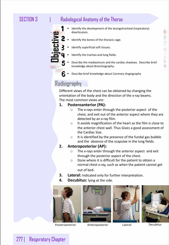
277 | Respiratory Chapter
SECTION 3 | Radiological Anatomy of the Thorax
Radiography
§ Identifythedevelopmentofthelaryngotracheal(respiratory)diverticulum.
§ Identifythebonesofthethoraciccage.
§ Identifysuperficialsofttissues.
§ Identifythetracheaandlungfields.
§ Describethemediastinumandthecardiacshadows.DescribebriefknowledgeaboutBronchography.
§ DescribebriefknowledgeaboutCoronaryAngiography
Objec
tive
Differentviewsofthechestcanbeobtainedbychangingtheorientationofthebodyandthedirectionofthex-raybeams.Themostcommonviewsare:1. Posteroanterior(PA):
o Thex-raysenterthroughtheposterioraspectofthechest,andexitoutoftheanterioraspectwheretheyaredetectedbyanx-rayfilm.
o Itavoidsmagnificationoftheheartasthefilmisclosetotheanteriorchestwall.ThusGivesagoodassessmentoftheCardiacSize.
o Itisidentifiedbythepresenceofthefundalgasbubbleandtheabsenceofthescapulaeinthelungfields.
2. Anteroposterior(AP):o Thex-raysenterthroughtheanterioraspectandexit
throughtheposterioraspectofthechest.o Donewhereitisdifficultforthepatienttoobtaina
normalchestx-ray,suchaswhenthepatientcannotgetoutofbed.
3. Lateral: Indicatedonlyforfurtherinterpretation.4. Decubitus: lyingattheside.
Posteroanterior Anteroposterior Lateral Decubitus
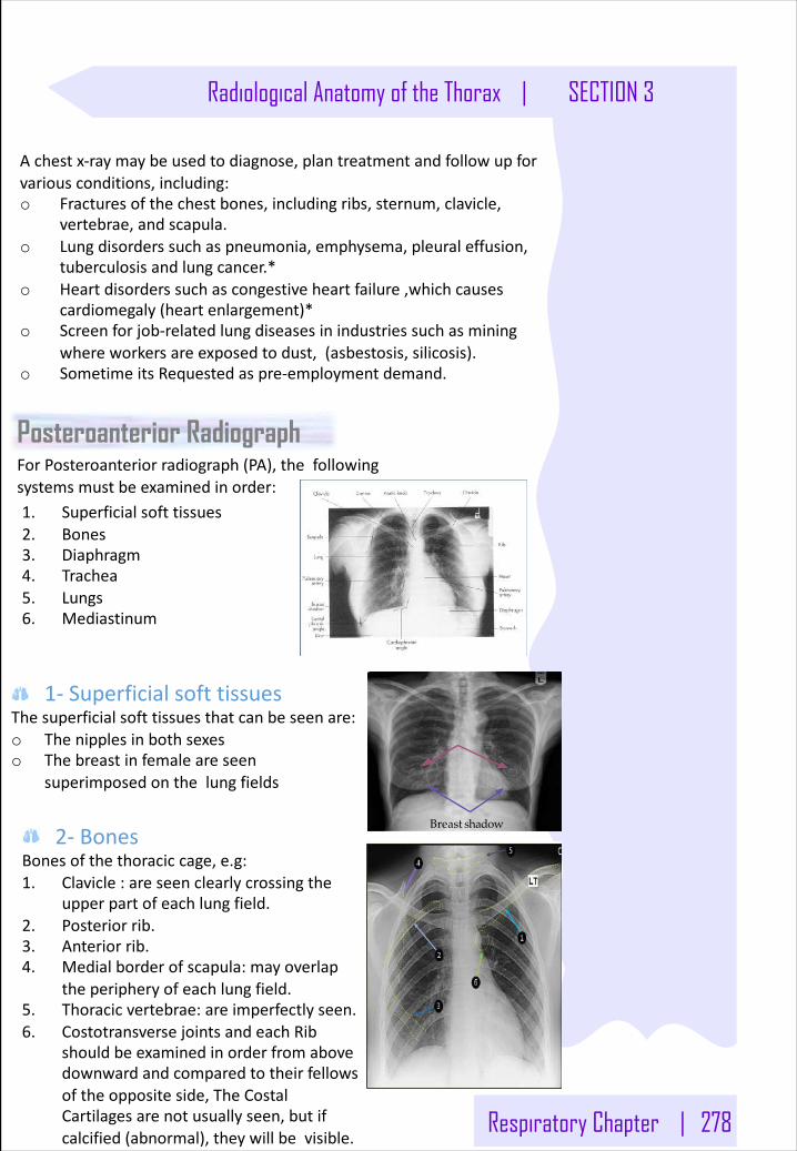
Respiratory Chapter | 278
Radiological Anatomy of the Thorax | SECTION 3
Achestx-raymaybeusedtodiagnose,plantreatmentandfollowupforvariousconditions,including:o Fracturesofthechestbones,includingribs,sternum,clavicle,
vertebrae,andscapula.o Lungdisorderssuchaspneumonia,emphysema,pleuraleffusion,
tuberculosisandlungcancer.*o Heartdisorderssuchascongestiveheartfailure,whichcauses
cardiomegaly(heartenlargement)*o Screenforjob-relatedlungdiseasesinindustriessuchasmining
whereworkersareexposedtodust,(asbestosis,silicosis).o SometimeitsRequestedaspre-employmentdemand.
Breast shadow
Posteroanterior Radiograph
1. Superficialsofttissues2. Bones3. Diaphragm4. Trachea5. Lungs6. Mediastinum
ForPosteroanteriorradiograph(PA),thefollowingsystemsmustbeexaminedinorder:
1- SuperficialsofttissuesThesuperficialsofttissuesthatcanbeseenare:o Thenipplesinbothsexeso Thebreastinfemaleareseen
superimposedonthelungfields
2- BonesBonesofthethoraciccage,e.g:1. Clavicle:areseenclearlycrossingthe
upperpartofeachlungfield.2. Posteriorrib.3. Anteriorrib.4. Medialborderofscapula:mayoverlap
theperipheryofeachlungfield.5. Thoracicvertebrae:areimperfectlyseen.6. CostotransversejointsandeachRib
shouldbeexaminedinorderfromabovedownwardandcomparedtotheirfellowsoftheoppositeside,TheCostalCartilagesarenotusuallyseen,butifcalcified(abnormal),theywillbevisible.
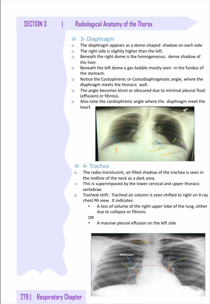
279 | Respiratory Chapter
SECTION 3 | Radiological Anatomy of the Thorax
3- Diaphragmo Thediaphragmappearsasadome-shapedshadowoneachside.o Therightsideisslightlyhigherthantheleft.o Beneaththerightdomeisthehomogeneous,denseshadowof
theliver.o Beneaththeleftdomeagasbubblemostlyseeninthefundusof
thestomach.o NoticetheCostophrenicorCostodiaphragmatic angle,wherethe
diaphragmmeetsthethoracicwall.o Theanglebecomesbluntorobscuredduetominimalpleuralfluid
(effusion)orfibrosis.o Alsonotethecardiophrenicanglewherethediaphragmmeetthe
heart.
4- Tracheao Theradio-translucent,air-filledshadowofthetracheaisseenin
themidlineoftheneckasadarkarea.o Thisissuperimposedbythelowercervicalandupperthoracic
vertebrae.o Trachealshift:TrachealaircolumnisseenshiftedtorightonX-ray
chestPAview.Itindicates:• Alossofvolumeoftherightupperlobeofthelung,either
duetocollapseorfibrosis.OR• Amassivepleuraleffusionontheleftside
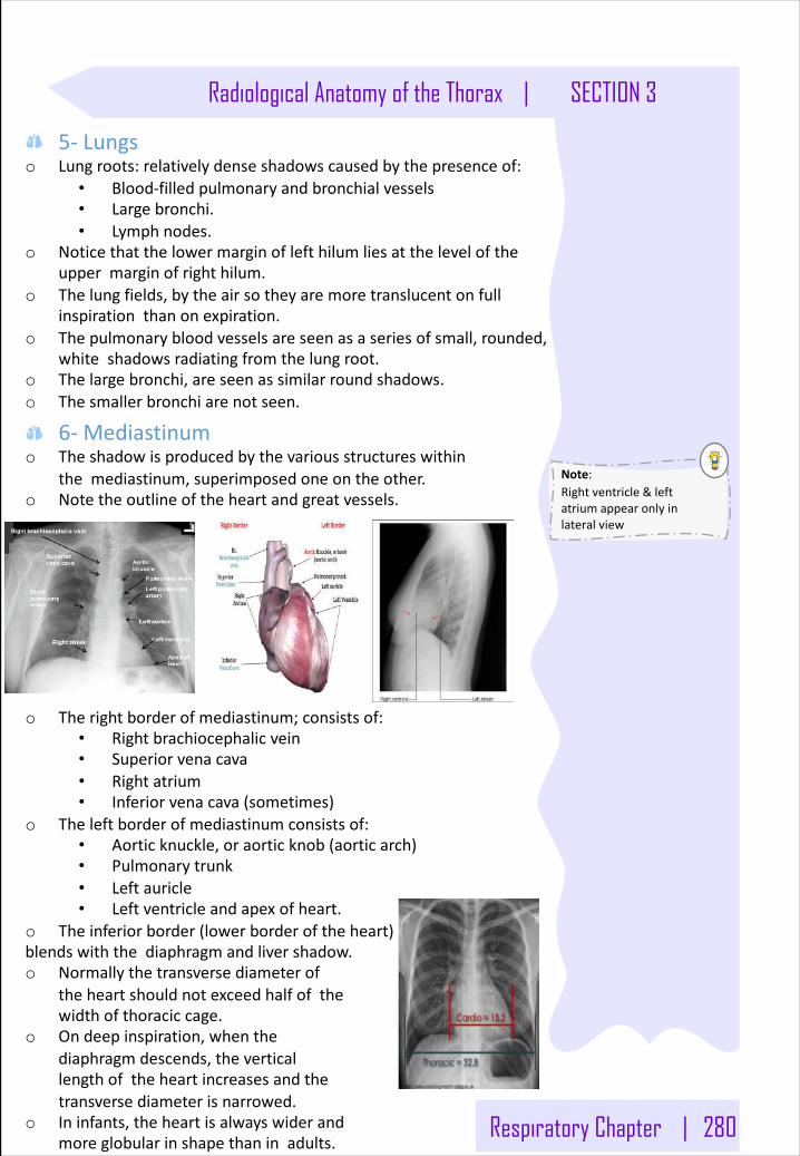
Respiratory Chapter | 280
Radiological Anatomy of the Thorax | SECTION 3
5- Lungso Lungroots:relativelydenseshadowscausedbythepresenceof:
• Blood-filledpulmonaryandbronchialvessels• Largebronchi.• Lymphnodes.
o Noticethatthelowermarginoflefthilumliesattheleveloftheuppermarginofrighthilum.
o Thelungfields,bytheairsotheyaremoretranslucentonfullinspirationthanonexpiration.
o Thepulmonarybloodvesselsareseenasaseriesofsmall,rounded,whiteshadowsradiatingfromthelungroot.
o Thelargebronchi,areseenassimilarroundshadows.o Thesmallerbronchiarenotseen.
6- Mediastinumo Theshadowisproducedbythevariousstructureswithin
themediastinum,superimposedoneontheother.o Notetheoutlineoftheheartandgreatvessels.
o Therightborderofmediastinum;consistsof:• Rightbrachiocephalicvein• Superiorvenacava• Rightatrium• Inferiorvenacava(sometimes)
o Theleftborderofmediastinumconsistsof:• Aorticknuckle,oraorticknob(aorticarch)• Pulmonarytrunk• Leftauricle• Leftventricleandapexofheart.
o Theinferiorborder(lowerborderoftheheart)blendswiththediaphragmandlivershadow.o Normallythetransversediameterof
theheartshouldnotexceedhalfofthewidthofthoraciccage.
o Ondeepinspiration,whenthediaphragmdescends,theverticallengthoftheheartincreasesandthetransversediameterisnarrowed.
o Ininfants,theheartisalwayswiderandmoreglobularinshapethaninadults.
Note:Rightventricle&leftatriumappearonlyinlateralview
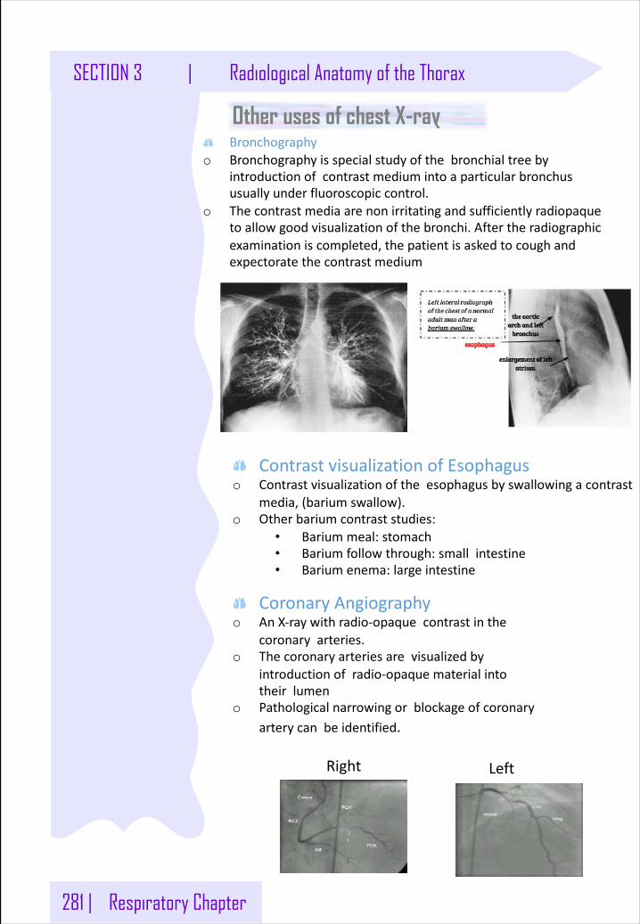
281 | Respiratory Chapter
SECTION 3 | Radiological Anatomy of the Thorax
ContrastvisualizationofEsophaguso Contrastvisualizationoftheesophagus byswallowingacontrast
media,(bariumswallow).o Otherbariumcontraststudies:
• Bariummeal:stomach• Bariumfollowthrough:smallintestine• Bariumenema:largeintestine
Right Left
Bronchographyo Bronchographyisspecialstudyofthebronchialtreeby
introductionofcontrastmediumintoaparticularbronchususuallyunderfluoroscopiccontrol.
o Thecontrastmediaarenonirritatingandsufficientlyradiopaquetoallowgoodvisualizationofthebronchi.Aftertheradiographicexaminationiscompleted,thepatientisaskedtocoughandexpectoratethecontrast medium
Other uses of chest X-ray
CoronaryAngiographyo AnX-raywithradio-opaquecontrastinthe
coronaryarteries.o Thecoronaryarteriesarevisualizedby
introductionofradio-opaquematerialintotheirlumen
o Pathologicalnarrowingorblockageofcoronaryarterycanbeidentified.
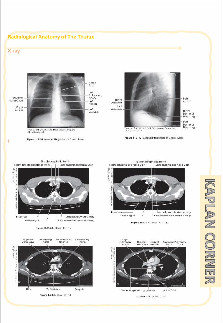
RadiologicalAnatomyofTheThorax
X-ray
CTscan
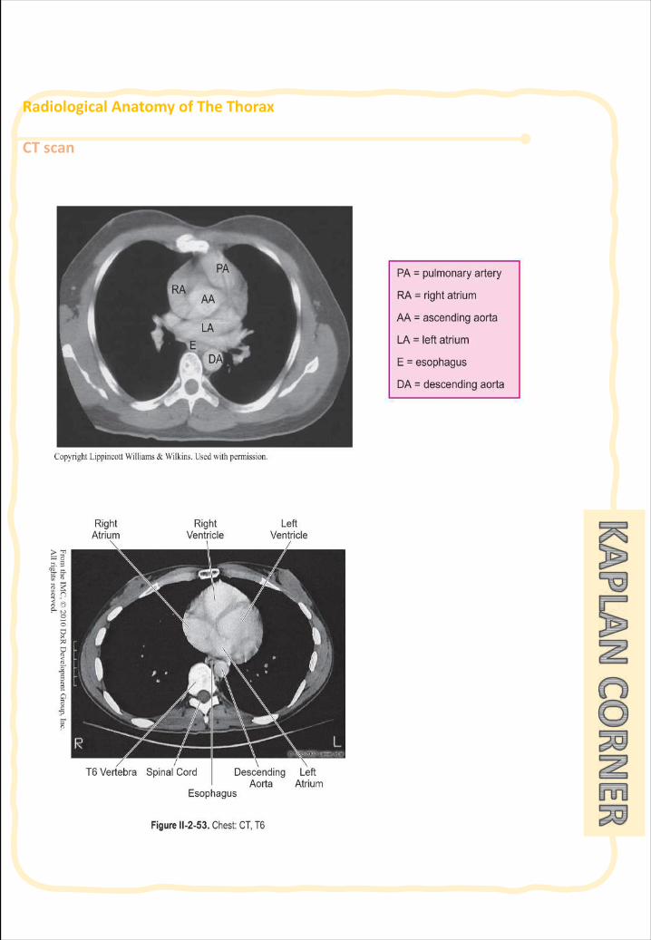
RadiologicalAnatomyofTheThorax
CTscan
282BIOCHEMISTERY:Phospholipids of physiological significance
SECTION 4 :
MISCELLANEOUS
BIOCHEMISTERY Respiratory chain
PHYSIOLOGY:Effects of low and high gas pressure on the body
PHARMACOLOGY:Anti-cholinergic drugs
PHYSIOLOGY:Effect of Exercise on respiratory
311PHARMACOLOGY:Adrenergic drugs
PHARMACOLOGY:Anaphylactic shock
FAMILY MEDICINE: Tobacco smoking
289295302
PHARMACOLOGY:Antibiotics307
315324330
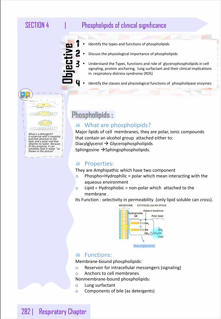
282 | Respiratory Chapter
SECTION 4 | Phospholipids of clinical significance
Whatarephospholipids?Majorlipidsofcellmembranes,theyarepolar,ioniccompoundsthatcontainanalcoholgroupattachedeitherto:Diacylglycerolà GlycerophospholipidsSphingosineàSphingophospholipids.
Properties:TheyareAmphipathicwhichhavetwocomponento Phospho=Hydrophilic=polarwhichmeaninteractingwiththe
aqueousenvironmento Lipid=Hydrophobic=non-polarwhichattachedtothe
membrane.ItsFunction:selectivityinpermeability(onlylipidsolublecancross).
Functions:Membrane-boundphospholipids:o Reservoirforintracellularmessengers(signaling)o AnchorstocellmembranesNonmembrane-boundphospholipids:o Lungsurfactanto Componentsofbile(asdetergents)
Phospholipids :
§ Identifythetypesandfunctionsofphospholipids
§ Discussthephysiologicalimportanceofphospholipids
§ UnderstandtheTypes,functionsandroleofglycerophospholipidsincellsignaling,proteinanchoring,lungsurfactantandtheirclinicalimplicationsinrespiratorydistresssyndrome(RDS)
§ IdentifytheclassesandphysiologicalfunctionsofphospholipaseenzymesObjec
tive
Diacylglycerol
Whatisadetergent?Amoleculewithanonpolarendthatattachestothelipid,andapolarendthatattachestowater.Becauseofthisproperty,itcansolubilizelipidinwater “asShowninthepicture”
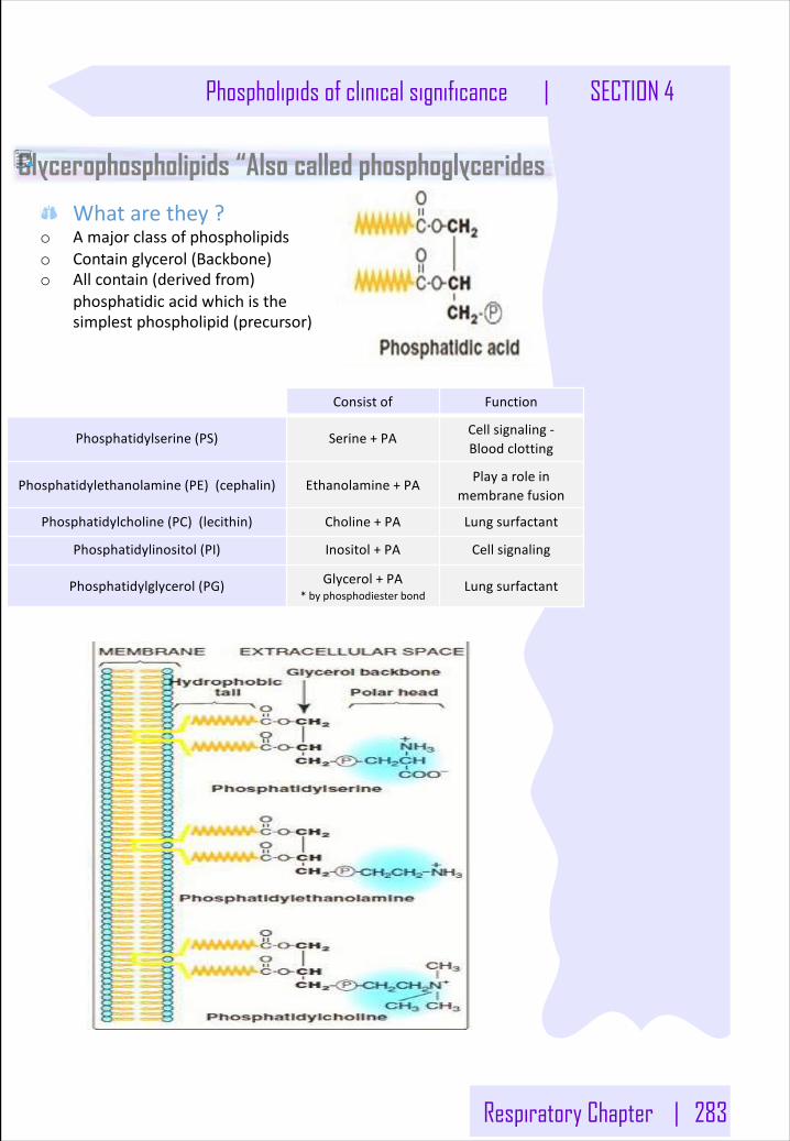
Respiratory Chapter | 283
Phospholipids of clinical significance | SECTION 4
Glycerophospholipids “Also called phosphoglycerides
Whatarethey?o Amajorclassofphospholipidso Containglycerol(Backbone)o Allcontain(derivedfrom)
phosphatidicacidwhichisthesimplestphospholipid(precursor)
Consistof Function
Phosphatidylserine(PS) Serine+PA Cellsignaling-Bloodclotting
Phosphatidylethanolamine(PE)(cephalin) Ethanolamine+PA Playaroleinmembranefusion
Phosphatidylcholine(PC)(lecithin) Choline+PA Lungsurfactant
Phosphatidylinositol(PI) Inositol+PA Cellsignaling
Phosphatidylglycerol(PG) Glycerol+PA*byphosphodiesterbond
Lungsurfactant
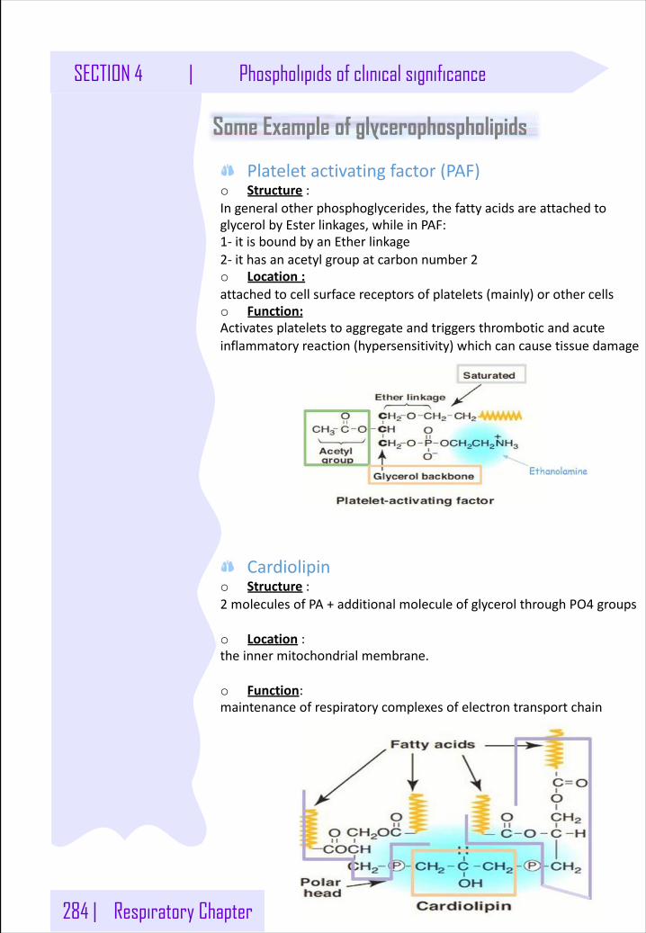
284 | Respiratory Chapter
SECTION 4 | Phospholipids of clinical significance
Some Example of glycerophospholipids
Plateletactivatingfactor(PAF)o Structure :Ingeneralotherphosphoglycerides,thefattyacidsareattachedtoglycerolbyEsterlinkages,whileinPAF:1- itisboundbyanEtherlinkage2- ithasanacetylgroupatcarbonnumber2o Location: attachedtocellsurfacereceptorsofplatelets(mainly)orothercellso Function:Activatesplateletstoaggregateandtriggersthromboticandacuteinflammatoryreaction(hypersensitivity)whichcancausetissuedamage
Cardiolipino Structure :2 moleculesofPA+additionalmoleculeofglycerolthroughPO4 groups
o Location : theinnermitochondrialmembrane.
o Function:maintenanceofrespiratorycomplexesofelectrontransportchain
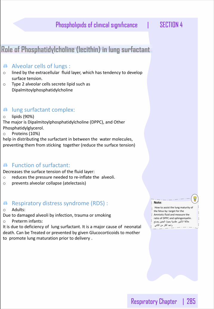
Respiratory Chapter | 285
Phospholipids of clinical significance | SECTION 4
Role of Phosphatidylcholine (lecithin) in lung surfactant
Alveolarcellsoflungs:o linedbytheextracellularfluidlayer,whichhastendencytodevelop
surfacetension.o Type2 alveolarcellssecretelipidsuchas
Dipalmitoylphosphatidylcholine
lungsurfactantcomplex:o lipids(90%)ThemajorisDipalmitoylphosphatidylcholine(DPPC),andOtherPhosphatidylglycerol.o Proteins(10%)helpindistributingthesurfactantinbetweenthewatermolecules,preventingthemfromstickingtogether(reducethesurfacetension)
Functionofsurfactant:Decreasesthesurfacetensionofthefluidlayer:o reducesthepressureneededtore-inflatethealveoli.o preventsalveolarcollapse(atelectasis)
Respiratorydistresssyndrome(RDS):o Adults:Duetodamagedalveolibyinfection,traumaorsmokingo Preterminfants:Itisduetodeficiencyoflungsurfactant.Itisamajorcauseofneonataldeath.CanbeTreatedorpreventedbygivenGlucocorticoidstomothertopromotelungmaturationpriortodelivery .
Note:Howtoassistthelungmaturityofthefetusby:targetfortheAmnioticfluidandmeasuretheratioofDPPCandsphingomyelin.
عنصی نینجلا ثیحب ةیسكع نینثالا ةقالع يناثلا نم رثكأ دحاو
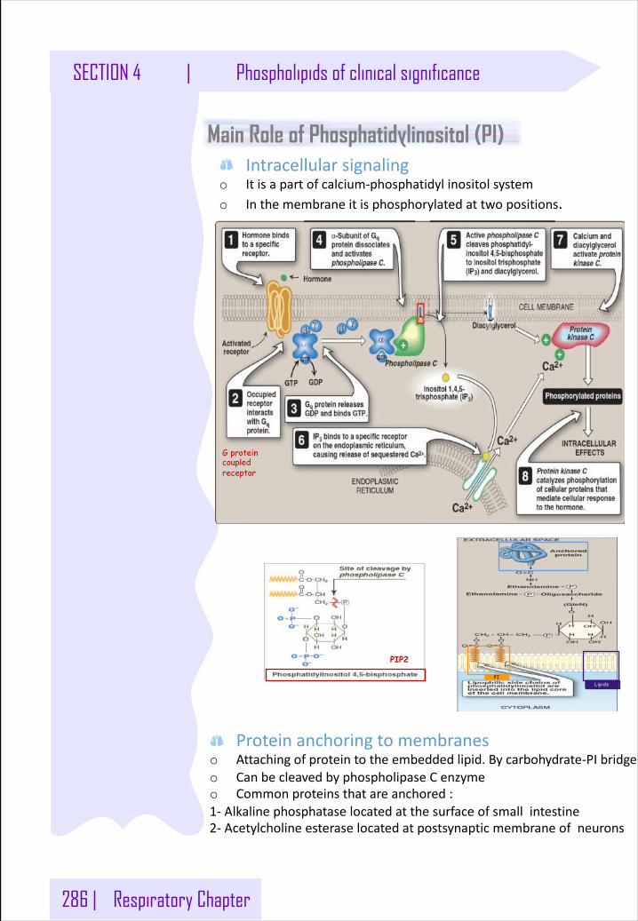
286 | Respiratory Chapter
SECTION 4 | Phospholipids of clinical significance
Main Role of Phosphatidylinositol (PI)Intracellularsignaling
o Itisapartofcalcium-phosphatidylinositolsystemo Inthemembraneitisphosphorylatedattwopositions.
PIP2
PILipids
G proteincoupled receptor
Proteinanchoringtomembraneso Attachingofproteintotheembeddedlipid.Bycarbohydrate-PIbridgeo CanbecleavedbyphospholipaseCenzymeo Commonproteinsthatareanchored:1- Alkalinephosphataselocatedatthesurfaceofsmallintestine2- Acetylcholineesteraselocatedatpostsynapticmembraneofneurons
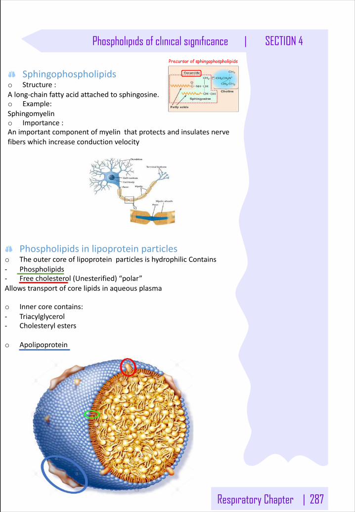
Respiratory Chapter | 287
Phospholipids of clinical significance | SECTION 4
Sphingophospholipidso Structure:Along-chainfattyacidattachedtosphingosine.o Example:Sphingomyelino Importance:Animportantcomponentofmyelinthatprotectsandinsulatesnervefiberswhichincreaseconductionvelocity
Precursor of sphingophospholipids
Phospholipidsinlipoproteinparticleso TheoutercoreoflipoproteinparticlesishydrophilicContains- Phospholipids- Freecholesterol(Unesterified)“polar”Allowstransportofcorelipidsinaqueousplasma
o Innercorecontains:- Triacylglycerol- Cholesterylesters
o Apolipoprotein
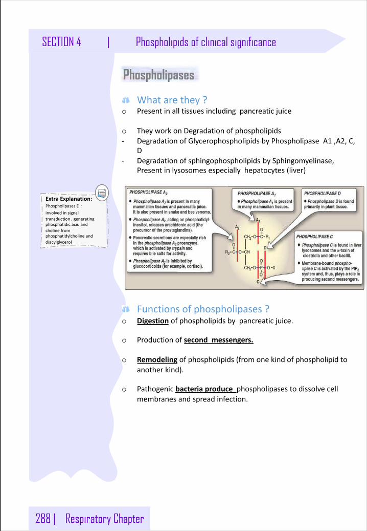
288 | Respiratory Chapter
SECTION 4 | Phospholipids of clinical significance
Phospholipases
Whatarethey? o Presentinalltissuesincludingpancreaticjuice
o TheyworkonDegradationofphospholipids- DegradationofGlycerophospholipidsbyPhospholipaseA1 ,A2,C,
D- Degradationofsphingophospholipids bySphingomyelinase,
Presentinlysosomesespeciallyhepatocytes(liver)
Functionsofphospholipases?o Digestion ofphospholipidsbypancreaticjuice.
o Productionofsecondmessengers.
o Remodeling ofphospholipids(fromonekindofphospholipidtoanotherkind).
o Pathogenicbacteriaproducephospholipasestodissolvecellmembranesandspreadinfection.
ExtraExplanation:PhospholipasesD:involvedinsignaltransduction,generatingphosphatidicacidandcholinefromphosphatidylcholineanddiacylglycerol
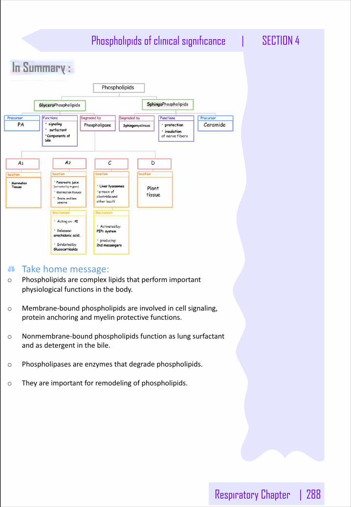
Respiratory Chapter | 288
Phospholipids of clinical significance | SECTION 4
In Summary :
Takehomemessage:o Phospholipidsarecomplexlipidsthatperformimportant
physiologicalfunctionsinthebody.
o Membrane-boundphospholipidsareinvolvedincellsignaling,proteinanchoringandmyelinprotectivefunctions.
o Nonmembrane-boundphospholipidsfunctionaslungsurfactantandasdetergentinthebile.
o Phospholipasesareenzymesthatdegradephospholipids.
o Theyareimportantforremodelingofphospholipids.
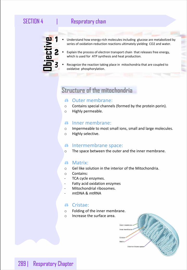
289 | Respiratory Chapter
SECTION 4 | Respiratory chain
§ Understandhowenergy-richmoleculesincludingglucosearemetabolizedbyseriesofoxidation-reductionreactionsultimatelyyieldingCO2 andwater.
§ Explaintheprocessofelectrontransportchainthatreleasesfreeenergy,whichisusedforATPsynthesisandheatproduction.
§ Recognizethereactiontakingplaceinmitochondriathatarecoupledtooxidativephosphorylation.Ob
jectiv
e
Structure of the mitochondria Outermembrane:
o Containsspecialchannels(formedbytheproteinporin).o Highlypermeable.
Innermembrane:o Impermeabletomostsmallions,smallandlargemolecules.o Highlyselective.
Intermembranespace:o Thespacebetweentheouterandtheinnermembrane.
Matrix:o GellikesolutionintheinterioroftheMitochondria.o Contains:- TCAcycleenzymes.- Fattyacidoxidationenzymes- Mitochondrialribosomes.- mtDNA &mtRNA
Cristae:o Foldingoftheinnermembrane.o Increasethesurfacearea.
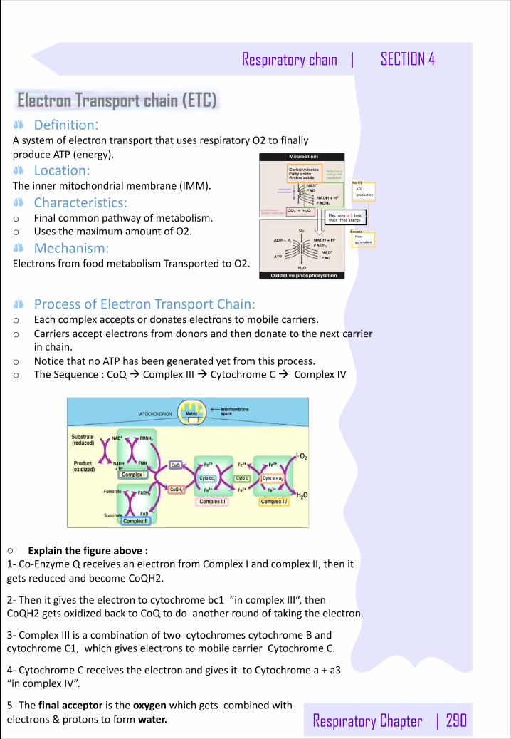
Respiratory Chapter | 290
Respiratory chain | SECTION 4
Electron Transport chain (ETC)Definition:
AsystemofelectrontransportthatusesrespiratoryO2 tofinallyproduceATP(energy).
Location:Theinnermitochondrialmembrane(IMM).
Characteristics:o Finalcommonpathwayofmetabolism.o UsesthemaximumamountofO2.
Mechanism:ElectronsfromfoodmetabolismTransportedtoO2.
metabolic breakdown
Reductionof energy rich coenzymes
Electrons (e-) lose their free energy.
productionof simple molecules
ATPproduction
Heatgeneration
mainly
Excess
ProcessofElectronTransportChain:o Eachcomplexacceptsordonateselectronstomobilecarriers.o Carriersacceptelectronsfromdonorsandthendonatetothenextcarrier
inchain.o NoticethatnoATPhasbeengeneratedyetfromthisprocess.o TheSequence:CoQà ComplexIIIà CytochromeCà ComplexIV
o Explainthefigureabove:1- Co-EnzymeQreceivesanelectronfromComplexIandcomplexII,thenitgetsreducedandbecomeCoQH2.
2- Thenitgivestheelectrontocytochromebc1 “incomplexIII“,thenCoQH2 getsoxidizedbacktoCoQtodoanotherroundoftakingtheelectron.
3- ComplexIIIisacombinationoftwocytochromescytochromeBandcytochromeC1,whichgiveselectronstomobilecarrierCytochromeC.
4- CytochromeCreceivestheelectronandgivesittoCytochromea+a3“incomplexIV”.
5- Thefinalacceptoristheoxygen whichgetscombinedwithelectrons&protonstoformwater.
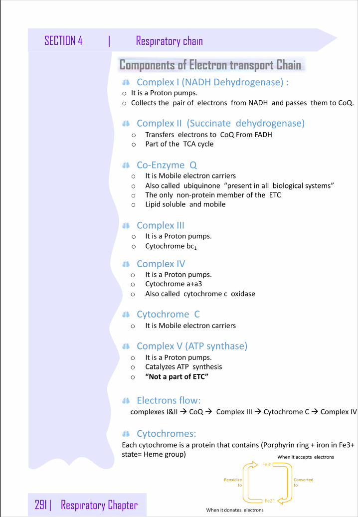
291 | Respiratory Chapter
SECTION 4 | Respiratory chain
Components of Electron transport ChainComplexI(NADHDehydrogenase):
o ItisaProtonpumps.o CollectsthepairofelectronsfromNADHandpassesthemtoCoQ.
ComplexII(Succinatedehydrogenase)o TransferselectronstoCoQFromFADHo PartoftheTCAcycle
Co-EnzymeQo ItisMobileelectroncarrierso Alsocalledubiquinone“presentinallbiologicalsystems”o Theonlynon-proteinmemberoftheETCo Lipidsolubleandmobile
ComplexIIIo ItisaProtonpumps.o Cytochromebc1
ComplexIVo ItisaProtonpumps. o Cytochromea+a3o Alsocalledcytochromecoxidase
CytochromeCo ItisMobileelectroncarriers
ComplexV(ATPsynthase)o ItisaProtonpumps. o CatalyzesATPsynthesiso “NotapartofETC”
Electronsflow:complexesI&IIà CoQà ComplexIIIà CytochromeCà ComplexIV
Cytochromes:Eachcytochromeisaproteinthatcontains(Porphyrinring+ironinFe3+state=Hemegroup)
Fe3+
Fe2+
Convertedto
Whenitacceptselectrons
Reoxidizeto
Whenit donateselectrons
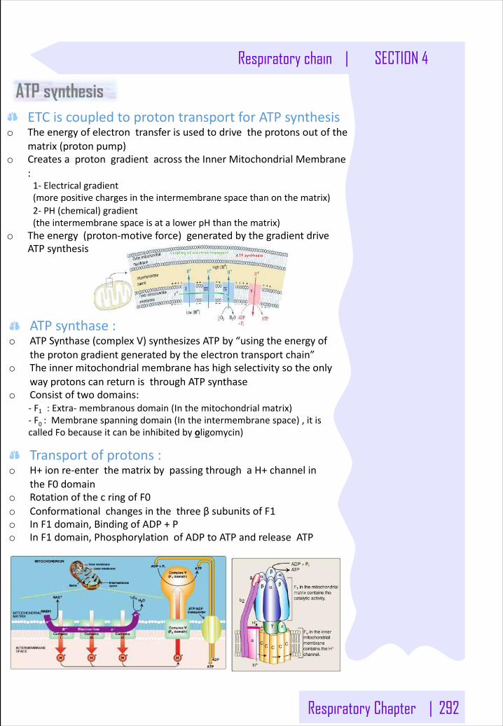
Respiratory Chapter | 292
Respiratory chain | SECTION 4
ATP synthesisETCiscoupledtoprotontransportforATPsynthesis
o Theenergyofelectrontransferisusedtodrivetheprotonsoutofthematrix(protonpump)
o CreatesaprotongradientacrosstheInnerMitochondrialMembrane:1- Electricalgradient(morepositivechargesintheintermembranespacethanonthematrix)2- PH(chemical)gradient(theintermembranespaceisatalowerpHthanthematrix)
o Theenergy(proton-motiveforce)generatedbythegradientdriveATPsynthesis Coupling of electron transport
ATP synthesis
ATPsynthase:o ATPSynthase(complexV)synthesizesATPby“usingtheenergyof
theprotongradientgeneratedbytheelectrontransportchain”o Theinnermitochondrialmembrane hashighselectivitysotheonly
wayprotonscanreturnisthroughATPsynthaseo Consistoftwodomains:
- F1 :Extra- membranousdomain(Inthemitochondrialmatrix)- F0 :Membranespanningdomain(Intheintermembranespace),itiscalledFobecauseitcanbeinhibitedbyoligomycin)
Transportofprotons:o H+ionre-enterthematrixbypassingthroughaH+channelin
theF0 domaino RotationofthecringofF0o ConformationalchangesinthethreeβsubunitsofF1o InF1 domain,BindingofADP+Po InF1 domain,PhosphorylationofADPtoATPandreleaseATP
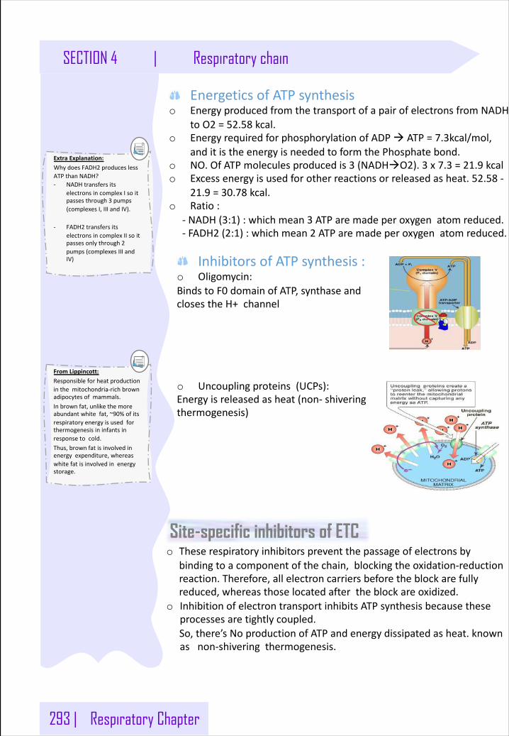
293 | Respiratory Chapter
SECTION 4 | Respiratory chain
EnergeticsofATPsynthesiso EnergyproducedfromthetransportofapairofelectronsfromNADH
toO2 =52.58 kcal.o EnergyrequiredforphosphorylationofADPà ATP=7.3kcal/mol,
anditistheenergyisneededtoformthePhosphatebond.o NO.OfATPmoleculesproducedis3 (NADHàO2).3 x7.3 =21.9 kcalo Excessenergyisusedforotherreactionsorreleasedasheat.52.58 -
21.9 =30.78 kcal.o Ratio:
- NADH(3:1):whichmean3 ATParemadeperoxygenatomreduced.- FADH2 (2:1):whichmean2 ATParemadeperoxygenatomreduced.
ExtraExplanation:WhydoesFADH2 produceslessATPthanNADH?- NADHtransfersits
electronsincomplexIsoitpassesthrough3 pumps(complexesI,IIIandIV).
- FADH2 transfersitselectronsincomplexIIsoitpassesonlythrough2pumps(complexesIIIandIV) InhibitorsofATPsynthesis:
o Oligomycin:BindstoF0 domainofATP,synthaseandclosestheH+channel
o Uncouplingproteins(UCPs):Energyisreleasedasheat(non- shiveringthermogenesis)
FromLippincott:Responsibleforheatproductioninthemitochondria-richbrownadipocytesofmammals.Inbrownfat,unlikethemoreabundantwhitefat,~90%ofitsrespiratoryenergyisusedforthermogenesisininfantsinresponsetocold.Thus,brownfatisinvolvedinenergyexpenditure,whereaswhitefatisinvolvedinenergystorage.
Site-specific inhibitors of ETCo Theserespiratoryinhibitorspreventthepassageofelectronsby
bindingtoacomponentofthechain,blockingtheoxidation-reductionreaction.Therefore,allelectroncarriersbeforetheblockarefullyreduced,whereasthoselocatedaftertheblockareoxidized.
o InhibitionofelectrontransportinhibitsATPsynthesisbecausetheseprocessesaretightlycoupled.So,there’sNoproductionofATPandenergydissipatedasheat.knownasnon-shiveringthermogenesis.
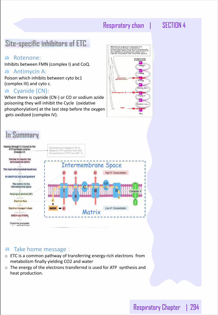
Respiratory Chapter | 294
Respiratory chain | SECTION 4
Site-specific inhibitors of ETC
Rotenone:InhibitsbetweenFMN(complexI)andCoQ.
AntimycinA:Poisonwhichinhibitsbetweencytobc1(complexIII)andcytoc.
Cyanide(CN):Whenthereiscyanide(CN-)orCOorsodiumazidepoisoningtheywillinhibittheCycle(oxidativephosphorylation)atthelaststepbeforetheoxygengetsoxidized(complexIV).
In Summary
Takehomemessage : o ETCisacommonpathwayoftransferringenergy-richelectronsfrom
metabolismfinallyyieldingCO2 andwater o TheenergyoftheelectronstransferredisusedforATPsynthesisand
heatproduction.
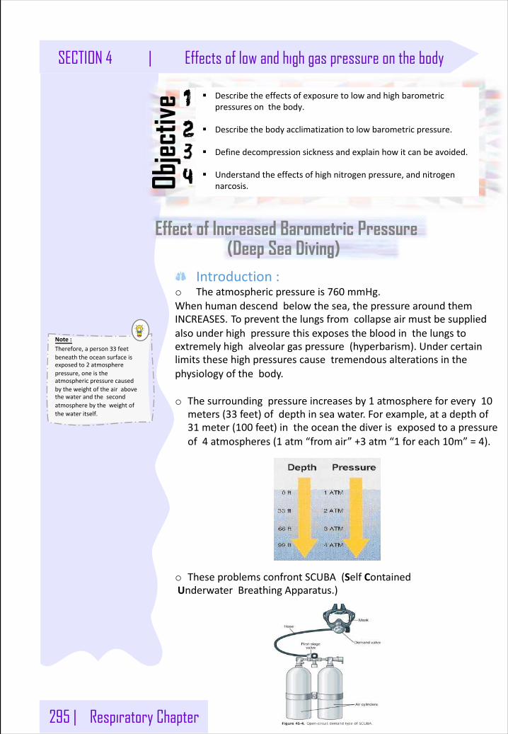
295 | Respiratory Chapter
SECTION 4 | Effects of low and high gas pressure on the body
Effect of Increased Barometric Pressure
§ Describetheeffectsofexposuretolowandhighbarometricpressuresonthebody.
§ Describethebodyacclimatizationtolowbarometricpressure.
§ Definedecompressionsicknessandexplainhowitcanbeavoided.
§ Understandtheeffectsofhighnitrogenpressure,andnitrogennarcosis.Ob
jectiv
e
Introduction:o Theatmosphericpressureis760 mmHg.Whenhumandescendbelowthesea,thepressurearoundthemINCREASES. Topreventthelungsfromcollapseairmustbesuppliedalsounderhighpressurethisexposesthebloodinthelungstoextremelyhighalveolargaspressure(hyperbarism).Undercertainlimitsthesehighpressurescausetremendousalterationsinthephysiologyofthebody.
o Thesurroundingpressureincreasesby1 atmosphereforevery10meters(33 feet)ofdepthinseawater.Forexample,atadepthof31 meter(100 feet)intheoceanthediverisexposedtoapressureof4 atmospheres(1 atm“fromair”+3 atm“1 foreach10m”=4).
o TheseproblemsconfrontSCUBA(SelfContained UnderwaterBreathingApparatus.)
(Deep Sea Diving)
Note:Therefore,aperson33 feetbeneaththeoceansurfaceisexposedto2 atmospherepressure,oneistheatmosphericpressurecausedbytheweightoftheairabovethewaterandthesecondatmospherebytheweightofthewateritself.
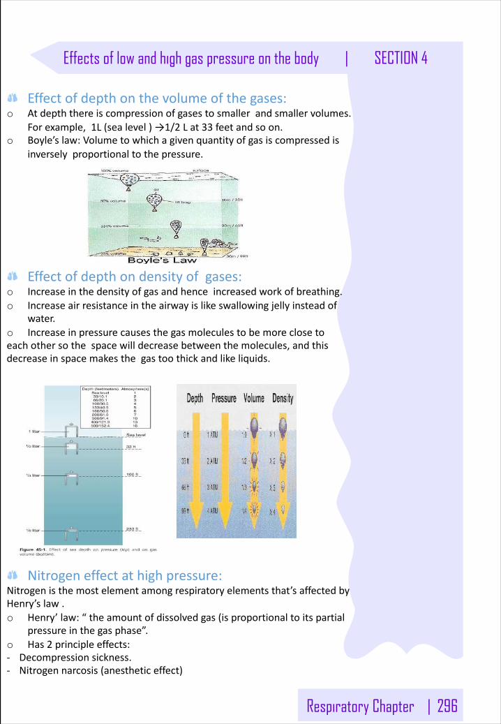
Respiratory Chapter | 296
Effects of low and high gas pressure on the body | SECTION 4
Effectofdepthonthevolumeofthegases:o Atdepththereiscompressionofgasestosmallerandsmallervolumes.
Forexample,1L(sealevel)→1/2 Lat33 feetandsoon.o Boyle’slaw:Volumetowhichagivenquantityofgasiscompressedis
inverselyproportionaltothepressure.
Effectofdepthondensityofgases:o Increaseinthedensityofgasandhenceincreasedworkofbreathing.o Increaseairresistanceintheairwayislikeswallowingjellyinsteadof
water.o Increaseinpressurecausesthegasmoleculestobemoreclosetoeachothersothespacewilldecreasebetweenthemolecules,andthisdecreaseinspacemakesthegastoothickandlikeliquids.
Nitrogeneffectathighpressure:Nitrogenisthemostelementamongrespiratoryelementsthat’saffectedbyHenry’slaw.o Henry’law:“theamountofdissolvedgas(isproportionaltoitspartial
pressureinthegasphase”.o Has2 principleeffects:- Decompressionsickness.- Nitrogennarcosis(anestheticeffect)
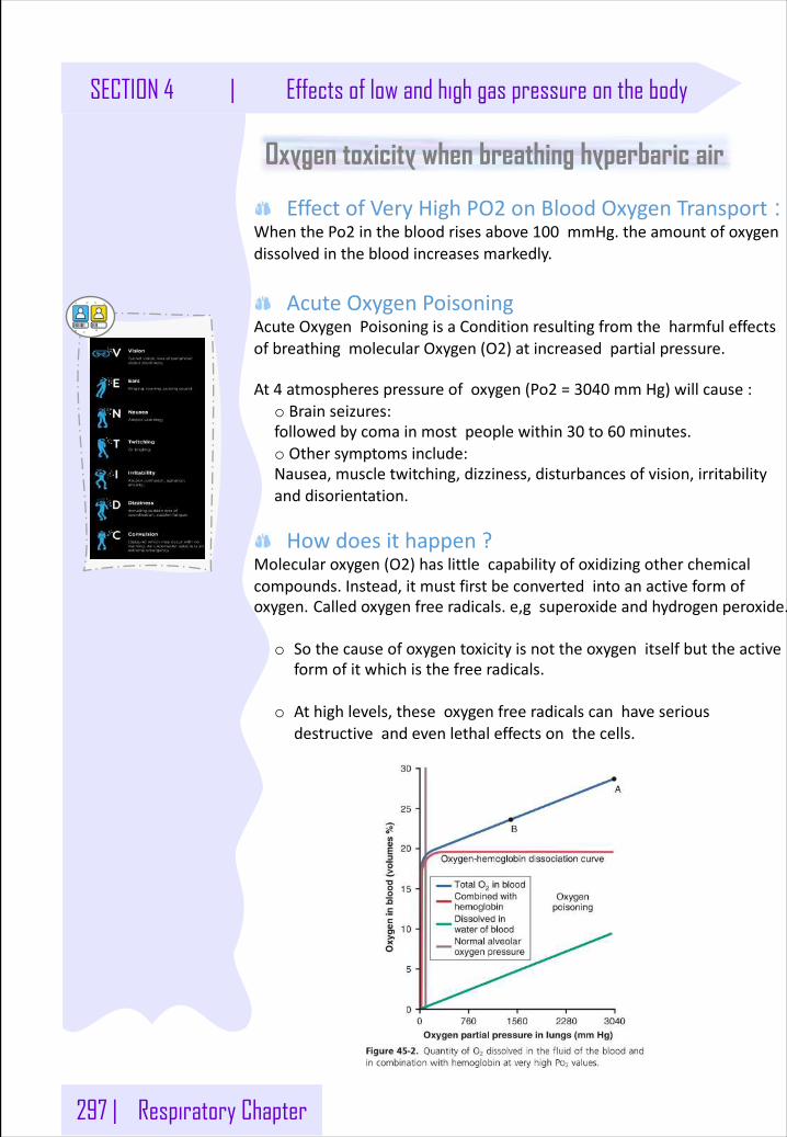
297 | Respiratory Chapter
Oxygen toxicity when breathing hyperbaric air
EffectofVeryHighPO2 onBloodOxygenTransport : WhenthePo2 inthebloodrisesabove100 mmHg.theamountofoxygendissolvedinthebloodincreasesmarkedly.
AcuteOxygenPoisoningAcuteOxygenPoisoningisaConditionresultingfromtheharmfuleffectsofbreathingmolecularOxygen(O2)atincreasedpartialpressure.
At4 atmospherespressureofoxygen(Po2 =3040 mmHg)willcause:o Brainseizures:followedbycomainmostpeoplewithin30 to60 minutes.o Othersymptomsinclude:Nausea,muscletwitching,dizziness,disturbancesofvision,irritabilityanddisorientation.
Howdoesithappen? Molecularoxygen(O2)haslittlecapabilityofoxidizingotherchemicalcompounds.Instead,itmustfirstbeconvertedintoanactiveformofoxygen. Calledoxygenfreeradicals.e,g superoxideandhydrogenperoxide.
o Sothecauseofoxygentoxicityisnottheoxygenitselfbuttheactiveformofitwhichisthefreeradicals.
o Athighlevels,theseoxygenfreeradicalscanhaveseriousdestructiveandevenlethaleffectsonthecells.
SECTION 4 | Effects of low and high gas pressure on the body
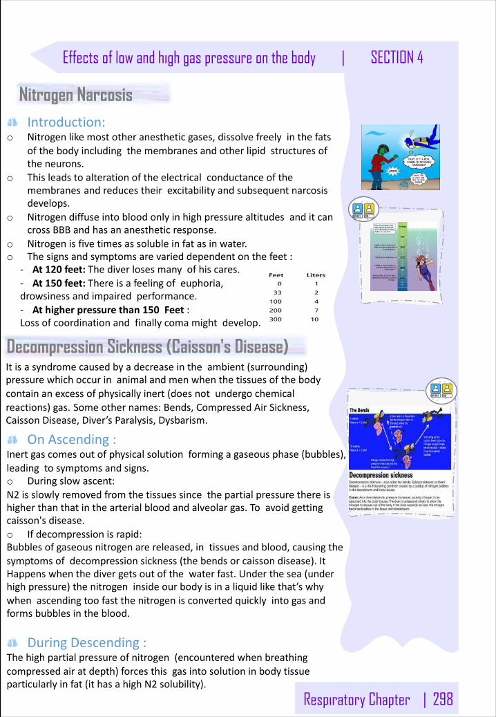
Respiratory Chapter | 298
Nitrogen NarcosisIntroduction:
o Nitrogenlikemostotheranestheticgases,dissolvefreelyinthefatsofthebodyincludingthemembranesandotherlipidstructuresoftheneurons.
o Thisleadstoalterationoftheelectricalconductanceofthemembranes andreducestheirexcitabilityandsubsequentnarcosisdevelops.
o NitrogendiffuseintobloodonlyinhighpressurealtitudesanditcancrossBBBandhasananestheticresponse.
o Nitrogenisfivetimesassolubleinfatasinwater.o Thesignsandsymptomsarevarieddependentonthefeet:
- At120 feet:Thediverlosesmanyofhiscares.- At150 feet:Thereisafeelingofeuphoria, drowsinessandimpairedperformance.- Athigherpressurethan150 Feet :Lossofcoordinationandfinallycomamightdevelop.
Decompression Sickness (Caisson's Disease)
OnAscending:Inertgascomesoutofphysicalsolutionformingagaseousphase(bubbles),leadingtosymptomsandsigns.o Duringslowascent:N2 isslowlyremovedfromthetissuessincethepartialpressurethereishigherthanthatinthearterialbloodandalveolargas.Toavoidgettingcaisson'sdisease.o Ifdecompressionisrapid:Bubblesofgaseousnitrogenarereleased,intissuesandblood,causingthesymptomsofdecompressionsickness(thebendsorcaissondisease).ItHappenswhenthedivergetsoutofthewaterfast.Underthesea(underhighpressure)thenitrogeninsideourbodyisinaliquidlikethat’swhywhenascendingtoofastthenitrogenisconvertedquicklyintogasandformsbubblesintheblood.
DuringDescending:Thehighpartialpressureofnitrogen(encounteredwhenbreathingcompressedairatdepth)forcesthisgasintosolutioninbodytissueparticularlyinfat(ithasahighN2 solubility).
Itisasyndromecausedbyadecreaseintheambient(surrounding)pressurewhichoccurinanimalandmenwhenthetissuesofthebodycontainanexcessofphysicallyinert(doesnotundergochemicalreactions)gas. Someothernames:Bends,CompressedAirSickness,CaissonDisease,Diver’sParalysis,Dysbarism.
Effects of low and high gas pressure on the body | SECTION 4
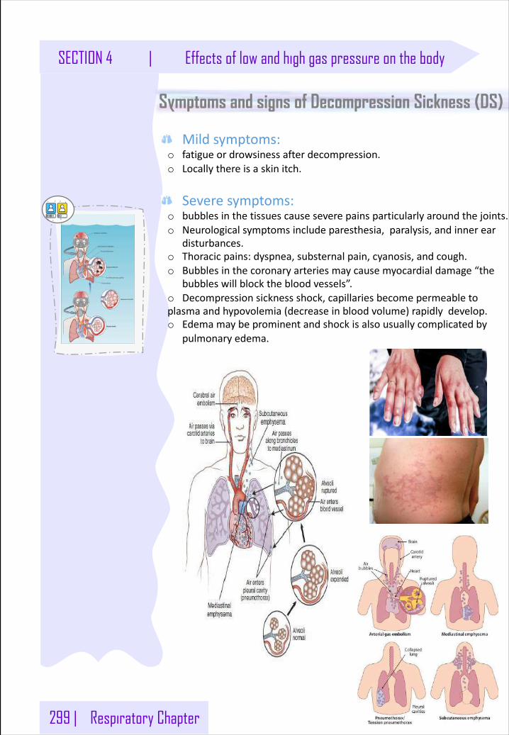
299 | Respiratory Chapter
Symptoms and signs of Decompression Sickness (DS)
Mildsymptoms:o fatigueordrowsinessafterdecompression.o Locallythereisaskinitch.
Severesymptoms:o bubblesinthetissuescauseseverepainsparticularlyaroundthejoints.o Neurologicalsymptomsincludeparesthesia,paralysis,andinnerear
disturbances.o Thoracicpains:dyspnea,substernalpain,cyanosis,andcough.o Bubblesinthecoronaryarteriesmaycausemyocardialdamage“the
bubbleswillblockthebloodvessels”.o Decompressionsicknessshock,capillariesbecomepermeabletoplasmaandhypovolemia(decreaseinbloodvolume)rapidlydevelop.o Edemamaybeprominentandshockisalsousuallycomplicatedby
pulmonaryedema.
SECTION 4 | Effects of low and high gas pressure on the body
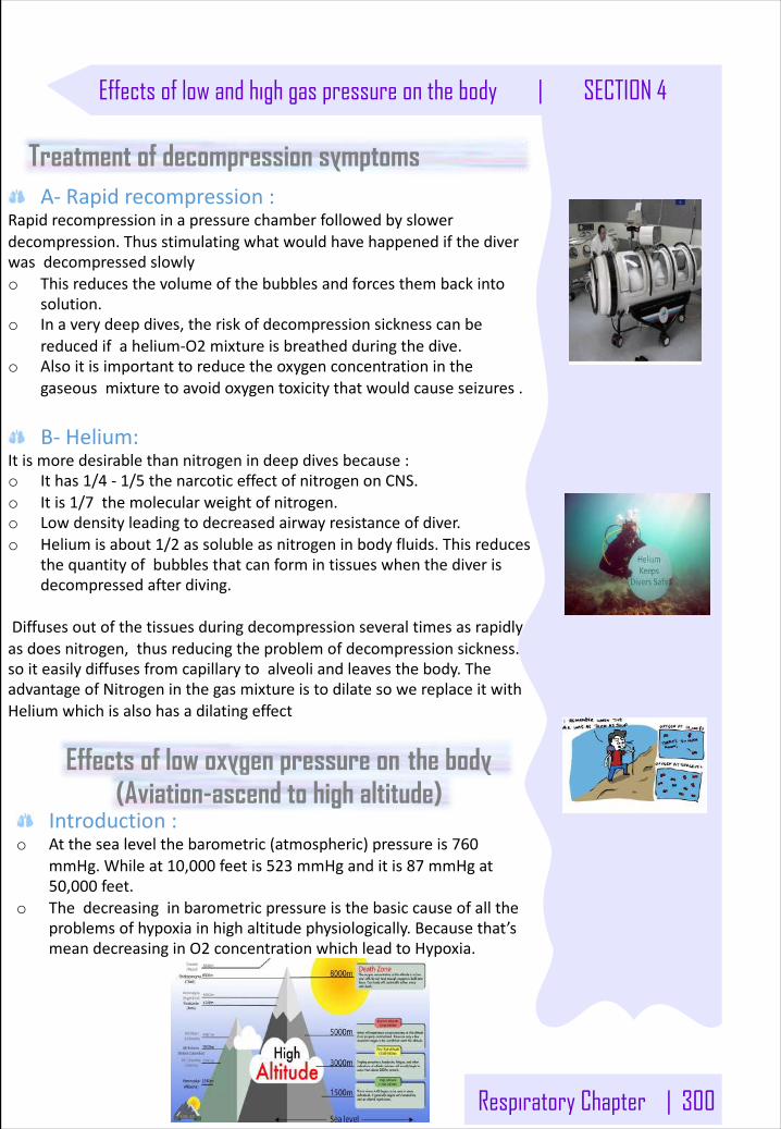
Respiratory Chapter | 300
Treatment of decompression symptomsA- Rapidrecompression:
Rapidrecompressioninapressurechamberfollowedbyslowerdecompression.Thusstimulatingwhatwouldhavehappenedifthediverwasdecompressedslowlyo Thisreducesthevolumeofthebubblesandforcesthembackinto
solution.o Inaverydeepdives,theriskofdecompressionsicknesscanbe
reducedifahelium-O2 mixtureisbreathedduringthedive.o Alsoitisimportanttoreducetheoxygenconcentrationinthe
gaseousmixturetoavoidoxygentoxicitythatwouldcauseseizures.
B- Helium:Itismoredesirablethannitrogenindeepdivesbecause:o Ithas1/4 - 1/5 thenarcoticeffectofnitrogenonCNS.o Itis1/7 themolecularweightofnitrogen.o Lowdensityleadingtodecreasedairwayresistanceofdiver.o Heliumisabout1/2 assolubleasnitrogeninbodyfluids.Thisreduces
thequantityofbubblesthatcanformintissueswhenthediverisdecompressedafterdiving.
Diffusesoutofthetissuesduringdecompressionseveraltimesasrapidlyasdoesnitrogen,thusreducingtheproblemofdecompressionsickness.soiteasilydiffusesfromcapillarytoalveoliandleavesthebody.TheadvantageofNitrogeninthegasmixtureistodilatesowereplaceitwithHeliumwhichisalsohasadilatingeffect
Effects of low oxygen pressure on the body (Aviation-ascend to high altitude)
Introduction:o Atthesealevelthebarometric(atmospheric)pressureis760
mmHg.Whileat10,000 feetis523 mmHganditis87 mmHgat50,000 feet.
o Thedecreasinginbarometricpressureisthebasiccauseofalltheproblemsofhypoxiainhighaltitudephysiologically.Becausethat’smeandecreasinginO2 concentrationwhichleadtoHypoxia.
Effects of low and high gas pressure on the body | SECTION 4
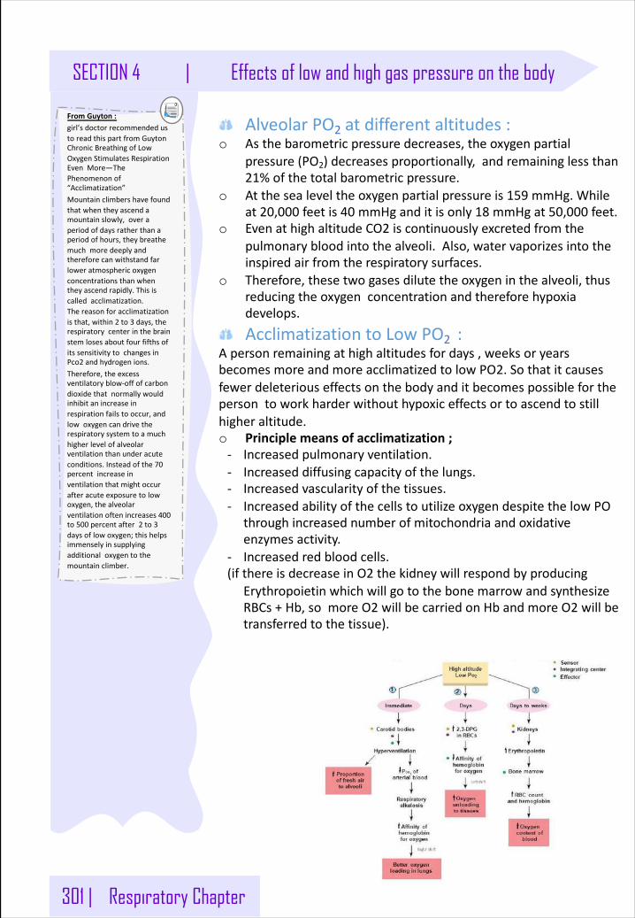
301 | Respiratory Chapter
AlveolarPO2 atdifferentaltitudes:o Asthebarometricpressuredecreases,theoxygenpartial
pressure(PO2)decreasesproportionally,andremaininglessthan21%ofthetotalbarometricpressure.
o Atthesealeveltheoxygenpartialpressureis159 mmHg.Whileat20,000 feetis40 mmHganditisonly18 mmHgat50,000 feet.
o EvenathighaltitudeCO2 iscontinuouslyexcretedfromthepulmonarybloodintothealveoli.Also,watervaporizesintotheinspiredairfromtherespiratorysurfaces.
o Therefore,thesetwogasesdilutetheoxygeninthealveoli,thusreducingtheoxygenconcentrationandthereforehypoxiadevelops.
AcclimatizationtoLowPO2 :Apersonremainingathighaltitudesfordays,weeksoryearsbecomesmoreandmoreacclimatizedtolowPO2.Sothatitcausesfewerdeleteriouseffectsonthebodyanditbecomespossibleforthepersontoworkharderwithouthypoxiceffectsortoascendtostillhigheraltitude.o Principlemeansofacclimatization;- Increasedpulmonaryventilation.- Increaseddiffusingcapacityofthelungs.- Increasedvascularityofthetissues.- IncreasedabilityofthecellstoutilizeoxygendespitethelowPO
throughincreasednumberofmitochondriaandoxidativeenzymesactivity.
- Increasedredbloodcells.(ifthereisdecreaseinO2 thekidneywillrespondbyproducing
ErythropoietinwhichwillgotothebonemarrowandsynthesizeRBCs+Hb,somoreO2 willbecarriedonHbandmoreO2 willbetransferredtothetissue).
FromGuyton:girl’sdoctorrecommendedustoreadthispartfromGuytonChronicBreathingofLowOxygenStimulatesRespirationEvenMore—ThePhenomenonof“Acclimatization”Mountainclimbershavefoundthatwhentheyascendamountainslowly,overaperiodofdaysratherthanaperiodofhours,theybreathemuchmoredeeplyandthereforecanwithstandfarloweratmosphericoxygenconcentrationsthanwhentheyascendrapidly.Thisiscalledacclimatization.Thereasonforacclimatizationisthat,within2 to3 days,therespiratorycenterinthebrainstemlosesaboutfourfifthsofitssensitivitytochangesinPco2 andhydrogenions.Therefore,theexcessventilatoryblow-offofcarbondioxidethatnormallywouldinhibitanincreaseinrespirationfailstooccur,andlowoxygencandrivetherespiratorysystemtoamuchhigherlevelofalveolarventilationthanunderacuteconditions.Insteadofthe70percentincreaseinventilationthatmightoccurafteracuteexposuretolowoxygen,thealveolarventilationoftenincreases400to500 percentafter2 to3daysoflowoxygen;thishelpsimmenselyinsupplyingadditionaloxygentothemountainclimber.
SECTION 4 | Effects of low and high gas pressure on the body
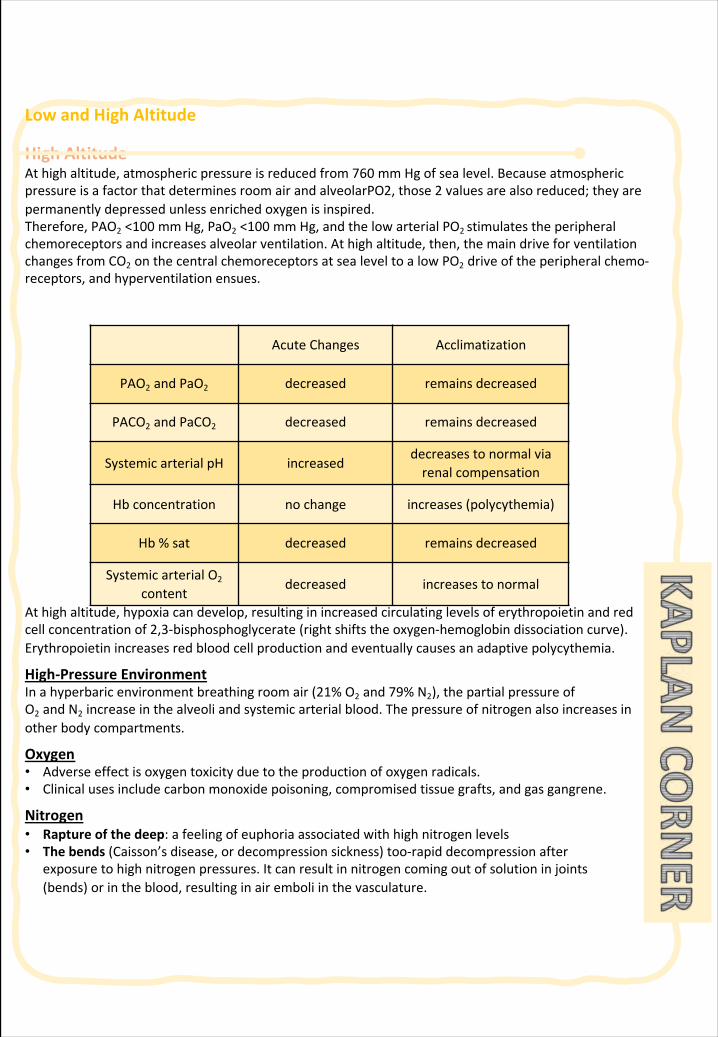
LowandHighAltitude
HighAltitudeAthighaltitude,atmosphericpressureisreducedfrom760 mmHgofsealevel.BecauseatmosphericpressureisafactorthatdeterminesroomairandalveolarPO2,those2 valuesarealsoreduced;theyarepermanentlydepressedunlessenrichedoxygenisinspired.Therefore,PAO2 <100 mmHg,PaO2 <100 mmHg,andthelowarterialPO2 stimulatestheperipheralchemoreceptorsandincreasesalveolarventilation.Athighaltitude,then,themaindriveforventilationchangesfromCO2 onthecentralchemoreceptorsatsealeveltoalowPO2 driveoftheperipheralchemo-receptors,andhyperventilationensues.
Athighaltitude,hypoxiacandevelop,resultinginincreasedcirculatinglevelsoferythropoietinandredcellconcentrationof2,3-bisphosphoglycerate(rightshiftstheoxygen-hemoglobindissociationcurve).Erythropoietinincreasesredbloodcellproductionandeventuallycausesanadaptivepolycythemia.
High-PressureEnvironmentInahyperbaricenvironmentbreathingroomair(21%O2 and79%N2),thepartialpressureofO2 andN2 increaseinthealveoliandsystemicarterialblood.Thepressureofnitrogenalsoincreasesinotherbodycompartments.
Oxygen• Adverseeffectisoxygentoxicityduetotheproductionofoxygenradicals.• Clinicalusesincludecarbonmonoxidepoisoning,compromisedtissuegrafts,andgasgangrene.
Nitrogen• Raptureofthedeep:afeelingofeuphoriaassociatedwithhighnitrogenlevels• Thebends(Caisson’sdisease,ordecompressionsickness)too-rapiddecompressionafter
exposuretohighnitrogenpressures.Itcanresultinnitrogencomingoutofsolutioninjoints(bends)orintheblood,resultinginairemboliinthevasculature.
AcuteChanges Acclimatization
PAO2 andPaO2 decreased remainsdecreased
PACO2 andPaCO2 decreased remainsdecreased
SystemicarterialpH increaseddecreasestonormalviarenalcompensation
Hbconcentration nochange increases(polycythemia)
Hb%sat decreased remainsdecreased
SystemicarterialO2
contentdecreased increasestonormal
LowandHighAltitude
NoteWhatprincipleexplainsthephysiologyofwhynitrogenwillbeforcedintosolution?Answer:Henry’slaw.Theamountofgasthatwilldissolveinaliquidvariesdirectlywiththepressureabovethatliquid.Highpressuresforcegasintosolution.However,solubilities andtemperaturealsocomeintoplaywhenconsideringHenry’slaw.EventhoughahugeN2 gradientmayexistbetweentheairandplasma,nitrogenisbarelysolubleatall.
ClinicalCorrelateHighaltitudeissometimescategorizedasafifthcauseofhypoxemia.HighaltitudecauseslowPAO2,similartohypoventilation.Alltheobservationsdescribedhereapply,exceptforPCO2.Athighaltitude,asubjecthyperventilates,andthusPACO2 andPACO2 arereduced.
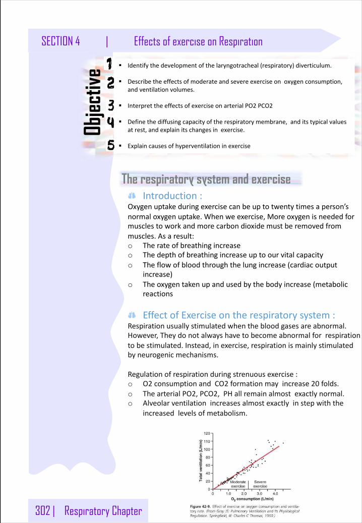
302 | Respiratory Chapter
SECTION 4 | Effects of exercise on Respiration
The respiratory system and exercise
§ Identifythedevelopmentofthelaryngotracheal(respiratory)diverticulum.
§ Describetheeffectsofmoderateandsevereexerciseonoxygenconsumption,andventilationvolumes.
§ InterprettheeffectsofexerciseonarterialPO2 PCO2
§ Definethediffusingcapacityoftherespiratorymembrane,anditstypicalvaluesatrest,andexplainitschangesinexercise.
§ Explaincausesofhyperventilationinexercise
Objec
tive
Introduction:Oxygenuptakeduringexercisecanbeuptotwentytimesaperson’snormaloxygenuptake.Whenweexercise,Moreoxygenisneededformusclestoworkandmorecarbondioxidemustberemovedfrommuscles.Asaresult:o Therateofbreathingincreaseo Thedepthofbreathingincreaseuptoourvitalcapacityo Theflowofbloodthroughthelungincrease(cardiacoutput
increase)o Theoxygentakenupandusedbythebodyincrease(metabolic
reactions
EffectofExerciseontherespiratorysystem:Respirationusuallystimulatedwhenthebloodgasesareabnormal.However,Theydonotalwayshavetobecomeabnormalforrespirationtobestimulated.Instead,inexercise,respirationismainlystimulatedbyneurogenicmechanisms.
Regulationofrespirationduringstrenuousexercise:o O2 consumptionandCO2 formationmayincrease20 folds.o ThearterialPO2,PCO2,PHallremainalmostexactlynormal.o Alveolarventilationincreasesalmostexactlyinstepwiththe
increasedlevelsofmetabolism.
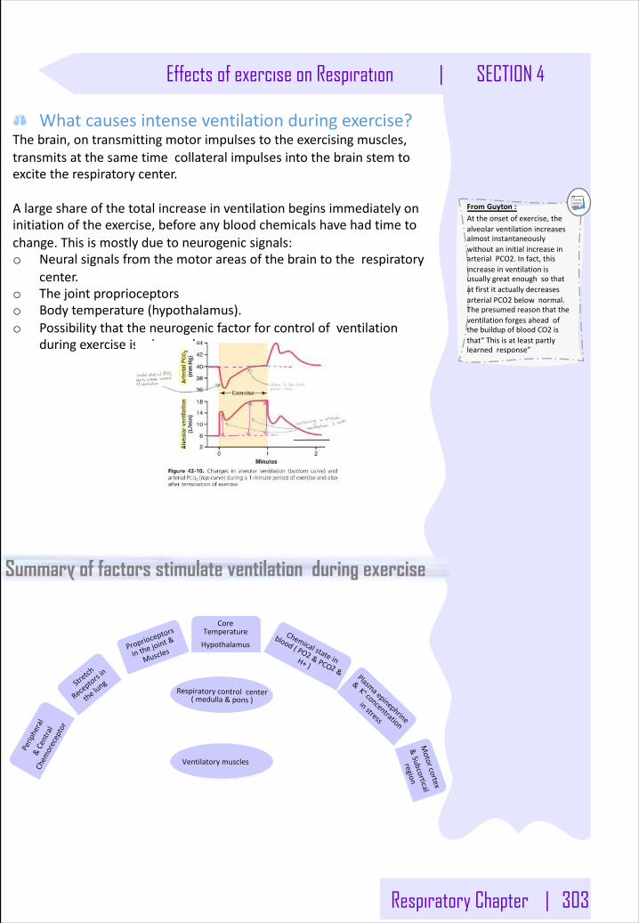
Respiratory Chapter | 303
Effects of exercise on Respiration | SECTION 4
Whatcausesintenseventilationduringexercise?Thebrain,ontransmittingmotorimpulsestotheexercisingmuscles,transmitsatthesametimecollateralimpulsesintothebrainstemtoexcitetherespiratorycenter.
Alargeshareofthetotalincreaseinventilationbeginsimmediatelyoninitiationoftheexercise,beforeanybloodchemicalshavehadtimetochange.Thisismostlyduetoneurogenicsignals:o Neuralsignalsfromthemotorareasofthebraintotherespiratory
center.o Thejointproprioceptorso Bodytemperature(hypothalamus).o Possibilitythattheneurogenicfactorforcontrolofventilation
duringexerciseisalearnedresponse.
FromGuyton:Attheonsetofexercise,thealveolarventilationincreasesalmostinstantaneouslywithoutaninitialincreaseinarterialPCO2.Infact,thisincreaseinventilationisusuallygreatenoughsothatatfirstitactuallydecreasesarterialPCO2 belownormal.ThepresumedreasonthattheventilationforgesaheadofthebuildupofbloodCO2 isthat“Thisisatleastpartlylearnedresponse”
Summary of factors stimulate ventilation during exercise
Peripheral
&Central
Chem
oreceptor
Stretch
Recep
torsin
thelun
g
Proprioce
ptors
inthejoint
&
Muscles
CoreTemperatureHypothalamus
Chemicalstatein
blood(PO2 &PCO2 &
H+)Plasmaepinephrine
&K +concentration
instress
Motorcortex
&Subcortical
region
Respiratorycontrolcenter(medulla&pons)
Ventilatorymuscles
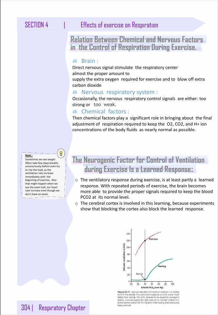
304 | Respiratory Chapter
SECTION 4 | Effects of exercise on Respiration
in the Control of Respiration During Exercise.
Brain:Directnervoussignalstimulatetherespiratorycenteralmosttheproperamounttosupplytheextraoxygenrequiredforexerciseandtoblowoffextracarbondioxide
Nervousrespiratorysystem:Occasionally,thenervousrespiratorycontrolsignalsareeither: toostrongor too weak.
Chemicalfactors:ThenchemicalfactorsplayasignificantroleinbringingaboutthefinaladjustmentofrespirationrequiredtokeeptheO2,CO2,andH+ionconcentrationsofthebodyfluidsasnearlynormalaspossible.
Relation Between Chemical and Nervous Factors
The Neurogenic Factor for Control of Ventilation during Exercise Is a Learned Response:
o Theventilatoryresponseduringexercise,isatleastpartlyalearnedresponse.Withrepeatedperiodsofexercise,thebrainbecomesmoreabletoprovidethepropersignalsrequiredtokeepthebloodPCO2 atitsnormallevel.
o Thecerebralcortexisinvolvedinthislearning,becauseexperimentsshowthatblockingthecortexalsoblockthelearnedresponse.
Note:Sometimesweseeweightlifterstakefewdeepbreathsunconsciouslybeforeeventrytorisetheload,sotheventilationrateincreaseimmediatelywiththebeginningofexercise.Alsothatmighthappenwhenweseetheexamhall,ourheartrateincreaseeventhoughwedon’thaveanexam.
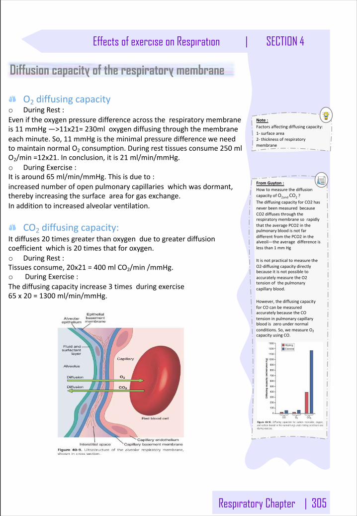
Respiratory Chapter | 305
Effects of exercise on Respiration | SECTION 4
Diffusion capacity of the respiratory membrane
O2 diffusingcapacityo DuringRest:Eveniftheoxygenpressuredifferenceacrosstherespiratorymembraneis11 mmHg—>11x21=230mloxygendiffusingthroughthemembraneeachminute.So,11 mmHgistheminimalpressuredifferenceweneedtomaintainnormalO2 consumption.Duringresttissuesconsume250 mlO2/min=12x21.Inconclusion,itis21 ml/min/mmHg.o DuringExercise:Itisaround65 ml/min/mmHg.Thisisdueto:increasednumberofopenpulmonarycapillarieswhichwasdormant,therebyincreasingthesurfaceareaforgasexchange.Inadditiontoincreasedalveolarventilation.
CO2 diffusingcapacity:Itdiffuses20 timesgreaterthanoxygenduetogreaterdiffusioncoefficientwhichis20 timesthatforoxygen.o DuringRest:Tissuesconsume,20x21 =400 mlCO2/min/mmHg.o DuringExercise:Thediffusingcapacityincrease3 timesduringexercise65 x20 =1300 ml/min/mmHg.
Note:Factorsaffectingdiffusingcapacity:1- surfacearea2- thicknessofrespiratorymembrane
FromGuyton:HowtomeasurethediffusioncapacityofO2andCO2 ?ThediffusingcapacityforCO2 hasneverbeenmeasuredbecauseCO2 diffusesthroughtherespiratorymembranesorapidlythattheaveragePCO2 inthepulmonarybloodisnotfardifferentfromthePCO2 inthealveoli—theaveragedifferenceislessthan1 mmHg
ItisnotpracticaltomeasuretheO2-diffusingcapacitydirectlybecauseitisnotpossibletoaccuratelymeasuretheO2tensionofthepulmonarycapillaryblood.
However,thediffusingcapacityforCOcanbemeasuredaccuratelybecausetheCOtensioninpulmonarycapillarybloodiszeroundernormalconditions.So,wemeasureO2capacityusingCO.
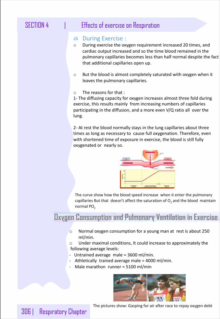
306 | Respiratory Chapter
SECTION 4 | Effects of exercise on Respiration
DuringExercise:o Duringexercisetheoxygenrequirementincreased20 times,and
cardiacoutputincreasedandsothetimebloodremainedinthe pulmonarycapillariesbecomeslessthanhalfnormaldespitethefactthatadditionalcapillariesopenup.
o Butthebloodisalmostcompletelysaturatedwithoxygenwhenitleavesthepulmonarycapillaries.
o Thereasonsforthat:1- Thediffusingcapacityforoxygenincreasesalmostthreefoldduringexercise,thisresultsmainlyfromincreasingnumbersofcapillariesparticipatinginthediffusion,andamoreevenV/Qratioalloverthelung.
2- Atrestthebloodnormallystaysinthelungcapillariesaboutthreetimesaslongasnecessarytocausefulloxygenation.Therefore,evenwithshortenedtimeofexposureinexercise,thebloodisstillfullyoxygenatedornearlyso.
Oxygen Consumption and Pulmonary Ventilation in Exercise
o Normaloxygenconsumptionforayoungmanatrestisabout250ml/min.
o Undermaximalconditions,Itcouldincreasetoapproximatelythe followingaveragelevels:- Untrainedaveragemale=3600 ml/min.- Athleticallytrainedaveragemale=4000 ml/min.- Malemarathonrunner=5100 ml/min
ThecurveshowhowthebloodspeedincreasewhenitenterthepulmonarycapillariesButthatdoesn’taffectthesaturationofO2 andthebloodmaintainnormal PO2
Thepicturesshow:Gaspingforairafterracetorepayoxygendebt
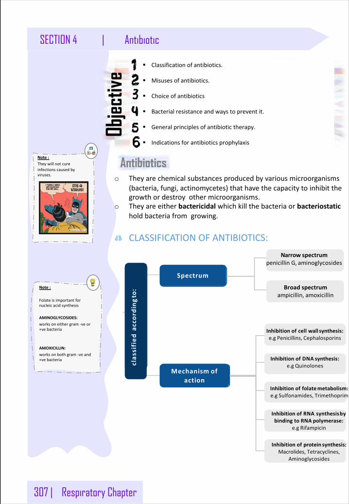
307 | Respiratory Chapter
SECTION 4 | Antibiotic
§ Classificationofantibiotics.
§ Misusesofantibiotics.
§ Choiceofantibiotics
§ Bacterialresistanceandwaystopreventit.
§ Generalprinciplesofantibiotictherapy.
§ Indicationsforantibioticsprophylaxis
Objec
tive
Antibioticso Theyarechemicalsubstancesproducedbyvariousmicroorganisms
(bacteria,fungi,actinomycetes)thathavethecapacitytoinhibitthegrowthordestroyothermicroorganisms.
o Theyareeitherbactericidal whichkillthebacteriaorbacteriostaticholdbacteriafromgrowing.
CLASSIFICATIONOFANTIBIOTICS:
clas
sified
according
to:
Spectrum
Mechanism ofaction
Note:Theywillnotcureinfectionscausedbyviruses.
Note:
Folateisimportantfornucleicacidsynthesis
AMINOGLYCOSIDES:worksoneithergram-veor+vebacteria
AMOXICILLIN:worksonbothgram-veand+vebacteria
Narrow spectrumpenicillinG, aminoglycosides
Inhibitionofcellwall synthesis:e.gPenicillins, Cephalosporins
InhibitionofDNA synthesis:e.g Quinolones
InhibitionofRNAsynthesisbybindingtoRNA polymerase:
e.g Rifampicin
Broad spectrumampicillin, amoxicillin
Inhibitionoffolatemetabolism:e.gSulfonamides, Trimethoprim
Inhibitionofprotein synthesis:Macrolides,Tetracyclines,
Aminoglycosides
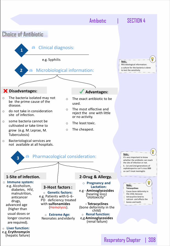
Respiratory Chapter | 308
Antibiotic | SECTION 4
Choice of Antibiotic
Clinicaldiagnosis:1
e.g. Syphilis
2
Advantages:Disadvantages:
o Theexactantibiotictobeused.
o Themosteffectiveandrejecttheonewithlittleorno activity.
o Theleast toxic.
o The cheapest.
o Thebacteriaisolatedmaynotbetheprimecauseofthedisease.
o donottakeinconsiderationsiteof infection.
o somebacteriacannotbecultivatedortaketimetogrow(e.g.M.Leprae,M.Tuberculosis)
o Bacteriologicalservicesarenotavailableatall hospitals.
3
1-Siteof infection.
3-Hostfactors :
2-Drug & Allergy.o Pregnancyand
Lactation:e.g - Aminoglycosides
(hearingloss)"ototoxicity”
- Tetracyclines(bonedeformityinthe
child)
o Genetic factors:e.g.Patientswith G-6-PDdeficiencytreatedwith sulfonamides
(Hemolysis).
o Renal function:e,gAminoglycosides
(renal failure)
o Extreme Age:Neonatesand elderly
o Liver function:e.g. Erythromycin(hepatic failure)
o Immune system:e.g.Alcoholism,
diabetes,HIV,malnutrition,anticancerdrugs,
advanced age(higherthanusualdosesorlongercoursesare required).
Microbiologicalinformation:
Pharmacologicalconsideration:
Note:Microbiologicalinformation: acultureforthebacteriaisdonetotestthesensitivity
Note:TetracyclinesCausebonedeformityinthechild,becausetetracyclinebindtocalciumandaffectsthebonesandteeth
Note:It’sveryimportanttoknowwhethertheantibioticcanreachthesiteofinfectionornot.E.:1stand2ndgenerationsofcephalosporinscan’tcrossBBBsocan’ttreatmeningitis
309 | Respiratory Chapter
SECTION 4 | Antibiotic
Definition:Concentrationofantibioticrequiredtoinhibitorkillthebacteriaisgreaterthantheconcentrationthatcansafelybeachievedintheplasma.
Whendoesbacterialresistanceemerge?Oneresultofthewidespreaduseofantibioticshasbeentheemergenceofresistantpathogensthathavebeensensitiveinthepast.
ReasonsforMisuseofAntibiotics : o Availabilityofaverywideselection.o Limitationofphysician’stime.o Physicianshortageandexpenses.o Availabilitywithoutprescriptionsinpharmacies.o Publicdemand(pressuretoprescribe)
MechanismofAcquiredAntibioticResistance : o Inactivationbyenzymeproducedbybacteria:Bacterialβ-
lactamaseinactivatespenicillin's&cephalosporinsbycleavingtheβ-lactamringofthedrug.
o OrBacteriadevelopsanalteredreceptorforthedrug.o OrBacteriadevelopsanalteredmetabolicpathway.o OrReducedbacterialpermeabilitytoantibioticthroughcell
membrane.o OrActivelytransporting the drugoutofthebacterial cell.
Preventionofbacterialresistance:o Useantibioticonlywhenabsolutelyrequired.o Useantibioticsinadequatedosageforsufficientperiodoftime.o Nottoobrieftherapy.o Nottooprolongedtherapy(exceptions,e.g.TB→6 months).o Combinationofantibioticsmayberequiredtodelayresistance.(e.g.
TB).
Bacterial resistance
Note:Presenceofpusornecrotictissue,orbloodatthesurgicalsite, canpreventtheabsorptionofantibioticssodrainageshouldbedonefirst
o Treatmentofdiseasescausedbyviruses.o Improperdosage.o Therapyoffeverofunknownorigin.o Presenceofpusornecrotictissue,orbloodatthesurgicalsite.o Lackofadequatebacteriologicalinformation.o Excessiveuseofprophylacticantibioticsintravelers.o Overuseasgrowthpromotersinanimalsandagriculture.o Patientsdonottakethemaccordingtotheirdoctor’sinstructions.o Somepatientssaveunusedantibioticsforanotherillness,orpass
toothers
Misuse of Antibiotics
Respiratory Chapter | 310
Antibiotic | SECTION 4
General principles of Antibacterial therapy
Exceptionswhere combiningantibiotics is a good option:
Desperatelyillpatient ofunknown etiology
Topreventemergenceofresistance(e.g.TB )
Higher cost
Possibility ofantagonism
Toachieve synergisme.g.piperacillin+gentamicin(pseudomonas aeruginosa)
Increasedriskofcolonizationand infectionwitharesistant bacteria
Increasedriskofsensitivityor toxicity
1 disadvantages: 2
IndicationsforAntibioticsProphylaxis:
o Administerdruginfulldose,atproperintervalandbythebestrouteo Whenapparentcureachieved,continueforabout3 daysfurtherto
avoidrelapseo Skippingdosesmaydecreaseeffectivenessofantibiotics&increasethe
incidenceofbacterialresistance.o Measurementofplasmaconc.ofantibioticsisseldom(rarely)needed,
exceptforsystemicAminoglycosidese.g.streptomycin,gentamicin.o Insomeinfections,bacteriologicalproofofcureisdesirable(e.g.TB,
UTI).
Usageofmultipleantimicrobial
Note:Antibioticsynergyiswhentwoormoreantibioticsareusedsimultaneouslytotreataninfection.Inthesynergisticresponse,theappliedantibioticsworktogethertoproduceaneffectmorepotentthanifeachantibioticwereappliedsingly.
Note:Frequentandinappropriateuseofantibioticscancausebacteriaorothermicrobestochangesoantibioticsdon’tworkagainstthem.
Surgicalprophylaxis
ImmunosuppressedPatients
Dentalextractionscontroversial
bowelsurgery,jointreplacement,etc.topreventpostoperativeinfections.
o Veryoldo Veryyoungo Diabeticso Anaemicso AIDSo Cancerpts
o Ptswithtotaljointreplacements
o Ptswithcardiacabnormalities
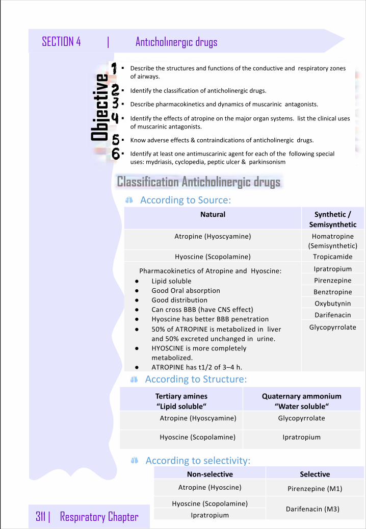
311 | Respiratory Chapter
SECTION 4 | Anticholinergic drugs
Classification Anticholinergic drugs
Objec
tive
AccordingtoSource:
§ Describethestructuresandfunctionsoftheconductiveandrespiratoryzonesofairways.
§ Identifytheclassificationofanticholinergicdrugs.
§ Describepharmacokineticsanddynamicsofmuscarinicantagonists.
§ Identifytheeffectsofatropineonthemajororgansystems.listtheclinicalusesofmuscarinicantagonists.
§ Knowadverseeffects&contraindicationsofanticholinergicdrugs.
§ Identifyatleastoneantimuscarinicagentforeachofthefollowingspecialuses:mydriasis,cyclopedia,pepticulcer&parkinsonism
Natural Synthetic/Semisynthetic
Atropine(Hyoscyamine) Homatropine(Semisynthetic)
Hyoscine(Scopolamine) Tropicamide
PharmacokineticsofAtropineandHyoscine:● Lipidsoluble● GoodOralabsorption● Gooddistribution● CancrossBBB(haveCNSeffect)● HyoscinehasbetterBBBpenetration● 50%ofATROPINEismetabolizedinliver
and50%excretedunchangedinurine.● HYOSCINEismorecompletely
metabolized.● ATROPINEhast1/2 of3–4 h.
IpratropiumPirenzepineBenztropineOxybutyninDarifenacin
Glycopyrrolate
Tertiaryamines“Lipidsoluble“
Quaternaryammonium“Watersoluble“
Atropine(Hyoscyamine) Glycopyrrolate
Hyoscine(Scopolamine) Ipratropium
Accordingtoselectivity:
AccordingtoStructure:
Non-selective Selective
Atropine(Hyoscine) Pirenzepine(M1)
Hyoscine(Scopolamine)Darifenacin(M3)
Ipratropium
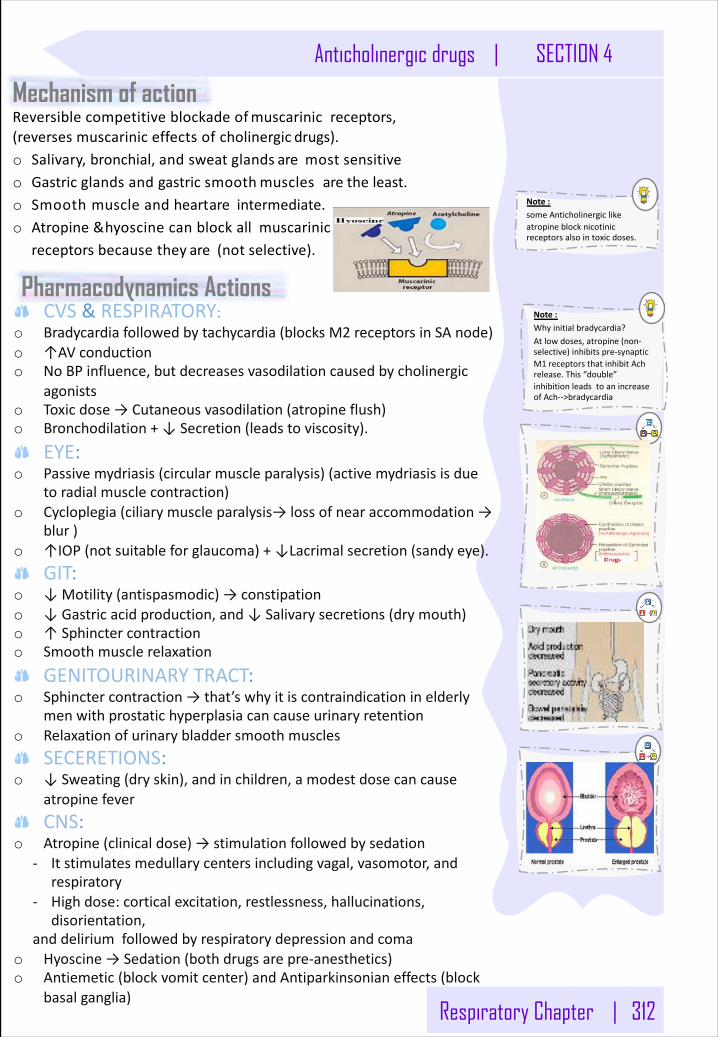
Respiratory Chapter | 312
Anticholinergic drugs | SECTION 4
Note:someAnticholinergiclikeatropineblocknicotinicreceptorsalsointoxicdoses.
Reversible competitiveblockadeofmuscarinicreceptors,(reversesmuscariniceffectsofcholinergic drugs).o Salivary,bronchial,andsweatglands aremost sensitiveo Gastricglandsandgastricsmooth musclesarethe least.o Smoothmuscleandheartareintermediate.o Atropine&hyoscinecanblockallmuscarinic
receptorsbecausethey are(not selective).
Mechanism of action
Pharmacodynamics ActionsCVS &RESPIRATORY:
o Bradycardiafollowedbytachycardia(blocksM2 receptorsinSAnode)o ↑AVconductiono NoBPinfluence,butdecreasesvasodilationcausedbycholinergic
agonistso Toxicdose→Cutaneousvasodilation(atropineflush)o Bronchodilation+↓Secretion(leadstoviscosity).
EYE:o Passivemydriasis(circularmuscleparalysis)(activemydriasisisdue
toradialmusclecontraction)o Cycloplegia(ciliarymuscleparalysis→lossofnearaccommodation→
blur)o ↑IOP(notsuitableforglaucoma)+↓Lacrimalsecretion(sandyeye).
GIT:o ↓Motility(antispasmodic)→constipationo ↓Gastricacidproduction,and↓Salivarysecretions(drymouth)o ↑Sphinctercontractiono Smoothmusclerelaxation
GENITOURINARYTRACT:o Sphinctercontraction→that’swhyitiscontraindicationinelderly
menwithprostatichyperplasiacancauseurinaryretentiono Relaxationofurinarybladdersmoothmuscles
SECERETIONS:o ↓Sweating(dryskin),andinchildren,amodestdosecancause
atropinefeverCNS:
o Atropine(clinicaldose)→stimulationfollowedbysedation- Itstimulatesmedullarycentersincludingvagal,vasomotor,and
respiratory- Highdose:corticalexcitation,restlessness,hallucinations,
disorientation,anddeliriumfollowedbyrespiratorydepressionandcoma
o Hyoscine→Sedation(bothdrugsarepre-anesthetics)o Antiemetic(blockvomitcenter)andAntiparkinsonianeffects(block
basalganglia)
Note:Whyinitialbradycardia?Atlowdoses,atropine(non-selective)inhibitspre-synapticM1 receptorsthatinhibitAchrelease.This“double”inhibitionleadstoanincreaseofAch-->bradycardia
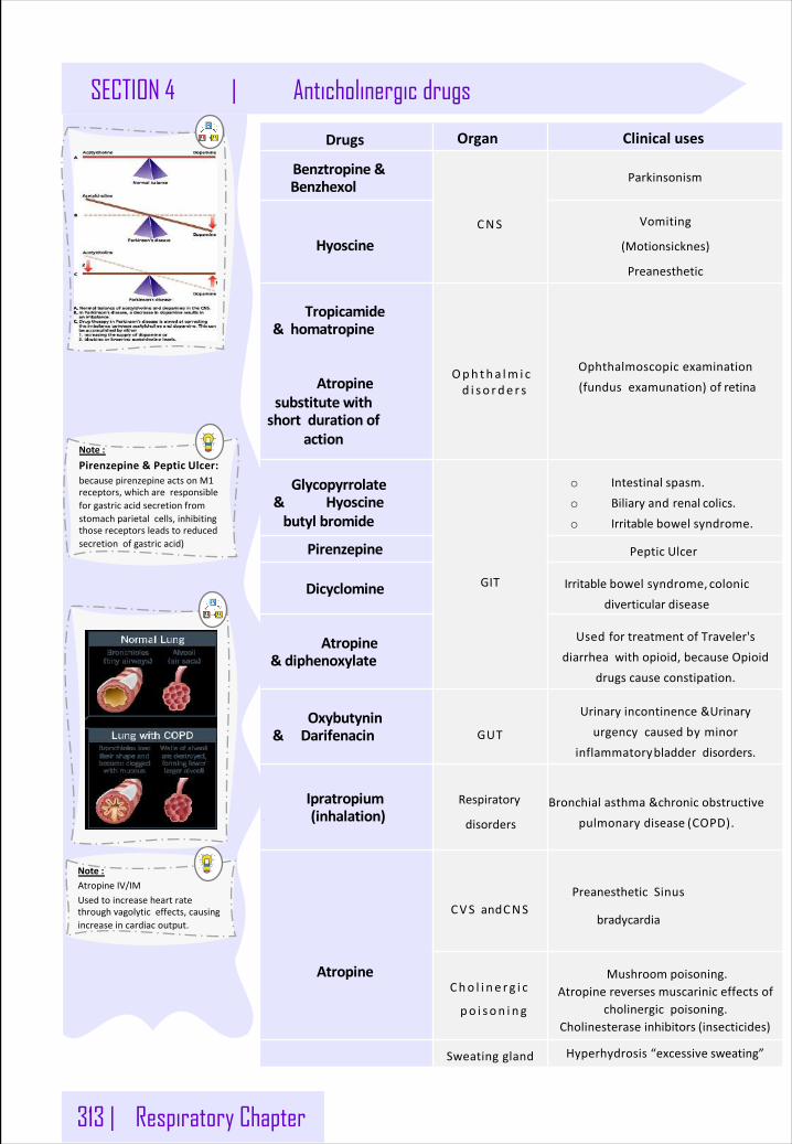
313 | Respiratory Chapter
SECTION 4 | Anticholinergic drugs
Drugs Organ Clinicaluses
Benztropine&Benzhexol
CNS
Parkinsonism
HyoscineVomiting
(Motionsicknes)
Preanesthetic
Tropicamide& homatropine
Atropinesubstitutewithshortdurationof
action
Ophthalmoscopicexamination(fundusexamunation)of retina
Glycopyrrolate& Hyoscinebutyl bromide
GIT
o Intestinal spasm.o Biliaryandrenal colics.o Irritablebowel syndrome.
Pirenzepine Peptic Ulcer
Dicyclomine Irritablebowelsyndrome, colonicdiverticular disease
Atropine&diphenoxylate
UsedfortreatmentofTraveler'sdiarrheawithopioid,becauseOpioid
drugscauseconstipation.
Oxybutynin&Darifenacin GUT
Urinaryincontinence&Urinaryurgencycausedbyminor
inflammatorybladderdisorders.
Ipratropium. (inhalation)
Bronchialasthma&chronicobstructivepulmonarydisease (COPD).
Atropine
CVS andCNSPreanestheticSinus
bradycardia
Mushroom poisoning.Atropinereversesmuscariniceffectsof
cholinergicpoisoning.Cholinesteraseinhibitors (insecticides)
Sweatinggland Hyperhydrosis“excessive sweating”
Note:Pirenzepine&Peptic Ulcer:becausepirenzepineactsonM1receptors,whichareresponsibleforgastricacidsecretionfromstomachparietalcells,inhibitingthosereceptorsleadstoreducedsecretionofgastricacid)
Note:AtropineIV/IMUsedtoincreaseheartratethroughvagolyticeffects,causingincreaseincardiacoutput.
Cho l i ne r g i c
po i son i ng
Oph tha lm i c d i so rde r s
Respiratory
disorders
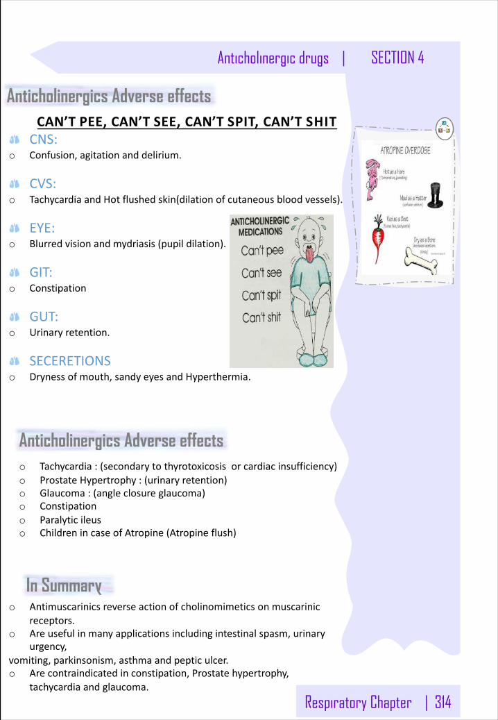
Respiratory Chapter | 314
Anticholinergic drugs | SECTION 4
CAN’TPEE,CAN’TSEE,CAN’TSPIT,CAN’T SHITCNS:
o Confusion,agitationanddelirium.
CVS:o TachycardiaandHotflushedskin(dilationofcutaneousbloodvessels).
EYE:o Blurredvisionandmydriasis(pupildilation).
GIT:o Constipation
GUT:o Urinaryretention.
SECERETIONSo Drynessofmouth,sandyeyesandHyperthermia.
Anticholinergics Adverse effects
Anticholinergics Adverse effectso Tachycardia:(secondarytothyrotoxicosisorcardiacinsufficiency)o ProstateHypertrophy:(urinaryretention)o Glaucoma:(angleclosureglaucoma)o Constipationo Paralyticileuso ChildrenincaseofAtropine(Atropineflush)
In Summaryo Antimuscarinicsreverseactionofcholinomimeticsonmuscarinic
receptors.o Areusefulinmanyapplicationsincludingintestinalspasm,urinary
urgency,vomiting,parkinsonism,asthmaandpepticulcer.o Arecontraindicatedinconstipation,Prostatehypertrophy,
tachycardiaandglaucoma.
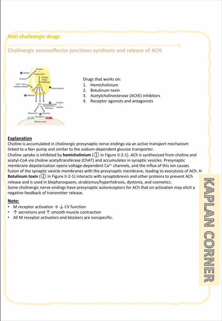
Anti-cholinergicdrugs
Cholinergicneuroeffectorjunctionssynthesisandreleaseof ACH
ExplanationCholineisaccumulatedincholinergicpresynapticnerveendingsviaanactivetransportmechanismlinkedtoaNa+pumpandsimilartothesodium-dependentglucosetransporter.Cholineuptakeisinhibitedbyhemicholinium (① inFigureII-2-1).ACh issynthesizedfromcholineandacetyl-CoAviacholineacetyltransferase(ChAT)andaccumulatesinsynapticvesicles.Presynapticmembranedepolarizationopensvoltage-dependentCa2+ channels,andtheinfluxofthisioncauses fusionofthesynapticvesiclemembraneswiththepresynapticmembrane,leadingtoexocytosisofACh.HBotulinumtoxin (② inFigureII-2-1)interactswithsynaptobrevin andotherproteinstopreventAChreleaseandisusedinblepharospasm,strabismus/hyperhidrosis,dystonia,andcosmetics.Somecholinergicnerveendingshavepresynapticautoreceptors forACh thatonactivationmayelicitanegativefeedbackoftransmitterrelease.
Note:• Mreceptoractivation→ ↓CVfunction• ↑secretionsand↑smoothmusclecontraction• AllMreceptoractivatorsandblockersarenonspecific.
Drugsthatworkson:1. Hemicholinium2. Botulinumtoxin3. Acetylcholinesterase(AChE)inhibitors4. Receptoragonistsandantagonists
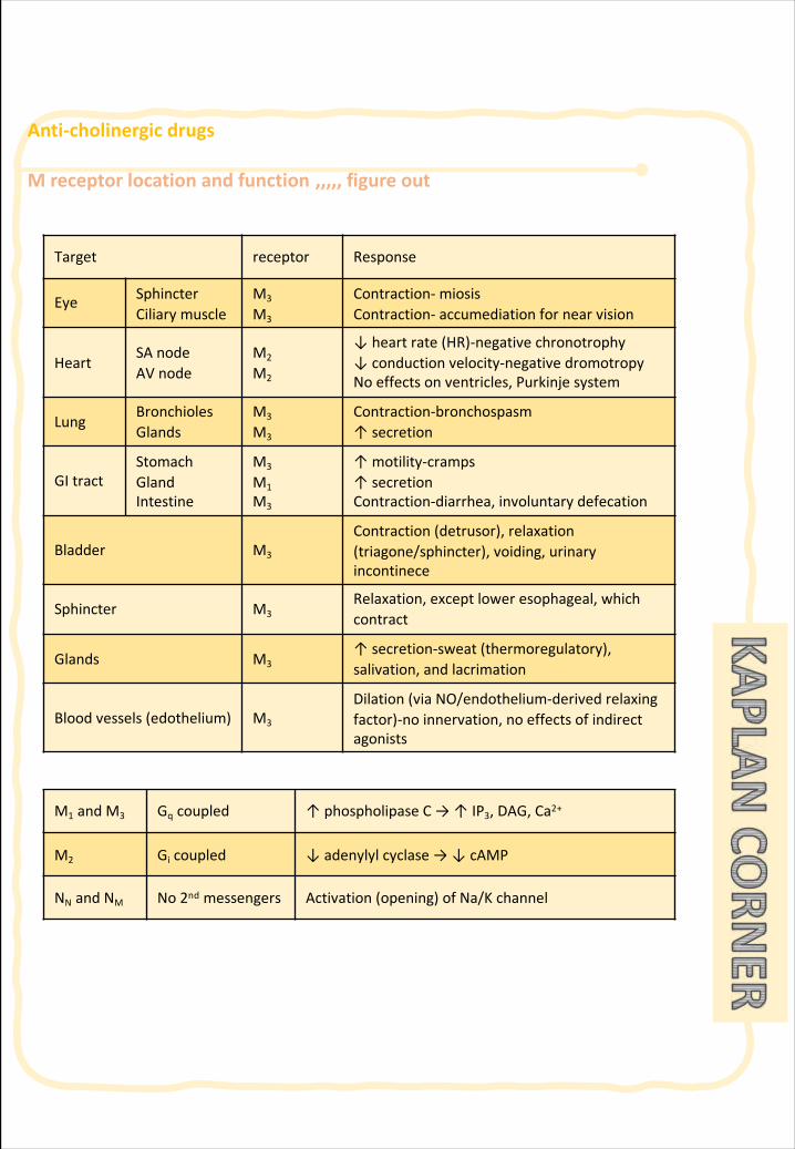
Anti-cholinergicdrugs
Mreceptorlocationandfunction ,,,,,figureout
Target receptor Response
Eye SphincterCiliary muscle
M3
M3
Contraction- miosisContraction- accumediation fornearvision
HeartSAnodeAVnode
M2
M2
↓heartrate(HR)-negative chronotrophy↓conductionvelocity-negativedromotropyNoeffectsonventricles,Purkinjesystem
LungBronchiolesGlands
M3
M3
Contraction-bronchospasm↑secretion
GItractStomachGlandIntestine
M3
M1M3
↑motility-cramps↑secretionContraction-diarrhea,involuntarydefecation
Bladder M3
Contraction(detrusor), relaxation(triagone/sphincter),voiding,urinaryincontinece
Sphincter M3Relaxation, exceptloweresophageal,whichcontract
Glands M3↑secretion-sweat (thermoregulatory),salivation,andlacrimation
Bloodvessels(edothelium) M3
Dilation(viaNO/endothelium-derived relaxingfactor)-noinnervation,noeffectsofindirectagonists
M1 andM3 Gq coupled ↑phospholipase C→↑IP3,DAG,Ca2+
M2 Gi coupled ↓adenylylcyclase→↓cAMP
NN andNM No 2nd messengers Activation(opening)ofNa/Kchannel
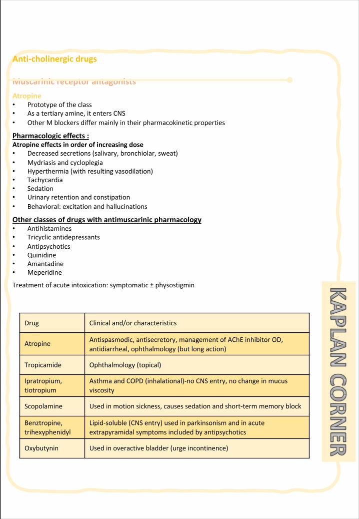
Anti-cholinergicdrugs
MuscarinicreceptorantagonistsAtropine• Prototypeoftheclass• Asatertiaryamine,itentersCNS• OtherMblockersdiffermainlyintheirpharmacokinetic properties
Pharmacologiceffects:Atropineeffectsinorderofincreasingdose• Decreasedsecretions(salivary,bronchiolar,sweat)• Mydriasisandcycloplegia• Hyperthermia(withresultingvasodilation)• Tachycardia• Sedation• Urinaryretentionandconstipation• Behavioral:excitationandhallucinations
Otherclassesofdrugswithantimuscarinic pharmacology• Antihistamines• Tricyclicantidepressants• Antipsychotics• Quinidine• Amantadine• Meperidine
Treatmentofacuteintoxication:symptomatic± physostigmin
Drug Clinical and/orcharacteristics
Atropine Antispasmodic, antisecretory,managementofAChE inhibitorOD,antidiarrheal,ophthalmology(butlongaction)
Tropicamide Ophthalmology(topical)
Ipratropium,tiotropium
AsthmaandCOPD(inhalational)-no CNSentry,nochangeinmucusviscosity
Scopolamine Usedinmotionsickness,causessedationand short-termmemoryblock
Benztropine,trihexyphenidyl
Lipid-soluble (CNSentry)usedinparkinsonismandinacuteextrapyramidalsymptomsincludedbyantipsychotics
Oxybutynin Used inoveractivebladder(urgeincontinence)
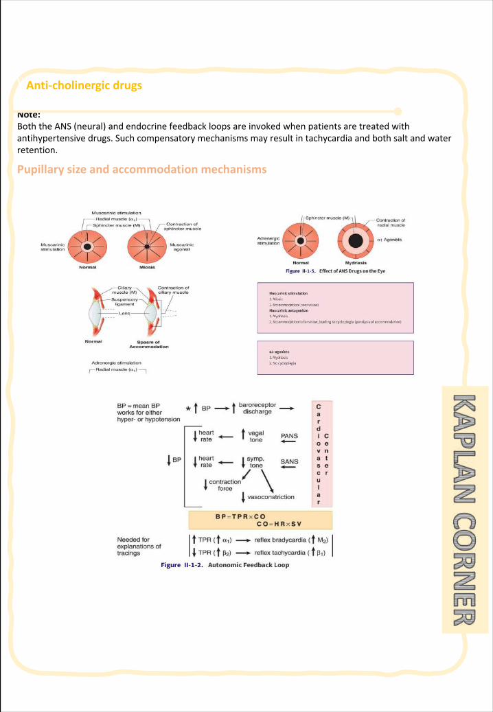
\\ Anti-cholinergicdrugs
Note:BoththeANS(neural)andendocrinefeedbackloopsareinvokedwhenpatientsaretreated withantihypertensivedrugs.Suchcompensatorymechanismsmayresultintachycardiaandbothsaltandwaterretention.
Pupillarysizeandaccommodationmechanisms
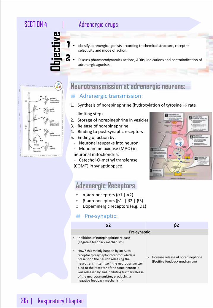
315 | Respiratory Chapter
SECTION 4 | Adrenergic drugs
Neurotransmission at adrenergic neurons:
Objec
tive
Adrenergictransmission:
§ classifyadrenergicagonistsaccordingtochemicalstructure,receptorselectivityandmodeofaction.
§ Discusspharmacodynamicsactions,ADRs,indicationsandcontraindicationofadrenergicagonists.
1. Synthesisofnorepinephrine(hydroxylationoftyrosine→rate
limitingstep)2. Storageofnorepinephrineinvesicles3. Releaseofnorepinephrine4. Bindingtopost-synapticreceptors5. Endingofactionby:- Neuronalreuptakeintoneuron.- Monoamineoxidase(MAO)inneuronalmitochondria.- Catechol-O-methyltransferase(COMT)insynapticspace
α2 β2Pre-synaptic
o Inhibitionofnorepinephrinerelease(negativefeedbackmechanism)
o How?thismainlyhappenbyanAuto-receptor‘presynapticreceptor’whichispresentontheneuronreleasingtheneurotransmitteritself,theneurotransmitterbindtothereceptorofthesameneuronitwasreleasedbyandinhibitingfurtherreleaseoftheneurotransmitter,producinganegativefeedbackmechanism)
o Increasereleaseofnorepinephrine(Positivefeedbackmechanism)
Adrenergic Receptorso α-adrenoceptors(α1 |α2)o β-adrenoceptors(β1 |β2 |β3)o Dopaminergicreceptors(e.g.D1)
Pre-synaptic:
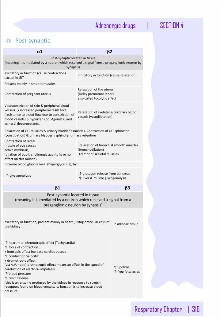
Respiratory Chapter | 316
Adrenergic drugs | SECTION 4
α1 β2Post-synapticlocatedintissue
(meaningitismediatedbyaneuronwhichreceivedasignalfromapreganglionicneuronbysynapsis)
excitatoryinfunction(causecontraction)exceptinGIT
inhibitoryinfunction(causerelaxation)
Presentmainlyinsmoothmuscles
ContractionofpregnantuterusRelaxationoftheuterus(Delayprematurelabor)alsocalledtocolyticeffect
Vasoconstrictionofskin&peripheralbloodvessels→increasedperipheralresistance(resistancetobloodflowduetoconstrictionofbloodvessels)→hypertension.Agonistsusedasnasaldecongestants.
Relaxationofskeletal&coronarybloodvessels(vasodilatation)
RelaxationofGITmuscles&urinarybladder’smuscles.ContractionofGITsphincter(constipation)&urinarybladder’ssphincterurinaryretentionContractionofradialmuscleofeyecausesactivemydriasis,(dilationofpupil,cholinergicagentshavenoeffectonthismuscle)
.Relaxationofbronchialsmoothmuscles(bronchodilation).Tremorofskeletalmuscles
Increasebloodglucoselevel(hyperglycemia),by:
.↑glycogenolysis .↑glucagonreleasefrompancreas.↑liver&muscleglycogenolysis
β1 β3Post-synapticlocatedintissue
(meaningitismediatedbyaneuronwhichreceivedasignalfromapreganglionicneuronbysynapsis)
excitatoryinfunction,presentmainlyinheart,juxtaglomerularcellsofthekidney
Inadiposetissue
↑heartrate:chronotropiceffect(Tachycardia)↑forceofcontraction:+inotropiceffectIncreasecardiacoutput↑conductionvelocity:+dromotropiceffect(viaA.V.node)(dromotropiceffectmeansaneffectinthespeedofconductionofelectricalimpulses)↑bloodpressure↑reninrelease(thisisanenzymeproducedbythekidneyinresponsetostretchreceptorsfoundonbloodvessels,itsfunctionistoincreasebloodpressure)
↑lipolysis↑freefattyacids
Post-synaptic:
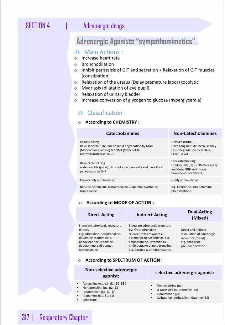
317 | Respiratory Chapter
SECTION 4 | Adrenergic drugs
Adrenergic Agonists “sympathomimetics’’
o Increaseheartrateo Bronchodilationo InhibitperistalsisofGITandsecretion+RelaxationofGITmuscles
(constipation)o Relaxationoftheuterus(Delayprematurelabor)tocolytico Mydriasis(dilatationofeyepupil)o Relaxationofurinarybladdero Increaseconversionofglycogentoglucose(hyperglycemia)
o AccordingtoCHEMISTRY:
MainActions:
Catecholamines Non-Catecholamines
RapidlyactingHaveshorthalf-life,duetorapiddegradationbyMAO(MonoamineOxidase)&COMT(Catechol-O-MethylTransferase)inGIT
DelayedactionHaveLonghalf-life,becausetheyresistdegradationbyMOA&COMTinGIT
Havecatecholringwatersoluble(polar),thusnoteffectiveorallyandhavePoorpenetrationtoCNS
LackcatecholringLipidsoluble,thusEffectiveorallyandCrossBBBwell,haveProminentCNSeffects
Parenterallyadministered Orallyadministered
Natural:Adrenaline,Noradrenaline,DopamineSynthetic:Isoprenaline.
e.g.Ephedrine,amphetamine,phenylephrine.
Direct-Acting Indirect-Acting Dual-Acting(Mixed)
Stimulateadrenergicreceptorsdirectlye.g.adrenaline,noradrenaline,dopamine,isoprenaline,phenylephrine,clonidine,dobutamine,salbutamol,methoxamine
Stimulateadrenergicreceptorsby:↑noradrenalinereleasefrompresynapticadrenergicnerveendings.e.g.amphetamine,TyramineOrInhibituptakeofnoradrenalinee.g.Cocaine&antidepressants
Directandindirectstimulationofadrenergicreceptors(mixed)e.g.ephedrine,pseudoephedrine
Non-selectiveadrenergicagonist:
selectiveadrenergicagonist:
• Adrenaline(α1,α2 ,β1 ,β2,β3 )• Noradrenaline(α1,α2 ,β1)• Isoprenaline(β1,β2,β3)• Dopamine(D1,β1,α1)• Ephedrine
• Phenylephrine(α1)• α-Methyldopa- clonidine(α2)• Dobutamine(β1)• Salbutamol,terbutaline,ritodrine(β2)
Classification:
o AccordingtoMODEOFACTION:
o AccordingtoSPECTRUMOFACTION:
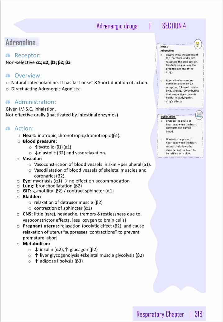
Respiratory Chapter | 318
Adrenergic drugs | SECTION 4
Adrenaline Receptor:
Non-selectiveα1;α2;β1;β2; β3
Overview:o Naturalcatecholamine.Ithasfastonset&Shortdurationof action.o DirectactingAdrenergicAgonists:
Administration:GivenI.V,S.C, inhalation.Noteffectiveorally(inactivatedbyintestinalenzymes).
Action:o Heart:inotropic,chronotropic,dromotropic (β1).o Bloodpressure:
o ↑systolic(β1)(α1)o ↓diastolic(β2)andvasorelaxation.
o Vascular:o Vasoconstrictionofbloodvesselsinskin+peripheral (α1).o Vasodilatationofbloodvesselsofskeletalmusclesand
coronaries(β2).o Eye:mydriasis(α1)→noeffectonaccommodationo Lung:bronchodilatation(β2)o GIT:↓motility(β2)/contractsphincter(α1)o Bladder:
o relaxationofdetrusormuscle(β2)o contractionofsphincter (α1)
o CNS:little(rare),headache,tremors&restlessnessduetovasoconstrictoreffects,lessoxygentobraincells)
o Pregnantuterus:relaxationtocolyticeffect(β2),andcauserelaxationofuterus“suppressescontractions”topreventpremature labor)
o Metabolism:o ↓insulin(α2),↑glucagon(β2)o ↑liverglycogenolysis+skeletalmuscleglycolysis(β2)o ↑adiposelipolysis(β3)
Note:Adrenalineo alwaysknowtheactionsof
thereceptors,andwhichreceptorsthedrugactson.Thishelpsinguessingtheprobableactionsofthedrug).
o Adrenalinehasamoredominantactiononβ2receptors,followedmainlybyα1 andβ1,rememberingtheirrespectiveactionsishelpfulinstudyingthisdrug’seffects
Explanation:o Systolic:thephaseof
heartbeatwhentheheartcontractsandpumpsblood.
o Diastolic:thephaseofheartbeatwhentheheartrelaxesandallowsthechambersofthehearttoberefilledwithblood
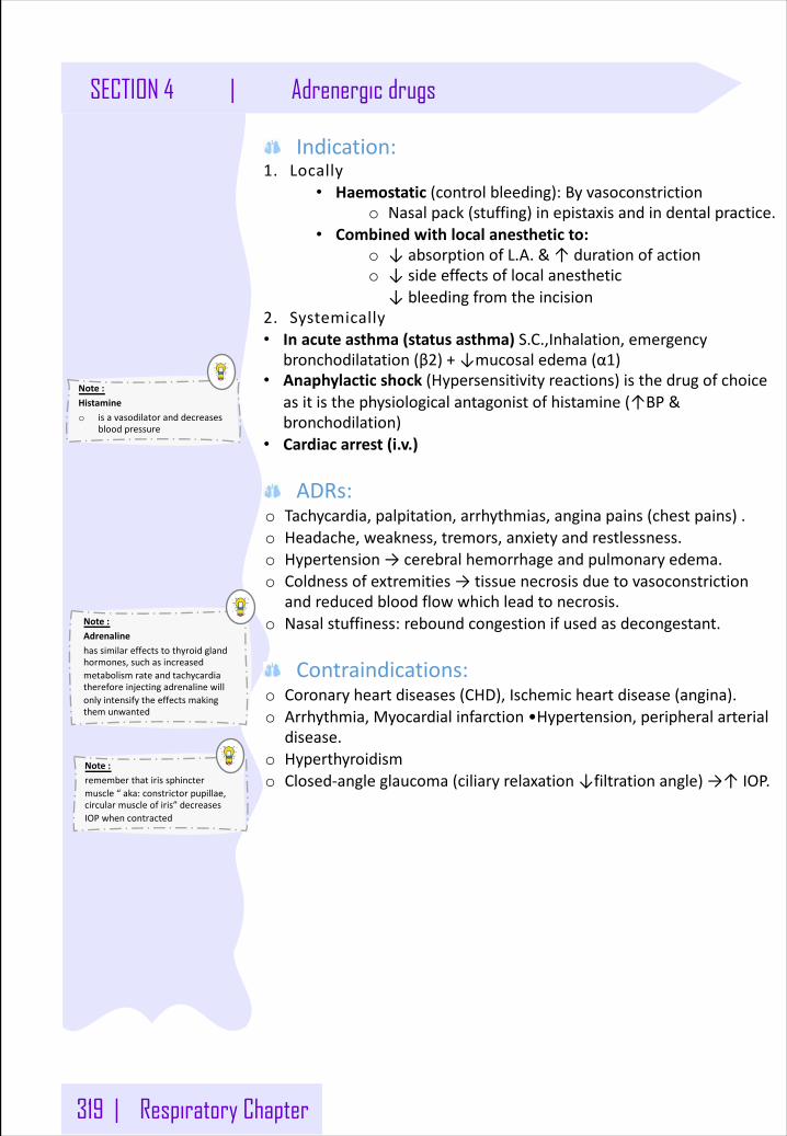
319 | Respiratory Chapter
SECTION 4 | Adrenergic drugs
Indication:1. Locally
• Haemostatic (controlbleeding):Byvasoconstrictiono Nasalpack(stuffing)inepistaxisandindentalpractice.
• Combinedwithlocalanestheticto:o ↓absorptionofL.A.&↑durationofactiono ↓sideeffectsoflocalanesthetic
↓bleedingfromtheincision2. Systemically• Inacuteasthma(statusasthma)S.C.,Inhalation,emergency
bronchodilatation(β2)+↓mucosaledema(α1)• Anaphylacticshock(Hypersensitivityreactions)isthedrugofchoice
asitisthephysiologicalantagonistofhistamine(↑BP&bronchodilation)
• Cardiacarrest(i.v.)
ADRs:o Tachycardia,palpitation,arrhythmias,anginapains(chestpains).o Headache,weakness,tremors,anxietyandrestlessness.o Hypertension→cerebralhemorrhageandpulmonaryedema.o Coldnessofextremities→tissuenecrosisduetovasoconstriction
andreducedbloodflowwhichleadtonecrosis.o Nasalstuffiness:reboundcongestionifusedasdecongestant.
Contraindications:o Coronaryheartdiseases(CHD),Ischemicheartdisease(angina).o Arrhythmia,Myocardialinfarction•Hypertension,peripheralarterial
disease.o Hyperthyroidismo Closed-angleglaucoma(ciliaryrelaxation↓filtrationangle)→↑IOP.
Note:Histamineo isavasodilatoranddecreases
bloodpressure
Note:Adrenalinehassimilareffectstothyroidglandhormones,suchasincreasedmetabolismrateandtachycardiathereforeinjectingadrenalinewillonlyintensifytheeffectsmakingthemunwanted
Note:rememberthatirissphinctermuscle“aka:constrictorpupillae,circularmuscleofiris”decreasesIOPwhencontracted
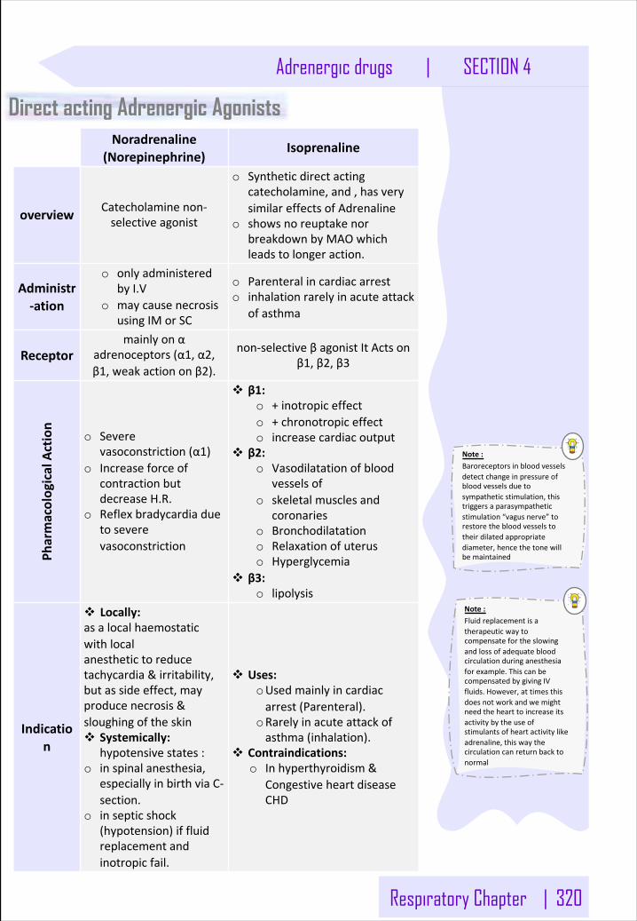
Respiratory Chapter | 320
Adrenergic drugs | SECTION 4
Direct acting Adrenergic Agonists Noradrenaline
(Norepinephrine)Isoprenaline
overview Catecholaminenon-selectiveagonist
o Syntheticdirectactingcatecholamine, and,hasverysimilareffectsof Adrenaline
o showsnoreuptakenorbreakdownbyMAOwhichleadstolongeraction.
Administr-ation
o onlyadministeredbyI.V
o maycausenecrosisusingIMorSC
o Parenteralincardiacarresto inhalationrarelyinacuteattack
ofasthma
Receptormainlyonα
adrenoceptors(α1,α2,β1,weakactiononβ2).
non-selectiveβagonistItActsonβ1,β2,β3
Pharmacolog
icalAction
o Severevasoconstriction(α1)
o IncreaseforceofcontractionbutdecreaseH.R.
o Reflexbradycardiaduetoseverevasoconstriction
v β1:o +inotropiceffecto +chronotropiceffecto increasecardiacoutput
v β2:o Vasodilatationofblood
vesselsofo skeletalmusclesand
coronarieso Bronchodilatationo Relaxationofuteruso Hyperglycemia
v β3:o lipolysis
Indication
v Locally: asalocalhaemostaticwithlocalanesthetictoreducetachycardia&irritability,butassideeffect,mayproducenecrosis&sloughingoftheskinv Systemically:
hypotensivestates:o inspinalanesthesia,
especiallyinbirthviaC-section.
o insepticshock(hypotension)iffluidreplacementandinotropicfail.
v Uses:oUsedmainlyincardiacarrest(Parenteral).
oRarelyinacuteattackofasthma(inhalation).
v Contraindications:o Inhyperthyroidism&
CongestiveheartdiseaseCHD
Note:Baroreceptorsinbloodvesselsdetectchangeinpressureofbloodvesselsduetosympatheticstimulation,thistriggersaparasympatheticstimulation“vagusnerve”torestorethebloodvesselstotheirdilatedappropriatediameter,hencethetonewillbemaintained
Note:Fluidreplacementisatherapeuticwaytocompensatefortheslowingandlossofadequatebloodcirculationduringanesthesiaforexample.ThiscanbecompensatedbygivingIVfluids.However,attimesthisdoesnotworkandwemightneedthehearttoincreaseitsactivitybytheuseofstimulantsofheartactivitylikeadrenaline,thiswaythecirculationcanreturnbacktonormal
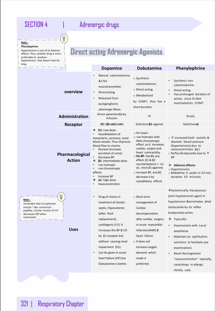
321 | Respiratory Chapter
SECTION 4 | Adrenergic drugs
Note:PhenylephrineHypertensionisoneofitsAdverseeffects.Thus,anotherdrugismorepreferabletoproducehypertensionthatdoesn’tlastforlong.
Dopamine Dobutamine Phenylephrine
overview
• Naturalcatecholamine
&CNS
neurotransmitter.
• Directacting.
• Releasedfrom
postganglionic
adrenergic fibres.
o Synthetic
catecholamine.
o Direct acting.
o Metabolized
by COMT, thus has a
shortduration
• Syntheticnoncatecholamine
• Direct acting• Hasprolongeddurationof
action,sinceit’sNotinactivatedbyCOMT
Administration Givenparenterally byinfusion IV Orally
Receptor D1>β1>α1(inorder) Selective β1–agonist Selectiveα1
PharmacologicalAction
v D1:Lowdose:• Vasodilatationofmesenteric,coronary,renalbloodvessels.Thusimprovesbloodflowtoviscera.• Diuresis(increase
excretionofurine)• DecreaseBPv β1:intermediatedose:• +veinotropic• +vechronotropiceffects• IncreaseBPv α1:highdose:• Vasoconstriction
oOnheart:o +veInotropicwithlittlechronotropiceffect.asitincreasescardiacoutputandheartcontractility.
oOnBP:Hardlyanyeffect;β1 &β2counterbalance+noα1.sinceβ1 agonists
o increaseBP,andβ2decreaseitbyvasodilatoryeffect)
o ↑increasedbothsystolic&diastolicbloodpressure(hypertension) duetovasoconstriction(α1)
o Reflex Bradycardia due to↑BP
v Adverse effects:oHypertension.oMidodrine.Itpeaksin2 0 min,duration3 0 minonly.
Uses
• Drugofchoicein
treatmentofshocks:
septic,Hypovolemic
(afterfluid
replacement),
cardiogenic(I.V).It
increasestheBP&CO
byβ1 receptorbut
withoutcausingrenal
impairment(D1)
• Canbegiveninacute
heartfailure(HF)but
Dobutamineisbetter.
o Shortterm
managementof
Cardiac
decompensation
aftercardiacsurgery,
inacutemyocardial
infarction(AMI)&
heartfailure.
o Itdoesnot
increase oxygen
demandwhich
madeit
preferred.
vSystemically: Vasopressor
(anti-hypotensive)agentin
hypotension&terminatesatrial
tachycardiabyitsreflex
bradycardia action.
v Topically:
• Haemostatic withLocal
anesthesia.
• Mydriatic (inophthalmic
solutionstofacilitateeye
examination).
• Nasaldecongestant
“vasoconstriction”topically,
nasal dropsinallergic
rhinitis,cold.
Direct acting Adrenergic Agonists
Note:rememberthatirissphinctermuscle“aka:constrictorpupillae,circularmuscleofiris”decreasesIOPwhencontracted
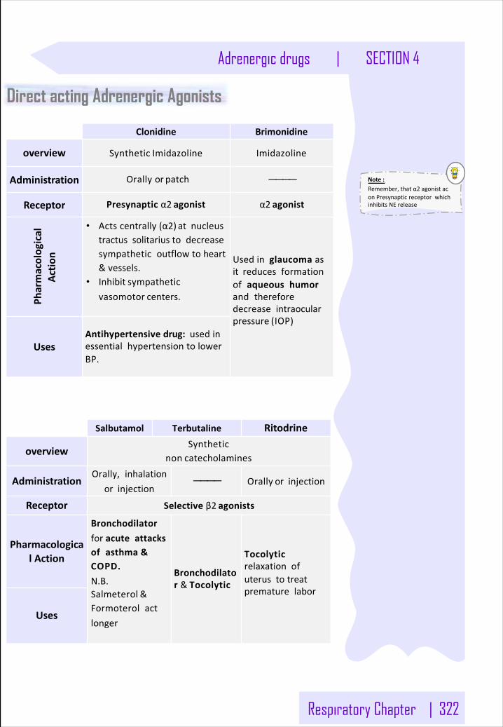
Respiratory Chapter | 322
Adrenergic drugs | SECTION 4
Direct acting Adrenergic Agonists
Note:Remember,thatα2 agonistaconPresynapticreceptorwhichinhibitsNErelease
Salbutamol Terbutaline Ritodrine
overviewSynthetic
non catecholamines
AdministrationOrally,inhalation
or injection———— Orally orinjection
Receptor Selectiveβ2 agonists
PharmacologicalAction
Bronchodilatorforacuteattacksofasthma&COPD.N.B.Salmeterol &Formoterolactlonger
Bronchodilator&Tocolytic
Tocolyticrelaxationofuterustotreatprematurelabor
Uses
Clonidine Brimonidine
overview Synthetic Imidazoline Imidazoline
Administration Orallyor patch ————
Receptor Presynapticα2 agonist α2 agonist
Pharmacolog
ical
Actio
n
• Actscentrally(α2) atnucleustractussolitariustodecreasesympatheticoutflowtoheart&vessels.
• Inhibit sympatheticvasomotor centers.
Usedinglaucomaasitreducesformationofaqueoushumorandthereforedecreaseintraocularpressure (IOP)
UsesAntihypertensivedrug:usedinessentialhypertensiontolowerBP.
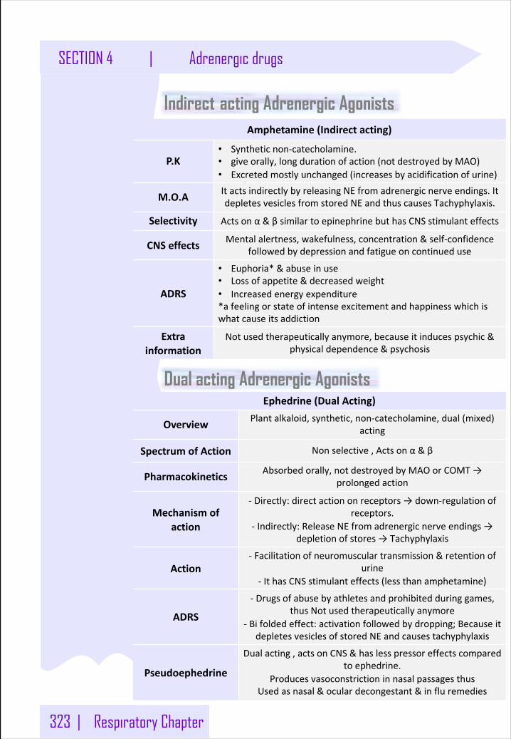
323 | Respiratory Chapter
SECTION 4 | Adrenergic drugs
Amphetamine(Indirectacting)
P.K• Syntheticnon-catecholamine.• giveorally,longdurationofaction(notdestroyedbyMAO)• Excretedmostlyunchanged(increasesbyacidificationofurine)
M.O.A ItactsindirectlybyreleasingNEfromadrenergicnerveendings.ItdepletesvesiclesfromstoredNEandthuscausesTachyphylaxis.
Selectivity Actsonα&βsimilartoepinephrinebuthasCNSstimulanteffects
CNSeffects Mentalalertness,wakefulness,concentration&self-confidencefollowedbydepressionandfatigueoncontinueduse
ADRS
• Euphoria*&abuseinuse• Lossofappetite&decreasedweight• Increasedenergyexpenditure*afeelingorstateofintenseexcitementandhappinesswhichiswhatcauseitsaddiction
Extrainformation
Notusedtherapeuticallyanymore,becauseitinducespsychic&physicaldependence&psychosis
Ephedrine(DualActing)
Overview Plantalkaloid,synthetic,non-catecholamine,dual(mixed)acting
SpectrumofAction Nonselective,Actsonα&β
Pharmacokinetics Absorbedorally,notdestroyedbyMAOorCOMT→prolongedaction
Mechanismofaction
- Directly:directactiononreceptors→down-regulationofreceptors.
- Indirectly:ReleaseNEfromadrenergicnerveendings→depletionofstores→Tachyphylaxis
Action- Facilitationofneuromusculartransmission&retentionof
urine- IthasCNSstimulanteffects(lessthanamphetamine)
ADRS
- Drugsofabusebyathletesandprohibitedduringgames,thusNotusedtherapeuticallyanymore
- Bifoldedeffect:activationfollowedbydropping;BecauseitdepletesvesiclesofstoredNEandcausestachyphylaxis
Pseudoephedrine
Dualacting,actsonCNS&haslesspressoreffectscomparedtoephedrine.
ProducesvasoconstrictioninnasalpassagesthusUsedasnasal&oculardecongestant&influremedies
Indirect acting Adrenergic Agonists
Dual acting Adrenergic Agonists
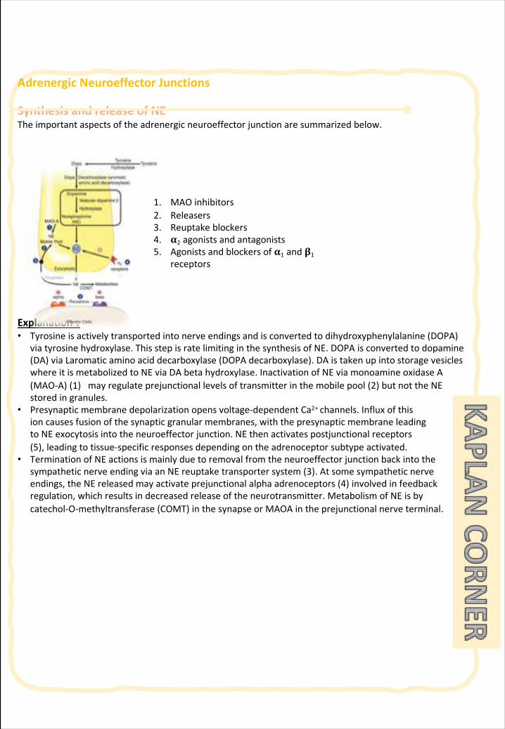
AdrenergicNeuroeffectorJunctions
SynthesisandreleaseofNETheimportantaspectsoftheadrenergicneuroeffectorjunctionaresummarizedbelow.
Explanation : • Tyrosineisactivelytransportedintonerveendingsandisconvertedtodihydroxyphenylalanine (DOPA)
viatyrosinehydroxylase.ThisstepisratelimitinginthesynthesisofNE.DOPAisconvertedtodopamine(DA)viaLaromatic aminoaciddecarboxylase(DOPAdecarboxylase).DAistakenupintostoragevesicleswhereitismetabolizedtoNEviaDAbetahydroxylase.InactivationofNEviamonoamineoxidaseA(MAO-A)(1)mayregulateprejunctional levelsoftransmitterinthemobilepool(2)butnottheNEstoredingranules.
• Presynapticmembranedepolarizationopensvoltage-dependentCa2+channels.Influxofthis ioncausesfusionofthesynapticgranularmembranes,withthepresynapticmembraneleadingtoNEexocytosisintotheneuroeffectorjunction.NEthenactivatespostjunctional receptors(5),leadingtotissue-specificresponsesdependingontheadrenoceptorsubtypeactivated.
• TerminationofNEactionsismainlyduetoremovalfromtheneuroeffectorjunctionbackintothesympatheticnerveendingviaanNEreuptaketransportersystem(3).Atsomesympatheticnerveendings,theNEreleasedmayactivateprejunctional alphaadrenoceptors(4)involvedinfeedbackregulation,whichresultsindecreasedreleaseoftheneurotransmitter.MetabolismofNEisbycatechol-O-methyltransferase(COMT)inthesynapseorMAOAintheprejunctional nerveterminal.
1. MAOinhibitors2. Releasers3. Reuptakeblockers4. 𝛂2 agonistsandantagonists5. Agonistsandblockersof𝛂1 and𝛃1
receptors
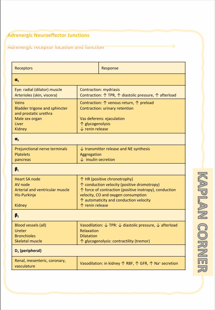
AdrenergicNeuroeffectorJunctions
Adrenergicreceptorlocationandfunction
Receptors Response
𝛂1
Eye:radial(dilator) muscleArterioles(skin,viscera)
Contraction:mydriasisContraction:↑TPR,↑diastolicpressure, ↑afterload
VeinsBladdertrigoneandsphincterandprostaticurethraMale sexorganLiverKidney
Contraction:↑venousreturn,↑preloadContraction:urinaryretention
Vasdeferens:ejaculation↑glycogenolysis↓reninrelease
𝛂2
Prejunctional nerveterminalsPlateletspancreas
↓transmitterreleaseandNEsynthesisAggregation↓insulinsecretion
𝛃1
HeartSAnodeAVnodeArterial andventricularmuscleHis-Purkinje
Kidney
↑HR(positivechronotrophy)↑conductionvelocity(positivedromotropy)↑forceofcontraction(positiveinotropy),conductionvelocity,COandoxygenconsumption↑automaticityandconductionvelocity↑reninrelease
𝛃2
Bloodvessels(all)UreterBronchiolesSkeletalmuscle
Vasodilation:↓TPR: ↓diastolicpressure, ↓afterloadRelaxationDilatation↑glycogenolysis: contractility(tremor)
D1 (peripheral)
Renal,mesenteric, coronary,vasculature
Vasodilation:inkidney↑RBF,↑GFR,↑Na+ secretion
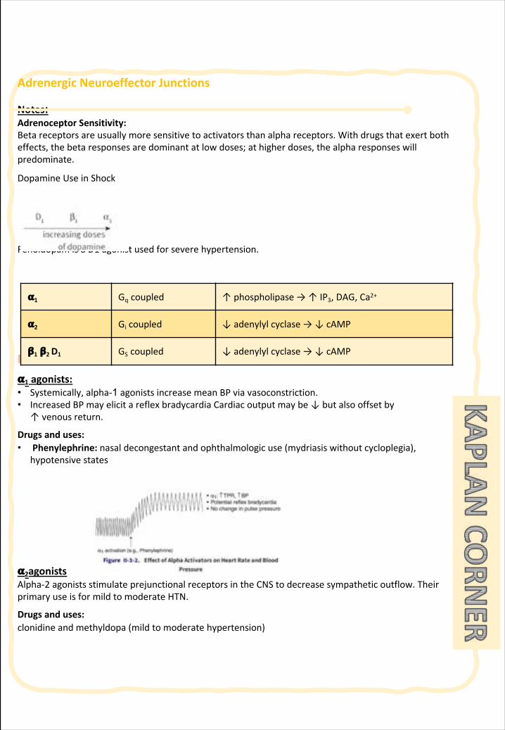
AdrenergicNeuroeffectorJunctions
Notes:AdrenoceptorSensitivity:Betareceptorsareusuallymoresensitivetoactivatorsthanalphareceptors.Withdrugsthatexertbotheffects,thebetaresponsesaredominantatlowdoses;athigherdoses,thealpharesponseswillpredominate.
DopamineUseinShock
Fenoldopam isaD1 agonistusedforseverehypertension.
Direct-actingadrenoceptoragonists 𝛂1 agonists:• Systemically,alpha-1 agonistsincreasemeanBPviavasoconstriction.• IncreasedBPmayelicitareflexbradycardiaCardiacoutputmaybe↓butalsooffsetby
↑venousreturn.
Drugsanduses:• Phenylephrine:nasaldecongestantandophthalmologicuse(mydriasiswithoutcycloplegia),
hypotensivestates
𝛂2agonistsAlpha-2 agonistsstimulateprejunctional receptorsintheCNStodecreasesympatheticoutflow.TheirprimaryuseisformildtomoderateHTN.
Drugsanduses:clonidineandmethyldopa(mildtomoderatehypertension)
𝛂1 Gq coupled ↑phospholipase→↑IP3,DAG,Ca2+
𝛂2 Gi coupled ↓adenylylcyclase→↓cAMP
𝛃1 𝛃2 D1 GS coupled ↓adenylylcyclase→↓cAMP
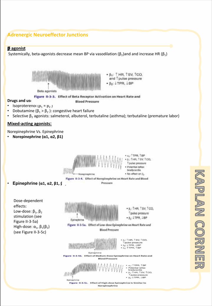
AdrenergicNeuroeffectorJunctions
𝛃 agonistSystemically,beta-agonistsdecreasemeanBPviavasodilation(β2)andandincreaseHR(β1)
Drugsanduses:• Isoproterenol(β1 =β2 )• Dobutamine (β1 >β2 ):congestiveheartfailure• Selectiveβ2 agonists:salmeterol,albuterol,terbutaline(asthma);terbutaline(prematurelabor)
Mixed-actingagonists:NorepinephrineVs.Epinephrine• Norepinephrine(α1,α2,β1)
• Epinephrine (α1,α2,β1,β2)
Dose-dependenteffects:Low-dose:β1,β2stimulation(seeFigureII-3-5a)High-dose:α1,β1(β2)(seeFigureII-3-5c)
AdrenergicNeuroeffectorJunctions
β2-specificeffectsSmoothmusclerelaxation:bronchioles,uterus,bloodvessels
Metaboliceffects:• ↑glycogenolysis (muscleandliver)• ↑gluconeogenesis• ↑mobilizationanduseoffat
Differentiationofhigh-doseepinephrineversusnorepinephrine:Epinephrinereversal:Useofα1 blockertoreversehypertensiontohypotensioninapatientreceivingtoomuchepinephrineHypertensionwasduetopredominantα1 toneonthevasculatureHypotensionresultsfromunmaskingβ2 receptors
Usesofnorepinephrineandepinephrine• Cardiacarrest• Adjuncttolocalanesthetic• Hypotension• Anaphylaxis(epinephrineonly)• Asthma(epinephrineonly)
Indirect-actingadrenergicreceptoragonists
RELEASERSReleasersdisplacenorepinephrinefromthemobilepool.• Releasersdisplacenorepinephrinefromthemobilepool• Druginteraction:MAOA inhibitors(hypertensivecrisis)• Tyramine(redwine,cheese)OralbioavailabilityislimitedbyMAO-Ametabolismingutand
liverMAO-Ainhibition↑bioavailability,resultinginhypertensivecrisis• Amphetamine:ClinicaluseofmethylphenidateinnarcolepsyandADHDPsychostimulant
duetocentralreleaseofDA,NE,5HT• Ephedrine(coldmedication)
Clinicalcorrelate:• Indirect-actingadrenoceptoragonistsactonlyoneffectortissuesinnervatedbySANS.• Denervated effectortissuesarenonresponsivebecausethesedrugsacteithertoreleasetransmitter
fromnerveterminalsortoinhibitneurotransmitterreuptake.
FormsofMAO:MAOtypeA:mainlyinliver,butAnywhere(metabolizesNE,5HT,andtyramine)MAOtypeB:mainlyin Brain(metabolizesDA)
ReuptakeInhibitors
• Cocaine• Tricyclicantidepressant(inpart)
AdrenergicAntagonists
αreceptorantagonistsAlpha-receptorantagonistsdecreaseTPRanddecreasemeanBP.• Maycausereflextachycardiaandsaltandwaterretention• Majoruses:
o Hypertensiono Pheochromocytoma (nonselectiveαblocker)o Benignprostatichyperplasia(BPH;selectiveα1 blocker)
Drugs:• Nonselectiveblocker:phentolamine (competitiveinhibitor)• phenoxybenzamine (noncompetitiveinhibitor)• Selectiveα1 blocker:prazosin,doxazosin,terazosin,tamsulosin• Selectiveα2 blocker:mirtazapine(usedasantidepressant)
βreceptorantagonists• β1 blockade
o ↓HR,↓SV,↓COo ↓reninrelease
• β2 blockadeo Mayprecipitatebronchospasm(inasthmatics)andvasospasm(inpatientswith
vasospasticdisorders)o ↓aqueoushumorproduction
Metaboliceffects• Blocksglycogenolysis,gluconeogenesis• ↑LDLs,TGs
ClinicalcorrelateChronicuseofbetablockers(e.g.,inangina,HTN)leadstoreceptorupregulation.Duringwithdrawalfromuse,itisimportanttotaperdosetoavoidexcessivecardiovasculareffects(reboundeffects)ofendogenousamines.
ClinicalcorrelateGlucagonandtheHeartPositiveinotropicandchronotropic,notviaactivationofβ1 receptors,butthroughglucagonreceptorsthatareG-proteinlinkedtoadenylylcyclase→basisforitsuseinbeta-blockeroverdose.
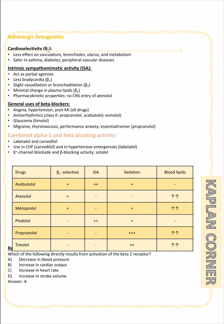
AdrenergicAntagonists
Cardioselectivity (β1):• Lesseffectonvasculature,bronchioles,uterus,andmetabolism• Saferinasthma,diabetes,peripheralvasculardiseases
Intrinsicsympathomimeticactivity(ISA):• Actaspartialagonists• Lessbradycardia(β1)• Slightvasodilationorbronchodilation(β2)• Minimalchangeinplasmalipids(β2)• Pharmacokineticproperties:noCNSentryofatenolol
Generalusesofbeta-blockers:• Angina,hypertension,post-MI(alldrugs)• Antiarrhythmics (classII:propranolol,acebutolol,esmolol)• Glaucoma(timolol)• Migraine,thyrotoxicosis,performanceanxiety,essentialtremor (propranolol)
Combinedalpha-1 andbetablockingactivity:• Labetalolandcarvedilol• UseinCHF(carvedilol)andinhypertensiveemergencies(labetalol)• K+-channelblockadeandβ-blockingactivity:sotalol
RecallQuestionWhichofthefollowingdirectlyresultsfromactivationofthebeta2 receptor?A) DecreaseinbloodpressureB) IncreaseincardiacoutputC) IncreaseinheartrateD) IncreaseinstrokevolumeAnswer:A
Drugs β1 -selective ISA Sedation Bloodlipids
Acebutolol + ++ + -
Atenolol + - - ↑↑
Metoprolol + - + ↑↑
Pindolol - ++ + -
Propranolol - - +++ ↑↑
Timolol - - ++ ↑↑
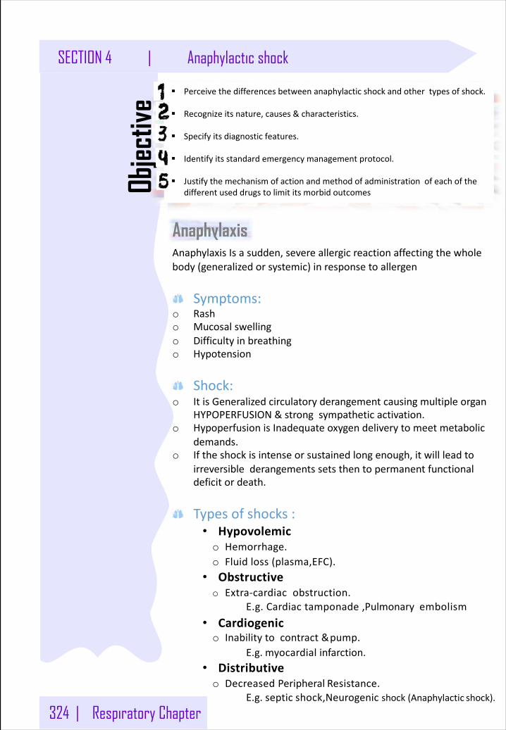
324 | Respiratory Chapter
SECTION 4 | Anaphylactic shock
§ Perceivethedifferencesbetweenanaphylacticshockandothertypesofshock.
§ Recognizeitsnature,causes&characteristics.
§ Specifyitsdiagnosticfeatures.
§ Identifyitsstandardemergencymanagementprotocol.
§ JustifythemechanismofactionandmethodofadministrationofeachofthedifferentuseddrugstolimititsmorbidoutcomesOb
jectiv
e
AnaphylaxisAnaphylaxisIsasudden,severeallergicreactionaffectingthewholebody(generalizedorsystemic)inresponsetoallergen
Symptoms:o Rasho Mucosalswellingo Difficultyinbreathingo Hypotension
Shock:o ItisGeneralizedcirculatoryderangementcausingmultipleorgan
HYPOPERFUSION&strongsympatheticactivation.o HypoperfusionisInadequateoxygendeliverytomeetmetabolic
demands.o Iftheshockisintenseorsustainedlongenough,itwillleadto
irreversiblederangementssetsthentopermanentfunctionaldeficitordeath.
Typesofshocks:• Hypovolemico Hemorrhage.o Fluidloss(plasma,EFC).
• Obstructiveo Extra-cardiacobstruction.
E.g.Cardiactamponade,Pulmonaryembolism• Cardiogenico Inabilitytocontract&pump.
E.g. myocardial infarction.• Distributiveo DecreasedPeripheral Resistance.
E.g.septic shock,Neurogenic shock(Anaphylactic shock).
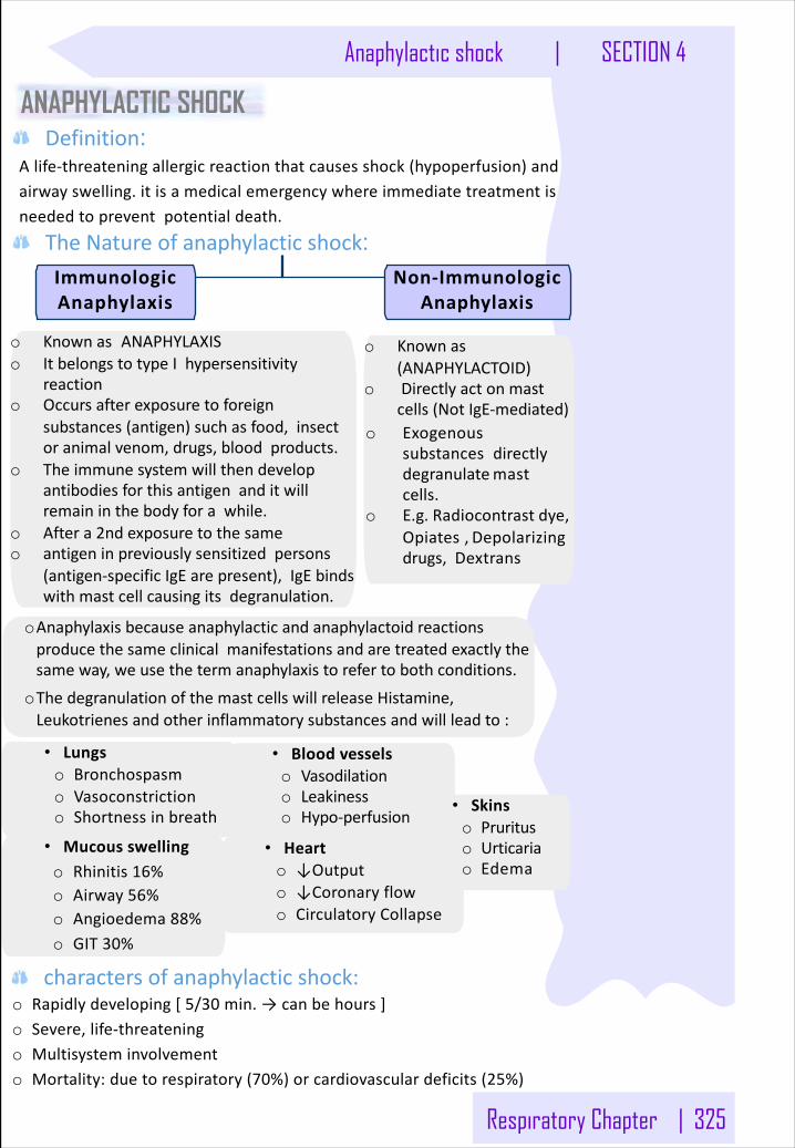
Respiratory Chapter | 325
Anaphylactic shock | SECTION 4
ANAPHYLACTIC SHOCK Definition:
Alife-threateningallergicreactionthatcausesshock(hypoperfusion)andairwayswelling.itisamedicalemergencywhereimmediatetreatmentisneededtopreventpotentialdeath.
TheNatureofanaphylacticshock:
o Knownas ANAPHYLAXISo ItbelongstotypeIhypersensitivity
reactiono Occursafterexposuretoforeign
substances(antigen)suchasfood,insectoranimalvenom,drugs,bloodproducts.
o Theimmunesystemwillthendevelopantibodiesforthisantigenanditwillremaininthebodyforawhile.
o Aftera2ndexposuretothesameo antigeninpreviouslysensitizedpersons
(antigen-specificIgE arepresent),IgE bindswithmastcellcausingitsdegranulation.
o Knownas(ANAPHYLACTOID)
o Directlyactonmastcells(NotIgE-mediated)
o Exogenoussubstancesdirectlydegranulatemastcells.
o E.g.Radiocontrast dye,Opiates,Depolarizingdrugs,Dextrans
charactersofanaphylacticshock: o Rapidlydeveloping[5/30 min.→canbehours]o Severe,life-threateningo Multisysteminvolvemento Mortality:duetorespiratory(70%)orcardiovasculardeficits(25%)
oAnaphylaxisbecauseanaphylacticandanaphylactoidreactionsproducethesameclinicalmanifestationsandaretreatedexactlythesameway,weusethetermanaphylaxistorefertobothconditions.
oThedegranulationofthemastcellswillreleaseHistamine,Leukotrienesandotherinflammatorysubstancesandwillleadto:
• Lungso Bronchospasmo Vasoconstrictiono Shortnessinbreath
• Bloodvesselso Vasodilationo Leakinesso Hypo-perfusion
• Mucousswellingo Rhinitis16%o Airway56%o Angioedema88%o GIT30%
• Hearto ↓Outputo ↓Coronaryflowo CirculatoryCollapse
• Skinso Prurituso Urticariao Edema
ImmunologicAnaphylaxis
Non-ImmunologicAnaphylaxis
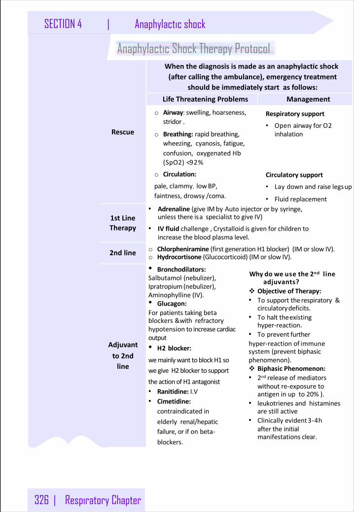
326 | Respiratory Chapter
SECTION 4 | Anaphylactic shock
Rescue
Whenthediagnosisismadeasananaphylacticshock(aftercallingtheambulance),emergencytreatment
shouldbeimmediatelystartasfollows:LifeThreateningProblems Management
o Airway:swelling,hoarseness,stridor.
o Breathing:rapidbreathing,wheezing,cyanosis,fatigue,confusion,oxygenatedHb(SpO2)<92%
Respiratory support
• OpenairwayforO2inhalation
o Circulation:
pale,clammy,lowBP,faintness,drowsy /coma.
Circulatory support
• Laydownandraiselegsup
• Fluid replacement
1stLineTherapy
• Adrenaline(giveIMbyAutoinjectororbysyringe,unlessthereisaspecialisttogive IV)
• IV fluid challenge ,Crystalloid isgiven for children toincrease the blood plasma level.
2ndline o Chlorpheniramine (firstgenerationH1 blocker)(IMorslowIV).o Hydrocortisone (Glucocorticoid)(IMorslowIV).
Adjuvantto2ndline
• Bronchodilators:Salbutamol(nebulizer),Ipratropium(nebulizer),Aminophylline (IV).• Glucagon:Forpatientstakingbetablockers &withrefractoryhypotension toincreasecardiacoutput• H2 blocker:
wemainlywanttoblockH1 sowegiveH2 blockertosupporttheactionofH1 antagonist• Ranitidine: I.V• Cimetidine:
contraindicatedinelderlyrenal/hepaticfailure,orifon beta-blockers.
Why do we use the 2nd lineadjuvants?
v Objectiveof Therapy:• Tosupporttherespiratory&
circulatorydeficits.• Tohalttheexisting
hyper-reaction.• Toprevent furtherhyper-reactionof immunesystem(preventbiphasicphenomenon).v Biphasic Phenomenon:• 2ndreleaseofmediators
withoutre-exposuretoantigeninupto20%).
• leukotrienesandhistaminesarestillactive
• Clinicallyevident3-4haftertheinitialmanifestations clear.
Anaphylactic Shock Therapy Protocol
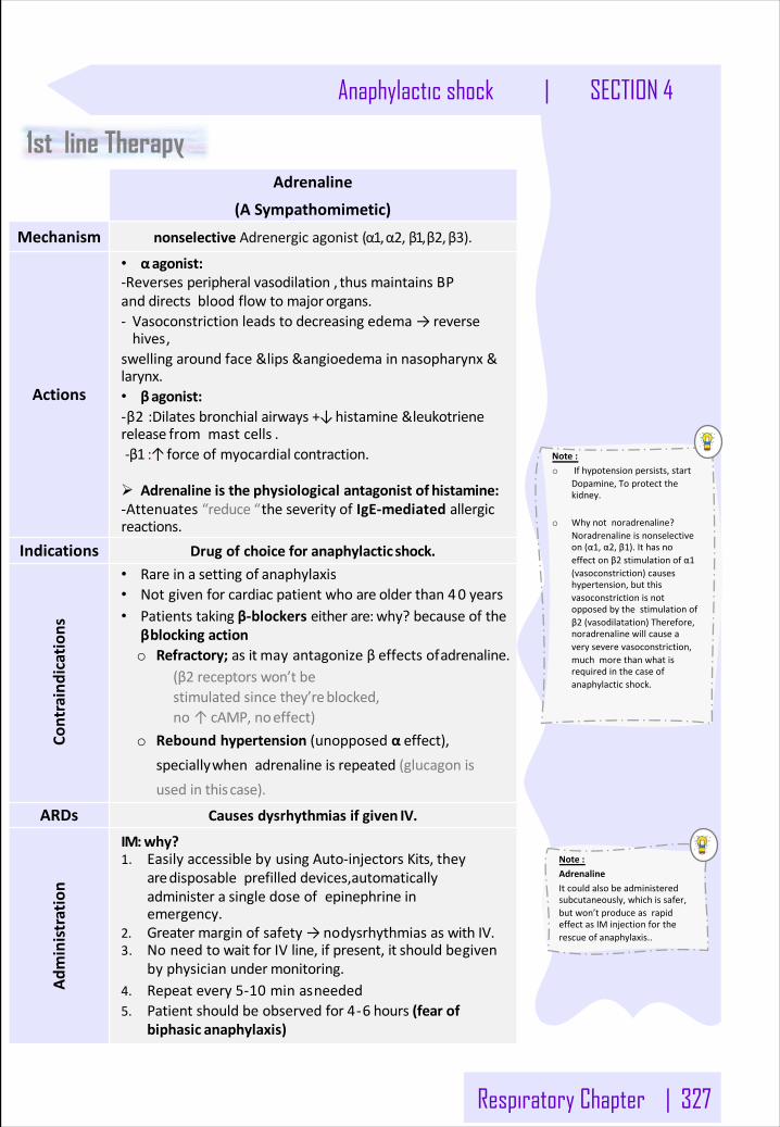
Respiratory Chapter | 327
Anaphylactic shock | SECTION 4
1st line TherapyAdrenaline
(ASympathomimetic)
Mechanism nonselectiveAdrenergicagonist(α1,α2,β1,β2,β3).
Actions
• αagonist:-Reversesperipheralvasodilation,thusmaintainsBPand directsbloodflowtomajororgans.- Vasoconstrictionleadstodecreasingedema→reversehives,
swellingaroundface&lips&angioedemainnasopharynx&larynx.• βagonist:-β2 :Dilatesbronchialairways+↓histamine&leukotrienerelease frommastcells .-β1 :↑forceofmyocardial contraction.
Ø Adrenalineisthephysiologicalantagonistof histamine:-Attenuates“reduce“theseverityofIgE-mediatedallergicreactions.
Indications Drugofchoiceforanaphylactic shock.
Contraindicatio
ns
• Rareinasettingof anaphylaxis• Notgivenforcardiacpatientwhoareolderthan40 years• Patientstakingβ-blockerseitherare:why?becauseofthe
βblockingactiono Refractory;asitmayantagonizeβeffectsofadrenaline.
(β2 receptorswon’tbestimulatedsince they’reblocked,no↑cAMP,noeffect)
o Reboundhypertension(unopposedαeffect),speciallywhenadrenalineisrepeated(glucagonisusedinthiscase).
ARDs Causesdysrhythmiasifgiven IV.
Administration
IM:why?1. EasilyaccessiblebyusingAuto-injectorsKits,they
aredisposableprefilleddevices,automaticallyadministerasingledoseofepinephrineinemergency.
2. Greatermarginofsafety→nodysrhythmiasaswithIV.3. NoneedtowaitforIVline,ifpresent,itshouldbegiven
by physicianundermonitoring.4. Repeatevery5-10 minasneeded5. Patientshouldbeobservedfor4-6 hours (fearof
biphasic anaphylaxis)
Note:o Ifhypotensionpersists,start
Dopamine,Toprotectthekidney.
o Whynotnoradrenaline?Noradrenalineisnonselectiveon(α1,α2,β1).Ithasnoeffectonβ2 stimulationofα1(vasoconstriction)causeshypertension,butthisvasoconstrictionisnotopposedbythestimulationofβ2 (vasodilatation)Therefore,noradrenalinewillcauseaveryseverevasoconstriction,muchmorethanwhatisrequiredinthecaseofanaphylacticshock.
Note:AdrenalineItcouldalsobeadministeredsubcutaneously,whichissafer,butwon’tproduceasrapideffectasIMinjectionfortherescueofanaphylaxis..
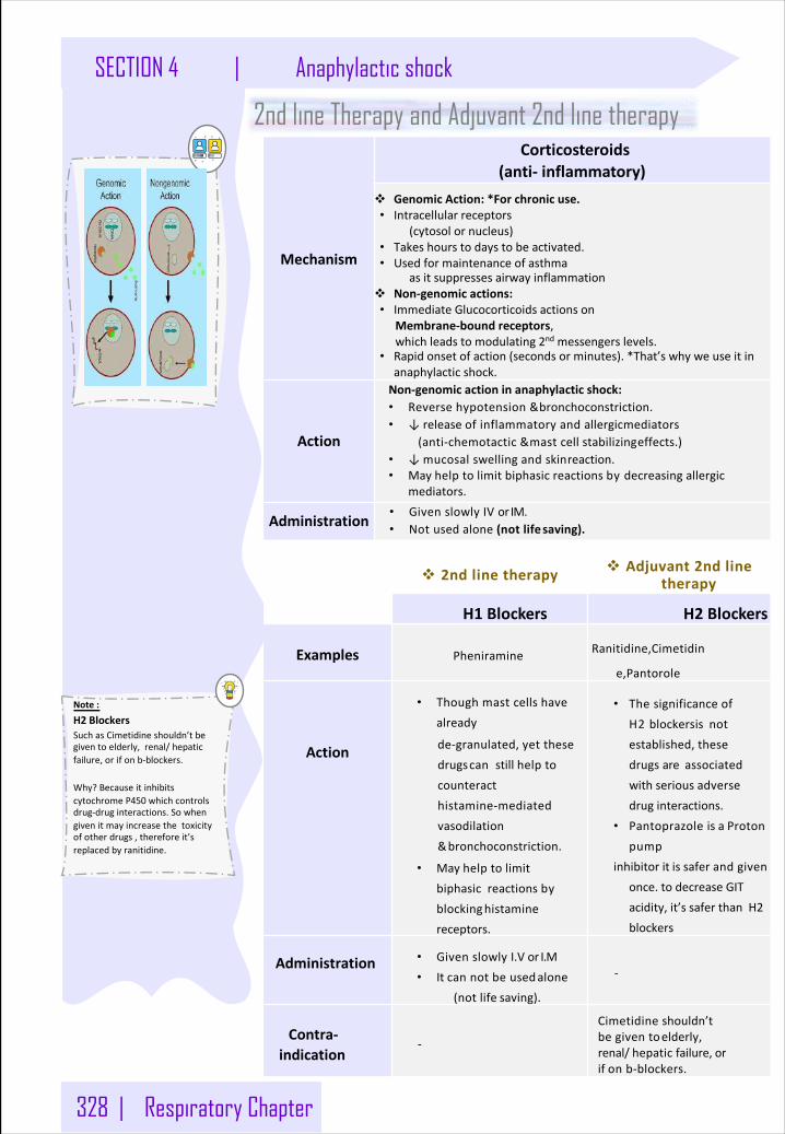
328 | Respiratory Chapter
SECTION 4 | Anaphylactic shock
2nd line Therapy and Adjuvant 2nd line therapy
Mechanism
Corticosteroids(anti- inflammatory)
v GenomicAction:*Forchronicuse.• Intracellularreceptors
(cytosolornucleus)• Takeshourstodaystobeactivated.• Usedformaintenanceofasthma
asitsuppressesairwayinflammationv Non-genomicactions:• ImmediateGlucocorticoidsactionson
Membrane-boundreceptors,whichleadstomodulating2nd messengerslevels.
• Rapidonsetofaction(seconds orminutes).*That’swhyweuseitinanaphylacticshock.
Action
Non-genomicactioninanaphylacticshock:• Reversehypotension&bronchoconstriction.• ↓releaseofinflammatoryandallergicmediators
(anti-chemotactic&mastcellstabilizingeffects.)• ↓mucosalswellingandskinreaction.• Mayhelptolimitbiphasicreactionsbydecreasingallergic
mediators.
Administration• GivenslowlyIVor IM.• Notusedalone(notlife saving).
v 2ndlinetherapy v Adjuvant2ndlinetherapy
H1 Blockers H2 Blockers
Examples Pheniramine Ranitidine,Cimetidin
e,Pantorole
Action
• Thoughmastcellshavealready
de-granulated,yetthesedrugscanstillhelptocounteracthistamine-mediatedvasodilation&bronchoconstriction.
• Mayhelptolimitbiphasicreactionsbyblockinghistaminereceptors.
• ThesignificanceofH2 blockersisnotestablished,thesedrugsareassociatedwithseriousadversedrug interactions.
• PantoprazoleisaProtonpump
inhibitoritissaferandgivenonce.todecreaseGITacidity,it’ssaferthanH2blockers
Administration • GivenslowlyI.Vor I.M• Itcannotbeusedalone
(notlife saving).
-
Contra-indication
-
Cimetidineshouldn’tbegiventoelderly,renal/hepaticfailure,orifon b-blockers.
Note:H2 BlockersSuchasCimetidineshouldn’tbegiventoelderly,renal/hepaticfailure,orifonb-blockers.
Why?BecauseitinhibitscytochromeP450 whichcontrolsdrug-druginteractions.Sowhengivenitmayincreasethetoxicityofotherdrugs,thereforeit’sreplacedbyranitidine.
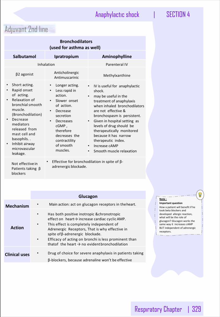
Respiratory Chapter | 329
Anaphylactic shock | SECTION 4
Adjuvant 2nd line
Note:ImportantquestionHowapatientwillbenefitifhetookbetablockersanddevelopedallergicreaction,whatwillbetheroleofglucagon?GlucagonworksthesamewayitincreasescAMPBUTindependentofadrenergicreceptors.
Bronchodilators(usedforasthmaaswell)
Salbutamol Ipratropium Aminophylline
Inhalation Parenteral IV
β2 agonist AnticholinergicAntimuscarinic Methylxanthine
• Effectiveforbronchodilationinspiteofβ-adrenergic blockade.
Glucagon
Mechanism • Mainaction:actonglucagonreceptorsintheheart.
Action
• Hasbothpositiveinotropic&chronotropiceffect onheart→increasecardiaccyclic AMP.
• ThiseffectiscompletelyindependentofAdrenergicReceptors,Thatiswhyeffectiveinspiteofβ-adrenergicblockade.
• Efficacyofactingonbronchiislessprominentthanthatoftheheart→noevidentbronchodilation
Clinicaluses • Drugofchoiceforsevereanaphylaxisinpatientstaking
β-blockers,becauseadrenalinewon’tbe effective
• IVisusefulforanaphylacticshock.
• maybeusefulinthetreatmentofanaphylaxiswheninhaledbronchodilatorsarenoteffective&bronchospasmispersistent.
• Giveninhospitalsettingaslevelsofdrugshouldbetherapeuticallymonitoredbecauseithasnarrowtherapeuticindex.
• IncreasecAMP• Smoothmusclerelaxation
• Longeracting.• Lessrapidin
action.• Sloweronset
ofaction.• Decrease
secretion• Decreases
cGMP,thereforedecreasesthecontractilityofsmoothmuscles.
• Short acting.• Rapidonset
ofacting.• Relaxationof
bronchial smoothmuscle.(Bronchodilation)
• Decreasemediatorsreleasedfrommastcellandbasophils. .
• Inhibitairwaymicrovascularleakage.
Noteffective inPatientstakingβblockers
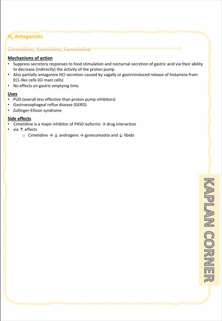
H2 Antagonists
Cimetidine,Ranitidine,FamotidineMechanismsofaction• Suppresssecretoryresponsestofoodstimulationandnocturnalsecretionofgastricacidviatheirability
todecrease(indirectly)theactivityoftheprotonpump.• AlsopartiallyantagonizeHClsecretioncausedbyvagallyorgastrininducedreleaseofhistaminefrom
ECL-likecells(GImastcells)• Noeffectsongastricemptyingtime
Uses• PUD(overalllesseffectivethanprotonpumpinhibitors)• Gastroesophagealrefluxdisease(GERD)• Zollinger-Ellisonsyndrome
Sideeffects• CimetidineisamajorinhibitorofP450 isoforms→druginteraction• via↑effects
o Cimetidine→↓androgens→gynecomastiaand↓libido
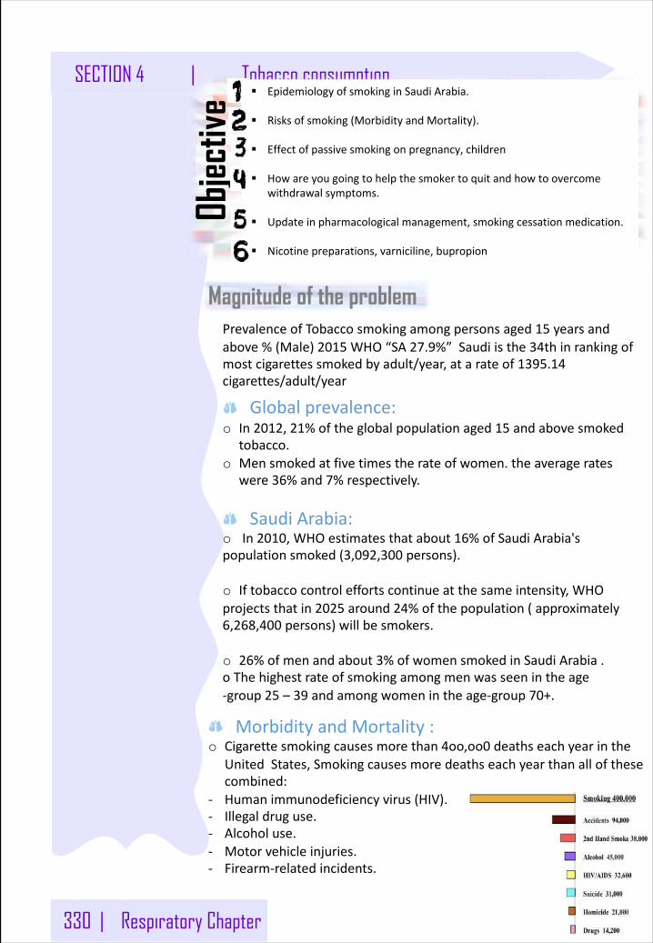
330 | Respiratory Chapter
SECTION 4 | Tobacco consumption
Magnitude of the problem
Objec
tive
PrevalenceofTobaccosmokingamongpersonsaged15 yearsandabove%(Male)2015 WHO“SA27.9%”Saudiisthe34thinrankingofmostcigarettessmokedbyadult/year,atarateof1395.14cigarettes/adult/year
Globalprevalence:o In2012,21%oftheglobalpopulationaged15 andabovesmoked
tobacco.o Mensmokedatfivetimestherateofwomen.theaveragerates
were36%and7%respectively.
SaudiArabia:o In2010,WHOestimatesthatabout16%ofSaudiArabia'spopulationsmoked(3,092,300 persons).
o Iftobaccocontroleffortscontinueatthesameintensity,WHOprojectsthatin2025 around24%ofthepopulation(approximately6,268,400 persons)willbesmokers.
o 26%ofmenandabout3%ofwomensmokedinSaudiArabia.oThehighestrateofsmokingamongmenwasseenintheage-group25 – 39 andamongwomenintheage-group70+.
§ EpidemiologyofsmokinginSaudiArabia.
§ Risksofsmoking(MorbidityandMortality).
§ Effectofpassivesmokingonpregnancy,children
§ Howareyougoingtohelpthesmokertoquitandhowtoovercomewithdrawalsymptoms.
§ Updateinpharmacologicalmanagement,smokingcessationmedication.
§ Nicotinepreparations,varniciline,bupropion
MorbidityandMortality:o Cigarettesmokingcausesmorethan4oo,oo0 deathseachyearinthe
UnitedStates,Smokingcausesmoredeathseachyearthanallofthesecombined:
- Humanimmunodeficiencyvirus(HIV).- Illegaldruguse.- Alcoholuse.- Motorvehicleinjuries.- Firearm-relatedincidents.
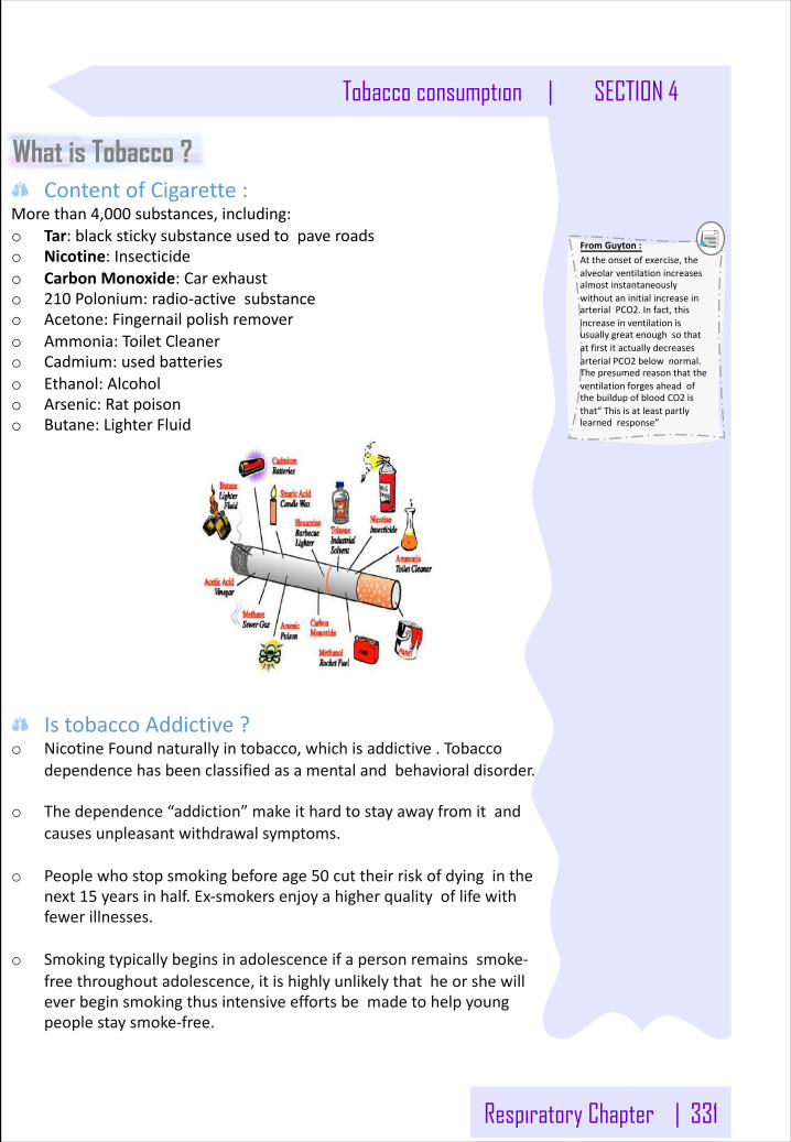
Respiratory Chapter | 331
Tobacco consumption | SECTION 4
FromGuyton:Attheonsetofexercise,thealveolarventilationincreasesalmostinstantaneouslywithoutaninitialincreaseinarterialPCO2.Infact,thisincreaseinventilationisusuallygreatenoughsothatatfirstitactuallydecreasesarterialPCO2 belownormal.ThepresumedreasonthattheventilationforgesaheadofthebuildupofbloodCO2 isthat“Thisisatleastpartlylearnedresponse”
ContentofCigarette:Morethan4,000 substances,including:o Tar:blackstickysubstanceusedtopaveroadso Nicotine:Insecticideo CarbonMonoxide:Carexhausto 210 Polonium:radio-activesubstanceo Acetone:Fingernailpolishremovero Ammonia:ToiletCleanero Cadmium:usedbatterieso Ethanol:Alcoholo Arsenic:Ratpoisono Butane:LighterFluid
IstobaccoAddictive?o NicotineFoundnaturallyintobacco,whichisaddictive.Tobacco
dependencehasbeenclassifiedasamentalandbehavioraldisorder.
o Thedependence“addiction”makeithardtostayawayfromitandcausesunpleasantwithdrawalsymptoms.
o Peoplewhostopsmokingbeforeage50 cuttheirriskofdyinginthenext15 yearsinhalf.Ex-smokersenjoyahigherqualityoflifewithfewerillnesses.
o Smokingtypicallybeginsinadolescenceifapersonremainssmoke-freethroughoutadolescence,itishighlyunlikelythatheorshewilleverbeginsmokingthusintensiveeffortsbemadetohelpyoungpeoplestaysmoke-free.
What is Tobacco ?
332 | Respiratory Chapter
SECTION 4 | Tobacco consumption
Smoking: Whatisit?:
Itreferstotheinhalationandexhalationoffumesfromburningtobaccoincigars,cigarettesandpipe.
Waysofsmoking:
o Cigarettes:areuniforminsizeandcontainlessthan1goftobaccoeach.
o Cigar:arecomposedprimarilyofasingletypeoftobacco,andtheyhaveatobaccowrapper.Theycontainbetween1 gramand20 gramsoftobacco.
o ElectronicCigarette:Itisabattery-poweredvaporizerwhichhasasimilarfeeltotobaccosmoking,butdonotcontaintobacco,althoughtheydousenicotinefromtobaccoplants.Theydonotproducecigarettesmokebutratheranaerosol,whichisreferredtoasvapor.
o Water-Pipe(Shisha):Notsaferthanregulartobaccosmoke, CausesthesamediseasesbutmorePolycythemia whichisincreaseinRBCsandHemoglobin. ItRaisestheriskoflipcancer,spreadinginfectionslikeTuberculosis.Userscaninhalethesameamountofsmokeasfrommorethan100 cigarettes.
Types:
o Active(Conventionalsmoking).o Passive(Secondhandsmoking).o Thirdhandsmoking.
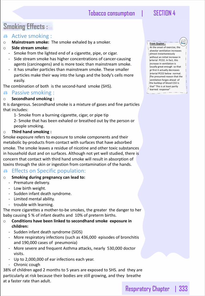
Respiratory Chapter | 333
Tobacco consumption | SECTION 4
FromGuyton:Attheonsetofexercise,thealveolarventilationincreasesalmostinstantaneouslywithoutaninitialincreaseinarterialPCO2.Infact,thisincreaseinventilationisusuallygreatenoughsothatatfirstitactuallydecreasesarterialPCO2 belownormal.ThepresumedreasonthattheventilationforgesaheadofthebuildupofbloodCO2 isthat“Thisisatleastpartlylearnedresponse”
Activesmoking:o Mainstreamsmoke:Thesmokeexhaledbyasmoker.o Sidestreamsmoke:
- Smokefromthelightedendofacigarette,pipe,orcigar.- Sidestreamsmokehashigherconcentrationsofcancer-causing
agents(carcinogens)andismoretoxicthanmainstreamsmoke.- ithassmallerparticlesthanmainstreamsmoke.Thesesmaller
particlesmaketheirwayintothelungsandthebody’scellsmoreeasily.
Thecombinationofbothisthesecond-handsmoke(SHS).Passivesmoking:
o Secondhandsmoking:Itisdangerous.Secondhandsmokeisamixtureofgasesandfineparticlesthatincludes:
1- Smokefromaburningcigarette,cigar,orpipetip2- Smokethathasbeenexhaledorbreathedoutbythepersonorpeoplesmoking.
o Thirdhandsmoking:Smokeexposurereferstoexposuretosmokecomponentsandtheirmetabolicby-productsfromcontactwithsurfacesthathaveadsorbedsmoke.Thesmokeleavesaresidueofnicotineandothertoxicsubstancesinhouseholddustandonsurfaces.Althoughnotyetwellstudied,thereisconcernthatcontactwiththirdhandsmokewillresultinabsorptionoftoxinsthroughtheskinoringestionfromcontaminationofthehands.
EffectsonSpecificpopulation:o Smokingduringpregnancycanleadto:
- Prematuredelivery. - Lowbirthweight. - Suddeninfantdeathsyndrome. - Limitedmentalability. - troublewithlearning.
Themorecigarettesamother-to-besmokes,thegreaterthedangertoherbabycausing5 %ofinfantdeathsand10%ofpretermbirths.o Conditionshavebeenlinkedtosecondhandsmokeexposurein
children:- Suddeninfantdeathsyndrome(SIDS)- Morerespiratoryinfections(suchas436,000 episodesofbronchitis
and190,000 casesofpneumonia)- MoresevereandfrequentAsthmaattacks,nearly530,000 doctor
visits.- Upto2,000,000 ofearinfectionseachyear.- Chroniccough
38%ofchildrenaged2 monthsto5 yearsareexposedtoSHS.andtheyareparticularlyatriskbecausetheirbodiesarestillgrowing,andtheybreatheatafasterratethanadult.
Smoking Effects :

334 | Respiratory Chapter
SECTION 4 | Tobacco consumption
Consequencesoftobaccouse:o Health(shortterm,longterm)o Economic(individual,family,community)o Social(family,community)o Development(community)o Religious(individual,community)o Prematuredeath
EffectonhealthIngeneral:Causesmorethan25 differentdiseases.Smokingcancausecanceralmostanywhereinyourbody.Affectsdifferentbody-systems,especially:- Gastro-intestinalsystem.- Respiratorytract.- Cardio-vascularsystem.- Urinarysystem.- Others,suchasSkin :wrinkles,prematurescaringandaging.Oro-dentalproblems:stainedteeth,guminflammation,blackhairytounge,oralcancer,Leukoplakia.FetalSmokingSyndrome:Ifthemotherweresmokerthebabywilldevelopthissyndromewhichinclude:(Birthdefects,Prematurestillbirth,Lowbirthweight,LoweredimmunecapacityandPronenesstoSuddenInfantDeathSyndrome(SIDS).
EffectofSmokingonRespiratory:o Laryngealcancer:Over80%ofdeathsfromlaryngealcancerarelinkedtosmoking,someofitsSymptoms:(Persistenthoarseness,Chronicsorethroat,Painfulswallowing,PainintheearandLumpintheneck.o Emphysemachronicbronchitis:ItssymptomsincludeoShortnessofbreath,chroniccough,Wheezing,anxiety,fatigue,weightloss,andswellingofankle,feetandleg.o Lungcancer:Killsmorepeoplethananyothertypeofcancer,Cigarettesmokingcausesmostcasesoflungcancerby25 times
EffectofSmokingonCardiovascular:o Arteriosclerosis&atherosclerosis.o Peripheralvasculardisease.o Heartattack:SmokersaretwiceaslikelyasNonsmokerstohaveaheartattack.Quittingsmokingrapidlyreducestheriskofcoronaryheartdisease.o Stroke:Whichcancausedeathorseverementalorphysicaldisability.
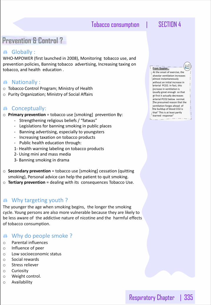
Respiratory Chapter | 335
Tobacco consumption | SECTION 4
FromGuyton:Attheonsetofexercise,thealveolarventilationincreasesalmostinstantaneouslywithoutaninitialincreaseinarterialPCO2.Infact,thisincreaseinventilationisusuallygreatenoughsothatatfirstitactuallydecreasesarterialPCO2 belownormal.ThepresumedreasonthattheventilationforgesaheadofthebuildupofbloodCO2 isthat“Thisisatleastpartlylearnedresponse”
Globally:WHO-MPOWER(firstlaunchedin2008),Monitoringtobaccouse,andpreventionpolicies,Banningtobaccoadvertising,Increasingtaxingontobacco,andhealtheducation.
Nationally:o TobaccoControlProgram;MinistryofHealtho PurityOrganization;MinistryofSocialAffairs
Conceptually:o Primaryprevention =tobaccouse[smoking]preventionBy:
- Strengtheningreligiousbeliefs/“fatwas”- Legislationsforbanningsmokinginpublicplaces- Banningadvertising,especiallytoyoungsters- Increasingtaxationontobaccoproducts- Publichealtheducationthrough:1- Healthwarninglabelingontobaccoproducts2- Usingminiandmassmedia3- Banningsmokingindrama
o Secondaryprevention=tobaccouse[smoking]cessation(quittingsmoking),Personaladvicecanhelpthepatienttoquitsmoking.
o Tertiaryprevention=dealingwithitsconsequencesTobaccoUse.
Whytargetingyouth?Theyoungertheagewhensmokingbegins,thelongerthesmokingcycle.Youngpersonsarealsomorevulnerablebecausetheyarelikelytobelessawareoftheaddictivenatureofnicotineandtheharmfuleffectsoftobaccoconsumption.
Whydopeoplesmoke?o Parentalinfluenceso Influenceofpeero Lowsocioeconomicstatuso Socialrewardso Stressrelievero Curiosityo Weightcontrol.o Availability
Prevention & Control ?
336 | Respiratory Chapter
SECTION 4 | Tobacco consumption
Smoking cessationDramaticallyreducestheriskofmostsmoking-relateddiseases.o Pickingaquitdateo Keepingarecordofwhy,when,whereandwithwhomyousmokeo Gettingsupportandencouragementfromyourfamily,friends,and
healthproviders.o Joiningaquitgroup,counselingo QuittingClinicsavailable.
Withdrawalsymptoms:Symptomspeakinthefirsttwoweeks,whererelapseishigh:o Dizziness(whichmaylast1 to2 daysafterquitting).o Feelingsoffrustration,impatience,andanger.o Depression,Anxiety.o SleepdisturbancesandRestlessness.o Troubleconcentratingandheadaches.o IncreasedappetiteandWeightgain.o Constipationandgas.o Cough,drymouth,sorethroat,andnasaldrip.o Chesttightness.
Tipstoovercomewithdrawal:o Avoidtemptation:Stayawayfrompeopleandplacesthattemptyou
tosmoke.o Changeyourhabits:Takeabriskwalkinsteadofasmokebreak.o Chooseotherthingsforyourmouth:Usesubstitutessuchas
sugarlessgum.o Getactivewithyourhands:Dosomethingtoreduceyourstresssuch
aswoodworking.o Breathedeeply:imagineyoubreatheddeeplyasyouinhaledthe
smoke.o Delay:Ifyoufeelthatyou'reabouttolightup,holdoff.Tellyourself
youmustwaitatleast10 minutes.o Rewardyourself.
Immediaterewardsofquittingsmoking:o Breathsmellsbetter,Badsmellinclothesandhairgoaway.o Stainedteethgetwhiter,andYellowfingernailsdisappear.o Foodtastesbetter.o Senseofsmellreturnstonormal.o Everydayactivitiesnolongerleavethemoutofbreath.o ReducetheCosto Socialacceptance
Respiratory Chapter | 337
Tobacco consumption | SECTION 4
FromGuyton:Attheonsetofexercise,thealveolarventilationincreasesalmostinstantaneouslywithoutaninitialincreaseinarterialPCO2.Infact,thisincreaseinventilationisusuallygreatenoughsothatatfirstitactuallydecreasesarterialPCO2 belownormal.ThepresumedreasonthattheventilationforgesaheadofthebuildupofbloodCO2 isthat“Thisisatleastpartlylearnedresponse”
Themodelof5 A’sThismodelallowingphysicianstoincorporatesmokingcessationcounselingintobusyclinicalpractices:o Ask:Allpatientsshouldbeaskedabouttobaccouseandassessedformotivationtoquitateveryclinicalencounter.o Advise:Advicetopatientsshouldbeclear,strong,personalized.o Assess:Smokinghistory,Willingnesstoquit,Patientsshouldbeaskedabouttheirtimelineforquittingandaboutpreviousattempts.o Assist:Offersupportandhelppatientstoanticipatedifficulties(Nicotinewithdrawalsymptom,depression)andencouragethemtopreparetheirsocialsupportsystems.o Arrange:Follow-upplansshouldbeset.
Themodelof5 R’s:Thismodelallowingphysicianstoincorporatesmokingcessationcounselingintobusyclinicalpractices:o Relevance:Motivationalinformationhasagreatimpactwhenitisrelevanttothepatiento Risks:Askthepatienttoidentifypotentialnegativeconsequencesassociatedwithtobaccouse.o Rewards:Encouragethepatienttoidentifypotentialbenefitsofquittingsmokingandhighlightthosemostrelevanttothepatient.o RoadblocksInvitethepatienttoidentifybarriersorimpedimentstoquittingandsuggesttreatmentso RepetitionRepeatthemotivationalinterventioneverytimeanunmotivatedpatientvisitstheclinicsetting.
Models help in smoking cessation
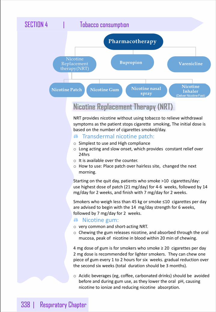
338 | Respiratory Chapter
SECTION 4 | Tobacco consumption
Nicotine Replacement Therapy (NRT)NRTprovidesnicotinewithoutusingtobaccotorelievewithdrawalsymptomsasthepatientstopscigarettesmoking,Theinitialdoseisbasedonthenumberofcigarettessmoked/day.
Transdermalnicotinepatch:o SimplesttouseandHighcomplianceo Longactingandslowonset,whichprovidesconstantreliefover
24hrso Itisavailableoverthecounter.o Howtouse:Placepatchoverhairlesssite,changedthenext
morning.
Startingonthequitday,patientswhosmoke>10 cigarettes/day:usehighestdoseofpatch(21 mg/day)for4-6 weeks,followedby14mg/dayfor2 weeks,andfinishwith7 mg/dayfor2 weeks.
Smokerswhoweighlessthan45 kgorsmoke≤10 cigarettesperdayareadvisedtobeginwiththe14 mg/daystrengthfor6 weeks,followedby7 mg/dayfor2 weeks.
Nicotinegum:o verycommonandshort-actingNRT.o Chewingthegumreleasesnicotine,andabsorbedthroughtheoral
mucosa,peakofnicotineinbloodwithin20 minofchewing.
4 mgdoseofgumisforsmokerswhosmoke≥20 cigarettesperday2 mgdoseisrecommendedforlightersmokers.Theycanchewonepieceofgumevery1 to2 hoursforsixweeks.gradualreductionoverthesecondsixweeks(totaldurationshouldbe3 months).
o Acidicbeverages(eg,coffee,carbonateddrinks)shouldbeavoidedbeforeandduringgumuse,astheylowertheoralpH,causingnicotinetoionizeandreducingnicotineabsorption.
Pharmacotherapy
Nicotine Replacement therapy(NRT)
Nicotine Patch Nicotine GumNicotine Inhaler
(Deliver NicotineFast)
Nicotine nasal spray
VareniclineBupropion
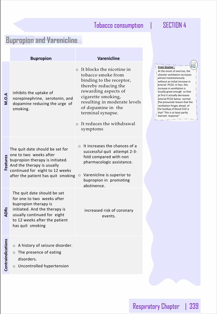
Respiratory Chapter | 339
Tobacco consumption | SECTION 4
FromGuyton:Attheonsetofexercise,thealveolarventilationincreasesalmostinstantaneouslywithoutaninitialincreaseinarterialPCO2.Infact,thisincreaseinventilationisusuallygreatenoughsothatatfirstitactuallydecreasesarterialPCO2 belownormal.ThepresumedreasonthattheventilationforgesaheadofthebuildupofbloodCO2 isthat“Thisisatleastpartlylearnedresponse”
Bupropion and Varenicline
Bupropion Varenicline
M.O.A inhibitstheuptakeof
norepinephrine,serotonin,anddopaminereducingtheurgeofsmoking.
o It blocks the nicotine intobacco smoke from binding to the receptor,thereby reducing the rewarding aspects of cigarette smoking, resulting in moderate levels of dopamine in the terminal synapse.
o It reduces the withdrawalsymptoms
Features
Thequitdateshouldbesetforonetotwoweeksafterbupropiontherapyisinitiated.Andthetherapyisusuallycontinuedforeightto12 weeksafterthepatienthasquitsmoking
o Itincreasesthechancesofasuccessfulquitattempt2-3-foldcomparedwithnonpharmacologicassistance.
o Vareniclineissuperiortobupropioninpromotingabstinence.
ADRs
Thequitdateshouldbesetforonetotwoweeksafterbupropiontherapyisinitiated.Andthetherapyisusuallycontinuedforeightto12 weeksafterthepatienthasquitsmoking
increasedriskofcoronaryevents.
Contraindicatio
ns
o Ahistoryofseizuredisorder.o Thepresenceofeating
disorders.o Uncontrolledhypertension

Appendix
Mind MapsBy Olivia Smith
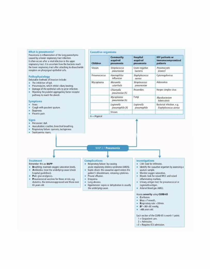
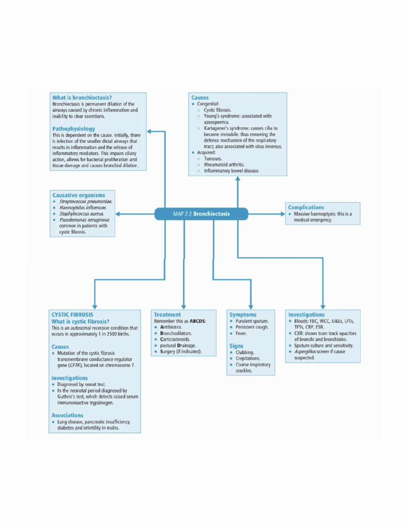
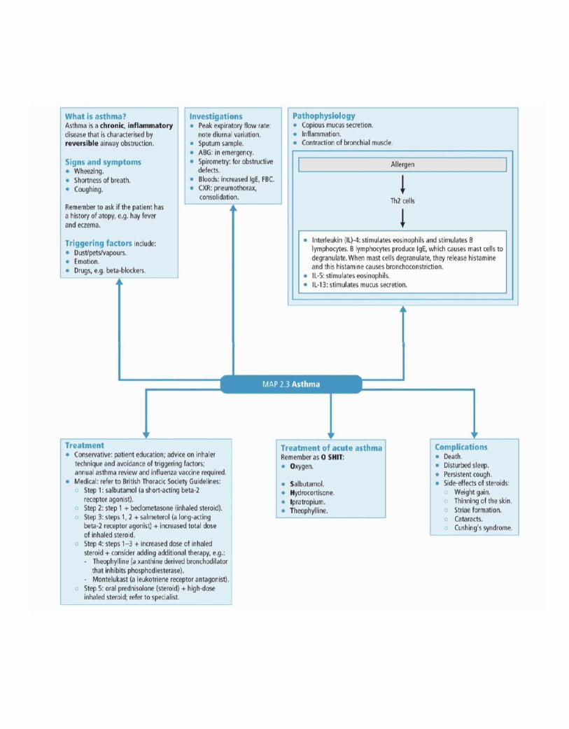
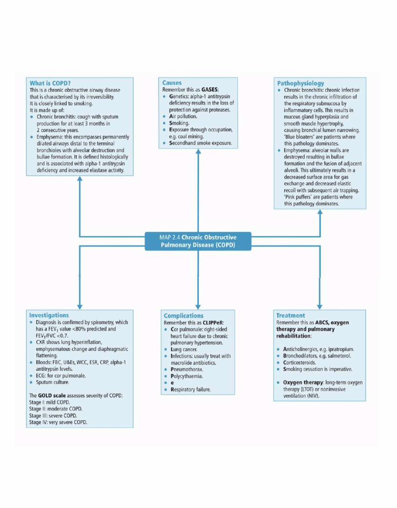
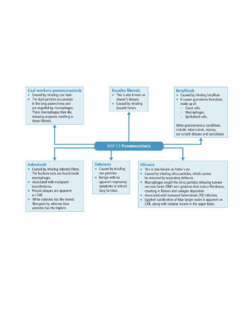
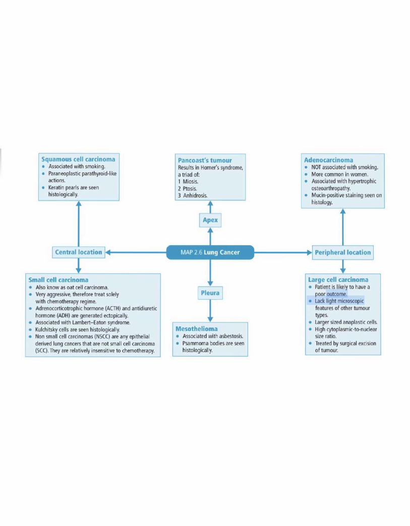
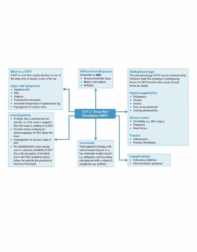
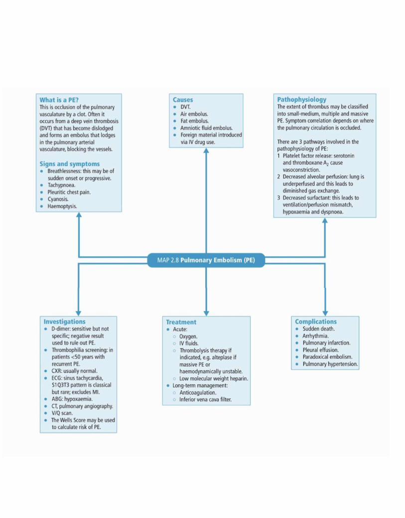
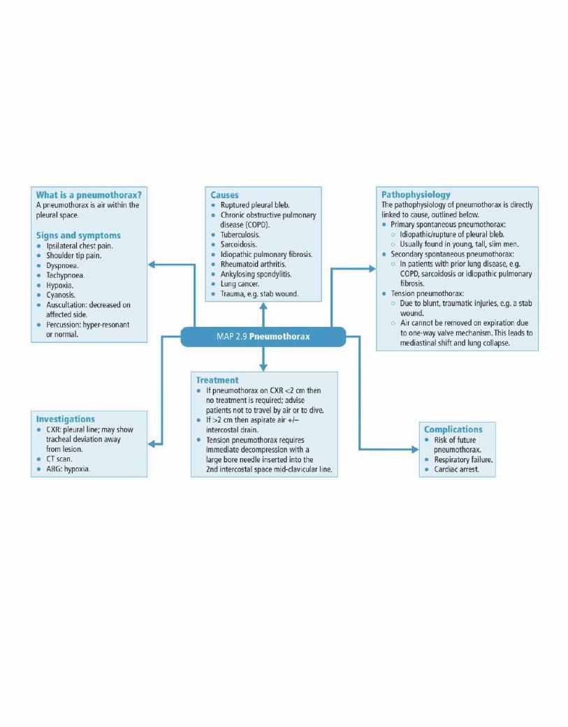
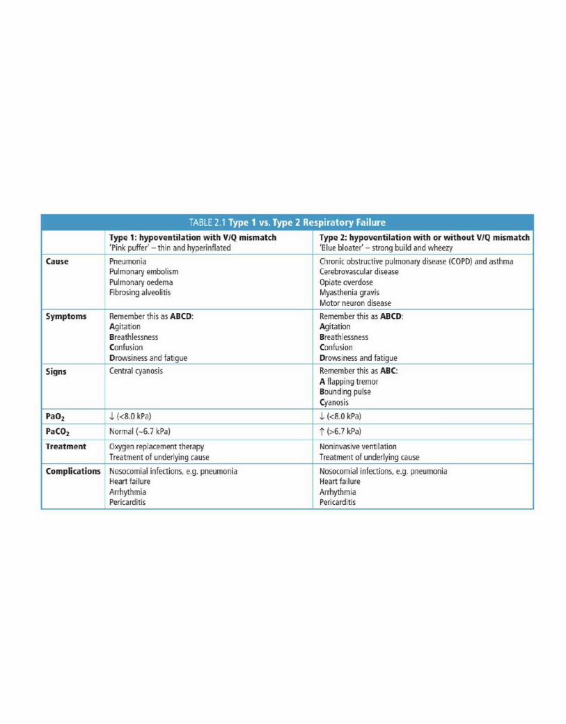

Appendix :
Medcomics
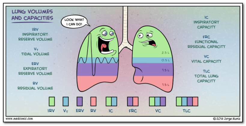
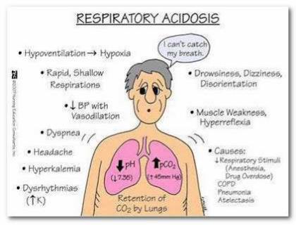
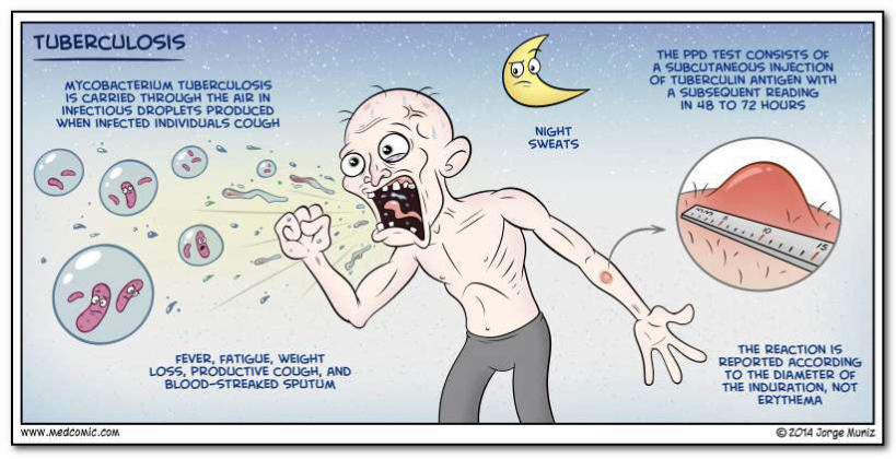
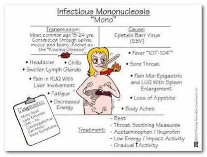
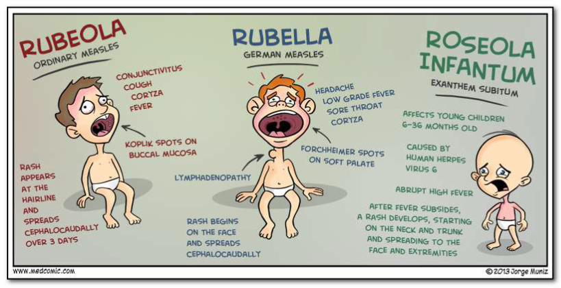
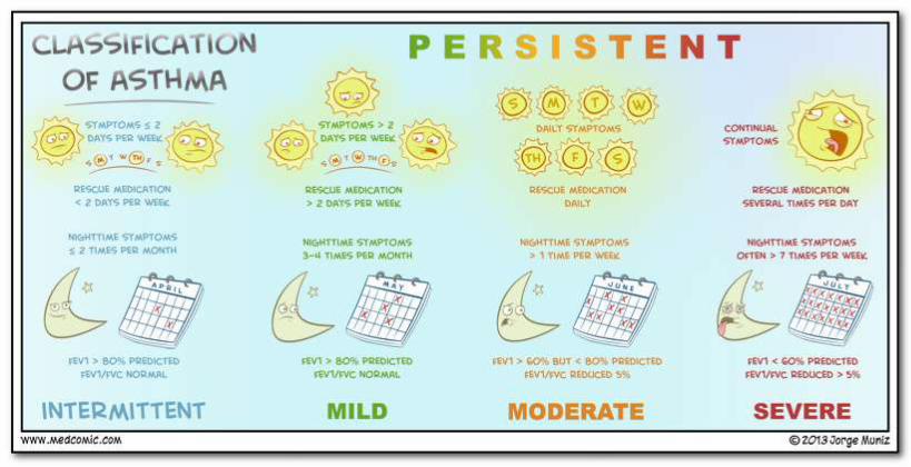
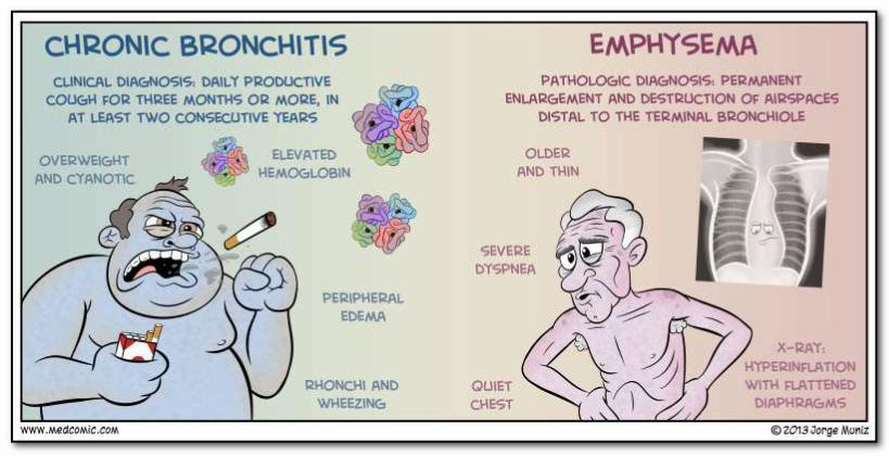
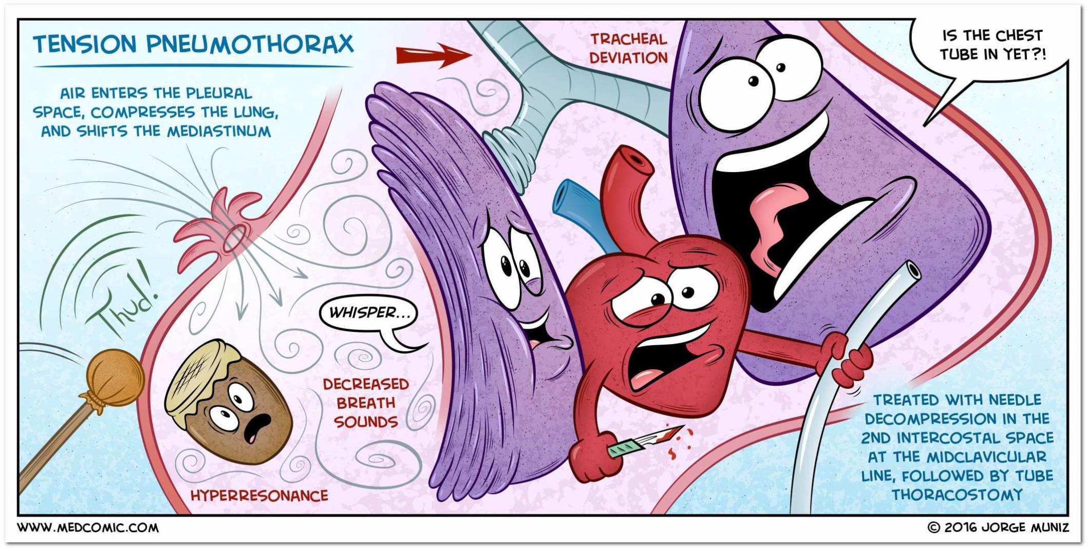
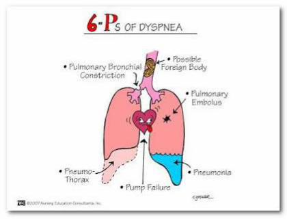
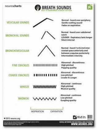
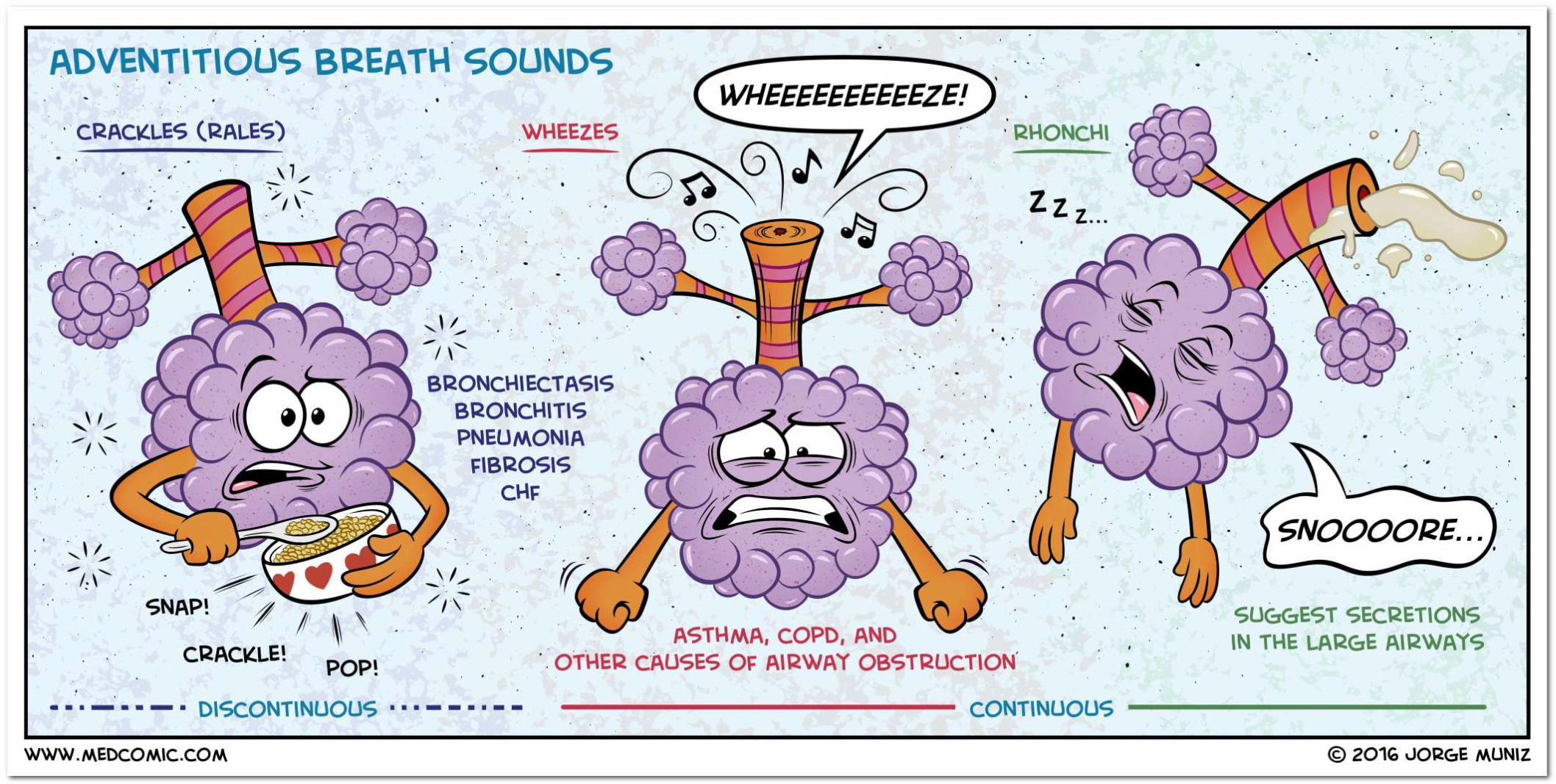
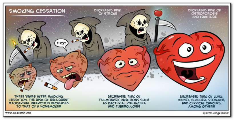
Appendix :
First Aid For USMLE STEP 1 2019
H IGH- YI ELD SYST EMS
Respiratory
"There's so much pollution in the air now that if it weren't for our lungs, there'd be no place to put it all."
-Robert Orben
"Freedom is the oxygen of the soul." - Moshe Dayan
"Whenever I feel blue, I start breathing again."
-L. Frank Baum
"Life is not the amount of breaths you take; it's the moments that take your breath away."
- Will Smith, Hitch
Group key respiratory, cardiovascular, and renal concepts together for study whenever possible. Know obstructive vs restrictive lung disorders, V/Q mismatch, lung volumes, mechanics of respiration, and hemoglobin physiology. Lung cancers and other causes of lung masses are high yield. Be comfortable reading basic chest x-rays, CT scans, and PFTs.
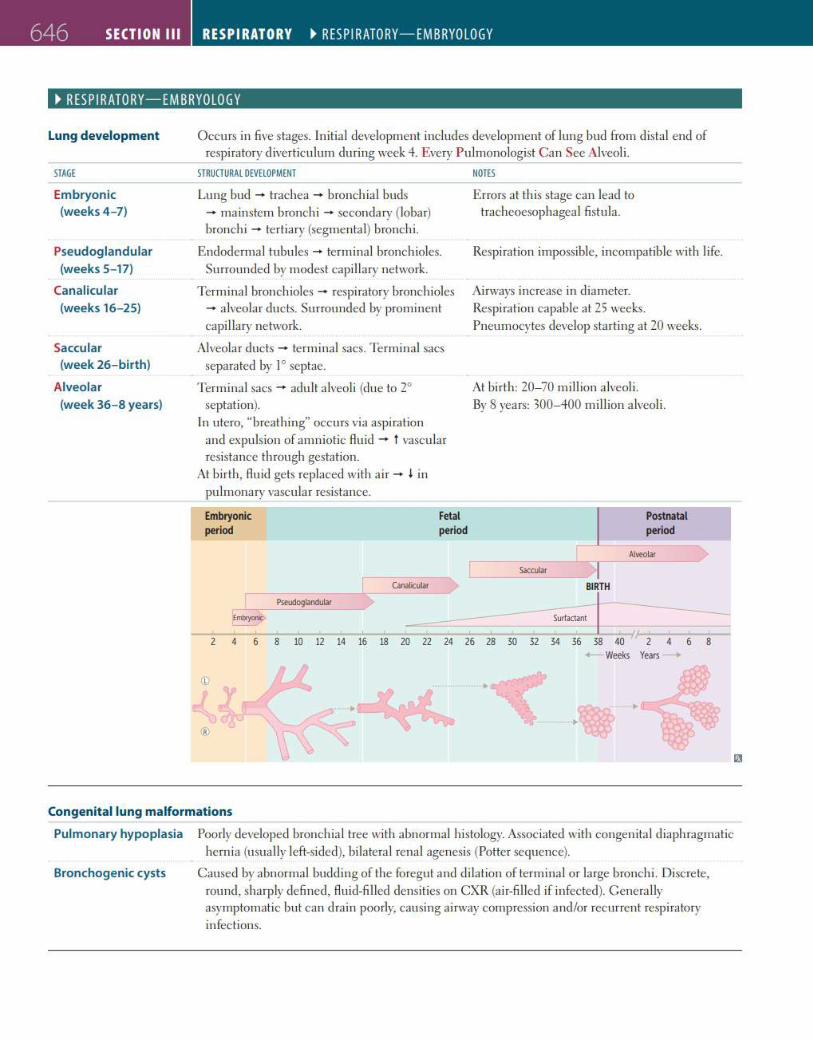
646 SECTION Ill RESPIRATORY ~ RESP I RATORY-EMBRYO LOGY
~ RES PIRATORY-EMBRYOLOGY
Lung development
STAGE
Embryonic (weeks 4-7)
Pseudoglandular (weeks 5-17)
Canalicular (weeks 16- 25)
Saccular (week 26-birth)
Alveolar (week 36-8 years)
Occurs in five stages. Initia l development includes development of lung bud from distal end of respiratory diverticulum during week 4. Every Pulmonologist Can See Alveoli.
STRUCTURAL DEVELOPMENT
Lung bud - trachea - bronchial buds - mainstem bronchi - secondary (lobar) bronchi - tertiary (segmental) bronchi.
Endodermal tubules - terminal bronchioles. Surrounded by modest capillary network.
Terminal bronchioles - respiratory bronchioles - alveolar ducts. Surrounded by prominent capi llary network.
Alveolar ducts - terminal sacs. Terminal sacs separated by 1° septae.
Terminal sacs - adult alveoli (due to 2° septation).
In utero, "breathing" occurs via aspiration and expulsion of amniotic flu id - t vascular resistance th rough gestation.
At birth, fluid gets replaced with air - I in pulmonary vascular resistance.
Embryonic period
Eml:tyol'IC.
©
I
®
Pseudoglandular
Fetal period
NOTES
Errors at this stage can lead to tracheoesophageal fistula.
Respiration impossible, incompatible wi th life.
Airways increase in diameter. Respiration capable at 25 weeks. Pneumocytes develop starting at 20 weeks.
At bi rth: 20-70 million alveoli. By 8 years: 300- 400 mi ll ion alveoli.
Postnatal period
Alveolar
Saccular I BIRTH
Surfactant t 30 32 34 36 38 40
>--Weeks Years
•
4 6 8
Congenital lung malformations
Pulmonary hypoplasia Poorly developed bronchial tree with abnormal histology. Associated with congenital diaphragmatic hernia (usually left-sided), bilateral renal agenesis (Potter sequence).
Bronchogenic cysts Caused by abnormal budding of the foregut and di lation of terminal or large bronchi. Discrete, round, sharply defined, fluid-filled densities on CXR (air-filled if infected). Genera lly asymptomatic but can drain poorly, causing ai rway compression and/or recurrent respiratory infections.
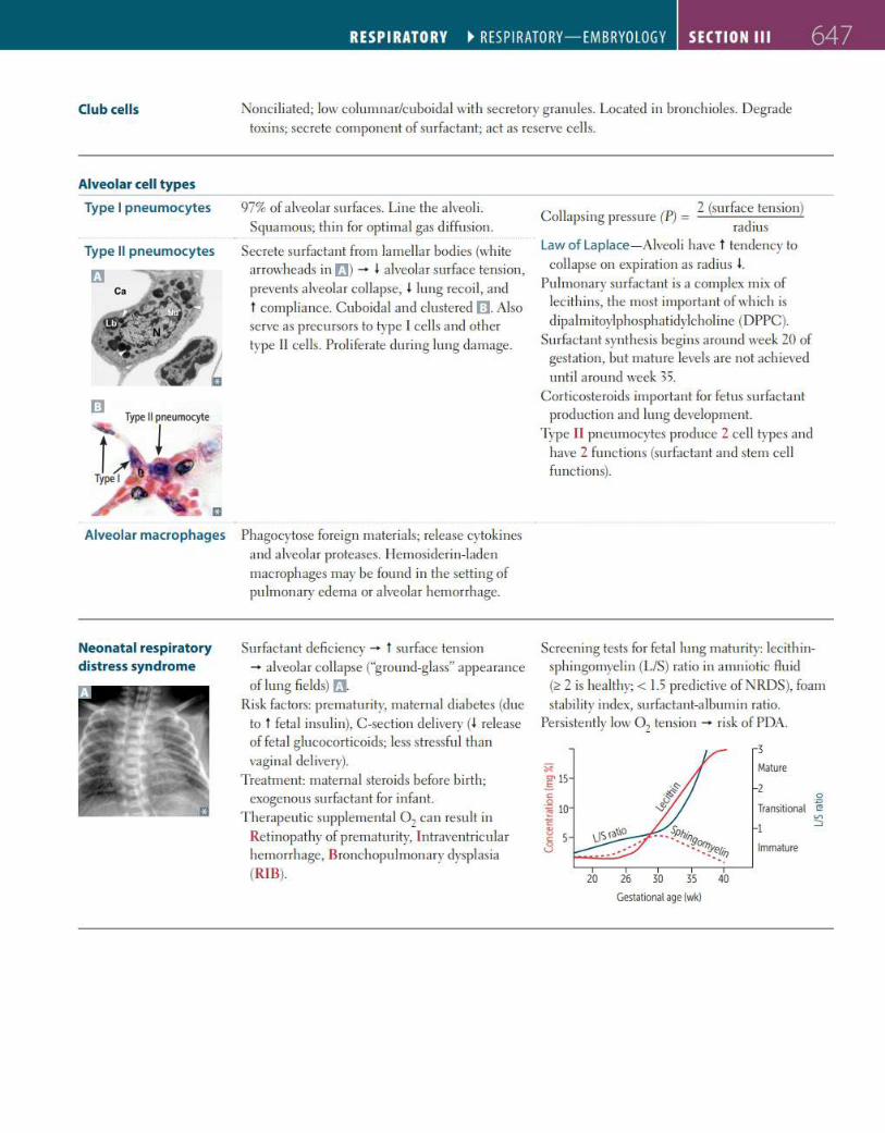
RESPIRATORY ~ RESP I RATORY-EMBRYOLOGY SECTION Ill 647
Club cells Noncil iated; low columnar/cuboida l with secretory granules. Located in bronchioles. Degrade toxins; secrete component of surfactant; act as reserve cells.
Alveolar cell types
Type I pneumocytes
Type II pneumocytes
Et
973 of a lveolar surfaces. Line the a lveoli. Squamous; th in for optimal gas diffusion.
Secrete surfactant from lamellar bodies (white arrowheads in rm - • alveola r surface tension, prevents a lveolar collapse, l lung recoil, and t compliance. Cuboidal and clustered Ill Also serve as precursors to type I cells and other type II cells. Proliferate during lung damage.
Alveolar macrophages Phagocytose foreign mater ia ls; release cytokines and a lveolar proteases. Ilemosiderin-laden macrophages may be found in the setting of pulmonary edema or a lveolar hemorrhage.
Neonatal respiratory distress syndrome
Surfactant deficiency - t surface tension - alveola r collapse ("ground-glass" appearance of lung fie lds) f'J.
Risk factors: prematurity, maternal diabetes (due to t fetal insulin), C -section delivery (I release of feta l g lucocorticoids; less stressfu l than vaginal delivery).
Treatment: maternal steroids before bi rth; exogenous surfactant for infant.
Therapeutic supplemental Oz can result in Re tinopathy of prematurity, l ntraventricula r hemorrhage, Bronchopulmonary dysplas ia (RIB).
C II . (P'l 2 (surface tension)
<> apsmg pressure / = d. ra IUS
Law of Laplace- Alveol i have t tendency to collapse on expiration as radius l.
Pulmonary surfactant is a complex mix of lec ithins, the most important of which is d ipalmitoylphosphatidylcholine (DPPC).
Surfactant synthesis begins around week 20 of gestation, but mature levels are not achieved unti l around week 35.
Corticosteroids important for fetus surfactant production and lung development.
Type II pneumocytes produce 2 cell types and have 2 functions (surfactant and stem cell functions).
Screening tests for fetal lung maturity: lecithinsphingomyelin (LIS) ratio in amniotic fluid (~ 2 is hea lthy;< 1.5 predictive ofNRDS), foam stabi lity index, surfactant-a lbumin ratio.
Persistently low Oz tension - risk of PDA.
30 35 40
Gestational age (wl<)
3
Mature
2 0
Trans~iooal -~ ::;
Immature
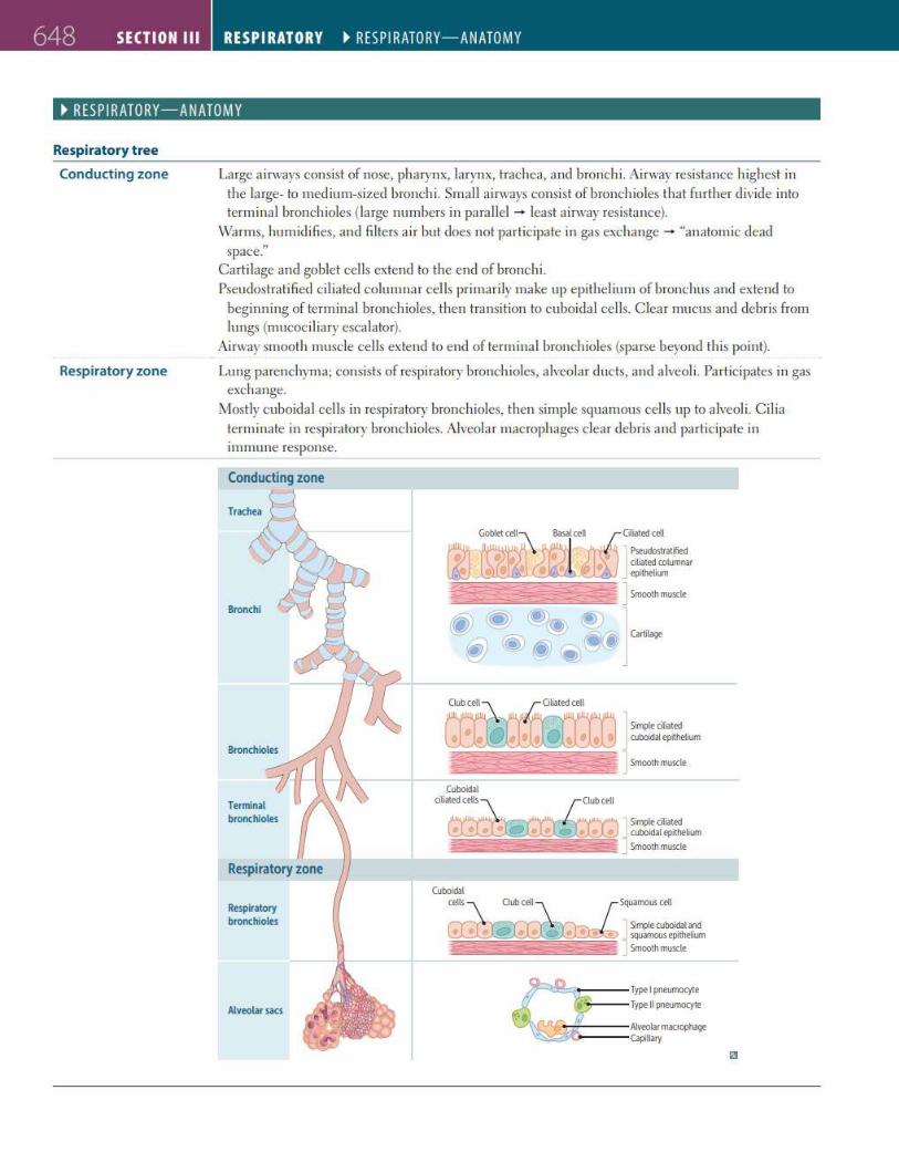
648 SECTION Ill RESPIRATORY ~ RESP I RATORY-ANATOMY
~ RES P IRATORY-ANATOMY
Respiratory tree
Conducting zone
Respiratory zone
Large airways consist of nose, pharynx, larynx, trachea, and bronchi. Airway resistance highest in the large- to medium-sized bronchi. Small airways consist of bronchioles that further divide into terminal bronchioles (large numbers in parallel - least airway resistance).
Warms, humidifies, and fi lters air but does not participate in gas exchange - "anatomic dead space."
Carli lage and goblet cells extend to the end of bronchi. Pseudostratified ci liated columnar cells primari ly make up epithel ium of bronchus and extend to
beginning of terminal bronchioles, then transition to cuboidal cells. Clear mucus and debris from lungs (mucociliary escalator).
Airway smooth muscle cells extend to end of terminal bronchioles (sparse beyond th is point).
Lung parenchyma; consists of respiratory bronchioles, alveolar ducts, and alveoli. Participates in gas exchange.
Mostly cuboidal cells in respiratory bronchioles, then simple squamous cells up to alveoli. Cilia terminate in respiratory bronchioles. Alveolar macrophages clear debris and participate in immune response.
Respiratory zone
Respiratory bronchioles
Alveolar sacs
Cwb cell Oliated cell
---=::::::::=~......._._ Cuboidal
Simple ciliated ooboidal epithelium
1 Smooth muscle
citiat~dcells . r (h.Jbcetl
/v Simple ciliated • • • • • _ cuboidal epitheUum
i=;;s: J Smooth muscle
Type 1 pneumocyte
Type II pneumocyte
Alveolar mauophage Capillary
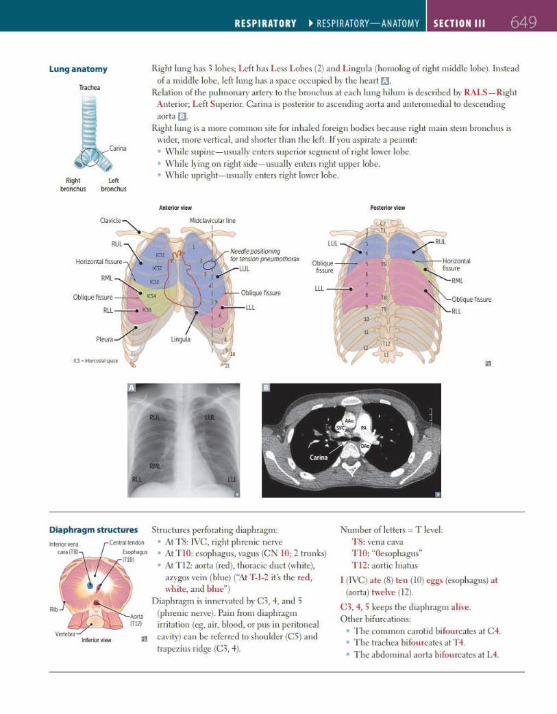
Lung anatomy
Trachea
Right bronchus
Left bronchus
-;~ Oblique fissure L
Rll ---,---e ICSS
ICS • Wttercostat space
RESPIRATORY ~ RESP I RATORY-ANATOMY SECTION Ill 649
Right lung has 3 lobes; Left has Less Lobes (2) and L ingula (homolog of right middle lobe). Instead of a m iddle lobe, left lung has a space occupied by the heart [J.
Relation of the pulmonary artery to the bwnchus at each lung h ilum is described by RALS- Right Anterior; Left Superior. Carina is posterior to ascending aorta and anteromedia l to descending aorta a.
Right lung is a more common si te for inha led foreign bodies because right main stem bronchus is wider, more vertica l, and shorter than the left. If you aspirate a peanut: • Whi le supine- usually enters superior segment of right lower lobe. • Whi le lying on right side-usually enters right upper lobe. • Whi le upright- usually enters right lower lobe.
Anterior view
lingula
Needle positioning 2 for tension pneumothorax
l\·.r.-LUL 3 I r"' I
4( I Oblique fissure Is I _.,._LLL I 6
I I I
Posterior view
~~~ -l~:. ~.
Oblique -- s rs ---Horizontal
fissure ~;~l
rs '~Oblique fissure
r9 ~Rll
Diaphragm structures Structures perforating diaphragm: Number of letters = T level: • At TS: IVC, right phrenic nerve • At T IO: esophagus, vagus (CN IO; 2 trunks) • At T l2: aorta (red), thoracic d uct (white),
azygos vein (blue) ("At T-1-2 it's the red, white, and blue")
Diaphragm is innervated by C3, 4, and 5 (phrenic nerve). Pain from diaphragm irritation (eg, air, blood, or pus in peritoneal
Inferior view 13 cavity) can be referred to shoulder (CS) and trapezius ridge (C3, 4).
TS: vena cava T IO: "Oesophagus" T l2: aortic h iatus
I (IVC) ate (S) ten ( IO) eggs (esophagus) at (aorta) twelve (12).
C3, 4, 5 keeps the diaphragm alive. Other bifurcations:
• The common carotid bi fourcates at C4. • The trachea bifourcates at T4. • The abdominal aorta bifourcates at L4.
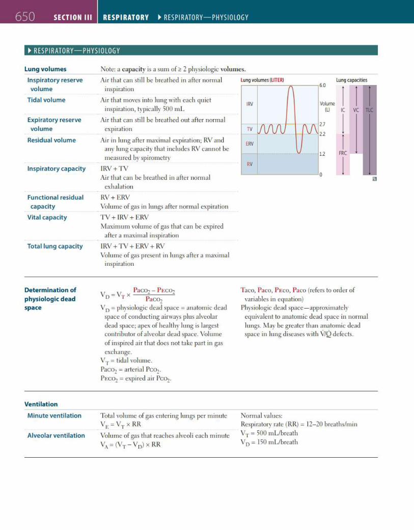
650 SECTION Ill RESPIRATORY ~ RESP I RATORY-PHYS I OLOGY
~ RESPIRATORY-PHYSIOLOGY
Lung volumes
lnspiratory reserve volume
Tidal volume
Expiratory reserve volume
Residual volume
lnspi ratory capacity
Functional residual capacity
Vita l capacity
Total lung capacity
Determination of physiologic dead space
Ventilation
Minute ventilation
Alveolar ventilation
Note: a capacity is a sum of~ 2 physio logic volumes.
Air that can still be breathed in after norma l l ung volumes (LITER)
inspiration
Air that moves into lung with each qu iet IRV
6.0 lung capacities
1 1 l Volume inspiration, typically 500 mL (l ) IC vc TIC
Air that can still be breathed out after normal expiration
Air in lung after maximal exp iration; RV and any lung capacity that includes RV cannot be measured by spi rometry
IRV + TV Air that can be breathed in after normal
exhalation
RV+ ERV Volume of gas in lungs a fter norma l expiration
TV + IRV + ERV Maximum volume of gas that can be expi red
after a maximal inspiration
IRV + TV + ERV + RV Volume of gas present in lungs after a maximal
inspiration
V = V x PaCOz - P ECOz D T PaCOz
VD = phys iologic dead space = anatomic dead space of conducting a irways p lus alveolar dead space; apex of hea lthy lung is largest contributor of a lveolar dead space. Volume of inspired ai r that does not take part in gas exchange.
VT= tida l volume. Paco2 =arteria l Pco2.
PECOz =expired a ir Pco2.
Tota l volume of gas entering lungs per minute VE=V,.x RR
Volume of gas that reaches alveoli each minute
VA= (VT - Vo) x RR
2.7 l TV
ERV l 1.2 FRC
RV l 0
Taco, Paco, PEco, Paco (refers to order of variables in equation)
Physiologic dead space- approximately equiva lent to anatomic dead space in normal lungs. May be g reater than anatomic dead space in lung d iseases with V/Q defects.
Normal va lues: Respiratory rate (RR) = 12- 20 breaths/min VT= 500 mL/breath VD = 150 mL/breath
liJ
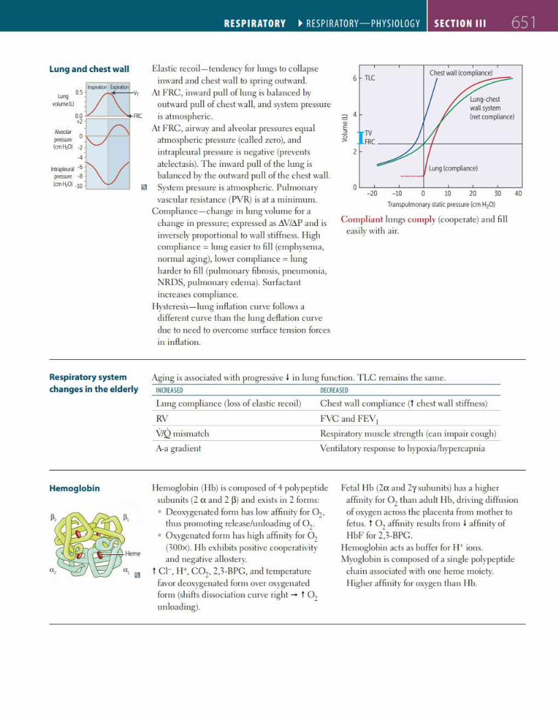
Lung and chest wall
Alvoo~r O pressure (cmH,OJ -2
·4
lntrapleur~ ·6 pressure ·8 (cmH,OJ ·10
Respiratory system changes in the elderly
Hemoglobin
RESPIRATORY ~ RESP I RATORY-PHYSIOLOGY SECTION Ill 651
Elastic recoi l- tendency for lungs to collapse inward and chest wall to spring outward.
At FRC, inward pull of lung is balanced by outward pull of chest wall, and system pressure is atmospheric.
At FRC, ai rway and alveolar pressures equal atmospheric pressure (ca lled zero), and intrapleural pressure is negative (prevents atelectasis). The inward pull of the lung is balanced by the outward pull of the chest wall.
Ii!! System pressure is atmospheric. Pulmonary vascular resistance (PVR) is at a minimum.
Compliance- change in lung volume for a change in pressure; expressed as tN/tiP and is inversely proportional to wall stiffness. lligh compliance= lung easier to fill (emphysema, normal aging), lower compliance= lung harder to fill (pulmonary fi brosis, pneumonia, NRDS, pulmonary edema). Surfactant increases compliance.
Hysteresis-lung inAation curve follows a different curve than the lung deAation curve due to need to overcome surface tension forces in inAation.
6 TLC Chest wan (comp!iance)
.--::::::=-
2 4 "' E " ;g TV
FRC----+--+-+-----------1 2
Lung (compliance)
-20 -10 0 10 20 30 Transpulmonary static pressure (cm H20)
Compliant lungs comply (cooperate) and fill easily with air.
40
Aging is associated with progressive l in lung function. TLC remains the same. INCREASED DECREASED
Lung compliance (loss of elastic recoil) Chest wa ll compliance (t chest wa ll stiffness)
RV FVC and FEV1
\1/Q mismatch
A-a gradient
Respiratory muscle strength (can impair cough) -- --
Venti latory response to hypoxia/hypercapnia
Hemoglobin (I lb) is composed of 4 polypeptide subunits (2 a and 2 ~) and exists in 2 forms: • Deoxygenated form has low affin ity for Oz,
thus promoting release/unloading of Oz· • Oxygenated form has high affinity for Oz
(300x). I lb exhibits positive cooperativi ty and negative allostery.
t CI-, ll+, COz, 2,3-BPC, and temperature favor deoxygenated form over oxygenated form (shifts dissociation curve right - t Oz unloading).
Fetal lib (2a and 2y subunits) has a higher affinity for Oz than adult lib, driving diffusion of oxygen across the placenta from mother to fetus. t Oz affinity results from I affin ity of llbF for 2,3-BPC.
I lemoglobin acts as buffer for I 1+ ions. Myoglobin is composed of a single polypeptide
chain associated with one heme moiety. ll igher affinity for oxygen than lib.
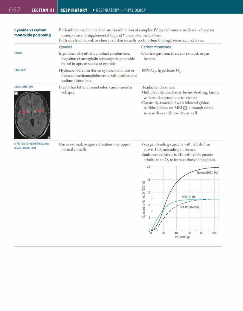
652 SECTION Ill
Cyanide vs carbon monoxide poisoning
SOURCE
TREATMENT
SIGNS/SYMPTOMS
EFFECT ON OXYGEN-HEMOGLOBIN OISSOCIATION CURVE
RESPIRATORY ~ RESP I RATORY-PHYS I OLOGY
Both inh ibit aerobic metabolism via in hibition of complex JV (cytochrome c oxidase) - hypoxia unresponsive to supp lemental 0 2 and t anaerobic metabolism.
Both can lead to pin k or cherry red sk in (usually postmortem find ing), seizures, and coma.
Cyanide Carbon monoxide
Byproduct of synthetic product combustion, ingestion of amygda lin (cyanogenic g lucos ide fou nd in apricot seeds) or cyanide.
Hydroxocoba la min (forms cyanocobalamin) or induced methemoglobinemia with n itrites and sodium th iosulfate.
Breath has bitter almond odor; cardiovascu lar collapse.
Curve normal; oxygen saturation may appear normal in itially.
O dorless gas from fi res, ca r exhaust, or gas heaters.
1003 Oz, hyperbaric 0 2.
lleadache, dizziness . Multiple ind ividuals may be involved (eg, fam ily
with simi lar symptoms in winter). Classica lly associated with bilatera l globus
pall idus lesions on MRI rJ, although rarely seen with cyanide tox icity as well.
I oxygen-binding capacity with left sh ift in curve, I 0 2 un loading in tissues.
Binds competitively to llb with 200x greater affinity than 0 2 to form carboxyhemoglobin.
20
'--" -..... ' "' .......... ~.~ ....... ~ ........ : ...... ~ ................ ! .... . / , 50% Hb (anemia) ; ;
' / ' ' ' : / . . . . ;r--· .... ····+ ··············t ··············~·· ··········· +···
I . y
0o,._.....__2_0_...__4_0_.__~60_.....___8_0_...__10~0--
Po, (mm Hg)
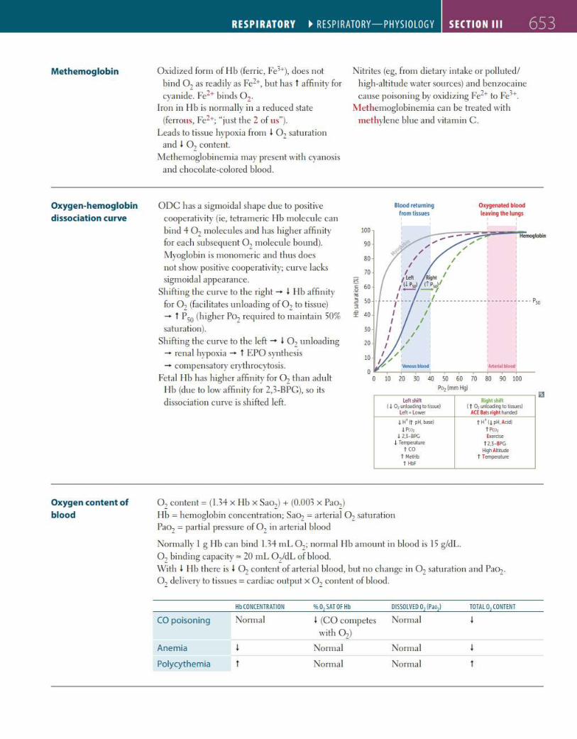
Methemoglobin
Oxygen-hemoglobin
dissociation curve
Oxygen content of blood
RESPIRATORY ~ RESP I RATORY-PHYSIOLOGY SECTION Ill 653
Oxidized form of llb (ferric, Fe3+), does not bind 0 7 as read ily as Fez+, but has t a ffin ity for cyan i d~. Fe2+ binds 0 2
Iron in llb is normally in a reduced state (ferrous, Fe2+; "just the 2 of us"}.
Leads to tissue hypoxia from l 0 2 saturation and I Oz content.
Methemoglobinemia may present with cyanosis and c hocolate-colored blood.
ODC has a sigmoidal shape due to positive cooperativi ty (ie, tetra meric l ib molecule can bind 4 Oz molecules and has higher a ffin ity for each subsequent Oz molecule bound}. Myoglobin is monomeric and thus does not show positive cooperativity; c urve lacks sigmoida l appearance.
Shifting the c urve to the right - I I lb a ffinity for Oz (fac ilitates un loading of Oz to tissue) - t P50 (h igher Poz requ ired to maintain 503 saturation}.
Shifting the curve to the left - I Oz un loading - rena l hypoxia - t EPO synthesis - compensatory eryth rocytosis.
Fetal l ib has higher affinity for Oz than adult I lb (due to low affin ity for 2,3-BPC), so its d issociation curve is sh ifted left.
Oz content = (1.34 x Hb x Saoz) + (0.003 x Paoz}
Nitri tes (eg, from dietary intake or polluted / high-altitude water sources} and benzocaine cause poisoning by oxidizing Fez+ to Fe3+.
M ethemoglobinemia can be treated with methylene blue a nd vitamin C .
x c .<!
~ :JI ,, :I:
100
90
80
70
60
50
40
30
20
10
Blood returning from tissues
Oxygenated blood leaving the lungs
0"'-~~-~~-~~~-~~-~-0 10 m W ~ ~ W M ~ ~ ~
Po, Imm Hg)
Left shift RightsUt I j . Di unloading to ti~ue) ( t Ol ooloading to tissues!
l eft • lower ACE a.ts right handed
LH' ltpH. b.>sel f H~ It pH, Acid) I Pto, t Pco,
I U -BPG Exercise .J. Temperalllre t U - BPG
HO ligh Altwcle t MetHb f Temperawre t ll>f
Hb = hemoglobin concentration; SaOz = a rteria l Oz saturation Paoz = partial pressure of Oz in arteria l blood
Normally I g llb can bind 1.34 mL Oz; normal lib amount in blood is 15 g/dL. Oz binding capacity= 20 mL Oz'dL of blood. With l I lb there is l 0 2 content of arterial blood, but no change in Oz saturation a nd PaOz. Oz del ivery to tissues = cardiac output x Oz content of blood.
Hb CONCENTRATION %01 SATOFHb OISSOLVEO 01 (Pao,) TOTAL 01 CONTENT
CO poisoning Normal l (CO competes Normal • with Oz}
Anemia Normal Normal
Polycyt hemia Normal Normal
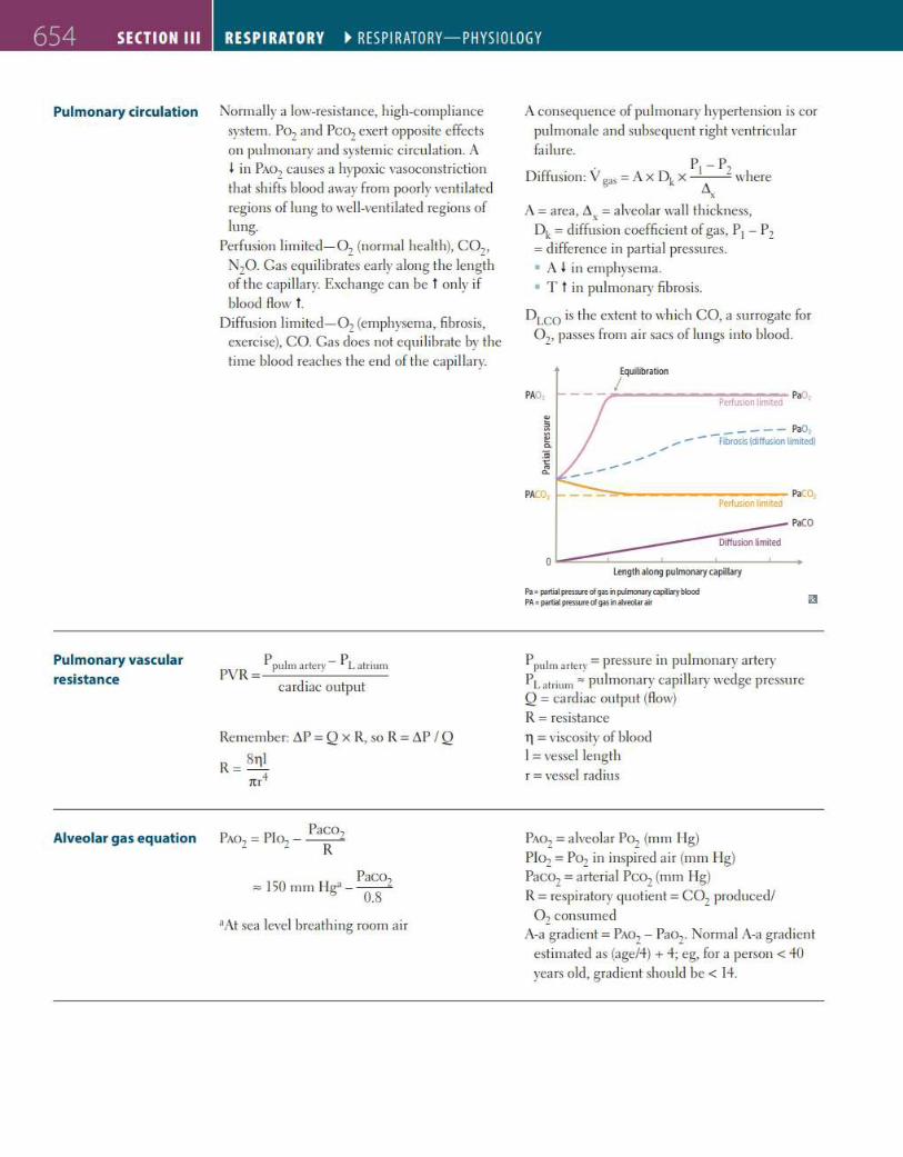
654 SECTION Ill RESPIRATORY ~ RESP I RATORY-PHYS I OLOGY
Pulmonary circulation Normally a low-resistance, high-compliance
Pulmonary vascular resistance
Alveolar gas equation
system. Poz and Pcoz exert opposite effects on pulmonary and systemic ci rculation. A I in PAOz causes a hypoxic vasoconstriction that shifts blood away from poorly ventilated regions of lung to well-ventilated regions of lung.
Perfusion limited- Oz (normal health), COz, NzO. Gas equilibrates early along the length of the capillary. Exchange can be t only if blood Aow t .
Diffusion limi ted- Oz (emphysema, fibrosis, exercise), CO. Gas does not equilibrate by the time blood reaches the end of the capi llary.
PVR = ppulm arler) - p L alrium cardiac output
Remember: D.P = Q x R, so R = D.P IQ
R = 8111 m 4
PaC07 = 150 mm Ilg" - ---0.8
•At sea level breathing room air
A consequence of pulmonary hypertension is cor pulmonale and subsequent right ventricular fai lure. . . · P1 - Pz
D1ffus1on: V gas = AX Dk X - /),.--where x
A= area, D. , =alveolar wa ll th ickness, Dk= diffusion coefficient of gas, P1 - Pz =difference in partial pressures. • A I in emphysema. • T t in pulmonary fibrosis.
DLCO is the extent to which CO, a surrogate for Oz, passes from air sacs of lungs into blood.
Equilibration
PAO, I
Perfusion lir'ited PaOz
----- PaO: - - Fibrosis (liffusion limited) ----
.:-::::-...----------- P•C01 ?effusion ~mited
Diffusion timited
0 Length along pulmonary «1piUary
Pa • partial pressure of gas in pulmooaty capllary blood PA• paniat PftS'SUl'eofgasinatveotar air
P.CO
Ppulm arlery = pressure in pulmonary artery PLalrium" pulmonary capillary wedge pressure Q = cardiac outpu t (Aow) R = resistance 11 = viscosity of blood I = vessel length r = vessel radius
PAOz = alveolar Poz (mm I lg) Pk>z = Poz in inspired ai r (mm l lg) PaCOz = arteria l PCOz (mm I lg) R =respiratory quotient= COz produced/
Oz consumed A-a gradient = PAOz - Paoz. Normal A-a gradient
estimated as (age/4) + 4; eg, for a person < 40 years old, gradient should be< 14.
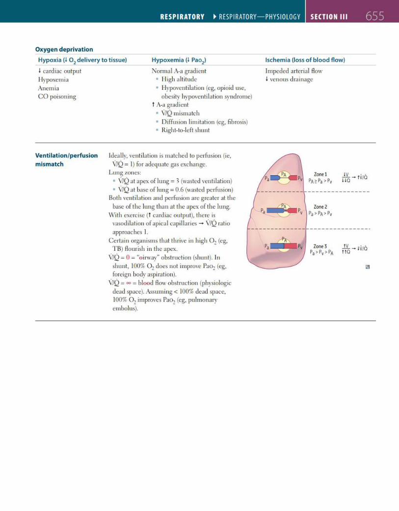
RESPIRATORY ~ RESP I RATORY-PHYSIOLOGY SECTION Ill 655
Oxygen deprivation
Hypoxia (I 02
delivery to t issue)
I card iac output
Hypoxemia (I Pao2
)
Normal A-a grad ient Hypoxemia Anemia CO poisoning
Ventilation/perfusion mismatch
• Iligh a ltitude • Ilypoventi lation (eg, opioid use,
obesity hypoventilation syndrome) t A-a grad ient • V/Q m ismatch
• Diffusion limitation (eg, fibrosis) • Right-to-left shunt
Ideally, ventilation is matched to perfusion (ie, V/Q = I) for adequate gas exchange.
Lung zones: • V/Q at apex of lung= 3 (wasted venti lation) • V/Q at base of lung = 0.6 (wasted perfusion)
Both ventilation and perfusion are greater at the base of the lung than at the apex of the lung.
With exercise (t cardiac output), there is vasod ilation of apica l capillaries - V/Q ratio approaches I.
Certain organisms that thrive in h igh 0 2 (eg, TB) Aourish in the apex.
V/Q = 0 = "o irway" obstruction (shunt). In shunt, 1003 0 2 does not improve Pao2 (eg, foreign body aspiration).
V/Q = oo = blood Aow obstruction (physiologic dead space). Assuming< 1003 dead space, 1003 0 2 improves Pao2 (eg, pulmonary embolus).
lschemia (loss of blood flow)
Impeded arterial Aow I venous drainage
Zonel PA~ Pa> Pv
IV · · iiQ - IV/Q
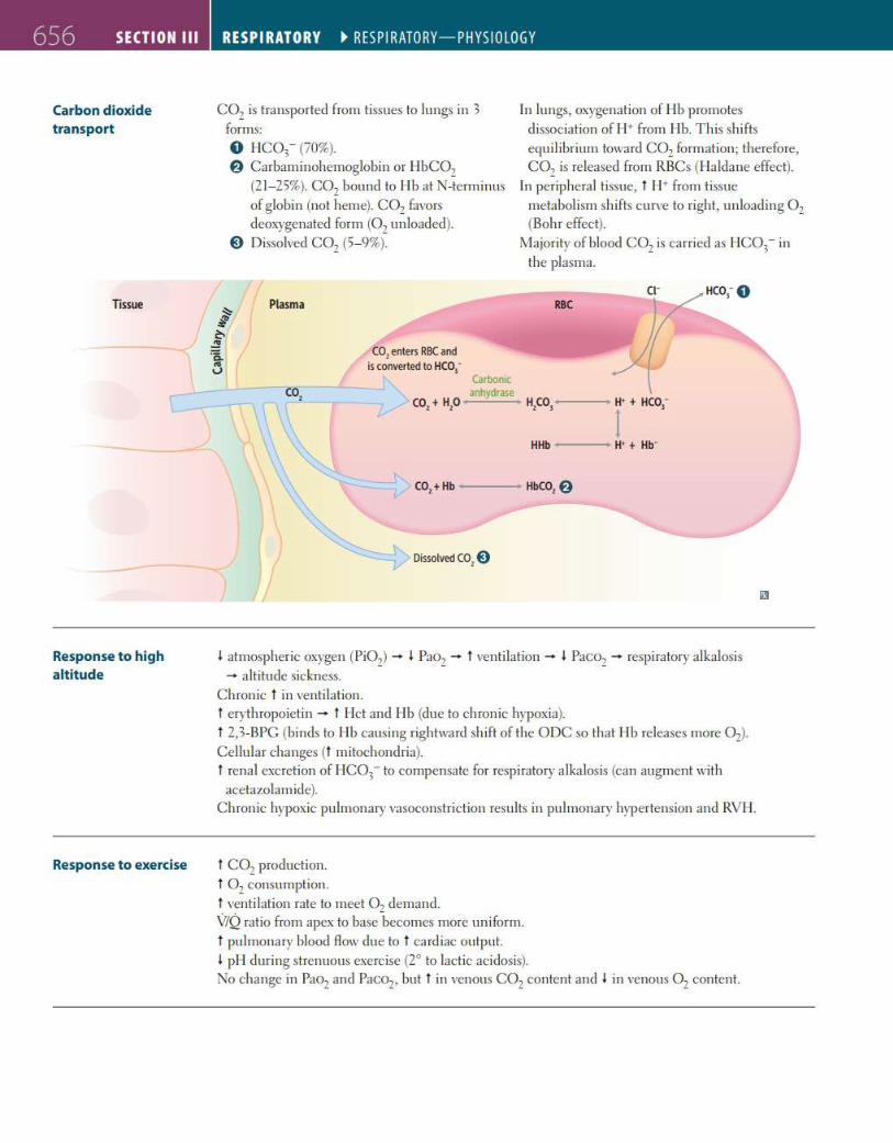
656 SECTION Ill
Carbon d ioxide transport
nssue
Response to high altitude
Response to exercise
RESPIRATORY ~ RESP I RATORY-PHYS I OLOGY
col is transported from tissues to lungs in 3 forms: 0 IIC03- (703). f) Carbaminohemoglobin or IIbCOz
(21- 253). COz bound to IIb at N-terminus of globin (not heme). C02 favors deoxygenated form (Oz unloaded).
0 Dissolved C02 (5- 93).
Plasma
co, enters RBC and is converted to HCo,-
In lungs, oxygenation of Hb promotes d issociation of J J+ from I lb. T his shifl~ equilibrium toward COz formation; therefore, COz is released from RBCs ( IJa ldane effect).
In periphera l tissue, t IJ+ from tissue
metabolism shifts curve to right, unloading Oz (Bohr effect).
Majority of blood C02 is carried as IIC03- in the plasma.
Carbonic anhydrase
co,+ H,O 11,co,---H· + HCO -I . HHb ---H• + Hb-
CO,+ Hb ___ ,.. HbC02 f)
Dissolved co, 0
I atmospheric oxygen (Pi02) - I Pao2 - t venti lation - I PaCOz - respiratory a lkalosis - a ltitude sickness.
Chronic t in ventilation. t erythropoietin - t IJct and IIb (due to chronic hypoxia). t 2,3-BPC (binds to IIb causing rightward shift of the ODC so that l ib releases more 0 2) .
Cellular changes (t m itochondria). t renal excretion of I IC03- to compensate for respiratory alka losis (can augment with acetazolamide).
Chronic hypoxic pulmonary vasoconstri ction results in pulmonary hypertension and RVJ I.
t COz production. t Oz consumption. t ventilation rate to meet 0 1 demand. V/Q ratio from apex to base -becomes more un iform.
t pulmonary blood flow due to t ca rd iac output. I pl I during strenuous exerc ise (2° to lactic acidosis). No change in Paoz and Paco2, but t in venous COz content and I in venous 0 2 content.
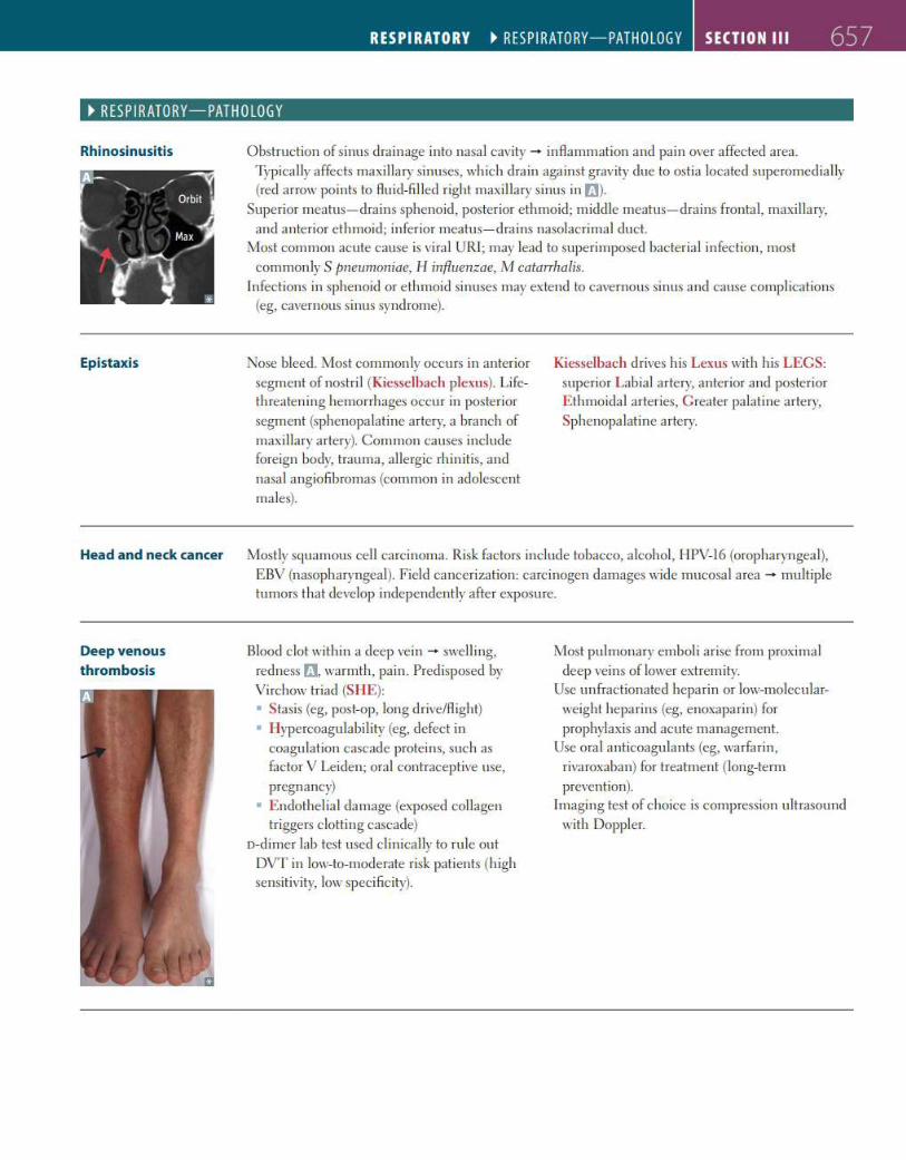
RESPIRATORY ~ RESPIRATORY-PATHOLOGY SECTION Ill 657
~ RES PIRATORY-PATHOLOGY
Rhinosinusitis
Epistaxis
Head and neck cancer
Deep venous
thrombosis
Obstruction of sinus drainage into nasa l cavity - inflammation and pain over affected area. Typically affects maxillary sinuses, which drain against gravity due to ostia located superomedia lly (red arrow points to flu id-fi lled right maxillary sinus in rm.
Superior meatus- dra ins sphenoid, posterior e thmoid; m iddle meatus- drains frontal, maxi lla ry, and anterior ethmoid; infer ior meatus- drains nasolacrimal duct.
Most common acute cause is vira l URI; may lead to superimposed bacteria l infection, most commonly S pneumoniae, l I influen;zae, M catarrhalis.
Infections in sphenoid or e thmoid sinuses may ex tend to cavernous sinus and cause complications (eg, cavernous sinus syndrome).
Nose bleed. Most commonly occurs in anterior segment of nostri I (Kiesselbach plexus). Lifethreatening hemorrhages occur in posterior segment (sphenopalatine artery, a branch of maxi llary artery). Common causes include foreign body, trauma, allergic rh initis, and nasal angiofibromas (common in adolescent males).
Kiesselbach d rives his Lexus with h is LEGS: superior Lab ia l artery, anterior and posterior E thmoidal arter ies, G reater pa latine artery, Sphenopalatine a rtery.
Mostly squamous cell carcinoma. Risk factors include tobacco, a lcohol, HPV-16 (oropharyngeal), EBV (nasopharyngea l). Field canceri zation: carc inogen damages wide mucosa! area - multiple tumors that develop independently after exposure.
Blood clot within a deep vein - swelling, redness t'J, warmth, pa in. Predisposed by Vi rchow triad (SIIE): • Stasis (eg, post-op, long drive/Aight) • Ilypercoagulabi lity (eg, defect in
coagulation cascade proteins, such as factor V Leiden; oral contraceptive use, pregnancy)
• E ndothel ia l damage (exposed collagen triggers clotting cascade)
o-d imer lab test used cl inically to ru le out DVT in low-to-moderate risk patients (high sensitivity, low specificity).
Most pulmonary emboli arise from proximal deep veins of lower extremity.
Use unfractionated heparin or low-molecularweight heparins (eg, enoxaparin) for prophylax is and acute management.
Use oral anticoagulants (eg, warfarin, riva roxaban) for treatment (long-term prevention).
Imaging test of choice is compression u ltrasound with Doppler.
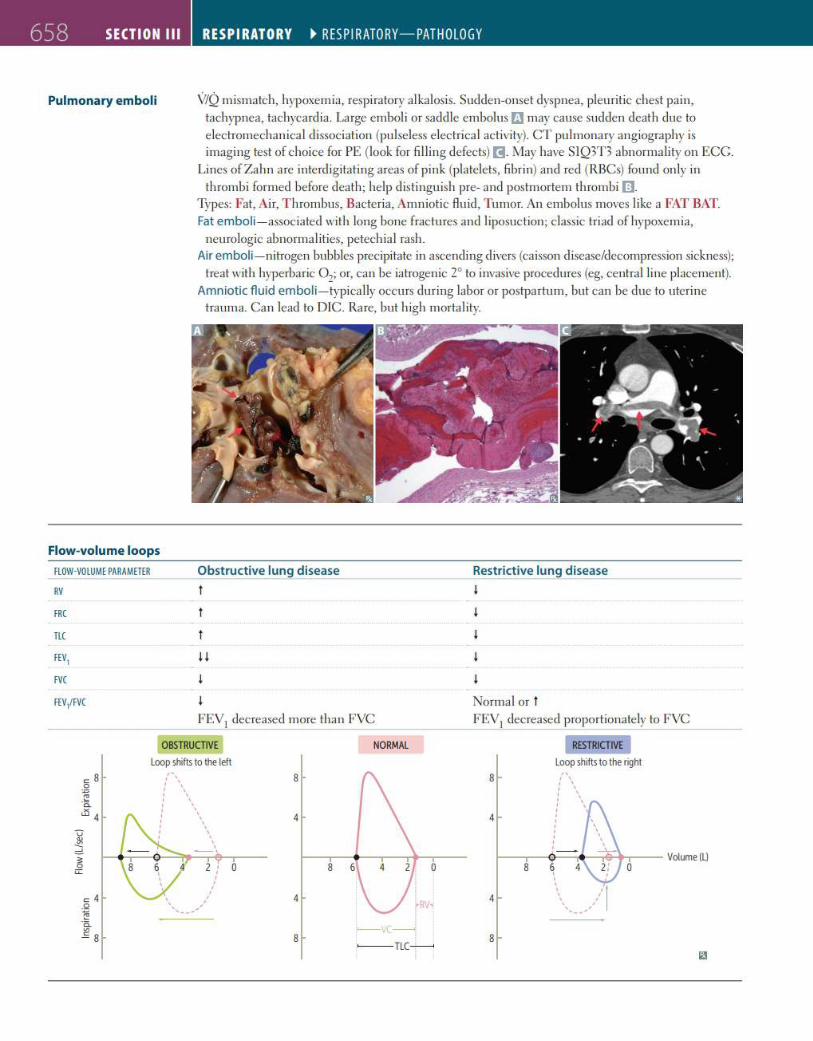
658 SECTION Ill
Pulmonary emboli
Flow-volume loops
FLOW-VOLUME PARAMETER
RV
FRC
TLC
FEV1
FVC
FEV1/FVC
RESPIRATORY ~ RESPIRATORY-PATHOLOGY
V/Q m ismatch, hypoxemia, respiratory alkalosis. Sudden-onset dyspnea, pleuritic chest pain, tachypnea, tachycardia. Large emboli or sadd le embolus rJ may cause sudden death due to electromechanical dissociation (pulseless electrical activity). CT pulmonary ang iography is imaging test of choice for PE (look for fi lling defecl~) ~. May have SlQ3T 3 abnormality on ECG.
Lines of Zahn are interd ig itating areas of pink (platelets, fibrin) and red (RBCs) found only in thrombi formed before death; help distinguish pre- and postmortem th rombi rn.
Types: Fat, Air, T hrombus, Bacteria, Amniotic flu id, Tumor. An embolus moves like a FAT BAT. Fat emboli- assoc iated with long bone fractures and liposuc tion; c lassic triad of hypoxemia,
neurologic abnormalities, petechia l rash. Air emboli- nitrogen bubbles precipitate in ascending d ivers (caisson d isease/decompression sickness);
treat with hyperbaric 0 2; or, can be iatrogenic 2° to invasive procedures (eg, central line placement). Amniotic fluid emboli- typica lly occurs during labor or postpartum, but can be due to uterine
trauma. Can lead to DIC. Ra re, but h igh mortal ity.
Obstructive lung disease
t
j j
FE\11 decreased more than FVC
Restrictive lung disease j
Normal or t FE\11 decreased proportionate ly to FVC
OBSTRUCTIVE NORMAL RESTRICTIVE
Loop shifts to the left Loop shifts to the right
c: 8 ,,
8 ' ' 0 ' \ ~ , ' -~ ' \ ' ' ' d5 4 ' ' ' ' 4
' ' \ v . ' Jl! ' ' ' ' "'
_ ,
4
~ ;:;: 0 8 6 0
c: 4 4 4 .Q
~ ·a
-----< .E 8 8
~nc~ 8
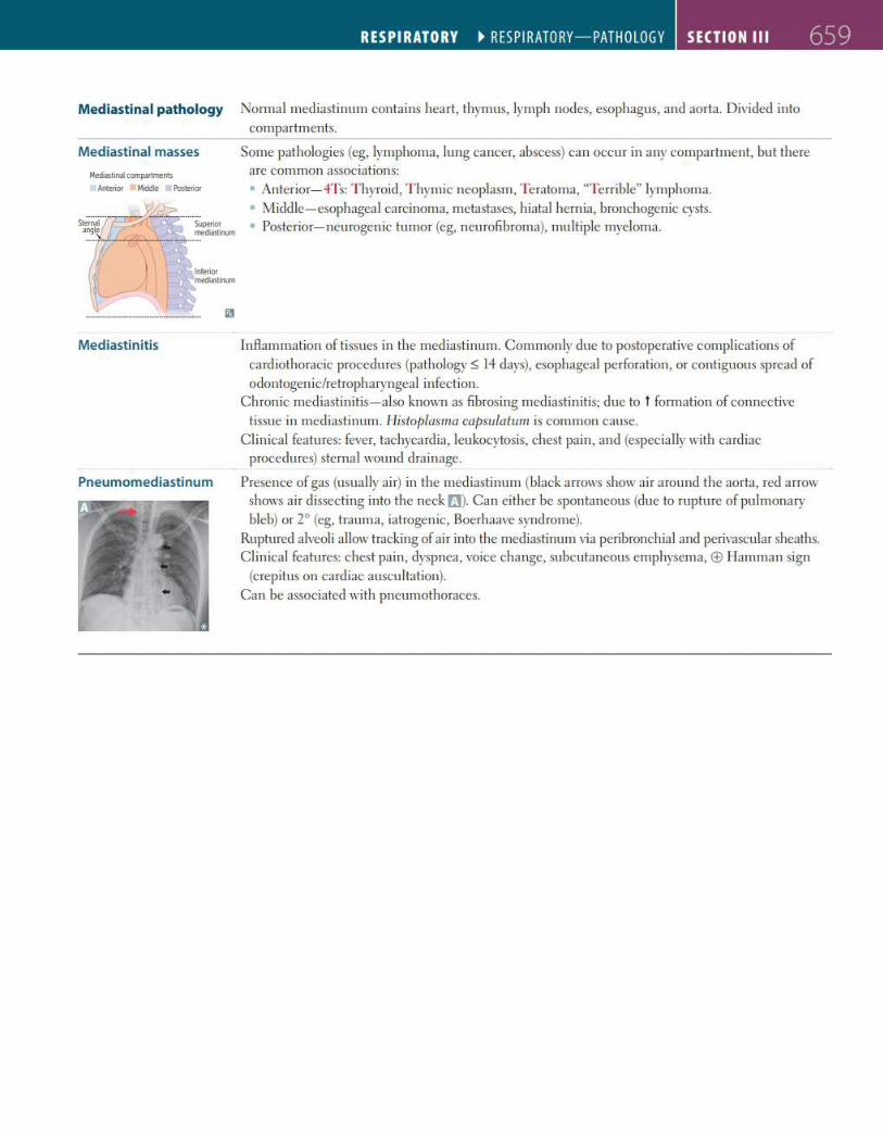
Mediastinal pathology
Mediast inal masses
Me<ia:stinal ccmpanments
Anterior 8 Middle Posterior
Mediast init is
Pneumomediastinum
RESPIRATORY ~ RESPIRATORY-PATHOLOGY SECTION Ill 659
Normal mediastinum conta ins heart, thymus, lymph nodes, esophagus, and aorta. Divided into compartments.
Some pathologies (eg, lymphoma, lung cancer, abscess) can occur in any compartment, bu t there are common associations: • Anterior- 4Ts: T hyroid, T hymic neoplasm, Teratoma, "Terrible" lymphoma. • Middle - esophagea l carcinoma, metastases, hiatal hern ia, bronchogenic cysts. • Posterior- neurogenic tumor (eg, neurolibroma), multiple myeloma.
In flammation of tissues in the med iastinum. Commonly due to postoperative complications of card iothoracic procedures (pathology s; 14 days), esophageal perforation, or contiguous spread of odontogenic/retropharyngea l infection.
Chronic mediastinitis- a lso known as librosing mediastinitis; due to t formation of connective tissue in mediastinum. llistoplasma capsulatum is common cause.
Clinical features: fever, tachyca rdia, leukocytosis, chest pain, and (especially with cardiac procedures) sterna l wound dra inage.
Presence of gas (usually air) in the mediastinum (black arrows show ai r around the aorta, red a rrow shows ai r dissecting into the neck t'I). Can either be spontaneous (due to rupture of pu lmonary bleb) or 2° (eg, trauma, iatrogenic, Boerhaave synd rome).
Ruptured a lveoli allow tracking of air into the mediastinum via peribronchia l and perivascular sheaths. Clinical features: chest pain, dyspnea, voice cha nge, subcutaneous emphysema, EE> I lamman sign
{crep itus on cardiac auscu ltation). Can be associated wi th pneumothoraces.
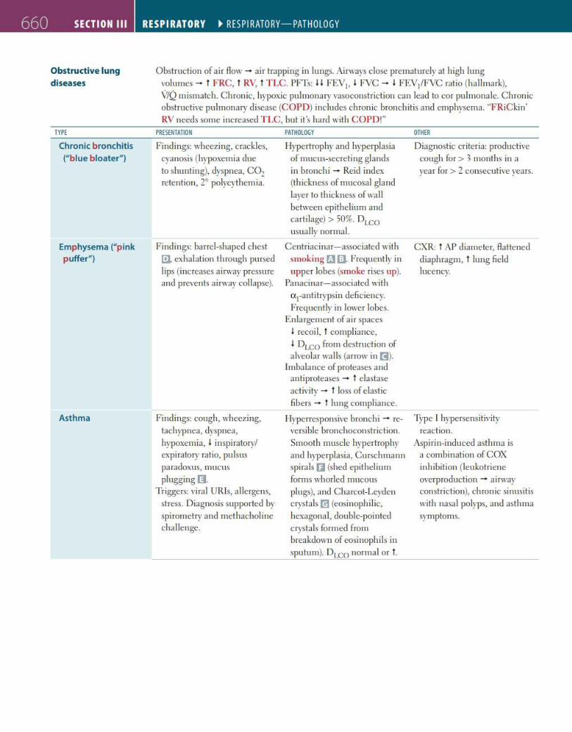
660 SECTION Ill
Obstructive lung
diseases
TYPE
Chronic bronchitis ("blue bloater")
Emphysema ("pink puffer")
Asthma
RESPIRATORY ~ RESP I RATORY-PATHOLOGY
Obstruction of air flow - ai r trapping in lungs. Airways close prematurely at high lung volumes - t F RC , t RV, t TLC. PFTs: H FE\11, I FVC - I FEV/FVC ratio (ha llmark), V/Q m ismatch. C hronic, hypoxic pu lmonary vasoconstriction can lead to cor pulmonale. C hronic obstructive pulmonary disease (COPD) inc ludes chronic bronchitis and emphysema. "FRiC kin' RV needs some increased TLC, but it's hard with COPD!"
PRESENTATION
Findings: wheezing, crackles, cyanosis (hypoxemia due to shunting), dyspnea, C02 retention, 2° polycythemia.
Findings: barrel-shaped chest [!J, exhalation th rough pursed lips (increases airway pressure and prevents airway collapse).
Findings: cough, wheezing, tachypnea, dyspnea, hypoxemia, I inspiratory/ expiratory ratio, pulsus paradoxus, mucus plugging 11.
Triggers: viral UR!s, allergens, stress. Diagnosis supported by spirometry and methachol ine challenge.
PATHOLOGY
Hypertrophy and hyperplasia of mucus-secreting glands in bronchi - Reid index (thickness of mucosa! gland layer to th ickness of wa ll between ep ithelium and carti lage) > 503 . DLCO usua lly normal.
Centriacinar- associated with smoking rJ I]. Frequent ly in upper lobes (smoke rises up).
Panacinar- associated with et1-antitrypsin deficiency. Frequently in lower lobes.
Enlargement of air spaces I recoil, t compliance, I DLCO from destruction of a lveolar walls (arrow in I]).
Imbalance of proteases and antiproteases - t elastase activity - t loss of elastic fi bers - t lung compliance.
Hyperresponsive bronchi - reversible bronchoconstric tion. Smooth muscle hypertrophy and hyperplasia, Curschmann spira ls 0 (shed epithelium forms whorled mucous plugs), and Charcot-Leyden crystals m (eosinophi lic, hexagonal, double-pointed crystals formed from breakdown of eosinophi ls in sputum). DLCO normal or t.
OTHER
Diagnostic cr iteria: productive cough for> 3 months in a year for > 2 consecutive years.
CXR: t AP d iameter, flattened d iaphragm, t lung fie ld lucency.
Type I hypersensitivity reaction.
Aspirin-induced asthma is a combination of COX inhibition (leukotriene overproduction - a irway constri ction), chronic sinusitis with nasa l polyps, and asthma symptoms.
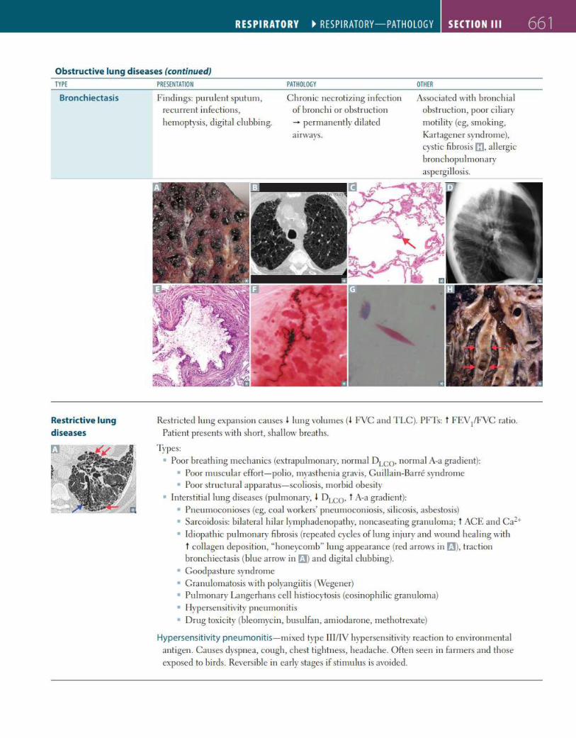
RESPIRATORY ~ RESPIRATORY-PATHOLOGY SECTION Ill 661
Obstructive lung diseases (continued) TYPE
Bronchiectasis
Restrictive lung
diseases
PRESENTATION
Findings: purulent sputum, recurrent infections, hemoptysis, d igital clubbing.
PATHOLOGY
Chronic necrotizing infection of bronchi or obstruction - permanent ly di lated ai rways.
OTHER
A5sociated wi th bronchia l obstruction, poor ci liary motility (eg, smoking, Kartagener syndrome}, cystic fi brosis m. allerg ic bronchopu lmonary aspergi llosis.
Restricted lung expansion causes I lung volumes (I FVC and TLC). PFTs: t FEV1/FVC ratio. Patient presents with short, sha llow breaths.
Types: • Poor breathing mechanics (extrapulmonary, normal DLCO' normal A-a gradient}:
• Poor muscula r effort-polio, myasthenia gravis, C uilla in-Barre syndrome • Poor structural apparatus- scoliosis, morbid obesity
• Interstitia l lung diseases (pulmonary, I DLCO• t A-a gradient): • Pneumoconioses (eg, coal workers' pneumoconiosis, silicosis, asbestos is) • Sarcoidosis: bilatera l h ilar lymphadenopathy, noncaseating gra nuloma; t ACE and Ca2+
• Idiopathic pulmonary fibrosis (repeated cycles of lung injury and wound healing with t collagen deposition, "honeycomb" lung appearance (red arrows in t'J), traction bronchiectasis (blue arrow in fJ) and dig ita l clubbing).
• Goodpasture syndrome • Cranulomatosis with polyangi itis (Wegener) • Pulmonary Langerhans cell histiocytosis (eosinophilic granuloma) • Hypersensi tivity pneumonitis • Drug tox icity (bleomycin, busulfan, amiodarone, methotrexate)
Hypersensit ivity pneumo nitis - mixed type III/IV hypersensitivity reaction to environmental antigen. Causes dyspnea, cough, chest t ightness, headache. Often seen in farmers and those exposed to birds. Reversible in early stages if stimulus is avoided .
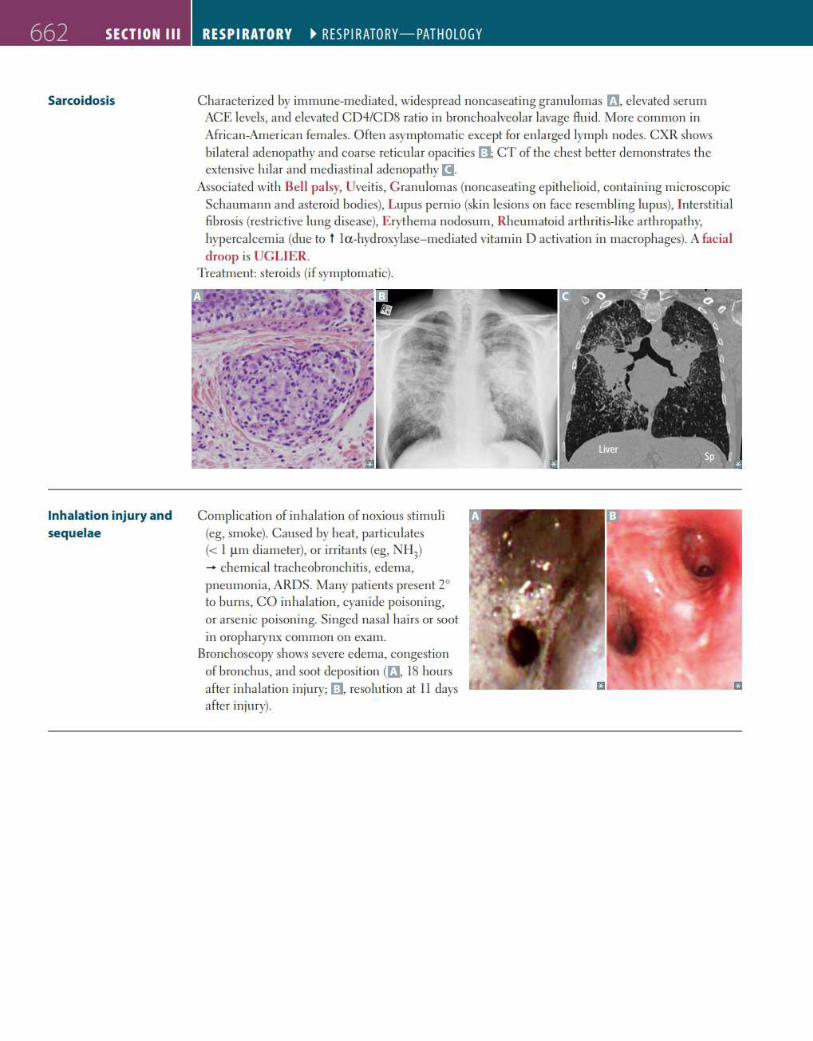
662 SECTION Ill
Sarcoidosis
Inhalation injury and sequelae
RESPIRATORY ~ RESPIRATORY-PATHOLOGY
Characterized by immune-mediated, widespread noncaseating granu lomas fJ, e levated serum ACE levels, and e levated CD4/CD8 ratio in bronchoalveolar lavage Au id. More common in African-American females. Often asymptomatic except for enla rged lymph nodes. CXR shows bilateral adenopathy and coarse reticular opacities (1 CT of the chest better demonstrates the extensive hi lar and mediastina l adenopathy l!I.
A~sociated with Bell palsy, Uveitis, C ranu lomas (noncaseating epithel ioid, contain ing microscopic Schaumann and asteroid bodies), Lupus pernio (skin lesions on face resembling lupus), Interstitia l fibrosis (restrict ive lung d isease), Erythema nodosum, Rheumatoid a rthritis-like arth ropathy, hypercalcemia (due to t la -hyd roxylase- mediated vitamin D activation in macrophages). A facial droop is UGLIER.
Treatment: steroids (if symptomatic).
Complication of inhalation of noxious stimuli (eg, smoke). Caused by heat, particulates (< I µ m d iameter), or irritants (eg, NH3)
- chemical tracheobronchitis, edema, pneumonia, ARDS. Many patients present 2° to bu rns, CO inhalation, cyanide poisoning, or arsen ic poisoning. Singed nasal ha irs or soot in oropharynx common on exam.
Bronchoscopy shows severe edema, congestion of bronchus, and soot deposition (fl, 18 hours after inha lation injury; 111, resolu tion at 11 days after injury).
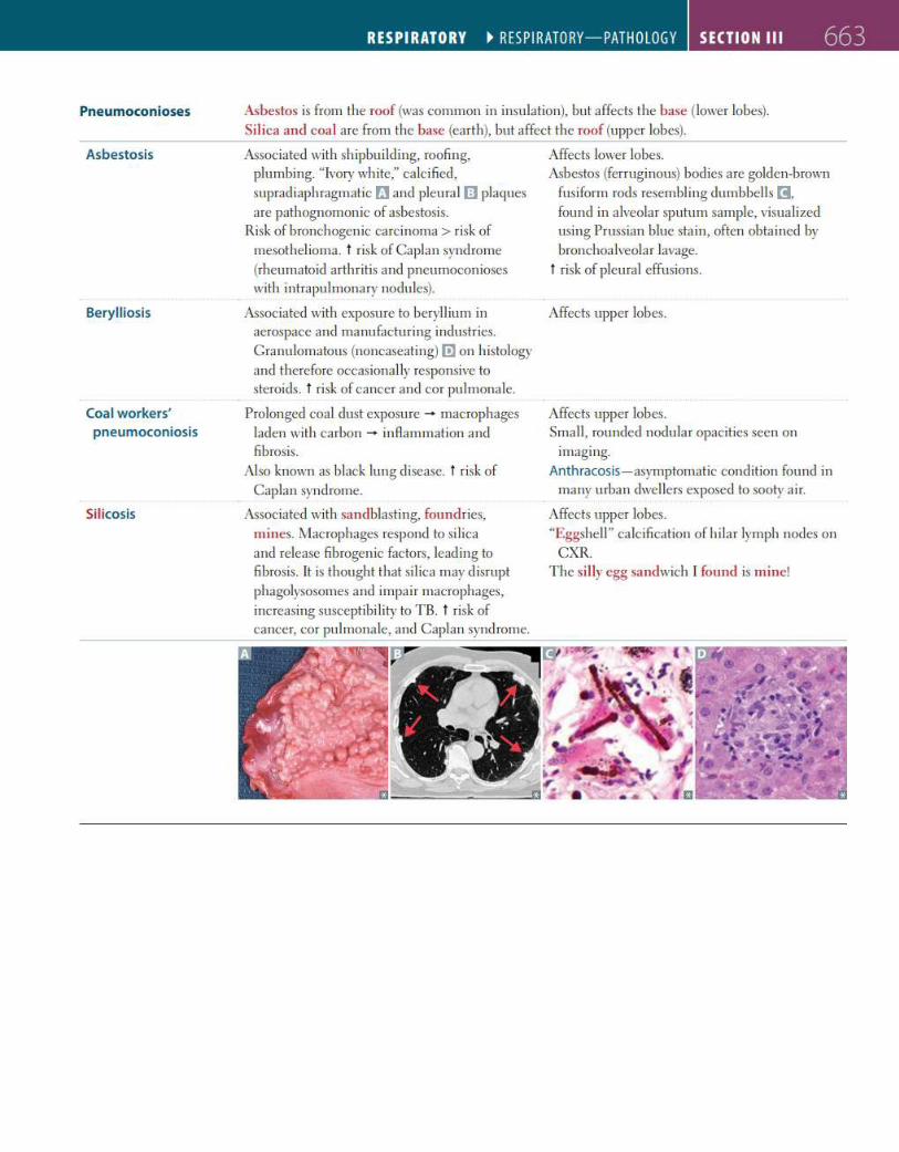
Pneumoconioses
Asbestosis
Berylliosis
Coal workers' pneumoconiosis
Silicosis
RESPIRATORY ~ RESPIRATORY-PATHOLOGY SECTION Ill 663
Asbestos is from the roof (was common in insulation), but affects the base (lower lobes). Silica and coal are from the base (earth), but affect the roof (upper lobes).
A5sociatecl with shipbuilding, roofing, plumbing. "Ivory white," calcified, supradiaphragmatic t1 and pleural rn plaques are pathognomonic of asbestosis.
Risk of bronchogenic carcinoma > risk of mesothelioma. t risk of Caplan syndrome (rheumatoid arth ritis and pneumoconioses with intrapulmonary nodules).
A5sociatecl with exposure to beryll ium in aerospace and manufacturing industries. Cranulomatous (noncaseating) [!] on histology and therefore occasionally responsive to steroids. t risk of cancer and cor pulmonale.
Prolonged coal dust exposure - macrophages laden with carbon - inAammation and fibrosis.
Also known as black lung disease. t risk of Caplan syndrome.
A5sociatecl with sandblasting, foundries, mines. Macrophages respond to silica and release fibrogen ic factors, leading to fi brosis. It is thought that si lica may disrupt phagolysosomes and impai r macrophages, increasing susceptibility to TB. t risk of cancer, cor pulmonale, and Caplan syndrome.
Affects lower lobes. A5bestos (ferruginous) bodies are golden-brown
fusiform rods resembling dumbbell s ~'
fou nd in alveolar sputum sample, visualized using Prussian blue stain, often obtainecl by bronchoalveolar lavage.
t risk of pleural effusions.
Affects upper lobes.
Affects upper lobes. Small, rounded nodular opacities seen on
imaging. Anthracosis- asymptomatic condition fou nd in
many urban clwellers exposecl to sooty air.
Affects upper lobes. "Eggshell" calcification of hilar lymph nodes on
CXR. The silly egg sandwich I found is mine!
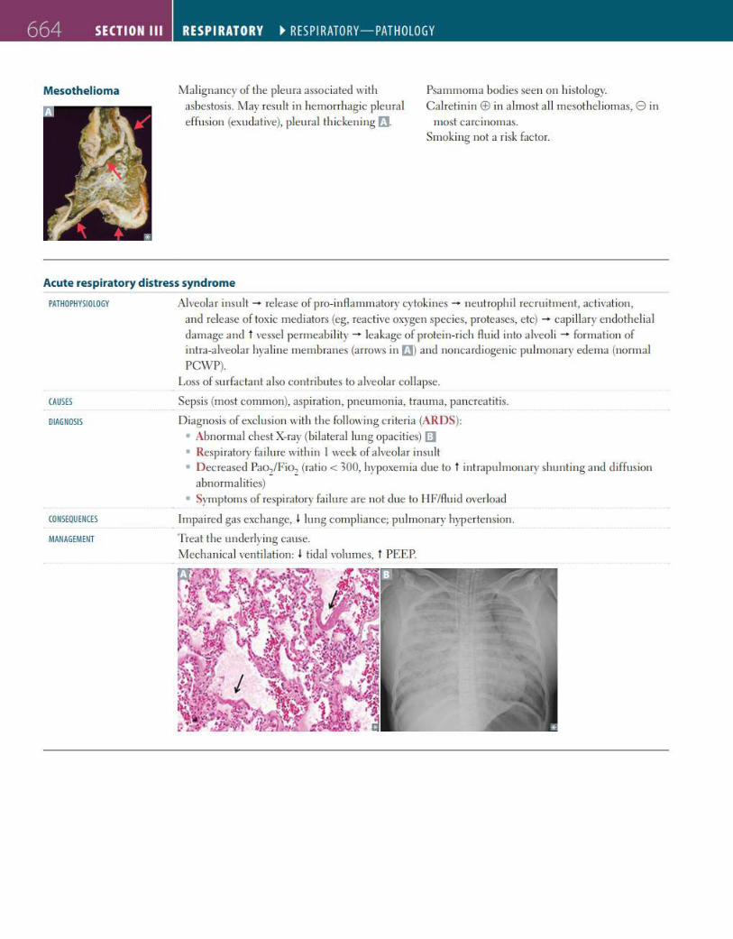
664 SECTION Ill
Mesothelioma
RESPIRATORY ~ RESPIRATORY-PATHOLOGY
Malignancy of the pleura associated with asbestosis. May result in hemorrhagic pleural effusion (exudative), pleura l thickening rJ.
Psammoma bodies seen on histology. Calre tin in EE> in a lmost a ll mesotheliomas, 8 in
most carcinomas. Smoking not a r isk factor.
Acute respiratory distress syndrome
PATHOPHYSIOLOGY
CAUSES
DIAGNOSIS
CONSEQUENCES
MANAGEMENT
Alveolar insu lt - release of pro-inAammatory cytokines - neutroph il recruitment, activation, and release of toxic mediators (eg, reactive oxygen species, proteases, etc) - capi llary endothel ia l damage a nd t vessel permeabi lity - leakage of protein-ri ch Auid into a lveol i - formation of intra-alveolar hyal ine membranes (arrows in fi.1) and noncardiogenic pulmonary edema (normal PCWP).
Loss of surfactant also contri butes to alveolar collapse.
Sepsis (most common), aspiration, pneumonia, trauma, pancreatitis.
Diagnosis of exclusion with the following criteria (ARDS): • Abnormal chest X-ray (bilatera l lu ng opacities) 1JJ • Respiratory fai lure with in I week of alveo lar insult • D ecreased Pao/Fio2 (ratio< 300, hypoxemia due to t intrapulmonary shunting and d iffusion
abnormal ities) • Symptoms of respiratory fa ilure are not due to I IF/Auid overload
Impai red gas exchange, I lung compl ia nce; pulmonary hypertension.
Treat the underlying cause. Mechanical ventilation: I tida l volumes, t PEEP.
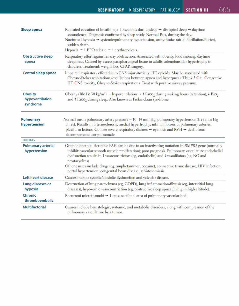
Sleepapnea
Obstructive sleep apnea
Central sleep apnea
Obesity hypoventilation syndrome
Pulmonary hypertension
ETIOLOGIES
Pulmonary arterial
hypertension
Left heart disease
Lung diseases or hypoxia
Chronic
thromboembolic
Multifactorial
RESPIRATORY ~ RESPIRATORY-PATHOLOGY SECTION Ill 665
Repeated cessation of breathing> lO seconds during sleep - d isrupted sleep - daytime somnolence. Diagnosis confi rmed by sleep study. Normal Pao2 during the day.
Nocturnal hypoxia - systemic/pu lmonary hypertension, arrhythmias (atria l fibri llation/flutter), sudden death.
Hypoxia - t EPO release - t erythropoiesis.
Respiratory effort aga inst a irway obstruction. A~sociated with obes ity, loud snoring, daytime sleepiness . Caused by excess parapharyngeal tissue in adu lts, adenotonsillar hypertrophy in children. T reatment: weight loss, CPAP, surgery.
Impai red respiratory effort due to CNS injury/toxicity, I IF, opioids. May be associated with Cheyne-Stokes respirations (osci llations between apnea and hyperpnea). T hink 3 C 's: C ongestive HF, C NS toxicity, C heyne-Stokes respirations. Treat with positive airway pressure.
Obesity (BMI 2: 30 kg/m2) - hypoventilation - t Paco2 during waking hours (retention); I Pao2 and t Paco2 during sleep. Also known as Pickwickian syndrome.
Normal mean pulmonary artery pressure = I0- 14 mm Hg; pulmonary hypertension 2: 25 mm Ilg at rest. Results in arteriosd er<>sis, med ial hypertrophy, intimal fibrosis of pulmonary arteries, plexiform les ions. Course: severe respiratory d istress - cyanosis and RVI I - death from decompensated cor pulmonale.
Often id iopathic. lleritable PAii can be due to an inactivating mutation in BMPR2 gene (normally in hibits vascu la r smooth muscle pr<> liferation); poor prognosis. Pu lmonary vascu lature e ndothe lial dysfu nc tion results in t vasoconstrictors (eg, e ndothelin) and I vasodi lators (eg, NO and prostacyd ins).
Other causes inc lude drugs (eg, amphetamines, cocaine), connective tissue d isease, J JJV infection, porta l hypertension, congenita l heart disease, schistosomiasis.
Causes inc lude systolic/diastolic dysfu nction and va lvular d isease.
Destruction of lung parenchyma (eg, COPD), lung inflammation/fibros is (eg, interstitial lung d iseases), hypoxemic vasoconstriction (eg, obstructive sleep apnea, living in h igh altitude).
Recurrent microthrombi - I cross-sectional area of pu lmonary vascu la r bed.
Causes inc lude hematologic, systemic, and metabolic disorders, a long with compress ion of the pu lmonary vascu lature by a tumor.
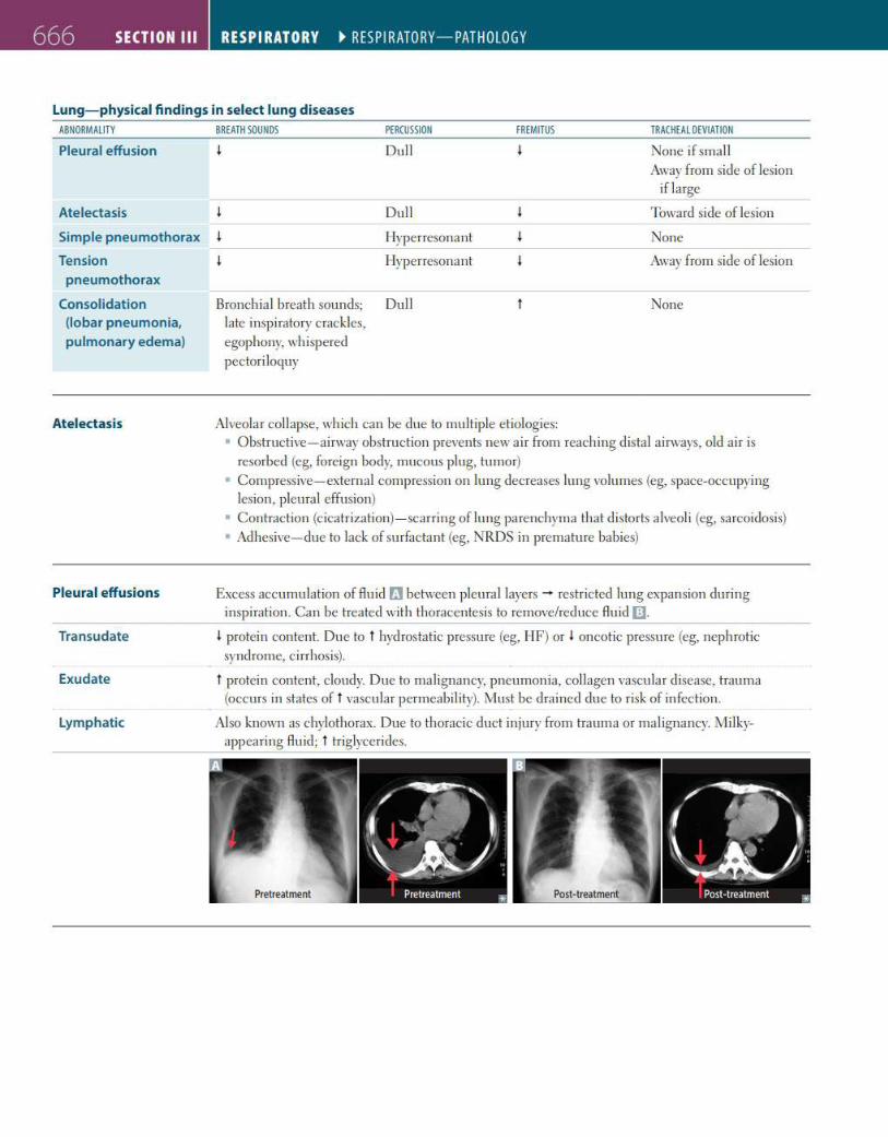
666 SECTION Ill RESPIRATORY ~ RESPIRATORY-PATHOLOGY
Lung- physical findings in select lung diseases ABNORMALITY
Pleural effusion
Atelectasis
Simple pneumothorax
Tension pneumothorax
Consolidation (lobar pneumonia, pulmonary edema)
Atelectasis
Pleural effusions
Transudate
Exudate
Lymphatic
BREATH SOUNDS PERCUSSION
Dull
Dull
I lyperresonant
I lyperresonant
Bronchia l breath sounds; Dull late inspiratory crackles, egophony, whispered pectori loguy
FREMITUS
Alveolar collapse, which can be due to multiple etiologies:
TRACHEAL DEVIATION
None if small Away from side of lesion
if large
Toward side of lesion
None
Away from side of lesion
None
• Obstructive- ai rway obstruction prevents new air from reaching distal ai rways, old a ir is resorbed (eg, foreign body, mucous plug, tumor)
• Compress ive- external compression on lung decreases lung volumes (eg, space-occupying lesion, pleural effusion)
• Contraction (c icatrization)- scarring of lung parenchyma that distorts a lveoli (eg, sarcoidosis) • Adhesive-due to lack of surfactant (eg, NRDS in premature babies)
Excess accumulation of Auid fl between pleural layers - restricted lung expansion during inspiration. Can be treated with thoracentesis to remove/reduce Au id Ill
I protein content. Due to t hydros tatic pressure (eg, IIF) or I oncotic pressure (eg, nephrotic syndrome, cirrhosis).
t protein content, cloudy. Due to mal ignancy, pneumonia, collagen vascula r disease, trauma (occurs in states oft vascular permeabi lity). Must be drained due to risk of infection.
Also known as chylothorax. Due to thoracic duct injury from trauma or malignancy. Milkyappea ring Auid; t triglycerides.
Post-treatment
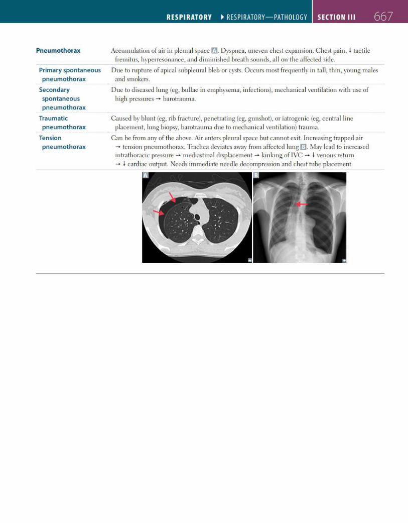
Pneumothorax
Primary spontaneous
pneumothorax
Secondary
spontaneous
pneumothorax
Traumatic
pneumothorax
Tension
pneumothorax
RESPIRATORY ~ RESPIRATORY-PATHOLOGY SECTION Ill 667
Accumulation of ai r in pleura l space f'J. Dyspnea, uneven chest expansion. Chest pa in, I tacti le fremitus, hyperresonance, and dimin ished breath sounds, a ll on the affected side.
Due to rupture of apical subpleura l bleb or cysts. Occurs most frequently in tall, thin, young males and smokers.
Due to diseased lung (eg, bullae in emphysema, infections), mechanical venti lation with use of high pressures - barotrauma.
Caused by blunt (eg, rib fracture), penetrating (eg, gunshot), or iatrogen ic (eg, centra l line placement, lung biopsy, barotrauma due to mechanical ventilation) trauma.
Can be from any of the above. Ai r enters pleura l space but cannot exit. Increasing trapped a ir - tension pneumothorax. Trachea deviates away from affected lung [lJ. May lead to increased intrathoracic pressure - mediastinal d isplacement - kinking of IVC - I venous return - I card iac output. Needs immediate need le decompression and c hest tube placement.
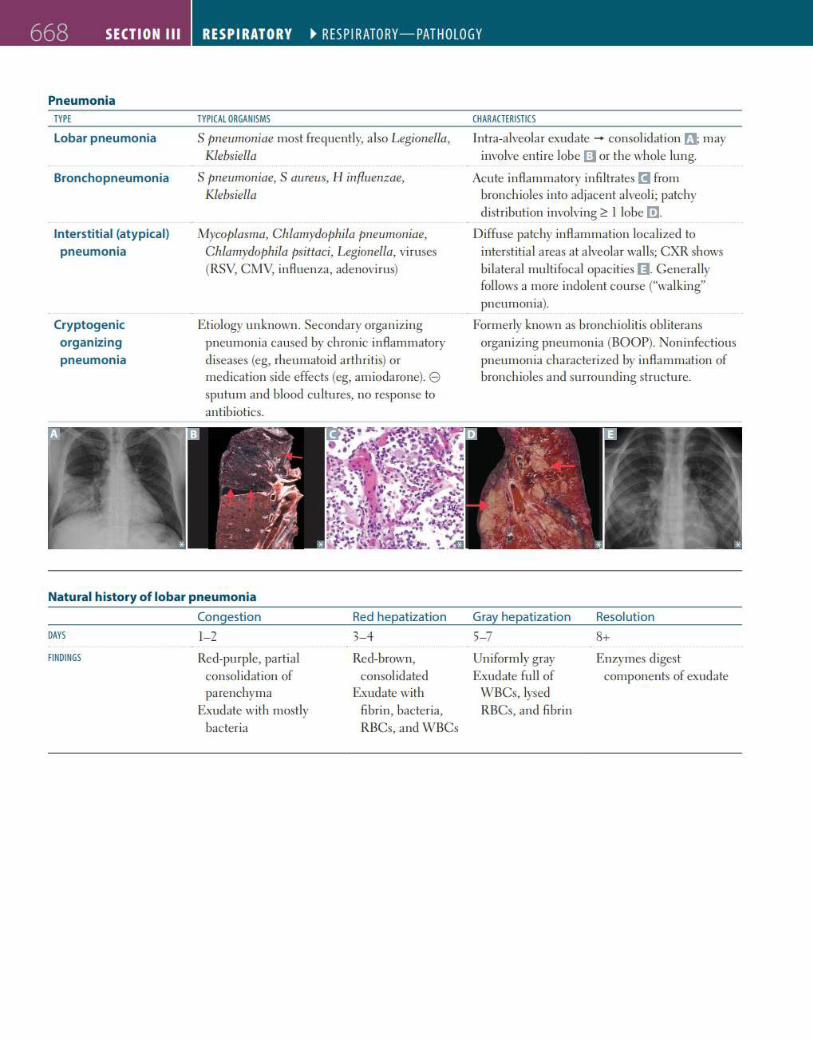
668 SECTION Ill RESPIRATORY ~ RESPIRATORY-PATHOLOGY
Pneumonia TYPE
Lobar pneumonia
Bronchopneumonia
Interstitial (atypical) pneumonia
Cryptogenic organizing
pneumonia
TYPICAL ORGANISMS
S pneumoniae most frequently, also Legionella,
Klebsiella
S pneumoniae, S aureus, 11 inf/uen;zae,
Klebsiella
Mycoplasma, Chlamydophila pneumoniae,
Chlamydophila psittaci, Legionella, viruses (RSV, CMV, inAuenza, adenovirus)
Etiology unknown. Secondary organizing pneumonia caused by chronic inAammatory diseases (eg, rheumatoid arthritis) or medication side effects (eg, amiodarone). e sputum and blood cultures, no response to antibiotics.
Natural history of lobar pneumonia
Congestion Red hepatizat ion
DAYS 1-2 3- 4 FINDINGS Red-purple, partial Red-brown,
consolidation of consolidated parenchyma Exudate with
Exudate with mostly fibrin, bacteria, bacteria RBCs, and WBCs
CHARACTERISTICS
Intra-alveolar exudate - consolidation rJ; may involve entire lobe a or the whole lung.
Acute inAammatory infi ltrates E!I from bronchioles into adjacent alveol i; patchy distribution involvi ng ~ I lobe (!].
Diffuse patchy inAammation localized to interstitial areas at alveolar walls; CXR shows bilateral multifocal opacities I]. Generally follows a more indolent course ("walking" pneumonia).
Formerly known as bronchiolitis obliterans organizing pneumonia (BOOP). Noninfectious pneumonia characterized by inAammation of bronchioles and surrounding structure.
Gray hepatization Resolution
5- 7 8+
Uniformly gray Enzymes diges t Exudate fu ll of components of exudate
WBCs, lysed RBCs, and fibrin
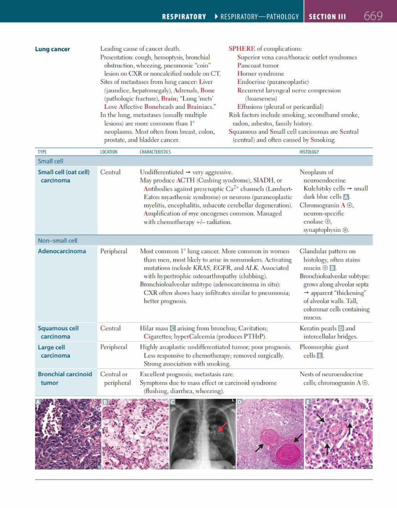
Lung cancer
TYPE
Small cell
Small cell (oat cell) carcinoma
Non-small cell
Adenocarcinoma
RESPIRATORY ~ RESPIRATO RY-PATHOLOGY SECTION Ill 669
Leading cause of cancer death. SPHERE of complications: Presentation: cough, hemoptysis, bronchial
obstruction, wheezing, pneumonic "coin" lesion on CXR or noncalcified nodule on CT.
Superior vena cava/thoracic outlet syndromes Pancoast tumor Horner syndrome
Sites of metastases from lung cancer: Liver (jaundice, hepatomegaly), Ad renals, Bone (pathologic fracture), Brain; "Lung 'mets' Love Affective Boneheads and Brainiacs."
E ndocrine (paraneoplastic) Recurrent laryngeal nerve compression
(hoarseness) E ffusions (pleural or pericardia!)
In the lung, metastases (usually multiple lesions) are more common than 1° neop lasms. Most often from breast, colon, prostate, and bladder cancer.
Risk factors include smoking, secondhand smoke, radon, asbestos, fam ily h istory.
Squamous and Small cell carcinomas are Sentra l (centra l) and often caused by Smoking.
LOCATION
Central
Peripheral
CHARACTERISTICS
Undifferentiated - very aggressive. May produce ACTI I (C ushing syndrome), SIADI I, or
Antibodies against presynaptic Ca2+ channels (LambertEaton myasthenic syndrome) or neurons (para neoplastic myeli tis, encepha litis, subacute cerebellar degeneration). Amplification of myc oncogenes common. Managed with chemotherapy+/- rad iation.
Most common 1° lung cancer. More common in women than men, most likely to arise in nonsmokers. Activating mutations inc lude KRAS, ECFR, and /\LK. Associated with hypertrophic osteoarthropathy (c lubbing).
Bronchioloalveolar subtype (adenoca rcinoma in situ): CXR often shows hazy infi ltrates simi lar to pneumonia; better prognosis.
HISTOLOGY
Neoplasm of neuroendocrine Ku lchitsky cells - small dark blue cells rJ.
C hromogranin A ®, neuron-speci fie enolase®, synaptophysin ®·
C landula r pattern on histology, often stains mucin ® [1J.
Bronchioloalveolar subtype: grows a long alveolar septa - apparent "thickening" of alveolar wa lls. Tall, columnar cells conta ining mucus.
Squamous cell C entra l ll ilar mass m a rising from bronchus; Cavitation; C igarettes; hyperC alcemia (produces PTHrP).
Keratin pearls [!J and intercellular bridges. carcinoma
Large ce ll Peripheral carcinoma
Bronchial carcinoid Central or tumor periphera l
ll ighly anaplastic undi fferentiated tumor; poor prognosis. Less responsive to chemotherapy; removed surgically. Strong association with smoking.
Excellent prognosis; metastasis rare. Symptoms due to mass effect or carcinoid syndrome
(flushing, diarrhea, wheezing).
Pleomorph ic giant cells O .
Nests of neuroendocrine cells; chromogranin A®·
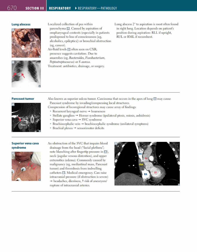
6 70 SECTION Ill
Lung abscess
Pancoast tumor
Superior vena cava
syndrome
RESPIRATORY ~ RESPIRATORY-PATHOLOGY
Localized collection of pus with in parenchyma ri.]. Caused by aspiration of oropharyngeal contents (especially in patients predisposed to loss of consciousness [eg, a lcoholics, epileptics)) or bronchia l obstruction (eg, cancer).
Air-Auid levels I] often seen on CXR; presence suggests cavitation. Due to anaerobes (eg, Bacteroides, Fusobacterium, Peptostreptococcus) or S aureus.
Treatment: antibiotics, dra inage, or surgery.
Lung abscess 2° to aspiration is most often found in right lung. Location depends on patien t's position dur ing aspiration: RLL if upright, RUL or RML if recumbent.
Also known as superior su lcus tumor. Carcinoma that occurs in the apex of lung f'J may cause Pancoast syndrome by invad ing/compressing local structures.
Compress ion of locoregional structures may cause array of find ings: • Recurrent laryngeal nerve - hoarseness • Stellate ganglion - Ilorner syndrome (ipsilateral ptosis, miosis, anh idrosis) • Superior vena cava - SVC syndrome • Brachiocepha lic vein - brachiocepha lic syndrome (un ilateral symptoms) • Brachia! plexus - sensorimotor deficits
An obstruction of the SVC that impairs blood dra inage from the head ("facia l plethora"; note blanching after fingertip pressure in f'J}, neck (jugula r venous distention), and upper extremities (edema). Commonly caused by malignancy (eg, mediastinal mass, Pancoast tumor) and thrombosis from indwell ing cathe ters I]. Medical emergency. Can raise intracran ia l pressure (if obstruction is severe) - headaches, dizziness, t risk of aneurysm/ rupture of intracranial arteries.
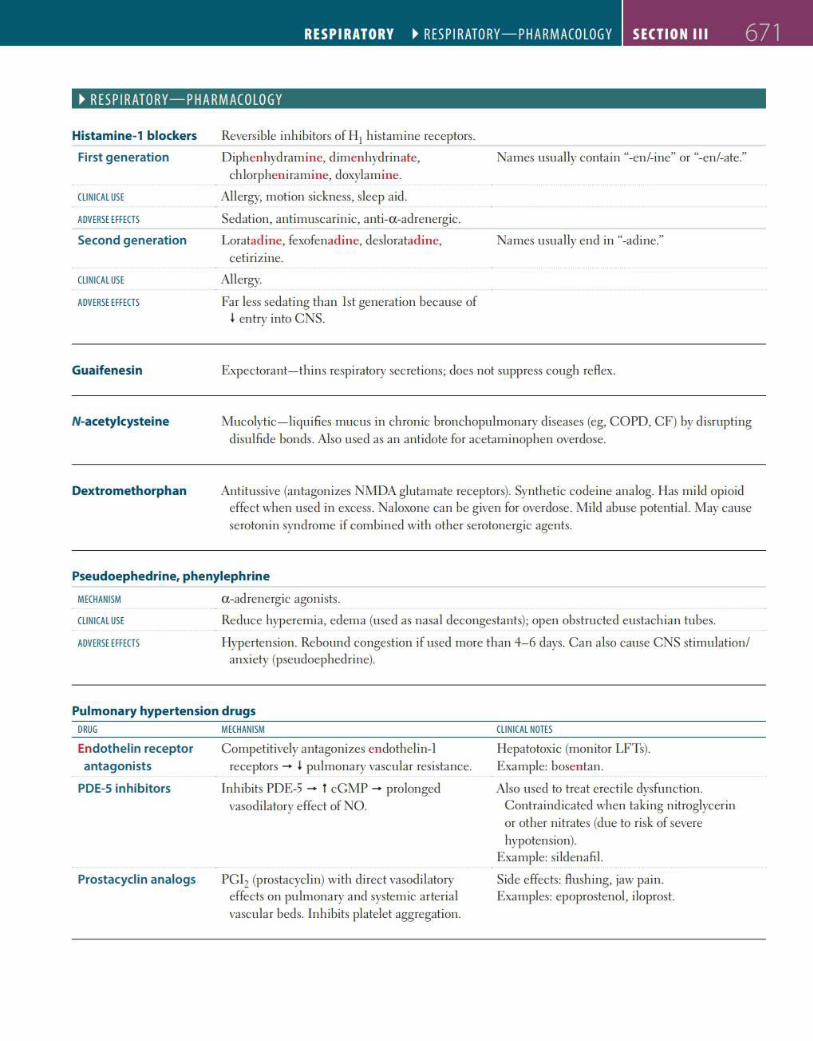
RESP IRATORY ~ RESPIRATORY-PHARMACOLOGY SECTION Ill 671
~ RES PIRATORY-PHA RMACOLOGY
Histamine-1 blockers
First generation
CLINICAL USE
ADVERSE EFFECTS
Second generation
CLINICAL USE
ADVERSE EFFECTS
Guaifenesin
N-acetylcysteine
Dextromethorphan
Reversible inh ibitors of I 11 histamine receptors .
Diphenhydramine, dimenhydrinate, ch lorpheniramine, doxylamine.
Allergy, motion sickness, sleep aid.
Sedation, antimuscarinic, anti-a -adrenerg ic.
Loratadine, fexofenadine, desloratadine, ceti ri zine.
Allergy.
Far less sedating than !st generation because of I en try into CNS.
Names usually contain "-en/-ine" or "-en/-ate."
Names usually end in "-ad ine."
Expectora nt-th ins respiratory secretions; does not suppress cough reflex.
Mucolytic- liquifies mucus in chronic bronchopu lmonary diseases (eg, COPD, CF) by disrupting d isulfide bonds. Also used as an antidote for acetaminophen overdose.
Antitussive (antagonizes NMDA glutamate receptors). Synthetic codeine analog. Ilas mi ld opioid effect when used in excess. Naloxone can be g iven for overdose. Mild abuse potentia l. May cause serotonin syndrome if combined with other serotonergic agents.
Pseudoephedrine, phenylephrine
MECHANISM
CLINICAL USE
ADVERSE EFFECTS
a -ad renergic agonists.
Reduce hyperemia, edema (used as nasa l decongestants); open obstructed eustachian tubes.
Hypertension. Rebound congestion if used more than 4- 6 days. Can a lso cause CNS stimulation/ anxiety (pseudoephed rine).
Pulmonary hypertension drugs DRUG
Endothelin receptor
antagonists
PDE-5 inhibitors
Prostacyclin analogs
MECHANISM
Competitively antagonizes endothel in-1 receptors - I pulmonary vascular resistance.
Inhibits PDE-5 - t cGMP - prolonged vasodilatory effect of NO.
PGI2 (prostacyclin) with d irect vasodilatory effects on pulmonary and systemic arterial vascular beds. Inh ibits platelet aggregation.
CLINICAL NOTES
I lepatotoxic (monitor LFTs). Example: bosentan.
Also used to treat erectile dysfunction. Contraindicated when ta king nitroglycerin or other n itrates (due to risk of severe hypotension).
Example: si ldenafil.
Side effects: flush ing, jaw pain. Examples: epoprostenol, iloprost.
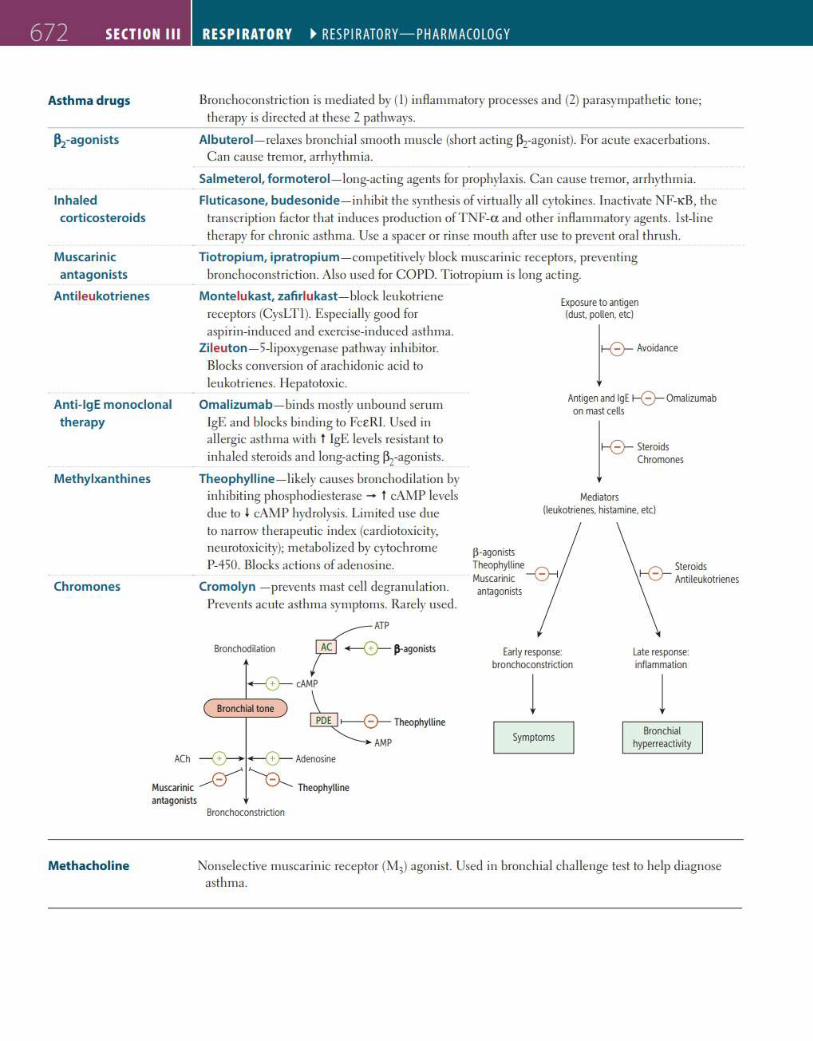
672 SECTION Ill
Asthma drugs
~-agonists
Inhaled corticosteroids
Muscarinic antagonists
Antileukotrienes
Anti-lgE monoclonal therapy
Methylxanthines
Chromones
RESPIRATORY ~ RESP I RATORY-PHARMACOLOGY
Bronchoconstric tion is mediated by ( I) inAammatory processes and (2) parasympathetic tone; therapy is directed at these 2 pathways.
Albuterol-relaxes bronchia l smooth muscle (short acting ~z-agon ist). For acute exacerbations. Can cause tremor, arrhythmia.
Salmeterol, formoterol-long-acting agents for prophylaxis. Can cause tremor, arrhythmia.
Fluticasone, budesonide-inhibit the synthesis of virtually all cytokines. Inactivate NF-KB, the transcription factor that induces production ofTNF-et and other inAammatory agents. 1st-line therapy for chronic asthma. Use a spacer or rinse mouth after use to prevent ora l thrush.
Tiotropium, ip ratropium - competitively block muscarinic receptors, preventing bronchoconstriction. Also used for COPD. Tiotropium is long ac ting.
Montelukast, zafi rlukast-block leukotriene
receptors (CysLTI). Especially good for aspirin-induced and exercise-induced asthma.
Zileuton- 5-1 ipoxygenase pathway inhibitor. Blocks conversion of arachidonic acid to leukotrienes. I lepatotoxic.
Omalizumab-binds mostly unbound serum IgE and blocks binding to FceRL Used in allerg ic asthma wi th t IgE levels resistant to inha led steroids and long-acting ~z-agonists.
Theophyll ine- likely causes bronchodi lation by inhibiting phosphodies terase - t cAMP levels due to I cAMP hydrolysis. Limited use due to na rrow therapeutic index (cardiotoxicity, neurotoxicity); metabolized by cytochrome P-450. Blocks actions of adenosine.
Exposure to antigen (dust pollen. etc)
1~~··· Antigen and lg£ r-0- Omalizumab
on mast cells
lr-0- Steroids Chromones
Mediators (leukotrienes. histamine. etc)
Cromolyn - prevents mast cell degranulation. antagonists
Prevents acute asthma symptoms. Rare ly used.
ATP
Bronchodilatioo ~ jl-agonists
-0-cAMP
Bronchial tone ~ i--0- Theophylline
AMP
Early response: bronchoconstriction
l Symptoms
l ate response: inflammation
l Bronchial
hyperreactivity
ACh --0--+ -0-Adenosine
Muscarinic ~ "R Theophylline antagonists
Methacholine
Bronchoconstrictioo
Nonselective muscarinic receptor (M,) agonist. Used in bronchia l challenge test to help d iagnose asthma.
Appendix :
First Aid for Basic Sciences (ORGAN SYSTEMS)
747
C H A P T E R 1 0
Respiratory
EMBRYOLOGY 748Respiratory Development 748Congenital Malformations 751
ANATOMY 753Airways 754Lungs 754Diaphragm 756External Anatomy 757Muscles of Respiration 757Flail Chest 758
HISTOLOGY 758Respiratory Epithelium 759Alveoli 759Olfactory Cells 761
PHYSIOLOGY 761Lung Volumes and Capacities 761Ventilation 763Blood Gases 770
PATHOLOGY 779Pathology on Physical Examination 779Nasopharynx 781Obstructive Lung Diseases 783Restrictive Lung Diseases 790Pulmonary Vascular Diseases 798Respiratory Tract Cancers 803Pulmonary Infections 808Pleural Effusion 815Pneumothorax 815Allergy 817Hypersensitivity Pneumonitis 818
PHARMACOLOGY 818Histamine Blockers 818Mucoactive Agents 819Dextromethorphan 819α-Adrenergic Agonists 820Pulmonary Hypertension Drugs 820Asthma Drugs 821
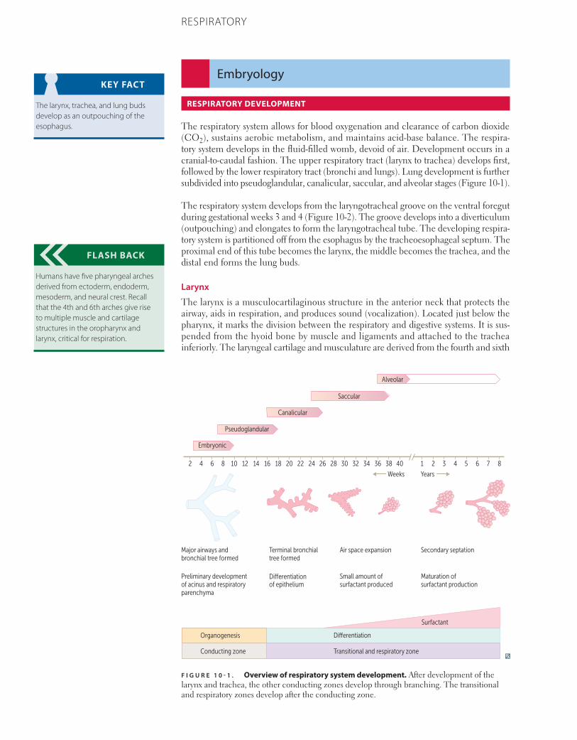
748 CHAPTER 10 RESPIRATORY
Embryology
RESPIRATORY DEVELOPMENT
The respiratory system allows for blood oxygenation and clearance of carbon dioxide (CO2), sustains aerobic metabolism, and maintains acid-base balance. The respira-tory system develops in the fluid-filled womb, devoid of air. Development occurs in a cranial-to-caudal fashion. The upper respiratory tract (larynx to trachea) develops first, followed by the lower respiratory tract (bronchi and lungs). Lung development is further subdivided into pseudoglandular, canalicular, saccular, and alveolar stages (Figure 10-1).
The respiratory system develops from the laryngotracheal groove on the ventral foregut during gestational weeks 3 and 4 (Figure 10-2). The groove develops into a diverticulum (outpouching) and elongates to form the laryngotracheal tube. The developing respira-tory system is partitioned off from the esophagus by the tracheoesophageal septum. The proximal end of this tube becomes the larynx, the middle becomes the trachea, and the distal end forms the lung buds.
Larynx
The larynx is a musculocartilaginous structure in the anterior neck that protects the airway, aids in respiration, and produces sound (vocalization). Located just below the pharynx, it marks the division between the respiratory and digestive systems. It is sus-pended from the hyoid bone by muscle and ligaments and attached to the trachea inferiorly. The laryngeal cartilage and musculature are derived from the fourth and sixth
KEY FACT
The larynx, trachea, and lung buds develop as an outpouching of the esophagus.
FLASH BACK
Humans have five pharyngeal arches derived from ectoderm, endoderm, mesoderm, and neural crest. Recall that the 4th and 6th arches give rise to multiple muscle and cartilage structures in the oropharynx and larynx, critical for respiration.
F I G U R E 1 0 - 1 . Overview of respiratory system development. After development of the larynx and trachea, the other conducting zones develop through branching. The transitional and respiratory zones develop after the conducting zone.
2 4 6 8 10 12 14 16 18 20 22 24 26 28 30 32 34 36 38 40 1 2 3 4 5 6 7 8
Embryonic
Major airways andbronchial tree formed
Preliminary developmentof acinus and respiratoryparenchyma
Terminal bronchialtree formed
of epitheliumDi�erentiation
Di�erentiation
Air space expansion
Small amount ofsurfactant produced
Secondary septation
Maturation ofsurfactant production
YearsWeeks
Pseudoglandular
Canalicular
Saccular
Alveolar
Organogenesis
Conducting zone
Surfactant
Transitional and respiratory zone
Di�erentiation
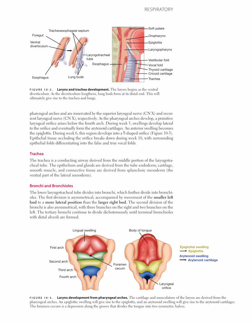
749CHAPTER 10RESPIRATORY
pharyngeal arches and are innervated by the superior laryngeal nerve (CN X) and recur-rent laryngeal nerve (CN X), respectively. As the pharyngeal arches develop, a primitive laryngeal orifice arises below the fourth arch. During week 5, swellings develop lateral to the orifice and eventually form the arytenoid cartilages. An anterior swelling becomes the epiglottis. During week 6, this region develops into a T-shaped orifice (Figure 10-3). Epithelial tissue occluding the orifice breaks down during week 10, with surrounding epithelial folds differentiating into the false and true vocal folds.
Trachea
The trachea is a conducting airway derived from the middle portion of the laryngotra-cheal tube. The epithelium and glands are derived from the tube endoderm; cartilage, smooth muscle, and connective tissue are derived from splanchnic mesoderm (the ventral part of the lateral mesoderm).
Bronchi and Bronchioles
The lower laryngotracheal tube divides into bronchi, which further divide into bronchi-oles. The first division is asymmetrical, accompanied by movement of the smaller left bud to a more lateral position than the larger right bud. The second division of the bronchi is also asymmetrical, with three branches on the right and two branches on the left. The tertiary bronchi continue to divide dichotomously until terminal bronchioles with distal alveoli are formed.
F I G U R E 1 0 - 2 . Larynx and trachea development. The larynx begins as the ventral diverticulum. As the diverticulum lengthens, lung buds form at its distal end. This will ultimately give rise to the trachea and lungs.
Soft palate
Oropharynx
Epiglottis
Laryngopharynx
EsophagusVestibular fold
Vocal foldThyroid cartilageCricoid cartilage
Trachea
Foregut
Ventral diverticulum
Esophagus
Tracheoesophageal septum
Lung buds
Laryngotrachealtube
F I G U R E 1 0 - 3 . Larynx development from pharyngeal arches. The cartilage and musculature of the larynx are derived from the pharyngeal arches. An epiglottic swelling will give rise to the epiglottis, and an arytenoid swelling will give rise to the arytenoid cartilages. The foramen cecum is a depression along the groove that divides the tongue into two symmetric halves.
Foramencecum
Laryngealori�ce
Lingual swelling
First arch
Second arch
Third arch
Fourth arch
Body of tongue
Epiglottal swellingEpiglottis
Arytenoid swellingArytenoid cartilage
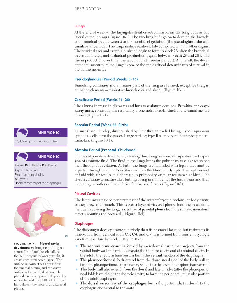
750 CHAPTER 10 RESPIRATORY
Lungs
At the end of week 4, the laryngotracheal diverticulum forms the lung buds as two lateral outpouchings (Figure 10-1). The two lung buds go on to develop the bronchi and bronchial tree between 2 and 7 months of gestation (the pseudoglandular and canalicular periods). The lungs mature relatively late compared to many other organs. The terminal sacs and eventually alveoli begin to form in week 26 when the bronchial tree is completed, and surfactant production begins between weeks 25 and 28 with a rise in production over time (the saccular and alveolar periods). As a result, the devel-opmental maturity of the lungs is one of the most critical determinants of survival in premature neonates.
Pseudoglandular Period (Weeks 5–16)
Branching continues and all major parts of the lung are formed, except for the gas-exchange elements—respiratory bronchioles and alveoli (Figure 10-1).
Canalicular Period (Weeks 16–26)
The airways increase in diameter and lung vasculature develops. Primitive end-respi-ratory units, consisting of a respiratory bronchiole, alveolar duct, and terminal sac, are formed (Figure 10-1).
Saccular Period (Week 26–Birth)
Terminal sacs develop, distinguished by their thin epithelial lining. Type I squamous epithelial cells form the gas-exchange surface; type II secretory pneumocytes produce surfactant (Figure 10-1).
Alveolar Period (Prenatal–Childhood)
Clusters of primitive alveoli form, allowing “breathing” in utero via aspiration and expul-sion of amniotic fluid. The fluid in the lungs keeps the pulmonary vascular resistance high throughout gestation. At birth, the lungs are half-filled with liquid that must be expelled through the mouth or absorbed into the blood and lymph. The replacement of fluid with air results in a decrease in pulmonary vascular resistance at birth. The alveoli continue to mature after birth, growing in number for the first 3 years and then increasing in both number and size for the next 5 years (Figure 10-1).
Pleural Cavities
The lungs invaginate to penetrate part of the intraembryonic coelom, or body cavity, as they grow and branch. This leaves a layer of visceral pleura from the splanchnic mesoderm covering the lung, and a layer of parietal pleura from the somatic mesoderm directly abutting the body wall (Figure 10-4).
Diaphragm
The diaphragm develops more superiorly than its postnatal location but maintains its innervation from cervical roots C3, C4, and C5. It is formed from four embryologic structures that fuse by week 7 (Figure 10-5):
■ The septum transversum is formed by mesodermal tissue that projects from the ventral body wall to partially separate the thoracic cavity and abdominal cavity. In the adult, the septum transversum forms the central tendon of the diaphragm.
■ The pleuroperitoneal folds extend from the dorsolateral sides of the body wall to form the pleuroperitoneal membranes, which then fuse with the septum transversum.
■ The body wall also extends from the dorsal and lateral sides (after the pleuroperito-neal folds have closed the thoracic cavity) to form the peripheral, muscular portion of the adult diaphragm.
■ The dorsal mesentery of the esophagus forms the portion that is dorsal to the esophagus and ventral to the aorta.
MNEMONIC
C3, 4, 5 keep the diaphragm alive.
MNEMONIC
Several Parts Build a Diaphragm:
Septum transversumPleuroperitoneal foldsBody wallDorsal mesentery of the esophagus
F I G U R E 1 0 - 4 . Pleural cavity development. Imagine pushing on a partially inflated beach ball. As the ball invaginates over your fist, it creates two juxtaposed layers. The surface in contact with your fist is the visceral pleura, and the outer surface is the parietal pleura. The pleural cavity is a potential space that normally contains < 10 mL fluid and lies between the visceral and parietal pleura.
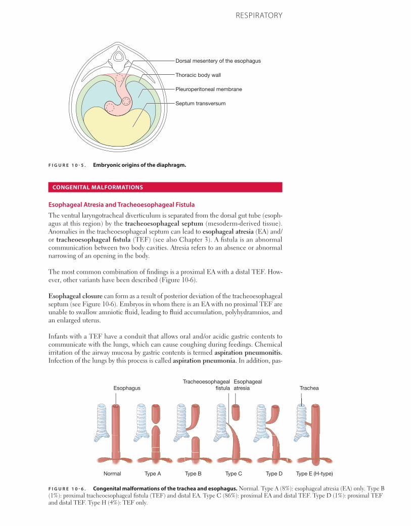
751CHAPTER 10RESPIRATORY
CONGENITAL MALFORMATIONS
Esophageal Atresia and Tracheoesophageal Fistula
The ventral laryngotracheal diverticulum is separated from the dorsal gut tube (esoph-agus at this region) by the tracheoesophageal septum (mesoderm-derived tissue). Anomalies in the tracheoesophageal septum can lead to esophageal atresia (EA) and/or tracheoesophageal fistula (TEF) (see also Chapter 3). A fistula is an abnormal communication between two body cavities. Atresia refers to an absence or abnormal narrowing of an opening in the body.
The most common combination of findings is a proximal EA with a distal TEF. How-ever, other variants have been described (Figure 10-6).
Esophageal closure can form as a result of posterior deviation of the tracheoesophageal septum (see Figure 10-6). Embryos in whom there is an EA with no proximal TEF are unable to swallow amniotic fluid, leading to fluid accumulation, polyhydramnios, and an enlarged uterus.
Infants with a TEF have a conduit that allows oral and/or acidic gastric contents to communicate with the lungs, which can cause coughing during feedings. Chemical irritation of the airway mucosa by gastric contents is termed aspiration pneumonitis. Infection of the lungs by this process is called aspiration pneumonia. In addition, pas-
F I G U R E 1 0 - 5 . Embryonic origins of the diaphragm.
Thoracic body wall
Septum transversum
Dorsal mesentery of the esophagus
Pleuroperitoneal membrane
F I G U R E 1 0 - 6 . Congenital malformations of the trachea and esophagus. Normal. Type A (8%): esophageal atresia (EA) only. Type B (1%): proximal tracheoesophageal fistula (TEF) and distal EA. Type C (86%): proximal EA and distal TEF. Type D (1%): proximal TEF and distal TEF. Type H (4%): TEF only.
Esophagealatresia
Tracheoesophageal�stulaEsophagus Trachea
Normal Type A Type B Type C Type D Type E (H-type)
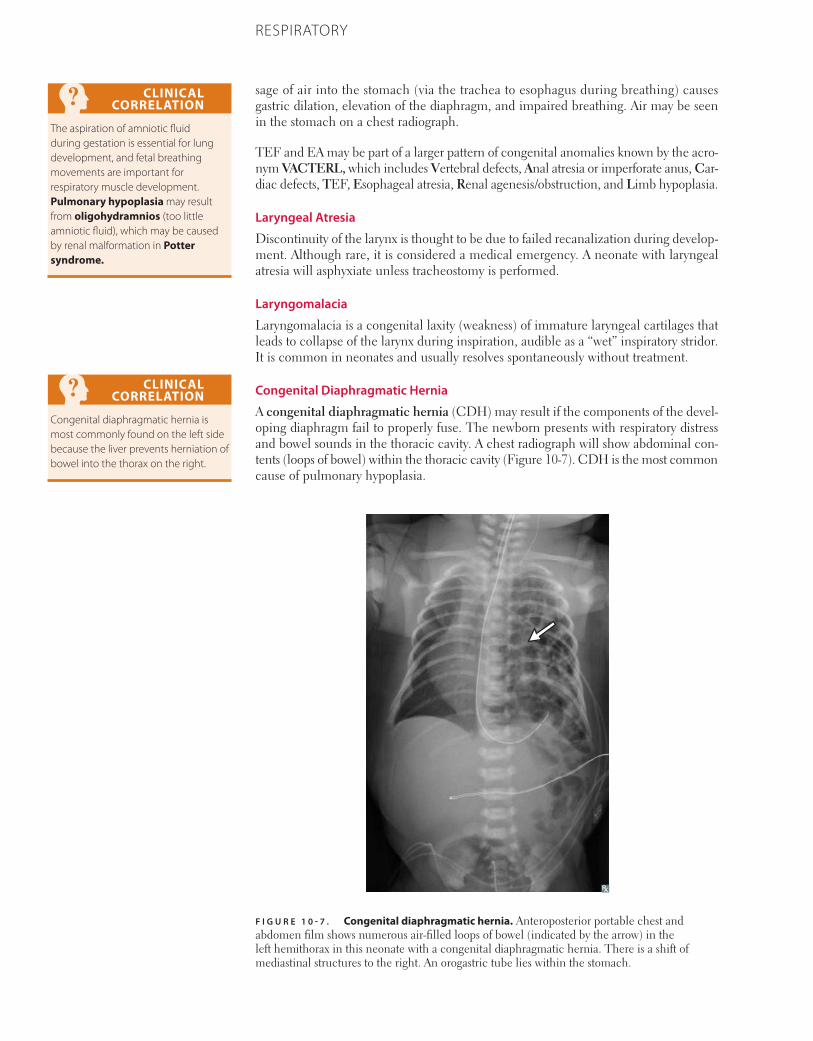
752 CHAPTER 10 RESPIRATORY
sage of air into the stomach (via the trachea to esophagus during breathing) causes gastric dilation, elevation of the diaphragm, and impaired breathing. Air may be seen in the stomach on a chest radiograph.
TEF and EA may be part of a larger pattern of congenital anomalies known by the acro-nym VACTERL, which includes Vertebral defects, Anal atresia or imperforate anus, Car-diac defects, TEF, Esophageal atresia, Renal agenesis/obstruction, and Limb hypoplasia.
Laryngeal Atresia
Discontinuity of the larynx is thought to be due to failed recanalization during develop-ment. Although rare, it is considered a medical emergency. A neonate with laryngeal atresia will asphyxiate unless tracheostomy is performed.
Laryngomalacia
Laryngomalacia is a congenital laxity (weakness) of immature laryngeal cartilages that leads to collapse of the larynx during inspiration, audible as a “wet” inspiratory stridor. It is common in neonates and usually resolves spontaneously without treatment.
Congenital Diaphragmatic Hernia
A congenital diaphragmatic hernia (CDH) may result if the components of the devel-oping diaphragm fail to properly fuse. The newborn presents with respiratory distress and bowel sounds in the thoracic cavity. A chest radiograph will show abdominal con-tents (loops of bowel) within the thoracic cavity (Figure 10-7). CDH is the most common cause of pulmonary hypoplasia.
CLINICAL CORRELATION
The aspiration of amniotic fluid during gestation is essential for lung development, and fetal breathing movements are important for respiratory muscle development. Pulmonary hypoplasia may result from oligohydramnios (too little amniotic fluid), which may be caused by renal malformation in Potter syndrome.
CLINICAL CORRELATION
Congenital diaphragmatic hernia is most commonly found on the left side because the liver prevents herniation of bowel into the thorax on the right.
F I G U R E 1 0 - 7 . Congenital diaphragmatic hernia. Anteroposterior portable chest and abdomen film shows numerous air-filled loops of bowel (indicated by the arrow) in the left hemithorax in this neonate with a congenital diaphragmatic hernia. There is a shift of mediastinal structures to the right. An orogastric tube lies within the stomach.
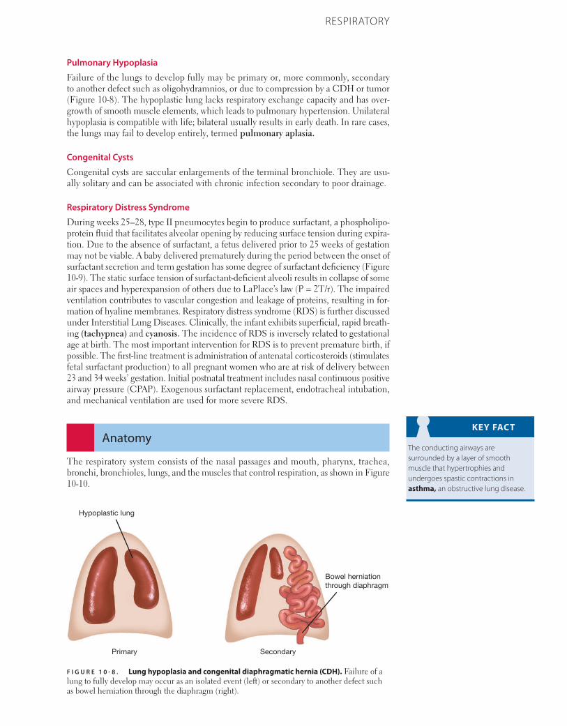
753CHAPTER 10RESPIRATORY
Pulmonary Hypoplasia
Failure of the lungs to develop fully may be primary or, more commonly, secondary to another defect such as oligohydramnios, or due to compression by a CDH or tumor (Figure 10-8). The hypoplastic lung lacks respiratory exchange capacity and has over-growth of smooth muscle elements, which leads to pulmonary hypertension. Unilateral hypoplasia is compatible with life; bilateral usually results in early death. In rare cases, the lungs may fail to develop entirely, termed pulmonary aplasia.
Congenital Cysts
Congenital cysts are saccular enlargements of the terminal bronchiole. They are usu-ally solitary and can be associated with chronic infection secondary to poor drainage.
Respiratory Distress Syndrome
During weeks 25–28, type II pneumocytes begin to produce surfactant, a phospholipo-protein fluid that facilitates alveolar opening by reducing surface tension during expira-tion. Due to the absence of surfactant, a fetus delivered prior to 25 weeks of gestation may not be viable. A baby delivered prematurely during the period between the onset of surfactant secretion and term gestation has some degree of surfactant deficiency (Figure 10-9). The static surface tension of surfactant-deficient alveoli results in collapse of some air spaces and hyperexpansion of others due to LaPlace’s law (P = 2T/r). The impaired ventilation contributes to vascular congestion and leakage of proteins, resulting in for-mation of hyaline membranes. Respiratory distress syndrome (RDS) is further discussed under Interstitial Lung Diseases. Clinically, the infant exhibits superficial, rapid breath-ing (tachypnea) and cyanosis. The incidence of RDS is inversely related to gestational age at birth. The most important intervention for RDS is to prevent premature birth, if possible. The first-line treatment is administration of antenatal corticosteroids (stimulates fetal surfactant production) to all pregnant women who are at risk of delivery between 23 and 34 weeks’ gestation. Initial postnatal treatment includes nasal continuous positive airway pressure (CPAP). Exogenous surfactant replacement, endotracheal intubation, and mechanical ventilation are used for more severe RDS.
Anatomy
The respiratory system consists of the nasal passages and mouth, pharynx, trachea, bronchi, bronchioles, lungs, and the muscles that control respiration, as shown in Figure 10-10.
KEY FACT
The conducting airways are surrounded by a layer of smooth muscle that hypertrophies and undergoes spastic contractions in asthma, an obstructive lung disease.
F I G U R E 1 0 - 8 . Lung hypoplasia and congenital diaphragmatic hernia (CDH). Failure of a lung to fully develop may occur as an isolated event (left) or secondary to another defect such as bowel herniation through the diaphragm (right).
Hypoplastic lung
Primary Secondary
Bowel herniationthrough diaphragm
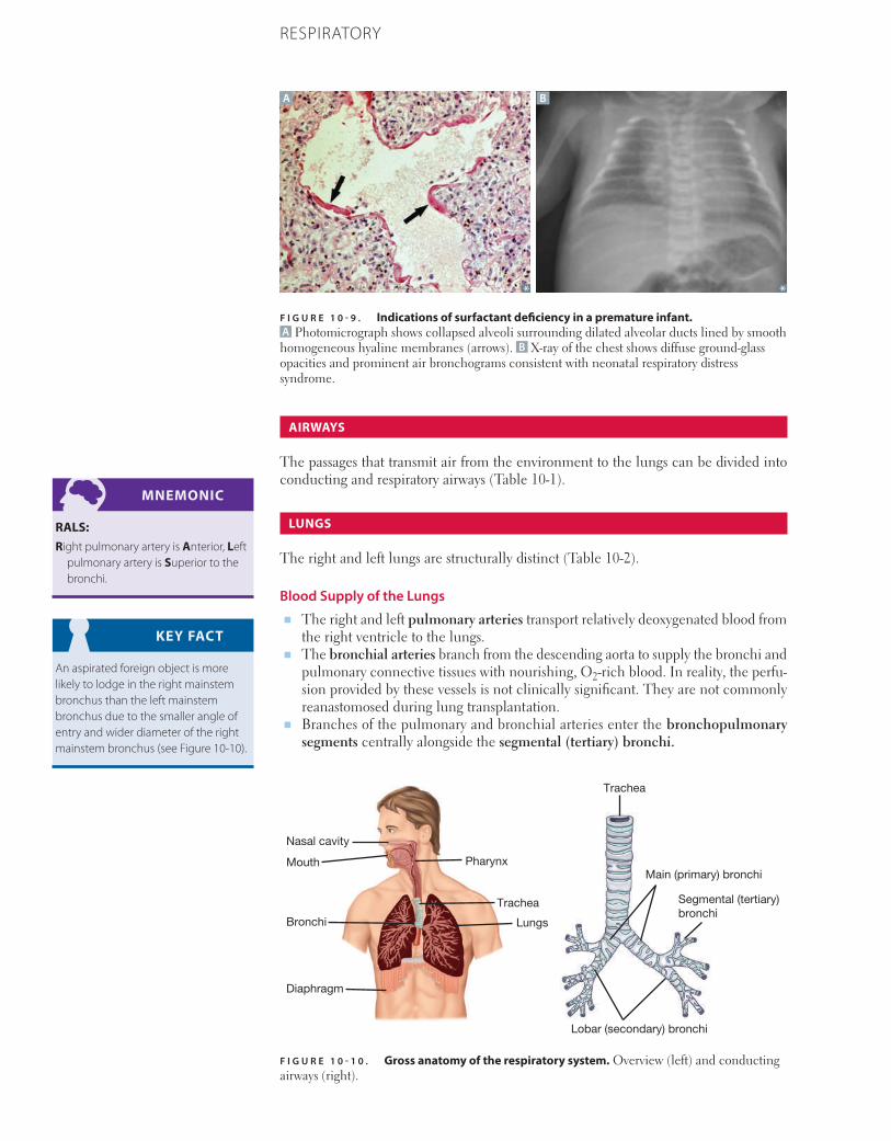
754 CHAPTER 10 RESPIRATORY
AIRWAYS
The passages that transmit air from the environment to the lungs can be divided into conducting and respiratory airways (Table 10-1).
LUNGS
The right and left lungs are structurally distinct (Table 10-2).
Blood Supply of the Lungs■ The right and left pulmonary arteries transport relatively deoxygenated blood from
the right ventricle to the lungs.■ The bronchial arteries branch from the descending aorta to supply the bronchi and
pulmonary connective tissues with nourishing, O2-rich blood. In reality, the perfu-sion provided by these vessels is not clinically significant. They are not commonly reanastomosed during lung transplantation.
■ Branches of the pulmonary and bronchial arteries enter the bronchopulmonary segments centrally alongside the segmental (tertiary) bronchi.
MNEMONIC
RALS: Right pulmonary artery is Anterior, Left
pulmonary artery is Superior to the bronchi.
KEY FACT
An aspirated foreign object is more likely to lodge in the right mainstem bronchus than the left mainstem bronchus due to the smaller angle of entry and wider diameter of the right mainstem bronchus (see Figure 10-10).
F I G U R E 1 0 - 1 0 . Gross anatomy of the respiratory system. Overview (left) and conducting airways (right).
Trachea
Lungs
Pharynx
Trachea
Diaphragm
Bronchi
Mouth
Nasal cavity
Main (primary) bronchi
Lobar (secondary) bronchi
Segmental (tertiary)bronchi
F I G U R E 1 0 - 9 . Indications of surfactant deficiency in a premature infant. A Photomicrograph shows collapsed alveoli surrounding dilated alveolar ducts lined by smooth homogeneous hyaline membranes (arrows). B X-ray of the chest shows diffuse ground-glass opacities and prominent air bronchograms consistent with neonatal respiratory distress syndrome.
A B
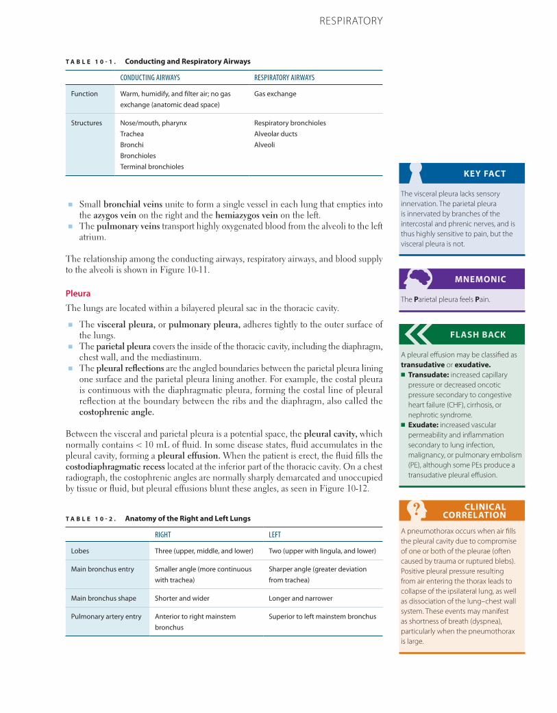
755CHAPTER 10RESPIRATORY
■ Small bronchial veins unite to form a single vessel in each lung that empties into the azygos vein on the right and the hemiazygos vein on the left.
■ The pulmonary veins transport highly oxygenated blood from the alveoli to the left atrium.
The relationship among the conducting airways, respiratory airways, and blood supply to the alveoli is shown in Figure 10-11.
Pleura
The lungs are located within a bilayered pleural sac in the thoracic cavity.
■ The visceral pleura, or pulmonary pleura, adheres tightly to the outer surface of the lungs.
■ The parietal pleura covers the inside of the thoracic cavity, including the diaphragm, chest wall, and the mediastinum.
■ The pleural reflections are the angled boundaries between the parietal pleura lining one surface and the parietal pleura lining another. For example, the costal pleura is continuous with the diaphragmatic pleura, forming the costal line of pleural reflection at the boundary between the ribs and the diaphragm, also called the costophrenic angle.
Between the visceral and parietal pleura is a potential space, the pleural cavity, which normally contains < 10 mL of fluid. In some disease states, fluid accumulates in the pleural cavity, forming a pleural effusion. When the patient is erect, the fluid fills the costodiaphragmatic recess located at the inferior part of the thoracic cavity. On a chest radiograph, the costophrenic angles are normally sharply demarcated and unoccupied by tissue or fluid, but pleural effusions blunt these angles, as seen in Figure 10-12.
KEY FACT
The visceral pleura lacks sensory innervation. The parietal pleura is innervated by branches of the intercostal and phrenic nerves, and is thus highly sensitive to pain, but the visceral pleura is not.
MNEMONIC
The Parietal pleura feels Pain.
FLASH BACK
A pleural effusion may be classified as transudative or exudative.■ Transudate: increased capillary
pressure or decreased oncotic pressure secondary to congestive heart failure (CHF), cirrhosis, or nephrotic syndrome.
■ Exudate: increased vascular permeability and inflammation secondary to lung infection, malignancy, or pulmonary embolism (PE), although some PEs produce a transudative pleural effusion.
CLINICAL CORRELATION
A pneumothorax occurs when air fills the pleural cavity due to compromise of one or both of the pleurae (often caused by trauma or ruptured blebs). Positive pleural pressure resulting from air entering the thorax leads to collapse of the ipsilateral lung, as well as dissociation of the lung–chest wall system. These events may manifest as shortness of breath (dyspnea), particularly when the pneumothorax is large.
T A B L E 1 0 - 1 . Conducting and Respiratory Airways
CONDUCTING AIRWAYS RESPIRATORY AIRWAYS
Function Warm, humidify, and filter air; no gas exchange (anatomic dead space)
Gas exchange
Structures Nose/mouth, pharynxTracheaBronchiBronchiolesTerminal bronchioles
Respiratory bronchiolesAlveolar ductsAlveoli
T A B L E 1 0 - 2 . Anatomy of the Right and Left Lungs
RIGHT LEFT
Lobes Three (upper, middle, and lower) Two (upper with lingula, and lower)
Main bronchus entry Smaller angle (more continuous with trachea)
Sharper angle (greater deviation from trachea)
Main bronchus shape Shorter and wider Longer and narrower
Pulmonary artery entry Anterior to right mainstem bronchus
Superior to left mainstem bronchus
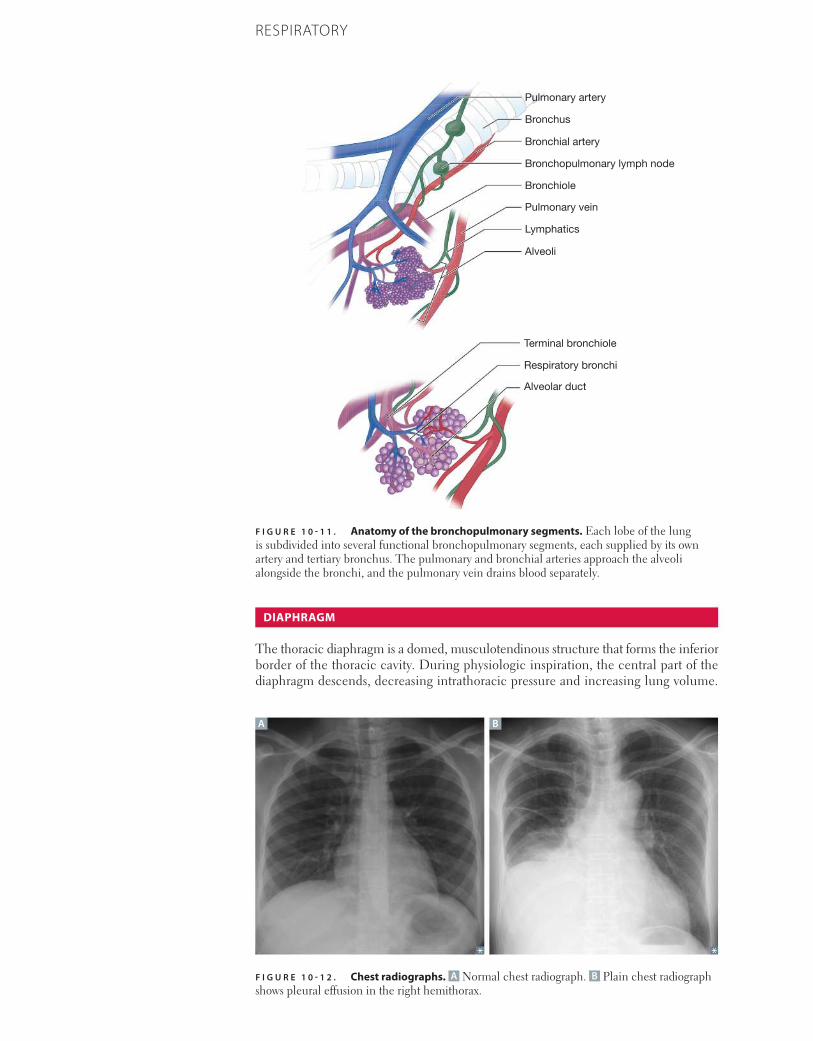
756 CHAPTER 10 RESPIRATORY
DIAPHRAGM
The thoracic diaphragm is a domed, musculotendinous structure that forms the inferior border of the thoracic cavity. During physiologic inspiration, the central part of the diaphragm descends, decreasing intrathoracic pressure and increasing lung volume.
F I G U R E 1 0 - 1 1 . Anatomy of the bronchopulmonary segments. Each lobe of the lung is subdivided into several functional bronchopulmonary segments, each supplied by its own artery and tertiary bronchus. The pulmonary and bronchial arteries approach the alveoli alongside the bronchi, and the pulmonary vein drains blood separately.
Bronchiole
Lymphatics
Alveoli
Pulmonary vein
Alveolar duct
Respiratory bronchi
Terminal bronchiole
Bronchopulmonary lymph node
Bronchial artery
Bronchus
Pulmonary artery
F I G U R E 1 0 - 1 2 . Chest radiographs. A Normal chest radiograph. B Plain chest radiograph shows pleural effusion in the right hemithorax.
A B
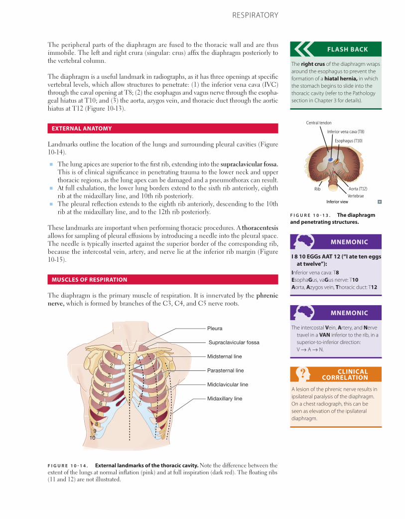
757CHAPTER 10RESPIRATORY
The peripheral parts of the diaphragm are fused to the thoracic wall and are thus immobile. The left and right crura (singular: crus) affix the diaphragm posteriorly to the vertebral column.
The diaphragm is a useful landmark in radiographs, as it has three openings at specific vertebral levels, which allow structures to penetrate: (1) the inferior vena cava (IVC) through the caval opening at T8; (2) the esophagus and vagus nerve through the esopha-geal hiatus at T10; and (3) the aorta, azygos vein, and thoracic duct through the aortic hiatus at T12 (Figure 10-13).
EXTERNAL ANATOMY
Landmarks outline the location of the lungs and surrounding pleural cavities (Figure 10-14).
■ The lung apices are superior to the first rib, extending into the supraclavicular fossa. This is of clinical significance in penetrating trauma to the lower neck and upper thoracic regions, as the lung apex can be damaged and a pneumothorax can result.
■ At full exhalation, the lower lung borders extend to the sixth rib anteriorly, eighth rib at the midaxillary line, and 10th rib posteriorly.
■ The pleural reflection extends to the eighth rib anteriorly, descending to the 10th rib at the midaxillary line, and to the 12th rib posteriorly.
These landmarks are important when performing thoracic procedures. A thoracentesis allows for sampling of pleural effusions by introducing a needle into the pleural space. The needle is typically inserted against the superior border of the corresponding rib, because the intercostal vein, artery, and nerve lie at the inferior rib margin (Figure 10-15).
MUSCLES OF RESPIRATION
The diaphragm is the primary muscle of respiration. It is innervated by the phrenic nerve, which is formed by branches of the C3, C4, and C5 nerve roots.
FLASH BACK
The right crus of the diaphragm wraps around the esophagus to prevent the formation of a hiatal hernia, in which the stomach begins to slide into the thoracic cavity (refer to the Pathology section in Chapter 3 for details).
MNEMONIC
I 8 10 EGGs AAT 12 (“I ate ten eggs at twelve”):
Inferior vena cava: T8EsophaGus, vaGus nerve: T10Aorta, Azygos vein, Thoracic duct: T12
MNEMONIC
The intercostal Vein, Artery, and Nerve travel in a VAN inferior to the rib, in a superior-to-inferior direction: V → A → N.
CLINICAL CORRELATION
A lesion of the phrenic nerve results in ipsilateral paralysis of the diaphragm. On a chest radiograph, this can be seen as elevation of the ipsilateral diaphragm.
Central tendon
Inferior view
Inferior vena cava (T8)
Esophagus (T10)
Aorta (T12)
Vertebrae
Rib
F I G U R E 1 0 - 1 3 . The diaphragm and penetrating structures.
F I G U R E 1 0 - 1 4 . External landmarks of the thoracic cavity. Note the difference between the extent of the lungs at normal inflation (pink) and at full inspiration (dark red). The floating ribs (11 and 12) are not illustrated.
Midclavicular line
Midaxillary line
Parasternal line
Midsternal line
Pleura
1
2
3
4
5
6
7
89
10
Supraclavicular fossa
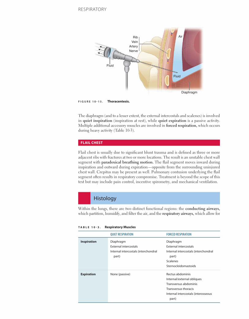
758 CHAPTER 10 RESPIRATORY
The diaphragm (and to a lesser extent, the external intercostals and scalenes) is involved in quiet inspiration (inspiration at rest), while quiet expiration is a passive activity. Multiple additional accessory muscles are involved in forced respiration, which occurs during heavy activity (Table 10-3).
FLAIL CHEST
Flail chest is usually due to significant blunt trauma and is defined as three or more adjacent ribs with fractures at two or more locations. The result is an unstable chest wall segment with paradoxical breathing motion. The flail segment moves inward during inspiration and outward during expiration—opposite from the surrounding uninjured chest wall. Crepitus may be present as well. Pulmonary contusion underlying the flail segment often results in respiratory compromise. Treatment is beyond the scope of this text but may include pain control, incentive spirometry, and mechanical ventilation.
Histology
Within the lungs, there are two distinct functional regions: the conducting airways, which partition, humidify, and filter the air, and the respiratory airways, which allow for
F I G U R E 1 0 - 1 5 . Thoracentesis.
NerveArteryVeinRib Air
Fluid
Fluid
Diaphragm
T A B L E 1 0 - 3 . Respiratory Muscles
QUIET RESPIRATION FORCED RESPIRATION
Inspiration DiaphragmExternal intercostalsInternal intercostals (interchondral
part)
DiaphragmExternal intercostalsInternal intercostals (interchondral
part)ScalenesSternocleidomastoids
Expiration None (passive) Rectus abdominisInternal/external obliquesTransversus abdominisTransversus thoracisInternal intercostals (interosseous
part)
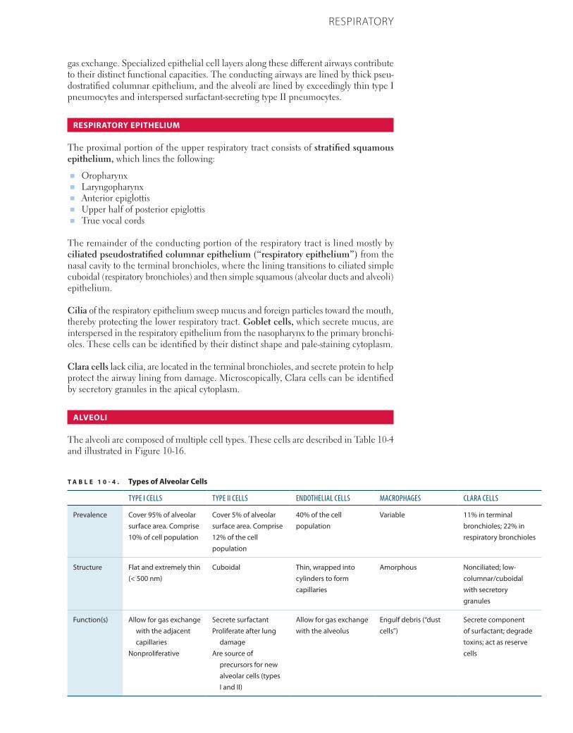
759CHAPTER 10RESPIRATORY
gas exchange. Specialized epithelial cell layers along these different airways contribute to their distinct functional capacities. The conducting airways are lined by thick pseu-dostratified columnar epithelium, and the alveoli are lined by exceedingly thin type I pneumocytes and interspersed surfactant-secreting type II pneumocytes.
RESPIRATORY EPITHELIUM
The proximal portion of the upper respiratory tract consists of stratified squamous epithelium, which lines the following:
■ Oropharynx■ Laryngopharynx■ Anterior epiglottis■ Upper half of posterior epiglottis■ True vocal cords
The remainder of the conducting portion of the respiratory tract is lined mostly by ciliated pseudostratified columnar epithelium (“respiratory epithelium”) from the nasal cavity to the terminal bronchioles, where the lining transitions to ciliated simple cuboidal (respiratory bronchioles) and then simple squamous (alveolar ducts and alveoli) epithelium.
Cilia of the respiratory epithelium sweep mucus and foreign particles toward the mouth, thereby protecting the lower respiratory tract. Goblet cells, which secrete mucus, are interspersed in the respiratory epithelium from the nasopharynx to the primary bronchi-oles. These cells can be identified by their distinct shape and pale-staining cytoplasm.
Clara cells lack cilia, are located in the terminal bronchioles, and secrete protein to help protect the airway lining from damage. Microscopically, Clara cells can be identified by secretory granules in the apical cytoplasm.
ALVEOLI
The alveoli are composed of multiple cell types. These cells are described in Table 10-4 and illustrated in Figure 10-16.
T A B L E 1 0 - 4 . Types of Alveolar Cells
TYPE I CELLS TYPE II CELLS ENDOTHELIAL CELLS MACROPHAGES CLARA CELLS
Prevalence Cover 95% of alveolar surface area. Comprise 10% of cell population
Cover 5% of alveolar surface area. Comprise 12% of the cell population
40% of the cell population
Variable 11% in terminal bronchioles; 22% in respiratory bronchioles
Structure Flat and extremely thin (< 500 nm)
Cuboidal Thin, wrapped into cylinders to form capillaries
Amorphous Nonciliated; low-columnar/cuboidal with secretory granules
Function(s) Allow for gas exchange with the adjacent capillaries
Nonproliferative
Secrete surfactantProliferate after lung
damageAre source of
precursors for new alveolar cells (types I and II)
Allow for gas exchange with the alveolus
Engulf debris (“dust cells”)
Secrete component of surfactant; degrade toxins; act as reserve cells
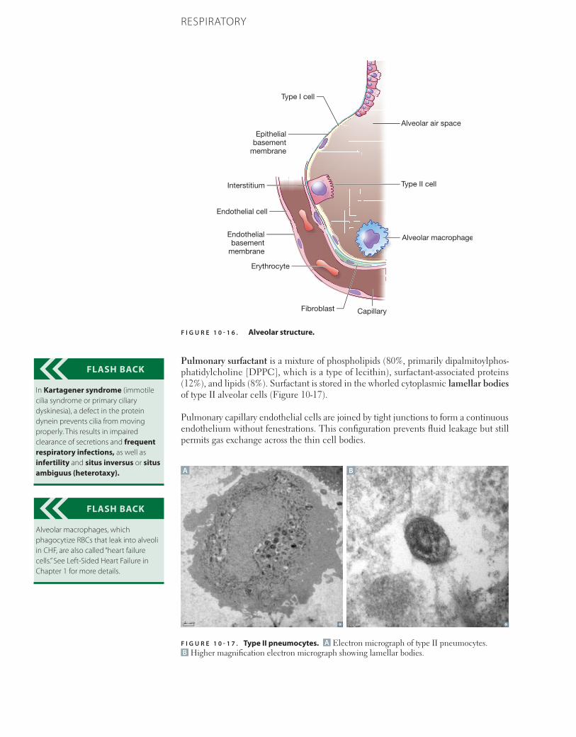
760 CHAPTER 10 RESPIRATORY
Pulmonary surfactant is a mixture of phospholipids (80%, primarily dipalmitoylphos-phatidylcholine [DPPC], which is a type of lecithin), surfactant-associated proteins (12%), and lipids (8%). Surfactant is stored in the whorled cytoplasmic lamellar bodies of type II alveolar cells (Figure 10-17).
Pulmonary capillary endothelial cells are joined by tight junctions to form a continuous endothelium without fenestrations. This configuration prevents fluid leakage but still permits gas exchange across the thin cell bodies.
FLASH BACK
In Kartagener syndrome (immotile cilia syndrome or primary ciliary dyskinesia), a defect in the protein dynein prevents cilia from moving properly. This results in impaired clearance of secretions and frequent respiratory infections, as well as infertility and situs inversus or situs ambiguus (heterotaxy).
FLASH BACK
Alveolar macrophages, which phagocytize RBCs that leak into alveoli in CHF, are also called “heart failure cells.” See Left-Sided Heart Failure in Chapter 1 for more details.
A B
F I G U R E 1 0 - 1 7 . Type II pneumocytes. A Electron micrograph of type II pneumocytes. B Higher magnification electron micrograph showing lamellar bodies.
F I G U R E 1 0 - 1 6 . Alveolar structure.
Capillary
Alveolar macrophage
Alveolar air space
Type II cell
Type I cell
Interstitium
Epithelialbasement
membrane
Endothelialbasement
membrane
Erythrocyte
Fibroblast
Endothelial cell
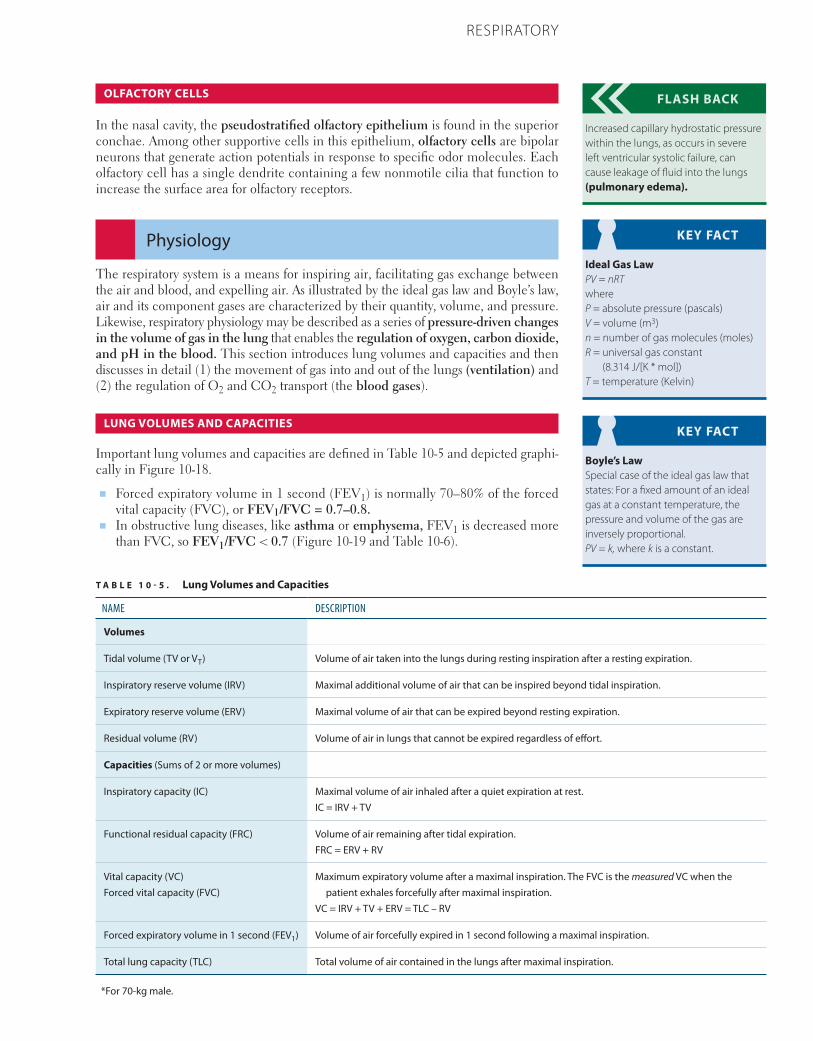
761CHAPTER 10RESPIRATORY
OLFACTORY CELLS
In the nasal cavity, the pseudostratified olfactory epithelium is found in the superior conchae. Among other supportive cells in this epithelium, olfactory cells are bipolar neurons that generate action potentials in response to specific odor molecules. Each olfactory cell has a single dendrite containing a few nonmotile cilia that function to increase the surface area for olfactory receptors.
Physiology
The respiratory system is a means for inspiring air, facilitating gas exchange between the air and blood, and expelling air. As illustrated by the ideal gas law and Boyle’s law, air and its component gases are characterized by their quantity, volume, and pressure. Likewise, respiratory physiology may be described as a series of pressure-driven changes in the volume of gas in the lung that enables the regulation of oxygen, carbon dioxide, and pH in the blood. This section introduces lung volumes and capacities and then discusses in detail (1) the movement of gas into and out of the lungs (ventilation) and (2) the regulation of O2 and CO2 transport (the blood gases).
LUNG VOLUMES AND CAPACITIES
Important lung volumes and capacities are defined in Table 10-5 and depicted graphi-cally in Figure 10-18.
■ Forced expiratory volume in 1 second (FEV1) is normally 70–80% of the forced vital capacity (FVC), or FEV1/FVC = 0.7–0.8.
■ In obstructive lung diseases, like asthma or emphysema, FEV1 is decreased more than FVC, so FEV1/FVC < 0.7 (Figure 10-19 and Table 10-6).
FLASH BACK
Increased capillary hydrostatic pressure within the lungs, as occurs in severe left ventricular systolic failure, can cause leakage of fluid into the lungs (pulmonary edema).
KEY FACT
Ideal Gas LawPV = nRTwhereP = absolute pressure (pascals)V = volume (m3)n = number of gas molecules (moles)R = universal gas constant
(8.314 J/[K * mol]) T = temperature (Kelvin)
KEY FACT
Boyle’s LawSpecial case of the ideal gas law that states: For a fixed amount of an ideal gas at a constant temperature, the pressure and volume of the gas are inversely proportional.PV = k, where k is a constant.
T A B L E 1 0 - 5 . Lung Volumes and Capacities
NAME DESCRIPTION
Volumes
Tidal volume (TV or VT) Volume of air taken into the lungs during resting inspiration after a resting expiration.
Inspiratory reserve volume (IRV) Maximal additional volume of air that can be inspired beyond tidal inspiration.
Expiratory reserve volume (ERV) Maximal volume of air that can be expired beyond resting expiration.
Residual volume (RV) Volume of air in lungs that cannot be expired regardless of effort.
Capacities (Sums of 2 or more volumes)
Inspiratory capacity (IC) Maximal volume of air inhaled after a quiet expiration at rest.IC = IRV + TV
Functional residual capacity (FRC) Volume of air remaining after tidal expiration.FRC = ERV + RV
Vital capacity (VC) Forced vital capacity (FVC)
Maximum expiratory volume after a maximal inspiration. The FVC is the measured VC when the patient exhales forcefully after maximal inspiration.
VC = IRV + TV + ERV = TLC – RV
Forced expiratory volume in 1 second (FEV1) Volume of air forcefully expired in 1 second following a maximal inspiration.
Total lung capacity (TLC) Total volume of air contained in the lungs after maximal inspiration.
*For 70-kg male.
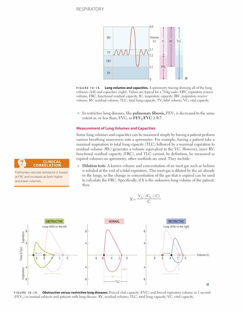
762 CHAPTER 10 RESPIRATORY
■ In restrictive lung diseases, like pulmonary fibrosis, FEV1 is decreased to the same extent as, or less than, FVC, so FEV1/FVC ≥ 0.7.
Measurement of Lung Volumes and Capacities
Some lung volumes and capacities can be measured simply by having a patient perform various breathing maneuvers into a spirometer. For example, having a patient take a maximal inspiration to total lung capacity (TLC) followed by a maximal expiration to residual volume (RV) generates a volume equivalent to the VC. However, since RV, functional residual capacity (FRC), and TLC cannot, by definition, be measured as expired volumes on spirometry, other methods are used. They include:
■ Dilution tests: A known volume and concentration of an inert gas such as helium is inhaled at the end of a tidal expiration. This inert gas is diluted by the air already in the lungs, so the change in concentration of the gas that is expired can be used to calculate the FRC. Specifically, if X is the unknown lung volume of the patient, then
X = Vo ⋅ (Co − C)
C
CLINICAL CORRELATION
Pulmonary vascular resistance is lowest at FRC and increases at both higher and lower volumes.
F I G U R E 1 0 - 1 8 . Lung volumes and capacities. A spirometry tracing showing all of the lung volumes (left) and capacities (right). Values are typical for a 70-kg male. ERV, expiratory reserve volume; FRC, functional residual capacity; IC, inspiratory capacity; IRV, inspiratory reserve volume; RV, residual volume; TLC, total lung capacity; TV, tidal volume; VC, vital capacity.
6.0
ICIRV
TV
ERV
RV
Volume(L)
2.7
2.2
1.2
0
FRC
VC TLC
F I G U R E 1 0 - 1 9 . Obstructive versus restrictive lung diseases. Forced vital capacity (FVC) and forced expiratory volume in 1 second (FEV1) in normal subjects and patients with lung disease. RV, residual volume; TLC, total lung capacity; VC, vital capacity.
8
8 6 2
OBSTRUCTIVE NORMAL RESTRICTIVE
Loop shifts to the left
Flow
(L/s
ec)
Expi
ratio
nIn
spira
tion
Loop shifts to the right
Volume (L)0
8
4
4
4
8
8 6 2 0
RV
TLC8
4
4
4
8
8 6 2 0
8
4
4
4
VC
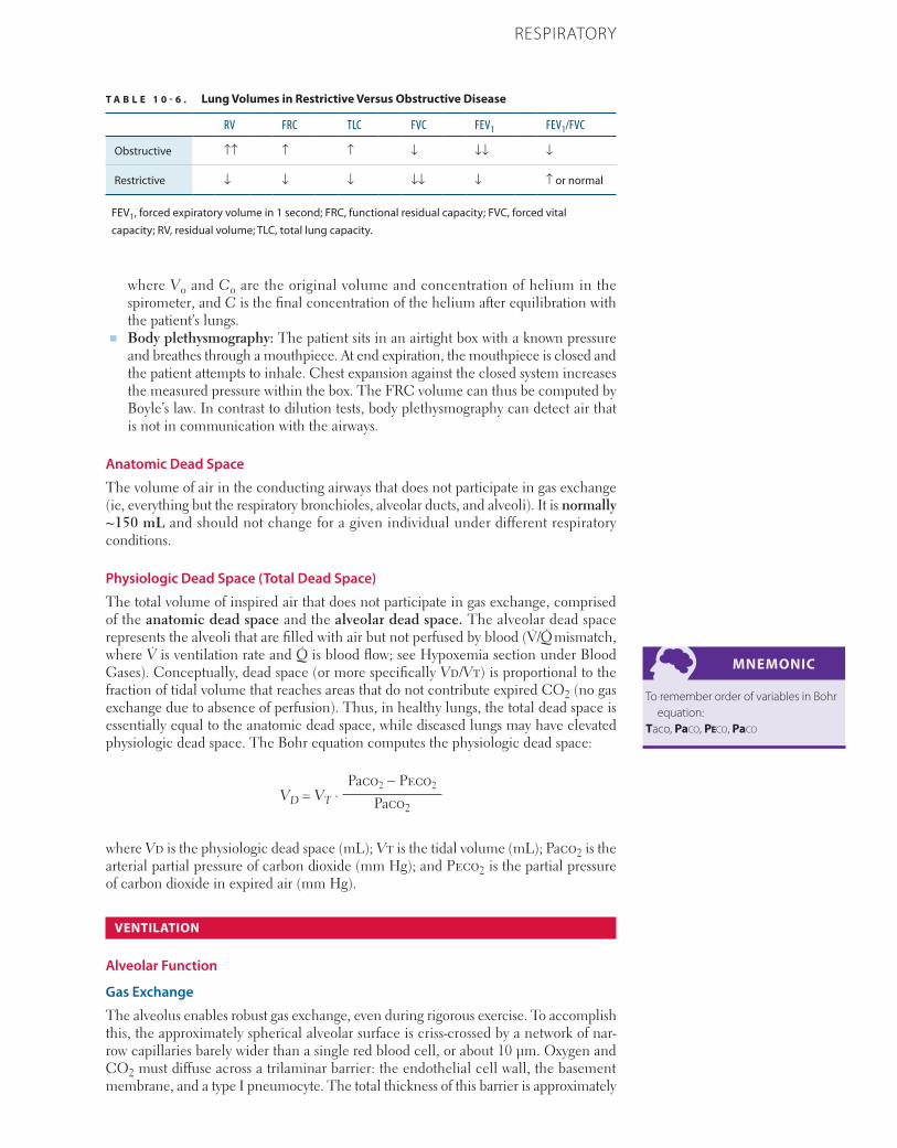
763CHAPTER 10RESPIRATORY
where Vo and Co are the original volume and concentration of helium in the spirometer, and C is the final concentration of the helium after equilibration with the patient’s lungs.
■ Body plethysmography: The patient sits in an airtight box with a known pressure and breathes through a mouthpiece. At end expiration, the mouthpiece is closed and the patient attempts to inhale. Chest expansion against the closed system increases the measured pressure within the box. The FRC volume can thus be computed by Boyle’s law. In contrast to dilution tests, body plethysmography can detect air that is not in communication with the airways.
Anatomic Dead Space
The volume of air in the conducting airways that does not participate in gas exchange (ie, everything but the respiratory bronchioles, alveolar ducts, and alveoli). It is normally ~150 mL and should not change for a given individual under different respiratory conditions.
Physiologic Dead Space (Total Dead Space)
The total volume of inspired air that does not participate in gas exchange, comprised of the anatomic dead space and the alveolar dead space. The alveolar dead space represents the alveoli that are filled with air but not perfused by blood (V̇/Q̇ mismatch, where V̇ is ventilation rate and Q̇ is blood flow; see Hypoxemia section under Blood Gases). Conceptually, dead space (or more specifically Vd/Vt) is proportional to the fraction of tidal volume that reaches areas that do not contribute expired CO2 (no gas exchange due to absence of perfusion). Thus, in healthy lungs, the total dead space is essentially equal to the anatomic dead space, while diseased lungs may have elevated physiologic dead space. The Bohr equation computes the physiologic dead space:
VD = VT ⋅ Paco2 − Peco2
Paco2
where Vd is the physiologic dead space (mL); Vt is the tidal volume (mL); Paco2 is the arterial partial pressure of carbon dioxide (mm Hg); and Peco2 is the partial pressure of carbon dioxide in expired air (mm Hg).
VENTILATION
Alveolar Function
Gas Exchange
The alveolus enables robust gas exchange, even during rigorous exercise. To accomplish this, the approximately spherical alveolar surface is criss-crossed by a network of nar-row capillaries barely wider than a single red blood cell, or about 10 μm. Oxygen and CO2 must diffuse across a trilaminar barrier: the endothelial cell wall, the basement membrane, and a type I pneumocyte. The total thickness of this barrier is approximately
MNEMONIC
To remember order of variables in Bohr equation:
Taco, Paco, Peco, Paco
T A B L E 1 0 - 6 . Lung Volumes in Restrictive Versus Obstructive Disease
RV FRC TLC FVC FEV1 FEV1/FVC
Obstructive ↑↑ ↑ ↑ ↓ ↓↓ ↓
Restrictive ↓ ↓ ↓ ↓↓ ↓ ↑ or normal
FEV1, forced expiratory volume in 1 second; FRC, functional residual capacity; FVC, forced vital capacity; RV, residual volume; TLC, total lung capacity.
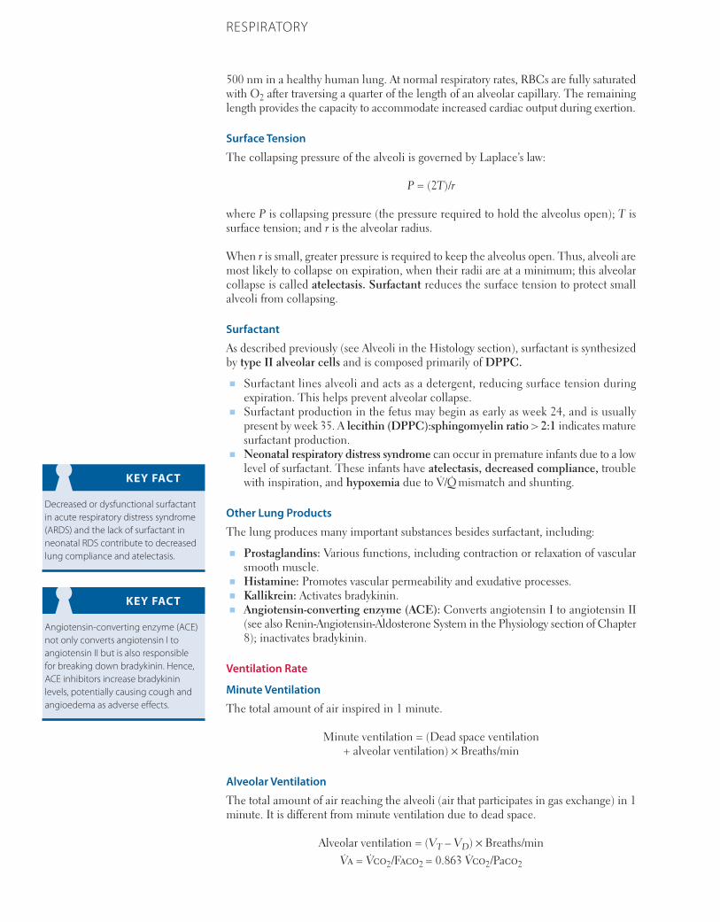
764 CHAPTER 10 RESPIRATORY
500 nm in a healthy human lung. At normal respiratory rates, RBCs are fully saturated with O2 after traversing a quarter of the length of an alveolar capillary. The remaining length provides the capacity to accommodate increased cardiac output during exertion.
Surface Tension
The collapsing pressure of the alveoli is governed by Laplace’s law:
P = (2T)/r
where P is collapsing pressure (the pressure required to hold the alveolus open); T is surface tension; and r is the alveolar radius.
When r is small, greater pressure is required to keep the alveolus open. Thus, alveoli are most likely to collapse on expiration, when their radii are at a minimum; this alveolar collapse is called atelectasis. Surfactant reduces the surface tension to protect small alveoli from collapsing.
Surfactant
As described previously (see Alveoli in the Histology section), surfactant is synthesized by type II alveolar cells and is composed primarily of DPPC.
■ Surfactant lines alveoli and acts as a detergent, reducing surface tension during expiration. This helps prevent alveolar collapse.
■ Surfactant production in the fetus may begin as early as week 24, and is usually present by week 35. A lecithin (DPPC):sphingomyelin ratio > 2:1 indicates mature surfactant production.
■ Neonatal respiratory distress syndrome can occur in premature infants due to a low level of surfactant. These infants have atelectasis, decreased compliance, trouble with inspiration, and hypoxemia due to V̇/Q̇ mismatch and shunting.
Other Lung Products
The lung produces many important substances besides surfactant, including:
■ Prostaglandins: Various functions, including contraction or relaxation of vascular smooth muscle.
■ Histamine: Promotes vascular permeability and exudative processes.■ Kallikrein: Activates bradykinin.■ Angiotensin-converting enzyme (ACE): Converts angiotensin I to angiotensin II
(see also Renin-Angiotensin-Aldosterone System in the Physiology section of Chapter 8); inactivates bradykinin.
Ventilation Rate
Minute Ventilation
The total amount of air inspired in 1 minute.
Minute ventilation = (Dead space ventilation + alveolar ventilation) × Breaths/min
Alveolar Ventilation
The total amount of air reaching the alveoli (air that participates in gas exchange) in 1 minute. It is different from minute ventilation due to dead space.
Alveolar ventilation = (VT – VD) × Breaths/minV̇a = V̇co2/Faco2 = 0.863 V̇co2/Paco2
KEY FACT
Decreased or dysfunctional surfactant in acute respiratory distress syndrome (ARDS) and the lack of surfactant in neonatal RDS contribute to decreased lung compliance and atelectasis.
KEY FACT
Angiotensin-converting enzyme (ACE) not only converts angiotensin I to angiotensin II but is also responsible for breaking down bradykinin. Hence, ACE inhibitors increase bradykinin levels, potentially causing cough and angioedema as adverse effects.
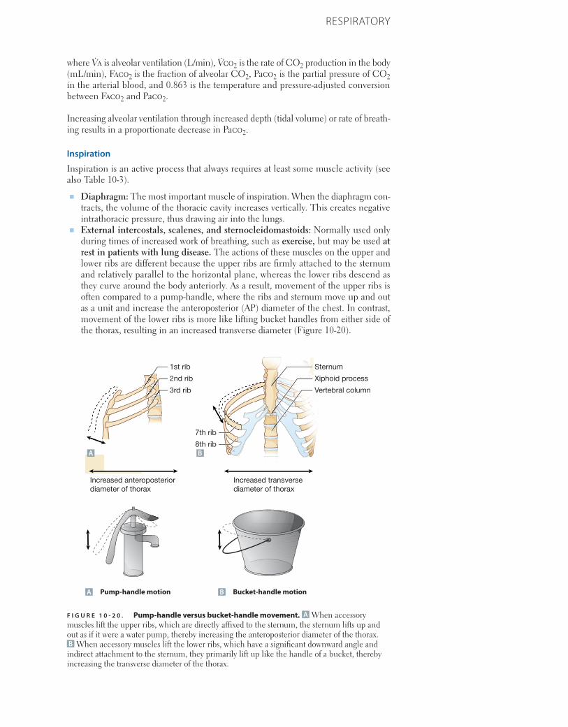
765CHAPTER 10RESPIRATORY
where V̇a is alveolar ventilation (L/min), V̇co2 is the rate of CO2 production in the body (mL/min), Faco2 is the fraction of alveolar CO2, Paco2 is the partial pressure of CO2 in the arterial blood, and 0.863 is the temperature and pressure-adjusted conversion between Faco2 and Paco2.
Increasing alveolar ventilation through increased depth (tidal volume) or rate of breath-ing results in a proportionate decrease in Paco2.
Inspiration
Inspiration is an active process that always requires at least some muscle activity (see also Table 10-3).
■ Diaphragm: The most important muscle of inspiration. When the diaphragm con-tracts, the volume of the thoracic cavity increases vertically. This creates negative intrathoracic pressure, thus drawing air into the lungs.
■ External intercostals, scalenes, and sternocleidomastoids: Normally used only during times of increased work of breathing, such as exercise, but may be used at rest in patients with lung disease. The actions of these muscles on the upper and lower ribs are different because the upper ribs are firmly attached to the sternum and relatively parallel to the horizontal plane, whereas the lower ribs descend as they curve around the body anteriorly. As a result, movement of the upper ribs is often compared to a pump-handle, where the ribs and sternum move up and out as a unit and increase the anteroposterior (AP) diameter of the chest. In contrast, movement of the lower ribs is more like lifting bucket handles from either side of the thorax, resulting in an increased transverse diameter (Figure 10-20).
F I G U R E 1 0 - 2 0 . Pump-handle versus bucket-handle movement. A When accessory muscles lift the upper ribs, which are directly affixed to the sternum, the sternum lifts up and out as if it were a water pump, thereby increasing the anteroposterior diameter of the thorax. B When accessory muscles lift the lower ribs, which have a significant downward angle and indirect attachment to the sternum, they primarily lift up like the handle of a bucket, thereby increasing the transverse diameter of the thorax.
A Pump-handle motion B Bucket-handle motion
A B
Vertebral column
8th rib
Xiphoid process
7th rib
2nd rib
1st rib
3rd rib
Sternum
Increased transverse diameter of thorax
Increased anteroposterior diameter of thorax
A Pump-handle motion B Bucket-handle motion
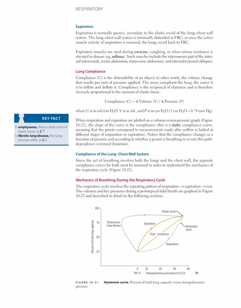
766 CHAPTER 10 RESPIRATORY
Expiration
Expiration is normally passive, secondary to the elastic recoil of the lung–chest wall system. The lung–chest wall system is minimally distended at FRC, so once the active muscle activity of inspiration is removed, the lungs recoil back to FRC.
Expiratory muscles are used during exercise, coughing, or when airway resistance is elevated in disease (eg, asthma). Such muscles include the interosseous part of the inter-nal intercostals, rectus abdominis, transversus abdominis, and internal/external obliques.
Lung Compliance
Compliance (C) is the distensibility of an object; in other words, the volume change that results per unit of pressure applied. The more compliant the lung, the easier it is to inflate and deflate it. Compliance is the reciprocal of elastance and is therefore inversely proportional to the amount of elastic tissue.
Compliance (C) = Δ Volume (V) / Δ Pressure (P)
where C is in mL/cm H2O, V is in mL, and P is in cm H2O (1 cm H2O = 0.74 mm Hg).
When inspiration and expiration are plotted on a volume-versus-pressure graph (Figure 10-21), the slope of the curve is the compliance (this is a static compliance curve, meaning that the points correspond to measurements made after airflow is halted at different stages of inspiration or expiration). Notice that the compliance changes as a function of pressure and according to whether a person is breathing in or out (this path-dependence is termed hysteresis).
Compliance of the Lung–Chest Wall System
Since the act of breathing involves both the lungs and the chest wall, the separate compliance curves for both must be summed in order to understand the mechanics of the respiratory cycle (Figure 10-22).
Mechanics of Breathing During the Respiratory Cycle
The respiratory cycle involves the repeating pattern of inspiration → expiration → rest. The volumes and key pressures during a prototypical tidal breath are graphed in Figure 10-23 and described in detail in the following sections.
KEY FACT
In emphysema, there is destruction of elastic tissue, so C ↑.
In fibrotic lung disease, the lungs become stiffer, so C ↓.
F I G U R E 1 0 - 2 1 . Hysteresis curve. Percent of total lung capacity versus transpulmonary pressure.
Obstructivelung disease
Expiration
Inspiration
Elastic work
Nonelasticwork
Flow - resistance
FRC 0 Transpulmonary pressure (cm H2O)
Perc
ent o
f tot
al lu
ng c
apac
ity
100
75
50
25
100 20 30 40
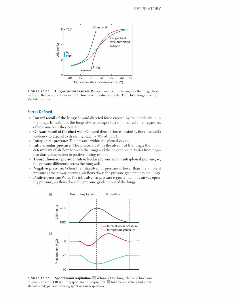
767CHAPTER 10RESPIRATORY
Forces Defined■ Inward recoil of the lungs: Inward-directed force created by the elastic tissue in
the lungs. In isolation, the lungs always collapse to a minimal volume, regardless of how much air they contain.
■ Outward recoil of the chest wall: Outward-directed force created by the chest wall’s tendency to expand to its resting state (~70% of TLC).
■ Intrapleural pressure: The pressure within the pleural cavity.■ Intra-alveolar pressure: The pressure within the alveoli of the lungs; the major
determinant of air flow between the lungs and the environment. Varies from nega-tive during inspiration to positive during expiration.
■ Transpulmonary pressure: Intra-alveolar pressure minus intrapleural pressure, ie, the pressure difference across the lung wall.
■ Negative pressure: When the intra-alveolar pressure is lower than the ambient pressure at the airway opening, air flows down the pressure gradient into the lungs.
■ Positive pressure: When the intra-alveolar pressure is greater than the airway open-ing pressure, air flows down the pressure gradient out of the lungs.
F I G U R E 1 0 - 2 2 . Lung–chest wall system. Pressure and volume tracings for the lung, chest wall, and the combined system. FRC, functional residual capacity; TLC, total lung capacity; VT, tidal volume.
Chest wallTLC
VTFRC
Lung
Transorgan static pressure (cm H2O)
−20 −10 00
2
4
6
10 20 30 40
Lung–chestwall combinedsystem
Volu
me
(L)
F I G U R E 1 0 - 2 3 . Spontaneous respiration. A Volume of the lung relative to functional residual capacity (FRC) during spontaneous respiration. B Intrapleural (blue) and intra-alveolar (red) pressures during spontaneous respiration.
FRC
Rest Inspiration Expiration
+0.5
Volu
me
(L)
−10
−5
0
Pre
ssur
e (c
m H
2O)
Intra-alveolar pressureIntrapleural pressure
A
B
768 CHAPTER 10 RESPIRATORY
At Rest
At rest, when the gas volume in the lungs is equal to FRC, the pressures created by the lungs (inward recoil) and the chest wall (outward recoil) are equal and opposite (Figure 10-22). The lungs create positive pressure because they tend to collapse due to their elasticity. At the same time, the chest wall generates negative pressure because the ribcage and the rest of the thoracic wall to which the lungs are affixed resist deformation from their natural shape. These opposing forces cancel out, establishing a distending pressure (alveolar pressure) of 0 (Figure 10-23). The respiratory muscles are not involved in this process.
Intrapleural pressure = –5 cm H2OIntra-alveolar pressure = 0 cm H2O
In emphysema, the lungs have a decreased tendency to collapse due to a loss of elas-ticity (compliance ↑). As a result, the lung–chest wall system recalibrates to a new, higher FRC at which the forces balance. This is why patients with emphysema are barrel-chested.
In lung fibrosis, the lungs have an increased tendency to collapse (compliance ↓), so the system equilibrates to a new, lower FRC at which the forces balance.
During Inspiration
The muscles of inspiration contract, generating negative pressure. The intra-alveolar pressure is therefore negative. However, inspiration does not continue indefinitely because the pressure exerted by the chest wall becomes positive as it expands beyond its natural shape, thus opposing the muscles of inspiration. Approximate values for a young, healthy subject are given below; note that there can be significant variation based on age, weight, or health:
Intrapleural pressure: Decreases from –5 to –8 cm H2OIntra-alveolar pressure < 0 cm H2O, so air flows into the lungs
At Maximum Inspiration
At TLC, the positive inward pressures due to the distension of the chest wall and lungs have increased to the point where they exactly cancel out the negative outward pres-sure generated by the muscles of inspiration. Thus, the lungs are held at full capacity, neither expanding nor contracting.
Intrapleural pressure = –8 cm H2OIntra-alveolar pressure = 0 cm H2O, so no air flows
During Expiration
The muscles of inspiration relax, removing their strong negative outward force and allowing the intra-alveolar pressure to become positive. This allows the lung–chest wall complex to return to its equilibrium at FRC.
Intrapleural pressure: Increases from –8 to –5 cm H2O (may increase into positive range, depending on the patient)
Intra-alveolar pressure > 0 cm H2O, so air flows out of the lungs
At Maximum Expiration
At RV, there is still some gas left in the lungs. That is, we can never exhale enough to fully collapse the lungs. At RV, the chest wall exerts such a strong negative outward pressure (due to its tendency to recoil outward to its resting shape) that the expiratory muscles are unable to create enough positive inward pressure to exhale any further.
769CHAPTER 10RESPIRATORY
Intrapleural pressure = –5 cm H2OIntra-alveolar pressure = 0 cm H2O, so no air flows
Mechanical Ventilation
Mechanical ventilators allow physicians to manipulate the pressures and volumes that govern inspiration and expiration. A detailed explanation of mechanical ventilation is beyond the scope of this text, but a brief discussion of the most common modes of mechanical ventilation and how they work may be useful.
Positive end-expiratory pressure (PEEP): With this setting, airway pressure at the end of expiration does not fall to 0, but is instead maintained at a fixed value (eg, 10 cm H2O). This helps to maintain airway patency during expiration and is particularly useful in hypoxemic states such as acute respiratory distress syndrome (ARDS). In a patient who is initiating all breaths, the equivalent of PEEP is continuous positive airway pres-sure (CPAP), which may be applied by mask or endotracheal tube in order to maintain airway patency. CPAP is commonly used in the treatment of obstructive sleep apnea.
Airways
Flow
Airflow is proportional to the pressure difference between the mouth (or nose) and the alveoli and is inversely proportional to the resistance of the airway.
V̇ = Δ P/R
where V̇ is the ventilation rate (airflow); Δ P is the pressure gradient; and R is resistance.
Note that the dot over the V̇ in the ventilation rate indicates that it is the change in volume with respect to time (ie, dV/dt).
Resistance
Governed by Poiseuille’s law:
R = (8ηl)/(πr4)
where R is resistance, η is the viscosity of the gas, l is airway length, and r is airway radius.
Since airway radius is the major determinant of resistance (r4), the major source of airway resistance is the medium-sized bronchi (the smaller bronchi have greater num-bers arranged in parallel, thus offering less net resistance than the medium-sized bronchi).
Factors That Influence Pulmonary Resistance
■ Contraction of bronchial smooth muscle:■ Sympathetic stimulation: Airways dilate via β2-adrenergic receptors, thus
decreasing resistance. Albuterol is a common β2 agonist and is used in an inhaled form by patients with asthma or chronic obstructive pulmonary disease (COPD).
■ Parasympathetic stimulation: Airways constrict via M3-cholinergic recep-tors, thus increasing resistance. This is seen in asthma as part of the immune response. Ipratropium is a common anticholinergic drug used to counter this parasympathetic bronchoconstriction in asthma or COPD.
■ Secretions: Increased and/or thickened airway secretions, a hallmark of chronic bron-chitis and cystic fibrosis (CF), lead to increased airway obstruction and resistance.
CLINICAL CORRELATION
Obstructive sleep apnea occurs when excess body weight, extra pharyngeal tissue, or abnormal anatomy (eg, tonsillar hypertrophy or short mandible) blocks the upper airway passages when the patient is sleeping. This obstruction causes periods of hypoventilation and hypoxia, resulting in nocturnal awakenings, poor sleep, and daytime somnolence. Treatment includes continuous positive airway pressure (CPAP), which is the equivalent in spontaneous breathers of PEEP.
CLINICAL CORRELATION
For a mechanically ventilated patient, hypoxia (low O2) can be corrected by increasing Fio2 or PEEP, whereas hypercarbia (high CO2) can be corrected by increasing minute ventilation or tidal volume.
CLINICAL CORRELATION
Patients with ARDS are routinely treated with PEEP at low tidal volumes to protect the lungs. CO2 is lowered by increasing the respiratory rate rather than the tidal volume.
770 CHAPTER 10 RESPIRATORY
■ Lung volumes:■ High lung volumes: The lung tissue surrounding and attached to the airways
expands, pulling the airways open, so resistance is decreased.■ Low lung volumes: When the lung volume is low, there is less traction and
increased resistance. Airways are more prone to collapse.
BLOOD GASES
Oxygen Transport
Hemoglobin■ Structure: Hemoglobin is a globular protein composed of four subunits (two α-family
chains and two β-family chains). Each subunit contains a heme moiety, which is a porphyrin ring containing a single iron atom at its core. The iron in hemoglobin is in the ferrous (Fe2+) state and can bind O2. If the iron is in the ferric (Fe3+) state, it is called methemoglobin and is unable to bind O2. Hemoglobin can exist in two forms: taut, which has low affinity for O2, and relaxed, which has high affinity for O2.
■ O2 saturation (Spo2): The percentage of total oxygen-binding sites on hemoglobin that are actually occupied by oxygen, also called the saturation of peripheral oxygen.
■ O2 content: The total amount of O2 in the blood, both dissolved and bound to hemoglobin. Measured in mL of O2 per deciliter of blood. Depends on hemoglobin concentration, partial pressure of O2 (Po2), and the 50% hemoglobin capacity (P50). Calculated by the equation:■ O2 content = O2 bound to hemoglobin + O2 dissolved in blood■ O2 content (mL/dL blood) = (1.34 mL O2/dL blood × [Hemoglobin] × Spo2)
+ (0.0031 mL/mm Hg O2 × Pao2)■ Using typical values of hemoglobin = 14 g/dL, Spo2 = 1.00 (100%), and partial
arterial pressure of oxygen (Pao2) = 100 mm Hg, one finds that the vast majority (98.5%) of oxygen in the blood is bound to hemoglobin.
■ O2 capacity: The maximum amount of O2 that can be bound to hemoglobin (in mL/dL blood), computed as 1.34 mL O2/dL blood × [Hemoglobin]. This is approxi-mately equal to the O2 content of blood at 100% saturation.
Oxygen-Hemoglobin Dissociation Curve
The oxygen-hemoglobin dissociation curve describes how the oxygen saturation of hemoglobin varies with the Po2 in the blood (Figure 10-24). Its sigmoidal shape reflects positive cooperativity among the four subunits, such that the more oxygen molecules that are bound, the easier it is for an additional oxygen molecule to bind. Factors that decrease the affinity of hemoglobin for oxygen cause the curve to shift right, leading to greater oxygen unloading. On the other hand, a left shift causes more oxygen to become bound in the blood.
■ Increases in Pco2, altitude, 2,3-bisphosphoglycerate (2,3-BPG), or temperature, or a decrease in pH, will cause a rightward shift of the curve.
■ Decreases in Pco2, altitude, 2,3-diphosphoglycerate, or temperature, or an increase in pH, will cause a leftward shift of the curve.
■ During exercise, Pco2 and temperature rise, and pH falls in the active muscle tissue. This promotes a right shift and greater O2 unloading to the tissues. This is known as the Bohr effect.
■ At high altitudes, 2,3-DPG synthesis is increased, facilitating O2 unloading.■ Fetal hemoglobin (α2γ2) does not bind 2,3-DPG as strongly as adult hemoglobin
(α2β2), shifting the curve to the left. This helps the fetus obtain O2 from the mother’s RBCs.
KEY FACT
Fe2 binds O2.
MNEMONIC
Hemoglobin:Taut in Tissues, Relaxed in Respiratory
tract.
KEY FACT
Methemoglobinemia may result from treatment with nitrites and is thus sometimes induced when amyl nitrite or sodium nitrite is given to treat cyanide poisoning. Methemoglobinemia is treated with methylene blue, which reduces the ferric iron reducing agent that converts ferric iron (Fe3+) in methemoglobin to ferrous iron (Fe2+). Remember that only reduced Fe can bind O2.
MNEMONIC
Causes of a right-shifted hemoglobin dissociation curve—BAT ACESBPG (2,3-BPG)AltitudeTemperatureAcidCO2ExerciseSickle cell
MNEMONIC
A rule of thumb for translating between Po2 and Spo2 is the 40–50–60: 70–80–90 rule, where Po2s of 40, 50, and 60 mm Hg translate to 70%, 80%, and 90% saturation, respectively.
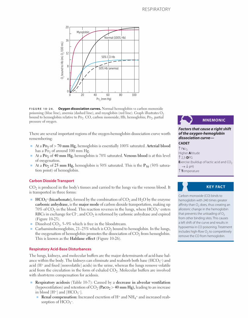
771CHAPTER 10RESPIRATORY
There are several important regions of the oxygen-hemoglobin dissociation curve worth remembering:
■ At a Po2 of > 70 mm Hg, hemoglobin is essentially 100% saturated. Arterial blood has a Po2 of around 100 mm Hg.
■ At a Po2 of 40 mm Hg, hemoglobin is 70% saturated. Venous blood is at this level of oxygenation.
■ At a Po2 of 25 mm Hg, hemoglobin is 50% saturated. This is the P50 (50% satura-tion point) of hemoglobin.
Carbon Dioxide Transport
CO2 is produced in the body’s tissues and carried to the lungs via the venous blood. It is transported in three forms:
■ HCO3– (bicarbonate), formed by the combination of CO2 and H2O by the enzyme
carbonic anhydrase, is the major mode of carbon dioxide transportation, making up 70% of CO2 in the blood. This reaction reverses in the lungs, where HCO3
– enters RBCs in exchange for Cl–, and CO2 is reformed by carbonic anhydrase and expired (Figure 10-25).
■ Dissolved CO2, 5–9% which is free in the bloodstream.■ Carbaminohemoglobin, 21–25% which is CO2 bound to hemoglobin. In the lungs,
the oxygenation of hemoglobin promotes the dissociation of CO2 from hemoglobin. This is known as the Haldane effect (Figure 10-26).
Respiratory Acid-Base Disturbances
The lungs, kidneys, and molecular buffers are the major determinants of acid-base bal-ance within the body. The kidneys can eliminate and reabsorb both base (HCO3
–) and acid (H+ and fixed [nonvolatile] acids) in the urine, whereas the lungs remove volatile acid from the circulation in the form of exhaled CO2. Molecular buffers are involved with short-term compensation for acidosis.
■ Respiratory acidosis (Table 10-7): Caused by a decrease in alveolar ventilation (hypoventilation) and retention of CO2 (Paco2 > 40 mm Hg), leading to an increase in blood [H+] and [HCO3
–].■ Renal compensation: Increased excretion of H+ and NH4
+ and increased reab-sorption of HCO3
–.
MNEMONIC
Factors that cause a right shift of the oxygen-hemoglobin dissociation curve—CADET↑ Pco2Higher Altitude↑ 2,3-DPGExercise (buildup of lactic acid and CO2
→ ↓ pH)↑ Temperature
KEY FACT
Carbon monoxide (CO) binds to hemoglobin with 240 times greater affinity than O2 does, thus creating an allosteric change in the hemoglobin that prevents the unloading of O2 from other binding sites. This causes a left shift of the curve and results in hypoxemia in CO poisoning. Treatment includes high-flow O2 to competitively remove the CO from hemoglobin.
F I G U R E 1 0 - 2 4 . Oxygen dissociation curves. Normal hemoglobin vs carbon monoxide poisoning (blue line), anemia (dashed line), and myoglobin (red line). Graph illustrates O2 bound to hemoglobin relative to Po2. CO, carbon monoxide; Hb, hemoglobin; Po2, partial pressure of oxygen.
PO2 (mm Hg)
0 20 40 60 80 100
O2 b
ound
to H
b (m
L O
2 /1
00 m
L)
0
4
8
12
16
20
Normal (100% Hb)
50% Hb (anemia)
50% CO Hb
Myoglobin
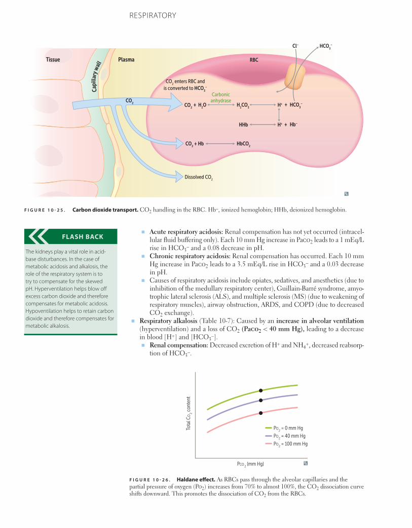
772 CHAPTER 10 RESPIRATORY
■ Acute respiratory acidosis: Renal compensation has not yet occurred (intracel-lular fluid buffering only). Each 10 mm Hg increase in Paco2 leads to a 1 mEq/L rise in HCO3
– and a 0.08 decrease in pH.■ Chronic respiratory acidosis: Renal compensation has occurred. Each 10 mm
Hg increase in Paco2 leads to a 3.5 mEq/L rise in HCO3– and a 0.03 decrease
in pH.■ Causes of respiratory acidosis include opiates, sedatives, and anesthetics (due to
inhibition of the medullary respiratory center), Guillain-Barré syndrome, amyo-trophic lateral sclerosis (ALS), and multiple sclerosis (MS) (due to weakening of respiratory muscles), airway obstruction, ARDS, and COPD (due to decreased CO2 exchange).
■ Respiratory alkalosis (Table 10-7): Caused by an increase in alveolar ventilation (hyperventilation) and a loss of CO2 (Paco2 < 40 mm Hg), leading to a decrease in blood [H+] and [HCO3
–].■ Renal compensation: Decreased excretion of H+ and NH4
+, decreased reabsorp-tion of HCO3
–.
FLASH BACK
The kidneys play a vital role in acid-base disturbances. In the case of metabolic acidosis and alkalosis, the role of the respiratory system is to try to compensate for the skewed pH. Hyperventilation helps blow off excess carbon dioxide and therefore compensates for metabolic acidosis. Hypoventilation helps to retain carbon dioxide and therefore compensates for metabolic alkalosis.
F I G U R E 1 0 - 2 6 . Haldane effect. As RBCs pass through the alveolar capillaries and the partial pressure of oxygen (Po2) increases from 70% to almost 100%, the CO2 dissociation curve shifts downward. This promotes the dissociation of CO2 from the RBCs.
PCO2 (mm Hg)
Tota
l CO
2 con
tent
PO2 = 0 mm Hg
PO2 = 40 mm Hg
PO2 = 100 mm Hg
F I G U R E 1 0 - 2 5 . Carbon dioxide transport. CO2 handling in the RBC. Hb–, ionized hemoglobin; HHb, deionized hemoglobin.
Plasma RBC
Dissolved CO2
CO2 enters RBC and
is converted to HCO3
–
Carbonicanhydrase
H2O H
2CO
3
HHb
CO2
+
CO2 + Hb HbCO
2
CO2
Cl– HCO3
–
HCO3
–H+
H+ Hb–
+
+
Capi
llary
wal
lTissue
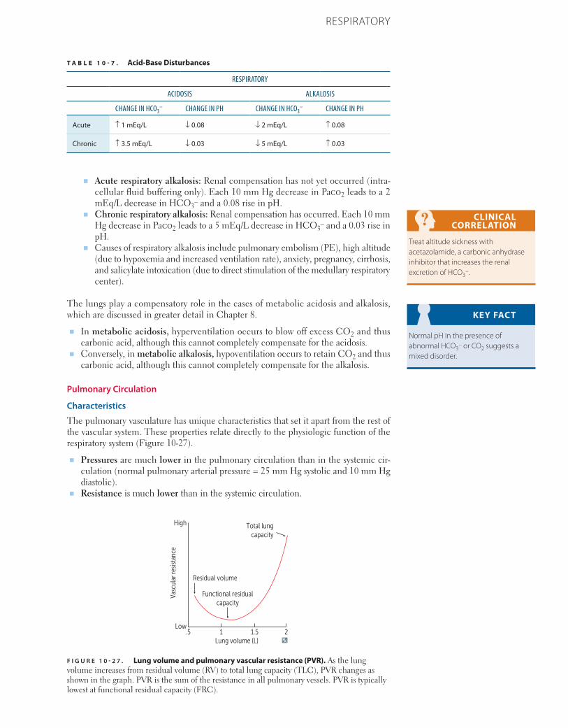
773CHAPTER 10RESPIRATORY
■ Acute respiratory alkalosis: Renal compensation has not yet occurred (intra-cellular fluid buffering only). Each 10 mm Hg decrease in Paco2 leads to a 2 mEq/L decrease in HCO3
– and a 0.08 rise in pH.■ Chronic respiratory alkalosis: Renal compensation has occurred. Each 10 mm
Hg decrease in Paco2 leads to a 5 mEq/L decrease in HCO3– and a 0.03 rise in
pH.■ Causes of respiratory alkalosis include pulmonary embolism (PE), high altitude
(due to hypoxemia and increased ventilation rate), anxiety, pregnancy, cirrhosis, and salicylate intoxication (due to direct stimulation of the medullary respiratory center).
The lungs play a compensatory role in the cases of metabolic acidosis and alkalosis, which are discussed in greater detail in Chapter 8.
■ In metabolic acidosis, hyperventilation occurs to blow off excess CO2 and thus carbonic acid, although this cannot completely compensate for the acidosis.
■ Conversely, in metabolic alkalosis, hypoventilation occurs to retain CO2 and thus carbonic acid, although this cannot completely compensate for the alkalosis.
Pulmonary Circulation
Characteristics
The pulmonary vasculature has unique characteristics that set it apart from the rest of the vascular system. These properties relate directly to the physiologic function of the respiratory system (Figure 10-27).
■ Pressures are much lower in the pulmonary circulation than in the systemic cir-culation (normal pulmonary arterial pressure = 25 mm Hg systolic and 10 mm Hg diastolic).
■ Resistance is much lower than in the systemic circulation.
CLINICAL CORRELATION
Treat altitude sickness with acetazolamide, a carbonic anhydrase inhibitor that increases the renal excretion of HCO3
–.
KEY FACT
Normal pH in the presence of abnormal HCO3
– or CO2 suggests a mixed disorder.
T A B L E 1 0 - 7 . Acid-Base Disturbances
RESPIRATORY
ACIDOSIS ALKALOSIS
CHANGE IN HCO3– CHANGE IN PH CHANGE IN HCO3
– CHANGE IN PH
Acute ↑ 1 mEq/L ↓ 0.08 ↓ 2 mEq/L ↑ 0.08
Chronic ↑ 3.5 mEq/L ↓ 0.03 ↓ 5 mEq/L ↑ 0.03
F I G U R E 1 0 - 2 7 . Lung volume and pulmonary vascular resistance (PVR). As the lung volume increases from residual volume (RV) to total lung capacity (TLC), PVR changes as shown in the graph. PVR is the sum of the resistance in all pulmonary vessels. PVR is typically lowest at functional residual capacity (FRC).
Lung volume (L)
High
Low
Vasc
ular
resis
tanc
e
Total lungcapacity
Residual volume
Functional residualcapacity
.5 1 1.5 2
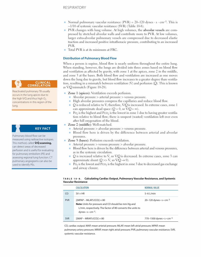
774 CHAPTER 10 RESPIRATORY
■ Normal pulmonary vascular resistance (PVR) = 20–120 dynes ⋅ s ⋅ cm–5. This is ~1/10 of systemic vascular resistance (SVR) (Table 10-8).
■ PVR changes with lung volume. At high volumes, the alveolar vessels are com-pressed by stretched alveolar walls and contribute more to PVR. At low volumes, larger extra-alveolar pulmonary vessels are compressed due to decreased elastic traction and increased positive intrathoracic pressure, contributing to an increased PVR.
■ Total PVR is at its minimum at FRC.
Distribution of Pulmonary Blood Flow
When a person is supine, blood flow is nearly uniform throughout the entire lung. When standing, however, the lungs are divided into three zones based on blood flow and ventilation as affected by gravity, with zone 1 at the apices, zone 2 in the middle, and zone 3 at the bases. Both blood flow and ventilation are increased as one moves down the lung due to gravity, but blood flow increases to a greater degree than ventila-tion, resulting in a mismatch between ventilation (V̇) and perfusion (Q̇). This is known as V̇/Q̇ mismatch (Figure 10-28).
■ Zone 1 (apices): Ventilation exceeds perfusion.■ Alveolar pressure > arterial pressure > venous pressure.■ High alveolar pressures compress the capillaries and reduce blood flow.■ Q̇ is reduced relative to V̇; therefore, V̇/Q̇ is increased. In extreme cases, zone 1
can approximate dead space (Q̇ = 0, so V̇/Q̇ = ∞).■ Po2 is the highest and Pco2 is the lowest in zone 1 due to having greater ventila-
tion relative to blood flow; there is unspent (wasted) ventilation left over even after full oxygenation of the blood.
■ Zone 2 (middle): Well-matched.■ Arterial pressure > alveolar pressure > venous pressure.■ Blood flow here is driven by the difference between arterial and alveolar
pressures.■ Zone 3 (bases): Perfusion exceeds ventilation.
■ Arterial pressure > venous pressure > alveolar pressure.■ Blood flow here is driven by the difference between arterial and venous pressures,
as in the systemic circulation.■ Q̇ is increased relative to V̇, so V̇/Q̇ is decreased. In extreme cases, zone 3 can
approximate shunt (Q̇ >> V̇, so V̇/Q̇ → 0).■ Po2 is the lowest and Pco2 is the highest in zone 3 due to decreased gas exchange
and airway closure.
CLINICAL CORRELATION
Reactivated pulmonary TB usually occurs in the lung apices due to the high V̇/Q̇ and thus high O2 concentrations in this region of the lung.
KEY FACT
Pulmonary blood flow can be measured using radioactive isotopes. This method, called V
./Q
. scanning,
can detect areas of decreased perfusion and is useful for evaluating for pulmonary embolism (PE) and assessing regional lung function. CT pulmonary angiograms can also be used to identify PEs.
T A B L E 1 0 - 8 . Calculating Cardiac Output, Pulmonary Vascular Resistance, and Systemic Vascular Resistance
CALCULATION NORMAL VALUE
CO SV × HR 5–6 L/min
PVR [(MPAP – MLAP)/(CO)] × 80Note: Units for pressure and CO should be mm Hg and
L/min, respectively. The factor of 80 converts the units to dynes • s • cm–5.
20–120 dynes • s • cm–5
SVR [(MAP – MRAP)/(CO)] × 80 770–1500 dynes • s • cm–5
CO, cardiac output; MAP, mean arterial pressure; MLAP, mean left atrial pressure; MPAP, mean pulmonary artery pressure; MRAP, mean right atrial pressure; PVR, pulmonary vascular resistance; SVR, systemic vascular resistance.
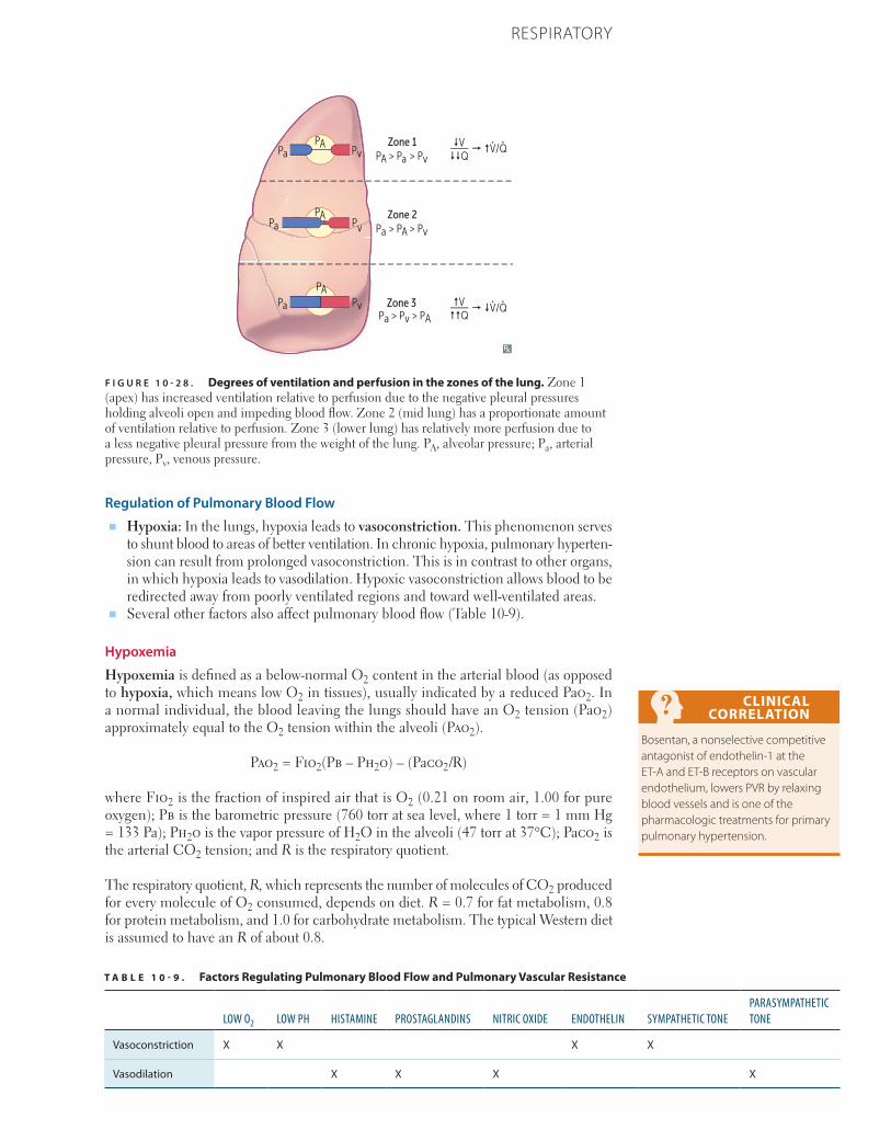
775CHAPTER 10RESPIRATORY
Regulation of Pulmonary Blood Flow■ Hypoxia: In the lungs, hypoxia leads to vasoconstriction. This phenomenon serves
to shunt blood to areas of better ventilation. In chronic hypoxia, pulmonary hyperten-sion can result from prolonged vasoconstriction. This is in contrast to other organs, in which hypoxia leads to vasodilation. Hypoxic vasoconstriction allows blood to be redirected away from poorly ventilated regions and toward well-ventilated areas.
■ Several other factors also affect pulmonary blood flow (Table 10-9).
Hypoxemia
Hypoxemia is defined as a below-normal O2 content in the arterial blood (as opposed to hypoxia, which means low O2 in tissues), usually indicated by a reduced Pao2. In a normal individual, the blood leaving the lungs should have an O2 tension (Pao2) approximately equal to the O2 tension within the alveoli (Pao2).
Pao2 = Fio2(Pb – Ph2o) – (Paco2/R)
where Fio2 is the fraction of inspired air that is O2 (0.21 on room air, 1.00 for pure oxygen); Pb is the barometric pressure (760 torr at sea level, where 1 torr = 1 mm Hg = 133 Pa); Ph2o is the vapor pressure of H2O in the alveoli (47 torr at 37°C); Paco2 is the arterial CO2 tension; and R is the respiratory quotient.
The respiratory quotient, R, which represents the number of molecules of CO2 produced for every molecule of O2 consumed, depends on diet. R = 0.7 for fat metabolism, 0.8 for protein metabolism, and 1.0 for carbohydrate metabolism. The typical Western diet is assumed to have an R of about 0.8.
CLINICAL CORRELATION
Bosentan, a nonselective competitive antagonist of endothelin-1 at the ET-A and ET-B receptors on vascular endothelium, lowers PVR by relaxing blood vessels and is one of the pharmacologic treatments for primary pulmonary hypertension.
F I G U R E 1 0 - 2 8 . Degrees of ventilation and perfusion in the zones of the lung. Zone 1 (apex) has increased ventilation relative to perfusion due to the negative pleural pressures holding alveoli open and impeding blood flow. Zone 2 (mid lung) has a proportionate amount of ventilation relative to perfusion. Zone 3 (lower lung) has relatively more perfusion due to a less negative pleural pressure from the weight of the lung. PA, alveolar pressure; Pa, arterial pressure, Pv, venous pressure.
Zone 1PA > Pa > Pv
Zone 2Pa > PA > Pv
Zone 3Pv
PvPa
Pa
PA
PA
PA
Pa > Pv > PA
PvPa
� �V/Q�V��Q
�V��Q
� �V/Q. .
. .
T A B L E 1 0 - 9 . Factors Regulating Pulmonary Blood Flow and Pulmonary Vascular Resistance
LOW O2 LOW PH HISTAMINE PROSTAGLANDINS NITRIC OXIDE ENDOTHELIN SYMPATHETIC TONEPARASYMPATHETIC TONE
Vasoconstriction X X X X
Vasodilation X X X X
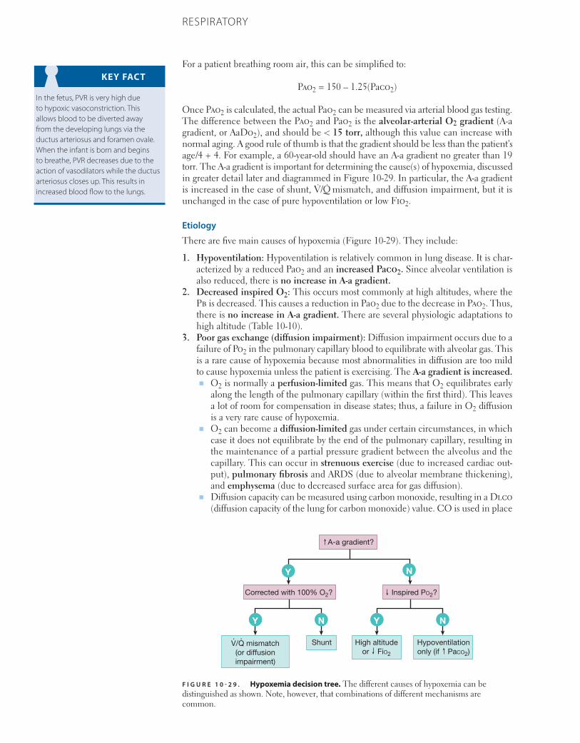
776 CHAPTER 10 RESPIRATORY
For a patient breathing room air, this can be simplified to:
Pao2 = 150 – 1.25(Paco2)
Once Pao2 is calculated, the actual Pao2 can be measured via arterial blood gas testing. The difference between the Pao2 and Pao2 is the alveolar-arterial O2 gradient (A-a gradient, or AaDo2), and should be < 15 torr, although this value can increase with normal aging. A good rule of thumb is that the gradient should be less than the patient’s age/4 + 4. For example, a 60-year-old should have an A-a gradient no greater than 19 torr. The A-a gradient is important for determining the cause(s) of hypoxemia, discussed in greater detail later and diagrammed in Figure 10-29. In particular, the A-a gradient is increased in the case of shunt, V̇/Q̇ mismatch, and diffusion impairment, but it is unchanged in the case of pure hypoventilation or low Fio2.
Etiology
There are five main causes of hypoxemia (Figure 10-29). They include:
1. Hypoventilation: Hypoventilation is relatively common in lung disease. It is char-acterized by a reduced Pao2 and an increased Paco2. Since alveolar ventilation is also reduced, there is no increase in A-a gradient.
2. Decreased inspired O2: This occurs most commonly at high altitudes, where the Pb is decreased. This causes a reduction in Pao2 due to the decrease in Pao2. Thus, there is no increase in A-a gradient. There are several physiologic adaptations to high altitude (Table 10-10).
3. Poor gas exchange (diffusion impairment): Diffusion impairment occurs due to a failure of Po2 in the pulmonary capillary blood to equilibrate with alveolar gas. This is a rare cause of hypoxemia because most abnormalities in diffusion are too mild to cause hypoxemia unless the patient is exercising. The A-a gradient is increased.■ O2 is normally a perfusion-limited gas. This means that O2 equilibrates early
along the length of the pulmonary capillary (within the first third). This leaves a lot of room for compensation in disease states; thus, a failure in O2 diffusion is a very rare cause of hypoxemia.
■ O2 can become a diffusion-limited gas under certain circumstances, in which case it does not equilibrate by the end of the pulmonary capillary, resulting in the maintenance of a partial pressure gradient between the alveolus and the capillary. This can occur in strenuous exercise (due to increased cardiac out-put), pulmonary fibrosis and ARDS (due to alveolar membrane thickening), and emphysema (due to decreased surface area for gas diffusion).
■ Diffusion capacity can be measured using carbon monoxide, resulting in a Dlco (diffusion capacity of the lung for carbon monoxide) value. CO is used in place
KEY FACT
In the fetus, PVR is very high due to hypoxic vasoconstriction. This allows blood to be diverted away from the developing lungs via the ductus arteriosus and foramen ovale. When the infant is born and begins to breathe, PVR decreases due to the action of vasodilators while the ductus arteriosus closes up. This results in increased blood flow to the lungs.
F I G U R E 1 0 - 2 9 . Hypoxemia decision tree. The different causes of hypoxemia can be distinguished as shown. Note, however, that combinations of different mechanisms are common.
A-a gradient?
Corrected with 100% O2?
V/Q mismatch(or diffusionimpairment)
Shunt. .
High altitudeor FiO2
Inspired PO2?
N
N N
Y
Y Y
Hypoventilationonly (if PaCO2)
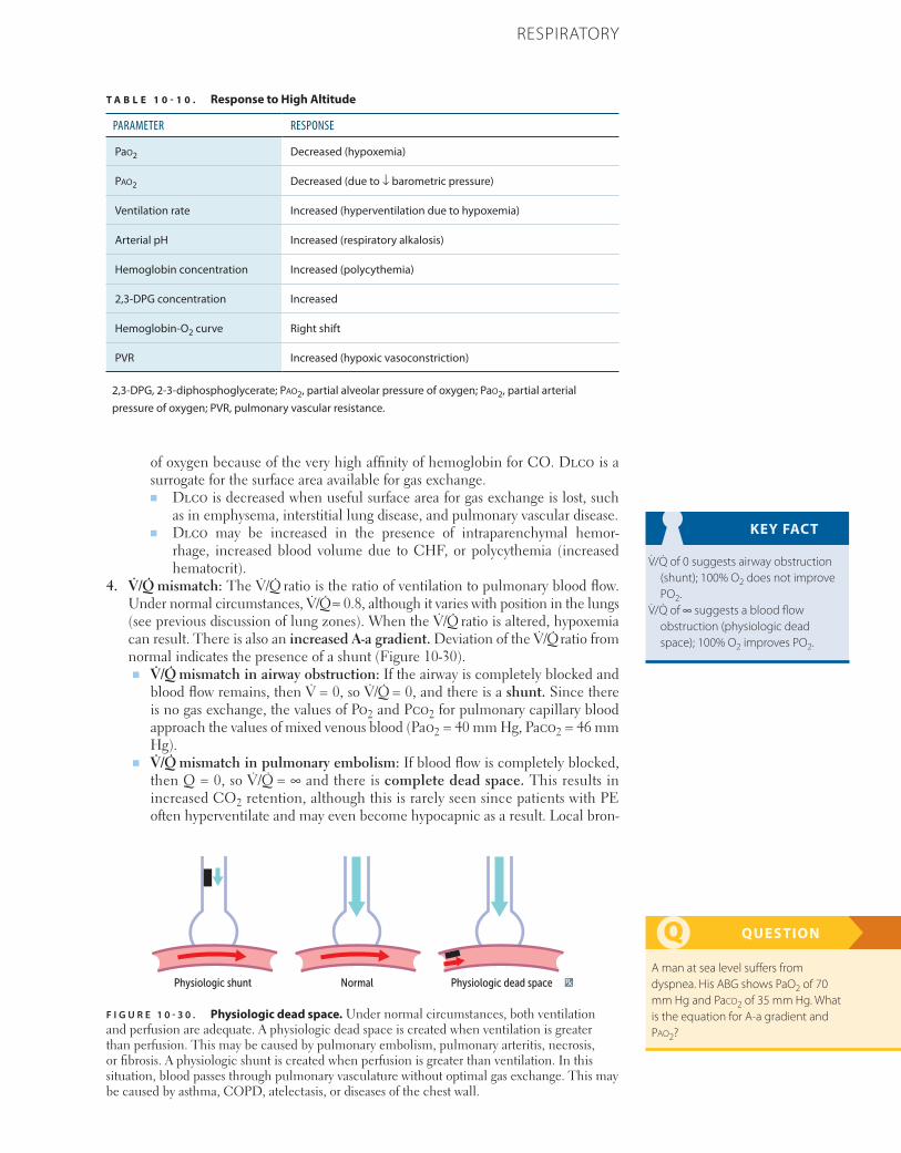
777CHAPTER 10RESPIRATORY
of oxygen because of the very high affinity of hemoglobin for CO. Dlco is a surrogate for the surface area available for gas exchange.■ Dlco is decreased when useful surface area for gas exchange is lost, such
as in emphysema, interstitial lung disease, and pulmonary vascular disease.■ Dlco may be increased in the presence of intraparenchymal hemor-
rhage, increased blood volume due to CHF, or polycythemia (increased hematocrit).
4. V̇/Q̇ mismatch: The V̇/Q̇ ratio is the ratio of ventilation to pulmonary blood flow. Under normal circumstances, V̇/Q̇ ≈ 0.8, although it varies with position in the lungs (see previous discussion of lung zones). When the V̇/Q̇ ratio is altered, hypoxemia can result. There is also an increased A-a gradient. Deviation of the V̇/Q̇ ratio from normal indicates the presence of a shunt (Figure 10-30).■ V̇/Q̇ mismatch in airway obstruction: If the airway is completely blocked and
blood flow remains, then V̇ = 0, so V̇/Q̇ = 0, and there is a shunt. Since there is no gas exchange, the values of Po2 and Pco2 for pulmonary capillary blood approach the values of mixed venous blood (Pao2 = 40 mm Hg, Paco2 = 46 mm Hg).
■ V̇/Q̇ mismatch in pulmonary embolism: If blood flow is completely blocked, then Q = 0, so V̇/Q̇ = ∞ and there is complete dead space. This results in increased CO2 retention, although this is rarely seen since patients with PE often hyperventilate and may even become hypocapnic as a result. Local bron-
KEY FACT
V̇/Q̇ of 0 suggests airway obstruction (shunt); 100% O2 does not improve PO2.
V̇/Q̇ of ∞ suggests a blood flow obstruction (physiologic dead space); 100% O2 improves PO2.
QUESTION
A man at sea level suffers from dyspnea. His ABG shows PaO2 of 70 mm Hg and Paco2 of 35 mm Hg. What is the equation for A-a gradient and Pao2?
F I G U R E 1 0 - 3 0 . Physiologic dead space. Under normal circumstances, both ventilation and perfusion are adequate. A physiologic dead space is created when ventilation is greater than perfusion. This may be caused by pulmonary embolism, pulmonary arteritis, necrosis, or fibrosis. A physiologic shunt is created when perfusion is greater than ventilation. In this situation, blood passes through pulmonary vasculature without optimal gas exchange. This may be caused by asthma, COPD, atelectasis, or diseases of the chest wall.
Physiologic shunt Normal Physiologic dead space
T A B L E 1 0 - 1 0 . Response to High Altitude
PARAMETER RESPONSE
Pao2 Decreased (hypoxemia)
Pao2 Decreased (due to ↓ barometric pressure)
Ventilation rate Increased (hyperventilation due to hypoxemia)
Arterial pH Increased (respiratory alkalosis)
Hemoglobin concentration Increased (polycythemia)
2,3-DPG concentration Increased
Hemoglobin-O2 curve Right shift
PVR Increased (hypoxic vasoconstriction)
2,3-DPG, 2-3-diphosphoglycerate; Pao2, partial alveolar pressure of oxygen; Pao2, partial arterial pressure of oxygen; PVR, pulmonary vascular resistance.
778 CHAPTER 10 RESPIRATORY
chospasm due to the PE can also contribute to hypoxemia. If the blood flow is low but not zero, the values of Po2 and Pco2 for pulmonary capillary blood approach that of inspired air (Pao2 = 150 mm Hg, Paco2 = 0 mm Hg).
■ In most cases of V̇/Q̇ mismatch, there is neither true shunt nor complete dead space, but simply an abnormal V̇/Q̇ ratio. Blood from well-ventilated areas is already saturated at baseline, so no amount of effort from well-ventilated areas can compensate for the desaturated blood emerging from areas that are poorly ventilated. Giving the patient 100% O2 increases the patient’s Pao2.
5. Shunt: As mentioned previously, shunt is an extreme case of V̇/Q̇ mismatch that occurs when some blood reaches the systemic circulation without being oxygenated, reducing Pao2. Since the Pao2 is unaffected, the A-a gradient is increased.■ Right-to-left shunt: Occurs when blood from the right side of the heart enters
the systemic circulation without passing through the lungs. It is seen in tetralogy of Fallot (and other congenital heart conditions causing right-to-left shunts) and always causes a reduction in Pao2.
■ Left-to-right shunt: More common than right-to-left shunt because pressures are higher on the left side of the heart. It is seen with several congenital abnor-malities, including patent ductus arteriosus (PDA), atrial septal defect, and ven-tricular septal defect, as well as with traumatic injury. Left-to-right shunts do not decrease Pao2 since oxygenated blood is returning to the right side of the heart, raising the Po2.
■ True shunt can be differentiated from V̇/Q̇ mismatch by giving the hypoxemic patient 100% O2. This increases Pao2 in the case of V̇/Q̇ mismatch but not in the case of a shunt, since in the latter, the blood never communicates with the alveolar gas, regardless of its composition. A patient without a shunt should achieve a Pao2 of at least 400 torr on 100% oxygen.
Hypercapnia
Alveolar ventilation is the main determinant of Paco2. Hypercapnia occurs when alveo-lar ventilation is reduced, which can happen in a number of ways:
■ Decreased total minute ventilation without a change in the Vd/Vt ratio.■ Constant minute ventilation with increasing Vd/Vt. This can occur with decreased
Vt (eg, a greater percentage of the Vt is taken up by dead space) and increased respiratory rate.
■ V̇/Q̇ mismatch. Well-perfused areas may be underventilated, whereas underperfused areas may be overventilated. When a large amount of ventilation is “wasted” on underperfused sections of lung, the effect is similar to increasing the dead space: less air is available to exchange gases with the blood, and CO2 levels in the blood increase.
The response of the body to hypercapnia is often to increase alveolar ventilation by hyperventilating and blowing off more CO2. Thus, CO2 retention may not occur even if the preceding criteria are met as long as the body is able to compensate.
Control of Respiration
Central Control of Respiration■ Medullary respiratory center: Located in the reticular formation. Damage to or
suppression of this region due to stroke, opioid overdose, or other causes can lead to respiratory failure and death.■ Dorsal respiratory group: Responsible for inspiration and determines the
rhythm of breathing (normally 12–20 breaths/minute with an I:E [inspiration-to-expiration] ratio of 1:2). The dorsal respiratory group receives sensory input from peripheral chemoreceptors and lung mechanoreceptors via the vagus and glossopharyngeal nerves. Output travels via the phrenic nerve (C3–C5) and
CLINICAL CORRELATION
Over time, left-to-right shunts can cause pressures on the right side of the heart to become greater than those on the left. This leads to a reversal of the shunt to right-to-left, causing hypoxemia. This is called Eisenmenger syndrome.
KEY FACT
CO2 is the most potent cerebral vasodilator. Increased CO2 decreases cerebral vascular resistance, resulting in increased perfusion and intracranial pressure.
ANSWER
A-a gradient = 35.25, Pao2 = 106.25 A-a = Pao2 – Pao2 Pao2 = 150 –1.25 (Paco2) Pao2 = 150 – 1.25 (35) Pao2 = 150 – 43.75Pao2 = 106.25 A-a = 106.25 – 70 = 35.25 A-a > 15 is abnormal
779CHAPTER 10RESPIRATORY
the intercostal nerves (T1–T11) to the diaphragm and the external intercostal muscles, respectively.
■ Ventral respiratory group: Responsible for forced expiration; not active during ordinary passive expiration. Also involved with increased inspiratory effort (eg, during exercise).
■ Pons:■ Pneumotaxic center: Located in the upper pons. Inhibits inspiration, helping
to regulate inspiratory volume and rate.■ Apneustic center: Located in the lower pons. Stimulates inspiration.
■ Cerebral cortex: Exerts voluntary control over breathing.
Chemoreceptors■ Central chemoreceptors in the medulla: Respond to the pH of the cerebrospinal
fluid (CSF), with decreases in pH causing hyperventilation. CO2 from arterial blood diffuses into the CSF and combines with H2O to form H+ and HCO3
–.■ Peripheral chemoreceptors in the carotid and aortic bodies: Increased Paco2 or
decreased pH or Pao2 stimulate these chemoreceptors to increase respiratory rate. Pao2 must reach low levels (< 60 mm Hg) before breathing is stimulated.
Other Receptors■ Lung stretch receptors: Mechanoreceptors located in the airway smooth muscle
that are stimulated by distention of the lungs and produce reflex inspiratory time shortening and Hering-Breuer inflation and deflation reflexes. In the Hering-Breuer inflation reflex, excessive stretching of the lungs during a large inspiratory effort leads to inhibition of the dorsal respiratory group and the apneustic center to promote expiration. The deflation reflex acts during expiration to activate the inspiratory control areas.
■ Irritant receptors (nociceptors): Located between airway epithelial cells and stimu-lated by noxious substances.
■ Juxtacapillary (J) receptors: Located close to the capillaries in the alveolar walls. Increases in interstitial fluid, such as during pulmonary edema, PE, or pneumonia, stimulate these receptors, causing rapid, shallow breathing.
■ Joint and muscle receptors: These are activated by limb movement and help to stimulate breathing early in exercise.
Pathology
Discussion of respiratory pathology will begin with an overview of physical examination findings commonly associated with respiratory dysfunction. Pathologic conditions of the upper airways (eg, nasopharynx, oropharynx, larynx) are then covered, followed by those of the lower airways (eg, tracheobronchial tree, lung parenchyma).
PATHOLOGY ON PHYSICAL EXAMINATION
The pulmonary physical examination has four components: inspection, auscultation, percussion, palpation. This section provides an overview of each component in the context of the USMLE Step 1. The technical aspects of the physical examination are beyond the scope of this text.
Inspection
Signs of respiratory distress include dyspnea (labored breathing), tachypnea (respiratory rate > 20 breaths/min), cyanosis, grunting, nasal flaring, retractions, and using accessory muscles of respiration. Retractions refer to the inward “pulling” of muscles during
KEY FACT
Suspect foreign body aspiration in a child who presents with acute onset wheezing.
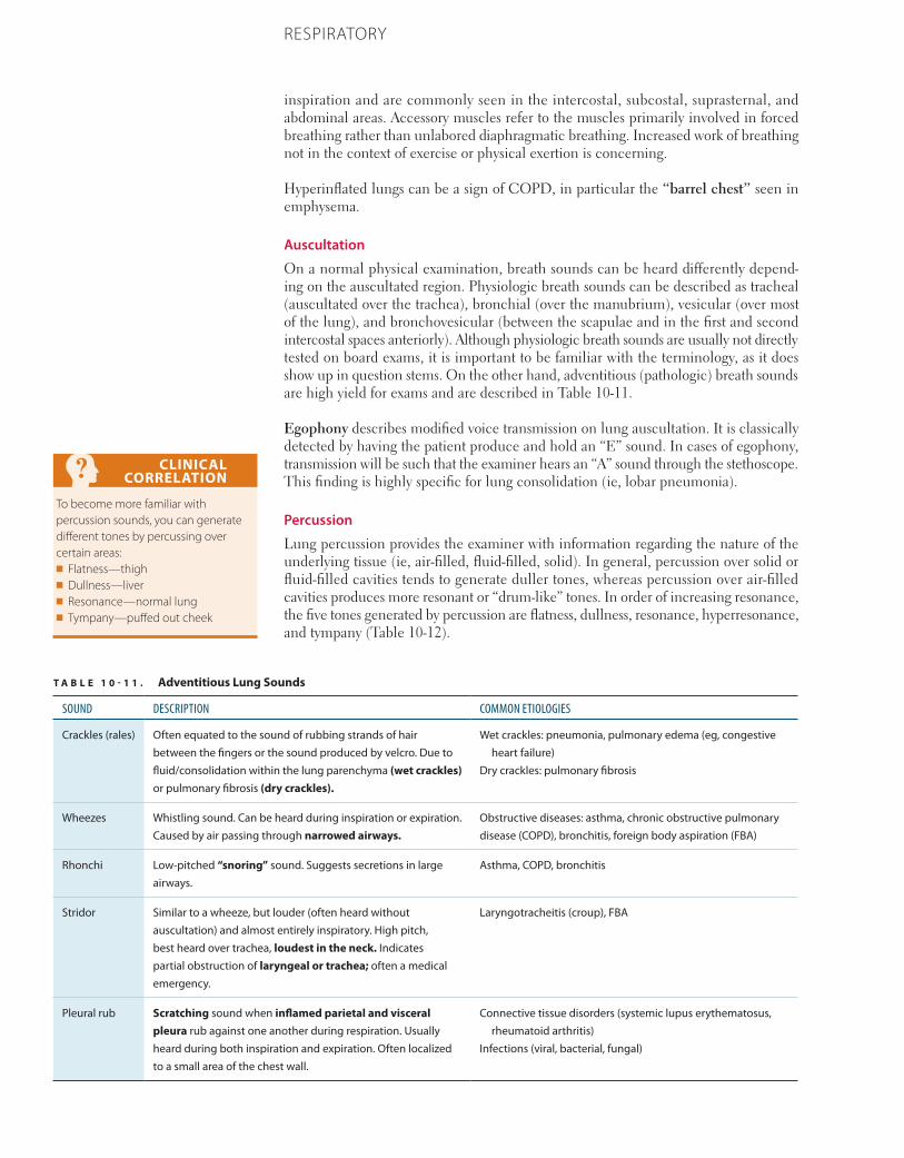
780 CHAPTER 10 RESPIRATORY
inspiration and are commonly seen in the intercostal, subcostal, suprasternal, and abdominal areas. Accessory muscles refer to the muscles primarily involved in forced breathing rather than unlabored diaphragmatic breathing. Increased work of breathing not in the context of exercise or physical exertion is concerning.
Hyperinflated lungs can be a sign of COPD, in particular the “barrel chest” seen in emphysema.
Auscultation
On a normal physical examination, breath sounds can be heard differently depend-ing on the auscultated region. Physiologic breath sounds can be described as tracheal (auscultated over the trachea), bronchial (over the manubrium), vesicular (over most of the lung), and bronchovesicular (between the scapulae and in the first and second intercostal spaces anteriorly). Although physiologic breath sounds are usually not directly tested on board exams, it is important to be familiar with the terminology, as it does show up in question stems. On the other hand, adventitious (pathologic) breath sounds are high yield for exams and are described in Table 10-11.
Egophony describes modified voice transmission on lung auscultation. It is classically detected by having the patient produce and hold an “E” sound. In cases of egophony, transmission will be such that the examiner hears an “A” sound through the stethoscope. This finding is highly specific for lung consolidation (ie, lobar pneumonia).
Percussion
Lung percussion provides the examiner with information regarding the nature of the underlying tissue (ie, air-filled, fluid-filled, solid). In general, percussion over solid or fluid-filled cavities tends to generate duller tones, whereas percussion over air-filled cavities produces more resonant or “drum-like” tones. In order of increasing resonance, the five tones generated by percussion are flatness, dullness, resonance, hyperresonance, and tympany (Table 10-12).
CLINICAL CORRELATION
To become more familiar with percussion sounds, you can generate different tones by percussing over certain areas:■ Flatness—thigh■ Dullness—liver■ Resonance—normal lung■ Tympany—puffed out cheek
T A B L E 1 0 - 1 1 . Adventitious Lung Sounds
SOUND DESCRIPTION COMMON ETIOLOGIES
Crackles (rales) Often equated to the sound of rubbing strands of hair between the fingers or the sound produced by velcro. Due to fluid/consolidation within the lung parenchyma (wet crackles) or pulmonary fibrosis (dry crackles).
Wet crackles: pneumonia, pulmonary edema (eg, congestive heart failure)
Dry crackles: pulmonary fibrosis
Wheezes Whistling sound. Can be heard during inspiration or expiration. Caused by air passing through narrowed airways.
Obstructive diseases: asthma, chronic obstructive pulmonary disease (COPD), bronchitis, foreign body aspiration (FBA)
Rhonchi Low-pitched “snoring” sound. Suggests secretions in large airways.
Asthma, COPD, bronchitis
Stridor Similar to a wheeze, but louder (often heard without auscultation) and almost entirely inspiratory. High pitch, best heard over trachea, loudest in the neck. Indicates partial obstruction of laryngeal or trachea; often a medical emergency.
Laryngotracheitis (croup), FBA
Pleural rub Scratching sound when inflamed parietal and visceral pleura rub against one another during respiration. Usually heard during both inspiration and expiration. Often localized to a small area of the chest wall.
Connective tissue disorders (systemic lupus erythematosus, rheumatoid arthritis)
Infections (viral, bacterial, fungal)
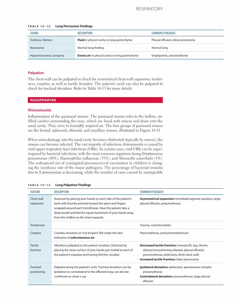
781CHAPTER 10RESPIRATORY
Palpation
The chest wall can be palpated to check for symmetrical chest wall expansion, tender-ness, crepitus, as well as tactile fremitus. The patient’s neck can also be palpated to check for tracheal deviation. Refer to Table 10-13 for more details.
NASOPHARYNX
Rhinosinusitis
Inflammation of the paranasal sinuses. The paranasal sinuses refer to the hollow, air-filled cavities surrounding the nose, which are lined with mucus and drain into the nasal cavity. They serve to humidify inspired air. The four groups of paranasal sinuses are the frontal, sphenoid, ethmoid, and maxillary sinuses, illustrated in Figure 10-31.
When sinus drainage into the nasal cavity becomes obstructed (typically by mucus), the sinuses can become infected. The vast majority of infectious rhinosinusitis is caused by viral upper respiratory tract infections (URIs). In certain cases, viral URIs can be super-imposed by bacterial infections, with the most common organisms being Streptococcus pneumoniae (40%), Haemophilus influenzae (35%), and Moraxella catarrhalis (5%). The widespread use of conjugated pneumococcal vaccination in children is chang-ing the incidence rate of the major pathogens. The percentage of bacterial sinusitis due to S pneumoniae is decreasing, while the number of cases caused by nontypeable
T A B L E 1 0 - 1 2 . Lung Percussion Findings
SOUND DESCRIPTION COMMON ETIOLOGIES
Dullness, flatness Fluid in pleural cavity or lung parenchyma Pleural effusion, lobar pneumonia
Resonance Normal lung finding Normal lung
Hyperresonance, tympany Excess air in pleural cavity or lung parenchyma Emphysema, pneumothorax
T A B L E 1 0 - 1 3 . Lung Palpation Findings
FEATURE DESCRIPTION COMMON ETIOLOGIES
Chest wall expansion
Assessed by placing your hands on each side of the patient’s back with thumbs pointed toward the spine and fingers wrapped around each hemithorax. Have the patient take a deep breath and feel for equal movement of your hands away from the midline as the chest expands.
Asymmetrical expansion: hemidiaphragmatic paralysis, large pleural effusion, pneumothorax
Tenderness Trauma, costochondritis
Crepitus Crackles sensation or “rice krispies” felt under the skin. Indicative of subcutaneous air.
Pneumothorax, pneumomediastinum
Tactile fremitus
Vibrations palpated as the patient vocalizes. Detected by placing the ulnar surface of your hands just medial to each of the patient’s scapulae and having him/her vocalize.
Decreased tactile fremitus: nonspecific (eg, chronic obstructive pulmonary disease, pleural effusion, pneumothorax, atelectasis, thick chest wall)
Increased tactile fremitus: lobar pneumonia
Tracheal positioning
Palpated along the patient’s neck. Tracheal deviation can be ipsilateral or contralateral to the affected lung; can also be confirmed on chest x-ray.
Ipsilateral deviation: atelectasis, spontaneous (simple) pneumothorax
Contralateral deviation: pneumothorax, large pleural effusion
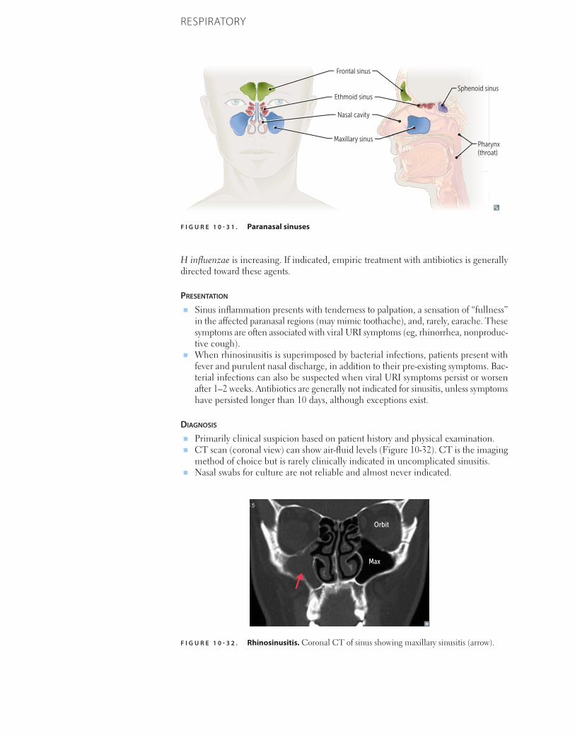
782 CHAPTER 10 RESPIRATORY
H influenzae is increasing. If indicated, empiric treatment with antibiotics is generally directed toward these agents.
Presentation
■ Sinus inflammation presents with tenderness to palpation, a sensation of “fullness” in the affected paranasal regions (may mimic toothache), and, rarely, earache. These symptoms are often associated with viral URI symptoms (eg, rhinorrhea, nonproduc-tive cough).
■ When rhinosinusitis is superimposed by bacterial infections, patients present with fever and purulent nasal discharge, in addition to their pre-existing symptoms. Bac-terial infections can also be suspected when viral URI symptoms persist or worsen after 1–2 weeks. Antibiotics are generally not indicated for sinusitis, unless symptoms have persisted longer than 10 days, although exceptions exist.
Diagnosis
■ Primarily clinical suspicion based on patient history and physical examination. ■ CT scan (coronal view) can show air-fluid levels (Figure 10-32). CT is the imaging
method of choice but is rarely clinically indicated in uncomplicated sinusitis.■ Nasal swabs for culture are not reliable and almost never indicated.
F I G U R E 1 0 - 3 1 . Paranasal sinuses
Frontal sinus
Ethmoid sinus
Nasal cavity
Maxillary sinus
Sphenoid sinus
Pharynx(throat)
F I G U R E 1 0 - 3 2 . Rhinosinusitis. Coronal CT of sinus showing maxillary sinusitis (arrow).
Orbit
Max
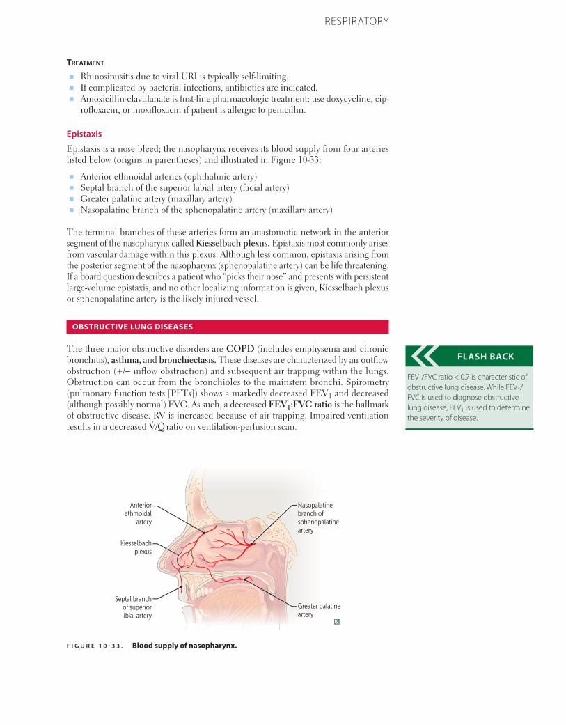
783CHAPTER 10RESPIRATORY
treatment ■ Rhinosinusitis due to viral URI is typically self-limiting.■ If complicated by bacterial infections, antibiotics are indicated.■ Amoxicillin-clavulanate is first-line pharmacologic treatment; use doxycycline, cip-
rofloxacin, or moxifloxacin if patient is allergic to penicillin.
Epistaxis
Epistaxis is a nose bleed; the nasopharynx receives its blood supply from four arteries listed below (origins in parentheses) and illustrated in Figure 10-33:
■ Anterior ethmoidal arteries (ophthalmic artery)■ Septal branch of the superior labial artery (facial artery)■ Greater palatine artery (maxillary artery)■ Nasopalatine branch of the sphenopalatine artery (maxillary artery)
The terminal branches of these arteries form an anastomotic network in the anterior segment of the nasopharynx called Kiesselbach plexus. Epistaxis most commonly arises from vascular damage within this plexus. Although less common, epistaxis arising from the posterior segment of the nasopharynx (sphenopalatine artery) can be life threatening. If a board question describes a patient who “picks their nose” and presents with persistent large-volume epistaxis, and no other localizing information is given, Kiesselbach plexus or sphenopalatine artery is the likely injured vessel.
OBSTRUCTIVE LUNG DISEASES
The three major obstructive disorders are COPD (includes emphysema and chronic bronchitis), asthma, and bronchiectasis. These diseases are characterized by air outflow obstruction (+/− inflow obstruction) and subsequent air trapping within the lungs. Obstruction can occur from the bronchioles to the mainstem bronchi. Spirometry (pulmonary function tests [PFTs]) shows a markedly decreased FEV1 and decreased (although possibly normal) FVC. As such, a decreased FEV1:FVC ratio is the hallmark of obstructive disease. RV is increased because of air trapping. Impaired ventilation results in a decreased V̇/Q̇ ratio on ventilation-perfusion scan.
FLASH BACK
FEV1/FVC ratio < 0.7 is characteristic of obstructive lung disease. While FEV1/FVC is used to diagnose obstructive lung disease, FEV1 is used to determine the severity of disease.
F I G U R E 1 0 - 3 3 . Blood supply of nasopharynx.
Anteriorethmoidal
artery
Kiesselbachplexus
Septal branchof superiorlibial artery
Greater palatineartery
Nasopalatinebranch ofsphenopalatineartery
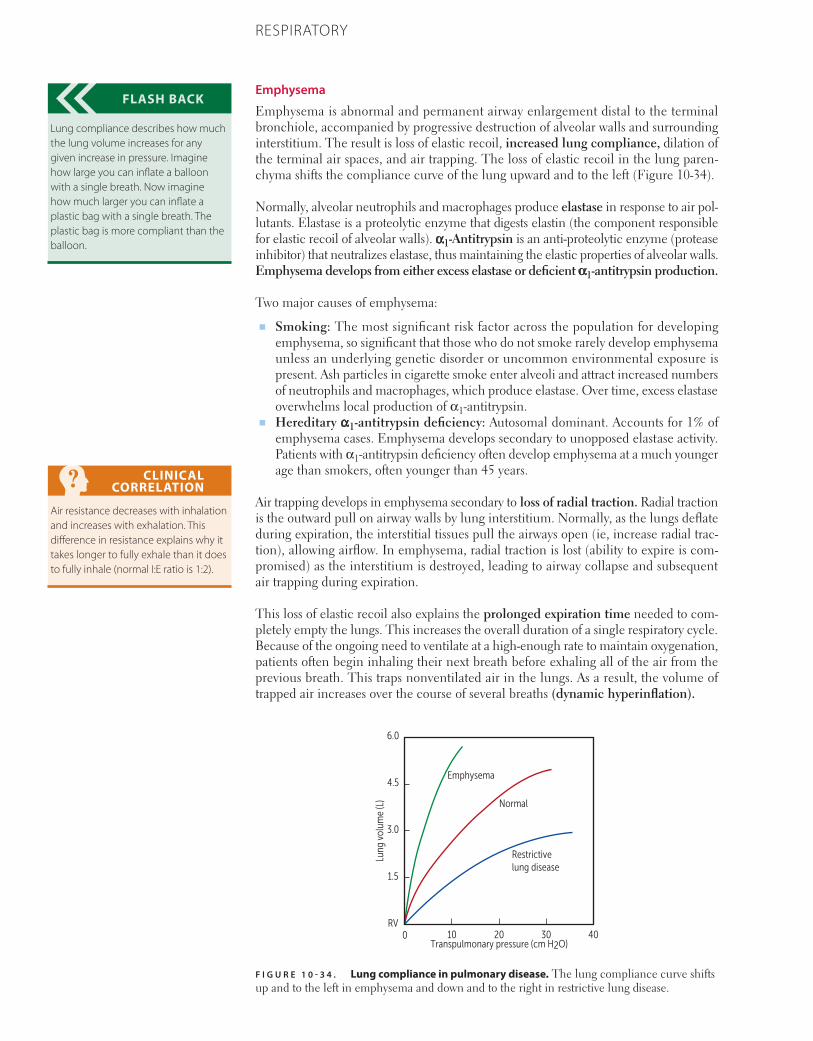
784 CHAPTER 10 RESPIRATORY
Emphysema
Emphysema is abnormal and permanent airway enlargement distal to the terminal bronchiole, accompanied by progressive destruction of alveolar walls and surrounding interstitium. The result is loss of elastic recoil, increased lung compliance, dilation of the terminal air spaces, and air trapping. The loss of elastic recoil in the lung paren-chyma shifts the compliance curve of the lung upward and to the left (Figure 10-34).
Normally, alveolar neutrophils and macrophages produce elastase in response to air pol-lutants. Elastase is a proteolytic enzyme that digests elastin (the component responsible for elastic recoil of alveolar walls). α1-Antitrypsin is an anti-proteolytic enzyme (protease inhibitor) that neutralizes elastase, thus maintaining the elastic properties of alveolar walls. Emphysema develops from either excess elastase or deficient α1-antitrypsin production.
Two major causes of emphysema:
■ Smoking: The most significant risk factor across the population for developing emphysema, so significant that those who do not smoke rarely develop emphysema unless an underlying genetic disorder or uncommon environmental exposure is present. Ash particles in cigarette smoke enter alveoli and attract increased numbers of neutrophils and macrophages, which produce elastase. Over time, excess elastase overwhelms local production of α1-antitrypsin.
■ Hereditary α1-antitrypsin deficiency: Autosomal dominant. Accounts for 1% of emphysema cases. Emphysema develops secondary to unopposed elastase activity. Patients with α1-antitrypsin deficiency often develop emphysema at a much younger age than smokers, often younger than 45 years.
Air trapping develops in emphysema secondary to loss of radial traction. Radial traction is the outward pull on airway walls by lung interstitium. Normally, as the lungs deflate during expiration, the interstitial tissues pull the airways open (ie, increase radial trac-tion), allowing airflow. In emphysema, radial traction is lost (ability to expire is com-promised) as the interstitium is destroyed, leading to airway collapse and subsequent air trapping during expiration.
This loss of elastic recoil also explains the prolonged expiration time needed to com-pletely empty the lungs. This increases the overall duration of a single respiratory cycle. Because of the ongoing need to ventilate at a high-enough rate to maintain oxygenation, patients often begin inhaling their next breath before exhaling all of the air from the previous breath. This traps nonventilated air in the lungs. As a result, the volume of trapped air increases over the course of several breaths (dynamic hyperinflation).
FLASH BACK
Lung compliance describes how much the lung volume increases for any given increase in pressure. Imagine how large you can inflate a balloon with a single breath. Now imagine how much larger you can inflate a plastic bag with a single breath. The plastic bag is more compliant than the balloon.
CLINICAL CORRELATION
Air resistance decreases with inhalation and increases with exhalation. This difference in resistance explains why it takes longer to fully exhale than it does to fully inhale (normal I:E ratio is 1:2).
F I G U R E 1 0 - 3 4 . Lung compliance in pulmonary disease. The lung compliance curve shifts up and to the left in emphysema and down and to the right in restrictive lung disease.
10 20 30
Restrictive lung disease
Normal
Emphysema
400RV
1.5
3.0
4.5
6.0
Transpulmonary pressure (cm H2O)
Lung
vol
ume
(L)
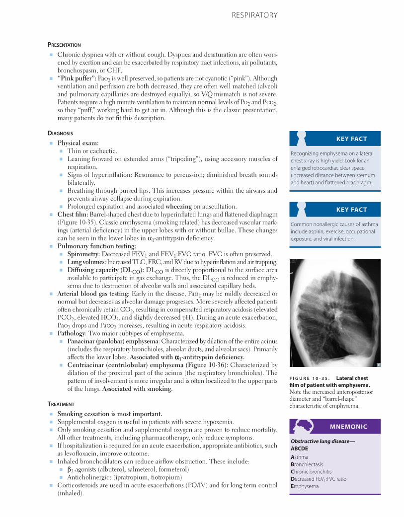
785CHAPTER 10RESPIRATORY
Presentation
■ Chronic dyspnea with or without cough. Dyspnea and desaturation are often wors-ened by exertion and can be exacerbated by respiratory tract infections, air pollutants, bronchospasm, or CHF.
■ “Pink puffer”: Pao2 is well preserved, so patients are not cyanotic (“pink”). Although ventilation and perfusion are both decreased, they are often well matched (alveoli and pulmonary capillaries are destroyed equally), so V̇/Q̇ mismatch is not severe. Patients require a high minute ventilation to maintain normal levels of Po2 and Pco2, so they “puff,” working hard to get air in. Although this is the classic presentation, many patients do not fit this description.
Diagnosis
■ Physical exam:■ Thin or cachectic.■ Leaning forward on extended arms (“tripoding”), using accessory muscles of
respiration.■ Signs of hyperinflation: Resonance to percussion; diminished breath sounds
bilaterally. ■ Breathing through pursed lips. This increases pressure within the airways and
prevents airway collapse during expiration.■ Prolonged expiration and associated wheezing on auscultation.
■ Chest film: Barrel-shaped chest due to hyperinflated lungs and flattened diaphragm (Figure 10-35). Classic emphysema (smoking related) has decreased vascular mark-ings (arterial deficiency) in the upper lobes with or without bullae. These changes can be seen in the lower lobes in α1-antitrypsin deficiency.
■ Pulmonary function testing: ■ Spirometry: Decreased FEV1 and FEV1:FVC ratio. FVC is often preserved. ■ Lung volumes: Increased TLC, FRC, and RV due to hyperinflation and air trapping.■ Diffusing capacity (DLCO): DLCO is directly proportional to the surface area
available to participate in gas exchange. Thus, the DLCO is reduced in emphy-sema due to destruction of alveolar walls and associated capillary beds.
■ Arterial blood gas testing: Early in the disease, Pao2 may be mildly decreased or normal but decreases as alveolar damage progresses. More severely affected patients often chronically retain CO2, resulting in compensated respiratory acidosis (elevated PCO2, elevated HCO3, and slightly decreased pH). During an acute exacerbation, Pao2 drops and Paco2 increases, resulting in acute respiratory acidosis.
■ Pathology: Two major subtypes of emphysema.■ Panacinar (panlobar) emphysema: Characterized by dilation of the entire acinus
(includes the respiratory bronchioles, alveolar ducts, and alveolar sacs). Primarily affects the lower lobes. Associated with α1-antitrypsin deficiency.
■ Centriacinar (centrilobular) emphysema (Figure 10-36): Characterized by dilation of the proximal part of the acinus (the respiratory bronchioles). The pattern of involvement is more irregular and is often localized to the upper parts of the lungs. Associated with smoking.
treatment
■ Smoking cessation is most important.■ Supplemental oxygen is useful in patients with severe hypoxemia.■ Only smoking cessation and supplemental oxygen are proven to reduce mortality.
All other treatments, including pharmacotherapy, only reduce symptoms. ■ If hospitalization is required for an acute exacerbation, appropriate antibiotics, such
as levofloxacin, improve outcome.■ Inhaled bronchodilators can reduce airflow obstruction. These include:
■ β2-agonists (albuterol, salmeterol, formeterol)■ Anticholinergics (ipratropium, tiotropium)
■ Corticosteroids are used in acute exacerbations (PO/IV) and for long-term control (inhaled).
KEY FACT
Recognizing emphysema on a lateral chest x-ray is high yield. Look for an enlarged retrocardiac clear space (increased distance between sternum and heart) and flattened diaphragm.
KEY FACT
Common nonallergic causes of asthma include aspirin, exercise, occupational exposure, and viral infection.
MNEMONIC
Obstructive lung disease—ABCDEAsthma Bronchiectasis Chronic bronchitis Decreased FEV1:FVC ratio Emphysema
F I G U R E 1 0 - 3 5 . Lateral chest film of patient with emphysema. Note the increased anteroposterior diameter and “barrel-shape” characteristic of emphysema.
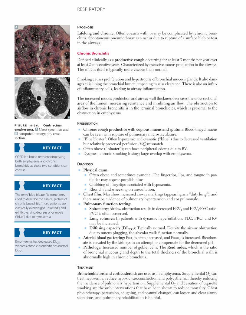
786 CHAPTER 10 RESPIRATORY
Prognosis
Lifelong and chronic. Often coexists with, or may be complicated by, chronic bron-chitis. Spontaneous pneumothorax can occur due to rupture of a surface bleb or tear in the airways.
Chronic Bronchitis
Defined clinically as a productive cough occurring for at least 3 months per year over at least 2 consecutive years. Characterized by excessive mucus production in the airways. The mucus itself is typically more viscous than normal.
Smoking causes proliferation and hypertrophy of bronchial mucous glands. It also dam-ages cilia lining the bronchial lumen, impeding mucus clearance. There is also an influx of inflammatory cells, leading to airway inflammation.
The increased mucus production and airway wall thickness decreases the cross-sectional area of the lumen, increasing resistance and inhibiting air flow. The obstruction to airflow in chronic bronchitis is in the terminal bronchioles, which is proximal to the obstruction in emphysema.
Presentation
■ Chronic cough productive with copious mucus and sputum. Blood-tinged mucus can be seen with rupture of pulmonary microvasculature.
■ “Blue bloater”: Often hypoxemic and cyanotic (“blue”) due to decreased ventilation but relatively preserved perfusion; V̇/Q̇ mismatch.
■ Often obese (“bloater”); can have peripheral edema due to RV. ■ Dyspnea, chronic smoking history; large overlap with emphysema.
Diagnosis
■ Physical exam:■ Often obese and sometimes cyanotic. The fingertips, lips, and tongue in par-
ticular may appear purplish blue.■ Clubbing of fingertips associated with hypoxemia.■ Rhonchi and wheezing on auscultation.
■ Chest film: May show increased airway markings (appearing as a “dirty lung”), and there may be evidence of pulmonary hypertension and cor pulmonale.
■ Pulmonary function testing: ■ Spirometry: Airflow obstruction results in decreased FEV1 and FEV1:FVC ratio.
FVC is often preserved. ■ Lung volumes: In patients with dynamic hyperinflation, TLC, FRC, and RV
may be increased.■ Diffusing capacity (DLCO): Typically normal. Despite the airway obstruction
due to mucus plugging, the alveolar walls function normally.■ Arterial blood gas testing: Pao2 is often decreased, and Paco2 is increased. Bicarbon-
ate is elevated by the kidneys in an attempt to compensate for the decreased pH.■ Pathology: Increased number of goblet cells. The Reid index, which is the ratio
of bronchial mucous gland depth to the total thickness of the bronchial wall, is abnormally high in chronic bronchitis.
treatment
Bronchodilators and corticosteroids are used as in emphysema. Supplemental O2 can treat hypoxemia, reduce hypoxic vasoconstriction and polycythemia, thereby reducing the incidence of pulmonary hypertension. Supplemental O2 and cessation of cigarette smoking are the only interventions that have been shown to reduce mortality. Chest physiotherapy (percussion, coughing, and postural changes) can loosen and clear airway secretions, and pulmonary rehabilitation is helpful.
KEY FACT
COPD is a broad term encompassing both emphysema and chronic bronchitis, as these two conditions can coexist.
KEY FACT
The term “blue bloater” is sometimes used to describe the clinical picture of chronic bronchitis. These patients are classically overweight (“bloated”) and exhibit varying degrees of cyanosis (“blue”) due to hypoxemia.
KEY FACT
Emphysema has decreased DLCO, whereas chronic bronchitis has normal DLCO.
A
B
F I G U R E 1 0 - 3 6 . Centriacinar emphysema. A Gross specimen andB computed tomography cross-section.
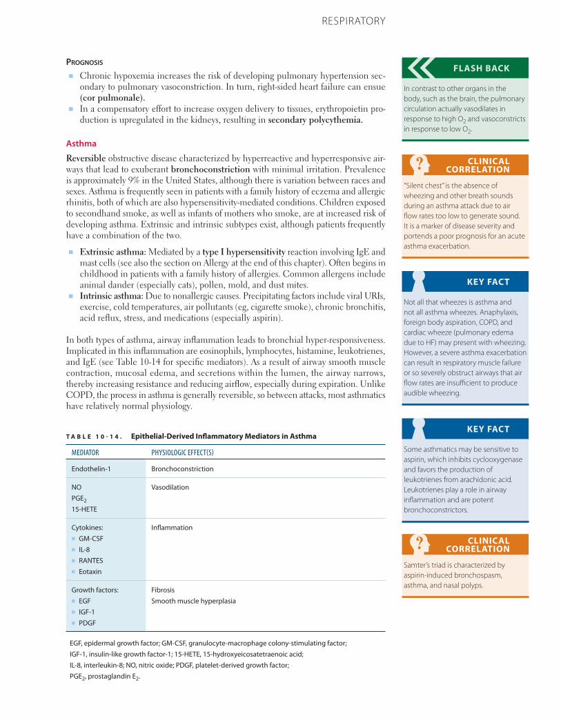
787CHAPTER 10RESPIRATORY
Prognosis
■ Chronic hypoxemia increases the risk of developing pulmonary hypertension sec-ondary to pulmonary vasoconstriction. In turn, right-sided heart failure can ensue (cor pulmonale).
■ In a compensatory effort to increase oxygen delivery to tissues, erythropoietin pro-duction is upregulated in the kidneys, resulting in secondary polycythemia.
Asthma
Reversible obstructive disease characterized by hyperreactive and hyper responsive air-ways that lead to exuberant bronchoconstriction with minimal irritation. Prevalence is approximately 9% in the United States, although there is variation between races and sexes. Asthma is frequently seen in patients with a family history of eczema and allergic rhinitis, both of which are also hypersensitivity-mediated conditions. Children exposed to secondhand smoke, as well as infants of mothers who smoke, are at increased risk of developing asthma. Extrinsic and intrinsic subtypes exist, although patients frequently have a combination of the two.
■ Extrinsic asthma: Mediated by a type I hypersensitivity reaction involving IgE and mast cells (see also the section on Allergy at the end of this chapter). Often begins in childhood in patients with a family history of allergies. Common allergens include animal dander (especially cats), pollen, mold, and dust mites.
■ Intrinsic asthma: Due to nonallergic causes. Precipitating factors include viral URIs, exercise, cold temperatures, air pollutants (eg, cigarette smoke), chronic bronchitis, acid reflux, stress, and medications (especially aspirin).
In both types of asthma, airway inflammation leads to bronchial hyper-responsiveness. Implicated in this inflammation are eosinophils, lymphocytes, histamine, leukotrienes, and IgE (see Table 10-14 for specific mediators). As a result of airway smooth muscle contraction, mucosal edema, and secretions within the lumen, the airway narrows, thereby increasing resistance and reducing airflow, especially during expiration. Unlike COPD, the process in asthma is generally reversible, so between attacks, most asthmatics have relatively normal physiology.
FLASH BACK
In contrast to other organs in the body, such as the brain, the pulmonary circulation actually vasodilates in response to high O2 and vasoconstricts in response to low O2.
CLINICAL CORRELATION
“Silent chest” is the absence of wheezing and other breath sounds during an asthma attack due to air flow rates too low to generate sound. It is a marker of disease severity and portends a poor prognosis for an acute asthma exacerbation.
KEY FACT
Not all that wheezes is asthma and not all asthma wheezes. Anaphylaxis, foreign body aspiration, COPD, and cardiac wheeze (pulmonary edema due to HF) may present with wheezing. However, a severe asthma exacerbation can result in respiratory muscle failure or so severely obstruct airways that air flow rates are insufficient to produce audible wheezing.
KEY FACT
Some asthmatics may be sensitive to aspirin, which inhibits cyclooxygenase and favors the production of leukotrienes from arachidonic acid. Leukotrienes play a role in airway inflammation and are potent bronchoconstrictors.
CLINICAL CORRELATION
Samter’s triad is characterized by aspirin-induced bronchospasm, asthma, and nasal polyps.
T A B L E 1 0 - 1 4 . Epithelial-Derived Inflammatory Mediators in Asthma
MEDIATOR PHYSIOLOGIC EFFECT(S)
Endothelin-1 Bronchoconstriction
NOPGE2
15-HETE
Vasodilation
Cytokines:■ GM-CSF■ IL-8■ RANTES■ Eotaxin
Inflammation
Growth factors:■ EGF■ IGF-1■ PDGF
FibrosisSmooth muscle hyperplasia
EGF, epidermal growth factor; GM-CSF, granulocyte-macrophage colony-stimulating factor; IGF-1, insulin-like growth factor-1; 15-HETE, 15-hydroxyeicosatetraenoic acid; IL-8, interleukin-8; NO, nitric oxide; PDGF, platelet-derived growth factor; PGE2, prostaglandin E2.
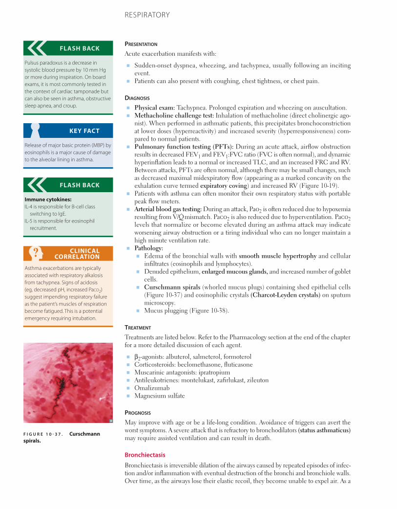
788 CHAPTER 10 RESPIRATORY
Presentation
Acute exacerbation manifests with:
■ Sudden-onset dyspnea, wheezing, and tachypnea, usually following an inciting event.
■ Patients can also present with coughing, chest tightness, or chest pain.
Diagnosis
■ Physical exam: Tachypnea. Prolonged expiration and wheezing on auscultation.■ Methacholine challenge test: Inhalation of methacholine (direct cholinergic ago-
nist). When performed in asthmatic patients, this precipitates bronchoconstriction at lower doses (hyperreactivity) and increased severity (hyperresponsiveness) com-pared to normal patients.
■ Pulmonary function testing (PFTs): During an acute attack, airflow obstruction results in decreased FEV1 and FEV1:FVC ratio (FVC is often normal), and dynamic hyperinflation leads to a normal or increased TLC, and an increased FRC and RV. Between attacks, PFTs are often normal, although there may be small changes, such as decreased maximal mid expiratory flow (appearing as a marked concavity on the exhalation curve termed expiratory coving) and increased RV (Figure 10-19).
■ Patients with asthma can often monitor their own respiratory status with portable peak flow meters.
■ Arterial blood gas testing: During an attack, Pao2 is often reduced due to hypoxemia resulting from V̇/Q̇ mismatch. Paco2 is also reduced due to hyperventilation. Paco2 levels that normalize or become elevated during an asthma attack may indicate worsening airway obstruction or a tiring individual who can no longer maintain a high minute ventilation rate.
■ Pathology: ■ Edema of the bronchial walls with smooth muscle hypertrophy and cellular
infiltrates (eosinophils and lymphocytes).■ Denuded epithelium, enlarged mucous glands, and increased number of goblet
cells.■ Curschmann spirals (whorled mucus plugs) containing shed epithelial cells
(Figure 10-37) and eosinophilic crystals (Charcot-Leyden crystals) on sputum microscopy.
■ Mucus plugging (Figure 10-38).
treatment
Treatments are listed below. Refer to the Pharmacology section at the end of the chapter for a more detailed discussion of each agent.
■ β2-agonists: albuterol, salmeterol, formoterol ■ Corticosteroids: beclomethasone, fluticasone ■ Muscarinic antagonists: ipratropium ■ Antileukotrienes: montelukast, zafirlukast, zileuton ■ Omalizumab■ Magnesium sulfate
Prognosis
May improve with age or be a life-long condition. Avoidance of triggers can avert the worst symptoms. A severe attack that is refractory to bronchodilators (status asthmaticus) may require assisted ventilation and can result in death.
Bronchiectasis
Bronchiectasis is irreversible dilation of the airways caused by repeated episodes of infec-tion and/or inflammation with eventual destruction of the bronchi and bronchiole walls. Over time, as the airways lose their elastic recoil, they become unable to expel air. As a
FLASH BACK
Pulsus paradoxus is a decrease in systolic blood pressure by 10 mm Hg or more during inspiration. On board exams, it is most commonly tested in the context of cardiac tamponade but can also be seen in asthma, obstructive sleep apnea, and croup.
KEY FACT
Release of major basic protein (MBP) by eosinophils is a major cause of damage to the alveolar lining in asthma.
FLASH BACK
Immune cytokines:IL-4 is responsible for B-cell class
switching to IgE.IL-5 is responsible for eosinophil
recruitment.
CLINICAL CORRELATION
Asthma exacerbations are typically associated with respiratory alkalosis from tachypnea. Signs of acidosis (eg, decreased pH, increased Paco2) suggest impending respiratory failure as the patient’s muscles of respiration become fatigued. This is a potential emergency requiring intubation.
F I G U R E 1 0 - 3 7 . Curschmann spirals.
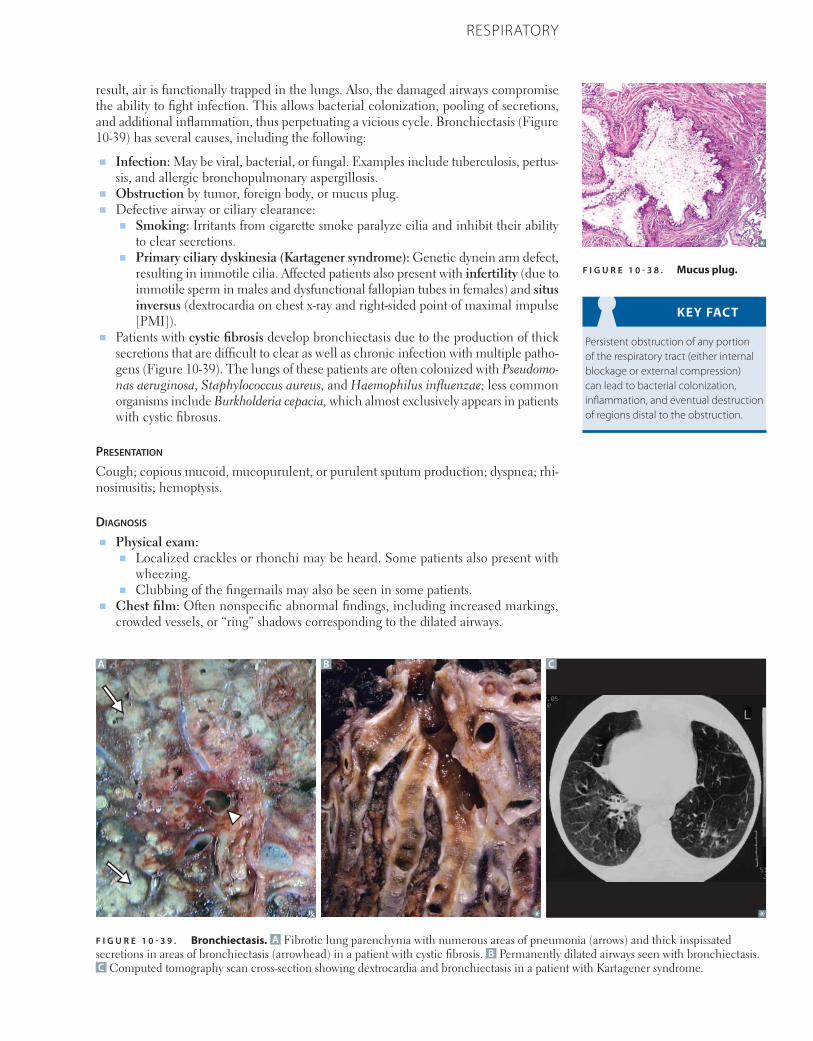
789CHAPTER 10RESPIRATORY
result, air is functionally trapped in the lungs. Also, the damaged airways compromise the ability to fight infection. This allows bacterial colonization, pooling of secretions, and additional inflammation, thus perpetuating a vicious cycle. Bronchiectasis (Figure 10-39) has several causes, including the following:
■ Infection: May be viral, bacterial, or fungal. Examples include tuberculosis, pertus-sis, and allergic bronchopulmonary aspergillosis.
■ Obstruction by tumor, foreign body, or mucus plug.■ Defective airway or ciliary clearance:
■ Smoking: Irritants from cigarette smoke paralyze cilia and inhibit their ability to clear secretions.
■ Primary ciliary dyskinesia (Kartagener syndrome): Genetic dynein arm defect, resulting in immotile cilia. Affected patients also present with infertility (due to immotile sperm in males and dysfunctional fallopian tubes in females) and situs inversus (dextrocardia on chest x-ray and right-sided point of maximal impulse [PMI]).
■ Patients with cystic fibrosis develop bronchiectasis due to the production of thick secretions that are difficult to clear as well as chronic infection with multiple patho-gens (Figure 10-39). The lungs of these patients are often colonized with Pseudomo-nas aeruginosa, Staphylococcus aureus, and Haemophilus influenzae; less common organisms include Burkholderia cepacia, which almost exclusively appears in patients with cystic fibrosus.
Presentation
Cough; copious mucoid, mucopurulent, or purulent sputum production; dysp nea; rhi-nosinusitis; hemoptysis.
Diagnosis
■ Physical exam:■ Localized crackles or rhonchi may be heard. Some patients also pre sent with
wheezing.■ Clubbing of the fingernails may also be seen in some patients.
■ Chest film: Often nonspecific abnormal findings, including increased markings, crowded vessels, or “ring” shadows corresponding to the dilated airways.
KEY FACT
Persistent obstruction of any portion of the respiratory tract (either internal blockage or external compression) can lead to bacterial colonization, inflammation, and eventual destruction of regions distal to the obstruction.
F I G U R E 1 0 - 3 8 . Mucus plug.
F I G U R E 1 0 - 3 9 . Bronchiectasis. A Fibrotic lung parenchyma with numerous areas of pneumonia (arrows) and thick inspissated secretions in areas of bronchiectasis (arrowhead) in a patient with cystic fibrosis. B Permanently dilated airways seen with bronchiectasis. C Computed tomography scan cross-section showing dextrocardia and bronchiectasis in a patient with Kartagener syndrome.
B CA
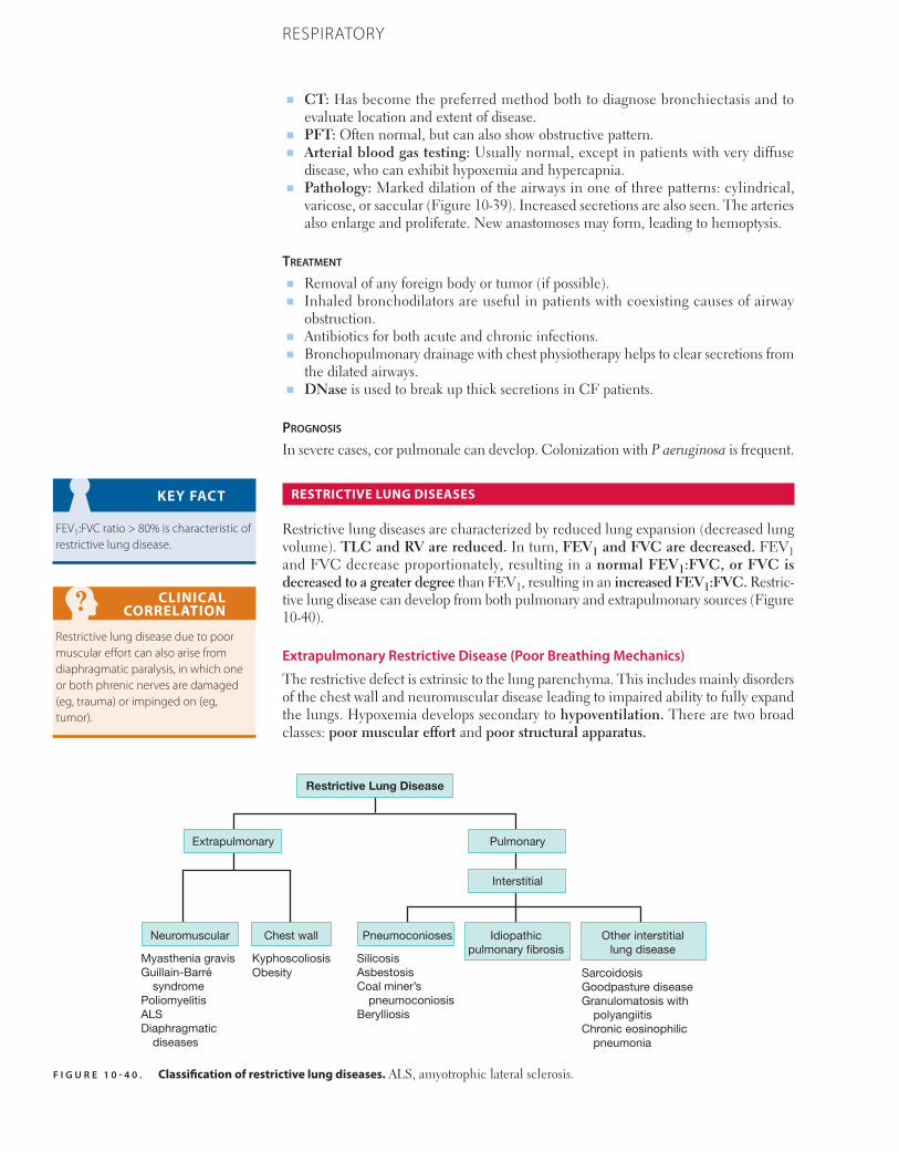
790 CHAPTER 10 RESPIRATORY
■ CT: Has become the preferred method both to diagnose bronchiectasis and to evaluate location and extent of disease.
■ PFT: Often normal, but can also show obstructive pattern.■ Arterial blood gas testing: Usually normal, except in patients with very diffuse
disease, who can exhibit hypoxemia and hypercapnia.■ Pathology: Marked dilation of the airways in one of three patterns: cylindrical,
varicose, or saccular (Figure 10-39). Increased secretions are also seen. The arteries also enlarge and proliferate. New anastomoses may form, leading to hemoptysis.
treatment
■ Removal of any foreign body or tumor (if possible).■ Inhaled bronchodilators are useful in patients with coexisting causes of airway
obstruction.■ Antibiotics for both acute and chronic infections.■ Bronchopulmonary drainage with chest physiotherapy helps to clear secretions from
the dilated airways.■ DNase is used to break up thick secretions in CF patients.
Prognosis
In severe cases, cor pulmonale can develop. Colonization with P aeruginosa is frequent.
RESTRICTIVE LUNG DISEASES
Restrictive lung diseases are characterized by reduced lung expansion (decreased lung volume). TLC and RV are reduced. In turn, FEV1 and FVC are decreased. FEV1 and FVC decrease proportionately, resulting in a normal FEV1:FVC, or FVC is decreased to a greater degree than FEV1, resulting in an increased FEV1:FVC. Restric-tive lung disease can develop from both pulmonary and extrapulmonary sources (Figure 10-40).
Extrapulmonary Restrictive Disease (Poor Breathing Mechanics)
The restrictive defect is extrinsic to the lung parenchyma. This includes mainly disorders of the chest wall and neuromuscular disease leading to impaired ability to fully expand the lungs. Hypoxemia develops secondary to hypoventilation. There are two broad classes: poor muscular effort and poor structural apparatus.
KEY FACT
FEV1:FVC ratio > 80% is characteristic of restrictive lung disease.
CLINICAL CORRELATION
Restrictive lung disease due to poor muscular effort can also arise from diaphragmatic paralysis, in which one or both phrenic nerves are damaged (eg, trauma) or impinged on (eg, tumor).
F I G U R E 1 0 - 4 0 . Classification of restrictive lung diseases. ALS, amyotrophic lateral sclerosis.
Extrapulmonary Pulmonary
Interstitial
Neuromuscular Chest wall Pneumoconioses Idiopathicpulmonary �brosis
Other interstitiallung disease
SarcoidosisGoodpasture diseaseGranulomatosis with
polyangiitisChronic eosinophilic
pneumonia
KyphoscoliosisMyasthenia gravisGuillain-Barré
syndromePoliomyelitisALSDiaphragmatic
diseases
SilicosisAsbestosisCoal miner’s
pneumoconiosisBerylliosis
Restrictive Lung Disease
Obesity
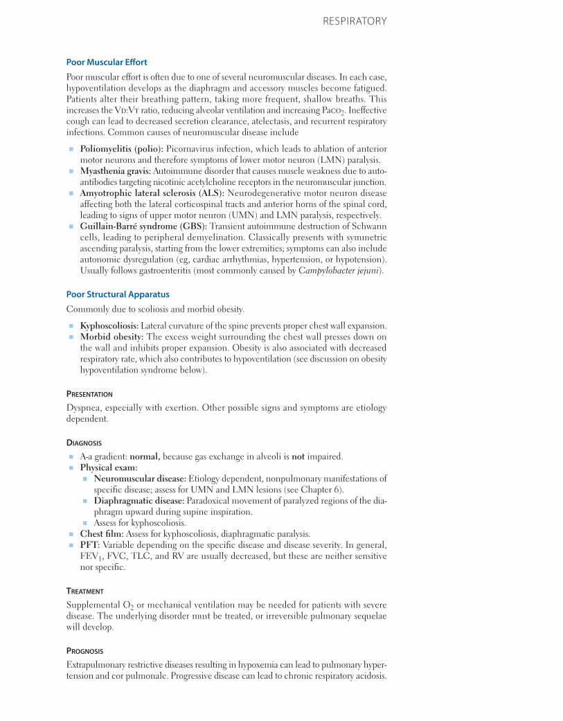
791CHAPTER 10RESPIRATORY
Poor Muscular Effort
Poor muscular effort is often due to one of several neuromuscular diseases. In each case, hypoventilation develops as the diaphragm and accessory muscles become fatigued. Patients alter their breathing pattern, taking more frequent, shallow breaths. This increases the Vd:Vt ratio, reducing alveolar ventilation and increasing Paco2. Ineffective cough can lead to decreased secretion clearance, atelectasis, and recurrent respiratory infections. Common causes of neuromuscular disease include
■ Poliomyelitis (polio): Picornavirus infection, which leads to ablation of anterior motor neurons and therefore symptoms of lower motor neuron (LMN) paralysis.
■ Myasthenia gravis: Autoimmune disorder that causes muscle weakness due to auto-antibodies targeting nicotinic acetylcholine receptors in the neuromuscular junction.
■ Amyotrophic lateral sclerosis (ALS): Neurodegenerative motor neuron disease affecting both the lateral corticospinal tracts and anterior horns of the spinal cord, leading to signs of upper motor neuron (UMN) and LMN paralysis, respectively.
■ Guillain-Barré syndrome (GBS): Transient autoimmune destruction of Schwann cells, leading to peripheral demyelination. Classically presents with symmetric ascending paralysis, starting from the lower extremities; symptoms can also include autonomic dysregulation (eg, cardiac arrhythmias, hypertension, or hypotension). Usually follows gastroenteritis (most commonly caused by Campylobacter jejuni).
Poor Structural Apparatus
Commonly due to scoliosis and morbid obesity.
■ Kyphoscoliosis: Lateral curvature of the spine prevents proper chest wall expansion.■ Morbid obesity: The excess weight surrounding the chest wall presses down on
the wall and inhibits proper expansion. Obesity is also associated with decreased respiratory rate, which also contributes to hypoventilation (see discussion on obesity hypoventilation syndrome below).
Presentation
Dyspnea, especially with exertion. Other possible signs and symptoms are etiology dependent.
Diagnosis
■ A-a gradient: normal, because gas exchange in alveoli is not impaired.■ Physical exam:
■ Neuromuscular disease: Etiology dependent, nonpulmonary manifestations of specific disease; assess for UMN and LMN lesions (see Chapter 6).
■ Diaphragmatic disease: Paradoxical movement of paralyzed regions of the dia-phragm upward during supine inspiration.
■ Assess for kyphoscoliosis.■ Chest film: Assess for kyphoscoliosis, diaphragmatic paralysis.■ PFT: Variable depending on the specific disease and disease severity. In general,
FEV1, FVC, TLC, and RV are usually decreased, but these are neither sensitive nor specific.
treatment
Supplemental O2 or mechanical ventilation may be needed for patients with severe disease. The underlying disorder must be treated, or irreversible pulmonary sequelae will develop.
Prognosis
Extrapulmonary restrictive diseases resulting in hypoxemia can lead to pulmonary hyper-tension and cor pulmonale. Progressive disease can lead to chronic respiratory acidosis.
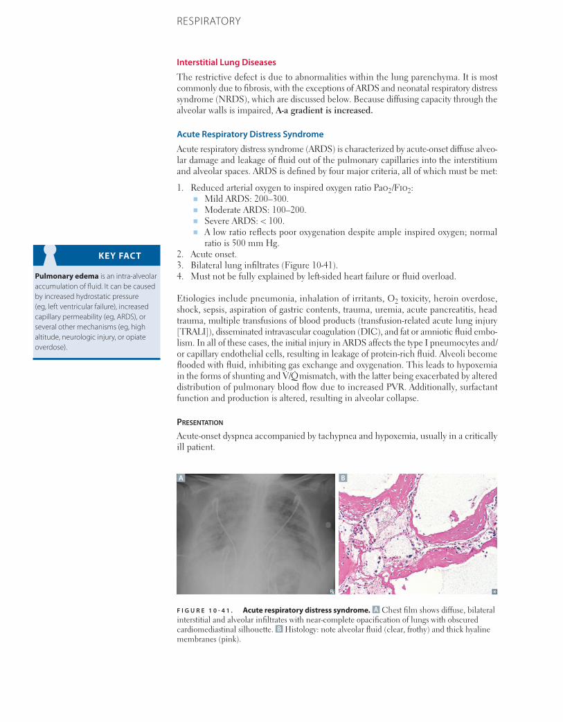
792 CHAPTER 10 RESPIRATORY
Interstitial Lung Diseases
The restrictive defect is due to abnormalities within the lung parenchyma. It is most commonly due to fibrosis, with the exceptions of ARDS and neonatal respiratory distress syndrome (NRDS), which are discussed below. Because diffusing capacity through the alveolar walls is impaired, A-a gradient is increased.
Acute Respiratory Distress Syndrome
Acute respiratory distress syndrome (ARDS) is characterized by acute-onset diffuse alveo-lar damage and leakage of fluid out of the pulmonary capillaries into the interstitium and alveolar spaces. ARDS is defined by four major criteria, all of which must be met:
1. Reduced arterial oxygen to inspired oxygen ratio Pao2/Fio2:■ Mild ARDS: 200–300.■ Moderate ARDS: 100–200. ■ Severe ARDS: < 100. ■ A low ratio reflects poor oxygenation despite ample inspired oxygen; normal
ratio is 500 mm Hg.2. Acute onset.3. Bilateral lung infiltrates (Figure 10-41).4. Must not be fully explained by left-sided heart failure or fluid overload.
Etiologies include pneumonia, inhalation of irritants, O2 toxicity, heroin overdose, shock, sepsis, aspiration of gastric contents, trauma, uremia, acute pancreatitis, head trauma, multiple transfusions of blood products (transfusion-related acute lung injury [TRALI]), disseminated intravascular coagulation (DIC), and fat or amniotic fluid embo-lism. In all of these cases, the initial injury in ARDS affects the type I pneumocytes and/or capillary endothelial cells, resulting in leakage of protein-rich fluid. Alveoli become flooded with fluid, inhibiting gas exchange and oxygenation. This leads to hypoxemia in the forms of shunting and V̇/Q̇ mismatch, with the latter being exacerbated by altered distribution of pulmonary blood flow due to increased PVR. Additionally, surfactant function and production is altered, resulting in alveolar collapse.
Presentation
Acute-onset dyspnea accompanied by tachypnea and hypoxemia, usually in a critically ill patient.
KEY FACT
Pulmonary edema is an intra-alveolar accumulation of fluid. It can be caused by increased hydrostatic pressure (eg, left ventricular failure), increased capillary permeability (eg, ARDS), or several other mechanisms (eg, high altitude, neurologic injury, or opiate overdose).
F I G U R E 1 0 - 4 1 . Acute respiratory distress syndrome. A Chest film shows diffuse, bilateral interstitial and alveolar infiltrates with near-complete opacification of lungs with obscured cardiomediastinal silhouette. B Histology: note alveolar fluid (clear, frothy) and thick hyaline membranes (pink).
A B
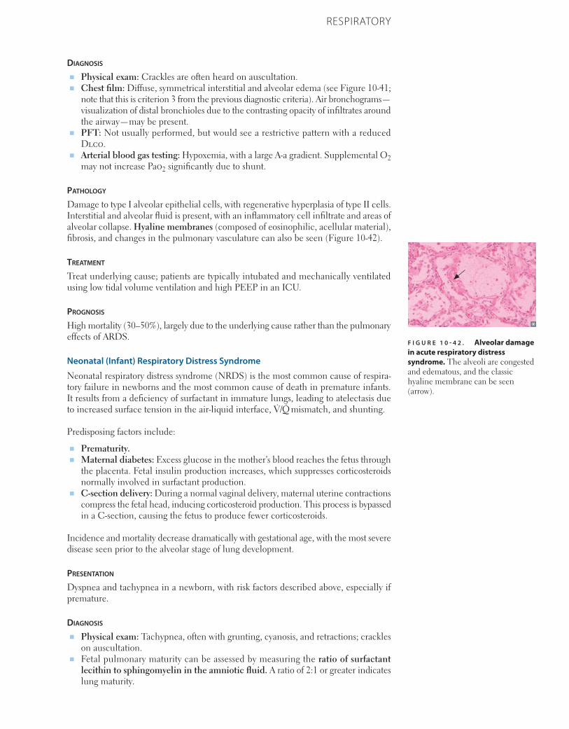
793CHAPTER 10RESPIRATORY
Diagnosis
■ Physical exam: Crackles are often heard on auscultation.■ Chest film: Diffuse, symmetrical interstitial and alveolar edema (see Figure 10-41;
note that this is criterion 3 from the previous diagnostic criteria). Air bronchograms—visualization of distal bronchioles due to the contrasting opacity of infiltrates around the airway—may be present.
■ PFT: Not usually performed, but would see a restrictive pattern with a reduced Dlco.
■ Arterial blood gas testing: Hypoxemia, with a large A-a gradient. Supplemental O2 may not increase Pao2 significantly due to shunt.
Pathology
Damage to type I alveolar epithelial cells, with regenerative hyperplasia of type II cells. Interstitial and alveolar fluid is present, with an inflammatory cell infiltrate and areas of alveolar collapse. Hyaline membranes (composed of eosinophilic, acellular material), fibrosis, and changes in the pulmonary vasculature can also be seen (Figure 10-42).
treatment
Treat underlying cause; patients are typically intubated and mechanically ventilated using low tidal volume ventilation and high PEEP in an ICU.
Prognosis
High mortality (30–50%), largely due to the underlying cause rather than the pulmonary effects of ARDS.
Neonatal (Infant) Respiratory Distress Syndrome
Neonatal respiratory distress syndrome (NRDS) is the most common cause of respira-tory failure in newborns and the most common cause of death in premature infants. It results from a deficiency of surfactant in immature lungs, leading to atelectasis due to increased surface tension in the air-liquid interface, V̇/Q̇ mismatch, and shunting.
Predisposing factors include:
■ Prematurity.■ Maternal diabetes: Excess glucose in the mother’s blood reaches the fetus through
the placenta. Fetal insulin production increases, which suppresses corticosteroids normally involved in surfactant production.
■ C-section delivery: During a normal vaginal delivery, maternal uterine contractions compress the fetal head, inducing corticosteroid production. This process is bypassed in a C-section, causing the fetus to produce fewer corticosteroids.
Incidence and mortality decrease dramatically with gestational age, with the most severe disease seen prior to the alveolar stage of lung development.
Presentation
Dyspnea and tachypnea in a newborn, with risk factors described above, especially if premature.
Diagnosis
■ Physical exam: Tachypnea, often with grunting, cyanosis, and retractions; crackles on auscultation.
■ Fetal pulmonary maturity can be assessed by measuring the ratio of surfactant lecithin to sphingomyelin in the amniotic fluid. A ratio of 2:1 or greater indicates lung maturity.
F I G U R E 1 0 - 4 2 . Alveolar damage in acute respiratory distress syndrome. The alveoli are congested and edematous, and the classic hyaline membrane can be seen (arrow).
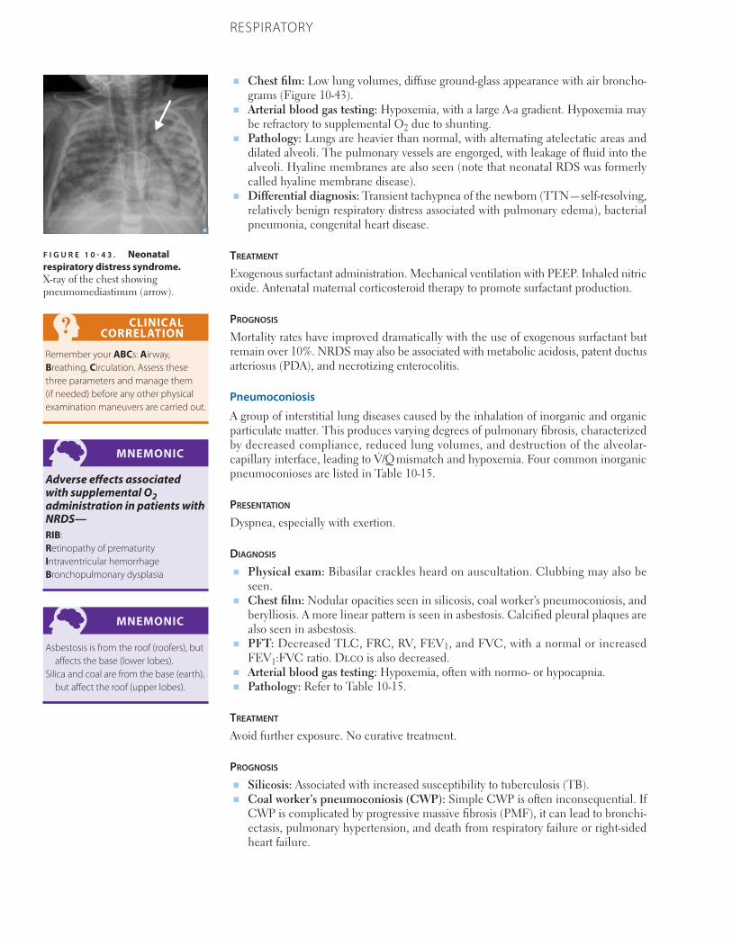
794 CHAPTER 10 RESPIRATORY
■ Chest film: Low lung volumes, diffuse ground-glass appearance with air broncho-grams (Figure 10-43).
■ Arterial blood gas testing: Hypoxemia, with a large A-a gradient. Hypoxemia may be refractory to supplemental O2 due to shunting.
■ Pathology: Lungs are heavier than normal, with alternating atelectatic areas and dilated alveoli. The pulmonary vessels are engorged, with leakage of fluid into the alveoli. Hyaline membranes are also seen (note that neonatal RDS was formerly called hyaline membrane disease).
■ Differential diagnosis: Transient tachypnea of the newborn (TTN—self-resolving, relatively benign respiratory distress associated with pulmonary edema), bacterial pneumonia, congenital heart disease.
treatment
Exogenous surfactant administration. Mechanical ventilation with PEEP. Inhaled nitric oxide. Antenatal maternal corticosteroid therapy to promote surfactant production.
Prognosis
Mortality rates have improved dramatically with the use of exogenous surfactant but remain over 10%. NRDS may also be associated with metabolic acidosis, patent ductus arteriosus (PDA), and necrotizing enterocolitis.
Pneumoconiosis
A group of interstitial lung diseases caused by the inhalation of inorganic and organic particulate matter. This produces varying degrees of pulmonary fibrosis, characterized by decreased compliance, reduced lung volumes, and destruction of the alveolar- capillary interface, leading to V̇/Q̇ mismatch and hypoxemia. Four common inorganic pneumoconioses are listed in Table 10-15.
Presentation
Dyspnea, especially with exertion.
Diagnosis
■ Physical exam: Bibasilar crackles heard on auscultation. Clubbing may also be seen.
■ Chest film: Nodular opacities seen in silicosis, coal worker’s pneumoconiosis, and berylliosis. A more linear pattern is seen in asbestosis. Calcified pleural plaques are also seen in asbestosis.
■ PFT: Decreased TLC, FRC, RV, FEV1, and FVC, with a normal or increased FEV1:FVC ratio. Dlco is also decreased.
■ Arterial blood gas testing: Hypoxemia, often with normo- or hypocapnia.■ Pathology: Refer to Table 10-15.
treatment
Avoid further exposure. No curative treatment.
Prognosis
■ Silicosis: Associated with increased susceptibility to tuberculosis (TB).■ Coal worker’s pneumoconiosis (CWP): Simple CWP is often inconsequential. If
CWP is complicated by progressive massive fibrosis (PMF), it can lead to bronchi-ectasis, pulmonary hypertension, and death from respiratory failure or right-sided heart failure.
CLINICAL CORRELATION
Remember your ABCs: Airway, Breathing, Circulation. Assess these three parameters and manage them (if needed) before any other physical examination maneuvers are carried out.
MNEMONIC
Adverse effects associated with supplemental O2 administration in patients with NRDS—RIB:Retinopathy of prematurity Intraventricular hemorrhage Bronchopulmonary dysplasia
MNEMONIC
Asbestosis is from the roof (roofers), but affects the base (lower lobes).
Silica and coal are from the base (earth), but affect the roof (upper lobes).
F I G U R E 1 0 - 4 3 . Neonatal respiratory distress syndrome. X-ray of the chest showing pneumomediastinum (arrow).
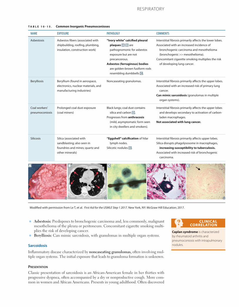
795CHAPTER 10RESPIRATORY
T A B L E 1 0 - 1 5 . Common Inorganic Pneumoconioses
NAME EXPOSURE PATHOLOGY COMMENTS
Asbestosis Asbestos fibers (associated with shipbuilding, roofing, plumbing, insulation, construction work)
“Ivory white” calcified pleural plaques A B are pathognomonic for asbestos exposure but are not precancerous.
Asbestos (ferruginous) bodies are golden-brown fusiform rods resembling dumbbells C .
Interstitial fibrosis primarily affects the lower lobes.Associated with an increased incidence of
bronchogenic carcinoma and mesothelioma (bronchogenic >> mesothelioma).
Concomitant cigarette smoking multiplies the risk of developing lung cancer.
Berylliosis Beryllium (found in aerospace, electronics, nuclear materials, and manufacturing industries)
Noncaseating granulomas. Interstitial fibrosis primarily affects the upper lobes.Associated with an increased risk of primary lung
cancer.Can mimic sarcoidosis (granulomas in multiple
organ systems).
Coal workers’ pneumoconiosis
Prolonged coal dust exposure (coal miners)
Black lungs; coal dust contains silica and carbon D .
Progresses from anthracosis (mild, asymptomatic form seen in city dwellers and smokers).
Interstitial fibrosis primarily affects the upper lobes and develops secondary to activation of carbon-laden macrophages.
Not associated with lung cancer.
Silicosis Silica (associated with sandblasting; also seen in foundries and mines; quartz and other minerals)
“Eggshell” calcification of hilar lymph nodes.
Silicotic nodules E .
Interstitial fibrosis primarily affects upper lobes.Silica disrupts phagolysosome in macrophages,
increasing susceptibility to tuberculosis.Associated with increased risk of bronchogenic
carcinoma.
A B C D E
Modified with permission from Le T, et al. First Aid for the USMLE Step 1 2017. New York, NY: McGraw Hill Education; 2017.
■ Asbestosis: Predisposes to bronchogenic carcinoma and, less commonly, malignant mesothelioma of the pleura or peritoneum. Concomitant cigarette smoking multi-plies the risk of developing cancer.
■ Berylliosis: Can mimic sarcoidosis, with granulomas in multiple organ systems.
Sarcoidosis
Inflammatory disease characterized by noncaseating granulomas, often involving mul-tiple organ systems. The initial exposure that leads to granuloma formation is unknown.
Presentation
Classic presentation of sarcoidosis is an African-American female in her thirties with progressive dyspnea, often accompanied by a dry or nonproductive cough. More com-mon in women and African Americans. Presents in young adulthood. Often discovered
CLINICAL CORRELATION
Caplan syndrome is characterized by rheumatoid arthritis and pneumoconiosis with intrapulmonary nodules.
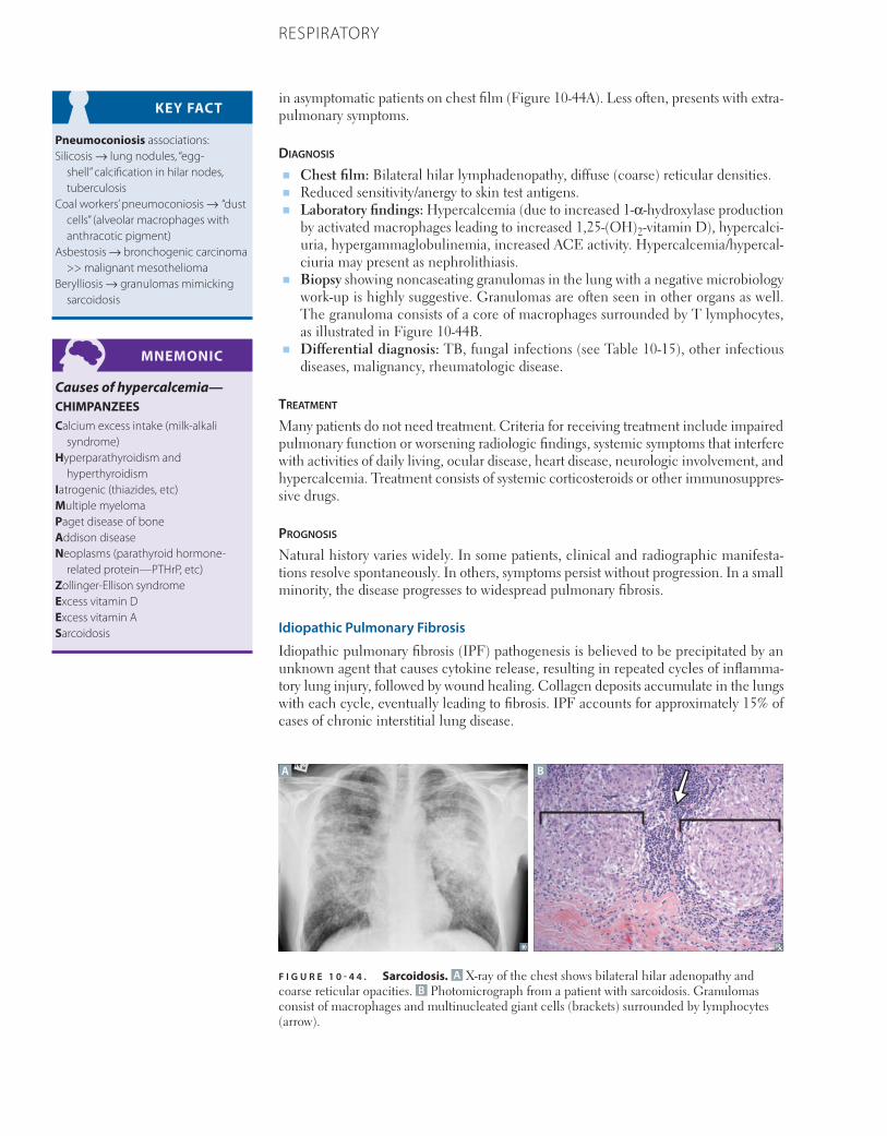
796 CHAPTER 10 RESPIRATORY
in asymptomatic patients on chest film (Figure 10-44A). Less often, presents with extra-pulmonary symptoms.
Diagnosis
■ Chest film: Bilateral hilar lymphadenopathy, diffuse (coarse) reticular densities.■ Reduced sensitivity/anergy to skin test antigens.■ Laboratory findings: Hypercalcemia (due to increased 1-α-hydroxylase production
by activated macrophages leading to increased 1,25-(OH)2-vitamin D), hypercalci-uria, hypergammaglobulinemia, increased ACE activity. Hypercalcemia/hypercal-ciuria may present as nephrolithiasis.
■ Biopsy showing noncaseating granulomas in the lung with a negative microbiology work-up is highly suggestive. Granulomas are often seen in other organs as well. The granuloma consists of a core of macrophages surrounded by T lymphocytes, as illustrated in Figure 10-44B.
■ Differential diagnosis: TB, fungal infections (see Table 10-15), other infectious diseases, malignancy, rheumatologic disease.
treatment
Many patients do not need treatment. Criteria for receiving treatment include impaired pulmonary function or worsening radiologic findings, systemic symptoms that interfere with activities of daily living, ocular disease, heart disease, neurologic involvement, and hypercalcemia. Treatment consists of systemic corticosteroids or other immunosuppres-sive drugs.
Prognosis
Natural history varies widely. In some patients, clinical and radiographic manifesta-tions resolve spontaneously. In others, symptoms persist without progression. In a small minority, the disease progresses to widespread pulmonary fibrosis.
Idiopathic Pulmonary Fibrosis
Idiopathic pulmonary fibrosis (IPF) pathogenesis is believed to be precipitated by an unknown agent that causes cytokine release, resulting in repeated cycles of inflamma-tory lung injury, followed by wound healing. Collagen deposits accumulate in the lungs with each cycle, eventually leading to fibrosis. IPF accounts for approximately 15% of cases of chronic interstitial lung disease.
KEY FACT
Pneumoconiosis associations:Silicosis → lung nodules, “egg-
shell” calcification in hilar nodes, tuberculosis
Coal workers’ pneumoconiosis → “dust cells” (alveolar macrophages with anthracotic pigment)
Asbestosis → bronchogenic carcinoma >> malignant mesothelioma
Berylliosis → granulomas mimicking sarcoidosis
MNEMONIC
Causes of hypercalcemia—CHIMPANZEESCalcium excess intake (milk-alkali
syndrome)Hyperparathyroidism and
hyperthyroidismIatrogenic (thiazides, etc)Multiple myelomaPaget disease of boneAddison diseaseNeoplasms (parathyroid hormone-
related protein—PTHrP, etc)Zollinger-Ellison syndrome Excess vitamin DExcess vitamin ASarcoidosis
F I G U R E 1 0 - 4 4 . Sarcoidosis. A X-ray of the chest shows bilateral hilar adenopathy and coarse reticular opacities. B Photomicrograph from a patient with sarcoidosis. Granulomas consist of macrophages and multinucleated giant cells (brackets) surrounded by lymphocytes (arrow).
A B
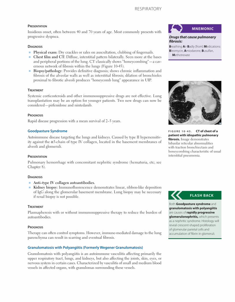
797CHAPTER 10RESPIRATORY
Presentation
Insidious onset, often between 40 and 70 years of age. Most commonly pre sents with progressive dyspnea.
Diagnosis
■ Physical exam: Dry crackles or rales on auscultation, clubbing of fingernails.■ Chest film and CT: Diffuse, interstitial pattern bilaterally. Seen more at the bases
and peripheral portions of the lung. CT classically shows “honeycombing”—a cav-ernous network of fibrosis within the lungs (Figure 10-45).
■ Biopsy/pathology: Provides definitive diagnosis; shows chronic inflammation and fibrosis of the alveolar walls as well as interstitial fibrosis; dilation of bronchioles proximal to fibrotic alveoli produces “honeycomb lung” appearance in UIP.
treatment
Systemic corticosteroids and other immunosuppressive drugs are not effective. Lung transplantation may be an option for younger patients. Two new drugs can now be considered—pirfenidone and nintedanib.
Prognosis
Rapid disease progression with a mean survival of 2–5 years.
Goodpasture Syndrome
Autoimmune disease targeting the lungs and kidneys. Caused by type II hypersensitiv-ity against the α3-chain of type IV collagen, located in the basement membranes of alveoli and glomeruli.
Presentation
Pulmonary hemorrhage with concomitant nephritic syndrome (hematuria, etc; see Chapter 8).
Diagnosis
■ Anti–type IV collagen autoantibodies. ■ Kidney biopsy: Immunofluorescence demonstrates linear, ribbon-like deposition
of IgG along the glomerular basement membrane. Lung biopsy may be necessary if renal biopsy is not possible.
treatment
Plasmapheresis with or without immunosuppressive therapy to reduce the burden of autoantibodies.
Prognosis
Therapy can often control symptoms. However, immune-mediated damage to the lung parenchyma can result in scarring and eventual fibrosis.
Granulomatosis with Polyangiitis (Formerly Wegener Granulomatosis)
Granulomatosis with polyangiitis is an autoimmune vasculitis affecting primarily the upper respiratory tract, lungs, and kidneys, but also affecting the joints, skin, eyes, or nervous system in certain cases. Characterized by vasculitis of small and medium blood vessels in affected organs, with granulomas surrounding these vessels.
MNEMONIC
Drugs that cause pulmonary fibrosis:Breathing Air Badly [from] Medications:Bleomycin, Amiodarone, Busulfan,
Methotrexate
FLASH BACK
Both Goodpasture syndrome and granulomatosis with polyangiitis are causes of rapidly progressive glomerulonephritis, which presents as a nephritic syndrome. Histology will reveal crescent-shaped proliferation of glomerular parietal cells and accumulation of fibrin in glomeruli.
F I G U R E 1 0 - 4 5 . CT of chest of a patient with idiopathic pulmonary fibrosis. Image demonstrates bibasilar reticular abnormalities with traction bronchiectasis and honeycombing characteristic of usual interstitial pneumonia.
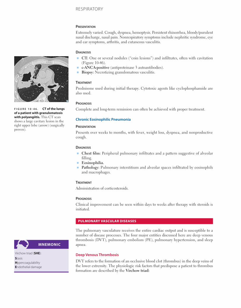
798 CHAPTER 10 RESPIRATORY
Presentation
Extremely varied. Cough, dyspnea, hemoptysis. Persistent rhinorrhea, bloody/purulent nasal discharge, nasal pain. Nonrespiratory symptoms include nephritic syndrome, eye and ear symptoms, arthritis, and cutaneous vasculitis.
Diagnosis
■ CT: One or several nodules (“coin lesions”) and infiltrates, often with cavitation (Figure 10-46).
■ c-ANCA-positive (antiproteinase 3 autoantibodies). ■ Biopsy: Necrotizing granulomatous vasculitis.
treatment
Prednisone used during initial therapy. Cytotoxic agents like cyclophospha mide are also used.
Prognosis
Complete and long-term remission can often be achieved with proper treatment.
Chronic Eosinophilic Pneumonia
Presentation
Presents over weeks to months, with fever, weight loss, dyspnea, and nonproductive cough.
Diagnosis
■ Chest film: Peripheral pulmonary infiltrates and a pattern suggestive of alveolar filling.
■ Eosinophilia.■ Pathology: Pulmonary interstitium and alveolar spaces infiltrated by eosinophils
and macrophages.
treatment
Administration of corticosteroids.
Prognosis
Clinical improvement can be seen within days to weeks after therapy with steroids is initiated.
PULMONARY VASCULAR DISEASES
The pulmonary vasculature receives the entire cardiac output and is susceptible to a number of disease processes. The four major entities discussed here are deep venous thrombosis (DVT), pulmonary embolism (PE), pulmonary hypertension, and sleep apnea.
Deep Venous Thrombosis
DVT refers to the formation of an occlusive blood clot (thrombus) in the deep veins of the lower extremity. The physiologic risk factors that predispose a patient to thrombus formation are described by the Virchow triad:
MNEMONIC
Virchow triad (SHE):
StasisHypercoagulabilityEndothelial damage
F I G U R E 1 0 - 4 6 . CT of the lungs of a patient with granulomatosis with polyangiitis. This CT scan shows a large cavitary lesion in the right upper lobe (arrow) (surgically proven).
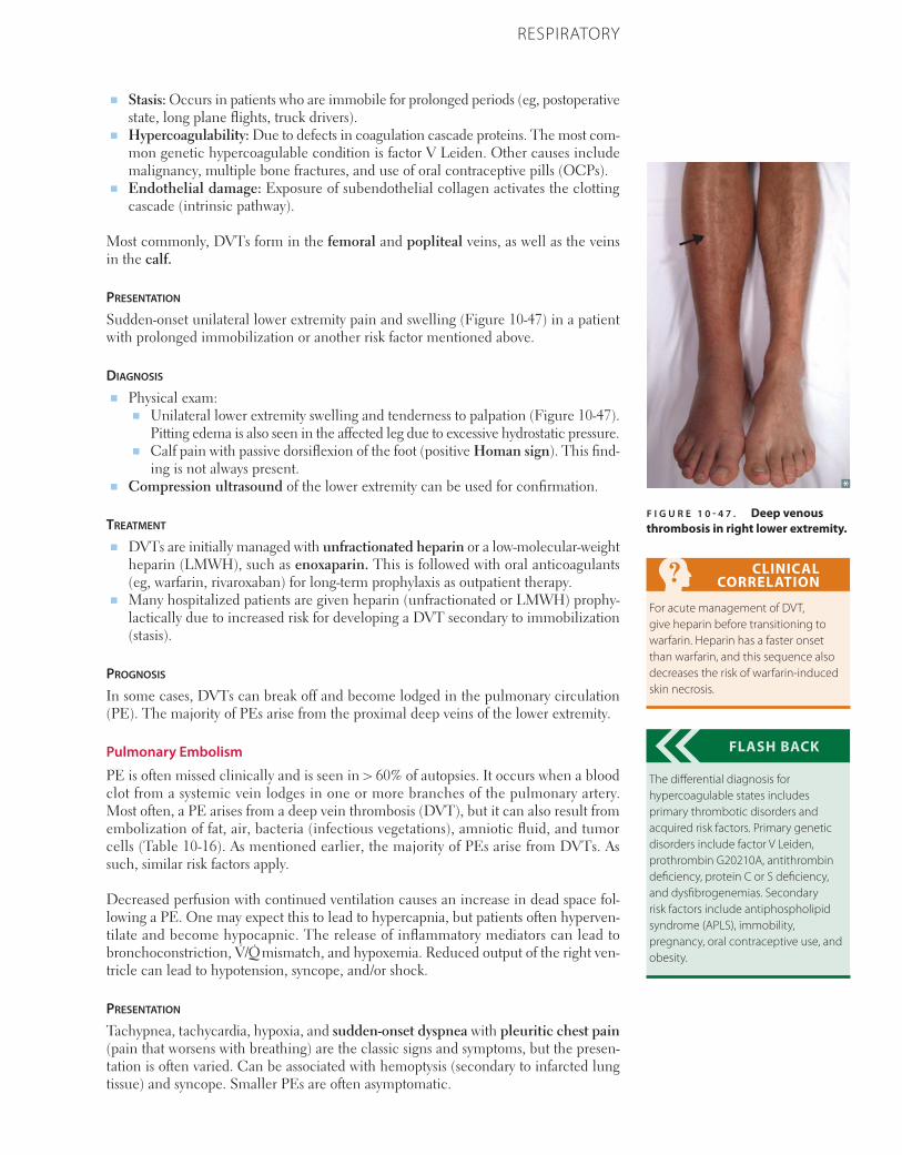
799CHAPTER 10RESPIRATORY
■ Stasis: Occurs in patients who are immobile for prolonged periods (eg, postoperative state, long plane flights, truck drivers).
■ Hypercoagulability: Due to defects in coagulation cascade proteins. The most com-mon genetic hypercoagulable condition is factor V Leiden. Other causes include malignancy, multiple bone fractures, and use of oral contraceptive pills (OCPs).
■ Endothelial damage: Exposure of subendothelial collagen activates the clotting cascade (intrinsic pathway).
Most commonly, DVTs form in the femoral and popliteal veins, as well as the veins in the calf.
Presentation Sudden-onset unilateral lower extremity pain and swelling (Figure 10-47) in a patient with prolonged immobilization or another risk factor mentioned above.
Diagnosis ■ Physical exam:
■ Unilateral lower extremity swelling and tenderness to palpation (Figure 10-47). Pitting edema is also seen in the affected leg due to excessive hydrostatic pressure.
■ Calf pain with passive dorsiflexion of the foot (positive Homan sign). This find-ing is not always present.
■ Compression ultrasound of the lower extremity can be used for confirmation.
treatment ■ DVTs are initially managed with unfractionated heparin or a low-molecular-weight
heparin (LMWH), such as enoxaparin. This is followed with oral anticoagulants (eg, warfarin, rivaroxaban) for long-term prophylaxis as outpatient therapy.
■ Many hospitalized patients are given heparin (unfractionated or LMWH) prophy-lactically due to increased risk for developing a DVT secondary to immobilization (stasis).
Prognosis
In some cases, DVTs can break off and become lodged in the pulmonary circulation (PE). The majority of PEs arise from the proximal deep veins of the lower extremity.
Pulmonary Embolism
PE is often missed clinically and is seen in > 60% of autopsies. It occurs when a blood clot from a systemic vein lodges in one or more branches of the pulmonary artery. Most often, a PE arises from a deep vein thrombosis (DVT), but it can also result from embolization of fat, air, bacteria (infectious vegetations), amniotic fluid, and tumor cells (Table 10-16). As mentioned earlier, the majority of PEs arise from DVTs. As such, similar risk factors apply.
Decreased perfusion with continued ventilation causes an increase in dead space fol-lowing a PE. One may expect this to lead to hypercapnia, but patients often hyperven-tilate and become hypocapnic. The release of inflammatory mediators can lead to bronchoconstriction, V̇/Q̇ mismatch, and hypoxemia. Reduced output of the right ven-tricle can lead to hypotension, syncope, and/or shock.
Presentation
Tachypnea, tachycardia, hypoxia, and sudden-onset dyspnea with pleuritic chest pain (pain that worsens with breathing) are the classic signs and symptoms, but the presen-tation is often varied. Can be associated with hemoptysis (secondary to infarcted lung tissue) and syncope. Smaller PEs are often asymptomatic.
CLINICAL CORRELATION
For acute management of DVT, give heparin before transitioning to warfarin. Heparin has a faster onset than warfarin, and this sequence also decreases the risk of warfarin-induced skin necrosis.
FLASH BACK
The differential diagnosis for hypercoagulable states includes primary thrombotic disorders and acquired risk factors. Primary genetic disorders include factor V Leiden, prothrombin G20210A, antithrombin deficiency, protein C or S deficiency, and dysfibrogenemias. Secondary risk factors include antiphospholipid syndrome (APLS), immobility, pregnancy, oral contraceptive use, and obesity.
F I G U R E 1 0 - 4 7 . Deep venous thrombosis in right lower extremity.
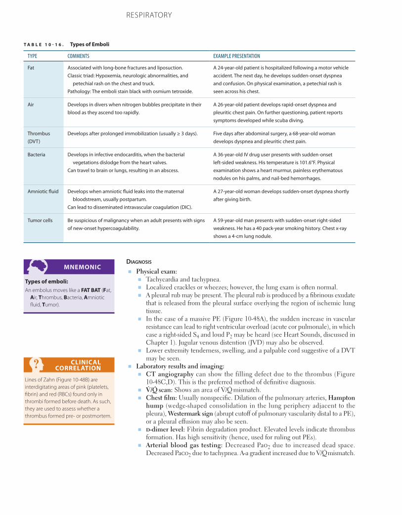
800 CHAPTER 10 RESPIRATORY
Diagnosis
■ Physical exam:■ Tachycardia and tachypnea.■ Localized crackles or wheezes; however, the lung exam is often normal.■ A pleural rub may be present. The pleural rub is produced by a fibrinous exudate
that is released from the pleural surface overlying the region of ischemic lung tissue.
■ In the case of a massive PE (Figure 10-48A), the sudden increase in vascular resistance can lead to right ventricular overload (acute cor pulmonale), in which case a right-sided S4 and loud P2 may be heard (see Heart Sounds, discussed in Chapter 1). Jugular venous distention (JVD) may also be observed.
■ Lower extremity tenderness, swelling, and a palpable cord suggestive of a DVT may be seen.
■ Laboratory results and imaging:■ CT angiography can show the filling defect due to the thrombus (Figure
10-48C,D). This is the preferred method of definitive diagnosis.■ V̇/Q̇ scan: Shows an area of V̇/Q̇ mismatch. ■ Chest film: Usually nonspecific. Dilation of the pulmonary arteries, Hampton
hump (wedge-shaped consolidation in the lung periphery adjacent to the pleura), Westermark sign (abrupt cutoff of pulmonary vascularity distal to a PE), or a pleural effusion may also be seen.
■ d-dimer level: Fibrin degradation product. Elevated levels indicate thrombus formation. Has high sensitivity (hence, used for ruling out PEs).
■ Arterial blood gas testing: Decreased Pao2 due to increased dead space. Decreased Paco2 due to tachypnea. A-a gradient increased due to V̇/Q̇ mismatch.
MNEMONIC
Types of emboli:An embolus moves like a FAT BAT (Fat,
Air, Thrombus, Bacteria, Amniotic fluid, Tumor).
CLINICAL CORRELATION
Lines of Zahn (Figure 10-48B) are interdigitating areas of pink (platelets, fibrin) and red (RBCs) found only in thrombi formed before death. As such, they are used to assess whether a thrombus formed pre- or postmortem.
T A B L E 1 0 - 1 6 . Types of Emboli
TYPE COMMENTS EXAMPLE PRESENTATION
Fat Associated with long-bone fractures and liposuction.Classic triad: Hypoxemia, neurologic abnormalities, and
petechial rash on the chest and truck.Pathology: The emboli stain black with osmium tetroxide.
A 24-year-old patient is hospitalized following a motor vehicle accident. The next day, he develops sudden-onset dyspnea and confusion. On physical examination, a petechial rash is seen across his chest.
Air Develops in divers when nitrogen bubbles precipitate in their blood as they ascend too rapidly.
A 26-year-old patient develops rapid-onset dyspnea and pleuritic chest pain. On further questioning, patient reports symptoms developed while scuba diving.
Thrombus (DVT)
Develops after prolonged immobilization (usually ≥ 3 days). Five days after abdominal surgery, a 68-year-old woman develops dyspnea and pleuritic chest pain.
Bacteria Develops in infective endocarditis, when the bacterial vegetations dislodge from the heart valves.
Can travel to brain or lungs, resulting in an abscess.
A 36-year-old IV drug user presents with sudden-onset left-sided weakness. His temperature is 101.6°F. Physical examination shows a heart murmur, painless erythematous nodules on his palms, and nail-bed hemorrhages.
Amniotic fluid Develops when amniotic fluid leaks into the maternal bloodstream, usually postpartum.
Can lead to disseminated intravascular coagulation (DIC).
A 27-year-old woman develops sudden-onset dyspnea shortly after giving birth.
Tumor cells Be suspicious of malignancy when an adult presents with signs of new-onset hypercoagulability.
A 59-year-old man presents with sudden-onset right-sided weakness. He has a 40 pack-year smoking history. Chest x-ray shows a 4-cm lung nodule.
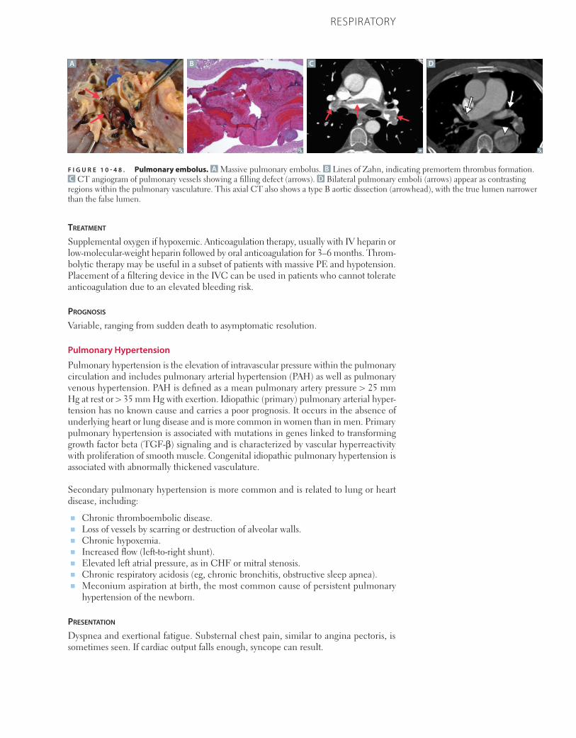
801CHAPTER 10RESPIRATORY
treatment
Supplemental oxygen if hypoxemic. Anticoagulation therapy, usually with IV heparin or low-molecular-weight heparin followed by oral anticoagulation for 3–6 months. Throm-bolytic therapy may be useful in a subset of patients with massive PE and hypotension. Placement of a filtering device in the IVC can be used in patients who cannot tolerate anticoagulation due to an elevated bleeding risk.
Prognosis
Variable, ranging from sudden death to asymptomatic resolution.
Pulmonary Hypertension
Pulmonary hypertension is the elevation of intravascular pressure within the pulmonary circulation and includes pulmonary arterial hypertension (PAH) as well as pulmonary venous hypertension. PAH is defined as a mean pulmonary artery pressure > 25 mm Hg at rest or > 35 mm Hg with exertion. Idiopathic (primary) pulmonary arterial hyper-tension has no known cause and carries a poor prognosis. It occurs in the absence of underlying heart or lung disease and is more common in women than in men. Primary pulmonary hypertension is associated with mutations in genes linked to transforming growth factor beta (TGF-β) signaling and is characterized by vascular hyperreactivity with proliferation of smooth muscle. Congenital idiopathic pulmonary hypertension is associated with abnormally thickened vasculature.
Secondary pulmonary hypertension is more common and is related to lung or heart disease, including:
■ Chronic thromboembolic disease.■ Loss of vessels by scarring or destruction of alveolar walls.■ Chronic hypoxemia.■ Increased flow (left-to-right shunt).■ Elevated left atrial pressure, as in CHF or mitral stenosis.■ Chronic respiratory acidosis (eg, chronic bronchitis, obstructive sleep apnea).■ Meconium aspiration at birth, the most common cause of persistent pulmonary
hypertension of the newborn.
Presentation
Dyspnea and exertional fatigue. Substernal chest pain, similar to angina pectoris, is sometimes seen. If cardiac output falls enough, syncope can result.
F I G U R E 1 0 - 4 8 . Pulmonary embolus. A Massive pulmonary embolus. B Lines of Zahn, indicating premortem thrombus formation. C CT angiogram of pulmonary vessels showing a filling defect (arrows). D Bilateral pulmonary emboli (arrows) appear as contrasting regions within the pulmonary vasculature. This axial CT also shows a type B aortic dissection (arrowhead), with the true lumen narrower than the false lumen.
A B C D
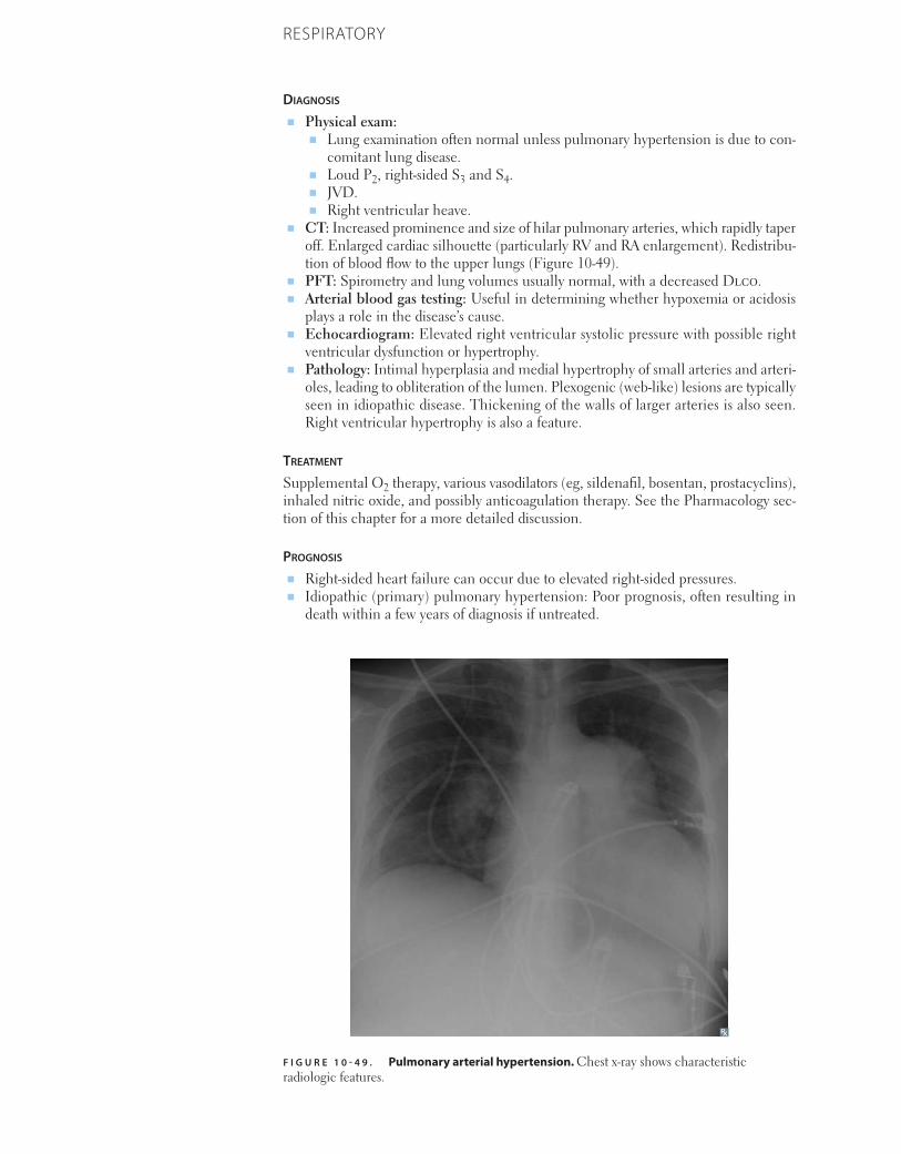
802 CHAPTER 10 RESPIRATORY
Diagnosis
■ Physical exam:■ Lung examination often normal unless pulmonary hypertension is due to con-
comitant lung disease.■ Loud P2, right-sided S3 and S4.■ JVD.■ Right ventricular heave.
■ CT: Increased prominence and size of hilar pulmonary arteries, which rapidly taper off. Enlarged cardiac silhouette (particularly RV and RA enlargement). Redistribu-tion of blood flow to the upper lungs (Figure 10-49).
■ PFT: Spirometry and lung volumes usually normal, with a decreased Dlco.■ Arterial blood gas testing: Useful in determining whether hypoxemia or acidosis
plays a role in the disease’s cause.■ Echocardiogram: Elevated right ventricular systolic pressure with possible right
ventricular dysfunction or hypertrophy.■ Pathology: Intimal hyperplasia and medial hypertrophy of small arteries and arteri-
oles, leading to obliteration of the lumen. Plexogenic (web-like) lesions are typically seen in idiopathic disease. Thickening of the walls of larger arteries is also seen. Right ventricular hypertrophy is also a feature.
treatment
Supplemental O2 therapy, various vasodilators (eg, sildenafil, bosentan, prostacyclins), inhaled nitric oxide, and possibly anticoagulation therapy. See the Pharmacology sec-tion of this chapter for a more detailed discussion.
Prognosis
■ Right-sided heart failure can occur due to elevated right-sided pressures.■ Idiopathic (primary) pulmonary hypertension: Poor prognosis, often resulting in
death within a few years of diagnosis if untreated.
F I G U R E 1 0 - 4 9 . Pulmonary arterial hypertension. Chest x-ray shows characteristic radiologic features.
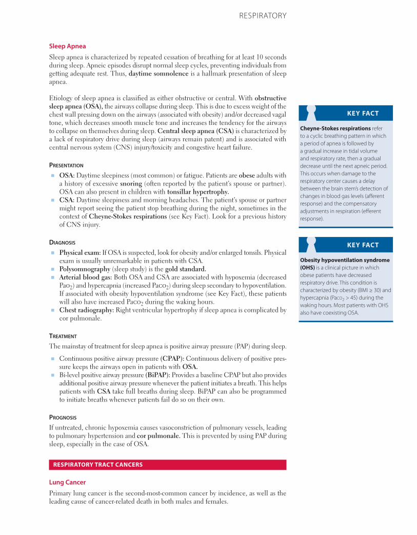
803CHAPTER 10RESPIRATORY
Sleep Apnea
Sleep apnea is characterized by repeated cessation of breathing for at least 10 seconds during sleep. Apneic episodes disrupt normal sleep cycles, preventing individuals from getting adequate rest. Thus, daytime somnolence is a hallmark presentation of sleep apnea.
Etiology of sleep apnea is classified as either obstructive or central. With obstructive sleep apnea (OSA), the airways collapse during sleep. This is due to excess weight of the chest wall pressing down on the airways (associated with obesity) and/or decreased vagal tone, which decreases smooth muscle tone and increases the tendency for the airways to collapse on themselves during sleep. Central sleep apnea (CSA) is characterized by a lack of respiratory drive during sleep (airways remain patent) and is associated with central nervous system (CNS) injury/toxicity and congestive heart failure.
Presentation
■ OSA: Daytime sleepiness (most common) or fatigue. Patients are obese adults with a history of excessive snoring (often reported by the patient’s spouse or partner). OSA can also present in children with tonsillar hypertrophy.
■ CSA: Daytime sleepiness and morning headaches. The patient’s spouse or partner might report seeing the patient stop breathing during the night, sometimes in the context of Cheyne-Stokes respirations (see Key Fact). Look for a previous history of CNS injury.
Diagnosis
■ Physical exam: If OSA is suspected, look for obesity and/or enlarged tonsils. Physical exam is usually unremarkable in patients with CSA.
■ Polysomnography (sleep study) is the gold standard.■ Arterial blood gas: Both OSA and CSA are associated with hypoxemia (decreased
Pao2) and hypercapnia (increased Paco2) during sleep secondary to hypoventilation. If associated with obesity hypoventilation syndrome (see Key Fact), these patients will also have increased Paco2 during the waking hours.
■ Chest radiography: Right ventricular hypertrophy if sleep apnea is complicated by cor pulmonale.
treatment
The mainstay of treatment for sleep apnea is positive airway pressure (PAP) during sleep.
■ Continuous positive airway pressure (CPAP): Continuous delivery of positive pres-sure keeps the airways open in patients with OSA.
■ Bi-level positive airway pressure (BiPAP): Provides a baseline CPAP but also provides additional positive airway pressure whenever the patient initiates a breath. This helps patients with CSA take full breaths during sleep. BiPAP can also be programmed to initiate breaths whenever patients fail do so on their own.
Prognosis
If untreated, chronic hypoxemia causes vasoconstriction of pulmonary vessels, leading to pulmonary hypertension and cor pulmonale. This is prevented by using PAP during sleep, especially in the case of OSA.
RESPIRATORY TRACT CANCERS
Lung Cancer
Primary lung cancer is the second-most-common cancer by incidence, as well as the leading cause of cancer-related death in both males and females.
KEY FACT
Cheyne-Stokes respirations refer to a cyclic breathing pattern in which a period of apnea is followed by a gradual increase in tidal volume and respiratory rate, then a gradual decrease until the next apneic period. This occurs when damage to the respiratory center causes a delay between the brain stem’s detection of changes in blood gas levels (afferent response) and the compensatory adjustments in respiration (efferent response).
KEY FACT
Obesity hypoventilation syndrome (OHS) is a clinical picture in which obese patients have decreased respiratory drive. This condition is characterized by obesity (BMI ≥ 30) and hypercapnia (Paco2 > 45) during the waking hours. Most patients with OHS also have coexisting OSA.
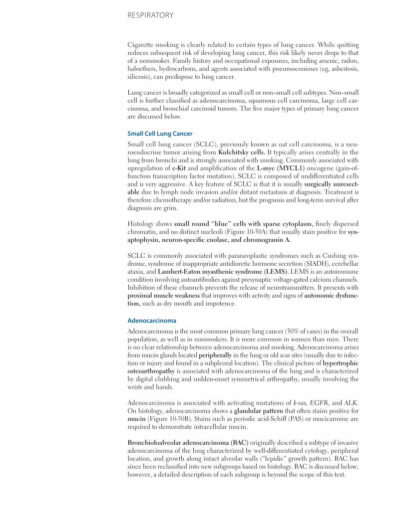
804 CHAPTER 10 RESPIRATORY
Cigarette smoking is clearly related to certain types of lung cancer. While quitting reduces subsequent risk of developing lung cancer, this risk likely never drops to that of a nonsmoker. Family history and occupational exposures, including arsenic, radon, haloethers, hydrocarbons, and agents associated with pneumoconioses (eg, asbestosis, silicosis), can predispose to lung cancer.
Lung cancer is broadly categorized as small cell or non–small cell subtypes. Non–small cell is further classified as adenocarcinoma, squamous cell carcinoma, large cell car-cinoma, and bronchial carcinoid tumors. The five major types of primary lung cancer are discussed below.
Small Cell Lung Cancer
Small cell lung cancer (SCLC), previously known as oat cell carcinoma, is a neu-roendocrine tumor arising from Kulchitsky cells. It typically arises centrally in the lung from bronchi and is strongly associated with smoking. Commonly associated with upregulation of c-Kit and amplification of the L-myc (MYCL1) oncogene (gain-of-function transcription factor mutation), SCLC is composed of undifferentiated cells and is very aggressive. A key feature of SCLC is that it is usually surgically unresect-able due to lymph node invasion and/or distant metastasis at diagnosis. Treatment is therefore chemotherapy and/or radiation, but the prognosis and long-term survival after diagnosis are grim.
Histology shows small round “blue” cells with sparse cytoplasm, finely dispersed chromatin, and no distinct nucleoli (Figure 10-50A) that usually stain positive for syn-aptophysin, neuron-specific enolase, and chromogranin A.
SCLC is commonly associated with paraneoplastic syndromes such as Cushing syn-drome, syndrome of inappropriate antidiuretic hormone secretion (SIADH), cerebellar ataxia, and Lambert-Eaton myasthenic syndrome (LEMS). LEMS is an autoimmune condition involving autoantibodies against presynaptic voltage-gated calcium channels. Inhibition of these channels prevents the release of neurotransmitters. It presents with proximal muscle weakness that improves with activity and signs of autonomic dysfunc-tion, such as dry mouth and impotence.
Adenocarcinoma
Adenocarcinoma is the most common primary lung cancer (50% of cases) in the overall population, as well as in nonsmokers. It is more common in women than men. There is no clear relationship between adenocarcinoma and smoking. Adenocarcinoma arises from mucin glands located peripherally in the lung or old scar sites (usually due to infec-tion or injury and found in a subpleural location). The clinical picture of hypertrophic osteoarthropathy is associated with adenocarcinoma of the lung and is characterized by digital clubbing and sudden-onset symmetrical arthropathy, usually involving the wrists and hands.
Adenocarcinoma is associated with activating mutations of k-ras, EGFR, and ALK. On histology, adenocarcinoma shows a glandular pattern that often stains positive for mucin (Figure 10-50B). Stains such as periodic acid-Schiff (PAS) or mucicarmine are required to demonstrate intracellular mucin.
Bronchioloalveolar adenocarcinoma (BAC) originally described a subtype of invasive adenocarcinoma of the lung characterized by well-differentiated cytology, peripheral location, and growth along intact alveolar walls (“lepidic” growth pattern). BAC has since been reclassified into new subgroups based on histology. BAC is discussed below; however, a detailed description of each subgroup is beyond the scope of this text.
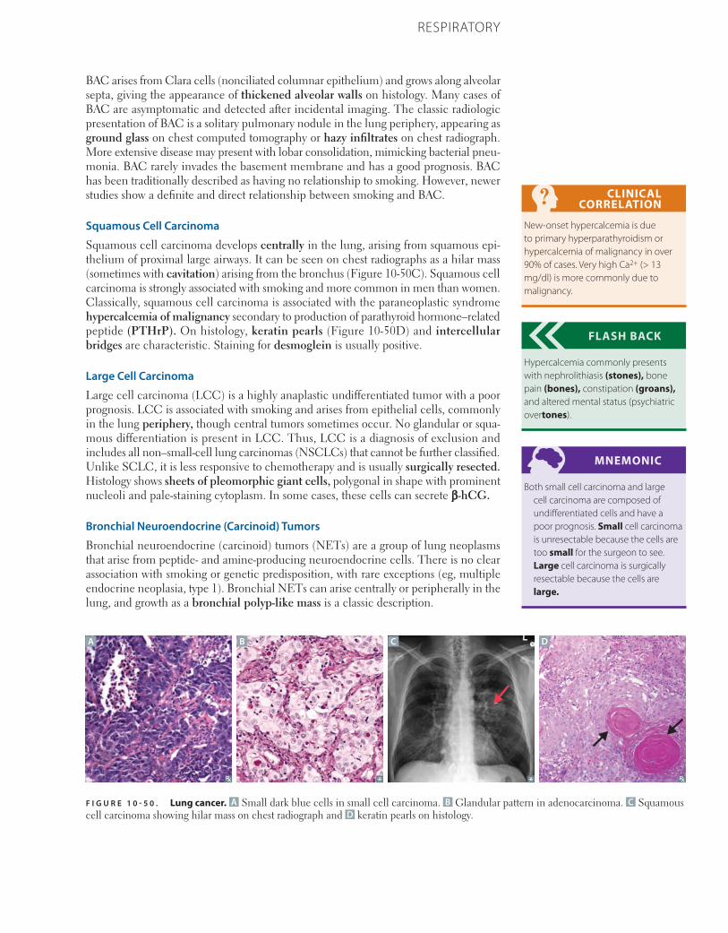
805CHAPTER 10RESPIRATORY
BAC arises from Clara cells (nonciliated columnar epithelium) and grows along alveolar septa, giving the appearance of thickened alveolar walls on histology. Many cases of BAC are asymptomatic and detected after incidental imaging. The classic radiologic presentation of BAC is a solitary pulmonary nodule in the lung periphery, appearing as ground glass on chest computed tomography or hazy infiltrates on chest radiograph. More extensive disease may present with lobar consolidation, mimicking bacterial pneu-monia. BAC rarely invades the basement membrane and has a good prognosis. BAC has been traditionally described as having no relationship to smoking. However, newer studies show a definite and direct relationship between smoking and BAC.
Squamous Cell Carcinoma
Squamous cell carcinoma develops centrally in the lung, arising from squamous epi-thelium of proximal large airways. It can be seen on chest radiographs as a hilar mass (sometimes with cavitation) arising from the bronchus (Figure 10-50C). Squamous cell carcinoma is strongly associated with smoking and more common in men than women. Classically, squamous cell carcinoma is associated with the paraneoplastic syndrome hypercalcemia of malignancy secondary to production of parathyroid hormone–related peptide (PTHrP). On histology, keratin pearls (Figure 10-50D) and intercellular bridges are characteristic. Staining for desmoglein is usually positive.
Large Cell Carcinoma
Large cell carcinoma (LCC) is a highly anaplastic undifferentiated tumor with a poor prognosis. LCC is associated with smoking and arises from epithelial cells, commonly in the lung periphery, though central tumors sometimes occur. No glandular or squa-mous differentiation is present in LCC. Thus, LCC is a diagnosis of exclusion and includes all non–small-cell lung carcinomas (NSCLCs) that cannot be further classified. Unlike SCLC, it is less responsive to chemotherapy and is usually surgically resected. Histology shows sheets of pleomorphic giant cells, polygonal in shape with prominent nucleoli and pale-staining cytoplasm. In some cases, these cells can secrete β-hCG.
Bronchial Neuroendocrine (Carcinoid) Tumors
Bronchial neuroendocrine (carcinoid) tumors (NETs) are a group of lung neoplasms that arise from peptide- and amine-producing neuroendocrine cells. There is no clear association with smoking or genetic predisposition, with rare exceptions (eg, multiple endocrine neoplasia, type 1). Bronchial NETs can arise centrally or peripherally in the lung, and growth as a bronchial polyp-like mass is a classic description.
CLINICAL CORRELATION
New-onset hypercalcemia is due to primary hyperparathyroidism or hypercalcemia of malignancy in over 90% of cases. Very high Ca2+ (> 13 mg/dl) is more commonly due to malignancy.
FLASH BACK
Hypercalcemia commonly presents with nephrolithiasis (stones), bone pain (bones), constipation (groans), and altered mental status (psychiatric overtones).
MNEMONIC
Both small cell carcinoma and large cell carcinoma are composed of undifferentiated cells and have a poor prognosis. Small cell carcinoma is unresectable because the cells are too small for the surgeon to see. Large cell carcinoma is surgically resectable because the cells are large.
A B C D
F I G U R E 1 0 - 5 0 . Lung cancer. A Small dark blue cells in small cell carcinoma. B Glandular pattern in adenocarcinoma. C Squamous cell carcinoma showing hilar mass on chest radiograph and D keratin pearls on histology.
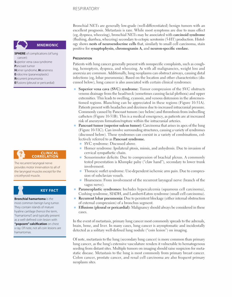
806 CHAPTER 10 RESPIRATORY
Bronchial NETs are generally low-grade (well-differentiated) benign tumors with an excellent prognosis. Metastasis is rare. While most symptoms are due to mass effect (eg, dyspnea, wheezing), bronchial NETs may be associated with carcinoid syndrome (flushing, diarrhea, wheezing) secondary to ectopic serotonin (5-HT) production. Histol-ogy shows nests of neuroendocrine cells that, similarly to small cell carcinoma, stain positive for synaptophysin, chromogranin A, and neuron-specific enolase.
Presentation
Patients with lung cancer generally present with nonspecific complaints, such as cough-ing, hemoptysis, dyspnea, and wheezing. As with all malignancies, weight loss and anorexia are common. Additionally, lung neoplasms can obstruct airways, causing distal infections (eg, lobar pneumonia). Based on the location and other characteristics (dis-cussed below), lung cancer is also associated with certain clinical syndromes:
■ Superior vena cava (SVC) syndrome: Tumor compression of the SVC obstructs venous drainage from the head/neck (sometimes causing facial plethora) and upper extremities. This leads to swelling, cyanosis, and venous distension in the aforemen-tioned regions. Blanching can be appreciated in these regions (Figure 10-51A). Patients present with headaches and dizziness due to increased intracranial pressure. Commonly caused by Pancoast tumors (see below) and thrombosis from indwelling catheters (Figure 10-51B). This is a medical emergency, as patients are at increased risk of aneurysm formation/rupture within the intracranial arteries.
■ Pancoast tumor (superior sulcus tumor): Carcinoma that arises in apex of the lung (Figure 10-51C). Can involve surrounding structures, causing a variety of syndromes (discussed below). These syndromes can coexist in a variety of combinations, col-lectively referred to as Pancoast syndrome.■ SVC syndrome: Discussed above.■ Horner syndrome: Ipsilateral ptosis, miosis, and anhydrosis. Due to invasion of
cervical sympathetic chain.■ Sensorimotor deficits: Due to compression of brachial plexus. A commonly
tested presentation is Klumpke palsy (“claw hand”), secondary to lower trunk involvement.
■ Thoracic outlet syndrome: Use-dependent ischemic arm pain. Due to compres-sion of subclavian vessels.
■ Hoarseness: From involvement of the recurrent laryngeal nerve (branch of the vagus nerve).
■ Paraneoplastic syndromes: Includes hypercalcemia (squamous cell carcinoma), Cushing syndrome, SIADH, and Lambert-Eaton syndrome (small cell carcinoma).
■ Recurrent lobar pneumonia: Due to persistent blockage (either internal obstruction of external compression) of a bronchus segment.
■ Effusions (pleural or pericardial): Malignancy should always be considered in these cases.
In the event of metastasis, primary lung cancer most commonly spreads to the adrenals, brain, bone, and liver. In many cases, lung cancer is asymptomatic and incidentally detected as a solitary well-defined lung nodule (“coin lesion”) on imaging.
Of note, metastasis to the lung (secondary lung cancer) is more common than primary lung cancer, as the lung’s extensive vasculature renders it vulnerable to hematogenous seeding from distant sites. Multiple tumors on imaging should raise suspicion for meta-static disease. Metastasis to the lung is most commonly from primary breast cancer. Colon cancer, prostate cancer, and renal cell carcinoma are also frequent primary neoplasm sites.
MNEMONIC
SPHERE of complications (of lung cancer):
Superior vena cava syndromePancoast tumorHorner syndrome, HoarsenessEndocrine (paraneoplastic)Recurrent pneumoniaEffusions (pleural or pericardial)
CLINICAL CORRELATION
The recurrent laryngeal nerve provides motor innervation to all of the laryngeal muscles except for the cricothyroid muscle.
KEY FACT
Bronchial hamartoma is the most common benign lung tumor. They contain islands of mature hyaline cartilage (hence the term, “hamartoma”) and typically present as a well-defined coin lesion with “popcorn” calcification on chest x-ray. Of note, not all coin lesions are hamartomas.
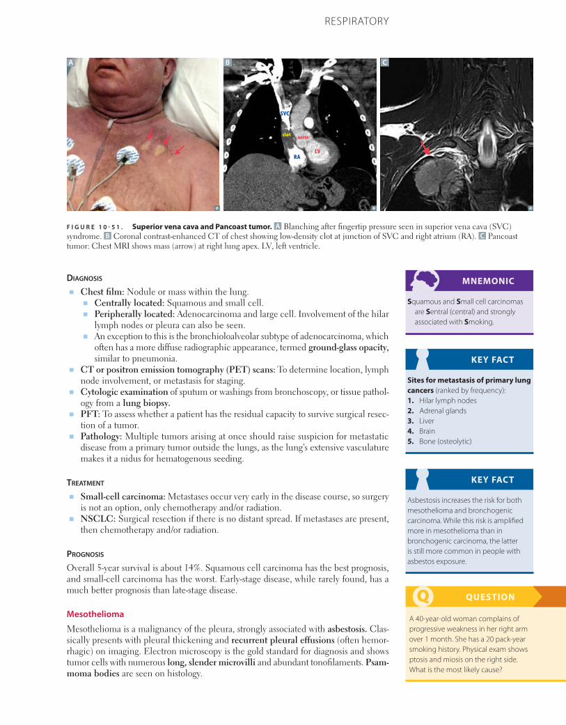
807CHAPTER 10RESPIRATORY
Diagnosis
■ Chest film: Nodule or mass within the lung. ■ Centrally located: Squamous and small cell. ■ Peripherally located: Adenocarcinoma and large cell. Involvement of the hilar
lymph nodes or pleura can also be seen.■ An exception to this is the bronchioloalveolar subtype of adenocarcinoma, which
often has a more diffuse radiographic appearance, termed ground-glass opacity, similar to pneumonia.
■ CT or positron emission tomography (PET) scans: To determine location, lymph node involvement, or metastasis for staging.
■ Cytologic examination of sputum or washings from bronchoscopy, or tissue pathol-ogy from a lung biopsy.
■ PFT: To assess whether a patient has the residual capacity to survive surgical resec-tion of a tumor.
■ Pathology: Multiple tumors arising at once should raise suspicion for metastatic disease from a primary tumor outside the lungs, as the lung’s extensive vasculature makes it a nidus for hematogenous seeding.
treatment
■ Small-cell carcinoma: Metastases occur very early in the disease course, so surgery is not an option, only chemotherapy and/or radiation.
■ NSCLC: Surgical resection if there is no distant spread. If metastases are present, then chemotherapy and/or radiation.
Prognosis
Overall 5-year survival is about 14%. Squamous cell carcinoma has the best prognosis, and small-cell carcinoma has the worst. Early-stage disease, while rarely found, has a much better prognosis than late-stage disease.
Mesothelioma
Mesothelioma is a malignancy of the pleura, strongly associated with asbestosis. Clas-sically presents with pleural thickening and recurrent pleural effusions (often hemor-rhagic) on imaging. Electron microscopy is the gold standard for diagnosis and shows tumor cells with numerous long, slender microvilli and abundant tonofilaments. Psam-moma bodies are seen on histology.
MNEMONIC
Squamous and Small cell carcinomas are Sentral (central) and strongly associated with Smoking.
KEY FACT
Sites for metastasis of primary lung cancers (ranked by frequency):1. Hilar lymph nodes2. Adrenal glands3. Liver4. Brain5. Bone (osteolytic)
KEY FACT
Asbestosis increases the risk for both mesothelioma and bronchogenic carcinoma. While this risk is amplified more in mesothelioma than in bronchogenic carcinoma, the latter is still more common in people with asbestos exposure.
QUESTION
A 40-year-old woman complains of progressive weakness in her right arm over 1 month. She has a 20 pack-year smoking history. Physical exam shows ptosis and miosis on the right side. What is the most likely cause?
F I G U R E 1 0 - 5 1 . Superior vena cava and Pancoast tumor. A Blanching after fingertip pressure seen in superior vena cava (SVC) syndrome. B Coronal contrast-enhanced CT of chest showing low-density clot at junction of SVC and right atrium (RA). C Pancoast tumor: Chest MRI shows mass (arrow) at right lung apex. LV, left ventricle.
A B C
SVC
RALV
aortaclot
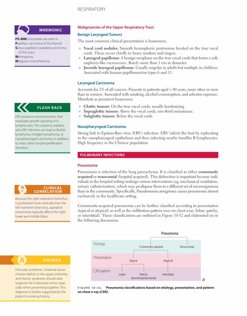
808 CHAPTER 10 RESPIRATORY
Malignancies of the Upper Respiratory Tract
Benign Laryngeal Tumors
The most common clinical presentation is hoarseness.
■ Vocal cord nodules: Smooth hemispheric protrusions located on the true vocal cords. These occur chiefly in heavy smokers and singers.
■ Laryngeal papilloma: A benign neoplasm on the true vocal cords that forms a soft, raspberry-like excrescence. Rarely more than 1 cm in diameter.
■ Juvenile laryngeal papillomas: Usually singular in adults but multiple in children. Associated with human papillomavirus types 6 and 11.
Laryngeal Carcinoma
Accounts for 2% of all cancers. Presents in patients aged > 40 years, more often in men than in women. Associated with smoking, alcohol consumption, and asbestos exposure. Manifests as persistent hoarseness.
■ Glottic tumors: On the true vocal cords, usually keratinizing.■ Supraglottic tumors: Above the vocal cords; one-third metastasize.■ Subglottic tumors: Below the vocal cords.
Nasopharyngeal Carcinoma
Strong link to Epstein-Barr virus (EBV) infection. EBV infects the host by replicating in the nasopharyngeal epithelium and then infecting nearby tonsillar B lymphocytes. High frequency in the Chinese population.
PULMONARY INFECTIONS
Pneumonia
Pneumonia is infection of the lung parenchyma. It is classified as either community acquired or nosocomial (hospital acquired). This distinction is important because indi-viduals in the hospital setting undergo various interventions (eg, mechanical ventilation, urinary catheterization), which may predispose them to a different set of microorganisms than in the community. Specifically, Pseudomonas aeruginosa causes pneumonia almost exclusively in the healthcare setting.
Community-acquired pneumonia can be further classified according to presentation (typical or atypical) as well as the infiltration pattern seen on chest x-ray (lobar, patchy, or interstitial). These classifications are outlined in Figure 10-52 and elaborated on in the following discussions.
MNEMONIC
PSaMMoma bodies are seen inPapilllary carcinoma of the thyroidSerous papillary cystadenocarcinoma
of the ovaryMeningiomaMalignant mesothelioma
FLASH BACK
EBV produces several proteins that modulate growth signaling in B lymphocytes. This property explains why EBV infection can lead to Burkitt lymphoma, Hodgkin lymphoma, or nasopharyngeal carcinoma, in addition to many other lymphoproliferative disorders.
CLINICAL CORRELATION
Because the right mainstem bronchus is positioned more vertically than the left mainstem bronchus, aspiration pneumonia typically affects the right lower and middle lobes.
ANSWER
Pancoast syndrome. Unilateral senso-rimotor deficits in the upper extremity and Horner syndrome should raise suspicion for a Pancoast tumor, espe-cially when presenting together. This diagnosis is further supported by the patient’s smoking history.
F I G U R E 1 0 - 5 2 . Pneumonia classifications based on etiology, presentation, and pattern on chest x-ray (CXR).
Pneumonia
Community-aquired
Typical
Lobar InterstitialPatchy(bronchopneumonia)
Atypical
NosocomialEtiology
Presentation
CXR pattern
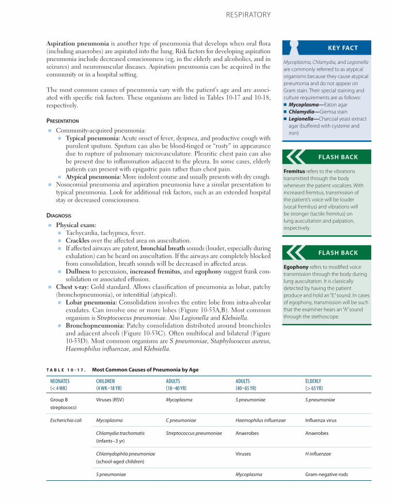
809CHAPTER 10RESPIRATORY
Aspiration pneumonia is another type of pneumonia that develops when oral flora (including anaerobes) are aspirated into the lung. Risk factors for developing aspiration pneumonia include decreased consciousness (eg, in the elderly and alcoholics, and in seizures) and neuromuscular diseases. Aspiration pneumonia can be acquired in the community or in a hospital setting.
The most common causes of pneumonia vary with the patient’s age and are associ-ated with specific risk factors. These organisms are listed in Tables 10-17 and 10-18, respectively.
Presentation
■ Community-acquired pneumonia:■ Typical pneumonia: Acute onset of fever, dyspnea, and productive cough with
purulent sputum. Sputum can also be blood-tinged or “rusty” in appearance due to rupture of pulmonary microvasculature. Pleuritic chest pain can also be present due to inflammation adjacent to the pleura. In some cases, elderly patients can present with epigastric pain rather than chest pain.
■ Atypical pneumonia: More indolent course and usually presents with dry cough.■ Nosocomial pneumonia and aspiration pneumonia have a similar presentation to
typical pneumonia. Look for additional risk factors, such as an extended hospital stay or decreased consciousness.
Diagnosis
■ Physical exam:■ Tachycardia, tachypnea, fever.■ Crackles over the affected area on auscultation.■ If affected airways are patent, bronchial breath sounds (louder, especially during
exhalation) can be heard on auscultation. If the airways are completely blocked from consolidation, breath sounds will be decreased in affected areas.
■ Dullness to percussion, increased fremitus, and egophony suggest frank con-solidation or associated effusion.
■ Chest x-ray: Gold standard. Allows classification of pneumonia as lobar, patchy (bronchopneumonia), or interstitial (atypical). ■ Lobar pneumonia: Consolidation involves the entire lobe from intra-alveolar
exudates. Can involve one or more lobes (Figure 10-53A,B). Most common organism is Streptococcus pneumoniae. Also Legionella and Klebsiella.
■ Bronchopneumonia: Patchy consolidation distributed around bronchioles and adjacent alveoli (Figure 10-53C). Often multifocal and bilateral (Figure 10-53D). Most common organisms are S pneumoniae, Staphylococcus aureus, Haemophilus influenzae, and Klebsiella.
KEY FACT
Mycoplasma, Chlamydia, and Legionella are commonly referred to as atypical organisms because they cause atypical pneumonia and do not appear on Gram stain. Their special staining and culture requirements are as follows:■ Mycoplasma—Eaton agar■ Chlamydia—Giemsa stain■ Legionella—Charcoal yeast extract
agar (buffered with cysteine and iron)
FLASH BACK
Fremitus refers to the vibrations transmitted through the body whenever the patient vocalizes. With increased fremitus, transmission of the patient’s voice will be louder (vocal fremitus) and vibrations will be stronger (tactile fremitus) on lung auscultation and palpation, respectively.
FLASH BACK
Egophony refers to modified voice transmission through the body during lung auscultation. It is classically detected by having the patient produce and hold an “E” sound. In cases of egophony, transmission will be such that the examiner hears an “A” sound through the stethoscope.
T A B L E 1 0 - 1 7 . Most Common Causes of Pneumonia by Age
NEONATES (< 4 WK)
CHILDREN (4 WK–18 YR)
ADULTS (18–40 YR)
ADULTS (40–65 YR)
ELDERLY(> 65 YR)
Group B streptococci
Viruses (RSV) Mycoplasma S pneumoniae S pneumoniae
Escherichia coli Mycoplasma C pneumoniae Haemophilus influenzae Influenza virus
Chlamydia trachomatis (infants−3 yr)
Streptococcus pneumoniae Anaerobes Anaerobes
Chlamydophila pneumoniae (school-aged children)
Viruses H influenzae
S pneumoniae Mycoplasma Gram-negative rods
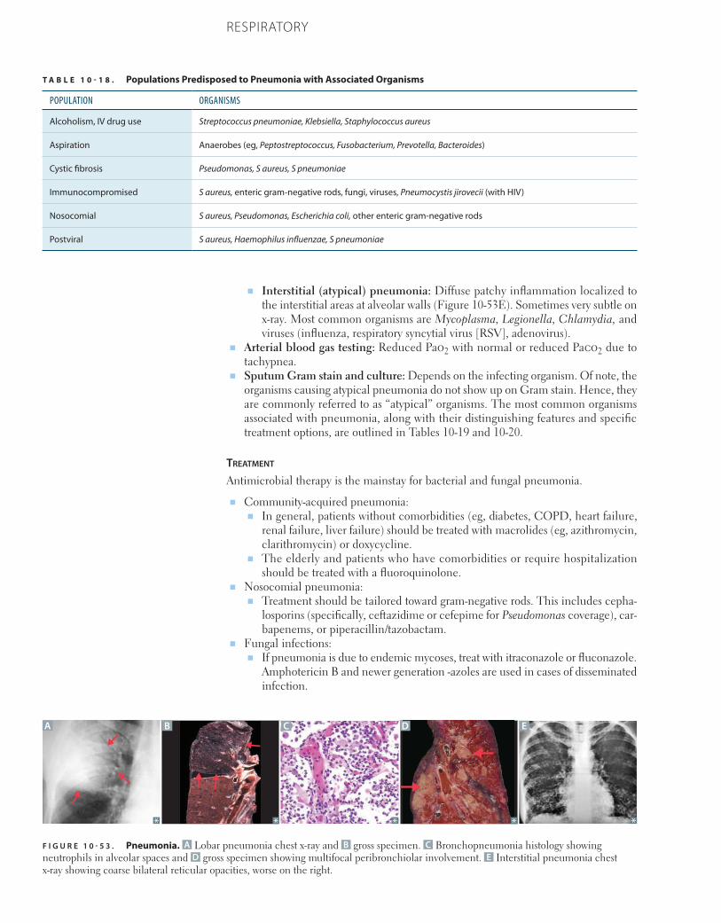
810 CHAPTER 10 RESPIRATORY
■ Interstitial (atypical) pneumonia: Diffuse patchy inflammation localized to the interstitial areas at alveolar walls (Figure 10-53E). Sometimes very subtle on x-ray. Most common organisms are Mycoplasma, Legionella, Chlamydia, and viruses (influenza, respiratory syncytial virus [RSV], adenovirus).
■ Arterial blood gas testing: Reduced Pao2 with normal or reduced Paco2 due to tachypnea.
■ Sputum Gram stain and culture: Depends on the infecting organism. Of note, the organisms causing atypical pneumonia do not show up on Gram stain. Hence, they are commonly referred to as “atypical” organisms. The most common organisms associated with pneumonia, along with their distinguishing features and specific treatment options, are outlined in Tables 10-19 and 10-20.
treatment
Antimicrobial therapy is the mainstay for bacterial and fungal pneumonia.
■ Community-acquired pneumonia:■ In general, patients without comorbidities (eg, diabetes, COPD, heart failure,
renal failure, liver failure) should be treated with macrolides (eg, azithromycin, clarithromycin) or doxycycline.
■ The elderly and patients who have comorbidities or require hospitalization should be treated with a fluoroquinolone.
■ Nosocomial pneumonia:■ Treatment should be tailored toward gram-negative rods. This includes cepha-
losporins (specifically, ceftazidime or cefepime for Pseudomonas coverage), car-bapenems, or piperacillin/tazobactam.
■ Fungal infections:■ If pneumonia is due to endemic mycoses, treat with itraconazole or fluconazole.
Amphotericin B and newer generation -azoles are used in cases of disseminated infection.
T A B L E 1 0 - 1 8 . Populations Predisposed to Pneumonia with Associated Organisms
POPULATION ORGANISMS
Alcoholism, IV drug use Streptococcus pneumoniae, Klebsiella, Staphylococcus aureus
Aspiration Anaerobes (eg, Peptostreptococcus, Fusobacterium, Prevotella, Bacteroides)
Cystic fibrosis Pseudomonas, S aureus, S pneumoniae
Immunocompromised S aureus, enteric gram-negative rods, fungi, viruses, Pneumocystis jirovecii (with HIV)
Nosocomial S aureus, Pseudomonas, Escherichia coli, other enteric gram-negative rods
Postviral S aureus, Haemophilus influenzae, S pneumoniae
F I G U R E 1 0 - 5 3 . Pneumonia. A Lobar pneumonia chest x-ray and B gross specimen. C Bronchopneumonia histology showing neutrophils in alveolar spaces and D gross specimen showing multifocal peribronchiolar involvement. E Interstitial pneumonia chest x-ray showing coarse bilateral reticular opacities, worse on the right.
A B C D E
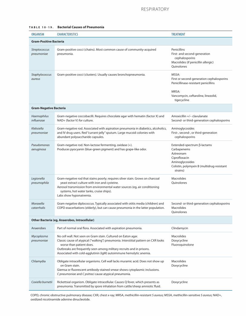
811CHAPTER 10RESPIRATORY
T A B L E 1 0 - 1 9 . Bacterial Causes of Pneumonia
ORGANISM CHARACTERISTICS TREATMENT
Gram-Positive Bacteria
Streptococcus pneumoniae
Gram-positive cocci (chains). Most common cause of community-acquired pneumonia.
PenicillinsFirst- and second-generation
cephalosporinsMacrolides (if penicillin allergic)Quinolones
Staphylococcus aureus
Gram-positive cocci (clusters). Usually causes bronchopneumonia. MSSA:First or second-generation cephalosporinsPenicillinase-resistant penicillins MRSA:Vancomycin, ceftaroline, linezolid,
tigecycline
Gram-Negative Bacteria
Haemophilus influenzae
Gram-negative coccobacilli. Requires chocolate agar with hematin (factor X) and NAD+ (factor V) for culture.
Amoxicillin +/– clavulanateSecond- or third-generation cephalosporins
Klebsiella pneumoniae
Gram-negative rod. Associated with aspiration pneumonia in diabetics, alcoholics, and IV drug users. Red “currant-jelly” sputum. Large mucoid colonies with abundant polysaccharide capsules.
AminoglycosidesFirst-, second-, or third-generation
cephalosporins
Pseudomonas aeruginosa
Gram-negative rod. Non-lactose fermenting, oxidase (+).Produces pyocyanin (blue-green pigment) and has grape-like odor.
Extended-spectrum β-lactamsCarbapenemsAztreonamCiprofloxacinAminoglycosidesColistin, polymyxin B (multidrug-resistant
strains)
Legionella pneumophila
Gram-negative rod that stains poorly; requires silver stain. Grows on charcoal yeast extract culture with iron and cysteine.
Aerosol transmission from environmental water sources (eg, air conditioning systems, hot water tanks, cruise ships).
Labs show hyponatremia.
MacrolidesQuinolones
Moraxella catarrhalis
Gram-negative diplococcus. Typically associated with otitis media (children) and COPD exacerbations (elderly), but can cause pneumonia in the latter population.
Second- or third-generation cephalosporinsMacrolidesQuinolones
Other Bacteria (eg, Anaerobes, Intracellular)
Anaerobes Part of normal oral flora. Associated with aspiration pneumonia. Clindamycin
Mycoplasma pneumoniae
No cell wall. Not seen on Gram stain. Cultured on Eaton agar.Classic cause of atypical (“walking”) pneumonia. Interstitial pattern on CXR looks
worse than patient does.Outbreaks are frequently seen among military recruits and in prisons.Associated with cold-agglutinin (IgM) autoimmune hemolytic anemia.
MacrolidesDoxycyclineFluoroquinolone
Chlamydia Obligate intracellular organisms. Cell wall lacks muramic acid. Does not show up on Gram stain.
Giemsa or fluorescent antibody-stained smear shows cytoplasmic inclusions.C pneumoniae and C psittaci cause atypical pneumonia.
MacrolidesDoxycycline
Coxiella burnetii Rickettsial organism. Obligate intracellular. Causes Q fever, which presents as pneumonia. Transmitted by spore inhalation from cattle/sheep amniotic fluid.
Doxycycline
COPD, chronic obstructive pulmonary disease; CXR, chest x-ray; MRSA, methicillin-resistant S aureus; MSSA, methicillin-sensitive S aureus; NAD+, oxidized nicotinamide adenine dinucleotide.
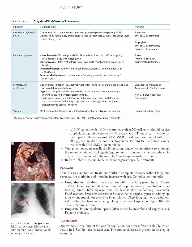
812 CHAPTER 10 RESPIRATORY
■ All HIV patients with a CD4+ count lower than 200 cells/mm3 should receive prophylaxis against Pneumocystis jirovecii (PCP). Therapy can include tri-methoprim-sulfamethoxazole (TMP-SMX; most common, except with sulfa allergy), pentamidine, dapsone, or atovaquone. Existing PCP infections can be treated with TMP-SMX or pentamidine.
■ Viral pneumonias are usually self-limited, requiring only supportive care, although the use of certain antiviral agents (eg, oseltamivir, zanamavir) has been shown to decrease the duration of influenza infections by approximately 24 hours.
■ Refer to Table 10-19 and Table 10-20 for organism-specific treatments.
Prognosis
In most cases, appropriate treatment results in complete recovery without long-term sequelae, but morbidity and mortality increase with age. Complications include:
■ Lung abscess: Localized pus collection within the lung parenchyma (Figure 10-54A). Common complication of aspiration pneumonia or bronchial obstruc-tion (eg, tumor). Infecting organisms include anaerobic oral flora (eg, Bacteroides, Fusobacterium, Peptostreptococcus) or S aureus. Patients typically present with symp-toms of pneumonia unresponsive to antibiotics. Chest imaging shows cavitations with air-fluid levels, often in the right lung in the case of aspiration (Figure 10-54B). Treat with clindamycin.
■ Empyema: Pus in the pleural space. Often caused by anaerobes and staphylococci. Requires drainage.
Tuberculosis
Approximately one-third of the world’s population has been infected with TB, which results in 2–3 million deaths each year. The burden of disease is greatest in developing countries.
T A B L E 1 0 - 2 0 . Fungal and Viral Causes of Pneumonia
ORGANISM CHARACTERISTICS TREATMENT
Pneumocystis jirovecii (PCP)
Causes interstitial pneumonia in immunosuppressed patients (especially AIDS).Diagnosed by lung biopsy or lavage. Disc-shaped yeast seen with methenamine silver
stain of lung tissue.
Treatment:TMP-SMX, pentamidine Prophylaxis:TMP-SMX, pentamidine, dapsone, atovaquone
Endemic mycoses Histoplasmosis: Mississippi and Ohio River valleys. Found in bird/bat droppings. Macrophages filled with Histoplasma.
Blastomycosis: States east of Mississippi River and Central America. Broad-based budding.
Coccidiomycosis: Southwestern United States, California. Spherules filled with endospores.
Paracoccidioidomycosis: Latin America. Budding yeast with “captain’s wheel” formation.
AzolesAmphotericin B (for disseminated infections)
Cryptococcus neoformans
Opportunistic infection (classically HIV patients). Found in soil and pigeon droppings. Acquired through inhalation.
Cryptococcosis presents like pneumonia. Can disseminate hematogenously to meninges, causing cryptococcal meningitis.
Heavily encapsulated yeast. Culture on Sabouraud agar; stains with India ink and mucicarmine. Definitively diagnosed with latex agglutinin test (detects polysaccharide capsular antigen).
Cryptococcal meningitis:Amphotericin B + flucytosine
Non-CNS cryptococcosis: Fluconazole
Viruses Most commonly influenza virus, RSV, adenovirus. Causes atypical pneumonia. Paracoccidioidomycosis
CNS, central nervous system; RSV, respiratory syncytial virus; TMP-SMX, trimethoprim-sulfamethoxazole.
A
B
F I G U R E 1 0 - 5 4 . Lung abscess. A Gross specimen. B Cavitation with air-fluid levels (arrows) visible on x-ray of the chest.
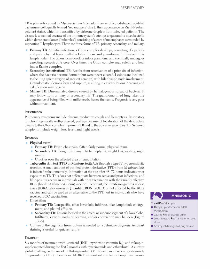
813CHAPTER 10RESPIRATORY
TB is primarily caused by Mycobacterium tuberculosis, an aerobic, rod-shaped, acid-fast bacterium (colloquially termed “red snappers” due to their appearance on Ziehl-Neelsen acid-fast stain), which is transmitted by airborne droplets from infected patients. The disease is so named because of the immune system’s attempt to quarantine mycobacteria within dense granulomas (“tubercles”) consisting of a core of macrophages surrounded by supporting T lymphocytes. There are three forms of TB: primary, secondary, and miliary.
■ Primary TB: At initial infection, a Ghon complex develops, consisting of a periph-eral parenchymal lesion called a Ghon focus and granulomas in involved hilar lymph nodes. The Ghon focus develops into a granuloma and eventually undergoes caseating necrosis at its core. Over time, the Ghon complex may calcify and heal into a Ranke complex.
■ Secondary (reactivation) TB: Results from reactivation of a prior site of infection, where the bacteria became dormant but were never cleared. Lesions are localized to the lung apices (region of greatest aeration) with hilar lymph node involvement. Granulomatous lesions form and rupture, resulting in cavitary lesions. Scarring and calcification may be seen.
■ Miliary TB: Disseminated disease caused by hematogenous spread of bacteria. It may follow from primary or secondary TB. The granuloma-filled lung takes the appearance of being filled with millet seeds, hence the name. Prognosis is very poor without treatment.
Presentation
Pulmonary symptoms include chronic productive cough and hemoptysis. Respiratory function is generally well-preserved, perhaps because of localization of the destructive disease to the Ghon complex in primary TB and to the apices in secondary TB. Systemic symptoms include weight loss, fever, and night sweats.
Diagnosis
■ Physical exam:■ Primary TB: Fever, chest pain. Often fairly normal physical exam.■ Secondary TB: Cough (evolving into hemoptysis), weight loss, wasting, night
sweats.■ Crackles over the affected area on auscultation.
■ Tuberculin skin test (PPD or Mantoux test): Acts through a type IV hypersensitivity reaction. A small amount of purified protein derivative (PPD) from M tuberculosis is injected subcutaneously. Induration at the site after 48–72 hours indicates prior exposure to TB. This does not differentiate between active and prior infections, and false-positives occur in individuals with prior vaccination with the variably effective BCG (bacillus Calmette-Guérin) vaccine. In contrast, the interferon-gamma release assay (IGRA; also known as QuantiFERON GOLD) is not affected by the BCG vaccine and can be used as an alternative to the PPD test in individuals who have received BCG vaccination.
■ Chest film:■ Primary TB: Nonspecific, often lower lobe infiltrate, hilar lymph node enlarge-
ment, and pleural effusion.■ Secondary TB: Lesions located in the apices or superior segment of a lower lobe.
Infiltrates, cavities, nodules, scarring, and/or contraction may be seen (Figure 10-55).
■ Culture of the organism from sputum is needed for a definitive diagnosis. Acid-fast staining is useful for quicker results.
treatment
Six months of treatment with isoniazid (INH), pyridoxine (vitamin B6), and rifampin, supplemented during the first 2 months with pyrazinamide and ethambutol. A current global challenge is the rise of multidrug-resistant (MDR) and, more recently, extensively drug-resistant (XDR) tuberculosis. MDR-TB is resistant to at least rifampin and isonia-
MNEMONIC
The 4 R’s of rifampin: ■ Ramps up cytochrome P450
metabolism■ Causes Red or orange urine■ Leads to rapid Resistance when used
alone■ Acts by inhibiting RNA polymerase
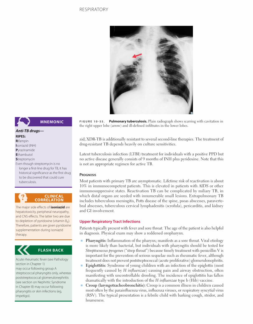
814 CHAPTER 10 RESPIRATORY
zid; XDR-TB is additionally resistant to several second-line therapies. The treatment of drug-resistant TB depends heavily on culture sensitivities.
Latent tuberculosis infection (LTBI) treatment for individuals with a positive PPD but no active disease generally consists of 9 months of INH plus pyridoxine. Note that this is not an appropriate regimen for active TB.
Prognosis
Most patients with primary TB are asymptomatic. Lifetime risk of reactivation is about 10% in immunocompetent patients. This is elevated in patients with AIDS or other immunosuppressive states. Reactivation TB can be complicated by miliary TB, in which distal organs are seeded with innumerable small lesions. Extrapulmonary TB includes tuberculous meningitis, Potts disease of the spine, psoas abscesses, paraverte-bral abscesses, tuberculous cervical lymphadenitis (scrofula), pericarditis, and kidney and GI involvement.
Upper Respiratory Tract Infections
Patients typically present with fever and sore throat. The age of the patient is also helpful in diagnosis. Physical exam may show a reddened oropharynx.
■ Pharyngitis: Inflammation of the pharynx; manifests as a sore throat. Viral etiology is more likely than bacterial, but individuals with pharyngitis should be tested for Streptococcus pyogenes (“strep throat”) because timely treatment with penicillin V is important for the prevention of serious sequelae such as rheumatic fever, although treatment does not prevent poststreptococcal (acute proliferative) glomerulonephritis.
■ Epiglottitis: Syndrome of young children with an infection of the epiglottis (most frequently caused by H influenzae) causing pain and airway obstruction, often manifesting with uncontrollable drooling. The incidence of epiglottitis has fallen dramatically with the introduction of the H influenzae type b (Hib) vaccine.
■ Croup (laryngotracheobronchitis): Croup is a common illness in children caused most often by the parainfluenza virus, influenza viruses, or respiratory syncytial virus (RSV). The typical presentation is a febrile child with barking cough, stridor, and hoarseness.
MNEMONIC
Anti-TB drugs—RIPES: RifampinIsoniazid (INH)PyrazinamideEthambutolStreptomycinEven though streptomycin is no
longer a first-line drug for TB, it has historical significance as the first drug to be discovered that could cure tuberculosis.
CLINICAL CORRELATION
The major side effects of isoniazid are hepatotoxicity, peripheral neuropathy, and CNS effects. The latter two are due to depletion of pyridoxine (vitamin B6). Therefore, patients are given pyridoxine supplementation during isoniazid therapy.
FLASH BACK
Acute rheumatic fever (see Pathology section in Chapter 1) may occur following group A streptococcal pharyngitis only, whereas poststreptococcal glomerulonephritis (see section on Nephritic Syndrome in Chapter 8) may occur following pharyngitis or skin infections (eg, impetigo).
F I G U R E 1 0 - 5 5 . Pulmonary tuberculosis. Plain radiograph shows scarring with cavitation in the right upper lobe (arrow) and ill-defined infiltrates in the lower lobes.
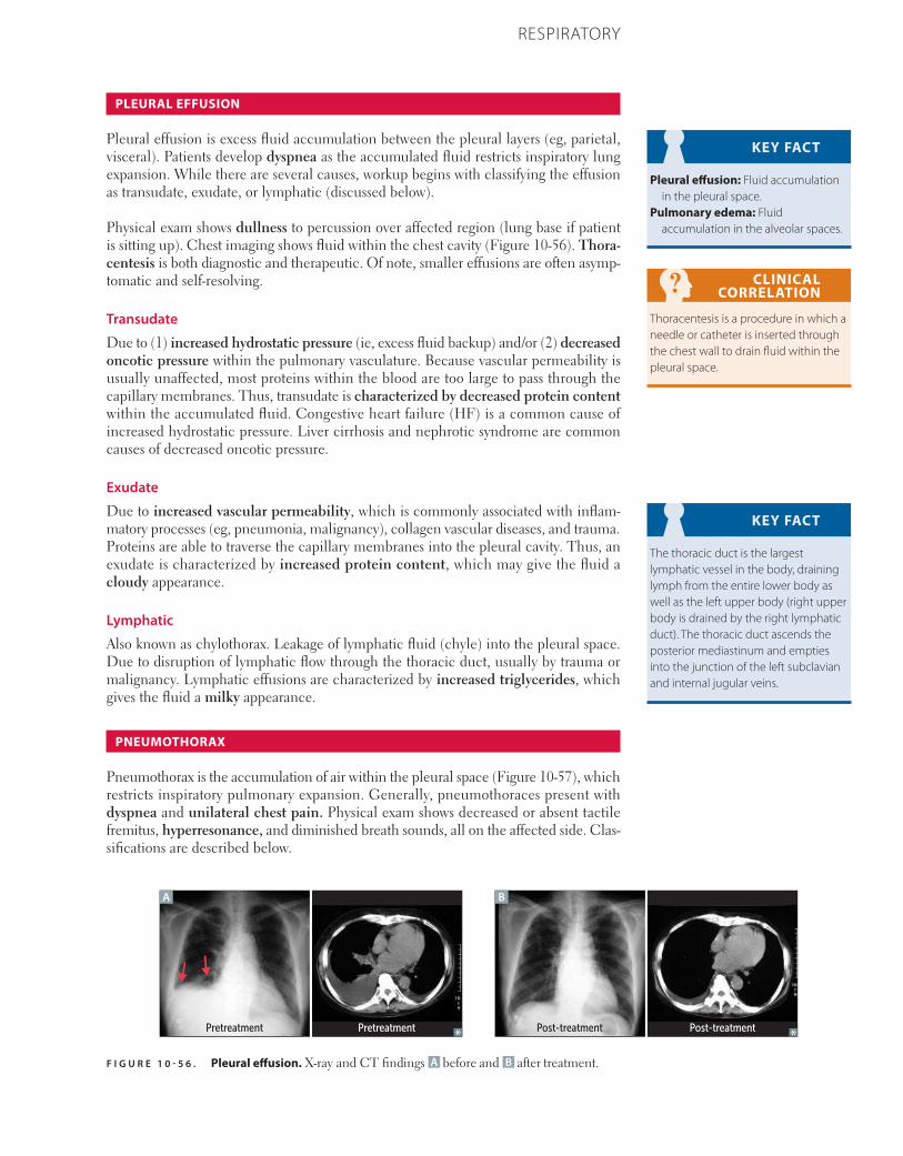
815CHAPTER 10RESPIRATORY
PLEURAL EFFUSION
Pleural effusion is excess fluid accumulation between the pleural layers (eg, parietal, visceral). Patients develop dyspnea as the accumulated fluid restricts inspiratory lung expansion. While there are several causes, workup begins with classifying the effusion as transudate, exudate, or lymphatic (discussed below).
Physical exam shows dullness to percussion over affected region (lung base if patient is sitting up). Chest imaging shows fluid within the chest cavity (Figure 10-56). Thora-centesis is both diagnostic and therapeutic. Of note, smaller effusions are often asymp-tomatic and self-resolving.
Transudate
Due to (1) increased hydrostatic pressure (ie, excess fluid backup) and/or (2) decreased oncotic pressure within the pulmonary vasculature. Because vascular permeability is usually unaffected, most proteins within the blood are too large to pass through the capillary membranes. Thus, transudate is characterized by decreased protein content within the accumulated fluid. Congestive heart failure (HF) is a common cause of increased hydrostatic pressure. Liver cirrhosis and nephrotic syndrome are common causes of decreased oncotic pressure.
Exudate
Due to increased vascular permeability, which is commonly associated with inflam-matory processes (eg, pneumonia, malignancy), collagen vascular diseases, and trauma. Proteins are able to traverse the capillary membranes into the pleural cavity. Thus, an exudate is characterized by increased protein content, which may give the fluid a cloudy appearance.
Lymphatic
Also known as chylothorax. Leakage of lymphatic fluid (chyle) into the pleural space. Due to disruption of lymphatic flow through the thoracic duct, usually by trauma or malignancy. Lymphatic effusions are characterized by increased triglycerides, which gives the fluid a milky appearance.
PNEUMOTHORAX
Pneumothorax is the accumulation of air within the pleural space (Figure 10-57), which restricts inspiratory pulmonary expansion. Generally, pneumothoraces present with dyspnea and unilateral chest pain. Physical exam shows decreased or absent tactile fremitus, hyperresonance, and diminished breath sounds, all on the affected side. Clas-sifications are described below.
KEY FACT
Pleural effusion: Fluid accumulation in the pleural space.
Pulmonary edema: Fluid accumulation in the alveolar spaces.
CLINICAL CORRELATION
Thoracentesis is a procedure in which a needle or catheter is inserted through the chest wall to drain fluid within the pleural space.
KEY FACT
The thoracic duct is the largest lymphatic vessel in the body, draining lymph from the entire lower body as well as the left upper body (right upper body is drained by the right lymphatic duct). The thoracic duct ascends the posterior mediastinum and empties into the junction of the left subclavian and internal jugular veins.
F I G U R E 1 0 - 5 6 . Pleural effusion. X-ray and CT findings A before and B after treatment.
PretreatmentPretreatment
A B
Post-treatment Post-treatment
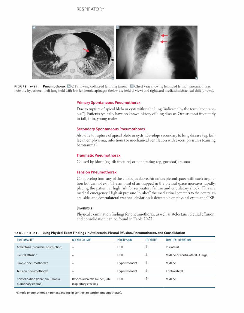
816 CHAPTER 10 RESPIRATORY
Primary Spontaneous Pneumothorax
Due to rupture of apical blebs or cysts within the lung (indicated by the term “spontane-ous”). Patients typically have no known history of lung disease. Occurs most frequently in tall, thin, young males.
Secondary Spontaneous Pneumothorax
Also due to rupture of apical blebs or cysts. Develops secondary to lung disease (eg, bul-lae in emphysema, infections) or mechanical ventilation with excess pressures (causing barotrauma).
Traumatic Pneumothorax
Caused by blunt (eg, rib fracture) or penetrating (eg, gunshot) trauma.
Tension Pneumothorax
Can develop from any of the etiologies above. Air enters pleural space with each inspira-tion but cannot exit. The amount of air trapped in the pleural space increases rapidly, placing the patient at high risk for respiratory failure and circulatory shock. This is a medical emergency. High air pressure “pushes” the mediastinal contents to the contralat-eral side, and contralateral tracheal deviation is detectable on physical exam and CXR.
Diagnosis
Physical examination findings for pneumothorax, as well as atelectasis, pleural effusion, and consolidation can be found in Table 10-21.
F I G U R E 1 0 - 5 7 . Pneumothorax. A CT showing collapsed left lung (arrow). B Chest x-ray showing left-sided tension pneumothorax; note the hyperlucent left lung field with low left hemidiaphragm (below the field of view) and rightward mediastinal/tracheal shift (arrows).
BA
T A B L E 1 0 - 2 1 . Lung Physical Exam Findings in Atelectasis, Pleural Effusion, Pneumothorax, and Consolidation
ABNORMALITY BREATH SOUNDS PERCUSSION FREMITUS TRACHEAL DEVIATION
Atelectasis (bronchial obstruction) ↓ Dull ↓ Ipsilateral
Pleural effusion ↓ Dull ↓ Midline or contralateral (if large)
Simple pneumothoraxa ↓ Hyperresonant ↓ Midline
Tension pneumothorax ↓ Hyperresonant ↓ Contralateral
Consolidation (lobar pneumonia, pulmonary edema)
Bronchial breath sounds; late inspiratory crackles
Dull ↑ Midline
aSimple pneumothorax = nonexpanding (in contrast to tension pneumothorax).
817CHAPTER 10RESPIRATORY
treatment
■ Supplemental oxygen to increase the rate of resorption of intrapleural air. In cases of a small asymptomatic pneumothorax, this may be sufficient for spontaneous recovery to occur.
■ In larger and/or symptomatic pneumothoraces, air should be evacuated from the intrapleural space via thoracentesis (needle aspiration) or chest tube placement (tube thoracostomy) with a water seal, which acts as a one-way valve.
■ In cases of recurrent pneumothorax, the pleurae may be sealed together through pleurodesis, in which chemical or mechanical irritation is employed in order to encourage fibrous scar tissue formation, sealing the visceral and parietal pleurae together. This effectively glues the lung to the chest wall.
ALLERGY
Presentation
The term allergy is typically used to refer to type I hypersensitivity, mediated by IgE cross-linking after exposure to an allergen, leading to mast cell degranulation and his-tamine-mediated vascular permeability. Allergies manifest in a myriad of ways, but many of the symptoms affect the respiratory system, in particular, allergic rhinitis (“hay fever”—congestion, sneezing, itching), extrinsic asthma, and anaphylaxis. Anaphylaxis is the most severe allergy syndrome, characterized by multiorgan involvement including urticaria, edema, airway obstruction, low blood pressure, and GI symptoms. Any airway obstruction must be addressed immediately, usually through epinephrine administration.
Diagnosis
The symptoms of allergies are classic and generally sufficient to establish a diagnosis. However, specific testing for allergen sensitivities may be instructive in certain cases; this is accomplished through either a radioallergosorbent test (RAST) of the blood for ingestion/inhalation allergies or a skin test for contact allergies.
treatment
In many cases, the main “treatment” of allergies is allergen avoidance, especially in the case of hypersensitivities to foods, animals, or materials. If this is not possible, several drug classes may be used, which are listed below and discussed further in the Pharma-cology section at the end of this chapter.
■ H1 histamine blockers (first and second generation) to treat inflammation.■ α-Adrenergic agonists (pseudoephedrine, phenylephrine, xylometazoline, oxy-
metazoline) for nasal decongestion.■ Epinephrine for anaphylactic shock.
Another method occasionally employed to treat allergies is immunotherapy (desensiti-zation), in which successively escalating doses of allergen are injected with the goal of inducing tolerance. This is particularly useful for unpredictable and difficult-to-avoid allergens such as bee venom.
Prognosis
Most cases of allergy are primarily a lifelong nuisance with seasonal or environmental variation. However, a severe allergic reaction may result in anaphylaxis, which has a poor prognosis unless immediately managed.
818 CHAPTER 10 RESPIRATORY
HYPERSENSITIVITY PNEUMONITIS
Presentation
Results from inhalation of biological or chemical dust such as aerosolized mold or droppings, leading to a lymphocyte-mediated inflammatory response in the alveoli. Distinguished from asthma in that this is an alveolar disease rather than one of bronchi; additionally, unlike asthma and allergy, this is not a type I hypersensitivity reaction. Symptoms of acute disease include chest tightness, cough, wheezing, fever, and dyspnea, resolving hours after discontinuation of exposure. Symptoms of chronic disease include dyspnea, fatigue, cough, and weight loss.
Diagnosis
■ Probable diagnosis made with positive history of exposure, consistent CT scan (retic-ular, nodular, or ground glass opacities), bronchoalveolar lavage showing increased lymphocytes.
■ Definitive diagnosis can be made with lung biopsy (findings include loosely orga-nized granulomas) in conjunction with a consistent history.
■ Differential diagnosis includes pneumoconiosis, IPF, COPD, and asthma.
treatment
Avoid further exposure to offending agents. Glucocorticoids may help resolve symptoms.
Prognosis
Usually complete or near complete recovery of lung function following cessation of antigen exposure.
Pharmacology
HISTAMINE BLOCKERS
First-Generation Histamine Blockers
Drug names
Diphenhydramine, dimenhydrinate, chlorpheniramine.
mechanism
Reversibly inhibit H1 histamine receptors, which are involved in the inflammatory process. Major effects of H1 receptor stimulation include:
■ Increased nasal and bronchial mucus production■ Contraction of bronchioles■ Increased vascular permeability■ Pruritus■ Pain
uses
■ Allergies: Due to anti-inflammatory effects (see above).■ Motion sickness: H1 blockers also competitively inhibit muscarinic receptors, which
contribute to the signs and symptoms associated with motion sickness.■ Sleep aid: H1 blockers are lipophilic, which allows them to cross the blood-brain
barrier (BBB) and act on the CNS.
KEY FACT
1st generation H1 blocker names usually contain -en/-ine or -en/-ate.
FLASH BACK
H1 receptors mediate inflammation, whereas H2 receptors mediate gastric acid secretion.
819CHAPTER 10RESPIRATORY
siDe effects
■ Sedation: Due to CNS effects (see above).■ Muscarinic antagonism: Blurry vision, dry mouth, urinary retention. Can also cause
confusion and hallucinations in the elderly.■ α-Adrenergic antagonism: Postural hypotension.
Second-Generation Histamine Blockers
Drug names
Loratadine, fexofenadine, desloratadine, cetirizine.
mechanism
■ Reversibly inhibit H1 receptors. ■ Unlike first-generation H1 blockers, second-generation H1 blockers do not readily
cross the BBB and are therefore far less sedating. They also do not act on muscarinic or α-adrenergic receptors.
uses
Allergies.
siDe effects
Generally well tolerated.
MUCOACTIVE AGENTS
Subtypes, based on mechanism, include expectorants and mucolytics.
Drug names
Guaifenesin, N-acetylcysteine, dornase alfa (DNAse).
mechanism
■ Expectorants (guaifenesin): Increase the volume of watery airway secretions. This serves to thin out respiratory secretions, making them easier to cough up.
■ Mucolytics: Loosen mucus plugs. N-acetylcysteine acts by cleaving disulfide bonds within the mucus glycoproteins. Dornase alfa (DNAse) clears leukocytic debris through hydrolysis of DNA polymers.
uses
■ Increases clearance of respiratory secretions (eg, common cold, pneumonia, COPD).■ N-acetylcysteine and DNAse are used in cystic fibrosis (CF) patients.
siDe effects
Generally well tolerated.
DEXTROMETHORPHAN
mechanism
Synthetic codeine analog. Antagonizes N-methyl-d-aspartate (NMDA) glutamate receptors.
KEY FACT
Second-generation H1 blocker names usually end in -adine.
FLASH BACK
N-acetylcysteine is also used as an antidote for acetaminophen overdose.
820 CHAPTER 10 RESPIRATORY
uses
Antitussive agent (suppresses cough).
siDe effects
Mild opioid effects when used in excess (euphoria, respiratory depression, miosis, constipation). Has mild abuse potential. Naloxone can be given for overdose.
α-ADRENERGIC AGONISTS
Drug names
Pseudoephedrine, phenylephrine, xylometazoline, oxymetazoline.
mechanism
α-Adrenergic agonist.
uses
■ Reduce hyperemia, edema, and nasal congestion.■ Pseudoephedrine is also illicitly used to make methamphetamine.
siDe effects
■ Hypertension.■ Pseudoephedrine can also cause CNS stimulation/anxiety.■ Rapid tolerance formation (tachyphylaxis).
PULMONARY HYPERTENSION DRUGS
Drug names
Bosentan, sildenafil, epoprostenol, iloprost.
mechanisms
■ Bosentan: Competitively inhibits endothelin-1 receptors, thereby preventing pul-monary vasoconstriction and decreasing pulmonary vascular resistance.
■ Sildenafil: Inhibits cGMP phosphodiesterase-5 (PDE-5), which normally breaks down nitric oxide. This prolongs the effects of nitric oxide, resulting in arterial vasodilation.
■ Epoprostenol, iloprost: Prostacyclins (PGI2). Have direct vasodilatory effects on pulmonary and systemic arterial vasculature. Also inhibit platelet aggregation.
uses
Pulmonary hypertension.
siDe effects
■ Bosentan: Hepatotoxic (monitor LFTs).■ Sildenafil: Headaches, hypotension.■ Epoprostenol, iloprost: Flushing, jaw pain.
FLASH BACK
Sildenafil is also used to treat erectile dysfunction.
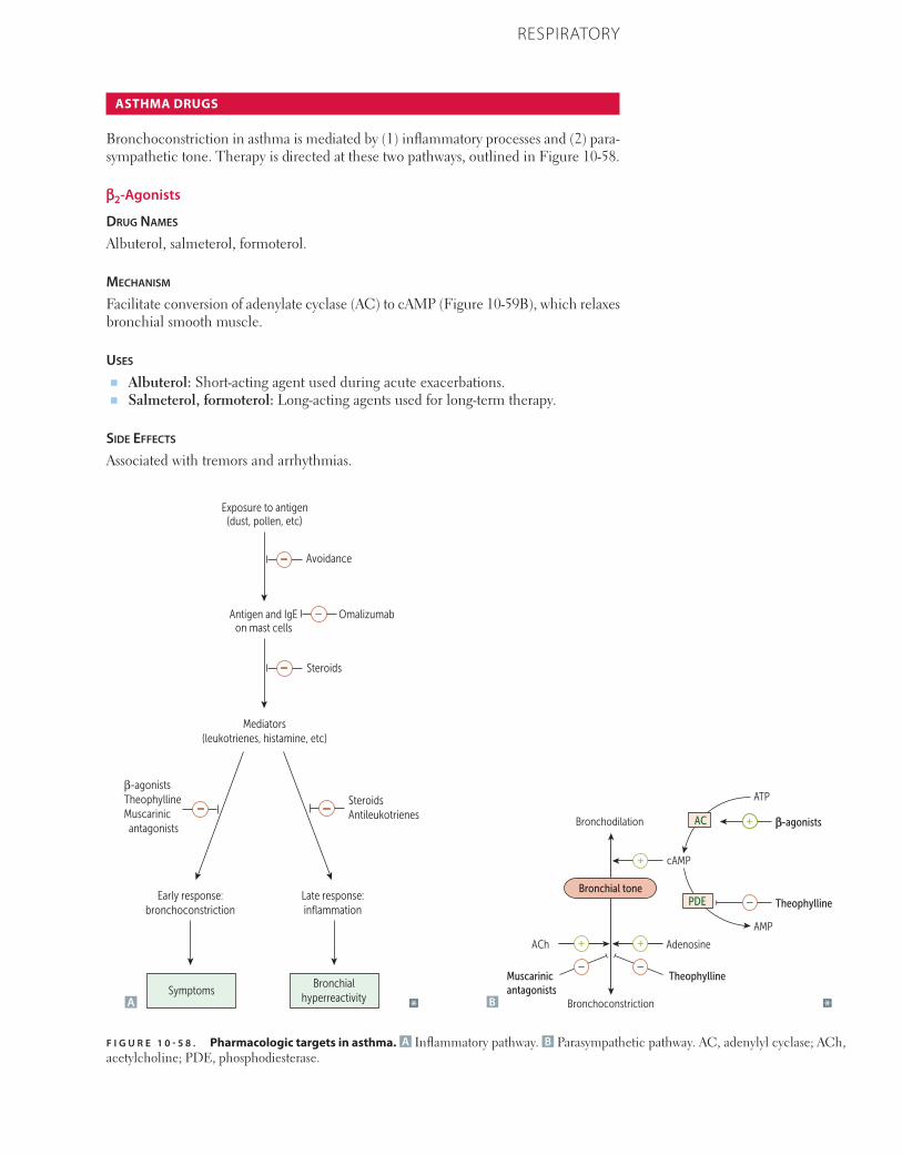
821CHAPTER 10RESPIRATORY
ASTHMA DRUGS
Bronchoconstriction in asthma is mediated by (1) inflammatory processes and (2) para-sympathetic tone. Therapy is directed at these two pathways, outlined in Figure 10-58.
β2-Agonists
Drug names
Albuterol, salmeterol, formoterol.
mechanism
Facilitate conversion of adenylate cyclase (AC) to cAMP (Figure 10-59B), which relaxes bronchial smooth muscle.
uses
■ Albuterol: Short-acting agent used during acute exacerbations.■ Salmeterol, formoterol: Long-acting agents used for long-term therapy.
siDe effects
Associated with tremors and arrhythmias.
F I G U R E 1 0 - 5 8 . Pharmacologic targets in asthma. A Inflammatory pathway. B Parasympathetic pathway. AC, adenylyl cyclase; ACh, acetylcholine; PDE, phosphodiesterase.
Exposure to antigen(dust, pollen, etc)
Avoidance
Antigen and IgEon mast cells
Steroids
Mediators(leukotrienes, histamine, etc)
β-agonistsTheophyllineMuscarinic antagonists
SteroidsAntileukotrienes
Late response:inflammation
Early response:bronchoconstriction
Bronchialhyperreactivity
Symptoms
Omalizumab
A
Bronchodilation
Bronchial tone
Bronchoconstriction
AdenosineACh
cAMP
TheophyllineMuscarinicantagonists
β-agonists
Theophylline
ATP
AMP
PDE
AC
B
822 CHAPTER 10 RESPIRATORY
Corticosteroids
Drug names
Beclomethasone, fluticasone, flunisolide.
mechanism
Inhibit the synthesis of virtually all cytokines. Inactivate NF-κB, which is the transcrip-tion factor that induces the production of TNF-α and other inflammatory agents.
uses
First-line therapy for chronic asthma.
siDe effects
Oral candidiasis (thrush): Prevented by rinsing mouth following administration.
Muscarinic Antagonists
Drug names Ipratropium, tiotropium.
mechanism
Competitively inhibit muscarinic receptors (M3), preventing bronchoconstriction. Tiotropium is long acting.
uses
■ COPD. ■ Asthma.
Antileukotrienes
Drug names
Zileuton, montelukast, zafirlukast.
mechanism
■ Zileuton: 5-lipoxygenase pathway inhibitor. Blocks the conversion of arachidonic acid to leukotrienes.
■ Montelukast, zafirlukast: Competitively inhibit leukotriene receptors (CysLT1).
uses
■ Considered when asthma is refractory to long-acting β2-agonists and inhaled corticosteroids.
■ Montelukast and zafirlukast are especially good for aspirin-induced asthma, in which bronchospasms result from increased leukotriene production.
siDe effects
Zileuton is associated with hepatotoxicity.
Omalizumab
mechanism
Monoclonal anti-IgE antibody. Binds unbound serum IgE at the Fc region (FcεRI).
823CHAPTER 10RESPIRATORY
uses
Considered when asthma is refractory to long-acting β2-agonists and inhaled corticosteroids.
siDe effects
Generally well tolerated, but very expensive.
Methylxanthines
Drug name
Theophylline.
mechanism
■ Inhibits phosphodiesterase, which normally hydrolyzes cAMP (Figure 10-59B). This increases cAMP levels, resulting in bronchodilation.
■ Also blocks the actions of adenosine (Figure 10-59B), thereby preventing bronchoconstriction.
uses
Has limited usage due to narrow therapeutic index.
siDe effects
■ Neurotoxicity (eg, seizures).■ Cardiotoxicity (eg, tachycardia, arrhythmias).■ Metabolized by cytochrome P-450.
Magnesium Sulfate
mechanism
Inhibits calcium influx into airway smooth muscle cells, thereby decreasing airway tone.
uses
Shown to be helpful, specifically in severe asthma exacerbations.
Methacholine
mechanism
Muscarinic receptor (M3) agonist. Causes bronchoconstriction.
uses
Used in bronchial provocation to help diagnose asthma. Of note, the methacholine challenge test has high sensitivity but low specificity. Therefore, it is useful for ruling out asthma, but a positive result is not diagnostic.

Appendix
Q-Bank
371
14 Respiratory
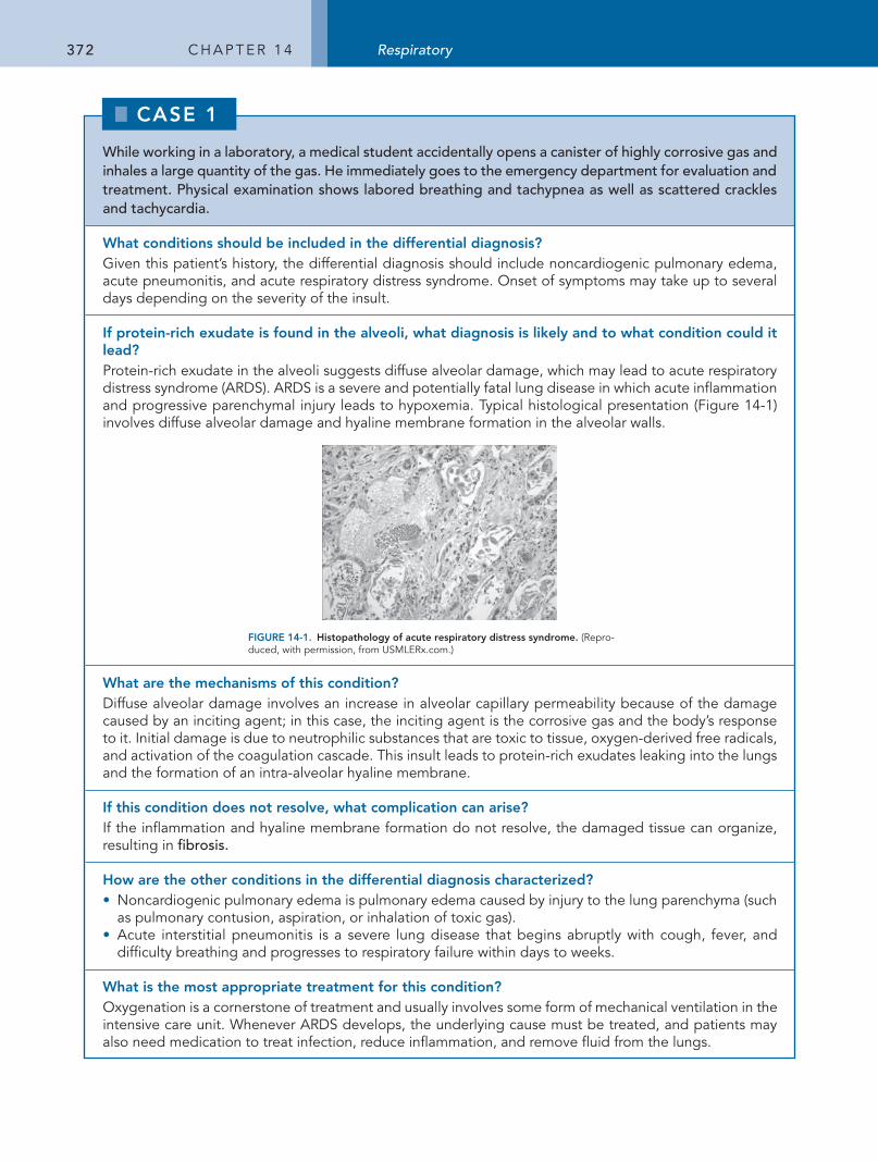
372 C h a p t e r 1 4 Respiratory
While working in a laboratory, a medical student accidentally opens a canister of highly corrosive gas and inhales a large quantity of the gas. He immediately goes to the emergency department for evaluation and treatment. Physical examination shows labored breathing and tachypnea as well as scattered crackles and tachycardia.
What conditions should be included in the differential diagnosis?Given this patient’s history, the differential diagnosis should include noncardiogenic pulmonary edema, acute pneumonitis, and acute respiratory distress syndrome. Onset of symptoms may take up to several days depending on the severity of the insult.
If protein-rich exudate is found in the alveoli, what diagnosis is likely and to what condition could it lead?protein-rich exudate in the alveoli suggests diffuse alveolar damage, which may lead to acute respiratory distress syndrome (arDS). arDS is a severe and potentially fatal lung disease in which acute inflammation and progressive parenchymal injury leads to hypoxemia. typical histological presentation (Figure 14-1) involves diffuse alveolar damage and hyaline membrane formation in the alveolar walls.
FIGURE 14-1. Histopathology of acute respiratory distress syndrome. (repro-duced, with permission, from USMLerx.com.)
What are the mechanisms of this condition?Diffuse alveolar damage involves an increase in alveolar capillary permeability because of the damage caused by an inciting agent; in this case, the inciting agent is the corrosive gas and the body’s response to it. Initial damage is due to neutrophilic substances that are toxic to tissue, oxygen-derived free radicals, and activation of the coagulation cascade. this insult leads to protein-rich exudates leaking into the lungs and the formation of an intra-alveolar hyaline membrane.
If this condition does not resolve, what complication can arise?If the inflammation and hyaline membrane formation do not resolve, the damaged tissue can organize, resulting in fibrosis.
How are the other conditions in the differential diagnosis characterized?• Noncardiogenic pulmonary edema is pulmonary edema caused by injury to the lung parenchyma (such
as pulmonary contusion, aspiration, or inhalation of toxic gas). • acute interstitial pneumonitis is a severe lung disease that begins abruptly with cough, fever, and
difficulty breathing and progresses to respiratory failure within days to weeks.
What is the most appropriate treatment for this condition?Oxygenation is a cornerstone of treatment and usually involves some form of mechanical ventilation in the intensive care unit. Whenever arDS develops, the underlying cause must be treated, and patients may also need medication to treat infection, reduce inflammation, and remove fluid from the lungs.
CASE 00
CASE 1
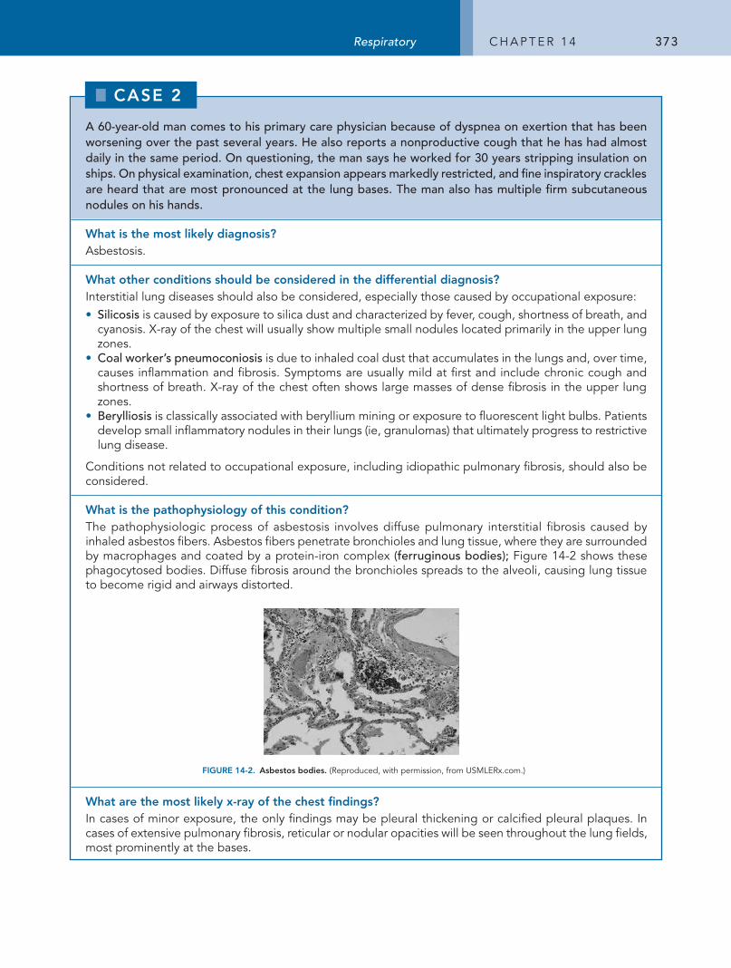
373Respiratory C h a p t e r 1 4
A 60-year-old man comes to his primary care physician because of dyspnea on exertion that has been worsening over the past several years. He also reports a nonproductive cough that he has had almost daily in the same period. On questioning, the man says he worked for 30 years stripping insulation on ships. On physical examination, chest expansion appears markedly restricted, and fine inspiratory crackles are heard that are most pronounced at the lung bases. The man also has multiple firm subcutaneous nodules on his hands.
What is the most likely diagnosis?asbestosis.
What other conditions should be considered in the differential diagnosis?Interstitial lung diseases should also be considered, especially those caused by occupational exposure:• Silicosis is caused by exposure to silica dust and characterized by fever, cough, shortness of breath, and
cyanosis. X-ray of the chest will usually show multiple small nodules located primarily in the upper lung zones.
• Coal worker’s pneumoconiosis is due to inhaled coal dust that accumulates in the lungs and, over time, causes inflammation and fibrosis. Symptoms are usually mild at first and include chronic cough and shortness of breath. X-ray of the chest often shows large masses of dense fibrosis in the upper lung zones.
• Berylliosis is classically associated with beryllium mining or exposure to fluorescent light bulbs. patients develop small inflammatory nodules in their lungs (ie, granulomas) that ultimately progress to restrictive lung disease.
Conditions not related to occupational exposure, including idiopathic pulmonary fibrosis, should also be considered.
What is the pathophysiology of this condition?the pathophysiologic process of asbestosis involves diffuse pulmonary interstitial fibrosis caused by inhaled asbestos fibers. asbestos fibers penetrate bronchioles and lung tissue, where they are surrounded by macrophages and coated by a protein-iron complex (ferruginous bodies); Figure 14-2 shows these phagocytosed bodies. Diffuse fibrosis around the bronchioles spreads to the alveoli, causing lung tissue to become rigid and airways distorted.
FIGURE 14-2. Asbestos bodies. (reproduced, with permission, from USMLerx.com.)
What are the most likely x-ray of the chest findings?In cases of minor exposure, the only findings may be pleural thickening or calcified pleural plaques. In cases of extensive pulmonary fibrosis, reticular or nodular opacities will be seen throughout the lung fields, most prominently at the bases.
CASE 00
CASE 2
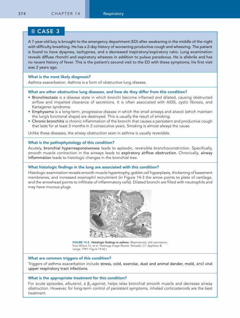
374 C h a p t e r 1 4 Respiratory
A 7-year-old boy is brought to the emergency department (ED) after awakening in the middle of the night with difficulty breathing. He has a 2-day history of worsening productive cough and wheezing. The patient is found to have dyspnea, tachypnea, and a decreased inspiratory/expiratory ratio. Lung examination reveals diffuse rhonchi and expiratory wheezes in addition to pulsus paradoxus. He is afebrile and has no recent history of fever. This is the patient’s second visit to the ED with these symptoms; his first visit was 2 years ago.
What is the most likely diagnosis?asthma exacerbation. asthma is a form of obstructive lung disease.
What are other obstructive lung diseases, and how do they differ from this condition?• Bronchiectasis is a disease state in which bronchi become inflamed and dilated, causing obstructed
airflow and impaired clearance of secretions. It is often associated with aIDS, cystic fibrosis, and Kartagener syndrome.
• Emphysema is a long-term, progressive disease in which the small airways and alveoli (which maintain the lung’s functional shape) are destroyed. this is usually the result of smoking.
• Chronic bronchitis is chronic inflammation of the bronchi that causes a persistent and productive cough that lasts for at least 3 months in 2 consecutive years. Smoking is almost always the cause.
Unlike these diseases, the airway obstruction seen in asthma is usually reversible.
What is the pathophysiology of this condition?acutely, bronchial hyperresponsiveness leads to episodic, reversible bronchoconstriction. Specifically, smooth muscle contraction in the airways leads to expiratory airflow obstruction. Chronically, airway inflammation leads to histologic changes in the bronchial tree.
What histologic findings in the lung are associated with this condition?histologic examination reveals smooth muscle hypertrophy, goblet cell hyperplasia, thickening of basement membranes, and increased eosinophil recruitment (in Figure 14-3 the arrow points to plate of cartilage, and the arrowhead points to infiltrate of inflammatory cells). Dilated bronchi are filled with neutrophils and may have mucous plugs.
FIGURE 14-3. Histologic findings in asthma. (reproduced, with permission, from Wilson FJ, et al. Histology Image Review. Norwalk, Ct: appleton & Lange, 1997: Figure 19-42.)
What are common triggers of this condition?triggers of asthma exacerbation include stress, cold, exercise, dust and animal dander, mold, and viral upper respiratory tract infections.
What is the appropriate treatment for this condition?For acute episodes, albuterol, a β2-agonist, helps relax bronchial smooth muscle and decrease airway obstruction. however, for long-term control of persistent symptoms, inhaled corticosteroids are the best treatment.
CASE 00
CASE 3
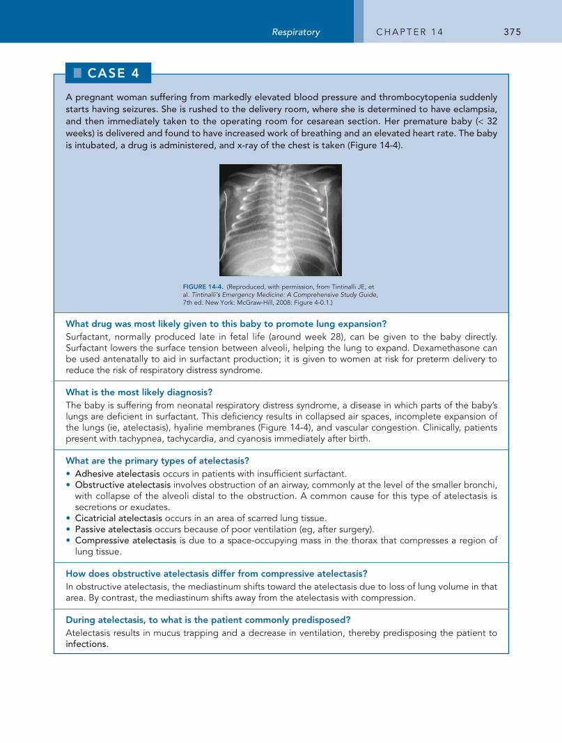
375Respiratory C h a p t e r 1 4
A pregnant woman suffering from markedly elevated blood pressure and thrombocytopenia suddenly starts having seizures. She is rushed to the delivery room, where she is determined to have eclampsia, and then immediately taken to the operating room for cesarean section. Her premature baby (< 32 weeks) is delivered and found to have increased work of breathing and an elevated heart rate. The baby is intubated, a drug is administered, and x-ray of the chest is taken (Figure 14-4).
FIGURE 14-4. (reproduced, with permission, from tintinalli Je, et al. Tintinalli’s Emergency Medicine: A Comprehensive Study Guide, 7th ed. New York: McGraw-hill, 2008: Figure 4-0.1.)
What drug was most likely given to this baby to promote lung expansion?Surfactant, normally produced late in fetal life (around week 28), can be given to the baby directly. Surfactant lowers the surface tension between alveoli, helping the lung to expand. Dexamethasone can be used antenatally to aid in surfactant production; it is given to women at risk for preterm delivery to reduce the risk of respiratory distress syndrome.
What is the most likely diagnosis?the baby is suffering from neonatal respiratory distress syndrome, a disease in which parts of the baby’s lungs are deficient in surfactant. this deficiency results in collapsed air spaces, incomplete expansion of the lungs (ie, atelectasis), hyaline membranes (Figure 14-4), and vascular congestion. Clinically, patients present with tachypnea, tachycardia, and cyanosis immediately after birth.
What are the primary types of atelectasis?• Adhesive atelectasis occurs in patients with insufficient surfactant. • Obstructive atelectasis involves obstruction of an airway, commonly at the level of the smaller bronchi,
with collapse of the alveoli distal to the obstruction. a common cause for this type of atelectasis is secretions or exudates.
• Cicatricial atelectasis occurs in an area of scarred lung tissue. • Passive atelectasis occurs because of poor ventilation (eg, after surgery). • Compressive atelectasis is due to a space-occupying mass in the thorax that compresses a region of
lung tissue.
How does obstructive atelectasis differ from compressive atelectasis?In obstructive atelectasis, the mediastinum shifts toward the atelectasis due to loss of lung volume in that area. By contrast, the mediastinum shifts away from the atelectasis with compression.
During atelectasis, to what is the patient commonly predisposed?atelectasis results in mucus trapping and a decrease in ventilation, thereby predisposing the patient to infections.
CASE 00
CASE 4
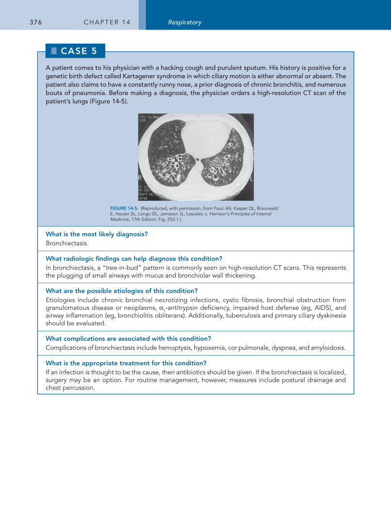
376 C h a p t e r 1 4 Respiratory
A patient comes to his physician with a hacking cough and purulent sputum. His history is positive for a genetic birth defect called Kartagener syndrome in which ciliary motion is either abnormal or absent. The patient also claims to have a constantly runny nose, a prior diagnosis of chronic bronchitis, and numerous bouts of pneumonia. Before making a diagnosis, the physician orders a high-resolution CT scan of the patient’s lungs (Figure 14-5).
FIGURE 14-5. (reproduced, with permission, from Fauci aS, Kasper DL, Braunwald e, hauser SL, Longo DL, Jameson JL, Loscalzo J. Harrison’s Principles of Internal Medicine, 17th edition; Fig. 252-1.)
What is the most likely diagnosis?Bronchiectasis.
What radiologic findings can help diagnose this condition?In bronchiectasis, a “tree-in-bud” pattern is commonly seen on high-resolution Ct scans. this represents the plugging of small airways with mucus and bronchiolar wall thickening.
What are the possible etiologies of this condition?etiologies include chronic bronchial necrotizing infections, cystic fibrosis, bronchial obstruction from granulomatous disease or neoplasms, α1-antitrypsin deficiency, impaired host defense (eg, aIDS), and airway inflammation (eg, bronchiolitis obliterans). additionally, tuberculosis and primary ciliary dyskinesia should be evaluated.
What complications are associated with this condition?Complications of bronchiectasis include hemoptysis, hypoxemia, cor pulmonale, dyspnea, and amyloidosis.
What is the appropriate treatment for this condition?If an infection is thought to be the cause, then antibiotics should be given. If the bronchiectasis is localized, surgery may be an option. For routine management, however, measures include postural drainage and chest percussion.
CASE 00
CASE 5
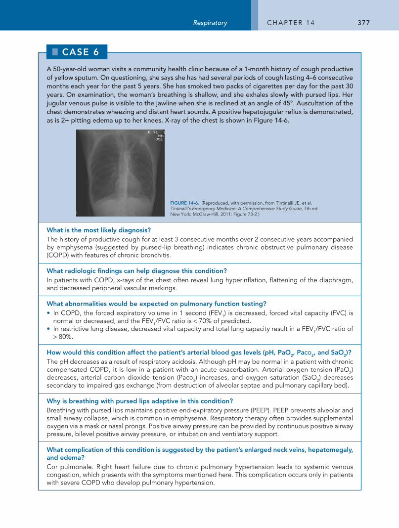
377Respiratory C h a p t e r 1 4
A 50-year-old woman visits a community health clinic because of a 1-month history of cough productive of yellow sputum. On questioning, she says she has had several periods of cough lasting 4–6 consecutive months each year for the past 5 years. She has smoked two packs of cigarettes per day for the past 30 years. On examination, the woman’s breathing is shallow, and she exhales slowly with pursed lips. Her jugular venous pulse is visible to the jawline when she is reclined at an angle of 45°. Auscultation of the chest demonstrates wheezing and distant heart sounds. A positive hepatojugular reflux is demonstrated, as is 2+ pitting edema up to her knees. X-ray of the chest is shown in Figure 14-6.
FIGURE 14-6. (reproduced, with permission, from tintinalli Je, et al. Tintinalli’s Emergency Medicine: A Comprehensive Study Guide, 7th ed. New York: McGraw-hill, 2011: Figure 73-2.)
What is the most likely diagnosis?the history of productive cough for at least 3 consecutive months over 2 consecutive years accompanied by emphysema (suggested by pursed-lip breathing) indicates chronic obstructive pulmonary disease (COpD) with features of chronic bronchitis.
What radiologic findings can help diagnose this condition?In patients with COpD, x-rays of the chest often reveal lung hyperinflation, flattening of the diaphragm, and decreased peripheral vascular markings.
What abnormalities would be expected on pulmonary function testing?• In COpD, the forced expiratory volume in 1 second (FeV1) is decreased, forced vital capacity (FVC) is
normal or decreased, and the FeV1/FVC ratio is < 70% of predicted.• In restrictive lung disease, decreased vital capacity and total lung capacity result in a FeV1/FVC ratio of
> 80%.
How would this condition affect the patient’s arterial blood gas levels (pH, PaO2, Paco2, and SaO2)?the ph decreases as a result of respiratory acidosis. although ph may be normal in a patient with chronic compensated COpD, it is low in a patient with an acute exacerbation. arterial oxygen tension (paO2) decreases, arterial carbon dioxide tension (paco2) increases, and oxygen saturation (SaO2) decreases secondary to impaired gas exchange (from destruction of alveolar septae and pulmonary capillary bed).
Why is breathing with pursed lips adaptive in this condition?Breathing with pursed lips maintains positive end-expiratory pressure (peep). peep prevents alveolar and small airway collapse, which is common in emphysema. respiratory therapy often provides supplemental oxygen via a mask or nasal prongs. positive airway pressure can be provided by continuous positive airway pressure, bilevel positive airway pressure, or intubation and ventilatory support.
What complication of this condition is suggested by the patient’s enlarged neck veins, hepatomegaly, and edema?Cor pulmonale. right heart failure due to chronic pulmonary hypertension leads to systemic venous congestion, which presents with the symptoms mentioned here. this complication occurs only in patients with severe COpD who develop pulmonary hypertension.
CASE 00
CASE 6
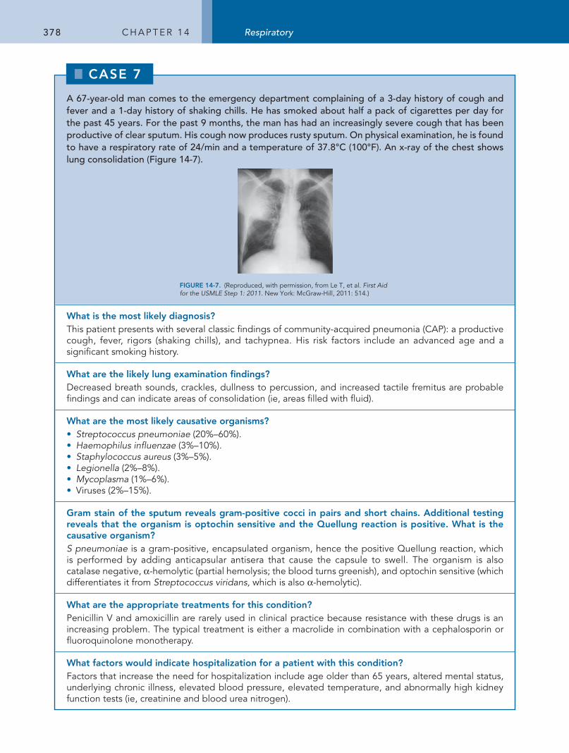
378 C h a p t e r 1 4 Respiratory
A 67-year-old man comes to the emergency department complaining of a 3-day history of cough and fever and a 1-day history of shaking chills. He has smoked about half a pack of cigarettes per day for the past 45 years. For the past 9 months, the man has had an increasingly severe cough that has been productive of clear sputum. His cough now produces rusty sputum. On physical examination, he is found to have a respiratory rate of 24/min and a temperature of 37.8°C (100°F). An x-ray of the chest shows lung consolidation (Figure 14-7).
FIGURE 14-7. (reproduced, with permission, from Le t, et al. First Aid for the USMLE Step 1: 2011. New York: McGraw-hill, 2011: 514.)
What is the most likely diagnosis?this patient presents with several classic findings of community-acquired pneumonia (Cap): a productive cough, fever, rigors (shaking chills), and tachypnea. his risk factors include an advanced age and a significant smoking history.
What are the likely lung examination findings?Decreased breath sounds, crackles, dullness to percussion, and increased tactile fremitus are probable findings and can indicate areas of consolidation (ie, areas filled with fluid).
What are the most likely causative organisms?• Streptococcus pneumoniae (20%–60%).• Haemophilus influenzae (3%–10%).• Staphylococcus aureus (3%–5%).• Legionella (2%–8%).• Mycoplasma (1%–6%).• Viruses (2%–15%).
Gram stain of the sputum reveals gram-positive cocci in pairs and short chains. Additional testing reveals that the organism is optochin sensitive and the Quellung reaction is positive. What is the causative organism?S pneumoniae is a gram-positive, encapsulated organism, hence the positive Quellung reaction, which is performed by adding anticapsular antisera that cause the capsule to swell. the organism is also catalase negative, α-hemolytic (partial hemolysis; the blood turns greenish), and optochin sensitive (which differentiates it from Streptococcus viridans, which is also α-hemolytic).
What are the appropriate treatments for this condition?penicillin V and amoxicillin are rarely used in clinical practice because resistance with these drugs is an increasing problem. the typical treatment is either a macrolide in combination with a cephalosporin or fluoroquinolone monotherapy.
What factors would indicate hospitalization for a patient with this condition?Factors that increase the need for hospitalization include age older than 65 years, altered mental status, underlying chronic illness, elevated blood pressure, elevated temperature, and abnormally high kidney function tests (ie, creatinine and blood urea nitrogen).
CASE 00
CASE 7
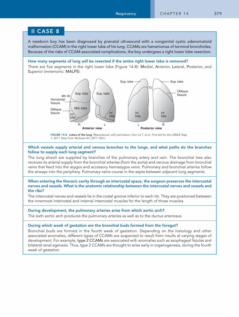
379Respiratory C h a p t e r 1 4
A newborn boy has been diagnosed by prenatal ultrasound with a congenital cystic adenomatoid malformation (CCAM) in the right lower lobe of his lung. CCAMs are hamartomas of terminal bronchioles. Because of the risks of CCAM-associated complications, the boy undergoes a right lower lobe resection.
How many segments of lung will be resected if the entire right lower lobe is removed?there are five segments in the right lower lobe (Figure 14-8): Medial, Anterior, Lateral, Posterior, and Superior (mnemonic: MALPS).
Horizontal fissure
Oblique fissure
Oblique fissure
Posterior viewAnterior viewR L L R
Sup. lobe Sup. lobe
Sup. lobe Sup. lobe
Mid. lobe
Inf.lobe
Inf.lobe
Inf.lobeInf.
lobe
4th rib
T2
FIGURE 14-8. Lobes of the lung. (reproduced, with permission, from Le t, et al. First Aid for the USMLE Step 1: 2011. New York: McGraw-hill, 2011: 503.)
Which vessels supply arterial and venous branches to the lungs, and what paths do the branches follow to supply each lung segment?the lung alveoli are supplied by branches of the pulmonary artery and vein. the bronchial tree also receives its arterial supply from the bronchial arteries (from the aorta) and venous drainage from bronchial veins that feed into the azygos and accessory hemiazygos veins. pulmonary and bronchial arteries follow the airways into the periphery. pulmonary veins course in the septa between adjacent lung segments.
When entering the thoracic cavity through an intercostal space, the surgeon preserves the intercostal nerves and vessels. What is the anatomic relationship between the intercostal nerves and vessels and the ribs?the intercostal nerves and vessels lie in the costal groove inferior to each rib. they are positioned between the innermost intercostal and internal intercostal muscles for the length of those muscles.
During development, the pulmonary arteries arise from which aortic arch?the sixth aortic arch produces the pulmonary arteries as well as to the ductus arteriosus.
During which week of gestation are the bronchial buds formed from the foregut?Bronchial buds are formed in the fourth week of gestation. Depending on the histology and other associated anomalies, different types of CCaMs are suspected to result from insults at varying stages of development. For example, type 2 CCAMs are associated with anomalies such as esophageal fistulas and bilateral renal agenesis. thus, type 2 CCaMs are thought to arise early in organogenesis, during the fourth week of gestation.
CASE 00
CASE 8
380 C h a p t e r 1 4 Respiratory
A 15-year-old girl is brought to the emergency department in acute respiratory distress and is stabilized with treatment. On questioning, she reports an increasingly productive cough over the past few days. Her pulse oximetry shows 93% oxygen saturation on 2 L of oxygen, and she often gasps for air midsentence. Examination shows nostril flaring, subcostal retractions, and clubbing of the fingers. A birth history reveals she had a meconium ileus.
What genetically transmitted condition does this patient likely have?the patient likely has cystic fibrosis (CF), which is caused by loss-of-function mutations in the cystic fibrosis transmembrane conductance regulator (CFtr) protein, a chloride channel found in all exocrine tissues. as a result of these mutations, secretions in the lung, intestine, pancreas, and reproductive tract are extremely viscous.
What test was likely conducted to confirm the diagnosis?a genetic screen during the patient’s infancy was most likely conducted. a sweat chloride test can also confirm the diagnosis, but it may be difficult to collect an adequate amount of sweat in a baby. patients with CF have elevated sweat chloride levels.
What is the probable etiology of the patient’s current symptoms?the lungs in patients with CF are colonized at an early age with various bacteria not normally found in the lung. therefore, patients suffer from repeated pulmonary bacterial infections (Staphylococcus aureus, Haemophilus influenzae, and Pseudomonas aeruginosa are the most common organisms), which increase production of viscous secretions. these increased secretions lead to increased cough and pulmonary obstruction, which can result in acute respiratory distress.
What vitamin supplements do patients with this condition usually require?patients with CF generally require the fat-soluble vitamins a, D, e, and K. the thick secretions block the release of pancreatic enzymes, resulting in pancreatic insufficiency.
What information can be provided if this patient asks for genetic counseling?the frequency of CF in white people is 1: 2000; the carrier rate of CF in white people is 1:25. CF is an autosomal recessive disease, so all children of a patient with CF will at a minimum become carriers. approximately 95% of males with CF are infertile because of defects in the transport of sperm. Infertility affects as many as 20% of women as a result of abnormally thick cervical mucus and amenorrhea from malnutrition.
What is the prognosis for patients with this condition?• prognosis for patients with CF is generally good.• Most patients are able to survive into their 30s and lead relatively normal lives.
CASE 00
CASE 9
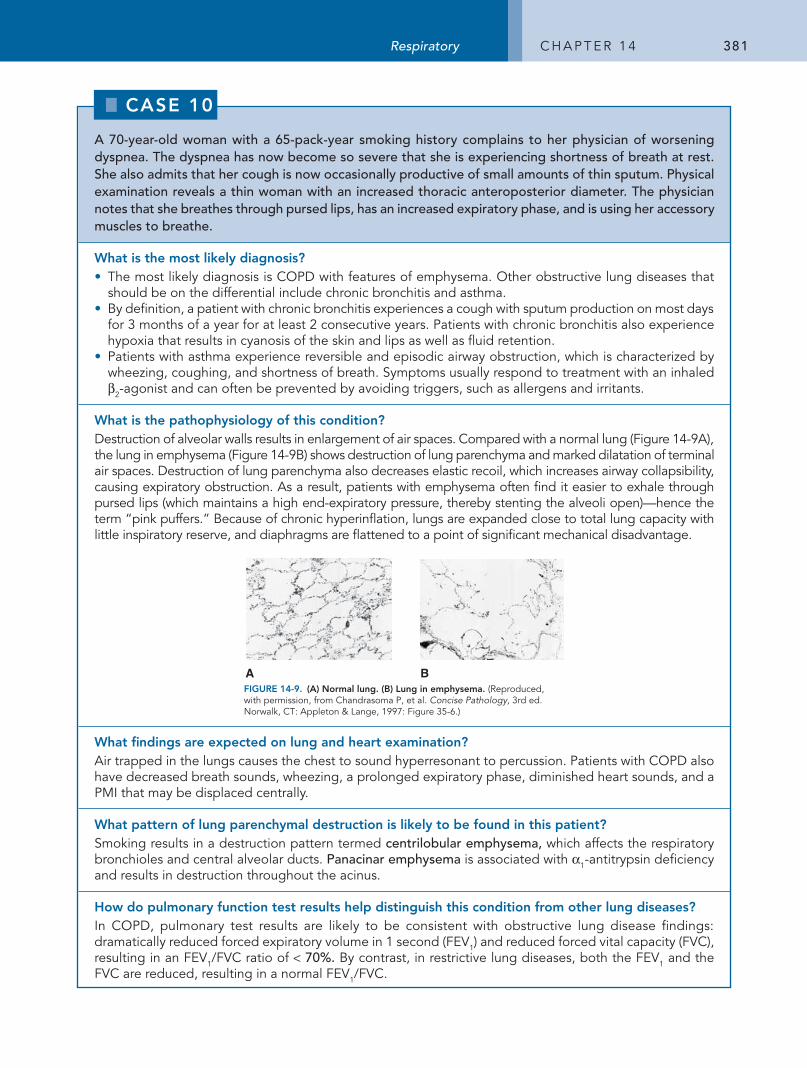
381Respiratory C h a p t e r 1 4
A 70-year-old woman with a 65-pack-year smoking history complains to her physician of worsening dyspnea. The dyspnea has now become so severe that she is experiencing shortness of breath at rest. She also admits that her cough is now occasionally productive of small amounts of thin sputum. Physical examination reveals a thin woman with an increased thoracic anteroposterior diameter. The physician notes that she breathes through pursed lips, has an increased expiratory phase, and is using her accessory muscles to breathe.
What is the most likely diagnosis?• the most likely diagnosis is COpD with features of emphysema. Other obstructive lung diseases that
should be on the differential include chronic bronchitis and asthma. • By definition, a patient with chronic bronchitis experiences a cough with sputum production on most days
for 3 months of a year for at least 2 consecutive years. patients with chronic bronchitis also experience hypoxia that results in cyanosis of the skin and lips as well as fluid retention.
• patients with asthma experience reversible and episodic airway obstruction, which is characterized by wheezing, coughing, and shortness of breath. Symptoms usually respond to treatment with an inhaled β2-agonist and can often be prevented by avoiding triggers, such as allergens and irritants.
What is the pathophysiology of this condition?Destruction of alveolar walls results in enlargement of air spaces. Compared with a normal lung (Figure 14-9a), the lung in emphysema (Figure 14-9B) shows destruction of lung parenchyma and marked dilatation of terminal air spaces. Destruction of lung parenchyma also decreases elastic recoil, which increases airway collapsibility, causing expiratory obstruction. as a result, patients with emphysema often find it easier to exhale through pursed lips (which maintains a high end-expiratory pressure, thereby stenting the alveoli open)—hence the term “pink puffers.” Because of chronic hyperinflation, lungs are expanded close to total lung capacity with little inspiratory reserve, and diaphragms are flattened to a point of significant mechanical disadvantage.
A BFIGURE 14-9. (A) Normal lung. (B) Lung in emphysema. (reproduced, with permission, from Chandrasoma p, et al. Concise Pathology, 3rd ed. Norwalk, Ct: appleton & Lange, 1997: Figure 35-6.)
What findings are expected on lung and heart examination?air trapped in the lungs causes the chest to sound hyperresonant to percussion. patients with COpD also have decreased breath sounds, wheezing, a prolonged expiratory phase, diminished heart sounds, and a pMI that may be displaced centrally.
What pattern of lung parenchymal destruction is likely to be found in this patient?Smoking results in a destruction pattern termed centrilobular emphysema, which affects the respiratory bronchioles and central alveolar ducts. Panacinar emphysema is associated with α1-antitrypsin deficiency and results in destruction throughout the acinus.
How do pulmonary function test results help distinguish this condition from other lung diseases?In COpD, pulmonary test results are likely to be consistent with obstructive lung disease findings: dramatically reduced forced expiratory volume in 1 second (FeV1) and reduced forced vital capacity (FVC), resulting in an FeV1/FVC ratio of < 70%. By contrast, in restrictive lung diseases, both the FeV1 and the FVC are reduced, resulting in a normal FeV1/FVC.
CASE 00
CASE 10

382 C h a p t e r 1 4 Respiratory
A 4-year-old boy is brought to the emergency department by his mother because he is lethargic, drooling, and having difficulty breathing. Physical examination reveals an elevated temperature and a high-pitched upper airway wheeze. Further questioning of the patient’s mother reveals that the child has not received any immunizations. A lateral x-ray of the neck shows soft tissue swelling.
What is the most likely diagnosis?the stridor found on lung examination and the drooling—findings consistent with both tracheal and esophageal obstruction—suggest acute epiglottitis. the obstruction is due to swelling of the epiglottis caused by infection and is a medical emergency. the x-ray shows the classic “thumbprint” sign caused by the thickening and swelling of the epiglottis.
What is the likely source of this infection?Given the child’s unimmunized status, the most likely cause is type b Haemophilus influenzae infection. H influenzae is considered part of the normal flora of the nasopharynx. the organism may thus be spread by direct contact with respiratory secretions and by airborne droplet contamination. epiglottitis may also represent a primary infection of the epiglottis rather than invasion from the nasopharynx.
What additional microorganisms can cause this presentation?epiglottitis can also be caused by Pasteurella multocida, which is often transmitted from dog or cat bites, and herpes simplex virus type 1.
What is the main virulence factor of the causative organism in this case?the polysaccharide capsule is the major virulence factor of H influenzae, which has both encapsulated and nonencapsulated strains. the nonencapsulated forms are limited to local infections such as otitis media in children and mild respiratory infection in adults (table 14-1). the encapsulated strains are significantly more virulent and can cause disseminated diseases such as meningitis, epiglottitis, and septic arthritis. there are six capsular types of H influenzae, designated a through f. the b-type capsule accounts for approximately 95% of serious H influenzae infections in children.
How has the vaccine for this infection been redesigned to improve its efficacy?the Hib vaccine consists of a purified b-type capsule conjugated to diphtheria toxin. the diphtheria toxin activates t lymphocytes, which are required for adequate antibody production against the capsular antigen. the original vaccine consisted only of b capsule and was not effective in eliciting an antibody response.
table 14-1 types of Infection Caused by Haemophilus
H influenzae H aegyptius H ducReyiTyPe b NoNTyPeAble
type of infection
MeningitisepiglottitisBacteremiaCellulitisSeptic arthritis
Otitis mediaSinusitistracheobronchitispneumonia
Conjunctivitispurpuric fever (Brazilian)
Chancroid (painful ulcers of genitals, lymphadenitis)
treatment CeftazidimeCefotaximeCeftriaxoneGentamicin
CephalosporinFluoroquinoloneazithromycin
rifampin azithromycinCephalosporinCiprofloxacin
CaSe 00
CaSe 11
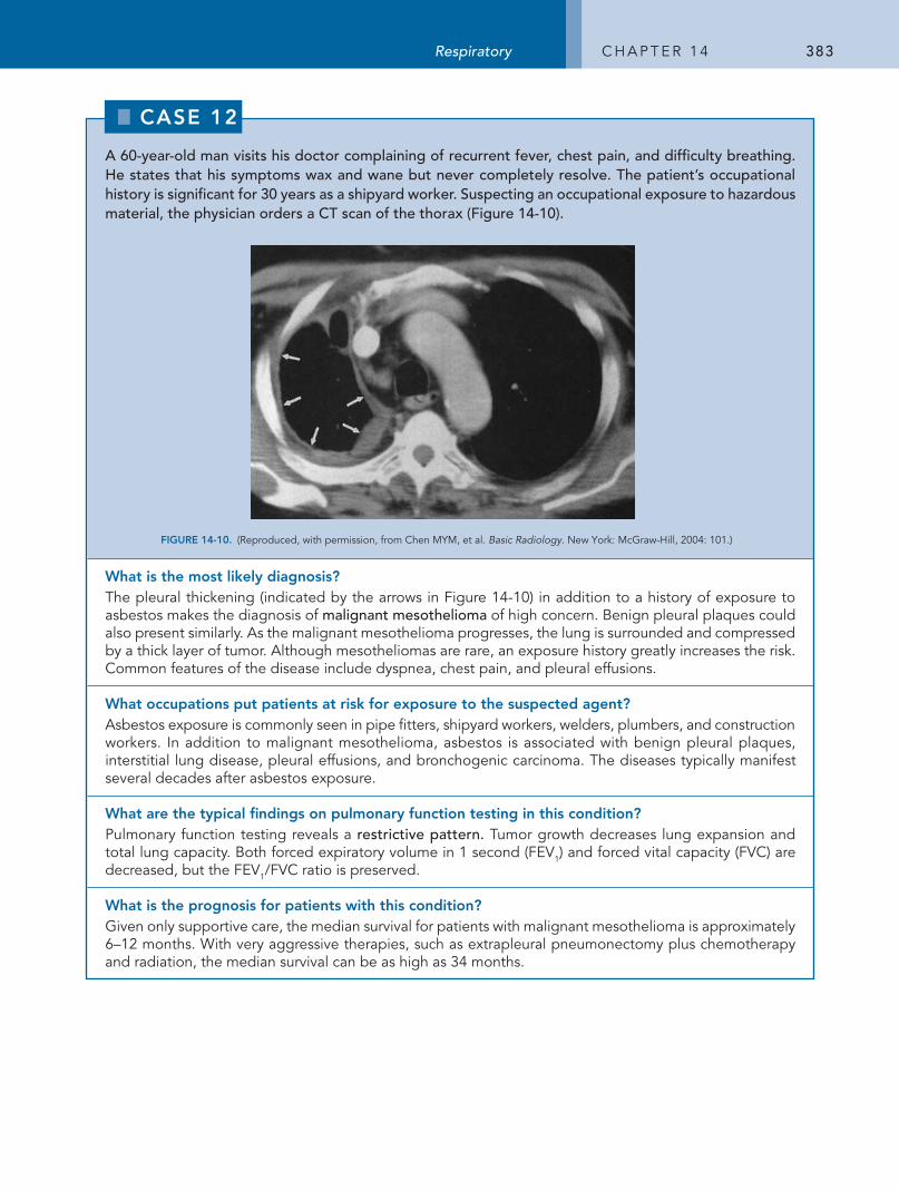
383Respiratory C h a p t e r 1 4
A 60-year-old man visits his doctor complaining of recurrent fever, chest pain, and difficulty breathing. He states that his symptoms wax and wane but never completely resolve. The patient’s occupational history is significant for 30 years as a shipyard worker. Suspecting an occupational exposure to hazardous material, the physician orders a CT scan of the thorax (Figure 14-10).
BFIGURE 14-10. (reproduced, with permission, from Chen MYM, et al. Basic Radiology. New York: McGraw-hill, 2004: 101.)
What is the most likely diagnosis?the pleural thickening (indicated by the arrows in Figure 14-10) in addition to a history of exposure to asbestos makes the diagnosis of malignant mesothelioma of high concern. Benign pleural plaques could also present similarly. as the malignant mesothelioma progresses, the lung is surrounded and compressed by a thick layer of tumor. although mesotheliomas are rare, an exposure history greatly increases the risk. Common features of the disease include dyspnea, chest pain, and pleural effusions.
What occupations put patients at risk for exposure to the suspected agent?asbestos exposure is commonly seen in pipe fitters, shipyard workers, welders, plumbers, and construction workers. In addition to malignant mesothelioma, asbestos is associated with benign pleural plaques, interstitial lung disease, pleural effusions, and bronchogenic carcinoma. the diseases typically manifest several decades after asbestos exposure.
What are the typical findings on pulmonary function testing in this condition?pulmonary function testing reveals a restrictive pattern. tumor growth decreases lung expansion and total lung capacity. Both forced expiratory volume in 1 second (FeV1) and forced vital capacity (FVC) are decreased, but the FeV1/FVC ratio is preserved.
What is the prognosis for patients with this condition?Given only supportive care, the median survival for patients with malignant mesothelioma is approximately 6–12 months. With very aggressive therapies, such as extrapleural pneumonectomy plus chemotherapy and radiation, the median survival can be as high as 34 months.
CASE 00
CASE 12
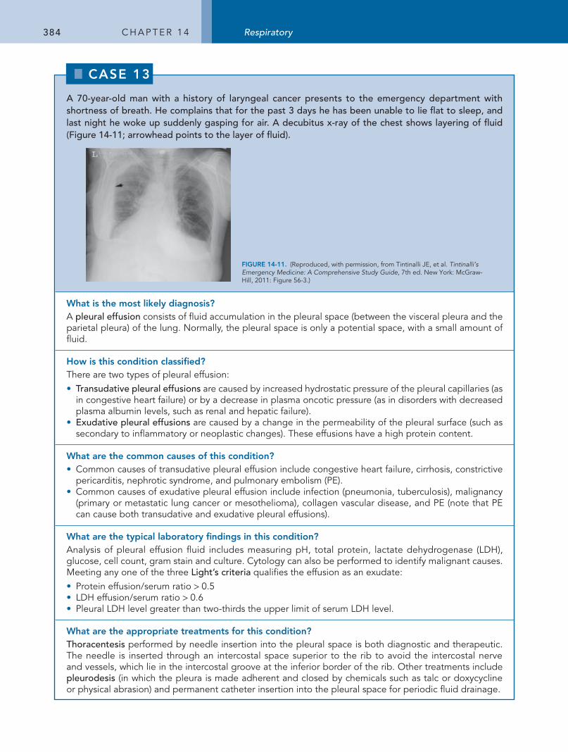
384 C h a p t e r 1 4 Respiratory
A 70-year-old man with a history of laryngeal cancer presents to the emergency department with shortness of breath. He complains that for the past 3 days he has been unable to lie flat to sleep, and last night he woke up suddenly gasping for air. A decubitus x-ray of the chest shows layering of fluid (Figure 14-11; arrowhead points to the layer of fluid).
FIGURE 14-11. (reproduced, with permission, from tintinalli Je, et al. Tintinalli’s Emergency Medicine: A Comprehensive Study Guide, 7th ed. New York: McGraw-hill, 2011: Figure 56-3.)
What is the most likely diagnosis?a pleural effusion consists of fluid accumulation in the pleural space (between the visceral pleura and the parietal pleura) of the lung. Normally, the pleural space is only a potential space, with a small amount of fluid.
How is this condition classified?there are two types of pleural effusion: • Transudative pleural effusions are caused by increased hydrostatic pressure of the pleural capillaries (as
in congestive heart failure) or by a decrease in plasma oncotic pressure (as in disorders with decreased plasma albumin levels, such as renal and hepatic failure).
• Exudative pleural effusions are caused by a change in the permeability of the pleural surface (such as secondary to inflammatory or neoplastic changes). these effusions have a high protein content.
What are the common causes of this condition?• Common causes of transudative pleural effusion include congestive heart failure, cirrhosis, constrictive
pericarditis, nephrotic syndrome, and pulmonary embolism (pe).• Common causes of exudative pleural effusion include infection (pneumonia, tuberculosis), malignancy
(primary or metastatic lung cancer or mesothelioma), collagen vascular disease, and pe (note that pe can cause both transudative and exudative pleural effusions).
What are the typical laboratory findings in this condition?analysis of pleural effusion fluid includes measuring ph, total protein, lactate dehydrogenase (LDh), glucose, cell count, gram stain and culture. Cytology can also be performed to identify malignant causes. Meeting any one of the three Light’s criteria qualifies the effusion as an exudate:• protein effusion/serum ratio > 0.5• LDh effusion/serum ratio > 0.6• pleural LDh level greater than two-thirds the upper limit of serum LDh level.
What are the appropriate treatments for this condition?Thoracentesis performed by needle insertion into the pleural space is both diagnostic and therapeutic. the needle is inserted through an intercostal space superior to the rib to avoid the intercostal nerve and vessels, which lie in the intercostal groove at the inferior border of the rib. Other treatments include pleurodesis (in which the pleura is made adherent and closed by chemicals such as talc or doxycycline or physical abrasion) and permanent catheter insertion into the pleural space for periodic fluid drainage.
CASE 00
CASE 13
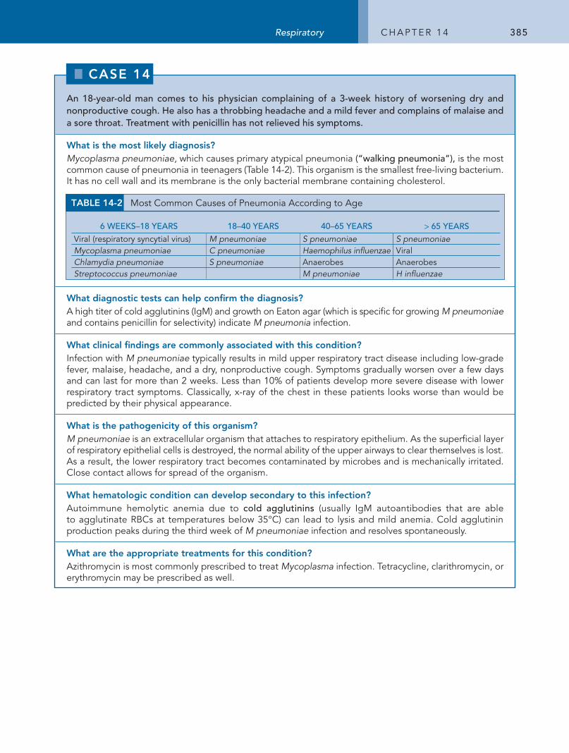
385Respiratory C h a p t e r 1 4
An 18-year-old man comes to his physician complaining of a 3-week history of worsening dry and nonproductive cough. He also has a throbbing headache and a mild fever and complains of malaise and a sore throat. Treatment with penicillin has not relieved his symptoms.
What is the most likely diagnosis?Mycoplasma pneumoniae, which causes primary atypical pneumonia (“walking pneumonia”), is the most common cause of pneumonia in teenagers (table 14-2). this organism is the smallest free-living bacterium. It has no cell wall and its membrane is the only bacterial membrane containing cholesterol.
What diagnostic tests can help confirm the diagnosis?a high titer of cold agglutinins (IgM) and growth on eaton agar (which is specific for growing M pneumoniae and contains penicillin for selectivity) indicate M pneumonia infection.
What clinical findings are commonly associated with this condition?Infection with M pneumoniae typically results in mild upper respiratory tract disease including low-grade fever, malaise, headache, and a dry, nonproductive cough. Symptoms gradually worsen over a few days and can last for more than 2 weeks. Less than 10% of patients develop more severe disease with lower respiratory tract symptoms. Classically, x-ray of the chest in these patients looks worse than would be predicted by their physical appearance.
What is the pathogenicity of this organism?M pneumoniae is an extracellular organism that attaches to respiratory epithelium. as the superficial layer of respiratory epithelial cells is destroyed, the normal ability of the upper airways to clear themselves is lost. as a result, the lower respiratory tract becomes contaminated by microbes and is mechanically irritated. Close contact allows for spread of the organism.
What hematologic condition can develop secondary to this infection?autoimmune hemolytic anemia due to cold agglutinins (usually IgM autoantibodies that are able to agglutinate rBCs at temperatures below 35°C) can lead to lysis and mild anemia. Cold agglutinin production peaks during the third week of M pneumoniae infection and resolves spontaneously.
What are the appropriate treatments for this condition?azithromycin is most commonly prescribed to treat Mycoplasma infection. tetracycline, clarithromycin, or erythromycin may be prescribed as well.
table 14-2 Most Common Causes of pneumonia according to age
6 Weeks–18 YeArs 18–40 YeArs 40–65 YeArs > 65 YeArs
Viral (respiratory syncytial virus) M pneumoniae S pneumoniae S pneumoniaeMycoplasma pneumoniae C pneumoniae Haemophilus influenzae ViralChlamydia pneumoniae S pneumoniae anaerobes anaerobesStreptococcus pneumoniae M pneumoniae H influenzae
CaSe 00
CaSe 14
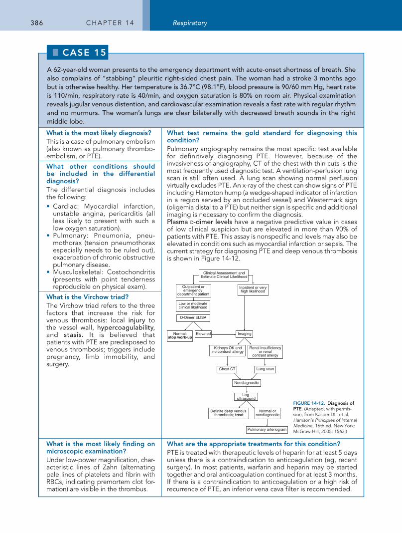
386 C h a p t e r 1 4 Respiratory
A 62-year-old woman presents to the emergency department with acute-onset shortness of breath. She also complains of “stabbing” pleuritic right-sided chest pain. The woman had a stroke 3 months ago but is otherwise healthy. Her temperature is 36.7°C (98.1°F), blood pressure is 90/60 mm Hg, heart rate is 110/min, respiratory rate is 40/min, and oxygen saturation is 80% on room air. Physical examination reveals jugular venous distention, and cardiovascular examination reveals a fast rate with regular rhythm and no murmurs. The woman’s lungs are clear bilaterally with decreased breath sounds in the right middle lobe.
What is the most likely diagnosis?this is a case of pulmonary embolism (also known as pulmonary thrombo-embolism, or pte).
What test remains the gold standard for diagnosing this condition?pulmonary angiography remains the most specific test available for definitively diagnosing pte. however, because of the invasiveness of angiography, Ct of the chest with thin cuts is the most frequently used diagnostic test. a ventilation-perfusion lung scan is still often used. a lung scan showing normal perfusion virtually excludes pte. an x-ray of the chest can show signs of pte including hampton hump (a wedge-shaped indicator of infarction in a region served by an occluded vessel) and Westermark sign (oligemia distal to a pte) but neither sign is specific and additional imaging is necessary to confirm the diagnosis. Plasma d-dimer levels have a negative predictive value in cases of low clinical suspicion but are elevated in more than 90% of patients with pte. this assay is nonspecific and levels may also be elevated in conditions such as myocardial infarction or sepsis. the current strategy for diagnosing pte and deep venous thrombosis is shown in Figure 14-12.
Renal insufficiencyor renal
contrast allergy
Clinical Assessment andEstimate Clinical Likelihood
Low or moderateclinical likelihood
Outpatient oremergency
department patient
Inpatient or veryhigh likelihood
Normal;stop work-up
Definite deep venous thrombosis; treat
Normal ornondiagnostic
Elevated Imaging
Chest CT Lung scan
Nondiagnostic
D-Dimer ELISA
Leg ultrasound
Pulmonary arteriogram
Kidneys OK andno contrast allergy
FIGURE 14-12. Diagnosis of PtE. (adapted, with permis-sion, from Kasper DL, et al. Harrison’s Principles of Internal Medicine, 16th ed. New York: McGraw-hill, 2005: 1563.)
What other conditions should be included in the differential diagnosis?the differential diagnosis includes the following:• Cardiac: Myocardial infarction,
unstable angina, pericarditis (all less likely to present with such a low oxygen saturation).
• pulmonary: pneumonia, pneu-mothorax (tension pneumothorax especially needs to be ruled out), exacerbation of chronic obstructive pulmonary disease.
• Musculoskeletal: Costochondritis (presents with point tenderness reproducible on physical exam).
What is the Virchow triad?the Virchow triad refers to the three factors that increase the risk for venous thrombosis: local injury to the vessel wall, hypercoagulability, and stasis. It is believed that patients with pte are predisposed to venous thrombosis; triggers include pregnancy, limb immobility, and surgery.
What is the most likely finding on microscopic examination?Under low-power magnification, char-acteristic lines of Zahn (alternating pale lines of platelets and fibrin with rBCs, indicating premortem clot for-mation) are visible in the thrombus.
What are the appropriate treatments for this condition?pte is treated with therapeutic levels of heparin for at least 5 days unless there is a contraindication to anticoagulation (eg, recent surgery). In most patients, warfarin and heparin may be started together and oral anticoagulation continued for at least 3 months. If there is a contraindication to anticoagulation or a high risk of recurrence of pte, an inferior vena cava filter is recommended.
CASE 00
CASE 15
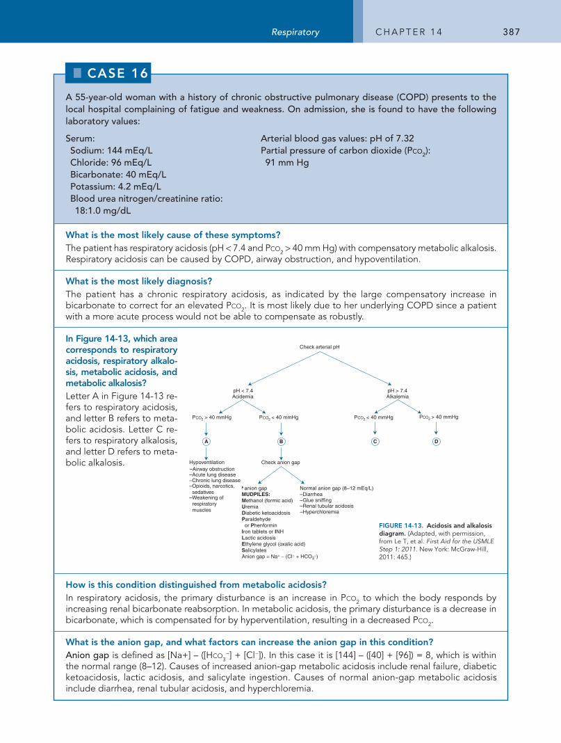
387Respiratory C h a p t e r 1 4
A 55-year-old woman with a history of chronic obstructive pulmonary disease (COPD) presents to the local hospital complaining of fatigue and weakness. On admission, she is found to have the following laboratory values:
Serum: Sodium: 144 mEq/LChloride: 96 mEq/LBicarbonate: 40 mEq/LPotassium: 4.2 mEq/L Blood urea nitrogen/creatinine ratio: 18:1.0 mg/dL
Arterial blood gas values: pH of 7.32Partial pressure of carbon dioxide (Pco2): 91 mm Hg
What is the most likely cause of these symptoms?the patient has respiratory acidosis (ph < 7.4 and pco2 > 40 mm hg) with compensatory metabolic alkalosis. respiratory acidosis can be caused by COpD, airway obstruction, and hypoventilation.
What is the most likely diagnosis?the patient has a chronic respiratory acidosis, as indicated by the large compensatory increase in bicarbonate to correct for an elevated pco2. It is most likely due to her underlying COpD since a patient with a more acute process would not be able to compensate as robustly.
In Figure 14-13, which area corresponds to respiratory acidosis, respiratory alkalo-sis, metabolic acidosis, and metabolic alkalosis?Letter a in Figure 14-13 re-fers to respiratory acidosis, and letter B refers to meta-bolic acidosis. Letter C re-fers to respiratory alkalosis, and letter D refers to meta-bolic alkalosis.
pH < 7.4 Acidemia
pH > 7.4 Alkalemia
> 40 mmHg < 40 mmHg
Hypo v entilation
–Acute lung disease –Chronic lung disease –Opioids, narcotics, sedatives –Weakening of respiratory muscles
P CO 2 P CO 2 P CO2 < 40 mmHg P CO 2 > 40 mmHg
Chec k anion gap
anion gapMUDPILES:Methanol (formic acid)UremiaDiabetic ketoacidosisParaldehyde or PhenforminIron tablets or INHLactic acidosisEthylene glycol (oxalic acid)SalicylatesAnion gap = Na+ − (CI− + HCO3
−)
–Hyperchloremia
Normal anion gap (8–12 mEq/L)–Diarrhea–Glue sniffing–Renal tubular acidosis
Check arterial pH
–Airway obstruction
A B C D
FIGURE 14-13. Acidosis and alkalosis diagram. (adapted, with permission, from Le t, et al. First Aid for the USMLE Step 1: 2011. New York: McGraw-hill, 2011: 465.)
How is this condition distinguished from metabolic acidosis?In respiratory acidosis, the primary disturbance is an increase in pco2 to which the body responds by increasing renal bicarbonate reabsorption. In metabolic acidosis, the primary disturbance is a decrease in bicarbonate, which is compensated for by hyperventilation, resulting in a decreased pco2.
What is the anion gap, and what factors can increase the anion gap in this condition?Anion gap is defined as [Na+] – ([hco3
−] + [Cl−]). In this case it is [144] – ([40] + [96]) = 8, which is within the normal range (8–12). Causes of increased anion-gap metabolic acidosis include renal failure, diabetic ketoacidosis, lactic acidosis, and salicylate ingestion. Causes of normal anion-gap metabolic acidosis include diarrhea, renal tubular acidosis, and hyperchloremia.
CASE 00
CASE 16
388 C h a p t e r 1 4 Respiratory
A 35-year-old African-American man presents to his primary care physician with progressive dyspnea on exertion. He has no history of congestive heart failure or asthma and has had no known contact with any individuals known to have tuberculosis. His laboratory results reveal normal creatinine kinase (CK), CK-MB fraction, and troponin levels. An x-ray of the chest shows bilateral hilar lymphadenopathy and evidence of interstitial lung disease (ILD). A bronchoscopic lung biopsy reveals the presence of several small, noncaseating granulomas.
What is ILD and what are the common causes?the term ILD is generically used to describe a collection of diseases that involve diffuse scarring and/or inflammation of lung tissue. Common causes of ILD are as follows:• prolonged exposure to occupationally inhaled inorganic agents such as silicone, coal, asbestos, talc,
mica, aluminum, and beryllium.• Idiopathic pulmonary fibrosis.• Connective tissue disease (eg, Wegener granulomatosis, systemic lupus erythematosus, scleroderma,
Sjögren disease).• Sarcoidosis.• hypersensitivity pneumonitis, such as “farmer’s lung” or “bird-breeder’s lung,” in which an immune
reaction to an organic dust induces a type III or type IV hypersensitivity reaction.• radiation-induced disease.• antitumor drugs (eg, bleomycin).
What is the most likely cause of ILD in this patient?Sarcoidosis is the most likely cause. this diagnosis is supported by the patient’s race, the presence of noncaseating granulomas (discrete collections of tissue macrophages termed histiocytes often organized into multinucleated giant cells without central necrosis), and the bilateral hilar lymphadenopathy on x-ray of the chest.
What laboratory abnormalities may be found in this patient?Vitamin D is secreted by the macrophages of the granulomas and is therefore elevated in serum. angiotensin-converting enzyme is also secreted by the macrophages of the granulomas and is also elevated.
What pulmonary function testing findings are expected?In ILD, lung compliance is decreased, reflecting increased stiffness from alveolar wall inflammation and fibrosis. tidal volume and total lung capacity are typically decreased. Diffusion capacity is also decreased as a result of inflammatory destruction of the air-capillary interface. Unlike most ILDs, sarcoidosis has features of both obstruction and restriction.
What are some extrapulmonary manifestations of this patient’s ILD?Common extrapulmonary manifestations of sarcoidosis are in the eye (anterior uveitis) and skin (papules and erythema nodosum), but granulomas can also occur in the heart, brain, lung, and peripheral lymph nodes.
What is the appropriate treatment for this condition?Corticosteroids.
CASE 00
CASE 17
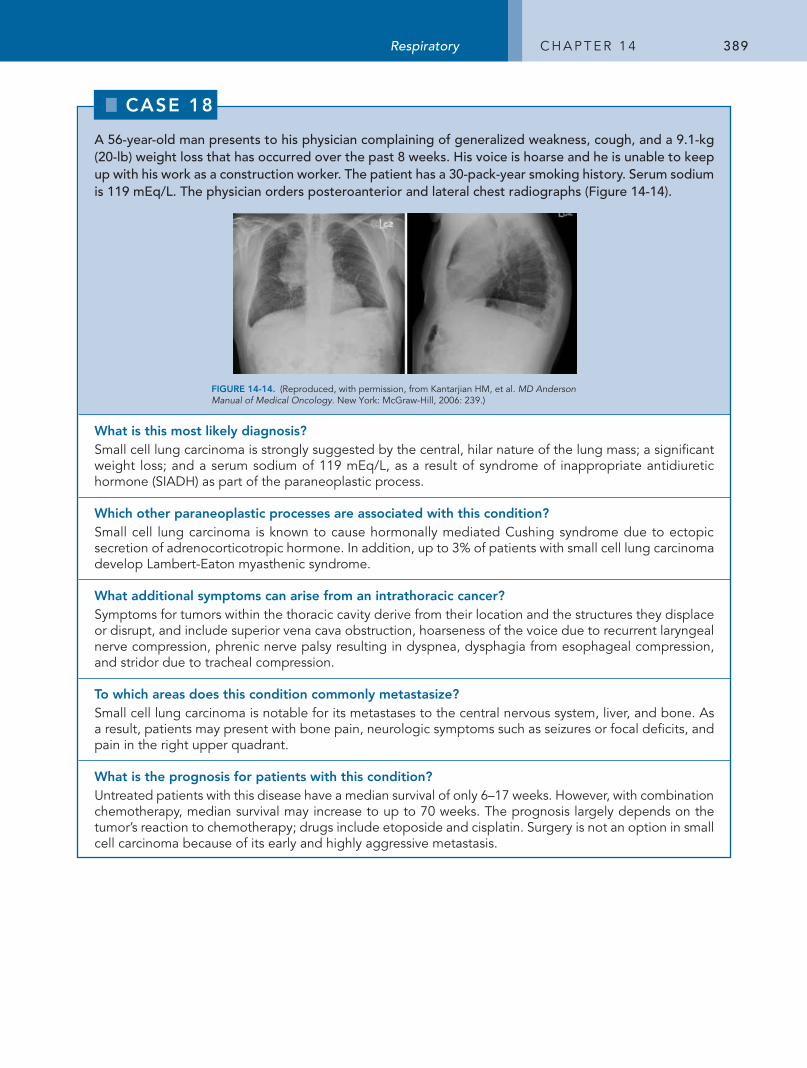
389Respiratory C h a p t e r 1 4
A 56-year-old man presents to his physician complaining of generalized weakness, cough, and a 9.1-kg (20-lb) weight loss that has occurred over the past 8 weeks. His voice is hoarse and he is unable to keep up with his work as a construction worker. The patient has a 30-pack-year smoking history. Serum sodium is 119 mEq/L. The physician orders posteroanterior and lateral chest radiographs (Figure 14-14).
FIGURE 14-14. (reproduced, with permission, from Kantarjian hM, et al. MD Anderson Manual of Medical Oncology. New York: McGraw-hill, 2006: 239.)
What is this most likely diagnosis?Small cell lung carcinoma is strongly suggested by the central, hilar nature of the lung mass; a significant weight loss; and a serum sodium of 119 meq/L, as a result of syndrome of inappropriate antidiuretic hormone (SIaDh) as part of the paraneoplastic process.
Which other paraneoplastic processes are associated with this condition?Small cell lung carcinoma is known to cause hormonally mediated Cushing syndrome due to ectopic secretion of adrenocorticotropic hormone. In addition, up to 3% of patients with small cell lung carcinoma develop Lambert-eaton myasthenic syndrome.
What additional symptoms can arise from an intrathoracic cancer?Symptoms for tumors within the thoracic cavity derive from their location and the structures they displace or disrupt, and include superior vena cava obstruction, hoarseness of the voice due to recurrent laryngeal nerve compression, phrenic nerve palsy resulting in dyspnea, dysphagia from esophageal compression, and stridor due to tracheal compression.
to which areas does this condition commonly metastasize?Small cell lung carcinoma is notable for its metastases to the central nervous system, liver, and bone. as a result, patients may present with bone pain, neurologic symptoms such as seizures or focal deficits, and pain in the right upper quadrant.
What is the prognosis for patients with this condition?Untreated patients with this disease have a median survival of only 6–17 weeks. however, with combination chemotherapy, median survival may increase to up to 70 weeks. the prognosis largely depends on the tumor’s reaction to chemotherapy; drugs include etoposide and cisplatin. Surgery is not an option in small cell carcinoma because of its early and highly aggressive metastasis.
CASE 00
CASE 18
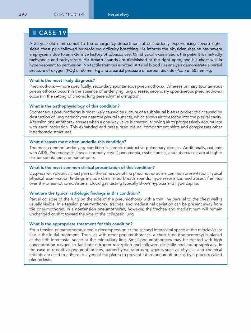
390 C h a p t e r 1 4 Respiratory
A 55-year-old man comes to the emergency department after suddenly experiencing severe right-sided chest pain followed by profound difficulty breathing. He informs the physician that he has severe emphysema due to an extensive history of tobacco use. On physical examination, the patient is markedly tachypneic and tachycardic. His breath sounds are diminished at the right apex, and his chest wall is hyperresonant to percussion. No tactile fremitus is noted. Arterial blood gas analysis demonstrate a partial pressure of oxygen (PO2) of 60 mm Hg and a partial pressure of carbon dioxide (Pco2) of 50 mm Hg.
What is the most likely diagnosis?pneumothorax—more specifically, secondary spontaneous pneumothorax. Whereas primary spontaneous pneumothorax occurs in the absence of underlying lung disease, secondary spontaneous pneumothorax occurs in the setting of chronic lung parenchymal disruption.
What is the pathophysiology of this condition?Spontaneous pneumothorax is most likely caused by rupture of a subpleural bleb (a pocket of air caused by destruction of lung parenchyma near the pleural surface), which allows air to escape into the pleural cavity. a tension pneumothorax ensues when a one-way valve is created, allowing air to progressively accumulate with each inspiration. this expanded and pressurized pleural compartment shifts and compresses other intrathoracic structures.
What diseases most often underlie this condition?the most common underlying condition is chronic obstructive pulmonary disease. additionally, patients with aIDS, Pneumocystis jiroveci (formerly carinii) pneumonia, cystic fibrosis, and tuberculosis are at higher risk for spontaneous pneumothorax.
What is the most common clinical presentation of this condition?Dyspnea with pleuritic chest pain on the same side of the pneumothorax is a common presentation. typical physical examination findings include diminished breath sounds, hyperresonance, and absent fremitus over the pneumothorax. arterial blood gas testing typically shows hypoxia and hypercapnia.
What are the typical radiologic findings in this condition?partial collapse of the lung on the side of the pneumothorax with a thin line parallel to the chest wall is usually visible. In a tension pneumothorax, tracheal and mediastinal deviation can be present away from the pneumothorax. In a nontension pneumothorax, however, the trachea and mediastinum will remain unchanged or shift toward the side of the collapsed lung.
What is the appropriate treatment for this condition?For a tension pneumothorax, needle decompression at the second intercostal space at the midclavicular line is the initial treatment. then, as with other pneumothoraces, a chest tube (thoracotomy) is placed at the fifth intercostal space at the midaxillary line. Small pneumothoraces may be treated with high concentration oxygen to facilitate nitrogen resorption and followed clinically and radiographically. In the case of repetitive pneumothoraces, parenchymal sclerosing agents such as physical and chemical irritants are used to adhere to layers of the pleura to prevent future pneumothoraces by a process called pleurodesis.
CASE 00
CASE 19
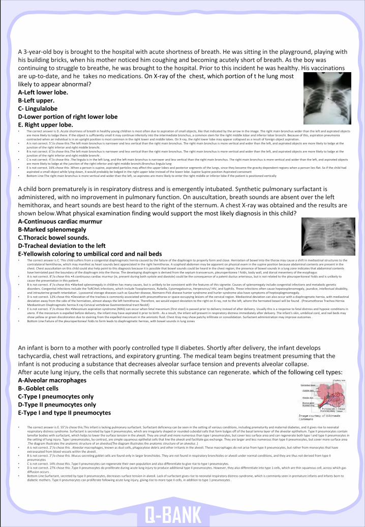
A3-year-oldboyisbroughttothehospitalwithacuteshortnessofbreath.Hewassittingintheplayground,playingwithhisbuildingbricks,whenhismothernoticedhimcoughingandbecomingacutelyshortofbreath.Astheboywascontinuingtostruggletobreathe,hewasbroughttothehospital.Priortothisincidenthewashealthy.Hisvaccinationsareup-to-date,andhetakesnomedications.OnX-rayofthe chest,whichportionofthelung mostlikelytoappearabnormal? A-Leftlowerlobe. B-Leftupper. C- Lingulalobe D-LowerportionofrightlowerlobeE.Rightupperlobe.• ThecorrectanswerisD..Acute shortnessofbreathinhealthyyoungchildrenismostoftenduetoaspirationofsmallobjects,likethatindicatedbythearrowintheimage.Therightmainbronchuswiderthantheleftandaspiratedobjects
aremorelikelytolodgethere.Iftheobjectissufficientlysmallitmaycontinueinferiorlyintotheintermediatebronchus,a commonstemfortherightmiddlelobarandinferiorlobarbronchi.Becauseofthis,aspirationpneumoniacontractedwhenanindividualisinanuprightpositionismostcommonintherightlowerandmiddlelobes.OnX-ray,therightlowerlobemayappearcollapsedasaresultofforeignobjectaspiration.
• Aisnotcorrect.5°/ochosethis.The leftmainbronchusisnarrowerandlessverticalthantherightmainbronchus.Therightmainbronchusismoreverticalandwiderthantheleft,andaspiratedobjectsaremorelikelytolodgeatthejunctionoftherightinferiorandrightmiddlebronchi.
• Bisnotcorrect.6°/ochosethis.The leftmainbronchusisnarrowerandlessverticalthantherightmainbronchus.Therightmainbronchusismoreverticalandwiderthantheleft,andaspiratedobjectsaremorelikelytolodgeatthejunctionoftherightinferiorandrightmiddlebronchi.
• Cisnotcorrect.4°/ochosethis.Thelingulaisintheleftlung,andtheleftmainbronchusisnarrowerandlessverticalthantherightmainbronchus.Therightmainbronchusismoreverticalandwiderthantheleft,andaspiratedobjectsaremorelikelytolodgeatthejunctionoftherightinferiorandrightmiddlebronchi.Bronchus lingulalung
• Eisnotcorrect.16%chosethis.Whenapersonissupine,aspiratedparticlesmayaffecttheupperlobesandposteriorsegmentsofthelungs,since theybecomethegravity-dependentregionswhenapersonliesflat.Soifthechildhadaspiratedasmallobjectwhilelyingdown,itwouldprobablybelodgedintherightupperlobeinsteadofthelowerlobe.Supine Supine positionAspiratedconsonant
• BottomLine:The rightmainbronchusismoreverticalandwiderthantheleft,soaspiratesaremorelikelytoentertherightmiddleorinferiorlobeifthepatientispositionedvertically
Q-BANK
Achildbornprematurelyisinrespiratorydistressandisemergentlyintubated.Syntheticpulmonarysurfactantisadministered,withnoimprovementinpulmonaryfunction.Onauscultation,breathsoundsareabsentoverthelefthemithorax,andheartsoundsarebestheardtotherightofthesternum.AchestX-raywasobtainedandtheresultsareshownbelow.What physicalexaminationfindingwouldsupportthemostlikelydiagnosisinthischild?A-ContinuouscardiacmurmurB-MarkedsplenomegalyC.Thoracic bowelsounds.D-TrachealdeviationtotheleftE-Yellowishcoloringtoumbilicalcordandnailbeds• ThecorrectanswerisC.Thischildsuffersfromacongenitaldiaphragmaticherniacausedbythefailureofthediaphragmtoproperlyformandclose.Herniationofbowelintothethoraxmaycauseashiftinmediastinalstructurestothe
contralateralhemithorax,whichmaymanifestasheartsoundsbestappreciatedintherighthemithorax.Ascaphoidabdomenmay be apparentonphysicalexaminthesupinepositionbecauseabdominalcontentsarepresentinthechest.Chestauscultationonthischildcouldalsohelppointtothisdiagnosisbecauseitispossiblethatbowelsoundscouldbeheardinthechestregion;thepresenceofbowelsoundsinalungzoneindicatesthatabdominalcontentshaveherniatedpasttheboundaryofthediaphragmintothethorax.Thedevelopingdiaphragmisderivedfromtheseptumtransversum,pleuroperitonea !folds,bodywall,anddorsalmesenteryoftheesophagus
• Aisnotcorrect.8°/ochosethis•Acontinuouscardiacmurmur(ie,presentduringbothsystoleanddiastole)couldbetheconsequenceofapatentductusarteriosus,butisnotrelatedtothe pleuroperitonea!foldsandisunlikelytocausethepresentationinthispatient.
• Bisnotcorrect.4°/ochosethis•Markedsplenomegalyinchildrenhasmanycauses,butisunlikelytobeconsistentwiththefeaturesofthisvignette.Causesofsplenomegalyincludecongenitalinfectionsandmetabolicgeneticdisorders.CongenitalinfectionsincludetheToRCHeS infections,whichincludeToxoplasmosis,Rubella,Cytomegalovirus,Herpesvirus/HIV,andSyphilis.Theseinfectionsoftencause hepatosplenomegaly,jaundice,intellectualdisability,andintrauterinegrowthretardation.LysosomalstoragediseasessuchasGaucherdisease,Niemann-Pickdiseasehuntersyndromeandhurlersyndromealsohavesymptomsofheptosplegmomegaly.
• Disnotcorrect.12%chosethis•Deviationofthetracheaiscommonlyassociatedwithpneumothoraxorspace-occupyinglesionsofthecervicalregion.Mediastinaldeviationcanalsooccurwithadiaphragmatichernia,withmediastinaldeviationawayfromthesideoftheherniation,almostalwaysthelefthemithorax.Therefore,wewouldexpectdeviationtotherightonX-ray,nottotheleft,wheretheherniatedbowelwillbefound.(PneumothoraxTracheaHerniaMediastinumDiaphragmaticherniaX-rayCervicalvertebraeGastrointestinaltractNeckE)
• Eisnotcorrect.3°/ochosethis•Meconiumaspirationsyndrome(MAS)canoccurwhenfetalmeconium(firststool)ispassedpriortodeliveryinsteadofafterdelivery.Usuallythisisaresponsetofetaldistressandhypoxicconditionsinutero.Ifthemeconiumisexpelledbeforedelivery,theinfantmayhaveaspirateditpriortobirth.Asaresult,theinfant willpresentinrespiratorydistressimmediatelyafterdelivery.Theinfant'sskin,umbilicalcord,andnailbedsmayshowyelloworgreendiscolorationduetostainingfromtheexpelledmeconiumintheamnioticfluid.ChestXraymayshowpatchyinfiltrateorconsolidation.Surfactantadministrationmayimproveoutcomes.
• BottomLine:Failure ofthepleuroperitonea!foldstoformleadstodiaphragmatichernias,withbowelsoundsinlungzones
AninfantisborntoamotherwithpoorlycontrolledtypeIIdiabetes.Shortlyafterdelivery,theinfantdevelopstachycardia,chestwallretractions,andexpiratorygrunting.Themedicalteambeginstreatmentpresumingthattheinfantisnotproducingasubstancethatdecreasesalveolarsurfacetensionandpreventsalveolarcollapse.Afteracutelunginjury,thecellsthatnormallysecretethissubstancecanregenerate.whichofthefollowingcelltypes:A-AlveolarmacrophagesB-.GobletcellsC-TypeIpneumocytesonlyD-TypeIIpneumocytesonlyE-TypeIandtypeIIpneumocytes
• ThecorrectanswerisE.55°/ochosethis.This infantislackingpulmonarysurfactant.Surfactantdeficiencycanbeseeninthesettingofvariousconditions,includingprematurityandmaternaldiabetes,anditgivesrisetoneonatalrespiratorydistresssyndrome.SurfactantissecretedbytypeIIpneumocytes,whichareirregularlyshapedorroundedcuboidalcellsthatformbulgesoffofthebasallaminalayerofthealveolarepithelium.TypeIIpneumocytescontainlamellarbodieswithsurfactant,whichhelpstolowerthesurfacetensioninthealveoli.TheyaresmallandmorenumerousthantypeIpneumocytes,butcoverlesssurfaceareaandcanregeneratebothtypeIandtypeIIpneumocytesinthesettingoflunginjury.TypeIpneumocytes,bycontrast,aresimplesquamousepithelialcellsthatlinethealveoliandfacilitategasexchange.TheyarelargerandlessnumerousthantypeIIpneumocytes,butcovermoresurfacearea.Thediagramillustratestheanatomicstructureofanalveolus(Thediagramillustratestheanatomicstructureofanalveolus.)
• Aisnotcorrect.2°/ochosethis.Alveolarmacrophages,knownasdustcells,phagocytosedebrisandotherirritantsinthealveoli.ThesemacrophagesdonotarisefromtypeIIpneumocytes,butratherfrommonocytesthathaveextravasatedfrombloodvesselswithinthealveoli.
• Bisnotcorrect.2°/ochosethis.Mucus-secretinggobletcellsarefoundonlyinlargerbronchioles.Theyarenotfoundinrespiratorybronchiolesoralveoliundernormalconditions,andtheyarethusnotderivedfromtypeIIpneumocytes
• Cisnotcorrect.14%chosethis.TypeIIpneumocytescanregeneratetheirownpopulationandalsodifferentiatetogiverisetotypeIpneumocytes.• Disnotcorrect.27%chosethis.TypeIIpneumocytesdoproliferateduringacutelunginjurytoproduceadditionaltypeIIpneumocytes.However, theyalsodifferentiateintotype1 cells,whicharethinsquamouscell,acrosswhichgas
diffusionoccurs.• BottomLine:Surfactant,secretedbytypeIIpneumocytes,decreasessurfacetensioninalveoli.Lackofsurfactantgivesrisetoneonatalrespiratorydistresssyndrome,whichiscommonlyseeninprematureinfantsandinfantsbornto
diabeticmothers.TypeIIpneumocytescanproliferatefollowingacutelunginjury,givingrisetomoretypeIIcells,inadditiontotype1 pneumocytes.
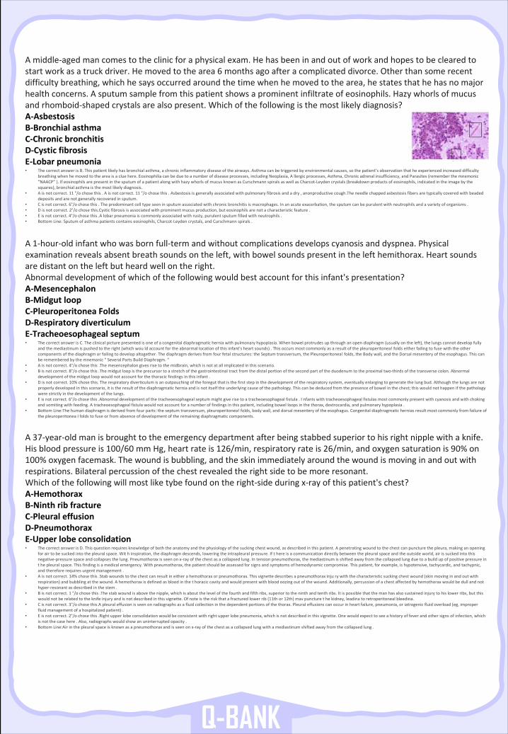
Q-BANK
Amiddle-agedmancomestotheclinicforaphysicalexam.Hehasbeeninandoutofworkandhopestobeclearedtostartworkasatruckdriver.Hemovedtothearea6 monthsagoafteracomplicateddivorce.Otherthansomerecentdifficultybreathing,whichhesaysoccurredaroundthetimewhenhemovedtothearea,hestatesthathehasnomajorhealthconcerns.Asputumsamplefromthispatientshowsaprominentinfiltrateofeosinophils.Hazywhorlsofmucusandrhomboid-shapedcrystalsarealsopresent.Whichofthefollowingisthemostlikelydiagnosis? A-AsbestosisB-BronchialasthmaC-ChronicbronchitisD-CysticfibrosisE-Lobarpneumonia• ThecorrectanswerisB.Thispatientlikelyhasbronchialasthma,achronicinflammatorydiseaseoftheairways.Asthmacanbetriggeredbyenvironmentalcauses,sothepatient’sobservationthatheexperiencedincreaseddifficulty
breathingwhenhemovedtotheareaisacluehere.Eosinophiliacanbeduetoanumberofdiseaseprocesses,includingNeoplasia,Allergic processes,Asthma,Chronicadrenalinsufficiency,andParasites(rememberthemnemonic“NAACP”).IfeosinophilsarepresentinthesputumofapatientalongwithhazywhorlsofmucusknownasCurschmann spiralsaswellasCharcot-Leydencrystals(breakdownproductsofeosinophils,indicatedintheimagebythesquares),bronchialasthmaisthemostlikelydiagnosis.
• Aisnotcorrect.11 °/ochosethis.Aisnotcorrect.11 °/ochosethis.Asbestosisisgenerallyassociatedwithpulmonaryfibrosisandadry,anonproductive cough.The needlechappedasbestosisfibersaretypicallycoveredwithbeadeddepositsandarenotgenerallyrecoveredinsputum.
• Cisnotcorrect.6°/ochosethis.Thepredominantcelltypeseeninsputumassociatedwithchronicbronchitisismacrophages.Inanacuteexacerbation,thesputumcanbepurulentwithneutrophilsandavarietyoforganisms.• Disnotcorrect.2°/ochosethis.Cystic fibrosisisassociatedwithprominentmucusproduction,buteosinophilsarenotacharacteristicfeature.• Eisnotcorrect.4°/ochosethis.Alobarpneumoniaiscommonlyassociatedwithrusty,purulentsputumfilledwithneutrophils.• BottomLine:Sputumofasthmapatientscontainseosinophils,Charcot-Leydencrystals,andCurschmann spirals.
A1-hour-oldinfantwhowasbornfull-termandwithoutcomplicationsdevelopscyanosisanddyspnea.Physicalexaminationrevealsabsentbreathsoundsontheleft,withbowelsoundspresentinthelefthemithorax.Heartsoundsaredistantontheleftbutheardwellontheright.Abnormaldevelopmentofwhichofthefollowingwouldbestaccountforthisinfant'spresentation?A-MesencephalonB-MidgutloopC-Pleuroperitonea FoldsD-RespiratorydiverticulumE-Tracheoesophagealseptum• ThecorrectanswerisC.Theclinicalpicturepresentedisoneofacongenitaldiaphragmaticherniawithpulmonaryhypoplasia.Whenbowelprotrudesupthroughanopendiaphragm(usuallyontheleft),thelungscannotdevelopfully
andthemediastinumispushedtotheright(whichwou ld accountfortheabnormallocationofthisinfant'sheartsounds).Thisoccursmostcommonlyasaresultofthepleuroperitonea!foldseitherfailingtofusewiththeothercomponentsofthediaphragmorfailingtodevelopaltogether.Thediaphragmderivesfromfourfetalstructures:theSeptumtransversum,thePleuroperitonea!folds,theBodywall,andtheDorsalmesenteryoftheesophagus.Thiscanberememberedbythemnemonic"SeveralPartsBuildDiaphragm.“
• Aisnotcorrect.4°/ochosethis.Themesencephalongivesrisetothemidbrain,whichisnotatallimplicatedinthisscenario.• Bisnotcorrect.8°/ochosethis.Themidgutloopistheprecursortoastretchofthegastrointestinaltractfromthedistalportionofthesecond partoftheduodenumtotheproximaltwo-thirdsofthetransversecolon.Abnormal
developmentofthemidgutloopwouldnotaccountforthethoracicfindingsinthisinfant.• Disnotcorrect.10%chosethis.Therespiratorydiverticulumisanoutpouchingoftheforegutthatisthefirststepinthedevelopmentoftherespiratorysystem,eventuallyenlargingtogeneratethelungbud.Althoughthelungsarenot
properlydevelopedinthisscenario,itistheresultofthediaphragmaticherniaandisnotitselftheunderlyingcauseofthepathology.Thiscanbededucedfromthepresenceofbowelinthechest;thiswouldnothappenifthepathologywerestrictlyinthedevelopmentofthelungs.
• Eisnotcorrect.6°/ochosethis.Abnormaldevelopmentofthetracheoesophagealseptummightgiverisetoatracheoesophagealfistula.Infants withtracheoesophagealfistulasmostcommonlypresentwithcyanosisandwithchokingandvomitingwithfeeding.Atracheoesophagealfistulawouldnotaccountforanumberoffindingsinthispatient,includingbowelloopsinthethorax,dextrocardia,andpulmonaryhypoplasia.
• BottomLine:The humandiaphragmisderivedfromfourparts:theseptumtransversum,pleuroperitonea!folds,bodywall,anddorsalmesenteryoftheesophagus.Congenitaldiaphragmaticherniasresultmostcommonlyfromfailure ofthepleuroperitonea Ifoldstofuseorfromabsenceofdevelopmentoftheremainingdiaphragmaticcomponents.
A37-year-oldmanisbroughttotheemergencydepartmentafterbeingstabbedsuperiortohisrightnipplewithaknife.Hisbloodpressureis100/60 mmHg,heartrateis126/min,respiratoryrateis26/min,andoxygensaturationis90%on100%oxygenfacemask.Thewoundisbubbling,andtheskinimmediatelyaroundthewoundismovinginandoutwithrespirations.Bilateralpercussionofthechestrevealedtherightsidetobemoreresonant.Whichofthefollowingwillmostliketybe foundontheright-sideduringx-rayofthispatient'schest?A-HemothoraxB-NinthribfractureC-PleuraleffusionD-PneumothoraxE-Upperlobeconsolidation• ThecorrectanswerisD.Thisquestionrequiresknowledgeofboththeanatomyandthephysiologyofthesuckingchestwound,asdescribedinthispatient.Apenetratingwoundtothechestcanpuncturethepleura,makinganopening
forairtobesuckedintothepleuralspace.Withinspiration,thediaphragmdescends,loweringtheintrapleuralpressure.Ift hereisacommunicationdirectlybetweenthepleuralspaceandtheoutsideworld,airissuckedintothisnegative-pressurespaceandcollapsesthelung.Pneumothoraxisseenonx-rayofthechestasacollapsedlung.Intensionpneumothorax,themediastinumisshiftedawayfromthecollapsedlungduetoabuildupofpositivepressureinthepleuralspace.Thisfindingisamedicalemergency.Withpneumothorax,thepatientshouldbeassessedforsignsandsymptomsofhemodynamiccompromise.Thispatient,forexample,ishypotensive,tachycardic,andtachypnic,andthereforerequiresurgentmanagement.
• Aisnotcorrect.14%chosethis.Stabwoundstothechestcanresultineitherahemothoraxorpneumothorax.Thisvignettedescribesapneumothorax inju ry withthecharacteristicsuckingchestwound(skinmovinginandoutwithrespiration)andbubblingatthewound.Ahemothoraxisdefinedasbloodinthethoracic cavityandwouldpresentwithbloodoozingoutofthewound.Additionally,percussionofachestaffectedbyhemothoraxwould bedullandnothyper-resonantasdescribedinthestem.
• Bisnotcorrect.1 °/ochosethis.Thestabwoundisabovethenipple,whichisaboutthelevelofthefourthandfifthribs,superiortotheninthandtenthribs.Itispossiblethatthemanhasalsosustainedinjurytohislowerribs,butthiswouldnotberelatedtotheknifeinjuryandisnotdescribedinthisvignette.Ofnoteistheriskthatafracturedlowerrib(11thor12th)mav puncturethekidney,leadina toretroperitonealbleedina.
• Cisnotcorrect.3°/ochosethis.A pleuraleffusionisseenonradiographsasafluidcollectioninthedependentportionsofthethorax.Pleuraleffusionscanoccurinheartfailure,pneumonia,oriatrogenicfluidoverload(eg,improperfluidmanagementofahospitalizedpatient).
• Eisnotcorrect.2°/ochosethis.Rightupperlobeconsolidationwouldbeconsistentwithrightupperlobepneumonia,whichisnotdescribedinthisvignette.Onewouldexpecttoseeahistoryoffeverandothersignsofinfection,whichisnotthecasehere.Also,radiographswouldshowanuninterruptedopacity.
• BottomLine:Air inthepleuralspaceisknownasapneumothoraxandisseenonx-rayofthechestasacollapsedlungwithamediastinumshiftedawayfromthecollapsedlung.
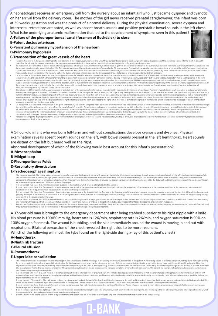
Q-BANK
Aneonatologistreceivesanemergencycallfromthenurseryaboutaninfantgirlwhojustbecamedyspneicandcyanoticonherarrivalfromthedeliveryroom.Themotherofthegirlneverreceivedprenatalcare;however,theinfantwasbornat39 weeks'gestationandwastheproductofanormaldelivery.Duringthephysicalexamination,severedyspneaandintercostalretractionsarenoted,aswellasabsentbreathsoundsandpositiveperistalticbowelsoundsintheleftchest.Whatisthe underlyinganatomicmalformationthatledtothedevelopmentofsymptomsseeninthispatient? A-Failureofthepleuroperitonea!canal(foramenofBochdalek)tocloseB-PatentductusarteriosusC-PersistentpulmonaryhypertensionofthenewbornD-PulmonaryhypoplasiaE-Transpositionofthegreatvesselsoftheheart• ThecorrectanswerisA..Congenitaldiaphragmatichernia(shownintheimage)usuallyrepresentsfailureofthepleuroperitonea!canaltoclosecompletely,leadingtoprotrusionoftheabdominalvisceraintothechest.Itisusually
locatedontheleftside.Pulmonaryhypoplasiaisthemostcommoncauseofdeathinthesepatients,whichdevelopssecondarytolackofspaceforthelungtogrow• Bisnotcorrect.5°/ochosethis.Apatentductusarteriosusproducesaleft-to-rightshunt.Inotherwords,itallowsbloodtogofromthesystemiccirculationtothepulmonarycirculation.Therefore,pulmonarybloodflowisexcessive.The
ductusarteriosusisnormallypatentduringfetallife.ThispatencyispromotedbycontinualproductionofprostaglandinE2 bytheductus.Prostaglandinantagonism,suchasmaternaluseofnonsteroidalanti-inflammatorymedications,cancausefetalclosureoftheductusarteriosus.Thiscanbeassociatedwithseverefetalcardiovascularcompromise.Normally,functionalclosureoftheductusarteriosusoccursbyabout15 hoursoflifeinhealthyinfantsbornatterm.Thisoccursbyabruptcontractionofthemuscularwalloftheductusarteriosus,whichisassociatedwithincreasesinthepartialpressureofoxygencoincidentwiththefirstbreath
• Cisnotcorrect.4°/ochosethis.Persistentpulmonaryhypertensionofthenewborn(PPHN)isfailureofthenormalcirculatorytransitionthatoccursafterbirth.Itisasyndromecharacterizedbymarkedpulmonaryhypertensionthatcauseshypoxemiaandright-to-leftextrapulmonaryshuntingofblood.Withinadequatepulmonaryperfusion,neonatesdeveloprefractoryhypoxemia,respiratorydistress,andacidosis.Respiratoryfailureandhypoxemiainthetermnewbornresultsfromaheterogeneousgroupofdisorders,andthetherapeuticapproachandresponseoftendependontheunderlyingdisease.PPHNoftenresultswhenstructurallynormalpulmonaryvesselsconstrictinresponsetoalveolarhypoxiaduetohypoventilationorparenchymaldisorders,suchashyalinemembranediseaseormeconiumaspirationsyndrome.However,PPHNcanalsooccuridiopathicallyintheabsenceofunderlyingparenchymaldisease.Inthesecases,thesyndromeisbelievedtobetheresultofanabnormally remodeledvasculaturethatdevelopsinuteroinresponsetoprolongedfetalstress,hypoxia,and/orpulmonaryhypertension.Excessiveandperipheralmuscularizationofpulmonaryarteriolescanbeseeninthesecases.
• Disnotcorrect.20%chosethis.Pulmonaryhypoplasiaoraplasiaispartofthespectrumofmalformationscharacterizedbyincompletedevelopmentoflungtissue.Pulmonaryhypoplasiacanresultsecondarytoadiaphragmatichernia,wherespaceforlunggrowthislimited.Theseverityofthelesiondependsonthetimingoftheinsultinrelationtothestage oflungdevelopmentandthepresenceofotheranatomicanomalies.Thehypoplasticlungconsistsofacarina,amalformedbronchialstump,andabsentorpoorlydifferentiateddistallungtissue.Inmorethan50%ofthesecases,coexistingcardiac,gastrointestinal,genitourinary,andskeletalmalformationsarepresent,aswellasvariationsinthebronchopulmonaryvasculature.Typically,inthephysicalexaminationtheexternalchestmayappearnormalormaybesmalland bellshaped,withorwithoutscoliosis.Amediastinalshiftisobservedtowardtheinvolvedside,anddullnessonpercussionisheardoverthedisplacedheart.Inright-sidedhypoplasiatheheartisdisplacedtotheright,whichmayleadtoamistakendiagnosisofdextrocardia.Breathsoundsmaybedecreasedorabsentonthesideofhypoplasia,especiallyoverthebasesandaxilla.
• Eisnotcorrect.6°/ochosethis.Transpositionofthegreatarteries(TGA)isacyanoticcongenitalheartlesionthatpresentsinneonates.The hallmarkofTGAisventriculoarterialdiscordance,inwhichtheaortaarisesfromthemorphologicrightventricleandthepulmonaryarteryarisesfromthemorphologicleftventricle.Thepulmonaryandsystemiccirculationsfunctioninparallel,ratherthaninseries.Oxygenatedpulmonaryvenousbloodreturnstotheleftatriumandleftventriclebutisrecirculatedtothepulmonaryvascularbedviatheabnormalpulmonaryarterialconnectiontotheleftventricle.Deoxygenatedsystemicvenousbloodreturnstotherightatriumandrightventricle,whereitissubsequentlypumpedtothesystemiccirculation,effectivelybypassingthelungs.Thisparallelcirculatoryarrangementresultsinadeficientoxygensupplytothetissuesandanexcessiverightandleftventricularworkload.Itisincompatiblewithprolongedsurvivalunlessmixingofoxygenatedanddeoxygenatedanddeoxygenatedbloodoccursatsomeanatomic level.
• BottomLine:Congenitaldiaphragmaticherniausuallyrepresentsfailureofthepleuroperitonea!canaltoclosecompletely,leadingtoprotrusionoftheabdominalvisceraintothechest.Secondarypulmonaryhypoplasiais themostcommoncauseofdeathinthesepatients.
A1-hour-oldinfantwhowasbornfull-termandwithoutcomplicationsdevelopscyanosisanddyspnea.Physicalexaminationrevealsabsentbreathsoundsontheleft,withbowelsoundspresentinthelefthemithorax.Heartsoundsaredistantontheleftbutheardwellontheright.Abnormaldevelopmentofwhichofthefollowingwouldbestaccountforthisinfant'spresentation?A-MesencephalonB-MidgutloopC-Pleuroperitonea FoldsD-RespiratorydiverticulumE-Tracheoesophagealseptum• ThecorrectanswerisC.Theclinicalpicturepresentedisoneofacongenitaldiaphragmaticherniawithpulmonaryhypoplasia.Whenbowelprotrudesupthroughanopendiaphragm(usuallyontheleft),thelungscannotdevelopfully
andthemediastinumispushedtotheright(whichwou ld accountfortheabnormallocationofthisinfant'sheartsounds).Thisoccursmostcommonlyasaresultofthepleuroperitonea!foldseitherfailingtofusewiththeothercomponentsofthediaphragmorfailingtodevelopaltogether.Thediaphragmderivesfromfourfetalstructures:theSeptumtransversum,thePleuroperitonea!folds,theBodywall,andtheDorsalmesenteryoftheesophagus.Thiscanberememberedbythemnemonic"SeveralPartsBuildDiaphragm.“
• Aisnotcorrect.4°/ochosethis.Themesencephalongivesrisetothemidbrain,whichisnotatallimplicatedinthisscenario.• Bisnotcorrect.8°/ochosethis.Themidgutloopistheprecursortoastretchofthegastrointestinaltractfromthedistalportionofthesecond partoftheduodenumtotheproximaltwo-thirdsofthetransversecolon.Abnormal
developmentofthemidgutloopwouldnotaccountforthethoracicfindingsinthisinfant.• Disnotcorrect.10%chosethis.Therespiratorydiverticulumisanoutpouchingoftheforegutthatisthefirststepinthedevelopmentoftherespiratorysystem,eventuallyenlargingtogeneratethelungbud.Althoughthelungsarenot
properlydevelopedinthisscenario,itistheresultofthediaphragmaticherniaandisnotitselftheunderlyingcauseofthepathology.Thiscanbededucedfromthepresenceofbowelinthechest;thiswouldnothappenifthepathologywerestrictlyinthedevelopmentofthelungs.
• Eisnotcorrect.6°/ochosethis.Abnormaldevelopmentofthetracheoesophagealseptummightgiverisetoatracheoesophagealfistula.Infants withtracheoesophagealfistulasmostcommonlypresentwithcyanosisandwithchokingandvomitingwithfeeding.Atracheoesophagealfistulawouldnotaccountforanumberoffindingsinthispatient,includingbowelloopsinthethorax,dextrocardia,andpulmonaryhypoplasia.
• BottomLine:The humandiaphragmisderivedfromfourparts:theseptumtransversum,pleuroperitonea!folds,bodywall,anddorsalmesenteryoftheesophagus.Congenitaldiaphragmaticherniasresultmostcommonlyfromfailure ofthepleuroperitonea Ifoldstofuseorfromabsenceofdevelopmentoftheremainingdiaphragmaticcomponents.
A37-year-oldmanisbroughttotheemergencydepartmentafterbeingstabbedsuperiortohisrightnipplewithaknife.Hisbloodpressureis100/60 mmHg,heartrateis126/min,respiratoryrateis26/min,andoxygensaturationis90%on100%oxygenfacemask.Thewoundisbubbling,andtheskinimmediatelyaroundthewoundismovinginandoutwithrespirations.Bilateralpercussionofthechestrevealedtherightsidetobemoreresonant.Whichofthefollowingwillmostliketybe foundontheright-sideduringx-rayofthispatient'schest?A-HemothoraxB-NinthribfractureC-PleuraleffusionD-PneumothoraxE-Upperlobeconsolidation• ThecorrectanswerisD.Thisquestionrequiresknowledgeofboththeanatomyandthephysiologyofthesuckingchestwound,asdescribedinthispatient.Apenetratingwoundtothechestcanpuncturethepleura,makinganopening
forairtobesuckedintothepleuralspace.Withinspiration,thediaphragmdescends,loweringtheintrapleuralpressure.Ift hereisacommunicationdirectlybetweenthepleuralspaceandtheoutsideworld,airissuckedintothisnegative-pressurespaceandcollapsesthelung.Pneumothoraxisseenonx-rayofthechestasacollapsedlung.Intensionpneumothorax,themediastinumisshiftedawayfromthecollapsedlungduetoabuildupofpositivepressureinthepleuralspace.Thisfindingisamedicalemergency.Withpneumothorax,thepatientshouldbeassessedforsignsandsymptomsofhemodynamiccompromise.Thispatient,forexample,ishypotensive,tachycardic,andtachypnic,andthereforerequiresurgentmanagement.
• Aisnotcorrect.14%chosethis.Stabwoundstothechestcanresultineitherahemothoraxorpneumothorax.Thisvignettedescribesapneumothorax inju ry withthecharacteristicsuckingchestwound(skinmovinginandoutwithrespiration)andbubblingatthewound.Ahemothoraxisdefinedasbloodinthethoracic cavityandwouldpresentwithbloodoozingoutofthewound.Additionally,percussionofachestaffectedbyhemothoraxwould bedullandnothyper-resonantasdescribedinthestem.
• Bisnotcorrect.1 °/ochosethis.Thestabwoundisabovethenipple,whichisaboutthelevelofthefourthandfifthribs,superiortotheninthandtenthribs.Itispossiblethatthemanhasalsosustainedinjurytohislowerribs,butthiswouldnotberelatedtotheknifeinjuryandisnotdescribedinthisvignette.Ofnoteistheriskthatafracturedlowerrib(11thor12th)mav puncturethekidney,leadina toretroperitonealbleedina.
• Cisnotcorrect.3°/ochosethis.A pleuraleffusionisseenonradiographsasafluidcollectioninthedependentportionsofthethorax.Pleuraleffusionscanoccurinheartfailure,pneumonia,oriatrogenicfluidoverload(eg,improperfluidmanagementofahospitalizedpatient).
• Eisnotcorrect.2°/ochosethis.Rightupperlobeconsolidationwouldbeconsistentwithrightupperlobepneumonia,whichisnotdescribedinthisvignette.Onewouldexpecttoseeahistoryoffeverandothersignsofinfection,whichisnotthecasehere.Also,radiographswouldshowanuninterruptedopacity.
• BottomLine:Air inthepleuralspaceisknownasapneumothoraxandisseenonx-rayofthechestasacollapsedlungwithamediastinumshiftedawayfromthecollapsedlung.
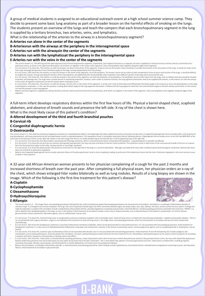
Q-BANK
Agroupofmedicalstudentsisassignedtoaneducationaloutreacheventatahighschoolsummersciencecamp.Theydecidetopresentsomebasiclunganatomyaspartofabroaderlessonontheharmfuleffectsofsmokingonthelungs.Thestudentspresentanoverviewofthelungsandteachthecampersthateachbronchopulmonarysegmentinthelungissuppliedbyatertiarybronchus,twoarteries,veins,andlymphatics.Whatistherelationshipofthearteriestotheairwayinabronchopulmonarysegment?A-ArteriesrunaloneinthecenterofthesegmentsB-Arteriesrun withtheairwaysattheperipheryintheintersegmentalspaceC-Arteriesrunwiththeairwaysin thecenterofthesegmentsD-Arteriesrunwiththelymphaticsat theperipheryintheintersegmentalspaceE-Arteriesrunwiththeveinsinthecenterofthesegments• ThecorrectanswerisC..The leftlungandtherightlunghave8 and10 bronchopulmonarysegments,respectively.Eachsegmentfunctionsasaseparateunitandissuppliedbyatertiarybronchusand twoarteries(abronchialanda
pulmonaryartery,asshownintheright-handillustration),allofwhichruntogetherinthecenterofthesegments.Veinsandlymphaticsdraintogetheralongtheedgesofthesegments..• Aisnotcorrect.5°/ochosethis.Thearteriesrunwiththeairways,notaloneinthecenterofthesegments.Awaytoruleoutthisanswerchoicewouldbetoconsiderthefunctionofthevasculatureofthelungs- itwouldnotmakesense
fromagas-exchangeperspectivetohavethebloodsupplytothelungsrunningindependentlyfromtheairways.• Bisnotcorrect.20%chosethis.The arteriesrunwiththeairwaysinthecenterofthesegments,notattheperiphery.Thisanswerchoicecanberuledoutwithconsiderationtothestructureandfunctionofthelungs.Itwouldbedifficult
toimaginetheairwaysrunningonlyalongthebordersofthelungsegments,andadditionallythiswouldprobablymakerespirationmoredifficultaprocessifthelungswerestructuredthisway.• Disnotcorrect.12%chosethis.Thearteriesrunwiththeairwaysinthecenterofthesegments,notwiththelymphaticsattheperiphery.Thelymphaticsservetodraintoxinsfromthelungs,andsoitfollowsthattheywouldbesituated
atbordersanddrainagesites.Thelungshaveavestedinterestinmaximizingrespiration,however,andinordertomaximizefunction,thelungsegmentsthemselvesneedtobesuppliedwitharteries,sothearteriesrunwiththeairwaysinthecenterofsegmentsandbranchoutintothealveoliwhererespirationoccurs.
• Eisnotcorrect.17%chosethis.Thearteriesrunwiththeairways,nottheveins,inthecenterofthesegments.Itcanbeabittricky tothinkaboutanddifferentiatethebloodsupplytothelungtissueversusthebloodgoingintothelungstoparticipateinrespiration.Inthiscasethequestionisaskingaboutbloodsupplytothelungsegmentsthemselves.Itfollowsthatthelungsegmentsneedtheirownarterialbloodsupplytomaintainactivityandfunction,sothearteriesrunwiththeairwaysineachlungsegment.
• BottomLine:Each segmentissuppliedbyatertiarybronchusandtwoarteries(onebronchialandonepulmonary),allofwhichruntogetherinthe centerofthesegments.Veinsandlymphaticsdraintogetheralongtheedgesofthesegments.
Afull-terminfantdevelopsrespiratorydistresswithinthefirstfewhoursoflife.Physicalabarrel-shapedchest,scaphoidabdomen,andabsenceofbreathsoundsandpresencetheleftside.X-rayofthechestisshownhere.Whatisthemostlikelycauseofthispatient'scondition?A-AltereddevelopmentofthethirdandfourthbranchialpouchesB-CervicalribC-CongenitaldiaphragmaticherniaD-DextrocardiaThecorrectanswerisC..This childhaspulmonaryhypoplasiasecondarytoadevelopmentaldefectinthediaphragmthatallowsabdominalvisceratoherniate intothechest.Acongenitaldiaphragmaticherniacoincideswithacriticalperiodoflungdevelopment,andlungcompressionbytheherniatedbowelresultsinunderdevelopment.Thishypoplasiaresultsinimmediaterespiratorydistressfollowingdelivery.Diaphragmaticherniausuallyoccursontheleftside(80%-85%ofthecases)duetothepresenceofliverontherightside.Noticeinthestemimagethatthereisnovisibleaeratedlungontheleftsidebutanormalappearinglungontherightside.• Aisnotcorrect.6°/ochosethis.This abnormalityisassociatedwithDiGeorgesyndrome,whichischaracterizedbythymicandparathyroidhypoplasia.Lungdevelopmentistypicallynotaffected.• Bisnotcorrect.3°/ochosethis.Cervical ribsarecommonandtypicallyasymptomatic,buttheymayalsocontributetothoracicoutletsyndrome.Thissyndromeresultsinobstructionoftheneurovascularbundleofthearmasitpasses
fromthethoracocervical regiontotheaxilla,causingvascularorneurologicsymptoms.• Disnotcorrect.6°/ochosethis.Dextrocardia isthepresenceoftheheartontherightsideofthebodyasaresultofmalrotation.Althoughassociatedwithmanyotherconditions(particularlyKartagenersyndrome),dextrocardiaalone
doesnotcontributetopulmonaryhypoplasia.• BottomLine:Congenital diaphragmaticherniausuallyrepresentsfailureofthepleuroperitonea!canaltoclosecompletely,leadingtoprotrusionoftheabdominalvisceraintothechest.Pulmonaryhypoplasiaisthemostcommoncause
ofdeathinthesepatients.
A32-year-oldAfrican-Americanwomanpresentstoherphysiciancomplainingofacoughforthepast2 monthsandincreasedshortnessofbreathoverthepastyear.Aftercompletingafullphysicalexam,herphysicianordersanx-rayofthechest,whichshowsenlargedhilarnodesbilaterallyaswellaslungnodules.Resultsofalungbiopsyareshownintheimage.Whichofthefollowingisthefirst-linetreatmentforthispatient'sdisease?A-CisplatinB-CyclophosphamideC-DexamethasoneD-HydroxychloroquineE-Rifampin• ThecorrectanswerisC..Thisimageshowsnoncaseatinggranulomas(indicatedbythecircle)involvinglungseptae Noncaseatinggranulomasarecharacteristicofsarcoidosis.Sarcoidosisisamultiorganinflammatorydisorderof
unknownorigin.Itisthoughttobeimmunemediated.Thelungisthemostfrequentlyinvolvedorgan,butothercommonlyaffected organsarelymphnodes,skin,eyes,kidneys,theheart,andthecentralnervoussystem.Findingsthatmightbeexpectedinapatientwithsarcoidosisincludey-Giobulinemia,Rheumatoidarthritis,elevatedAngiotensin-convertingenzymelevels,Interstitialfibrosis,andNoncaseatinggranulomas(rememberthemnemonic"GRAIN"),aswellasbilateralhilarlymphadenopathyinthelungs,asseeninthispatient.Initialtreatmentofsarcoidosisincludesashort courseofglucocorticoids,suchasdexamethasoneifthepatientissymptomatic.Forchronicdisease,glucocorticoidsmaybecontinuedoralternativeagents,suchasmethotrexate,maybeused.
• Aisnotcorrect.7°/ochosethis.Smallcelllungcancerisrecognizedbynumeroussmallblueneoplasticcellsonhistologicexam.Smallcelllungcanceristreatedwithchemotherapy(etoposide+cisplatin)andpossiblyradiation.Thereisnosurvivalbenefitwithsurgery;therefore,surgeryisnotindicatedinthetreatmentofsmallcelllungcancers.Theimageshowsnoncaseatinggranulomas,whicharecharacteristicofsarcoidosisandwou ld notbetreatedwithchemotherapy.
• Bisnotcorrect.16%chosethis.Goodpasture syndromeiscausedbyantibasement membraneantibodies,whichcanbedemonstratedonimmunofluorescence.Itisnotassociatedwithnoncaseatinggranulomas.InitialtreatmentofGoodpasturesyndromeisa5-daycourseofmethylprednisolonefollowedbyalongtaperandmaintenance.However,ifthediseaseisparticularlysevere,immunosuppressiveagents,suchascyclophosphamideorazathioprine,maybestarted.
• Disnotcorrect.6°/ochosethis.Systemiclupuserythematosus(SLE)canbeassociatedwithpleuritis,butitisnotassociatedwithnoncaseatinggranulomas.Initialtreatmentofnon-life-threateningSLEincludesanalgesicsandantimalarialssuchashydroxychloroquine.Sincethispatienthassarcoidosis,notlupus,hydroxychloroquinewouldnotbethefirst-linetreatmentforher.Nonetheless,hydroxychloroquinecanbeusedtocontrolsymptomsofsarcoidosisinpatientswhofailtorespondtocorticosteroidsordevelopseveresideeffects.
• Eisnotcorrect.14%chosethis.Tuberculosisischaracterizedbycaseatinggranulomas,atypeofgranulomawithaformofnecrosisinwhichdiseasedtissuecentraltothegranulomaformsafirm,drymasswithcheese-likeappearance.Bacteriawithinthegranulomamaynotbedestroyed,butmayratherbedormantonlytobelaterreactivated.Thisismorelikely ifthepatientisimmunosuppressedatanytime.Tuberculosisistreatedwithamultidrugregimenconsistingofisoniazid,rifampin,pyrazinamide,andethambutolfor2 monthsfollowedbya4-monthcourseofisoniazidandrifampin.
• BottomLine:Sarcoidosis,amultiorganinflammatorydisorder,ischaracterizedbynoncaseatinggranulomas.Otherfindingsincludeyglobulinemia,rheumatoidarthritis,elevatedlevelsofangiotensin-convertingenzyme,andinterstitialfibrosis.Initialtreatmentofsarcoidosisisglucocorticoids.
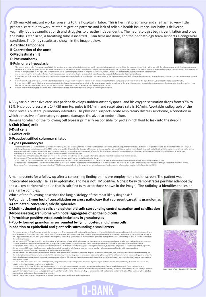
Q-BANK
A19-year-oldmigrantworkerpresentstothehospitalinlabor.Thisisherfirstpregnancyandshehashadverylittleprenatalcareduetowork-relatedmigrationpatternsandlackofreliablehealthinsurance.Herbabyisdeliveredvaginally,butiscyanoticatbirthandstrugglestobreatheindependently.Theneonatologistbeginsventillation andoncethebabyisstabilized,abreathingtubeisinserted.Plainfilmsaredone,andtheneonatologyteamsuspectsacongenitalcondition.TheX-rayresultsareshownintheimagebelow.A-CardiactamponadeB-CoarctationoftheaortaC-MediastinalshiftD-PneumothoraxE-Pulmonaryhypoplasia• ThecorrectanswerisE..Pulmonaryhypoplasiaisthemostcommoncauseofdeathininfantsbornwithcongenitaldiaphragmatichernia.Whenthepleuroperitonea!foldsfailtofusewith theothercomponentsofthediaphragmduring
development,aholeiscreatedthatallowsbowelintothethorax(redcircleinimage).Thephysicalcompressionofthebowelsonthelungbudsthenpreventsfulldevelopmentoftherespiratorysystem.Thebluearrowsindicatethesmallbowelpushingthehearttotheright.Thiscompressionleadstoacommonpresentationofdyspneaandcyanosis,which,unlessitcanberepairedsurgically,eventuallyleadstodeath.
• Aisnotcorrect.with apericardia!effusion.ThisisnotacommoncomplicationCardiac tamponadeismostfrequentlyassociatedofcongenitaldiaphragmatichernia.• Bisnotcorrect.7°/ochosethis.Cardiac abnormalitiessuchasventriculoseptal defects,vascularrings,andcoarctationoftheaortaareassociatedwithcongenitaldiaphragmatichernias;however,theyare notthemostcommoncauseof
death.• Cisnotcorrect.12%chosethis.Mediastinalshiftdoesoccurincongenitaldiaphragmatichernia,asthebowelinvadesthethoraxandpushesthe mediastinumtotheright.However,thisinitselfisnotacauseofdeath.• Disnotcorrect.10%chosethis.Pneumothoraxischaracterizedbyanabnormalcollectionofairinthepleuralspace,leadingtocollapseofthe lung.Itiscommonlyassociatedwithtraumaandotherunderlyingdisorderssuchascystic
fibrosis,necrotizingpneumonia,chronicobstructivepulmonarydisease,etc.Itisnotassociatedwithdiaphragmatichernia.• BottomLine:Pulmonary hypoplasiaisthemostcommoncauseofdeat hininfantsbornwithcongenitaldiaphragmatichernia.
A56-year-oldintensivecareunitpatientdevelopssudden-onsetdyspnea,andhisoxygensaturationdropsfrom97%to82%.Hisbloodpressureis144/89 mmHg,pulseis94/min,andrespiratoryrateis30/min.Aportable radiographofthechestrevealsbilateralpulmonaryinfiltrates.Hisphysiciansuspectsacuterespiratorydistresssyndrome,aconditioninwhichamassiveinflammatoryresponsedamagesthealveolarendothelium.Damagetowhichofthefollowingcelltypesisprimarilyresponsibleforprotein-richfluidtoleakintothealveoli?A-Club(Clara)cellsB-DustcellsC-GobletcellsD-PseudostratifiedcolumnarciliatedE-TypeIpneumocytes• ThecorrectanswerisE..Acuterespiratorydistresssyndrome(ARDS)isaclinicalsyndromeofacute-onsetdyspnea,hypoxemia,anddiffusepulmonaryinfiltratesthatleadstorespiratoryfailure.Itisassociatedwithawiderangeof
underlyingdisorders,includingpancreatitis.ARDSischaracterizedbydiffusealveolardamage,whichleadstoalveolarcapillarypermeabilityandprotein-richleakageintoalveoli,andultimatelytheformationofanintra-alveolarhyalinemembrane,indicatedbythearrowintheimage.Thealveoliarethereforethelungstructuresmostaffectedbythissyndrome.Although,manycelltypesaredamagedandlikelyparticipateinthepathogenesisofARDS,typeIpneumocytesline97%ofthesurfaceofthealveoli,andarethereforeprimarilyinvolved.
• Aisnotcorrect.15°/ochosethis.Clubcellsarefoundintheterminalbronchioles,notthealveoli,wherethecytokine-mediatedassociatedwith ARDSoccurs.• Bisnotcorrect.5°/ochosethis.Dustcellsarealveolarmacrophages,which arenotpartofthealveolarlining.• Cisnotcorrect.8°/ochosethis.Goblet cellsextendonlytotheterminalbronchiolesandarethereforenotfoundinthealveoli,wherethecytokine-mediateddamageassociatedwithARDSoccurs.• Disnotcorrect.8°/ochosethis.Pseudostratifiedcolumnarciliatedcellsextendonlytotherespiratorybronchiolesandarethereforenotfoundinthealveoli,wherethecytokine-mediateddamageassociatedwithARDSoccurs.• BottomLine:Type Ipneumocyteslineapproximately97%ofthesurfaceofalveoliofthelungandareprimarilydamagedbythemassiveinflammatoryreactioninthelungsduringARDS.
Amanpresentsforafollowupafteraconcerningfindingonhispre-employmenthealthscreen.Thepatientwasincarceratedrecently.Heisasymptomatic,andheisnotHIVpositive.AchestX-raydemonstratesperihilaradenopathyanda1-cmperipheralnodulethatiscalcified(similartothoseshownintheimage).TheradiologistidentifiesthelesionasaRankecomplex.Whichofthefollowingdescribesthelunghistologyofthemostlikelydiagnosis?A-Abundant2-mmfociofconsolidationongrosspathologythatrepresentcaseatinggranulomasB-Laminated,concentric,calcificspherulesC-MultinucleatedgiantcellsandepithelioidcellssurroundingcentralcaseationandcalcificationD-NoncaseatinggranulomawithnodalaggregatesofepithelioidcellsE-Peroxidase-positivecytoplasmicinclusionsingranulocytesF-Poorlyformedgranulomassurroundedbylymphocytes,andplasmacells,inadditiontoepithelioidandgiantcellssurroundingasmallartery
• ThecorrectanswerisC..ARankecomplexistheevolutionofaGhon complex,withradiographiccalcificationofthenoduleinsidethecomplex(showninthevignetteimage).GhoncomplexesevolvefromGhon foci(theincipientmassofinflammationwithcaseation)andrepresentaprimarytuberculosisinfectioninwhichcaseatinggranulomashaveformedinthelungparenchymaandthehilarlymphnodes.Histologically,acaseatinggranulomahasmultinucleatedgiantcellsandepithelioidcellssurroundinganareaofcentralnecrosis(asshowninthisimage).
• Aisnotcorrect.15°/ochosethis.Thisisadescriptionofmiliary tuberculosis,whichoftenoccursinelderlyorimmunocompromisedpatientswhohavehadinadequatetreatment.Theinfectioncandisseminatefromlunglesionsthroughtheairway,vessels,orlymphchannels.Grosspathologicspecimensof thelungwillhavenumeroussmallfociofconsolidation.Theselesionscancoalescetoformconsolidationsthatmaymanifestclinicallyasaserouspleuraleffusion,tuberculousempyema,oranobliterativefibrouspleurisy.
• Bisnotcorrect.10%chosethis.Psammoma bodies(laminated,concentric,calcificspherules)areseeninpapillaryadenocarcinomaofthethyroid,serouspapillarycystadenocarcinomaoftheovary,meningioma,andmalignantmesothelioma.
• Disnotcorrect.15%chosethis.Sarcoidosis canmanifestwithsymptomssuchasfever,anorexia,dyspneaonexertion,chestpain,and,rarely,bilateralhilarlymphadenopathy,sotheclinicalpicturecouldbesomewhatsimilartothisvignette.However,thediagnosisofsarcoidosisrequireslungbiopsy,and thehal lmark featureisnoncaseatinggranulomas.Thedistinctionbetweencaseatingandnoncaseatinggranulomasiskeyasthisdistinguishesinfectious(causingcaseatinggranulomas) causesfromnoninfectious(causingnoncaseatinggranulomas)causes.
• Eisnotcorrect.4°/ochosethis.Auerbodies(orrods)areperoxidase-positivecytoplasmicinclusionsingranulocytesandmyeloblasts.CellscontainingAuerrodsareseeninthebloodofpatientswithacutemyelogenousleukemia.
• Fisnotcorrect.7°/ochosethis.This isahistologicdescriptionofthelungpathologyforgranulomatosiswithpolyangiitis(formerlycalledWegener).Thesepatientshaveanecrotizingvasculitisaffectingthreeorgansystems:therespiratorytract,thesmall- tomedium-sizedvessels(capillaries,venules,arterioles,andarteries),andthekidneys.Patientstypically havebothrenaldiseaseandupperorlowerrespiratoryinvolvement,oftenmanifestingaspneumonitiswithnodularandcavitaryinfiltrates.Mostpatientswillbepositiveforcirculatingantineutrophiliccytoplasmicantibodies.
• BottomLine:A Ghon complexiscomposedofaGhon focusandassociatedlymph-nodeinvolvement
Q-BANK
Thisisanimageofadimorphicsoilfunguswithbarrel-shapedarthroconidia.Ifinhaled,itcaninfectthelungs.Fromthere,itmayenterthebloodstreamandinfecttheskin,bones,joints,lymphnodes,adrenalglands,orcentralnervoussystem.WhereintheUnitedStates isapersonmostlikelytobeexposedtothisfungus?A-Anywhere(ubiquitous)B-Mississippi,Ohio,andMissouririvervalleysC-SouthernArizonaD-Tennessee-Ohio-Mississi.• ThecorrectanswerisC..CoccidioidomycosisisadiseasecausedbythesporesofthefungusCoccidioidesimmitis,showninthevignetteimage.ThisfungusisendemictothesoilinCalifornia'sCentral
Valley;southernArizona;andpartsofUtah,Nevada,NewMexico,andTexas(seemap).Theriskofcontractingcoccidioidomycosis justfromtravelingtotheendemicregionsislow;theriskofinfectionincreasesinparticularlydustysettingsandaftermajorenvironmentaleventssuchasearthquakes.Anyagegroupcanbeaffected,butusuallythehigher-riskgroupsarepatientsovertheageof60 andimmunocompromisedindividuals.Intheimage,abluestainfromculturedmaterialshowsthetypicalbarrel-shapedarthroconidiaand90-degreebranchingpatternofC.immitis.Althoughnottheclassicimageofspheruleswithendospores,itisanotherwayC.immitis maybeshownonanexam.
• Aisnotcorrect.13°/ochosethis.Aspergillus species(showninthisimage)areubiquitousmoldsfoundinorganicmatter.MosthumanillnessiscausedbyAspergillusfumigatus,A.niger,A.flavus,andA.c/avatus.Humansbecomeinfectedthroughinhalationoffungalspores.Aspergillusmaycauseabroadspectrumofdiseaseinthehumanhost,fromhypersensitivityreactionstodirectinvasionintothebloodstream.Itcancausevariouspulmonarysyndromes,includingallergicbronchopulmonaryaspergillosis,chronicnecrotizingAspergilluspneumonia,aspergilloma,andinvasiveaspergillosis.
• 8 isnotcorrect.33°/ochosethis.Blastomyces dermatitidisisadimorphicfungusthatgrowswithinhumansasbudding,roundyeast-likecells.Inthemoldformitpossessessmallsporesonitshyphae.ItisfoundintheMississippi,Ohio,andMissouririvervalleys(seemap).
• Disnotcorrect.15%chosethis.Histoplasmosis isaninfectioncausedbyHistoplasmacapsulatum,adimorphicfungusfoundinsoilcontaminatedwithbirdorbatdroppings.EndemicareasaretheTennessee-Ohio-Mississippiriverbasins(seemap).Itusuallycausesacuteorchronicpulmonaryinfections.
• BottomLine:Coccidioides immitis,thefungusthatcausescoccidioidomycosis,isendemictothesoilinCalifornia'sCentralValley;southernArizona;andpartsofUtah,Nevada,NewMexico,andTexas.
A56-year-oldmanofthechestrevealstestsshow:presents withfatigue,fever,weightloss,andhemoptysisof5 weeks'duration.Imagingacentrallylocatedmass.Resultsofalungbiopsyareshown(seeimage).Laboratorytestsshow:Sodium:130 mEq/L.Potassium:3.9 mEq/LChloride:101 mEq/L.Bicarbonate:24 mEq/L.Calcium:9.8 mg/dl.WBCcount:11,600/mmHemoglobin:12 g/dl.Hematocrit:38.1%Plateletcount:420,000/mm3.Bloodureanitrogen:8 mg/dlCreatinine:0.8 mg/dlGlucose:108 mg/dlWhatisthereasoningbehindthebestmanagementforthispatient?A-Surgerycarriesariskofprovokingpara-neoplasticsyndromesB-Surgeryhasnotbeenshowntoimprovesurvival.C-Surgeryispalliativebutnotcurativeon removalofthemassD-SurgeryofteniscurativeineverylungcancerwithoutidentifiablemetastasesE-Thislesionislikelytoregressin7 years• ThecorrectanswerisB..Thestemimageisrepresentativeofsmall(oat)celllungcancer,characterizedbysmall,dark-bluecellsknownasKulchitsky cells.Thepatienthashyponatremia,whichcanbeattributedtothesyndromeof
inappropriateADHsecretionfromapresentingpara-neoplasticphenomenon,whichismorecommoninsmallcellthannon-smallcell lungcancers.Lungcancermanagementinitiallyinvolvesdistinguishingsmallcellfromnon-smallcellcarcinomas.Earlysmallcellcarcinomamanagemententailschemotherapy;earlynon-smallcellcarcinomascanbetreatedviasurgicalresection.Survivaltimeforthosewithuntreatedsmallcellcarcinomais6-17 weeks.Withchemotherapy,mediansurvivalincreasesto18 months
• Aisnotcorrect.14%chosethis.Surgery doesnotcarryariskofprovokingpara-neoplasticsyndromesinsmallcellcarcinomaofthelung.Inpheochromocytoma,however, manipulationofthetumorduringsurgicalresectionisknowntostimulatecatecholaminerelease.
• Cisnotcorrect.19%chosethis.Surgicalresectionhasnotbeenshowntoimprovemorbidityormortalityforsmallcelllungcancer,andisthuscontraindicatedbecauseofincreasedandunnecessarymorbidity.Resectionmaybemoreeffectivefornon-smallcelllungcarcinoma.Somepatientswithveryadvancedgastrointestinaltumorsbenefitfromsurgicalresectiontorelieveobstructivesymptoms.
• Disnotcorrect.10%chosethis.Surgical resectionismoreeffectivefornon-smallcelllungcarcinoma.Itleadstoincreasedmorbidityandnoimprovementinsurvivalinpatientswithsmallcellcarcinoma.• Eisnotcorrect.3°/ochosethis.This explanationwouldsuggestthatnotherapyisthebestcourseofaction,whichisfalse.Therearefewmassesforwhichnothingisdone;anexampleisthecutaneoushemangiomaseeninpediatric
patients.• BottomLine:Smallcelllungcanceristhelungneoplasmmostlikelytobeassociatedwithpara-neoplasticsyndromessuchasLambert-Eatonsyndrome,para-neoplasticcerebellardegeneration,syndromeofinappropriatesecretionof
ADH,andCushing'ssyndrome.Intuitively itmayseemobviousthatremovingabulkytumorwouldhelpapatient,butsurgeryisnotindicatedinpatientswithsmallcelllungcancer,becauseitdoesnotimprovesurvival.
A65-year-oldmanpresentstothephysicianwitha4-monthhistoryofaproductivecoughwithwhitesputum.Hereportsthathehadsimilarsymptomslastyear.HisFEV1 :FVCratiois60%.Physicalexaminationrevealswheezingandcrackles.X-rayofthechestrevealscardiomegaly.Hesaysthatbothofhischildrenarehealthy.Alungbiopsyofthispatientwouldmostlikelyshowwhichofthefollowing?A-AirspaceenlargementB-Desquamationof epitheliumC-Hyperplasiaofmucus-secretingglandsD-MucusplugswithwhorlsofshedepitheliumE-Smoothmusclehyperplasia• ThecorrectanswerisC..Thepatientpresentswithaclassiccaseofchronicbronchitis,whichisdefinedasaproductivecough for3 ormoreconsecutivemonthsin2 ormoreyears.Chronicbronchitisisanobstructivelungdisease
characterizedbyanFEV1 /FVCratio<80%.Pathologyrevealsbothmucus-secretingglandhypertrophy(asshownintheimage)andhyperplasia.ThesechangesarereflectedbytheReidindex,whichistheratioofthethickness ofthemucusglandlayertothethicknessoftheentireairwaywal l(fromendofepit heliumtothestartofthecartilage).TheReidindex,whichisnormally0.4,isincreasedinchronicbronchitis(>0.5).Chronicbronchitisisassociatedwithsmokingandisanobstructivelungdisease.Physicalfindingsincludewheezing,cyanosis,andlungcrackles.Corpulmonale,orrightheartfailure,isacommoncomplication.
• Aisnotcorrect.17%chosethis.Airspaceenlargementoccursinemphysema.Likebronchitis,emphysemaisanobstructivelungdiseasethatiscloselyassociatedwithsmoking.Destructionofalveolileadstoenlargedairspaces.Clinicalfindingsofemphysemaincludeincreasedanteroposteriorchestdiameter(barrel-shapedchest),increasedtotallungcapacity,tachycardia,andrespiratoryacidosis.Aproductivecoughfor2 yearsinarowismoreconsistentwithchronicbronchitis.
• Bisnotcorrect.6°/ochosethis.Bronchiectasis isalsoanobstructivelungdiseaseinwhichbronchiandbronchiolesarepermanentlydilatedfromchronicnecrotizinginfectionsthathavedestroyedmuscleandelastictissue.Thepatienttypicallypresentswithcough,fever,andproductionofpurulentsputum,andusuallywillhaveeitheraninfectionoranobstructioninthelung.Onhistology,therecanbeinflammatoryexudation,desquamationoftheliningepithelium,necrotizingulceration,andpseudostratification ofthecolumnarcells.Thispatient'shistoryandpresentationisnottypicalforbronchiectasis.
• Disnotcorrect.14%chosethis.Mucusplugswithwhorlsofshedepithelium,orCurschmann spirals,arehistologicfindingsofasthma.Althoughtherearevariantsofasthmathatmanifestwithcoughing,thecoughisnot typicallyproductive.Cardiomegalyisalsonotassociatedwithasthma.
• Eisnotcorrect.7°/ochosethis.Smoothmusclehyperplasiaisapredominanthistologicfindinginasthma,whichisalsoanobstructivedisease.Cardiomegalyisnotassociatedwithasthma.• BottomLine:Pathologic findingsofchronicbronchitisincludemucus-secretingglandhypertrophyandhyperplasia.
Q-BANK
A40-year-oldwomanpresentswithachiefcomplaintofprogressivedyspneaandanonproductivecough.Shehasa40-pack-yearsmokinghistory.X-rayofthechestrevealsahoneycombappearance,andaCTimageshowscysticlesions.Anelectronmicrographofhertissuebiopsyisshown.Thispatient'sdefectivecellsaremostlikelytostainpositiveforwhichofthefollowingA-CD20B-CD30C-CD5D-S-100E-TdT• ThecorrectanswerisD.This womanhashistiocytosisX,aconditioncharacterizedbytheabnormalproliferationofcellsofmononuclearphagocyticorigin, whicharecalledhistiocytes.Thehistiocytesthatproliferateinthisconditionare
dendriticcellsthatarerelatedinlineagetoLangerhanscells(thedendriticcellsoftheskin),andforthisreason,thediseaseisalsocalledLangerhanshistiocytosis.Patientswiththisdiseasecanpresentwithhepatosplenomegaly,lymphadenopathy,cysticlungandlyticbonelesions,andcutaneouseruptions.Thehistiocytesarestellate(dendritic)cellswithanovalorirregularpalenucleus,palecytoplasm,andcharacteristiccytoplasmicgranulesBirbeck granules).Birbeck granulesarepathognomonicofLangerhanscellhistiocytosis.Theyaresaidtoresembletennisrackets,asseenintheelectronmicrographinthevignette.ThedefectivedendriticcellsinthisdiseasestainpositivelyforS-100 andCDla.Ofnote,Hand-Schuller-ChristiandiseaseisapediatricvariantofhistiocytosisXandisassociatedwithlyticlesionsinthe skull.
• Aisnotcorrect.11%chosethis.CD20 isa8-lymphocytemarker.Itcanbehelpfulindiagnosing8-lymphocyte-derivedmalignanciessuchasfollicularlymphoma,diffuselarge8-celllymphoma,orchroniclymphocyticleukemia.• Bisnotcorrect.13%chosethis.CD30 ispresentonReed-Sternbergcells(thecharacteristiccellsofHodgkinlymphoma).• Cisnotcorrect.14%chosethis.CDS isaT-cellmarker.However,itisalsoatumormarkerforchroniclymphocyticleukemia(CLL)cells.CLLcellsarenegativeforcyclin D1 andareoftenpostive forCD23.Additionally,CDSisatumormarker
formantlecelllymphoma,whichiscausedbythet(ll;14)translocation.MantlecelllymphomasexpresshighlevelsofcyclinD1 andtendtomanifestinmenintheir50sand60saspainlesslymphadenopathy.• Eisnotcorrect.11%chosethis.TdT +cellsareseeninacutelymphoblasticleukemia.TdT standsforterminaldeoxynucleotidyltransferase,whichisaspecialDNApolymeraseexpressedinB- andT-lymphocyteprecursors.• BottomLine:Langerhans cellscontaincharacteristicBirbeck granules.DendriticcellsinLangerhanscellhistiocytosisstainpositiveforS-100 andCD1a.
• ThecorrectanswerisC..Therearetwomainclinicallysignificantkindsofemphysemas:centriacinar andpanacinar.Eachaffectsadifferentpartoftheacini,whicharetheapproximatelysphericalunitsofthelungcontainingthealveoli,distaltotheconductingbronchioles.Panacinar emphysemaenlargestheaciniuniformlyfromtherespiratorybronchioletothealveoli(asshownintheimage).Itisassociatedwithadeficiencyofproperlyfoldeda1 -antitrypsin.Normally,ar antitrypsinisreleasedintothebloodstreamandtravelstothelung,whereitprotectsthelungsfromdestructionviaexcessproteaseactivity.However,individualswiththePiZZ genotypehavelessthan15%ofthenormalamountofa1 -antitrypsinandwilldeveloppanacinaremphysemaatayoungage.(Torememberthegenotype-phenotypeassociation,thinkaboutPiZZa inthePAN!)Thislackofnormala1 -antit rypsin leadstoprogressiveandunregulatedImagecourtesyofYaleRosen,lungdamagebyproteases.SymptomsincludechroniccoughandshortnessofMDbreaththatinitially occursonlyuponexertion,andthengraduallyoccursatrestaswell.Thesesymptomsusuallydevelopover15-20 yearsandcaneventuallyleadtorespiratoryfailureandprematuredeath.Additionally,asthemisfoldedenzymecannotbesecreted,itaccumulateswithintheliver,whichcanleadtohepatocytedestructionandassociatedtransaminitis..
• Aisnotcorrect.12°/ochosethis.entriacinar emphysemaaffectsthecentralandproximalpartsoftheacini.Ittendstooccurintheupperlunglobesofheavysmokers.However,thepatientdoesnotsmoke,socentriacinar emphysemaisunlikely.
• Bisnotcorrect.15%chosethis.Distal acinaremphysemaisalsoknownasparaseptal emphysema.ItcanoccuraspartofCOPDorindependently,inwhichcaseitisusuallyassociatedwithspontaneouspneumothoraxinyoungadults.Itmayoccurbyitselforincombinationwithproximalacinarandpanacinar emphysema.
• Disnotcorrect.3°/ochosethis.An increaseinthesizeofthemucousglandsisafeatureofbronchitis.Thisincreasecanbequantifiedbymeasuringtheratiobetweenthethicknessoftheglandandthethicknessoftheairwaywall.Thisratio,theReidindex,isnormally 0.4 orless.Avalue>0.5 indicatesbronchitis.Theprimarysymptomofchronicbronchitisisproductivecough,whichthispatientdoesnothave.
• Eisnotcorrect.2°/ochosethis.The subpleuralregion,betweenthepleuralmembraneandtheparenchyma,isafocusoffibrosisinthecaseofidiopathicpulmonaryfibrosis.Likeemphysema,itbeginswithdyspneaonexertion.However,idiopathicpulmonaryfibrosisusuallypresentslaterinlife,between theagesof40 and70,andisaccompaniedbyadrycough.
• BottomLine:a1 Antitrypsindeficiencymanifestsaspanacinar emphysemainrelativelyyoungnonsmokers.
Atacheck-upduringweek22 ofherfourthpregnancy,awomantellsherdoctorthatshefeelslikesheis"abnormallylarge"comparedwithpriorpregnancies.Thedoctoragrees,andultrasonographyrevealsexcessfluidintheuterus.Inaddition,thefetus'sstomach,spleen,andaportionofthesmallintestinearevisibleinthefetalthorax.Whatstructure(s)mostlikelyhasfailedtoformcompletelyinthefetus?A-DorsalmesenteryoftheesophagusB-ForegutC-LateralbodywallD-Pleuroperitonea!FoldsE-Septumtransversum.
• ThecorrectanswerisD..Theultrasoundrevealsacongenitaldiaphragmatichernia(CDH)inthefetus.Thediaphragmisderivedfromfourembryologicstructures:theseptumtransversum,thepleuroperitonea!folds,thedorsalmesenteryoftheesophagus,andamuscularoutgrowthofthelateralbodywall.Thepleuroperitonea!foldsformalargeportionofthefetaldiaphragm;iftheyfailtoformcompletely,thethoraxandtheabdomenareincompletelyseparatedposterolaterally,andtheabdominalcontentsoftenherniateintothethorax(knownasaBochdalek hernia).Pressurefromabdominalorgansresultsinlunghypoplasia.Thepolyhydramnioscouldresulteitherfrommechanicalcompressionoftheesophagusbytheherniatedviscera(mostlikely),and/orfromthelunghypoplasia,asthelungsmayofferaresorptivesurfacefortherecyclingofamnioticfluid.NewbornswithCDHtypicallyhaveaflatstomachandaheartdisplacedtotheright.
• Aisnotcorrect.7°/ochosethis.The dorsalmesenteryoftheesophagusformsthecentralpartofthefetaldiaphragm.Postembryonically,thisstructurebecomesthecrura ofthediaphragm.Itisnotnormallydefectiveincongenitaldiaphragmatichernia.
• Bisnotcorrect.9°/ochosethis.00Althoughtheforegutisdisplacedfromtheabdomenintothethoraxinthepresenceofacongenitaldiaphragmatichernia,itsformationisnormal.Theforegutistheembryonicprecursortothelungs,esophagus,stomach,duodenum,liver,gallbladder,andpartofthepancreas.Arterialsupplytoallthesestructuresexceptforthelungsisfromtheceliactrunk.
• Cisnotcorrect.10%chosethis.Muscular outgrowthsofthelateralbodywallformthelateraledgeofthediaphragm,borderingtheleftandrightcostodiaphragmatic recesses.Thesestructuresarenotcommonlydefectiveincongenitaldiaphragmatichernia.
• Eisnotcorrect.16%chosethis.The septumtransversumgrowsoutfromtheventrolateralbodywallandseparatestheheartfromtheliverintheembryo.Ultimatelyitgivesrisetothecentraltendonofthediaphragm.However,defectsintheseptumtransversumarerarelythecauseofcongenitaldiaphragmatichernia.
• BottomLine:Polyhydramnios iscommonwithcongenitaldiaphragmaticherniaduetofailureofthepleuroperitonea!foldstoform.• .
A34-year-oldmanpresentstothephysicianwithprogressiveshortnessofbreathofseveralyears'duration.Physicalexaminationshowsanincreaseintheanteroposteriordiameterofthechest,hyperresonancetopercussion,anddiffusewheezes.Thepatientisadministeredacombinationnebulizertreatmentofalbuterolandipratropiumwithonlymodestreliefofsymptoms.Laboratorystudiesareremarkableforelevatedaspartateaminotransferaseandalanineaminotransferase.Adetailedhistoryrevealsthatthepatienthasneversmokedcigarettesorcigars,drinksonetotwobeersperweekmaximum,andhasnohistoryofillicitdruguse.HehasnevertraveledoutsideoftheUnitedStatesandworksinbilling.Hismotherishealthy,andhisfatherdiedrecentlyofliverfailure.Whichofthefollowingpartsoftherespiratorypathwayismostaffectedbyhisdisease?A-thecentralacinusB-thedistalacinusC-theentiresizeofthemucousglandsE-thesubpleuralregion

MainReferences
vTeams work and Notes (433-438)
vSTEP1 Kaplan Lectures Notes 2018 (Physiology)
vSTEP1 Kaplan Lectures Notes 2018 (Anatomy)
vSTEP1 Kaplan Lectures Notes 2018 (Microbiology)
vSTEP1 Kaplan Lectures Notes 2018 (Pharmacology)
vFirst Aid for Basic Sciences (ORGAN SYSTEMS)
vFirst Aid for USMLE STEP 1 2019
vUworld Q-bank + First Aid Cases for USMLE STEP 1
vGyton and Hall Textbook of Medical Physiology
vLinda S. Costanzo for Physiology
vLippincott Illustrated Reviews Pharmacology
vFundamentals of Pathology (PATHOMA)
vSketchyMicro,Path and Pharma by SketchyMedical.com
vMindMaps formedical students by Olivia Smith
vMedComics.com
v Orahbeeni.com
Related Documents