RED CELLS, IRON, AND ERYTHROPOIESIS Patient-derived C-terminal mutation of FANCI causes protein mislocalization and reveals putative EDGE motif function in DNA repair Luca Colnaghi, 1-3 Mathew J. K. Jones, 1 Xiomaris M. Cotto-Rios, 1,2 Detlev Schindler, 4 Helmut Hanenberg, 5,6 and Tony T. Huang 1 1 Department of Biochemistry, New York University School of Medicine, New York, NY; 2 Sackler graduate program in Cellular and Molecular Biology, New York University School of Medicine, New York, NY; 3 Dipartimento di Scienze Biomolecolari e Biotecnologie, Universita ` degli Studi di Milano, Milano, Italy; 4 Department of Human Genetics, University of Wurzberg, Wurzberg, Germany; 5 Department of Pediatric Hematology, Oncology and Clinical Immunology, Children’s Hospital, Heinrich Heine University, Dusseldorf, Germany; and 6 Department of Pediatrics, Wells Center for Pediatric Research, Riley Hospital for Children, Indiana University School of Medicine, Indianapolis, IN Fanconi anemia (FA) is a rare familial genome instability syndrome caused by mutations in FA genes that results in defective DNA crosslink repair. Activation of the FA pathway requires the FA core ubiquitin ligase complex-dependent monoubiquitination of 2 interacting FA proteins, FANCI and FANCD2. Although loss of either FANCI or FANCD2 is known to prevent monoubiquitination of its re- spective partner, it is unclear whether FANCI has any additional domains that may be important in promoting DNA re- pair, independent of its monoubiquiti- nation. Here, we focus on an FA-I patient- derived FANCI mutant protein, R1299X (deletion of 30 residues from its C- terminus), to characterize important struc- tural region(s) in FANCI that is required to activate the FA pathway. We show that, within this short 30 amino acid stretch contains 2 separable functional signa- tures, a nuclear localization signal and a putative EDGE motif, that is critical for the ability of FANCI to properly monoubiq- uitinate FANCD2 and promote DNA crosslink resistance. Our study enable us to conclude that, although proper nuclear localization of FANCI is crucial for robust FANCD2 monoubiquitination, the puta- tive FANCI EDGE motif is important for DNA crosslink repair. (Blood. 2011;117(7): 2247-2256) Introduction Fanconi anemia (FA) is a rare, chromosome instability syndrome associated with developmental abnormalities, bone marrow failure, predisposition to cancer, and cellular hypersensitivity to DNA crosslinking agents. 1,2 The disorder is genetically heterogeneous with at least 13 complementation groups currently defined. To date, at least 8 FA and other FA-associated proteins (FANCA, -B, -C, -E, -F, -G, -L, -M, and FAAP100) form a multisubunit nuclear core complex that contains a catalytic ubiquitin E3 ligase activity required for the activation of FANCD2 and its paralog FANCI by monoubiquitination. 3,4 Monoubiquitinated and phosphorylated FANCD2 and FANCI 5-9 are then targeted to BRCA1-containing DNA damage and repair sites in chromatin (nuclear foci) to assist in DNA crosslink repair along with at least 4 other downstream FA members, FANCJ (BRIP1, a DNA helicase), 10-12 FANCD1 (BRCA2, a protein that mediates homologous recombination), 13 FANCN (PALB2, partner and localizer of BRCA2), 14,15 and RAD51C (a member of the RAD51-like gene family involved in HR-mediated DNA repair). 16,17 Currently, it is thought that the key trigger in the activation of the FA pathway lies in the molecular events surrounding the DNA damage-inducible monoubiquitination of the FANCD2 and FANCI proteins. Insights into the mechanism of activation came with the discovery of the gene FANCI, responsible for the FA-I complemen- tation group. 6,7,18 The FANCI protein is thought to function similarly to that of FANCD2. Besides possessing regions of the protein with weak homology to FANCD2, FANCI also gets monoubiquitinated in a lysine site-specific and DNA damage- dependent manner by the FA core E3 ubiquitin ligase complex. 6,7 A fraction of FANCI is found to be associated with FANCD2, forming the FANCI-FANCD2 (ID) complex. Interestingly, the monoubiquitination of FANCI and FANCD2 is dependent on each other. Loss of either FANCI or FANCD2 prevents the monoubiquiti- nation of its other protein partner. As expected, negative regulation of the FA pathway can be achieved, in part, through FANCI and FANCD2 deubiquitination, which is mediated by the deubiquitinat- ing enzyme USP1. 6,19,20 More recently, it was shown that monoubiq- uitinated ID complex is required for replication-coupled interstrand crosslink repair in a cell-free system. 21 However, it is still unclear how FANCI can contribute to both the upstream monoubiquitination of FANCD2 and subsequent downstream repair of DNA crosslinks. FANCI is a relatively large protein with 1328 amino acids (Mr 149 kDa) and, like other Fanconi proteins, contains little or no known domain(s) that may provide clues to possible protein function. To gain insight into potential structural region(s) on FANCI that may be important for mediating FANCD2 monoubiq- uitination and FA pathway function, we sought to analyze patient- derived FANCI mutant proteins and determine their functional defect in a reconstituted cell line system. From our previous study, we identified 7 different allelic mutations in the FANCI gene from 4 different FA-I patient-derived cell lines. 6 Interestingly, cells from most, if not all, FA-I persons studied have retained expression of mutant FANCI proteins. Similar to what has been reported for patients harboring mutations in the FANCD2 gene, 22 it is probable that complete null mutations of FANCI are lethal in humans. 6 Submitted July 14, 2010; accepted October 16, 2010. Prepublished online as Blood First Edition paper, October 22, 2010; DOI 10.1182/blood-2010-07-295758. The publication costs of this article were defrayed in part by page charge payment. Therefore, and solely to indicate this fact, this article is hereby marked ‘‘advertisement’’ in accordance with 18 USC section 1734. © 2011 by The American Society of Hematology 2247 BLOOD, 17 FEBRUARY 2011 VOLUME 117, NUMBER 7

Welcome message from author
This document is posted to help you gain knowledge. Please leave a comment to let me know what you think about it! Share it to your friends and learn new things together.
Transcript

RED CELLS, IRON, AND ERYTHROPOIESIS
Patient-derived C-terminal mutation of FANCI causes protein mislocalization andreveals putative EDGE motif function in DNA repairLuca Colnaghi,1-3 Mathew J. K. Jones,1 Xiomaris M. Cotto-Rios,1,2 Detlev Schindler,4 Helmut Hanenberg,5,6 andTony T. Huang1
1Department of Biochemistry, New York University School of Medicine, New York, NY; 2Sackler graduate program in Cellular and Molecular Biology, New YorkUniversity School of Medicine, New York, NY; 3Dipartimento di Scienze Biomolecolari e Biotecnologie, Universita degli Studi di Milano, Milano, Italy;4Department of Human Genetics, University of Wurzberg, Wurzberg, Germany; 5Department of Pediatric Hematology, Oncology and Clinical Immunology,Children’s Hospital, Heinrich Heine University, Dusseldorf, Germany; and 6Department of Pediatrics, Wells Center for Pediatric Research, Riley Hospital forChildren, Indiana University School of Medicine, Indianapolis, IN
Fanconi anemia (FA) is a rare familialgenome instability syndrome caused bymutations in FA genes that results indefective DNA crosslink repair. Activationof the FA pathway requires the FA coreubiquitin ligase complex-dependentmonoubiquitination of 2 interacting FAproteins, FANCI and FANCD2. Althoughloss of either FANCI or FANCD2 is knownto prevent monoubiquitination of its re-spective partner, it is unclear whether
FANCI has any additional domains thatmay be important in promoting DNA re-pair, independent of its monoubiquiti-nation. Here, we focus on an FA-I patient-derived FANCI mutant protein, R1299X(deletion of 30 residues from its C-terminus), to characterize important struc-tural region(s) in FANCI that is required toactivate the FA pathway. We show that,within this short 30 amino acid stretchcontains 2 separable functional signa-
tures, a nuclear localization signal and aputative EDGE motif, that is critical forthe ability of FANCI to properly monoubiq-uitinate FANCD2 and promote DNAcrosslink resistance. Our study enable usto conclude that, although proper nuclearlocalization of FANCI is crucial for robustFANCD2 monoubiquitination, the puta-tive FANCI EDGE motif is important forDNA crosslink repair. (Blood. 2011;117(7):2247-2256)
Introduction
Fanconi anemia (FA) is a rare, chromosome instability syndromeassociated with developmental abnormalities, bone marrow failure,predisposition to cancer, and cellular hypersensitivity to DNAcrosslinking agents.1,2 The disorder is genetically heterogeneouswith at least 13 complementation groups currently defined. To date,at least 8 FA and other FA-associated proteins (FANCA, -B, -C, -E,-F, -G, -L, -M, and FAAP100) form a multisubunit nuclear corecomplex that contains a catalytic ubiquitin E3 ligase activityrequired for the activation of FANCD2 and its paralog FANCI bymonoubiquitination.3,4 Monoubiquitinated and phosphorylatedFANCD2 and FANCI5-9 are then targeted to BRCA1-containingDNA damage and repair sites in chromatin (nuclear foci) to assistin DNA crosslink repair along with at least 4 other downstream FAmembers, FANCJ (BRIP1, a DNA helicase),10-12 FANCD1 (BRCA2,a protein that mediates homologous recombination),13 FANCN(PALB2, partner and localizer of BRCA2),14,15 and RAD51C (amember of the RAD51-like gene family involved in HR-mediatedDNA repair).16,17
Currently, it is thought that the key trigger in the activation ofthe FA pathway lies in the molecular events surrounding the DNAdamage-inducible monoubiquitination of the FANCD2 and FANCIproteins. Insights into the mechanism of activation came with thediscovery of the gene FANCI, responsible for the FA-I complemen-tation group.6,7,18 The FANCI protein is thought to functionsimilarly to that of FANCD2. Besides possessing regions of theprotein with weak homology to FANCD2, FANCI also getsmonoubiquitinated in a lysine site-specific and DNA damage-
dependent manner by the FA core E3 ubiquitin ligase complex.6,7 Afraction of FANCI is found to be associated with FANCD2,forming the FANCI-FANCD2 (ID) complex. Interestingly, themonoubiquitination of FANCI and FANCD2 is dependent on eachother. Loss of either FANCI or FANCD2 prevents the monoubiquiti-nation of its other protein partner. As expected, negative regulationof the FA pathway can be achieved, in part, through FANCI andFANCD2 deubiquitination, which is mediated by the deubiquitinat-ing enzyme USP1.6,19,20 More recently, it was shown that monoubiq-uitinated ID complex is required for replication-coupled interstrandcrosslink repair in a cell-free system.21 However, it is still unclear howFANCI can contribute to both the upstream monoubiquitination ofFANCD2 and subsequent downstream repair of DNA crosslinks.
FANCI is a relatively large protein with 1328 amino acids(Mr � 149 kDa) and, like other Fanconi proteins, contains little orno known domain(s) that may provide clues to possible proteinfunction. To gain insight into potential structural region(s) onFANCI that may be important for mediating FANCD2 monoubiq-uitination and FA pathway function, we sought to analyze patient-derived FANCI mutant proteins and determine their functionaldefect in a reconstituted cell line system. From our previous study,we identified 7 different allelic mutations in the FANCI gene from4 different FA-I patient-derived cell lines.6 Interestingly, cells frommost, if not all, FA-I persons studied have retained expression ofmutant FANCI proteins. Similar to what has been reported forpatients harboring mutations in the FANCD2 gene,22 it is probablethat complete null mutations of FANCI are lethal in humans.6
Submitted July 14, 2010; accepted October 16, 2010. Prepublished online as BloodFirst Edition paper, October 22, 2010; DOI 10.1182/blood-2010-07-295758.
The publication costs of this article were defrayed in part by page charge
payment. Therefore, and solely to indicate this fact, this article is herebymarked ‘‘advertisement’’ in accordance with 18 USC section 1734.
© 2011 by The American Society of Hematology
2247BLOOD, 17 FEBRUARY 2011 � VOLUME 117, NUMBER 7

Studying the different hypomorphic mutations in FANCI mayprovide clues to determining important functional regions/domainsin FANCI for promoting genome stability. In one particular patientcell line (F010191), we identified 2 truncating mutations in FANCI.The first results in a severe deletion of more than half of the codingsequence, whereas the other results in a deletion of the last30 residues of FANCI (R1299X).6 Despite having similar levels ofmutant FANCI compared with normal patient cells, the FA-IF010191 lymphoblasts cells have severely reduced levels ofFANCD2 and FANCI monoubiquitination.6 This suggests that theextreme C-terminal region of FANCI may be critical for mediatingthe activation of the FA pathway.
The goal of the present study is to characterize the function ofthe C-terminal domain of FANCI in promoting FANCD2 monou-biquitination and DNA crosslink repair. We found that the last30 residues of FANCI contain a functional nuclear localizationsignal (NLS) that is critical for proper nuclear localization of theFANCI protein. Mislocalization of FANCI resulting from the lossof the C-terminal NLS sequence results in defective FANCD2 andFANCI monoubiquitination and DNA crosslink sensitivity inhuman cells. We also suggest a DNA crosslink repair function forthe putative EDGE motif6,7,23 that also resides within the C-termi-nal region of FANCI. Our present findings establish the importanceof separable motifs, such as protein localization, as one type ofmechanism that contributes to the molecular pathogenesis of FA.
Methods
Cell culture
U20S (ATCC) were maintained in Dulbecco modified Eagle mediumsupplemented with 10% fetal bovine serum, glutamine, and antibiotics.Patient-derived FA-I F010191 lymphoblasts, as described,6,18 were propa-gated in RPMI 1640 medium supplemented with 15% fetal bovine serum,glutamine, and antibiotics. After informed consent, patient-derived FA-IF010191 fibroblasts were established from a small skin biopsy under localanesthesia and primary fibroblasts were grown out over several weeks inDulbecco modified Eagle medium supplemented with 15% fetal bovineserum, glutamine, and antibiotics. Fibroblasts were immortalized bytransduction with a retroviral vector that expresses the large and smallT antigens from the SV40 viruses under an SFFV promoter (H.H.,unpublished, July 2010). All cells were grown at 37°C.
Cell transfections
siRNA transfections were performed using HiPerfect (QIAGEN) andplasmid transfections were performed using Fugene6 (Roche Diagnostics)according to the manufacturer’s protocol. To replace endogenous FANCIwith siRNA-resistant FANCI constructs (WT FANCI, FANCI R1299X,FANCI NLS2, and FANCI NLS3), cells were first transfected with FANCIsiRNA and followed by transfection of siRNA-resistant FANCI constructs24 hours later.
Plasmids and siRNAs
FANCI and negative control small interfering RNA (siRNA) oligonucleo-tide sequences were purchased from QIAGEN: KIAA1794-3(CACGGGCATCTGGGAGATATA) for FANCI siRNA and All-Stars Nega-tive Control siRNA for control. Flag-tagged wild-type (WT) FANCI wasconstructed by cloning the full-length human FANCI cDNA into N-terminalpFLAG-CMV vector (Sigma-Aldrich). WT Flag-FANCI was mutagenizedby polymerase chain reaction using QuikChange Site-Directed MutagenesisKit (Stratagene) to generate Flag-FANCI resistant to siRNA KIAA1794-3(see Figure 4A). Other FANCI mutants, such as K523R, R1299X, D1301A,�NLS (K1323X), NLS2, and NLS3, were generated by polymerase chainreaction mutagenesis using the Flag-FANCI WT siRNA resistant clone as
DNA template. Flag-FANCI-NLS2 and Flag-FANCI-NLS3 were amplifiedby polymerase chain reaction from N-terminal-pFLAG-CMV FANCIR1299X construct with the reverse primers containing FANCI NLSsequence for Flag-FANCI NLS2 (5-GAACGTGGATCCTCATTATTTTT-TCCTTTTCTTCTTAAGAACCATGTCTAGGAT-3) and c-Myc NLS se-quence for Flag-FANCI NLS3 (5-GAACGTGGATCCTTAGTCCAGTTTA-ACCCTTTTTGCTGCTGGAAGAACCATGTCTAGGAT-3).
Immunocytochemistry
Detection of FANCD2 and BRCA1 foci, U2OS cells, or F010191 fibro-blasts were treated with 2mM hydroxyurea (HU) for indicated times. Cellswere permeabilized with Scully buffer (0.5% Triton X-100, 20mMN-2-hydroxyethylpiperazine-N�-2-ethanesulfonic acid, pH 7.4, 50mM NaCl,3mM MgCl2, 300mM sucrose) for 2 minutes and fixed with 2% (vol/vol)paraformaldehyde in phosphate-buffered saline (PBS) for 20 minutesat room temperature. Cells were then blocked in blocking solution (5% goatserum 0.1% NP-40 in PBS) and incubated with anti FANCD2 (1:500; NB100-182 Novus) and anti-BRCA1 (1:400; D9 Santa Cruz Biotechnol-ogy) antibodies diluted in blocking solution for 2 hours. Cells werewashed 3 times with 0.1% NP-40 PBS and incubated with AlexaFluor546-conjugated goat anti–rabbit immunoglobulin G (IgG) (Invitrogen) andAlexaFluor 488-conjugated goat anti–mouse IgG (Invitrogen) at a dilutionof 1:500 in blocking solution for 1 hour. Cells were washed 3 times with0.1% NP-40 PBS and mounted in Vectashield with 4,6-diamidino-2-phenylindole (Vector Laboratories). For WT FANCI, FANCI R1299X,FANCI �NLS, FANCI NLS2, and FANCI NLS3 localization, cells werefirst fixed with 2% (vol/vol) paraformaldehyde in PBS for 20 minutes andthen permeabilized with Scully buffer for 2 minutes. Anti-FLAG (1:500,Sigma-Aldrich M2) antibody and AlexaFluor 546-conjugated goat anti–mouse IgG (Invitrogen) were used. Images were captured using a Deltavi-sion personal DV system (Applied Precision) on a base Olympus IX71microscope and a CoolSnap HQ camera (Photometrics). Images weredeconvolved using Softworx Suite software (Applied Precision). Theimages were opened and then sized and placed into figures using Image JVersion 1.43 software (available at http://rsb.info.nih.gov/ij) or AdobePhotoshop Version 7.0. Professional (Adobe Systems).
Western blotting
Western blots were performed with whole-cell extracts prepared in lysisbuffer (0.1M Tris, pH 6.8, 2% [wt/vol] sodium dodecyl sulfate and 12%[vol/vol] �-mercaptoethanol) and separated on Nupage 3% to 8% Tris-acetate gels (Invitrogen). Immunoblotting was performed as previouslydescribed6 using anti-FANCD2, anti-FANCI (A301-254A, Bethyl Laborato-ries), antiphospho ATM (ab2888, Abcam), anti-ATM (ab17995, Abcam),antiphospho-Chk1 (ab2834, Abcam), antitubulin (ab4074, Abcam), anti-proliferating cell nuclear antigen (sc-56, Santa Cruz Biotechnology),anti-FANCA (A301-980A, Bethyl Laboratories), and anti-FLAG (M2,Sigma-Aldrich) antibodies.
Subcellular fractionation
FA-I F010191 lymphoblast cells were fractionated into cytoplasmic andnuclear fractions by hypotonic swelling with a protocol adapted fromMendez and Stillman.24 Cells were resuspended in buffer A (10mMN-2-hydroxyethylpiperazine-N�-2-ethanesulfonic acid, pH 7.9, 10mM KCl,1.5mM MgCl2, 0.34M sucrose, 10% [vol/vol] glycerol 1mM dithiothreitol,and 1mM phenylmethylsulfonyl fluoride) and Triton X-100 (0.1% [vol/vol]final concentration) was added for 5 minutes on ice. Nuclei were pelleted bylow-speed centrifugation (1300g at 4°C for 5 minutes). The supernatant(cytoplasmic fraction) was transferred to a new tube and further clarified byhigh-speed centrifugation (20 000g at 4°C for 15 minutes). Nuclei werewashed once with buffer A and lysed in lysis buffer (0.1M Tris, pH 6.8,2% [wt/vol] sodium dodecyl sulfate and 12% [vol/vol] �-mercaptoethanol).Fractionation of the cell lysates was confirmed by Western blot withantitubulin antibody (ab6160, Abcam) and anti-LSD1 antibody (ReinbergLaboratory) as cytoplasmic and nuclear markers, respectively.
2248 COLNAGHI et al BLOOD, 17 FEBRUARY 2011 � VOLUME 117, NUMBER 7

MMC sensitivity assay
F010191 fibroblasts were transfected with different FANCI constructs(WT FANCI, FANCI NLS2, FANCI NLS3, FANCI D1301A, and FANCI�NLS). Twenty-four hours after transfection, cells were replated at750 cells per well in 96-well plates. Twenty-four hours after replating, cellswere treated with mitomycin C (MMC; Sigma-Aldrich) at the desired finalconcentrations. Cells were incubated with the drug for 5 days and theirsurvival rates were measured using a CellTiter Glo kit (Promega) followingthe manufacturer’s instructions.
Results
DNA damage-induced monoubiquitination of FANCD2 isseverely reduced in FA-I F010191 lymphoblasts
In our previous study, we showed that biallelic mutations in theFANCI gene were found in different FA-I patient-derived cells.6
Different allelic mutations of FANCI may provide clues to therequirement of different structural regions of FANCI toward theirfunction in the FA pathway. Figure 1A shows a schematic diagramof predicted FANCI motifs and FANCI proteins expressed fromnormal or FA-I cell line (F010191). Interestingly, the mutant alleledetected in the FA-I cell line (F010191) results in a deletion of thelast 30 residues of FANCI (Figure 1A). As shown previously and inFigure 1B, the expression level of the truncated FANCI protein(R1299X) in F010191 lymphoblastoid cells (LCLs) is similar tothat of full-length FANCI from normal LCLs. However, DNAdamage-induced monoubiquitination of both FANCI and FANCD2was inhibited after HU treatment in the F010191 LCLs expressingthe FANCI R1299X protein. Unlike FA patient-derived cellsharboring severe deletions of FA core ubiquitin ligase components,the levels of FANCI and FANCD2 monoubiquitination in theF010191 LCLs were still detectable with a long exposure of aWestern blot, albeit extremely low (Figure 1B, long exposure).ATM (pSer1981) and Chk1 (pSer317) phosphorylation levels werecomparable between the normal and F010191 lymphoblasts afterHU treatment, implicating that the DNA damage response in theF010191 cells is functioning properly despite the defect in the FApathway (Figure 1B; and data not shown). This suggests that thefunction of the extreme C-terminal region of FANCI may becritical for the efficiency or amplification of the monoubiquitina-tion signal for the FA pathway.
Reduced nuclear accumulation of endogenous FANCI R1299Xmutant in F010191 LCLs
According to the schematic of predicted FANCI motifs, FANCI hasa putative EDGE motif and NLS sequence that resides in the last30 residues of the protein.6,7,25 Because these 2 putative motifs aremissing in the FANCI R1299X mutant, we speculate that either oneor both of these motifs are required for efficient FANCI andFANCD2 monoubiquitination after DNA damage. Interestingly,when cell fractionation studies were performed using normal andF010191 LCLs, the percentage of total FANCI protein in thenucleus of endogenous R1299X expressing cells was significantlyreduced compared with WT cells (Figure 1C-D). It is known thatFANCI and FANCD2 can associate with each other to form the IDcomplex.6,7 However, the mislocalization of R1299X did notdramatically alter the nuclear accumulation of endogenousFANCD2, suggesting that the defect is largely confined to subopti-mal nuclear localization of FANCI (Figure 1C-D). It is probablethat FANCD2 nuclear localization is not dependent on FANCI.
Ectopic expression of FANCI R1299X in U2OS cells reveals itsdefect in nuclear localization
To test whether the FANCI R1299X mutant protein is defective innuclear localization, we generated and expressed different FANCIconstructs in U2OS cells to determine their overall subcellularlocalization by indirect immunofluorescence (Figure 2B). As
Figure 1. FANCI is mislocalized in FA-I F010191 lymphoblasts. (A) Schematicrepresentation of WT FANCI and mutant FANCI proteins expressed in F010191FA-I cells. In silico predicted leucine zipper, armadillo (ARM) repeat domain, EDGEmotif and NLS, and lysine 523 (the monoubiquitination target of the FA core complex).(B) Time course of FANCI and FANCD2 monoubiquitination after 2mM HU treatmentin normal or F010191 LCLs evaluated by Western blot analysis with the indicatedantibodies. Note the slightly faster migrating FANCI protein in the F010191 LCLsresulting from the missing residues from the C-terminus. (C) Equal amounts offractionated cytoplasmic (C) and nuclear (N) protein extracts from normal orFA-I lymphoblast lines were analyzed by Western blot with the indicated antibodies.Procedure for fractionation is described in “Subcellular fractionation.” (D) Nuclearaccumulation of FANCI and FANCD2 proteins was quantified by determining theband intensity of underexposed Western blot film with ImageJ Version 1.43 software.Error bars represent SD from 3 independent experiments. The percentage of proteinin the nucleus is compared with total from both cytoplasmic and nuclear fractions.
DEFECT IN FANCI NLS AND EDGE DOMAIN 2249BLOOD, 17 FEBRUARY 2011 � VOLUME 117, NUMBER 7

expected, we showed that WT Flag-FANCI localized predomi-nantly in the nucleus. However, Flag-FANCI R1299X was found tobe predominantly cytoplasmic (Figure 2C). To confirm whether thelocalization defect is the result of the loss of the C-terminal NLS onFANCI R1299X, the FANCI NLS sequence (Figure 2A, KKKRKK)was added back to the C-terminal end of the FANCI R1299Xconstruct (Flag-FANCI-NLS2). We also used an unrelated NLS
sequence from the c-Myc protein26,27 (Figure 2A, PAAKRVKLD)for control (Flag-FANCI-NLS3). The expression of both Flag-FANCI-NLS2 and -NLS3 were found to be predominantly nuclear,suggesting that the mislocalization of FANCI 1299X could berescued by the addition of an NLS sequence (Figure 2C). We notedthat the Flag-FANCI-NLS3 nuclear localization was not as com-plete or efficient as the -NLS2 construct (Figure 2C). This may
Figure 2. Exogenous expression of FANCI R1299Xmutant localizes to cytoplasm in U2OS cells. (A) Tableshowing FANCI and c-Myc NLS amino acid sequenceused in the study. NLS software (www.predictprotein.org)was used to identify the putative C-terminal NLS se-quence for FANCI. (B) Schematic diagram of FANCI WT,FANCI R1299X, and FANCI R1299X proteins with thefusion of NLS sequences from FANCI (FANCI-NLS2) orc-Myc (FANCI-NLS3) at the C-terminus and C-terminalNLS deleted (FANCI-�NLS). (C) U2OS were transfectedwith the indicated Flag-tagged FANCI constructs. Forty-eight hours after transfection, cells were fixed and stainedwith anti-Flag antibody. 4,6-diamidino-2-phenylindole is usedfor DNA staining in the nucleus. A total of 200 cells werecounted and scored for predominantly nuclear (N), bothnuclear and cytoplasmic (N-C), or cytoplasmic (C) stainingand graphed as percentage of total cells. Representativeimages of each expression constructs are shown.
2250 COLNAGHI et al BLOOD, 17 FEBRUARY 2011 � VOLUME 117, NUMBER 7
explain the subtle FANCD2 monoubiquitination differences ob-served between these 2 mutant expression constructs describedlater in the paper (Figures 3A, 4C). We also mutated KKKRKK toRRRKRR in the Flag-FANCI-NLS2 construct and found that theexpression of the mutant NLS2 was primarily in the cytoplasm,similar to that of R1299X (data not shown). In addition, the EDGEmotif, which lies immediately upstream of the NLS and is present onlyin the FANCI WT construct but absent in the other 3 expressionconstructs tested (FANCI-R1299X, -NLS2, -NLS3) is probably notrequired for the nuclear localization of FANCI. To demonstrate that thisis the case, we deleted the C-terminal NLS sequence in FANCI whilekeeping the EDGE motif intact (Flag-FANCI-�NLS or FANCI K1323X)and showed that the protein was primarily localized in the cytoplasm,similar to the R1299X mutant (Figure 2C).
The addition of an NLS sequence to the FANCI R1299X protein canrescue DNA damage-inducible FANCD2 monoubiquitination
To determine whether the loss of FANCI nuclear localization is thecause for the monoubiquitination defect observed in the F010191
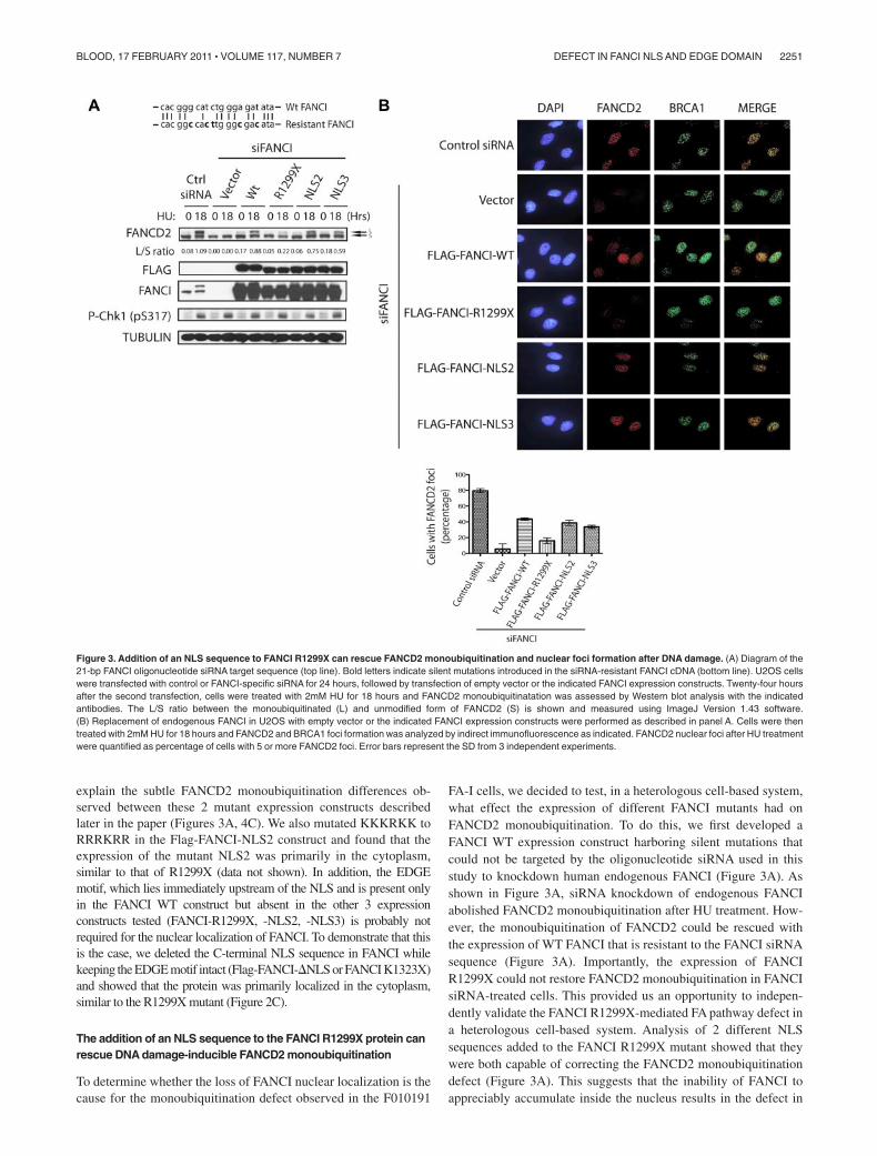
FA-I cells, we decided to test, in a heterologous cell-based system,what effect the expression of different FANCI mutants had onFANCD2 monoubiquitination. To do this, we first developed aFANCI WT expression construct harboring silent mutations thatcould not be targeted by the oligonucleotide siRNA used in thisstudy to knockdown human endogenous FANCI (Figure 3A). Asshown in Figure 3A, siRNA knockdown of endogenous FANCIabolished FANCD2 monoubiquitination after HU treatment. How-ever, the monoubiquitination of FANCD2 could be rescued withthe expression of WT FANCI that is resistant to the FANCI siRNAsequence (Figure 3A). Importantly, the expression of FANCIR1299X could not restore FANCD2 monoubiquitination in FANCIsiRNA-treated cells. This provided us an opportunity to indepen-dently validate the FANCI R1299X-mediated FA pathway defect ina heterologous cell-based system. Analysis of 2 different NLSsequences added to the FANCI R1299X mutant showed that theywere both capable of correcting the FANCD2 monoubiquitinationdefect (Figure 3A). This suggests that the inability of FANCI toappreciably accumulate inside the nucleus results in the defect in
Figure 3. Addition of an NLS sequence to FANCI R1299X can rescue FANCD2 monoubiquitination and nuclear foci formation after DNA damage. (A) Diagram of the21-bp FANCI oligonucleotide siRNA target sequence (top line). Bold letters indicate silent mutations introduced in the siRNA-resistant FANCI cDNA (bottom line). U2OS cellswere transfected with control or FANCI-specific siRNA for 24 hours, followed by transfection of empty vector or the indicated FANCI expression constructs. Twenty-four hoursafter the second transfection, cells were treated with 2mM HU for 18 hours and FANCD2 monoubiquitinatation was assessed by Western blot analysis with the indicatedantibodies. The L/S ratio between the monoubiquitinated (L) and unmodified form of FANCD2 (S) is shown and measured using ImageJ Version 1.43 software.(B) Replacement of endogenous FANCI in U2OS with empty vector or the indicated FANCI expression constructs were performed as described in panel A. Cells were thentreated with 2mM HU for 18 hours and FANCD2 and BRCA1 foci formation was analyzed by indirect immunofluorescence as indicated. FANCD2 nuclear foci after HU treatmentwere quantified as percentage of cells with 5 or more FANCD2 foci. Error bars represent the SD from 3 independent experiments.
DEFECT IN FANCI NLS AND EDGE DOMAIN 2251BLOOD, 17 FEBRUARY 2011 � VOLUME 117, NUMBER 7

FANCD2 monoubiquitination. This underscores the relevance andsignificance of proper protein localization as a mean to fullyactivate and engage a DNA damage response pathway.
It has been shown that monoubiquitination of FANCI andFANCD2 is required for its targeting to DNA damage/repair foci inthe nucleus.5-7 As expected, the expression of FANCI constructsthat were capable of supporting FANCD2 monoubiquitination, wasalso necessary to promote DNA damage-induced FANCD2 nuclearfoci formation in cells treated with FANCI siRNA (Figure 3B).As shown previously, the loss of FANCI did not perturbBRCA1 nuclear foci formation6 (Figure 3B). These studies suggestthat loss of the C-terminal NLS on FANCI can dramatically alterthe cell’s ability to mediate a DNA damage response through the
FA pathway. Despite the loss of the EDGE motif, artificial fusion ofNLS to FANCI R1299X mutant can rescue the FANCD2 monoubiq-uitination and localization to DNA damage/repair sites on chromatin.
Correction of the F010191 FA-I patient-derived fibroblasts withectopic expression of FANCI
Despite the increasing number of FA-I patients being characterized,only one FA-I patient cell line has been functionally corrected withthe introduction of WT FANCI.7 It is unclear why FA-I patient-derived cells have been difficult to functionally correct. Severallaboratories, including ours, have used different methods of genetransduction to achieve FANCI WT expression into the differentFA-I lymphoblasts, but with minimal success (data not shown).Instead of using patient lymphoblasts, we decided to obtainfibroblasts from the same F010191 FA-I patient to determinewhether these cells would be easier to functionally correct.Interestingly, the level of FANCI R1299X protein expression issignificantly lower than in other normal fibroblast or transformedepithelial cells compared with their FANCI levels (Figure 4A; anddata not shown). It is unclear what causes the decrease in totalFANCI protein in F010191 FA-I fibroblasts but not in the lympho-blasts. Nevertheless, the F010191 fibroblasts mimic a cell line withreduced FANCI protein levels, similar to the condition duringFANCI siRNA knockdown treatment. Surprisingly, simple tran-sient expression of WT FANCI was capable of increasing the levelsof FANCD2 monoubiquitination (Figure 4B). As expected, expres-sion of FANCI K523R (FANCI mutant that cannot be monoubiquiti-nated) and R1299X failed to increase FANCD2 monoubiquitina-tion in the F010191 fibroblasts (Figure 4B). This is an importantcontrol to show that simple overexpression of FANCI mutants isinsufficient to activate FANCD2 monoubiquitination. Importantly,expression of FANCI-NLS2 and -NLS3 was capable ofincreasing the monoubiquitination levels of FANCD2 (compareWT with NLS2 and NLS3 L/S ratios; Figure 4C). This was alsotrue for the correction of FANCD2 nuclear foci formation in theF010191 fibroblasts with WT versus NLS2 and NLS3 mutants(Figure 5A-B). Thus, the molecular events associated with FApathway activation appears to be operational after complementingthe F010191 FA-I fibroblasts with WT FANCI or a FANCI mutantmissing the EDGE motif that is still capable of localizing andaccumulating to the nucleus.
FANCI missing the EDGE motif fails to correct DNAcrosslinker sensitivity
The FANCI R1299X mutant has a deletion of the last 30 aminoacids. Within this short region contains 2 putative motifs: one is anNLS and the other is a lesser-known or characterized EDGE motif,which lies upstream of the NLS. Throughout this study, we haveshown that the defect in FANCD2 monoubiquitination and nuclearfoci localization can be attributed to the loss of the C-terminal NLSsequence in the naturally occurring R1299X FANCI mutant.However, it is unclear whether the EDGE motif plays anyfunctional role in the FA pathway because it is not required forFANCI nuclear localization, FANCD2 monoubiquitination, andFANCD2 nuclear foci formation. Therefore, we tested whetherexpression of FANCI without the EDGE motif could still correctDNA crosslinker sensitivity in the F010191 fibroblasts. Surpris-ingly, expression of WT FANCI, but not the FANCI-NLS2and -NLS3 mutants, was found to be more resistant to the DNAcrosslinker MMC in the F010191 fibroblasts (Figure 5C). Thus,artificial nuclear accumulation of the FANCI R1299X mutant is
Figure 4. Recovery of FANCD2 monoubiquitination in the FA-I F010191 fibro-blasts. (A) HeLa and F010191 fibroblasts were either untreated or treated with HU(2mM for 18 hours). (B-C) F010191 fibroblasts were transfected with empty vector orthe indicated FANCI expression constructs. Forty-eight hours after transfection, cellswere treated with 2mM HU for 24 hours and FANCD2 monoubiquitination wasassessed by Western blot analysis and probed with the indicated antibodies. The L/Sratio was calculated as described in Figure 3A.
2252 COLNAGHI et al BLOOD, 17 FEBRUARY 2011 � VOLUME 117, NUMBER 7

not sufficient to fully correct FA-I cells. This opens thepossibility that the EDGE motif is required for an unspecifiedfunction that is independent or downstream of FANCD2-FANCIDNA damage-induced monoubiquitination and foci formation.To further characterize the EDGE motif, we tested the D1301Apoint mutant of the EDGE motif that was first shown tomodulate in vitro DNA-binding activity of FANCI.25 We foundthat the D1301A mutant of FANCI behaved similarly to theNLS2 construct, in terms of rescuing the FANCD2 monoubiquiti-nation and foci formation defect in the F010191 fibroblasts(Figure 6A and data not shown). However, the D1301A mutantcould not fully complement the MMC sensitivity to WT levels(Figure 6B). The slight increase in MMC resistance of theD1301A mutant compared with the NLS2 construct is probablythe result of the milder mutation of the EDGE motif generated inthe mutants. Although highly improbable, it is a possibility thatthe FANCI EDGE motif may mediate DNA crosslinker resis-tance outside of the nucleus. To rule out a potential EDGE motiffunction that is independent of its nuclear localization, we testedwhether the FANCI-�NLS could rescue MMC sensitivity in theF010191 fibroblasts. As expected, FANCI-�NLS constructfailed to functionally complement the FA-I fibroblasts forFANCD2 monoubiquitination and MMC resistance (Figure6C-D), suggesting that the intact EDGE motif is not sufficientfor its role in DNA crosslink repair without proper protein
localization. The potential role for the EDGE motif in FANCIwill be addressed in “Discussion.”
Discussion
In this study, we provide evidence to explain the molecular defectin a patient-derived FA-I cell line (F010191) expressing a shortC-terminal truncation mutant form of FANCI. Although onlymissing the last 30 amino acids from the C-terminal end of theFANCI protein, this FA-I cell line has been reported previously tohave defective FANCD2 and FANCI monoubiquitination.6 Thus, itwas unclear how the C-terminal region mechanistically contributesto the activation of the FA pathway. Here we show that mislocaliza-tion of the FANCI protein is responsible for the loss of monoubiq-uitination and nuclear foci formation in the FANCD2 effectorprotein. Predicted motifs in the C-terminal region of FANCIsuggest an EDGE motif and NLS sequence. To test the contributionof the NLS motif, we artificially fused an NLS sequence (one fromthe original FANCI NLS, the other from c-Myc NLS) to theC-terminal region of the truncated FANCI protein (R1299X).Importantly, fusion proteins carrying the C-terminal NLS se-quences were capable of rescuing the FANCD2 monoubiquitina-tion and nuclear foci formation defect from the R1299X FANCImutant in different cell types.
Figure 5. FANCI missing the EDGE motif fails to correct MMC sensitivity in the FA-I F010191 fibroblasts. (A) F010191 fibroblasts were transfected with empty vector orthe indicated FANCI expression constructs. Forty-eight hours after transfection, cells were treated with 2mM HU for 24 hours and processed for immunostaining. FANCD2 andBRCA1 nuclear foci formation was analyzed by immunofluorescence as previously described. (B) FANCD2 nuclear foci after HU treatment were quantified. Error barsrepresent SD from 3 independent experiments. (C) FA-I F010191 fibroblasts were transfected with either empty vector or the indicated FANCI expression constructs; and48 hours after transfection, cells were treated with MMC at the indicated dose for 5 more days. Percentage cell survival is shown as an average of 3 independent experiments.Error bars represent the SD for the different experiments performed.
DEFECT IN FANCI NLS AND EDGE DOMAIN 2253BLOOD, 17 FEBRUARY 2011 � VOLUME 117, NUMBER 7

For the first time, we were able to functionally complement theFA-I fibroblast cell line (F010191) with WT FANCI expression.This enabled us to directly test the functional contribution ofdifferent FANCI mutants toward the monoubiquitination andnuclear foci formation of FANCD2 during DNA damage. We wereable to confirm our heterologous cell-based studies and show thatthe addition of an NLS to the R1299X FANCI mutant wassufficient to overcome the localization defect of the R1299XFANCI mutant and rescue the monoubiquitination and nuclear fociformation of FANCD2 in the FA-I patient-derived fibroblasts(F010191). However, unlike the WT FANCI, the NLS fusedR1299X FANCI could not correct MMC sensitivity in the patientfibroblasts. This suggests that the putative EDGE motif (which isstill missing in the NLS fused R1299X FANCI mutant) may beimportant in mediating a function of FANCD2-FANCI independentof FANCI, monoubiquitination and foci formation, such as in DNAdamage sensing or DNA crosslink repair.
As mentioned in “Results,” FANCI has a putative EDGE motifthat resides in the last 30 residues of the protein. To study the role ofdifferent structural domains on FANCI, we initially focused ourattention on patient-derived mutations of the FANCI gene that mayshed some light on critical structural regions of the FANCI proteinfor normal FA pathway function. Fortunately, the FA-I F010191patient-derived cells have one mutant expressing allele that resultsin the deletion of the last 30 residues of FANCI (the other mutantallele results in a more severe protein truncation).6 This gave us anexcellent opportunity to dissect the structure and function of theC-terminal region containing the EDGE motif in the patient-derived cells because it was already known that the human patientmissing this region results in the FA phenotype. The EDGE motifwas previously defined by the EDGE sequence itself, containing acluster of acidic residues (letters denote amino acids).23 It wasoriginally discovered and characterized in a splice variant form ofFANCD2 containing exon 44. Reconstitution of FANCD2 mutantswith a defective EDGE motif failed to rescue MMC sensitivity inFANCD2-deficient fibroblasts, implying a crucial function of theEDGE motif in DNA damage sensing or DNA repair.23 Interest-ingly, expression of FANCD2 with defective EDGE motif in theFA-D2 cells displayed normal cellular phenotype regarding thelevels observed for FANCD2 monoubiquitination and nuclear foci
formation. This molecular signature is strikingly similar to theFANCI EDGE motif scenario portrayed in our present study.
In vitro studies have suggested that purified FANCI can directlybind to different types of DNA substrates.21,25,28 The intrinsicDNA-binding activity of FANCI does not require its monoubiquiti-nation or association with FANCD2.25,28 Interestingly, it wasshown that changing a single residue (D1301A) in the FANCIEDGE motif dramatically enhanced its DNA binding for differentsubstrates, suggesting that the EDGE motif may be involved inregulating the dynamics of FANCI-FANCD2 DNA binding duringinterstrand crosslink repair.25 However, from these previous stud-ies, it was still unclear whether the intrinsic DNA-binding activityof FANCI functions upstream or downstream of FANCI andFANCD2 monoubiquitination and nuclear foci formation in vivo.Evidence from our present study points to an independent functionfor the FANCI EDGE motif. This is in agreement with the currentmodel suggesting that both FANCI and FANCD2 may be directlyinvolved in binding to specific DNA structural intermediates tofunction in DNA repair.25,29 It will be interesting to test whethermutations in the EDGE motif prevent FANCI-FANCD2 fromdissociating from DNA in a timely manner to insure properrecovery from DNA crosslink repair, in vivo. Recently, severalstudies suggest that the monoubiquitinated FANCI-FANCD2 pairis responsible for recruiting a novel FA-associated nuclease, FAN1,which is critical for DNA crosslink repair.30-33 It will be interestingto test whether the EDGE motif in FANCI is required forsubsequent FAN1 binding after DNA damage. It has been sug-gested, although not proven, that the monoubiquitination ofFANCI-FANCD2 provides a dock for the recruitment of FAN1 toFANCD2 via its UBZ domain (ubiquitin-binding zinc finger motif).Generally, ubiquitin-binding domain-containing substrates thatinteract with their ubiquitinated receptors have a secondary bindingsite with their cognate receptors. This appears to be the case forproliferating cell nuclear antigen and the translesion synthesispolymerases, whereby the translesion synthesis polymerases inter-act with monoubiquitinated proliferating cell nuclear antigen viatheir PIP box and ubiquitin-binding domains for a more specificinteraction.34
Mutations in FA genes that cause protein mislocalizationprobably play an important role in the molecular pathogenesis of
Figure 6. A deletion of the C-terminal NLS of FANCIfails to correct MMC sensitivity in the FA-I F010191fibroblasts. (A,C) F010191 fibroblasts were transfectedwith empty vector or the indicated FANCI expressionconstructs. Forty-eight hours after transfection, cells weretreated with 2mM HU for 24 hours and FANCD2 monou-biquitination was assessed by Western blot analysis andprobed with the indicated antibodies. The L/S ratio wascalculated as described in Figure 3A. (B,D) FA-I F010191fibroblasts were transfected with either empty vector orthe indicated FANCI expression constructs; and 48 hoursafter transfection, cells were treated with MMC at theindicated dose for 5 more days. Percentage cell survivalis shown as an average of 3 independent experiments.Error bars represent the SD for the different experimentsperformed.
2254 COLNAGHI et al BLOOD, 17 FEBRUARY 2011 � VOLUME 117, NUMBER 7

FA. For example, the 8 proteins forming the FA core ubiquitinligase complex, and other FA-associated proteins are normallylocalized predominantly in the nucleus.35-42 Specific patient-derived mutations in FA core complex genes, such as in FANCA,can result in aberrant cytoplasmic localization of the FANCAprotein, which can lead to defects in FA core complex assembly,FANCD2 monoubiquitination, and interstrand crosslink repair.43
Although it is possible that FA proteins may possess cytoplasmicfunction, the majority of the FA pathway function takes place in thenucleus and possibly on chromatin-bound DNA. Localization ofFA proteins, including FANCI, to the nuclear compartment isprobably critical for the efficient activation of DNA damagesensing, signaling, and repair of DNA lesions. In our present study,we provide evidence that a patient-derived mutant form of FANCI(R1299X) is mislocalized into the cytoplasmic compartment. Thisis the result of the loss of its C-terminal NLS sequence. We foundthat the localization defect in the FANCI mutant did not drasticallyaffect or change the localization of FANCD2. This suggests thatFANCI may be targeted to the nucleus independently of FANCD2and that FANCD2 subcellular localization is not completelydependent on FANCI. Based on primary amino acid sequenceanalysis, human FANCD2 has 2 NLS sequences that are unrelatedto the C-terminal NLS on FANCI.44 However, to the best of ourknowledge, the function of these 2 FANCD2 NLS sequences hasnot been tested. Currently, the model for FA pathway activationinvolves the interaction between FANCI with FANCD2 on nuclearchromatin-bound DNA to facilitate their mutual monoubiquitina-tion.6,7,21 In addition, it is probable that the nuclear PI3-like kinase,ATR, is important in stimulating this reaction through phosphoryla-tion of the ID complex.8,45,46
Mislocalization of FA proteins may also play a role in carcino-genesis. In a recent study, FANCD2 was found to be largelycytoplasmic in more malignant human breast tumor tissues.47
Cytoplasmic localization of FANCD2 in cells, similar to that ofFANCI, also probably prevents FA pathway activation, leading togenome instability. It is still unclear what is the precise mechanismthat causes the FANCD2 protein mislocalization in these humanbreast cancer tissues.47 It will be interesting to determine whether
FANCI may also be abnormally expressed and cytoplasmicallylocalized in human breast cancers and whether this is the result ofsporadic mutations that target the C-terminal NLS function ofFANCI. It is known that defects in protein NLSs have beenassociated with human disease conditions, such as numerouscancers and developmental disorders.48 The increasing prevalenceof FA gene mutations observed in human cancers1,49 underscoresthe importance of careful structure/function analyses of FA proteinsand their role in facilitating genome stability.
Acknowledgments
The authors thank the affected FA-I persons and their families forproviding tissue samples for these studies, the many physicians thathave referred families for participation in the research, A. Auerbachand F. Lach for normal patient fibroblasts and lymphoblast controlcells and for helping with the FA-I F010191 characterization andcell line work, Y. Deng and NYU Skirball Imaging core facility forassistance with microscopy, and members of the laboratories ofT.T.H., D. Bar-Sagi, and D. Reinberg for their technical assistance,equipment, and helpful discussions.
This work was supported in part by the Wendy Will CaseFoundation, National Institutes of Health (grant RO1GM084244),and New York University School of Medicine Start-up funding (T.T.H.).
Authorship
Contribution: L.C., M.J.K.J., X.M.C.-R., and T.T.H. designed andexecuted experiments; D.S. and H.H. provided patient-derivedfibroblasts and other critical reagents for this study; and L.C. andT.T.H. wrote the manuscript.
Conflict-of-interest disclosure: The authors declare no compet-ing financial interests.
Correspondence: Tony T. Huang, Department of Biochemistry,New York University School of Medicine, 522 First Ave, SmilowBldg 210, New York, NY 10016; e-mail: [email protected].
References
1. D’Andrea AD. Susceptibility pathways in Fanconi’sanemia and breast cancer. N Engl J Med. 2010;362(20):1909-1919.
2. Auerbach AD. Fanconi anemia and its diagnosis.Mutat Res. 2009;668(1):4-10.
3. Moldovan GL, D’Andrea AD. How the Fanconianemia pathway guards the genome. Annu RevGenet. 2009;43:223-249.
4. Wang W. Emergence of a DNA-damage re-sponse network consisting of Fanconi anaemiaand BRCA proteins. Nat Rev Genet. 2007;8(10):735-748.
5. Garcia-Higuera I, Taniguchi T, Ganesan S, et al.Interaction of the Fanconi anemia proteins andBRCA1 in a common pathway. Mol Cell. 2001;7(2):249-262.
6. Sims AE, Spiteri E, Sims RJ III, et al. FANCI is asecond monoubiquitinated member of the Fan-coni anemia pathway. Nat Struct Mol Biol. 2007;14(6):564-567.
7. Smogorzewska A, Matsuoka S, Vinciguerra P,et al. Identification of the FANCI protein, a mo-noubiquitinated FANCD2 paralog required forDNA repair. Cell. 2007;129(2):289-301.
8. Ishiai M, Kitao H, Smogorzewska A, et al. FANCIphosphorylation functions as a molecular switchto turn on the Fanconi anemia pathway. NatStruct Mol Biol. 2008;15(11):1138-1146.
9. Zhi G, Wilson JB, Chen X, et al. Fanconi anemiacomplementation group FANCD2 protein serine331 phosphorylation is important for Fanconi ane-mia pathway function and BRCA2 interaction.Cancer Res. 2009;69(22):8775-8783.
10. Bridge WL, Vandenberg CJ, Franklin RJ, Hiom K.The BRIP1 helicase functions independently ofBRCA1 in the Fanconi anemia pathway for DNAcrosslink repair. Nat Genet. 2005;37(9):953-957.
11. Levran O, Attwooll C, Henry RT, et al. TheBRCA1-interacting helicase BRIP1 is deficient inFanconi anemia. Nat Genet. 2005;39(9):931-933.
12. Levitus M, Waisfisz Q, Godthelp BC, et al. TheDNA Helicase BRIP1 is defective in Fanconi ane-mia complementation group J. Nat Genet. 2005;37(9):934-935.
13. Howlett NG, Taniguchi T, Olson S, et al. Biallelicinactivation of BRCA2 in Fanconi anemia. Sci-ence. 2002;297(5581):606-609.
14. Reid S, Schindler D, Hanenberg H, et al. Biallelicmutations in PALB2 cause Fanconi anemia sub-type FA-N and predispose to childhood cancer.Nat Genet. 2007;39(2):162-164.
15. Xia B, Dorsman JC, Ameziane N, et al. Fanconianemia is associated with a defect in the BRCA2partner PALB2. Nat Genet. 2007;39(2):159-161.
16. Vaz F, Hanenberg H, Schuster B, et al. Mutation
of the RAD51C gene in a Fanconi anemia-likedisorder. Nat Genet. 2010;42(5):406-409.
17. Meindl A, Hellebrand H, Wiek C, et al. Germlinemutations in breast and ovarian cancer pedigreesestablish RAD51C as a human cancer suscepti-bility gene. Nat Genet. 2010;42(5):410-414.
18. Dorsman JC, Levitus M, Rockx D, et al. Identifica-tion of the Fanconi anemia complementationgroup I gene, FANCI. Cell Oncol. 2007;29(3):211-218.
19. Nijman SM, Huang TT, Dirac AM, et al. The deu-biquitinating enzyme USP1 regulates the Fanconianemia pathway. Mol Cell. 2005;17(3):331-339.
20. Huang TT, Nijman SM, Mirchandani KD, et al.Regulation of monoubiquitinated PCNA by DUBautocleavage. Nat Cell Biol. 2006;8(4):339-347.
21. Knipscheer P, Raschle M, Smogorzewska A,et al. The Fanconi anemia pathway promotes rep-lication-dependent DNA interstrand cross-link re-pair. Science. 2009;326(5960):1698-1701.
22. Kalb R, Neveling K, Hoehn H, et al. Hypomorphicmutations in the gene encoding a key Fanconianemia protein, FANCD2, sustain a significantgroup of FA-D2 patients with severe phenotype.Am J Hum Genet. 2007;80(5):895-910.
23. Montes de Oca R, Andreassen PR, Margossian SP,et al. Regulated interaction of the Fanconi anemia
DEFECT IN FANCI NLS AND EDGE DOMAIN 2255BLOOD, 17 FEBRUARY 2011 � VOLUME 117, NUMBER 7

protein, FANCD2, with chromatin. Blood. 2005;105(3):1003-1009.
24. Mendez J, Stillman B. Chromatin association ofhuman origin recognition complex, cdc6, andminichromosome maintenance proteins duringthe cell cycle: assembly of prereplication com-plexes in late mitosis. Mol Cell Biol. 2000;20(22):8602-8612.
25. Yuan F, El Hokayem J, Zhou W, Zhang Y. FANCIprotein binds to DNA and interacts with FANCD2to recognize branched structures. J Biol Chem.2009;284(36):24443-24452.
26. Dang CV, Lee WM. Identification of the humanc-myc protein nuclear translocation signal. MolCell Biol. 1988;8(10):4048-4054.
27. Saphire AC, Bark SJ, Gerace L. All four homo-chiral enantiomers of a nuclear localization se-quence derived from c-Myc serve as functionalimport signals. J Biol Chem. 1998;273(45):29764-29769.
28. Longerich S, San Filippo J, Liu D, Sung P. FANCIbinds branched DNA and is monoubiquitinated byUBE2T-FANCL. J Biol Chem. 2009;284(35):23182-23186.
29. Moldovan GL, D’Andrea AD. FANCD2 hurdles theDNA interstrand crosslink. Cell. 2009;139(7):1222-1224.
30. MacKay C, Declais AC, Lundin C, et al. Identifica-tion of KIAA1018/FAN1, a DNA repair nucleaserecruited to DNA damage by monoubiquitinatedFANCD2. Cell. 2010;142(1):65-76.
31. Kratz K, Schopf B, Kaden S, et al. Deficiency ofFANCD2-associated nuclease KIAA1018/FAN1sensitizes cells to interstrand crosslinking agents.Cell. 2010;142(1):77-88.
32. Smogorzewska A, Desetty R, Saito TT, et al. A
genetic screen identifies FAN1, a Fanconianemia-associated nuclease necessary for DNAinterstrand crosslink repair. Mol Cell. 2010;39(1):36-47.
33. Liu T, Ghosal G, Yuan J, Chen J, Huang J. FAN1acts with FANCI-FANCD2 to promote DNA inter-strand cross-link repair. Science. 2010;329(5992):693-696.
34. Bienko M, Green CM, Crosetto N, et al. Ubiquitin-binding domains in y-family polymerases regulatetranslesion synthesis. Science. 2005;310(5755):1821-1824.
35. Savoia A, Garcia-Higuera I, D’Andrea AD.Nuclear localization of the Fanconi anemia pro-tein FANCC is required for functional activity.Blood. 1999;93(11):4025-4026.
36. Taniguchi T, D’Andrea AD. The Fanconi anemiaprotein, FANCE, promotes the nuclear accumula-tion of FANCC. Blood. 2002;100(7):2457-2462.
37. Yamashita T, Kupfer GM, Naf D, et al. The Fan-coni anemia pathway requires FAA phosphoryla-tion and FAA/FAC nuclear accumulation. ProcNatl Acad Sci U S A. 1998;95(22):13085-13090.
38. Naf D, Kupfer GM, Suliman A, Lambert K,D’Andrea AD. Functional activity of the Fanconianemia protein FAA requires FAC binding andnuclear localization. Mol Cell Biol. 1998;18(10):5952-5960.
39. Kupfer G, Naf D, Garcia-Higuera I, et al. A patient-derived mutant form of the Fanconi anemia pro-tein, FANCA, is defective in nuclear accumula-tion. Exp Hematol. 1999;27(4):587-593.
40. Garcia-Higuera I, Kuang Y, Naf D, Wasik J,D’Andrea AD. Fanconi anemia proteins FANCA,FANCC, and FANCG/XRCC9 interact in a func-tional nuclear complex. Mol Cell Biol. 1999;19(7):4866-4873.
41. Kupfer GM, Naf D, Suliman A, Pulsipher M,D’Andrea AD. The Fanconi anaemia proteins,FAA and FAC, interact to form a nuclear complex.Nat Genet. 1997;17(4):487-490.
42. Garcia-Higuera I, Kuang Y, Denham J, D’Andrea AD.The Fanconi anemia proteins FANCA and FANCGstabilize each other and promote the nuclear accu-mulation of the Fanconi anemia complex. Blood.2000;96(9):3224-3230.
43. Adachi D, Oda T, Yagasaki H, et al. Heteroge-neous activation of the Fanconi anemia pathwayby patient-derived FANCA mutants. Hum MolGenet. 2002;11(25):3125-3134.
44. Dequen F, St-Laurent JF, Gagnon SN, Carreau M,Desnoyers S. The Caenorhabditis elegans FancD2ortholog is required for survival following DNA dam-age. Comp Biochem Physiol B Biochem Mol Biol.2005;141(4):453-460.
45. Andreassen PR, D’Andrea AD, Taniguchi T. ATRcouples FANCD2 monoubiquitination to the DNA-damage response. Genes Dev. 2004;18(16):1958-1963.
46. Ho GP, Margossian S, Taniguchi T, D’Andrea AD.Phosphorylation of FANCD2 on two novel sites isrequired for mitomycin C resistance. Mol CellBiol. 2006;26(18):7005-7015.
47. Rudland PS, Platt-Higgins AM, Davies LM, et al.Significance of the Fanconi anemia FANCD2 pro-tein in sporadic and metastatic human breastcancer. Am J Pathol. 2010;176(6):2935-2947.
48. McLane LM, Corbett AH. Nuclear localization sig-nals and human disease. IUBMB Life. 2009;61(7):697-706.
49. Kennedy RD, D’Andrea AD. DNA repair pathwaysin clinical practice: lessons from pediatric cancersusceptibility syndromes. J Clin Oncol. 2006;24(23):3799-3808.
2256 COLNAGHI et al BLOOD, 17 FEBRUARY 2011 � VOLUME 117, NUMBER 7
Related Documents