Pathological and serological findings in wild boars (Sus scrofa) from Gran Sasso and Monti della Laga National Park (Central Italy) SUMMARY The Italian Gran Sasso e Monti della Laga National Park represents one of the largest protected areas in Europe, and it is par- ticularly suitable for the preservation of the wild fauna including some endangered species, such as Abruzzi chamois, Apenni- ne wolf and Marsicano brown bear. Wild boar population residing within the National Park Gran Sasso e Monti della Laga has been sharply increasing and nega- tively impacts on sensitive ecosystems and human activities. Thus, a management plan, based on wild boar capture by means of catch fences and slaughter, has been implemented. About the 77% of wild boars under study were “young adult” (aged 4-12 months, body weight = 20-60 kg), and the remaining 23% were adult (aged > 12 months). At slaughterhouse, pathological findings have been recorded from 101 wild boars. Microscopic investigations have been car- ried out on diseased, as well as on apparently healthy tissues. Blood samples collected from 126 wild boars were tested for Auje- szky’s disease virus, Brucella suis and Leptospira interrogans. Lesions were mainly caused by parasites, and parasitic bronchopneumonia by Metastrongylus spp. was most frequently obser- ved (92%). Hepatic white spots (28%), lymphoproliferative nodules, hepatic distomatosis (3%), Cysticercus tenuicollis (15%) and hydatid cysts (6%) were also commonly detected. Serology demonstrated a high prevalence of Aujeszky’s disease (35%) and Brucella suis (15%) antibodies in the population under study. Zoonoses represent about the 60% of emerging public health concerns, and the majority of emerging infectious diseases arise from wild animals. In that context, wild boar is of particular relevance because of its extreme adaptability in different habitats, its wide geographical distribution and its high reproductive rates. The implementation of this management plan represented a good chance to face the wild fauna living within the Gran Sasso e Monti della Laga National Park. Pathological findings herein described, along with concurrent microbiological, serological and parasitological investigations, contribute to evaluate the health status of wild boar population residing in such a relevant pro- tected area. KEY WORDS Wild boar, Central Italy, management, pathology, serology. U. DI NICOLA 1 , M. SCACCHIA 2 , G. MARRUCHELLA 2,3 1 Servizio Scientifico Ente Parco Nazionale del Gran Sasso e Monti della Laga, via del Convento 1, 67010 Assergi (L’Aquila), Italia 2 Istituto Zooprofilattico Sperimentale dell’Abruzzo e del Molise “G. Caporale”, Campo Boario, 64100, Teramo, Italy 3 Università degli Studi di Teramo, Facoltà di Medicina Veterinaria, Piazza A. Moro 45, 64100, Teramo, Italia U. Di Nicola et al. Large Animal Review 2015; 21: 167-171 167 Autore per la corrispondenza: Giuseppe Marruchella ([email protected]). O INTRODUCTION Gran Sasso e Monti della Laga National Park (GSMLNP) co- vers an area of about 150,000 hectares throughout the territo- ries of three regions (Abruzzo, Lazio and Marche) in Central Italy, thus representing one of the largest protected areas in Europe. The extension and the variety of environments make GSMLNP suitable for the preservation of the wild fauna, whi- ch includes endangered species such as Abruzzi chamois (Ru- picapra pyrenaica ornata), Apennine wolf (Canis lupus italicus) and Marsicano brown bear (Ursos marsicanus arctos) 1 . Wild boar (Sus scrofa) population is widely distributed in Italy and has been progressively increasing and widening its distribution range, a finding which confirms the extraordi- nary adaptability of that animal species 29 . Wild boar popula- tion residing within the GSMLNP has been also significantly increasing (estimated average density = 11 animals/km 2 ) with negative impacts on sensitive ecosystems and human activities. As a consequence, a plan for the management of wild boars within the territory of GSMLNP has been imple- mented 1 . We report herein the pathological and serological findings observed in wild boars, which have been kept and slaughtered in the context of such management plan between 2008 and 2010. MATERIALS AND METHODS Animals Wild boars were captured in the provinces of Teramo (Abruzzo region) and Rieti (Lazio region) by means of catch fences, identified by applying ear tags, and then delivered to two abattoirs located near Teramo and Ascoli Piceno (Mar- che region).

Welcome message from author
This document is posted to help you gain knowledge. Please leave a comment to let me know what you think about it! Share it to your friends and learn new things together.
Transcript

Pathological and serological findings inwild boars (Sus scrofa) from Gran Sasso andMonti della Laga National Park (Central Italy)
SUMMARYThe Italian Gran Sasso e Monti della Laga National Park represents one of the largest protected areas in Europe, and it is par-ticularly suitable for the preservation of the wild fauna including some endangered species, such as Abruzzi chamois, Apenni-ne wolf and Marsicano brown bear.Wild boar population residing within the National Park Gran Sasso e Monti della Laga has been sharply increasing and nega-tively impacts on sensitive ecosystems and human activities. Thus, a management plan, based on wild boar capture by meansof catch fences and slaughter, has been implemented. About the 77% of wild boars under study were “young adult” (aged 4-12months, body weight = 20-60 kg), and the remaining 23% were adult (aged > 12 months).At slaughterhouse, pathological findings have been recorded from 101 wild boars. Microscopic investigations have been car-ried out on diseased, as well as on apparently healthy tissues. Blood samples collected from 126 wild boars were tested for Auje-szky’s disease virus, Brucella suis and Leptospira interrogans.Lesions were mainly caused by parasites, and parasitic bronchopneumonia by Metastrongylus spp. was most frequently obser-ved (92%). Hepatic white spots (28%), lymphoproliferative nodules, hepatic distomatosis (3%), Cysticercus tenuicollis (15%)and hydatid cysts (6%) were also commonly detected. Serology demonstrated a high prevalence of Aujeszky’s disease (35%)and Brucella suis (15%) antibodies in the population under study.Zoonoses represent about the 60% of emerging public health concerns, and the majority of emerging infectious diseases arisefrom wild animals. In that context, wild boar is of particular relevance because of its extreme adaptability in different habitats,its wide geographical distribution and its high reproductive rates.The implementation of this management plan represented a good chance to face the wild fauna living within the Gran Sasso eMonti della Laga National Park. Pathological findings herein described, along with concurrent microbiological, serological andparasitological investigations, contribute to evaluate the health status of wild boar population residing in such a relevant pro-tected area.
KEY WORDSWild boar, Central Italy, management, pathology, serology.
U. DI NICOLA1, M. SCACCHIA2, G. MARRUCHELLA2,3
1 Servizio Scientifico Ente Parco Nazionale del Gran Sasso e Monti della Laga, via del Convento 1, 67010 Assergi (L’Aquila), Italia
2 Istituto Zooprofilattico Sperimentale dell’Abruzzo e del Molise “G. Caporale”, Campo Boario, 64100, Teramo, Italy
3 Università degli Studi di Teramo, Facoltà di Medicina Veterinaria, Piazza A. Moro 45, 64100, Teramo, Italia
U. Di Nicola et al. Large Animal Review 2015; 21: 167-171 167
O
Autore per la corrispondenza:Giuseppe Marruchella ([email protected]).
O
INTRODUCTION
Gran Sasso e Monti della Laga National Park (GSMLNP) co-vers an area of about 150,000 hectares throughout the territo-ries of three regions (Abruzzo, Lazio and Marche) in CentralItaly, thus representing one of the largest protected areas inEurope. The extension and the variety of environments makeGSMLNP suitable for the preservation of the wild fauna, whi-ch includes endangered species such as Abruzzi chamois (Ru-picapra pyrenaica ornata), Apennine wolf (Canis lupus italicus)and Marsicano brown bear (Ursos marsicanus arctos)1.Wild boar (Sus scrofa) population is widely distributed inItaly and has been progressively increasing and widening itsdistribution range, a finding which confirms the extraordi-nary adaptability of that animal species29. Wild boar popula-
tion residing within the GSMLNP has been also significantlyincreasing (estimated average density = 11 animals/km2)with negative impacts on sensitive ecosystems and humanactivities. As a consequence, a plan for the management ofwild boars within the territory of GSMLNP has been imple-mented1. We report herein the pathological and serologicalfindings observed in wild boars, which have been kept andslaughtered in the context of such management planbetween 2008 and 2010.
MATERIALS AND METHODS
AnimalsWild boars were captured in the provinces of Teramo(Abruzzo region) and Rieti (Lazio region) by means of catchfences, identified by applying ear tags, and then delivered totwo abattoirs located near Teramo and Ascoli Piceno (Mar-che region).
Di Nicola_imp:ok 20-06-2016 11:03 Pagina 167

168 Pathological and serological findings in wild boars (Sus scrofa) from Gran Sasso and Monti della Laga National Park
About the 77% of wild boars under study were “young adult”(aged 4-12 months, body weight = 20-60 kg), and the remai-ning 23% were adult (aged > 12 months). Males and femaleswere equally distributed. Wild boars were usually slaughte-red soon after their arrival at the abattoir, with the only ex-ception of 25 animals, which were housed in pens for a fewdays before slaughtering.
PathologyA total of 101 wild boars have been investigated. At post-mortem inspection, lesions affecting a wide range of organswere recorded: lungs, liver, spleen, kidneys, heart, palatinetonsils, retro-pharyngeal lymph nodes, stomach.Pathological samples were promptly fixed in 10% neutralbuffered formalin, embedded in paraffin and routinely pro-cessed for histopathology (Haematoxylin & Eosin stain,H&E). Likewise, samples from the apparently healthy tissuesand organs (lungs, liver, spleen, kidneys, heart, palatine ton-sils, retro-pharyngeal lymph nodes; n = 30) were also micro-scopically investigated.Muscle samples were taken from the diaphragm pillars andthe artificial enzymatic digestion method was carried out forthe direct detection of Trichinella spp., as required by the Eu-ropean Commission Regulations n. 2075/2005 and n.1245/2007.
SerologySera obtained from blood samples of 126 wild boars - inclu-ding all those inspected post-mortem - were collected atslaughtering and then tested for the following infectiousagents specific antibodies:a) Aujeszky’s disease virus (ADV), using a commercially
available kit (Test Elisa, IDEXX PRV/ADV gB Ab);b) Brucella suis (B. suis), by means of Elisa and agglutination
tests11;c) Leptospira interrogans, using the immunological method
(Martin & Pettit micro-agglutination technique) specificfor different serovars (L. australis bratislava, L. ballumballum, L. canicola, L. gryppothyphosa, L. icterohaemorra-giae copenhageni, L. pomona pomona, L. sejroe hardjo, L.tarassovi tarassovi).
RESULTS
PathologyLungs - Parasitic bronchopneumonia by Metastrongylusspp. was observed in 92 wild boars and typically affected the caudodorsal borders of both diaphragmatic lung lobes (Fig.1). On cut section, the presence of lungworms could be ea-sily appreciated within the terminal airways. Microscopical-ly, parasitic bodies were seen in the bronchial and bronchio-lar lumina. Alveolar emphysema, infiltration of eosinophils,granulomas and ectopic lymphoid follicles were additionalconsistent findings.A lower percentage of wild boars (12%) showed areas ofconsolidation affecting the cranioventral portions of bothlungs (Fig. 2A). Microscopically, such lesions correspondedto areas of pulmonary parenchymal atelectasis associated tofoci of granulomatous and/or eosinophilic bronchopneumo-nia with the presence of parasitic larvae (Fig. 2B). Hydatidcysts - the larval stage of the tapeworm Echinococcus granu-
losus - were detected in 4 wild boars, their number rangingfrom 1 to 5 per subject (Fig. 3).
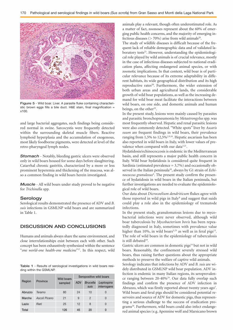
Liver - “White spots”, likely due to the migration of Ascarissuum larvae, were reported in 28 animals, often along withectopic lymphoid follicles (Fig. 4 A-B). Cysticercus tenuicollis- the larval stage of Taenia hydatigena - and hydatid cysts we-re seen, respectively, in the 15% and 6% of wild boars. Infec-tions by Dicrocoelium dendriticum were detected in 3 animals(Fig. 5).
Spleen - So-called multiple sub-serosal hernias of thesplenic pulp were seen in 5 wild boars, while a single hydatidcyst was reported in one subject.
Kidneys - Grossly, scattered pinpoint hemorrhages wereobserved in a relevant percentage of wild boars (36%); mi-croscopically, such lesions corresponded to recently occur-red intra-tubular hemorrhages, most likely due to slaugh-ter technique. Occasionally, renal infarcts were also obser-ved (4%).Discrete foci of tubule-interstitial nephritis were microsco-pically seen in all investigated animals, usually in absence ofgross lesions; the inflammatory infiltrates mainly consistedof eosinophils and multinucleated giant cells, such findingssuggesting a parasitic etiology. “Parasite-induced” ectopiclymphoid follicles were also occasionally detected withinthe renal parenchyma. Large congenital cysts were recordedin 2 subjects.
Figure 1 - Wild Boar. Lungs. Emphysematous lobules along thecaudodorsal borders of both lungs in a Metastrongylus spp.- infec-ted subject.
Di Nicola_imp:ok 20-06-2016 11:03 Pagina 168

U. Di Nicola et al. Large Animal Review 2015; 21: 167-171 169
Heart - The occasional detection of rounded and hypereo-sinophilic cardiomyocytes with protozoal cysts within affec-ted cells are individualized, and diagnosed as sarcocystis, re-presenting the only relevant finding.
Palatine tonsils and retro-pharyngeal lymphnodes - No relevant gross lesion was observed at the levelof palatine tonsils. Microscopically, some tonsil crypts weredilated and filled with neutrophils, exfoliated epithelial cells
Figure 2 - Wild Boar. Lung. An entire lobe is consolidated andgreysh-greenish in colour (A). Microscopically, a granulomatous le-sion with a necrotic core and embedding a number of parasitic lar-vae is seen (B). H&E stain, final magnification = x200.
Figure 3 - Wild boar. Lung. A single, very large hydatid cyst almo-st entirely occupies the right diaphragmatic lobe. The simultaneousand overlying inflammatory chronic pleural involvement is likely dueto compression.
A
B
Figure 4 - Wild boar. Liver. A whitish, firm, round-shaped noduleprotrudes from the hepatic surface (A). Microscopically, such no-dule (Kisselev’s nodule) mainly consists of lymphoid follicles andcontains foci of necrosis and dystrophic calcification (B). H&E stain,final magnification = x50.
B
A
Di Nicola_imp:ok 20-06-2016 11:03 Pagina 169
170 Pathological and serological findings in wild boars (Sus scrofa) from Gran Sasso and Monti della Laga National Park
and large bacterial aggregates, such findings being conside-red normal in swine. Sarcocystis were frequently detectedwithin the surrounding skeletal muscle fibers. Reactivelymphoid hyperplasia and the accumulation of exogenous,most likely foodborne pigments, were detected at level of theretro-pharyngeal lymph nodes.
Stomach - Notably, bleeding gastric ulcers were observedonly in wild boars housed for some days before slaughtering.Catarrhal chronic gastritis, characterized by a more or lessprominent hyperemia and thickening of the mucosa, was al-so a common finding in wild boars herein investigated.
Muscle - All wild boars under study proved to be negativefor Trichinella spp.
SerologySerological results demonstrated the presence of ADV and B.suis infections in GSMLNP wild boars and are summarizedin Table 1.
DISCUSSION AND CONCLUSIONS
Humans and animals always share the same environment, andclose interrelationships exist between each with other. Suchconcept has been exhaustively synthesized within the sentence“one world-one health-one medicine”17. In this respect, wild
animals play a relevant, though often underestimated role. Asa matter of fact, zoonoses represent about the 60% of emer-ging public health concerns, and the majority of emerging in-fectious diseases (> 70%) arise from wild animals20.The study of wildlife diseases is difficult because of the fre-quent lack of reliable demographic data and of validated la-boratory tests25. However, understanding the epidemiologi-cal role played by wild animals is of crucial relevance, mostlyin the case of infectious diseases subjected to national eradi-cation plans, affecting endangered animal species, or withzoonotic implications. In that context, wild boar is of parti-cular relevance because of its extreme adaptability in diffe-rent habitats, its wide geographical distribution and its highreproductive rates28. Furthermore, the wider extension ofboth urban areas and agricultural lands, the considerablegrowth of wild boar populations, as well as the increasing de-mand for wild boar meat facilitate the interactions betweenwild boars, on one side, and domestic animals and humanbeings, on the other22.In the present study, lesions were mainly caused by parasitesand parasitic bronchopneumonia by Metastrongylus spp. wasmost frequently observed. Hepatic and renal parasitic lesionswere also commonly detected. “White spots” liver by Ascarissuum are frequent findings in wild boars, their prevalenceranging from 1,5% to 12,5%2,9,16. Hepatic ascariasis has beenalso reported in wild boars in Italy, with lower values of pre-valence when compared with our data3,21.Hydatidosis/echinococcosis is endemic in the Mediterraneanbasin, and still represents a major public health concern inItaly. Wild boar hydatidosis is considered quite frequent inSardinia (estimated prevalence = 3,7%) and occasionally ob-served in the Italian peninsula18, always by G1 strain of Echi-nococcus granulosus6. The present study confirm the presen-ce of hydatidosis in wild boars in the Italian peninsula, butfurther investigations are needed to evaluate the epidemiolo-gical role of wild boars.Our data about Dicrocoelium dendriticum flukes agree withthose reported in wild pigs in Italy8 and suggest that suidscould play a role also in the epidemiology of trematode infections.In the present study, granulomatous lesions due to myco-bacterial infections were never observed, although wildboar tuberculosis by Mycobacterium bovis has been repea-tedly diagnosed in Italy, sometimes with prevalence valuehigher than 10%, in wild boars5,23 as well as in feral pigs13.The role of wild boars in the epidemiology of tuberculosisis still debated19.Gastric ulcers are common in domestic pigs14 but not in wildboars. Reasonably, the confinement severely stressed wildboars, thus raising further questions about the appropriatemethods to preserve the welfare of captive wild animals.Serology indicates that infections by ADV and B. suis are wi-dely distributed in GSMLNP wild boar population. ADV in-fection is endemic in many Italian regions, its seroprevalen-ce ranging between 20-40%24. Our data fully overlap suchfindings and confirm the presence of ADV infection inAbruzzo, which was firstly reported about twenty years ago7.Wild boars and feral pigs should be considered potential re-servoirs and source of ADV for domestic pigs, thus represen-ting a serious challenge to the success of eradication pro-grams26. Furthermore, wild boars could also infect endange-red animal species (e.g. Apennine wolf and Marsicano brown
Figure 5 - Wild boar. Liver. A parasite fluke containing characteri-stic brown eggs fills a bile duct. H&E stain, final magnification =x100.
Abruzzo Teramo 80 24 10 0
Marche Ascoli Piceno 21 9 2 0
Lazio Rieti 25 12 8 0
Total 126 45 20 0
Table 1 - Results of serological investigations in wild boars resi-ding within the GSMLNP.
Wild boarsSeropositive wild boars
Region Province sampled ADV Brucella Leptospirasuis interrogans
Di Nicola_imp:ok 20-06-2016 11:03 Pagina 170

U. Di Nicola et al. Large Animal Review 2015; 21: 167-171 171
bear), which reside within the GSMLNP and are susceptibleto ADV with fatal outcome30.Evidences suggest that B. suis biovar 2 was introduced intoItaly through the importation of hares and/or wild boars fromendemically infected European countries. B. suis biovar 2 in-fection has been firstly reported in wild boars in Piedmont,with seroprevalence close to 20%4 and very similar to that ob-served in the present study. Remarkably, B. suis biovar 2 hasbeen recently isolated in a wild boar from Abruzzo region10.L. interrogans infection has been repeatedly reported in wildboars in Italy, with variable and usually low values of sero-prevalence15,24. L. interrogans infections are quite frequent inwild animals (e.g. mustelids) living within the GSMLNP(unpublished data). However, our negative results are notsurprising and most likely due to the biology of wild boars inGSMLNP. In fact, puddles used for mud baths, which are po-tential sources of infection, are usually formed from rain wa-ter and exist only for short time.The estimated prevalence of Trichinella spp. infection in wildboars in Italy is also low27. In particular, a single wild boarproved to be positive for Trichinella britovi in GSMLNP du-ring the last six years12, such infection being quite commonin red foxes and wolves (unpublished data).In conclusion, the implementation of this “managementplan” represented a good chance to “face” the wild fauna li-ving within the GSMLNP. Pathological findings herein de-scribed, along with concurrent microbiological, serologicaland parasitological investigations, contribute to evaluate thehealth status of wild boar population residing in such a rele-vant protected area.
CONFLICT OF INTEREST STATEMENT
Authors disclose any financial and personal relationshipswith other people or organization that could inappropriatelybias their work.
References
1. Anonymous (2013) Piano di Gestione del Cinghiale. http://www.gransassolagapark.it/pdf/PGC2013.pdf
2. Antolova D., Reiterova K., Dubinsky P. (2006) The role of wild boars(Sus scrofa) in circulation of trichinellosis, toxocarosis and ascariosis inthe Slovak Republic. Helminthologia, 2:43-47.
3. Beraldo P., Codollo L., Pascotto E., Busatta S., Amorena A.L., De Luc-chi D. (2008) Elmintofauna in cinghiali (Sus scrofa) del territorio preal-pino trevigiano. Hystrix, Suppl, 71.
4. Bergagna S., Zoppi S., Ferroglio E., Gobetto M., Dondo A., Di Gianna-tale E., Gennero M.S., Grattarola C. (2009) Epidemiologic survey forBrucella suis biovar 2 in a wild boar (Sus scrofa) population innorthwest Italy. J Wildl Dis, 45(4):1178-1181.
5. Bollo E., Ferroglio E., Dini V., Mignone W., Biolatti B., Rossi L. (2000)Detection of Mycobacterium tuberculosis complex in lymph nodes ofwild boar (Sus scrofa) by a target-amplified test system. J Vet Med B In-fect Dis Vet Public Health, 47(5):337-342.
6. Busi M., Snábel V., Varcasia A., Garippa G., Perrone V., De Liberato C.,D’Amelio S. (2007) Genetic variation within and between G1 and G3genotypes of Echinococcus granulosus in Italy revealed by multilocusDNA sequencing. Vet Parasitol, 150(1-2):75-83.
7. Capua I., Fico R., Banks M., Tamba M., Calzetta G. (1997) Isolation andcharacterisation of an Aujeszky’s disease virus naturally infecting a wildboar (Sus scrofa). Vet Microbiol, 55(1-4):141-146.
8. Capucchio M.T., Deborah C., Vincenzo D.M., Miriam R., Vincenzo A.,Amedeo T., Alessandro L., Stefano A., Eleonora S.F., Bruno D., FrancoG. (2009) Natural trematode infestation in feral Nebrodi Black pigs:pathological investigations. Vet Parasitol, 159(1):37-42.
9. de la Muela N., Hernàndez-de-Lujàn S., Ferre I. (2001) Helminths ofwild boar in Spain. J Wildl Dis, 37(4):840-843.
10. De Massis F., Di Provvido A., Di Sabatino D., Di Francesco D., Zilli K.,Ancora M., Tittarelli M. (2012) Isolation of Brucella suis biovar 2 froma wild boar in the Abruzzo Region of Italy. Vet Ital, 48(4):387-404.
11. Di Febo T., Luciani M., Portanti O., Bonfini B., Lelli R., Tittarelli M.(2012) Development and evaluation of diagnostic tests for the serolo-gical diagnosis of brucellosis in swine. Vet Ital, 48(2):133-156.
12. Di Giacomo L., Morelli M.S., Marilungo L., Ferretti E., Angellotti A.,Mattozzi C. (2011) Presence of Trichinella britovi in wild boar in theMarche region regularly slaughtered. Atti dell’Associazione Italiana deiVeterinari Igienisti, 1.
13. Di Marco V., Mazzone P., Capucchio M.T., Boniotti M.B., Aronica V.,Russo M., Fiasconaro M., Cifani N., Corneli S., Biasibetti E., BiagettiM., Pacciarini M.L., Cagiola M., Pasquali P., Marianelli C. (2012) Epi-demiological significance of the domestic black pig (Sus scrofa) inmaintenance of bovine tuberculosis in Sicily. J Clin Microbiol,50(4):1209-1218.
14. Doster R. (2000) Porcine gastric ulcer. Vet Clin North Am Food AnimPract, 16:163-173.
15. Ebani V.V., Cerri D., Poli A., Andreani E. (2003) Prevalence of Lepto-spira and Brucella antibodies in wild boars (Sus scrofa) in Tuscany,Italy. J Wildl Dis, 39(3):718-22.
16. Fernandez-de-Mera I.G., Gortazar C., Vicente J., Höfle U., Fierro Y.(2003) Wild boar helminths: risks in animal translocations. Vet Parasi-tol, 115(4):335-341.
17. Frank D. (2008) One world, one health, one medicine. Can Vet J,49(11):1063-1065.
18. Garippa G., Manfredi M.T. (2009) Cystic echinococcosis in Europe andin Italy. Vet Res Commun, 33, Suppl 1:35-39.
19. Gortazar C., Vicente J., Gavier-Widén D. (2003) Pathology of bovinetuberculosis in the European wild boar (Sus scrofa). Vet Rec,152(25):779-780.
20. Jones K.E., Patel N.G., Levy M.A., Storeygard A., Balk D., GittlemanJ.L., Daszak P. (2008) Global trends in emerging infectious diseases.Nature, 451(7181):990-993.
21. Magi M., Bertani M., Dell’Omodarme M., Prati M.C. (2002) Epide-miological study of the intestinal helminths of wild boar (Sus scrofa)and mouflon (Ovis gmelini musimon) in central Italy. Parassitologia,44(3-4):203-205.
22. Meng X.J., Lindsay D.S., Sriranganathan N. (2009) Wild boars as sour-ces for infectious diseases in livestock and humans. Philos Trans R SocLond B Biol Sci, 364(1530):2697-7207.
23. Mignone W., Poggi M., Pistone G.C., Caramelli M., Bollo E., Biolatti B.(1995) Pathology of wild boar (Sus scrofa) in Liguria, Italy, between1989 and 1992. J Mt Ecol, 3:85-87.
24. Montagnaro S., Sasso S., De Martino L., Longo M., Iovane V., Ghiur-mino G., Pisanelli G., Nava D., Baldi L., Pagnini U. (2010) Prevalence ofantibodies to selected viral and bacterial pathogens in wild boar (Susscrofa) in Campania Region, Italy. J Wildl Dis, 46(1):316-319.
25. Mörner T., Obendorf D.L., Artois M., Woodford M.H. (2002) Surveil-lance and monitoring of wildlife diseases. Rev Sci Tech, 21(1):67-76.
26. Pejsak Z.K., Truszczynski M.J., (2006) Aujeszky’s disease (pseudora-bies). In: Disease of Swine, Eds. Straw B.E., Zimmerman J.J., D’AllaireS., Taylor D.J., 9th ed., 419-434, Blackwell Publishing, Ames, Iowa.
27. Pozio E. (2007) World distribution of Trichinella spp. infections in ani-mals and humans. Vet Parasitol, 149(1-2):3-21.
28. Ruiz-Fons F., Segalés J., Gortázar C. (2008) A review of viral diseases ofthe European wild boar: effects of population dynamics and reservoirrôle. Vet J, 176(2):158-169.
29. Toso S., Pedrotti L. (2001) Linee guida per la gestione del cinghiale (Susscrofa) nelle aree protette. Quaderni di Conservazione della Natura,3:9-30.
30. Zanin E., Capua I., Casaccia C., Zuin A., Moresco A. (1997) Isolationand characterization of Aujeszky’s disease virus in captive brown bearsfrom Italy. J Wildl Dis, 33(3):632-634.
Di Nicola_imp:ok 20-06-2016 11:03 Pagina 171
Related Documents