PATHOGENESIS AND CONTROL OF INCLUSION BODY HEPATITIS IN BROILER CHICKENS A thesis submitted to the College of Graduate and Postdoctoral Studies in partial fulfillment of the requirements for the Degree of Doctor of Philosophy in the Department of Veterinary Pathology University of Saskatchewan Saskatoon by Ashish Gupta © Copyright Ashish Gupta, April 2018. All rights reserved.

Welcome message from author
This document is posted to help you gain knowledge. Please leave a comment to let me know what you think about it! Share it to your friends and learn new things together.
Transcript

PATHOGENESIS AND CONTROL OF INCLUSION BODY
HEPATITIS IN BROILER CHICKENS
A thesis submitted to the
College of Graduate and Postdoctoral Studies
in partial fulfillment of the requirements for the
Degree of Doctor of Philosophy
in the Department of Veterinary Pathology
University of Saskatchewan
Saskatoon
by
Ashish Gupta
© Copyright Ashish Gupta, April 2018. All rights reserved.

i
PERMISSION TO USE
In presenting this thesis in partial fulfillment of the requirements for a Postgraduate
degree from the University of Saskatchewan, I agree that the Libraries of this University
may make it freely available for inspection. I further agree that permission for copying of
this thesis in any manner, in whole or in part, for scholarly purposes may be granted by
the professor who supervised my thesis work, or in their absence, permission may be
granted by the Head of the Department or the Dean of the College in which my thesis
work was done. It is understood that any copying or publication or use of this thesis or
parts thereof for financial gain shall not be allowed without my written permission. It is
also understood that due recognition shall be given to me and to the University of
Saskatchewan in any scholarly use which may be made of any material in my thesis.
Requests for permission to copy or to make other uses of materials in this
thesis/dissertation in whole or part should be addressed to:
Head of the Department of Veterinary Pathology
Western College of Veterinary Medicine
University of Saskatchewan
52 Campus Drive
Saskatoon, Saskatchewan S7N 5B4
Canada
OR
Dean
College of Graduate and Postdoctoral Studies
University of Saskatchewan
116 Thorvaldson Building, 110 Science Place
Saskatoon, Saskatchewan S7N 5C9
Canada

ii
ABSTRACT
Inclusion body hepatitis (IBH) is an economically important fowl adenovirus
(FAdV) disease of broiler chickens. In Canada, FAdV-8a, FAdV-8b, FAdV-11, FAdV-7
and FAdV-2 are the prevalent FAdV serotypes. Currently, there is no commercial vaccine
available in Canada to prevent IBH in broiler chickens.
The objectives of this study were to develop live, inactivated or subunit FAdV
vaccines to control IBH and to identify a suitable adjuvant for an inactivated FAdV
vaccine. In chapter 2, we analyzed the efficacy and safety of live and inactivated bivalent
FAdV vaccines (FAdV-8b-SK+FAdV-11-1047) against IBH. We demonstrated
significant immunoprotection of broiler chickens (98 – 100%) (P<0.01) against IBH by
vaccinating broiler breeders with FAdV-8b-SK+FAdV-11-1047 with either a bivalent
live vaccine (1x10⁴ TCID50) at 16 weeks of age or a bivalent inactivated vaccine (1x10⁶
TCID50) at 16 and 19 weeks of age. Both the live and inactivated bivalent FAdV vaccines
induced broad-spectrum protection against all common serotypes of FAdV circulating in
the Canada. Both the live and inactivated FAdV vaccines were equally efficacious in
protecting broiler chickens against IBH by passive transfer of maternal antibodies
(MtAb) from broiler breeders to their broiler progeny.
In chapter 3, we demonstrated that FAdV-8b-SK adjuvanted with CpG-ODN
induced a long-lasting humoral immunity similar to inactivated FAdV-8b-SK adjuvanted
with Emulsigen-D. FAdV-8b-SK adjuvanted with CpG-ODN induced T helper (Th)-1
and Th-2 type immunity. CpG-ODN as an adjuvant enhanced cytotoxic T-cell memory
response of FAdV-8b-SK vaccine.
Propagation of some serotypes of FAdVs are difficult in cell lines. Hence, we
explored the possibility of developing a subunit FAdV vaccine. In chapter 4, we
demonstrated significant protection of broiler chickens against IBH by vaccinating their
broiler breeder parents using a FAdV-8b-SK subunit vaccine [fiber protein or virus-like
particles (VLPs)]. We also demonstrated that the FAdV-8b-SK fiber and VLPs induce
strong cytotoxic T-cell responses in the broiler breeders. The results of this study will
help in designing FAdV control strategies for the prevention of IBH in Canada.

iii
ACKNOWLDEGEMENTS
I would like to thank Dr. Susantha Gomis for shaping my career in the right
direction. His contributions to my career are highly appreciated.
I would like to thank all the committee members Drs. Suresh K. Tikoo, Davor
Ojkic, Dr. Philip Willson and Elemir Simko for their immense contribution, favorable
criticism, directions to the project and advice.
My special thanks to Shelly Popowich and Betty Chow-Lockerbie, Drs.
Lisanework Ayalew and Khawaja Ashfaque Ahmad for their help in the lab, animal
experiments and in the project.
I am highly thankful to Kalahari, Thushari, Shanika, Ruwani, Mengying and
exchange and summer students; Byongyoon, Tara and Natalia for their help in my
project. My gratitude to graduate students; Sarah, Jolanda, Micheal, Lilani, Erin, Tara,
Tony and Ivana for making my Canadian living experience memorable.
My sincere thanks to Department of Veterinary Pathology for the provision of the
excellent academic environment, plethora of learning resources, wonderful people and
their social gatherings. My special thanks to Tyler, Sandy, Angie and Ian for their help in
the department and to the people of GMP and Animal Care for their invaluable
contribution to my project.
Special thanks to Drs. Gregorio Rosales and Eric Jensen, Aviagen North America,
Huntsville, Alabama for donation of broiler breeders for animal experimental studies.
My sincere thanks to funding agencies (Chicken Farmers of Saskatchewan
(SCIDF) Agriculture Development Fund (ADF), NSERC and AAFC Growing Forward 2
and to College of Graduate and Postdoctoral studies providing my personnel support
through a Graduate Student Scholarship.
My gratitude to wife, mom, dad, brother and Dr. Sidharth and his family for their
love, support and care.

iv
DEDICATION
To my beloved parents and to my wife

v
TABLE OF CONTENTS
PERMISSION TO USE ..................................................................................................... i
ABSTRACT ....................................................................................................................... ii
ACKNOWLDEGEMENTS ............................................................................................ iii
DEDICATION.................................................................................................................. iv
TABLE OF CONTENTS ................................................................................................. v
LIST OF TABLES ........................................................................................................... ix
LIST OF FIGURES .......................................................................................................... x
LIST OF ABBREVIATIONS ........................................................................................ xii
CHAPTER 1: INTRODUCTION AND LITERATURE REVIEW .......................... 1
1.1. Introduction ................................................................................................................ 1
1.2. History ......................................................................................................................... 1
1.3. Adenoviridae taxonomy .............................................................................................. 2
1.3.1. Mastadenovirus ................................................................................................... 3
1.3.2. Atadenovirus ....................................................................................................... 3
1.3.3. Siadenovirus ........................................................................................................ 3
1.3.4. Icthadenovirus..................................................................................................... 4
1.3.5. Aviadenovirus ..................................................................................................... 4
1.4. Adenovirus structure ................................................................................................. 5
1.4.1. Major capsid proteins .......................................................................................... 6
1.4.2. Minor capsid proteins ......................................................................................... 8
1.4.3. Core proteins ....................................................................................................... 9
1.4.4. Adenovirus genome ............................................................................................ 9
1.5. Cell infection and replication .................................................................................. 12
1.6. Immune response to FAdV ..................................................................................... 14
1.6.1. Innate immune response ................................................................................... 14
1.6.2. Adaptive immune response ............................................................................... 17
1.7. Adenovirus as vaccine vectors ................................................................................ 18
1.7.1. Avian adeno-associated viruses ........................................................................ 20
1.8. Oncogenicity of Adenoviruses ................................................................................. 21
1.9. Common diseases in poultry ................................................................................... 22
1.9.1. Hemorrhagic enteritis ........................................................................................ 22
1.9.2. Egg drop syndrome ........................................................................................... 22
1.9.3. Quail bronchitis ................................................................................................. 23
1.9.4. Hepatitis Hydropericardium Syndrome ............................................................ 23

vi
1.9.5. Gizzard erosions and ulcerations ...................................................................... 24
1.9.6. Inclusion body hepatitis .................................................................................... 25
1.10. Pathobiology of IBH .............................................................................................. 26
1.10.1. Epidemiology .................................................................................................. 26
1.10.2. Pathogenesis .................................................................................................... 27
1.10.3. Primary or secondary pathogens ..................................................................... 29
1.10.4. Transmission ................................................................................................... 29
1.10.5. Diagnosis......................................................................................................... 31
1.11. Control of IBH........................................................................................................ 33
1.11.1. Live vaccines .................................................................................................. 34
1.11.2. Inactivated vaccines ........................................................................................ 35
1.11.3. Subunit vaccines ............................................................................................. 35
1.12. Current problems associated with IBH in Canada ............................................. 37
1.13. Objectives................................................................................................................ 37
CHAPTER 2: INACTIVATED OR LIVE BIVALENT FOWL ADENOVIRUS
(FADV-8B+FADV-11) BREEDER VACCINES PROVIDE BROAD-
SPECTRUM PROTECTION IN CHICKS AGAINST INCLUSION BODY
HEPATITIS (IBH) .............................................................................................. 38
2.1. Abstract ..................................................................................................................... 39
2.2. Introduction .............................................................................................................. 39
2.3. Materials and methods ............................................................................................ 41
2.3.1. Propagation of FAdVs for FAdV vaccine ........................................................ 41
2.3.2. Preparation of FAdV inoculum for broiler challenge ....................................... 41
2.3.3. Safety evaluation of live FAdV in broiler breeders .......................................... 42
2.3.4. Animals and experimental design ..................................................................... 42
2.3.5. Measurement of neutralizing antibodies against FAdVs .................................. 43
2.3.6. Quantitation of fecal shedding in cloacal swabs ............................................... 43
2.3.7. Statistical analysis ............................................................................................. 45
2.4. Results ....................................................................................................................... 45
2.4.1. Safety of live FAdV virus in broiler breeder .................................................... 45
2.4.2. Neutralizing antibody response in broiler breeders .......................................... 48
2.4.3. Cross-neutralizing antibody response against heterologous FAdVs ................ 49
2.4.4. Fecal shedding of FAdVs in broiler breeders vaccinated with live bivalent
FAdV vaccine ............................................................................................................. 49
2.4.5. Maternal antibodies and protection of broilers against FAdV challenge ......... 50
2.5. Discussion.................................................................................................................. 53
2.6. Conclusions ............................................................................................................... 54
vii
PREFACE TO CHAPTER 3 ......................................................................................... 56
CHAPTER 3: CHARACTERIZATION OF CELLULAR AND HUMORAL
IMMUNE RESPONSES OF BROILER BREEDERS FOLLOWING
VACCINATION WITH A FOWL ADENOVIRUS ANTIGEN
ADJUVNATED WITH EMULISGEN-D OR OLIGODEOXYNUCLETIDES
CONTAINING CPG MOTIFS .......................................................................... 57
3.1. Abstract ..................................................................................................................... 58
3.2. Introduction .............................................................................................................. 58
3.3. Materials and methods ............................................................................................ 60
3.3.1. Adjuvants, virus and vaccine ............................................................................ 60
3.3.2. Animals and experimental design ..................................................................... 60
3.3.3. Detection of serum IgY antibody by ELISA .................................................... 61
3.3.4. Detection of neutralizing antibodies by virus neutralization assay .................. 62
3.3.5. Determination of CD4+:CD8+ T-cell ratio in peripheral blood mononuclear
cells ............................................................................................................................. 62
3.3.6. Quantification of cytokine expression in peripheral blood mononuclear cells . 62
3.3.7. Statistical analysis ............................................................................................. 63
3.4. Results ....................................................................................................................... 63
3.4.1. Serum IgY antibody and neutralizing antibody response in broiler breeders ... 63
3.4.2. Progeny protection ............................................................................................ 65
3.4.3. CD4+:CD8+ T-cell ratio in peripheral blood mononuclear cells ....................... 66
3.4.4. Cytokine expression on peripheral blood mononuclear cells ........................... 67
3.5. Discussion.................................................................................................................. 70
PREFACE TO CHAPTER 4 ......................................................................................... 75
CHAPTER 4: IMMUNOGENECITY AND PROTECTIVE EFFICACY OF
VIRUS-LIKE PARTICLES AND RECOMBINANT FIBER PROTEINS OF
FOWL ADENOVIRUS (FADV)-8B VACCINES IN BROILER BREEDERS
AGAINST INCLUSION BODY HEPATITIS ................................................. 76
4.1. Abstract ..................................................................................................................... 77
4.2. Introduction .............................................................................................................. 77
4.3. Material and methods .............................................................................................. 79
4.3.1. Virus, cell line and antibodies ........................................................................... 79
4.3.2. Purification of fowl adenovirus (FAdV-8b-SK) virus-like particles ................ 80
4.3.3. Transmission electron microscopy ................................................................... 80

viii
4.3.4. Cloning of fiber and fiber-knob gene of FAdV-8b-SK .................................... 81
4.3.5. Protein expression and purification .................................................................. 82
4.3.6. Coomassie blue staining and Western blotting ................................................. 83
4.3.7. Preparation of FAdV-8b-SK challenge virus .................................................... 84
4.3.8. Broiler breeder vaccination ............................................................................... 84
4.3.9. Progeny challenge ............................................................................................. 84
4.3.10. Virus isolation from cloacal swabs ................................................................. 85
4.3.11. Measurement of serum IgY antibodies ........................................................... 85
4.3.12. Measurement of neutralizing antibodies against fowl adenovirus .................. 86
4.3.13. Quantification of CD4+ and CD8+ T-cells in peripheral blood mononuclear
cells ............................................................................................................................. 86
4.3.14. Statistical analysis ........................................................................................... 86
4.4. Results ....................................................................................................................... 87
4.4.1. Analysis of purified 6XHis tagged fiber and fiber-knob proteins .................... 87
4.4.2. Isolation and examination of mature FAdV-8b-SK virion and purified Virus-
like particles ................................................................................................................ 87
4.4.3. Serum IgY and neutralizing antibodies against FAdV following broiler breeder
vaccination .................................................................................................................. 88
4.4.4. CD4+ and CD8+ T-cell ratio in PBMC following booster vaccination ............. 90
4.4.5. Challenge and protection studies in progenies .................................................. 91
4.4.6. Fowl adenovirus shedding following challenge ............................................... 92
4.5. Discussion.................................................................................................................. 93
4.6. Conclusions ............................................................................................................... 96
CHAPTER 5: DISCUSSION AND CONCLUSIONS .............................................. 97
REFERENCES .............................................................................................................. 102

ix
LIST OF TABLES
Table 1-1: List of genotypes, serotypes and strains of fowl adenoviruses. ....................... 5
Table 1-2: Genomic differences among the genera of the Adenoviridae family ............. 11
Table 2-1: Primer-probe sequences used for real-time qPCR .......................................... 44
Table 4-1: Primer used for cloning of fiber and fiber-knob of FAdV-8b-SK.................. 81

x
LIST OF FIGURES
Figure 1-1: Structural proteins associated with the adenovirus capsid. ............................. 6
Figure 1-2: Arrangements of hexon trimmers in a triangular face. ................................... 7
Figure 1-3: GOS configuration. ......................................................................................... 8
Figure 1-4: Adenovirus genome organization. ................................................................ 10
Figure 1-5: The cell entry pathway of adenovirus. .......................................................... 13
Figure 1-6: Normal and FAdV infected cells. ................................................................. 32
Figure 2-1: Safety evaluation of wild-type live FAdV-8b-SK in broiler breeders in egg
production (29 weeks of age). ........................................................................................... 46
Figure 2-2: Breeder vaccination with live bivalent FAdV-8b-SK+FAdV-11-1047 vaccine
at 16 weeks of age is safe and does not cause mortality in hatching chicks and vertical
transmission of the vaccine viruses................................................................................... 47
Figure 2-3: NAb response of broiler breeders. ................................................................ 48
Figure 2-4: Comparison of cross-neutralizing antibody levels with heterologous FAdV
serotypes. .......................................................................................................................... 49
Figure 2-5: Fecal shedding of FAdVs in broiler breeders vaccinated with live bivalent
FAdV vaccine. .................................................................................................................. 50
Figure 2-6: Maternal NAb levels in day old broiler chicken progeny. ............................ 51
Figure 2-7: Assessment of maternal antibody mediated protection against homologous
and heterologous FAdV serotypes. ................................................................................... 52
Figure 3-1: Serum IgY and neutralizing antibody levels against FAdV-8b-SK adjuvanted
with Emulsigen D or CpG-ODN, FAdV-8b-SK with no adjuvant and saline in broiler
breeders following vaccination ......................................................................................... 65
Figure 3-2: Challenge protection study in broiler progeny at 14 days post-hatch ........... 66
Figure 3-3: CD4+:CD8+ T-cell ratio in peripheral blood mononuclear cells. .................. 67
Figure 3-4: Cytokine expression in peripheral blood mononuclear cells at 9 days post-
vaccination. ....................................................................................................................... 69
Figure 3-5: Cytokine expression on peripheral blood mononuclear cells at 24 days post-
vaccination. ....................................................................................................................... 70
Figure 4-1: Phylogenetic tree analysis of FAdV-8b-SK. ................................................. 79

xi
Figure 4-2: Cloning and expression of recombinant fiber or truncated fiber-knob proteins
of FAdV-8b-SK. ............................................................................................................... 82
Figure 4-3: SDS-PAGE, Coomassie blue staining and western blot of fiber and fiber-
knob proteins of FAdV-8b-SK.......................................................................................... 87
Figure 4-4: Purification of FAdV-8b-SK VLPs. ............................................................. 88
Figure 4-5: Serum IgY and neutralizing antibody levels in broiler breeders at various
ages. .................................................................................................................................. 89
Figure 4-6: CD4+:CD8+ T-cell ratio in peripheral blood mononuclear cells. .................. 90
Figure 4-7: Neutralizing antibody levels in broiler progenies and virus challenge. ........ 92
Figure 4-8: Fecal shedding by broiler progeny following FAdV-8b-SK challenge. ....... 93

xii
LIST OF ABBREVIATIONS
AAV Adenovirus-associated virus
AAAV Avian adenovirus associated virus
ADP Adenovirus protease
AGID Agar gel immunodiffusion
APC Antigen-presenting cells
CAR Coxsackie-adenovirus receptor
CD Cluster of determinant
CELO Chicken embryo lethal orphan virus
CpG-ODN Cytosine phosphodiester guanine oligodeoxynucleotide
chTLR Chicken toll-like receptor
CsCl Cesium chloride
DAI DNA dependent INF-regulatory factor
DBP DNA binding proteins
DC Dendritic cell
dph Days post-hatch
ds Double stranded
dpi Days post-infection
DMEM: F-12 Dulbecco’s modified eagle medium: nutrient mixture F-12
dpv Days post-vaccination
ELISA Enzyme linked immunosorbent assay
FAdV Fowl adenovirus
FITC Fluorescein isothiocynate
GAGs Glucosaminoglycans
GON Group-of-nine
GOS Group-of-six
HAdV Human adenovirus
HHS Hepatitis-hydropericardium syndrome
HRM High resolution melt curve
IBH Inclusion body hepatitis
IBDV Infectious bursal disease virus
xiii
ICTV International Committee on Taxonomy of Viruses
Ig Immunoglobulins
IL Interleukin
INF Interferon
ITR Inverted terminal repeats
IRF Interferon regulatory factor
LB Luria broth
LMH Leghorn male hepatoma
MDA-5 Melanoma differentiation associated protein 5
MDV Marek disease virus
MHC Major histocompatibility complex
MIP Monocyte inflammatory protein
MLP Major late promoter
mRNA Messenger ribonucleic acid
MtAb Maternal antibodies
MyD88 Myeloid differentiation factor-88
NAb Neutralizing antibodies
NDV Newcastle disease virus
NLR NOD-like receptors
NK Natural killer
NOD Nuclear oligomerization domain
NF-kβ Nuclear factor- kappa β
OD Optical density
ORF Open reading frame
ORI Origin of replication
O/W Oil-in-water
PAMP Pathogen associated molecular patterns
PBMC Peripheral blood mononuclear cells
PBS Phosphate buffered saline
PCR Polymerase chain reaction
pTP Precursor terminal protein

xiv
PRRs Pattern recognition receptors
RANTES Regulated on activation T-cell excreted and secreted
RFLP Restriction fragment length polymophism
RIG-I Retinoic acid inducible gene
RGD Arginine-glutamine-aspartic acid
rt- PCR real-time polymerase chain reaction
SDS-PAGE Sodium dodecyl sulphate polyacrylamide gel
electrophoresis
SPF Specific pathogen free
ss Single stranded
SV40 Simian virus 40
TB Tryptose broth
Th T helper
TNF- α Tumour necrosis factor- α
TLR Toll-like receptors
TP Terminal protein
VA-RNA Viral encoded ribonucleic acid genes
VNT Virus neutralization test
VLP Virus-like particles
W/O Water-in-oil
W/O/W Water-in-oil-in water
1
CHAPTER 1: INTRODUCTION AND LITERATURE REVIEW
1.1. Introduction
Inclusion body hepatitis (IBH) is an acute fowl adenovirus (FAdV) disease of 1 to
5 week old chickens that begins as a sudden increase in mortality, peaking at 4 to 5 days
post-infection. Enlarged, pale swollen livers exhibiting necrosis and hemorrhages
characterize the disease. Basophilic intranuclear inclusions in the hepatocytes are the
main microscopic lesions. Mortality percentage is variable, usually below 10 % but may
exceed 30% [1]. IBH is prevalent worldwide and its spread is increasing in many
countries. FAdVs are highly diverse and are categorized into five species (A to E) and 12
serotypes (-1 to -7, -8a, -8b and -9 to -11) [2]. Most notably, FAdV serotypes FAdV-2,
FAdV-7, FAdV-8a, FAdV-8b and FAdV-11 are responsible for IBH in chickens [3].
Spread of the virus is mainly due to horizontal transmission but vertical transmission
plays a critical role. Control of IBH has historically been by imposing strict biosecurity
measures and vaccination of broiler breeder parents with autogenous inactivated
vaccines. However, their efficacy for protecting against IBH remains undetermined. A
commercial vaccine is not yet available in Canada; therefore, the aim of this thesis is to
develop FAdV vaccines (live, inactivated and subunit) to control IBH in Canada and to
evaluate adjuvants in the inactivated FAdV vaccines.
1.2. History
In the 1940’s, the first description of intranuclear basophilic inclusion bodies in
hepatocytes of dogs suffering from infectious canine hepatitis was recorded [4]. Similar
inclusion bodies were also observed by Olson. (1951) in cases of infectious bronchitis in
quails leading to a suspicion of a viral etiology [5]. Rowe et al. (1953) described a
filterable agent from such inclusion bodies in cell culture preparations derived from
human adenoid tissues [6] and named them “adenovirus” [7]. In 1957, Yates and Fry had
isolated a new virus from fertile chicken eggs [8] which was then named as chicken
embryo lethal orphan virus (CELO). Frequent isolation of CELO from eggs and egg-

2
based vaccines [9] and its widespread seropositivity in chicken flocks [8, 10] provided an
impetus to adenoviral research in the 1960s.
In 1963, Helmboldt and Frazier first described hepatic alterations in broiler flocks
at 5 weeks of age [11]. Livers were swollen with round edges and stellate hemorrhages.
They described the histopathological lesions as an “acute hepatic catastrophe” with 90%
of the parenchyma displaying fatty metamorphosis and Cowdry Type A inclusion bodies
[11]. Many Canadian researchers have found similar pathological findings and variable
mortality in broiler chickens in subsequent years [12-14]. Livers were grossly swollen,
mottled with a reticular pattern similar to Helmboldt and Frazier’s observations. The
etiological agent was identified in 1973 and named for the first time as FAdV [15].
Subsequent cases displaying similar pathological findings of IBH were reported from
several countries of the world. Today several serotypes of FAdVs (FAdV-2, -7, -8a, -8b
and -11) which cause IBH have been identified [1].
1.3. Adenoviridae taxonomy
Adenoviruses have diverse vertebrate host range, which include mammals, birds,
reptiles, amphibians and fish [2, 16]. Historically, the family Adenoviridae has been
divided two major genera, Mastadenovirus and Aviadenovirus, which included viruses of
mammals and birds, respectively [17]. Adenoviruses of birds were further categorized
into three separate groups: groups I, II and III. Groups II and III contained poultry
adenoviruses which were serologically unrelated to the group I [18] and were referred as
unconventional poultry adenoviruses.
In 2011, the International Committee on Taxonomy of Viruses (ICTV) re-
classified the family Adenoviridae into five genera; Mastadenovirus, Atadenovirus,
Siadenovirus, Icthadenovirus and Aviadenovirus based on the molecular criteria reviewed
in their 9th report of ICTV [2]. The new classification of the family Adenoviridae
categorized the unconventional poultry adenoviruses of group II as well as the
unconventional members of the genus Mastadenovirus into genera Siadenovirus and
members of group III poultry adenoviruses into the genus Atadenovirus. While the
members of the genus Siadenovirus only affect amphibians and birds, members of the

3
genus Atadenovirus have the most divergent host range among all known adenovirus
genera [16]. It is hypothesized that these viruses must have had undergone a host switch
during their evolution [19-21].
1.3.1. Mastadenovirus
These viruses affect a wide range of mammalian species, such as primates, cattle,
dogs, horses, pigs, sheep, mice, tree shrews, bats and human beings [2, 22]. The host
range was also extended to mammalian fish [23]. The most significant species within this
genus are human adenoviruses. There are seven species and 51 serotypes of human
adenoviruses (HAdVs) identified so far [24]. Many of these are the etiologic agents of
diseases such as pneumonia, gastroenteritis, conjunctivitis, hepatitis and myocarditis
(especially in children [25, 26]) and fatal pneumonia (in military recruits [27]). Besides
being a primary pathogen, they are identified as a potential source of nosocomial
infections in immunocompromised children [28-30]. Other than human beings, the most
notable adenoviral diseases of veterinary importance are infectious canine hepatitis and
tracheobronchitis associated with canine adenovirus-1 and -2 infection in puppies [31,
32].
1.3.2. Atadenovirus
Atadenoviruses are named so because of the exceptionally high adenine and
thymine content of their genome, 57 to 66.4%, which is highest among all known
adenoviruses [19, 33]. In contrast to other genera of adenoviruses, this genus has broad
hosts ranging from reptiles, mammals, marsupials and birds [19]. Of the various species
known, only Duck Adenovirus-1 of the species Duck Adenovirus-A causes an
economically important disease in chickens.
1.3.3. Siadenovirus
These viruses have the smallest genomic size (26,163 bp to 26,282 bp) among
adenoviruses [2]. They are named as Siadenovirus as they possess a unique protein
‘sialidase’ encoded by a gene located on the left end of the genome [33]. Members of this

4
genus only affect amphibians and birds. There are five officially recognized species
reviewed by Harrach et al. (2011) [2]. Turkey adenovirus-3 of the species Turkey
Adenovirus-A is a significant pathogen of poultry.
1.3.4. Icthadenovirus
Icthadenoviruses only affect fish (white sturgeon) and are non-pathogenic. White
Sturgeon Adenoviruses have the largest known adenovirus genome (48,395 bp) [2, 34].
1.3.5. Aviadenovirus
Members of the genus Aviadenovirus were traditionally referred as FAdVs. There
are many other recognized and proposed adenovirus species in this genus that affect birds
other than chickens, such as; falcons, goose, turkeys, ducks, pigeons and parrots [1].
Adenoviruses of ducks, pigeons, parrots and turkeys all still await official recognition in
the genus [2]. There are eight officially accepted species in this genus including Falcon
Adenovirus-A, Goose Adenovirus-A, Turkey Adenovirus-B and FAdV (A to E).
1.3.5.1. Fowl Adenoviruses
FAdVs are economically significant pathogens of domestic poultry. They cause
diseases such as IBH, hepatitis-hydropericardium syndrome (HHS) and gizzard erosion
and ulceration in broilers and layers as well as quail bronchitis in quails [1, 18]. There is
huge diversity among FAdVs. Historically, were classified into five genotypes, A to E,
based on polymerase chain reaction (PCR) and restriction fragment length polymorphism
(RFLP) [35-37] and into 12 serotypes (-1 to -7, -8a, -8b and -9 to -11) based on serum
neutralization profiles [22, 38, 39]. In 2011, the ICTV has accepted the previously
classified genotypes (A to E) as the five official species of FAdVs Table 1-1.

5
Table 1-1: List of genotypes, serotypes and strains of fowl adenoviruses.ICTV.
Species Strain Serotypes
A CELO, 112, QBV, Ote, H1 FAdV-1
B 340, TR-22, Tipton, M2 FAdV-5
C 506, J2, KR5, H2 FAdV-4
D GAL-1, 685, SR48, H3, P7 FAdV-2
SR48, 75, H5 FAdV-3
A2, 90, CFA19 FAdV-9
380, UF71 FAdV-11
E YT36, x-11a like, 122 FAdV-6, FAdV-7
TR59, T8-A, CFA40A FAdV-8a
764, b3, VRI-33 FAdV-8b
Information adapted from 9th report of ICTV [2] and Group I Adenovirus infections (Diseases of Poultry,
13th Ed) [1].
1.4. Adenovirus structure
Adenoviruses are non-enveloped double stranded (ds) DNA viruses of icosahedral
symmetry varying between 70 and 90 nm in diameter. A mature virion comprises of an
outer capsid and a central core [40]. The adenovirus capsid consists of major structural
proteins (designated as II, III and IV) and minor structural proteins (IIIa, VI, VII, VIII
and IX). The adenoviral core comprises of the viral genome and its associated proteins;
V, VII, mu-protein, and terminal protein (TP) [40-42].
6
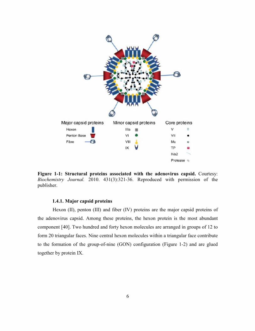
Figure 1-1: Structural proteins associated with the adenovirus capsid. Courtesy:
Biochemistry Journal. 2010. 431(3):321-36. Reproduced with permission of the
publisher.
1.4.1. Major capsid proteins
Hexon (II), penton (III) and fiber (IV) proteins are the major capsid proteins of
the adenovirus capsid. Among these proteins, the hexon protein is the most abundant
component [40]. Two hundred and forty hexon molecules are arranged in groups of 12 to
form 20 triangular faces. Nine central hexon molecules within a triangular face contribute
to the formation of the group-of-nine (GON) configuration (Figure 1-2) and are glued
together by protein IX.

7
Figure 1-2: Arrangements of hexon trimmers in a triangular face. Hexon (light blue),
penton (yellow) and fiber (dark blue) proteins make the major portion of adenovirus
capsid. The inset shows the GON arrangement of the hexon trimmers in a triangular face,
as well as the arrangement of minor structural proteins IIIa, VI, VIII and IX in relation to
hexamers. Courtesy: Viruses. 2012. 4:847-877. Reproduced with permission of the
publisher.
The remaining three hexon units at the corners of each triangular face contribute
to the formation of the group-of-six (GOS) configuration. A GOS configuration consists
of hexamer units from adjacent triangular faces around a central penton ring at each
vertex of the virion) [41]. These hexon units are called the peripentonal-hexon. The
components of the GOS are linked to each other by polypeptide IIIa. Another
polypeptide, VI, is located beneath the vertex which bridges hexon proteins to dsDNA
[42]. The second most abundant protein in the capsid is the penton ring. It has a
pentameric structure and is present at each vertex where it forms a non-covalent complex
with the fiber protein. The adenovirus fiber protein comprises of a head, shaft and a tail.
The head of the fiber protein carries sites for host cell receptors. The entire assembly
(fiber and penton proteins) is required for efficient entry of the virus into the host cell
[43].

8
Figure 1-3: GOS configuration. Arrangements of peripentonal-hexon molecules
(uncoloured) around the penton ring (blue) at the vertex forming GOS configuration.
Adapted from Viruses. 2012. 4:847-877.
1.4.2. Minor capsid proteins
Polypeptides IIIa, VI, VIII and IX are the minor structural proteins of the
adenovirus capsid. These proteins stabilize the capsid and are critical for the biological
properties of adenoviruses including temperature sensitivity, infectivity, nuclear import
of hexon proteins, capsid assembly, genome packaging and maturation of the virus [42].
Most of the structural proteins are evolutionarily conserved amongst
adenoviruses. A few proteins either vary in their number or are genus-specific to
adenoviruses. Members of the genus Aviadenovirus possess two fibers at the vertex [43]
whereas; only a few members of the genus Mastadenovirus possess two fibers at the
vertices (subgroup-F HAdV and simian adenoviruses). Members of the Siadenovirus and
Atadenovirus genus possess only one fiber per vertex. Lizard adenovirus-2, which is a
new member of the genus Atadenovirus possess three fibers on one to two vertices and
two fibers at the remainder of the vertices [16]. Besides the adenovirus fiber,
polypeptides V and IX are only present in the members of the genus Mastadenovirus [2].
9
1.4.3. Core proteins
Core proteins lie within the mature adenovirus virion along with the viral genome.
These proteins include polypeptides V and VII, mu-protein, TP, DNA-binding-protein
(DBP) and adenovirus protease (ADP). These proteins unwind the viral genome (i.e.
DBP) and initiate genome replication (i.e. TP). They also assist in tightly packaging the
viral genome inside the capsid shell (polypeptides V and VII and mu-protein). In
addition, they also aid in virion maturation (polypeptide VII) and bridging the core to the
capsid proteins (polypeptide V) [44, 45].
1.4.4. Adenovirus genome
The adenovirus genome is a linear dsDNA molecule. Each strand of it is non-
covalently linked to TP at its 5’ end [44]. The genome size ranges from 26,163 bp
(Atadenovirus) to as large as 48,395 bp (Icthadenovirus) [2]. In general, the genome is
organized into several functional regions: inverted terminal repeats (ITR) on each side of
the genome, early regions, late regions and many other open reading frames (ORFs) that
do not fall in these regions. The adenovirus genome contains evolutionary conserved ITR
on both ends. The rest of the genome is divided into different transcription units: early
regions (designated as E1A, E1B, E2A, E2B, E3 and E4 genes), delayed early genes (IX,
IVa2 and a few E2A genes), late regions (designated as L1, L2, L3, L4 and L5) and some
other transcriptional units such as viral encoded RNA (VA-RNA) genes (Figure 1-4).

10
Figure 1-4: Adenovirus genome organization. Forward reading strand encodes the
E1A, E1B, IX, major late proteins, VA-RNA and E3 units. The reverse strand contains
the E4, E2A, E2B and IVa2 genes, (Mastadenovirus; HAdV). Black arrows = early
genes, blue arrows = intermediate genes, green arrows = late genes, red arrow = viral
encoded RNA I and II. Courtesy: Biochemistry Journal. 2010. 431(3):321-36.
Reproduced with permission of the publisher.
Besides having conserved regions, striking differences exist in the genomic
organization of members among different adenovirus genera. The differences mainly
occur by the presence or absence of early transcriptional units (E1, E3 and E4) or the
presence of genus-specific genes (Table 1-2).

11
Table 1-2: Genomic differences among the genera of the Adenoviridae family
The information is adapted from the 9th report of ICTV [2]. NA = Information not available. – denotes absence of the genomic region and + denotes
presence of genomic region in the genome of adenoviruses.
Genomic region Mastadenovirus Aviadenovirus Siadenovirus Atadenovirus Icthadenovirus
E1A/B + - - + (E1B) NA
E2A/B + + + + NA
E3 + - - - NA
E4 + - - - NA
V + - - - NA
IX + - - - NA
GAM-1 - + - - NA
MDV-gp - + - - NA
dUTPase + (right end) + (left end) - - NA
LH1-3 - - - + NA
P32K - - + + NA
Silidase - - + - NA

12
All regions are present in the genus Mastadenovirus, whereas members of
Aviadenovirus and Siadenovirus lack all the E1, E3 and E4 regions [2]. Members of the
genus Atadenovirus only possess the E1B region in their genome in addition to the E2
and E4 regions. Proteins encoded by the E1A region of adenoviruses are essential for the
transactivation of genes from E2, E3, E4 and the late regions of the genome [46]. Besides
transactivation of the genes, the E1 gene products (proteins) are capable of initiating
cellular transformation along with the proteins from the E1B region [46, 47]. Similar to
E1 gene, E4 ORF-1 gene products also exhibit tumorigenic activity. Proteins encoded by
the genes of the E3 region possess immunomodulation activity [48].
In addition to early genes, each adenovirus genus has unique genus specific-
genes. These include genes V and IX in the genus Mastadenovirus, gene encoding
protein sialidase in the genus Siadenovirus, genes LH1-LH3 and p32K in the genus
Atadenovirus and genes GAM-1 and Marek’s disease virus (MDV)-gp in the genus
Aviadenovirus. Gene relocalization and duplication are other characteristic features of of
genera Aviadenovirus and Atadenovirus [2].
1.5. Cell infection and replication
Adenoviruses affect a variety of epithelial cells [48, 49], endothelial cells [50] as
well as cells of the monocytes/macrophage system [50, 51]. Adenovirus infections begin
with preliminary interactions between the adenovirus fiber-knob and the host cell
receptor (Figure 1-5), most commonly the coxsackie-adenovirus-receptor (CAR) [52-54].

13
Figure 1-5: The cell entry pathway of adenovirus. Receptor-mediated endocytosis into
the clathrin-coated pit (steps 1 and 2), the formation of the endosome (step 3), the release
of dismantled virus from the endosome (step 4), cytoplasmic transport through dyenine
on microtubules (step 5) and nuclear import into the nucleus (step 6). Courtesy: Virology.
2009. 384(2):380-388. Reproduced with permission of the publisher.
Numerous other cellular receptors, such as heparin-sulfate glycosaminoglycans
(GAGs), CD46, CD80, CD86, sialic acid and major histocompatibility complex (MHC)
molecules have been identified. These have been found to allow HAdVs to enter different
cell types [55]. Following the initial interaction, a secondary interaction of the arginine-
glutamine-aspartic acid (RGD) motif of the penton ring occurs with the αβ type integrin
receptor. The adenovirus becomes internalize into a clathrin-coated invagination
(endosome) of the cell membrane [56]. Upon acidification of the endosome, the vertex of
the virus dismantles and releases the core of the adenovirus [57]. Minor structural protein
VI is released from the dismantled virion causes endosome membrane lysis and
subsequent release of the viral core into the cytoplasm [58].
The core of the adenovirus is carried to the nuclear-pore complex on a
microtubule network with the help of a dynein protein [59]. Various interactions between

14
the shuttle proteins and the nuclear-pore complex proteins finally deliver the adenovirus
core into the nucleus [60]. Within the nucleus, early genes transcribe first and their
messenger RNAs (mRNA) are exported to the cytoplasm for protein synthesis. These
proteins regulate the cell cycle, modulate host immune responses, transactivate other
genes and are responsible for viral DNA replication.
Adenovirus genome replication is protein-primed in nature. It begins at the origin
of replication (ORI) in the ITR at both of the genomic ends. DNA replication is initiated
at the 3’ end of the antisense strand by a complex interplay between the precursor
terminal protein (pTP), DNA polymerase, DBP, and several nuclear factors [44]. Early
gene (E1A) products transactivate the major late promoters (MLP), which then initiate
the transcription of genes from the late regions [61]. Thereafter, the mRNA is exported to
the cytoplasm for protein synthesis. These proteins include major capsid proteins and
genome encapsidation proteins. Ahi et al. (2016) comprehensively reviewed the events of
the capsid assembly and genome packaging in detail [45]. Briefly, following the nuclear
import of structural proteins (II, III, IV, IIIa, VI, VIII, IX) and encapsidation proteins,
capsid assembly occurs in the nucleus. The adenoviral genome is encapsidated in a polar
fashion following the interaction of encapsidation proteins (IVa2, 52/55K, 33K, 22K)
with the packaging domain at the left end of the genome and various other viral and
cellular proteins. Adenovirus protease removes the scaffold proteins once the genome is
packaged inside the virus core and also cleaves the structural proteins to form the
infectious virus, which is then released by lysis of the cells.
1.6. Immune response to FAdV
Immunity to infectious agents broadly consists of innate immunity (immediate and
non-specific) and adaptive immunity (pathogen-specific) which develops over time and is
capable of clearing the pathogen from the host body [62].
1.6.1. Innate immune response
Innate immunity comes into play within minutes to hours, containing the
microbes and limiting their spread. Innate immune response occurs in many ways such
15
as enzymes (lysozymes), antimicrobial peptides (α and β-defensins), innate immune cells
(neutrophils, eosinophils, macrophages, natural killer (NK) cells and dendritic cells (DC))
and humoral components (complement systems) including anatomic and physiological
barriers [63]. Innate immune cells harbor an array of evolutionarily conserved pattern
recognition receptors (PRRs), which recognize pathogen-specific molecular patterns
(PAMPs). The PRRs includes a repertoire of extracellular and intracellular receptors [62],
which upon interaction with PAMPs results in the activation of many downstream
pathways. Cellular activation results in the secretion of antimicrobial substances
(enzymes and peptides) to kill the pathogen, releasing cytokines and chemokines to
attract more inflammatory cells (neutrophils, macrophages), antigen-presenting cells
(APCs) and subsequently activation of the adaptive immune response.
Innate immune system in mammals responds to viral pathogens by recognizing
viral PAMPs by PRRs. Viral PAMPs mainly includes viral proteins, viral DNA, single-
stranded (ss) RNA, dsRNA and RNA with 5’ phosphatases ends [64]. Antiviral innate
immune responses occur mainly through toll-like receptors (TLR-2, TLR-3, TLR-7, and
TLR-9), nuclear organization (NOD) like receptors (NLRs), retinoic-acid inducible gene-
I (RIG-I) like receptors and DNA dependent interferon (IFN) regulated factors (DAI)
[64-66]. Chickens share functional similarities with mammalian immune systems in
regard to pathogen sensing through innate immune receptors such as TLRs (TLR-3, TLR-
4 and TLR-7), retinoic acid inducible gene (RIG)-1-like receptors [Melanoma
Differentiation Associated protein-5 (MDA-5) and Laboratory of Genetics and
Physiology-2] and NLR (NOD-1). However, there are some striking differences in
chickens. Some of the PPRs are absent in chickens (e.g., TLR-9, RIG-1, NOD-1), while
others are either duplicated (chTLR-1a, chTLR-1b and chTLR-2a, chTLR-2b) or are
unique to chickens (chTLR-15 and chTLR-21) [67].
Adenovirus infections in mammals activate an array of immune mechanisms.
Adenovirus capsid proteins, viral DNA and VA-RNA are the chief triggers of innate
immunity [68]. Adenovirus-specific cellular receptors upon recognition of the adenovirus
capsid proteins triggers a downstream activation of phosphoinositol kinase [69-71] or
mitogen-activated protein kinase pathways [68, 72]. These pathways lead to nuclear

16
import of nuclear factor kappa-β (NF-kβ) or activation of interferon regulatory factors
(IRF). The NF-kβ and IRF pathways activate genes of inflammatory cytokines, such as
IL-1, IL-6, IL-8 and IL-18, tumor necrosis factor (TNF)-α, monocyte inflammatory
protein (MIP)-1α and MIP-2, RANTES (regulated on activation T-cell excreted and
secreted), CXC-type chemokines and IFN-γ [68, 73, 74].
Within infected cells, the naked adenoviral DNA triggers DAI or NLRs pathways.
Within the endosome, it stimulates the TLR-9 mediated pathway [68]. TLR-9 leads to the
production of inflammatory cytokines and type-1 IFNs by the myeloid differentiation
factor-88 (MyD88) pathway and IRF-7 pathways, respectively [75]. DAI induces type-1
IFNs by activating the IRF-7 pathway by a different set of adapter molecules such as
TANK-binding kinase/Inhibitors of IκB kinase [74]. In addition to the TLR-9 or DAI
dependent pathways, NLR mediated DNA sensing leads to the recruitment of apoptosis
spec protein and caspase-1 protein in the cytosol to form an inflammosome [76]. The
inflammosome cleaves the preformed IL-1 and IL-18 to their active forms that are
subsequently secreted form the cells [77]. IL-1 acts in an autocrine manner on the IL-1R
receptor to amplify its production through the MyD88 pathway [68]. The type of pathway
may vary depending on cell type. For instance, TLR-9 mediates responses in the major
pathway in the plasmacytoid dendritic cell [68, 75]. In contrast, the DAI pathway
operates chiefly in the myeloid dendritic cells, macrophages or fibroblast [74].
Chen et al. (2013) comprehensively reviewed the mechanisms of antiviral innate
immune response in birds [67]. Viral nucleic acids such as ssRNA and dsRNA are sensed
by TLR-3 and TLR-7 as in mammals; however, cytosine phosphodiester guanine
oligodeoxynucleotide (CpG-ODN) is sensed by TLR-21 instead of TLR-9 in the
endosomal compartment. TLR-3 or TLR-7 operate through NF-kβ and IRF-3/7 pathways
to induce inflammatory cytokines and a type-1 IFN response, while TLR-21 only induces
inflammatory cytokines through the NF-kβ pathway. Similar responses are induced by
long dsDNA, dsRNA or short 5’ triple phosphate dsRNA in the cytoplasmic
compartments through MDA-5 or RIG-I receptors through the NF-kβ and IRF-3/7
pathways [67, 78, 79]. Some experimental studies in chickens show that FAdV infections
result in the induction of type-1 IFNs (IFN-α), IL-12 and IL-18 [80-82] and down-

17
regulate IL-10 and IL-8 expression [80, 81].Further studies are needed to compare
cytokine responses of FAdV infections and chickens vaccinated with FAdV vaccines.
Innate immune responses lead to the recruitment of inflammatory cells,
macrophages, and antigen presenting cells APCs. These cells engulf the foreign
infectious agent, secrete cytokines and chemokines and stimulate adaptive immunity (T-
cell help) by the MHC-I and MHC-II pathways. This subsequently leads to the generation
of pathogen-specific cytotoxic T-cells and antiviral antibodies.
1.6.2. Adaptive immune response
Similar to mammals, the adaptive immune response in chickens has two arms,
cellular and humoral, which responds to viral pathogens by inducing cytotoxic T-cell
responses and antibody responses [83, 84].
Cell-mediated immunity (CMI) consist of CD4+ (helper T-cells) and CD8+
(cytotoxic T-cells) which is dominated by IFN- responses [84, 85] leading to increased
activation of macrophages and NK-cells (intracellular immunity). Chickens mount CMI
responses to a variety of intracellular pathogens [86, 87]. The role of CMI is critical in
the clearance of virus infected cells which is evident by experimental immunosuppression
induced by chemical agents [88] or infectious agents [89, 90]. A few reports have
documented the dynamics of CMI against FAdVs [91, 92]. However, more research is
warranted in this area in the future.
Humoral immunity is dominated by the increased expression of cytokines like IL-
4, IL-10 and antibody production (extracellular immunity) by plasma cells (differentiated
B lymphocytes) [90]. Unlike mammals, who have five types of antibodies, birds have
three principle antibody types, immunoglobulin (Ig) M, IgY (mammalian analog of IgG),
and IgA. IgM is the primary antibody produced upon infection and is switched by IgY as
the immune response matures [93]. These two are important in protecting internal organs
from pathogens. IgA plays an important role in protecting from pathogens at mucosal
surfaces [86].
Major capsid proteins of the virus are the major targets of adaptive immunity.
Adenoviral major capsid proteins (fiber, penton and hexon) are capable of eliciting both
18
cellular [94, 95] and antibody-mediated immunity [96-98]. Among the three capsid
proteins, hexon is the main constituent of the capsid [99] and, therefore, elicits the
highest level of cellular [94, 95] or antibody response [100, 101]. Similar to HAdV,
FAdV hexon proteins are the principle component of the capsid, which induce type-
specific neutralizing antibodies [1]. The hexon protein is a complex trimeric of proteins,
which is comprised of four loop structures. Of these loops, loops two and four contains
group and type-specific epitopes. Besides antibody responses to hexon proteins, a lower
fraction of antibodies are produced against the HAdV fiber and penton proteins [101].
Like HAdVs, the neutralizing potential of antifiber and antipenton antibodies of FAdVs
is currently doubtful. The humoral arm of the adaptive immune system is stimulated to
hyperimmunize the parents to protect their progeny against viral diseases by maternal
antibodies [102].
1.7. Adenovirus as vaccine vectors
Adenoviruses effectively deliver genes to cells due to their predilection to a
variety of cells, their enormous foreign DNA carrying capacity (up to 36 kb), ease of
mass production, safety, efficacy, stability and, more importantly, their inability to
integrate into the host genome [103-106]. HAdV-2 and HAdV-5 are under investigation
as gene delivery vehicles to treat genetic defects, to compensate immunodeficiency
diseases, as a vaccine vectors and as a modality for the treatment of cancers [103, 106-
109].
Danthinne and Imperiale (2000) have reviewed the advantages and disadvantages
of adenovirus vectors in detail [110]. Three generations of vectors are available based on
the genomic regions deleted from the virus. For example, primary adenovirus vectors
lack the E1 and E3 regions, whereas secondary vectors also lack E2 and E4 in addition to
previously mentioned genes. The tertiary vectors are called gutless vectors as all or most
of the genomic regions are removed, except the ITRs and cis-complementing packaging
sequence [110]. Unfortunately, therapeutic gene expression is ephemeral with adenovirus
vectors due to wide-spread pre-existing anti-vector antibodies (HAdV-5) among people
[98, 100], or quick vector clearance by strong host anti-vector specific CD8+ cytotoxic T-

19
cell responses [101, 103, 111]. Researchers have manipulated the adenovirus genome by
making chimeras with non-human primate adenoviruses to elude pre-existing
antiadenoviral antibodies [103, 112, 113]. Innovative methods like engineering fiber or
hexon proteins to make chimeric proteins have been investigated [114]. Moreover, other
options like the use of less prevalent adenovirus serotypes [115], targeting mucosal
delivery [116], microencapsulation [117] and generating gene deletion mutants [106]
have also been utilized.
Interestingly, non-human adenoviruses (bovine, porcine, canine, ovine, simian
and FAdVs) were preferred for many reasons such as the lack of antibodies in the human
population, potentially low pathogenicity in their hosts and similarity in the structural and
genomic organization to the HAdVs [103, 118, 119]. These viruses are under
investigation to develop potential future vaccine vectors. Besides human medicine,
vectored vaccines have also become popular in veterinary medicine to protect animal
health [120]. Olasumbo et al. (2013) reviewed numerous animal adenovirus vectored
vaccines developed to protect animal health [121], such as Canine Adenovirus-2
expressing rabies virus glycoprotein, HAdV-5 expressing hemagglutination of avian
influenza virus [122], Bovine Adenovirus-3 expressing group-D antigens of herpes virus
[123] and many other viruses [121].
Unprecedented growth occurred in the poultry vectored vaccine industry in the
last decade. Meeusen et al. (2007) have enumerated various viral vectored vaccines
licensed in the veterinary industry [120]. MDV vectored vaccines have pioneered poultry
vector vaccine industry. Its genome has been used in a number of chicken viruses such as
infectious bursal disease virus (IBDV) [124], Newcastle disease virus (NDV), avian
influenza virus [125] and infectious laryngotracheitis virus [126]. A recent study has also
documented the development of a MDV-vectored avian leucosis virus subgroup-J
vaccine [127]. Likewise, other promising vectored vaccines of avian origin viruses
include; avian poxvirus and canary poxvirus [120]. In recent years, chicken viruses like
NDV [128, 129] and FAdVs [121] are under investigation to develop vaccine vectors.
Numerous serotypes of FAdVs are being developed as vaccine vectors to deliver
foreign DNA to protect poultry health against various pathogens [121]. Unlike HAdV
20
vectors, they are still in the preliminary stages of development [130]. It had become
possible to manipulate FAdVs and engineer them to develop vectors because of the
availability of their full genome sequences (FAdV-1, FAdV-8, FAdV-9 and FAdV-10)
[131-134], and comparative genomic analysis with the members of the genus
Mastadenovirus [2]. FAdV-1, FAdV-8 and FAdV-10 are first-generation replication
competent FAdV vectors [133, 135-137] used for the development of FAdV vectored
IBDV vaccines carrying the viral protein-2 gene. These vectored vaccines have shown
protective efficacy against IBDV in animal experiments. FAdV-1, FAdV-8, FAdV-10
and FAdV-9 are currently in the experimental stages for the generation of a vaccine
vector [80]. More recently, another serotype of FAdV (FAdV-4) is under investigation
for developing a vaccine vector owing to its minimum host pathology [81]. In addition to
their use for vectored vaccine development for poultry pathogens, they are a suitable
candidate to make vectors for gene therapy in human beings owing to their defective
replication [138] or non-infectiousness in many human origin cell lines [139, 140]. CELO
virus has been successfully tested to express human interleukin (IL) genes in embryos as
well as for gene therapy for cancers [139, 141], which exemplifies their scope as potential
vectors for human medicine.
1.7.1. Avian adeno-associated viruses
Apart from the mammalian and avian adenoviruses (now called as FAdVs), Adeno-
associated viruses (AAVs) have become popular gene delivery vehicles and vaccine
vectors. AAVs are defective parvoviruses, which belong to the genus Dependovirus of
family Parvoviridae [142]. AAVs are unique as they require helper functions from
adenovirus (genes: E1a E1b, E4 and VA-RNA) or herpes virus (genes: DNA polymerase
and helicase) for productive infection (known as lytic phase) [142]. Helper viruses also
suppress cellular functions and create a milieu suitable for AAV replication. In the
absence of help, AAVs establish latency in the cells by site specific integration in the host
genome (known as lysogenic phase) [143]. Their non-pathogenic nature, ability to
replicate in both dividing and non-dividing cells, large gene inserts carrying capacity and
surviving the host immune response make them suitable agents for gene delivery vehicles

21
[142]. AAVs have been identified from human beings, primates and avian species [142,
144]. Avian adeno-associated viruses (AAAVs) are ubiquitous in the chicken population
and are often isolated from healthy chickens [144]. They readily grow in chicken
embryos and the cells of chicken origin when coinfected with FAdVs. Bauer et al. (1986)
have shown FAdV serotypes 1, 5 and 8 promote the productive infection of AAAVs in
chicken kidney cells and chicken fibroblast cells [145, 146] . While FAdVs impart helper
function for AAAV infection, it has been also demonstrated that AAAVs reduce the
virulence of FAdVs (Timpton strain) in chicks coinfected with FAdVs in a dose
dependent manner [147]. Due to lack of pathogenicity in chickens [144, 148] and
properties similar to mammalian AAVs, AAAVs are being developed to deliver gene
based vaccines against economically significant chicken pathogens [148, 149].
1.8. Oncogenicity of Adenoviruses
Most of the adenoviruses do not cause cancer in humans or animals [56, 150] but
are capable of transforming cells in non-permissive hosts [151]. HAdVs of subgroup A
(HAdV-12) and C (HAdV-2 and HAdV-5) cause undifferentiated sarcomas in rodents
[152], whereas, HAdV-9 of subgroup D has been implicated as a cause of mammary
gland carcinoma [153] or fibroadenomas in rats [154]. It is experimentally proven that
proteins encoded by the genes of E1A and E1B transcriptional units of HAdV-2, HAdV-
5, and HAdV-12 transform cells by inactivating retinoblastoma and p53 suppressor
proteins [46, 155-157]. In contrast to the members of subgroup A and B, HAdV-9
transform cells presumably by a hit and run mechanism [158] and this effect is attributed
to a protein encoded by ORF-1 of E4 transcriptional-unit [153]. Since E1A and E1B
transcripts/proteins share common pathways of cellular transformation with the
SV40TAg protein of simian virus 40 and early-6/7 proteins of papilloma viruses, these
viruses are considered as an important model to study viral carcinogenesis [151, 157].
Among FAdVs, only the CELO virus has been associated with the development
of sarcomas in golden Syrian hamsters [159, 160]. Proteins encoded by gene GAM-1 and
ORF-22 of CELO virus inactivates the retinoblastoma protein [160] similar to HAdVs to
transform cells. No reports are available on other FAdVs effects on carcinogenesis.

22
1.9. Common diseases in poultry
FAdVs are ubiquitous in chicken populations as evident by serological surveys
and it is not surprising to isolate them from healthy or sub-clinically infected birds [1,
18]. FAdVs cause a variety of conditions in birds. These include pulmonary congestion,
proventriculitis, gizzard-erosions, pancreatitis, hepatitis and immunosuppression in
chickens [161-165], bronchitis in quails [166, 167], tracheitis and hepatitis in turkeys
[168, 169], hepatitis and pancreatitis in pigeons [170, 171], pancreatitis in guinea fowl
[172], hepatitis in raptors [173], quails [174, 175], parrots [176], kestrels [177], tawny
frogmouths [178], geese [179] and ducks [180]. Some of the common economically
important diseases caused by adenoviruses in poultry are hemorrhagic enteritis, egg drop
syndrome, quail bronchitis, hepatitis hydropericardium syndrome (HHS), gizzard
erosions and ulcerations and IBH.
1.9.1. Hemorrhagic enteritis
Turkey Adenovirus-3 of the species Turkey Adenovirus-A is a significant
pathogen of poultry. It causes hemorrhagic enteritis in 2 to 8 week old turkeys.
Hemorrhagic enteritis is characterized by necrohemorrhagic enteritis and necrotic
splenitis accompanied with high mortality (1-60%). Two serologically indistinct viruses
from hemorrhagic enteritis virus cause marble spleen disease in pheasants and
splenomegaly in chickens [18]. They are known as marble spleen disease virus and avian
splenomegaly virus, respectively.
1.9.2. Egg drop syndrome
Of the various species known, only Duck Adenovirus-1 of the species Duck
Adenovirus-A causes an economically important disease in chickens. Duck Adenovirus-1
cause sudden egg drop syndrome in laying hens [18, 181] and quails [182, 183]. The eggs
shape and size distort considerably. The eggshells lose color (in case of colored shell
eggs), become soft or even fail to form. The internal egg quality remains unaffected. The
deterioration of egg shell quality is directly related to destruction of the shell glands by
23
virus replication. The lesions are characterized histologically by inflammation, edema,
loosening of shell glands and infiltration of heterophils and lymphocytes. The virus is
excreted in the eggs and in the reproductive tract secretions. Vertical transmission is the
main mode of virus spread among chicken flocks. The disease is prevented by
vaccination of pullets with inactivated egg drop syndrome vaccine.
1.9.3. Quail bronchitis
FAdV-1 causes a highly fatal contagious respiratory infection in 2 to 3 week old
bobwhite quails [184] and is known as quail bronchitis. Respiratory signs, swollen
sinuses, expectoration of mucus, asphyxiation and high mortality (up to 50%) in
susceptible flocks are characteristic features of the disease [167, 184]. Histologically,
necrosis of respiratory epithelium of the trachea, bronchi and lungs, necrosis of
hepatocytes, splenocytes, and bursal epithelium, with the formation of basophilic
intranuclear inclusions in dead and degenerating epithelial cells are the characteristic
lesions [166, 167, 174]. Quail bronchitis was first described by Olson in the 1950s,
however, the etiologic agent could not be identified at that time [5]. The disease is
prevalent in most quail-rearing areas of the world. Quail bronchitis is devastating to quail
farming and needs attention to prevent infection and subsequent losses.
1.9.4. Hepatitis Hydropericardium Syndrome
After the discovery of FAdV causing IBH, another peculiar disease resembling
IBH was reported in Angara Goth, Pakistan in 1988 [185]. HHS is an acute FAdV
(FAdV-4) disease of 3 to 6 week old broiler chickens. Very high mortality (20-80%),
development of fluid-filled pericardium (hydropericardium), pulmonary congestion,
nephritis and urates deposits in kidneys [162, 186-188] are the striking features of the
disease. HHS causes comparatively higher mortality than IBH and the affected chickens
develop hydropericardium, pulmonary congestion and nephritis in addition to hepatitis
[185, 189, 190]. Suspicions of an adenovirus etiology [191] were noted and later
confirmed as FAdV-4. After its first description, the disease was reported from many
areas; India, China, Korea, Japan, Russia, Middle East, Europe, South America and

24
Mexico [18, 162, 180, 189, 192-194]. The disease has never occurred in Australia and
New Zealand due to their geographical separations. In North America, the disease is
highly prevalent in Mexico [189], but has never been reported in the USA. Although the
disease is not present in Canada, FAdV-4 has been isolated in Ontario [195]. However,
this serotype was apathogenic to chickens [81]. Experimental studies failed to reproduce
disease due to non-pathogenic nature of the virus [81]. The disease has emerging and
remerging status. This may be due to its increasing spread to countries of non-prevalence
and failure of vaccine preparations in the countries of prevalence [162, 186]. HHS is of
great economic significance in the countries of its prevalence and perceived as a threat to
broiler industry.
1.9.5. Gizzard erosions and ulcerations
Gizzard erosions and ulcerations are frequently reported in broiler chickens due to
chemical or fungal causes [196]. However, Tanimura et al. (1993) first reported the
involvement of FAdV in cases of pancreatitis and gizzard erosions in 10 week old layers
in Japan [197]. Later in 2001, Abe et al. (2001) identified a group 1 avian adenovirus
(now designated as FAdV-1 of species A) from cases of gizzard erosions [198]. A few
other reports have also described FAdV-8 from gizzard erosions, however, the causal
relationship could not be established [163, 199]. In 2017, it was shown in experimental
studies that intranuclear inclusion bodies develop in the gizzard with infection with
FAdV-1 but not with infection with FAdV-8a [200]. Today, the disease is widely
prevalent in Japan [164, 201, 202], South Korea [203, 204] and European countries [165,
205-208]. Besides broilers, gizzard erosion and ulcerations have also been documented in
20 to 30 week old layer chickens [165, 200, 203]. More recently, they have been
associated with increased mortality in pullets and decreased egg production in adult
laying hens [200, 209]. The virus is epitheliotropic and multiplies in the epithelium of the
proventriculus, gizzard and small intestines. However, the lesions mainly develop in the
gizzard [165, 203]. Viral inclusions develop in infected epithelial cells in natural cases
and experimental studies [198, 206]. Experimental infection of chickens with FAdV-1 by
various routes reproduced clinical disease with characteristic lesions [165, 206, 208,

25
210]. Although the disease can be easily reproduced in embryos and day-old chicks
following experimental infection, clinical manifestation at adult age requires additional
unknown factors [209]. Loss of uniformity in broiler flocks and carcass condemnation are
the most consistent findings. Since the chicken gizzard is a delicacy in Asian countries,
condemnation of giblets (gizzards) is of considerable economic importance [189].
1.9.6. Inclusion body hepatitis
IBH mainly occurs in 1 to 5 week old broiler chickens. It has also been reported
as early as 2 to 4 day old broiler chickens [211-213] and adult chickens (broiler breeders
and layers) of varying ages [214]. The disease runs an acute course with sudden rise in
flock mortality following a short incubation period (24 to 48 hours) which peaks at 3 to 4
days post-infection gradually subsiding 5 to 6 days post-infection [215]. The mortality
varies from as low as 1% [214] to as high as 30% [216-218], but often remains between 5
to 10%. The variability in mortality mostly depends on factors such as the bird’s age,
status of maternal antibodies, presence of immunosuppressive pathogens [18, 161, 189,
219]. Swollen pale-yellow liver with widespread hemorrhages and necrosis in the
parenchyma characterize the disease. Microscopically, hepatocyte necrosis and
hemorrhage with basophilic intranuclear inclusion bodies are characteristic features of the
disease. Inclusion bodies also develop in pancreas, small intestines and kidneys but are
not consistent findings. Some early reports of disease from field outbreaks have also
described eosinophilic inclusions, which usually lack virus particles. The disease is
economically significant as there are huge monitory losses reported annually [220-222].
In the past, all serotypes (FAdV-1 to FAdV-12 of the old classification system of
adenoviruses) were isolated from cases of IBH [3, 189, 216, 223-226]. It is highly likely
as chickens are exposed to more than FAdVs [37, 227], but IBH has causal association
with FAdV-2, FAdV-7, FAdV-8a, FAdV-8b and FAdV-11 [3, 214, 228, 229]. Recently
Niczyporuk, (2016) reported occurrences of IBH due to FAdV-1 and FAdV-5 from
Poland [212].
26
1.10. Pathobiology of IBH
1.10.1. Epidemiology
IBH was first described in 1963 by Helmboldt and Frazier as a disease of
unknown significance in broiler chickens in USA [11]. Years later, Howell et al. (1970)
first described necrotizing hepatitis with intranuclear inclusion bodies in hepatocytes in
an outbreak of an unknown disease in broiler chickens in Ontario which killed 8% of the
chickens in the flock [12]. Petits and Carlson (1972) reported another epidemic of a
similar disease from broiler chickens in Ontario [13] and suspected a viral etiology. There
were subsequent reports from Alberta [230] and Montreal [14] in 1974 with similar
pathology. Today the disease has been reported from Australia [216, 229, 231, 232], New
Zealand [217, 233-235], Asia [194], Middle East [236], Europe [237-239], Africa [240,
241], North America [3, 161, 215, 219, 220, 242] and South America [243, 244], and an
increased incidence has been reported from several countries in the past 10 years [236,
240, 241, 245-249].
Today, IBH is widely prevalent in various Canadian provinces and has become an
economic concern [3, 161, 195, 215, 219, 250, 251]. Although the actual economic loses
have not been assessed in the country, a report from Ontario estimated an annual figure of
up to $300,000 in 2009 [222]. Several serotypes of FAdVs are present in Canada [3,
195]. Ojkic et al. (2008) genotyped Canadian FAdV isolates isolated from Alberta,
British Columbia, Saskatchewan, Manitoba, Ontario, Quebec, Nova Scotia [3]. These
viruses have very close nucleotide identities to serotypes, FAdV-2 strains P7A and
merlin, FAdV-7 strain x-11a like, FAdV-8a strains T8-A, TR-59 and Stanford, and
FAdV-11 strain 1047. Some isolates originating from Saskatchewan also had 99.9%
similarity to either FAdV-8a strain T8-A and FAdV-8b strain 764. The prevalence of
FAdV serotypes varies due to spread of the virus. This was noticed heterologous FAdV
serotypes broke the immunity induced by the monovalent inactivated autogenous
vaccine at affected farms [252]. The disease is remerging in western Canada as evident
by fresh outbreaks of disease in Alberta and British Columbia recently. Thus, the disease
is a lingering threat to broiler chicken production.

27
1.10.2. Pathogenesis
Fowl adenovirus infection occurs in chickens by the fecal-oral, respiratory or by
trans-ovarian routes [18, 189, 253]. FAdVs primarily replicate in respiratory tract
epithelium or enteric epithelium (cecal tonsils) [189, 254, 255]. Following initial
replication, viremia occurs and the virus spreads to other organs [256] including trachea,
lungs, liver, pancreas, small intestine, cecal tonsils, renal tubular epithelium and bursal
epithelium [184, 255-257]. Recently, Steer et al. (2015) studied the chronological
development of the disease process in day old specific pathogen free (SPF) leghorn
chickens by ocular route of infection using FAdV-1, FAdV-8b and FAdV-11 [231]. The
disease development process is categorised into incubation (1-3 days post-infection),
degeneration (4-7 days post-infection) and convalescent (12 days post-infection) stages
based on hepatological lesion development [231]. Matos et al. (2016) have correlated
abnormal biochemical parameters such as hypoglycemia and increased pancreatic lipase
activity in sera of affected birds in acute phase of IBH when hepatic and pancreatic
necrotic lesions predominat [257]. Virus infected cells are identified by the formation of
basophilic intranuclear inclusion bodies, which are characteristic of viral replication [166,
219, 231]. Upon completion of viral replication, the virus causes cellular lysis and leaves
the body through respiratory or fecal excretions. If chickens are in egg production, the
virus may transmit to eggs [189] and depending upon the immune status of the birds, may
cause mortality in progeny chicks.
Pathogenicity of FAdVs varies with the strain or serotype of the virus, age, line
and the immune status of the birds [81, 134, 214, 231, 257, 258] as well as the route of
inoculation in experimental studies. So far, there is no concrete molecular evidence which
differentiates FAdVs based on their virulence. Pallister et al. (1996) have differentiated
pathogenic FAdV-8 strains from apathogenic strains by using fiber gene sequence [259].
However, it is not applicable to other FAdVs species [258]. The most reliable method to
determine the pathogenicity of FAdVs is animal challenge and assessment of morbidity,
mortality and severity of the lesions. Several researchers have used a SPF chicken model
to study FAdV pathogenicity [43, 81, 134, 219, 239, 260]. Day-old SPF leghorn chicks
are more susceptible to infection and their susceptibility decreases with age [219].
28
Disease development is also route dependent [219]. While clinical disease is
reproducible experimentally in day-old chicks, intramuscular or intraperitoneal
inoculation is necessary to reproduce disease in older birds [219]. In addition to routes of
infection, differences in chicken lines (strains) affects the disease severity following
FAdV infection. Matos et al. (2016) compared chicken-line susceptibility to pathogenic
FAdV infection and reported that SPF broilers exhibited more severe disease, higher
clinical and biochemical scores and higher mortality (100% vs 30-20%) then SPF leghorn
chickens [257].
1.10.2.1. Virulence factors of FAdV
Virulence factors include structural and de-novo synthesized proteins which help
the pathogens to infect cells, replicate within them, their release from infected cells and
the infection of other cells as well as the mechanism of immune escape and
immunomodulation [63]. Like other adenoviruses, FAdVs use fiber and penton proteins
to infect the host cell [43]. FAdVs use the CAR receptor for preliminary interaction with
the susceptible epithelial cells [261]. Since the CAR receptor is widely distributed in
epithelial cells, FAdVs can infect the epithelial cells of respiratory, enteric, hepatic, and
pancreatic as well as the epithelial lining of the bursa of Fabricius. Adenoviral penton
protein is another protein which aids in viral infection. This protein is toxic and causes
rounding of the cells. However, there is a lack of studies showing the same for FAdVs.
Some de novo synthesized proteins such as GAM-1 and ORF-1 of FAdVs are capable of
cellular transformation, though, this activity is limited to CELO virus only. Recently,
Deng et al. (2013) have identified six ORFs (0, 1, 1A, 1B, 1C, and 2) on the left end of
FAdV-9 that may be involved in immunomodulation and modulation of virus replication
in chickens [80]. Besides this, there are also reports that FAdVs are capable of
modulating cytokine responses that helps in virus persistence in the host [81, 82]. FAdVs
augment the production IL-10, which suppress antiviral IFN-γ responses to FAdVs and
may be an important factor in viral pathogenesis [81, 134].

29
1.10.3. Primary or secondary pathogens
FAdVs are opportunistic pathogens but some of them can cause disease in the
absence of immunosuppression and hence they are also known as primary pathogens of
broiler chickens. FAdVs are frequently isolated from chickens that had suffered
immunosuppression due to infectious agents such as IBDV [214, 226], chicken infectious
anemia virus [214, 243, 262, 263] and less commonly with reovirus or parvoviruses [264,
265]. Interestingly, this hypothesis is true as numerous researchers have reproduced
FAdV diseases of greater severity following experimental immunosuppression by
chemicals [226, 263, 266] or fungal mycotoxins [267]. These findings likely explain the
field situation.
Contrary to these studies, reports of IBH epidemics from Australia [216, 218,
268] and New Zealand [217, 233, 269] in 1980 to 1990s and from Canada in 2006,
documented no evidence of immunosuppression [161]. Experimental reproduction of
these FAdV diseases by natural and experimental routes has confirmed the etiology of
field outbreaks [219, 231] and hence, FAdV are considered primary pathogens of broiler
chickens. Nevertheless, FAdVs can turn opportunistic in conjunction with
immunosuppression and cause secondary disease [214].
1.10.4. Transmission
1.10.4.1. Horizontal transmission
Horizontal transmission is the most common mode of FAdV spread amongst
chickens. Sub-clinically or clinically infected birds excrete FAdV in respiratory or fecal
excretions and contaminate the environment. Susceptible birds become infected by the
oral route [189]. Fecal shedding of FAdVs is species and age-dependent and is affected
by immune status of the birds [270, 271]. Young birds shed FAdVs intermitantlyfor long
periods of time, whereas adult birds shed low titer of virus in feces for a brief period
[271]. Furthermore, peak shedding time is highly variable among FAdVs [270]. Fecal
shedding is presumed to disappear with the development of virus-specific cellular and
humoral immunity [271, 272]. The virus is spread among flocks by contaminated farm
equipment, farm personnel and vehicles but rarely by aerosols [189]. Beside horizontal

30
transmission, FAdVs also maintain themselves in nature by way of vertical transmission
from broiler breeder parents to their broiler progenies [18, 189].
1.10.4.2. Vertical transmission
CELO virus was first discovered as an incidental finding in embryonated eggs in
1957 [8] and later as a contaminant of vaccines originating from chicken eggs [9].
Subsequently, wide-spread seropositivity was reported in chickens to CELO virus in
Japan and USA [8, 10] and therefore, for the first time, egg transmission was suspected
To understand egg transmission of CELO virus and other FAdVs, various researchers
have conducted experimental studies or field investigations of FAdV infections to
unravel the evidence [223, 243, 251, 254, 272-274]. Vertical transmission has been
reported experimentally for FAdV-1 [206, 272, 273] and FAdV-4 [223, 243], which
causes gizzard erosions and ulcerations and HHS in broiler chickens, respectively. It is
speculated that FAdV may establish latent infections in chicken tissues similar to HAdVs
[251] and become reactivated at the time of sexual maturity [275]. Although,
experimental evidence is not available on vertical transmission of FAdVs which cause
IBH, some field surveys have demonstrated viral antigens of FAdV-8 in egg contents
[254] or viral DNA in tissues [251]. Infectious virus was not demonstrated in these
studies. Time of occurrence of vertical transmission is another area that needs
investigation. Cowen et al. (1978) and Dawson et al. (1981) have reported the occurrence
of vertical transmission of FAdV-1 and CELO virus seven days post-infection [273, 275].
It is uncertain or undetermined for other serotypes. In addition to the period of
transmission, researchers have also correlated vertical transmission to the development of
virus-specific antibodies. While one faction of investigators has reported cessation of
vertical transmission following seroconversion of experimentally infected chickens [272,
273, 275], others disagreed [223, 254]. Nevertheless, vertical transmission is always
suspected upon isolation of a heterologous virus from broiler chickens that originate from
vaccinated flocks [229] or if a shift in the occurrence of the disease to a younger age
compared to its usual occurrence at 2 to 5 weeks of age [213, 220].

31
1.10.5. Diagnosis
Traditionally, the clinical history of the disease, gross and microscopic lesions or
electron microscopic examination of infected tissues were the methods to establish a
diagnosis of FAdV infections. FAdVs can be identified by various methods such virus
isolation in embryonating eggs, primary cell culture and continuous cell lines. Viruses
can be detected by identifying virus nucleic acids or viral antigens in tissues or by
measuring virus-specific antibodies in the serum sample. Among these, serological tests
and molecular diagnostic methods are the preferred tests in diagnostic or experimental
laboratories around the world.
1.10.5.1. Virus isolation
FAdVs grow in embryonating eggs via inoculation of the chorioallantoic and
yolk-sac routes [18, 276], or in primary cell cultures derived from chicken embryo
fibroblast [277], chicken embryo kidney cells and chicken embryo liver cells [43, 206,
223, 260]. In addition to primary cell cultures, many researchers have used continuous
cell lines of chicken origin to propagate FAdVs. These are chicken hepatoma cell line
(CH-SAH) [134, 221, 278] and leghorn male hepatocellular carcinoma cell line (LMH)
[279]. FAdV infection cause rounding and detachment of cells, which appear refractile
under a light microscope (Figure 1-6).
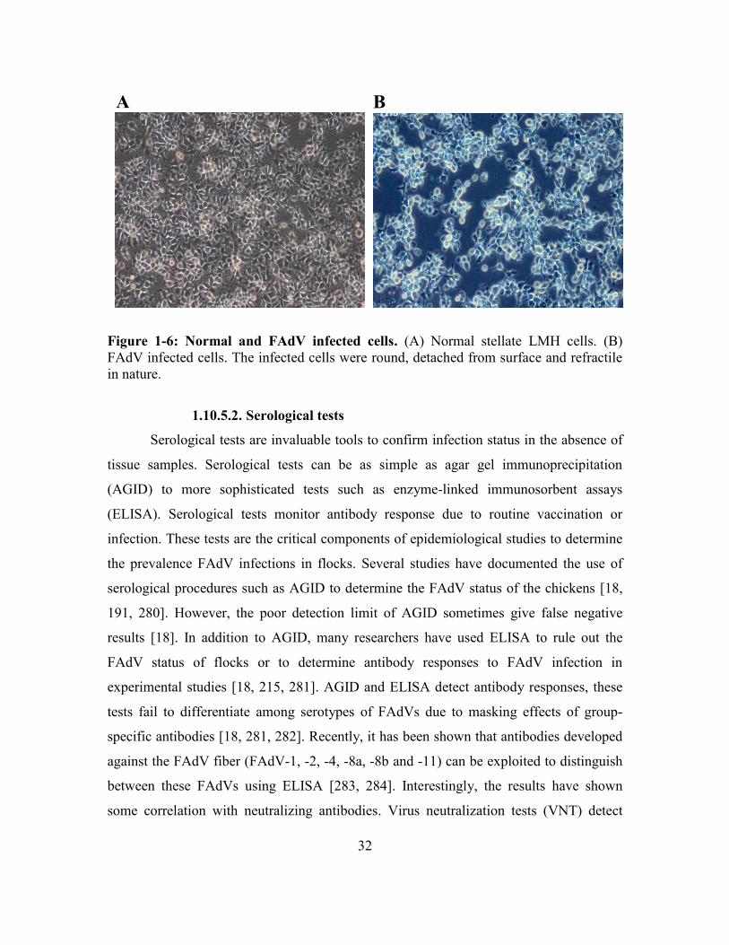
32
Figure 1-6: Normal and FAdV infected cells. (A) Normal stellate LMH cells. (B)
FAdV infected cells. The infected cells were round, detached from surface and refractile
in nature.
1.10.5.2. Serological tests
Serological tests are invaluable tools to confirm infection status in the absence of
tissue samples. Serological tests can be as simple as agar gel immunoprecipitation
(AGID) to more sophisticated tests such as enzyme-linked immunosorbent assays
(ELISA). Serological tests monitor antibody response due to routine vaccination or
infection. These tests are the critical components of epidemiological studies to determine
the prevalence FAdV infections in flocks. Several studies have documented the use of
serological procedures such as AGID to determine the FAdV status of the chickens [18,
191, 280]. However, the poor detection limit of AGID sometimes give false negative
results [18]. In addition to AGID, many researchers have used ELISA to rule out the
FAdV status of flocks or to determine antibody responses to FAdV infection in
experimental studies [18, 215, 281]. AGID and ELISA detect antibody responses, these
tests fail to differentiate among serotypes of FAdVs due to masking effects of group-
specific antibodies [18, 281, 282]. Recently, it has been shown that antibodies developed
against the FAdV fiber (FAdV-1, -2, -4, -8a, -8b and -11) can be exploited to distinguish
between these FAdVs using ELISA [283, 284]. Interestingly, the results have shown
some correlation with neutralizing antibodies. Virus neutralization tests (VNT) detect
A
S
B
S

33
virus-specific anti-FAdV fiber antibodies. This technique differentiates FAdV into
serotypes [189]. This test is the principle method to establish a serotype specific
diagnosis. Virus-specific antibodies have also been used to determine virus replication
sites in tissues by using immunohistochemical methods [256].
1.10.5.3. Molecular tests
PCR has revolutionized FAdV diagnostics. PCR alone or in combination with
restriction enzyme analysis has been used to differentiate FAdVs into different genotypes
[36, 37, 285-287] based on their electrophoretic mobility in agarose gel. RFLP techniques
gained popularity following the pioneering work of Zsak and Kisary (1984) [35], who
first used this method to differentiate avian adenoviruses. PCR along with DNA
sequencing of the hexon gene more accurately differentiate FAdVs into different
genotypes and are extensively used to establish evolutionary relationships with newly
identified FAdVs [3, 204, 214, 288-290]. In addition to FAdV hexon gene sequencing,
Schachner et al. (2016) have also used fiber-gene sequence to differentiate FAdV
serotypes [291]. In the last decade, researchers have devised faster ways to genotype
FAdVs within hours with high accuracy and confidence by using high resolution melt
curve (HRM) analysis of the hexon gene in a real-time (rt)-PCR platform [229, 292, 293].
Although, experimental data indicated a considerable consensus between genotyping and
serotypes based on HRM analysis of the hexon gene [229], sometimes more than one
technique is required to establish the identity of the virus.
1.11. Control of IBH
Strict biosecurity and farm hygiene are of prime importance. Any breach of these
invites infectious diseases to the chicken barns. Besides biosecurity at an acceptable
level, sound vaccination programs aid in the prevention of IBH.
Chickens contract FAdV infections by either horizontal or vertical transmission
[189]. While strict biosecurity can control the horizontal spread of the virus, it is
impossible to prevent vertical transmission of FAdVs without vaccinating broiler
breeders. Therefore, vertical transmission control strategies are mainly focused on
34
vaccinating broiler breeders to induce antibodies against FAdVs [189]. Seroconversion of
breeders before egg-laying aids in the prevention of vertical transmission [272, 273].
Moreover, it allows maternal antibody transfer to progenies, which protects them from
FAdV exposure in neonatal life [294]. Exposure of hens to heterologous viruses breaks
the maternal antibody barrier and causes disease in broiler progenies [229, 295].
Therefore, the effective preventative strategy requires vaccines that confer broad-
spectrum protection against various FAdVs. In addition to FAdV vaccination, vaccination
for immunosuppressive agents is also recommended as an aid to prevent opportunistic
infections by FAdVs [243, 295]. Li et al. (2017) comprehensively reviewed various
approaches of vaccinology for FAdV disease control. These include live vaccines,
autogenous inactivated vaccines, inactivated vaccines of cell culture and egg origin virus
as well as subunit vaccines [296].
1.11.1. Live vaccines
Live virus vaccines are economic, efficacious and easy to administer for mass
vaccination. Numerous live vaccines are in use to control infectious viral diseases of
poultry such as NDV, infectious laryngotracheitis and infectious bronchitis [297-299]. At
present, a monovalent live FAdV (FAdV-8b) vaccine is approved for broiler breeder
vaccination programs in Australia to protect broilers from IBH caused by FAdV-8b [229,
232]. The vaccination strategy includes oral exposure of increasing percentage of birds
multiple times between 6 and 30 weeks of age until they develop sufficient antibody
levels. Besides IBH, live FAdV vaccines have been developed against HHS (FAdV-4) by
attenuating live FAdV-4 strains in embryonated chicken eggs and QT cell lines [260,
300]. Protection up to 97.7 to 100% has been demonstrated in vaccinated chicks
following lethal virus exposure in these studies [260, 300]. Despite their advantage, an
important implication of live virus vaccines is fecal shedding by vaccinated birds until
immunity develops. Chicks shed FAdVs intermittently for several days [253, 270, 271].
Therefore, it is important to characterize FAdV infections in broiler breeders to determine
the duration of virus shedding, to study immunity and to assess the role of immunity in
prevention of viral shedding.
35
1.11.2. Inactivated vaccines
Most countries rely on inactivated autogenous liver tissue, cell culture or egg
propagated inactivated vaccines to vaccinate broiler breeders [161, 162, 186, 192, 294,
301]. The inactivated vaccines are formulated with mineral-oil or aluminium hydroxide
adjuvants and are administered by intramuscular injection into pectoral muscles in
breeders. FAdV vaccines are also formulated as inactivated polyvalent vaccines along
with other avian viruses such as NDV and egg drop syndrome in some countries [302].
The protection rate varied from 80 to 100% with inactivated FAdV-4 in against HHS
[303], 90- 100 % against IBH (FAdV-2) [304] and 98 % against FAdV-8, 92% against
FAdV-11 [294].
1.11.3. Subunit vaccines
Besides live and inactivated vaccines, several researchers have also developed
and tested subunit vaccines for the prevention of FAdV diseases [43, 305-308] mainly
HHS and non-FAdV adenoviral disease of chickens (egg drop syndrome) and turkeys
(hemorrhagic enteritis) [181, 309]. Shah et al. (2012) reported 90% protection against
HHS in broilers vaccinated with FAdV-4 penton subunit vaccine [306]. Approximately
62% and 96% protection has been reported in SPF chickens against HHS vaccinated with
fiber-1 and fiber-2 FAdV-4 subunit vaccines [43]. More studies that are recent have
shown 90-100% protection against HHS in SPF chicks that were vaccinated with fiber-2
subunit vaccine of FAdV4 [307, 308]. The protection was modest (22% and 40%) with
subunit hexon, penton, fiber-1 and 100k vaccine against HHS [305, 306]. However, it
was improved by increasing the dose of subunit vaccine (hexon and fiber-1) against HHS
[308] .Similar developments are lacking for IBH causing FAdVs.
1.11.3.1. Adjuvants
Adjuvant discovery is an active area of research and development for better
delivery of inactivated or subunit vaccines. Adjuvants are essential component of
inactivated vaccines and are required for inducing a superior immune response.
36
Emulsions of oil and water in various combinations such as water-in-oil (W/O), oil-in-
water (O/W), water-in-oil-in-water (W/O/W) are primarily used in poultry vaccines. The
use of saponins and alum-based adjuvants has also been reported [310, 311]. Among
these, W/O adjuvants are most robust, efficacious and safe formulations [312]. W/O
comprises tiny water droplets dispersed in oil. The preparation acts like a depot injection
in tissues, which slowly releases the antigen to induce a sustainable immune response by
inducing inflammatory responses aiding in antigen uptake and subsequent stimulation of
the immune system. Researchers are testing new formulations of W/O adjuvants in
poultry vaccines to minimize tissue reaction associated with them. Contrary to W/O
adjuvants, O/W adjuvants have oil phase dispersed in water allowing them to quickly
release the antigens, inducing strong but brief immune response. Oils of animal,
vegetable or synthetic origins are commonly used in these preparation and are shown to
be effective in poultry vaccines [313].
The aim of inactivated and subunit vaccines is to induce high-levels of antibodies
in hens before egg laying to ensure maternal antibody transfer to progenies. Therefore,
adjuvants that skew immune responses to Th2-type immunity are highly desired in
poultry vaccines. Several W/O emulsion adjuvants of Montanide series (Seppic, France)
and O/W mineral oil emulsions (Emulsigen-D) (MPV Technologies Inc.) were studied in
experimental avian viral or bacterial vaccines. They mainly favor T helper (Th)-2 type
immune responses [314, 315] similar to alum adjuvants. Besides chemical adjuvants,
microbial components, such as interleukins (e.g. IL-18), interferons (e.g. IFN-γ), avian-b
defensins, pattern recognition receptors such as CVCVA5 (combination of polyriboinosic
polyribocytidylic, resiquimod, muramyl dipeptide and levamisole) and non-methylated
prokaryotic DNA [221, 316-320]. Synthetic CpG-ODN is an analog of microbial DNA
that has been extensively studied for its immune stimulatory properties [314, 318, 321,
322], as a vaccine adjuvant or as an in ovo immunostimulant [321, 323]. There are
various type of CpG-ODNs [324] differing in their backbone and physicochemical
properties. They stimulate different immune cells, act by different subcellular pathways
and induce different cytokines [324]. CpG-ODN is sensed as a foreign molecule by
mammalian cells as it lacks methylation at cytosine residues and triggers cytokines and

37
chemokine responses upon recognition of TLR-9 (or chTLR-21 in chickens) [321]. CpG-
ODN is a potential vaccine adjuvant in poultry vaccines in the future as it adjuvants with
minimal or no tissue reaction [318] and potential low cost to benefit ratio [322]. Previous
studies have analyzed its adjuvanticity for short-term studies of 6 to 7 weeks. Whether it
can induce and maintain antibody levels similar to W/O emulsions needs investigation.
1.12. Current problems associated with IBH in Canada
IBH is the only FAdV disease present in Canada. FAdVs of species E (FAdV-8a,
-8b and -7) and D (FAdV-11 and FAdV-2) are the most prevalent viruses in Canada. At
present, commercial FAdV vaccines are not marketed in Canada. Broiler breeders are
vaccinated with autogenous inactivated vaccines in endemic areas as a preventative
measure against IBH. Despite their use, sporadic outbreaks of IBH commonly occur as a
result of heterologous FAdVs, which are not controlled by the autogenous vaccine.
Moreover, autogenous vaccines lack potency and efficacy data. Multivalent FAdV
vaccines, which are safe and efficacious, and offer broad-spectrum protection against
multiple serotypes are needed. Also, for industry feasibility and commercialization
purpose, methods of mass production and mass application need to be considered.
Therefore, the objective of this project was to develop a live FAdV vaccine using wild-
type FAdVs and compare its efficacy with an inactivated FAdV vaccine. In addition to
conventional vaccines, the possibility of a subunit vaccine and suitable adjuvants for
inactivated vaccines were explored. Briefly, the goals of this project are divided into
three objectives as follows:
1.13. Objectives
1. Control of IBH in the Canadian broiler chicken industry with a live or inactivated
FAdV vaccine.
2. Selection of appropriate adjuvant(s) for an inactivated FAdV vaccine.
3. Control of IBH in Canada using a FAdV subunit vaccine.

38
CHAPTER 2: INACTIVATED OR LIVE BIVALENT FOWL ADENOVIRUS
(FADV-8b+FADV-11) BREEDER VACCINES PROVIDE BROAD-SPECTRUM
PROTECTION IN CHICKS AGAINST INCLUSION BODY HEPATITIS (IBH)
Ashish Gupta1, Shelly Popowich1, Davor Ojkic2, Shanika Kurukulasuriya1, Betty Chow-
Lockerbie1, Thushari Gunawardana1, Kalhari Goonewardane1 Ruwani Karunarathna1,
Lisanework E. Ayalew1, Khawaja Ashfaque Ahmed1, Suresh K. Tikoo4, Philip Willson3,
Susantha Gomis1*
1Department of Veterinary Pathology, Western College of Veterinary Medicine,
University of Saskatchewan, Saskatoon, SK Canada S7N 5B4
2Animal Health Laboratory, University of Guelph, P.O. Box 3612, Guelph, ON, Canada
N1H 6R
3Canadian Centre for Health and Safety in Agriculture, University of Saskatchewan,
Saskatoon, SK Canada S7N 5E5
4Vaccinology and Immunotherapeutic Program, School of Public Health, University of
Saskatchewan, Saskatoon, SK Canada S7N 5E3
*Corresponding author mailing address: Department of Veterinary Pathology, Western
College of Veterinary Medicine, 52 Campus Drive, University of Saskatchewan,
Saskatoon, SK Canada S7N 5B4. Phone: (306) 966-7299. Fax: (306) 966-7439. Email:
Published in Vaccine, 2018. 36 (5): 744-750
(Elsevier permits the authors to include their articles in full or in part in a thesis or dissertation for non-
commercial purposes)

39
2.1. Abstract
FAdV is comprised of five species (A to E) and 12 serotypes (1-7, 8a, 8b, 9-11).
IBH is caused by FAdV-7, -8a, -8b (species E) and FAdV-2 and FAdV-11 (species D).
Commercial vaccines against IBH are not available in Canada. Autogenous FAdV broiler
breeder vaccines are now used in some areas where outbreaks of IBH are occurring. The
objective of this study was to evaluate the efficacy of a bivalent (species D and E) live
and an inactivated FAdV broiler breeder vaccine in protecting broiler chicks against IBH
through maternal antibody (MtAb) transfer. FAdV seronegative broiler breeders
(n=300/group) received either a live or inactivated bivalent (FAdV-8b-SK+FAdV-11-
1047) vaccine. The live vaccine (1x10⁴ TCID50 of each virus/bird) was given orally once
at 16 weeks of age and the inactivated vaccine (1x10⁶ TCID50 of each virus+20%
Emulsigen-D) was given intramuscularly at 16 and 19 weeks of age. Controls (n=150)
were given saline orally. The inactivated vaccine group was boosted 3 weeks later with
the same vaccine. Neutralizing antibodies (NAb) in sera (n=10) were detected at 19, 22,
30 and 48 weeks of age. NAb were able to neutralize various FAdV serotypes within
species D and E. Mean NAb were similar in the both live and killed vaccine groups at 19,
30 and 48 weeks and ranged from 2.40 log10 to 3.70 log10. Approximately 26±7% of
MtAbs were passively transferred through eggs to day-old chicks. Progeny challenged
with a lethal dose (1x10⁷ TCID50/bird intramuscularly) of FAdV-8b-SK, FAdV-11-1047,
or FAdV-2-685 (n=90/group) at 14 days post-hatch (dph) showed 98-100% protection in
broiler chicks to homologous or heterologous FAdV challenges. Our data suggests that a
bivalent live and an inactivated FAdV vaccine are equally effective and have the
potential for the control of IBH.
2.2. Introduction
IBH is an economically important FAdV disease of 2 to 5 week old broiler
chickens [189, 231, 279]. The disease has also been reported within a week of hatch in
broilers [213] as well as in pullets [214]. IBH is characterized by increased flock
mortality that varies between 2% to 30% [214, 216, 219]. Although five species (A to E)
and 12 serotypes (1-7, 8a, 8b, 9 to 11) of FAdVs are known [270, 279], IBH is primarily
40
caused by FAdV-7, -8a and -8b of species E and FAdV-2 and FAdV-11 of species D [3,
161, 214, 215, 229]. FAdV-8a, -8b and -11 are the predominant serotypes associated with
outbreaks of IBH in Canada [3, 195]. The existence of multiple FAdV serotypes and
species and lack of interspecies protection makes controlling IBH difficult [189]. Hence,
a broad-spectrum FAdV vaccine is needed for the prevention of IBH.
Since licensed FAdV vaccines are not available in Canada [3, 221], broiler
breeders in areas with significant IBH losses are vaccinated with autogenous vaccines for
the prevention of disease in their progeny. Several researchers have evaluated inactivated
FAdV vaccines of liver-tissue, cell-culture or egg origin in broilers against HHS [192,
193, 301, 325-327] or in broiler breeders against IBH [294, 304]. HHS is an acute FAdV
(FAdV-4) disease of 3 to 6 week old broiler chickens, which can cause 30-70% mortality
in susceptible flocks. Inclusion body hepatitis, hydropericardium, pulmonary hemorrhage
and nephrosis are characteristic lesions of the disease [300, 301]. Inactivated FAdV
(FAdV-4) vaccines have been used in various viral doses (1x103.8 EID50 [326], 5x105.5
TCID50 [301, 325] or 1x10⁶ TCID50 [193]) for providing immunity in chicks against HHS
between 7 to 42 days post-vaccination (dpv). An inactivated FAdV (FAdV-2) vaccine
(1x10⁶ TCID50) was shown to protect chicks against IBH by MtAb transfer from
vaccinated breeders [304]. Like inactivated vaccines, live FAdV vaccines protect
chickens against FAdV diseases [260, 300]. Live attenuated FAdV-4 vaccines have
shown protection against HHS when used at a dose of 1x103.3 EID50 [300] or 5x10⁴
TCID50 [260] in day-old chicks. Apart from attenuated FAdVs, wild-type FAdV (FAdV-
8b) virus (1x10³ TCID50 per bird) has been used as a broiler breeder vaccine by protecting
broiler chicks through MtAb [229].
These studies have led to monovalent FAdV vaccines based on FAdV-4 which
can help in preventing HHS, but not necessarily IBH, as the latter is caused by multiple
serotypes [189] Monovalent vaccines do not cross-protect against heterologous FAdV
species [189, 229]. For the effective prevention of IBH, multivalent vaccines are needed.
Multivalent vaccines have been shown to provide broad-spectrum protection against
avian viruses [294, 328, 329]. Therefore, the goal of this study was to compare and
evaluate the efficacy of a bivalent (FAdV-8b+FAdV-11) live versus a bivalent (FAdV-

41
8b+FAdV-11) inactivated broiler breeder vaccine for the protection of broiler chicks
against IBH by MtAb transfer.
2.3. Materials and methods
2.3.1. Propagation of FAdVs for FAdV vaccine
Plaque purified FAdV-8b-SK [279] and FAdV-11-1047 (Animal Health Lab,
University of Guelph, Ontario) were propagated in LMH cells by infecting 80%
confluent cells at a multiplicity-of-infection of 5. The cells were incubated for 1 h at 37 C
and 5% CO2 and subsequently replenished with Dulbecco’s Modified Eagle Medium:
Nutrient Mixture F-12 (DMEM: F-12) (Thermo Fisher Scientific) supplemented with 5%
fetal bovine serum, 20 mM glutamine, 2 mM HEPES and 1:1000 gentamicin (100 µL of
50 µg/mL per 100 mL of media). The infected cells were harvested 72 h post-infection
(FAdV-8b-SK) or 96 h post-infection (FAdV-11-1047), freeze-thawed five-times and
centrifuged at 3000 rpm to separate the supernatant. The supernatant was tittered by Reed
and Muench method [330].
The live bivalent FAdV vaccine was prepared by mixing FAdV-8b-SK and
FAdV-11-1047 in sterile saline to obtain a dose of 1x10⁴ TCID50 of each serotype.
Similarly, the inactivated bivalent FAdV vaccine (heat inactivation at 64 C for 1 h) was
prepared by mixing FAdV-8b-SK and FAdV-11-1047 with 20% Emulsigen-D (MVP
Laboratories, Omaha, NE) and sterile saline to make a dose of 1x10⁶ TCID50 of each
serotype. The final volume of the vaccine dose was 0.5 mL per bird.
2.3.2. Preparation of FAdV inoculum for broiler challenge
FAdV-8b-SK or FAdV-11-1047 infected livers of SPF broilers were macerated in
a mortar by a pestle and Waymouth’s media (Thermo Fisher Scientific) was added to
make a 40% liver homogenate. The homogenate was freeze-thawed (5X) to release the
virus into the supernatant, which was later separated by centrifugation at 6000 rpm for 30
min at 10 C. The supernatant was filter sterilized using 0.22 m syringe-filters (Cameo,
Maine Manufacturing LLC) and titrated as described above. FAdV-8b-SK and FAdV-11-
1047 hexon genes were amplified with in a standard polymerase chain reaction using Hex
42
A/B primers [37] and the identity was confirmed by analyzing 158 amino acid long
sequence between position 130 to 287 as described previously [3].
2.3.3. Safety evaluation of live FAdV in broiler breeders
The safety of the wild-type live FAdV was evaluated in broiler breeders by
intramuscular vaccination (n=20 females+4 males) with FAdV-8b-SK (1x10⁷ TCID50 per
bird) at 29 weeks of age. Eggs, sera and blood (n=5) were collected for 1 week before
vaccination (control). Following vaccination, fecal samples (n=6) were collected at 1, 3,
7, 10, 14, 17 and 21 dpv to determine fecal shedding. Blood samples (n=5) were collected
at 2, 3, 7, 10, 14, 17 and 21 dpv to detect viremia and 7, 14 and 21 dpv to measure NAb.
Eggs were collected in weekly batches for 3 weeks to detect egg transmission of FAdV-
8b-SK. The experiment was terminated at 21 dpv and liver, spleen, kidney, ovarian
follicles and cecal tonsils (n=5 females and 1 male) were collected for virus isolation and
DNA detection. We also examined the safety of the live bivalent FAdV-8b-SK+FAdV-
11-1047 vaccine. Broiler breeders were orally administered 1x10⁴ TCID50 of each
virus/bird once at 16 weeks of age. Eggs were collected 9 weeks after the breeder
vaccination from both the vaccinated and saline control groups. Mortality was recorded
in the progeny from both groups. Post-mortem examinations were conducted on dead
birds and virus detection was done using cell culture and PCR techniques.
2.3.4. Animals and experimental design
Day-old (Ross) broiler breeders were obtained from Aviagen Inc. (Huntsville,
AL) and were reared in the Animal Care Unit, Western College of Veterinary Medicine,
University of Saskatchewan as per Aviagen guidelines. Their FAdV seronegative status
was confirmed by a virus neutralization test (VNT) at 15 weeks of age. Broiler breeders
were randomly allocated into three groups: live (n=300), inactivated (n=300), and control
(n=150). The live vaccine group was vaccinated once orally with the live bivalent FAdV
vaccine (1x10⁴ TCID50/ bird) at 16 weeks of age. The inactivated vaccine group was
vaccinated intramuscularly with the inactivated bivalent FAdV vaccine (1x10⁶
TCID50+20% Emulsigen-D/bird) at 16 and 19 weeks of age. The control group received
43
saline orally at 16 and 19 weeks of age. Following vaccination, NAb were determined in
sera from all groups (n=10) at 19, 22, 30, and 48 weeks of age. Fecal shedding was
determined by detecting virus in cell culture and quantifying viral DNA in cloacal swabs
(n=10) from the live vaccine and control groups at 7, 14, 21 and 35 dpv.
Eggs were collected from broiler breeders at 30 weeks of age to hatch the broilers
for challenge protection studies. Day 0 sera samples (n=10) were collected to determine
the percentage of MtAb transfer [102]. MtAb mediated protection was assessed by
challenging randomly selected broiler chicks (n=90/group) intramuscularly with 1x10⁷
TCID50 of FAdV-8b-SK, FAdV-11-1047 or FAdV-2-685 liver homogenates at 14 dph.
The broilers were monitored for 10 days post-challenge for clinical signs and mortality.
Liver samples from dead or euthanized birds were preserved in 10% formalin to confirm
IBH by histopathology.
2.3.5. Measurement of neutralizing antibodies against FAdVs
NAb against FAdV-2-685, FAdV-7-x-11a like, FAdV-8b-SK, FAdV-8a-TR59
and FAdV-11-1047 were measured by VNT [279]. Two-fold serially diluted heat
inactivated (56 C, 30 min) sera were mixed with 200 TCID50 of FAdV. Samples were
incubated at 37 C and 5% CO2 for 1 h and then transferred to 96 well plates seeded with
5x10⁴ LMH cells. Cytopathic effects were investigated day 7 post-incubation.
2.3.6. Quantitation of fecal shedding in cloacal swabs
Cloacal swabs (n=10) were collected in Luria broth and used to inoculate LMH
cells to determine virus titer [279]. The shedding pattern was determined by measuring
FAdV-DNA copy numbers using the standard curve method in rt qPCR [270]. FAdV-
DNA was amplified in a 25 µL reaction from 200 ng of total DNA (Qiagen Viral RNA
mini kit) using FAdV-species-E/D primer-probes (Table 2-1) in 2-step fast cycle program
(MxPro-3005P, Agilent Technologies). Following the initial denaturation at 95 C for 10
min, 40 cycles of denaturation at 95 C for 15 sec, and extension at 60 C for 45 sec were
used for DNA amplification.
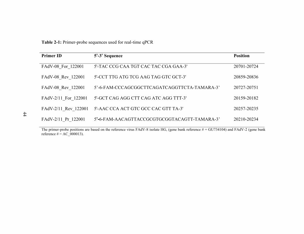
44
Table 2-1: Primer-probe sequences used for real-time qPCR
Primer ID 5’-3’ Sequence Position
FAdV-08_For_122001 5'-TAC CCG CAA TGT CAC TAC CGA GAA-3' 20701-20724
FAdV-08_Rev_122001 5'-CCT TTG ATG TCG AAG TAG GTC GCT-3' 20859-20836
FAdV-08_Rev_122001 5’-6-FAM-CCCAGCGGCTTCAGATCAGGTTCTA-TAMARA-3’ 20727-20751
FAdV-2/11_For_122001 5'-GCT CAG AGG CTT CAG ATC AGG TTT-3' 20159-20182
FAdV-2/11_Rev_122001 5'-AAC CCA ACT GTC GCC CAC GTT TA-3' 20257-20235
FAdV-2/11_Pr_122001 5’-6-FAM-AACAGTTACCGCGTGCGGTACAGTT-TAMARA-3’ 20210-20234
The primer-probe positions are based on the reference virus FAdV-8 isolate HG, (gene bank reference # = GU734104) and FAdV-2 (gene bank
reference # = AC_000013).

45
2.3.7. Statistical analysis
Differences in mean NAb titers of breeders was tested using the Student t–test
with Welch’s correction for the unequal variance (PRISM, Graph Pad, Inc. San Diego,
CA). Survival analysis of the challenge protection experiments was performed by the
Mantel-cox (Log-rank) test. The results were considered significant at P<0.05.
2.4. Results
2.4.1. Safety of live FAdV virus in broiler breeder
We first investigated the safety of the live wild-type FAdV-8b-SK virus as a
broiler breeder vaccine. Broiler breeders vaccinated with the wild-type FAdV-8b-SK did
not exhibit clinical signs, but were viremic at 2 and 3 dpv (16.6%) and were shedding
FAdV-8b-SK in feces from 1 dpv through 14 dpv (Figure 1A). NAb developed as early
as 7 dpv which increased to a higher level at 14 and 21 dpv (Figure 1B). No pathological
lesions were present in the organs at 21 dpv but a few samples of the kidney (1/6),
pancreas (2/6), cecal-tonsils (4/6), ovarian follicles (2/6), and shell glands (2/6) were
positive for FAdV DNA in qPCR. We next examined the duration of a possible viral
transfer to progeny through eggs and eventually pathogenicity in chicks. Very high
mortality (72.2%) due to IBH was observed in the progeny hatched from the eggs
collected during the first-week post vaccination (Figure 1C). However, progeny chicks
hatched from the eggs collected during the 2nd and 3rd weeks after vaccination had
MtAb (Figure 1D) and were free of clinical disease.

46
Figure 2-1: Safety evaluation of wild-type live FAdV-8b-SK in broiler breeders in
egg production (29 weeks of age). (A) Fecal shedding by the broiler breeders post-
vaccination. The limit of detection of rt qPCR was 36 copies (~ CT number 35.98).
Samples used for our analysis had CT numbers ≤ 35. (B) NAb response post-vaccination.
(C) Mortality in 1st, 2nd and 3rd week broiler chicken progeny. (D) Kaplan Meier survival
estimates. Maternal NAb titers in broiler chicken progeny, P<0.05.
Bro
iler
bre
eder
s B
roil
er p
rog
eny
A B
C D

47
We also vaccinated broiler breeders orally once at 16 weeks of age with a live
bivalent FAdV-8b-SK+FAdV-11-1047 vaccine (1x10⁴ TCID50 of each virus/bird) to
examine the possibility of transmission of the vaccine virus to progeny more than 9
weeks after the breeder vaccination. We did not find any vaccine virus transmission to
the progeny. The percent mortality in chicks hatched from the live vaccinated and saline
groups for 2 weeks post-hatch (n=90) were 1.1% and 2.2%, respectively (Figure 2-2).
The causes of death were sudden death syndrome and ascites. Samples collected from the
dead birds did not show evidence of vaccine virus in either cell culture or PCR detection,
suggesting a NAb response in broiler breeders.
Figure 2-2: Breeder vaccination with live bivalent FAdV-8b-SK+FAdV-11-1047
vaccine at 16 weeks of age is safe and does not cause mortality in hatching chicks
and vertical transmission of the vaccine viruses. Percent mortality in the chicks that
were hatched from live vaccinated group and saline group for 2 weeks post- hatch
(n=90). The mortality percentage was 2.2% in chicks hatched from saline control
breeders and 1.11% in chicks hatched from live vaccinated breeders, respectively. The
cause of death was sudden death syndrome and ascites. No IBH specific lesions were
present. FAdV was not detected by PCR and cell culture in chicks

48
2.4.2. Neutralizing antibody response in broiler breeders
Control broiler breeders remained seronegative throughout the experiment. NAb
were detected against FAdVs in groups vaccinated either with either the bivalent live or
inactivated FAdV vaccine. The mean NAb titer against the FAdV-8b-SK bivalent live or
inactivated FAdV vaccine group at 19 weeks of age were 2.80 log10±0.45 and 3.10
log10±0.37 respectively (Figure 2-3A). NAb titer against FAdV-8b-SK reached the
highest level at 48 weeks of age in both the live and inactivated vaccine groups. NAbs
against FAdV-11-1047 were also detected and were maintained between 3.20 log10±0.37
and 3.60 log10±0.31 in both vaccine groups from 19 through 48 weeks of age (Figure
2-3B).
Figure 2-3: NAb response of broiler breeders. NAb response of broiler breeders to
live and inactivated bivalent FAdV vaccines at various time points post-vaccination,
Mean±SD, n=10. (A) FAdV-8b-SK (B) FAdV-11-1047. The control broiler breeders
were serologically negative throughout the experiment.
A B FAdV-8b-SK FAdV-11-1047

49
2.4.3. Cross-neutralizing antibody response against heterologous FAdVs
Cross-NAb were determined against heterologous FAdV-2-685 FAdV-7-x11a and
FAdV-8a-TR59 in live (Figure 2-4A) and inactivated vaccinated (Figure 2-4B) broiler
breeders at 19, 22 and 48 weeks of age. The NAb varied between 3.0 log10±0.40 to 3.70
log10±0.40 in the live vaccinated breeders and between 2.40 log10±0.17 to 3.70 log10±0.20
in the inactivated vaccinated breeders during 19 to 48 weeks of age.
Figure 2-4: Comparison of cross-neutralizing antibody levels with heterologous
FAdV serotypes. Cross-neutralizing antibodies (NAb) against FAdV-7-x-11a like,
FAdV-8a-TR59 (species E) and FAdV-2-685 (species D) in live and inactivated bivalent
(FAdV-8b-SK+FAdV-11-1047) FAdV vaccine groups at 19, 22 and 48 weeks of age,
Mean±SD, n=10 birds. Control breeders were serological negative throughout the
experiment.
2.4.4. Fecal shedding of FAdVs in broiler breeders vaccinated with live
bivalent FAdV vaccine
FAdV shedding was not detected in the control breeders. However, FAdV
shedding in feces was detected in broiler breeders vaccinated with a live vaccine (FAdV-
8b-SK+FAdV-11-1047). Infectious virus was detected in the feces of 90% of broiler
A B Live bivalent vaccine Inactivated bivalent vaccine

50
breeders (9/10) at 7 dpv. The number of infectious shedders declined to 10% (1/10) at 14
dpv and no virus was detected thereafter. The mean FAdV titer (TCID50/mL) at 7 and 14
dpv was 3.60 log10 and 3.40 log10 respectively. Figure 2-5 shows virus specific fecal
shedding by live vaccinated breeders. FAdV-8b-SK DNA was detected in 90% (9/10)
and 70% (7/10) birds at 7 and 14 dpv, respectively and 10% (1/10) of the birds at 21 dpv.
Likewise, FAdV-11-1047 DNA was detected in 100% (10/10) of the birds at 7 dpv, 30%
(3/10) of the birds at 14dpv and 10% of the birds at 21 dpv. No viral DNA was detected
for either virus at 35 dpv.
Figure 2-5: Fecal shedding of FAdVs in broiler breeders vaccinated with live
bivalent FAdV vaccine. FAdV DNA copy number in cloacal swabs of broiler breeders
vaccinated with live bivalent (FAdV-8b-SK+FAdV-11-1047) vaccine at various time
points post vaccination, Mean±SD, (n=10). Limit of detection of rt qPCR was 36 copies
(~ CT number 35.98). Samples used for our analysis had CT numbers ≤ 35.
2.4.5. Maternal antibodies and protection of broilers against FAdV challenge
MtAb levels were measured in day-old chick sera by VNT. Figure 2-6A
demonstrates the MtAb level against FAdV-8b-SK in the live and inactivated vaccine
groups. These were 2.90 log10±2.70 in and 2.90 log10±0.0, respectively. Figure 2-6B
shows the MtAb titer against FAdV-11-1047 in the live and inactivated vaccine groups

51
which were 2.60 log10±2.60 and 2.20 log10±1.90 respectively. MtAb levels in day-old
chicks constituted 26.6±7.40% [32.2±0.04% (live vaccine) and 21±0.04% (inactivated
vaccine)] of NAb levels of broiler breeders at 30 week of age.
Figure 2-6: Maternal NAb levels in day old broiler chicken progeny. Mean MtAb
(n=10) in the progeny of live and inactivated vaccinated broiler breeders at day 0,
Mean±SD, (n=10). (A) FAdV-8b-SK. (B) FAdV-11-1047.
The protective efficacy of MtAbs were evaluated by exposing broiler progenies to a
lethal dose of FAdV-8b-SK, FAdV-11-1047, and FAdV-2-685 at 14 dph. Broiler progeny
from broiler breeders vaccinated with either the bivalent live or inactivated FAdV
vaccine had 98-100% survival (P<0.05) compared to the saline group (23% survival)
following a lethal challenge of FAdV-8b-SK (A). Whereas 99% survival (P<0.05) was
observed in broiler progeny compared to saline group (80% survival) following a lethal
challenge with FAdV-11-1047 (B). After challenge with a heterologous virus, broiler
progeny from either vaccine group had 100% (P<0.05) survival following a lethal
challenge of FAdV-2-685 compared to the saline group (95% survival) (C).
FAdV-8b-SK FAdV-11-1047 A B

52
Figure 2-7: Assessment of maternal antibody mediated protection against
homologous and heterologous FAdV serotypes. Kaplan-Meier survival estimate of
progeny following lethal virus challenge at 14 dph (P<0.05), n=90/ group. (A) FAdV-8b-
SK. (B) FAdV-11-1047. (C) FAdV-2-685.
FAdV-8b-SK FAdV-11-1047
FAdV-2-685 C
B A
53
2.5. Discussion
FAdVs are ubiquitous [18] and chickens often get infected with multiple FAdVs
[37, 227]. IBH control is challenging because multiple FAdV serotypes of species E
(FAdV-7, FAdV-8a, FAdV-8b) and species D (FAdV-2, and FAdV-11) cause the disease
in chicks [3, 189, 195, 241]. It is also known that NAb developed from a monovalent
vaccine does not cross-protect from infection with a heterologous FAdV species [229].
MtAb derived from such vaccines also fail to protect broiler chicks from exposure of
heterologous FAdVs [229]. Since FAdV-8a and FAdV-8b and FAdV-11 are predominant
serotypes in Canada [3, 195], we included FAdV-8b and FAdV-11 to develop a bivalent
FAdV vaccine.
FAdVs rarely cause disease in adult chickens [189] and were safely used for broiler
breeder vaccination (FAdV-8b) between 6 to 30 week of age for IBH prevention [229].
Since there is a risk of egg-transmission of FAdVs [189, 254, 274] during production, we
examined the safety of a wild-type FAdV vaccine in broiler breeders by vaccinating them
at egg-laying age. We observed that a high dose of a wild-type FAdV-8b vaccine which
otherwise causes very high mortality in chicks [219], did not induce clinical signs or
pathological changes when given intramuscularly to adult breeders (29 weeks of age).
However, egg transmission of virus did occur in chicks that were hatched in the first
week following the broiler breeder vaccination. The results are supported by previous
studies [272, 273] in which egg transmission is reported within the first week. Mortality
can be attributed to viremia occurring at 2 and 3 dpv. Remarkably, no mortality occurred
in the progeny hatched from the eggs collected during the 2nd and 3rd weeks after the live
breeder vaccination. This indicated cessation of egg transmission and is likely a response
to the rising NAb response at 7 dpv [272, 273]. Our data provides strong evidence that
broiler breeders can be safely vaccinated with wild-type FAdVs at around 16 weeks of
the age, long before the onset of egg laying.
FAdV fecal shedding facilitates the horizontal spread of FAdVs [189, 270]. In
broiler breeders vaccinated at 16 weeks of age, we found that infectious virus shedding
ceased as early as 14 dpv and viral DNA disappeared at 21 dpv, which coincided with the
detection of NAb in sera. Our findings are in agreement with previous studies in which
fecal shedding declined with time in FAdV infected chickens [80, 271]. Apart from

54
humoral immunity, cellular immunity could also play roles in early clearance of virus-
infected cells [279] thus resulting in a significant reduction of infectious virus shedding.
Live viral vaccines stimulate both cellular and humoral immunity. This is evident by
chick vaccination with a live FAdV-4 vaccine and survivability of 94.7-100% following
exposure to a lethal dose of FAdV [260, 300]. Here, we mainly examined the NAb to a
bivalent live FAdV vaccine in broiler breeders. A single inoculation with 1x10⁴ TCID50
of a bivalent live FAdV vaccine at 16 weeks of age induced long-lasting (32 weeks)
broad-spectrum NAb response (2.90 log100.45) against homologues or heterologous
FAdVs. NAb levels of broiler breeders in either vaccine group at 30 weeks of age were
3 log10 and were considered protective as 99-100% hatched chicks survived a lethal
challenge of FAdV-8b-SK, FAdV-11-1047, and FAdV-2 at 14 dph. MtAbs are critical for
protecting chicks against IBH [279, 294]. In our study, the protection against IBH
corresponded to the level of MtAbs in chicks of 32.2±0.04% of NAb in breeders at 30
weeks of age.
We next examined a bivalent inactivated FAdV vaccine. Historically, inactivated
autogenous, cell-culture or egg-propagated monovalent and polyvalent FAdV vaccines of
various doses of viral antigen have shown protection in chicks against FAdVs [192, 294,
301, 304, 325]. A single vaccination (FAdV-2) of chicks with 1x10⁶ TCID50 induced a
13-week long NAb response at a level of >6.17 log2 (~2.30 log10) [304]. Here, we
demonstrated a 32-week long broad-spectrum NAb response in broiler breeders
vaccinated twice with a bivalent inactivated FAdV vaccine containing 1x10⁶ TCID50 of
FAdV-8b-SK and FAdV-11-1047. The NAb level was 2.90 log100.45 against the
vaccine viruses and 2.40 log100.17 against heterologous FAdVs. The chicks had
21±0.04% MtAb and 98-100% (P<0.05) survival was observed against FAdV-8b-SK,
FAdV-11-1047 and FAdV-2-685 following virus challenge at 14 dph. Our results agree
with Alvarado et al. (2007) who observed similar findings with a bivalent autogenous
FAdV vaccine [294].
2.6. Conclusions
Our study demonstrated a long-lasting, broad-spectrum NAb response in broiler
breeders either by a single vaccination with 1x10⁴ TCID50 of a bivalent FAdV (FAdV-8b-

55
SK+FAdV-11-1047) vaccine or by two vaccinations at a dose of 1x10⁶ TCID50 of
inactivated bivalent vaccine. Broiler breeder vaccination successfully protected chicks
against IBH by MtAb transfer, which is a common industrial practice for protecting
chicks against infectious diseases [102]. The induction of cross-protective NAb by the
vaccines tested in this study strongly supports the idea of eliciting broad-spectrum
protection against IBH using bivalent or multivalent broiler breeder vaccines.
Collectively, our data suggest that live and inactivated bivalent vaccines can be used for
the prevention of IBH in the future.

56
PREFACE TO CHAPTER 3
In the previous chapter, we have demonstrated a remarkable antibody response to
inactivated FAdV vaccine adjuvanted with Emulsigen-D in broiler breeders. It is well
known that adjuvants induce and improve the quality of immune response to inactivated
vaccines; however, oil-in water adjuvants cause adverse tissue reactions at injection sites
of a vaccine. Emulsigen-D is an oil-in-water adjuvant commonly use in poultry vaccines
that has been associated with induction of a good antibody response but tissue necrosis at
the injection site. Therefore, to minimize tissue reactions and to improve the safety of
inactivated FAdV vaccine we wanted to explore adjuvant, which induce immune
responses similar or superior to Emulsigen-D with less tissue reaction. Therefore, we
have evaluated CpG-ODN to replace Emulsigen-D in our inactivated FAdV vaccine.
57
CHAPTER 3: CHARACTERIZATION OF CELLULAR AND HUMORAL
IMMUNE RESPONSES OF BROILER BREEDERS FOLLOWING
VACCINATION WITH A FOWL ADENOVIRUS ANTIGEN ADJUVNATED
WITH EMULISGEN-D OR OLIGODEOXYNUCLETIDES CONTAINING CPG
MOTIFS
Ashish Gupta1, Khwaja Ashfaque Ahmed1, Shelly Popowich1, Kalhari Goonewardene1,
Ruwani Karunarathana1, Davor Ojkic1, Betty Chow-Lockerbie1, Suresh K. Tikoo4, Philip
Willson3 and Susantha Gomis1*
1Department of Veterinary Pathology, Western College of Veterinary Medicine, University of
Saskatchewan, Saskatoon, SK Canada S7N 5B4
2Animal Health Laboratory, University of Guelph, P.O. Box 3612, Guelph, ON, Canada N1H
6R8
3Canadian Centre for Health and Safety in Agriculture, University of Saskatchewan, Saskatoon,
SK Canada S7N 5E5
4Vaccinology and Immunotherapeutics Program, School of Public Health, University of
Saskatchewan, Saskatoon, SK Canada S7N 2Z4
*Corresponding author mailing address: Department of Veterinary Pathology, Western
College of Veterinary Medicine, 52 Campus Drive, University of Saskatchewan,
Saskatoon, SK Canada S7N 5B4. Phone: (306) 966-7299. Fax: (306) 966-7439. Email:

58
3.1. Abstract
IBH is an economically important FAdV disease of broiler chickens worldwide.
Currently, there is no commercial vaccine available to control IBH in Canada.
Previously, we have demonstrated the protective efficacy of inactivated live and subunit
FAdV vaccines against IBH. The objective of this study was to characterize cellular and
humoral responses in broiler breeders following vaccination with a FAdV-8b-SK vaccine
adjuvanted with Emulsigen-D or CpG-ODN. Four groups (n=24/group) of broiler
breeders were vaccinated at 16 weeks of age with FAdV-8b-SK (1x10⁶ TCID50/bird)
adjuvanted with either 20% Emulsigen-D or 50 µg CpG-ODN. Control groups were
vaccinated with saline or FAdV-8b-SK with no adjuvant. Groups were boosted at 19
weeks of age with their respective vaccines. Humoral and cellular immune responses
were determined by measuring serum IgY and NAb, CD4+:CD8+ T-cell ratio and the
expression of IL-4 and IFN-γ in peripheral blood. Vaccine efficacy was determined by
challenging broiler progeny at 14 days post-hatch. As a vaccine adjuvant, CpG-ODN
induced a 0.20 to 0.30-fold higher IgY antibody response after the booster vaccination
compared to Emulsigen-D. Both the Emulsigen-D and CpG-ODN adjuvanted groups
induced NAb ≥2.90 log10 in broiler breeders and were equally protective (99% progeny
survival, P<0.05) against IBH. The CD4+:CD8++ T-cell ratio increased significantly
(P<0.05) in the Emulsigen-D adjuvanted group whereas it decreased significantly
(P<0.05) in the CpG-ODN adjuvanted group. Th-1 and Th-2 type cellular immune
responses were observed when broiler breeders were vaccinated with FAdV antigens
adjuvanted with CpG-ODN. Furthermore, FAdV-8b-SK adjuvanted with CpG-ODN
induced a significantly higher CD8+ T-cell memory response (P<0.05). In summary,
FAdV-8b-SK adjuvanted with CpG-ODN induced an immune profile to protect their
progeny against IBH.
3.2. Introduction
FAdVs are double-stranded DNA viruses of the family Adenoviridae and genus
Aviadenovirus [189]. FAdVs are classified into five species (A to E) and twelve serotypes

59
(1-7, 8a, 8b, 9-11) [2]. Some FAdV serotypes are economically important pathogens of
broiler chickens. Of these, FAdV-2, -7, -8a, -8b and -11 cause IBH, whereas FAdV-4 is
implicated in HHS and FAdV-1 in gizzard erosions and ulcerations [189]. IBH is the
most widespread FAdV disease [211]. Its incidence is rising in several continents
including North America [236, 240, 241, 245, 247, 248]. Unavailability of effective
vaccines against IBH in North America [221] leaves the industry to rely on the
application of autogenous vaccines in broiler breeders as a preventative measure in
endemic areas [3].
Several types of vaccines (live, inactivated, and subunit vaccines) were developed
and examined in chickens for their protective efficacy against FAdV diseases (IBH and
HHS) [221, 279, 294, 304]. Vaccine efficacy can also be improved by formulating with
different adjuvants. Studies with avian influenza virus and NDV vaccines have shown
that humoral immunity can be enhanced by mineral-oil-emulsions as adjuvants [331-334]
or by using modern adjuvants such as interleukins (e.g. IL-18), combination of
polyriboinosic polyribocytidylic, resiquimod, muramyl dipeptide and levamisole
(CVCVA5) and CpG-ODN [316, 317, 322]. However, studies that explore cell-mediated
immunity against FAdV with different adjuvants are rare.
Emulsigen-D is an oil-in-water emulsion adjuvant used in commercial poultry
vaccines [335]. Several researchers have evaluated its effects with inactivated vaccines in
mammals and chickens [279, 318, 336-339]. Studies in mammals have shown that
Emulsigen-D induces a Th-2 type immune response, which is characterized by IL-4
secretion by CD4+ T-cells and the production of IgG1 [315, 338, 340]. Like any mineral
oils, Emulsigen-D causes severe tissue necrosis and severe inflammation at the site of
vaccination [318, 340]. Severe vaccine reactions cause discomfort, reduced growth and
downgrading of meat at processing [322, 341]. Hence, it is important to identify safe
immunogenic adjuvants for poultry vaccines.
CpG-ODN has been used as a vaccine adjuvant in mammals and avian species
[315, 318, 319, 324, 333, 340]. Adjuvants such as PECP and avian b-defensins have been
tested in FAdV vaccine (FAdV-8b) and were demonstrated to be less irritating [221].
However, no study has examined CpG-ODN as an adjuvant in a FAdV vaccine. CpG-
ODN causes moderate inflammatory cell infiltration with no tissue necrosis at the site of
60
vaccination and has a better safety profile than Emulsigen-D in chickens [318, 342].
CpG-ODN mimics unmethyated CpG motifs of bacterial DNA and directly stimulates
immune cells by interacting with intracellular TLR-21 in avian [67] or TLR-9 in
mammalian cells [324]. This interaction induces various pro-inflammatory cytokines (IL-
1α, IL-1β, TNF-α and INFs) and subsequently induces adaptive immune responses [324].
Studies in mice have shown the induction of B-cell differentiation and indirect activation
of monocytes/macrophages, T-cells and NK cells by CpG-ODN [318, 324]. When CpG
was administered with inactivated or subunit vaccines, antibody responses increased by
several folds in both mice and chickens [320, 333, 343, 344]. Antibodies produced by
vaccines adjuvanted with CpG-ODN in mice are known to persist for more than a year
[324]. In chickens, CpG-ODN has been shown to induce specific humoral immunity (IgG
and IgA) against enteric (Escherichia coli) and respiratory pathogens (avian influenza
virus and NDV) in SPF chicks for 2 weeks post booster vaccination [318-320, 322].
However, the effect of CpG-ODN as an adjuvant on cellular immunity in chickens is
limited only to the study of mRNA expression of a few T helper (Th)-1 type cytokines
[322]. Therefore, the objective of this study was to characterize T-cell and humoral
immune responses of a FAdV-8b-SK vaccine adjuvanted with Emulsigen-D or CpG-
ODN in broiler breeders.
3.3. Materials and methods
3.3.1. Adjuvants, virus and vaccine
Oligodeoxynucleotides containing CpG motifs (CpG-ODN2007) were purchased
(Operon Biotechnologies Inc., Huntsville, AL) and reconstituted in TE buffer (pH 8)
before use. Emulsigen-D was purchased from MVP Technologies (Omaha, NE). The
FAdV-8b-SK inactivated vaccine (1x10⁶ TCID50/dose) was prepared as described
previously [279] and was adjuvanted with either Emulsigen-D (20%/dose) or CpG-ODN
(50 µg/dose).
3.3.2. Animals and experimental design
Day-old broiler breeders (Ross) were obtained from Aviagen Inc. (Huntsville,
AL). Broiler breeders were raised in the Animal Care Unit, Western College of

61
Veterinary Medicine, University of Saskatchewan as per Aviagen guidelines. All
experiments were conducted according to the Canadian Council of Animal Care
guidelines and were approved by the campus council on animal use.
Broiler breeders with no antibodies to FAdVs were divided into 4 groups (n=24)
which were designated as; Group 1 = FAdV-8b-SK (1x10⁶ TCID50/bird)+20%
Emulsigen-D; Group 2 = FAdV-8b-SK (1x10⁶ TCID50/bird)+50 µg CpG-ODN; Group 3
= unadjuvanted FAdV-8b-SK (1x10⁶ TCID50/bird) and Group 4 = saline. Broiler breeders
were vaccinated intramuscularly at 16 weeks of age and boosted at 19 weeks of age with
a final volume of 0.5 mL of the respective vaccines. Serum IgY antibodies (n=5) were
determined at 17, 18, 19, 21 and 23 weeks of age. NAb (n=5) were determined at 17, 18,
19, 21, 23, 33 and 48 weeks of age. Blood samples (n=5) were collected at 4 days post-
vaccination (dpv) and 9 days post-booster vaccination (dpbv) to measure the CD4+:CD8+
T-cell ratio and cytokine expression of T-cells. Eggs were collected from broiler breeders
between 33 and 37 weeks of age to assess the protective efficacy of the FAdV-8b-SK
vaccines by FAdV challenge protection of broiler progeny. The FAdV challenge of
broiler chicks (n=30/group) were conducted by intramuscularly injecting FAdV-8b-SK
(1x10⁷ TCID50 /bird) at 14 days post-hatch.
3.3.3. Detection of serum IgY antibody by ELISA
Serum IgY levels were determined by direct ELISA with some modifications
[345] . Briefly, polystyrene plates were coated with purified FAdV-8b-SK capsid protein
(purified from FAdV infected cells by the cesium chloride (CsCl) gradient method) with
coating buffer (R&D Systems, Minneapolis, MN) at a concentration of 7.5 µg/mL and
incubated overnight at 4 C. The plates were blocked with TBST (1X PBS+0.05% tween-
20+2.5% bovine serum albumin, pH 7.6) for 1.5 h at room temperature. The plates were
then washed two times with 1X wash buffer (R&D Systems, Minneapolis, MN) and
incubated with 1:100 diluted sera samples in duplicate for 1.5 h at room temperature.
Following washing, anti-FAdV-8b-SK IgY antibodies were detected by incubating plates
with goat-anti-chicken IgY–AP conjugate (Thermo Fisher Scientific, Waltham, MA) for
1.5 h at room temperature. After washing with wash buffer, the plates were developed
with 100 µL of ready to use p-nitrophenyl phosphate liquid substrate (Sigma Chemical

62
Co., St. Louis, MO) for 10 min at room temperature. The sample absorbance was
measured at an optical density (OD) of 405 nm in the spectrophotometer (Molecular
Devices, Sunnyvale, CA). Positive and negative control sera and blank wells with no sera
were used as controls.
3.3.4. Detection of neutralizing antibodies by virus neutralization assay
NAb against FAdV were measured as described previously [279]. Briefly, two-
fold serial dilutions of heat-inactivated sera samples (30 min at 56 C) were mixed with
200 TCID50 of FAdV-8b-SK in triplicates. The samples were incubated for 1 h at 37 C in
5% CO2. The samples were then transferred to 96-well plates containing 5x10⁴ freshly
prepared leghorn male hepatoma cells per well from the highest dilution to least dilution
and incubated for 7 days to visualize the cytopathic effects.
3.3.5. Determination of CD4+:CD8+ T-cell ratio in peripheral blood
mononuclear cells
Heparinized blood was diluted 1:1 with 1X PBS, layered over histopaque-1077 (Sigma-
Aldrich, Oakville Ontario) and centrifuged at 2000 rpm for 15 min at 10 C to separate
peripheral blood mononuclear cells (PBMC). PBMC were washed twice with 5 mL 1X
PBS to remove histopaque. An aliquot of 50 µL/sample was stained with anti-chicken
CD4-PE and anti-chicken CD8-FITC conjugate (1 µL of each/sample) for 30 min on ice.
The samples were washed twice and 300 µL of 1X PBS (pH 7.6) was added to each for
flow cytometry (CytoFlex Flow cytometer, Beckman Coulter, Mississauga, Ontario). The
results were analyzed by using FlowJO software (TreeStar; Ashland, OR).
3.3.6. Quantification of cytokine expression in peripheral blood mononuclear
cells
Intracellular IFN-γ and IL-4 expression in CD4+ and CD8+ T-cells in PBMC was
determined by flow cytometry. Briefly, 5x10⁵ PBMC were seeded into 96-well round
bottom plates in DMEM: F-12 supplemented with 5% fetal bovine serum, 2mM
glutamine, 20 mM HEPES and 1:1000 gentamicin (100 µL of 50 mg/mL/100 mL of
media). PBMC were stimulated in vitro for 8 h either with Concanavalin-A (5 µg/mL) as

63
a positive control or FAdV-8b-SK capsid protein (10 µg/mL) formulated with Golgi-stop
(4 µL/6 mL). Following incubation, the cells were washed twice with 1X PBS and
stained for anti-chicken CD4 and CD8 T-cell markers as described above. For
intracytoplasmic staining of IL-4 and IFN-γ, cells were fixed by adding 250 µL
permeabilization solution/sample (BD Biosciences, San Jose, CA) during vortexing and
incubating for 20 min on ice. Following fixation, cells were incubated for 30 min on ice
with biotinylated-rabbit-anti-chicken IL-4 (LS Bio, Seattle, WA) or rabbit-anti-chicken
IFN-γ (Thermo Fisher Scientific, Waltham, MA) primary antibodies. The samples were
washed twice with 1X BD wash-buffer and incubated for 30 min on ice with
Streptavidin-PE/CY5.5 (BioLegend, San Diego, CA) and goat-anti-rabbit IgG-PE/CY5.5
(Thermo Fisher Scientific, Waltham, MA) secondary antibodies. Subsequently, the cells
were washed and resuspended in 300 µL for flow cytometry. Positive, negative and
isotype controls were processed simultaneously.
3.3.7. Statistical analysis
The experimental data was analyzed in Prism 7 (Graph Pad Inc. San Diego, CA).
Broiler breeder antibody responses (OD values), cytokine expression on T-cells and
CD4+:CD8+ T-cell ratio were compared among the groups using one-way ANOVA and
either Dunnett’s or Bonferroni’s post-test correction. The normality of the data was
analyzed with Shapiro-wilk test before using ANOVA. The median survival time in the
broiler progeny following virus challenge was compared using Mantel-cox (Log-rank)
test. The results were considered statistically significant at P<0.05.
3.4. Results
3.4.1. Serum IgY antibody and neutralizing antibody response in broiler
breeders
Serum IgY levels in broiler breeders were determined up to 6 weeks post-
vaccination by ELISA. No significant differences were found in OD values (P<0.05) of
serum in broiler breeders vaccinated with FAdV-8b-SK with no adjuvant and saline
group at any of the time points. IgY antibodies were detected in broiler breeders
vaccinated with FAdV-8b-SK adjuvanted with either Emulsigen-D or CpG-ODN at 18

64
weeks of age. The mean OD values in the FAdV-8b-SK adjuvanted with Emulsigen-D
and CpG-ODN groups at 18 weeks of age were 0.30±0.23 and 0.31±0.37, respectively
(Figure 3-1A). The mean OD values in the FAdV-8b-SK adjuvanted with Emulsigen-D
or CpG-ODN groups at 19 weeks of age (booster vaccination) were 0.45±0.41 and
0.37±0.57 respectively. At 2 to 3 weeks after the booster vaccination (i.e. 21 to 22 weeks
of age) the serum OD values became significantly higher (P<0.05) in broiler breeders
vaccinated with the FAdV-8b-SK vaccines adjuvanted with either Emulsigen-D or CpG-
ODN compared to broiler breeders that were vaccinated with FAdV-8b-SK with no
adjuvant or saline. The serum OD values in the FAdV-8b-SK adjuvanted with
Emulsigen-D at 21 and 23 weeks of age were 1.10±0.30 and 0.86±0.00, respectively
while serum OD values inFAdV-8b-SK adjuvanted with CpG-ODN were 1.36±0.07 and
1.18±0.12 respectively.
NAbs against FAdVs were measured at 17, 18, 19, 21, 23, 33 and 48 weeks of
age. No NAb were detected in broiler breeders vaccinated with FAdV-8b-SK with no
adjuvant or saline. NAbs became detectable at 19 weeks of age in broiler breeders
vaccinated with FAdV-8b-SK adjuvanted with either Emulsigen-D or CpG-ODN (Figure
3-1B). NAb levels in the groups vaccinated with FAdV-8b-SK adjuvanted with either
Emulsigen-D or CpG-ODN were significantly higher (P<0.05) than the FAdV-8b-SK
with no adjuvant or saline groups at 19, 21, 23, 33 and 48 weeks of age. NAb (mean±SD)
levels of broiler breeders vaccinated with FAdV-8b-SK adjuvanted with Emulsigen-D
and FAdV-8b-SK adjuvanted with CpG-ODN at 19 weeks of age were 2.90 log10±0.30
and 2.90 log10±0.00, respectively. Both the FAdV-8b-SK adjuvanted with Emulsigen-D
and FAdV-8b-SK adjuvanted with CpG-ODN groups reached their peak mean NAb titers
at 23 weeks of age. At 23 weeks of age, NAb titers were 3.10 log10±0.32 (FAdV-8b-SK
adjuvanted with Emulsigen-D) and 3.10 log10±0.36 (FAdV-8b-SK adjuvanted with CpG-
ODN). At 48 weeks of age, the mean NAb in the FAdV-8b-SK adjuvanted with CpG-
ODN group were 3.40 log10± 0.46 while in the FAdV-8b-SK adjuvanted with Emulsigen-
D group mean NAb were 3.10 log10±0.42.

65
Figure 3-1: Serum IgY and neutralizing antibody levels against FAdV-8b-SK
adjuvanted with Emulsigen D or CpG-ODN, FAdV-8b-SK with no adjuvant and
saline in broiler breeders following vaccination. (A) Serum IgY against FAdV-8b-SK
(mean OD±SD), n=5, P<0.05, ****=0.0001, One-way ANOVA with Bonferroni’s post-
test (B) NAb against FAdV-8b-SK (mean ±SD) n=5, P<0.05, * = 0.003 (C) Survival of
broiler chickens following homologus challenge of FAdV-8b-SK (Kapan-Meier survival
estimates) FAdV-8b-SK at 14 days post-hatch. (P<0.05), Mantel cox (Log-rank) test,
n=30. P<0.05.
3.4.2. Progeny protection
Maternal antibody mediated protection was confirmed by directly challenging the
chicks with a lethal dose (1x10⁷ TCID50/bird) of FAdV-8b-SK at 14 dph. Approximately,
99% survival (P<0.05) was observed in the chicks hatched from broiler breeders
vaccinated with FAdV-8b-SK vaccine adjuvanted with Emulsigen-D or CpG-ODN
compared to the progeny from broiler breeder parents vaccinated with saline (P<0.05)
(Figure 3-2).
A B

66
Figure 3-2: Challenge protection study in broiler progeny at 14 days post-hatch Survival of broiler chickens following homologus challenge of FAdV-8b-SK (Kapan-
Meier survival estimates) FAdV-8b-SK at 14 days post-hatch. (P<0.05), Mantel cox
(Log-rank) test, n=30. P<0.05.
3.4.3. CD4+:CD8+ T-cell ratio in peripheral blood mononuclear cells
Cellular immunity of broiler breeders vaccinated with FAdV-8b-SK adjuvanted
with Emulsigen-D or CpG-ODN was evaluated by measuring the ratio of CD4+:CD8+ T-
cells in PBMC. At 9 dpv, the mean CD4+:CD8+ T-cell ratios were 1.27, 0.82, 1.04 and
0.82 in broiler breeders vaccinated with FAdV-8b-SK adjuvanted with Emulsigen-D,
FAdV-8b-SK adjuvanted with CpG-ODN, FAdV-8b-SK with no adjuvant and saline
respectively (Figure 3-3A and B). The mean CD4+:CD8+ T-cell ratio was significantly
higher (P<0.05) in broiler breeders vaccinated with FAdV-8b-SK adjuvanted with
Emulsigen-D or FAdV-8b-SK with no adjuvant compared to the saline control. At 24
dpv, the mean CD4+:CD8+ T-cell ratios were 1.35, 0.92, 1.06 and 1.00 in broiler breeders
vaccinated with FAdV-8b-SK adjuvanted with Emulsigen-D, FAdV-8b-SK adjuvanted
with CpG-ODN, FAdV-8b-SK with no adjuvant and saline respectively. At 24 dpv, the
CD4+:CD8+ T-cell ratio was significant (P<0.05) in the broiler breeders that were
vaccinated with FAdV-8b-SK adjuvanted with Emulsigen-D.

67
Figure 3-3: CD4+:CD8+ T-cell ratio in peripheral blood mononuclear cells. (A)
CD4+:CD8+ T-cell ratio at 9 and 24 days post-vaccination. n=5, P<0.05, *** = 0.001, **
= 0.01, One-way ANOVA with Dunnett’s post-test (B) Flow-cytometric profile at 9 dpv
in broiler breeders vaccinated with FAdV-8b-SK vaccine adjuvanted with 20%
Emusigen-D, FAdV-8b-SK adjuvanted with 50 µg CpG-ODN, FAdV-8b-SK with no
adjuvant and saline. The percentage of cell populations were calculated on 25000 events.
The mean CD4+:CD8+ T-cell ratio elevated in FAdV-8b-SK with no adjuvant and FAdV-
8b-SK adjuvanted with Emulsigen-D, and it lower in FAdV-8b-SK adjuvanted with CpG-
ODN.
3.4.4. Cytokine expression on peripheral blood mononuclear cells
IFN-γ and IL-4 expression in CD4+ T-cells and IFN-γ expression in CD8+ T-cells
in the PBMC of broiler breeders was analyzed to categorize the pattern of immune
response supported by these adjuvants. Figures
B
A

68
Figure 3-4A and B show the percentages of IL-4 and IFN-γ expressing CD4+ T-
cells in various groups at 9 dpv. Broiler breeders vaccinated with FAdV-8b-SK without
an adjuvant had a significantly higher percentage of CD4+ T-cells that expressed IFN-γ
(1.91%) compared to CD4+ T-cells that expressed IL-4 (0.52%). The percentage of CD4+
T-cells which expressed IL-4 and IFN-γ were increased significantly (P<0.05) in FAdV-
8b-SK vaccine adjuvnated with Emulsigen-D or CpG-ODN. The mean IL-4 expression in
CD4+ T-cells was 5.53% and 3.56% in broiler breeders vaccinated with FAdV-8b-SK
adjuvanted with Emulsigen-D or CpG-ODN compared to 0.52% in the FAdV-8b-SK
group with no adjuvant. The mean CD4+ T-cells with IL-4 expression was significantly
higher (P<0.05) in the FAdV-8b-SK adjuvanted with Emulsigen-D group compared to
the FAdV-8b-SK adjuvanted with CpG-ODN. Similarly, the percentage of CD4+ T-cells
expressing IFN-γ significantly increased (P<0.05) in broiler breeders vaccinated with
FAdV-8b-SK adjuvanted with Emulsigen-D or CpG-ODN compared to the FAdV-8b-SK
group with no adjuvant. The mean CD4+ T-cells expressing IFN-γ was 3.51% and 4.25%
in broiler breeders vaccinated with FAdV-8b-SK adjuvanted with Emulsigen-D or CpG-
ODN compared to 1.91% in the FAdV-8b-SK group with no adjuvant. The mean CD4+
T-cells expressing IFN-γ was significantly higher (P<0.05) in the FAdV-8b-SK
adjuvanted with CpG-ODN group compared to the FAdV-8b-SK adjuvanted with
Emulsigen-D group.

69
Figure 3-4: Cytokine expression in peripheral blood mononuclear cells at 9 days
post-vaccination. (A) Percentage of IL-4 and IFN-γ expressing CD4+ T-cells. n=5,
P<0.05, **** =0.0001, ***= 0.001, One-way ANOVA with Bonferroni’s post-test (B)
Fow-cytometric profile of IL-4 and IFN-γ expressing CD4+ T-cells in broiler breeders
vaccinated with FAdV-8b-SK adjuvnated with 20% Emulsigen-D, FAdV-8b-SK
adjuvanted with 50 µg CpG-ODN and FAdV-8b-SK with no adjuvant. (n=5). The
percentage was calculated from 25000 events. Percentage of IL-4 and IFN-γ expressing
CD4+ T-cells were evelated in the FAdV-8b-SK adjuvnated with Emulsigen-D or CpG-
ODN. IL-4 expressing CD4+ T-cells predominated with Emulsigen-D and the percentage
of IL-4 and IFN-γ expressing CD4+ T-cells predominated with CpG-ODN.
A
B
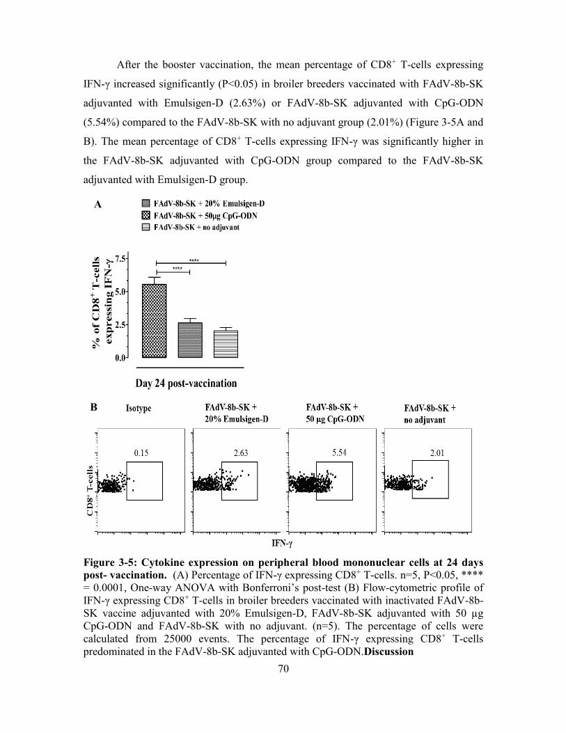
70
After the booster vaccination, the mean percentage of CD8+ T-cells expressing
IFN-γ increased significantly (P<0.05) in broiler breeders vaccinated with FAdV-8b-SK
adjuvanted with Emulsigen-D (2.63%) or FAdV-8b-SK adjuvanted with CpG-ODN
(5.54%) compared to the FAdV-8b-SK with no adjuvant group (2.01%) (Figure 3-5A and
B). The mean percentage of CD8+ T-cells expressing IFN-γ was significantly higher in
the FAdV-8b-SK adjuvanted with CpG-ODN group compared to the FAdV-8b-SK
adjuvanted with Emulsigen-D group.
Figure 3-5: Cytokine expression on peripheral blood mononuclear cells at 24 days
post- vaccination. (A) Percentage of IFN-γ expressing CD8+ T-cells. n=5, P<0.05, ****
= 0.0001, One-way ANOVA with Bonferroni’s post-test (B) Flow-cytometric profile of
IFN-γ expressing CD8+ T-cells in broiler breeders vaccinated with inactivated FAdV-8b-
SK vaccine adjuvanted with 20% Emulsigen-D, FAdV-8b-SK adjuvanted with 50 µg
CpG-ODN and FAdV-8b-SK with no adjuvant. (n=5). The percentage of cells were
calculated from 25000 events. The percentage of IFN-γ expressing CD8+ T-cells
predominated in the FAdV-8b-SK adjuvanted with CpG-ODN.Discussion
A
B
71
Formulation of vaccines with adjuvants to enhance the efficacy is important in
mammalian and avian vaccinology. Adjuvants enhance and modulate immune responses
to inactivated vaccine antigens [346] and improve their efficacy by inducing pathogen-
specific antibodies in blood and mucosal surfaces [333]. Certain adjuvants can promote
development of cellular immunity to enhance vaccine efficacy against pathogens [346].
Some vaccine efficacy studies conducted in chickens using avian influenza and NDV
vaccines have examined the effects of Emulsigen-D and CpG-ODN on humoral
immunity by examining IgY and and IgM in serum and IgA in mucosal surfaces [318-
320, 322, 333]. In this study, we have characterized cell-mediated immune responses by
quantifying CD4+ T-cells and CD8+ T-cells and humoral immune responses by IFN-γ and
IL-4 production following FAdV-8b-SK vaccines adjuvanted with Emulsigen-D or CpG-
ODN in broiler breeders.
Humoral immunity plays a critical role in the neutralization of FAdVs in blood
and internal organs [189, 279, 294]. Maternal antibodies are able to neutralize FAdV and
prevent the development of clinical disease of IBH in broiler chickens [102, 294]. Hence,
induction of a robust and long-lasting humoral immunity in broiler breeders is an
important prerequisite for an effective FAdV vaccine program. We investigated IgY and
NAb in serum against a FAdV-8b-SK vaccine and the correlation to FAdV challenge
protection in broiler chickens. We did not detect any antibodies against FAdVs when the
FAdV-8b-SK vaccine was formulated with no adjuvant. This observation could be
associated with two possibilities; a lower sensitivity of our assay (ELISA) or an
insignificant amount of antibody production with FAdV-8b-SK vaccine with no adjuvant.
Between adjuvants used in FAdV vaccine, IgY levels were low in FAdV-8b-SK
adjuvanted with CpG-ODN compared to FAdV-8b-SK adjuvanted with Emulsigen-D
group for initial 3 weeks following vaccination. However, IgY levels were 0.20 to 0.30-
fold higher in FAdV-8b-SK adjuvanted with CpG-ODN group compared to the FAdV-
8b-SK adjuvanted with Emulsigen-D group after booster vaccination. Similar
observations were reported earlier in chickens vaccinated with an E. coli vaccine
adjuvanted with CpG-ODN compared to an E. coli vaccine adjuvanted with Emulsigen-D
after booster vaccination [318]. Similarly, a remarkable increase in IgY was
demonstrated following booster vaccination in chickens that were vaccinated with avian

72
influenza virus or Newcastle disease virus vaccines adjuvanted with CpG-ODN [320,
333].
Antibodies detected by ELISA do not correlate with protection of broiler chickens
against IBH [331]. In contrast, NAb against FAdVs are correlated with protection [279,
294]. Group that were vaccinated with FAdV-8b-SK with no adjuvant did not have NAb
against FAdVs which corroborated with ELISA readings for IgY antibodies. Neutralizing
antibody titers against FAdV in broiler breeders vaccinated with FAdV-8b-SK
adjuvanted with Emulsigen-D or CpG-ODN were similar until 48 weeks of age.
However, at 48 weeks of age, NAb titers became significantly higher in broiler breeders
vaccinated with FAdV-8b-SK adjuvanted with CpG-ODN compared to the FAdV-8b-SK
adjuvanted with Emulsigen-D group (3.40 log10±0.46 vs 3.10 log10±0.42) (P<0.05). In
this study, the mean NAb levels at all time points tested in either vaccine group were
≥2.90 log10 and were similar (≥3.0 log10) to another vaccine efficacy study conducted
using large group of birds (n=300 birds/group) [347]. In order to confirm the protective
ability of NAb, broiler chickens were challenged with FAdV-8b-SK at 14 days of age. As
expected, broiler chickens from broiler breeders vaccinated with FAdV-8b-SK-
adjuvanted with Emulsigen-D or CpG-ODN were protected against FAdV challenge
(99% survival, P<0.05).
In addition to characterization of humoral immune responses of FAdV-8b-SK
vaccines adjuvanted with Emulsigen-D or CpG-ODN, we evaluated T-cell responses in
broiler breeders. Cellular mediated immunity helps in clearing virus infected cells from
the host [348] and also aids in the development of humoral immunity [349]. Although
Emulsigen-D and CpG-ODN were used as vaccine adjuvants against AIV and NDV in
chickens [319, 320, 322] cell mediated immunity was not well characterized. Unlike
antibody responses, our FAdV-8b-SK vaccine with no adjuvant induced a T-CD4+ T-cell
response (elevated CD4+:CD8+ T-cell ratio). The CD4+ T-cell response increased 0.22-
fold higher in broiler breeders vaccinated with FAdV-8b-SK adjuvanted with Emulsigen-
D. Interestingly, FAdV-8b-SK adjuvanted with Emulsigen-D increased only CD4+ T-
cells in contrast; FAdV-8b-SK adjuvanted with CpG-ODN induced both CD4+ and CD8+
T-cells. Moreover, CD8+ T-cell response was predominated in FAdV-8b-SK adjuvanted
with CpG-ODN compared to CD4+ T-cell response. Induction of CD 8+ T-cells were also

73
observed in mice vaccinated with a subunit hepatitis B vaccine and tetanus toxoid
adjuvanted with CpG-ODN as well as in pigs vaccinated with a rcC1 cycticercosis
vaccine adjuvanted with CpG-ODN [343, 350]. Hence, induction of CD8+ T-cells may be
an inherent feature of CpG-ODN as an adjuvant irrespective of the host species.
In addition to measuring T-cell responses by CD4+:CD8+ T-cell ratio, cytokine
secretion by T-cells was explored to determine the effect of adjuvants in the development
of type cellular immunity. Early in the development of adaptive immunity, naïve CD4+
T-cells differentiate into Th-1 or Th-2 type cells following interaction with antigen
presenting cells and commit to a lineage (Th-1 or Th-2). Hence, we determined IL-4 and
IFN-γ expression of CD4+ T-cells at 9 dpv. CD4+ T helper subsets can be identified by
their cytokine profile [349]. IFN-γ, IL-2, IL-6 and IL-12 are the hallmark of Th-1 type
immunity, whereas, IL-4, IL-5 and IL-10 and IL-13 indicate Th-2 type immunity [349].
In this study, we selected IFN-γ and IL-4 cytokines to identify Th-1 and Th-2 type CD4+
T-cells. Enhanced IL-4 secretion was documented in chickens following vaccination with
FAdV-4 adjuvanted with mineral-oil adjuvant [193], but no information is available for
the effect of Emulsigen-D on the cytokine profile. Similarly, the Th-1 type immune
response after vaccination with an avian influenza virus vaccine adjuvanted with CpG-
ODN have documented an mRNA expression [322] however, mRNA expression may
correlate with protein expression. Here, we have measured intracellular cytokine
expression in PBMC of broiler breeders. We noticed that FAdV-8b-SK vaccine with no
adjuvant induced predominantly a Th-1 type response (IFN-γ producing CD4+ T-cells).
Formulation of FAdV-8b-SK with Emulsigen-D or CpG-ODN as an adjuvant induced
Th-1 (CD+ T-cells secreting IL-4) or Th-2 (CD+ T-cells secreting IFN-γ) type immune
responses in broiler breeders. Interestingly, the immune response was polarised towards a
Th-2 type in FAdV-8b-SK adjuvanted with Emulsigen-D by increased IL-4 producing
CD4+ T-cells, whereas the immune response was polarised towards Th-1 type immune
response in FAdV-8b-SK adjuvanted with CpG-ODN by producing IFN-γ expressing
CD4+ T-cells.
After the booster vaccination, the objective was to measure the memory response
in broiler breeders to the FAdV vaccine. Hence, we measured the number of CD8+ T-
cells expressing IFN-γ. Following booster vaccination, a strong CD8+ T immune

74
response was noted in the FAdV-8b-SK adjuvanted with CpG-ODN compared to the
FAdV-8b-SK vaccine adjuvanted with Emulsigen-D. Increased induction of CD8+ T-cell
response was correlated with a higher percentage of Th-1 type CD4+ T-cells in FAdV-8b-
SK adjuvanted with CpG-ODN. Th-1 type CD4+ T-cells play an important role in
priming and memory response of CD8+ T-cells [324, 351]. It is evident that FAdV-8b-SK
vaccine with no adjuvant or addition of CpG-ODN favors a Th-1 type immune response,
whereas the immune response is skewed towards a Th-2 type immunity by the addition of
Emulsigen-D. It is also apparent that the FAdV-8b-SK vaccine adjuvanted with CpG-
ODN had better immune memory cell development.
It is summary, both FAdV-8b-SK vaccines adjuvanted with Emulsigen-D or CpG-
ODN had strong, durable and protective humoral immunity in broiler breeders and this
humoral immunity passively transferred to their progeny to protect them against IBH. We
also observed Th-1 and Th-2 type immune responses with FAdV-8b-SK antigens
adjuvanted with CpG-ODN. . Moreover, FAdV-8b-SK adjuvanted with CpG-ODN had
better immune memory cell development by the production of CD8+ T-cell responses.
Furthermore, since no necrosis at the injection site was observed with vaccines
formulated with CpG-ODN [318, 342], CpG-ODN seems to be better choice of adjuvant
for a FAdV-8b-SK vaccine.

75
PREFACE TO CHAPTER 4
There are advantages and disadvantages of live and inactivated vaccines. For
safety reasons and in the absence of suitable live vaccines, inactivated vaccines are
preferred. Moreover, inactivated vaccines are main components of prime-boost
vaccination programs to boost the immunity following priming with live viruses. In
previous chapters, we have propagated FAdVs in cell culture systems to make live and
inactivated FAdV vaccines. We observed that FAdV-11 grows to a lower titer of 1x10⁶
TCID50/mL in LMH cells compared to FAdV-8b which could give a higher titer of 1x10⁹
TCID50 per mL (personal observation). We also noticed that it takes a longer time (>100
hours) to attain 1x10⁶ TCID50/mL for FAdV-11 compared to 72 to 96 hours for FAdV-8b
when LMH cells were infected at an equal multiplicity of infection with respective
viruses. This can be a problem in the mass production of the inactivated vaccine antigens
for inactivated vaccines. In such cases, subunit vaccines can be an alternative.
Subunit vaccines are small immunogenic microbial proteins that can elicit
protective immune responses following formulation with an adjuvant. In the next chapter,
we have investigated immunogenicity and protective efficacy of recombinant fiber and
fiber-knob subunit antigens and virus-like-particles (VLPs) of FAdV-8b-SK as a breeder
vaccine.

76
CHAPTER 4: IMMUNOGENECITY AND PROTECTIVE EFFICACY OF VIRUS-
LIKE PARTICLES AND RECOMBINANT FIBER PROTEINS OF FOWL
ADENOVIRUS (FADV)-8b VACCINES IN BROILER BREEDERS AGAINST
INCLUSION BODY HEPATITIS
Ashish Gupta1, Khawaja Ashfaque Ahmed1, Lisanework E Ayalew1, Shelly Popowich1,
Shanika Kurukulasuriya1, Kalhari Goonewardene1, Thushari Gunawardana1, Ruwani
Karunarathna1, Davor Ojkic2, Suresh K. Tikoo3, Philip Willson4 and Susantha Gomis1*
1Department of Veterinary Pathology, Western College of Veterinary Medicine, University of
Saskatchewan, Saskatoon, SK Canada S7N 5B4
2Animal Health Laboratory, University of Guelph, P.O. Box 3612, Guelph, ON, Canada N1H
6R8
3Canadian Centre for Health and Safety in Agriculture, University of Saskatchewan, Saskatoon,
SK Canada S7N 5E5
4Vaccinology and Immunotherapeutic Program, School of Public Health, University of
Saskatchewan, Saskatoon, SK Canada S7N 5E3
*Corresponding author mailing address: Department of Veterinary Pathology, Western
College of Veterinary Medicine, 52 Campus Drive, University of Saskatchewan,
Saskatoon, SK Canada S7N 5B4. Phone: (306) 966-7299. Fax: (306) 966-7439. Email:
Published in Vaccine, 2017. 35 (20): 2716-2722
(Elsevier permits the authors to include their articles in full or in part in a thesis or dissertation for non-
commercial purposes)

77
4.1. Abstract
IBH is one of the most economically important diseases in the broiler chicken
industry. Several serotypes of FAdVs can cause IBH; among them, serotype FAdV-8b is
one of the emerging serotypes associated with IBH cases in Canada. In the present study,
we evaluated FAdV-8b-SK VLPs and two recombinant FAdV-8b-SK fiber proteins
(expressed in E. coli) as potential broiler breeder vaccines against IBH. For assessing the
immunogenicity of vaccines, we investigated both humoral and cellular immunity. The
humoral immunity was evaluated by determining the total IgY levels and neutralizing
antibodies in serum at 14, 28, 35 and 60 days post-vaccination (dpv). The cellular
immunity was examined using flow cytometry by determining the CD4+:CD8+ T-cell
ratio change in peripheral blood after the booster vaccination. The protective effect of
vaccines was tested through progeny challenges of 14 day-old broiler chicks
(n=30/group), by exposing them to a virulent FAdV-8b-SK virus (1x10⁷ TCID50, FAdV-
8b-SK). Although total IgY levels were comparable in all groups, the neutralizing
antibody response at 35 and 60 dpv was significantly (P<0.05) higher in the broiler
breeders immunized with FAdV-8b-SK VLPs, followed by FAdV-8b-SK fiber vaccine,
compared to FAdV-8b-SK fiber-knob vaccine. Moreover, FAdV-8b-SK VLPs and
FAdV-8b-SK fiber (but not FAdV-8b-SK fiber-knob vaccine) efficiently elicited the cell-
mediated immune response as evidenced by statistically significant (P<0.05) CD8+ T-cell
proliferative response in broiler breeders four days after the booster vaccination. Unlike
the FAdV-8b-SK fiber-knob, FAdV-8b-SK VLPs, and FAdV-8b-SK fiber vaccinated
broiler breeders were able to transfer a substantial amount (28.4±90%) of MAbs to their
progenies. The progeny challenge revealed that MAbs provided 100% and 82.7%
protection in chicks from FAdV-8b-SK VLPs, and FAdV-8b-SK fiber vaccinated broiler
breeders, respectively. Collectively, our data suggest that the FAdV-8b-SK subunit
vaccine-induced MAbs efficiently and protected broilers against clinical IBH. Broiler
breeder vaccination with subunit vaccines is a potential approach for the control of IBH.
4.2. Introduction
FAdVs are non-enveloped, dsDNA viruses of the genus Aviadenovirus in the
family Adenoviridae [2], which often cause disease in 2 to 5 weeks old broilers [189].

78
They are classified into five species (A-E) and twelve serotypes (FAdV -1 to -7, -8a, -8b,
-9 and -11) [2, 189, 270, 293]. FAdV-1 and FAdV-4 are associated with gizzard erosions
and ulcerations and HHS, respectively [189]. Several serotypes, like FAdV-2, FAdV-8a,
FAdV-8b, and FAdV-11 have been associated with IBH [3, 161, 215, 218, 229, 352,
353], which is a worldwide problem. FAdV-8b is one of the emerging serotypes of
FAdVs. The disease is characterized by liver necrosis, hemorrhage and basophilic
intranuclear inclusion bodies in the hepatocytes [219, 229, 231]. Mortality associated
with the disease ranges from 10% to 30% [39, 217]. To control IBH in broiler progenies,
broiler breeders are vaccinated with autogenous vaccines [3, 221, 294]. Passive immunity
through MAb transfer protects against infectious diseases during the first few weeks of
the neonatal life. Despite the use of autogenous vaccines, though the incidence of the
FAdV diseases have declined [161], and the broiler chicken industry still incur
considerable economic losses. This is due to sporadic outbreaks of IBH which are
attributed to exposure to heterologous serotypes. Hence, the development of an effective
and safe IBH vaccine is highly desired.
Among the various vaccine types, subunit or VLP based vaccines are potential
alternatives to live or inactivated vaccines due to their safety, and ease of production on a
large scale. A subunit vaccine is a recombinant small immunogenic component of a
microorganism, which elicits a protective immune response in combination with an
adjuvant [352]. VLP is a type of subunit vaccine, which mimics the natural virus
conformation, but lacks the infectious genome [354, 355]. VLPs have been developed for
poultry pathogens such as NDV [356], however, these vaccines remain in preliminary
stages of development. Although, several fowl adenoviral proteins including hexon,
penton and fiber capsid have been identified as potential subunit vaccine antigens [43,
221, 306], FAdV fiber protein has been proposed as the most suitable immunogen for
subunit vaccine development [43]. This is due to its ability to induce neutralizing
antibody responses [181, 309] as well preventing clinical disease (e.g., HHS) in chicks
upon exposure to a lethal virus dose [43]. All previous studies conducted on subunit
FAdV vaccines against FAdVs were carried out in chicks as a broiler vaccine [43, 306].
Evaluation of subunit vaccines for use in broiler breeder vaccination programs has not
previously been reported.

79
Therefore, in the present study we investigated the effectiveness of vaccination of
broiler breeders with VLPs and subunit-based vaccines in preventing IBH in their
progeny. We compared the effectiveness of VLPs, recombinant fiber, or recombinant
fiber-knob vaccines through progeny challenge.
4.3. Material and methods
4.3.1. Virus, cell line and antibodies
A FAdV (FAdV-8b-SK) isolated from a clinical case of IBH in a broiler chicken
flock in Saskatchewan was used in this study. Based on the loop-1 hexon protein
sequence, it had 93.9% amino acid similarity to FAdV-8a-T8-A (which was later
suggested to be renamed as FAdV-8b [204, 212]) and FAdV-8b strain 764 [292]. Due to
its genetic relatedness to FAdV-8b strains (Figure 4-1), we named this isolate as FAdV-
8b-SK.
Figure 4-1: Phylogenetic tree analysis of FAdV-8b-SK. Sequences of other isolates of
FAdV were extracted from the Genbank. Jukes-Cantor genetic distance model with Boot
strap neighbor-joining method with 1000 replicates was used to construct the tree with
the Geneious 9.1.5 software.
A LMH cell line was (ATCC #CRL-2117) used to propagate FAdVs DMEM: F-
12 (Thermo Fisher Scientific) containing 20 mM HEPES (pH=7.2), 2 mM glutamine,
1:1000 gentamicin (100 µL of 50 µg/mL per 100 mL of media) and 10% fetal bovine

80
serum was used to propagate LMH cells. Anti-6x-His epitope tag mouse monoclonal
antibody (Thermo Fisher Scientific), and Alkaline Phospahte conjugate Goat anti-mouse
IgG (H+L) polyclonal antibody (ThermoFisher Scientific) were used for detection of
recombinant expressed proteins.
4.3.2. Purification of fowl adenovirus (FAdV-8b-SK) virus-like particles
The empty capsids of FAdV-8b-SK were conventionally purified from a cesium
chloride (CsCl) density gradient method. Hereafter, we referred to it as VLPs, as they
lack the genome. Generation of VLPs were confirmed by transmission electron
microscopy. Briefly, FAdV-8b-SK was grown in LMH cell line in T175 flasks (Corning).
The cells were infected at a multiplicity of infection of 1 and incubated at 37 C with 5%
CO2 and harvested when 80% or more cells were showing adenovirus-specific cytopathic
effect. The lysates were freeze-thawed five times. Later, 1.5 mL of sodium deoxycholate
(5%) was added per 5 mL of lysate and incubated for 30 min at room temperature. Once
the suspension became viscous, it was incubated with 2 M MgCl2 and DNase-I solution
for 1 h at 37 C. The cell lysate was spun at 4000 rpm in a tabletop centrifuge for 30 min.
The supernatant was loaded onto CsCl (1.25/1.34 g/mL) cushion and spun at 35,000 rpm
for 24 h at 10 C. The capsid (upper band) and virus (lower band) was aspirated separately
and dialyzed in 0.1 M Tris-HCl (pH8) three times for 8 hr each. The capsid protein
concentration was measured by Bradford assay in 96-well plate as per manufacturer’s
instructions. The optical density was read in SoftmaxPro5 data acquisition and analysis
software (Molecular Devices, LLC) at 590 nm. A sample of the dialyzed protein was
subjected to transmission electron microscopy to identify the morphology of VLPs. In
addition, another sample of the VLP was passaged in LMH cells two times to confirm
non-infectiousness of the VLP protein.
4.3.3. Transmission electron microscopy
Samples for negative staining and transmission electron microscopy were
processed using standard techniques as described before [357]. Briefly, 300-mesh copper
carbon/formvar-coated grid was floated on 20 μL droplet of sample for 2 min. Then, the
grid was rinsed through two water droplets for 30 sec with each drop. Finally, the grid

81
was floated on a 0.5% phosphotungstic acid for 1 min and drew off excess and allowed
the thin layer to dry on grid and the grids were examined under transmission electron
microscopy.
4.3.4. Cloning of fiber and fiber-knob gene of FAdV-8b-SK
LMH cells were grown in six well plates and infected with FAdV-8b-SK at 1
multiplicity of infection. Then, after 36 h of infection, total DNA was extracted using a
viral DNA isolation kit (Qiagen) as per the manufacturer’s protocol. FAdV DNA that
encoded full fiber or fiber-knob was amplified from the extracted DNA using primers
shown in the Table 4-1. PCR was performed using cycling conditions of initial
denaturation at 94 C for 2 min, and 40 cycles of denaturation at 94 C for 30 sec,
annealing at 62 C, extension at 72 C followed by a final extension at 72 C for 5 min. To
express the full-length fiber protein, coding approximately 62 kDa protein, a 1575 bp
long fiber gene of FAdV-8b-SK was amplified in a standard polymerase reaction using
Accustart Taq polymerase (Quanta Biosciences) with primers F1 and F2.
Table 4-1: Primer used for cloning of fiber and fiber-knob of FAdV-8b-SK
Primer ID 5’-3’ Sequence Reference
F1 AAGCATGCATGGCGACCTCIACTCCTCA This study
F2 ATTAAGCTTTTACGGAGCGTTGGCTGTGCTTAGGG This study
F3 CGGGATCCTATTTCACGTTCTGGGTAGG This study
F4 ATTAAGCTTTTACGGAGCGTTGGCTGTGCTTAGGG This study
Italic underlined letters designate restriction enzyme sites for SphI and BamHI in F1 and F3 primers, and
HindIII in F2 and F4 primers.
The PCR product was then electrophoresed in a 1% gel to confirm 1575 bp long
product, which was subsequently gel-excised and purified by Genejet gel extraction kit
(Thermo Fisher Scientific). The purified PCR product was digested with SphI and
HindIII enzymes and cloned into the SphI and HindIII sites of linearized 3429 bp
fragment of pQE30 plasmid vector (Addgene) in frame with the 6X-His gene using
T4DNA ligase (Thermo Fisher Scientific) (Figure 4-2A). The product was named as
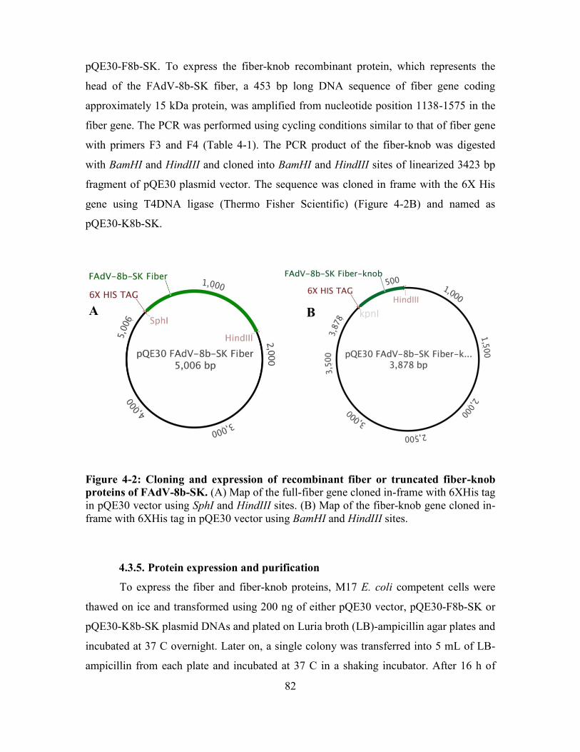
82
pQE30-F8b-SK. To express the fiber-knob recombinant protein, which represents the
head of the FAdV-8b-SK fiber, a 453 bp long DNA sequence of fiber gene coding
approximately 15 kDa protein, was amplified from nucleotide position 1138-1575 in the
fiber gene. The PCR was performed using cycling conditions similar to that of fiber gene
with primers F3 and F4 (Table 4-1). The PCR product of the fiber-knob was digested
with BamHI and HindIII and cloned into BamHI and HindIII sites of linearized 3423 bp
fragment of pQE30 plasmid vector. The sequence was cloned in frame with the 6X His
gene using T4DNA ligase (Thermo Fisher Scientific) (Figure 4-2B) and named as
pQE30-K8b-SK.
Figure 4-2: Cloning and expression of recombinant fiber or truncated fiber-knob
proteins of FAdV-8b-SK. (A) Map of the full-fiber gene cloned in-frame with 6XHis tag
in pQE30 vector using SphI and HindIII sites. (B) Map of the fiber-knob gene cloned in-
frame with 6XHis tag in pQE30 vector using BamHI and HindIII sites.
4.3.5. Protein expression and purification
To express the fiber and fiber-knob proteins, M17 E. coli competent cells were
thawed on ice and transformed using 200 ng of either pQE30 vector, pQE30-F8b-SK or
pQE30-K8b-SK plasmid DNAs and plated on Luria broth (LB)-ampicillin agar plates and
incubated at 37 C overnight. Later on, a single colony was transferred into 5 mL of LB-
ampicillin from each plate and incubated at 37 C in a shaking incubator. After 16 h of
A B kpnI

83
incubation, the broth was transferred into 500 mL of LB-ampicillin. When the OD
reached 0.6, IPTG (Sigma) was added at a final concentration of 1 mM. After overnight
induction of protein expression, the cells were centrifuged at 6500 rpm for 20 min. The
pellets were re-suspended in resuspension buffer (NaH2PO4 (50 mM), NaCl (300 mM)
and imidazole (10 mM), pH 8) and lysed by lysozyme (10 mg/mL) (Sigma) digestion and
sonication. The crude-lysate was centrifuged at 10,000 g for 30 min at 4 C. The
supernatant was discarded, and the pellet was stirred in resuspension buffer for 30 min
and centrifuged as described above. Following centrifugation, the pellet was dissolved by
addition of 2 mL binding buffer (resuspension buffer with 8 M urea) per gram of the
pellet which was stirred for 60 min. The resuspended pellet was centrifugation at 10,000
g for 20 min at 20 C, and the supernatant was collected. The supernatant was used to
purify the fiber and fiber-knob proteins by affinity column chromatography using
ProfinityTM IMAC Nickel Charged Resins (Bio-Rad Laboratories Inc.) as per the
manufacturer’s protocol. The purified proteins were dialyzed in 10 mM Tris-HCl (pH 8),
three times, for 8 h each and concentrated using Amicon ultra centrifugal units with
ultracell-10 membrane (Millipore). The protein concentrations were measured by
Bradford assay.
4.3.6. Coomassie blue staining and Western blotting
To check for proper expression and purity of the fiber and fiber-knob proteins,
sodium dodecyl polyacrylamide gel electrophoresis (SDS-PAGE) and Coomassie blue
staining was performed. Briefly, the proteins were separated on 10% polyacrylamide gel.
The electrophoresed proteins were stained with Coomassie blue for 45 min and destained
using a destaining solution. Simultaneously, proteins on another set of gels were
transferred onto a nitrocellulose membrane (Bio-Rad Laboratories Inc.) in a cold chamber
and Western blot was performed. In short, the membranes were blocked with 5% BSA in
TBST for 1 h. The membranes were washed with TBST three times for 10 min each.
Membranes were incubated with anti-6X His epitope- tag monoclonal antibody (Life
Technologies) for 1 h. Following incubation, the membranes were washed like previous
step and incubated with AP-conjugated goat anti-mouse IgG secondary antibody (Life

84
Technologies) for 1 h. Finally, the membranes were washed and developed with
NBT/BCIP substrate (Sigma).
4.3.7. Preparation of FAdV-8b-SK challenge virus
FAdV-8b-SK infected liver tissues from previous studies were macerated in
mortar with pestle and minimal essential media was added to make 40% liver
homogenate. The homogenate was freeze-thawed four times to release the virus particles
from the cells and then centrifuged at 6000 rpm for 30 min at 10 C in a high-speed
centrifuge (Thermo Fisher Scientific). The supernatant was collected and filter sterilized
by passing through 0.22 m filter (Cameo, Maine Manufacturing, LLC, USA). The
filtrate was titrated and stored at -80 C.
4.3.8. Broiler breeder vaccination
Day old broiler breeders (Aviagen, Huntsville, AL) were reared in the Animal
Care Unit of the Western College of Veterinary Medicine as per Aviagen guidelines. The
animal experiments were conducted following the approved protocols from Animal
Research Ethics Board of University of Saskatchewan. At the 30 weeks of age, the broiler
breeders were randomly divided into 4 groups (n=20 birds/group). Before vaccination,
blood samples at 0 days of age were collected from each group. Each of the three
principle groups received FAdV-8b-SK VLPs, FAdV-8b-SK fiber or FAdV-8b-SK fiber-
knob protein (50 µg/bird) with 20% Emulsigen-D (MVP Technologies) intramuscularly
in the pectoral muscle. The fourth group was kept as unvaccinated control. Each group
was boosted with the same dose of the particular vaccine at 21 days after the primary
vaccination. To determine serum-neutralizing antibodies, sera samples (n=10) were
collected at 14, 21, 28, 35 and 60 dpv for serum neutralization test. In addition, peripheral
blood mononuclear cells were collected at 21 days after primary vaccination and 4 day
following booster vaccination (day 25) to measure the CMI response.
4.3.9. Progeny challenge
Eggs were collected from broiler breeders 3 wk following the booster vaccination,
which were subsequently hatched (n=30/group) and kept in a single pen for challenge

85
protection studies. Broiler progenies were bled at 13 dph to test for MtAbs against
FAdVs. At 14 dph, the progenies were challenged with 1x10⁷ TCID50 of FAdV-8b-SK
per bird to study the immunoprotective ability of vaccine candidates. Cloacal swabs
(n=5) were collected from all the groups prior to FAdV challenge (0 day) and at 3, 7 and
10 dpi to determine fecal shedding of FAdV. Liver samples were collected from dead,
sick (following euthanasia), or clinically healthy birds (at the end of experiment) and
fixed in 10% neutral buffered formalin for histopathological examination.
4.3.10. Virus isolation from cloacal swabs
Cloacal swabs were collected with dry cotton swabs (Starplex) and weighted
before placing in 1 mL Tryptose broth (TB) (Sigma). TB was supplemented with 1:100
antibiotic-antimycotic solution (1 mL/100 mL) (Gibco). The swabs in TB were vortexed
for 15 sec and centrifuged at 13000 rpm for 3 min. The supernatant from the cloacal swab
was filter sterilized using 0.22 m filter (Cameo, Maine Manufacturing LLC, USA). The
filtrate was transferred into a 96 well plate in triplicate at 1:10 dilution for virus isolation
and titration.
4.3.11. Measurement of serum IgY antibodies
Serum IgY was determined by direct ELISA as described in the previous chapter.
Briefly, polystyrene plates were coated with purified FAdV-8b-SK capsid protein
(purified from FAdV infected cells by CsCl gradient method) with coating buffer (R&D
Systems, Minneapolis, MN) at a concentration of 7.5 µg/mL and incubated overnight at 4
C. The plates were blocked with TBST (1X PBS+0.05% tween-20+2.5% bovine serum
albumin, pH 7.6) for 1.5 h at room temperature. The plates were then washed two times
with 1X wash buffer (R&D Systems, Minneapolis, MN) and incubated with 1:100 diluted
sera samples in duplicate for 1.5 h at room temperature. Following washing, anti-FAdV-
8b-SK IgY antibodies were detected by incubating plates with goat-anti-chicken IgY–AP
conjugate (Thermo Fisher Scientific, Waltham, MA) for 1.5 h at room temperature. After
washing with wash buffer, the plates were developed with 100 µL of ready to use p-
nitrophenyl phosphate liquid substrate (Sigma Chemical Co., St. Louis, MO) for 10 min
at room temperature. The sample absorbance was measured at an optical density (OD) of

86
405 nm in the spectrophotometer (Molecular Devices, Sunnyvale, CA). Positive and
negative control sera and blank wells with no sera were used as controls.
4.3.12. Measurement of neutralizing antibodies against fowl adenovirus
VNT was performed in LMH cells with a few modifications to the protocol
described by Gunes et al. (2012) [270] Serum samples were heat inactivated at 56 C for
30 min. The samples were serially diluted 2-fold in 96 well plates in triplicates with
diluent (DMEM-12 containing gentamicin @ 1:1000). 200 TCID50 of FAdV-8b-SK was
added into each well and the serum-virus mixture was incubated for 1 hr at 37 C with 5%
CO2. The mixture was then transferred to freshly split cells in 96 well plates (5x10⁴
cells/well) starting from the highest dilution. Subsequently, the plates were incubated at
37 C with 5% CO2 and results were read on the day 7.
4.3.13. Quantification of CD4+ and CD8+ T-cells in peripheral blood
mononuclear cells
Blood samples were collected in heparinized tubes and diluted 1:1 with 1X PBS
(pH7.6). 1 mL of diluted blood was layered on histopaque (Gibco) and spun at 2000 rpm
for 20 min in tabletop centrifuge. PBMC were separated and washed two times with 500
L of 1X PBS. The cells were incubated with mouse anti-chicken CD4 PE and mouse
anti-chicken CD8a FITC monoclonal antibodies (1:50 dilution in 1X PBS) (Southern
Biotech) for 30 min on ice. The cells were washed twice, re-suspended in 400 L of PBS
and read using a flow cytometer (Beckman coulter) to quantify the lymphocytes. The
CD4+: CD8+ T-cell ratio was determined by using FlowJo software (Treestar).
4.3.14. Statistical analysis
The statistical analysis was performed using Prism7 (Graph Pad, Inc, San Diego,
USA). The survival analysis was performed by Mantel-Cox (Log-rank) test and Student
T-test with Welch’s correction was used to compare neutralizing antibody responses and
CD8+ T-cell responses between the vaccine groups. The level of significance (α) was kept
at 0.05.
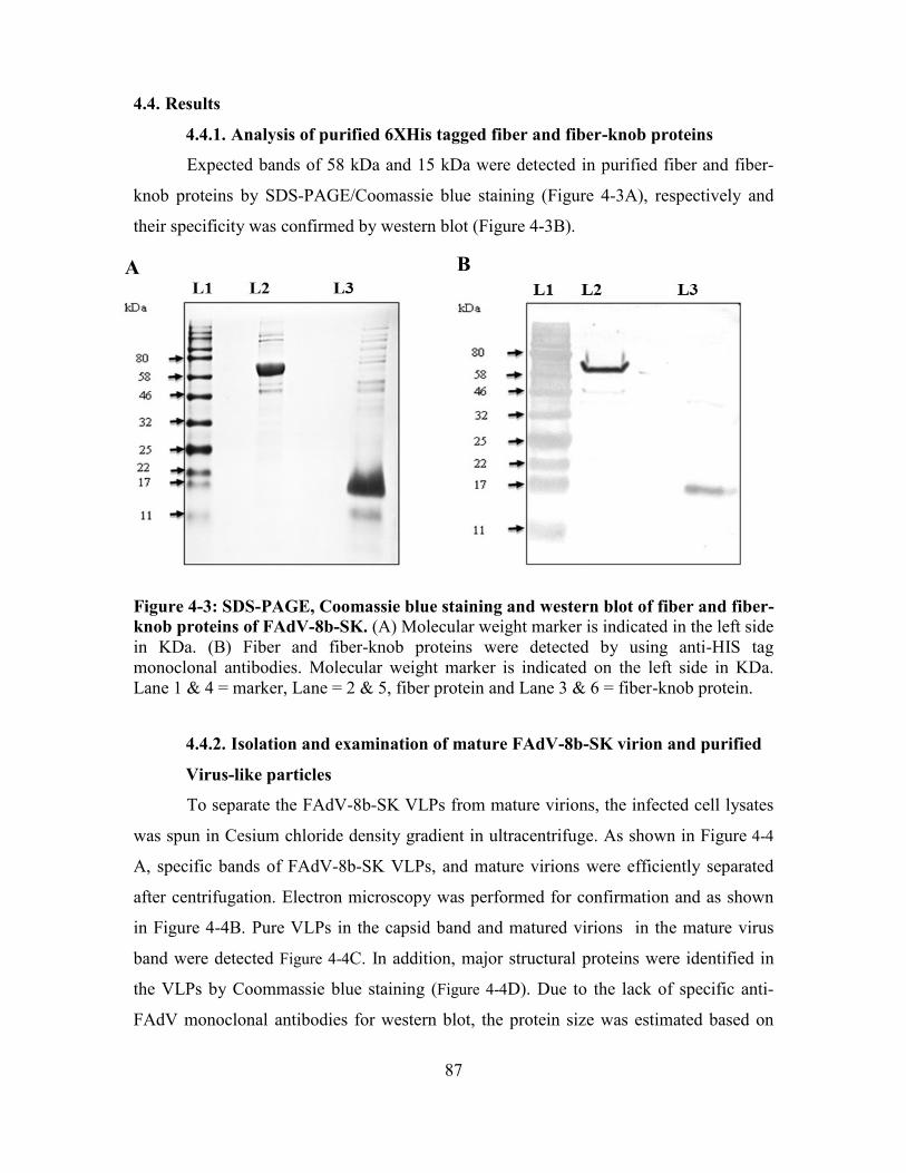
87
4.4. Results
4.4.1. Analysis of purified 6XHis tagged fiber and fiber-knob proteins
Expected bands of 58 kDa and 15 kDa were detected in purified fiber and fiber-
knob proteins by SDS-PAGE/Coomassie blue staining (Figure 4-3A), respectively and
their specificity was confirmed by western blot (Figure 4-3B).
Figure 4-3: SDS-PAGE, Coomassie blue staining and western blot of fiber and fiber-
knob proteins of FAdV-8b-SK. (A) Molecular weight marker is indicated in the left side
in KDa. (B) Fiber and fiber-knob proteins were detected by using anti-HIS tag
monoclonal antibodies. Molecular weight marker is indicated on the left side in KDa.
Lane 1 & 4 = marker, Lane = 2 & 5, fiber protein and Lane 3 & 6 = fiber-knob protein.
4.4.2. Isolation and examination of mature FAdV-8b-SK virion and purified
Virus-like particles
To separate the FAdV-8b-SK VLPs from mature virions, the infected cell lysates
was spun in Cesium chloride density gradient in ultracentrifuge. As shown in Figure 4-4
A, specific bands of FAdV-8b-SK VLPs, and mature virions were efficiently separated
after centrifugation. Electron microscopy was performed for confirmation and as shown
in Figure 4-4B. Pure VLPs in the capsid band and matured virions in the mature virus
band were detected Figure 4-4C. In addition, major structural proteins were identified in
the VLPs by Coommassie blue staining (Figure 4-4D). Due to the lack of specific anti-
FAdV monoclonal antibodies for western blot, the protein size was estimated based on
A B

88
predicted size of protein from FAdV-8b virus. The purified VLPs did not cause any
cytopathic effects in LMH cells on two continuous passages.
Figure 4-4: Purification of FAdV-8b-SK VLPs. (A) CsCl density gradient purification
of mature virions and VLPs of FAdV-8b-SK. (B) & (C), Transmission electron
microscopy of VLPs and mature virion fractions of FAdV-8b-SK. (D) SDS-PAGE and
Coomassie blue staining of the capsid proteins. Some of the structural proteins are
indicated on the right based on their molecular weight. Molecular weight markers are
indicated on the left of the panel.
4.4.3. Serum IgY and neutralizing antibodies against FAdV following broiler
breeder vaccination
To examine the immunogenicity of FAdV-8b-SK VLPs, fiber and fiber-knob,
serum samples were subjected to ELISA and neutralization testing. Mean log10 IgY
antibody levels in groups vaccinated with fiber, fiber-knob and VLPs vaccinated groups
at 14 dpv were 3.6 log10±0.35, 4.0 log10±0.07 and 4.0 log10±0.07, respectively (Figure
B
C D
A

89
B A
4-5A) IgY response peaked in VLP group at 35 dpv with mean IgY level of 3.9
log10±0.22. Mean IgY levels were similar in all the groups at 60 dpv.
To determine the neutralizing ability of these IgY antibodies, the serum samples
were analyzed in a VNT. Serum neutralizing antibodies were first detected at 14 dpv with
mean titers of 2.47 log100.16, 1.82 log100.23 and 2.75 log100.25 in fiber, fiber-knob
and VLPs vaccinated groups, respectively (Figure 4-5B). Only 70%, and 10% of the
tested samples had detectable neutralizing antibodies response at 14 dpv in fiber and
fiber-knob vaccinated groups, whereas 100% birds had serum-neutralizing antibodies in
the VLPs vaccinated group. Mean log10 serum neutralizing antibody levels in breeders
vaccinated with VLPs were significantly higher than fiber or fiber-knob vaccinated
breeders at all the sampling time points (P0.05)
The neutralizing antibody levels peaked at 35 dpv in fiber and VLPs vaccinated
groups with mean antibody levels of 3.0 log10±0.24 and 3.71 log10±0.31, respectively.
The mean serum neutralizing antibody titers in fiber-knob vaccinated group remained
low at all the time points and ranged between 1.82 log10±0.23 to 2.3 log10±0.02
throughout the experiment. The mean neutralizing antibody levels declined to a lower
level in all the groups at 60 dpv.
Figure 4-5: Serum IgY and neutralizing antibody levels in broiler breeders at
various ages. (A) IgY antibody levels (Mean log10SD) in broiler breeders vaccinated
with fiber, fiber-knob or whole capsid proteins at 7, 14, 28, 35 and 60 days post-

90
vaccination. (B) Mean serum neutralizing antibody titer in broiler breeders immunized
with the indicated proteins at 7, 14, 28, 35 and 60 days post-vaccination.
4.4.4. CD4+ and CD8+ T-cell ratio in PBMC following booster vaccination
Induction of cellular immunity, CD4+ and CD8+ T-cells, were monitored before
and after booster vaccination. No significant difference was observed in the mean
CD4+:CD8+ T–cell ratio in any groups before booster vaccination (Figure 4-6). However,
following booster vaccination, there was a significant proliferation of CD8+ cytotoxic T-
cells in broiler breeders vaccinated with the fiber protein (P<0.05) or VLPs (P<0.05).
Mean CD4+:CD8+ T–cell ratio lowered from 3.231.35 (before booster) to 1.20.14
(following booster) in fiber protein vaccinated breeders. Likewise, mean CD4+:CD8+ T–
cell ratio declined from 2.720.72 (before booster) to 1.520.86 (following booster), in
VLPs vaccinated breeders. In contrast, there was no difference in CD4+:CD8+ T–cell
ratio between fiber-knob vaccinated and the control group.
CD
4+
:CD
8+
T-c
ell
ra
tio
pb
mc
Con
trol
Con
trol
Fib
e r
Fib
e r -4d
pb
Fib
e r -kn
ob
Fib
e r -kn
ob
-4d
pb
VL
Ps
VL
Ps -4
dp
b
0
1
2
3
4
5 *
*
Figure 4-6: CD4+:CD8+ T-cell ratio in peripheral blood mononuclear cells. CD4+ and
CD8+ T-cell levels in the peripheral blood mononuclear cells before (at 21 days) and four
days after booster vaccination of breeders with the indicated vaccines. [(P0.05),
unpaired student t-test with Welch’s correction]

91
4.4.5. Challenge and protection studies in progenies
Progenies from broiler breeders that were vaccinated with VLPs had significantly
higher mean serum neutralizing antibodies (3 log10±0.32) than progenies from broiler
breeders vaccinated with recombinant fiber protein (2.4 log10±0.15) (P0.05). No serum
neutralizing antibodies were detected in progenies that originated from either the
recombinant fiber-knob vaccinated broiler breeders or unvaccinated control breeders
(Figure 4-7A). A significant survival, following FAdV-8b-SK challenge, was observed in
the broiler progenies of VLP and recombinant fiber vaccinated breeders (P0.05)
compared to broiler progenies in the control group (Figure 4-7B). No significant
difference in the survival was observed between the fiber-knob and the control groups.
The broiler chickens that died of disease or clinically sick birds that were euthanized
post-challenge had similar lesions irrespective of the group. Macroscopic lesions were
severe multifocal necrotizing hepatitis (pale-yellow, swollen and hemorrhagic livers with
prominent reticular pattern) and multifocal pancreatic necrosis. On microscopic
examination, multifocal to coalescing areas of necrosis were observed in the livers of the
dead or clinically sick birds which were characterized by the lysis of hepatocytes with
fragmentation and karyolysis of nuclei. Hepatocytes adjacent to the necrotic areas were
degenerated and had karyomegalic nuclei, which were identified as basophilic adenoviral
inclusion bodies. Pancreatic lesions included necrosis of pancreatic acinar cells and
basophilic intranuclear inclusions in the adjacent cells.

92
A B
Figure 4-7: Neutralizing antibody levels in broiler progenies and virus challenge. (A)
Mean neutralizing maternal antibody level in broiler progeny at13 dph from broiler
breeders immunized with the indicated vaccine antigens. (B) Kaplan-Meier survival
estimate of broiler progeny following lethal virus challenge with FAdV-8b-SK at 14 dph
(P<0.05), n=30/group.
4.4.6. Fowl adenovirus shedding following challenge
FAdV shedding was examined in broiler chickens for 10 dpi following challenge
with virulent FAdV-8b-SK at 14 days of age. As shown in the Figure 4-8, peak fecal
FAdV shedding was observed at day 3 with the highest titer in the control group (8.69
log101.12), followed by knob (8.56 log101.01), and fiber group (7.1 log101.37), but no
shedding was detected in VLPs group. The shedding declined by approximately one log
at 7 dpi in fiber and fiber-knob groups, but not in the control group. At 7 dpi, FAdV
shedding was also observed for the first time in the VLPs group. The quantity of the virus
shed by the broilers in VLPs group was lowest (4.46 log102.03), followed by the fiber
(6.16 log101.38), fiber-knob (7.36 log101.38) and control (8.49 log100.74),
respectively. No fecal FAdV shedding was detected at 10 dpi in any of the groups.
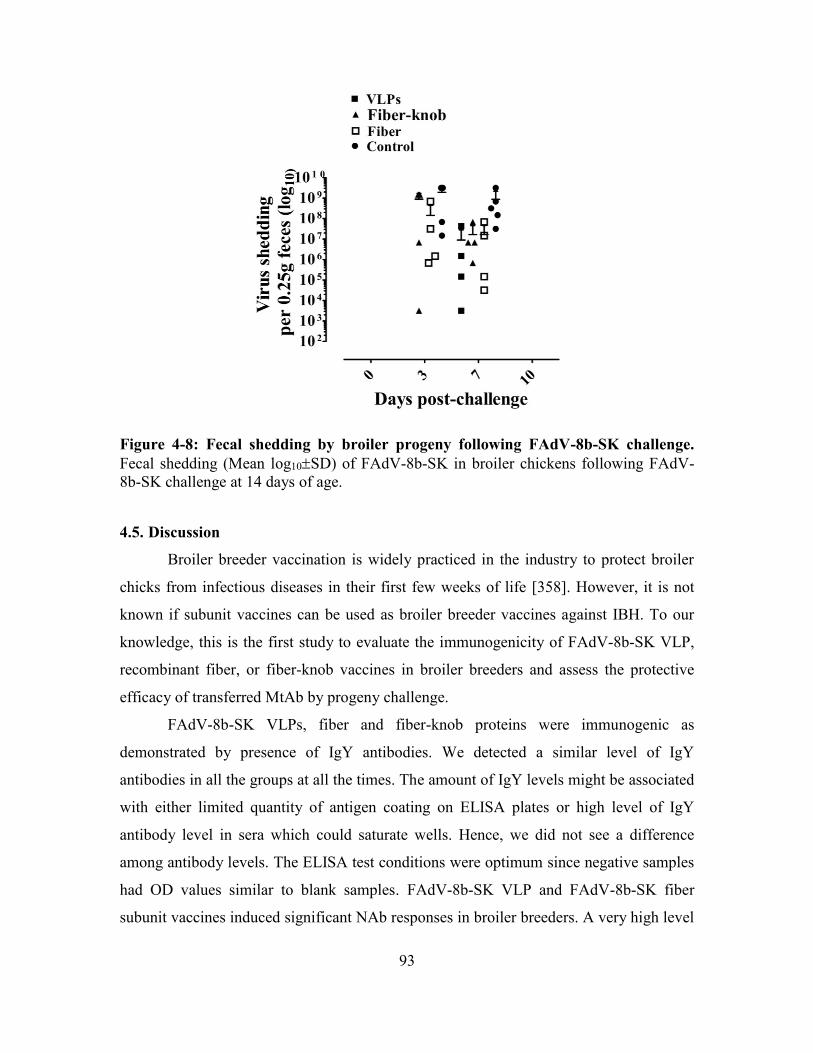
93
Figure 4-8: Fecal shedding by broiler progeny following FAdV-8b-SK challenge.
Fecal shedding (Mean log10SD) of FAdV-8b-SK in broiler chickens following FAdV-
8b-SK challenge at 14 days of age.
4.5. Discussion
Broiler breeder vaccination is widely practiced in the industry to protect broiler
chicks from infectious diseases in their first few weeks of life [358]. However, it is not
known if subunit vaccines can be used as broiler breeder vaccines against IBH. To our
knowledge, this is the first study to evaluate the immunogenicity of FAdV-8b-SK VLP,
recombinant fiber, or fiber-knob vaccines in broiler breeders and assess the protective
efficacy of transferred MtAb by progeny challenge.
FAdV-8b-SK VLPs, fiber and fiber-knob proteins were immunogenic as
demonstrated by presence of IgY antibodies. We detected a similar level of IgY
antibodies in all the groups at all the times. The amount of IgY levels might be associated
with either limited quantity of antigen coating on ELISA plates or high level of IgY
antibody level in sera which could saturate wells. Hence, we did not see a difference
among antibody levels. The ELISA test conditions were optimum since negative samples
had OD values similar to blank samples. FAdV-8b-SK VLP and FAdV-8b-SK fiber
subunit vaccines induced significant NAb responses in broiler breeders. A very high level

94
of NAb observed in FAdV-8b-SK VLP group was likely associated with hexon and
penton proteins which is supported by previous studies that reported neutralizing nature
of anti-hexon and anti-penton antibodies [100, 101, 306]. The FAdV-8b-SK fiber-knob
subunit vaccine, although it induced a very high level of IgY in broiler breeders, the
neutralizing activity of these antibodies was either minimal or undetectable. The lack of
neutralizing activity of antibodies to the fiber-knob protein in our study could be
explained by the absence of amino acid repeats critical to the tertiary structure of the fiber
knob [359].
In addition to humoral immunity, cell-mediated immune responses play a
significant role in controlling virus infections in chickens [360]. Therefore, we next
investigated whether such subunit vaccines can induce cellular immunity in vaccinated
birds. After the vaccination or pathogenic challenge, primary cytotoxic T-cell-mediated
immunity develops within a week after the antigenic stimulation. About 3-weeks later
memory cytotoxic T-cells robustly proliferate on secondary stimulation and provide
protection against the reinfection [201]. Therefore, in this study, we examined the
secondary cytotoxic T-cell response. Just before booster/secondary vaccination with the
FAdV-8b VLPs, fiber and fiber-knob, we quantified the CD4+:CD8+ T-cell ratio in
PBMC and then again four days after boosting. We did not find any significant change in
the CD4+:CD8+ T-cell ratio in fiber-knob vaccinated breeders and unvaccinated controls.
However, a significant increase (P0.05) in the level of cytotoxic CD8+ T-cells was
observed following boosting with FAdV-8b-SK VLPs, and FAdV-8b-SK fiber vaccines;
suggesting these vaccines were able to induce memory cytotoxic T-cell immunity. There
are no such studies with FAdVs available in the literature to compare. Thus, our results
clearly demonstrated that VLP, and subunit vaccines can induce potent humoral and
cellular immunity in broiler breeders.
We next investigated, whether the antibodies induced through broiler breeder
vaccination with FAdV-8b-SK VLPs, FAdV-8b-SK fiber, and FAdV-8b-SK fiber-knob
vaccines are transferred to their progeny and if they provide protection against FAdV-8b-
SK virus. We tested neutralizing MtAb levels in progeny hatched from all groups at 13
dph. The neutralizing MtAb levels at 13 days of age, constituted approximately
28.49.9% of serum neutralizing antibodies of their parents vaccinated with FAdV-8b-

95
SK VLP or FAdV-8b-SK fiber vaccine at 35 dpv. Our results are supported by previous
studies that demonstrated approximately 25.621.42 to 36.064.34% or 27-31% of MtAb
(IgY) transfer to eggs/progeny following vaccination of hens with Salmonella [361],
NDV and infectious bronchitis virus vaccines [358], respectively. However, like
unvaccinated control breeders, we could not detect neutralizing MtAb in progeny hatched
from FAdV-8b-SK fiber-knob vaccinated broiler breeders. This observation is most
likely partial or complete lack of neutralizing antibodies in broiler breeders injected with
fiber-knob protein or rapid depletion of maternal antibodies post-hatch.
To test the efficacy of MtAb in the prevention of clinical disease, we performed
progeny challenge studies at 14 dph, which is the most likely age of disease occurrence in
the field outbreaks. Our results demonstrated 100% protection of progeny (P0.05)
against FAdV-8b-SK challenge when breeders were vaccinated with FAdV-8b-SK VLPs.
Similarly, 82.7% survival (P0.05) was observed in progeny hatched from broiler
breeders immunized with FAdV-8b-SK fiber protein. Few progeny in FAdV-8b-SK fiber
group died following FAdV-8b-SK challenge. Retrospective analysis of their sera
samples showed that these dead chicks did not have detectable levels (2.0 log10) of MtAb
at 13 dph. Previous studies have also reported similar observations [43]. This could be
explained by the progressive decline of maternal antibodies [362] or lower level of serum
neutralizing antibodies in breeders at the time of egg collection. Like unimmunized
controls, progeny hatched from FAdV-8b-SK fiber-knob vaccinated breeders were not
protected against FAdV-8b-SK challenge, which is an expected result as no MtAb were
detected before challenge.
Fecal shedding is an important parameter, which reflects the efficacy of viral
vaccines. Fecal shedding was monitored to evaluate the efficacy of MtAb in the
prevention of shedding following intramuscular challenge with virulent FAdV-8b-SK.
Fecal shedding was observed in progeny post-challenge despite having maternal
antibodies. However, it is noteworthy that it was delayed in progeny hatched from FAdV-
8b-SK VLP vaccinated breeders compared to those of other groups that who shed the
virus from the beginning. The early high level of antibodies and synergistic activity of
anti-fiber, anti-hexon, and anti-penton antibodies induced by FAdV-8b-SK VLPs might
explain the delayed occurrence of shedding. Synergism between anti-fiber and anti-

96
penton neutralizing antibodies has been reported to inactivate adenovirus in humans
following HAdV-5 vector therapy [101]. Furthermore, delayed appearance of fecal
shedding in the progeny of the VLP group was likely due to declining MtAb titers in the
progeny, which is a common phenomenon observed in broiler chickens. Failure to
prevent shedding in the first week following virus challenge was reported by Schachner
et al. (2014) [43] and Schonewille et al. (2010) [260], in SPF chicks immunized with
recombinant fiber antigens or attenuated FAdV-4 virus. It might also be likely that the
virus was neutralized in the blood, but replication was not prevented in the intestinal
epithelial cells. Enterocytes in the region of the cecal tonsils are a common site of FAdV
replication in chickens [363]. Infectious virus was not detected in any of the broiler
groups ten days post-challenge. Similarly, Schonewille et al. (2010) failed to isolate
infectious virus after one week following infection of SPF chicks with cell culture
attenuated FAdV-4 [260].
4.6. Conclusions
In summary, we have demonstrated that immunization of broiler breeders with
VLPs or subunit vaccines composed of the fiber protein of FAdV-8b-SK induced
neutralizing antibodies and cytotoxic T-cell responses in breeders as well as successfully
prevented clinical disease in progeny through MtAb transfer. Collectively, our data
provide evidence supporting the potential of subunit vaccines in broiler breeder
vaccination programs and may have implications for vaccine development against IBH.

97
CHAPTER 5: DISCUSSION AND CONCLUSIONS
IBH is an acute viral disease of broiler chickens characterized by hepatic necrosis
and death in 1 to 5 week-old broiler chickens. IBH has been reported in pullets. IBH is
characterized by a sudden increase in flock mortality 3 to 4 days post-infection which
then subsides 6 days post-infection onwards. The percentage of mortality due to IBH
outbreaks are highly variable but may reach as high as 30%. Pale-yellow, necrotic and
hemorrhagic livers with basophilic intranuclear inclusion bodies in hepatocytes are the
characteristic lesions of IBH [189]. IBH is a primary disease of broilers, however, it is
not uncommon to see it as an opportunistic infection in immunosuppressed chickens
[161]. IBH is caused by FAdVs belonging to five species (A to E). These five species of
FADVs contain twelve serotypes (-1 to -7, -8a, -8b, -9 to -11). FAdV-2, -7, -8a, -8b and -
11 are associated with outbreaks of IBH worldwide including Canada [3, 211, 214]. IBH
as a primary disease has been emerging over the last fifteen years in Canada but reasons
associated with this emergence have not been identified. FAdVs disseminate vertically
from broiler breeders to their chicks and then horizontally by contaminated feed, water,
fomites and farm equipment [189]. Therefore, breaking vertical transmission by
vaccination of broiler breeders is the main disease control strategy. As there is no
commercial FAdV vaccine available in Canada, broiler breeder autogenous vaccines are
commonly used as a preventative measure in endemic areas. Therefore, the goal of this
project was to develop live, inactivated or subunit vaccines to control IBH and to identify
an immunogenic and safe adjuvant for an inactivated FAdV vaccine.
In the second chapter, we have demonstrated the development of a bivalent live
and inactivated FAdV vaccine to prevent IBH caused by a prevalent serotype of FAdV in
broiler chickens. We have also demonstrated the safety and efficacy of a live FAdV
vaccine in broiler breeders by vaccinating them and challenging their broiler progeny
with different serotypes of FAdVs. We have further demonstrated that protection of
broiler progeny against IBH depends on passive transfer of NAbs against FAdVs from
their broiler breeder parents vaccinated with a live or inactivated FAdV vaccine.
Live FAdV vaccines were previously developed and demonstrated the ability to
protect broiler chickens against HHS [260, 300] and IBH [229]. Although vaccination of

98
broiler breeders with a wild-type live FAdV has been reported previously [229], FAdV
shedding in broiler breeders was not evaluated in detail. Vaccination of broiler breeders
during egg production is risky due to the possibility of vertical transmission of FAdVs
[189]. We were able to demonstrate vertical transmission of a wild-type FAdV (FAdV-
8b-SK) from broiler breeders to their progeny for a period of 7 days if broiler breeders
were vaccinated with live FAdV-8b-SK while in egg production [272, 273]. We were
able to demonstrate discontinuation of vertical transmission of FAdV-8b-SK 2 weeks
following live FAdV vaccination in broiler breeders due to production of NAb against
FAdV. We have also found that FAdV shedding in feces continues for 3 weeks
following vaccination of broiler breeders with a live FAdV vaccine. Hence, we concluded
that live wild-type FAdVs can be safely included in a broiler breeder vaccination
program by vaccinating broiler breeders at 16 weeks of age, well before they begin egg
production.
NAbs neutralize FAdVs and protect broiler progeny from clinical disease of IBH
if there is an adequate amount of MtAb against FAdV in the progeny. We were able to
demonstrate protection of broiler chickens against IBH by vaccinating broiler breeders
with a live FAdV vaccine containing 1x10⁴ TCID50 of FAdV-8b-SK+FAdV-11-1047 or
vaccinating broiler breeders twice with 1x10⁶ TCID50 of FAdV-8b-SK+FAdV11-1047.
We also found that protective NAb titers (i.e. ≥3.0 log10) can be successfully maintained
in broiler breeders until at least the end of peak egg production either by vaccinating
broiler breeders with a single dose of the live FAdV vaccine or with two doses of the
inactivated FAdV vaccine. We were able to demonstrate significant protection (i.e. 98-
100%) of broiler chickens against homologous and heterologous FAdV challenge
(FAdV-2-685, FAdV-7-x-11a like and FAdV-8a-TR59) by vaccinating their parents with
a bivalent FAdV vaccine (FAdV-8b-SK+FAdV-11-1047). Hence it was concluded that
both the bivalent live or inactivated FAdV vaccine were equally efficacious in protecting
broiler chickens against IBH. Since we have demonstrated the above observations in a
large-scale efficacy experiment, potential commercial companies have the ability to
access and use our data for commercialization of these vaccine candidates to prevent IBH
in broiler chickens in Canada.
99
We have seen a remarkable humoral immune response (NAb) to inactivated
bivalent FAdV antigens when Emulsigen-D was included as a vaccine adjuvant.
However, Emulsigen-D, as do other mineral oils, causes severe muscle necrosis and
inflammation at the injection site [318]. Therefore, to make the inactivated FAdV vaccine
safe and less irritating, we have analyzed the cellular and humoral immune responses of
the inactivated FAdV-8b-SK vaccine adjuvanted with CpG-ODNs (Chapter 3). CpG-
ODN is a potent immunostimulant of mammalian and avian immune systems [321, 336].
It causes a transient, moderate inflammatory reaction without tissue necrosis at the
injection site in chickens [318, 342]. The immune response due to CpG-ODN as an
adjuvant with FAdV antigens was polarized towards Th-1 type immunity (IFN-γ, CD4+
and CD8+ T-cells) while the immune response due to Emulsigen-D with FAdV antigens
was polarized towards Th-2 type immunity (IL-4 and CD+ T-cells). Moreover, CpG-
ODN enhanced cytotoxic T-cell memory responses to the FAdV vaccine, which may be
beneficial in protection against FAdVs in broiler breeders during the entire egg
production period. Our data suggests that CpG-ODN can provide an alternative to
mineral oil emulsions to improve the safety and quality of immune responses to
inactivated FAdV vaccines.
It can be challenging to propagate viruses large-scale in in vitro systems for
making inactivated vaccines. Alternatively, subunit antigens can be prepared for such
pathogens. Several studies have demonstrated the efficacy of subunit avian adenoviral
vaccines against egg drop syndrome virus [181], hemorrhagic enteritis virus [309] and
FAdV-4 [43]. In chapter 4, we demonstrated the construction of fiber and VLP subunit
vaccines for FAdV-8b-SK. Although several FAdV proteins such as fiber, penton, hexon
or 100K can be used as vaccine candidates [43, 305], fiber protein has been presented as
the best option because of its immunogenicity [43]. Adenoviral fibers interact with
cellular receptors [54] hence antibodies against fiber proteins can neutralize FAdV
infections [181]. We have studied truncated fiber protein (fiber-knob) as suggested by
Figerut et al. (2003) [181] to improve the yield of the protein in our studies. We
measured the vaccine efficacy by assessing passive transfer of MtAb to broiler chickens.
We demonstrated significant protection [i.e. 82.7% (fiber vaccine) to 100% (VLP
vaccine) (P<0.05)] in broiler chickens against a lethal challenge of FAdV when NAb

100
were at ≥3.0 log10 in broiler breeders. This NAb titer against FAdV peaked at 6 weeks
post-vaccination. We also demonstrated that FAdV-8b-SK fiber and VLPs induce a
strong cytotoxic T-cell response in broiler breeders. Our study provided novel
information that FAdV subunit vaccines can be used as a broiler breeder vaccine to
prevent IBH in their broiler progeny.
We have demonstrated conventional vaccines (live and inactivated FAdV
vaccines), subunit vaccines against FAdV and efficacy of different adjuvants designed for
FAdV vaccines in broiler breeders to prevent IBH in their progeny. Based on results of
this study we can design and investigate possibility of controlling IBH in broiler in
different geographical locations by using multiple serotypes of FAdV as a vaccine. In
addition to different combinations of FAdVs in a vaccine, immune responses of broiler
breeders against FAdVs can be improved by different adjuvants. It has been
demonstrated previously that tissue reactions due to Emulsigen-D can be reduce by
vaccines containing CpG-ODN as an adjuvant and this can be used in FAdV vaccination
in broiler breeders to reduce condemnations at processing. Furthermore, the advantages
of recombinant DNA technology can be harnessed in developing VLPs vaccines against
IBH. Recombinant DNA technology can be used to develop fiber gene-based vectored
vaccines. Since fiber protein encodes epitopes for both cellular and humoral immunity,
fiber protein can activate both cellular and humoral immunity to control FAdV infections
efficiently. Utility of fiber protein to control IBH can be achieved by producing fiber
protein in large quantities using DNA technology. Furthermore, immunogenic tetramers
recognizing T-cell epitopes of fiber proteins can be developed to further characterize T-
cell responses against IBH.
Overall this project demonstrated conventional and new control strategies for the
prevention of IBH in broiler chickens. We also demonstrated the safety and efficacy of
live, inactivated and subunit FAdV vaccines. We were also able to demonstrate that
FAdV-8b-SK fiber has the capability of inducing NAb production and induction of CD8+
T-cells which may be important in immune memory. This information can be exploited
by in ovo vaccination technology by using FAdV fiber as a subunit or vectored vaccine;
hence offering the potential of activating both cellular and adaptive immunity of broiler

101
chickens against FAdV. The results of this study will help in designing FAdV control
strategies in broiler chickens in Canada.

102
REFERENCES
1. Fitzgerlad, S.D., et al., Adenovirus Infections. 13 ed. Diseases of Poultry, ed. J.R.
Glisson, et al. 2013, New Jersey: John Wiley & Sons, Ltd. 289-331.
2. Harrach, B., et al., Virus Taxonomy. In: Family Adenoviridae: Classification and
Nomenclature of Viruses. Ninth Report of the International Committee on
Taxonomy of Viruses, A.M.Q. king, et al., Editors. 2011, Elsevier: San Diego. p.
125-141.
3. Ojkic, D., et al., Genotyping of Canadian isolates of fowl adenoviruses. Avian
Pathol, 2008. 37(1): p. 95-100.
4. Rubarth, S., An acute virus disease with liver lesions in dogs (hepatitia
contagiasa canis): A pathologico-anatomical and etiolocal investigation. Acta
Pathologica Microbiological Scandinavica, 1947. 69(9 Suppl.): p.: 207-: 207.
5. Olson, N.O., A respiratory disease (bronchitis) of quail caused by a virus.
Veterinary medicine, 1951. 46(1): p. 22-22.
6. Rowe, W.P., et al., Isolation of a cytopathogenic agent from human adenoids
undergoing spontaneous degeneration in tissue culture. Proceedings of the
Society for Experimental Biology and Medicine. Society for Experimental
Biology and Medicine (New York, N.Y.), 1953. 84(3): p. 570-3.
7. Enders, J.F., et al., Adenoviruses: group name proposed for new respiratory-tract
viruses. Science (New York, N.Y.), 1956. 124(3212): p. 119-20.
8. Yates, V.J. and D.E. Fry, Observations on a chicken embryo lethal orphan
(CELO) virus. Am J Vet Res, 1957. 18(68): p. 657-60.
9. Oxford, J.S. and C.W. Potter, Chick embryo lethal orphan (CELO) virus as a
possible contaminant of egg-grown virus vaccines. J Hyg (Lond), 1969. 67(1): p.
41-7.
10. Kawamura, H., et al., Isolation of CELO virus frrom chiken trachea. National
Institute Aanimal Health Quaterly Tokyo, 1963(3): p. 1.
11. Helmboldt, C.F. and M.N. Frazier, Avian hepatic inclusion bodies of unknown
significance. Avian diseases, 1963. 7(4): p. 446-50.
12. Howell, J., D.W. MacDonald, and R.G. Christian, Inclusion body hepatitis in
chickens. Can Vet J, 1970. 11(5): p. 99-101.
13. Petits, J.R. and H.C. Carlson, Inclusion body hepatitis in chcikens. Avian
Diseases, 1972. 16(4): p. 878-863.
14. Franco, E.D., et al., Inclusion body hepatitis in broilers: isolation of a virus.
Canadian Veterinary Journal, 1974. 15(5): p. 144-147.
15. Bickford, A.A., M.A. Krasovich, and A.M. Fadly, Demonstration of virus
particles in hepatic cells of chickens with inclusion body hepatitis. Avian Dis,
1973. 17(3): p. 629-38.
16. Penzes, J.J., et al., Molecular characterization of a lizard adenovirus reveals the
first atadenovirus with two fiber genes and the first adenovirus with either one
short or three long fibers per penton. J Virol, 2014. 88(19): p. 11304-14.
17. Benko, M. and B. Harrach, A proposal for a new (third) genus within the family
Adenoviridae. Arch Virol, 1998. 143(4): p. 829-37.
18. McFerran, J.B. and J.A. Smyth, Avian adenoviruses. Rev. sci. tech. Off. int. Epi,
2000. 19(2): p. 589-601.
103
19. Benko, M., et al., First molecular evidence for the existence of distinct fish and
snake adenoviruses. J Virol, 2002. 76(19): p. 10056-9.
20. Harrach, B., Reptile adenoviruses in cattle? Acta Vet Hung, 2000. 48(4): p. 485-
90.
21. Benkö, M. and B. Harrach, Molecular evolution of adenoviruses. Current topics in
microbiology and immunology, 2003. 272: p. 3-35.
22. Benko, M., et al., Adenoviridae. In:Virus Taxonomy. Eighth Report of the
International Committee on the Taxonomy of Viruses. 2005: San Diego, CA,
USA. p. 213-228.
23. Malmberg, M., et al., Phylogenomic analysis of the complete sequence of a
gastroenteritis-associated cetacean adenovirus (bottlenose dolphin adenovirus 1)
reveals a high degree of genetic divergence. Infect Genet Evol, 2017. 53: p. 47-
55.
24. Huang, G.H. and W.B. Xu, [Recent advance in new types of human adenovirus].
Bing Du Xue Bao, 2013. 29(3): p. 342-8.
25. Ghebremedhin, B., Human adenovirus: Viral pathogen with increasing
importance. Eur J Microbiol Immunol (Bp), 2014. 4(1): p. 26-33.
26. Singh-Naz, N. and W. Rodriguez, Adenoviral infections in children. Adv Pediatr
Infect Dis, 1996. 11: p. 365-88.
27. Lewis, P.F., et al., A Community-Based Outbreak of Severe Respiratory Illness
Caused by Human Adenovirus Serotype 14. The Journal of Infectious Diseases,
2009. 199(10): p. 1427-1434.
28. Singh-Naz, N., M. Brown, and M. Ganeshananthan, Nosocomial adenovirus
infection: molecular epidemiology of an outbreak. Pediatr Infect Dis J, 1993.
12(11): p. 922-5.
29. Aitken, C. and D.J. Jeffries, Nosocomial spread of viral disease. Clin Microbiol
Rev, 2001. 14(3): p. 528-46.
30. Lion, T., Adenovirus infections in immunocompetent and immunocompromised
patients. Clin Microbiol Rev, 2014. 27(3): p. 441-62.
31. Decaro, N., V. Martella, and C. Buonavoglia, Canine adenoviruses and
herpesvirus. Vet Clin North Am Small Anim Pract, 2008. 38(4): p. 799-814, viii.
32. Cowdry, E.V. and G.H. Scott, A Comparison of Certain Intranuclear Inclusions
found in the Livers of Dogs without History of Infection with Intranuclear
Inclusions Characteristic of the Action of Filtrable Viruses. Archives of
Pathology, 1930. 9: p. 1884-1896.
33. Davison, A.J., M. Benko, and B. Harrach, Genetic content and evolution of
adenoviruses. J Gen Virol, 2003. 84(Pt 11): p. 2895-908.
34. Kovacs, G.M., et al., Phylogenetic analysis of the hexon and protease genes of a
fish adenovirus isolated from white sturgeon (Acipenser transmontanus) supports
the proposal for a new adenovirus genus. Virus Res, 2003. 98(1): p. 27-34.
35. Zsák, L. and J. Kisary, Grouping of fowl adenoviruses based upon the restriction
patterns of DNA generated by BamHI and HindIII. Intervirology, 1984. 22(2): p.
110-4.
36. Raue, R. and M. Hess, Hexon based PCRs combined with restriction enzyme
analysis for rapid detection and differentiation of fowl adenoviruses and egg drop
syndrome virus. J Virol Methods, 1998. 73(2): p. 211-7.
104
37. Meulemans, G., et al., Polymerase chain reaction combined with restriction
enzyme analysis for detection and differentiation of fowl adenoviruses. Avian
Pathol, 2001. 30(6): p. 655-60.
38. Grimes, T.M. and D.J. King, Serotyping avian adenoviruses by a
microneutralization procedure. Am J Vet Res, 1977. 38(3): p. 317-21.
39. Grimes, T.M., D.J. King, and S.H. Kleven, Application of a microtiter cell-culture
method to characterization of avian adenoviruses. Avian Dis, 1976. 20(2): p. 299-
301.
40. Russell, W.C., Adenoviruses: update on structure and function. J Gen Virol,
2009. 90(Pt 1): p. 1-20.
41. San Martin, C., Latest insights on adenovirus structure and assembly. Viruses,
2012. 4(5): p. 847-77.
42. Vellinga, J., S. Van der Heijdt, and R.C. Hoeben, The adenovirus capsid: major
progress in minor proteins. J Gen Virol, 2005. 86(Pt 6): p. 1581-8.
43. Schachner, A., et al., Recombinant FAdV-4 fiber-2 protein protects chickens
against hepatitis-hydropericardium syndrome (HHS). Vaccine, 2014. 32(9): p.
1086-92.
44. Hoeben, R.C. and T.G. Uil, Adenovirus DNA replication. Cold Spring Harb
Perspect Biol, 2013. 5(3): p. a013003.
45. Ahi, Y.S. and S.K. Mittal, Components of Adenovirus Genome Packaging. Front
Microbiol, 2016. 7: p. 1503.
46. Grand, R.J., The structure and functions of the adenovirus early region 1 proteins.
Biochem J, 1987. 241(1): p. 25-38.
47. Sohn, S.Y. and P. Hearing, Adenovirus Early Proteins and Host Sumoylation.
MBio, 2016. 7(5).
48. Horwitz, M.S., Function of adenovirus E3 proteins and their interactions with
immunoregulatory cell proteins. J Gene Med, 2004. 6 Suppl 1: p. S172-83.
49. Shenk, T., Adenoviridae, in Fundamental Virology, B.N. Fields, D.M. Knipe, and
P.M. Howley, Editors. 1996, Lippincott- Raven: New York. p. 979-1016.
50. Ashley, S.L., et al., Mouse adenovirus type 1 infection of macrophages. Virology,
2009. 390(2): p. 307-14.
51. Gomez-Villamandos, J.C., et al., Hemorrhagic enteritis by adenovirus-like
particles in turkeys: a possible pathogenic mechanism. Avian Dis, 1994. 38(3): p.
647-52.
52. Schoggins, J.W. and E. Falck-Pedersen, Fiber and penton base capsid
modifications yield diminished adenovirus type 5 transduction and
proinflammatory gene expression with retention of antigen-specific humoral
immunity. J Virol, 2006. 80(21): p. 10634-44.
53. Roelvink, P.W., et al., The coxsackievirus-adenovirus receptor protein can
function as a cellular attachment protein for adenovirus serotypes from
subgroups A, C, D, E, and F. J Virol, 1998. 72(10): p. 7909-15.
54. Louis, N., et al., Cell-binding domain of adenovirus serotype 2 fiber. J Virol,
1994. 68(6): p. 4104-6.
55. Zhang, Y. and J.M. Bergelson, Adenovirus receptors. J Virol, 2005. 79(19): p.
12125-31.
56. Wickham, T.J., et al., Integrins alpha v beta 3 and alpha v beta 5 promote
adenovirus internalization but not virus attachment. Cell, 1993. 73(2): p. 309-19.
105
57. Greber, U.F., et al., Stepwise dismantling of adenovirus 2 during entry into cells.
Cell, 1993. 75(3): p. 477-86.
58. Wiethoff, C.M., et al., Adenovirus protein VI mediates membrane disruption
following capsid disassembly. J Virol, 2005. 79(4): p. 1992-2000.
59. Suomalainen, M., et al., Microtubule-dependent plus- and minus end-directed
motilities are competing processes for nuclear targeting of adenovirus. J Cell
Biol, 1999. 144(4): p. 657-72.
60. Trotman, L.C., et al., Import of adenovirus DNA involves the nuclear pore
complex receptor CAN/Nup214 and histone H1. Nat Cell Biol, 2001. 3(12): p.
1092-100.
61. Reach, M., L.X. Xu, and C.S. Young, Transcription from the adenovirus major
late promoter uses redundant activating elements. EMBO J, 1991. 10(11): p.
3439-46.
62. Tizzard, I., General principles of vaccination and vaccines. 3rd ed. 1987,
Philadelphia, PA: Saunders. 185-199.
63. Zachary, J.F., Mechanisms of Microbial Infections, in Pathologic Basis of
Veterinary Disease, J.F. Zachary, Editor. 2016, Elsevier: St. Loiuse, Missouri. p.
196-200.
64. Takeuchi, O. and S. Akira, Innate immunity to virus infection. Immunol Rev,
2009. 227(1): p. 75-86.
65. Kanneganti, T.D., M. Lamkanfi, and G. Nunez, Intracellular NOD-like receptors
in host defense and disease. Immunity, 2007. 27(4): p. 549-59.
66. Petrilli, V., et al., The inflammasome: a danger sensing complex triggering innate
immunity. Curr Opin Immunol, 2007. 19(6): p. 615-22.
67. Chen, S., A. Cheng, and M. Wang, Innate sensing of viruses by pattern
recognition receptors in birds. Vet Res, 2013. 44: p. 82.
68. Thaci, B., et al., The challenge for gene therapy: innate immune response to
adenoviruses. Oncotarget, 2011. 2(3): p. 113-21.
69. Verdino, P., et al., The molecular interaction of CAR and JAML recruits the
central cell signal transducer PI3K. Science, 2010. 329(5996): p. 1210-4.
70. Philpott, N.J., et al., Adenovirus-induced maturation of dendritic cells through a
PI3 kinase-mediated TNF-alpha induction pathway. Proc Natl Acad Sci U S A,
2004. 101(16): p. 6200-5.
71. Li, E., et al., Adenovirus endocytosis via alpha(v) integrins requires
phosphoinositide-3-OH kinase. J Virol, 1998. 72(3): p. 2055-61.
72. Bruder, J.T. and I. Kovesdi, Adenovirus infection stimulates the Raf/MAPK
signaling pathway and induces interleukin-8 expression. J Virol, 1997. 71(1): p.
398-404.
73. Liu, Q. and D.A. Muruve, Molecular basis of the inflammatory response to
adenovirus vectors. Gene Ther, 2003. 10(11): p. 935-40.
74. Takaoka, A., et al., DAI (DLM-1/ZBP1) is a cytosolic DNA sensor and an
activator of innate immune response. Nature, 2007. 448(7152): p. 501-5.
75. Hoshino, K., et al., IkappaB kinase-alpha is critical for interferon-alpha
production induced by Toll-like receptors 7 and 9. Nature, 2006. 440(7086): p.
949-53.
76. Guo, H., J.B. Callaway, and J.P. Ting, Inflammasomes: mechanism of action, role
in disease, and therapeutics. Nat Med, 2015. 21(7): p. 677-87.

106
77. Dinarello, C.A., Interleukin-1 beta, interleukin-18, and the interleukin-1 beta
converting enzyme. Ann N Y Acad Sci, 1998. 856: p. 1-11.
78. Brownlie, R., et al., Chicken TLR21 acts as a functional homologue to
mammalian TLR9 in the recognition of CpG oligodeoxynucleotides. Mol
Immunol, 2009. 46(15): p. 3163-70.
79. Keestra, A.M., et al., Chicken TLR21 is an innate CpG DNA receptor distinct
from mammalian TLR9. J Immunol, 2010. 185(1): p. 460-7.
80. Deng, L., S. Sharif, and E. Nagy, Oral inoculation of chickens with a candidate
fowl adenovirus 9 vector. Clin Vaccine Immunol, 2013. 20(8): p. 1189-96.
81. Grgic, H., et al., Pathogenicity and cytokine gene expression pattern of a serotype
4 fowl adenovirus isolate. PLoS One, 2013. 8(10): p. e77601.
82. Grgic, H., et al., Cytokine patterns associated with a serotype 8 fowl adenovirus
infection. Viral Immunol, 2013. 26(2): p. 143-9.
83. Kapczynski, D.R., C.L. Afonso, and P.J. Miller, Immune responses of poultry to
Newcastle disease virus. Dev Comp Immunol, 2013. 41(3): p. 447-53.
84. Okino, C.H., et al., Humoral and cell-mediated immune responses to different
doses of attenuated vaccine against avian infectious bronchitis virus. Viral
Immunol, 2013. 26(4): p. 259-67.
85. Thomas, P.G., et al., Cell-mediated protection in influenza infection. Emerg Infect
Dis, 2006. 12(1): p. 48-54.
86. Sheela, R.R., et al., Immune responses against Salmonella enterica serovar
enteritidis infection in virally immunosuppressed chickens. Clin Diagn Lab
Immunol, 2003. 10(4): p. 670-9.
87. Erf, G.F., Cell-mediated immunity in poultry. Poult Sci, 2004. 83(4): p. 580-90.
88. Arnold, J.W. and P.S. Holt, Response to Salmonella enteritidis infection by the
immunocompromised avian host. Poult Sci, 1995. 74(4): p. 656-65.
89. Hu, L.B., B. Lucio, and K.A. Schat, Depletion of CD4+ and CD8+ T lymphocyte
subpopulations by CIA-1, a chicken infectious anemia virus. Avian Dis, 1993.
37(2): p. 492-500.
90. Sharma, J.M., et al., Infectious bursal disease virus of chickens: pathogenesis and
immunosuppression. Dev Comp Immunol, 2000. 24(2-3): p. 223-35.
91. Lal, B., et al., Cell mediated immune response of chicks following fowl adenovirus
type-1 infection. Comp Immunol Microbiol Infect Dis, 1991. 14(1): p. 55-8.
92. Wang, C.J., et al., Regulation of T lymphocyte subpopulations in specific
pathogen-free chickens following experimental fowl adenovirus-VIII infection.
Braz J Microbiol, 2012. 43(4): p. 1281-90.
93. Carlander, D., J. Stalberg, and A. Larsson, Chicken antibodies: a clinical
chemistry perspective. Ups J Med Sci, 1999. 104(3): p. 179-89.
94. Molinier-Frenkel, V., et al., Adenovirus hexon protein is a potent adjuvant for
activation of a cellular immune response. J Virol, 2002. 76(1): p. 127-35.
95. Tang, J., et al., Human CD8+ cytotoxic T cell responses to adenovirus capsid
proteins. Virology, 2006. 350(2): p. 312-22.
96. Rux, J.J. and R.M. Burnett, Type-specific epitope locations revealed by X-ray
crystallographic study of adenovirus type 5 hexon. Mol Ther, 2000. 1(1): p. 18-
30.
97. Norrby, E., The structural and functional diversity of Adenovirus capsid
components. J Gen Virol, 1969. 5(2): p. 221-36.

107
98. Bradley, R.R., et al., Adenovirus serotype 5 neutralizing antibodies target both
hexon and fiber following vaccination and natural infection. J Virol, 2012. 86(1):
p. 625-9.
99. Athappily, F.K., et al., The Refined Crystal Structure of Hexon, the Major Coat
Protein of Adenovirus Type 2, at 2·9 Å Resolution. Journal of Molecular Biology,
1994. 242(4): p. 430-455.
100. Sumida, S.M., et al., Neutralizing antibodies to adenovirus serotype 5 vaccine
vectors are directed primarily against the adenovirus hexon protein. J Immunol,
2005. 174(11): p. 7179-85.
101. Gahery-Segard, H., et al., Immune response to recombinant capsid proteins of
adenovirus in humans: antifiber and anti-penton base antibodies have a
synergistic effect on neutralizing activity. J Virol, 1998. 72(3): p. 2388-97.
102. Gharaibeh, S., K. Mahmoud, and M. Al-Natour, Field evaluation of maternal
antibody transfer to a group of pathogens in meat-type chickens. Poult Sci, 2008.
87(8): p. 1550-5.
103. Bangari, D.S. and S.K. Mittal, Development of nonhuman adenoviruses as
vaccine vectors. Vaccine, 2006. 24(7): p. 849-62.
104. Kurian, K.M., C.J. Watson, and A.H. Wyllie, Retroviral vectors. Mol Pathol,
2000. 53(4): p. 173-6.
105. Roelants, V., et al., Comparison between adenoviral and retroviral vectors for the
transduction of the thymidine kinase PET reporter gene in rat mesenchymal stem
cells. J Nucl Med, 2008. 49(11): p. 1836-44.
106. Alba, R., A. Bosch, and M. Chillon, Gutless adenovirus: last-generation
adenovirus for gene therapy. Gene Ther, 2005. 12 Suppl 1: p. S18-27.
107. Lasaro, M.O. and H.C. Ertl, New insights on adenovirus as vaccine vectors. Mol
Ther, 2009. 17(8): p. 1333-9.
108. Wold, W.S. and K. Toth, Adenovirus vectors for gene therapy, vaccination and
cancer gene therapy. Curr Gene Ther, 2013. 13(6): p. 421-33.
109. Kasuya, H., et al., The potential of oncolytic virus therapy for pancreatic cancer.
Cancer Gene Ther, 2005. 12(9): p. 725-36.
110. Danthinne, X. and M.J. Imperiale, Production of first generation adenovirus
vectors: a review. Gene Ther, 2000. 7(20): p. 1707-14.
111. Pinto, A.R., et al., Induction of CD8<sup>+</sup> T Cells to an HIV-1 Antigen
through a Prime Boost Regimen with Heterologous E1-Deleted Adenoviral
Vaccine Carriers. The Journal of Immunology, 2003. 171(12): p. 6774-6779.
112. McCoy, K., et al., Effect of preexisting immunity to adenovirus human serotype 5
antigens on the immune responses of nonhuman primates to vaccine regimens
based on human- or chimpanzee-derived adenovirus vectors. J Virol, 2007.
81(12): p. 6594-604.
113. Farina, S.F., et al., Replication-defective vector based on a chimpanzee
adenovirus. J Virol, 2001. 75(23): p. 11603-13.
114. Gall, J.G., R.G. Crystal, and E. Falck-Pedersen, Construction and
characterization of hexon-chimeric adenoviruses: specification of adenovirus
serotype. J Virol, 1998. 72(12): p. 10260-4.
115. Vogels, R., et al., Replication-deficient human adenovirus type 35 vectors for
gene transfer and vaccination: efficient human cell infection and bypass of
preexisting adenovirus immunity. J Virol, 2003. 77(15): p. 8263-71.

108
116. Xiang, Z., et al., Mucosally delivered E1-deleted adenoviral vaccine carriers
induce transgene product-specific antibody responses in neonatal mice. J
Immunol, 2003. 171(8): p. 4287-93.
117. Croyle, M.A., et al., PEGylation of E1-deleted adenovirus vectors allows
significant gene expression on readministration to liver. Hum Gene Ther, 2002.
13(15): p. 1887-900.
118. Capone, S., et al., Development of chimpanzee adenoviruses as vaccine vectors:
challenges and successes emerging from clinical trials. Expert Rev Vaccines,
2013. 12(4): p. 379-93.
119. Colloca, S., et al., Vaccine vectors derived from a large collection of simian
adenoviruses induce potent cellular immunity across multiple species. Sci Transl
Med, 2012. 4(115): p. 115ra2.
120. Meeusen, E.N., et al., Current status of veterinary vaccines. Clin Microbiol Rev,
2007. 20(3): p. 489-510, table of contents.
121. Olasumbo, L.N., M.D. Barton, and T. Vanniasinkam, Adenovirus vectors in
veterinary vaccine development: Potential for future development. World Journal
of Vaccines, 2013(3): p. 11-121.
122. Hu, R., et al., Prevention of rabies virus infection in dogs by a recombinant
canine adenovirus type-2 encoding the rabies virus glycoprotein. Microbes Infect,
2006. 8(4): p. 1090-7.
123. Reddy, P.S., et al., The immunogenicity and efficacy of replication-defective and
replication-competent bovine adenovirus-3 expressing bovine herpesvirus-1
glycoprotein gD in cattle. Vet Immunol Immunopathol, 2000. 76(3-4): p. 257-68.
124. Darteil, R., et al., Herpesvirus of turkey recombinant viruses expressing infectious
bursal disease virus (IBDV) VP2 immunogen induce protection against an IBDV
virulent challenge in chickens. Virology, 1995. 211(2): p. 481-90.
125. Cui, H., et al., Avirulent Marek's disease virus type 1 strain 814 vectored vaccine
expressing avian influenza (AI) virus H5 haemagglutinin induced better
protection than turkey herpesvirus vectored AI vaccine. PLoS One, 2013. 8(1): p.
e53340.
126. Gimeno, I.M., et al., Evaluation of the Protection Efficacy of a Serotype 1
Marek's Disease Virus-Vectored Bivalent Vaccine Against Infectious
Laryngotracheitis and Marek's Disease. Avian Dis, 2015. 59(2): p. 255-62.
127. Liu, Y., et al., Recombinant Marek's Disease Virus as a Vector-Based Vaccine
against Avian Leukosis Virus Subgroup J in Chicken. Viruses, 2016. 8(11).
128. Nakaya, T., et al., Recombinant Newcastle disease virus as a vaccine vector. J
Virol, 2001. 75(23): p. 11868-73.
129. Huang, Z., et al., Recombinant Newcastle disease virus as a vaccine vector. Poult
Sci, 2003. 82(6): p. 899-906.
130. Griffin, B.D. and E. Nagy, Coding potential and transcript analysis of fowl
adenovirus 4: insight into upstream ORFs as common sequence features in
adenoviral transcripts. J Gen Virol, 2011. 92(Pt 6): p. 1260-72.
131. Chiocca, S., et al., The complete DNA sequence and genomic organization of the
avian adenovirus CELO. J Virol, 1996. 70(5): p. 2939-49.
132. Ojkic, D. and E. Nagy, The complete nucleotide sequence of fowl adenovirus type
8. J Gen Virol, 2000. 81(Pt 7): p. 1833-7.

109
133. Sheppard, M., R.J. McCoy, and W. Werner, Genomic mapping and sequence
analysis of the fowl adenovirus serotype 10 hexon gene. J Gen Virol, 1995. 76 (
Pt 10): p. 2595-600.
134. Grgic, H., D.H. Yang, and E. Nagy, Pathogenicity and complete genome
sequence of a fowl adenovirus serotype 8 isolate. Virus Res, 2011. 156(1-2): p.
91-7.
135. Francois, A., et al., Avian adenovirus CELO recombinants expressing VP2 of
infectious bursal disease virus induce protection against bursal disease in
chickens. Vaccine, 2004. 22(17-18): p. 2351-60.
136. Greenall, S.A., et al., Antibody fragments, expressed by a fowl adenovirus vector,
are able to neutralize infectious bursal disease virus. Avian Pathol, 2010. 39(5):
p. 339-48.
137. Mittal, S.K., Y.S. Ahi, and S.V. Vemula, Xenogenic Adenoviral Vectors, in
Adenovirus Vectors for Gene Therapy, D.T. Curiel, Editor. 2002, Elsevier:
Academic Press: UK. p. 516.
138. Russell, W.C., Update on adenovirus and its vectors. J Gen Virol, 2000. 81(Pt
11): p. 2573-604.
139. Cherenova, L.V., et al., Recombinant avian adenovirus CELO expressing the
human interleukin-2: characterization in vitro, in ovo and in vivo. Virus Res,
2004. 100(2): p. 257-61.
140. Francois, A., et al., Construction of avian adenovirus CELO recombinants in
cosmids. J Virol, 2001. 75(11): p. 5288-301.
141. Shashkova, E.V., et al., Avian adenovirus vector CELO-TK displays anticancer
activity in human cancer cells and suppresses established murine melanoma
tumors. Cancer Gene Ther, 2005. 12(7): p. 617-26.
142. Daya, S. and K.I. Berns, Gene therapy using adeno-associated virus vectors. Clin
Microbiol Rev, 2008. 21(4): p. 583-93.
143. Kotin, R.M., et al., Site-specific integration by adeno-associated virus. Proc Natl
Acad Sci U S A, 1990. 87(6): p. 2211-5.
144. Yates, V.J., et al., The presence of avian adenoviruses and adeno-associated
viruses in healthy chickens. Avian Dis, 1976. 20(1): p. 146-52.
145. Bauer, H.J., et al., Avian adeno-associated virus (AAAV) and fowl adenoviruses
(FAV): studies of viral interactions in chicken cell cultures. Avian Pathol, 1986.
15(3): p. 357-66.
146. Bauer, A., G. Monreal, and H.J. Bauer, Growth of avian adeno-associated virus in
chicken cells transfected with fowl adenovirus serotype 1 DNA. J Virol Methods,
1990. 29(3): p. 335-9.
147. Pronovost, A.D., V.J. Yates, and D.E. Fry, Effect of avian adeno-associated virus
on pathogenicity of Tipton virus in chicks. Avian Dis, 1978. 22(2): p. 354-7.
148. Perozo, F., et al., Avian adeno-associated virus-based expression of Newcastle
disease virus hemagglutinin-neuraminidase protein for poultry vaccination.
Avian Dis, 2008. 52(2): p. 253-9.
149. Perozo, F., et al., Protection against infectious bursal disease virulent challenge
conferred by a recombinant avian adeno-associated virus vaccine. Avian Dis,
2008. 52(2): p. 315-9.
150. McLaughlin-Drubin, M.E. and K. Munger, Viruses associated with human
cancer. Biochim Biophys Acta, 2008. 1782(3): p. 127-50.

110
151. Cooper, G.M., Tumor viruses, in The Cell: A Molecular Approach. 2000, Sinauer
Associates: Sunderland, MA.
152. Thomas, D.L., et al., Early region 1 transforming functions are dispensable for
mammary tumorigenesis by human adenovirus type 9. J Virol, 1999. 73(4): p.
3071-9.
153. Javier, R.T., Adenovirus type 9 E4 open reading frame 1 encodes a transforming
protein required for the production of mammary tumors in rats. J Virol, 1994.
68(6): p. 3917-24.
154. Ankerst, J. and N. Jonsson, Adenovirus type 9-induced tumorigenesis in the rat
mammary gland related to sex hormonal state. J Natl Cancer Inst, 1989. 81(4): p.
294-8.
155. Gallimore, P.H., R.J. Grand, and P.J. Byrd, Transformation of human embryo
retinoblasts with simian virus 40, adenovirus and ras oncogenes. Anticancer Res,
1986. 6(3 Pt B): p. 499-508.
156. Jochemsen, A.G., J.L. Bos, and A.J. van der Eb, The first exon of region E1a
genes of adenoviruses 5 and 12 encodes a separate functional protein domain.
EMBO J, 1984. 3(12): p. 2923-7.
157. van der Eb, A.J., et al., Structure and function of the transforming genes of human
adenoviruses and SV40. Cold Spring Harb Symp Quant Biol, 1980. 44 Pt 1: p.
383-99.
158. Nevels, M., et al., "Hit-and-run" transformation by adenovirus oncogenes. J
Virol, 2001. 75(7): p. 3089-94.
159. Asch, B.B., et al., Virus-specific markers and virus-like particles in cell lines of
tumors produced by CELO virus in Syrian golden hamsters. J Natl Cancer Inst,
1978. 61(1): p. 163-71.
160. Lehrmann, H. and M. Cotten, Characterization of CELO Virus Proteins That
Modulate the pRb/E2F Pathway. Journal of Virology, 1999: p. 6517-6525.
161. Gomis, S., et al., Inclusion body hepatitis as a primary disease in broilers in
Saskatchewan, Canada. Avian Dis, 2006. 50(4): p. 550-5.
162. Balamurugan, V. and J.M. Kataria, The hydropericardium syndrome in poultry--a
current scenario. Vet Res Commun, 2004. 28(2): p. 127-48.
163. Okuda, Y., et al., Pathogenicity of serotype 8 fowl adenovirus isolated from
gizzard erosions of slaughtered broiler chickens. The Journal of veterinary
medical science the Japanese Society of Veterinary Science, 2004. 66(12): p.
1561-1566.
164. Ono, M., et al., Outbreaks of adenoviral gizzard erosion in slaughtered broiler
chickens in Japan. Vet Rec, 2003. 153(25): p. 775-9.
165. Domanska-Blicharz, K., et al., Molecular characterization of fowl adenoviruses
isolated from chickens with gizzard erosions. Poultry Science, 2011. 90(5): p.
983-989.
166. Jack, S.W. and W.M. Reed, Pathology of experimentally induced quail bronchitis.
Avian Dis, 1990. 34(1): p. 44-51.
167. Jack, S.W., W.M. Reed, and T. Burnstein, The pathogenesis of quail bronchitis.
Avian Dis, 1994. 38(3): p. 548-56.
168. Guy, J.S., J.L. Schaeffer, and H.J. Barnes, Inclusion-body hepatitis in day-old
turkeys. Avian Dis, 1988. 32(3): p. 587-90.

111
169. Crespo, R., et al., Inclusion body tracheitis associated with avian adenovirus in
turkeys. Avian Dis, 1998. 42(3): p. 589-96.
170. Takase, K., et al., Avian adenovirus isolated from pigeons affected with inclusion
body hepatitis. Nihon Juigaku Zasshi, 1990. 52(2): p. 207-15.
171. Vereecken, M., P. de Herdt, and R. Ducatelle, Adenovirus infections in pigeons: A
review. Avian Pathol, 1998. 27(4): p. 333-8.
172. Zellen, G.K., D.W. Key, and S.W. Jack, Adenoviral pancreatitis in guinea fowl
(Numida meleagris). Avian Dis, 1989. 33(3): p. 586-9.
173. Ramis, A., et al., Inclusion body hepatitis (IBH) in a group of eclectus parrots
(Eclectus roratus). Avian Pathol, 1992. 21(1): p. 165-9.
174. Jack, S.W., W.M. Reed, and T.A. Bryan, Inclusion body hepatitis in bobwhite
quail (Colinus virginianus). Avian Dis, 1987. 31(3): p. 662-5.
175. Singh, H., et al., Histochemical/histoenzymic studies in broiler chicks fed
aflatoxin, ochratoxin and inoculated with inclusion body hepatitis virus singly
and in concurrence. Mycopathologia, 1994. 125(2): p. 119-28.
176. Bradley, G.A., et al., Inclusion body hepatitis in Gambel's quail (Callipepla
gambelii). J Wildl Dis, 1994. 30(2): p. 281-4.
177. Schelling, S.H., D.S. Garlick, and J. Alroy, Adenoviral hepatitis in a merlin
(Falco columbarius). Vet Pathol, 1989. 26(6): p. 529-30.
178. Rosen, M.N., B.F. Hunter, and O.A. Brunetti, Preliminary Study of an Infectious
Hepatitis in Pheasants. Avian Dis, 1965. 9(3): p. 382-93.
179. Ivanics, E., et al., Hepatitis and hydropericardium syndrome associated with
adenovirus infection in goslings. Acta Vet Hung, 2010. 58(1): p. 47-58.
180. Pan, Q., et al., The first whole genome sequence and pathogenicity
characterization of a fowl adenovirus 4 isolated from ducks associated with
inclusion body hepatitis and hydropericardium syndrome. Avian Pathology, 2017:
p. 1-8.
181. Fingerut, E., et al., A subunit vaccine against the adenovirus egg-drop syndrome
using part of its fiber protein. Vaccine, 2003. 21(21-22): p. 2761-6.
182. Das, B.B. and H.K. Pradhan, Outbreaks of egg drop syndrome due to EDS-76
virus in quail (Coturnix coturnix japonica). Vet Rec, 1992. 131(12): p. 264-5.
183. Mohapatra, N., et al., Egg Drop Syndrome-76 (EDS-76) in Japanese quails
(Coturnix coturnix japonica): an experimental study revealing pathology, effect
on egg production/quality and immune responses. Pak J Biol Sci, 2014. 17(6): p.
821-8.
184. Reed, W.M. and S.W. Jack, Quail Bronchitis, in Diseases of poultry, Y.M. Saif, et
al., Editors. 2008, Blackwell Professional Publishing: Iowa. p. 287-290.
185. Jaffery, M.S., A treatise on Angara disease (hydropericardium-pulmonary
odema-hepatonephritis syndrome). Journal of the Pakistan Medical Association,
1988(5): p. 1-33.
186. Khan, A.A., et al., Hydropericardium syndrome in Pakistan: A review. World's
Poultry Science Journal, 2005. 61(4): p. 647-654.
187. Akhtar, S., Hydropericardium syndrome in broiler chickens in Pakistan. World's
Poultry Science Journal, 1994. 50(02): p. 177-182.
188. Chandra, R., S.K. Shukla, and M. Kumar, The hydropericardium syndrome and
inclusion body hepatitis in domestic fowl. Trop Anim Health Prod, 2000. 32(2): p.
99-111.
112
189. Adair, B.M. and S.D. Fitzgerald, Group I Adewnovirus Infections, in Diseases of
Poultry A.M. Fadly, et al., Editors. 2008, Blackwell Publishing: Iowa. p. 252-256.
190. Cheema, A.H., J. Ahmad, and M. Afzal, An adenovirus infection of poultry in
Pakistan. Rev. sci. tech. Off. int. Epiz, 1989. 8(3): p. 789-795.
191. Naeem, K., et al., Immunosuppressive potential and pathogenicity of an avian
adenovirus isolate involved in hydropericardium syndrome in broilers. Avian Dis,
1995. 39(4): p. 723-8.
192. Du, D., et al., Cell-culture derived fowl adenovirus serotype 4 inactivated
vaccine provides complete protection for virus infection on SPF chickens. Virus
Disease, 2017. 28(2): p. 182-188.
193. Pan, Q., et al., An Inactivated Novel Genotype Fowl Adenovirus 4 Protects
Chickens against the Hydropericardium Syndrome That Recently Emerged in
China. Viruses, 2017. 9(8).
194. Xia, J., et al., Isolation and molecular characterization of prevalent Fowl
adenovirus strains in southwestern China during 2015–2016 for the development
of a control strategy. Emerging Microbes &Amp; Infections, 2017. 6: p. e103.
195. Ojkic, D., et al., Characterization of fowl adenoviruses isolated in Ontario and
Quebec, Canada. Can J Vet Res, 2008. 72(3): p. 236-41.
196. Gjevre, A.-G., M. Kaldhusdal, and G.S. Eriksen, Gizzard erosion and ulceration
syndrome in chickens and turkeys: a review of causal or predisposing factors.
Avian Pathology, 2013. 42(4): p. 297-303.
197. Tanimura, N., et al., Necrotizing pancreatitis and gizzard erosion associated with
adenovirus infection in chickens. Avian Dis, 1993. 37(2): p. 606-11.
198. Abe, T., et al., Gizzard erosion in broiler chicks by group I avian adenovirus.
Avian Dis, 2001. 45(1): p. 234-9.
199. Mase, M. and K. Nakamura, Phylogenetic analysis of fowl adenoviruses isolated
from chickens with gizzard erosion in Japan. J Vet Med Sci, 2014. 76(11): p.
1535-8.
200. Grafl, B., et al., Fowl aviadenovirus serotype 1 confirmed as the aetiological
agent of gizzard erosions in replacement pullets and layer flocks in Great Britain
by laboratory and in vivo studies. Avian Pathol, 2018. 47(1): p. 63-72.
201. Ahmed, K.A., et al., Differential expression of mannose-6-phosphate receptor
regulates T cell contraction. J Leukoc Biol, 2015. 98(3): p. 313-8.
202. Ono, M., et al., Adenoviral gizzard erosion in commercial broiler chickens. Vet
Pathol, 2003. 40(3): p. 294-303.
203. Lim, T.H., et al., Outbreak of gizzard erosion associated with fowl adenovirus
infection in Korea. Poult Sci, 2012. 91(5): p. 1113-7.
204. Lim, T.H., et al., Identification and virulence characterization of fowl
adenoviruses in Korea. Avian Dis, 2011. 55(4): p. 554-60.
205. Schade, B., et al., Adenoviral gizzard erosion in broiler chickens in Germany.
Avian Dis, 2013. 57(1): p. 159-63.
206. Grafl, B., et al., Vertical transmission and clinical signs in broiler breeders and
broilers experiencing adenoviral gizzard erosion. Avian Pathol, 2012. 41(6): p.
599-604.
207. Manarolla, G., et al., Adenoviral gizzard erosions in Italian chicken flocks. Vet
Rec, 2009. 164(24): p. 754-6.

113
208. Marek, A., et al., Comparison of the fibers of Fowl adenovirus A serotype 1
isolates from chickens with gizzard erosions in Europe and apathogenic reference
strains. J Vet Diagn Invest, 2010. 22(6): p. 937-41.
209. Matczuk, A.K., et al., Whole genome sequencing of Fowl aviadenovirus A - a
causative agent of gizzard erosion and ulceration, in adult laying hens. Infection,
Genetics and Evolution, 2017. 48: p. 47-53.
210. Okuda, Y., et al., Pathogenicity of serotype 1 fowl adenovirus in commercial
broiler chickens. Avian Dis, 2001. 45(4): p. 819-27.
211. Schachner, A., et al., Fowl adenovirus (FAdV) induced diseases and strategies for
their control – a review on the current global situation. Avian Pathology, 2018.
47(2): p. 111-126.
212. Niczyporuk, J.S., Phylogenetic and geographic analysis of fowl adenovirus field
strains isolated from poultry in Poland. Arch Virol, 2016. 161(1): p. 33-42.
213. Pilkington, P., et al., Adenovirus-induced inclusion body hepatitis in four-day-old
broiler breeders. Avian Dis, 1997. 41(2): p. 472-4.
214. Choi, K.S., et al., Epidemiological investigation of outbreaks of fowl adenovirus
infection in commercial chickens in Korea. Poult Sci, 2012. 91(10): p. 2502-6.
215. Philippe, C., et al., Serologic monitoring of a broiler breeder flock previously
affected by inclusion body hepatitis and testing of the progeny for vertical
transmission of fowl adenoviruses. Can J Vet Res, 2007. 71(2): p. 98-102.
216. Reece, R.L., et al., Observations on naturally occurring inclusion body hepatitis
in Victorian chickens. Aust Vet J, 1986. 63(6): p. 201-2.
217. Christensen, N.H. and M. Saifuddin, A primary epidemic of inclusion body
hepatitis in broilers. Avian Dis, 1989. 33(4): p. 622-30.
218. Wells, R.J., et al., Epidemic adenovirus inclusions body hepatitis of the chicken in
Australia. Aust Vet J, 1977. 53(12): p. 586-90.
219. Dar, A., et al., Pathotypic and molecular characterization of a fowl adenovirus
associated with inclusion body hepatitis in Saskatchewan chickens. Avian Dis,
2012. 56(1): p. 73-81.
220. Senties-Cue, C.G., et al., Epidemiology and effect on production parameters of an
outbreak of inclusion body hepatitis in broilers. Avian Dis, 2010. 54(1): p. 74-8.
221. Dar, A., et al., Administration of Poly[di(sodium
carboxylatoethylphenoxy)phosphazene] (PCEP) and Avian Beta Defensin as
Adjuvants in Inactivated Inclusion Body Hepatitis Virus and its Hexon Protein-
Based Experimental Vaccine Formulations in Chickens. Avian Dis, 2015. 59(4):
p. 518-24.
222. Sanei, B., IBH in Ontario broilers, in Canadian Poultry Magazine 2009,
Canadianpoultrymag.com: Ontario. p. 22-28.
223. Mazaheri, A., et al., Vertical transmission of fowl Adenovirus serotype 4
investigated in specified pathogen-free birds after experimental infection. Archiv
für Geflügelkunde, 2003. 67(1): p. 6-10.
224. Singh, A., et al., Effect of fowl adenovirus-1 (IBH isolate) on humoral and
cellular immune competency of broiler chicks. Comp Immunol Microbiol Infect
Dis, 2006. 29(5-6): p. 315-21.
225. McCracken, R.M., et al., Experimental studies on the aetiology of inclusion body
hepatitis. Avian Pathol, 1976. 5(4): p. 325-39.

114
226. Fadly, A.M., R.W. Winterfield, and H.J. Olander, Role of the bursa of Fabricius
in the pathogenicity of inclusion body hepatitis and infectious bursal disease
viruses. Avian diseases, 1976. 20(3): p. 467-77.
227. Mittal, D., et al., Characterization of fowl adenoviruses associated with
hydropericardium syndrome and inclusion body hepatitis in broiler chickens.
Virusdisease, 2014. 25(1): p. 114-9.
228. Nakamura, K., et al., Inclusion Body Hepatitis Caused by Fowl Adenovirus in
Broiler Chickens in Japan, 2009-2010. Avian Diseases, 2011. 55(4): p. 719-723.
229. Steer, P.A., et al., Application of high-resolution melting curve analysis for typing
of fowl adenoviruses in field cases of inclusion body hepatitis. Aust Vet J, 2011.
89(5): p. 184-92.
230. Howell, J. Alberta experience with Inclusion Body Hepatitis (I.B.H) in poultry. in
23rd Western Poultry Disease Conference. 1974. Davis, California: American
College of Poutry Veterinarians.
231. Steer, P.A., et al., Chronological analysis of gross and histological lesions
induced by field strains of fowl adenovirus serotypes 1, 8b and 11 in one-day-old
chickens. Avian Pathology, 2015. 44(2): p. 106-113.
232. Grimes, T., INCLUSION BODY HEPATITIS OF CHICKENS-OCCURENCE
AND CONTROL IN AUSTRLIA, in Dander. 2007, Australian Veterinary Poultry
Alliance: Melbourne, Australia. p. 1-14.
233. Saifuddin, M. and C.R. Wilks, Reproduction of inclusion body hepatitis in
conventionally raised chickens inoculated with a New Zealand isolate of avian
adenovirus. New Zealand veterinary journal, 1990. 38(2): p. 62-5.
234. Saifuddin, M. and C.R. Wilks, Pathogenesis of an acute viral hepatitis: inclusion
body hepatitis in the chicken. Arch Virol, 1991. 116(1-4): p. 33-43.
235. Saifuddin, M. and C.R. Wilks, Effects of fowl adenovirus infection on the immune
system of chickens. J Comp Pathol, 1992. 107(3): p. 285-94.
236. Morshed, R., et al., Fowl Adenoviruses D and E Cause Inclusion Body Hepatitis
Outbreaks in Broiler and Broiler Breeder Pullet Flocks. Avian Dis, 2017. 61(2):
p. 205-210.
237. Hoffmann, R., et al., Lesions in chickens with spontaneous or experimental
infectious hepato-myelopoietic disease (inclusion body hepatitis) in Germany.
Avian Dis, 1975. 19(2): p. 224-36.
238. Young, J.A., D.A. Purcell, and P.J. Kavanagh, Inclusion body hepatitis outbreak
in broiler flocks. Vet Rec, 1972. 90(3): p. 72.
239. Oliver-Ferrando, S., et al., Epidemiological and pathological investigation of fowl
aviadenovirus serotypes 8b and 11 isolated from chickens with inclusion body
hepatitis in Spain (2011–2013). Avian pathology, 2017. 46(2): p. 157-165.
240. Joubert, H.W., et al., Molecular differentiation and pathogenicity of
Aviadenoviruses isolated during an outbreak of inclusion body hepatitis in South
Africa. J S Afr Vet Assoc, 2014. 85(1): p. 1058.
241. Maartens, L.H., et al., Inclusion body hepatitis associated with an outbreak of
fowl adenovirus type 2 and type 8b in broiler flocks in South Africa. J S Afr Vet
Assoc, 2014. 85(1): p. e1-5.
242. El-Attrache, J. and P. Villegas, Genomic Identification and Characterization of
Avian Adenoviruses Associated with Inclusion Body Hepatitis. Avian Diseases,
2001. 45(4): p. 780-787.

115
243. Toro, H., et al., Vertical Induction of the Inclusion Body
Hepatitis/Hydropericardium Syndrome with Fowl Adenovirus and Chicken
Anemia Virus. Avian Diseases, 2001. 45(1): p. 215-222.
244. Toro, H., et al., Characterization of fowl adenoviruses from outbreaks of inclusion
body hepatitis/hydropericardium syndrome in Chile. Avian Dis, 1999. 43(2): p.
262-70.
245. Morales, G., F. Galiote, and Z. Gabeirla. Inclusion Body hepatiis in Mexcio. in
66th Western Poultry Disease Conference. 2017. Sacramento, CA: American
College of Poultry Veterinarians.
246. Gawel, A., et al., Prevalence of inclusion body hepatitis (IBH) in Poland from
2010-2014. Pol J Vet Sci, 2016. 19(4): p. 889-891.
247. Sellers, H. A Multi-Year Analysis of Avian Adenoviruses from Clinical Cases Of
IBH. in 66th Western Poultry Disease Conference. 2017. Sacramento, CA:
American College of Poultry Veterinarians.
248. Stoute, S. Outbreak of Inclusion Body Hepatitis in Commercial California
Broilers. in 66th Western Poultry Disease Conference. 2017. Sacramento, CA:
American College of Polurty Veterinarians.
249. Zadravec, M., et al., Inclusion body hepatitis (IBH) outbreak associated with
Fowl adenovirus type 8b in broilers. Acta Veterinaria, 2013. 63(1): p. 101-110.
250. Eregae, M.E., et al., Flock prevalence of exposure to avian adeno-associated
virus, chicken anemia virus, fowl adenovirus, and infectious bursal disease virus
among Ontario broiler chicken flocks. Avian Dis, 2014. 58(1): p. 71-7.
251. Grgic, H., et al., Study of vertical transmission of fowl adenoviruses. Can J Vet
Res, 2006. 70(3): p. 230-3.
252. Venne, D., Field data of the changing clinical picture over time of IBH in Canada
with an emphasis on diagnosis, prevention and trials on supportive treatments, in
24th Annual Australian Poultry Science Symposium. 2013, Australian Poultry
Alliance: Sydney, New South Wales, Australia. p. 1-8.
253. McFerran, J.B. and B.M. Adair, Avian adenoviruses--a review. Avian Pathol,
1977. 6(3): p. 189-217.
254. Saifuddin, M. and C.R. Wilks, Vertical transmission of avian adenovirus
associated with inclusion body hepatitis. N Z Vet J, 1991. 39(2): p. 50-2.
255. Cook, J.K., Fowl adenoviruses: studies on aspects of the pathogenicity of six
strains for 1-day-old chicks. Avian Pathol, 1983. 12(1): p. 35-43.
256. Saifuddin, M. and C.R. Wilks, Development of an enzyme-linked immunosorbent
assay to detect and quantify adenovirus in chicken tissues. Avian Dis, 1990.
34(2): p. 239-45.
257. Matos, M., et al., The outcome of experimentally induced inclusion body hepatitis
(IBH) by fowl aviadenoviruses (FAdVs) is crucially influenced by the genetic
background of the host. Vet Res, 2016. 47(1): p. 69.
258. Grgic, H., P.J. Krell, and E. Nagy, Comparison of fiber gene sequences of
inclusion body hepatitis (IBH) and non-IBH strains of serotype 8 and 11 fowl
adenoviruses. Virus Genes, 2014. 48(1): p. 74-80.
259. Pallister, J.A. and M. Sheppard, Comparison by restriction enzyme analysis of
three fowl adenoviruses of varying pathogenicity. Vet Microbiol, 1996. 48(1-2):
p. 155-63.

116
260. Schonewille, E., et al., Specific-Pathogen-Free Chickens Vaccinated with a Live
FAdV-4 Vaccine Are Fully Protected Against a Severe Challenge Even in the
Absence of Neutralizing Antibodies. Avian Diseases, 2010. 54(2): p. 905-910.
261. Tan, P.K., et al., Defining CAR as a cellular receptor for the avian adenovirus
CELO using a genetic analysis of the two viral fibre proteins. Journal of General
Virology, 2001(83): p. 1465-1472.
262. Rosenberger, J.K., et al., Characterization of several viruses isolated from
chickens with inclusion body hepatitis and aplastic anemia. Avian diseases, 1974.
18(3): p. 399-409.
263. Rosenberger, J.K., et al., The roles of the infectious bursal agent and several
avian adenoviruses in the hemorrhagic-aplastic-anemia syndrome and
gangrenous dermatitis. Avian Dis, 1975. 19(4): p. 717-29.
264. McFerran, J.B., T.J. Connor, and R.M. McCracken, Isolation of Adenoviruses and
Reoviruses from Avian Species Other Than Domestic Fowl. Avian Diseases,
1976. 20(3): p. 519-524.
265. Davis, J.F., et al., Hypoglycemia, enteritis, and spiking mortality in Georgia
broiler chickens: experimental reproduction in broiler breeder chicks. Avian Dis,
1995. 39(1): p. 162-74.
266. Nakamura, K., et al., Reproduction of hydropericardium syndrome in three-week-
old cyclophosphamide-treated specific-pathogen-free chickens by adenoviruses
from inclusion body hepatitis. Avian Dis, 2003. 47(1): p. 169-74.
267. Sandhu, B.S., H. Singh, and B. Singh, Pathological studies in broiler chicks fed
aflatoxin or ochratoxin and inoculated with inclusion body hepatitis virus singly
and in concurrence. Vet Res Commun, 1995. 19(1): p. 27-37.
268. Erny, K.M., D.A. Barr, and K.J. Fahey, Molecular characterization of highly
virulent fowl adenoviruses associated with outbreaks of inclusion body hepatitis.
Avian Pathol, 1991. 20(4): p. 597-606.
269. Saifuddin, M., C.R. Wilks, and A. Murray, Characterisation of avian
adenoviruses associated with inclusion body hepatitis. New Zealand Veterinary
Journal, 1992. 40(2): p. 52-55.
270. Gunes, A., et al., Real-time PCR assay for universal detection and quantitation of
all five species of fowl adenoviruses (FAdV-A to FAdV-E). J Virol Methods, 2012.
183(2): p. 147-53.
271. Clemmer, D.I., Age-associated changes in fecal excretion patterns of strain 93
chick embryo lethal orphan virus in chicks. Infect Immun, 1972. 5(1): p. 60-4.
272. Dawson, G.J., et al., Egg transmission of avian adenovirus-associated virus and
CELO virus during a naturally occurring infection. American journal of
veterinary research, 1979. 40(11): p. 1624-7.
273. Dawson, G.J., et al., Egg transmission of avian adenovirus-associated virus and
CELO virus during experimental infections. American journal of veterinary
research, 1981. 42(10): p. 1833-7.
274. Reece, R.L., D.A. Barr, and D.C. Grix, An investigation of vertical transmission
of a fowl adenovirus serotype 8. Aust Vet J, 1985. 62(4): p. 136-7.
275. Cowen, B., et al., Avian adenoviruses: effect on egg production, shell quality, and
feed consumption. Avian Dis, 1978. 22(3): p. 459-70.

117
276. Winterfield, R.W., A.M. Fadly, and A.M. Gallina, Adenovirus Infection and
Disease. I. Some Characteristics of an Isolate from Chickens in Indiana. Avian
Diseases, 1973. 17(2): p. 334-342.
277. Zsak, L. and J. Kisary, Characterisation of adenoviruses isolated from geese.
Avian Pathol, 1984. 13(2): p. 253-64.
278. Alexander, H.S., et al., Growth characteristics of fowl adenovirus type 8 in a
chicken hepatoma cell line. J Virol Methods, 1998. 74(1): p. 9-14.
279. Gupta, A., et al., Immunogenicity and protective efficacy of virus-like particles
and recombinant fiber proteins in broiler-breeder vaccination against fowl
adenovirus (FAdV)-8b. Vaccine, 2017. 35(20): p. 2716-2722.
280. Girshick, T., C.K. Crary, and R.E. Luginbuhl, Serologic detection of adenovirus
infections in specific-pathogen-free chickens. Avian diseases. 24(2): p. 527-31.
281. Calnek, B.W., et al., Serological cross-reactivity of avian adenovirus serotypes in
an enzyme-linked immunosorbent assay. Avian Dis, 1982. 26(4): p. 897-906.
282. Mockett, A.P. and J.K. Cook, The use of an enzyme-linked immunosorbent assay
to detect IgG antibodies to serotype-specific and group-specific antigens of fowl
adenovirus serotypes 2, 3 and 4. J Virol Methods, 1983. 7(5-6): p. 327-35.
283. Feichtner, F., et al., Development of sensitive indirect enzyme-linked
immunosorbent assays for specific detection of antibodies against fowl
adenovirus serotypes 1 and 4 in chickens. Avian Pathology, 2018. 47(1): p. 73-82.
284. Feichtner, F., et al., Fiber-based fluorescent microsphere immunoassay (FMIA) as
a novel multiplex serodiagnostic tool for simultaneous detection and
differentiation of all clinically relevant fowl adenovirus (FAdV) serotypes. J
Immunol Methods, 2018.
285. Sheppard, M., et al., Fowl adenovirus recombinant expressing VP2 of infectious
bursal disease virus induces protective immunity against bursal disease. Arch
Virol, 1998. 143(5): p. 915-30.
286. Hess, M., Detection and differentiation of avian adenoviruses: a review. Avian
pathology journal of the WVPA, 2000. 29(3): p. 195-206.
287. Jiang, P., et al., Application of the polymerase chain reaction to detect fowl
adenoviruses. Can J Vet Res, 1999. 63(2): p. 124-8.
288. Whittaker, G.R. and A. Helenius, Nuclear import and export of viruses and virus
genomes. Virology, 1998. 246(1): p. 1-23.
289. Bailey, A. and V. Mautner, Phylogenetic relationships among adenovirus
serotypes. Virology, 1994. 205(2): p. 438-52.
290. Kajan, G.L., et al., Molecular typing of fowl adenoviruses, isolated in Hungary
recently, reveals high diversity. Vet Microbiol, 2013. 167(3-4): p. 357-63.
291. Schachner, A., et al., Detailed molecular analyses of the hexon loop-1 and fibers
of fowl aviadenoviruses reveal new insights into the antigenic relationship and
confirm that specific genotypes are involved in field outbreaks of inclusion body
hepatitis. Vet Microbiol, 2016. 186: p. 13-20.
292. Steer, P.A., et al., Classification of fowl adenovirus serotypes by use of high-
resolution melting-curve analysis of the hexon gene region. J Clin Microbiol,
2009. 47(2): p. 311-21.
293. Marek, A., et al., Classification of fowl adenoviruses by use of phylogenetic
analysis and high-resolution melting-curve analysis of the hexon L1 gene region.
J Virol Methods, 2010. 170(1-2): p. 147-54.

118
294. Alvarado, I.R., et al., Genetic characterization, pathogenicity, and protection
studies with an avian adenovirus isolate associated with inclusion body hepatitis.
Avian Dis, 2007. 51(1): p. 27-32.
295. Grimes, T.M. Inclusion body hepatitis of chickens-occurence and control. in 56th
Western Poultry Disease Conference. 2007. Los Vegas, Nevada: American
College of Poultry Veterinarians.
296. Li, P.H., et al., Fowl adenovirus serotype 4: Epidemiology, pathogenesis,
diagnostic detection, and vaccine strategies. Poult Sci, 2017.
297. Collett, S.R., Principles of Disease Prevention, Diagnosis and Control, in
Diseases of Poultry, D.E. Swayne, Editor. 2013, Willey-Blackwell: Iowa. p. 1-67.
298. Bande, F., et al., Progress and challenges toward the development of vaccines
against avian infectious bronchitis. J Immunol Res, 2015. 2015: p. 424860.
299. Cook, J.K., M. Jackwood, and R.C. Jones, The long view: 40 years of infectious
bronchitis research. Avian Pathol, 2012. 41(3): p. 239-50.
300. Mansoor, M.K., et al., Preparation and evaluation of chicken embryo-adapted
fowl adenovirus serotype 4 vaccine in broiler chickens. Trop Anim Health Prod,
2011. 43(2): p. 331-8.
301. Kim, M.S., et al., An inactivated oil-emulsion fowl Adenovirus serotype 4 vaccine
provides broad cross-protection against various serotypes of fowl Adenovirus.
Vaccine, 2014. 32(28): p. 3564-8.
302. Shah, M.S., et al., Fowl adenovirus: history, emergence, biology and development
of a vaccine against hydropericardium syndrome. Arch Virol, 2017. 162(7): p.
1833-1843.
303. Schachner, A., et al., Fowl adenovirus (FAdV) induced diseases and strategies for
their control - a review on the current global situation. Avian Pathol, 2017: p. 1-
77.
304. Junnu, S., et al., An Inactivated Vaccine for Prevention and Control of Inclusion
Body Hepatitis in Broiler Breeders. Thialand Journal of Veterinary Medicine,
2015. 45(1): p. 55-62.
305. Shah, M.S., et al., Molecular cloning, expression and characterization of 100K
gene of fowl adenovirus-4 for prevention and control of hydropericardium
syndrome. Biologicals, 2016. 44(1): p. 19-23.
306. Shah, M.S., et al., A subunit vaccine against hydropericardium syndrome using
adenovirus penton capsid protein. Vaccine, 2012. 30(50): p. 7153-6.
307. Chen, L., et al., Immunogenicity and protective efficacy of recombinant fiber-2
protein in protecting SPF chickens against fowl adenovirus 4. Vaccine, 2018.
36(9): p. 1203-1208.
308. Wang, X., et al., Immune protection efficacy of FAdV-4 surface proteins fiber-1,
fiber-2, hexon and penton base. Virus Res, 2018. 245: p. 1-6.
309. Pitcovski, J., et al., A subunit vaccine against hemorrhagic enteritis adenovirus.
Vaccine, 2005. 23(38): p. 4697-702.
310. Hilgers, L.A., et al., Effect of various adjuvants on secondary immune response in
chickens. Vet Immunol Immunopathol, 1998. 66(2): p. 159-71.
311. Aucouturier, J., S. Ascarateil, and L. Dupuis, The use of oil adjuvants in
therapeutic vaccines. Vaccine, 2006. 24 Suppl 2: p. S2-44-5.
312. Dupuis, L., et al., SEPPIC vaccine adjuvants for poultry. Ann N Y Acad Sci,
2006. 1081: p. 202-5.

119
313. Stone, H.D., Newcastle disease oil emulsion vaccines prepared with animal,
vegetable, and synthetic oils. Avian Dis, 1997. 41(3): p. 591-7.
314. Mutwiri, G.K., et al., Strategies for enhancing the immunostimulatory effects of
CpG oligodeoxynucleotides. J Control Release, 2004. 97(1): p. 1-17.
315. Ioannou, X.P., et al., The immunogenicity and protective efficacy of bovine
herpesvirus 1 glycoprotein D plus Emulsigen are increased by formulation with
CpG oligodeoxynucleotides. J Virol, 2002. 76(18): p. 9002-10.
316. Hung, L.-H., et al., Adjuvant effects of chicken interleukin-18 in avian Newcastle
disease vaccine. Vaccine, 2010. 28(5): p. 1148-1155.
317. Tang, Y., et al., Inactivated vaccine with adjuvants consisting of pattern
recognition receptor agonists confers protection against avian influenza viruses
in chickens. Vet Microbiol, 2014. 172(1-2): p. 120-8.
318. Gomis, S., et al., Protection of chickens against a lethal challenge of Escherichia
coli by a vaccine containing CpG oligodeoxynucleotides as an adjuvant. Avian
Dis, 2007. 51(1): p. 78-83.
319. Mallick, A.I., et al., Vaccination with CpG-adjuvanted avian influenza virosomes
promotes antiviral immune responses and reduces virus shedding in chickens.
Viral Immunol, 2012. 25(3): p. 226-31.
320. Linghua, Z., T. Xingshan, and Z. Fengzhen, Vaccination with Newcastle disease
vaccine and CpG oligodeoxynucleotides induces specific immunity and protection
against Newcastle disease virus in SPF chicken. Vet Immunol Immunopathol,
2007. 115(3-4): p. 216-22.
321. Gunawardana, T., et al., Protection of Neonatal Broiler Chickens Following in
ovo Delivery of Oligodeoxynucleotides Containing CpG Motifs (CpG-ODN)
Formulated with Carbon Nanotubes or Liposomes. Avian Dis, 2015. 59(1): p. 31-
7.
322. Fu, J., et al., Effects of different CpG oligodeoxynucleotides with inactivated avian
H5N1 influenza virus on mucosal immunity of chickens. Poult Sci, 2013. 92(11):
p. 2866-75.
323. Thapa, S., et al., In ovo delivery of CpG DNA reduces avian infectious
laryngotracheitis virus induced mortality and morbidity. Viruses, 2015. 7(4): p.
1832-52.
324. Bode, C., et al., CpG DNA as a vaccine adjuvant. Expert Rev Vaccines, 2011.
10(4): p. 499-511.
325. Kataria, J.M., et al., Efficacy of an inactivated oil emulsified vaccine against
inclusion body hepatitis -hydropericardium syndrome (Litchi disease) in chicken
prepared from cell culture propagated fowl adenovirus. Indian Journal of
Comparative Microbiology, Immunology and Infectious diseases, 1997(18): p.
38-42.
326. Aslam, A., et al., Development and Evaluation of Montanide-adjuvanted Vaccines
for the Protection of Chickens against Inclusion Body Hepatitis-Hydropericarum
Syndrome. Pakistan Journal of Zoology, 2012. 44(1): p. 109-116.
327. Shahid Mahmood, M., et al., The development of hydropericardium syndrome
vaccines. World's Poultry Science Journal, 2014. 70(02): p. 355-364.
328. Vemula, S.V., et al., Broadly Protective Adenovirus-Based Multivalent Vaccines
against Highly Pathogenic Avian Influenza Viruses for Pandemic Preparedness.
PLOS ONE, 2013. 8(4): p. e62496.
120
329. Jackwood, D.J., Multivalent virus-like-particle vaccine protects against classic
and variant infectious bursal disease viruses. Avian Dis, 2013. 57(1): p. 41-50.
330. Reed, L.J. and H. Muench, A Simple Method of Estimating Fifty percent
Endpoints. American Journal of Epidemiology, 1938. 27(3): p. 493-497.
331. Lone, N.A., E. Spackman, and D. Kapczynski, Immunologic evaluation of 10
different adjuvants for use in vaccines for chickens against highly pathogenic
avian influenza virus. Vaccine, 2017. 35(26): p. 3401-3408.
332. Arous, J.B., et al., Reduction of Newcastle Disease Vaccine Dose Using a Novel
Adjuvant for Cellular Immune Response in Poultry. Procedia in Vaccinology,
2013. 7(Supplement C): p. 28-33.
333. Singh, S.M., et al., Delivery of an inactivated avian influenza virus vaccine
adjuvanted with poly(D,L-lactic-co-glycolic acid) encapsulated CpG ODN
induces protective immune responses in chickens. Vaccine, 2016. 34(40): p. 4807-
13.
334. Liu, C.G., et al., Evaluation of several adjuvants in avian influenza vaccine to
chickens and ducks. Virol J, 2011. 8: p. 321.
335. Emulsigen-D. Emulsified Oil-in-Water Adjuvnat + Immunostimulant, in Technical
Bulletin. MVP Technologies, NE, Omaha, USA.
336. Mapletoft, J.W., et al., Formulation with CpG oligodeoxynucleotides increases
cellular immunity and protection induced by vaccination of calves with formalin-
inactivated bovine respiratory syncytial virus. Virology, 2006. 353(2): p. 316-23.
337. Park, M.E., et al., Enhanced immune responses of foot-and-mouth disease vaccine
using new oil/gel adjuvant mixtures in pigs and goats. Vaccine, 2014. 32(40): p.
5221-7.
338. Park, M.-E., et al., Altered adjuvant of foot-and-mouth disease vaccine improves
immune response and protection from virus challenge. Trials in Vaccinology,
2016. 5: p. 97-104.
339. Van Reeth, K., et al., Heterologous prime-boost vaccination with H3N2 influenza
viruses of swine favors cross-clade antibody responses and protection. npj
Vaccines, 2017. 2(1): p. 11.
340. Ioannou, X.P., et al., CpG-containing oligodeoxynucleotides, in combination with
conventional adjuvants, enhance the magnitude and change the bias of the
immune responses to a herpesvirus glycoprotein. Vaccine, 2002. 21(1-2): p. 127-
37.
341. Xiaowen, Z., et al., Co-administration of inactivated avian influenza virus with
CpG or rIL-2 strongly enhances the local immune response after intranasal
immunization in chicken. Vaccine, 2009. 27(41): p. 5628-32.
342. Gomis, S., et al., Protection of Chickens against Escherichia coli Infections by
DNA Containing CpG Motifs. Infection and Immunity, 2003. 71(2): p. 857-863.
343. McCluskie, M.J., et al., CpG DNA is an effective oral adjuvant to protein antigens
in mice. Vaccine, 2000. 19(7-8): p. 950-7.
344. Xie, H., et al., CpG oligodeoxynucleotides adsorbed onto polylactide-co-glycolide
microparticles improve the immunogenicity and protective activity of the licensed
anthrax vaccine. Infect Immun, 2005. 73(2): p. 828-33.
345. Abcam. Direct ELISA protocol.
http://www.abcam.com/ps/pdf/protocols/direct%20elisa%20protocol.pdf.
121
Available from:
http://www.abcam.com/ps/pdf/protocols/direct%20elisa%20protocol.pdf.
346. Vogel, F.R., Improving vaccine performance with adjuvants. Clin Infect Dis,
2000. 30 Suppl 3: p. S266-70.
347. Gupta, A., et al., Inactivated and live bivalent fowl adenovirus
(FAdV8b + FAdV11) breeder vaccines provide broad-spectrum protection in
chicks against inclusion body hepatitis (IBH). Vaccine, 2017: p. In press.
348. Timms, L.M. and C.D. Bracewell, Cell mediated and humoral immune response
of chickens to inactivated oil-emulsion infectious bronchitis vaccine. Research in
veterinary science, 1983. 34(2): p. 224-30.
349. Spellberg, B. and J.E. Edwards, Jr., Type 1/Type 2 immunity in infectious
diseases. Clin Infect Dis, 2001. 32(1): p. 76-102.
350. Guo, Y.J., et al., Adjuvant effects of bacillus Calmette-Guerin DNA or CpG-
oligonucleotide in the immune response to Taenia solium cysticercosis vaccine in
porcine. Scand J Immunol, 2007. 66(6): p. 619-27.
351. Behrens, G., et al., Helper T cells, dendritic cells and CTL Immunity. Immunol
Cell Biol, 2004. 82(1): p. 84-90.
352. Moyle, P.M. and I. Toth, Modern subunit vaccines: development, components,
and research opportunities. ChemMedChem, 2013. 8(3): p. 360-76.
353. Choi, J.G., et al., An inactivated vaccine to control the current H9N2 low
pathogenic avian influenza in Korea. J Vet Sci, 2008. 9.
354. Noad, R. and P. Roy, Virus-like particles as immunogens. Trends Microbiol,
2003. 11(9): p. 438-44.
355. Kushnir, N., S.J. Streatfield, and V. Yusibov, Virus-like particles as a highly
efficient vaccine platform: diversity of targets and production systems and
advances in clinical development. Vaccine, 2012. 31(1): p. 58-83.
356. Park, J.-K., et al., Virus-like particle vaccine confers protection against a lethal
newcastle disease virus challenge in chickens and allows a strategy of
differentiating infected from vaccinated animals. Clinical and vaccine
immunology : CVI, 2014. 21(3): p. 360-5.
357. Kulshrestha, S., et al., Molecular characterization of an Iris severe mosaic virus
isolate from India. Acta Virol, 2004. 48(1): p. 65-7.
358. Hamal, K.R., et al., Maternal Antibody Transfer from Dams to Their Egg Yolks,
Egg Whites, and Chicks in Meat Lines of Chickens. Poult Sci, 2006. 85: p. 1364-
1372.
359. Hong, S.S., et al., Adenovirus type 5 fiber knob binds to MHC class I alpha2
domain at the surface of human epithelial and B lymphoblastoid cells. The
EMBO journal, 1997. 16(9): p. 2294-306.
360. Timms, L.M. and C.D. Bracewell, Cell mediated and humoral immune response
of chickens to inactivated oil-emulsion infectious bronchitis vaccine. Res Vet Sci,
1983. 34(2): p. 224-30.
361. Agrawal, R., et al., Comparative study on immunoglobulin Y transfer from
breeding hens to egg yolk and progeny chicks in different breeds of poultry. Vet
World, 2016. 9(4): p. 425-31.
362. Gomis, S., et al., Inclusion Body Hepatitis as a Primary Disease in Broilers in
Saskatchewan , Canada Inclusion Body Hepatitis as a Primary Disease in
Broilers in Saskatchewan , Canada. Avian Diseases, 2006. 50: p. 550-555.

122
363. Saifuddin, M. and C.R. Wilks, Effects of fowl adenovirus infection on the immune
system of chickens. Journal of Comparative Pathology, 1992. 107: p. 285-294.
Related Documents