ORIGINAL ARTICLES Open-labeled study of unilateral autologous bone-marrow-derived mesenchymal stem cell transplantation in Parkinson’s disease NEELAM K. VENKATARAMANA, SATISH K. V. KUMAR, SUDHEER BALARAJU, RADHIKA CHEMMANGATTU RADHAKRISHNAN, ABHILASH BANSAL, ASHISH DIXIT, DEEPTHI K. RAO, MADHULITA DAS, MAJAHAR JAN, PAWAN KUMAR GUPTA, and SATISH M. TOTEY BANGALORE, INDIA Parkinson’s disease (PD) is a progressive neurodegenerative disease for which stem cell research has created hope in the last few years. Seven PD patients aged 22 to 62 years with a mean duration of disease 14.7 6 7.56 years were enrolled to participate in the prospective, uncontrolled, pilot study of single-dose, unilateral transplantation of autologous bone-marrow-derived mesenchymal stem cells (BM-MSCs). The BM- MSCs were transplanted into the sublateral ventricular zone by stereotaxic surgery. Patients were followed up for a period that ranged from 10 to 36 months. The mean baseline ‘‘off’’ score was 65 6 22.06, and the mean baseline ‘‘on’’ score was 50.6 6 15.85. Three of 7 patients have shown a steady improvement in their ‘‘off’’/ ‘‘on’’ Unified Parkinson’s Disease Rating Scale (UPDRS). The mean ‘‘off’’ score at their last follow-up was 43.3 with an improvement of 22.9% from the baseline. The mean ‘‘on’’ score at their last follow-up was 31.7, with an improvement of 38%. Hoehn and Yahr (H&Y) and Schwab and England (S&E) scores showed similar improvements from 2.7 and 2.5 in H&Y and 14% improvement in S&E scores, respectively. A subjec- tive improvement was found in symptoms like facial expression, gait, and freezing episodes; 2 patients have significantly reduced the dosages of PD medicine. These results indicate that our protocol seems to be safe, and no serious adverse events occurred after stem-cell transplantation in PD patients. The number of patients recruited and the uncontrolled nature of the trial did not permit demonstration of effectiveness of the treatment involved. However, the results encourage future trials with more patients to demonstrate efficacy. (Translational Research 2010;155:62–70) Abbreviations: 7-AAD ¼ 7- amino actinomycin D; ADL ¼ activities of daily living; bFGF ¼ basic fibroblast growth factor; BM-MSC ¼ bone-marrow-derived mesenchymal stem cell; COA ¼ certificate of analysis; CT ¼ computed tomography; DA ¼ dopamine; DBS ¼ deep brain stimu- lation; DPBS ¼ Dulbecco’s phosphate buffered saline; EDTA ¼ ethylenediaminetetraacetic acid; FBS ¼ fetal bovine serum; H&Y ¼ Hoehn and Yahr; IEC ¼ Institutional Ethics Committee; From the Advanced Neuroscience Institute, BGS-Global Hospital; Stempeutics Research Private Limited, Bangalore, India; Stempeutics Research Pvt. Ltd., Bangalore, India; Manipal Hospital, Bangalore, India. Submitted for publication March 23, 2009; revision submitted July 14, 2009; accepted for publication July 15, 2009. Reprint requests: Neelam K. Venkataramana, MD, Advanced Neuroscience Institute, BGS-Global Hospital, BGS Health & Education City, #67 Uttarahalli Road, Kengeri, Bangalore-560 060, India; e-mail: [email protected]. 1931-5244/$ – see front matter Ó 2010 Mosby, Inc. All rights reserved. doi:10.1016/j.trsl.2009.07.006 62

Welcome message from author
This document is posted to help you gain knowledge. Please leave a comment to let me know what you think about it! Share it to your friends and learn new things together.
Transcript
ORIGINAL ARTICLESOpen-labeled study of unilateral autologousbone-marrow-derived mesenchymal stemcell transplantation in Parkinson’s disease
NEELAM K. VENKATARAMANA, SATISH K. V. KUMAR, SUDHEER BALARAJU,RADHIKA CHEMMANGATTU RADHAKRISHNAN, ABHILASH BANSAL, ASHISH DIXIT,DEEPTHI K. RAO, MADHULITA DAS, MAJAHAR JAN, PAWAN KUMAR GUPTA,and SATISH M. TOTEY
BANGALORE, INDIA
Parkinson’s disease (PD) is a progressive neurodegenerative disease for which stemcell research has created hope in the last few years. Seven PD patients aged 22 to 62years with a mean duration of disease 14.76 7.56 years were enrolled to participatein the prospective, uncontrolled, pilot study of single-dose, unilateral transplantationof autologous bone-marrow-derived mesenchymal stem cells (BM-MSCs). The BM-MSCs were transplanted into the sublateral ventricular zone by stereotaxic surgery.Patients were followed up for a period that ranged from 10 to 36 months. The meanbaseline ‘‘off’’ score was 656 22.06, and the mean baseline ‘‘on’’ score was50.66 15.85. Three of 7 patients have shown a steady improvement in their ‘‘off’’/‘‘on’’ Unified Parkinson’s Disease Rating Scale (UPDRS). The mean ‘‘off’’ score at theirlast follow-up was 43.3 with an improvement of 22.9% from the baseline. The mean‘‘on’’ score at their last follow-up was 31.7, with an improvement of 38%. Hoehnand Yahr (H&Y) and Schwab and England (S&E) scores showed similar improvementsfrom 2.7 and 2.5 in H&Y and 14% improvement in S&E scores, respectively. A subjec-tive improvement was found in symptoms like facial expression, gait, and freezingepisodes; 2 patients have significantly reduced the dosages of PD medicine. Theseresults indicate that our protocol seems to be safe, and no serious adverse eventsoccurred after stem-cell transplantation in PD patients. The number of patientsrecruited and the uncontrolled nature of the trial did not permit demonstration ofeffectiveness of the treatment involved. However, the results encourage future trialswith more patients to demonstrate efficacy. (Translational Research 2010;155:62–70)
Abbreviations: 7-AAD ! 7- amino actinomycin D; ADL ! activities of daily living; bFGF ! basicfibroblast growth factor; BM-MSC ! bone-marrow-derived mesenchymal stem cell; COA !certificate of analysis; CT ! computed tomography; DA ! dopamine; DBS ! deep brain stimu-lation; DPBS ! Dulbecco’s phosphate buffered saline; EDTA ! ethylenediaminetetraaceticacid; FBS ! fetal bovine serum; H&Y ! Hoehn and Yahr; IEC ! Institutional Ethics Committee;
From the Advanced Neuroscience Institute, BGS-Global Hospital;
Stempeutics Research Private Limited, Bangalore, India; Stempeutics
Research Pvt. Ltd., Bangalore, India; Manipal Hospital, Bangalore,India.
Submitted for publication March 23, 2009; revision submitted July 14,
2009; accepted for publication July 15, 2009.
Reprint requests: Neelam K. Venkataramana, MD, Advanced
Neuroscience Institute, BGS-Global Hospital, BGS Health &
Education City, #67 Uttarahalli Road, Kengeri, Bangalore-560 060,
India; e-mail: [email protected].
1931-5244/$ – see front matter
! 2010 Mosby, Inc. All rights reserved.
doi:10.1016/j.trsl.2009.07.006
62

KO-DMEM ! Knockout Dulbecco’s Modified Eagle’s Medium; MNC ! mononuclear cell;PET ! positron emission tomography; PD ! Parkinson’s disease; RT-PCR ! reverse transcrip-tase-polymerase chain reaction; S&E ! Schwab and England; SVZ ! subventricular zone;UPDRS ! Unified Parkinson Disease Rating Scale
Parkinson’s disease (PD) is a progressive neurodegenera-tive disease. The clinical features occur as a consequenceof the degeneration of dopaminergic nigrostriatal neurons.Theprimary symptomsofPD include tremor, rigidity, bra-dykinesia, and postural instability. Additional symptoms,such as motor fluctuations, dyskinesias, dementia, dysto-nia, and a range of non-motor symptoms, emerge as thedisease progresses and at times may dominate the clinicalpicture.1 PD causes a significant decline in the quality oflife for patients and is a significant economic burden tocaregivers and society.2 The rate of clinical deteriorationis rapid in the early phase with a decline of approximately8 to 10 points on the Unified Parkinson Disease RatingScale (UPDRS) in the 1st year.3,4 Effective symptomatictreatment for PD has been available since the introductionof L-dopa more than 40 years ago. Every year, additionaldrugs have been added to the PD armamentarium, but pre-dominantly they remain focused on the symptomatic treat-ment. One school promulgates that drug treatment shouldbe delayed until the symptoms of PD significantly limitedthe patient’s ability to function at work or socially.5 Therationale for this is reasonable: The treatments availablecurrently are only symptomatic and cannot modify thecourse of the disease, so a delay in L-dopa therapy can
alsohelp in delaying the troublesomemotor complicationslike dyskinesias and motor fluctuations, which inadver-tently develops from prolonged L-dopa use. In general,it is also known that the efficacy of medical treatmentdeclines as the disease progresses. The clinical onsetof PD motor features is directly associated with a seriesof functional changes in basal ganglia circuits and theirtarget projections.6 The output of basal ganglia throughthe nigrostriatal pathway becomes abnormal, and clinicalfeatures of PD appear when striatal dopamine levels de-crease to less than 7% in 1-methyl 4 phenyl 1,2,3,6tetrahydropyridine-treated nonhuman primates.7,8 Thecorresponding figure in humans is not known but maybe approximately 20% to 30%. Depleted dopamine levelscause increased neuronal activity in the subthalamic nu-cleus that drives the globus pallidum pars interna and sub-stantia nigra pars reticulata through its glutamatergic andpotentially toxic excitatory connections, and it also en-hances corticostriatal excitatory activity.9 Even thoughthe dopamine levels start to decrease, it takes a whilefor the development of clinically evident symptoms basedon compensatory mechanisms. These compensatorymechanisms include increased striatal dopamine turnoverand receptor sensitivity, upregulation of striatopallidal en-kephalin levels, increased subthalamic excitation of theglobus pallidum pars externa, and maintenance of corticalmotor area activation.10,11 For the last 2 decades, it wasbelieved that regeneration was never possible in the brain.However, recent discoveries by neuroscientists havechanged this dogma. The presence of stem cells in thesubventricular zone (SVZ) below the lateral ventriclesand their propensity to migrate to the traumatized or de-generated areas of the nervous system have been provedbeyond doubt.12 Many studies using different kinds ofstem cells have shown that they can migrate and differen-tiate into dopaminergic neurons.13,14
In this pilot study, we have included 7 PD patients forbone-marrow-derived mesenchymal stem cell (BM-MSC) transplantation. The clinical study was designedto ascertain the safety and feasibility of BM-MSCs asa possible therapeutic strategy for PD. Ten to 36 monthsof follow-up after BM-MSC transplantation have alsobeen described.
METHODS
A pilot clinical study was designed to ascertain thesafety and feasibility of BM-MSCs in PD patients. As
AT A GLANCE COMMENTARY
Background
Global pharmaceutical companies have been pour-ing billions of dollars into basic research of the lifesciences, and these organizations are realizing thereturn on investment is not worth their researchdollars.
Translational Significance
Translational research is considered the key miss-ing component, which can accelerate the healthcare outcomes. Although embryonic stem cellsare more prolific in regeneration, they are marredwith ethical issues and have the possibility oftumorogenicity. Organs in adults that also possessstem cells include the marrow, dental pulp, andliver. Bone-marrow-derived mesenchymal stemcells have the propensity to migrate home anddifferentiate. Thus, they are much closer to clinicaluse.
Translational ResearchVolume 155, Number 2 Venkataramana et al 63

a mandatory procedure according to the Indian NationalStem Cell Guidelines, necessary accreditation wasobtained from regulatory bodies for stem cellmanufacturing, research, and therapy. Similarly, accord-ing to the National Guidelines, clinical protocol was firstapproved by the Institutional Committee for Stem CellResearch and Therapy and followed by the InstitutionalEthics Committee (IEC). Informed consent was takenfrom every patient who participated in the study. Anydeviations, dropouts, and adverse events were docu-mented and informed to the IEC.
Study design. The study was performed as a prospec-tive, 1-year, single-dose, uncontrolled, pilot study ofautologous BM-MSCs unilaterally transplanted in pa-tients with advanced PD (2 or more classic symptoms).
Patient selection. Seven PD patients aged between 22and 62 years were enrolled to participate in the study.The criteria to include were at least 2 cardinal features ofPD (tremor, rigidity, or bradykinesia) and a good responseto L-dopa at the time of diagnosis, as well as intact highermental functions to understand the requirements of ther-apy, procedures, investigations, interventions, and fol-low-up visits. A written informed consent was obtainedfrom all the patients, and they were informed about theprocedure, risks, benefits, complications, and long-termoutcomes. Patients who suffered from neurodegenerativedisorders other than PD with a history (within 1 year) ofpsychiatric illness that prevented them from givinginformed consent or suffering from preexisting medicalconditions, such as bleedingdisorders, sepsis, hemoglobin,10 g/dL, serum creatinine .2 mg/dL, or serum totalbilirubin .2 mg/dL, were excluded from study. At thetime of obtaining informed consent, they were alsoscreened for infection with HIV, Hepatitis B, HepatitisC, cytomegalovirus, or syphilis using the reverse tran-scriptase-polymerase chain reaction (RT-PCR) methodand excluded if found positive.
Randomization. No randomization was performedbecause this study was an open-label design. The neuro-surgeon was aware of the treatment regimen of all the pa-tients. Neurologic evaluation and clinical rating scales andscores were documented by an independent investigator.
Isolation of MSCs. BM-MSCs were isolated and ex-panded using a modification of methods previouslyreported.15 Briefly, 60 mL of bonemarrowwas asepticallyaspirated from the iliac crest of all patients under deepsedation. Henceforth, all processing of the samples wereperformed inside a class 100 biosafety hood in a class10,000 cyclic guanosine monophosphate facility. Thebonemarrowwas diluted (1:1) with Knockout Dulbecco’sModified Eagle’s Medium (KO-DMEM) (Invitrogen,Carlsbad, Calif). The bone marrow was centrifuged at1800 rpm for 10 min to remove anticoagulants. The super-
natant was discarded and the bone marrow was washedonce with culture medium. Mononuclear cells (MNCs)were isolated by layering onto a lymphoprep density gradi-ent (1:2) (Axis-Shield PoC AS; Axis-Shield, Oslo, Nor-way). The MNCs present in the buffy coat were washedagain with culture medium. The mononuclear fractionthat also containedMSCs was plated onto T-75-cm2 flasks(BD Biosciences, San Jose, Calif) and cultured inKO-DMEM. Themedia was supplementedwith 10% fetalbovine serum (FBS) (Hyclone; Thermo Scientific, Logan,Utah), 200 mmol/L Glutamax (Invitrogen), Pen-Strep(Invitrogen), and basic fibroblast growth factor (bFGF; 2hg/mL). FBS that was used in themedia was of Australianorigin and as per the U.S. Food and Drug Administrationguidelines. The nonadherent cells were removed after48 h of culture and were replenished with fresh medium.Subsequently, the medium was replenished every 48 h.
Subculturing and expansion of MSCs. Once the cellsbecame confluent, they were dissociated with 0.25%trypsin/0.53 mmol/L ethylenediaminetetraacetic acid(EDTA) (Invitrogen) and further upscaled and expandedto provide the required number of cells to the patient.Briefly, trypsinized cells were reseeded at a density of5000 cells per cm2 in 1 cell stack (Corning Inc., Corning,NY). After 5 days in culture, the cells reached 90% con-fluency and were ready for transplantation.
Preparation of cells for transplantation. In all, 80% con-fluent single cell stacks were selected for transplantation.Each single-cell stack was washed twice with Dulbec-co’s phosphate buffered saline (DPBS). Then, 0.25%trypsin-EDTA was added to harvest the cells. Culturemedium was added to neutralize the action of trypsin.The cell suspension was centrifuged, and the cell pelletwas washed 5 times with DPBS and once with normalsaline in order to remove traces of FBS. The entire cellpellet was resuspended in 1 mL saline and then loadedinto a syringe for transplantation.
Quality control testing. In-process testing. Before re-leasing the cells for transplantation, in-process testingof the cells was performed for cell surface markers anal-ysis CD45, CD73, and CD90 (BD Pharmingen, SanDiego, Calif).Karyotyping was performed by visualizing chromo-
somes using the standard G-banding procedure andreported according to the International System forHuman Cytogenetic Nomenclature. The endotoxin levelwas tested using the limulus amebocyte lysate test andmycoplasma using PCR-enzyme-linked immunosorbentassay was performed. At any step, if any sample wasdetected to be positive, it was discarded immediatelyand appropriately.
End-product testing. The final cell suspension that wasprovided to the clinician for transplantation was again
Translational Research64 Venkataramana et al February 2010

tested for cell-surface marker analysis as mentionedabove. In addition, karyotyping, endotoxin, and myco-plasma were also performed as mandatory quality testing.Cell viability was measured by flow cytometry using 7-amino actinomycin D (7AAD). A certificate of analysis(COA) was prepared and cells were released along withCOA for transplantation.
Transplantation protocol and surgical procedure.Stereotaxy. All surgical procedures were performed bythe same neurosurgeon to minimize variation. Surgerywas performed under local anesthesia, and in case of un-cooperative patients an anesthetist was kept in standbyfor induction. A Cosman–Roberts–Wells stereotaxicframe was fixed over the head with pins after injectinglocal anesthetic. A computed tomography (CT) scanwas performed on the patient. Standard CT imagingwith OM line as reference line, 2-mm sections weretaken. An anterior commissure/posterior commissureline was identified. Subfrontal and SVZ lateral to thefrontal horn of the lateral ventricles was taken as targeton both sides, and target coordinates were calculated.The cells were transplanted into the lateral walls of thelateral ventricles of the left/right cerebral hemisphere,which represented the side of the body with maximumbothersome symptoms. The MSCs were transplantedin the subventricular zone through a precoronal burrhole with sterotactic or neuronavigational assistance.The dose was 1 million cells/kg body weight. Postoper-atively, antiparkinsonian medications were reinstitutedat preoperative doses and manipulated only in the caseof inadequate symptom control or adverse events.
Evaluations. Clinical evaluations were performed asbaseline at the time of induction into the study and at3, 6, 9, and 12 months after transplantation. All evalua-tions were performed by an independent evaluator whoplayed no other role in the study. Evaluations includedthe UPDRS.16 performed in the practically defined‘‘off’’ state (approximately 12 h after the last evening
dose of medication) and in the best ‘‘on’’ state (peakresponse, approximately 1 h after administration ofmorning medication).17 Dyskinesias were assessed atthe beginning and end of the study by a rater in the prac-tically defined ‘‘off’’ and best ‘‘on’’ states. Patients’quality of life was assessed by Hoehn and Yahr(H&Y) scale and Schwab and England (S&E) score.
Outcome measures and statistical analysis. Theprimary outcomemeasure in the studywas the change be-tween baseline and final visit in UPDRS (range, 0–147;0 was the best and 147 worst) in the ‘‘off’’ state and the‘‘on’’ state. Secondary end points included H&Y scoreranging from stage 0 (best/unilateral) to stage 5 (worst/bilateral), S&E score ranging from 100% (best) to0 (worst), and symptomatic improvement from baseline.
RESULTS
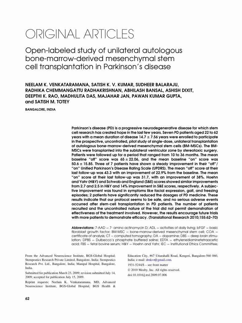
MSCs were isolated from bone marrow of Parkinson’sdisease patients and were cultured until sufficient num-bers were obtained. In this protocol ,1 million cells perkg body weight were transplanted through stereotaxicsurgery. An adequate number of cells was obtained atpassage 1 or 2. BM-MSCs were tested for quality controland found clinically eligible (Fig 1). Each batch of cellswas subjected to endotoxin testing and sterility testing,were found to be negative for mycoplasma, and werekaryotypically normal. Immunophenotypic analysisshowed that they were positive for CD73 and CD90and were negative for CD45 (Fig 2).Seven patients were enrolled into this study according
to the protocol. They underwent intracerebral transplan-tation of autologous BM5 –MSCs and were followedup over a period of 12–36 months. Final clinical evalu-ation was performed in a period that ranged from 10 to32 months after transplantation. Patient demographicsand time of last evaluation have been presented. Patientswho participated in this study had a mean disease dura-tion of 14.7 years. Surgical procedures were well
Fig 1. The morphology of BM-MSCs derived from PD patients at passage 2 (A) before the cells become confluent
and (B) after the cells becomes confluent. Adherent cells derived from bone marrow displayed normal fibroblastic
morphology (magnification 1003). (Color version of figure is available online.)
Translational ResearchVolume 155, Number 2 Venkataramana et al 65
tolerated and all were discharged from the hospitalwithin 3 to 5 days (Table I).All the patients enrolled in the study were males. This
reflects the fact that PD is more prevalent in the male sexwith the occurrence in men being higher than that for
women.18 Most patients were middle aged at the timeof transplantation (mean age, 55.46 15.2 years); theoldest was 62 years and the youngest was 21 years.They suffered from Parkinsonian symptoms a mean du-ration of 14.76 7.6 years. The UPDRS was used, which
Fig 2. Immunophenotyping of BM-MSCs derived from all 7 patients. Cells were cultured for passage 2, harvested,and labeled with antibodies against human antigen CD45, CD73, and CD90, and they were analyzed by fluores-
cence-activated cell sorting. The viability of the cells was tested by 7AADmarkers. (Color version of figure is avail-
able online.)
Translational Research66 Venkataramana et al February 2010
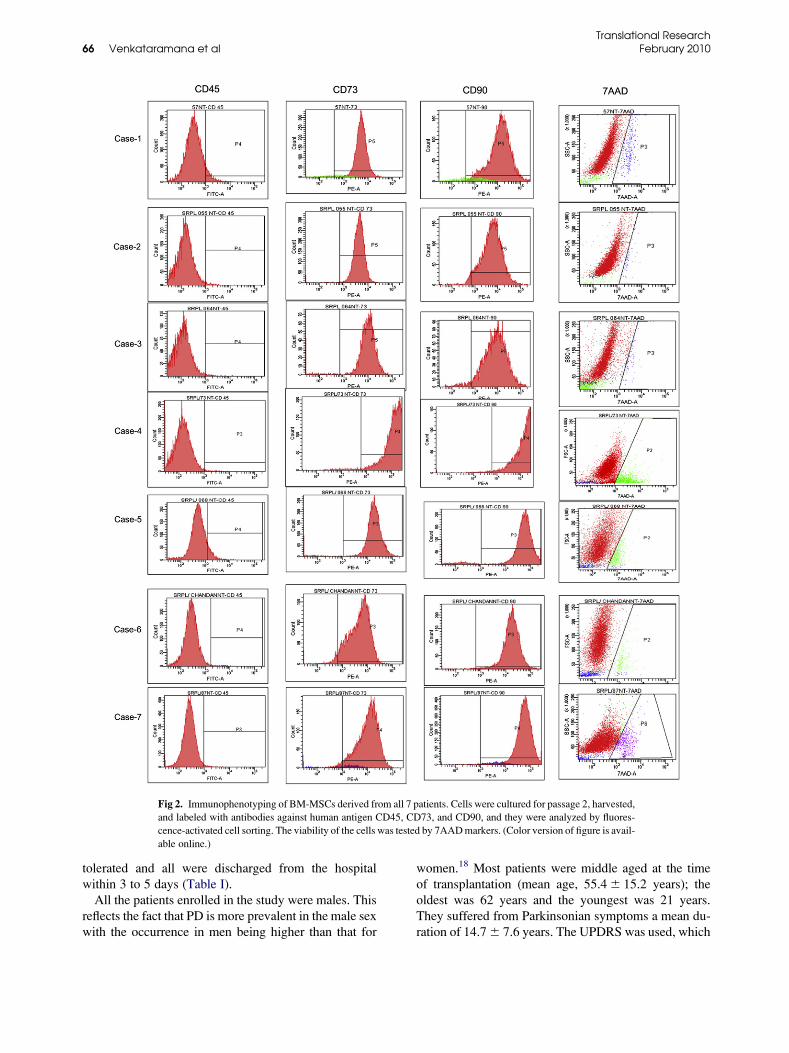
assessed 4 different parameters such as (1) mentation,behavior, and mood; (2) activities of daily living; (3)motor skills; and (4) complications of therapy. Theprimary outcome measure was improvement in UPDRSin ‘‘off’’ and ‘‘on’’ periods. The ‘‘off’’ period evalua-tions were performed when patients had been withdrawnovernight from antiparkinsonian medications forapproximately 8–10 h. ‘‘On’’ period evaluation wasperformed when during periods of maximum symptom-atic benefit, which was approximately 1 to 2 h after thefirst morning dose of medication. The mean baseline‘‘off’’ score was 656 22.0; the best score was 34 andthe worst score was 96. The mean baseline ‘‘on’’ scorewas 50.66 15.9; the best score was 43 and the worstscore was 73. Three patients who improved showeda steady improvement in their ‘‘off’’/‘‘on’’ scores. Themean ‘‘off’’ score at their last follow-up was 43.3,with an improvement of 22.9% from the baseline. Themean ‘‘on’’ score at their last follow-up was 31.7, withan improvement of 38%. We have observed marginalclinical benefit after a follow-up of 12–36 months in atleast 3 of 7 patients with PD, who underwent BM-MSC transplantation according to the protocol. Despitethe small numbers, an improvement was observed in to-tal UPDRS scores during ‘‘off’’ and ‘‘on’’ periods, S&Escore, and activities of daily living (ADL) scores (Fig 3).Among the patients in whom improvement was seen,
the total UPDRS ‘‘off’’ score improved by 22.16 5.8 %and the ‘‘on’’ score by 386 19.8 %. The mean H & Yscore (used for evaluating the secondary outcome mea-sures) was 2.7 with a low of 1.5 and a high of 5. Themean H&Y score at last follow-up was 2.5 thus virtuallyshowing no change from the baseline . As a secondaryoutcome measure S&E score showed 14% improvementat last follow-up. In addition, patients subjectivelyreported marginal improvement in symptoms, overallwell being, facial expression, gait and reduction in freez-ing episodes which never got benefited from traditionalmodes of therapy. Even we were able to marginallyreduce the dosage of anti-parkinsonian medicines.
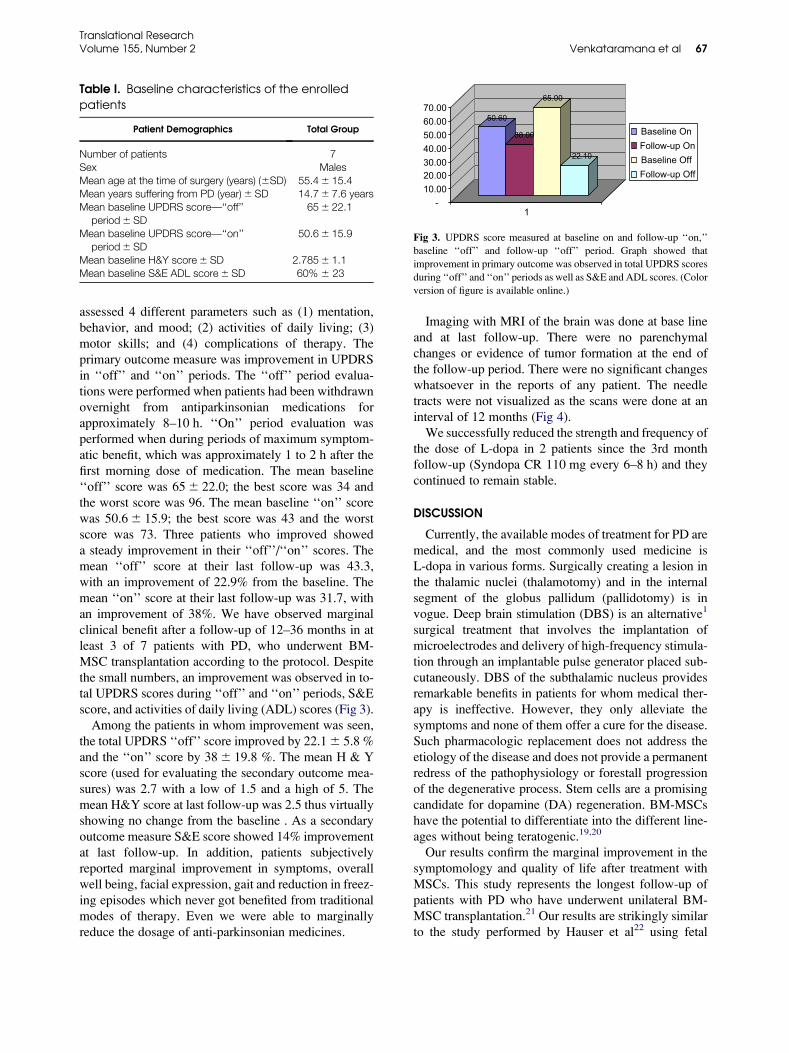
Imaging with MRI of the brain was done at base lineand at last follow-up. There were no parenchymalchanges or evidence of tumor formation at the end ofthe follow-up period. There were no significant changeswhatsoever in the reports of any patient. The needletracts were not visualized as the scans were done at aninterval of 12 months (Fig 4).We successfully reduced the strength and frequency of
the dose of L-dopa in 2 patients since the 3rd monthfollow-up (Syndopa CR 110 mg every 6–8 h) and theycontinued to remain stable.
DISCUSSION
Currently, the available modes of treatment for PD aremedical, and the most commonly used medicine isL-dopa in various forms. Surgically creating a lesion inthe thalamic nuclei (thalamotomy) and in the internalsegment of the globus pallidum (pallidotomy) is invogue. Deep brain stimulation (DBS) is an alternative1
surgical treatment that involves the implantation ofmicroelectrodes and delivery of high-frequency stimula-tion through an implantable pulse generator placed sub-cutaneously. DBS of the subthalamic nucleus providesremarkable benefits in patients for whom medical ther-apy is ineffective. However, they only alleviate thesymptoms and none of them offer a cure for the disease.Such pharmacologic replacement does not address theetiology of the disease and does not provide a permanentredress of the pathophysiology or forestall progressionof the degenerative process. Stem cells are a promisingcandidate for dopamine (DA) regeneration. BM-MSCshave the potential to differentiate into the different line-ages without being teratogenic.19,20
Our results confirm the marginal improvement in thesymptomology and quality of life after treatment withMSCs. This study represents the longest follow-up ofpatients with PD who have underwent unilateral BM-MSC transplantation.21 Our results are strikingly similarto the study performed by Hauser et al22 using fetal
Table I. Baseline characteristics of the enrolledpatients
Patient Demographics Total Group
Number of patients 7Sex MalesMean age at the time of surgery (years) (6SD) 55.46 15.4Mean years suffering from PD (year)6 SD 14.76 7.6 yearsMean baseline UPDRS score—‘‘off’’
period6 SD656 22.1
Mean baseline UPDRS score—‘‘on’’period6 SD
50.66 15.9
Mean baseline H&Y score6 SD 2.7856 1.1Mean baseline S&E ADL score6 SD 60% 6 23
50.60
38.00
65.00
22.10
-10.0020.0030.0040.0050.0060.0070.00
1
Baseline OnFollow-up OnBaseline OffFollow-up Off
Fig 3. UPDRS score measured at baseline on and follow-up ‘‘on,’’baseline ‘‘off’’ and follow-up ‘‘off’’ period. Graph showed that
improvement in primary outcome was observed in total UPDRS scores
during ‘‘off’’ and ‘‘on’’ periods as well as S&E and ADL scores. (Colorversion of figure is available online.)
Translational ResearchVolume 155, Number 2 Venkataramana et al 67

nigral transplantation, where the UPDRS ‘‘off’’ scoreswere decreased by 18% (in 1 year follow-up) and 26%(in 2 years follow-up) compared with 22% in our study.A trend of marginal deterioration in symptoms after
initial improvement was observed in 25% of patients af-ter 12–18 months of follow-up. This might be caused bythe continued degeneration on the nongrafted side. Somestudies have shown encouraging results where bilateraltransplantation of mesencephalic tissue have beenperformed.23 These reports have encouraged us toundertake bilateral grafting of MSCs in future studies.Wenning et al21 reported an increased uptake of fluo-dopa on positron emission tomography (PET) in the pu-tamen, by 68% after transplantation. Similarly, 61%uptake after 12 months was reported by Hauser et al.22
However, we could not support our results with flurodo-poa uptake because a PET scan facility was not availablein the hospital. The autopsy studies reported previouslyalso support robust graft survival prominent neuritic out-growth and extensive reinnervations in an organotypicpattern.13,23-26 In this study, we cannot exclude the pos-sibility of a placebo effect as it was an open-label study.However, the persistence of clinical improvementthrough 20–26 months solely caused by a placebo effectseems unlikely. Transplantation at different targets in-cluding bilateral hemispheres, different doses, and therole of booster dose needs to be explored in the future.Moreover, an improvement in dyskinesias was observedin some of our patients.BM-MSCswere transplanted into the lateralwalls of the
lateral ventricles of the left/right cerebral hemisphere. Thissite was chosen because along much of the lateral walls of
the lateral ventricles lies the largest germinal zone of theadult mammalian brain, which is called the SVZ.27 Stud-ies have shown that in adult mammals, new neurons areborn in the SVZ and migrate anteriorly into the olfactorybulb, where they mature into local interneurons.28-30 Insome studies, SVZ neural stem cells have been grown inculturewith epidermal growth factor, bFGF, or a combina-tion of these.31-33 The SVZ as such represents an impor-tant reservoir of progenitors in the adult brain harboringcell populations that help in neuroregeneration.The mechanism responsible for this benefit is not
exactly known, but it may be caused by more normalDA regulation as a result of the survival and functioningof transformed DA neurons and their terminals. Previousstudies have shown extensive DA transporter staining,which provides evidence of an increased number of DAterminals that may have the capacity to store DA andbufferfluctuations in striatalDAconcentrations associatedwith development of dyskinesia.34 Marginal dose reduc-tion could be another contributory factor. This study isthe first report to demonstrate beneficial effects of BM-MSCs in Parkinson’s disease patients. BM-MSCs showeddifferentiation into DA neurons, and a detectable level ofDA was observed in the culture media of differentiatedcells.35 Moreover, a significant behavioral improvementin PD rat models 3 months posttransplantation was alsoobserved.35,36 This proves thatBM-MSCshave a potentialto differentiate and exhibit several traits of DA precursors,which on transplantation in animal model, induce behav-ioral improvements in the hemiparkinsonian rat.36 Theseresults were corroborated by Trzaska et al,37 who demon-strated that adultmesenchymal stemcells indeed showDA
Fig 4. Imaging with MRI shown at the baseline before stem cell transplantation (A) and after 12-month follow-up
post-stem-cell transplantation (B). Cells were transplanted at SVZ. There are no perenchymal changes and no
abnormal evidence post-stem-cell transplantation. There are no significant changes of any patient.
Translational Research68 Venkataramana et al February 2010

phenotypes. An immunohistochemical analysis revealedthat the BM-MSCs were present more than 130 days aftertransplantation, and they showed integration into brain pa-renchyma, survival, and even migration toward theipsilateral nigra. However, the specific mechanism bywhich the beneficial behavior effect was accomplishedin animal models and also in our study is still difficult tointerpret. Several likely mechanisms have been postulatedrecently, and some of them have explained that trans-plantedBM-MSCsperhaps exhibit or secrete neurotrophicfactors.38 Some suggested the possibility of immunomo-dulation of host response to the lesion,39 and few impli-cated that transplanted BM-MSCs enhance endogenousneurogenesis.40 In our study, we cannot rule out any pos-sible mechanisms that have been suggested above. Never-thelesss, more studies need to be conducted to address andelucidate the possible mechanism of action so that bettertreatment options are available in future.
CONCLUSION
This study establishes the immediate and short-termsafety of autologous BM-MSCs in the unilateral trans-plantation therapy of PD. The clinical improvement isonly marginal; however, most patients experienced sub-jective well-being, without any notable adverse sideeffects. The exact mechanism of action is not clearlyunderstood, which warrants elaborate studies with pla-cebo control and bilateral transplantation with longerpatient follow-up. Studies in this direction are currentlybeing conducted at our center.
REFERENCES
1. Lang AE, Obeso JA. Time to move beyond nigrostriatal dopamine
deficiency in Parkinson’s disease. Ann Neurol 2004;55:761–5.2. HelyMA,Morris JG, ReidWG, et al. SydneyMulticenter Study of
Parkinson’s disease: non-L-dopa-responsive problems dominate at
15 years. Mov Disord 2005;20:190–9.3. Shults CW, Oakes D, Kieburtz K, et al. Effects of coenzyme Q10
in early Parkinson disease: evidence of slowing of the functional
decline. Arch Neurol 2002;59:1541–50.
4. Fahn S, Oakes D, Shoulson I, et al. Levodopa and the progressionof Parkinson’s disease. N Engl J Med 2004;351:2498–508.
5. Marsden CD, Parkes JD. Success and problems of long-term
levodopa therapy in Parkinson’s disease. Lancet 1977;1:345–9.
6. Obeso JA, Rodriguez-Oroz MC, Rodriguez M, et al. Pathophysi-ology of the basal ganglia in Parkinson’s disease. Trends Neurosci
2000;23:S8–19.
7. Pifl C, Schingnitz G, Hornykiewicz O. Striatal and non-striatalneurotransmitter changes in MPTP-parkinsonism in rhesus
monkey: the symptomatic versus the asymptomatic condition.
Neurochem Int 1992;20:295S–7.
8. Sossi V, Fuente-Fernandez R, Holden JE, Schulzer M, Ruth TJ,Stoessl J. Changes of dopamine turnover in the progression of
Parkinson’s disease as measured by positron emission tomogra-
phy: their relation to disease-compensatory mechanisms. J Cereb
Blood Flow Metab 2004;24:869–76.
9. Calabresi P, Mercuri NB, Sancesario G, Bernardi G. Electrophys-
iology of dopaminedenervated striatal neurons. Implications for
Parkinson’s disease. Brain 1993;116:433–52.10. Bezard E, Boraud T, Bioulac B, Gross CE. Involvement of the
subthalamic nucleus in glutamatergic compensatory mechanisms.
Eur J Neurosci 1999;11:2167–70.11. Bezard E, Dovero S, Prunier C, et al. Relationship between the
appearance of symptoms and the level of nigrostriatal degeneration
in a progressive 1-methyl-4-phenyl-1,2,3,6-tetrahydropyridine-
lesioned macaque model of Parkinson’s disease. J Neurosci
2001;21:6853–61.
12. Alvarez-Buylla A, Garcia-Verdugo JM, Tramonti AD. A unified
hypothesis on the lineage of neural stem cells. Nat Rev Neurosci
2001;2:287–93.
13. Olanow CW, Kordower JH, Freeman TB. Fetal nigral transplanta-
tion as a therapy for Parkinson’s disease. Trends Neurosci 1996;
19:102–9.
14. Freed CR, Greene PE, Breeze RE, et al. Transplantation of embry-
onic dopamine neurons for severe Parkinson’s disease. N Engl J
Med 2001;344:710–9.
15. Pal R, Hanwate M, Jan M, Totey S. Phenotypic and functional
comparison of optimum culture conditions for upscaling of bone
marrow-derived mesenchymal stem cells. J Tissue Eng Regen
Med 2009;3:163–74.
16. Fahn S, Marsden CD, Calne DB, Goldstein M. Recent develop-
ments in Parkinson’s disease. Florham Park, NJ: Macmillan
Healthcare Information, 1987. 153–163.
17. Langston JW, Widner H, Goetz CG, et al. Core assessment pro-
gram for intracerebral transplantations (CAPIT). Mov Disord
1992;7:2–13.
18. Eeden VD, Stephen K, Tanner CM, et al. Incidence of Parkinson’s
Disease: Variation by age, gender, and race/ethnicity. Am J Epide-
miol 2003;157:1015–22.
19. Morrison SJ, Uchida N, Weissman IL. The biology of hematopoi-
etic stem cells. Ann Rev Cell Dev Biol 1995;11:35–71.20. Deans RJ, Moseley AM. Mesenchymal stem cells: biology and
potential clinical uses. Exp Hematol 2000;28:875–84.
21. Wenning GK, Odin P, Morrish P, et al. Short- and long-term
survival and function of unilateral intrastriatal dopaminergic grafts
in Parkinson’s disease. Ann Neurol 1997;42:95–107.
22. Hauser RA, Freeman TB, Snow BJ, et al. Long-term evaluation of
bilateral fetal nigral transplantation in Parkinson’s disease.
Arch Neurol 1999;56:179–87.
23. Kordower JH, Freeman TB, Snow BJ, et al. Neuropathological
evidence of graft survival and striatal reinnervation after transplan-
tation of fetal mesencephalic tissue in a patient with Parkinson’s
disease. N Engl J Med 1995;332:1118–24.
24. Olanow CW, Goetz CG, Kordower JH, et al. A double-blind
controlled trial of bilateral fetal nigral transplantation in Parkin-
son’s Disease. Ann Neurol 2003;54:403–14.
25. Lindvall O, Sawle G, Widner H, et al. Evidence of long-term
survival and function of dopaminergic grafts in progressive
Parkinson’s disease. Ann Neurol 1994;35:172–80.
26. Freed CR, Breeze RE, Rosenberg NL, et al. Survival of implanted
fetal dopamine cells and neurologic improvement 12 to 46 months
after transplantation for Parkinson’s disease. N Engl J Med 1992;
327:1549–55.27. Doetsch F, Alvarez-Buylla A. Network of tangential pathways for
neuronal migration in adult mammalian brain. Proc Natl Acad Sci
USA 1996;93:14895–900.28. Altman J. Autoradiographic and histological studies of postnatal
neurogenesis. IV. Cell proliferation and migration in the anterior
forebrain, with special reference to persisting neurogenesis in the
olfactory bulb. J Comp Neurol 1969;137:433–58.
Translational ResearchVolume 155, Number 2 Venkataramana et al 69
29. Lois C, Alvarez-Buylla A. Long-distance neuronal migration in
the adult mammalian brain. Science 1994;264:1145–8.
30. Kornack DR, Rakic P. The generation, migration, and differentia-
tion of olfactory neurons in the adult primate brain. Proc Natl Acad
Sci USA 2001;98:4752–7.
31. Weiss S, Dunne C, Hewson J, et al. Multipotent CNS stem cells are
present in the adult mammalian spinal cord and ventricular neuro-
axis. J Neurosci 1996;16:7599–609.
32. Temple S, Alvarez-Buylla A. Stem cells in the adult mammalian
central nervous system. Curr Opin Neurobiol 1999;9:135–41.33. Gage FH. Mammalian neural stem cells. Science 2000;287:1433–8.
34. Pearce RKB, JacksonM, Smith L, Jenner P, Marsden CD. Chronic L-
dopa administration induces dyskinesias in the 1-methyl-4-phen
yl-1,2,3,6-tetrahydropyridinetreated common marmoset (Callithrix
Jacchus). Mov Disord 1995;10:731–40.
35. Shetty P, Ravindran G, Sarang S, Thakur A, Rao HS,
Viswanathan C. Clinical grade mesenchymal stem cells transdif-
ferentiated under xenofree conditions alleviates motor deficiencies
in a rat model of Parkinson’s disease. Cell Biol Int 2009;33(8):
830–8.
36. Levy YS, Bahat-Stroomza M, Barzilay R, et al. Regenerativeeffect of neural induced human mesenchymal stromal cells in rat
models of Parkinson’s disease. Cytotherapy 2008;10:340–52.
37. Trzaska KA, Kuzhikandathil EV, Rameshwar P. Specification of
a dopaminergic phenotype from adult human mesenchymal stemcells. Stem Cells 2007;25:2797–808.
38. Pisati F, Bossolasco P, Meregalli M, et al. Induction of neurotro-
phin expression via human adult mesenchymal stem cells: implica-
tion for cell therapy in neurodegenerative disease. CellTransplantation 2007;16:41–55.
39. Le Blanc K, Ringden O. Mesenchymal stem cell properties and
role in clinical bone marrow transplantation. Curr Opin Immunol
2006;18:586–91.40. Deng YB, Liu XG, Liu ZG, et al. Implantation of BM Mesenchy-
mal stem cells into injured spinal cord elicit de novo neurogenesis
and functional recovery: evidence from a study in rhesus monkey.Cytotherapy 2006;8:210–4.
Translational Research70 Venkataramana et al February 2010
Related Documents