3/27/2012 1 Paraesophageal Hiatal Hernia Stanley Rogers, MD Associate Clinical Professor of Surgery Ruth M. Dunn Chair and Chief, Minimally Invasive Surgery Director, Bariatric Surgery University of California, San Francisco UCSF Post Graduate Course in General Surgery Monday, March 26, 2012 PEH can be repaired laparoscopically safely and with excellent results. Although laparoscopic PEH repair is associated with high recurrence rates, excellent symptom improvement still occurs regardless of recurrence. Repair with synthetic mesh lowers recurrence, but is associated w/ dysphagia & visceral erosion Biologic mesh – shown to reduce recurrence without mesh-related complications / side effects Paraesophageal Hiatal Hernia Important Points Optimal management is controversial in several ways. Points of controversy Appropriate evaluation of patients Optimal surgical approach Option of laparoscopic technique +/- antireflux procedure accompanying PEH repair Mesh reinforcement of hiatus Short esophagus needing Collis Paraesophageal Hiatal Hernia Hiatal hernias are classified according to the position of the esophagogastric junction and the existence of a true hernia sac. Type I (sliding) Most common Leading edge of the hernia is the EGJ, which is displaced into an intrathoracic position. The longitudinal axis of the stomach is aligned with the esophagus. There is no true hernia sac nor any paraesophageal component. Classification

Welcome message from author
This document is posted to help you gain knowledge. Please leave a comment to let me know what you think about it! Share it to your friends and learn new things together.
Transcript

3/27/2012
1
ParaesophagealHiatal Hernia
Stanley Rogers, MDAssociate Clinical Professor of Surgery
Ruth M. Dunn Chair and Chief, Minimally Invasive SurgeryDirector, Bariatric Surgery
University of California, San Francisco
UCSF Post Graduate Course in General SurgeryMonday, March 26, 2012
� PEH can be repaired laparoscopically safely and with excellent results.
� Although laparoscopic PEH repair is associated with high recurrence rates, excellent symptom improvement still occurs regardless of recurrence.
� Repair with synthetic mesh lowers recurrence, but is associated w/ dysphagia & visceral erosion
� Biologic mesh – shown to reduce recurrence without mesh-related complications / side effects
Paraesophageal Hiatal HerniaImportant Points
� Optimal management is controversial in several ways.
� Points of controversy� Appropriate evaluation of patients� Optimal surgical approach� Option of laparoscopic technique� +/- antireflux procedure accompanying PEH repair� Mesh reinforcement of hiatus� Short esophagus needing Collis
Paraesophageal Hiatal Hernia
Hiatal hernias are classified according to the position of the
esophagogastric junction and the existence of a true hernia sac.
� Type I (sliding)� Most common� Leading edge of the hernia is the EGJ,
which is displaced into an intrathoracic position.
� The longitudinal axis of the stomach is aligned with the esophagus.
� There is no true hernia sac nor any paraesophageal component.
Classification

3/27/2012
2
5
� The EGJ moves through the hiatus to the visceral mediastinum.
� Increased abdominal pressure (pregnancy, obesity, or vomiting ) and vigorous esophageal contraction may contribute the development of the hernia.
� GERD & esophagitis may occur due to loss of tone of the LES.
Classification: Type I
Abbara S et al. AJR 2003;181:403-414
Type II & Type III are referred to as “paraesophageal hernias”.
� Type II (rolling)� The EGJ is in its normal
intraabdominal location
� The hernia sac (containing portions of the gastric fundus and body) develops alongsidethe esophagus
Classification
7
� Uncommon� Phrenoesophageal
membrane is not weakened diffusely but focally
� Gastric fundus protrudes through hiatus
Abbara S et al. AJR 2003;181:403-414
Classification: Type II
� Combination of Type I & Type II
� EGJ displaced into thorax (Type I), and a hernia sac contains portions of gastric fundus / body (Type II)
� Frequently occurs when Type II PEH present for many years.
Classification: Type III
Abbara S et al. AJR 2003;181:403-414

3/27/2012
3
� Refers to herniation of organs other than the stomach
� T-colon & omentum most commonly involved.
� Spleen & small intestine are less commonly involved.
Classification: Type IV
Abbara S et al. AJR 2003;181:403-414
• Type I: hatched bars
• Type II & III: solid bars
PEH: Basic Prevalence
PEH: Relative FrequencyAccording to Age
PEH: Associated Symptoms
� Increasingly common with advancing age� More often among women than men� PEH often associated with GERD
� Typical symptoms
� Suspicious CXR
� Chest CT� Upper GI Series
� In urgent situations:� Placement of NG tube with subsequent
“coiling in the chest”
Often difficult to assess the location of the actual junction…
Diagnosis
3/27/2012
4
Intrathoracicstomach / “elevatedleft hemidiaphragm”
PEH: Presentation
� Classic barium UGI study confirms the diagnosis.
Diagnosis
� ± Evaluation of LES� Endoscopy� Esophageal manometry / motility studies
� 24 hr pH monitoring� 1/3 of pts will have atypical peristalsis of the
esophageal body� ½ of symptomatic pts will have abnormal pH
results � Indications for Surgery� Type I – significant GERD � Elevated DeMeester score: > 14.72� EGD, 24 hr pH, esophageal manometry
PEH: Management
� Indications for Surgery - Type II & III
� Associated with a high incidence of complications from volvulus� Bleeding, incarceration, obstruction, strangulation and
perforation. � Gastritis and ulceration can occur - the result of poor gastric
emptying & torsion / ischemia / inflammation of gastric wall
� Schedule surgery electively when PEH identified� Urgent repair when symptomatic (symptoms do not
predict risk however)� “Catastrophic presentation” in up to 20% of
symptomatic pts with delay in surgery
Management
3/27/2012
5
� Findings that may prompt urgent surgery � Symptoms of obstruction� Reflux – Severe GERD� Anemia� Systemic concern of gastric ischemia
� Goal: to avoid� Aspiration� Hemorrhage / transfusion requirements � Gastric necrosis - mortality 50%.
Management:Clinical presentation (risk) related to gastric volvulus
Abbara S et al. AJR 2003;181:403-414
PEH Risk: Volvulus
Abbara S et al. AJR 2003;181:403-414
PEH Risk: Volvulus
� Organoaxial rotation � Mobile greater curvature
moves anteriorly and superiorly so that in 180°organoaxial rotation, mirror image of stomach is created with convex greater curvature located above and to right of concave lesser curvature
Abbara S et al. AJR 2003;181:403-414
PEH Risk: Volvulus
� Mesenteroaxial rotation� Rotation of stomach is shown
along axis (dotted lines) perpendicular to long axis (solid line).
� Mobile antrum and duodenum move anteriorly and superiorly.
� Greater curvature remains on left.
� Gastric fundus and antrum may be in reversed positions.
3/27/2012
6
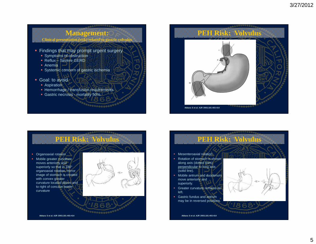
Greater curveof stomach
Pylorus
PEH: UGI with Type III mesoaxial volvulus
� Transthoracic or transabdominal approach� Thoracoscopic / laparoscopic or open
� Principles similar to other hernia operations� Reduction of hernia contents� Excision of sac� Mediastinal mobilization of esophagus to ensure adequate
intraabdominal length� Closure of hiatus primarily (with or without biologic mesh
buttressed repair)� Controversy
� Fundoplication� Anchor stomach: Hill gastropexy / Stamm gastrostomy� Collis gastroplasty
Surgical Technique
� Operative approach – surgeon experience / comfort� Intrathoracic / mediastinal dissection easier via
thoracotomy� Transthoracic PEH reduction may not eliminate
risk of gastric volvulus, & can actually produce or precipitate gastric body volvulus
� Laparoscopy – typical benefits� Less pain� Fewer days in ICU / hospital� Earlier resumption of diet� Earlier return to normal activity / work
Surgical Technique: DomainThoracic -> laparoscopic surgeons
� Previously controversial – less so now� Indicated in all Type II / III pts, & � Type I with GERD
� Most surgeons perform 360°wrap� GERD- 20-30% will reflux postoperatively� Circumferential dissection of GEJ disrupts natural
antireflux mechanism� Facilitates intraabdominal gastric fixation / anchor
Surgical TechniqueConcurrent fundoplication / antireflux surgery?
3/27/2012
7
25
� Hill suture plication: � 3 interrupted nonabsorbable sutures between lesser
curve of the stomach and preaortic fascia � Stamm gastrostomy: 2 functions� Eliminates the need of NG tube� Fixes the stomach to the abdominal wall / can help
prevent volvulus
� Non-physiologic & not commonly performed anymore due to preponderance of laparoscopic approach
Operative Approach PEH: Operative TechniqueLaparoscopic Approach
� Technically feasible, safe, effective
� Can be very difficult
� Advantages over open repair� However….can be much more
challenging for less experienced surgeons
PEH: Laparoscopic Repair
� Relocated EGJ / hernia contents ± volvulus� can obscure normal anatomy
� Dissection of large sac - complex� Mediastinal bleeding� Redundant tissue at GEJ � Can make fundoplication difficult
� Large diaphragmatic defect
� Experience – these procedure should be done by experienced hands
PEH: Laparoscopic Challenges
3/27/2012
8
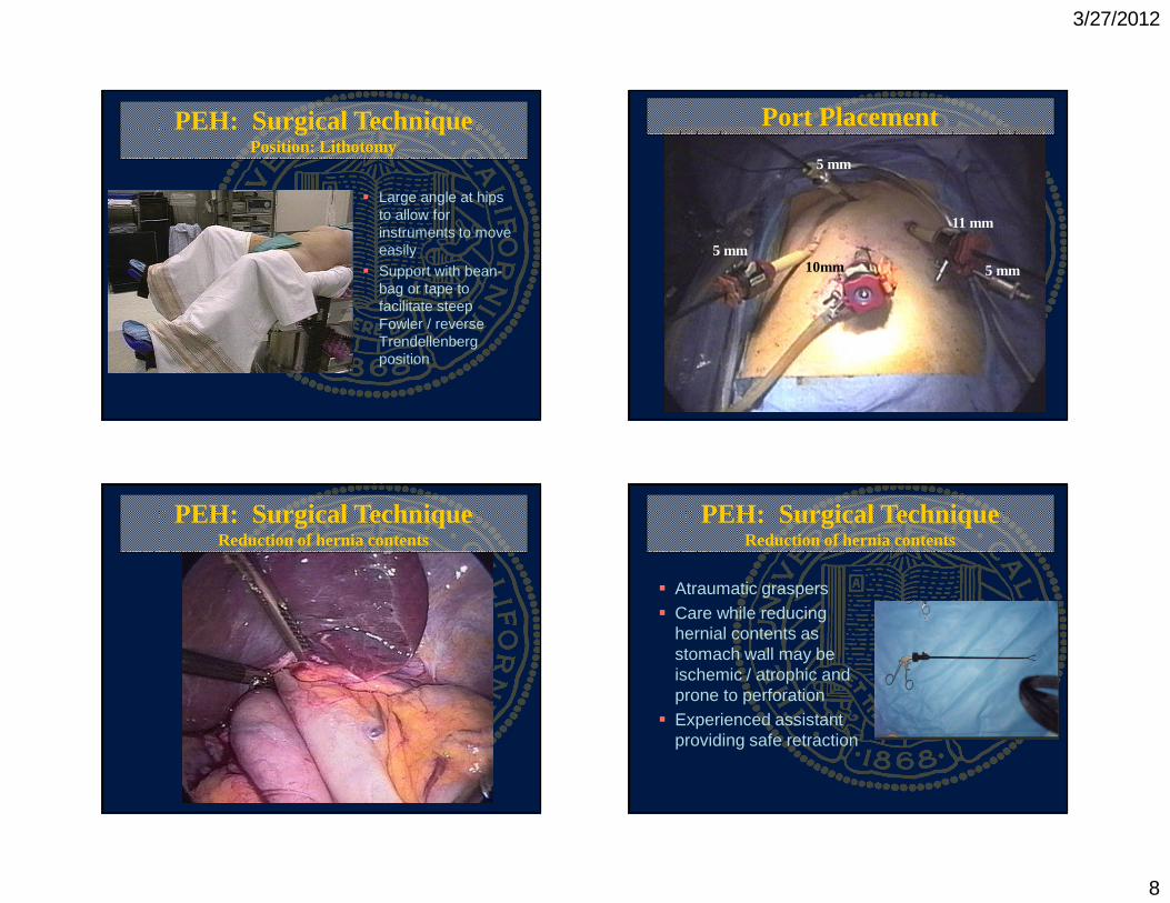
� Large angle at hips to allow for instruments to move easily
� Support with bean-bag or tape to facilitate steep Fowler / reverse Trendellenberg position
PEH: Surgical TechniquePosition: Lithotomy
5 mm
11 mm
10mm
5 mm
5 mm
Port Placement
PEH: Surgical TechniqueReduction of hernia contents
� Atraumatic graspers� Care while reducing
hernial contents as stomach wall may be ischemic / atrophic and prone to perforation
� Experienced assistant providing safe retraction
PEH: Surgical TechniqueReduction of hernia contents
3/27/2012
9
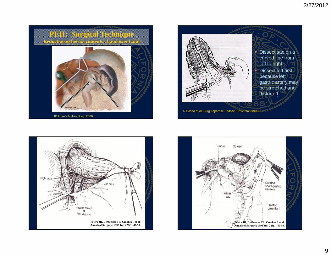
PEH: Surgical TechniqueReduction of hernia contents: hand over hand
JD Luketich, Ann Surg 2000
N Basso et al. Surg Laparosc Endosc 9:257-262, 1999
� Dissect sac on a curved line from left to right
� Dissect left first because left gastric artery may be stretched and distorted
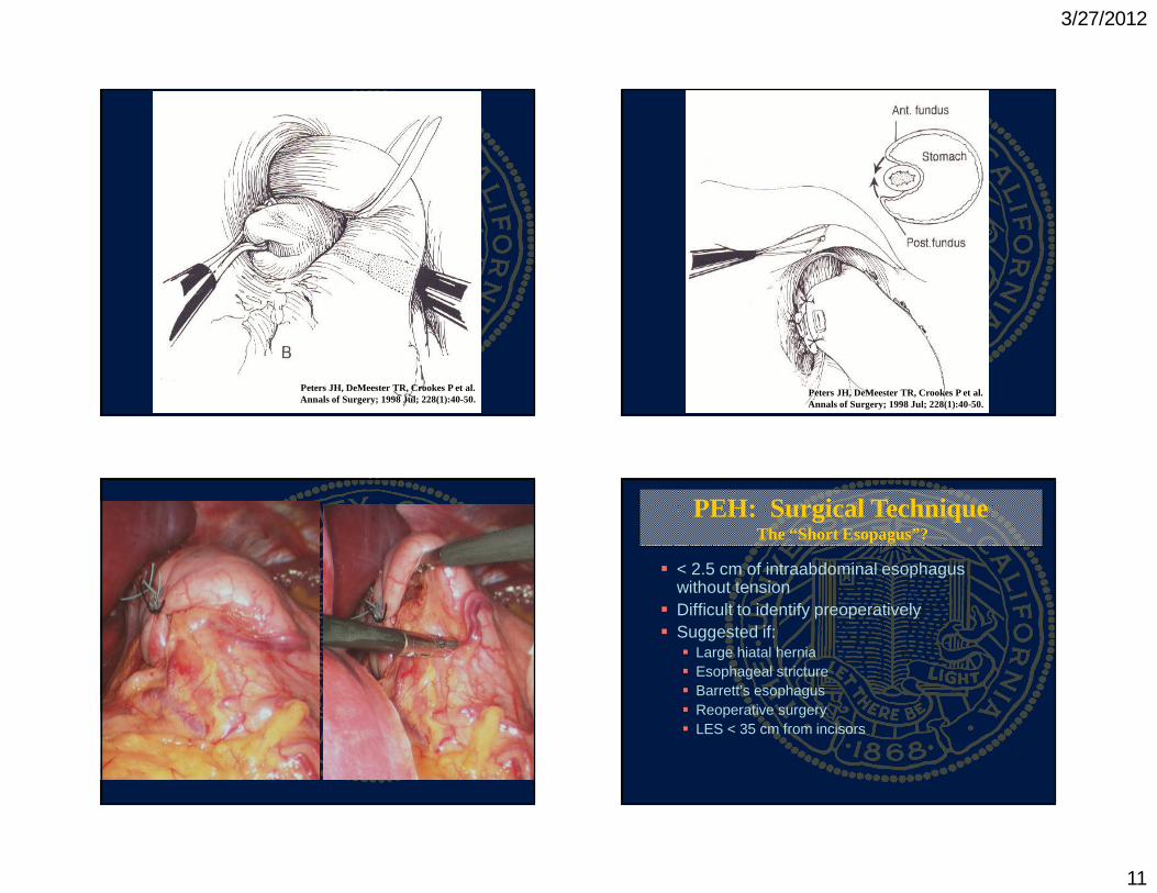
Peters JH, DeMeester TR, Crookes P et al.Annals of Surgery; 1998 Jul; 228(1):40-50.
Peters JH, DeMeester TR, Crookes P et al.Annals of Surgery; 1998 Jul; 228(1):40-50.
3/27/2012
10
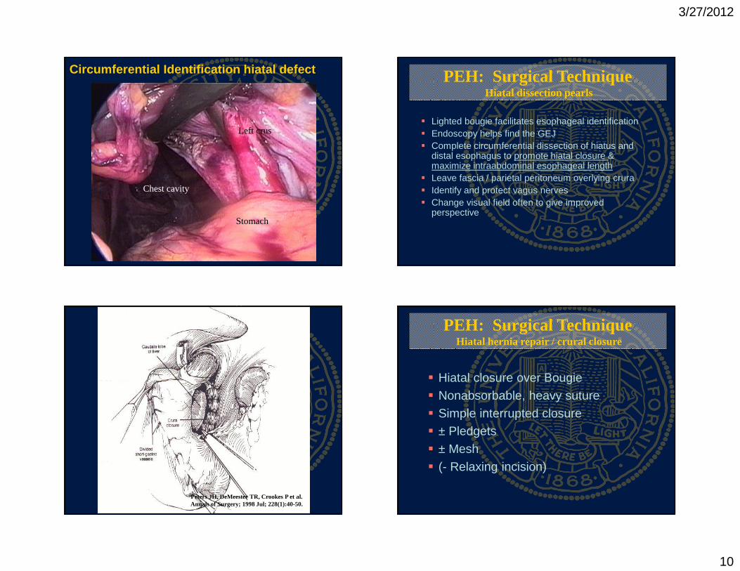
Circumferential Identification hiatal defect
Chest cavity
Stomach
Left crus� Lighted bougie facilitates esophageal identification� Endoscopy helps find the GEJ� Complete circumferential dissection of hiatus and
distal esophagus to promote hiatal closure & maximize intraabdominal esophageal length
� Leave fascia / parietal peritoneum overlying crura� Identify and protect vagus nerves� Change visual field often to give improved
perspective
PEH: Surgical TechniqueHiatal dissection pearls
Peters JH, DeMeester TR, Crookes P et al.Annals of Surgery; 1998 Jul; 228(1):40-50.
� Hiatal closure over Bougie
� Nonabsorbable, heavy suture
� Simple interrupted closure� ± Pledgets
� ± Mesh
� (- Relaxing incision)
PEH: Surgical TechniqueHiatal hernia repair / crural closure
3/27/2012
11
Peters JH, DeMeester TR, Crookes P et al.Annals of Surgery; 1998 Jul; 228(1):40-50.
Peters JH, DeMeester TR, Crookes P et al.Annals of Surgery; 1998 Jul; 228(1):40-50.
� < 2.5 cm of intraabdominal esophagus without tension
� Difficult to identify preoperatively� Suggested if:� Large hiatal hernia� Esophageal stricture� Barrett’s esophagus� Reoperative surgery� LES < 35 cm from incisors
PEH: Surgical TechniqueThe “Short Esopagus”?

3/27/2012
12
� Excise GEJ redundant tissue� Mobilize mediastinal esophagus
circumferentially as high as possible� A few cm gained by anterior displacement of
esophagus with posterior diaphragm repair� Lengthening procedure required in 5-10%� Nissen-Collis
PEH: Surgical TechniqueThe “Short Esopagus”?
LL Swanstrom et al. Arch Surg 133:869, 1998
JD Luketich, Ann Surg 2000 JD Luketich, Ann Surg 2000
3/27/2012
13
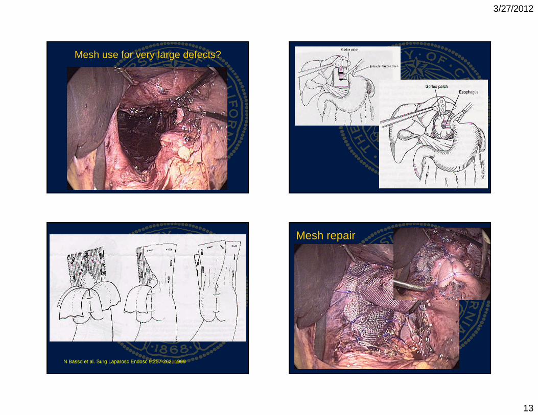
Mesh use for very large defects?
N Basso et al. Surg Laparosc Endosc 9:257-262, 1999
Mesh repair

3/27/2012
14
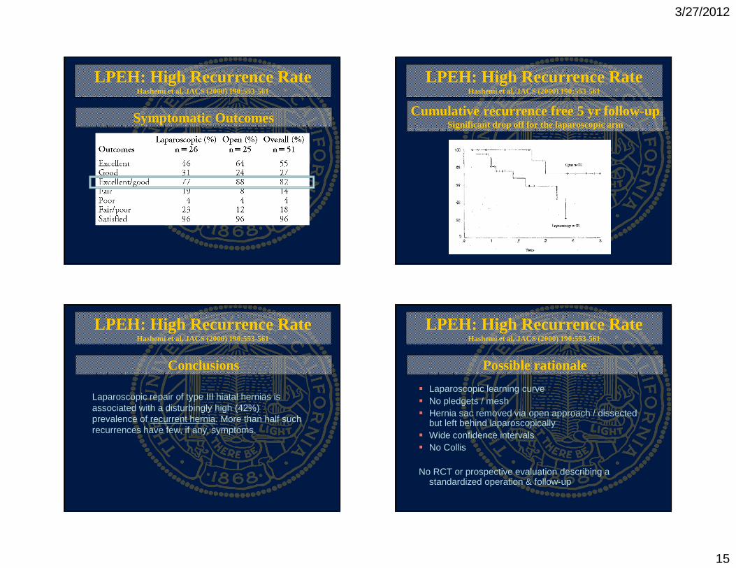
PEH: Surgical Results LPEH: High Recurrence RateHashemi et al, JACS (2000) 190:553-561
Review of Reported LPEH Repair Outcomes
LPEH: High Recurrence RateHashemi et al, JACS (2000) 190:553-561
� 1985 – 1998� 54 pts surgical repair type III PEH� Laparotomy: 13� Thoracotomy: 14� Laparocopy: 27
� Antireflux procedure in all� Outcomes:� Symptom questionnaire
� Median of 24 months 94% answered questionnaire
� Video esophagram in 75% by one radiologist
LPEH: High Recurrence RateHashemi et al, JACS (2000) 190:553-561
Perioperative Course
3/27/2012
15
LPEH: High Recurrence RateHashemi et al, JACS (2000) 190:553-561
Symptomatic Outcomes
LPEH: High Recurrence RateHashemi et al, JACS (2000) 190:553-561
Cumulative recurrence free 5 yr follow-upSignificant drop off for the laparoscopic arm
LPEH: High Recurrence RateHashemi et al, JACS (2000) 190:553-561
Conclusions
Laparoscopic repair of type III hiatal hernias is associated with a disturbingly high (42%) prevalence of recurrent hernia. More than half such recurrences have few, if any, symptoms.
� Laparoscopic learning curve� No pledgets / mesh� Hernia sac removed via open approach / dissected
but left behind laparoscopically� Wide confidence intervals� No Collis
No RCT or prospective evaluation describing a standardized operation & follow-up
LPEH: High Recurrence RateHashemi et al, JACS (2000) 190:553-561
Possible rationale
3/27/2012
16
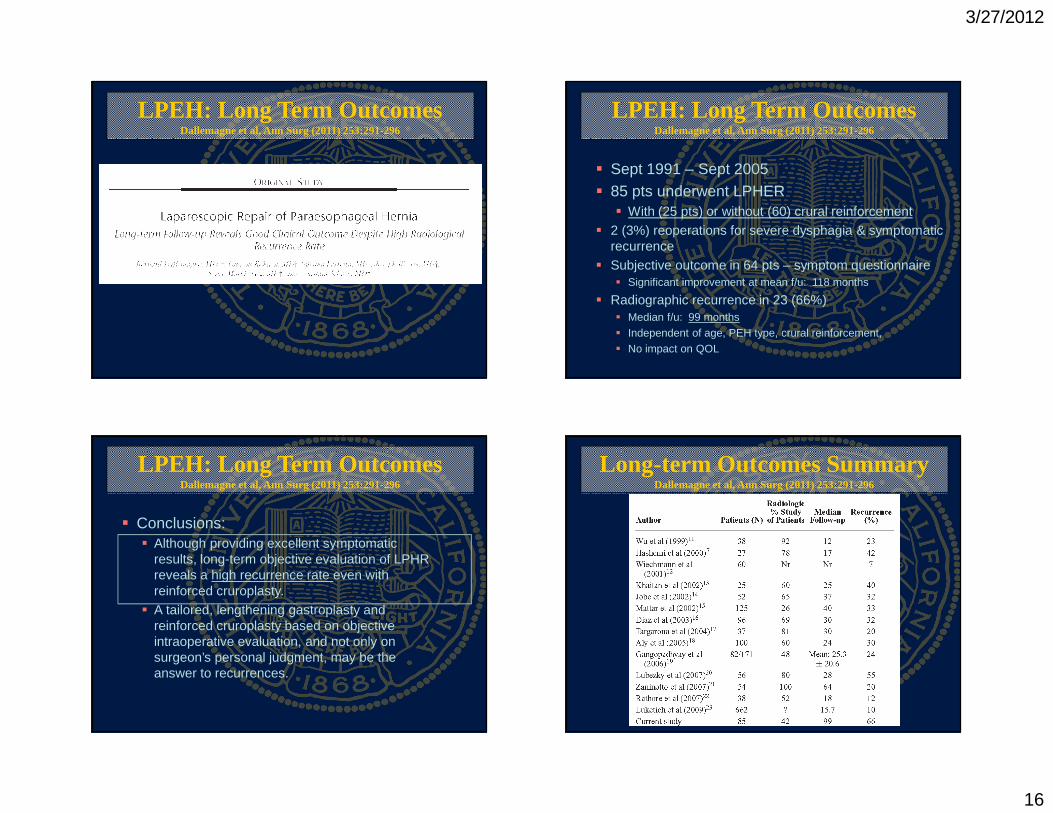
LPEH: Long Term OutcomesDallemagne et al, Ann Surg (2011) 253:291-296
LPEH: Long Term OutcomesDallemagne et al, Ann Surg (2011) 253:291-296
� Sept 1991 – Sept 2005� 85 pts underwent LPHER� With (25 pts) or without (60) crural reinforcement
� 2 (3%) reoperations for severe dysphagia & symptomatic recurrence
� Subjective outcome in 64 pts – symptom questionnaire� Significant improvement at mean f/u: 118 months
� Radiographic recurrence in 23 (66%) � Median f/u: 99 months� Independent of age, PEH type, crural reinforcement, � No impact on QOL
LPEH: Long Term OutcomesDallemagne et al, Ann Surg (2011) 253:291-296
� Conclusions:� Although providing excellent symptomatic
results, long-term objective evaluation of LPHR reveals a high recurrence rate even with reinforced cruroplasty.
� A tailored, lengthening gastroplasty and reinforced cruroplasty based on objective intraoperative evaluation, and not only on surgeon’s personal judgment, may be the answer to recurrences.
Long-term Outcomes SummaryDallemagne et al, Ann Surg (2011) 253:291-296
3/27/2012
17
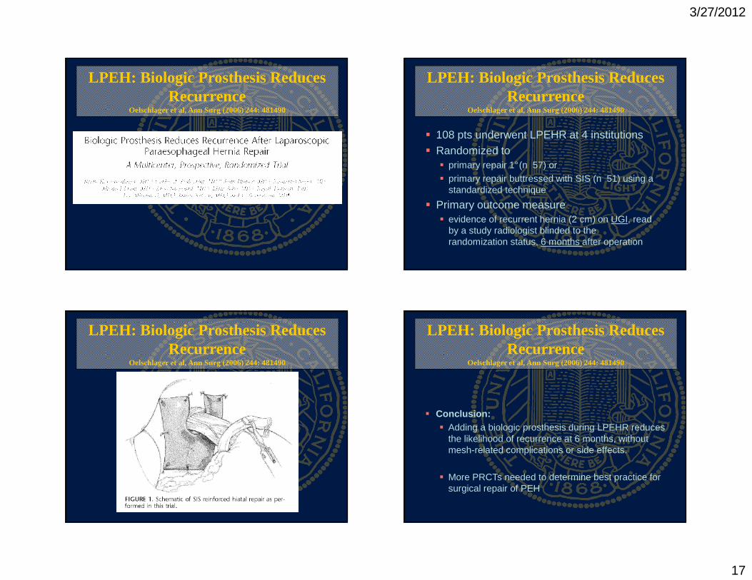
LPEH: Biologic Prosthesis Reduces Recurrence
Oelschlager et al, Ann Surg (2006) 244: 481490
LPEH: Biologic Prosthesis Reduces Recurrence
Oelschlager et al, Ann Surg (2006) 244: 481490
� 108 pts underwent LPEHR at 4 institutions� Randomized to � primary repair 1°(n 57) or � primary repair buttressed with SIS (n 51) using a
standardized technique
� Primary outcome measure � evidence of recurrent hernia (2 cm) on UGI, read
by a study radiologist blinded to the randomization status, 6 months after operation
LPEH: Biologic Prosthesis Reduces Recurrence
Oelschlager et al, Ann Surg (2006) 244: 481490
LPEH: Biologic Prosthesis Reduces Recurrence
Oelschlager et al, Ann Surg (2006) 244: 481490
� Conclusion: � Adding a biologic prosthesis during LPEHR reduces
the likelihood of recurrence at 6 months, without mesh-related complications or side effects.
� More PRCTs needed to determine best practice for surgical repair of PEH

3/27/2012
18
� Management of PEH can be challenging� Optimal management is controversial – though
becoming more standard (slowly)� Operative principles of hernia surgery must be
maintined� Reduce hernia contents atraumatically� Excise sac & mobilize esophagus� Repair diaphragm without tension over Bougie
� Simple sutures � pledgets � mesh (biologic)
� 360º fundoplication
PEH repair: in summary
� PEH can be repaired laparoscopically safely and with excellent results.
� Laparoscopic PEH repair is associated with high recurrence rates, though excellent symptom improvement still occurs regardless of recurrence.
� Biologic mesh – shown to reduce recurrence without mesh-related complications / side effects� Repair with synthetic mesh lowers recurrence, but is
associated w/ dysphagia & visceral erosion
� Difficult cases -> experienced surgeon.� More long-term, well organized prospective
randomized studies needed
Paraesophageal Hiatal Hernia
Mahalo
Related Documents