Marksphysicshelp 1 Paper 2 A2 Biology Notes 3.5 Energy transfers in and between organisms 3.5.1 Photosynthesis Content • The light-dependent reaction in such detail as to show that: o Chlorophyll absorbs light, leading to photoionisation of chlorophyll o Some of the energy from electrons released during photoionisation is conserved in the production of ATP and reduced NADP o The production of ATP involves electron transfer associated with the transfer of electrons down the electron transfer chain and passage of protons across chloroplast membranes and is catalysed by ATP synthase embedded in these membranes (chemiosomotic theory) o Photolysis of water produces protons, electrons and oxygen. • The light-independent reaction uses reduced NADP from the light-dependent reaction to form a simple sugar. The hydrolysis of ATP, also from the light-dependent reaction, provides the additional energy for this reaction. • The light-independent reaction in such detail as to show that: o Carbon dioxide reacts with ribulose bisphosphate (RuBP) to form two molecules of glycerate 3-phosphate (GP). This reaction is catalysed by the enzyme rubisco o ATP and reduced NADP from the light-dependent reaction are used to reduce GP to triose phosphate o Some of the triose phosphate is used to regenerate RuBP in the Calvin cycle o Some of the triose phosphate is converted to useful organic substances. • Students should be able to: o Identify environmental factors that limit the rate of photosynthesis o Evaluate data relating to common agricultural practices used to overcome the effect of these limiting factors Opportunities for Skills Development • Students could devise and carry out experiments to investigate the effect of named environmental variables on the rate of photosynthesis using aquatic plants, algae or immobilised algal beads.

Welcome message from author
This document is posted to help you gain knowledge. Please leave a comment to let me know what you think about it! Share it to your friends and learn new things together.
Transcript
Marksphysicshelp
1
Paper 2 A2 Biology Notes 3.5 Energy transfers in and between organisms 3.5.1 Photosynthesis
Content
• The light-dependent reaction in such detail as to show that: o Chlorophyll absorbs light, leading to photoionisation of chlorophyll o Some of the energy from electrons released during photoionisation is
conserved in the production of ATP and reduced NADP o The production of ATP involves electron transfer associated with the transfer
of electrons down the electron transfer chain and passage of protons across chloroplast membranes and is catalysed by ATP synthase embedded in these membranes (chemiosomotic theory)
o Photolysis of water produces protons, electrons and oxygen. • The light-independent reaction uses reduced NADP from the light-dependent reaction
to form a simple sugar. The hydrolysis of ATP, also from the light-dependent reaction, provides the additional energy for this reaction.
• The light-independent reaction in such detail as to show that: o Carbon dioxide reacts with ribulose bisphosphate (RuBP) to form two
molecules of glycerate 3-phosphate (GP). This reaction is catalysed by the enzyme rubisco
o ATP and reduced NADP from the light-dependent reaction are used to reduce GP to triose phosphate
o Some of the triose phosphate is used to regenerate RuBP in the Calvin cycle o Some of the triose phosphate is converted to useful organic substances.
• Students should be able to: o Identify environmental factors that limit the rate of photosynthesis o Evaluate data relating to common agricultural practices used to overcome the
effect of these limiting factors
Opportunities for Skills Development
• Students could devise and carry out experiments to investigate the effect of named environmental variables on the rate of photosynthesis using aquatic plants, algae or immobilised algal beads.
Marksphysicshelp
2
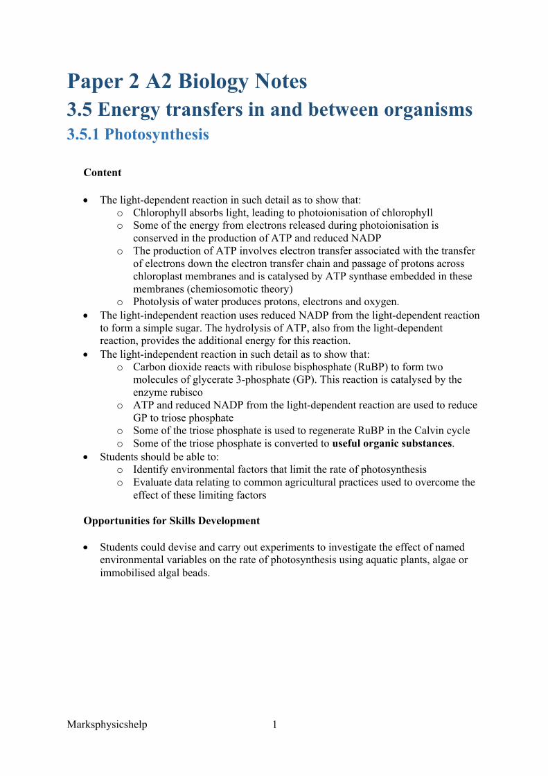
The Light-Dependent Reaction occurs in the thylakoid space of chloroplasts. The diagram below shows a chloroplast; the thylakoid space lies within the thylakoid membrane.
The first stage is for a photon of light to excite electrons within the chlorophyll molecule held in photosystem II. These excited electrons have enough energy to leave the chlorophyll molecule, and as a result cause the photoionisation of chlorophyll (ionisation with light). Some of the energy from electrons released during photoionisation is conserved in the production of ATP and reduced NADP.
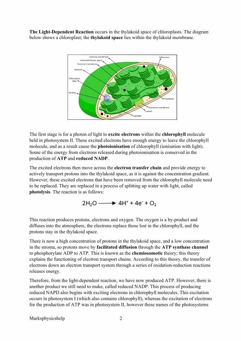
The excited electrons then move across the electron transfer chain and provide energy to actively transport protons into the thylakoid space, as it is against the concentration gradient. However, these excited electrons that have been removed from the chlorophyll molecule need to be replaced. They are replaced in a process of splitting up water with light, called photolysis. The reaction is as follows:
This reaction produces protons, electrons and oxygen. The oxygen is a by-product and diffuses into the atmosphere, the electrons replace those lost in the chlorophyll, and the protons stay in the thylakoid space.
There is now a high concentration of protons in the thylakoid space, and a low concentration in the stroma, so protons move by facilitated diffusion through the ATP synthase channel to phosphorylate ADP to ATP. This is known as the chemiosomotic theory; this theory explains the functioning of electron transport chains. According to this theory, the transfer of electrons down an electron transport system through a series of oxidation-reduction reactions releases energy.
Therefore, from the light-dependent reaction, we have now produced ATP. However, there is another product we still need to make, called reduced NADP. This process of producing reduced NAPD also begins with exciting electrons in chlorophyll molecules. This excitation occurs in photosystem I (which also contains chlorophyll), whereas the excitation of electrons for the production of ATP was in photosystem II, however these names of the photosystems
Marksphysicshelp
3
are not required for the specification. These excited electrons are taken up by the NADP, alongside protons, to produce reduced NADP. NAPD is sometimes written as NADPH + H+, as the NADP has taken electrons and protons in.
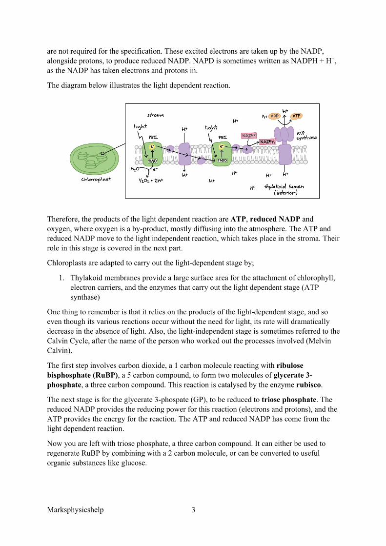
The diagram below illustrates the light dependent reaction.
Therefore, the products of the light dependent reaction are ATP, reduced NADP and oxygen, where oxygen is a by-product, mostly diffusing into the atmosphere. The ATP and reduced NADP move to the light independent reaction, which takes place in the stroma. Their role in this stage is covered in the next part.
Chloroplasts are adapted to carry out the light-dependent stage by;
1. Thylakoid membranes provide a large surface area for the attachment of chlorophyll, electron carriers, and the enzymes that carry out the light dependent stage (ATP synthase)
One thing to remember is that it relies on the products of the light-dependent stage, and so even though its various reactions occur without the need for light, its rate will dramatically decrease in the absence of light. Also, the light-independent stage is sometimes referred to the Calvin Cycle, after the name of the person who worked out the processes involved (Melvin Calvin).
The first step involves carbon dioxide, a 1 carbon molecule reacting with ribulose bisphosphate (RuBP), a 5 carbon compound, to form two molecules of glycerate 3-phosphate, a three carbon compound. This reaction is catalysed by the enzyme rubisco.
The next stage is for the glycerate 3-phospate (GP), to be reduced to triose phosphate. The reduced NADP provides the reducing power for this reaction (electrons and protons), and the ATP provides the energy for the reaction. The ATP and reduced NADP has come from the light dependent reaction.
Now you are left with triose phosphate, a three carbon compound. It can either be used to regenerate RuBP by combining with a 2 carbon molecule, or can be converted to useful organic substances like glucose.
Marksphysicshelp
4
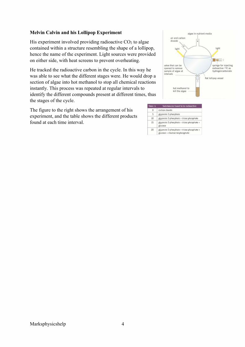
Melvin Calvin and his Lollipop Experiment
His experiment involved providing radioactive CO2 to algae contained within a structure resembling the shape of a lollipop, hence the name of the experiment. Light sources were provided on either side, with heat screens to prevent overheating.
He tracked the radioactive carbon in the cycle. In this way he was able to see what the different stages were. He would drop a section of algae into hot methanol to stop all chemical reactions instantly. This process was repeated at regular intervals to identify the different compounds present at different times, thus the stages of the cycle.
The figure to the right shows the arrangement of his experiment, and the table shows the different products found at each time interval.
Marksphysicshelp
5
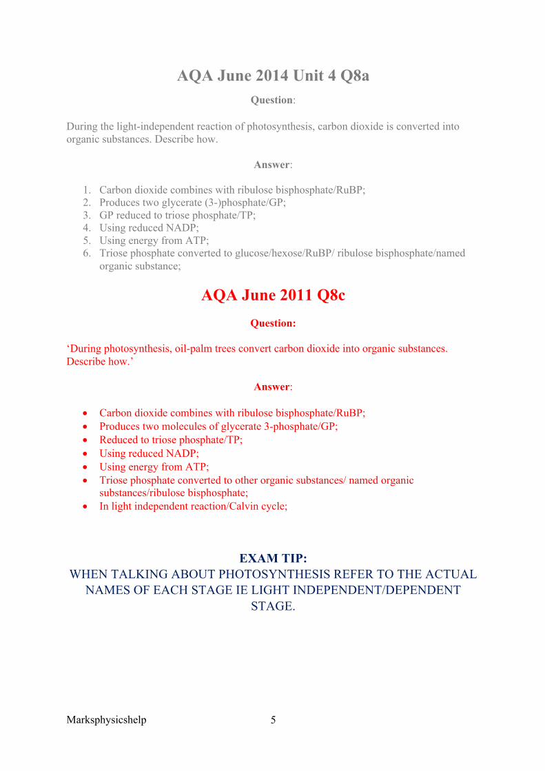
AQA June 2014 Unit 4 Q8a Question:
During the light-independent reaction of photosynthesis, carbon dioxide is converted into organic substances. Describe how.
Answer:
1. Carbon dioxide combines with ribulose bisphosphate/RuBP; 2. Produces two glycerate (3-)phosphate/GP; 3. GP reduced to triose phosphate/TP; 4. Using reduced NADP; 5. Using energy from ATP; 6. Triose phosphate converted to glucose/hexose/RuBP/ ribulose bisphosphate/named
organic substance;
AQA June 2011 Q8c Question:
‘During photosynthesis, oil-palm trees convert carbon dioxide into organic substances. Describe how.’
Answer:
• Carbon dioxide combines with ribulose bisphosphate/RuBP; • Produces two molecules of glycerate 3-phosphate/GP; • Reduced to triose phosphate/TP; • Using reduced NADP; • Using energy from ATP; • Triose phosphate converted to other organic substances/ named organic
substances/ribulose bisphosphate; • In light independent reaction/Calvin cycle;
EXAM TIP: WHEN TALKING ABOUT PHOTOSYNTHESIS REFER TO THE ACTUAL
NAMES OF EACH STAGE IE LIGHT INDEPENDENT/DEPENDENT STAGE.
Marksphysicshelp
6
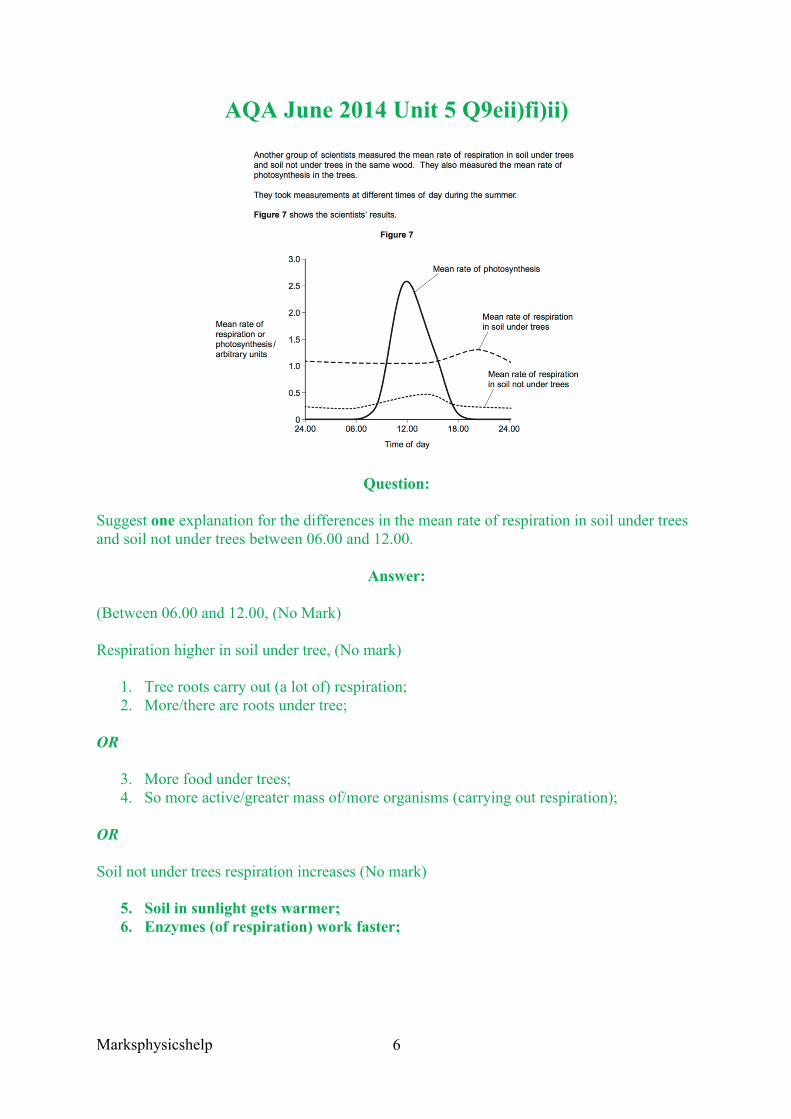
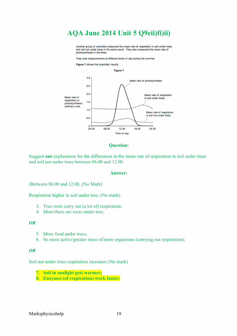
AQA June 2014 Unit 5 Q9eii)fi)ii)
Question:
Suggest one explanation for the differences in the mean rate of respiration in soil under trees and soil not under trees between 06.00 and 12.00.
Answer:
(Between 06.00 and 12.00, (No Mark)
Respiration higher in soil under tree, (No mark)
1. Tree roots carry out (a lot of) respiration; 2. More/there are roots under tree;
OR
3. More food under trees; 4. So more active/greater mass of/more organisms (carrying out respiration);
OR
Soil not under trees respiration increases (No mark)
5. Soil in sunlight gets warmer; 6. Enzymes (of respiration) work faster;
Marksphysicshelp
7
Question:
The scientists suggested that the rise in the mean rate of photosynthesis was the cause of the rise in the mean rate of respiration in soil under trees.
Suggest how the rise in the mean rate of photosynthesis could lead to the rise in the mean rate of respiration in soil under trees.
Answer:
1. Photosynthesis produces sugars; 2. Sugars moved to roots; 3. (Sugars) are used/required for respiration;
Question:
Suggest why there is a delay between the rise in the mean rate of photosynthesis and the rise in the mean rate of respiration.
Answer:
Takes time to move sugars to roots;
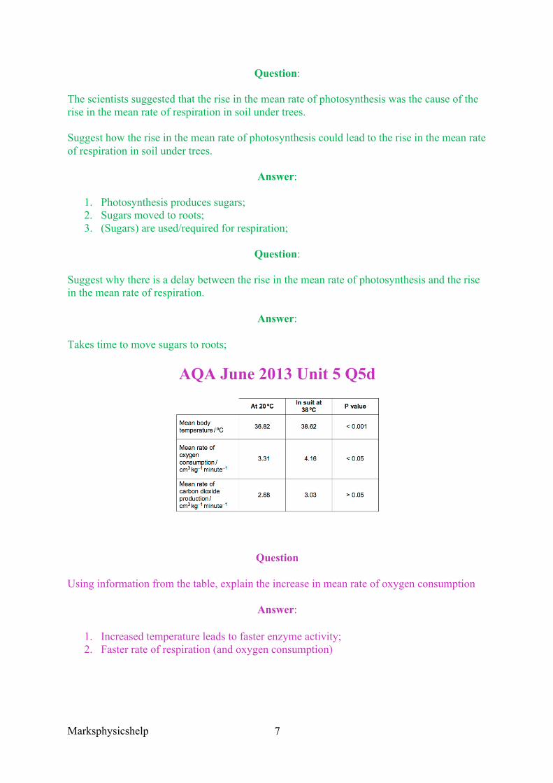
AQA June 2013 Unit 5 Q5d
Question
Using information from the table, explain the increase in mean rate of oxygen consumption
Answer:
1. Increased temperature leads to faster enzyme activity; 2. Faster rate of respiration (and oxygen consumption)
Marksphysicshelp
8
IF QUESTIONS TALK ABOUT PHOTOSYNTHESIS/RESPIRATION ALWAYS LINK TEMP TO
ENZYMES!!!!!!!!!!
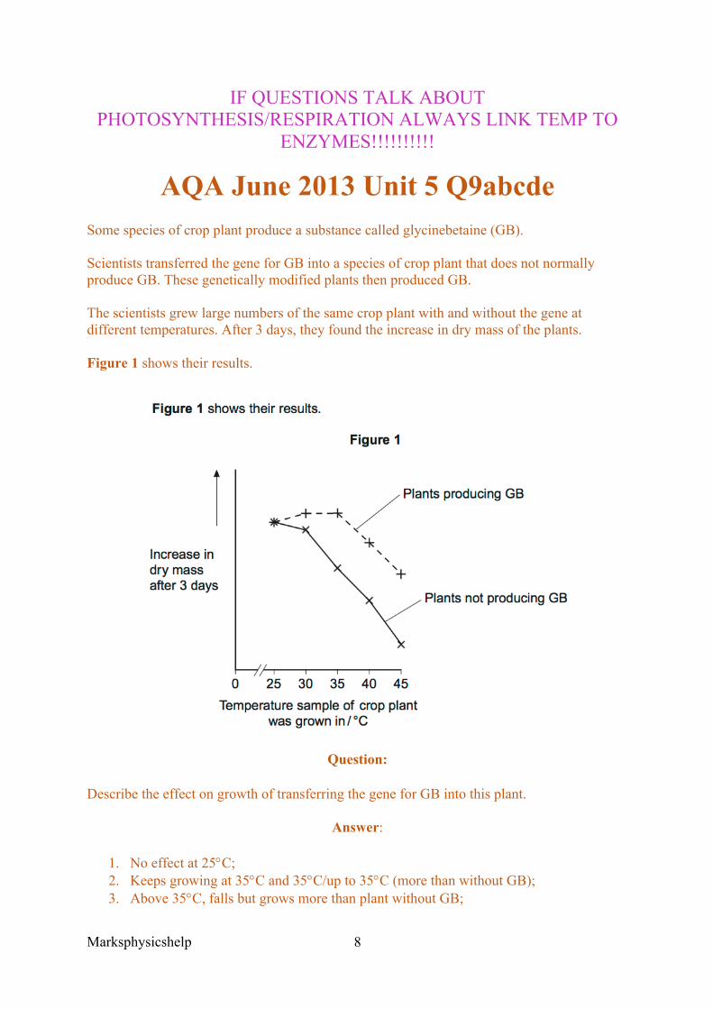
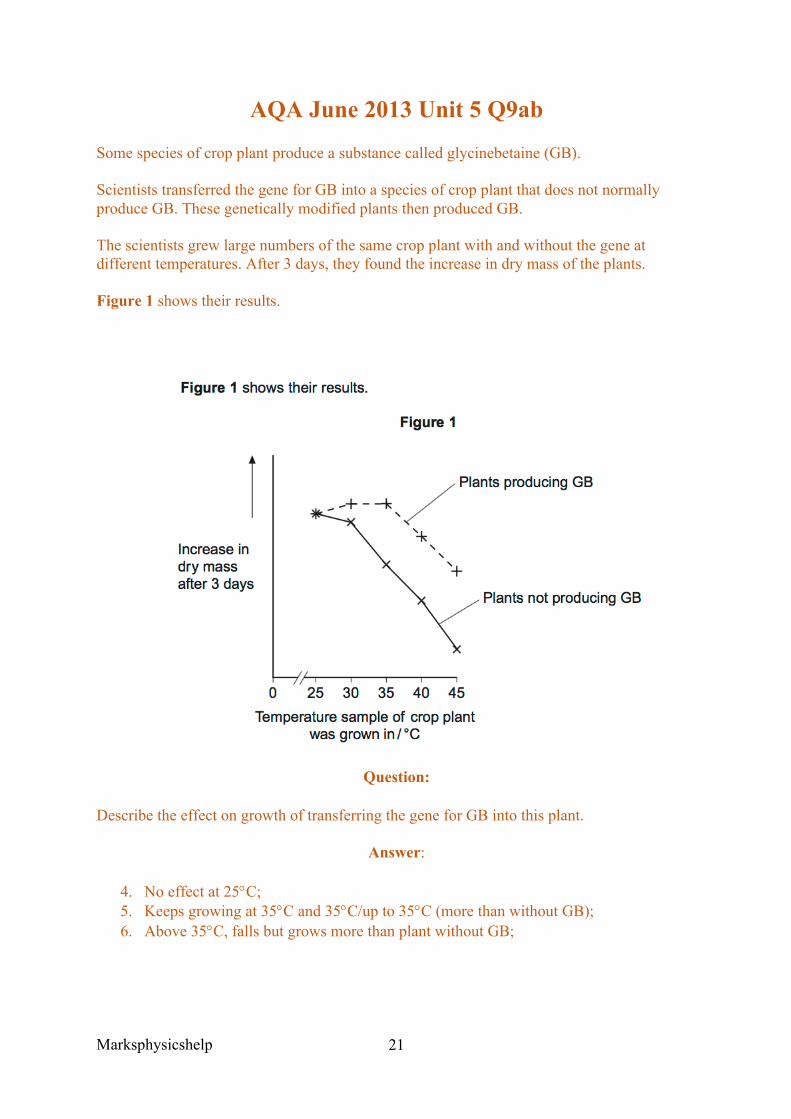
AQA June 2013 Unit 5 Q9abcde Some species of crop plant produce a substance called glycinebetaine (GB).
Scientists transferred the gene for GB into a species of crop plant that does not normally produce GB. These genetically modified plants then produced GB.
The scientists grew large numbers of the same crop plant with and without the gene at different temperatures. After 3 days, they found the increase in dry mass of the plants.
Figure 1 shows their results.
Question:
Describe the effect on growth of transferring the gene for GB into this plant.
Answer:
1. No effect at 25°C; 2. Keeps growing at 35°C and 35°C/up to 35°C (more than without GB); 3. Above 35°C, falls but grows more than plant without GB;
Marksphysicshelp
9
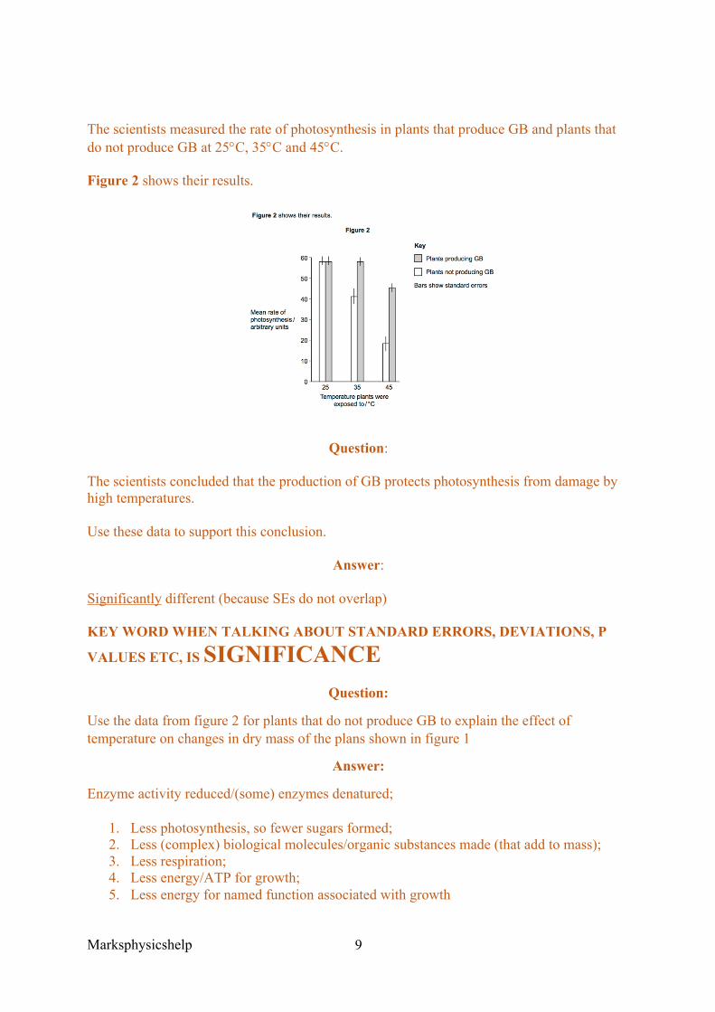
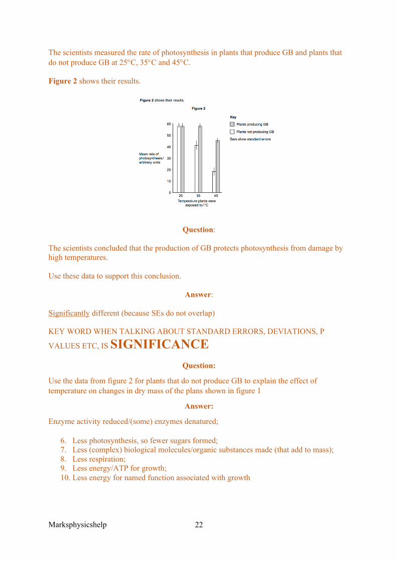
The scientists measured the rate of photosynthesis in plants that produce GB and plants that do not produce GB at 25°C, 35°C and 45°C.
Figure 2 shows their results.
Question:
The scientists concluded that the production of GB protects photosynthesis from damage by high temperatures.
Use these data to support this conclusion.
Answer:
Significantly different (because SEs do not overlap)
KEY WORD WHEN TALKING ABOUT STANDARD ERRORS, DEVIATIONS, P
VALUES ETC, IS SIGNIFICANCE Question:
Use the data from figure 2 for plants that do not produce GB to explain the effect of temperature on changes in dry mass of the plans shown in figure 1
Answer:
Enzyme activity reduced/(some) enzymes denatured;
1. Less photosynthesis, so fewer sugars formed; 2. Less (complex) biological molecules/organic substances made (that add to mass); 3. Less respiration; 4. Less energy/ATP for growth; 5. Less energy for named function associated with growth

Marksphysicshelp
10
Question:
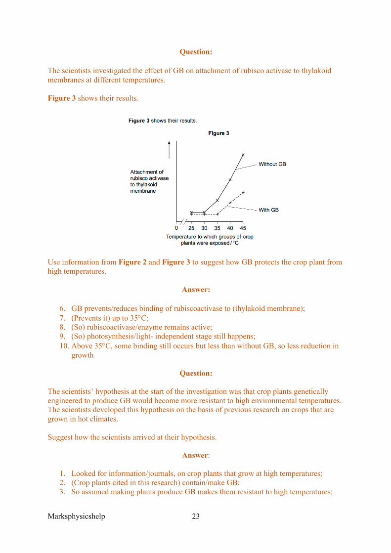
The scientists investigated the effect of GB on attachment of rubisco activase to thylakoid membranes at different temperatures.
Figure 3 shows their results.
Use information from Figure 2 and Figure 3 to suggest how GB protects the crop plant from high temperatures.
Answer:
1. GB prevents/reduces binding of rubiscoactivase to (thylakoid membrane); 2. (Prevents it) up to 35°C; 3. (So) rubiscoactivase/enzyme remains active; 4. (So) photosynthesis/light- independent stage still happens; 5. Above 35°C, some binding still occurs but less than without GB, so less reduction in
growth
Question:
The scientists’ hypothesis at the start of the investigation was that crop plants genetically engineered to produce GB would become more resistant to high environmental temperatures. The scientists developed this hypothesis on the basis of previous research on crops that are grown in hot climates.
Suggest how the scientists arrived at their hypothesis.
Answer:
1. Looked for information/journals, on crop plants that grow at high temperatures; 2. (Crop plants cited in this research) contain/make GB; 3. So assumed making plants produce GB makes them resistant to high temperatures;

Marksphysicshelp
11
AQA Jan 2012 Unit 4 Q4b
Scientists measured the concentration of carbon dioxide in the air in one part of the forest. They took measurements at different times of day and at two different heights above the ground. Their results are shown in the bar chart.
Question:
Use your knowledge of photosynthesis and respiration to explain the data in the bar chart.
Answer:
1. Correct explanation for differences between day and night e.g. photosynthesises only during the daytime / no photosynthesis/only respiration at night;
2. Net carbon dioxide uptake during the day/in light OR No carbon dioxide taken up at night/in dark / carbon dioxide released at night/in dark;
3. At ground level more respiration / in leaves more photosynthesis; 4. Carbon dioxide produced at ground level/carbon dioxide taken up in leaves (because
less sunlight passes through).
Marksphysicshelp
12
AQA Jan 2012 Unit 4 Q8a
Question:
ATP is useful in many biological processes. Explain why.
Answer:
1. Releases energy in small / manageable amounts; 2. (Broken down) in a one step / single bond broken; 3. Immediate energy compound/makes energy available rapidly; 4. Phosphorylates/adds phosphate; 5. Makes (phosphorylated substances) more reactive / lowers activation energy; 6. Reformed/made again;
Question:
Describe how ATP is made in mitochondria.
Answer:
1. Substrate level phosphorylation / ATP produced in Krebs cycle; 2. Krebs cycle/link reaction produces reduced coenzyme/reduced NAD/reduced FAD; 3. Electrons released from reduced /coenzymes/ NAD/FAD; 4. (Electrons) pass along carriers/through electron transport chain/through series of
redox reactions; 5. Energy released; 6. ADP/ADP + Pi; 7. Protons move into intermembrane space; 8. ATP synthase;
Question:
Plants produce ATP in their chloroplasts during photosynthesis. They also produce ATP during respiration. Explain why it is important for plants to produce ATP during respiration in addition to during photosynthesis.
Answer:
1. In the dark no ATP production in photosynthesis; 2. Some tissues unable to photosynthesise/produce ATP; 3. ATP cannot be moved from cell to cell/stored; 4. Plant uses more ATP than produced in photosynthesis; 5. ATP for active transport; 6. ATP for synthesis (of named substance)

Marksphysicshelp
13
Marksphysicshelp
14
3.5.2 Respiration
Content
• Respiration produces ATP. • Glycolysis is the first stage of anaerobic and aerobic respiration. It occurs in the
cytoplasm and is an anaerobic process. • Glycolysis involves the following stages
o Phosphorylation of glucose to glucose phosphate, using ATP o Production of triose phosphate o Oxidation of triose phosphate to pyruvate with a net gain of ATP and reduced
NAD. • If respiration is only anaerobic, pyruvate can be converted to ethanol or lactate using
reduced NAD. The oxidised NAD produced in this way can be used in further glycolysis.
• If respiration is aerobic, pyruvate from glycolysis enters the mitochondrial matrix by active transport.
• Aerobic respiration in such detail as to show that: o Pyruvate is oxidised to acetate, producing reduced NAD in the process o Acetate combines with coenzyme A in the link reaction to produce
acetylcoenzyme A o Acetylcoenzyme A reacts with a four-carbon molecule, releasing o Coenzyme A and producing a six-carbon molecule that enters the Krebs cycle o In a series of oxidation-reduction reactions, the Krebs cycle generates reduced
coenzymes and ATP by substrate-level phosphorylation, and carbon dioxide is lost
• Synthesis of ATP by oxidative phosphorylation is associated with the transfer of electrons down the electron transfer chain and passage of protons across inner mitochondrial membranes and is catalysed by ATP synthase embedded in these membranes (chemiosomotic theory)
• Other respiratory substrates include the breakdown products of lipids and amino acids, which enter the Krebs cycle.
Opportunities for Skills Development
• Students could use a redox indicator to investigate dehydrogenase activity.
As an overview, respiration is the process by which ATP is produced, using hexose sugars like glucose, or using lipids/proteins.
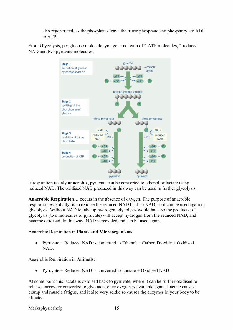
Glycolysis is the first stage of both anaerobic and aerobic respiration. It occurs in the cytoplasm, and as the name suggests does not require the presence of oxygen.
1. The first stage required glucose to be made more reactive, so by the addition of two phosphates (phosphorylation of glucose). This process required two ATP molecules.
2. Glucose is then split into two molecules of the 3 carbon triose phosphate. 3. Triose phosphate is oxidised to pyruvate. This results in a loss of hydrogen which
goes to reduce NAD and form reduced NAD. In this process 4 ATP molecules are
Marksphysicshelp
15
also regenerated, as the phosphates leave the triose phosphate and phosphorylate ADP to ATP.
From Glycolysis, per glucose molecule, you get a net gain of 2 ATP molecules, 2 reduced NAD and two pyruvate molecules.
If respiration is only anaerobic, pyruvate can be converted to ethanol or lactate using reduced NAD. The oxidised NAD produced in this way can be used in further glycolysis.
Anaerobic Respiration… occurs in the absence of oxygen. The purpose of anaerobic respiration essentially, is to oxidise the reduced NAD back to NAD, so it can be used again in glycolysis. Without NAD to take up hydrogen, glycolysis would halt. So the products of glycolysis (two molecules of pyruvate) will accept hydrogen from the reduced NAD, and become oxidised. In this way, NAD is recycled and can be used again.
Anaerobic Respiration in Plants and Microorganisms:
• Pyruvate + Reduced NAD is converted to Ethanol + Carbon Dioxide + Oxidised NAD.
Anaerobic Respiration in Animals:
• Pyruvate + Reduced NAD is converted to Lactate + Oxidised NAD.
At some point this lactate is oxidised back to pyruvate, where it can be further oxidised to release energy, or converted to glycogen, once oxygen is available again. Lactate causes cramp and muscle fatigue, and it also very acidic so causes the enzymes in your body to be affected.
Marksphysicshelp
16
However in the presence of oxygen, aerobic respiration will occur. In this process pyruvate is actively transported into the matrix of the mitochondria, the site of aerobic respiration. Aerobic respiration involves three main stages, the link reaction, Krebs cycle and oxidative phosphorylation.
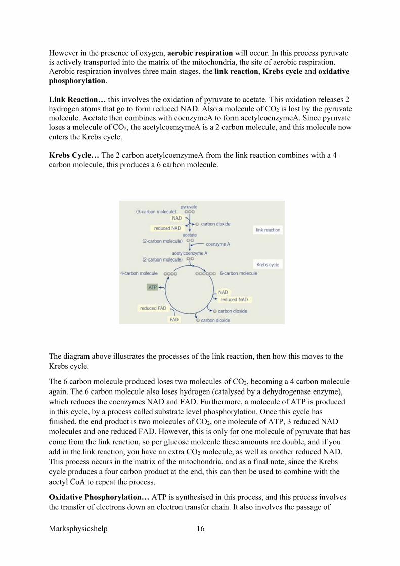
Link Reaction… this involves the oxidation of pyruvate to acetate. This oxidation releases 2 hydrogen atoms that go to form reduced NAD. Also a molecule of CO2 is lost by the pyruvate molecule. Acetate then combines with coenzymeA to form acetylcoenzymeA. Since pyruvate loses a molecule of CO2, the acetylcoenzymeA is a 2 carbon molecule, and this molecule now enters the Krebs cycle.
Krebs Cycle… The 2 carbon acetylcoenzymeA from the link reaction combines with a 4 carbon molecule, this produces a 6 carbon molecule.
The diagram above illustrates the processes of the link reaction, then how this moves to the Krebs cycle.
The 6 carbon molecule produced loses two molecules of CO2, becoming a 4 carbon molecule again. The 6 carbon molecule also loses hydrogen (catalysed by a dehydrogenase enzyme), which reduces the coenzymes NAD and FAD. Furthermore, a molecule of ATP is produced in this cycle, by a process called substrate level phosphorylation. Once this cycle has finished, the end product is two molecules of CO2, one molecule of ATP, 3 reduced NAD molecules and one reduced FAD. However, this is only for one molecule of pyruvate that has come from the link reaction, so per glucose molecule these amounts are double, and if you add in the link reaction, you have an extra CO2 molecule, as well as another reduced NAD. This process occurs in the matrix of the mitochondria, and as a final note, since the Krebs cycle produces a four carbon product at the end, this can then be used to combine with the acetyl CoA to repeat the process.
Oxidative Phosphorylation… ATP is synthesised in this process, and this process involves the transfer of electrons down an electron transfer chain. It also involves the passage of
Marksphysicshelp
17
protons moving across the inner mitochondrial membranes and is catalysed by ATP synthase embedded in these membranes.
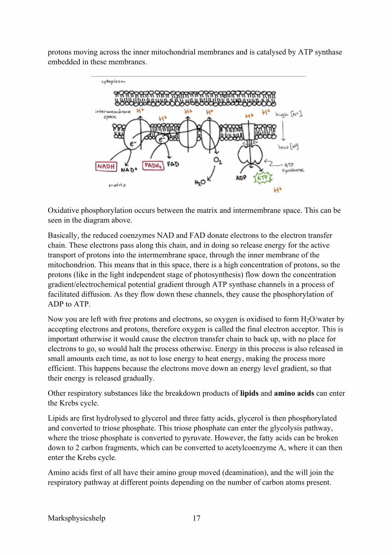
Oxidative phosphorylation occurs between the matrix and intermembrane space. This can be seen in the diagram above.
Basically, the reduced coenzymes NAD and FAD donate electrons to the electron transfer chain. These electrons pass along this chain, and in doing so release energy for the active transport of protons into the intermembrane space, through the inner membrane of the mitochondrion. This means that in this space, there is a high concentration of protons, so the protons (like in the light independent stage of photosynthesis) flow down the concentration gradient/electrochemical potential gradient through ATP synthase channels in a process of facilitated diffusion. As they flow down these channels, they cause the phosphorylation of ADP to ATP.
Now you are left with free protons and electrons, so oxygen is oxidised to form H2O/water by accepting electrons and protons, therefore oxygen is called the final electron acceptor. This is important otherwise it would cause the electron transfer chain to back up, with no place for electrons to go, so would halt the process otherwise. Energy in this process is also released in small amounts each time, as not to lose energy to heat energy, making the process more efficient. This happens because the electrons move down an energy level gradient, so that their energy is released gradually.
Other respiratory substances like the breakdown products of lipids and amino acids can enter the Krebs cycle.
Lipids are first hydrolysed to glycerol and three fatty acids, glycerol is then phosphorylated and converted to triose phosphate. This triose phosphate can enter the glycolysis pathway, where the triose phosphate is converted to pyruvate. However, the fatty acids can be broken down to 2 carbon fragments, which can be converted to acetylcoenzyme A, where it can then enter the Krebs cycle.
Amino acids first of all have their amino group moved (deamination), and the will join the respiratory pathway at different points depending on the number of carbon atoms present.
Marksphysicshelp
18
AQA June 2015 Unit 4 Q8c Question:
‘Large areas of tropical forest are still found on some Caribbean islands. The concentration of carbon dioxide in the air of these forests changes over a period of 24 hours and at different heights above ground.
Use your knowledge of photosynthesis and respiration to describe and explain how the concentration of carbon dioxide in the air changes:
• over a period of 24 hours • different heights above ground’
Answer:
1. High concentration of/increase in carbon dioxide linked with respiration at night/in darkness;
2. No photosynthesis in dark/night / photosynthesis only in light/day; 3. In light net uptake of carbon dioxide / use more carbon dioxide than produced / (rate
of) photosynthesis greater than rate of respiration; 4. Decrease in carbon dioxide concentration with height; 5. (At ground level) less photosynthesis / less photosynthesising tissue / more respiration
/ more micro-organisms / / micro-organisms produce carbon dioxide;
Marksphysicshelp
19
AQA June 2014 Unit 5 Q9eii)fi)ii)
Question:
Suggest one explanation for the differences in the mean rate of respiration in soil under trees and soil not under trees between 06.00 and 12.00.
Answer:
(Between 06.00 and 12.00, (No Mark)
Respiration higher in soil under tree, (No mark)
3. Tree roots carry out (a lot of) respiration; 4. More/there are roots under tree;
OR
5. More food under trees; 6. So more active/greater mass of/more organisms (carrying out respiration);
OR
Soil not under trees respiration increases (No mark)
7. Soil in sunlight gets warmer; 8. Enzymes (of respiration) work faster;
Marksphysicshelp
20
Question:
The scientists suggested that the rise in the mean rate of photosynthesis was the cause of the rise in the mean rate of respiration in soil under trees.
Suggest how the rise in the mean rate of photosynthesis could lead to the rise in the mean rate of respiration in soil under trees.
Answer:
4. Photosynthesis produces sugars; 5. Sugars moved to roots; 6. (Sugars) are used/required for respiration;
Question:
Suggest why there is a delay between the rise in the mean rate of photosynthesis and the rise in the mean rate of respiration.
Answer:
Takes time to move sugars to roots;
Marksphysicshelp
21
AQA June 2013 Unit 5 Q9ab
Some species of crop plant produce a substance called glycinebetaine (GB).
Scientists transferred the gene for GB into a species of crop plant that does not normally produce GB. These genetically modified plants then produced GB.
The scientists grew large numbers of the same crop plant with and without the gene at different temperatures. After 3 days, they found the increase in dry mass of the plants.
Figure 1 shows their results.
Question:
Describe the effect on growth of transferring the gene for GB into this plant.
Answer:
4. No effect at 25°C; 5. Keeps growing at 35°C and 35°C/up to 35°C (more than without GB); 6. Above 35°C, falls but grows more than plant without GB;
Marksphysicshelp
22
The scientists measured the rate of photosynthesis in plants that produce GB and plants that do not produce GB at 25°C, 35°C and 45°C.
Figure 2 shows their results.
Question:
The scientists concluded that the production of GB protects photosynthesis from damage by high temperatures.
Use these data to support this conclusion.
Answer:
Significantly different (because SEs do not overlap)
KEY WORD WHEN TALKING ABOUT STANDARD ERRORS, DEVIATIONS, P
VALUES ETC, IS SIGNIFICANCE Question:
Use the data from figure 2 for plants that do not produce GB to explain the effect of temperature on changes in dry mass of the plans shown in figure 1
Answer:
Enzyme activity reduced/(some) enzymes denatured;
6. Less photosynthesis, so fewer sugars formed; 7. Less (complex) biological molecules/organic substances made (that add to mass); 8. Less respiration; 9. Less energy/ATP for growth; 10. Less energy for named function associated with growth
Marksphysicshelp
23
Question:
The scientists investigated the effect of GB on attachment of rubisco activase to thylakoid membranes at different temperatures.
Figure 3 shows their results.
Use information from Figure 2 and Figure 3 to suggest how GB protects the crop plant from high temperatures.
Answer:
6. GB prevents/reduces binding of rubiscoactivase to (thylakoid membrane); 7. (Prevents it) up to 35°C; 8. (So) rubiscoactivase/enzyme remains active; 9. (So) photosynthesis/light- independent stage still happens; 10. Above 35°C, some binding still occurs but less than without GB, so less reduction in
growth
Question:
The scientists’ hypothesis at the start of the investigation was that crop plants genetically engineered to produce GB would become more resistant to high environmental temperatures. The scientists developed this hypothesis on the basis of previous research on crops that are grown in hot climates.
Suggest how the scientists arrived at their hypothesis.
Answer:
1. Looked for information/journals, on crop plants that grow at high temperatures; 2. (Crop plants cited in this research) contain/make GB; 3. So assumed making plants produce GB makes them resistant to high temperatures;
Marksphysicshelp
24
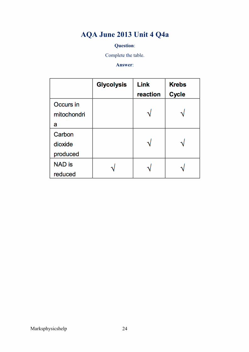
AQA June 2013 Unit 4 Q4a Question:
Complete the table.
Answer:
Marksphysicshelp
25
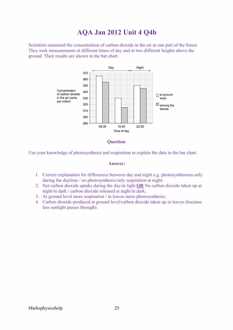
AQA Jan 2012 Unit 4 Q4b
Scientists measured the concentration of carbon dioxide in the air in one part of the forest. They took measurements at different times of day and at two different heights above the ground. Their results are shown in the bar chart.
Question:
Use your knowledge of photosynthesis and respiration to explain the data in the bar chart.
Answer:
1. Correct explanation for differences between day and night e.g. photosynthesises only during the daytime / no photosynthesis/only respiration at night;
2. Net carbon dioxide uptake during the day/in light OR No carbon dioxide taken up at night/in dark / carbon dioxide released at night/in dark;
3. At ground level more respiration / in leaves more photosynthesis; 4. Carbon dioxide produced at ground level/carbon dioxide taken up in leaves (because
less sunlight passes through).
Marksphysicshelp
26
3.5.3 Energy and ecosystems
Content
• In any ecosystem, plants synthesise organic compounds from atmospheric, or aquatic, carbon dioxide.
• Most of the sugars synthesised by plants are used by the plant as respiratory substrates. The rest are used to make other groups of biological molecules. These biological molecules form the biomass of the plants.
• Biomass can be measured in terms of mass of carbon or dry mass of tissue per given area. The chemical energy store in dry biomass can be estimated using calorimetry.
• Gross primary production (GPP) is the chemical energy store in plant biomass, in a given area or volume.
• Net primary production (NPP) is the chemical energy store in plant biomass after respiratory losses to the environment have been taken into account,
• i.e. NPP = GPP – R • where GPP represents gross production and R represents respiratory losses to the
environment • This net primary production is available for plant growth and reproduction. It is also
available to other trophic levels in the ecosystem, such as herbivores and decomposers.
• The net production of consumers (N), such as animals, can be calculated as: • N = I - (F+R) • Where I represents the chemical energy store in ingested food, F represents the
chemical energy lost to the environment in faeces and urine and R represents the respiratory losses to the environment.
• Primary and secondary productivity is the rate of primary or secondary production, respectively. It is measured as biomass in a given area in a given time eg kJ ha-1year-1.
• Students should be able to: appreciate the ways in which production is affected by farming practices designed to increase the efficiency of energy transfer by:
• Simplifying food webs to reduce energy losses to non-human food chains • Reducing respiratory losses within a human food chain.
In any ecosystem, plants synthesise organic compounds from atmospheric, or aquatic, carbon dioxide. These organic compounds include sugars, most of which are used by the plant as respiratory substrates. The rest are used to make other biological molecules; these biological molecules form the biomass of the plant.
Organisms living within ecosystems can be divided into three groups, producers, consumer and Saprobionts.
• Producers manufacture their organic substances using light energy, water, carbon dioxide and mineral ions.
• Consumers obtain their energy by feeding on other organisms as opposed to getting it directly from sunlight. Those feeding directly on producers are called primary consumers, then secondary consumers etc.
Marksphysicshelp
27
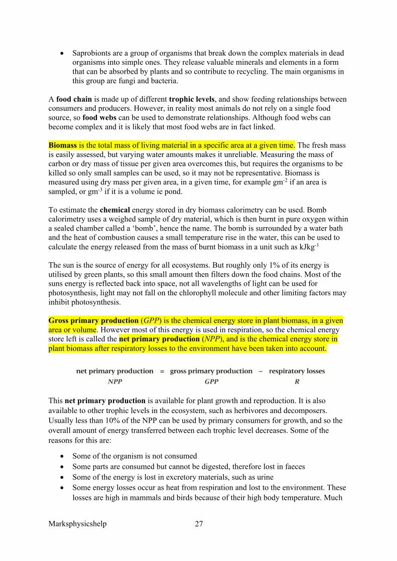
• Saprobionts are a group of organisms that break down the complex materials in dead organisms into simple ones. They release valuable minerals and elements in a form that can be absorbed by plants and so contribute to recycling. The main organisms in this group are fungi and bacteria.
A food chain is made up of different trophic levels, and show feeding relationships between consumers and producers. However, in reality most animals do not rely on a single food source, so food webs can be used to demonstrate relationships. Although food webs can become complex and it is likely that most food webs are in fact linked.
Biomass is the total mass of living material in a specific area at a given time. The fresh mass is easily assessed, but varying water amounts makes it unreliable. Measuring the mass of carbon or dry mass of tissue per given area overcomes this, but requires the organisms to be killed so only small samples can be used, so it may not be representative. Biomass is measured using dry mass per given area, in a given time, for example gm-2 if an area is sampled, or gm-3 if it is a volume ie pond.
To estimate the chemical energy stored in dry biomass calorimetry can be used. Bomb calorimetry uses a weighed sample of dry material, which is then burnt in pure oxygen within a sealed chamber called a ‘bomb’, hence the name. The bomb is surrounded by a water bath and the heat of combustion causes a small temperature rise in the water, this can be used to calculate the energy released from the mass of burnt biomass in a unit such as kJkg-1
The sun is the source of energy for all ecosystems. But roughly only 1% of its energy is utilised by green plants, so this small amount then filters down the food chains. Most of the suns energy is reflected back into space, not all wavelengths of light can be used for photosynthesis, light may not fall on the chlorophyll molecule and other limiting factors may inhibit photosynthesis.
Gross primary production (GPP) is the chemical energy store in plant biomass, in a given area or volume. However most of this energy is used in respiration, so the chemical energy store left is called the net primary production (NPP), and is the chemical energy store in plant biomass after respiratory losses to the environment have been taken into account.
This net primary production is available for plant growth and reproduction. It is also available to other trophic levels in the ecosystem, such as herbivores and decomposers. Usually less than 10% of the NPP can be used by primary consumers for growth, and so the overall amount of energy transferred between each trophic level decreases. Some of the reasons for this are:
• Some of the organism is not consumed • Some parts are consumed but cannot be digested, therefore lost in faeces • Some of the energy is lost in excretory materials, such as urine • Some energy losses occur as heat from respiration and lost to the environment. These
losses are high in mammals and birds because of their high body temperature. Much
Marksphysicshelp
28
energy is needed to maintain their body temperature when heat is constantly being lost to the environment.
The net production of consumers can be calculated as:
N = NPP, I = Chemical energy store of ingested food, F = The energy lost in faeces and urine, R = The energy lost in respiration.
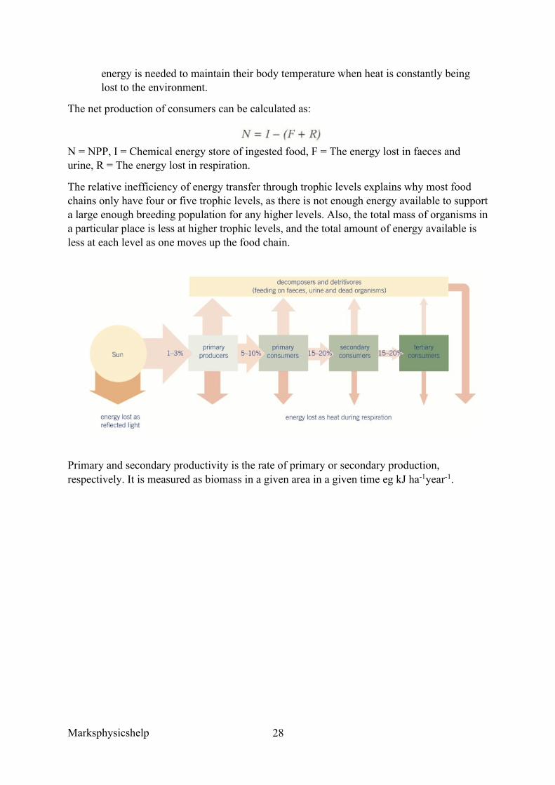
The relative inefficiency of energy transfer through trophic levels explains why most food chains only have four or five trophic levels, as there is not enough energy available to support a large enough breeding population for any higher levels. Also, the total mass of organisms in a particular place is less at higher trophic levels, and the total amount of energy available is less at each level as one moves up the food chain.
Primary and secondary productivity is the rate of primary or secondary production, respectively. It is measured as biomass in a given area in a given time eg kJ ha-1year-1.
Marksphysicshelp
29
AQA Jan 2012 Unit 4
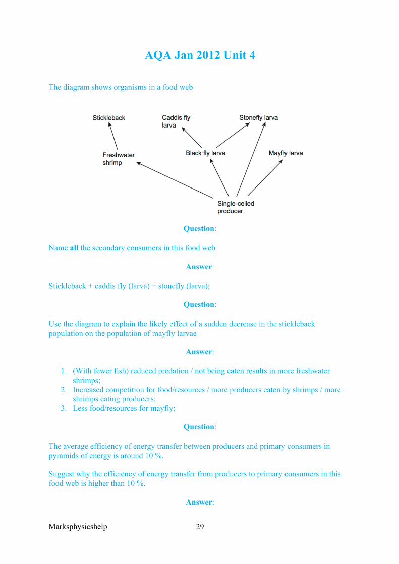
The diagram shows organisms in a food web
Question:
Name all the secondary consumers in this food web
Answer:
Stickleback + caddis fly (larva) + stonefly (larva);
Question:
Use the diagram to explain the likely effect of a sudden decrease in the stickleback population on the population of mayfly larvae
Answer:
1. (With fewer fish) reduced predation / not being eaten results in more freshwater shrimps;
2. Increased competition for food/resources / more producers eaten by shrimps / more shrimps eating producers;
3. Less food/resources for mayfly;
Question:
The average efficiency of energy transfer between producers and primary consumers in pyramids of energy is around 10 %.
Suggest why the efficiency of energy transfer from producers to primary consumers in this food web is higher than 10 %.
Answer:
Marksphysicshelp
30
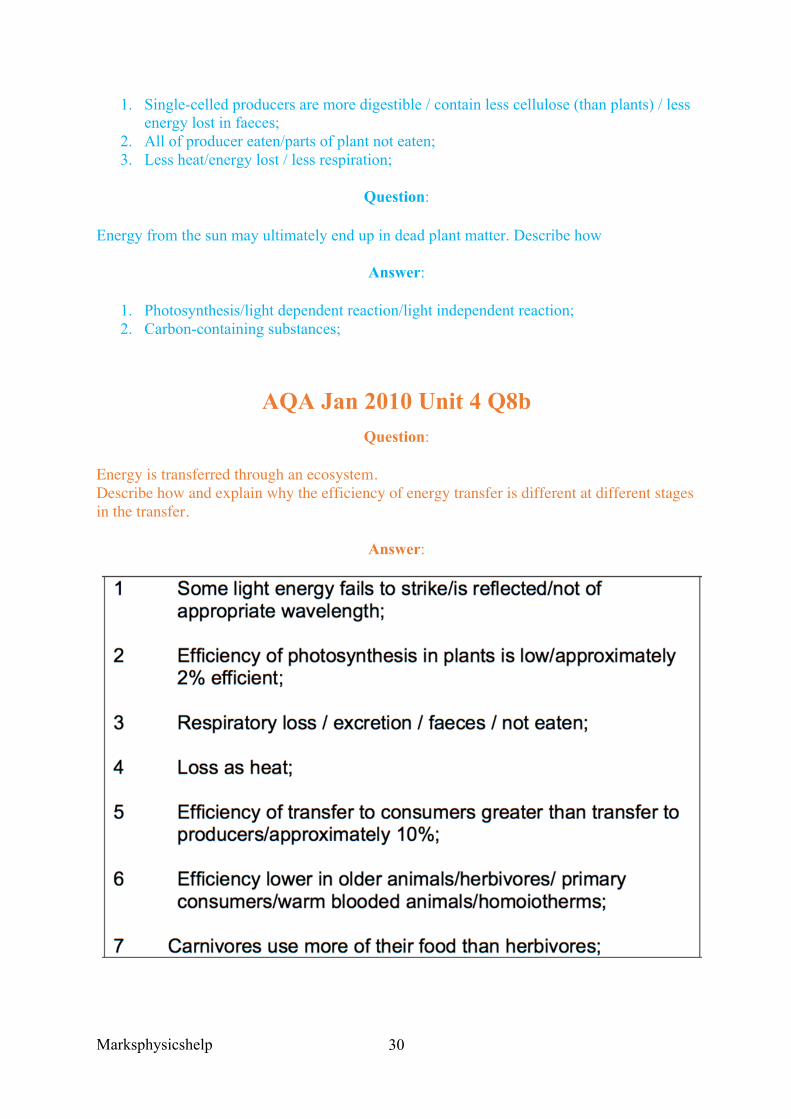
1. Single-celled producers are more digestible / contain less cellulose (than plants) / less energy lost in faeces;
2. All of producer eaten/parts of plant not eaten; 3. Less heat/energy lost / less respiration;
Question:
Energy from the sun may ultimately end up in dead plant matter. Describe how
Answer:
1. Photosynthesis/light dependent reaction/light independent reaction; 2. Carbon-containing substances;
AQA Jan 2010 Unit 4 Q8b Question:
Energy is transferred through an ecosystem. Describe how and explain why the efficiency of energy transfer is different at different stages in the transfer.
Answer:
Marksphysicshelp
31
AQA Jan 2012 Unit 4 Q7cii
Question:
The efficiency of conversion of food to biomass is lower at 0°C than it is at 20°C. Suggest an explanation for the lower efficiency.
Answer:
1. Will lose more heat / not as much energy used to maintain body temperature; 2. Heat resulting from respiration/more respiration; 3. More food used in respiration;

Marksphysicshelp
32
Marksphysicshelp
33
3.5.4 Nutrient cycles
Content
• Nutrients are recycled within natural ecosystems, exemplified by the nitrogen cycle and the phosphorus cycle.
• Microorganisms play a vital role in recycling chemical elements such as phosphorus and nitrogen.
• The role of Saprobionts in decomposition. • The role of mycorrhizae in facilitating the uptake of water and inorganic ions by
plants. • The role of bacteria in the nitrogen cycle in sufficient detail to illustrate the processes
of saprobiotic nutrition, ammonification, nitrification, nitrogen fixation and denitrification.
• (The names of individual species of bacteria are not required) • The use of natural and artificial fertilisers to replace the nitrates and phosphates lost
by harvesting plants and removing livestock. • The environmental issues arising from the use of fertilisers including leaching and
eutrophication
Two main nutrient cycles are the nitrogen cycle and phosphorous cycle. Another example is the carbon cycle; however, this is not part of the course. Nitrogen is an essential part of every living organism, forming part of all proteins, nucleic acids and their products. Phosphorous on the other hand is also essential to life, part of ATP, nucleic acids and phospholipids. Nitrogen is readily accessible in the atmosphere to the biotic component of an ecosystem, however phosphorous is locked up in sediment and is only made available to organisms by slow physical processes.
Microorganisms play a vital role in recycling chemical elements such as phosphorous and nitrogen. Saprobionts are microorganisms that have this role of decomposition and recycling. They break down the complex molecules in producers and consumers that contain the locked up nutrients.
The Phosphorous Cycle recycles phosphates in ecosystems. The phosphorous cycle, unlike the carbon and nitrogen cycle, lacks a gaseous stage as most phosphorous is locked up in mineral form and dissolved in oceans, lakes and soils. Most sedimentary rock deposits and fossils contain phosphates, originating from the seas. Weathering and erosion of these rocks allows phosphate ions to become dissolved and so available for absorption by plants. These dissolved phosphate ions can also be harvested by manufacturers of fertilisers. The phosphate ions taken up by plants are then passed on to consumer animals. These phosphates in the animals may be excreted out, or upon death of both plants and animals, certain bacteria and fungi decomposers break them down. This releases phosphate ions back into the water and soil. However a lot of the phosphate ions will remain in the bones or shells of animals, and these are very slow to breakdown. Thus the ions will be locked up by fossilisation or locked in sedimentary rocks awaiting erosion and weathering to begin the process again.
Marksphysicshelp
34
Mycorrhizae are associations between certain types of fungi, and the roots of the vast majority of plants. They have a role in facilitating the uptake of water and inorganic ions by plants. Essentially the fungi act like extensions of the plant’s root system and increase the total surface area for absorbing water and minerals. The mycorrhizae as a whole act like a sponge, where it holds the minerals and water close to the roots. It enables plants to much more quickly take up inorganic ions and water, so mycorrhizae also play a huge role in increasing uptake of phosphate ions. There is a mutualistic relationship between plants and fungi, as the plant receives better water and inorganic ion uptake, and on the other hand the fungus receives organic compounds like sugars and amino acids from the plant.
The Nitrogen Cycle includes four main stages, ammonification, nitrification, nitrogen fixation and denitrification. Saprobionts also play a big role in the nitrogen cycle.
• Nitrogen Fixation involves turning nitrogen to nitrogen containing compounds. Nitrogen makes up around 79% of the atmosphere, however very few organisms are able to use atmospheric nitrogen directly, or nitrogen that is dissolved in water. This is mainly due to nitrogen gas being chemically unreactive. Before plants and animals have access to nitrogen, it must first be converted to absorbable nitrogen containing compounds. This conversion is called nitrogen fixation. Naturally, some nitrogen fixation occurs when lighting strikes, as lightning provides energy to oxidise nitrogen to nitrogen oxides. These gases then dissolve in rain to form dilute nitric acid. However most nitrogen fixation is done by nitrogen-fixing bacteria. These bacteria are either free living, or live mutualistically in root nodules on plants like peas and beans. Nitrogen fixing bacteria contain nitrogenase, which is an enzyme that enables them to reduce nitrogen to ammonia or ammonia compounds. The reaction is shown below and is catalysed by nitrogenase. Nitrogen fixation can also be done artificially by industry in the production of nitrogen fertilisers.
• Ammonification is the process by which ammonia is produced in ecosystems from nitrogen-containing compounds. These compounds occur in faeces, urea (breakdown of excess amino acids), proteins, nucleic acids, dead organisms etc. The process of ammonification is predominantly carried out by saprobiontic microorganisms, mainly fungi and bacteria. They break down dead organisms, and feed on faeces to release ammonia which forms ammonium ions in the soil. In this process, nitrogen returns to the abiotic component of the ecosystem.
• Nitrification is where ammonium ions in the soil are oxidised to nitrites and nitrates by free-living soil microorganisms called nitrifying bacteria. Since oxidation is occurring, the nitrifying bacteria require oxygen, so it happens most rapidly in well aerated soils or well oxygenated bodies of water. The process involved ammonia being oxidised to nitrite (NO2-), and then nitrite is oxidised again to nitrate (NO3-). Also, since this is an oxidation reaction it will release energy. The nitrate ions produced can be taken up by plants and used to make proteins, consumers will then obtain their nitrogen in the form of proteins when they eat plants or other animals.
• Denitrification completes the cycle. Anaerobic denitrifying bacteria live in conditions of low oxygen content so reverse the nitrifying process, converting nitrates
Marksphysicshelp
35
to nitrites, then nitrites to nitrogen gas. This leads to loss of nitrogen from biotic components of an ecosystem. Denitrification occurs in soils that may have become waterlogged, as this causes a low oxygen concentration. This also means there are less aerobic nitrifying and nitrogen-fixing bacteria found.
Fertilisers are used in industry, both natural and artificial, to replace lost nitrates and phosphates in harvesting plants and removing livestock. The vast population is requiring larger crop yields, and more animals to be farmed on very concentrated pieces of land. This intensive food production puts large amounts of stress on the soil, as the large majority of mineral ions are being used by the crops being grown. These ions are then removed, whereas usually these crops and animals would die in the area and return the ions, a deficit now forms. The reduction of mineral ions will become the limiting factor for the growth of plants unless they are replaced. They are replaced by using natural fertilisers, consisting of the dead and decaying remains of plants and animals, as well as animal waste such as manure. They can also be replenished using artificial fertilisers, which are mined from rocks and deposits, then converted into an appropriate balance of minerals for a particular crop. Overall a combination of natural and artificial fertilisers seems to give the best long-term productivity. However, there seems to be a point in which further increase in the quantity of fertiliser no longer results in increased productivity.
Fertilisers are essential to plant growth. For example, nitrogen is essential in amino acids, ATP and nucleotides in DNA, which are all required for plant growth. Plants that have access to vast quantities of nitrate ions typically develop earlier, grow taller and have a greater leaf area. This results in an increased rate of photosynthesis, thus crop productivity is improved.
Despite the fact that fertilisers have increased crop productivity tremendously, there are some environmental issues that arise from the use of fertilisers like leaching and eutrophication. There are there main issues,
1. Reduced species diversity 2. Leaching 3. Eutrophication
Reduced species diversity is as a result of species like grasses and nettles that favour nitrogen-rich soils have been out-competing many other species that die as a result.
Leaching is where nutrients are removed from the soil, as rainfall will dissolve soluble nutrients like nitrate ions, carrying them into much deeper soil. They will eventually move too deep and out of reach of plants, moving into groundwater stores that will take them to streams and rivers, then possibly ending up in freshwater lakes. These lakes provide a source of drinking water for humans, and too high nitrate ion concentrations can prevent efficient oxygen transport in babies, and a link has been found to stomach cancer. This leaching also then causes eutrophication.
Eutrophication occurs where organic material, or inorganic nutrients, especially nitrates or phosphates enter a freshwater habitat. This can be as a result of pollution by sewage or agricultural runoff containing fertiliser. The process is as follows;
Marksphysicshelp
36
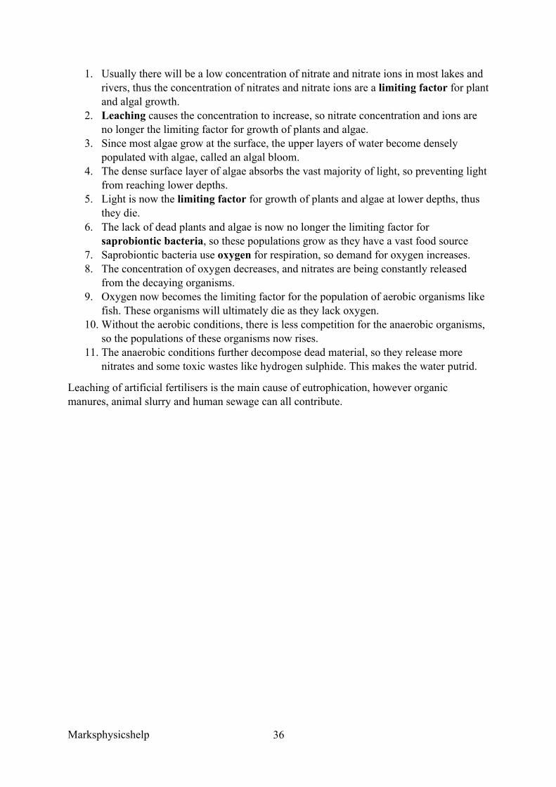
1. Usually there will be a low concentration of nitrate and nitrate ions in most lakes and rivers, thus the concentration of nitrates and nitrate ions are a limiting factor for plant and algal growth.
2. Leaching causes the concentration to increase, so nitrate concentration and ions are no longer the limiting factor for growth of plants and algae.
3. Since most algae grow at the surface, the upper layers of water become densely populated with algae, called an algal bloom.
4. The dense surface layer of algae absorbs the vast majority of light, so preventing light from reaching lower depths.
5. Light is now the limiting factor for growth of plants and algae at lower depths, thus they die.
6. The lack of dead plants and algae is now no longer the limiting factor for saprobiontic bacteria, so these populations grow as they have a vast food source
7. Saprobiontic bacteria use oxygen for respiration, so demand for oxygen increases. 8. The concentration of oxygen decreases, and nitrates are being constantly released
from the decaying organisms. 9. Oxygen now becomes the limiting factor for the population of aerobic organisms like
fish. These organisms will ultimately die as they lack oxygen. 10. Without the aerobic conditions, there is less competition for the anaerobic organisms,
so the populations of these organisms now rises. 11. The anaerobic conditions further decompose dead material, so they release more
nitrates and some toxic wastes like hydrogen sulphide. This makes the water putrid.
Leaching of artificial fertilisers is the main cause of eutrophication, however organic manures, animal slurry and human sewage can all contribute.
Marksphysicshelp
37
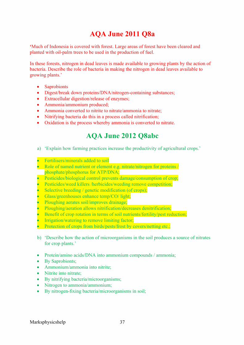
AQA June 2011 Q8a ‘Much of Indonesia is covered with forest. Large areas of forest have been cleared and planted with oil-palm trees to be used in the production of fuel.
In these forests, nitrogen in dead leaves is made available to growing plants by the action of bacteria. Describe the role of bacteria in making the nitrogen in dead leaves available to growing plants.’
• Saprobionts • Digest/break down proteins/DNA/nitrogen-containing substances; • Extracellular digestion/release of enzymes; • Ammonia/ammonium produced; • Ammonia converted to nitrite to nitrate/ammonia to nitrate; • Nitrifying bacteria do this in a process called nitrification; • Oxidation is the process whereby ammonia is converted to nitrate.
AQA June 2012 Q8abc a) ‘Explain how farming practices increase the productivity of agricultural crops.’
• Fertilisers/minerals added to soil • Role of named nutrient or element e.g. nitrate/nitrogen for proteins /
phosphate/phosphorus for ATP/DNA; • Pesticides/biological control prevents damage/consumption of crop; • Pesticides/weed killers /herbicides/weeding remove competition; • Selective breeding / genetic modification (of crops); • Glass/greenhouses enhance temp/CO/ light; • Ploughing aerates soil/improves drainage; • Ploughing/aeration allows nitrification/decreases denitrification; • Benefit of crop rotation in terms of soil nutrients/fertility/pest reduction; • Irrigation/watering to remove limiting factor; • Protection of crops from birds/pests/frost by covers/netting etc.;
b) ‘Describe how the action of microorganisms in the soil produces a source of nitrates for crop plants.’
• Protein/amino acids/DNA into ammonium compounds / ammonia; • By Saprobionts; • Ammonium/ammonia into nitrite; • Nitrite into nitrate; • By nitrifying bacteria/microorganisms; • Nitrogen to ammonia/ammonium; • By nitrogen-fixing bacteria/microorganisms in soil;
Marksphysicshelp
38
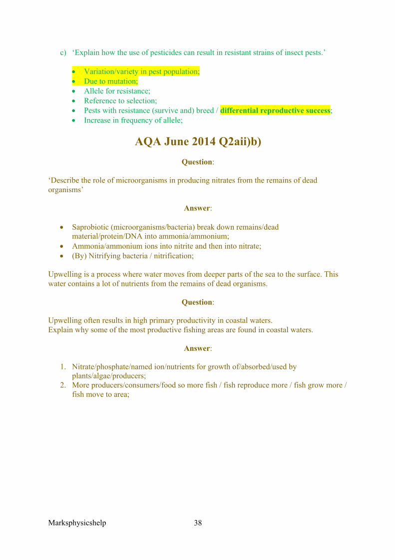
c) ‘Explain how the use of pesticides can result in resistant strains of insect pests.’
• Variation/variety in pest population; • Due to mutation; • Allele for resistance; • Reference to selection; • Pests with resistance (survive and) breed / differential reproductive success; • Increase in frequency of allele;
AQA June 2014 Q2aii)b) Question:
‘Describe the role of microorganisms in producing nitrates from the remains of dead organisms’
Answer:
• Saprobiotic (microorganisms/bacteria) break down remains/dead material/protein/DNA into ammonia/ammonium;
• Ammonia/ammonium ions into nitrite and then into nitrate; • (By) Nitrifying bacteria / nitrification;
Upwelling is a process where water moves from deeper parts of the sea to the surface. This water contains a lot of nutrients from the remains of dead organisms.
Question:
Upwelling often results in high primary productivity in coastal waters. Explain why some of the most productive fishing areas are found in coastal waters.
Answer:
1. Nitrate/phosphate/named ion/nutrients for growth of/absorbed/used by plants/algae/producers;
2. More producers/consumers/food so more fish / fish reproduce more / fish grow more / fish move to area;
Marksphysicshelp
39
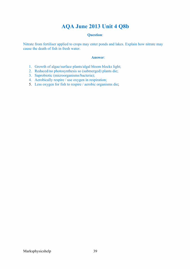
AQA June 2013 Unit 4 Q8b Question:
Nitrate from fertiliser applied to crops may enter ponds and lakes. Explain how nitrate may cause the death of fish in fresh water.
Answer:
1. Growth of algae/surface plants/algal bloom blocks light; 2. Reduced/no photosynthesis so (submerged) plants die; 3. Saprobiotic (microorganisms/bacteria); 4. Aerobically respire / use oxygen in respiration; 5. Less oxygen for fish to respire / aerobic organisms die;
Marksphysicshelp
40
AQA Jan 2012 Unit 4 Q6b
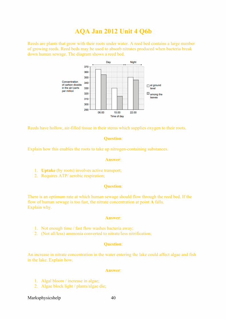
Reeds are plants that grow with their roots under water. A reed bed contains a large number of growing reeds. Reed beds may be used to absorb nitrates produced when bacteria break down human sewage. The diagram shows a reed bed.
Reeds have hollow, air-filled tissue in their stems which supplies oxygen to their roots.
Question:
Explain how this enables the roots to take up nitrogen-containing substances.
Answer:
1. Uptake (by roots) involves active transport; 2. Requires ATP/ aerobic respiration;
Question:
There is an optimum rate at which human sewage should flow through the reed bed. If the flow of human sewage is too fast, the nitrate concentration at point A falls. Explain why.
Answer:
1. Not enough time / fast flow washes bacteria away; 2. (Not all/less) ammonia converted to nitrate/less nitrification;
Question:
An increase in nitrate concentration in the water entering the lake could affect algae and fish in the lake. Explain how.
Answer:
1. Algal bloom / increase in algae; 2. Algae block light / plants/algae die;

Marksphysicshelp
41
3. Decomposers/saprobionts/bacteria break down dead plant materials; 4. Bacteria/decomposers/saprobionts use up oxygen in respiration / increase BOD; 5. Fish die due to lack of oxygen
Marksphysicshelp
42
3.6 Organisms respond to changes in their internal and external environments 3.6.1 Stimuli, both internal and external, are detected and lead to a response 3.6.1.1 Survival and response
Content
• Organisms increase their chance of survival by responding to changes in their environment.
• In flowering plants, species growth factors move from growing regions to other tissues, where they regulate growth in response to directional stimuli.
• The effect of different concentrations of indoleacetic acid (IAA) on cell elongation in the roots and shoots of flowering plants as an explanation of gravitropism and phototropism in flowering plants.
• Taxes and kineses as simple responses that can maintain a mobile organism in a favourable environment.
• The protective effect of a simple reflex, exemplified by a three-neurone simple reflex. Details of spinal cord and dorsal and ventral roots are not required.
A stimulus is a detectable change in the internal or external environment of an organism that leads to a response in the organism. These responses increase the chance of survival for organisms. Those organisms that survive have a greater chance of raising offspring and of passing their alleles to the next generation. Selection pressure favours organisms with more appropriate responses.
Stimuli are detected by receptors, and receptors are specific to one type of stimulus. Coordinators formulate a suitable response to a stimulus, from which a response is produced by an effector. One means of communication in large, multicellular organisms occurs via hormones, which is a relatively slow process.
Taxes and kinesis are simple responses that can maintain a mobile organism in a favourable environment.
A taxis is a simple response whose direction is determined by the direction of the stimulus. As a result, motile organisms move their whole body towards a favourable stimulus or in the other direction. Taxes are classified according to whether the movement is towards the stimulus (positive taxis) or away from the stimulus (negative taxis) and also by the nature of the stimulus. Some examples are:
• Single-celled algae will move towards light (positive phototaxis). This increases change of survival since being photosynthetic, they require light to manufacture their food.
• Earthworms will move away from light (negative phototaxis). This increases their chance of survival as it takes them into the soil, where they are better able to conserve water, find food and avoid predators
Marksphysicshelp
43
• Some species of bacteria move towards a region where glucose is more highly concentrated (positive chemotaxis). This increases their chances of survival because they use glucose as a source of food.
Kinesis are responses in which organisms do not move towards or away from a stimulus. They instead change the speed at which it moves and the rate at which it changes direction. If an organism crosses a sharp dividing line between a favourable and an unfavourable environment, its rate of turning increases. This raises its chances of a quick return to a favourable environment. However, if it moves a considerable distance into an unfavourable environment, its rate of turning may slowly decrease so that it moves in long straight lines before it turns, and it’ll often turn very sharply. This type of response tends to bring the organism into a new region with favourable conditions. It is important when a stimulus is less directional. Humidity and temperature are examples of things that do not produce clear gradients from one extreme to another.
An example of kinesis occurs in woodlice. Woodlice lose water from their bodies in dry conditions. When they move from a damp area into a dry one, they move more rapidly and change direction more often. This increases their chance of moving back into the damp area. Once they return back to a damp area their movement and rate of turning will slow, thus increasing their chances of staying in damp areas. However, if they remain in a dry area for a long period of time, then they move rapidly in straight lines until they move back into a damp area.
Tropisms are the parts of plants responsible to a directional stimulus. In almost all cases the plant part grows towards (positive response) or away (negative response) the stimulus. Examples of the usefulness of this are:
• Plant shoots grow towards light (positive phototropism) and away from gravity (negative gravitropism) so that their leaves are in the most favourable position to capture light for photosynthesis
• Plant roots grow away from light (negative phototropism) and towards gravity (positive gravitropism). In both cases the response increases the probability that roots will grow into the soil, where they are better able to absorb water and mineral ions.
Plants have no nervous system, but they still need to respond to changes in their external and internal environments in order to survive. Plants respond to light, ie shoots grow towards light. Plants respond to gravity; ie its roots are firmly anchored in the soil for stability. Also they respond to water, as almost all plant roots grow towards water for photosynthesis and other processes.
Plants respond to external stimuli with plant growth factors, which are hormone like substances. Their influence is felt by affecting growth, they may be made by cells located throughout the plant rather than in particular organs. Unlike animal hormones, some plant growth factors affect the tissues that release them rather than acting on a distance target organ. Indoleacetic acid (IAA) is an example of a plant growth factor, and is also an example of an auxin. IAA controls plant cell elongation amongst other things.
Marksphysicshelp
44
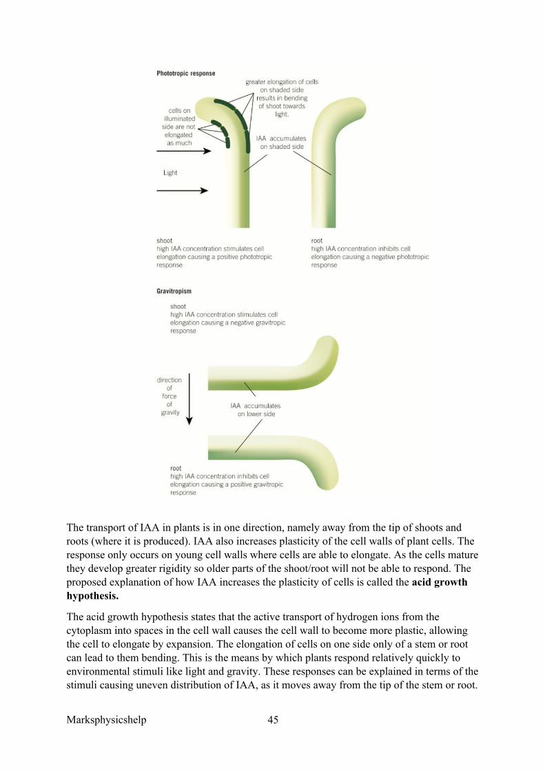
IAA controls tropisms, where a tropism is the directional growth of a plant in response to a directional stimulus. To take light as an example, young shoots grow towards light that is directed at it from one side, this is called positive phototropism.
Phototropism in flowering plants:
1. Cells in the tip of the shoot produce IAA, which is then transported down the shoot 2. The IAA is initially transported evenly throughout all regions as it begins to move
down the shoot 3. Light causes the movement of IAA from the light side to the shaded side of the shoot 4. A greater concentration of IAA builds on the shaded side 5. As IAA causes elongation of shoot cells and there is a greater concentration of IAA
on the shaded side of the shoot, the cells elongate more 6. The shaded side elongates faster -than the light side, so the shoot bends towards the
light.
IAA also controls the bending of roots in response to light, but in roots IAA inhibits cell elongation. Therefore, in roots the elongation of cells is greater on the light side than shaded side, so roots bend away from light, ie they are negatively phototropic.
Gravitropism in flowering plants:
1. Cells in the tip of the root produce IAA, which is then transported along the root 2. The IAA is initially transported to all sides of the root 3. Gravity influences the movement of IAA from the upper side to lower side of the root 4. A greater concentration of IAA builds up on the lower side of the root than on the
upper side 5. As IAA inhibits the elongation of root cells, and now there is a greater concentration
of IAA on the lower side, the cells on this side elongate less than those on the upper side
6. The relatively greater elongation of cells on the upper side compared to the lower side causes the root to bend downwards towards the force of gravity.
In shoots, the greater concentration of IAA on the lower side increases cell elongation and causes this side to elongate more than the upper side, so the root grows upwards away from gravitational forces.
In the diagram below the role of IAA in phototropism and gravitropism responses is summarised.
Marksphysicshelp
45
The transport of IAA in plants is in one direction, namely away from the tip of shoots and roots (where it is produced). IAA also increases plasticity of the cell walls of plant cells. The response only occurs on young cell walls where cells are able to elongate. As the cells mature they develop greater rigidity so older parts of the shoot/root will not be able to respond. The proposed explanation of how IAA increases the plasticity of cells is called the acid growth hypothesis.
The acid growth hypothesis states that the active transport of hydrogen ions from the cytoplasm into spaces in the cell wall causes the cell wall to become more plastic, allowing the cell to elongate by expansion. The elongation of cells on one side only of a stem or root can lead to them bending. This is the means by which plants respond relatively quickly to environmental stimuli like light and gravity. These responses can be explained in terms of the stimuli causing uneven distribution of IAA, as it moves away from the tip of the stem or root.
Marksphysicshelp
46
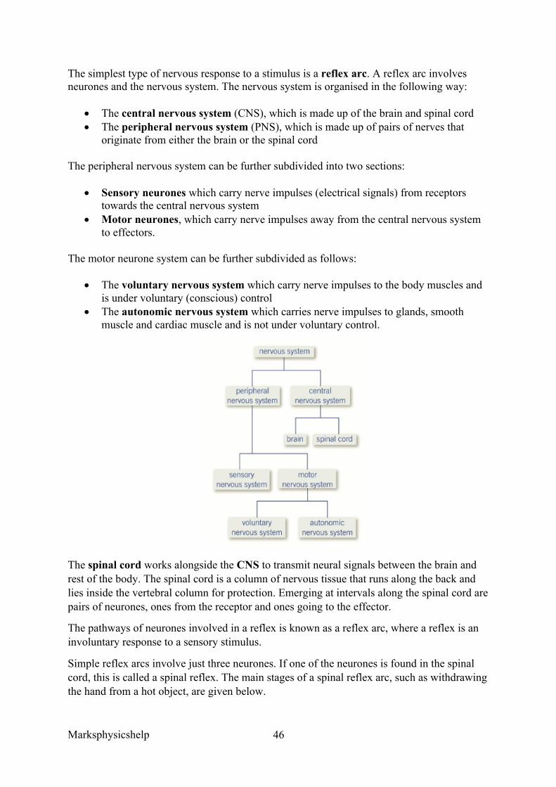
The simplest type of nervous response to a stimulus is a reflex arc. A reflex arc involves neurones and the nervous system. The nervous system is organised in the following way:
• The central nervous system (CNS), which is made up of the brain and spinal cord • The peripheral nervous system (PNS), which is made up of pairs of nerves that
originate from either the brain or the spinal cord
The peripheral nervous system can be further subdivided into two sections:
• Sensory neurones which carry nerve impulses (electrical signals) from receptors towards the central nervous system
• Motor neurones, which carry nerve impulses away from the central nervous system to effectors.
The motor neurone system can be further subdivided as follows:
• The voluntary nervous system which carry nerve impulses to the body muscles and is under voluntary (conscious) control
• The autonomic nervous system which carries nerve impulses to glands, smooth muscle and cardiac muscle and is not under voluntary control.
The spinal cord works alongside the CNS to transmit neural signals between the brain and rest of the body. The spinal cord is a column of nervous tissue that runs along the back and lies inside the vertebral column for protection. Emerging at intervals along the spinal cord are pairs of neurones, ones from the receptor and ones going to the effector.
The pathways of neurones involved in a reflex is known as a reflex arc, where a reflex is an involuntary response to a sensory stimulus.
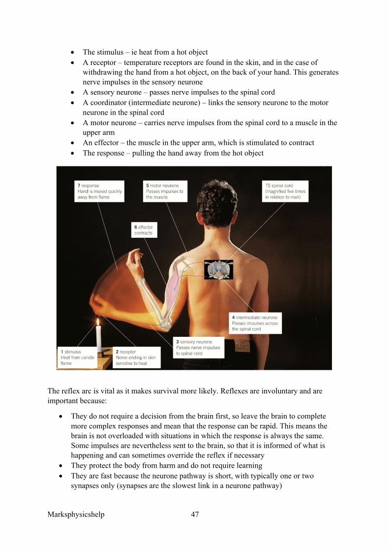
Simple reflex arcs involve just three neurones. If one of the neurones is found in the spinal cord, this is called a spinal reflex. The main stages of a spinal reflex arc, such as withdrawing the hand from a hot object, are given below.
Marksphysicshelp
47
• The stimulus – ie heat from a hot object • A receptor – temperature receptors are found in the skin, and in the case of
withdrawing the hand from a hot object, on the back of your hand. This generates nerve impulses in the sensory neurone
• A sensory neurone – passes nerve impulses to the spinal cord • A coordinator (intermediate neurone) – links the sensory neurone to the motor
neurone in the spinal cord • A motor neurone – carries nerve impulses from the spinal cord to a muscle in the
upper arm • An effector – the muscle in the upper arm, which is stimulated to contract • The response – pulling the hand away from the hot object
The reflex arc is vital as it makes survival more likely. Reflexes are involuntary and are important because:
• They do not require a decision from the brain first, so leave the brain to complete more complex responses and mean that the response can be rapid. This means the brain is not overloaded with situations in which the response is always the same. Some impulses are nevertheless sent to the brain, so that it is informed of what is happening and can sometimes override the reflex if necessary
• They protect the body from harm and do not require learning • They are fast because the neurone pathway is short, with typically one or two
synapses only (synapses are the slowest link in a neurone pathway)
Marksphysicshelp
48
AQA June 2015 Q7a
Question:
Give one similarity and one difference between a taxis and a tropism
Answer:
1. Similarity – directional response (to a stimulus)/movement towards/away from a stimulus;
2. Difference – taxis (whole) organism moves and tropism a growth (response)
Marksphysicshelp
49
3.6.1.2 Receptors
Content
• The Pacinian corpuscle should be used as an example of a receptor to illustrate that: o Receptors respond only to specific stimuli o Stimulation of a receptor leads to the establishment of a generator potential.
• The basic structure of a Pacinian corpuscle. • Deformation of stretch-mediated sodium ion channels in a Pacinian corpuscle leads to
the establishment of a generator potential. • The human retina in sufficient detail to show how differences in sensitivity to light,
sensitivity to colour and visual acuity are explained by differences in the optical pigments of rods and cones and the connections rods and cones make in the optic nerve.
Opportunities for Skills Development
• Students could design and carry out investigations into: o The sensitivity of temperature receptors in human skin o Habituation of touch receptors in human skin o Resolution of touch receptors in human skin.
The Pacinian corpuscle is a type of receptor that is used to illustrate that:
• Receptors will only respond to a specific type of stimulus, so in the case of the Pacinian corpuscle – mechanical pressure as opposed to light, heat, sound etc.
• Stimulation of a receptor leads to the establishment of a generator potential by acting as a transducer. All stimuli involve a change in some form of energy, and it is the transducers job to convert the change in form of energy by the stimulus into a form, namely nerve impulses, that can be understood by the body. The nerve impulse is also a form of energy and so receptors therefore convert (transduce) one form of energy into another. Receptors in the nervous system convert the energy of the stimulus into a nervous impulse called a generator potential. The Pacinian corpuscle transduces the mechanical energy of the stimulus into a generator potential.
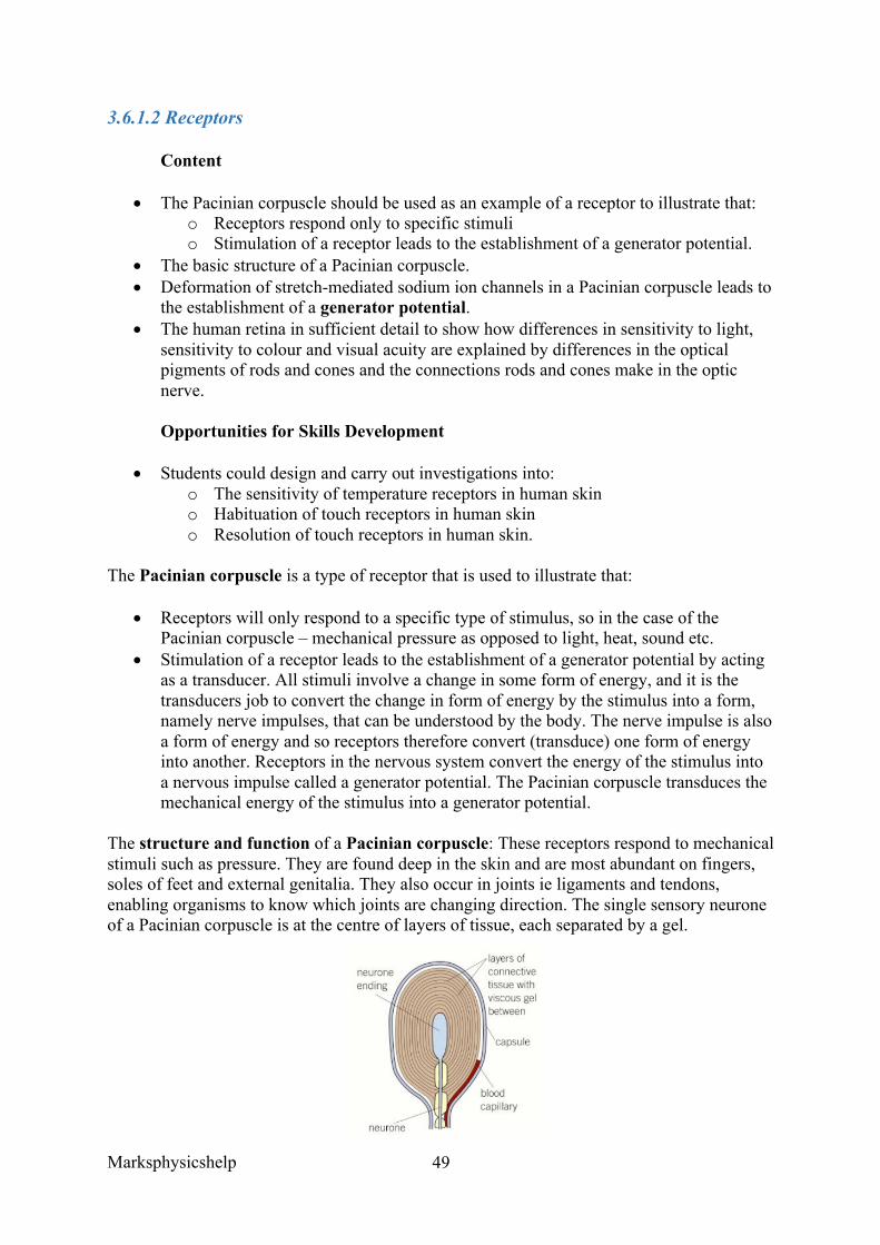
The structure and function of a Pacinian corpuscle: These receptors respond to mechanical stimuli such as pressure. They are found deep in the skin and are most abundant on fingers, soles of feet and external genitalia. They also occur in joints ie ligaments and tendons, enabling organisms to know which joints are changing direction. The single sensory neurone of a Pacinian corpuscle is at the centre of layers of tissue, each separated by a gel.
Marksphysicshelp
50
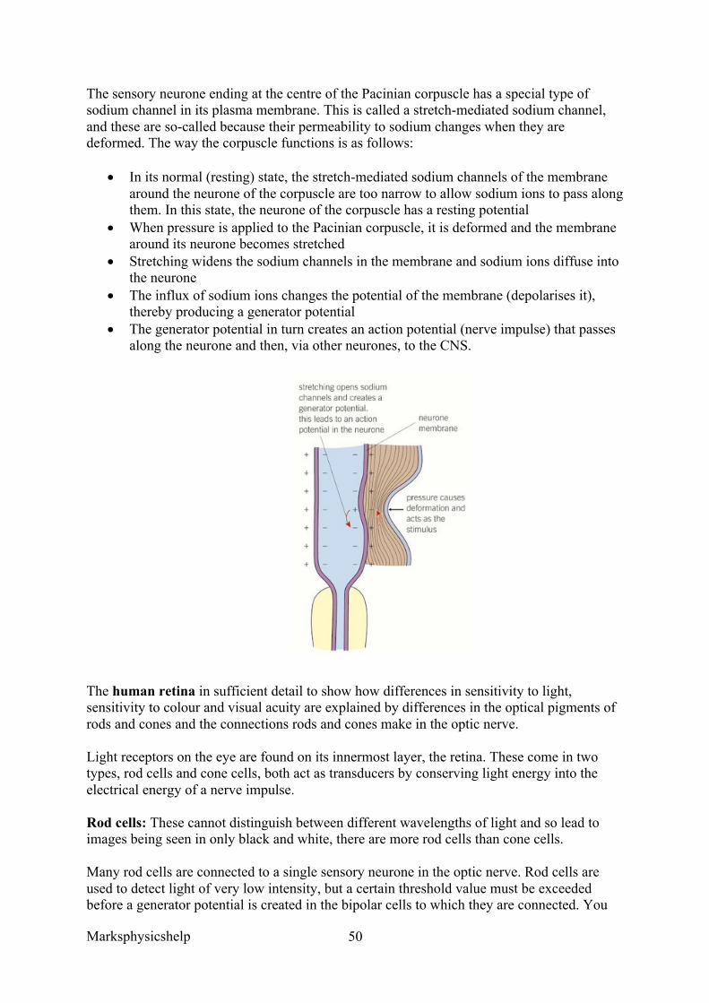
The sensory neurone ending at the centre of the Pacinian corpuscle has a special type of sodium channel in its plasma membrane. This is called a stretch-mediated sodium channel, and these are so-called because their permeability to sodium changes when they are deformed. The way the corpuscle functions is as follows:
• In its normal (resting) state, the stretch-mediated sodium channels of the membrane around the neurone of the corpuscle are too narrow to allow sodium ions to pass along them. In this state, the neurone of the corpuscle has a resting potential
• When pressure is applied to the Pacinian corpuscle, it is deformed and the membrane around its neurone becomes stretched
• Stretching widens the sodium channels in the membrane and sodium ions diffuse into the neurone
• The influx of sodium ions changes the potential of the membrane (depolarises it), thereby producing a generator potential
• The generator potential in turn creates an action potential (nerve impulse) that passes along the neurone and then, via other neurones, to the CNS.
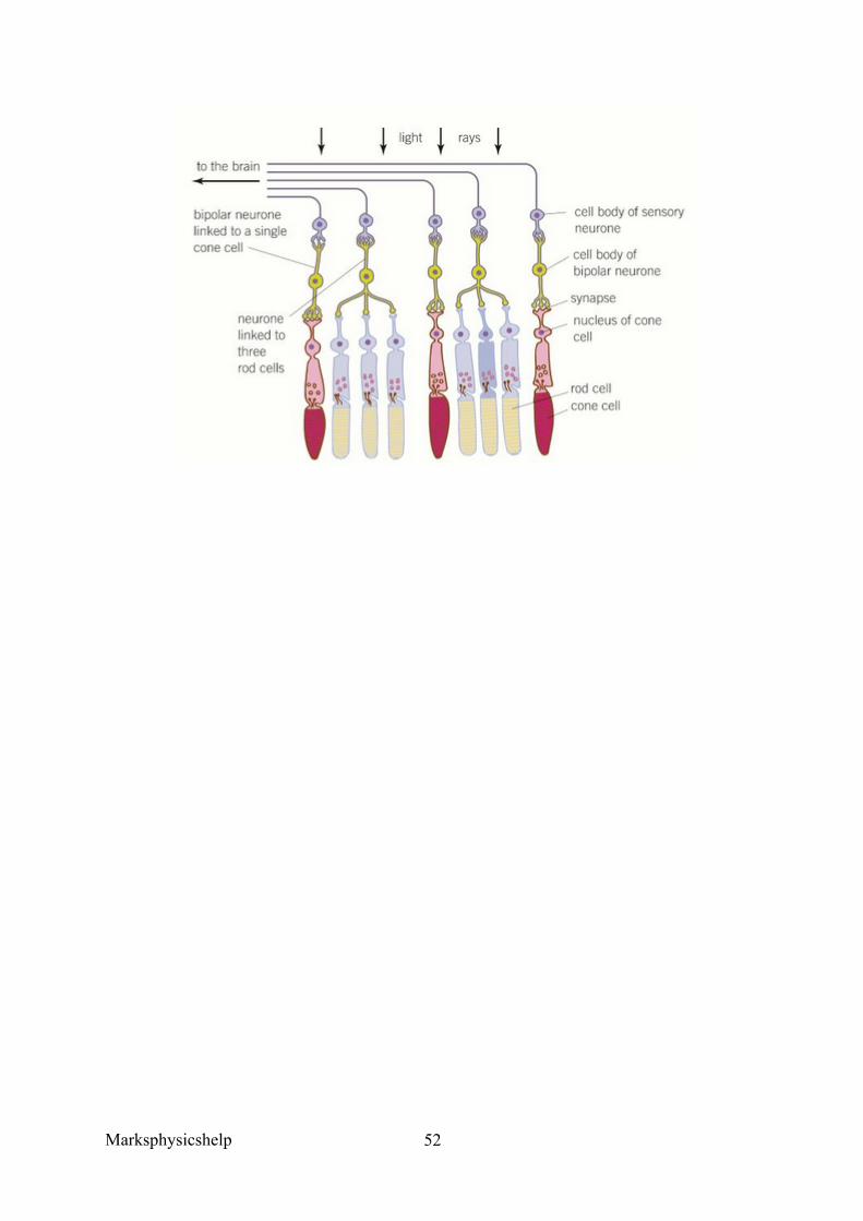
The human retina in sufficient detail to show how differences in sensitivity to light, sensitivity to colour and visual acuity are explained by differences in the optical pigments of rods and cones and the connections rods and cones make in the optic nerve. Light receptors on the eye are found on its innermost layer, the retina. These come in two types, rod cells and cone cells, both act as transducers by conserving light energy into the electrical energy of a nerve impulse. Rod cells: These cannot distinguish between different wavelengths of light and so lead to images being seen in only black and white, there are more rod cells than cone cells. Many rod cells are connected to a single sensory neurone in the optic nerve. Rod cells are used to detect light of very low intensity, but a certain threshold value must be exceeded before a generator potential is created in the bipolar cells to which they are connected. You
Marksphysicshelp
51
get many rod cells connected to the same single bipolar cells (retinal convergence), so there is a much greater chance that the threshold value will be exceeded than if only a single rod cells were connected to each bipolar cell (this is due to spatial summation). As a result, rod cells allow us to see in low light intensity ie at night, but only in black and white. In order to create a generator potential, the pigment in the rod cells (rhodopsin), must be broken down. There is enough energy from low-intensity light to cause this breakdown, hence rod cells respond to low intensity light. A consequence of many rod cells connected to a single bipolar cell is that light received by rod cells sharing the same neurone will only generate a single impulse that travels to the brain, regardless of how many neurones are stimulated. This means that the brain is unable to distinguish between separate sources of light that stimulated them. Rod cells give low visual acuity. Cone cells: Three different types, each responding to a range of wavelengths of light. Depending upon the proportion of each type that is stimulated, we are able to perceive images in full colour. Cone cells will usually have their own bipolar cell connected to a sensory neurone in the optic nerve. This means that the stimulation of a number of cone cells cannot be combined to help exceed the threshold value and so create a generator potential. As a result, cone cells respond to light of high intensity. Cone cells also contain different types of pigments than from those found in rod cells. The pigment in cone cells (iodopsin) requires a higher light intensity for its breakdown. There are three types of cone cell, each containing a different type of iodopsin and so as a result each cone cell is sensitive to a different specific range of wavelengths. Each cell has its own connection to a single bipolar cell, which means that if two adjacent cells are stimulated, the brain will receive separate impulses. This means two dots close together can be distinguished between, so cone cells give accurate vision, so good visual acuity. The distribution of rod and cone cells on the retina is uneven, light is focused by the lens on the part of the retina opposite to the pupil. This point is called the fovea, the fovea therefore receives the highest intensity of light. Therefore cone cells, but not rod cells, are found at the fovea. The concentration of cone cells diminishes further away from the fovea. At the peripheries of the retina, where light intensity is at its lowest, only rod cells are found. These different types of cells and differences in sensitivity and visual acuity ensure good vision at day and night.
Marksphysicshelp
52
Marksphysicshelp
53
AQA June 2015 Q5a Question:
Describe how a Pacinian corpuscle produces a generator potential when stimulated.
Answer:
1. (Increased pressure) deforms/changes stretch-mediated sodium (ion) channel; 2. (Sodium channels open and) sodium ions flow in; 3. Depolarisation (leading to generator potential);
Marksphysicshelp
54
3.6.1.3 Control of heart rate
Content
• Myogenic stimulation of the heart and transmission of a subsequent wave of electrical activity. The roles of the sinoatrial node (SAN), atrioventricular node (AVN) and Purkyne tissue in the bundle of His.
• The roles and locations of chemoreceptors and pressure receptors and the roles of the autonomic nervous system and effectors in controlling heart rate.
Opportunities for Skills Development
• Students could design and carry out an investigation into the effect of a named variable on human pulse rate.
• Students could use values of heart rate (R) and stroke volume (V) to calculate cardiac output (CO), using the formula CO = R x V
The autonomic (self-governing) nervous system controls the involuntary activities of internal muscles and glands like the heart beat, digestive system and breathing. It has two main sections:
• The sympathetic nervous system which stimulates effectors in general, speeding up any activity. It acts rather like an emergency controller, controlling effectors when we exercise strenuously or experience powerful emotions. It therefore helps us to cope with stressful situations by heightening our awareness and preparing us for activity (fight or flight response).
• The parasympathetic nervous system which in general inhibits effectors and so slows down any activity. It controls activities under normal resting conditions, and is concerned with conserving energy and replenishing the body’s reserves.
The actions of the sympathetic and parasympathetic nervous systems usually act antagonistically. So if one system contracts a muscle, the other relaxes it, this system is used in control of the heart rate.
Control of heart rate: Muscle in the heart is called cardiac muscle, and it is myogenic (contraction is initiated from within the muscle itself). This is opposite to neurogenic, where contraction is initiated from nervous impulses from outside of the muscle.
Within the wall of the right atrium of the heart is a distinct group of cells known as sinoatrial node (SAN). It is from here that the initial stimulus for contraction originates, and the SAN has a basic rhythm of stimulation that determines the beat of the heart. Therefore it is often called the pacemaker. The sequence of events that controls the basic heart rate is:
• A wave of electrical excitation spreads out from the SAN across both atria, causing them to contract
• A layer of non-conductive tissue (atrioventricular septum) prevents the wave crossing to the ventricles
• The wave of electrical excitation enters a second group of cells called the atrioventricular node (AVN) which lies between the atria.
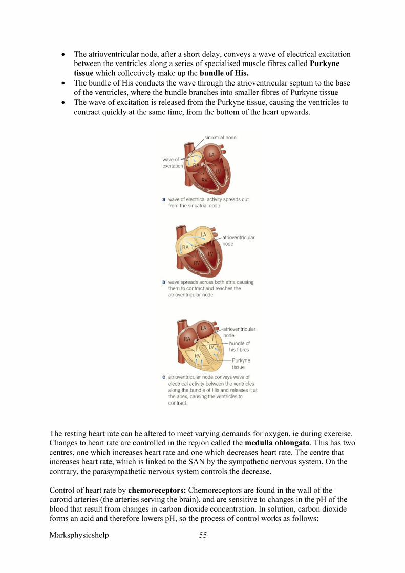
Marksphysicshelp
55
• The atrioventricular node, after a short delay, conveys a wave of electrical excitation between the ventricles along a series of specialised muscle fibres called Purkyne tissue which collectively make up the bundle of His.
• The bundle of His conducts the wave through the atrioventricular septum to the base of the ventricles, where the bundle branches into smaller fibres of Purkyne tissue
• The wave of excitation is released from the Purkyne tissue, causing the ventricles to contract quickly at the same time, from the bottom of the heart upwards.
The resting heart rate can be altered to meet varying demands for oxygen, ie during exercise. Changes to heart rate are controlled in the region called the medulla oblongata. This has two centres, one which increases heart rate and one which decreases heart rate. The centre that increases heart rate, which is linked to the SAN by the sympathetic nervous system. On the contrary, the parasympathetic nervous system controls the decrease.
Control of heart rate by chemoreceptors: Chemoreceptors are found in the wall of the carotid arteries (the arteries serving the brain), and are sensitive to changes in the pH of the blood that result from changes in carbon dioxide concentration. In solution, carbon dioxide forms an acid and therefore lowers pH, so the process of control works as follows:
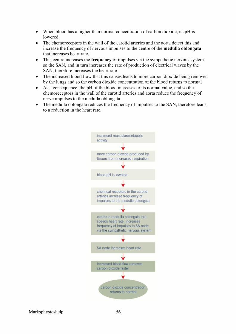
Marksphysicshelp
56
• When blood has a higher than normal concentration of carbon dioxide, its pH is lowered.
• The chemoreceptors in the wall of the carotid arteries and the aorta detect this and increase the frequency of nervous impulses to the centre of the medulla oblongata that increases heart rate.
• This centre increases the frequency of impulses via the sympathetic nervous system so the SAN, and in turn increases the rate of production of electrical waves by the SAN, therefore increases the heart rate
• The increased blood flow that this causes leads to more carbon dioxide being removed by the lungs and so the carbon dioxide concentration of the blood returns to normal
• As a consequence, the pH of the blood increases to its normal value, and so the chemoreceptors in the wall of the carotid arteries and aorta reduce the frequency of nerve impulses to the medulla oblongata.
• The medulla oblongata reduces the frequency of impulses to the SAN, therefore leads to a reduction in the heart rate.
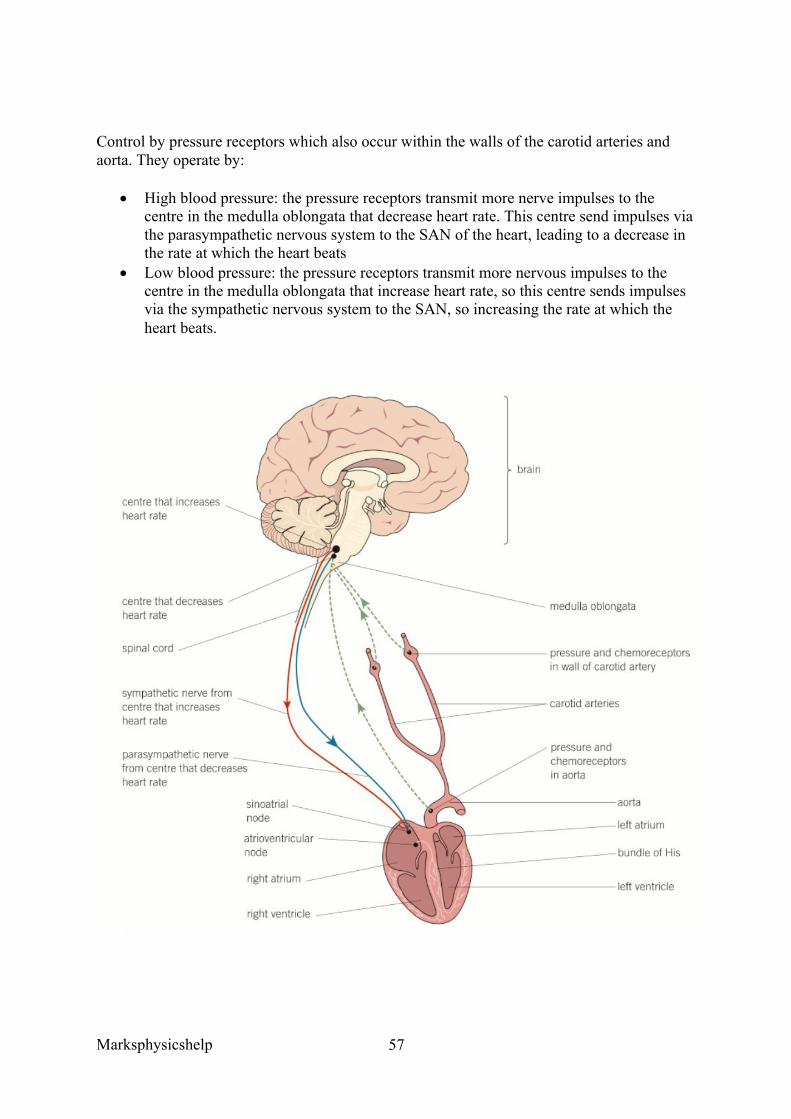
Marksphysicshelp
57
Control by pressure receptors which also occur within the walls of the carotid arteries and aorta. They operate by:
• High blood pressure: the pressure receptors transmit more nerve impulses to the centre in the medulla oblongata that decrease heart rate. This centre send impulses via the parasympathetic nervous system to the SAN of the heart, leading to a decrease in the rate at which the heart beats
• Low blood pressure: the pressure receptors transmit more nervous impulses to the centre in the medulla oblongata that increase heart rate, so this centre sends impulses via the sympathetic nervous system to the SAN, so increasing the rate at which the heart beats.
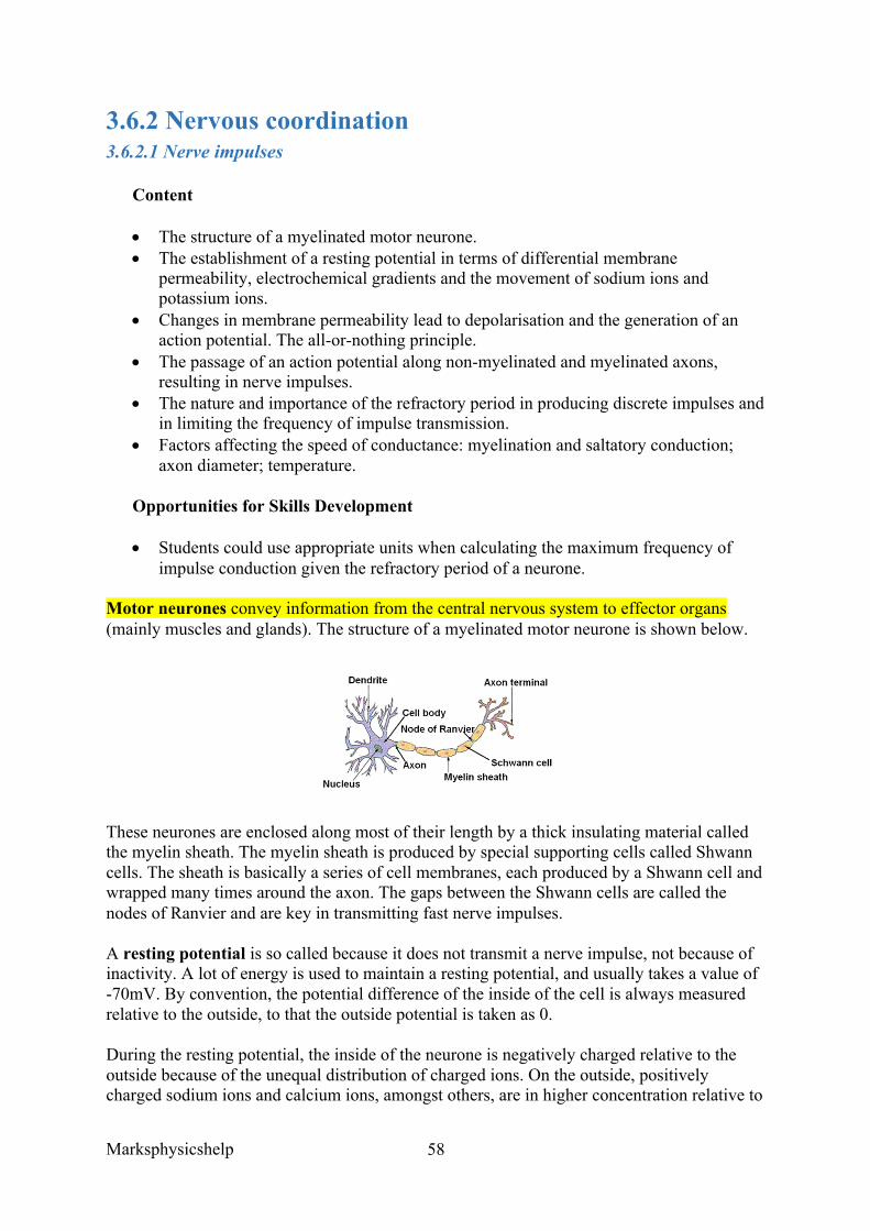
Marksphysicshelp
58
3.6.2 Nervous coordination 3.6.2.1 Nerve impulses
Content
• The structure of a myelinated motor neurone. • The establishment of a resting potential in terms of differential membrane
permeability, electrochemical gradients and the movement of sodium ions and potassium ions.
• Changes in membrane permeability lead to depolarisation and the generation of an action potential. The all-or-nothing principle.
• The passage of an action potential along non-myelinated and myelinated axons, resulting in nerve impulses.
• The nature and importance of the refractory period in producing discrete impulses and in limiting the frequency of impulse transmission.
• Factors affecting the speed of conductance: myelination and saltatory conduction; axon diameter; temperature.
Opportunities for Skills Development
• Students could use appropriate units when calculating the maximum frequency of impulse conduction given the refractory period of a neurone.
Motor neurones convey information from the central nervous system to effector organs (mainly muscles and glands). The structure of a myelinated motor neurone is shown below.
These neurones are enclosed along most of their length by a thick insulating material called the myelin sheath. The myelin sheath is produced by special supporting cells called Shwann cells. The sheath is basically a series of cell membranes, each produced by a Shwann cell and wrapped many times around the axon. The gaps between the Shwann cells are called the nodes of Ranvier and are key in transmitting fast nerve impulses.
A resting potential is so called because it does not transmit a nerve impulse, not because of inactivity. A lot of energy is used to maintain a resting potential, and usually takes a value of -70mV. By convention, the potential difference of the inside of the cell is always measured relative to the outside, to that the outside potential is taken as 0.
During the resting potential, the inside of the neurone is negatively charged relative to the outside because of the unequal distribution of charged ions. On the outside, positively charged sodium ions and calcium ions, amongst others, are in higher concentration relative to
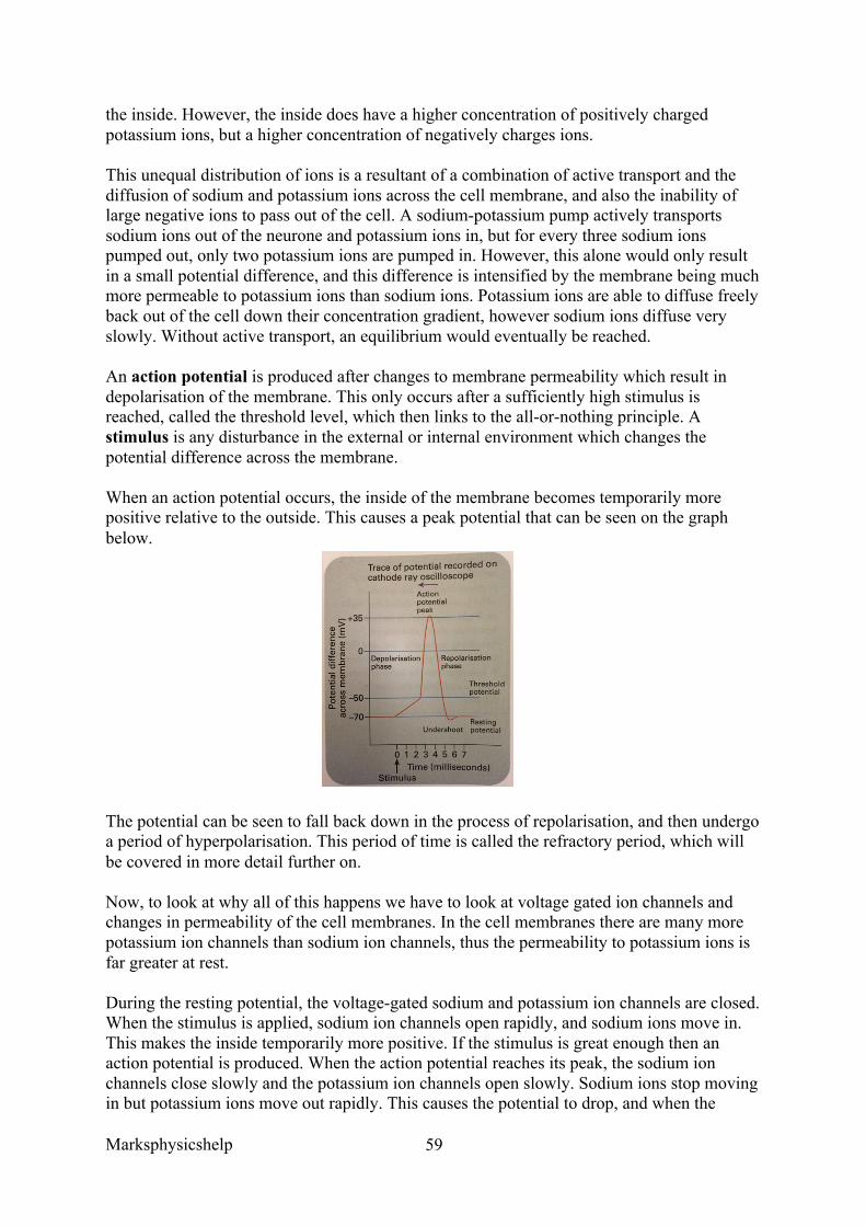
Marksphysicshelp
59
the inside. However, the inside does have a higher concentration of positively charged potassium ions, but a higher concentration of negatively charges ions.
This unequal distribution of ions is a resultant of a combination of active transport and the diffusion of sodium and potassium ions across the cell membrane, and also the inability of large negative ions to pass out of the cell. A sodium-potassium pump actively transports sodium ions out of the neurone and potassium ions in, but for every three sodium ions pumped out, only two potassium ions are pumped in. However, this alone would only result in a small potential difference, and this difference is intensified by the membrane being much more permeable to potassium ions than sodium ions. Potassium ions are able to diffuse freely back out of the cell down their concentration gradient, however sodium ions diffuse very slowly. Without active transport, an equilibrium would eventually be reached.
An action potential is produced after changes to membrane permeability which result in depolarisation of the membrane. This only occurs after a sufficiently high stimulus is reached, called the threshold level, which then links to the all-or-nothing principle. A stimulus is any disturbance in the external or internal environment which changes the potential difference across the membrane.
When an action potential occurs, the inside of the membrane becomes temporarily more positive relative to the outside. This causes a peak potential that can be seen on the graph below.
The potential can be seen to fall back down in the process of repolarisation, and then undergo a period of hyperpolarisation. This period of time is called the refractory period, which will be covered in more detail further on.
Now, to look at why all of this happens we have to look at voltage gated ion channels and changes in permeability of the cell membranes. In the cell membranes there are many more potassium ion channels than sodium ion channels, thus the permeability to potassium ions is far greater at rest.
During the resting potential, the voltage-gated sodium and potassium ion channels are closed. When the stimulus is applied, sodium ion channels open rapidly, and sodium ions move in. This makes the inside temporarily more positive. If the stimulus is great enough then an action potential is produced. When the action potential reaches its peak, the sodium ion channels close slowly and the potassium ion channels open slowly. Sodium ions stop moving in but potassium ions move out rapidly. This causes the potential to drop, and when the
Marksphysicshelp
60
membrane reaches resting potential again, the potassium ion channels close, but do this slowly so that the potential dips below resting level. When these channels close the membrane returns to its original potential.
The all-or-nothing principle states that no matter how strong the stimulus, the size of the action potential is always the same, therefore information about the strength of a stimulus is carried along a nerve fibre by the changes in frequency as opposed to the variations in size of the nerve impulses. There is a certain value, called the threshold value, which triggers an action potential. Below this value there will be no action potential, but any stimulus higher than this value will trigger an action potential, which relates to the ‘all’ in ‘all-or-nothing’.
The passage of an action potential along non-myelinated and myelinated axons ultimately results in nerve impulses:
• In an unmyelinated neurone, the localised current from an action potential acts as a stimulus for the next part of the nerve membrane, causing further depolarisation, and then so along. Thus, the nerve impulse is propagated as a wave of depolarisation, with one portion of the fibre repolarising as the next depolarises. However, the size of the action potential does not change on its journey.
• In a myelinated neurone, the fatty sheath of myelin around the axon acts as an electrical insulator. This means that action potentials cannot form in this region. But at the intervals of myelin insulation (nodes of Ranvier), action potentials can occur, and so action potentials essentially jump between the adjacent nodes. Therefore, these action potentials pass along the myelinated neurone much faster than an unmyelinated neurone. This process is called saltatory conduction.
Once an action potential has been created in any region of the axon, there is a period afterwards when inward movement of sodium ions is prevented because the sodium voltage-gated channels are closed. This is called the refractory period and is important in producing discrete impulses and limiting the frequency of impulse transmission.
The refractory period ensures that nerve impulses travel in one direction, as during this time another impulse cannot be generated. Action potentials can only pass from an active region to a resting region. This is because action potentials cannot be propagated in a region that is in its refractory period, so they can only move in a forward direction. Otherwise, after the impulse travelled across the nerve fibre, it could travel backwards if the membrane could be depolarised immediately again. It also means that you get discrete impulses that can be distinguished from one another, as there is a small period of time in which another impulse cannot be generated. Furthermore, it limits the frequency of impulse transmission, so also limiting the strength of a stimulus that can be detected.
Since action potentials are all the same, a person can only distinguish between the strength of an impulse by the frequency of the impulse sent. Also there are different neurones with different threshold values, thus the brain can interpret the number and type of neurones that pass impulses as a result of a given stimulus, thereby determining its size.
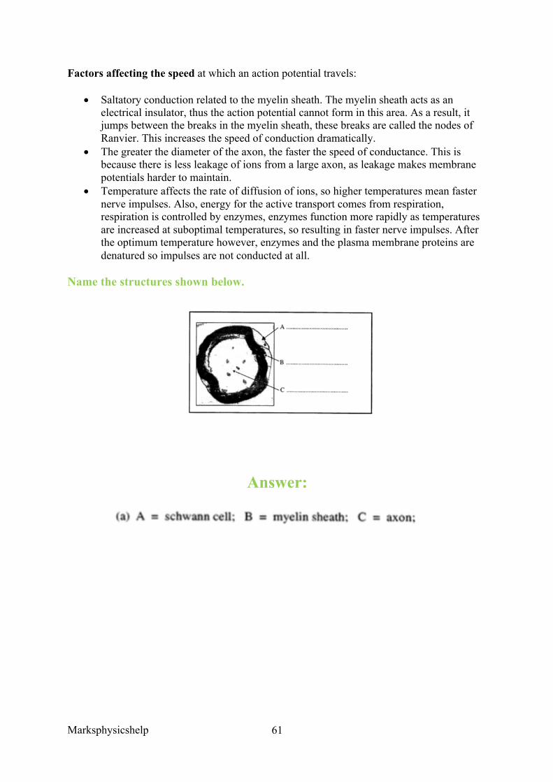
Marksphysicshelp
61
Factors affecting the speed at which an action potential travels:
• Saltatory conduction related to the myelin sheath. The myelin sheath acts as an electrical insulator, thus the action potential cannot form in this area. As a result, it jumps between the breaks in the myelin sheath, these breaks are called the nodes of Ranvier. This increases the speed of conduction dramatically.
• The greater the diameter of the axon, the faster the speed of conductance. This is because there is less leakage of ions from a large axon, as leakage makes membrane potentials harder to maintain.
• Temperature affects the rate of diffusion of ions, so higher temperatures mean faster nerve impulses. Also, energy for the active transport comes from respiration, respiration is controlled by enzymes, enzymes function more rapidly as temperatures are increased at suboptimal temperatures, so resulting in faster nerve impulses. After the optimum temperature however, enzymes and the plasma membrane proteins are denatured so impulses are not conducted at all.
Name the structures shown below.
Answer:
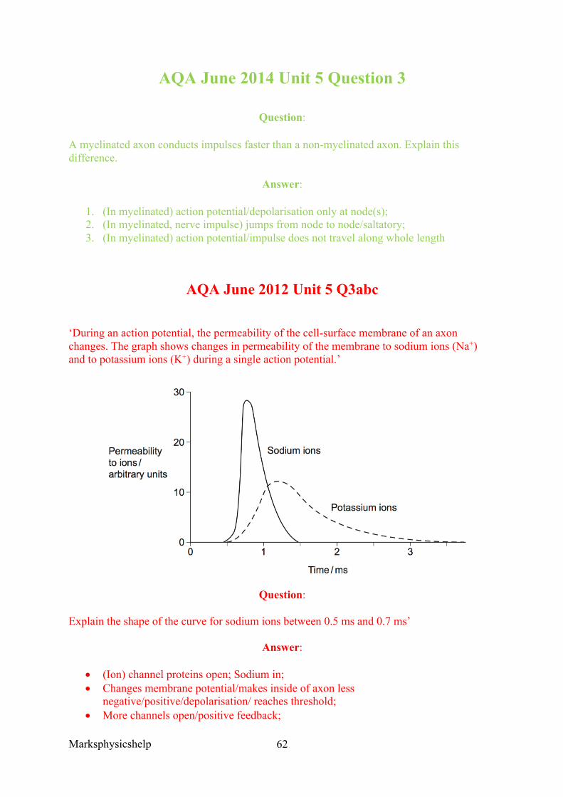
Marksphysicshelp
62
AQA June 2014 Unit 5 Question 3
Question:
A myelinated axon conducts impulses faster than a non-myelinated axon. Explain this difference.
Answer:
1. (In myelinated) action potential/depolarisation only at node(s); 2. (In myelinated, nerve impulse) jumps from node to node/saltatory; 3. (In myelinated) action potential/impulse does not travel along whole length
AQA June 2012 Unit 5 Q3abc
‘During an action potential, the permeability of the cell-surface membrane of an axon changes. The graph shows changes in permeability of the membrane to sodium ions (Na+) and to potassium ions (K+) during a single action potential.’
Question:
Explain the shape of the curve for sodium ions between 0.5 ms and 0.7 ms’
Answer:
• (Ion) channel proteins open; Sodium in; • Changes membrane potential/makes inside of axon less
negative/positive/depolarisation/ reaches threshold; • More channels open/positive feedback;
Marksphysicshelp
63
Question:
‘During an action potential, the membrane potential rises to +40 mV and then falls. Use information from the graph to explain the fall in membrane potential.’
Answer:
• Potassium channels open; • Potassium out; • Sodium channels close;
Question:
‘After exercise, some ATP is used to re-establish the resting potential in axons. Explain how the resting potential is re-established.’
Answer:
• Pump/active transport/transport against concentration gradient; • Of sodium from axon, sodium moving out and potassium in;
AQA June 2013 Unit 5 Q7abc
‘Serotonin is a neurotransmitter released in some synapses in the brain. It is transported back out of the synaptic gap by a transport protein in the pre-synaptic membrane.
Serotonin diffuses across the synaptic gap and binds to a receptor on the post-synaptic membrane.
Describe how this causes depolarisation of the post-synaptic membrane.’
• Causes sodium ion channels to open; • Sodium ions enter (cell and cause depolarisation);
‘It is important that a neurotransmitter such as serotonin is transported back out of synapses. Explain why.’
• (If not removed) keeps binding (to receptors); • Keeps causing action potentials/depolarisation (in post- synaptic membrane); • Prevents information being carried across synapse/described consequence;
‘Scientists investigated the effect of a drug called MDMA on movement of mice. They measured the amount of movement of three groups of mice, K, L and M.
Group K, mice not given MDMA. Group L, mice given MDMA. Group M, mutant mice that did not produce a serotonin receptor on their post-synaptic membranes and were given MDMA.
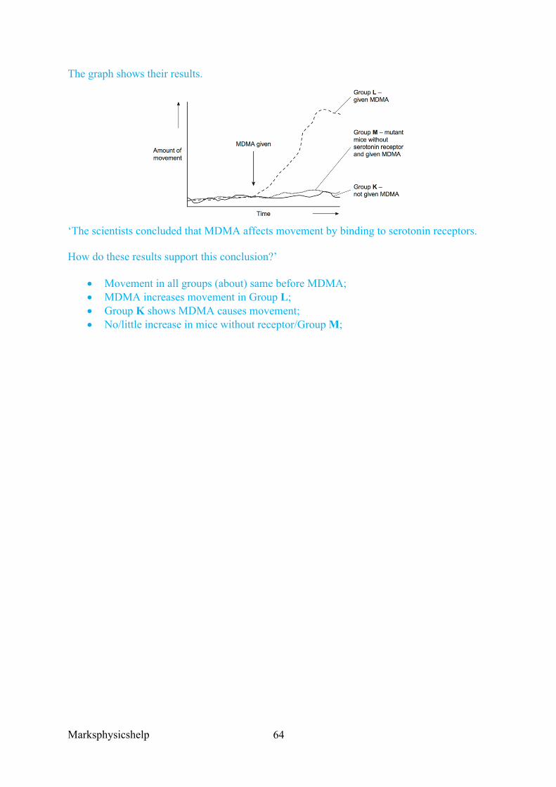
Marksphysicshelp
64
The graph shows their results.
‘The scientists concluded that MDMA affects movement by binding to serotonin receptors.
How do these results support this conclusion?’
• Movement in all groups (about) same before MDMA; • MDMA increases movement in Group L; • Group K shows MDMA causes movement; • No/little increase in mice without receptor/Group M;
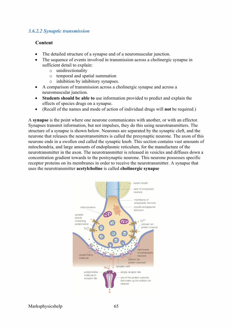
Marksphysicshelp
65
3.6.2.2 Synaptic transmission
Content
• The detailed structure of a synapse and of a neuromuscular junction. • The sequence of events involved in transmission across a cholinergic synapse in
sufficient detail to explain: o unidirectionality o temporal and spatial summation o inhibition by inhibitory synapses.
• A comparison of transmission across a cholinergic synapse and across a neuromuscular junction.
• Students should be able to use information provided to predict and explain the effects of species drugs on a synapse.
• (Recall of the names and mode of action of individual drugs will not be required.)
A synapse is the point where one neurone communicates with another, or with an effector. Synapses transmit information, but not impulses, they do this using neurotransmitters. The structure of a synapse is shown below. Neurones are separated by the synaptic cleft, and the neurone that releases the neurotransmitters is called the presynaptic neurone. The axon of this neurone ends in a swollen end called the synaptic knob. This section contains vast amounts of mitochondria, and large amounts of endoplasmic reticulum, for the manufacture of the neurotransmitter in the axon. The neurotransmitter is released in vesicles and diffuses down a concentration gradient towards to the postsynaptic neurone. This neurone possesses specific receptor proteins on its membranes in order to receive the neurotransmitter. A synapse that uses the neurotransmitter acetylcholine is called cholinergic synapse
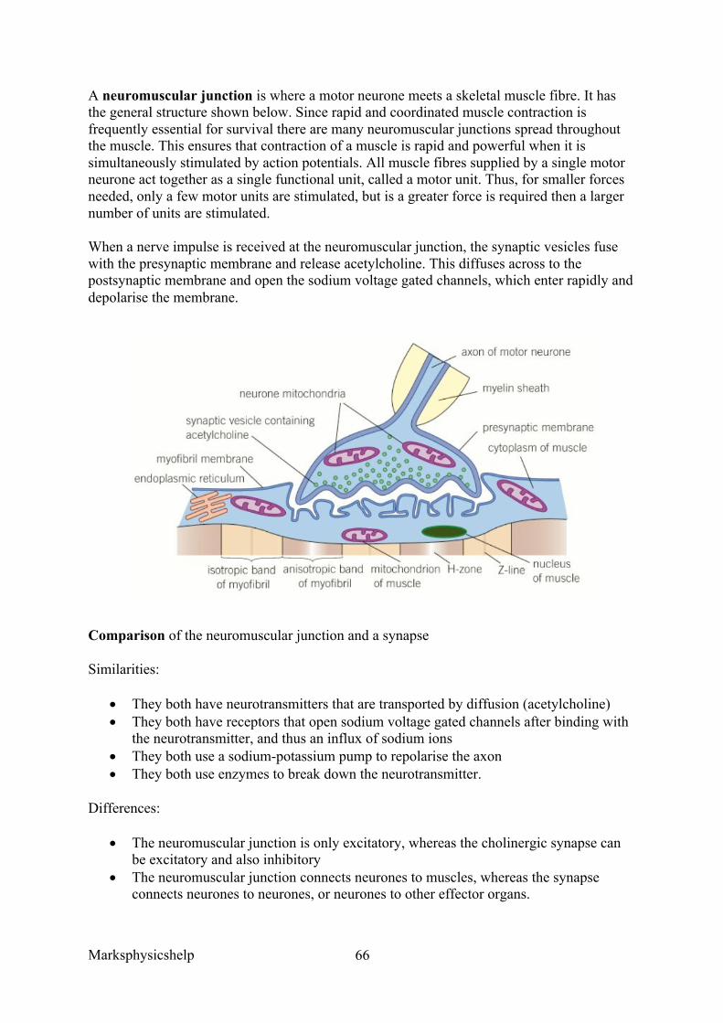
Marksphysicshelp
66
A neuromuscular junction is where a motor neurone meets a skeletal muscle fibre. It has the general structure shown below. Since rapid and coordinated muscle contraction is frequently essential for survival there are many neuromuscular junctions spread throughout the muscle. This ensures that contraction of a muscle is rapid and powerful when it is simultaneously stimulated by action potentials. All muscle fibres supplied by a single motor neurone act together as a single functional unit, called a motor unit. Thus, for smaller forces needed, only a few motor units are stimulated, but is a greater force is required then a larger number of units are stimulated.
When a nerve impulse is received at the neuromuscular junction, the synaptic vesicles fuse with the presynaptic membrane and release acetylcholine. This diffuses across to the postsynaptic membrane and open the sodium voltage gated channels, which enter rapidly and depolarise the membrane.
Comparison of the neuromuscular junction and a synapse
Similarities:
• They both have neurotransmitters that are transported by diffusion (acetylcholine) • They both have receptors that open sodium voltage gated channels after binding with
the neurotransmitter, and thus an influx of sodium ions • They both use a sodium-potassium pump to repolarise the axon • They both use enzymes to break down the neurotransmitter.
Differences:
• The neuromuscular junction is only excitatory, whereas the cholinergic synapse can be excitatory and also inhibitory
• The neuromuscular junction connects neurones to muscles, whereas the synapse connects neurones to neurones, or neurones to other effector organs.
Marksphysicshelp
67
• Only motor neurones are involved with the neuromuscular junction, however in the cholinergic synapse motor, sensory and intermediate neurones may be involved.
• The action potential ends here at the neuromuscular junction, but a new action potential may be produced along another neurone at a synapse.
• Acetylcholine binds to receptors on the membrane of the muscle fibre at the neuromuscular junction, but acetylcholine binds to the receptors on the membrane of the post-synaptic neurone in a cholinergic synapse.
Transmission across a cholinergic synapse is explained below:
An action potential is sent down the presynaptic neurones membrane, towards the presynaptic knob where it causes calcium voltage gated channels to open. These facilitate the diffusion of calcium down the concentration gradient into the presynaptic knob. This triggers the production of neurotransmitters to be formed in vesicles within the presynaptic knob. These neurotransmitters move towards the presynaptic membrane and fuse with it, then are released into the synaptic cleft by exocytosis. The neurotransmitters move down the concentration gradient towards the post-synaptic membrane and bind to the sodium channels on the post synaptic membrane. This causes sodium ‘voltage gated channels’ to open, so sodium ions rush in down the concentration gradient, depolarising the post synaptic neurones membrane. These neurotransmitters must then be returned to the pre synaptic knob, so are broken down by the enzyme acetylcholinesterase. The breakdown products (acetyl and choline), then diffuse back into the presynaptic knob where they can be resynthesised into acetylcholine, using energy from ATP. ATP also provides the energy for exocytosis when the neurotransmitters leave the pre synaptic knob.
Features of a synapse:
• Unidirectional: This means that synapses can only pass information in one direction from the presynaptic neurone to the postsynaptic neurone.
• Summation: Low frequency action potentials will often lead to the release of insufficient concentrations of neurotransmitter to trigger a new action potential in the postsynaptic neurone. However, they can do this in a process of summation, which can take place in two ways:
o Spatial summation: This is where a number of different presynaptic neurones together release enough neurotransmitter to exceed the threshold value of the postsynaptic neurone. So they will together trigger a new action potential.
o Temporal summation: This is where a single presynaptic neurone will release neurotransmitter many times over in a short period of time. If the concentration of neurotransmitter exceeds the threshold level, then a new action potential is produced in the postsynaptic neurone.
The two types of summation are shown in the diagram below.
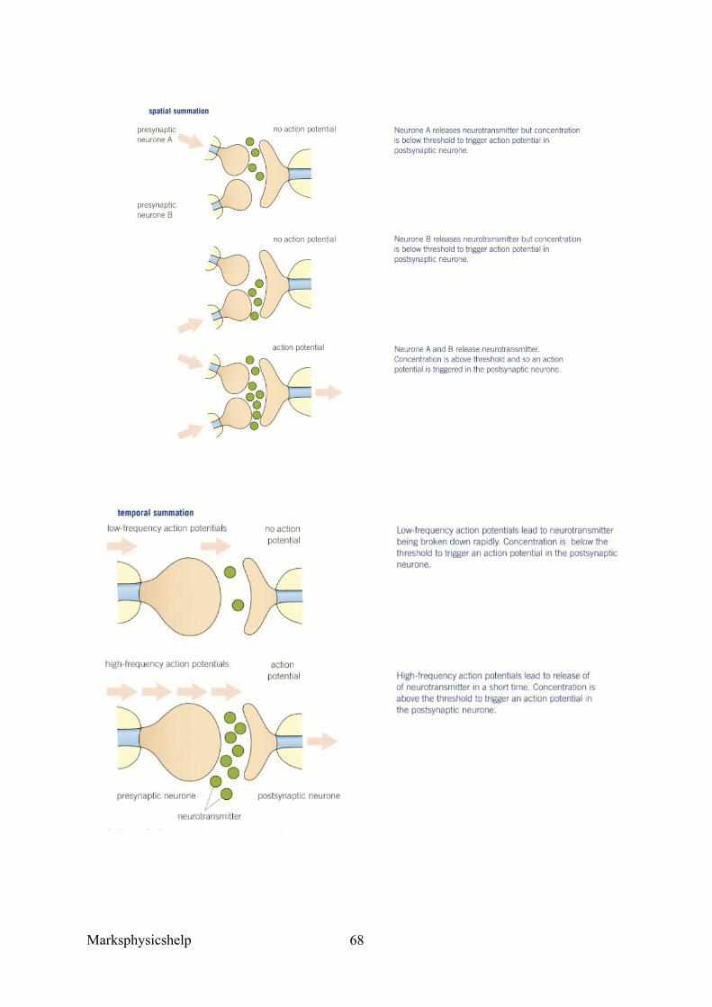
Marksphysicshelp
68
Marksphysicshelp
69
• Inhibition: Some synapses make it less likely that a new action potential will be created on the postsynaptic neurones, called inhibitory synapses. Essentially, these synapses cause the opening of chloride ion channels to make the postsynaptic membrane more negatively charged that normal (hyperpolarized), so are less likely to depolarise and form a new action potential. This happens because the presynaptic knob releases a type of neurotransmitter that binds to chloride ion protein channels on the postsynaptic neurone. The neurotransmitter causes the chloride ion protein channels to open and chloride ions move into the postsynaptic neurone by facilitated diffusion. The binding of the neurotransmitter causes nearby potassium protein channels to open, so potassium ions move out of the postsynaptic neurone into the synapse. This combined effect of negatively charged chloride ions moving in and positive potassium ions out, makes the inside of the postsynaptic membrane more negative and the outside more positive. Therefore, hyperpolarisation has occurred and this makes it less likely for a new action potential to be created, as a larger quantity of sodium ions is required to reach the threshold level.
Drugs can affect synapses in two main ways:
• They stimulate the nervous system by creating more action potentials in postsynaptic neurones.
o A drug may do this by mimicking a neurotransmitter, stimulating the release of more neurotransmitter, or inhibiting the enzyme that breaks down the neurotransmitter. The outcome is to enhance the body’s response to impulses passed along a postsynaptic neurone. For example, if the neurone transmits impulses from sound receptors, a person will perceive the sound as being louder.
• They inhibit the nervous system by creating fewer action potentials in postsynaptic neurones.
o A drug may do this by inhibiting the release of neurotransmitter, or blocking receptors on sodium/potassium ion channels on the postsynaptic neurone. The outcome is to reduce the impulses passed along the postsynaptic neurone. In this case, if the neurone transmits impulses from sound receptors, a person will perceive the sound as being quieter.

Marksphysicshelp
70
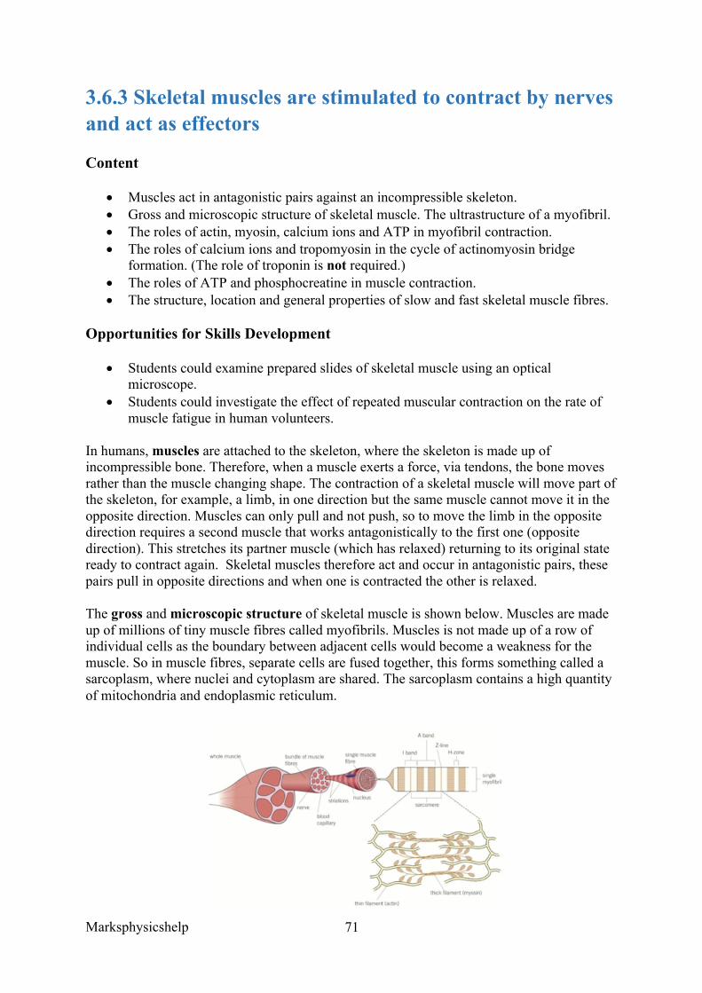
Marksphysicshelp
71
3.6.3 Skeletal muscles are stimulated to contract by nerves and act as effectors
Content
• Muscles act in antagonistic pairs against an incompressible skeleton. • Gross and microscopic structure of skeletal muscle. The ultrastructure of a myofibril. • The roles of actin, myosin, calcium ions and ATP in myofibril contraction. • The roles of calcium ions and tropomyosin in the cycle of actinomyosin bridge
formation. (The role of troponin is not required.) • The roles of ATP and phosphocreatine in muscle contraction. • The structure, location and general properties of slow and fast skeletal muscle fibres.
Opportunities for Skills Development
• Students could examine prepared slides of skeletal muscle using an optical microscope.
• Students could investigate the effect of repeated muscular contraction on the rate of muscle fatigue in human volunteers.
In humans, muscles are attached to the skeleton, where the skeleton is made up of incompressible bone. Therefore, when a muscle exerts a force, via tendons, the bone moves rather than the muscle changing shape. The contraction of a skeletal muscle will move part of the skeleton, for example, a limb, in one direction but the same muscle cannot move it in the opposite direction. Muscles can only pull and not push, so to move the limb in the opposite direction requires a second muscle that works antagonistically to the first one (opposite direction). This stretches its partner muscle (which has relaxed) returning to its original state ready to contract again. Skeletal muscles therefore act and occur in antagonistic pairs, these pairs pull in opposite directions and when one is contracted the other is relaxed.
The gross and microscopic structure of skeletal muscle is shown below. Muscles are made up of millions of tiny muscle fibres called myofibrils. Muscles is not made up of a row of individual cells as the boundary between adjacent cells would become a weakness for the muscle. So in muscle fibres, separate cells are fused together, this forms something called a sarcoplasm, where nuclei and cytoplasm are shared. The sarcoplasm contains a high quantity of mitochondria and endoplasmic reticulum.
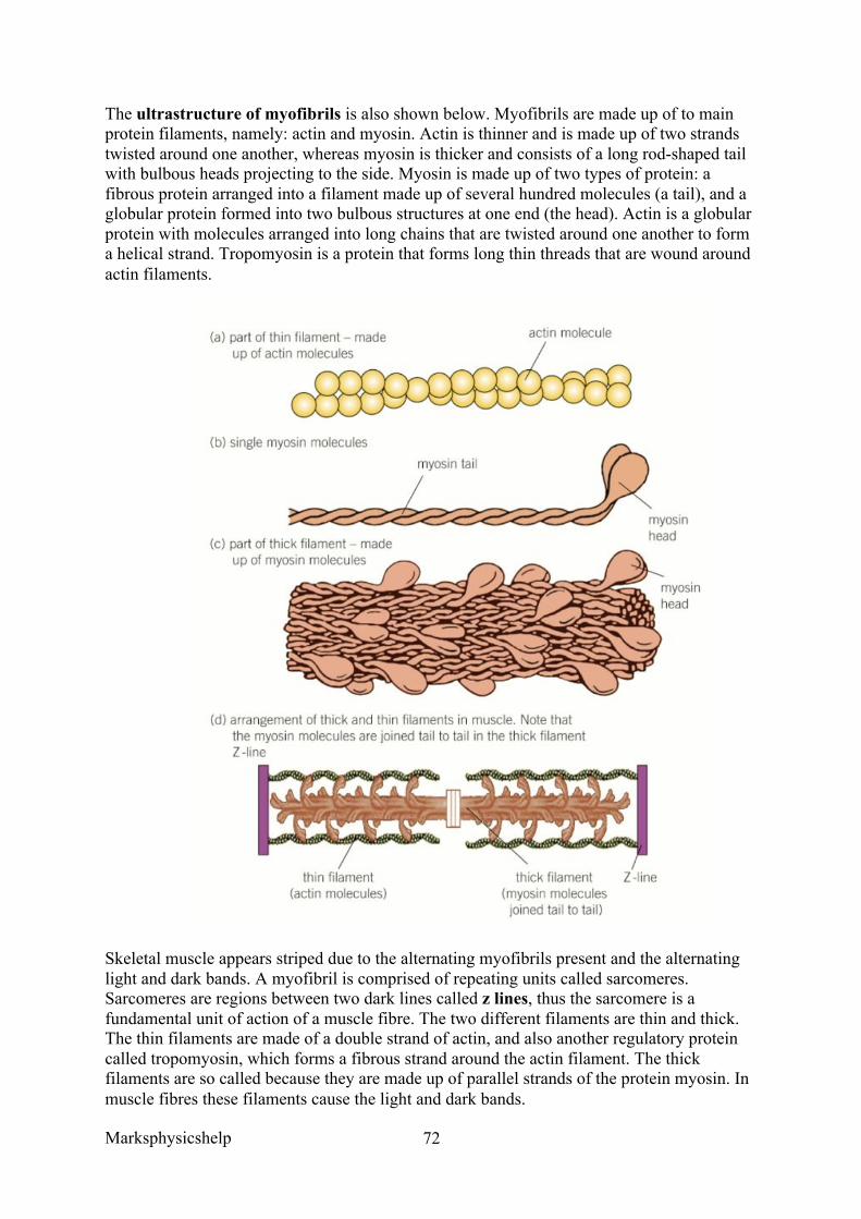
Marksphysicshelp
72
The ultrastructure of myofibrils is also shown below. Myofibrils are made up of to main protein filaments, namely: actin and myosin. Actin is thinner and is made up of two strands twisted around one another, whereas myosin is thicker and consists of a long rod-shaped tail with bulbous heads projecting to the side. Myosin is made up of two types of protein: a fibrous protein arranged into a filament made up of several hundred molecules (a tail), and a globular protein formed into two bulbous structures at one end (the head). Actin is a globular protein with molecules arranged into long chains that are twisted around one another to form a helical strand. Tropomyosin is a protein that forms long thin threads that are wound around actin filaments.
Skeletal muscle appears striped due to the alternating myofibrils present and the alternating light and dark bands. A myofibril is comprised of repeating units called sarcomeres. Sarcomeres are regions between two dark lines called z lines, thus the sarcomere is a fundamental unit of action of a muscle fibre. The two different filaments are thin and thick. The thin filaments are made of a double strand of actin, and also another regulatory protein called tropomyosin, which forms a fibrous strand around the actin filament. The thick filaments are so called because they are made up of parallel strands of the protein myosin. In muscle fibres these filaments cause the light and dark bands.
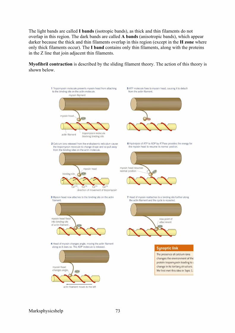
Marksphysicshelp
73
The light bands are called I bands (isotropic bands), as thick and thin filaments do not overlap in this region. The dark bands are called A bands (anisotropic bands), which appear darker because the thick and thin filaments overlap in this region (except in the H zone where only thick filaments occur). The I band contains only thin filaments, along with the proteins in the Z line that join adjacent thin filaments.
Myofibril contraction is described by the sliding filament theory. The action of this theory is shown below.
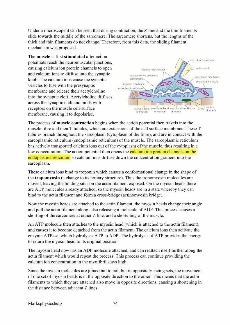
Marksphysicshelp
74
Under a microscope it can be seen that during contraction, the Z line and the thin filaments slide towards the middle of the sarcomere. The sarcomere shortens, but the lengths of the thick and thin filaments do not change. Therefore, from this data, the sliding filament mechanism was proposed.
The muscle is first stimulated after action potentials reach the neuromuscular junctions, causing calcium ion protein channels to open and calcium ions to diffuse into the synaptic knob. The calcium ions cause the synaptic vesicles to fuse with the presynaptic membrane and release their acetylcholine into the synaptic cleft. Acetylcholine diffuses across the synaptic cleft and binds with receptors on the muscle cell-surface membrane, causing it to depolarise.
The process of muscle contraction begins when the action potential then travels into the muscle fibre and then T-tubules, which are extensions of the cell surface membrane. These T-tubules branch throughout the sarcoplasm (cytoplasm of the fibre), and are in contact with the sarcoplasmic reticulum (endoplasmic reticulum) of the muscle. The sarcoplasmic reticulum has actively transported calcium ions out of the cytoplasm of the muscle, thus resulting in a low concentration. The action potential then opens the calcium ion protein channels on the endoplasmic reticulum so calcium ions diffuse down the concentration gradient into the sarcoplasm.
These calcium ions bind to troponin which causes a conformational change in the shape of the tropomyosin (a change to its tertiary structure). Thus the tropomyosin molecules are moved, leaving the binding sites on the actin filament exposed. On the myosin heads there are ADP molecules already attached, so the myosin heads are in a state whereby they can bind to the actin filament and form a cross-bridge (actinomyosin bridge).
Now the myosin heads are attached to the actin filament, the myosin heads change their angle and pull the actin filament along, also releasing a molecule of ADP. This process causes a shorting of the sarcomere at either Z line, and a shortening of the muscle.
An ATP molecule then attaches to the myosin head (which is attached to the actin filament), and causes it to become detached from the actin filament. The calcium ions then activate the enzyme ATPase, which hydrolyses ATP to ADP. The hydrolysis of ATP provides the energy to return the myosin head to its original position.
The myosin head now has an ADP molecule attached, and can reattach itself further along the actin filament which would repeat the process. This process can continue providing the calcium ion concentration in the myofibril stays high.
Since the myosin molecules are joined tail to tail, but in oppositely facing sets, the movement of one set of myosin heads is in the opposite direction to the other. This means that the actin filaments to which they are attached also move in opposite directions, causing a shortening in the distance between adjacent Z lines.
Marksphysicshelp
75
For muscle relaxation, once the nervous stimulation ceases, calcium ions are actively transported back into the sarcoplasmic reticulum against the concentration gradient. Energy is provided for this from the hydrolysis of ATP. The reabsorption of calcium ions means that the tropomyosin can return to its original position and block the actin filament binding sites. Myosin heads cannot bind anymore, contraction ceases and the muscle relaxes.
ATP and phosphocreatine are essential to the process of muscle contraction. ATP is hydrolysed to ADP and Pi to provide energy for the movement of the myosin heads, and for the reabsorption of calcium ions back into the endoplasmic reticulum. Since ATP is required in such vast amounts, in the absence of oxygen, a means of generating ATP rapidly is required. This is partially achieved using the chemical phosphocreatine, and partly by more glycolysis.
Phosphocreatine is unable to supply energy directly to the muscle, so instead regenerates ATP. Phosphocreatine provides a reserve supply of phosphate, so that it can combine with ADP to form ATP. The phosphocreatine store is replenished using the phosphate from ATP when the muscle is relaxed.
The structure, location and general properties of slow and fast skeletal muscle fibres:
Slow-twitch fibres contract more slowly than fast twitch fibres, and are more adapted to function over long periods of time. They are adapted to respire aerobically to avoid the build up of lactic acid, ensuring they are not fatigued very quickly. It also ensures they can contract over a long period of time. Some of their adaptations include:
• A high content of myoglobin to provide a source of oxygen. • A good blood supply to ensure they can obtain a sufficient amount of oxygen and
glucose. • A high density of mitochondria to provide ATP and use oxygen efficiently for this
purpose.
However, they cannot produce ATP very fast due to their aerobic metabolism, meaning that they are not very powerful.
Fast-twitch fibres contract more rapidly and produce powerful contractions but only for a short period of time. They generate ATP much quicker and anaerobically from stores of the high-energy compound Phosphocreatine (creatine phosphate), and also lactate fermentation. When creatine phosphate breaks down it releases energy and phosphate ions which can be used to make ATP for up to 10 seconds of activity. Creatine phosphate is regenerated during aerobic respiration. They are adapted for their role by:
• Having thicker and more numerous myosin filaments. • A high concentration of glycogen. • A large quantity of enzymes required for anaerobic respiration to provide ATP
rapidly. • A large store of phosphocreatine.
Marksphysicshelp
76
AQA June 2013 Q2ab
Question:
‘Describe the part played by each of the following in myofibril contraction.
Answer:
Tropomyosin’
• Moves out of the way when calcium ions bind; • Allowing myosin to bind (to actin)/cross bridge formation;
Myosin’
• Head (of myosin) binds to actin and moves/pulls/slides actin past; • (Myosin) detaches from actin and re-sets/moves further along (actin) • This uses ATP;
Question:
‘The table shows features of fast and slow muscle fibres.’
Use information from the table to suggest and explain one advantage of:
the high glycogen content of fast muscle fibres’
Answer:
• (Glycogen broken down) gives (lots of) glucose for glycolysis/anaerobic respiration; • Glycolysis/anaerobic respiration not very efficient/only yields 2 ATP per glucose;
‘the number of capillaries supplying slow muscle fibres.’
• ‘(Many capillaries) give high concentration/lots of oxygen/ shorter diffusion pathway for oxygen/large surface area for oxygen exchange/diffusion;
• Good glucose supplies with little glycogen present; • Allows high rate of/more aerobic respiration OR prevents build-up of lactic
acid/(muscle) fatigue;
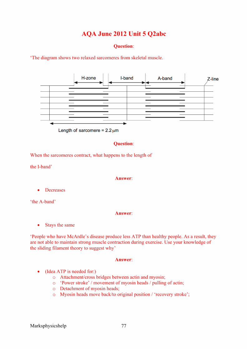
Marksphysicshelp
77
AQA June 2012 Unit 5 Q2abc
Question:
‘The diagram shows two relaxed sarcomeres from skeletal muscle.
Question:
When the sarcomeres contract, what happens to the length of
the I-band’
Answer:
• Decreases
‘the A-band’
Answer:
• Stays the same
‘People who have McArdle’s disease produce less ATP than healthy people. As a result, they are not able to maintain strong muscle contraction during exercise. Use your knowledge of the sliding filament theory to suggest why’
Answer:
• (Idea ATP is needed for:) o Attachment/cross bridges between actin and myosin; o ‘Power stroke’ / movement of myosin heads / pulling of actin; o Detachment of myosin heads; o Myosin heads move back/to original position / ‘recovery stroke’;
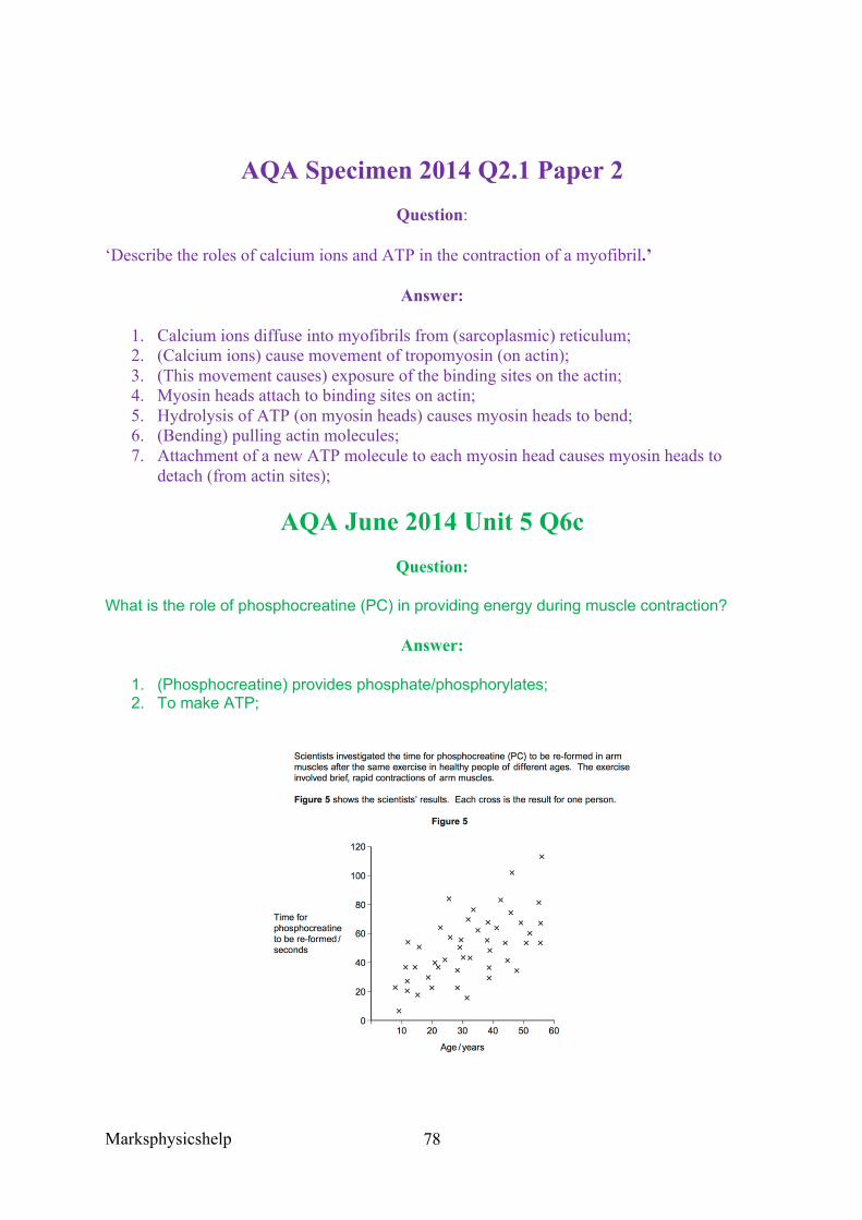
Marksphysicshelp
78
AQA Specimen 2014 Q2.1 Paper 2 Question:
‘Describe the roles of calcium ions and ATP in the contraction of a myofibril.’
Answer:
1. Calcium ions diffuse into myofibrils from (sarcoplasmic) reticulum; 2. (Calcium ions) cause movement of tropomyosin (on actin); 3. (This movement causes) exposure of the binding sites on the actin; 4. Myosin heads attach to binding sites on actin; 5. Hydrolysis of ATP (on myosin heads) causes myosin heads to bend; 6. (Bending) pulling actin molecules; 7. Attachment of a new ATP molecule to each myosin head causes myosin heads to
detach (from actin sites);
AQA June 2014 Unit 5 Q6c Question:
What is the role of phosphocreatine (PC) in providing energy during muscle contraction?
Answer:
1. (Phosphocreatine) provides phosphate/phosphorylates; 2. To make ATP;
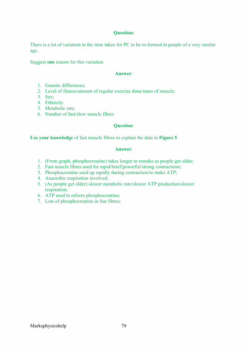
Marksphysicshelp
79
Question:
There is a lot of variation in the time taken for PC to be re-formed in people of a very similar age.
Suggest one reason for this variation
Answer:
1. Genetic differences; 2. Level of fitness/amount of regular exercise done/mass of muscle; 3. Sex; 4. Ethnicity 5. Metabolic rate; 6. Number of fast/slow muscle fibres
Question:
Use your knowledge of fast muscle fibres to explain the data in Figure 5
Answer:
1. (From graph, phosphocreatine) takes longer to remake as people get older; 2. Fast muscle fibres used for rapid/brief/powerful/strong contractions; 3. Phosphocreatine used up rapidly during contraction/to make ATP; 4. Anaerobic respiration involved; 5. (As people get older) slower metabolic rate/slower ATP production/slower
respiration; 6. ATP used to reform phosphocreatine; 7. Lots of phosphocreatine in fast fibres;
Marksphysicshelp
80
AQA Specimen Paper Q1.5
Question:
During vigorous exercise, the pH of skeletal muscle tissue falls. This fall in pH leads to a reduction in the ability of calcium ions to stimulate muscle contraction.
Suggest how
Answer:
1. Low pH changes shape of calcium ion receptors 2. Fewer calcium ions bind to tropomyosin 3. Fewer tropomyosin molecules move away; 4. Fewer binding sites on actin revealed; 5. Fewer cross-bridges can form ORFewer myosin heads can bind

Marksphysicshelp
81
Marksphysicshelp
82
3.6.4 Homeostasis is the maintenance of a stable internal environment 3.6.4.1 Principles of homeostasis and negative feedback
Content
• Homeostasis in mammals involves physiological control systems that maintain the internal environment within restricted limits.
• The importance of maintaining a stable core temperature and stable blood pH in relation to enzyme activity.
• The importance of maintaining a stable blood glucose concentration in terms of availability of respiratory substrate and of the water potential of blood.
• Negative feedback restores systems to their original level. • The possession of separate mechanisms involving negative feedback controls
departures in different directions from the original state, giving a greater degree of control.
• Students should be able to interpret information relating to examples of negative and positive feedback.
Homeostasis in mammals involves physiological control systems that maintain the internal environment within restricted limits. The internal environment within organisms is made of tissue fluids that bathe each cell, supplying nutrients and removing waste products. Homeostasis ensures that cells are in an environment in which meets all of their requirements and allows them to function normally despite external changes.
Essentially homeostasis is the ability to return to an optimum point and so maintain organisms in a balanced equilibrium. This is vital for the proper functioning of organisms, for example:
• The enzymes controlling the biochemical reactions within the cells, and proteins like channel proteins are sensitive to changes in pH and temperature. Any changes in the temperature reduce the rate of reactions of enzymes and can even denature them.
• Maintaining a stable blood glucose concentration is vital to provide a constant source of glucose for respiration. It is also important that blood glucose concentration is maintained in terms of maintaining a constant water potential in the blood and tissue fluids. Otherwise it may cause cells to shrink and expand as a result of the movement of water by osmosis. The consequence of this would be that the cell would not function properly.
Organisms with the ability to maintain a constant internal environment are more independent of changes in the external environment. They may have a wider geographical range and therefore have a greater chance of finding food, shelter etc. Mammals for example are found in many habitats, from hot arid deserts to cold, frozen polar regions.
Marksphysicshelp
83
Control mechanisms
In any self-regulating system there are a series of stages that feature:
• The optimum point - the point at which the system operates best. This is monitored by a…
• Receptor which detects any deviation from the optimum point ie a stimulus. This receptor then informs the…
• Coordinator, which coordinates information from receptors and sends instructions to an appropriate…
• Effector, often a muscle or gland which brings about the changes needed to return the system to the optimum point. This return to normality creates a…
• Feedback mechanism, by which a receptor responds to a stimulus created by the change to the system brought about by the effector.
Most systems use negative feedback mechanisms. This means that the change produced by the control system leads to a change in the stimulus detected by the receptor and turns the system off. Blood glucose level is controlled by a negative feedback mechanism.
Positive feedback mechanisms occur when deviations from an optimum cause changes that result in even greater deviations from the normal. One example is in neurones where a stimulus leads to a small influx of sodium ions. This influx increases permeability of the neurone membrane to sodium ions, more ions enter, causing a further increase in permeability and even more rapid entry of ions. In this way a stimulus can bring about a large and rapid response.
It is crucial that control systems have many receptors and effectors. In this way, they can have different systems that can restore a positive movement back to the optimum, allowing greater control. For example, in blood glucose concentration, having insulin and glucagon for high and low concentrations. The brain is able to analyse the messages from separate systems to choose the correct course of action.
EXTENSION • Endotherms and ectotherms.
Animals deriving most of their heat from metabolic activities taking place inside their body are known as endotherms. Some animals obtain a large proportion of their heat from sources outside their bodies, namely the environment. They are called ectotherms.
Ectotherms like lizards maintain a constant body temperature by exposing themselves to the sun or taking shelter. They also gain warmth from the ground by pressing their bodies against areas of hot ground to warm them up.
Endotherms rely on metabolic activities, and so their body temperature remains relatively constant. They also use behaviour to maintain a constant body temperature, but unlike ectotherms they use a wide range of physiological mechanisms to regulate their temperature.
Marksphysicshelp
84
3.6.4.2 Control of blood glucose concentration
Content
• The factors that influence blood glucose concentration. • The role of the liver in glycogenesis, glycogenolysis and gluconeogenesis. • The action of insulin by:
o Attaching to receptors on the surfaces of target cells o Controlling the uptake of glucose by regulating the inclusion of channel
proteins in the surface membranes of target cells o Activating enzymes involved in the conversion of glucose to glycogen.
• The action of glucagon by: o Attaching to receptors on the surfaces of target cells o Activating enzymes involved in the conversion of glycogen to glucose o Activating enzymes involved in the conversion of glycerol and amino acids
into glucose. • The role of adrenaline by:
o Attaching to receptors on the surfaces of target cells o Activating enzymes involved in the conversion of glycogen to glucose.
• The second messenger model of adrenaline and glucagon action, involving adenylate cyclase, cyclic AMP (cAMP) and protein kinase.
• The causes of types I and II diabetes and their control by insulin and/ or manipulation of the diet.
• Students should be able to evaluate the positions of health advisers and the food industry in relation to the increased incidence of type II diabetes.
There are three main factors influencing blood glucose concentration in you blood. The first factor is your diet, directly in the form of glucose but also from the hydrolysis of other carbohydrates like starch, maltose, lactose and sucrose. The hydrolysis of glycogen (glycogenolysis) stored in the liver and muscle cells, or from gluconeogenesis where glucose is produced from things other than carbohydrates ie glycerol and amino acids. Insulin, glucagon and adrenalin are the three main hormones that aim to regulate blood glucose concentration.
The liver has a variety of roles including regulating blood glucose concentration, and whilst the pancreas produces insulin and glucagon, it is the liver where they have their effects:
1. Glycogenesis: is the conversion of glucose into glycogen. When blood glucose concentration is higher than normal the liver removes glucose from the blood and converts it into glycogen.
2. Glycogenolysis: is the breakdown of glycogen to glucose. When blood glucose concentration is lower than normal, the liver can convert glycogen back into glucose which diffuses into the blood to restore the normal blood glucose concentration.
3. Gluconeogenesis: is the production of glucose from sources other than carbohydrates. When its supply of glycogen is exhausted, the liver can produce glucose from sources like glycerol and amino acids.
Hormones differ chemically but all have similar characteristics. Hormones are:
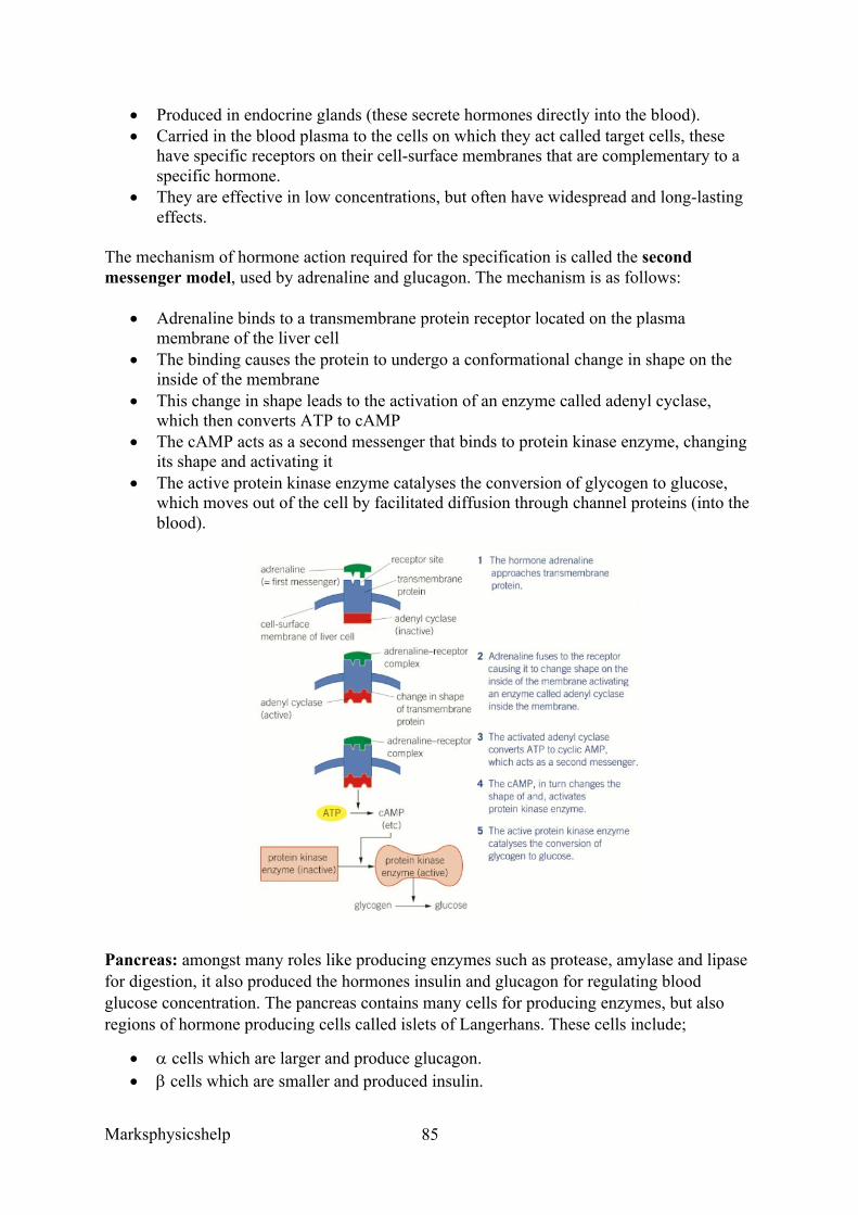
Marksphysicshelp
85
• Produced in endocrine glands (these secrete hormones directly into the blood). • Carried in the blood plasma to the cells on which they act called target cells, these
have specific receptors on their cell-surface membranes that are complementary to a specific hormone.
• They are effective in low concentrations, but often have widespread and long-lasting effects.
The mechanism of hormone action required for the specification is called the second messenger model, used by adrenaline and glucagon. The mechanism is as follows:
• Adrenaline binds to a transmembrane protein receptor located on the plasma membrane of the liver cell
• The binding causes the protein to undergo a conformational change in shape on the inside of the membrane
• This change in shape leads to the activation of an enzyme called adenyl cyclase, which then converts ATP to cAMP
• The cAMP acts as a second messenger that binds to protein kinase enzyme, changing its shape and activating it
• The active protein kinase enzyme catalyses the conversion of glycogen to glucose, which moves out of the cell by facilitated diffusion through channel proteins (into the blood).
Pancreas: amongst many roles like producing enzymes such as protease, amylase and lipase for digestion, it also produced the hormones insulin and glucagon for regulating blood glucose concentration. The pancreas contains many cells for producing enzymes, but also regions of hormone producing cells called islets of Langerhans. These cells include;
• a cells which are larger and produce glucagon. • b cells which are smaller and produced insulin.
Marksphysicshelp
86
The action of insulin (b cells) :
• The b cells of the islets of Langerhans in the pancreas have receptors that detect the stimulus of a rise in blood glucose concentration, responding by secreting the hormone insulin directly into the blood plasma. Insulin is a globular protein made of 51 amino acids.
• Almost all body cells (except the notable red blood cells), have glycoprotein receptors on their cell-surface membranes that bind specifically with insulin molecules. When it combines with the receptors it brings about the following changes:
o A change in the tertiary structure of the glucose transport carrier proteins, so a conformational change in shape causing them to open. Thus it allows more glucose into cells by facilitated diffusion down a concentration gradient.
o An increase in the number of carrier proteins responsible for glucose transport in the plasma membrane. At low insulin concentrations, the protein from which these channels are made is part of the membrane of the vesicles. A rise in insulin concentration results in these vesicles fusing with the cell-surface membrane so increasing the number of glucose transport channels.
o Activation of enzymes that convert glucose to glycogen and fat. • The resulting effect is that blood glucose concentration lowers as:
o It increases the rate of absorption of glucose into the cells, especially muscle cells
o Increases the respiratory rate of cells, using up more glucose, so increasing uptake of glucose from the blood
o Increasing the rate of conversion of glucose into glycogen (glycogenesis), in the cells of the liver and muscles
o Increases the rate of conversion of glucose to fat. • The effect of these processes is to remove glucose from the blood and return its value
to its optimum. The lowering of blood glucose concentration causes the b cells to reduce their secretion of insulin (negative feedback).
The action of glucagon (a cells):
• The a cells of the islets of Langerhans detect a fall in blood glucose concentration so respond by secreting glucagon directly into the blood (endocrine gland). Glucagon works by:
o Attaching to specific protein receptors on the cell-surface membrane of liver cells
o Activating enzymes that convert glycogen to glucose o Activating enzymes involved in the conversion of glycerol and amino acids
into glucose (gluconeogenesis). • The overall effect is to increase the concentration of glucose in the blood and return it
to its optimum concentration. This then causes a cells to reduce secretion of glucagon (negative feedback).
The action of adrenaline:
Marksphysicshelp
87
• There are around four other hormones not including glucagon that can increase blood glucose concentration, one being adrenaline. It does so by
o Attaching to protein receptors on the cell-surface membrane of target cells o Activating enzymes that cause the breakdown of glycogen to glucose in the
liver.
Hormone interaction in regulating blood glucose level involves glucagon and insulin acting antagonistically. The system is self regulating via negative feedback as it is the concentration of glucose in the blood that determines the quantity of insulin and glucagon produced.
Types of Diabetes
Diabetes is a disease in which a person is unable to metabolise carbohydrates, especially glucose, properly. The problems arise either from a lack of the hormone insulin, or a loss of responsiveness to insulin.
• Type I (insulin dependent) – due to bodies inability to produce insulin. It normally begins in childhood, and may be the result of an autoimmune response whereby the body’s immune system attacks its own b cells of the islets of Langerhans.
• Type II (insulin independent) – normally due to glycoprotein receptors on body cells being lost or losing their responsiveness to insulin. It can also be due to an inadequate supply of insulin from the pancreas. Type II diabetes usually develops in people over 40, although diet and obesity have increased the number of cases. It develops slowly unlike type I diabetes.
Diabetes and its control
Diabetes does not have a ‘cure’, but can be treated successfully. Transplanting insulin-producing cells may also be a possibility in the near future.
• Type I diabetes is controlled by insulin injections. However, they cannot be taken by mouth because, being a protein, it would be digested in the alimentary canal. Biosensors are used to ensure the correct dose is given. Using these injections and managing carbohydrate intake and exercise carefully, people can live with their diabetes with normal lives
• Type II diabetes is usually controlled by regulating the intake of carbohydrates in the diet, and matching this to the amount of exercise taken. In some cases, this may be supplemented by injections of insulin or by use of drugs that stimulate insulin production.
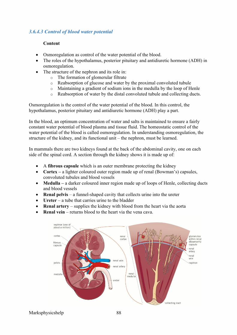
Marksphysicshelp
88
3.6.4.3 Control of blood water potential
Content
• Osmoregulation as control of the water potential of the blood. • The roles of the hypothalamus, posterior pituitary and antidiuretic hormone (ADH) in
osmoregulation. • The structure of the nephron and its role in:
o The formation of glomerular filtrate o Reabsorption of glucose and water by the proximal convoluted tubule o Maintaining a gradient of sodium ions in the medulla by the loop of Henle o Reabsorption of water by the distal convoluted tubule and collecting ducts.
Osmoregulation is the control of the water potential of the blood. In this control, the hypothalamus, posterior pituitary and antidiuretic hormone (ADH) play a part.
In the blood, an optimum concentration of water and salts is maintained to ensure a fairly constant water potential of blood plasma and tissue fluid. The homeostatic control of the water potential of the blood is called osmoregulation. In understanding osmoregulation, the structure of the kidney, and its functional unit – the nephron, must be learned.
In mammals there are two kidneys found at the back of the abdominal cavity, one on each side of the spinal cord. A section through the kidney shows it is made up of:
• A fibrous capsule which is an outer membrane protecting the kidney • Cortex – a lighter coloured outer region made up of renal (Bowman’s) capsules,
convoluted tubules and blood vessels • Medulla – a darker coloured inner region made up of loops of Henle, collecting ducts
and blood vessels • Renal pelvis – a funnel-shaped cavity that collects urine into the ureter • Ureter – a tube that carries urine to the bladder • Renal artery – supplies the kidney with blood from the heart via the aorta • Renal vein – returns blood to the heart via the vena cava.
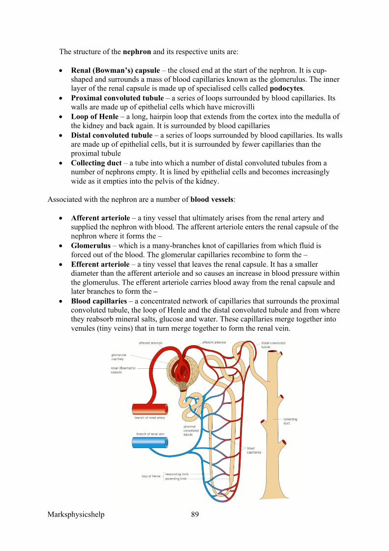
Marksphysicshelp
89
The structure of the nephron and its respective units are:
• Renal (Bowman’s) capsule – the closed end at the start of the nephron. It is cup-shaped and surrounds a mass of blood capillaries known as the glomerulus. The inner layer of the renal capsule is made up of specialised cells called podocytes.
• Proximal convoluted tubule – a series of loops surrounded by blood capillaries. Its walls are made up of epithelial cells which have microvilli
• Loop of Henle – a long, hairpin loop that extends from the cortex into the medulla of the kidney and back again. It is surrounded by blood capillaries
• Distal convoluted tubule – a series of loops surrounded by blood capillaries. Its walls are made up of epithelial cells, but it is surrounded by fewer capillaries than the proximal tubule
• Collecting duct – a tube into which a number of distal convoluted tubules from a number of nephrons empty. It is lined by epithelial cells and becomes increasingly wide as it empties into the pelvis of the kidney.
Associated with the nephron are a number of blood vessels:
• Afferent arteriole – a tiny vessel that ultimately arises from the renal artery and supplied the nephron with blood. The afferent arteriole enters the renal capsule of the nephron where it forms the –
• Glomerulus – which is a many-branches knot of capillaries from which fluid is forced out of the blood. The glomerular capillaries recombine to form the –
• Efferent arteriole – a tiny vessel that leaves the renal capsule. It has a smaller diameter than the afferent arteriole and so causes an increase in blood pressure within the glomerulus. The efferent arteriole carries blood away from the renal capsule and later branches to form the –
• Blood capillaries – a concentrated network of capillaries that surrounds the proximal convoluted tubule, the loop of Henle and the distal convoluted tubule and from where they reabsorb mineral salts, glucose and water. These capillaries merge together into venules (tiny veins) that in turn merge together to form the renal vein.
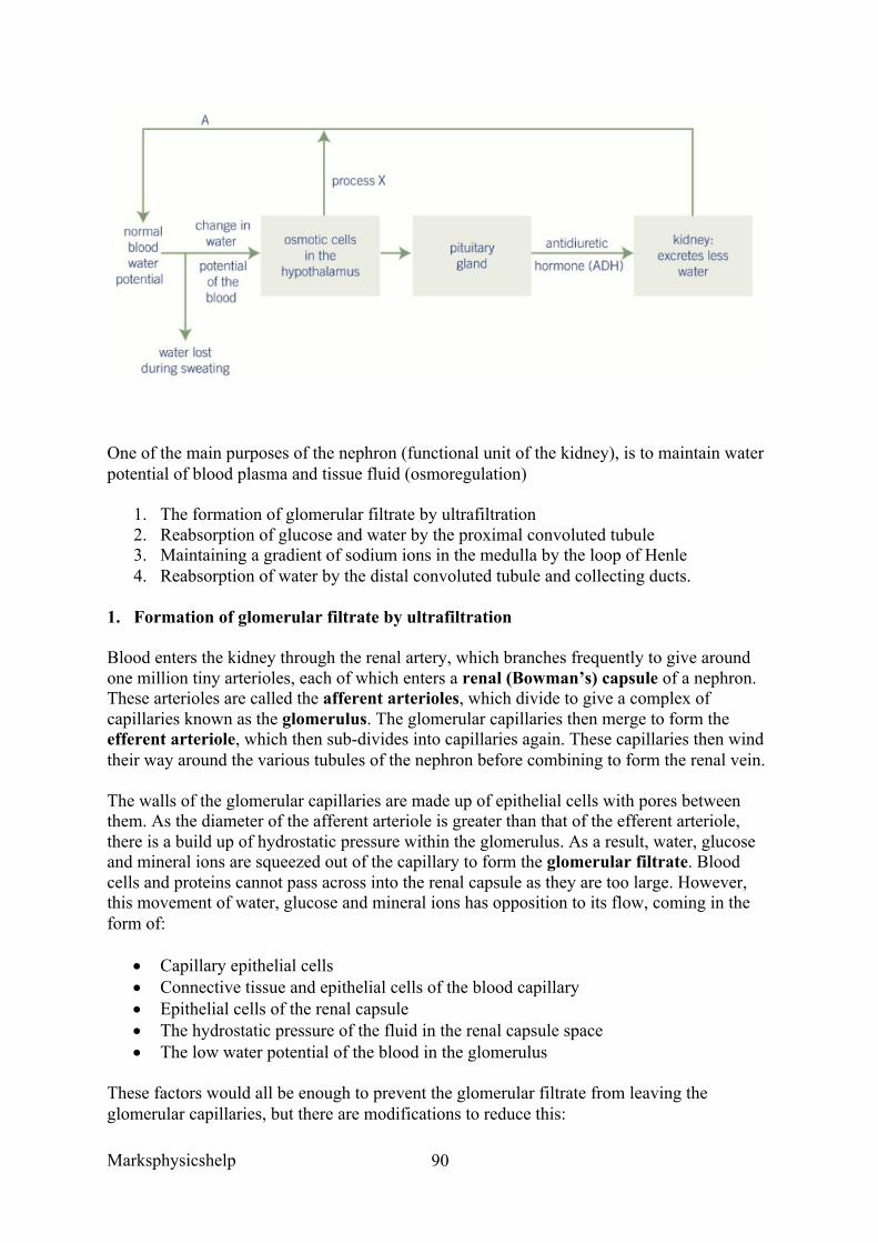
Marksphysicshelp
90
One of the main purposes of the nephron (functional unit of the kidney), is to maintain water potential of blood plasma and tissue fluid (osmoregulation)
1. The formation of glomerular filtrate by ultrafiltration 2. Reabsorption of glucose and water by the proximal convoluted tubule 3. Maintaining a gradient of sodium ions in the medulla by the loop of Henle 4. Reabsorption of water by the distal convoluted tubule and collecting ducts.
1. Formation of glomerular filtrate by ultrafiltration
Blood enters the kidney through the renal artery, which branches frequently to give around one million tiny arterioles, each of which enters a renal (Bowman’s) capsule of a nephron. These arterioles are called the afferent arterioles, which divide to give a complex of capillaries known as the glomerulus. The glomerular capillaries then merge to form the efferent arteriole, which then sub-divides into capillaries again. These capillaries then wind their way around the various tubules of the nephron before combining to form the renal vein.
The walls of the glomerular capillaries are made up of epithelial cells with pores between them. As the diameter of the afferent arteriole is greater than that of the efferent arteriole, there is a build up of hydrostatic pressure within the glomerulus. As a result, water, glucose and mineral ions are squeezed out of the capillary to form the glomerular filtrate. Blood cells and proteins cannot pass across into the renal capsule as they are too large. However, this movement of water, glucose and mineral ions has opposition to its flow, coming in the form of:
• Capillary epithelial cells • Connective tissue and epithelial cells of the blood capillary • Epithelial cells of the renal capsule • The hydrostatic pressure of the fluid in the renal capsule space • The low water potential of the blood in the glomerulus
These factors would all be enough to prevent the glomerular filtrate from leaving the glomerular capillaries, but there are modifications to reduce this:
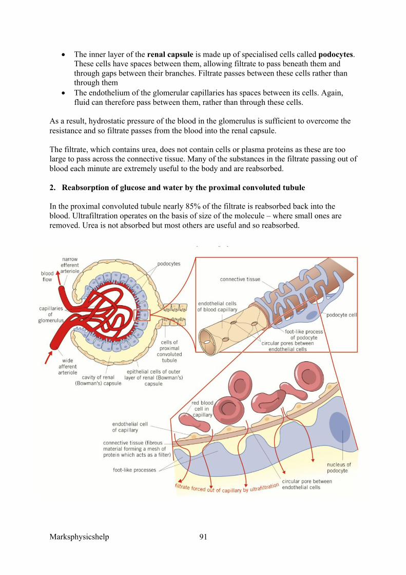
Marksphysicshelp
91
• The inner layer of the renal capsule is made up of specialised cells called podocytes. These cells have spaces between them, allowing filtrate to pass beneath them and through gaps between their branches. Filtrate passes between these cells rather than through them
• The endothelium of the glomerular capillaries has spaces between its cells. Again, fluid can therefore pass between them, rather than through these cells.
As a result, hydrostatic pressure of the blood in the glomerulus is sufficient to overcome the resistance and so filtrate passes from the blood into the renal capsule.
The filtrate, which contains urea, does not contain cells or plasma proteins as these are too large to pass across the connective tissue. Many of the substances in the filtrate passing out of blood each minute are extremely useful to the body and are reabsorbed.
2. Reabsorption of glucose and water by the proximal convoluted tubule
In the proximal convoluted tubule nearly 85% of the filtrate is reabsorbed back into the blood. Ultrafiltration operates on the basis of size of the molecule – where small ones are removed. Urea is not absorbed but most others are useful and so reabsorbed.
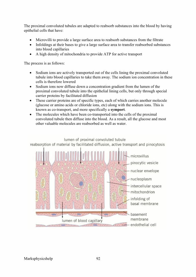
Marksphysicshelp
92
The proximal convoluted tubules are adapted to reabsorb substances into the blood by having epithelial cells that have:
• Microvilli to provide a large surface area to reabsorb substances from the filtrate • Infoldings at their bases to give a large surface area to transfer reabsorbed substances
into blood capillaries • A high density of mitochondria to provide ATP for active transport
The process is as follows:
• Sodium ions are actively transported out of the cells lining the proximal convoluted tubule into blood capillaries to take them away. The sodium ion concentration in these cells is therefore lowered
• Sodium ions now diffuse down a concentration gradient from the lumen of the proximal convoluted tubule into the epithelial lining cells, but only through special carrier proteins by facilitated diffusion
• These carrier proteins are of specific types, each of which carries another molecule (glucose or amino acids or chloride ions, etc) along with the sodium ions. This is known as co-transport, and more specifically a symport.
• The molecules which have been co-transported into the cells of the proximal convoluted tubule then diffuse into the blood. As a result, all the glucose and most other valuable molecules are reabsorbed as well as water.
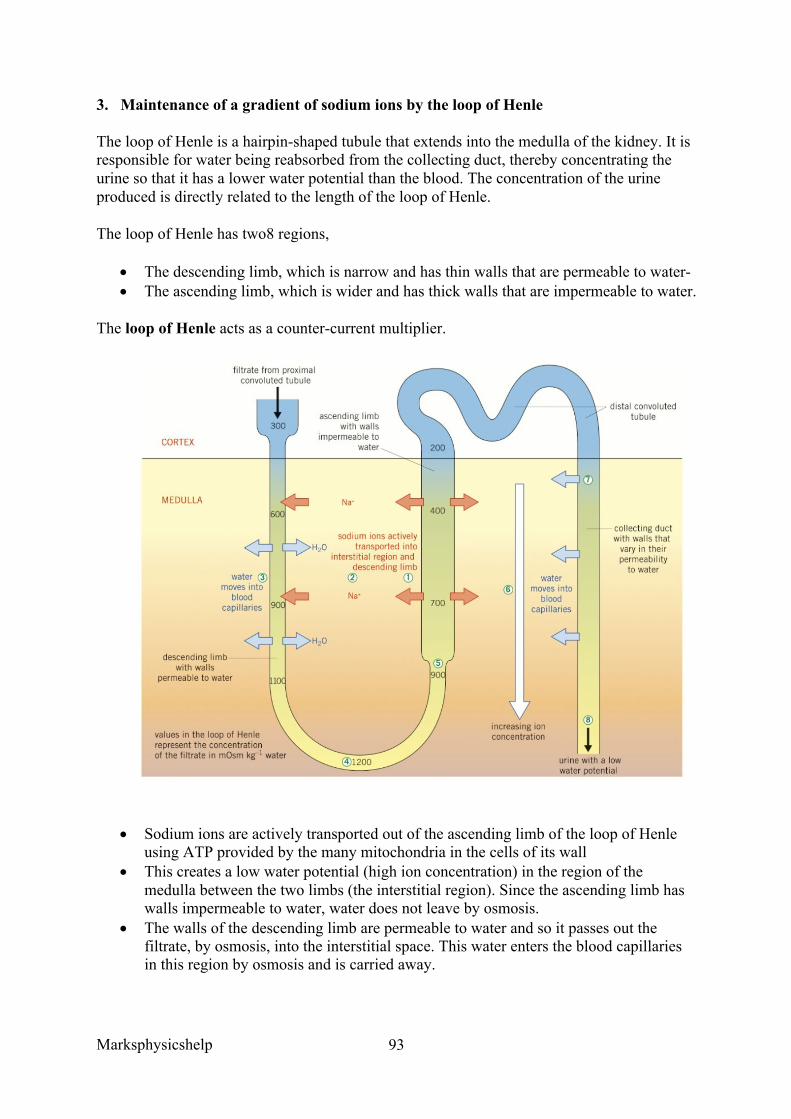
Marksphysicshelp
93
3. Maintenance of a gradient of sodium ions by the loop of Henle
The loop of Henle is a hairpin-shaped tubule that extends into the medulla of the kidney. It is responsible for water being reabsorbed from the collecting duct, thereby concentrating the urine so that it has a lower water potential than the blood. The concentration of the urine produced is directly related to the length of the loop of Henle.
The loop of Henle has two8 regions,
• The descending limb, which is narrow and has thin walls that are permeable to water- • The ascending limb, which is wider and has thick walls that are impermeable to water.
The loop of Henle acts as a counter-current multiplier.
• Sodium ions are actively transported out of the ascending limb of the loop of Henle using ATP provided by the many mitochondria in the cells of its wall
• This creates a low water potential (high ion concentration) in the region of the medulla between the two limbs (the interstitial region). Since the ascending limb has walls impermeable to water, water does not leave by osmosis.
• The walls of the descending limb are permeable to water and so it passes out the filtrate, by osmosis, into the interstitial space. This water enters the blood capillaries in this region by osmosis and is carried away.
Marksphysicshelp
94
• The filtrate progressively loses water in this way as it moves down the descending limb, lowering its water potential. It reaches its lowest water potential at the tip of the hairpin.
• At the base of the ascending limb, sodium ions diffuse out of the filtrate and as it moves up the ascending limb these ions are also actively pumped out, thus the filtrate develops a progressively higher water potential
• In the interstitial space between the ascending limb and the collecting duct there is a gradient of water potential with the highest water potential in the cortex and an increasingly lower water potential the further into the medulla you go
• The collecting duct is permeable to water and so, as the filtrate moves down it, water passes out of it by osmosis. This water passes by osmosis into the blood vessels that occupy this space, and is carried away
• As water passes out of the filtrate its water potential is lowered. However, the water potential is also lowered in the interstitial space and so water continues to move out by osmosis down the whole length of the collecting duct. The counter-current multiplier ensures that there is always a water potential gradient drawing water out of the tubule.
The water that passes out of the collecting duct by osmosis does so through channel proteins that are specific to water (aquaporins). ADH can alter the number of these channels and so control water loss. By the time the filtrate, which can be referred to as urine now, leaves the collecting duct on its way to the bladder, it has lost most of its water and so it has a lower water potential than the blood.
4. Reabsorption of water by the distal convoluted tubule and collecting ducts
The distal convoluted tubule has on its cells lining the wall, microvilli and many mitochondria. This allows them to reabsorb, by active transport, material rapidly from the filtrate. The main role of the distal tubule is to make final adjustments to the water and salts that are reabsorbed and to control the pH of the blood by selecting which ions to reabsorb. To achieve this, the permeability of its walls become altered under the influence of various hormones.
The counter-current multiplier is where two liquids flow in opposite directions past one another, like the principle found in fish. The loop of Henle utilises this principle as it means that the filtrate in the collecting duct with a lower water potential meets the interstitial fluid that has an even lower water potential. This means that, although the water potential gradient between the collecting duct and interstitial fluid is small, it exists for the whole length of the collecting duct. There is therefore a steady flow of water into the interstitial fluid, so almost all of the water enters the interstitial fluid and hence the blood. If the two flows were in the same direction less of the water would enter the blood.
The hypothalamus, posterior pituitary and antidiuretic hormone (ADH) all contribute in osmoregulation. The hormones act on the distal convoluted tubule and the collecting duct.
The water potential of the blood depends on the amount of solutes present, for example glucose, proteins, mineral ions and also the volume of water in the body. Rises in solute potential from too little water being consumed, a lot of sweating or large amounts of ions being taken in can all lower the water potential.
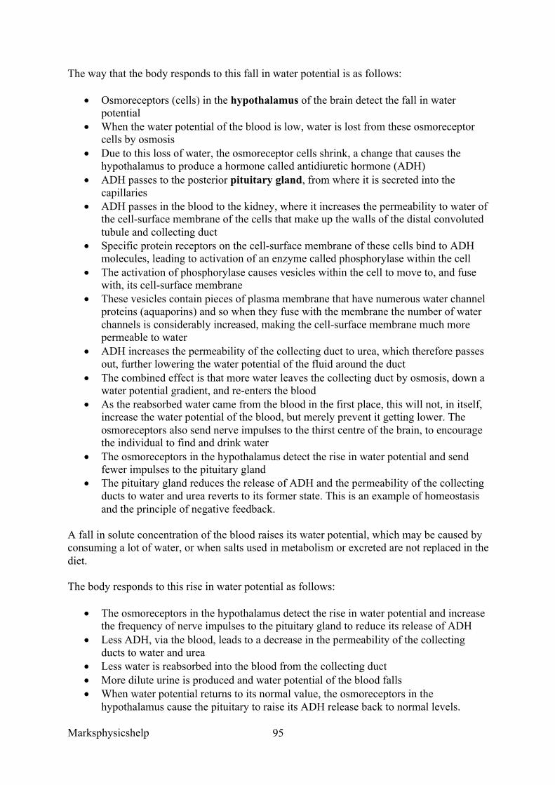
Marksphysicshelp
95
The way that the body responds to this fall in water potential is as follows:
• Osmoreceptors (cells) in the hypothalamus of the brain detect the fall in water potential
• When the water potential of the blood is low, water is lost from these osmoreceptor cells by osmosis
• Due to this loss of water, the osmoreceptor cells shrink, a change that causes the hypothalamus to produce a hormone called antidiuretic hormone (ADH)
• ADH passes to the posterior pituitary gland, from where it is secreted into the capillaries
• ADH passes in the blood to the kidney, where it increases the permeability to water of the cell-surface membrane of the cells that make up the walls of the distal convoluted tubule and collecting duct
• Specific protein receptors on the cell-surface membrane of these cells bind to ADH molecules, leading to activation of an enzyme called phosphorylase within the cell
• The activation of phosphorylase causes vesicles within the cell to move to, and fuse with, its cell-surface membrane
• These vesicles contain pieces of plasma membrane that have numerous water channel proteins (aquaporins) and so when they fuse with the membrane the number of water channels is considerably increased, making the cell-surface membrane much more permeable to water
• ADH increases the permeability of the collecting duct to urea, which therefore passes out, further lowering the water potential of the fluid around the duct
• The combined effect is that more water leaves the collecting duct by osmosis, down a water potential gradient, and re-enters the blood
• As the reabsorbed water came from the blood in the first place, this will not, in itself, increase the water potential of the blood, but merely prevent it getting lower. The osmoreceptors also send nerve impulses to the thirst centre of the brain, to encourage the individual to find and drink water
• The osmoreceptors in the hypothalamus detect the rise in water potential and send fewer impulses to the pituitary gland
• The pituitary gland reduces the release of ADH and the permeability of the collecting ducts to water and urea reverts to its former state. This is an example of homeostasis and the principle of negative feedback.
A fall in solute concentration of the blood raises its water potential, which may be caused by consuming a lot of water, or when salts used in metabolism or excreted are not replaced in the diet.
The body responds to this rise in water potential as follows:
• The osmoreceptors in the hypothalamus detect the rise in water potential and increase the frequency of nerve impulses to the pituitary gland to reduce its release of ADH
• Less ADH, via the blood, leads to a decrease in the permeability of the collecting ducts to water and urea
• Less water is reabsorbed into the blood from the collecting duct • More dilute urine is produced and water potential of the blood falls • When water potential returns to its normal value, the osmoreceptors in the
hypothalamus cause the pituitary to raise its ADH release back to normal levels.
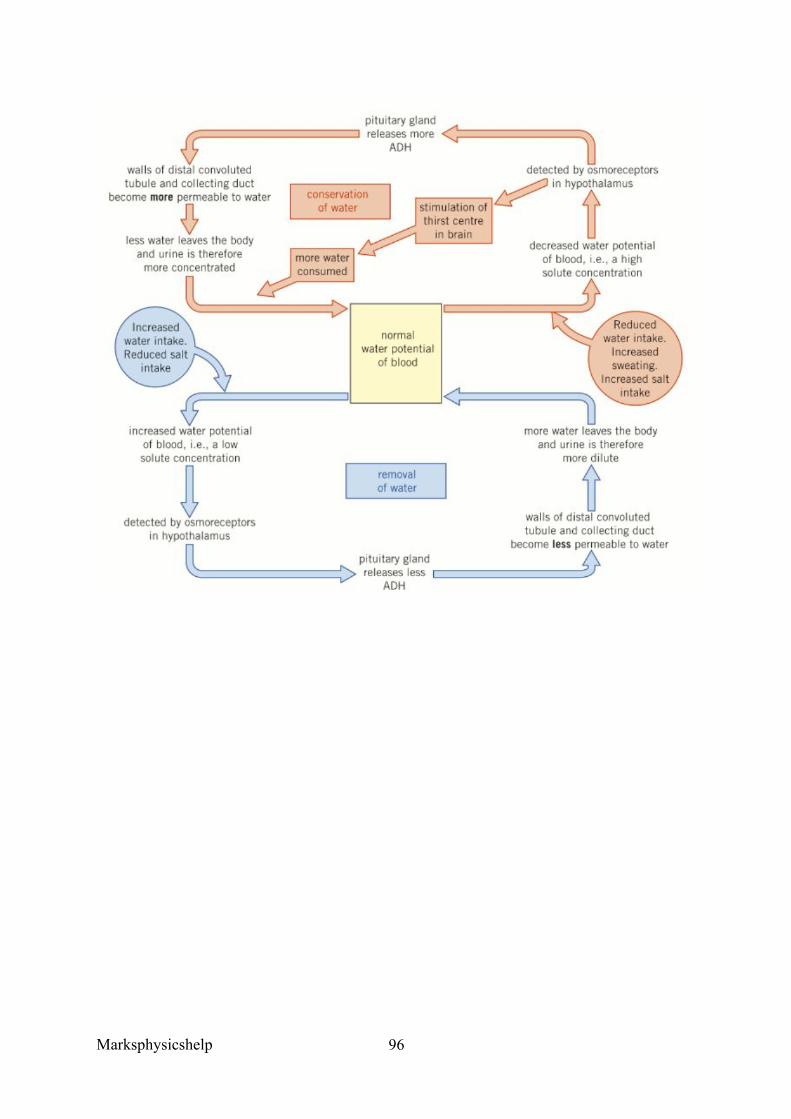
Marksphysicshelp
96

Marksphysicshelp
97
Marksphysicshelp
98
3.7 Genetics, populations, evolution and ecosystems 3.7.1 Inheritance
Content
• The genotype is the genetic constitution of an organism. • The phenotype is the expression of this genetic constitution and its interaction with
the environment. • There may be many alleles of a single genes • Alleles may be dominant, recessive or codominant. • In a diploid organism, the alleles at a specific locus may be either homozygous or
heterozygous. o Monohybrid and dihybrid crosses involving dominant, recessive and
codominant alleles. o Crosses involving sex-linkage, autosomal linkage, multiple alleles and
epistasis • Use of the chi-squared (c2) test to compare the goodness of fit observed phenotypic
ratios with expected ratios.
Opportunities for Skills Development
• Students could investigate genetic ratios using crosses of Drosophila or Fast Plant
• Students could use information to represent phenotypic ratios in monohybrid and
dihybrid crosses. • Students could show understanding of the probability associated with inheritance. • Students could test to investigate the significance of differences between expected and
observed phenotypic ratios.
The genotype of an organism is the genetic constitution of the organism. The phenotype, then, is the expression of this genetic constitution and its interaction with the environment. Essentially, the genotype describes the alleles that an organism has. For example, foot size may be determined to be a maximum amount by a given gene, but environmental factors may affect this. The phenotype is clearly then the observable or biochemical characteristic of an organism, resulting from the interaction between the expression of the genotype and the environment.
A single gene is a length of DNA, so a sequence of nucleotide bases that normally code for a particular polypeptide. There are many alleles of a single gene, and the position of a gene on a particular DNA molecule is known as the locus. Sometimes a gene has more than two allelic forms, the organism is then said to have multiple alleles for the character. Although only two alleles can be present in a single organism, so you get something like the ABO blood grouping system with more than the usual two distinctive outcomes for blood type.
Alleles can either be dominant, recessive or codominant, but only one allele of a gene can occur at the locus of any one chromosome. However, in diploid organisms the chromosomes occur in pairs called homologous chromosomes, and there are therefore two loci that each
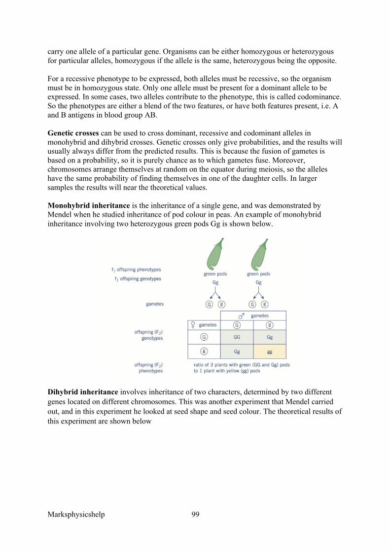
Marksphysicshelp
99
carry one allele of a particular gene. Organisms can be either homozygous or heterozygous for particular alleles, homozygous if the allele is the same, heterozygous being the opposite.
For a recessive phenotype to be expressed, both alleles must be recessive, so the organism must be in homozygous state. Only one allele must be present for a dominant allele to be expressed. In some cases, two alleles contribute to the phenotype, this is called codominance. So the phenotypes are either a blend of the two features, or have both features present, i.e. A and B antigens in blood group AB.
Genetic crosses can be used to cross dominant, recessive and codominant alleles in monohybrid and dihybrid crosses. Genetic crosses only give probabilities, and the results will usually always differ from the predicted results. This is because the fusion of gametes is based on a probability, so it is purely chance as to which gametes fuse. Moreover, chromosomes arrange themselves at random on the equator during meiosis, so the alleles have the same probability of finding themselves in one of the daughter cells. In larger samples the results will near the theoretical values.
Monohybrid inheritance is the inheritance of a single gene, and was demonstrated by Mendel when he studied inheritance of pod colour in peas. An example of monohybrid inheritance involving two heterozygous green pods Gg is shown below.
Dihybrid inheritance involves inheritance of two characters, determined by two different genes located on different chromosomes. This was another experiment that Mendel carried out, and in this experiment he looked at seed shape and seed colour. The theoretical results of this experiment are shown below
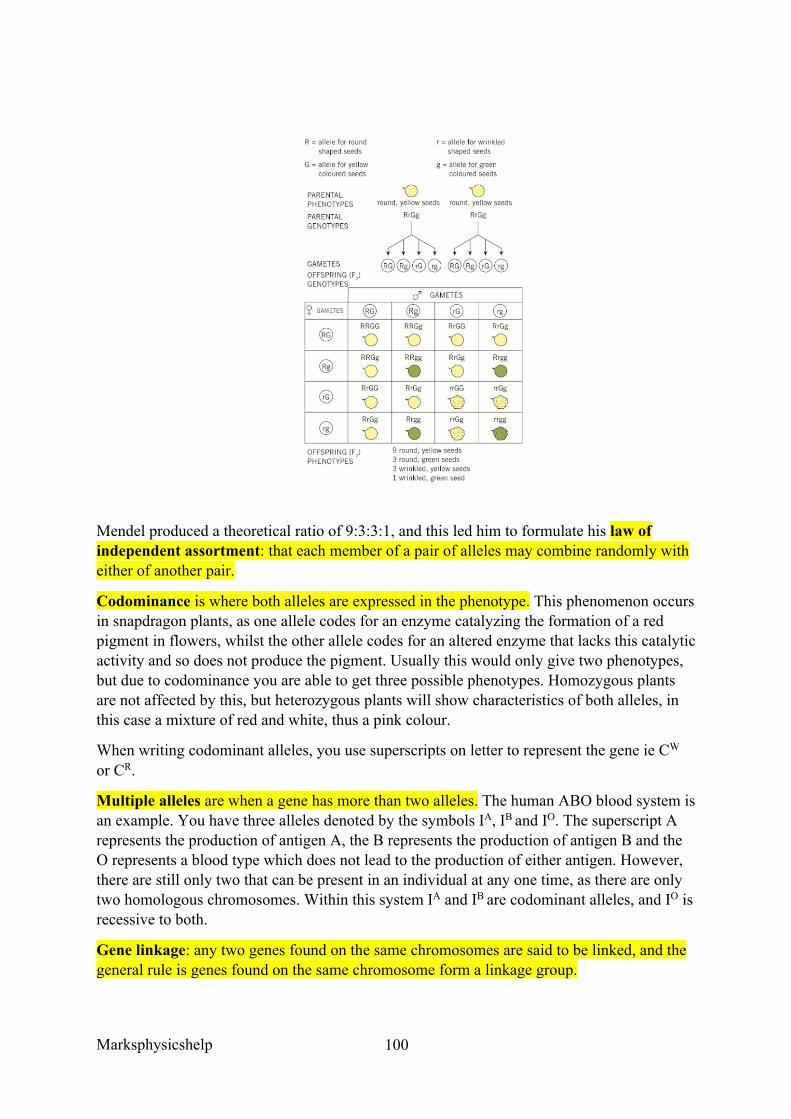
Marksphysicshelp
100
Mendel produced a theoretical ratio of 9:3:3:1, and this led him to formulate his law of independent assortment: that each member of a pair of alleles may combine randomly with either of another pair.
Codominance is where both alleles are expressed in the phenotype. This phenomenon occurs in snapdragon plants, as one allele codes for an enzyme catalyzing the formation of a red pigment in flowers, whilst the other allele codes for an altered enzyme that lacks this catalytic activity and so does not produce the pigment. Usually this would only give two phenotypes, but due to codominance you are able to get three possible phenotypes. Homozygous plants are not affected by this, but heterozygous plants will show characteristics of both alleles, in this case a mixture of red and white, thus a pink colour.
When writing codominant alleles, you use superscripts on letter to represent the gene ie CW or CR.
Multiple alleles are when a gene has more than two alleles. The human ABO blood system is an example. You have three alleles denoted by the symbols IA, IB and IO. The superscript A represents the production of antigen A, the B represents the production of antigen B and the O represents a blood type which does not lead to the production of either antigen. However, there are still only two that can be present in an individual at any one time, as there are only two homologous chromosomes. Within this system IA and IB are codominant alleles, and IO is recessive to both.
Gene linkage: any two genes found on the same chromosomes are said to be linked, and the general rule is genes found on the same chromosome form a linkage group.
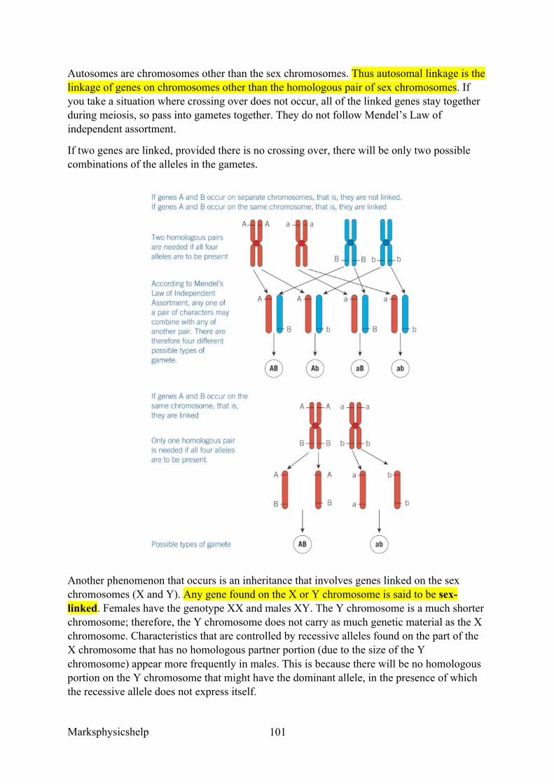
Marksphysicshelp
101
Autosomes are chromosomes other than the sex chromosomes. Thus autosomal linkage is the linkage of genes on chromosomes other than the homologous pair of sex chromosomes. If you take a situation where crossing over does not occur, all of the linked genes stay together during meiosis, so pass into gametes together. They do not follow Mendel’s Law of independent assortment.
If two genes are linked, provided there is no crossing over, there will be only two possible combinations of the alleles in the gametes.
Another phenomenon that occurs is an inheritance that involves genes linked on the sex chromosomes (X and Y). Any gene found on the X or Y chromosome is said to be sex-linked. Females have the genotype XX and males XY. The Y chromosome is a much shorter chromosome; therefore, the Y chromosome does not carry as much genetic material as the X chromosome. Characteristics that are controlled by recessive alleles found on the part of the X chromosome that has no homologous partner portion (due to the size of the Y chromosome) appear more frequently in males. This is because there will be no homologous portion on the Y chromosome that might have the dominant allele, in the presence of which the recessive allele does not express itself.
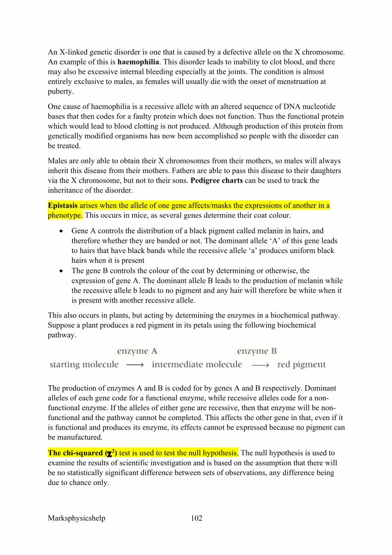
Marksphysicshelp
102
An X-linked genetic disorder is one that is caused by a defective allele on the X chromosome. An example of this is haemophilia. This disorder leads to inability to clot blood, and there may also be excessive internal bleeding especially at the joints. The condition is almost entirely exclusive to males, as females will usually die with the onset of menstruation at puberty.
One cause of haemophilia is a recessive allele with an altered sequence of DNA nucleotide bases that then codes for a faulty protein which does not function. Thus the functional protein which would lead to blood clotting is not produced. Although production of this protein from genetically modified organisms has now been accomplished so people with the disorder can be treated.
Males are only able to obtain their X chromosomes from their mothers, so males will always inherit this disease from their mothers. Fathers are able to pass this disease to their daughters via the X chromosome, but not to their sons. Pedigree charts can be used to track the inheritance of the disorder.
Epistasis arises when the allele of one gene affects/masks the expressions of another in a phenotype. This occurs in mice, as several genes determine their coat colour.
• Gene A controls the distribution of a black pigment called melanin in hairs, and therefore whether they are banded or not. The dominant allele ‘A’ of this gene leads to hairs that have black bands while the recessive allele ‘a’ produces uniform black hairs when it is present
• The gene B controls the colour of the coat by determining or otherwise, the expression of gene A. The dominant allele B leads to the production of melanin while the recessive allele b leads to no pigment and any hair will therefore be white when it is present with another recessive allele.
This also occurs in plants, but acting by determining the enzymes in a biochemical pathway. Suppose a plant produces a red pigment in its petals using the following biochemical pathway.
The production of enzymes A and B is coded for by genes A and B respectively. Dominant alleles of each gene code for a functional enzyme, while recessive alleles code for a non-functional enzyme. If the alleles of either gene are recessive, then that enzyme will be non-functional and the pathway cannot be completed. This affects the other gene in that, even if it is functional and produces its enzyme, its effects cannot be expressed because no pigment can be manufactured.
The chi-squared (c2) test is used to test the null hypothesis. The null hypothesis is used to examine the results of scientific investigation and is based on the assumption that there will be no statistically significant difference between sets of observations, any difference being due to chance only.
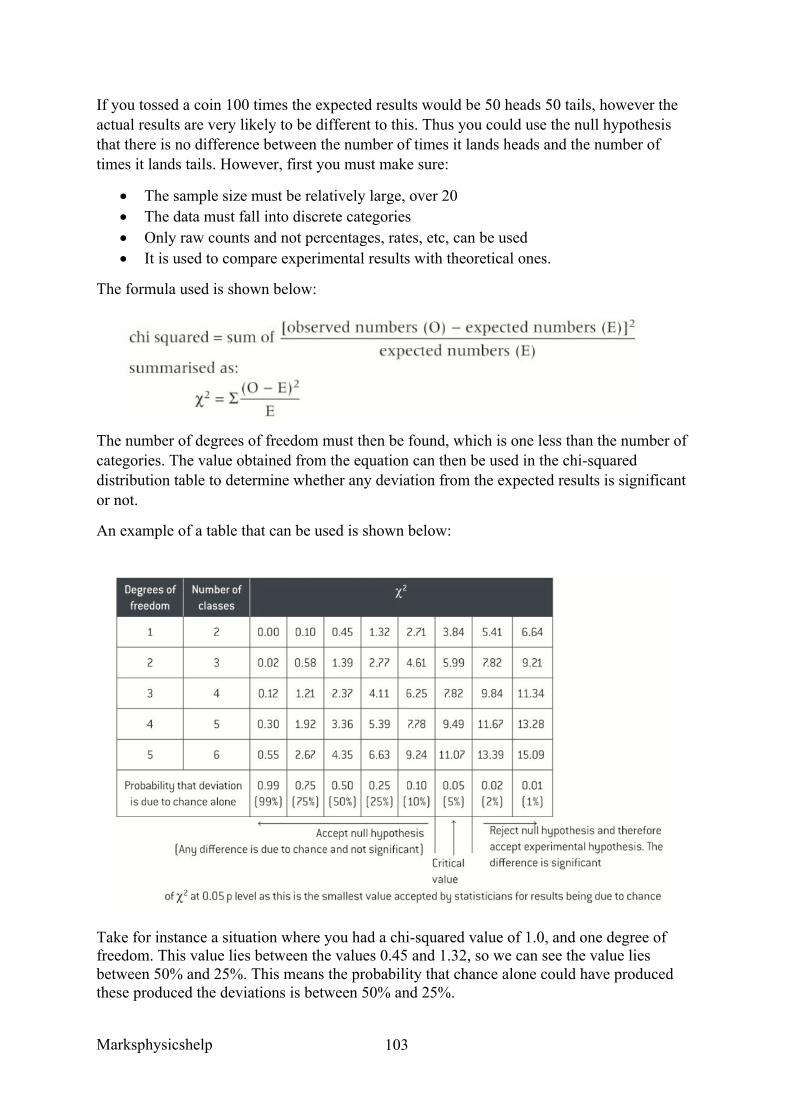
Marksphysicshelp
103
If you tossed a coin 100 times the expected results would be 50 heads 50 tails, however the actual results are very likely to be different to this. Thus you could use the null hypothesis that there is no difference between the number of times it lands heads and the number of times it lands tails. However, first you must make sure:
• The sample size must be relatively large, over 20 • The data must fall into discrete categories • Only raw counts and not percentages, rates, etc, can be used • It is used to compare experimental results with theoretical ones.
The formula used is shown below:
The number of degrees of freedom must then be found, which is one less than the number of categories. The value obtained from the equation can then be used in the chi-squared distribution table to determine whether any deviation from the expected results is significant or not.
An example of a table that can be used is shown below:
Take for instance a situation where you had a chi-squared value of 1.0, and one degree of freedom. This value lies between the values 0.45 and 1.32, so we can see the value lies between 50% and 25%. This means the probability that chance alone could have produced these produced the deviations is between 50% and 25%.
Marksphysicshelp
104
In the chi-squared test the critical value is p = 0.05, this is the attribution to chance accepted by statisticians. This means that the probability that the deviation is due to chance is equal to or greater than 5%, the deviation is said to be not significant and the null hypothesis would be accepted. If the deviation is less than 5% then the deviation is said to be significant.
The chi-squared test is very useful in genetics, take for example a test like Mendel’s. You could use this test to interpret results and prove that results were not significantly different to an expected ratio of say 9:3:3:1. Thus you could see whether the deviation was significant enough for the results to be due to chance or not.
Marksphysicshelp
105
3.7.2 Populations
Content
• Species exist as one or more populations. • A population as a group of organisms of the same species occupying a particular
space at a particular time that can potentially interbreed. • The concepts of gene pool and allele frequency. • The Hardy–Weinberg principle provides a mathematical model, which predicts that
allele frequencies will not change from generation to generation. The conditions under which the principle applies.
• The frequency of alleles, genotypes and phenotypes in a population can be calculated using the Hardy–Weinberg equation: p2 + 2pq + q2
• here p is the frequency of one (usually the dominant) allele and q is the frequency of the other (usually recessive) allele of the gene.
Species exist as one or more populations, where a population if a group of organisms of the same species occupying a particular space at a particular time that can potentially interbreed.
All of the alleles of all the genes of all the individuals in a population at a given time are known as the gene pool. The number of times an allele occurs within the gene pool is referred to as the allelic frequency.
If a population has 10,000 people, there will be twice as many alleles in the gene pool of a particular gene ie the allele for cystic fibrosis. F would be the probability of not having cystic fibrosis and f for having it. We can use the Hardy Weinberg principle to find the allele frequency of given populations.
The Hardy Weinberg equation to provide a mathematical model, which predicts allele frequencies will not change from generation to generation. This is one of the conditions in which this principle applies. This condition is met if:
• No mutations arise • The population is isolated, ie there is no flow of alleles into or out of the population • There is no selection, so alleles are equally likely to be passed on to the next
generation • The population is large • Mating within the population if random
Although these conditions are, in practice, never truly met, the principle is still useful when studying gene frequencies.
For example, a dominant allele F and recessive allele f, the probability of F will be p, and of f will be q. We know that p + q = 1.0 because there are only two alleles so their probability must be 100%. Thus AA + Aa + aA + aa = 1.0, otherwise written as p2 + 2pq + q2. Usually you will take p as the dominant allele and q as the recessive allele but the order does not matter.
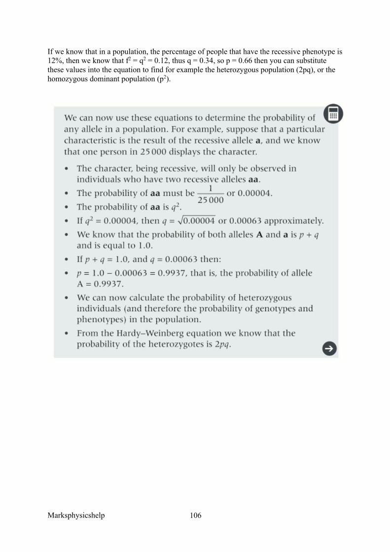
Marksphysicshelp
106
If we know that in a population, the percentage of people that have the recessive phenotype is 12%, then we know that f2 = q2 = 0.12, thus q = 0.34, so p = 0.66 then you can substitute these values into the equation to find for example the heterozygous population (2pq), or the homozygous dominant population (p2).
Marksphysicshelp
107
3.7.3 Evolution may lead to speciation
Content
• Individuals within a population of a species may show a wide range of variation in phenotype. This is due to genetic and environmental factors. The primary source of genetic variation is mutation. Meiosis and the random fertilisation of gametes during sexual reproduction produce further genetic variation.
• Predation, disease and competition for the means of survival result in differential survival and reproduction, ie natural selection.
• Those organisms with phenotypes providing selective advantages are likely to produce more offspring and pass on their favourable alleles to the next generation. The effect of this differential reproductive success on the allele frequencies within a gene pool.
• The effects of stabilising, directional and disruptive selection. Evolution as a change in the allele frequencies in a population.
• Reproductive separation of two populations can result in the accumulation of difference in their gene pools. New species arise when these genetic differences lead to an inability of members of the populations to interbreed and produce fertile offspring. In this way, new species arise from existing species.
• Allopatric and sympatric speciation. The importance of genetic drift in causing changes in allele frequency in small populations.
• Students should be able to: o Explain why individuals within a population of a species may show a wide
range of variation in phenotype o Explain why genetic drift is important only in small populations o Explain how natural selection and isolation may result in change in the allele
and phenotype frequency and lead to the formation of a new species o Explain how evolutionary change over a long period of time has resulted in a
great diversity of species.
Individuals within a population of a species may show a wide range of variation in phenotype. This is due to genetic and environmental factors. The primary source of genetic variation is mutation. Meiosis and the random fertilisation of gametes during sexual reproduction produce further genetic variation.
Every organism is subjected to a process of selection, based on its suitability for survival under the conditions that exist at the time. The environmental pressures limiting the population of a species are called selection pressures. These pressures include predations, disease and competition. They vary from time to time, and from place to place.
These pressures determined the frequency of alleles within a gene pool. A gene pool is the total number of all the alleles of all the genes of all the individuals within a particular population at a given time.
The process of evolution by natural selection depends upon factors including:
• Organisms produce more offspring than can be supported by the available supply of food, light, space etc.
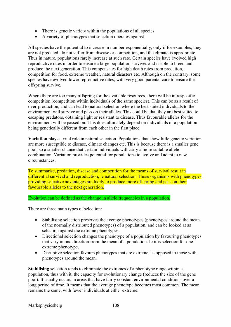
Marksphysicshelp
108
• There is genetic variety within the populations of all species • A variety of phenotypes that selection operates against
All species have the potential to increase in number exponentially, only if for examples, they are not predated, do not suffer from disease or competition, and the climate is appropriate. Thus in nature, populations rarely increase at such rate. Certain species have evolved high reproductive rates in order to ensure a large population survives and is able to breed and produce the next generation. This compensates for high death rates from predation, competition for food, extreme weather, natural disasters etc. Although on the contrary, some species have evolved lower reproductive rates, with very good parental care to ensure the offspring survive.
Where there are too many offspring for the available resources, there will be intraspecific competition (competition within individuals of the same species). This can be as a result of over-production, and can lead to natural selection where the best suited individuals to the environment will survive and pass on their alleles. This could be that they are best suited to escaping predators, obtaining light or resistant to disease. Thus favourable alleles for the environment will be passed on. This does ultimately depend on individuals of a population being genetically different from each other in the first place.
Variation plays a vital role in natural selection. Populations that show little genetic variation are more susceptible to disease, climate changes etc. This is because there is a smaller gene pool, so a smaller chance that certain individuals will carry a more suitable allele combination. Variation provides potential for populations to evolve and adapt to new circumstances.
To summarise, predation, disease and competition for the means of survival result in differential survival and reproduction, ie natural selection. Those organisms with phenotypes providing selective advantages are likely to produce more offspring and pass on their favourable alleles to the next generation.
Evolution can be defined as the change in allele frequencies in a population.
There are three main types of selection:
• Stabilising selection preserves the average phenotypes (phenotypes around the mean of the normally distributed phenotypes) of a population, and can be looked at as selection against the extreme phenotypes.
• Directional selection changes the phenotype of a population by favouring phenotypes that vary in one direction from the mean of a population. Ie it is selection for one extreme phenotype.
• Disruptive selection favours phenotypes that are extreme, as opposed to those with phenotypes around the mean.
Stabilising selection tends to eliminate the extremes of a phenotype range within a population, thus with it, the capacity for evolutionary change (reduces the size of the gene pool). It usually occurs in areas that have fairly constant environmental conditions over a long period of time. It means that the average phenotype becomes most common. The mean remains the same, with fewer individuals at either extreme.
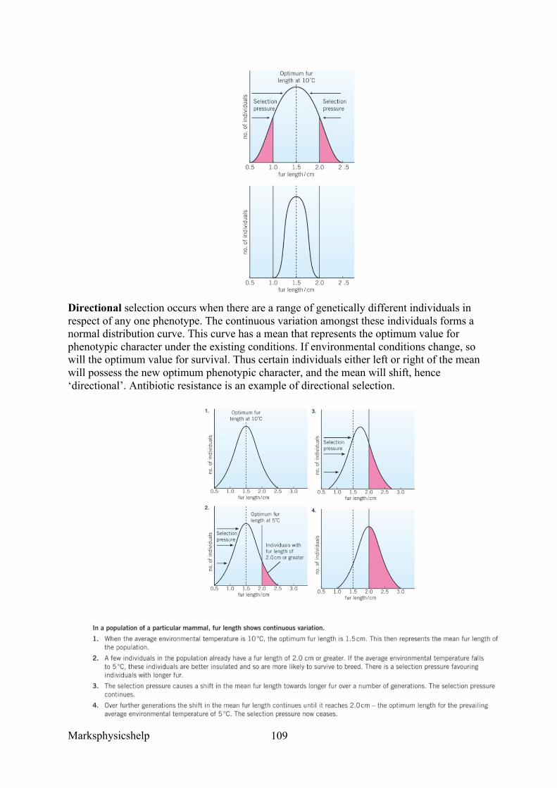
Marksphysicshelp
109
Directional selection occurs when there are a range of genetically different individuals in respect of any one phenotype. The continuous variation amongst these individuals forms a normal distribution curve. This curve has a mean that represents the optimum value for phenotypic character under the existing conditions. If environmental conditions change, so will the optimum value for survival. Thus certain individuals either left or right of the mean will possess the new optimum phenotypic character, and the mean will shift, hence ‘directional’. Antibiotic resistance is an example of directional selection.
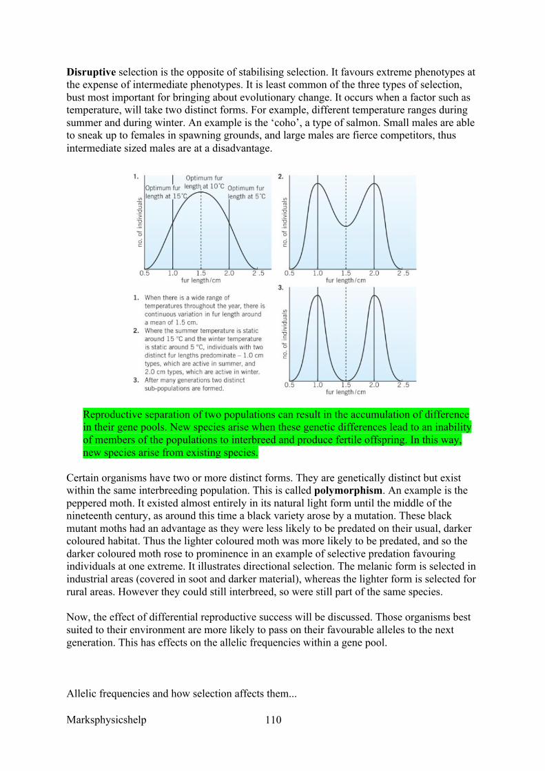
Marksphysicshelp
110
Disruptive selection is the opposite of stabilising selection. It favours extreme phenotypes at the expense of intermediate phenotypes. It is least common of the three types of selection, bust most important for bringing about evolutionary change. It occurs when a factor such as temperature, will take two distinct forms. For example, different temperature ranges during summer and during winter. An example is the ‘coho’, a type of salmon. Small males are able to sneak up to females in spawning grounds, and large males are fierce competitors, thus intermediate sized males are at a disadvantage.
Reproductive separation of two populations can result in the accumulation of difference in their gene pools. New species arise when these genetic differences lead to an inability of members of the populations to interbreed and produce fertile offspring. In this way, new species arise from existing species.
Certain organisms have two or more distinct forms. They are genetically distinct but exist within the same interbreeding population. This is called polymorphism. An example is the peppered moth. It existed almost entirely in its natural light form until the middle of the nineteenth century, as around this time a black variety arose by a mutation. These black mutant moths had an advantage as they were less likely to be predated on their usual, darker coloured habitat. Thus the lighter coloured moth was more likely to be predated, and so the darker coloured moth rose to prominence in an example of selective predation favouring individuals at one extreme. It illustrates directional selection. The melanic form is selected in industrial areas (covered in soot and darker material), whereas the lighter form is selected for rural areas. However they could still interbreed, so were still part of the same species.
Now, the effect of differential reproductive success will be discussed. Those organisms best suited to their environment are more likely to pass on their favourable alleles to the next generation. This has effects on the allelic frequencies within a gene pool.
Allelic frequencies and how selection affects them...
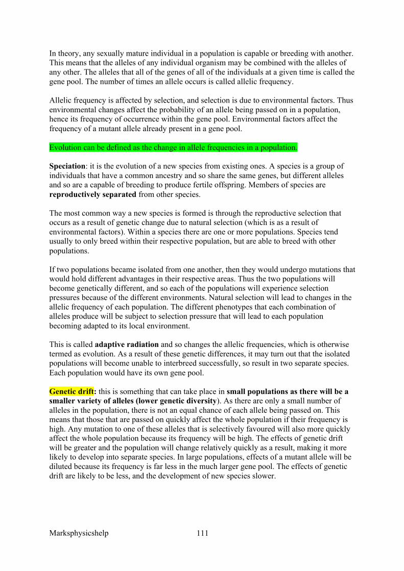
Marksphysicshelp
111
In theory, any sexually mature individual in a population is capable or breeding with another. This means that the alleles of any individual organism may be combined with the alleles of any other. The alleles that all of the genes of all of the individuals at a given time is called the gene pool. The number of times an allele occurs is called allelic frequency.
Allelic frequency is affected by selection, and selection is due to environmental factors. Thus environmental changes affect the probability of an allele being passed on in a population, hence its frequency of occurrence within the gene pool. Environmental factors affect the frequency of a mutant allele already present in a gene pool.
Evolution can be defined as the change in allele frequencies in a population.
Speciation: it is the evolution of a new species from existing ones. A species is a group of individuals that have a common ancestry and so share the same genes, but different alleles and so are a capable of breeding to produce fertile offspring. Members of species are reproductively separated from other species.
The most common way a new species is formed is through the reproductive selection that occurs as a result of genetic change due to natural selection (which is as a result of environmental factors). Within a species there are one or more populations. Species tend usually to only breed within their respective population, but are able to breed with other populations.
If two populations became isolated from one another, then they would undergo mutations that would hold different advantages in their respective areas. Thus the two populations will become genetically different, and so each of the populations will experience selection pressures because of the different environments. Natural selection will lead to changes in the allelic frequency of each population. The different phenotypes that each combination of alleles produce will be subject to selection pressure that will lead to each population becoming adapted to its local environment.
This is called adaptive radiation and so changes the allelic frequencies, which is otherwise termed as evolution. As a result of these genetic differences, it may turn out that the isolated populations will become unable to interbreed successfully, so result in two separate species. Each population would have its own gene pool.
Genetic drift: this is something that can take place in small populations as there will be a smaller variety of alleles (lower genetic diversity). As there are only a small number of alleles in the population, there is not an equal chance of each allele being passed on. This means that those that are passed on quickly affect the whole population if their frequency is high. Any mutation to one of these alleles that is selectively favoured will also more quickly affect the whole population because its frequency will be high. The effects of genetic drift will be greater and the population will change relatively quickly as a result, making it more likely to develop into separate species. In large populations, effects of a mutant allele will be diluted because its frequency is far less in the much larger gene pool. The effects of genetic drift are likely to be less, and the development of new species slower.
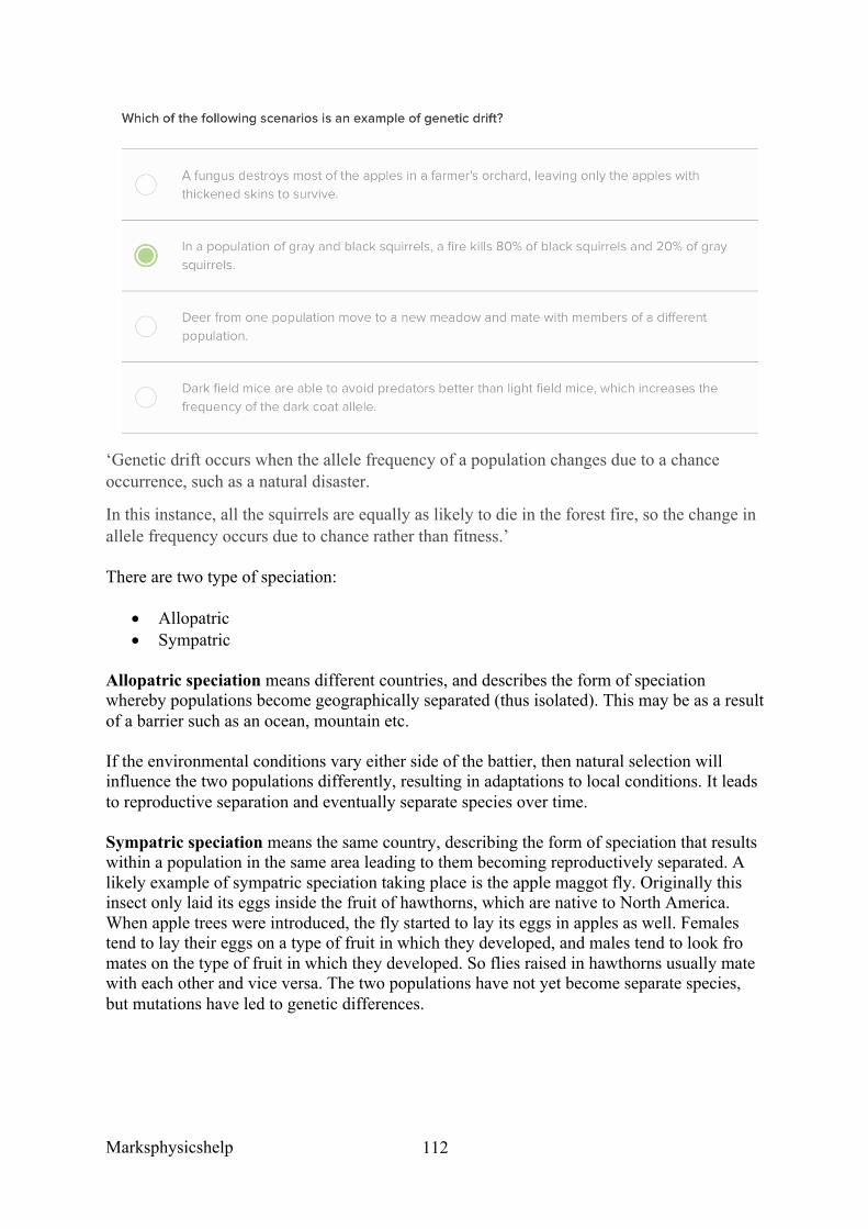
Marksphysicshelp
112
‘Genetic drift occurs when the allele frequency of a population changes due to a chance occurrence, such as a natural disaster.
In this instance, all the squirrels are equally as likely to die in the forest fire, so the change in allele frequency occurs due to chance rather than fitness.’
There are two type of speciation:
• Allopatric • Sympatric
Allopatric speciation means different countries, and describes the form of speciation whereby populations become geographically separated (thus isolated). This may be as a result of a barrier such as an ocean, mountain etc.
If the environmental conditions vary either side of the battier, then natural selection will influence the two populations differently, resulting in adaptations to local conditions. It leads to reproductive separation and eventually separate species over time.
Sympatric speciation means the same country, describing the form of speciation that results within a population in the same area leading to them becoming reproductively separated. A likely example of sympatric speciation taking place is the apple maggot fly. Originally this insect only laid its eggs inside the fruit of hawthorns, which are native to North America. When apple trees were introduced, the fly started to lay its eggs in apples as well. Females tend to lay their eggs on a type of fruit in which they developed, and males tend to look fro mates on the type of fruit in which they developed. So flies raised in hawthorns usually mate with each other and vice versa. The two populations have not yet become separate species, but mutations have led to genetic differences.
Marksphysicshelp
113
AQA Jan 2011 Unit 4 Q8c Question:
Changes in ecosystems can lead to speciation. In Southern California 10 000 years ago a number of interconnecting lakes contained a single species of pupfish. Increasing temperatures caused evaporation and the formation of separate, smaller lakes and streams. This led to the formation of a number of different species of pupfish. Explain how these different species evolved.
Answer:
1. Geographical isolation; 2. Separate gene pools / no interbreeding (between populations); 3. Variation due to mutation; 4. Different environmental/abiotic/biotic conditions / selection pressures; 5. Selection for different/advantageous, features/characteristics/mutation/ /allele; 6. Differential reproductive success / (selected) organisms survive and reproduce; 7. Leads to change in allele frequency; 8. Occurs over a long period of time;
Marksphysicshelp
114
AQA June 2015 Unit 4 Q8a Question:
On islands in the Caribbean, there are almost 150 species of lizards belonging to the genus Anolis. Scientists believe that these species evolved from two species found on mainland USA. Explain how the Caribbean species could have evolved.
Answer:
1. Geographical isolation; 2. Separate gene pools / no interbreeding/gene flow (between populations); 3. Variation due to mutation; 4. Different selection pressures / different abiotic/biotic conditions/
environments/habitats; 5. Different(ial) reproductive success / selected organisms (survive and) reproduce; 6. Leads to change/increase in allele frequency;
AQA June 2013 Unit 4 Q6a The Amazonian forest today contains a very high diversity of bird species.
• Over the last 2 000 000 years, long periods of dry climate caused this forest to separate into a number of smaller forests.
• Different plant communities developed in each of these smaller forests. • Each time the climate became wetter again, the smaller forests grew in size and
merged to reform the Amazonian forest.
Question:
Use the information provided to explain how a very high diversity of bird species has developed in the Amazonian forest.
Answer:
1. No interbreeding / gene pools are separate / geographic(al) isolation; 2. Mutation; 3. Different selection pressures / different foods/niches/habitats; 4. Adapted organisms survive and breed / differential reproductive success; 5. Change/increase in allele frequency/frequencies;
Marksphysicshelp
115
AQA Jan 2012 Unit 4
Question:
The population of trees in the forest evolved adaptations to the mountain environment. Use your knowledge of selection to explain how.
Answer:
1. Variation in original colonisers / mutations took place; 2. Some better (adapted for) survival (in mountains); 3. Greater reproductive success; 4. Allele frequencies change;
EXAM TIP:
ADAPTATIONS COULD HAVE EVOLVED FROM MUTATIONS OR INITIAL GENETIC VARIATION IN COLONISERS!
THINK ABOUT REPRODUCTIVE SUCCESS WHEN TALKING ABOUT EVOLUTION BY SELECTION!
KEY TERM: ALLELE FREQUENCIES!!!
Marksphysicshelp
116
3.7.4 Populations in ecosystems
Content
• Populations of different species form a community. A community and the non-living components of its environment together form an ecosystem. Ecosystems can range in size from the very small to the very large.
• Within a habitat, a species occupies a niche governed by adaptation to both abiotic and biotic conditions.
• An ecosystem supports a certain size of population of a species, called the carrying capacity. This population size can vary as a result of:
o the effect of abiotic factors o interactions between organisms: interspecific and intraspecific competition
and predation. • The size of a population can be estimated using:
o randomly placed quadrats, or quadrats along a belt transect, for slow-moving or non-motile organisms
o the mark-release-recapture method for motile organisms. The assumptions made when using the mark-release-recapture method.
• Ecosystems are dynamic systems. • Primary succession, from colonisation by pioneer species to climax community. • At each stage in succession, certain species may be recognised which change the
environment so that it becomes more suitable for other species with different adaptations. The new species may change the environment in such a way that it becomes less suitable for the previous species.
• Changes that organisms produce in their abiotic environment can result in a less hostile environment and change biodiversity.
• Conservation of habitats frequently involves management of succession. • Students should be able to: • Show understanding of the need to manage the conflict between human needs and
conservation in order to maintain the sustainability of natural resources • Evaluate evidence and data concerning issues relating to the conservation of species
and habitats and consider convicting evidence • Use given data to calculate the size of a population estimated using the mark-release-
recapture method.
Populations of different species form a community. A community and the non-living components of its environment together form an ecosystem. Ecosystems can range in size from the very small to the very large.
Within a habitat, a species occupies a niche governed by adaptation to both abiotic and biotic conditions.
Ecology is the study of the inter-relationships between organisms and their environment. The environment includes both abiotic and biotic factors like competition and predation.
Marksphysicshelp
117
Ecosystems are dynamic systems made up of a community and all the non-living factors of its environment. Ecosystems can range in size from very small to very large. Within an ecosystem there are two major processes to consider:
• The flow of energy through the system • The cycling of elements within the system.
A freshwater pond is an example of an ecosystem. It has its own community of plants to collect the necessary sunlight energy to supply the organisms within it. Nutrients such as nitrate ions and phosphate ions are recycled within the pond/lake. Within ecosystems there are a number of species. Each species is made of up a group of individuals that make up a population.
A population if a group of individuals of one species that occupy a habitat at the same time, and are able to interbreed.
An ecosystem supports a certain size of population of a species, called the carrying capacity. This population size can vary as a result of:
o the effect of abiotic factors o interactions between organisms: interspecific and intraspecific competition
and predation.
Population size can vary as a result of abiotic factors, and interaction between organisms like intraspecific and interspecific competition.
Populations of different species form a community. A community is defined as all of the populations of different species living and interacting in a particular place at a particular time.
A habitat is the place where an organism usually lives an is characterised by physical conditions and the other types of organisms present. Within ecosystems there will be multiple habitats. Within habitats you will find smaller units with their own microclimate, these called microhabitats. For example mud at the bottom of a stream may be a microhabitat for a bloodworm, or a crevice on the bark of an oak tree for a lichen.
Ecological niche: a niche describes how an organism fits into the environment. A niche refers to where an organism lives and what it does there. It includes all of the biotic and abiotic conditions to which an organism is adapted in order to survive, reproduce and maintain a viable population. Some species will appear very similar, but their nesting habitats or other aspects of their behaviour will be different, or they may show different levels of tolerance to environmental factor such as pollutant or a shortage of oxygen/nitrates. No two species occupy exactly the same niche, and this is known as the competitive exclusion principle.
The size of any population is eventually determined by a limiting factor. A species is only able to live within a certain range of abiotic factors, and this range differs from species to species. If there are no limiting factors, a population will grow in size. Although as a population grows in size, limiting factors may arise as a result of the population becoming so large that there is no longer enough variety and a high enough quantity of food to support it.
Marksphysicshelp
118
Or if other species may be introduced (interspecific competition) which results in the population size being limited.
Both biotic and abiotic factors influence the rate of growth and size of population.
To summarise, no population will grow indefinitely because certain factors will limit growth. Availability of food, light, water, oxygen, shelter, accumulation of toxic waste, disease and predators are all limiting factors. Each ecosystem has its carrying capacity which can be sustained over a long period of time, and is determined by these factors.
Abiotic factors:
• Temperature. This can affect rate of enzyme activity, or denature enzymes which can inhibit vital metabolic reactions required for survival. At temperatures that are deviated from the optimum, enzymes work less efficiently. Also if temperatures deviate from optimum conditions, then more energy is expended by these organisms to maintain a normal body temperature. This leaves less energy for growth and so they mature more slowly and the reproductive rate slows. Carrying capacity is thus reduced.
• Light. This is the ultimate source of energy for ecosystems, and a basic necessity for life. The rate of photosynthesis increases as light intensity increase, so a greater rate means plants grow faster and produce more spores/seeds. Therefore carrying capacity may be increased, so the carrying capacity of animals that feed on these plants will be increased.
• pH. This affects enzyme action as each enzyme has its optimum pH for normal functioning.
• Water and humidity. Where water is scarce, populations are small and consist only of species well adapted to dry conditions. Humidity affects the transpiration rates of plants and the evaporation of water from the bodies of animals. Again, in dry air conditions, the populations of species adapted to tolerate low humidity will be larger than those without the adaptations.
As a general rule, when any abiotic factor is below the optimum for a population, fewer individuals are able to survive because their adaptations are not suited to conditions. If no individuals have adaptations to survival, the populations become extinct.
Where two or more individuals share any resource ie light, food, oxygen, that is insufficient to satisfy all of their requirements, then competitions results. Competition between member of the same species is called intraspecific, and between different species is called interspecific.
Intraspecific:
Occurs when individuals of the same species are competing for resources such as food, water etc. The availability of these resources determines the size of the population. Examples of intraspecific competition are:
• Robins competing for breeding territory. Female birds usually only attracted to males with breeding territories. Each territory will supply for only one family of birds, so when food gets scarce territories become wider to find food. So then there are fewer territories in a given area.
Marksphysicshelp
119
• Oak trees compete for resources like light, water, minerals. Therefore, the oak trees with restricted availability will then die, so over time the population will reduce to a few large dominant oaks.
• Limpets competing for algae, which is their main food. The more algae available, the larger the population of limpets.
Interspecific
Occurs when individuals of different species compete for the same resources like food, light, water etc. One species will usually have the advantage over the other species, and so this population will gradually decrease in size over time. If conditions remained the same, one of the species would be completely removed. This is called competitive exclusion principle.
This principle says that where two species are competing for limited resources, the one that uses these resources most effectively will ultimately eliminate the other. No two species are able to occupy a niche indefinitely with finite resources (limiting factor). If two species of sea birds appear to occupy the same niche, eat fish from the sea and nest on the same type of cliff face, it could be thought that they were in competition. However, if they are eating different types of fish then they occupy different niches.
Therefore, competition has an affect on populations by having an impact on birth and death rates. For example, a decrease in food supply could lead to individuals dying of starvation thus directly reducing population size. The opposite can be said for a high population, having positive impacts on probability of offspring production due to higher survival rates, and thus shows increase in population size.
One type of interspecific relationship is the predator-prey relationship. A predator is an organism that feeds on another organism, namely the prey. Predators have evolved to become better at capturing their prey ie faster movement or camouflage. Likewise, prey have evolved to become equally adept at avoiding predators.
Predation occurs when one organism is consumed by another. Evidence collected regarding predators and prey and their behaviour is usually quite limited because it does not represent natural conditions and what happens in the wild. However, it is also difficult to obtain data on natural populations as it is not possible to count all the individuals in a natural population. Its size can only be estimated from sampling and surveys. Therefore neither technique provides complete accuracy.
The predator-prey relationship affects population size such that:
• Predators eat prey, so reduce the population of prey • With fewer prey available, the predators are in greater competition with each other for
the prey that are left • The predator population is reduced as some individuals are unable to obtain enough
prey for their survival or to reproduce • With fewer predators left, fewer prey are eaten and so more survive and are able to
reproduce • The prey population increases • With more prey now available as food, the predator population in turn increase.
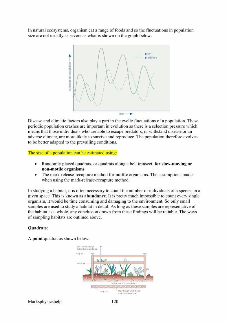
Marksphysicshelp
120
In natural ecosystems, organism eat a range of foods and so the fluctuations in population size are not usually as severe as what is shown on the graph below.
Disease and climatic factors also play a part in the cyclic fluctuations of a population. These periodic population crashes are important in evolution as there is a selection pressure which means that those individuals who are able to escape predators, or withstand disease or an adverse climate, are more likely to survive and reproduce. The population therefore evolves to be better adapted to the prevailing conditions.
The size of a population can be estimated using:
• Randomly placed quadrats, or quadrats along a belt transect, for slow-moving or non-motile organisms
• The mark-release-recapture method for motile organisms. The assumptions made when using the mark-release-recapture method.
In studying a habitat, it is often necessary to count the number of individuals of a species in a given space. This is known as abundance. It is pretty much impossible to count every single organism, it would be time consuming and damaging to the environment. So only small samples are used to study a habitat in detail. As long as these samples are representative of the habitat as a whole, any conclusion drawn from these findings will be reliable. The ways of sampling habitats are outlined above.
Quadrats:
A point quadrat as shown below.
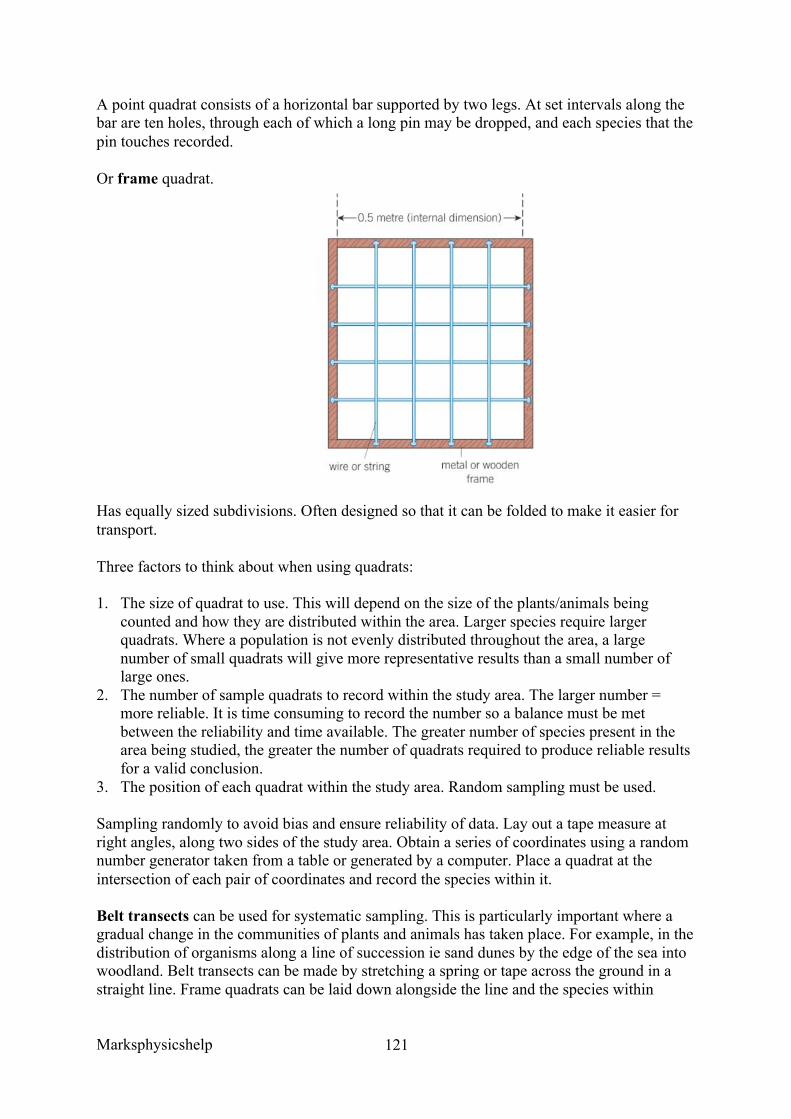
Marksphysicshelp
121
A point quadrat consists of a horizontal bar supported by two legs. At set intervals along the bar are ten holes, through each of which a long pin may be dropped, and each species that the pin touches recorded.
Or frame quadrat.
Has equally sized subdivisions. Often designed so that it can be folded to make it easier for transport.
Three factors to think about when using quadrats:
1. The size of quadrat to use. This will depend on the size of the plants/animals being counted and how they are distributed within the area. Larger species require larger quadrats. Where a population is not evenly distributed throughout the area, a large number of small quadrats will give more representative results than a small number of large ones.
2. The number of sample quadrats to record within the study area. The larger number = more reliable. It is time consuming to record the number so a balance must be met between the reliability and time available. The greater number of species present in the area being studied, the greater the number of quadrats required to produce reliable results for a valid conclusion.
3. The position of each quadrat within the study area. Random sampling must be used.
Sampling randomly to avoid bias and ensure reliability of data. Lay out a tape measure at right angles, along two sides of the study area. Obtain a series of coordinates using a random number generator taken from a table or generated by a computer. Place a quadrat at the intersection of each pair of coordinates and record the species within it.
Belt transects can be used for systematic sampling. This is particularly important where a gradual change in the communities of plants and animals has taken place. For example, in the distribution of organisms along a line of succession ie sand dunes by the edge of the sea into woodland. Belt transects can be made by stretching a spring or tape across the ground in a straight line. Frame quadrats can be laid down alongside the line and the species within
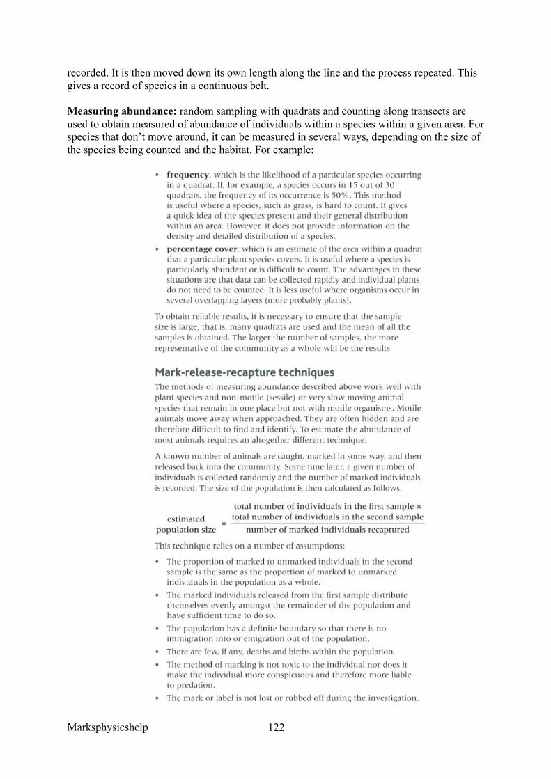
Marksphysicshelp
122
recorded. It is then moved down its own length along the line and the process repeated. This gives a record of species in a continuous belt.
Measuring abundance: random sampling with quadrats and counting along transects are used to obtain measured of abundance of individuals within a species within a given area. For species that don’t move around, it can be measured in several ways, depending on the size of the species being counted and the habitat. For example:
Marksphysicshelp
123
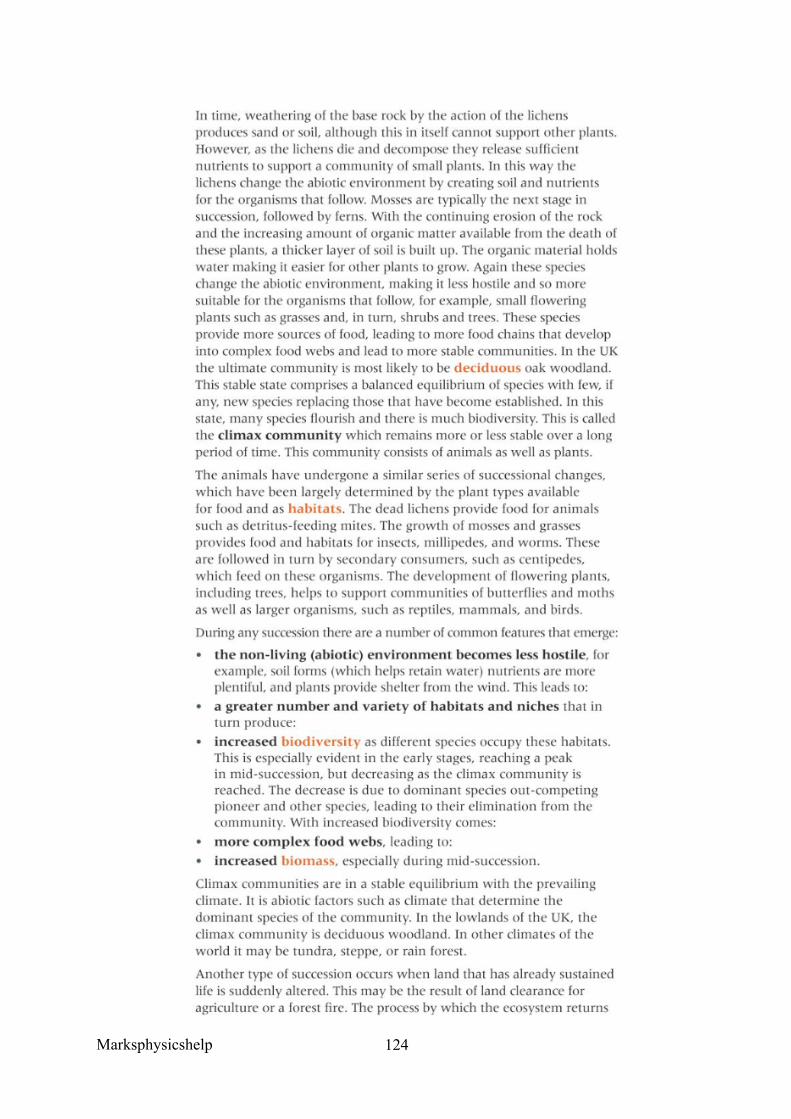
Marksphysicshelp
124
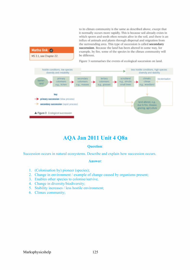
Marksphysicshelp
125
AQA Jan 2011 Unit 4 Q8a Question:
Succession occurs in natural ecosystems. Describe and explain how succession occurs.
Answer:
1. (Colonisation by) pioneer (species); 2. Change in environment / example of change caused by organisms present; 3. Enables other species to colonise/survive. 4. Change in diversity/biodiversity; 5. Stability increases / less hostile environment; 6. Climax community;
Marksphysicshelp
126
Source: Kerboodle Biology Textbook
Marksphysicshelp
127
AQA June 2015 Unit 4 Q8b Question:
‘Anolis sagrei is a species of lizard that is found on some of the smallest Caribbean islands. Describe how you could use the mark-release-recapture method to estimate the number of Anolis sagrei on one of these islands.’
Answer:
1. ‘Capture/collect sample, mark and release; 2. Method of marking does not harm lizard/make it more visible to predators; 3. Leave sufficient time for lizards to (randomly) distribute (on island) before collecting
a second sample; 4. (Population=) number in first sample × number in second sample divided by number
of marked lizards in second sample/number recaptured;
AQA June 2014 Unit 4 Q3ab Farmland previously used for growing crops was left for 30 years and developed into woodland. During this period, ecologists recorded an increase in the diversity of birds in the area.
Question:
Name the process that resulted in the development of woodland from farmland.
Answer:
Succession
Question:
Explain the increase in the diversity of birds as the woodland developed.
Answer:
1. Greater variety/diversity of plants/insects / more plant/insect species; 2. More food sources / more varieties of food; 3. Greater variety/more habitats/niches
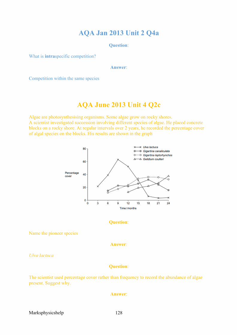
Marksphysicshelp
128
AQA Jan 2013 Unit 2 Q4a Question:
What is intraspecific competition?
Answer:
Competition within the same species
AQA June 2013 Unit 4 Q2c Algae are photosynthesising organisms. Some algae grow on rocky shores. A scientist investigated succession involving different species of algae. He placed concrete blocks on a rocky shore. At regular intervals over 2 years, he recorded the percentage cover of algal species on the blocks. His results are shown in the graph
Question:
Name the pioneer species
Answer:
Ulva lactuca
Question:
The scientist used percentage cover rather than frequency to record the abundance of algae present. Suggest why.
Answer:
Marksphysicshelp
129
Difficult/too many/too many to count / individual organisms not identifiable / too small to identify / grows in clumps;
Question:
Some scientists reviewing this investigation were concerned about the validity of the results because of the use of concrete blocks. Suggest one reason why these scientists were concerned about using concrete blocks for the growth of algae.
Answer:
Any described feature of concrete eg texture / flat / composition chemicals / nutrients etc;
Question:
Use the results of this investigation to describe and explain the process of succession.
Answer:
1. Pioneer species/Ulva increases then decreases; 2. Principle of a species changing the conditions / a species makes the conditions less
hostile; 3. New/named species better competitor / previous/named/pioneer species outcompeted; 4. G. coulteri/Gelidium increases and other/named species decreases
AQA Jan 2012 Unit 4 Question:
Ecologists studied a community of fish in a lake.
Explain what is meant by a community.
Answer:
All the fish/all the species/all the populations/all the organisms
Marksphysicshelp
130
AQA Jan 2012 Unit 4 Question:
The ecologists found that each species of fish had adaptations to its niche. One of these adaptations was the shape of its mouth.
Suggest how the shape of mouth is an adaptation to its niche.
Answer:
1. With different mouth eats different food / has different way of feeding / specific mouth shape for specific food;
2. Competition between species/interspecific competition is reduced;
AQA Jan 2012 Unit 4 Q3b
Question:
Marram grass is a pioneer species that grows on sand dunes. It has long roots and a vertically growing stem that grows up through the sand. Sand dunes are easily damaged by visitors and are blown by the wind. Planting marram grass is useful in helping sand dune ecosystems to recover from damage.
Use your knowledge of succession to explain how.
Answer:
1. Stabilises sand / stops sand shifting; 2. Forms/improves soil / makes conditions less hostile
Marksphysicshelp
131
AQA Jan 2011 Unit 4 Q8a Question:
Succession occurs in natural ecosystems. Describe and explain how succession occurs.
Answer:
1. (Colonisation by) pioneer (species); 2. Change in environment / example of change caused by organisms present; 3. Enables other species to colonise/survive. 4. Change in diversity/biodiversity; 5. Stability increases / less hostile environment; 6. Climax community;
Marksphysicshelp
132
3.8 The control of gene expression 3.8.1 Alteration of the sequence of bases in DNA can alter the structure of proteins
Content
• Gene mutations might arise during DNA replication. They include addition, deletion, substitution, inversion, duplication and translocation of bases.
• Gene mutations occur spontaneously. The mutation rate is increased by mutagenic agents. Mutations can result in a different amino acid sequence in the encoded polypeptide.
o Some gene mutations change only one triplet code. Due to the degenerate nature of the genetic code, not all such mutations result in a change to the encoded amino acid.
o Some gene mutations change the nature of all base triplets downstream from the mutation, ie result in a frame shift.
• Students should be able to relate the nature of a gene mutation to its effect on the encoded polypeptide.
Gene mutations arise during DNA replication, occurring spontaneously. Although there are mutagenic agents that can increase the rate of mutation. The final result of mutations can result in a different primary structure (amino acid sequence) in the encoded polypeptide, thus can result in non functional enzymes. Although since the genetic code is degenerate, and gene mutations sometimes change only one triplet code, not all mutations will result in a change in the encoded amino acid. On the other hand, some mutations will cause a change of the nature of all base triplets downstream from the mutation, called a frame shift. The different types of mutation include addition, deletion, substitution, inversion, duplication and translocation of bases.
Substitution of bases is where a nucleotide in a section of DNA molecule is replaced by another nucleotide containing a different base. This has three main impacts:
• A stop codon could be encoded, which would halt the production of the polypeptide coded for by that section of DNA. The final protein would then almost always be significantly different, so the protein could not perform its original function.
• A different amino acid could be coded for, so the primary structure would be different and so the structure of the encoded polypeptide would differ by one amino acid. This may cause changes to bonding, so the protein encoded for may not work properly. If it were an enzyme, it may cause a change in its active site and so its active site may no longer be complementary to the substrate.
• Due to the degenerate code, the same amino acid may be encoded. Thus the mutation would have no effect on the polypeptide produced.
Addition of bases is where an extra base becomes inserted in the sequence. This causes a frame shift, so has similar effects to deletion. The frame shift is to the right however, unlike to the left in deletion. Although if three extra bases are added, or any multiple of three then
Marksphysicshelp
133
there will not be a frame shift. The polypeptide produced will be different from the one produced from a non-mutant gene, but not to the same extent as if there was a frame shift.
Deletion of bases cause a frame shift to the left, so the gene is read in different triplet groups and the coded information is altered. This means that the amino acids encoded will most likely be different, leading to a non-functional protein which could alter a phenotype. Deletions at the beginning of the gene can be detrimental, whereas deletions at the end of the gene may not have such a drastic impact.
Inversion of bases is where a group of bases become separated from the DNA sequence and rejoin at the same position but in the inverse order. The base sequence is therefore reversed, so can impact the resulting amino acid sequence.
Duplication of bases is where one or more bases is repeated. This produces a frame shift to the right.
Translocation of bases occurs where a group of bases become separated from the DNA sequence on one chromosome and becomes inserted into the DNA sequence of a different chromosome. Translocations often have significant effects on gene expression leading to an abnormal phenotype. These effects include the development of certain forms of cancer and also reduced fertility.
Gene mutations can arise spontaneously during DNA replication. Spontaneous mutations are permanent changes in the DNA that occur without any outside influence. Mutations however, do occur with predictable frequency, usually around one or two mutations per 100,000 genes per generation. This basic mutation rate can be increased by outside factors known as mutagenic agents.
• High energy ionising radiation, for example, alpha and beta particles as well as short wavelength radiation such as X-rays and UV light. These can all disrupt the structure of DNA
• Chemicals such as nitrogen dioxide may directly alter the structure of DNA or interfere with transcription. Benzopyrene, a constituent of tobacco smoke, is a powerful mutagen that inactivates a tumour-suppressor gene TP53 leading to cancer.
Mutations do produce genetic diversity necessary for natural selection and speciation, but can also harm an organism leaving it less suited to its environment. Additionally, mutations that occur in body cells rather than in gametes lead to disruption of normal cellular activities, such as cell division.
EXTENSION
UV radiation from the sun or tanning lamps affects thymine in DNA, causing it to form bonds with the nucleotides on either side of it. This disrupts DNA replication.
Marksphysicshelp
134
AQA June 2013 Unit 5 Q1b
Question:
What is a codon?
Answer:
1. Triplet/three bases on mRNA; 2. That code for an amino acid
AQA June 2013 Q8b Question:
The DNA probe the geneticist used was for an exon in the DNA, not an intron. Explain why.
Answer:
1. Introns not translated/not in mRNA; 2. (Exons) code for amino acids/introns do not code for amino acids; 3. Mutations of these (exons) affect amino acid sequences; 4. (That produce) faulty protein/change tertiary structure of protein; 5. So important to know if parents’ exons affected, rather than any other part of
DNA/introns;
Marksphysicshelp
135
3.8.2 Gene expression is controlled by a number of features 3.8.2.1 Most of a cell’s DNA is not translated
Content
• Totipotent cells can divide and produce any type of body cell. • During development, totipotent cells translate only part of their DNA, resulting in
cell specialisation. • Totipotent cells occur only for a limited time in early mammalian embryos. • Pluripotent cells are found in embryos; multipotent and unipotent cells are found in
mature mammals and can divide to form a limited number of different cell types. o Pluripotent stem cells can divide in unlimited numbers and can be used in
treating human disorders. o Unipotent cells, exemplified by the formation of cardiomyocytes. o Induced pluripotent stem cells (iPS cells) can be produced from adult somatic
cells using appropriate protein transcription factors. • Students should be able to evaluate the use of stem cells in treating human disorders.
Opportunities for Skills Development
• Students could produce tissue cultures of explants of cauliflower (Brassica oleracea).
In multicellular organisms, no one cell can provide the best conditions for all functions. It is for this reason that cells are all adapted in different ways to best suit their particular role. All cells rise from mitotic division of the fertilised egg (zygote), and so they all contain the same genes. Therefore, each cell has the ability to make everything that the body requires. However, not all genes are always expressed in cells, although some genes for things like enzymes involved in respiration are always expressed within cells. Some genes are then always ‘switched off’, and some can be switched on and off as and when they are required.
In the cells of mature mammals, most lose the ability to develop into other cells, and this specialisation is pretty much irreversible. Stem cells however, are undifferentiated dividing cells that occur in adult tissues, and need to be constantly replaced. They can replicate into an identical copy of themselves in a process called self-renewal. There are various places stem cells originate from:
• Embryonic stem cells clearly come from embryos and can differentiate into any type of cell, but only in the early stages of development.
• Umbilical cord blood stem cells which come from the umbilical cord blood, and are similar to adult stem cells.
• Placental stem cells can develop into specific types of cells. • Adult stem cells, found in the body tissues of the fetus and through to adults. They are
specific to particular tissues or organs within which they remain, so tissues can be constantly repaired throughout a person’s life.
Stem cells come in many forms, they can be:
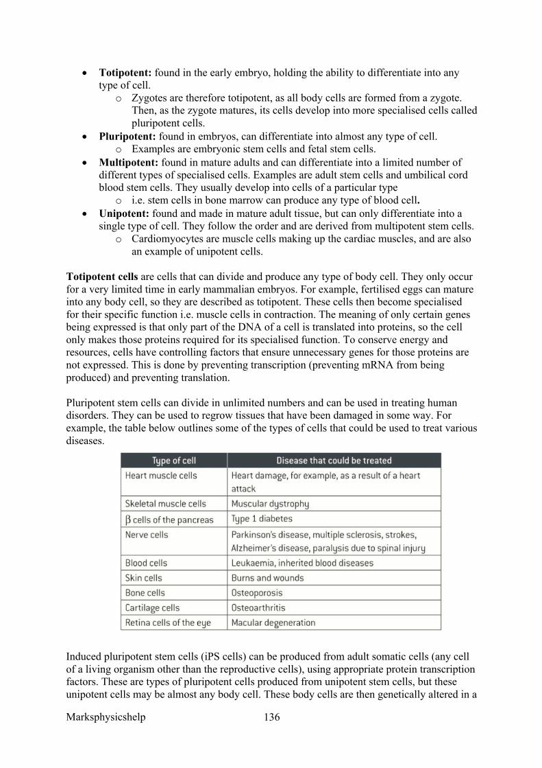
Marksphysicshelp
136
• Totipotent: found in the early embryo, holding the ability to differentiate into any type of cell.
o Zygotes are therefore totipotent, as all body cells are formed from a zygote. Then, as the zygote matures, its cells develop into more specialised cells called pluripotent cells.
• Pluripotent: found in embryos, can differentiate into almost any type of cell. o Examples are embryonic stem cells and fetal stem cells.
• Multipotent: found in mature adults and can differentiate into a limited number of different types of specialised cells. Examples are adult stem cells and umbilical cord blood stem cells. They usually develop into cells of a particular type
o i.e. stem cells in bone marrow can produce any type of blood cell. • Unipotent: found and made in mature adult tissue, but can only differentiate into a
single type of cell. They follow the order and are derived from multipotent stem cells. o Cardiomyocytes are muscle cells making up the cardiac muscles, and are also
an example of unipotent cells.
Totipotent cells are cells that can divide and produce any type of body cell. They only occur for a very limited time in early mammalian embryos. For example, fertilised eggs can mature into any body cell, so they are described as totipotent. These cells then become specialised for their specific function i.e. muscle cells in contraction. The meaning of only certain genes being expressed is that only part of the DNA of a cell is translated into proteins, so the cell only makes those proteins required for its specialised function. To conserve energy and resources, cells have controlling factors that ensure unnecessary genes for those proteins are not expressed. This is done by preventing transcription (preventing mRNA from being produced) and preventing translation.
Pluripotent stem cells can divide in unlimited numbers and can be used in treating human disorders. They can be used to regrow tissues that have been damaged in some way. For example, the table below outlines some of the types of cells that could be used to treat various diseases.
Induced pluripotent stem cells (iPS cells) can be produced from adult somatic cells (any cell of a living organism other than the reproductive cells), using appropriate protein transcription factors. These are types of pluripotent cells produced from unipotent stem cells, but these unipotent cells may be almost any body cell. These body cells are then genetically altered in a
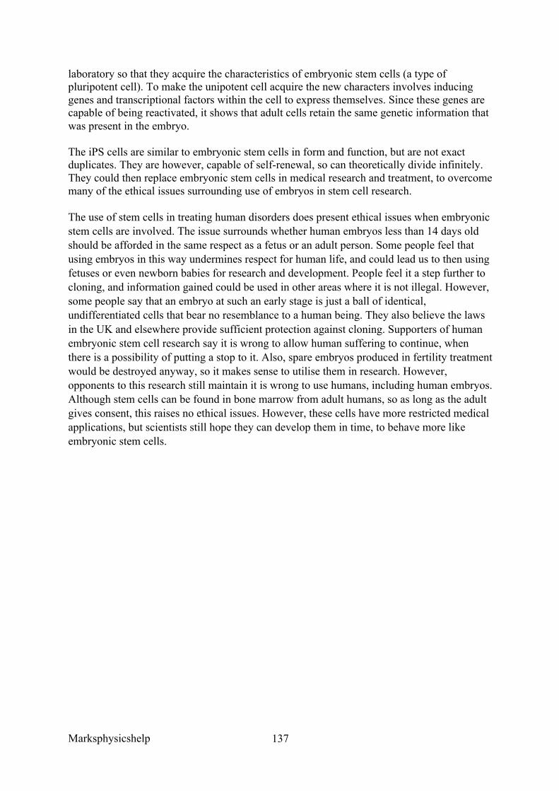
Marksphysicshelp
137
laboratory so that they acquire the characteristics of embryonic stem cells (a type of pluripotent cell). To make the unipotent cell acquire the new characters involves inducing genes and transcriptional factors within the cell to express themselves. Since these genes are capable of being reactivated, it shows that adult cells retain the same genetic information that was present in the embryo.
The iPS cells are similar to embryonic stem cells in form and function, but are not exact duplicates. They are however, capable of self-renewal, so can theoretically divide infinitely. They could then replace embryonic stem cells in medical research and treatment, to overcome many of the ethical issues surrounding use of embryos in stem cell research.
The use of stem cells in treating human disorders does present ethical issues when embryonic stem cells are involved. The issue surrounds whether human embryos less than 14 days old should be afforded in the same respect as a fetus or an adult person. Some people feel that using embryos in this way undermines respect for human life, and could lead us to then using fetuses or even newborn babies for research and development. People feel it a step further to cloning, and information gained could be used in other areas where it is not illegal. However, some people say that an embryo at such an early stage is just a ball of identical, undifferentiated cells that bear no resemblance to a human being. They also believe the laws in the UK and elsewhere provide sufficient protection against cloning. Supporters of human embryonic stem cell research say it is wrong to allow human suffering to continue, when there is a possibility of putting a stop to it. Also, spare embryos produced in fertility treatment would be destroyed anyway, so it makes sense to utilise them in research. However, opponents to this research still maintain it is wrong to use humans, including human embryos. Although stem cells can be found in bone marrow from adult humans, so as long as the adult gives consent, this raises no ethical issues. However, these cells have more restricted medical applications, but scientists still hope they can develop them in time, to behave more like embryonic stem cells.
Marksphysicshelp
138
AQA June 2014 Q7abc Question:
CREB is a transcription factor in the mitochondria of neurones. What is a transcription factor?
Answer:
1. (Protein/molecule) that moves from cytoplasm to DNA; 2. (TF) binds to specific gene/genes/ to specific part of/site on DNA/ binds to
promoter/RNA polymerase; 3. Leads to/blocks (pre)mRNA production / allows/blocks binding of RNA polymerase
(to DNA)/allows RNA polymerase to work
Question:
CREB leads to the formation of a protein that removes electrons and protons from reduced NAD in the mitochondrion.
Huntington’s disease (HD) causes the death of neurones. People with HD produce a substance called huntingtin. Some scientists have suggested that binding of huntingtin to CREB may lead to the death of neurones.
‘Suggest how binding of huntingtin to CREB may lead to the death of neurones.’
Answer:
1. (Binding to CREB) prevents transcription/mRNA formation; 2. (Binding of huntingtin) prevents production/translation of protein (that removes
electrons/protons from NAD); 3. Fewer electrons to electron transport chain/electron transport chain slows/stops/
stops/slower oxidative phosphorylation; 4. Fewer protons for proton gradient; 5. Not enough ATP produced/energy supplied to keep cells alive / anaerobic respiration
not enough to keep cell alive;
Question:
CREB is a protein synthesised in the cytoplasm of neurones. Transport of CREB from the cytoplasm into the matrix of a mitochondrion requires two carrier proteins.
Use your knowledge of the structure of a mitochondrion to explain why transport of CREB requires two carrier proteins.
Answer:
1. CREB/protein is too large/is water soluble so cannot cross membrane/phospholipid bilayer;

Marksphysicshelp
139
2. Mitochondrion has two membranes/inner and outer membranes; 3. For each (different) membrane a (different) carrier required
Marksphysicshelp
140
3.8.2 Gene expression is controlled by a number of features 3.8.2.2 Regulation of transcription and translation
Content
• In eukaryotes, transcription of target genes can be stimulated or inhibited when specific transcriptional factors move from the cytoplasm into the nucleus. The role of the steroid hormone, oestrogen, in initiating transcription.
• Epigenetic control of gene expression in eukaryotes. • Epigenetics involves heritable changes in gene function, without changes to the base
sequence of DNA. These changes are caused by changes in the environment that inhibit transcription by:
o Increased methylation of the DNA or o Decreased acetylation of associated histones.
• The relevance of epigenetics on the development and treatment of disease, especially cancer.
• In eukaryotes and some prokaryotes, translation of the mRNA produced from target genes can be inhibited by RNA interference (RNAi).
• Students should be able to: • Interpret data provided from investigations into gene expression • Evaluate appropriate data for the relative influences of genetic and environmental
factors on phenotype.
In eukaryotes, transcription of target genes can be stimulated or inhibited when specific transcriptional factors move from the cytoplasm into the nucleus. The steroid hormone called oestrogen plays a role in initiating transcription.
Oestrogen uses a different mechanism to the second messenger model that the hormone adrenaline uses.
• Before transcription begins, the genes are switched on by specific molecules that move from the cytoplasm to the nucleus.
• These molecules are called transcriptional factors, and each one has a site that binds to a specific base sequence of the DNA in the nucleus.
• Its binding causes this region of DNA to begin the process of transcription • mRNA is produced and the information it carries is then translated into a polypeptide • When a gene is not being expressed, the site on the transcriptional factor that binds to
the DNA is not active • As the site on the transcriptional factor binding to the DNA is inactive, it cannot cause
transcription and polypeptide synthesis.
Hormones like oestrogen can switch on a gene and thus start transcription by combining with a receptor site on the transcriptional factor. This activates the DNA binding site by causing it to change shape. This process operates as follows:
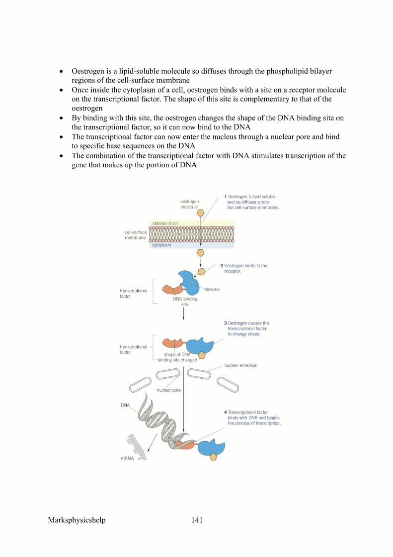
Marksphysicshelp
141
• Oestrogen is a lipid-soluble molecule so diffuses through the phospholipid bilayer regions of the cell-surface membrane
• Once inside the cytoplasm of a cell, oestrogen binds with a site on a receptor molecule on the transcriptional factor. The shape of this site is complementary to that of the oestrogen
• By binding with this site, the oestrogen changes the shape of the DNA binding site on the transcriptional factor, so it can now bind to the DNA
• The transcriptional factor can now enter the nucleus through a nuclear pore and bind to specific base sequences on the DNA
• The combination of the transcriptional factor with DNA stimulates transcription of the gene that makes up the portion of DNA.
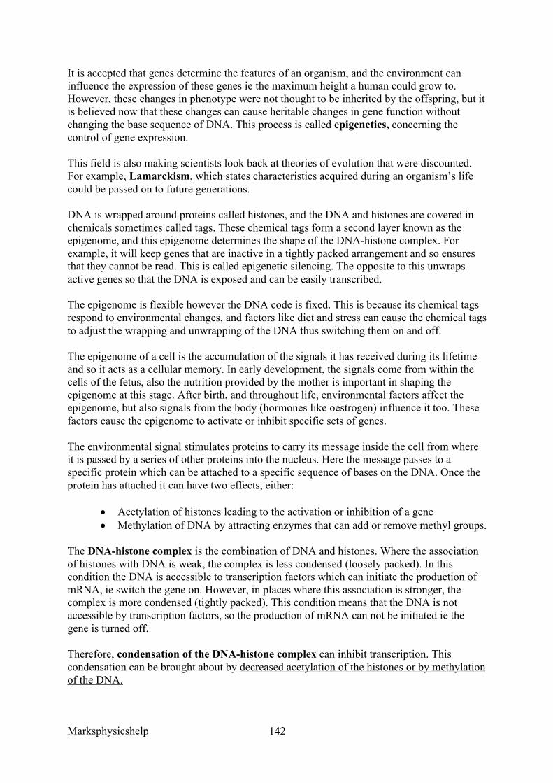
Marksphysicshelp
142
It is accepted that genes determine the features of an organism, and the environment can influence the expression of these genes ie the maximum height a human could grow to. However, these changes in phenotype were not thought to be inherited by the offspring, but it is believed now that these changes can cause heritable changes in gene function without changing the base sequence of DNA. This process is called epigenetics, concerning the control of gene expression.
This field is also making scientists look back at theories of evolution that were discounted. For example, Lamarckism, which states characteristics acquired during an organism’s life could be passed on to future generations.
DNA is wrapped around proteins called histones, and the DNA and histones are covered in chemicals sometimes called tags. These chemical tags form a second layer known as the epigenome, and this epigenome determines the shape of the DNA-histone complex. For example, it will keep genes that are inactive in a tightly packed arrangement and so ensures that they cannot be read. This is called epigenetic silencing. The opposite to this unwraps active genes so that the DNA is exposed and can be easily transcribed.
The epigenome is flexible however the DNA code is fixed. This is because its chemical tags respond to environmental changes, and factors like diet and stress can cause the chemical tags to adjust the wrapping and unwrapping of the DNA thus switching them on and off.
The epigenome of a cell is the accumulation of the signals it has received during its lifetime and so it acts as a cellular memory. In early development, the signals come from within the cells of the fetus, also the nutrition provided by the mother is important in shaping the epigenome at this stage. After birth, and throughout life, environmental factors affect the epigenome, but also signals from the body (hormones like oestrogen) influence it too. These factors cause the epigenome to activate or inhibit specific sets of genes.
The environmental signal stimulates proteins to carry its message inside the cell from where it is passed by a series of other proteins into the nucleus. Here the message passes to a specific protein which can be attached to a specific sequence of bases on the DNA. Once the protein has attached it can have two effects, either:
• Acetylation of histones leading to the activation or inhibition of a gene • Methylation of DNA by attracting enzymes that can add or remove methyl groups.
The DNA-histone complex is the combination of DNA and histones. Where the association of histones with DNA is weak, the complex is less condensed (loosely packed). In this condition the DNA is accessible to transcription factors which can initiate the production of mRNA, ie switch the gene on. However, in places where this association is stronger, the complex is more condensed (tightly packed). This condition means that the DNA is not accessible by transcription factors, so the production of mRNA can not be initiated ie the gene is turned off.
Therefore, condensation of the DNA-histone complex can inhibit transcription. This condensation can be brought about by decreased acetylation of the histones or by methylation of the DNA.
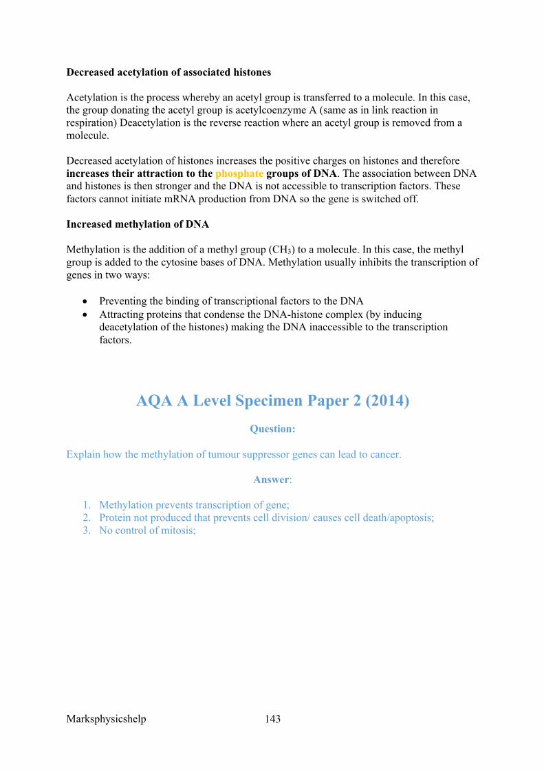
Marksphysicshelp
143
Decreased acetylation of associated histones
Acetylation is the process whereby an acetyl group is transferred to a molecule. In this case, the group donating the acetyl group is acetylcoenzyme A (same as in link reaction in respiration) Deacetylation is the reverse reaction where an acetyl group is removed from a molecule.
Decreased acetylation of histones increases the positive charges on histones and therefore increases their attraction to the phosphate groups of DNA. The association between DNA and histones is then stronger and the DNA is not accessible to transcription factors. These factors cannot initiate mRNA production from DNA so the gene is switched off.
Increased methylation of DNA
Methylation is the addition of a methyl group (CH3) to a molecule. In this case, the methyl group is added to the cytosine bases of DNA. Methylation usually inhibits the transcription of genes in two ways:
• Preventing the binding of transcriptional factors to the DNA • Attracting proteins that condense the DNA-histone complex (by inducing
deacetylation of the histones) making the DNA inaccessible to the transcription factors.
AQA A Level Specimen Paper 2 (2014) Question:
Explain how the methylation of tumour suppressor genes can lead to cancer.
Answer:
1. Methylation prevents transcription of gene; 2. Protein not produced that prevents cell division/ causes cell death/apoptosis; 3. No control of mitosis;
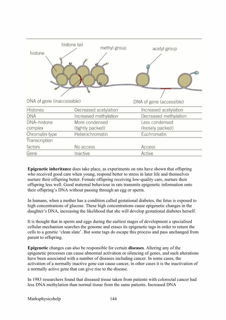
Marksphysicshelp
144
Epigenetic inheritance does take place, as experiments on rats have shown that offspring who received good care when young, respond better to stress in later life and themselves nurture their offspring better. Female offspring receiving low-quality care, nurture their offspring less well. Good maternal behaviour in rats transmits epigenetic information onto their offspring’s DNA without passing through an egg or sperm.
In humans, when a mother has a condition called gestational diabetes, the fetus is exposed to high concentrations of glucose. These high concentrations cause epigenetic changes in the daughter’s DNA, increasing the likelihood that she will develop gestational diabetes herself.
It is thought that in sperm and eggs during the earliest stages of development a specialised cellular mechanism searches the genome and erases its epigenetic tags in order to return the cells to a genetic ‘clean slate’. But some tags do escape this process and pass unchanged from parent to offspring.
Epigenetic changes can also be responsible for certain diseases. Altering any of the epigenetic processes can cause abnormal activation or silencing of genes, and such alterations have been associated with a number of diseases including cancer. In some cases, the activation of a normally inactive gene can cause cancer, in other cases it is the inactivation of a normally active gene that can give rise to the disease.
In 1983 researchers found that diseased tissue taken from patients with colorectal cancer had less DNA methylation than normal tissue from the same patients. Increased DNA
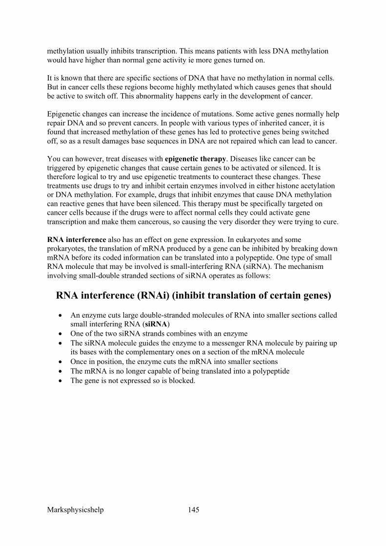
Marksphysicshelp
145
methylation usually inhibits transcription. This means patients with less DNA methylation would have higher than normal gene activity ie more genes turned on.
It is known that there are specific sections of DNA that have no methylation in normal cells. But in cancer cells these regions become highly methylated which causes genes that should be active to switch off. This abnormality happens early in the development of cancer.
Epigenetic changes can increase the incidence of mutations. Some active genes normally help repair DNA and so prevent cancers. In people with various types of inherited cancer, it is found that increased methylation of these genes has led to protective genes being switched off, so as a result damages base sequences in DNA are not repaired which can lead to cancer.
You can however, treat diseases with epigenetic therapy. Diseases like cancer can be triggered by epigenetic changes that cause certain genes to be activated or silenced. It is therefore logical to try and use epigenetic treatments to counteract these changes. These treatments use drugs to try and inhibit certain enzymes involved in either histone acetylation or DNA methylation. For example, drugs that inhibit enzymes that cause DNA methylation can reactive genes that have been silenced. This therapy must be specifically targeted on cancer cells because if the drugs were to affect normal cells they could activate gene transcription and make them cancerous, so causing the very disorder they were trying to cure.
RNA interference also has an effect on gene expression. In eukaryotes and some prokaryotes, the translation of mRNA produced by a gene can be inhibited by breaking down mRNA before its coded information can be translated into a polypeptide. One type of small RNA molecule that may be involved is small-interfering RNA (siRNA). The mechanism involving small-double stranded sections of siRNA operates as follows:
RNA interference (RNAi) (inhibit translation of certain genes)
• An enzyme cuts large double-stranded molecules of RNA into smaller sections called small interfering RNA (siRNA)
• One of the two siRNA strands combines with an enzyme • The siRNA molecule guides the enzyme to a messenger RNA molecule by pairing up
its bases with the complementary ones on a section of the mRNA molecule • Once in position, the enzyme cuts the mRNA into smaller sections • The mRNA is no longer capable of being translated into a polypeptide • The gene is not expressed so is blocked.
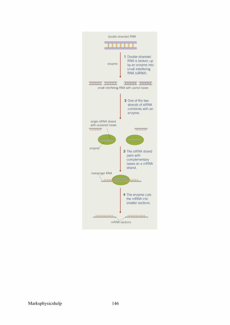
Marksphysicshelp
146
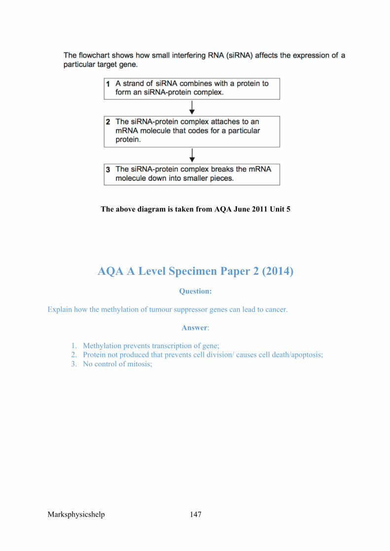
Marksphysicshelp
147
The above diagram is taken from AQA June 2011 Unit 5
AQA A Level Specimen Paper 2 (2014) Question:
Explain how the methylation of tumour suppressor genes can lead to cancer.
Answer:
1. Methylation prevents transcription of gene; 2. Protein not produced that prevents cell division/ causes cell death/apoptosis; 3. No control of mitosis;
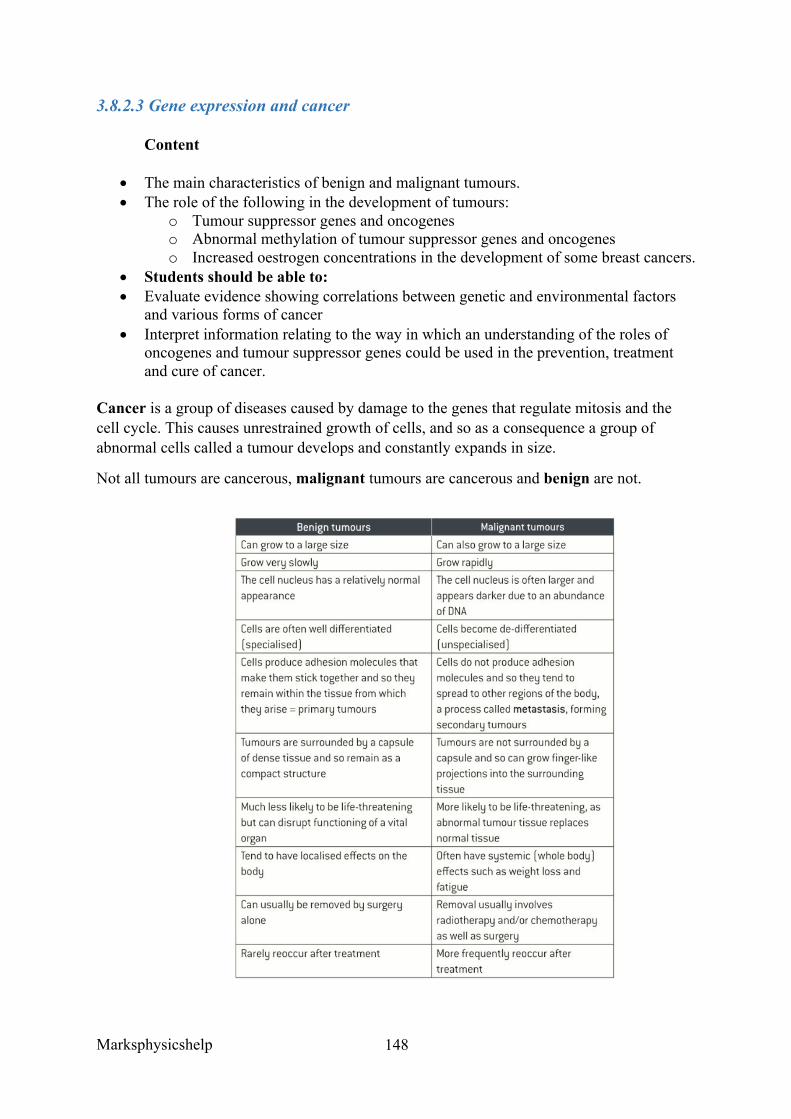
Marksphysicshelp
148
3.8.2.3 Gene expression and cancer
Content
• The main characteristics of benign and malignant tumours. • The role of the following in the development of tumours:
o Tumour suppressor genes and oncogenes o Abnormal methylation of tumour suppressor genes and oncogenes o Increased oestrogen concentrations in the development of some breast cancers.
• Students should be able to: • Evaluate evidence showing correlations between genetic and environmental factors
and various forms of cancer • Interpret information relating to the way in which an understanding of the roles of
oncogenes and tumour suppressor genes could be used in the prevention, treatment and cure of cancer.
Cancer is a group of diseases caused by damage to the genes that regulate mitosis and the cell cycle. This causes unrestrained growth of cells, and so as a consequence a group of abnormal cells called a tumour develops and constantly expands in size.
Not all tumours are cancerous, malignant tumours are cancerous and benign are not.
Marksphysicshelp
149
DNA analysis has shown that in general cancer cells are derived from a single mutant cell. The initial mutation causes uncontrolled mitosis in this cell. Later, a further mutation in a descendant cell leads to other changes that cause subsequent cells to be different from normal in growth and appearance. The two main types of genes that play a role in cancer are tumour suppressor genes and oncogenes.
Oncogenes are usually mutations of proto-oncogenes. These proto-oncogenes stimulate a cell to divide when growth factors attach to a protein receptor on its cell-surface membrane. This then activates genes that cause DNA to replicate and the cell to divide. If a proto-oncogene mutates into an oncogene it can become permanently activated for two reasons:
• The receptor protein on the cell-surface membrane can be permanently activated, so that cell division is switched on even in the absence of growth factors
• The oncogene may code for a growth factor that is then produced in excessive amounts, again stimulating excessive cell division.
The result is that cells divide too rapidly and out of control, and a tumour or cancer develops. A few cancers are caused by inherited mutations of proto-oncogenes that cause the oncogene to be activated but most cancer-causing mutations involving oncogenes are acquired, not inherited.
Tumour suppressor genes are genes that slow down cell division, repair mistakes in DNA and ‘tell’ cells when to die – a process called apoptosis (programmed cell death). They have the opposite role from proto-oncogenes. A normal tumour suppressor gene will maintain normal rates of cell division and so prevent further formation of tumours. If a tumour suppressor gene becomes mutated it is inactivated (switched off) and so as a result it stops inhibiting cell division and cells can grow out of control. The mutated cells that are formed are usually structurally and functionally different from normal cells. Most of these cells die, but those that survive can make clones of themselves and form tumours. Examples of tumour suppressor genes are TP53, BRCA1 and BRCA2.
Some cancers are caused by inherited mutations of tumour suppressor genes but most are acquired, not inherited. More than half of human cancers show abnormalities of the TP53 gene which codes for the p53 protein. An acquired mutation of the TP53 gene occurs in many cancers, including lung and breast cancer. The p53 protein is involved in the process of apoptosis (programmed cell death). This process is activated when a cell is unable to repair DNA, so if the gene for p53 is not functioning properly, cells with damaged DNA continue to divide leading to cancer.
An important difference between oncogenes and tumour suppressor genes is that while oncogenes cause cancer as a result of the activation of proto-oncogenes, tumour suppressor genes cause cancer when they are inactivated.
Abnormal methylation of tumour suppressor genes is a symptom of a variety of different cancers. The most common abnormality is hypermethylation, and the process by which hypermethylation may lead to cancer is as follows:
• Hypermethylation occurs in a specific region (promoter region) of a tumour suppressor gene
• This leads to the tumour suppressor gene being inactivated
Marksphysicshelp
150
• As a result, the transcription of the promoter regions of tumour suppressor genes is inhibited
• The tumour suppressor gene is silenced (switched off) • As the tumour suppressor gene normally slows the rate of cell division, its
inactivation leads to increased cell division and the formation of a tumour.
Abnormal methylation of this type is thought to occur in a tumour suppressor gene known as BRCA1 and leads to the development of breast cancer
Another form of abnormal methylation is hypomethylation (reduced methylation). This has been found to occur in oncogenes where it leads to their activation and hence the formation of tumours. The reduced methylation causes the oncogenes to be expressed, thus cells divide out of control and form tumours.
Oestrogen plays a central role in regulating the menstrual cycle in women. It is known that after the menopause, a woman’s risk of developing breast cancer increases. This is thought to be due to increased oestrogen concentrations.
After menopause the production of oestrogen from the ovaries diminishes, but production from the fat cells of breasts tend to produce more oestrogens after the menopause. These locally produced oestrogens appear to trigger breast cancer. Once a tumour has developed, it further increases oestrogen concentration which therefore leads to increased development of the tumour. It also appears that WBCs that are drawn to the tumour increase oestrogen production, leading to greater development of the tumour.
Oestrogen activates a gene to begin the process of transcription. If the gene that oestrogen acts on is one that controls cell division and growth, then it will be activated and its continued division could produce a tumour. It is known, for example, that oestrogen causes proto-oncogenes of cells in breast tissue to develop into oncogenes, so leading to the development of a tumour.
Marksphysicshelp
151
AQA June 2015 Q4c Question:
For each student, the scientists also determined how much methylation was present within the specific gene. DNA methylation is the attachment of substances called methyl groups to cytosine or adenine. This prevents transcription.
Suggest how methylation of DNA can prevent expression of a gene.
Answer:
1. Prevent binding of transcription factor; 2. Prevents RNA polymerase binding/working; 3. Prevents complementary/specific base-pairing; 4. (No transcription, so) no (pre)mRNA made; 5. No/less translation (of mRNA);
Marksphysicshelp
152
AQA Human Biology Unit 2 (old spec) Q6
Question:
Give two ways in which a malignant tumour differs from a benign tumour.
Answer:
Malignant tumour (contrary to benign tumour) …
• Metastises. • Is quick-growing; • Is not enclosed by a membrane; • Invades neighbouring tissues;

Marksphysicshelp
153
Marksphysicshelp
154
3.8.3 Using genome projects
Content
• Sequencing projects have read the genomes of a wide range of organisms, including humans.
• Determining the genome of simpler organisms allows the sequences of the proteins that derive from the genetic code (the proteome) of the organism to be determined. This may have many applications, including the identification of potential antigens for use in vaccine production.
• In more complex organisms, the presence of non-coding DNA and of regulatory genes means that knowledge of the genome cannot easily be translated into the proteome.
• Sequencing methods are continuously updated and have become automated.
Sequencing projects aim to determine the DNA nucleotide base sequence, and have read the genomes of a wide range of organisms, including humans. The human genome consists of over 3 billion base pairs organised into around 20,000 genes. The use of bioinformatics catalysed the process of obtaining this information, as it is the science of collecting and analysing complex biological data such as genetic codes. It uses computers to read, store and organise biological data at a much faster rate than before, also utilising algorithms to analyse and interpret data.
In determining the genome of simpler organisms, it allows the sequences of the proteins that derive from the genetic code (the proteome) of the organism to be determined. A definition of the proteome is all of the proteins produced by the genome. But since not all genes are switched on all the time to produce certain proteins, a more specific definition is all of the proteins produced in a given type of cell, or organism, in a given time, under specified conditions.
Determining the genome of more simple organisms like bacteria is hoped to give us more information on curing disease, and provide information on genes that can be usefully exploited. An example being genes from organisms that allow them to withstand extreme or toxic environmental conditions, so have potential uses in cleaning up pollutants or in manufacturing biofuels.
It is much easier to determine the proteome of prokaryotic organisms because:
• Most prokaryotes have just one circular piece of DNA (plasmid), not associated with histones.
• There are no non-coding sections of DNA (introns) present in eukaryotic cells.
One of the main applications of determining the genome of simpler organisms is the identification of potential proteins (antigens) for the use of vaccine production. Antigens can be manufactured and then administered to people in appropriate doses, which will ultimately cause the production of memory cells that can trigger a secondary response if the person encountered the antigen again.
Marksphysicshelp
155
Determining the proteome in complex organisms is more difficult because of the presence of non-coding DNA and regulatory genes. It makes it difficult to apply knowledge of the genome to the proteome. It is thought that, in humans, less than 1.5% of genes may code for proteins. Also, in humans, apart from identical twins, all DNA base sequences will map differently. Although sequencing methods are continuously updated and have become automated.

Marksphysicshelp
156
Marksphysicshelp
157
3.8.4 Gene technologies allow the study and alteration of gene function allowing a better understanding of organism function and the design of new industrial and medical processes 3.8.4.1 Recombinant DNA technology
Content
• Recombinant DNA technology involves the transfer of fragments of DNA from one organism, or species, to another. Since the genetic code is universal, as are transcription and translation mechanisms, the transferred DNA can be translated within cells of the recipient (transgenic) organism.
• Fragments of DNA can be produced by several methods, including: o Conversion of mRNA to complementary DNA (cDNA), using reverse
transcriptase o Using restriction enzymes to cut a fragment containing the desired gene from
DNA o Creating the gene in a ‘gene machine’.
• Fragments of DNA can be amplified by in vitro and in vivo techniques. • The principles of the polymerase chain reaction (PCR) as an in vitro method to
amplify DNA fragments. • The culture of transformed host cells as an in vivo method to amplify DNA fragments.
o The addition of promoter and terminator regions to the fragments of DNA.
o The use of restriction endonucleases and ligases to insert fragments of DNA into vectors. Transformation of host cells using these vectors.
o The use of marker genes to detect genetically modified (GM) cells or organisms. (Students will not be required to recall specific marker genes in a written paper.)
• Students should be able to: o Interpret information relating to the use of recombinant DNA technology o Evaluate the ethical, financial and social issues associated with the use and
ownership of recombinant DNA technology in agriculture, in industry and in medicine
o Balance the humanitarian aspects of recombinant DNA technology with the opposition from environmentalists and anti-globalisation activists
o Relate recombinant DNA technology to gene therapy.
Recombinant DNA technology involves the transfer of fragments of DNA from one organism, or species to another. Since the genetic code is universal, and likewise are transcription and translation mechanisms, the transferred DNA can be translated within cells of the recipient genetically modified (transgenic) organism. The process of making a protein using the DNA technology of gene transfer and cloning involves a number of stages:
1. Isolation of the DNA fragments that have the gene for the desired protein 2. Insertion of the DNA fragment into a vector
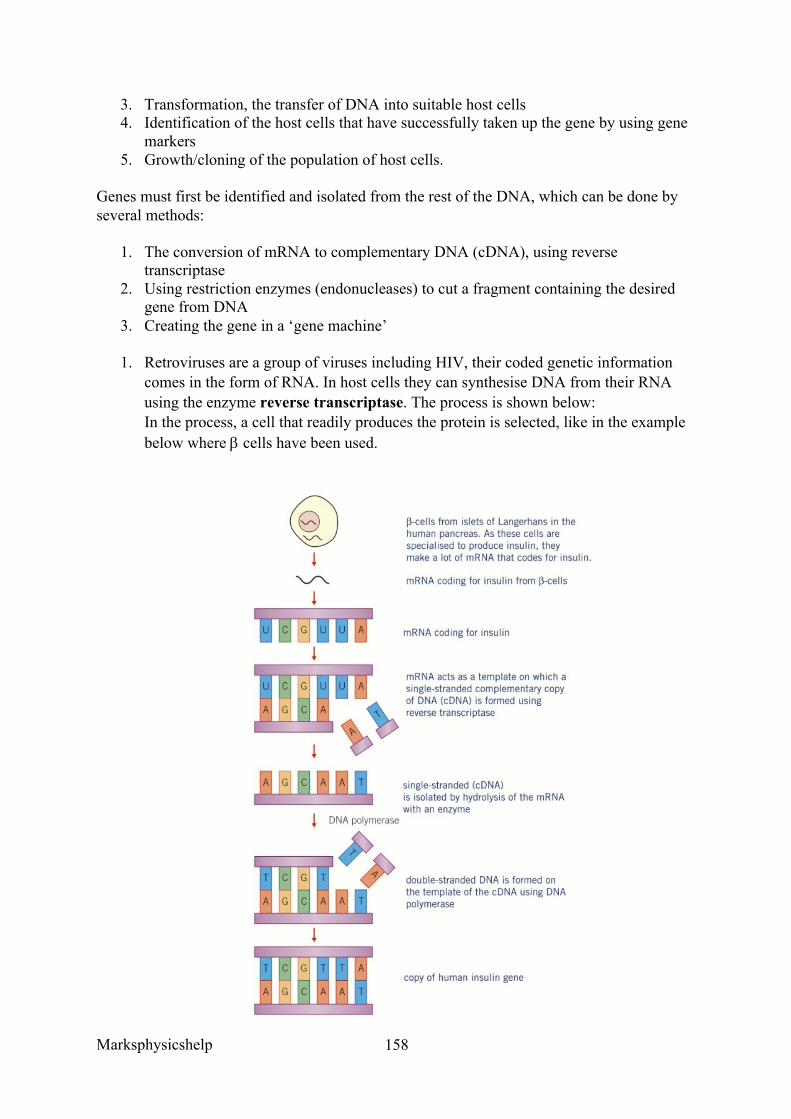
Marksphysicshelp
158
3. Transformation, the transfer of DNA into suitable host cells 4. Identification of the host cells that have successfully taken up the gene by using gene
markers 5. Growth/cloning of the population of host cells.
Genes must first be identified and isolated from the rest of the DNA, which can be done by several methods:
1. The conversion of mRNA to complementary DNA (cDNA), using reverse transcriptase
2. Using restriction enzymes (endonucleases) to cut a fragment containing the desired gene from DNA
3. Creating the gene in a ‘gene machine’
1. Retroviruses are a group of viruses including HIV, their coded genetic information comes in the form of RNA. In host cells they can synthesise DNA from their RNA using the enzyme reverse transcriptase. The process is shown below: In the process, a cell that readily produces the protein is selected, like in the example below where b cells have been used.
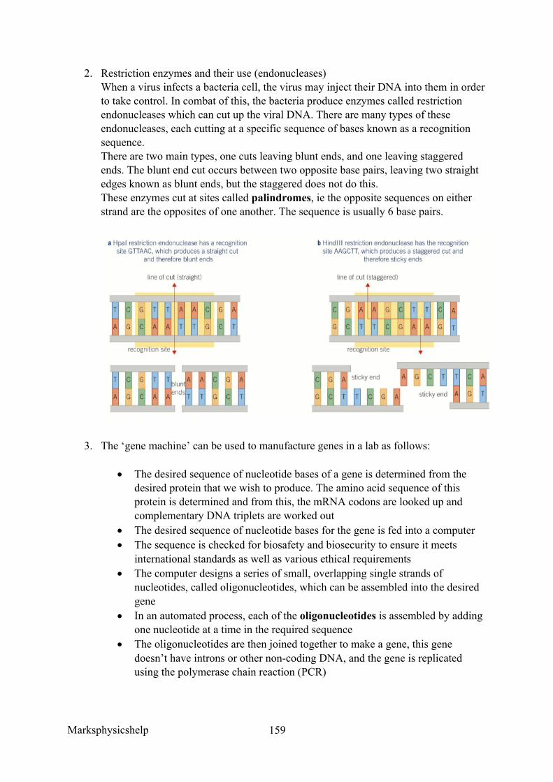
Marksphysicshelp
159
2. Restriction enzymes and their use (endonucleases) When a virus infects a bacteria cell, the virus may inject their DNA into them in order to take control. In combat of this, the bacteria produce enzymes called restriction endonucleases which can cut up the viral DNA. There are many types of these endonucleases, each cutting at a specific sequence of bases known as a recognition sequence. There are two main types, one cuts leaving blunt ends, and one leaving staggered ends. The blunt end cut occurs between two opposite base pairs, leaving two straight edges known as blunt ends, but the staggered does not do this. These enzymes cut at sites called palindromes, ie the opposite sequences on either strand are the opposites of one another. The sequence is usually 6 base pairs.
3. The ‘gene machine’ can be used to manufacture genes in a lab as follows:
• The desired sequence of nucleotide bases of a gene is determined from the desired protein that we wish to produce. The amino acid sequence of this protein is determined and from this, the mRNA codons are looked up and complementary DNA triplets are worked out
• The desired sequence of nucleotide bases for the gene is fed into a computer • The sequence is checked for biosafety and biosecurity to ensure it meets
international standards as well as various ethical requirements • The computer designs a series of small, overlapping single strands of
nucleotides, called oligonucleotides, which can be assembled into the desired gene
• In an automated process, each of the oligonucleotides is assembled by adding one nucleotide at a time in the required sequence
• The oligonucleotides are then joined together to make a gene, this gene doesn’t have introns or other non-coding DNA, and the gene is replicated using the polymerase chain reaction (PCR)
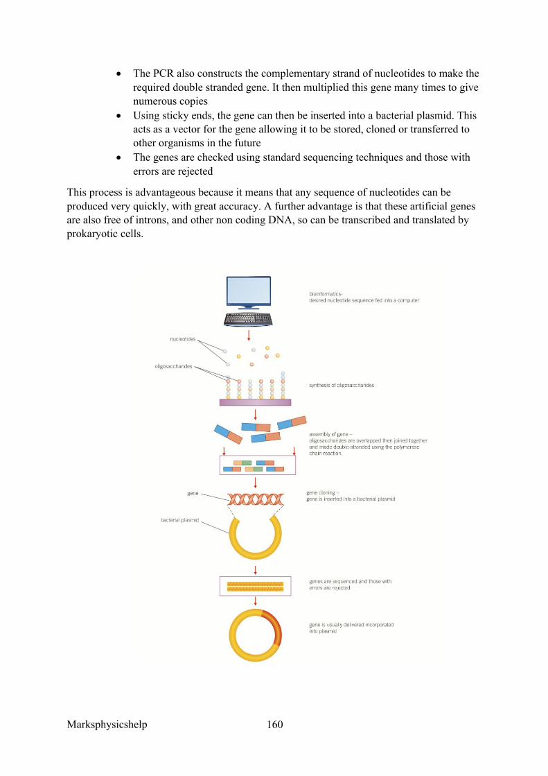
Marksphysicshelp
160
• The PCR also constructs the complementary strand of nucleotides to make the required double stranded gene. It then multiplied this gene many times to give numerous copies
• Using sticky ends, the gene can then be inserted into a bacterial plasmid. This acts as a vector for the gene allowing it to be stored, cloned or transferred to other organisms in the future
• The genes are checked using standard sequencing techniques and those with errors are rejected
This process is advantageous because it means that any sequence of nucleotides can be produced very quickly, with great accuracy. A further advantage is that these artificial genes are also free of introns, and other non coding DNA, so can be transcribed and translated by prokaryotic cells.
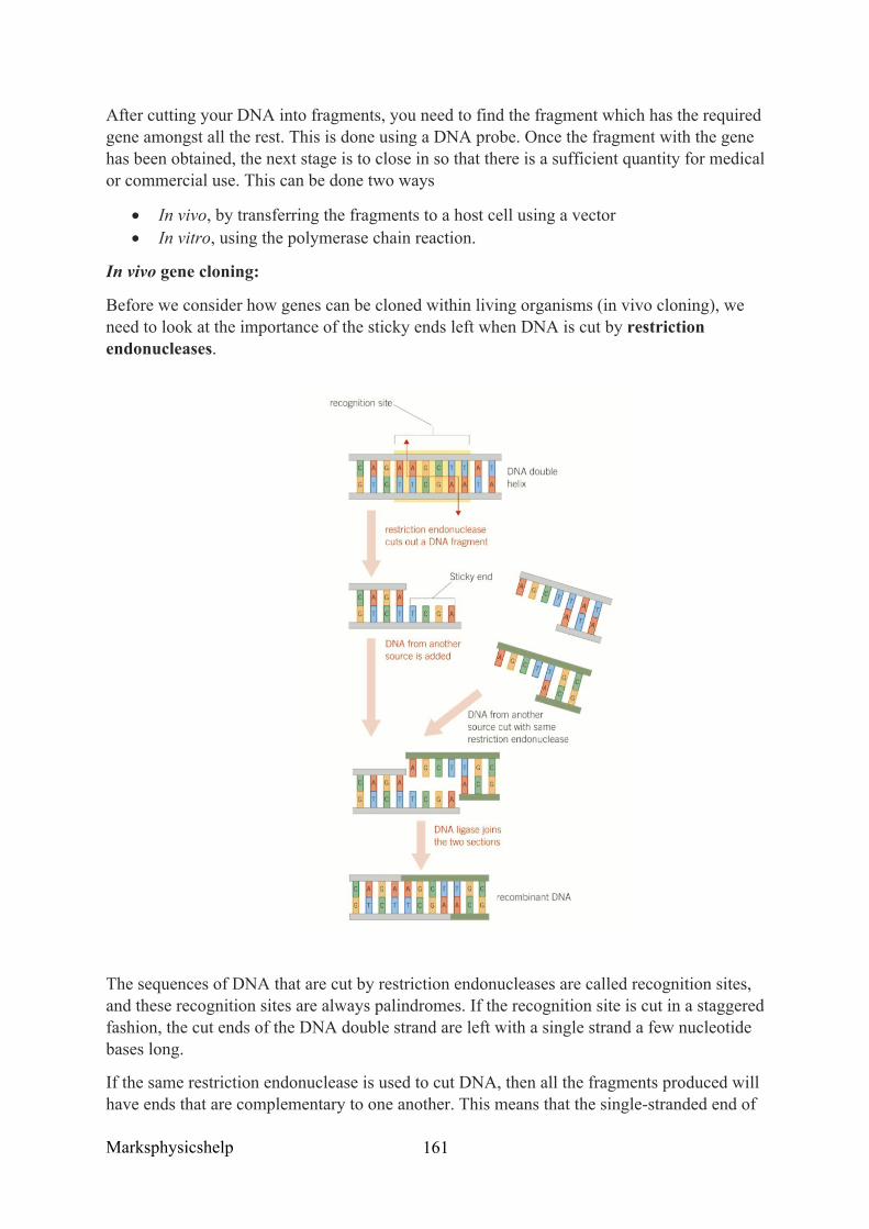
Marksphysicshelp
161
After cutting your DNA into fragments, you need to find the fragment which has the required gene amongst all the rest. This is done using a DNA probe. Once the fragment with the gene has been obtained, the next stage is to close in so that there is a sufficient quantity for medical or commercial use. This can be done two ways
• In vivo, by transferring the fragments to a host cell using a vector • In vitro, using the polymerase chain reaction.
In vivo gene cloning:
Before we consider how genes can be cloned within living organisms (in vivo cloning), we need to look at the importance of the sticky ends left when DNA is cut by restriction endonucleases.
The sequences of DNA that are cut by restriction endonucleases are called recognition sites, and these recognition sites are always palindromes. If the recognition site is cut in a staggered fashion, the cut ends of the DNA double strand are left with a single strand a few nucleotide bases long.
If the same restriction endonuclease is used to cut DNA, then all the fragments produced will have ends that are complementary to one another. This means that the single-stranded end of
Marksphysicshelp
162
any one fragment can be stuck to the single-stranded end of any other fragment, essentially, these ends are ‘sticky’. Once the complementary bases of two sticky ends have paired up, an enzyme called DNA ligase is used to bind the phosphate-sugar framework of the two sections of DNA and so unite them as one.
Sticky ends are very important, because provided the same restriction endonuclease is use, we can combine the DNA of one organism with that of any other organism.
Preparing the DNA fragment for insertion is the next stage, it involves the addition of extra lengths of DNA. For the transcription of any gene to take place, the enzyme that synthesises mRNA (RNA polymerase) must attach to the DNA near a gene. The binding site for RNA polymerase is a region of DNA, known as a promoter region. The nucleotide bases of the promoter region attach both RNA polymerase and transcription factors, then begin the process of transcription. If we want our DNA fragment to transcribe mRNA in order to make a protein, it is essential that we attach to it the necessary promoter region to start the process.
In the same way as a region of DNA binds RNA polymerase and begins transcription of a gene, another region releases RNA polymerase and ends transcription. This region of DNA is called a terminator. Again, we need to add a terminator region to the other end of the DNA fragment to stop transcription at an appropriate point.
Insertion of DNA fragments into a vector can happen once an appropriate fragment of DNA has been cut from the rest of the DNA and the promotor and terminator regions added. The next task is to join it into a carrying unit, known as a vector. This vector is used to transport the DNA into the host cell. There are different types of vector but the most commonly used is the plasmid. Plasmids are circular lengths of DNA, found in bacteria, which are separate from the main bacterial DNA. Plasmids almost always contain genes for antibiotic resistance, and restriction endonucleases are used at one of these antibiotic-resistance genes to break the plasmid loop.
The restriction endonuclease used is the same as one that cut out the DNA fragment, ensuring that the sticky ends of the opened-up plasmid are complementary to the sticky ends of the DNA fragment. When the DNA fragments are mixed with the opened-up plasmids, they may become incorporated into them. Where they are incorporated, the join is made permanent using the enzyme DNA. These plasmids now have recombinant DNA.
Introduction of DNA into host cells is the next stage, these host cells are bacterial cells. This process is called transformation, and involves the plasmids and bacterial cells being mixed together in a medium containing calcium ions. The calcium ions, and changes in temperature, make the bacterial membrane permeable, allowing the plasmids to pass through the cell-surface membrane into the cytoplasm. However, not all the bacterial cells will possess the DNA fragments with the desired gene for the desired protein. Some reasons for this are:
• Only a few bacterial cells take up the plasmids when the two are mixed together • Some plasmids have closed up again without incorporating the DNA fragment • Sometimes the DNA fragment ends join together to form its own plasmid
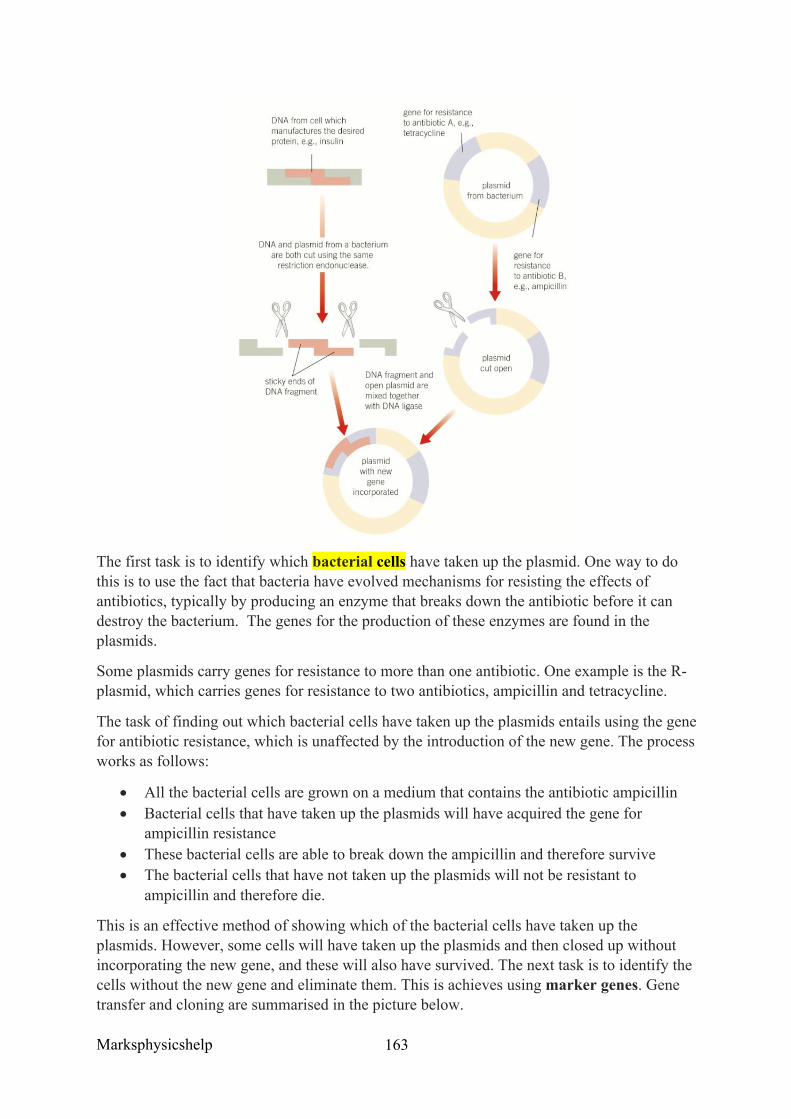
Marksphysicshelp
163
The first task is to identify which bacterial cells have taken up the plasmid. One way to do this is to use the fact that bacteria have evolved mechanisms for resisting the effects of antibiotics, typically by producing an enzyme that breaks down the antibiotic before it can destroy the bacterium. The genes for the production of these enzymes are found in the plasmids.
Some plasmids carry genes for resistance to more than one antibiotic. One example is the R-plasmid, which carries genes for resistance to two antibiotics, ampicillin and tetracycline.
The task of finding out which bacterial cells have taken up the plasmids entails using the gene for antibiotic resistance, which is unaffected by the introduction of the new gene. The process works as follows:
• All the bacterial cells are grown on a medium that contains the antibiotic ampicillin • Bacterial cells that have taken up the plasmids will have acquired the gene for
ampicillin resistance • These bacterial cells are able to break down the ampicillin and therefore survive • The bacterial cells that have not taken up the plasmids will not be resistant to
ampicillin and therefore die.
This is an effective method of showing which of the bacterial cells have taken up the plasmids. However, some cells will have taken up the plasmids and then closed up without incorporating the new gene, and these will also have survived. The next task is to identify the cells without the new gene and eliminate them. This is achieves using marker genes. Gene transfer and cloning are summarised in the picture below.
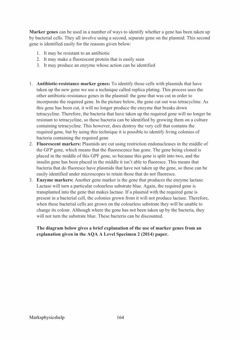
Marksphysicshelp
164
Marker genes can be used in a number of ways to identify whether a gene has been taken up by bacterial cells. They all involve using a second, separate gene on the plasmid. This second gene is identified easily for the reasons given below:
1. It may be resistant to an antibiotic 2. It may make a fluorescent protein that is easily seen 3. It may produce an enzyme whose action can be identified
1. Antibiotic-resistance marker genes: To identify those cells with plasmids that have taken up the new gene we use a technique called replica plating. This process uses the other antibiotic-resistance genes in the plasmid: the gene that was cut in order to incorporate the required gene. In the picture below, the gene cut out was tetracycline. As this gene has been cut, it will no longer produce the enzyme that breaks down tetracycline. Therefore, the bacteria that have taken up the required gene will no longer be resistant to tetracycline, so these bacteria can be identified by growing them on a culture containing tetracycline. This however, does destroy the very cell that contains the required gene, but by using this technique it is possible to identify living colonies of bacteria containing the required gene
2. Fluorescent markers: Plasmids are cut using restriction endonucleases in the middle of the GFP gene, which means that the fluorescence has gone. The gene being cloned is placed in the middle of this GPF gene, so because this gene is split into two, and the insulin gene has been placed in the middle it isn’t able to fluoresce. This means that bacteria that do fluoresce have plasmids that have not taken up the gene, so these can be easily identified under microscopes to retain those that do not fluoresce.
3. Enzyme markers: Another gene marker is the gene that produces the enzyme lactase. Lactase will turn a particular colourless substrate blue. Again, the required gene is transplanted into the gene that makes lactase. If a plasmid with the required gene is present in a bacterial cell, the colonies grown from it will not produce lactase. Therefore, when these bacterial cells are grown on the colourless substrate they will be unable to change its colour. Although where the gene has not been taken up by the bacteria, they will not turn the substrate blue. These bacteria can be discounted. The diagram below gives a brief explanation of the use of marker genes from an explanation given in the AQA A Level Specimen 2 (2014) paper.
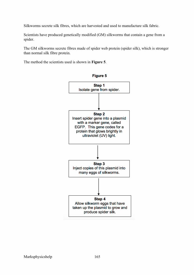
Marksphysicshelp
165
Silkworms secrete silk fibres, which are harvested and used to manufacture silk fabric.
Scientists have produced genetically modified (GM) silkworms that contain a gene from a spider.
The GM silkworms secrete fibres made of spider web protein (spider silk), which is stronger than normal silk fibre protein.
The method the scientists used is shown in Figure 5.
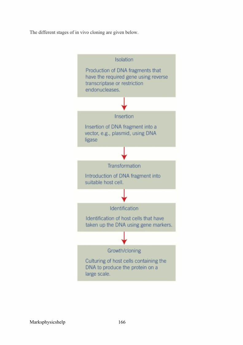
Marksphysicshelp
166
The different stages of in vivo cloning are given below.
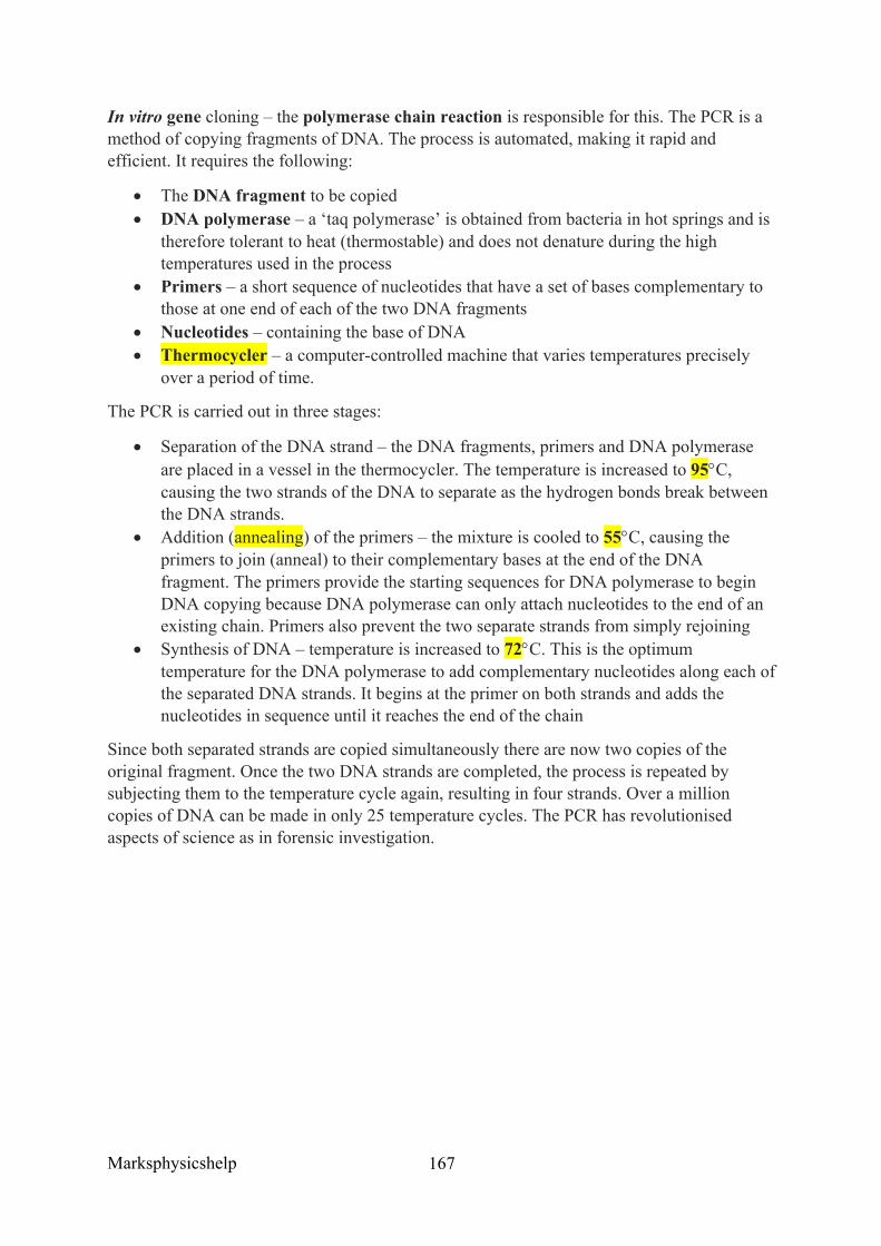
Marksphysicshelp
167
In vitro gene cloning – the polymerase chain reaction is responsible for this. The PCR is a method of copying fragments of DNA. The process is automated, making it rapid and efficient. It requires the following:
• The DNA fragment to be copied • DNA polymerase – a ‘taq polymerase’ is obtained from bacteria in hot springs and is
therefore tolerant to heat (thermostable) and does not denature during the high temperatures used in the process
• Primers – a short sequence of nucleotides that have a set of bases complementary to those at one end of each of the two DNA fragments
• Nucleotides – containing the base of DNA • Thermocycler – a computer-controlled machine that varies temperatures precisely
over a period of time.
The PCR is carried out in three stages:
• Separation of the DNA strand – the DNA fragments, primers and DNA polymerase are placed in a vessel in the thermocycler. The temperature is increased to 95°C, causing the two strands of the DNA to separate as the hydrogen bonds break between the DNA strands.
• Addition (annealing) of the primers – the mixture is cooled to 55°C, causing the primers to join (anneal) to their complementary bases at the end of the DNA fragment. The primers provide the starting sequences for DNA polymerase to begin DNA copying because DNA polymerase can only attach nucleotides to the end of an existing chain. Primers also prevent the two separate strands from simply rejoining
• Synthesis of DNA – temperature is increased to 72°C. This is the optimum temperature for the DNA polymerase to add complementary nucleotides along each of the separated DNA strands. It begins at the primer on both strands and adds the nucleotides in sequence until it reaches the end of the chain
Since both separated strands are copied simultaneously there are now two copies of the original fragment. Once the two DNA strands are completed, the process is repeated by subjecting them to the temperature cycle again, resulting in four strands. Over a million copies of DNA can be made in only 25 temperature cycles. The PCR has revolutionised aspects of science as in forensic investigation.
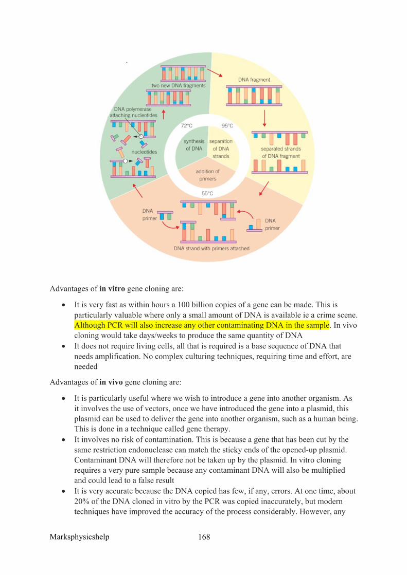
Marksphysicshelp
168
Advantages of in vitro gene cloning are:
• It is very fast as within hours a 100 billion copies of a gene can be made. This is particularly valuable where only a small amount of DNA is available ie a crime scene. Although PCR will also increase any other contaminating DNA in the sample. In vivo cloning would take days/weeks to produce the same quantity of DNA
• It does not require living cells, all that is required is a base sequence of DNA that needs amplification. No complex culturing techniques, requiring time and effort, are needed
Advantages of in vivo gene cloning are:
• It is particularly useful where we wish to introduce a gene into another organism. As it involves the use of vectors, once we have introduced the gene into a plasmid, this plasmid can be used to deliver the gene into another organism, such as a human being. This is done in a technique called gene therapy.
• It involves no risk of contamination. This is because a gene that has been cut by the same restriction endonuclease can match the sticky ends of the opened-up plasmid. Contaminant DNA will therefore not be taken up by the plasmid. In vitro cloning requires a very pure sample because any contaminant DNA will also be multiplied and could lead to a false result
• It is very accurate because the DNA copied has few, if any, errors. At one time, about 20% of the DNA cloned in vitro by the PCR was copied inaccurately, but modern techniques have improved the accuracy of the process considerably. However, any
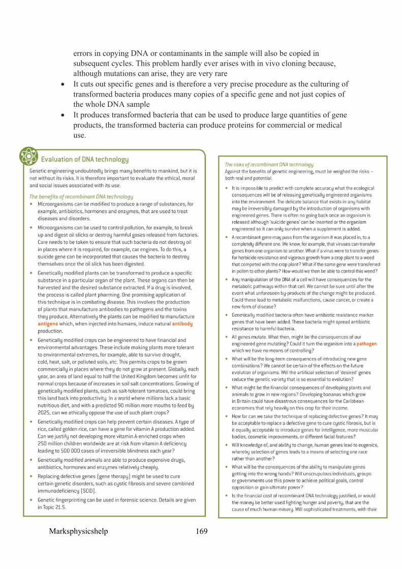
Marksphysicshelp
169
errors in copying DNA or contaminants in the sample will also be copied in subsequent cycles. This problem hardly ever arises with in vivo cloning because, although mutations can arise, they are very rare
• It cuts out specific genes and is therefore a very precise procedure as the culturing of transformed bacteria produces many copies of a specific gene and not just copies of the whole DNA sample
• It produces transformed bacteria that can be used to produce large quantities of gene products, the transformed bacteria can produce proteins for commercial or medical use.

Marksphysicshelp
170
Source: Kerboodle Biology A-Level Textbook
Marksphysicshelp
171
3.8.4.2 Differences in DNA between individuals of the same species can be exploited for identification and diagnosis of heritable conditions
Content
• The use of labelled DNA probes and DNA hybridisation to locate specific alleles of genes.
• The use of labelled DNA probes that can be used to screen patients for heritable conditions, drug responses or health risks.
• The use of this information in genetic counselling and personalised medicine. • Students should be able to evaluate information relating to screening individuals for
genetically determined conditions and drug responses.
Many human diseases are often the result of genetics from a gene mutation. Recombinant DNA technology has enabled us to diagnose and treat many of these genetic disorders. However, to do this we must know exactly where a particular DNA sequence (gene) is located. To achieve this, we use labelled DNA probes and DNA hybridisation.
DNA probes – A DNA probe is a short, single-stranded length of DNA that has some sort of label attached that makes it easily identifiable. The two most commonly used probes are:
• Radioactively labelled probes – these are identified using an X-ray film that is exposed by radioactivity
• Fluorescently labelled probes – these fluoresce under certain conditions, for instance when the probe has bound to the target DNA sequence
DNA probes are used to identify particular alleles of genes in the following way:
• A DNA probe is made that has base sequences that are complementary to part of the base sequence of the DNA that makes up the allele of the gene that we want to find
• The double-stranded DNA that is being tested is treated to separate its two strands • The separated DNA strands are mixed with the probe, which binds to the
complementary base sequence on one of the strands. This is known as DNA hybridisation
• The site at which the probe binds can be identified by the radioactivity or fluorescence that the probe emits.
Before we can make a specific probe we need to know the base sequence in the particular allele that we are trying to locate. A number of different methods are used to sequence the exact order of bases in a length of DNA.
DNA hybridisation
This takes place when a section of DNA or RNA is combined with a single-stranded section of DNA which has complementary bases. Before hybridisation can take place, the two strands of the DNA molecule must be separated. This is achieved by heating DNA until its double strand separates into its two complementary single strands (denaturation). When cooled, the complementary bases on each strand recombine (anneal) with each other to
Marksphysicshelp
172
reform the original double strand. Given sufficient time, all strands in a mixture of DNA will pair up with their partners. If, however, other complementary sections of DNA are present in the mixture as the DNA cools, these are just as likely to anneal with one of the separated DNA strands as the two strands are with one another.
Locating specific alleles of genes is done using DNA probes and DNA hybridisation. For example, we may wish to determine whether someone possesses a mutant allele that causes a particular genetic disorder. This process is as follows:
• First to determine the sequence of nucleotide bases of the mutant allele we are trying to locate. This can be achieved by using DNA sequencing techniques. We now have extensive genetic libraries that store the base sequences of most genetic diseases and so we can refer to these
• A fragment of DNA is produced that has a sequence of bases that are complementary to the mutant allele we are trying to locate
• Multiple copies of our DNA probe are formed using PCR • A DNA probe is made by attaching a marker, ie a fluorescent dye, to the DNA
fragment • DNA from the person suspected of having the mutant allele we want to locate is
heated to separate its two strands • The separated strands are cooled in a mixture containing many of our DNA probes • If the DNA contains the mutant allele, one of our probes is likely to bind to it because
the probe has base sequences that are exactly complementary to those on the mutant allele
• The DNA is washed clean of any unattached probes • The remaining hybridised DNA will now be fluorescently labelled with the dye
attached to the probe • The dye is detected by shining light onto the fragments causing the dye to fluoresce,
which can be seen using an optical microscope.
Genetic screening
Many genetic disorders, like sickle-cell anaemia, are the result of gene mutations. Gene mutations may arise if one or more nucleotide bases in DNA are changed. If a person is a carrier of the disorder, they have the ability of passing this disorder on to the next generation. Screening can determine the probabilities of a couple having offspring with a genetic disorder.
It is possible to fix hundreds of different DNA probes in a display on a glass slide. By adding a sample of DNA to the array, any complementary DNA sequences in the donor DNA will bind to one or more probes. In this way, it is possible to test simultaneously for many different genetic disorders by detecting fluorescence that occurs where binding has taken place.
Genetic screening can be valuable in identifying oncogenes, which are responsible for cancer. Cancers may develop as a result of mutations that prevent tumour suppressor genes inhibiting cell division. Mutations of both alleles must be present to inactivate the tumour suppressor genes and to initiate the development of a tumour. Some people inherit one mutated tumour suppressor gene, and these people are at a greater risk of cancer.
Marksphysicshelp
173
If a mutated gene is detected by genetic screening, individuals who are at a greater risk of cancer can can then adapt their lifestyle ie stop smoking to prevent any further risks.
Another advantage of genetic screening is personalised medicine. It allows doctors to provide advice and health care based on an individual’s genotype. Some people’s genes can mean that a particular drug may be either more or less effective in treating a condition. By genetically screening patients, doctors and pharmacists can determine, more exactly, the dose of a drug which will produce the desired outcome. This saves money that would otherwise be wasted on over prescription.
An example is in prescribing painkillers, as to function effectively many pain medications need a specific enzyme to activate them. About half the population have genes that alter the function of this enzyme. Screening for the presence of these genes allows the dosage to be adjusted to compensate for the ways in which the genes affect an individual’s metabolism of the painkiller. This ensures that their use is both safe and effective.
Another example involved vitamin E. It has been shown that among people who have diabetes, vitamin E reduces the risk of cardiovascular disease for those certain genotypes, but it can increase the risk for those with a different genotype. It is clearly advantageous to screen a person who has diabetes before advising on whether or not to take vitamin E supplements.
Genetic screening goes hand in hand with genetic counselling. The expert advice provided by a counsellor helps individuals to understand the results and implications of the screening and so make appropriate decision.
Genetic counselling – This like a special form of social work, where advice and information are given that enable people to make personal decisions about themselves to their offspring. One important aspect of genetic counselling is to research family history of an inherited disease and to advise parents on the likelihood of it arising in their children.
Counsellers can inform couples of the effects of the disorder and its emotional, psychological, medical, social and economic consequences. On the basis of this advice the couple can then choose whether or not to have children. Counselling can also make them aware of any further medical tests that might give a clearer indication of whether their children will have the condition, for example IVF with screening of embryos.
Genetic counselling is closely linked to genetic screening and the screening results provide the genetic counseller with a basis for informed discussion. For example, in the cases of cancer, screening can help in detecting:
• Oncogene mutations, which can determine the type of cancer that the patient has and hence the most effective drug and radiotherapy to use
• Gene changes that predict which patients are more likely to benefit from certain treatments and have the best chance of survival. For example, the drug herceptin is most effective at treating certain types of breast cancer.
• A single cancer cell among millions of normal cells, thus identifying patients at risk of relapse from certain forms of leukaemia.
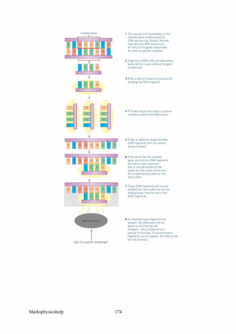
Marksphysicshelp
174
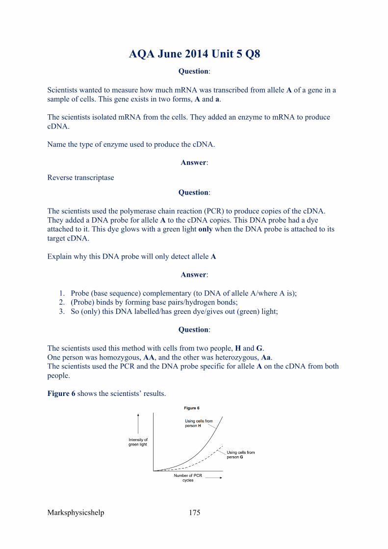
Marksphysicshelp
175
AQA June 2014 Unit 5 Q8 Question:
Scientists wanted to measure how much mRNA was transcribed from allele A of a gene in a sample of cells. This gene exists in two forms, A and a.
The scientists isolated mRNA from the cells. They added an enzyme to mRNA to produce cDNA.
Name the type of enzyme used to produce the cDNA.
Answer:
Reverse transcriptase
Question:
The scientists used the polymerase chain reaction (PCR) to produce copies of the cDNA. They added a DNA probe for allele A to the cDNA copies. This DNA probe had a dye attached to it. This dye glows with a green light only when the DNA probe is attached to its target cDNA.
Explain why this DNA probe will only detect allele A
Answer:
1. Probe (base sequence) complementary (to DNA of allele A/where A is); 2. (Probe) binds by forming base pairs/hydrogen bonds; 3. So (only) this DNA labelled/has green dye/gives out (green) light;
Question:
The scientists used this method with cells from two people, H and G. One person was homozygous, AA, and the other was heterozygous, Aa. The scientists used the PCR and the DNA probe specific for allele A on the cDNA from both people.
Figure 6 shows the scientists’ results.
Marksphysicshelp
176
Explain the curve for person H.
Answer:
1. More probe binding/more cDNA/mRNA/more allele/gene A means more light; 2. DNA (with A) doubles each (PCR) cycle; 3. So light (approximately) doubles/curve steepens more and more (each cycle) /curve
goes up exponentially/ increases even faster;
Question:
Which person, H or G, was heterozygous, Aa? Explain your answer.
Answer:
(G because)
1. (Heterozygous) only has half the amount of probe for A attaching / only half the amount of DNA/allele A (to bind to);
2. (So,) only produced (about) half the light/glow/intensity (of H) (per cycle of PCR);
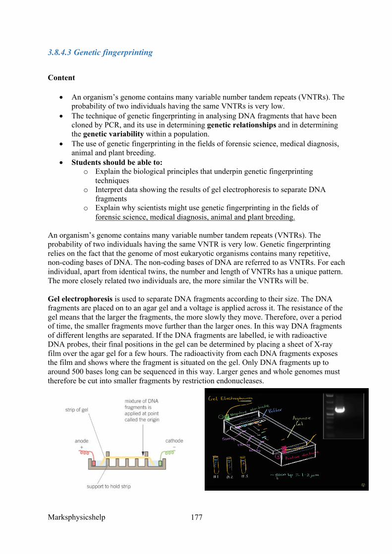
Marksphysicshelp
177
3.8.4.3 Genetic fingerprinting
Content
• An organism’s genome contains many variable number tandem repeats (VNTRs). The probability of two individuals having the same VNTRs is very low.
• The technique of genetic fingerprinting in analysing DNA fragments that have been cloned by PCR, and its use in determining genetic relationships and in determining the genetic variability within a population.
• The use of genetic fingerprinting in the fields of forensic science, medical diagnosis, animal and plant breeding.
• Students should be able to: o Explain the biological principles that underpin genetic fingerprinting
techniques o Interpret data showing the results of gel electrophoresis to separate DNA
fragments o Explain why scientists might use genetic fingerprinting in the fields of
forensic science, medical diagnosis, animal and plant breeding.
An organism’s genome contains many variable number tandem repeats (VNTRs). The probability of two individuals having the same VNTR is very low. Genetic fingerprinting relies on the fact that the genome of most eukaryotic organisms contains many repetitive, non-coding bases of DNA. The non-coding bases of DNA are referred to as VNTRs. For each individual, apart from identical twins, the number and length of VNTRs has a unique pattern. The more closely related two individuals are, the more similar the VNTRs will be.
Gel electrophoresis is used to separate DNA fragments according to their size. The DNA fragments are placed on to an agar gel and a voltage is applied across it. The resistance of the gel means that the larger the fragments, the more slowly they move. Therefore, over a period of time, the smaller fragments move further than the larger ones. In this way DNA fragments of different lengths are separated. If the DNA fragments are labelled, ie with radioactive DNA probes, their final positions in the gel can be determined by placing a sheet of X-ray film over the agar gel for a few hours. The radioactivity from each DNA fragments exposes the film and shows where the fragment is situated on the gel. Only DNA fragments up to around 500 bases long can be sequenced in this way. Larger genes and whole genomes must therefore be cut into smaller fragments by restriction endonucleases.
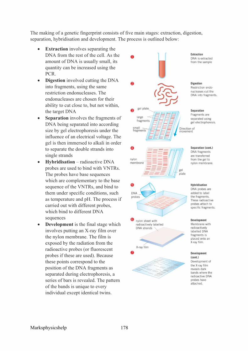
Marksphysicshelp
178
The making of a genetic fingerprint consists of five main stages: extraction, digestion, separation, hybridisation and development. The process is outlined below:
• Extraction involves separating the DNA from the rest of the cell. As the amount of DNA is usually small, its quantity can be increased using the PCR.
• Digestion involved cutting the DNA into fragments, using the same restriction endonucleases. The endonucleases are chosen for their ability to cut close to, but not within, the target DNA
• Separation involves the fragments of DNA being separated into according size by gel electrophoresis under the influence of an electrical voltage. The gel is then immersed to alkali in order to separate the double strands into single strands
• Hybridisation – radioactive DNA probes are used to bind with VNTRs. The probes have base sequences which are complementary to the base sequence of the VNTRs, and bind to them under specific conditions, such as temperature and pH. The process if carried out with different probes, which bind to different DNA sequences
• Development is the final stage which involves putting an X-ray film over the nylon membrane. The film is exposed by the radiation from the radioactive probes (or fluorescent probes if these are used). Because these points correspond to the position of the DNA fragments as separated during electrophoresis, a series of bars is revealed. The pattern of the bands is unique to every individual except identical twins.
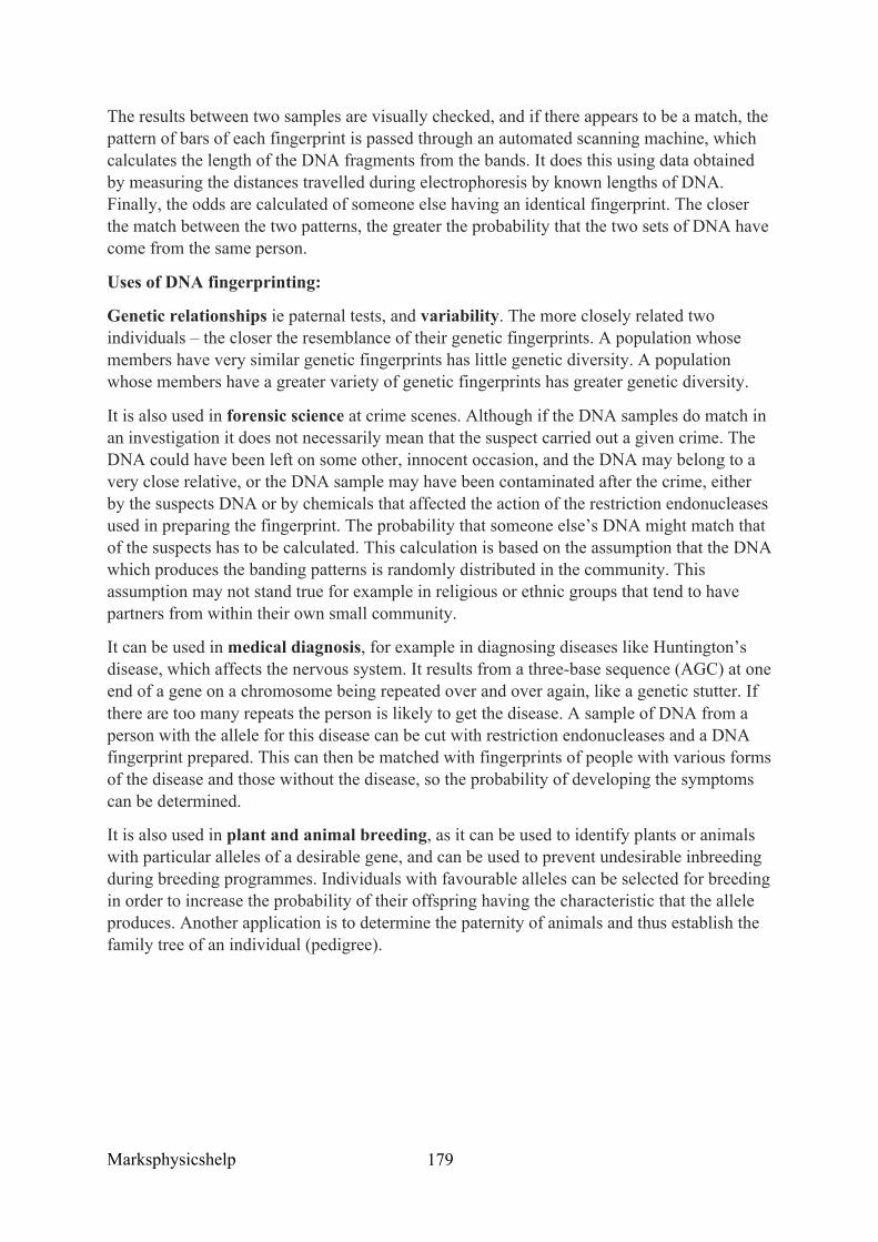
Marksphysicshelp
179
The results between two samples are visually checked, and if there appears to be a match, the pattern of bars of each fingerprint is passed through an automated scanning machine, which calculates the length of the DNA fragments from the bands. It does this using data obtained by measuring the distances travelled during electrophoresis by known lengths of DNA. Finally, the odds are calculated of someone else having an identical fingerprint. The closer the match between the two patterns, the greater the probability that the two sets of DNA have come from the same person.
Uses of DNA fingerprinting:
Genetic relationships ie paternal tests, and variability. The more closely related two individuals – the closer the resemblance of their genetic fingerprints. A population whose members have very similar genetic fingerprints has little genetic diversity. A population whose members have a greater variety of genetic fingerprints has greater genetic diversity.
It is also used in forensic science at crime scenes. Although if the DNA samples do match in an investigation it does not necessarily mean that the suspect carried out a given crime. The DNA could have been left on some other, innocent occasion, and the DNA may belong to a very close relative, or the DNA sample may have been contaminated after the crime, either by the suspects DNA or by chemicals that affected the action of the restriction endonucleases used in preparing the fingerprint. The probability that someone else’s DNA might match that of the suspects has to be calculated. This calculation is based on the assumption that the DNA which produces the banding patterns is randomly distributed in the community. This assumption may not stand true for example in religious or ethnic groups that tend to have partners from within their own small community.
It can be used in medical diagnosis, for example in diagnosing diseases like Huntington’s disease, which affects the nervous system. It results from a three-base sequence (AGC) at one end of a gene on a chromosome being repeated over and over again, like a genetic stutter. If there are too many repeats the person is likely to get the disease. A sample of DNA from a person with the allele for this disease can be cut with restriction endonucleases and a DNA fingerprint prepared. This can then be matched with fingerprints of people with various forms of the disease and those without the disease, so the probability of developing the symptoms can be determined.
It is also used in plant and animal breeding, as it can be used to identify plants or animals with particular alleles of a desirable gene, and can be used to prevent undesirable inbreeding during breeding programmes. Individuals with favourable alleles can be selected for breeding in order to increase the probability of their offspring having the characteristic that the allele produces. Another application is to determine the paternity of animals and thus establish the family tree of an individual (pedigree).
Marksphysicshelp
180
AQA June 2011 Unit 5 Q5ci
Question:
Scientists can separate fragments of DNA using electrophoresis. Suggest how they can use electrophoresis to estimate the number of base pairs in the separated fragments.
Answer:
• Give one mark for answer confined to smaller fragments move further/faster; • Give two marks for comparing with distance/speed moved by fragments of known
size/markers / DNA ladder (ie the distance/speed of fragments of known size can be compared to the distance/speed of unknown size to estimate size.
Marksphysicshelp
181
Exam Command Words
Analyse Separate information into components and identify their characteristics
Apply Put into effect in a recognised way
Argue Present a reasoned case
Assess Make an informed judgement
Comment Present an informed opinion
Consider Review and respond to given information
Contrast Identify differences
Criticise Access worth against explicit expectations
Debate Present different perspectives on an issue
Deduce Draw conclusions from information provided
Describe Give an account of
Marksphysicshelp
182
Design Set out how something will be done
Develop Take forward or build upon given information
Discuss Present key points
Evaluate Judge from available evidence
Explore Investigate without preconceptions about the outcome
Outline Set out main characteristics
Predict Give a plausible outcome
Relate Give a technical term or its equivalent
Show Provide structured evidence to reach a conclusion
State Express in clear terms
Suggest Present a possible case
Marksphysicshelp
183
References:
A Level Biology Textbook Kerboodle
https://micro.magnet.fsu.edu/cells/nucleus/images/nucleusfigure1.jpg
http://www.yourgenome.org/facts/what-is-mitosis
http://www.anatomy-diagram.info/digestive-tract-diagram/
http://academic.brooklyn.cuny.edu/biology/eckhardt/chap4.html
https://about-cancer.cancerresearchuk.org/about-cancer/cancer-in-general/treatment/targeted-cancer-drugs/types/monoclonal-antibodies#M2CrhAJYAxwSBp1b.99
http://www.tokresource.org/tok_classes/biobiobio/biomenu/membranes/c8.7x17.transport.jpg
Related Documents