p73 regulates maintenance of neural stem cell Massimiliano Agostini a , Paola Tucci a,b , Hailan Chen a , Richard A. Knight a , Daniele Bano c , Pierluigi Nicotera c , Frank McKeon d , and Gerry Melino a,b,⁎ a Medical Research Council, Toxicology Unit, Leicester University, Leicester LE1 9HN, UK b Biochemistry Laboratory, IDI-IRCCS, C/O University of Rome “Tor Vergata”, 00133 Rome, Italy c Deutsche Zentrum für Neurodegenerative Erkrankungen (DZNE), Bonn, Germany d Department of Cell Biology, Harvard Medical School, Boston, MA 02115, USA Research highlights ► TAp73 is expressed in neural stem cells and its expression increases following their differentiation. ► Neural stem cells from p73 null mice have a reduced proliferative potential. ► p73-deficient neural stem cells show reduced expression of members of the Sox-2 and Notch gene families. ► Neurogenic areas are reduced in the brains of embryonic and adult p73−/− mice. Abstract p73, a member of the p53 family, is a transcription factor that plays a key role in many biological processes. In the present study, we show that TAp73 is expressed in neural stem cells (NSC) and its expression increases following their differentiation. NSC from p73 null mice have a reduced proliferative potential, together with reduced expression of members of the Sox-2 and Notch gene families known to be important for NSC proliferation. In parallel with this in vitro data, the width of the neurogenic areas was reduced in the brains of embryonic and adult p73−/− mice. These data suggest that p73, and in particular TAp73, is important for maintenance of the NSC pool. Abbreviations TAp73, transcriptionally active p73; ΔNp73, amino truncated p73; −/−, knockout mice; DIV, day in vitro; DMED, Dulbecco minimal essential medium; FBS, foetal bovine serum; EGF, epidermal growth factor; C t , threshold cycle Keywords p73; Neural stem cell; Neurogenesis; Self-renewal; p53 family; Apoptosis © 2010 Elsevier Inc. ⁎ Corresponding author at: Medical Research Council, Toxicology Unit, Leicester University, Leicester LE1 9HN, UK. Fax: +44 (0) 116 252 5616. [email protected]. This document was posted here by permission of the publisher. At the time of deposit, it included all changes made during peer review, copyediting, and publishing. The U.S. National Library of Medicine is responsible for all links within the document and for incorporating any publisher-supplied amendments or retractions issued subsequently. The published journal article, guaranteed to be such by Elsevier, is available for free, on ScienceDirect. Sponsored document from Biochemical and Biophysical Research Communications Published as: Biochem Biophys Res Commun. 2010 December 03; 403(1): 13–17. Sponsored Document Sponsored Document Sponsored Document

Welcome message from author
This document is posted to help you gain knowledge. Please leave a comment to let me know what you think about it! Share it to your friends and learn new things together.
Transcript
p73 regulates maintenance of neural stem cell
Massimiliano Agostinia, Paola Tuccia,b, Hailan Chena, Richard A. Knighta, Daniele Banoc,Pierluigi Nicoterac, Frank McKeond, and Gerry Melinoa,b,⁎aMedical Research Council, Toxicology Unit, Leicester University, Leicester LE1 9HN, UKbBiochemistry Laboratory, IDI-IRCCS, C/O University of Rome “Tor Vergata”, 00133 Rome, ItalycDeutsche Zentrum für Neurodegenerative Erkrankungen (DZNE), Bonn, GermanydDepartment of Cell Biology, Harvard Medical School, Boston, MA 02115, USA
Research highlights► TAp73 is expressed in neural stem cells and its expression increases following theirdifferentiation. ► Neural stem cells from p73 null mice have a reduced proliferative potential. ►p73-deficient neural stem cells show reduced expression of members of the Sox-2 and Notch genefamilies. ► Neurogenic areas are reduced in the brains of embryonic and adult p73−/− mice.
Abstractp73, a member of the p53 family, is a transcription factor that plays a key role in many biologicalprocesses. In the present study, we show that TAp73 is expressed in neural stem cells (NSC) andits expression increases following their differentiation. NSC from p73 null mice have a reducedproliferative potential, together with reduced expression of members of the Sox-2 and Notch genefamilies known to be important for NSC proliferation. In parallel with this in vitro data, the widthof the neurogenic areas was reduced in the brains of embryonic and adult p73−/− mice. These datasuggest that p73, and in particular TAp73, is important for maintenance of the NSC pool.
AbbreviationsTAp73, transcriptionally active p73; ΔNp73, amino truncated p73; −/−, knockout mice; DIV, dayin vitro; DMED, Dulbecco minimal essential medium; FBS, foetal bovine serum; EGF, epidermalgrowth factor; Ct, threshold cycle
Keywordsp73; Neural stem cell; Neurogenesis; Self-renewal; p53 family; Apoptosis
© 2010 Elsevier Inc.⁎Corresponding author at: Medical Research Council, Toxicology Unit, Leicester University, Leicester LE1 9HN, UK. Fax: +44 (0)116 252 5616. [email protected] document was posted here by permission of the publisher. At the time of deposit, it included all changes made during peerreview, copyediting, and publishing. The U.S. National Library of Medicine is responsible for all links within the document and forincorporating any publisher-supplied amendments or retractions issued subsequently. The published journal article, guaranteed to besuch by Elsevier, is available for free, on ScienceDirect.
Sponsored document fromBiochemical and BiophysicalResearch Communications
Published as: Biochem Biophys Res Commun. 2010 December 03; 403(1): 13–17.
Sponsored Docum
ent Sponsored D
ocument
Sponsored Docum
ent
1 IntroductionEmbryonic and adult neural stem cells (NSC) are precursors that retain the capacity toproliferate and produce identical daughter cells (self-renewal), and also the ability todifferentiate into neurons, astrocytes and oligodendrocytes [1]. The process of self-renewalplays a key role in the development, as well as in the preservation of adult tissues. Theregulation of this process requires that the multiple pathways involved in the regulation ofproliferation and/or maintenance of the undifferentiated phenotype are tightly coordinated.Pathways that control differentiation and apoptosis are also involved in self-renewal [2].
The p53 family which includes p53 itself, p63 and p73 are transcription factors that play keyroles as regulators of proliferation, differentiation, cell death, stem cell renewal and cell fatecommitment [3–5]. So far, p53 and ΔNp63 are the only members of the family that havebeen reported to play a role in NSC [6,7].
Like other members of the p53 family, the Trp73 gene encodes a protein composed of aDNA-binding domain, a transactivation (TA) domain and an oligomerization domain.However, because of the presence of two distinct promoters, Trp73 is expressed as twomajor isoforms either containing (TAp73) or not (ΔNp73) the TA domain. As a result,TAp73 and ΔNp73 have distinct functions, being, for example, proapoptotic [8,9] orantiapoptotic [10], respectively. Furthermore, it has been shown that p73 can induce neuriteoutgrowth and expression of neuronal markers in neuroblastoma cell lines [11] and inprimary oligodendrocytes [12] indicating a possible role in brain development. Indeed, p73-deficient mice manifest complex defects in neuronal development [13], such as congenitalhydrocephalus, hippocampal dysgenesis with the lower blade of dentate gyrus truncated ormissing, together with defects in pheromone detection. This phenotype is conserved in theselective TAp73 [14] and ΔNp73 [15] knockouts mice.
In the present study, we have investigated the possibility that p73 could play a role in NSCbiology. Using the well-established in vitro system of embryonic NSC [16] we identify p73as a positive regulator of these cells since neurospheres derived from p73−/− miceproliferate less than wild-type neurospheres and show dysregulation of several genesinvolved in NSC self-renewal.
2 Materials and methods2.1 Mice
The p73−/− mice were generated as previously described [13]. Mice were bred andsubjected to listed procedures under the Project Licence released from the Home Office.
2.2 Neurosphere cultureNeural stem cells were obtained from mice at gestational day E14. Briefly, cortex wasdissected and triturated until a single cell suspension was achieved. Cells were counted andplated at 2 × 10⁶ cells/10 ml/25 cm2 flask in NeuroCult® NSC Basal Medium (Mouse) withproliferation supplements and rh-EGF (10 ng/ml) (all purchased from StemCellsTechnologies) following the manufacturer’s instructions, and this was considered passage 0.Neurospheres were allowed to form for 7 days. Neurospheres were passaged every 5 days.Passage 1 neurospheres were dissociated with NeuroCult® chemical dissociation kit(StemCells Technologies) and replated in triplicates at clonal density (20 × 103 cells/ml in24-well plate) and regeneration of new neurospheres was monitored. At the times indicated,the number and size of neurospheres and the total number of cells was analyzed. For thedifferentiation of NSC, a single cell suspension (5 × 10⁵ cells/well/24-well plate) was plated
Agostini et al. Page 2
Published as: Biochem Biophys Res Commun. 2010 December 03; 403(1): 13–17.
Sponsored Docum
ent Sponsored D
ocument
Sponsored Docum
ent
on the poly-L-ornithine glass coverslips in NeuroCult® NSC Basal Medium (Mouse) withdifferentiation supplements.
2.3 RNA extraction and real-time PCRTotal RNA from cells was isolated using Trizol (Invitrogen) according to the manufacturer’sinstructions. RNA samples were treated with RNase-free DNase I (Sigma). Total RNA wasreverse transcribed using Superscript III reverse transcriptase and oligo(dT) primer(Invitrogen). qRT-PCR was performed in ABI PRISM 7000 Sequence Detection System(Applied Biosystem) with SYBR green ready mix (Applied Biosystem) and specificprimers: TAp73 Fwd 5′-GCACCTACTTTGACCTCCCC-3′ and Rev 5′-GCACTGCTGAGCAAATTGAAC-3′; ΔNp73 Fwd 5′-ATGCTTTACGTCGGTGACCC-3′,and Rev 5′-GCACTGCTGAGCAAATTGAAC-3′; p53 Fwd 5′-CTCTCCCCCGCAAAAGAAAAA-3′ and Rev 5′-CGGAACATCTCGAAGCGTTTA-3′;GAPDH Fwd 5′-CAATGAATACGGCTACAGCAAC-3′ and Rev 5′-AGGGAGATGCTCAGTGTTGG-3′; Sox-2 For 5′-TCCAAAAACTAATCACAACAATCG-3′ and Rev 5′-GAAGTGCAATTGGGATGAAAA-3′; Hes-1 For 5′-ACACCGGACAAACCAAAGAC-3′and Rev 5′-CGCCTCTTCTCCATGATAGG-3′; Hes-5 For 5′-GATGCTCAGTCCCAAGGAGA-3′ and Rev 5′-AGCTTCAGCTGCTCTATGCTG-3′;Nanog For 5′-CACCCACCCATGCTAGTCTT-3′ and Rev 5′-ACCCTCAAACTCCTGGTCCT-3′; Notch-1 For 5′-ACTATCTCGGCGGCTTTTC-3′ andRev 5′-GGCACTCGTTGATCTCCTCT-3′.
The expression of each gene was defined from the threshold cycle (Ct), and relativeexpression levels were calculated by using the method after normalization withreference to expression of the housekeeping gene GAPDH.
2.4 ImmunofluorescenceE17.5 and P7 brains were fixed in 4% paraformaldehyde and embedded in paraffin. Coronalsections were de-waxed, re-hydrated in graded alcohols and rinsed in distilled water. Thesections were blocked in 10% goat serum in 0.1% PBS–Tween (vol/vol), and then incubatedwith primary and secondary antibodies in blocking buffer. Cell nuclei were stained withHoechst 33342 (Sigma) and mounted for confocal analysis. Two different images of eachsection were taken and the number of cells labeled was counted (n = 2 p73−/− compared ton = 2 control littermates). The following primary antibodies were used: nestin (mouse, 1/50,Millipore), GFAP (rabbit, 1/500, Dako).
2.5 Statistical analysisAll results are expressed as means ± SD. P ⩽ 0.05 was considered significant.
3 Results and discussion3.1 TAp73 is expressed in NSC and is up-regulated during neurosphere differentiation
In order to investigate whether p73 could play a role in the biology of NSC, we firstevaluated its expression under two different conditions. Wild-type (WT) NSC were isolatedfrom E14.5 embryos and maintained in culture either under proliferating conditions, orinduced to differentiate. After 4 days in vitro (DIV), cells were harvested and total RNA wasisolated. qPCR analysis (Fig. 1A) shows that TAp73 is the most abundant member of thep53 family expressed under proliferative conditions, and TAp73 expression is roughly 10-fold greater than that of ΔNp73 and 3-fold higher than that of p53. Next, we assayed theexpression of these three proteins during differentiation of neurospheres. After deprivationof mitogenic stimuli, WT neurospheres undergo differentiation as shown by the appearance
Agostini et al. Page 3
Published as: Biochem Biophys Res Commun. 2010 December 03; 403(1): 13–17.
Sponsored Docum
ent Sponsored D
ocument
Sponsored Docum
ent
of cells positive for the neuronal marker β-III-Tubulin (Fig. 1B). As shown in Fig. 1C, underthese differentiating conditions, we observed a marked increase in expression of TAp73 withonly minor changes in expression of ΔNp73 and p53. Together, these data suggest thatTAp73 is mechanistically involved in the biology of NSC, and confirm previousobservations [11] that TAp73 play a role in neuronal differentiation. In addition, we showfor the first time that TAp73 is more prominently expressed than ΔNp73 in the neurallineage.
3.2 p73-deficient NSC show decreased proliferationNext, employing the neurospheres assay, we focused on whether p73 is required forproliferation and self-renew of embryonic NSC. After 7 days in culture, NSC from both WTand p73−/− mice formed neurospheres, but those derived from p73−/− mice weremorphologically smaller (Fig. 2A) and the mean diameter of p73−/− neurospheres wassignificantly smaller than that of WT mice at both DIV 4 and DIV 7 (Fig. 2B). Moreover, asshown in Fig. 2C, p73−/− neurospheres tended to accumulate in the lower size range. Thesedata suggest that p73 is required for optimum proliferation of neurospheres.
3.3 Loss of p73−/− impairs expression of genes involved in NSC self-renewalWe next investigated the molecular mechanisms underlying the altered phenotype of p73−/−derived neurospheres. Pathways involving genes of the Sox [17] and Notch [18] familieshave been implicated in the proliferation/self-renewal of NSC. We therefore isolated RNAfrom DIV 7 neurospheres derived from WT and p73−/− mice and analyzed expression ofcandidate genes of these families by qPCR. As shown in Fig. 3 the absence of p73 results ina significant reduction in Sox-2, Notch-1 and Nanog expression, indicating that p73 directlyor indirectly regulates these factors. Indeed, Sox-2−/− mice have a phenotype similar to thatof p73−/− mice with a moderate lateral ventricle enlargement and a reduction of theposterior ventrolateral cortex. In addition, in the hippocampus the dentate gyrus isessentially absent [19].
3.4 Neurogenic zones are reduced in p73−/− miceThe ventricular/subventricular zone and the dentate gyrus in the hippocampus are theneurogenic areas in embryonic and in adult mice respectively [20,21]. To investigatewhether these zones were morphologically smaller in vivo, in parallel with the reducedproliferation of p73−/− derived neurospheres in vitro, we stained E17.5 brains from WT andp73−/− mice with anti-nestin antibodies. The results in Fig. 4A show that the width of theventricular zone, the neurogenic area, is reduced in embryonic p73−/− mice when comparedto WT mice. Similarly, the numbers of GFAP and nestin double positive cells in the dentategyrus, the source of neurogenesis in the adult, are reduced in P7 p73−/− mice (Fig. 4B andC).
4 ConclusionTwo very recent reports [22,23] are in line with our results, providing an independentconfirmation of the involvement of p73 in neuronal stemness.
Together, these data support an important role for p73, and particularly TAp73, in theproliferative potential of NSC and in their differentiation, probably by modulatingexpression of components of the Sox-2 and Notch pathways known to be involved in NSCproliferation. This requirement for p73 is apparent, both from the reduced size ofneurospheres derived from p73−/− mice in vitro, and in the reduced thickness of brain areasresponsible for neurogenesis in both embryonic and adult knockout mice in vivo. However,whether all the morphological and functional deficits in the brains of p73−/− mice arise
Agostini et al. Page 4
Published as: Biochem Biophys Res Commun. 2010 December 03; 403(1): 13–17.
Sponsored Docum
ent Sponsored D
ocument
Sponsored Docum
ent
from these actions of p73 on NSC or whether p73 exerts other independent effects has yet tobe established.
Conflict of interestThe authors declare no competing financial interests.
References1. GageF.H.Mammalian neural stem cellsScience287200071433714382. MolofskyA.V.PardalR.MorrisonS.J.Diverse mechanisms regulate stem cell self-renewalCurr. Opin.
Cell Biol.62004700707155307843. MelinoG.De LaurenziV.VousdenK.H.p73: friend or foe in tumorogenesisNat. Rev.
Cancer82002605615121543534. VousdenK.H.LaneD.P.p53 in health and diseaseNat. Rev. Mol. Cell Biol.82007275283173801615. YangA.KaghadM.CaputD.On the shoulder of giants: p63, p73, and the rise of p53Trends Genet.
220029095118181416. MeletisK.WirtaV.HedeS.M.p53 suppresses the self-renewal of adult neural stem
cellsDevelopment1332006363369163689337. DuganiC.B.PaquinA.FujitaniM.p63 antagonizes p53 to promote the survival of embryonic neural
precursor cellsJ. Neurosci.29200967106721194582408. MullerM.SchillingT.SayanA.E.TAp73/Delta Np73 influences apoptotic response, chemosensitivity
and prognosis in hepatocellular carcinomaCell Death Differ.12200515641577161957399. WangJ.LiuY.X.HandeM.P.TAp73 is a downstream target of p53 in controlling the cellular defense
against stressJ. Biol. Chem.282200729152291621769340510. GrobT.J.NovakU.MaisseC.Human delta Np73 regulates a dominant negative feedback loop for
TAp73 and p53Cell Death Differ.82001121312231175356911. De LaurenziV.RaschelláG.BarcaroliD.Induction of neuronal differentiation by p73 in a
neuroblastoma cell lineJ. Biol. Chem.275200015226152311080975812. BillonN.TerrinoniA.JolicoeurC.Roles for P53 and p73 during oligodendrocyte
developmentDevelopment1312004121112201496049613. YangA.WalkerN.BronsonR.p73-deficient mice have neurological, pheromonal and inflammatory
defects but lack spontaneous tumorsNature4042000991031071645114. TomasiniR.TsuchiharaK.WilhelmM.T.TAp73 knockout shows genomic instability with infertility
and tumor suppressor functionsGenes Dev.222008267726911880598915. WilhelmM.T.RufiniA.WetzelM.K.Isoform-specific p73 knockout mice reveal a novel role for delta
Np73 in the DNA damage response pathwayGenes Dev.2420105495602019443416. JensenJ.B.ParmarM.Strengths and limitations of the neurosphere culture systemMol. Neurobiol.
3420061531611730834917. PevnyL.PlaczekM.SOX genes and neural progenitor identityCurr. Opin. Neurobiol.
1520057131572173818. ArsenijevicY.Mammalian neural stem-cell renewal: nature versus nurtureMol. Neurobiol.
27200373981266890219. FavaroR.ValottaM.FerriA.L.Hippocampal development and neural stem cell maintenance require
Sox2-dependent regulation of ShhNat. Neurosci.122009124812561973489120. FarkasL.M.HuttnerW.B.The cell biology of neural stem and progenitor cells and its significance
for their proliferation versus differentiation during mammalian brain developmentCurr. Opin. CellBiol.20200870771518930817
21. KempermannG.JessbergerS.SteinerB.Milestones of neuronal development in the adulthippocampusTrends Neurosci.7200444745215271491
22. TalosF.AbrahamA.HolembowskiL.p73 is an essential regulator of neural stem cell maintenance inembryonal and adult CNS neurogenesisCell Death Differ.1720101816182921076477
Agostini et al. Page 5
Published as: Biochem Biophys Res Commun. 2010 December 03; 403(1): 13–17.
Sponsored Docum
ent Sponsored D
ocument
Sponsored Docum
ent
23. FujitaniM.CancinoG.I.DuganiC.B.TAp73 acts via the bHLH Hey2 to promote long-termmaintenance of neural precursorsCurr. Biol.2020101820036540
AcknowledgmentsThis work has been supported by the Medical Research Council, UK; grants from EU EPISTEM (LSHB-CT-019067), “Alleanza contro il Cancro” (ACC12), MIUR/PRIN (RBIP06LCA9_0023), AIRC(2008-2010_33-08), ISS “Program Italia-USA” N526D5, Italian Human ProteomeNet RBRN07BMCT_007 andTelethon (GGPO4110) to G.M.; and grants from the National Institutes of Health (R01-GM57587, R37-CA76584and R21-CA125173) and Multiple Myeloma Research Foundation to M.P. Research described in this article wasalso supported in part by Philip Morris USA Inc. and Philip Morris International to G.M.
Agostini et al. Page 6
Published as: Biochem Biophys Res Commun. 2010 December 03; 403(1): 13–17.
Sponsored Docum
ent Sponsored D
ocument
Sponsored Docum
ent
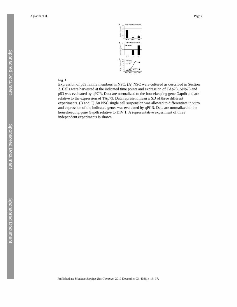
Fig. 1.Expression of p53 family members in NSC. (A) NSC were cultured as described in Section2. Cells were harvested at the indicated time points and expression of TAp73, ΔNp73 andp53 was evaluated by qPCR. Data are normalized to the housekeeping gene Gapdh and arerelative to the expression of TAp73. Data represent mean ± SD of three differentexperiments. (B and C) An NSC single cell suspension was allowed to differentiate in vitroand expression of the indicated genes was evaluated by qPCR. Data are normalized to thehousekeeping gene Gapdh relative to DIV 1. A representative experiment of threeindependent experiments is shown.
Agostini et al. Page 7
Published as: Biochem Biophys Res Commun. 2010 December 03; 403(1): 13–17.
Sponsored Docum
ent Sponsored D
ocument
Sponsored Docum
ent
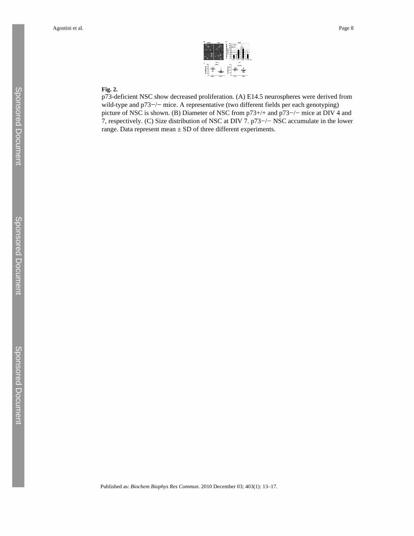
Fig. 2.p73-deficient NSC show decreased proliferation. (A) E14.5 neurospheres were derived fromwild-type and p73−/− mice. A representative (two different fields per each genotyping)picture of NSC is shown. (B) Diameter of NSC from p73+/+ and p73−/− mice at DIV 4 and7, respectively. (C) Size distribution of NSC at DIV 7. p73−/− NSC accumulate in the lowerrange. Data represent mean ± SD of three different experiments.
Agostini et al. Page 8
Published as: Biochem Biophys Res Commun. 2010 December 03; 403(1): 13–17.
Sponsored Docum
ent Sponsored D
ocument
Sponsored Docum
ent
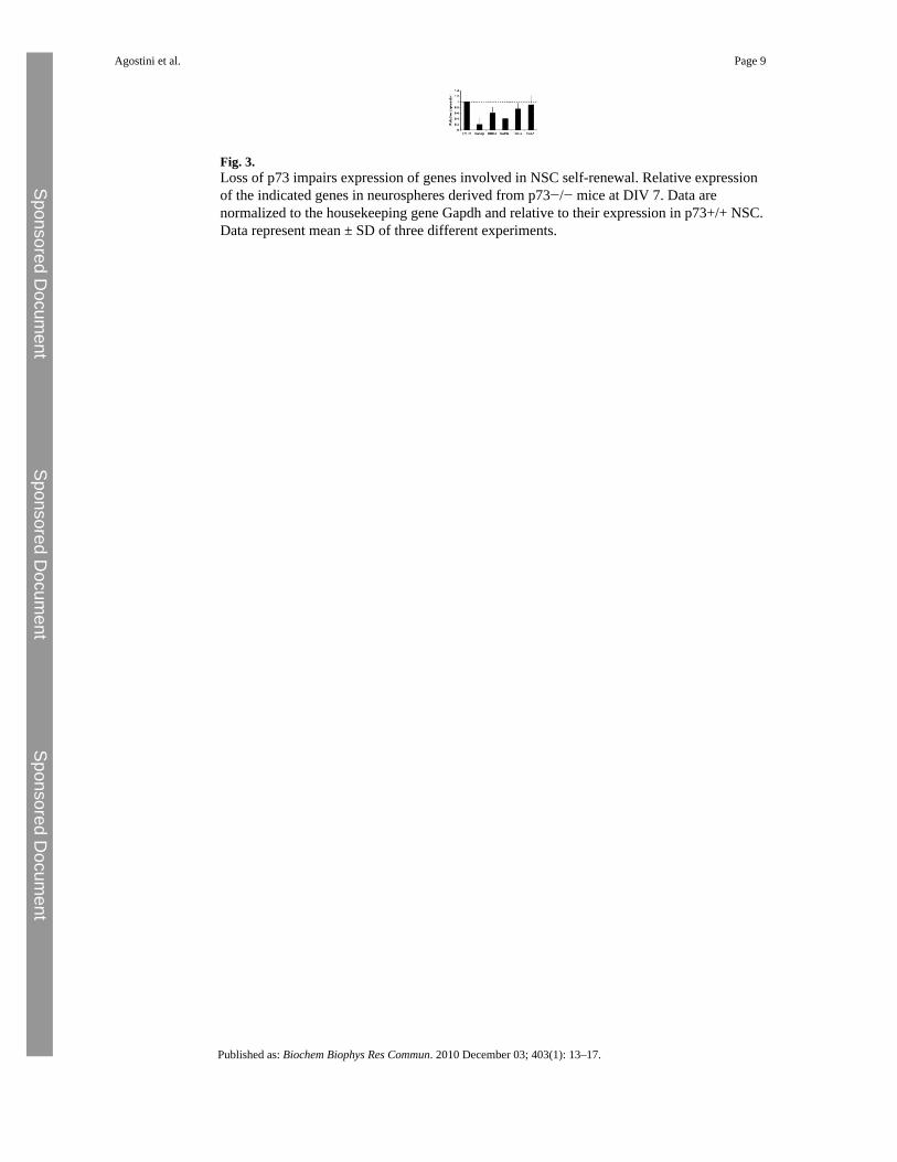
Fig. 3.Loss of p73 impairs expression of genes involved in NSC self-renewal. Relative expressionof the indicated genes in neurospheres derived from p73−/− mice at DIV 7. Data arenormalized to the housekeeping gene Gapdh and relative to their expression in p73+/+ NSC.Data represent mean ± SD of three different experiments.
Agostini et al. Page 9
Published as: Biochem Biophys Res Commun. 2010 December 03; 403(1): 13–17.
Sponsored Docum
ent Sponsored D
ocument
Sponsored Docum
ent
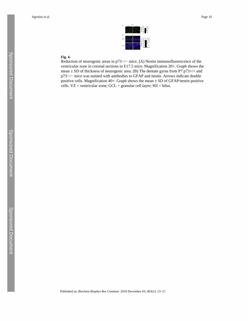
Fig. 4.Reduction of neurogenic areas in p73−/− mice. (A) Nestin immunofluorescence of theventricular zone in coronal sections in E17.5 mice. Magnification 20×. Graph shows themean ± SD of thickness of neurogenic area. (B) The dentate gyrus from P7 p73+/+ andp73−/− mice was stained with antibodies to GFAP and nestin. Arrows indicate doublepositive cells. Magnification 40×. Graph shows the mean ± SD of GFAP/nestin positivecells. VZ = ventricular zone; GCL = granular cell layer; Hil = hilus.
Agostini et al. Page 10
Published as: Biochem Biophys Res Commun. 2010 December 03; 403(1): 13–17.
Sponsored Docum
ent Sponsored D
ocument
Sponsored Docum
ent
Related Documents