p63RhoGEF Couples G αq/11 -mediated Signaling to Ca 2+ Sensitization of Vascular Smooth Muscle Contractility Ko Momotani, Ph.D., Mykhaylo V. Artamonov, Ph.D., Darkhan Utepbergenov, Ph.D., Urszula Derewenda, Ph.D., Zygmunt S. Derewenda, Ph.D., and Avril V. Somlyo, Ph.D. University of Virginia Abstract Rationale—In normal and diseased vascular smooth muscle (SM), the RhoA pathway, which is activated by multiple agonists through G protein-coupled receptors (GPCRs), plays a central role in regulating basal tone and peripheral resistance. This occurs through inhibition of myosin light chain phosphatase, leading to increased phosphorylation of the myosin regulatory light chain. While it is thought that specific agonists and GPCRs may couple to distinct RhoA guanine nucleotide exchange factors (GEFs), thus raising the possibility of selective targeting of specific GEFs for therapeutic use, this notion is largely unexplored for SM contraction. Objective—We examine whether p63RhoGEF, known to couple specifically to Gα q/11 in vitro, is functional in blood vessels as a mediator of RhoA activation, and if it is selectively activated by Gα q/11 coupled agonists. Methods and Results—We find that p63RhoGEF is present across SM tissues and demonstrate that silencing of the endogenous p63RhoGEF in mouse portal vein inhibits contractile force induced by endothelin-1 to a greater extent than the predominantly Gα 12/13 mediated thromboxane analogue, U46619. This is because endothelin-1 acts on Gα q/11 as well as Gα 12/13 . Introduction of the exogenous isolated pleckstrin-homology (PH) domain of p63RhoGEF (residues 331–580) into permeabilized rabbit portal vein inhibited Ca 2+ sensitized force and activation of RhoA, when phenylephrine was used as an agonist. This reinforces the results based on endothelin-1, because phenylephrine is thought to act exclusively through Gα q/11 . Conclusion—We demonstrate that p63RhoGEF selectively couples Gα q/11 , but not Gα 12/13 , to RhoA activation in blood vessels and cultured cells, and thus mediates the physiologically important Ca 2+ sensitization of force induced with Gα q/11 coupled agonists. Our results suggest that signaling through p63RhoGEF provides a novel mechanism for selective regulation of blood pressure. Keywords Ca 2+ sensitization; RhoA; Signal transduction; Vascular smooth muscle; RhoGEF Introduction The contractile response in smooth muscle (SM) to agonist stimulation is mediated through the phosphorylation of the myosin regulatory light chain (RLC 20 ). The level of this Name and complete address of corresponding author: Avril V. Somlyo, Molecular Physiology and Biological Physics, University of Virginia, School of Medicine, 1340 Jefferson Park Avenue, Charlottesville, VA 22908-0736, Fax number: (434)982-1616, Telephone number: (434)982-0838, [email protected]. Disclosures None. NIH Public Access Author Manuscript Circ Res. Author manuscript; available in PMC 2012 October 14. Published in final edited form as: Circ Res. 2011 October 14; 109(9): 993–1002. doi:10.1161/CIRCRESAHA.111.248898. NIH-PA Author Manuscript NIH-PA Author Manuscript NIH-PA Author Manuscript

Welcome message from author
This document is posted to help you gain knowledge. Please leave a comment to let me know what you think about it! Share it to your friends and learn new things together.
Transcript
p63RhoGEF Couples Gαq/11-mediated Signaling to Ca2+
Sensitization of Vascular Smooth Muscle Contractility
Ko Momotani, Ph.D., Mykhaylo V. Artamonov, Ph.D., Darkhan Utepbergenov, Ph.D.,Urszula Derewenda, Ph.D., Zygmunt S. Derewenda, Ph.D., and Avril V. Somlyo, Ph.D.University of Virginia
AbstractRationale—In normal and diseased vascular smooth muscle (SM), the RhoA pathway, which isactivated by multiple agonists through G protein-coupled receptors (GPCRs), plays a central rolein regulating basal tone and peripheral resistance. This occurs through inhibition of myosin lightchain phosphatase, leading to increased phosphorylation of the myosin regulatory light chain.While it is thought that specific agonists and GPCRs may couple to distinct RhoA guaninenucleotide exchange factors (GEFs), thus raising the possibility of selective targeting of specificGEFs for therapeutic use, this notion is largely unexplored for SM contraction.
Objective—We examine whether p63RhoGEF, known to couple specifically to Gαq/11 in vitro,is functional in blood vessels as a mediator of RhoA activation, and if it is selectively activated byGαq/11 coupled agonists.
Methods and Results—We find that p63RhoGEF is present across SM tissues anddemonstrate that silencing of the endogenous p63RhoGEF in mouse portal vein inhibits contractileforce induced by endothelin-1 to a greater extent than the predominantly Gα12/13 mediatedthromboxane analogue, U46619. This is because endothelin-1 acts on Gαq/11 as well as Gα12/13.Introduction of the exogenous isolated pleckstrin-homology (PH) domain of p63RhoGEF(residues 331–580) into permeabilized rabbit portal vein inhibited Ca2+ sensitized force andactivation of RhoA, when phenylephrine was used as an agonist. This reinforces the results basedon endothelin-1, because phenylephrine is thought to act exclusively through Gαq/11.
Conclusion—We demonstrate that p63RhoGEF selectively couples Gαq/11, but not Gα12/13, toRhoA activation in blood vessels and cultured cells, and thus mediates the physiologicallyimportant Ca2+ sensitization of force induced with Gαq/11 coupled agonists. Our results suggestthat signaling through p63RhoGEF provides a novel mechanism for selective regulation of bloodpressure.
KeywordsCa2+ sensitization; RhoA; Signal transduction; Vascular smooth muscle; RhoGEF
IntroductionThe contractile response in smooth muscle (SM) to agonist stimulation is mediated throughthe phosphorylation of the myosin regulatory light chain (RLC20). The level of this
Name and complete address of corresponding author: Avril V. Somlyo, Molecular Physiology and Biological Physics, University ofVirginia, School of Medicine, 1340 Jefferson Park Avenue, Charlottesville, VA 22908-0736, Fax number: (434)982-1616, Telephonenumber: (434)982-0838, [email protected].
NIH Public AccessAuthor ManuscriptCirc Res. Author manuscript; available in PMC 2012 October 14.
Published in final edited form as:Circ Res. 2011 October 14; 109(9): 993–1002. doi:10.1161/CIRCRESAHA.111.248898.
NIH
-PA Author Manuscript
NIH
-PA Author Manuscript
NIH
-PA Author Manuscript
phosphorylation reflects the balance of the opposing activities of the calcium-calmodulin-dependent myosin light chain kinase (MLCK) and myosin light chain phosphatase 1–3.Although an increase in intracellular Ca2+ concentration ([Ca2+]) serves as a primary on-switch for the activation of MLCK, both MLCK and myosin light chain phosphatase areadditionally regulated by diverse kinases, which in turn are regulated by diversephysiological agonists 3, 4. Importantly, the activity of myosin light chain phosphatase isnegatively regulated when Thr853, located in its regulatory, myosin targeting phosphatasesubunit (MYPT1) is phosphorylated by the Rho-kinase (ROCK) 1–3. This inhibition ofmyosin light chain phosphatase activity results in an increase in force generatingphosphorylated myosin motors, without further increase in intracellular [Ca2+]. Thisphysiologically important phenomenon is known as Ca2+ sensitization 3, 5–7.
The Ca2+ sensitization pathway is initiated when ROCK is activated by the GTP-bound,biologically active form of the cytosolic GTPase - RhoA 8. Like other GTPases of the Ras-homology (Rho) family 9, 10, RhoA functions as a molecular switch; it is biologically inertin the GDP-bound form, but upon exchange of GDP to GTP, it undergoes a conformationalchange enabling it to interact with various target effectors 11–13. This results, for example, inthe activation of kinases, such as ROCK. RhoA can serve as a switch, because it is a poorhydrolase, with kcat of ~1/min 14, and can therefore exist for considerable time period as asubstrate-enzyme complex (the on-state). To switch between the on- and off-states, RhoA istherefore assisted by other regulatory proteins in its catalytic cycle. The GTPase-activatingproteins enhance the hydrolysis of GTP to GDP 15, 16, thereby terminating the biologicalsignal. The GDP-bound RhoA is then sequestered in the cytosolic pool by the Rho-nucleotide dissociation inhibitor, RhoGDI 17–19. Finally, a family of guanine nucleotideexchange factors (GEFs) catalyze the loading of GTP for GDP, re-initiating the signalingcycle 20. Both GTPase-activating proteins and GEFs are themselves tightly regulatedupstream of RhoA.
Basal tone and the contractile state of SM are regulated by multiple agonists acting throughG-protein coupled-receptors (GPCRs) leading to a rise in intracellular [Ca2+]. However, themagnitude of force at a specific level of [Ca2+] is highly variable depending on the nature ofthe stimulus and on the vascular bed. In cases where the force/[Ca2+] ratio is high, the forceis enhanced due to the activation of a RhoA/ROCK signaling pathway, and consequentlyCa2+ sensitization. Thus, diverse agonists acting on heterotrimeric GPCRs can exertdifferent magnitudes of RhoA-mediated [Ca2+]-sensitized contractile force 3, 21–23. Thisstrongly suggests the presence of coupling in SM between agonist-activated GPCRs andRhoA, most likely through different GEFs. However, it is not clear how many RhoA-specific GEFs are functional in SM, whether specific GEFs are coupled to specific agonistsand GPCRs, or if they regulate different functions in SM cells.
Consistent with the notion that specific agonists and GPCRs couple to specific RhoGEFs, asmall family of GEFs was recently found to be activated directly by Gα12/13-coupledreceptors 24, 25. These proteins contain a unique RGSL (regulator of G-protein signaling-like) domain, which recognizes and binds to Gα12/13 26–28. It has been demonstrated that oneof these GEFs, the leukemia-associated RhoGEF (LARG) 29 plays a central role in theregulation of vascular SM tone in salt-dependent hypertension, but not in the maintenance ofbasal blood pressure 30. The other two related RGSL-containing GEFs, i.e. PDZRhoGEFprotein 31, 32 and p115-RhoGEF 33, were also found in SM tissues but whether they coupleto specific or multiple agonists and GPCRs is unknown.
Agonist-coupling through GPCRs to Gαq/11 is thought to be involved in the stimulation ofphospholipase C (PLC)-mediated, Ca2+-calmodulin activation of MLCK 3, 22, 34, 35 andactivation of RhoA 35, 36. It has been shown that the RhoA specific GEF, p63RhoGEF 37, 38,
Momotani et al. Page 2
Circ Res. Author manuscript; available in PMC 2012 October 14.
NIH
-PA Author Manuscript
NIH
-PA Author Manuscript
NIH
-PA Author Manuscript
is involved in SM signaling in response to stimulation by angiotensin II 35. In this paper, wepresent evidence for the involvement of p63RhoGEF specifically in mediation of Gαq/11-coupled, agonist-induced Ca2+-sensitized force in SM. We determined that p63RhoGEFmRNA and protein is ubiquitously present in SM cells and in vascular and gastrointestinaltissue across several species, including the human. We further show that followingstimulation through Gαq/11 with agonists such as endothelin-1 (ET-1) and phenylephrine,p63RhoGEF catalyzes GTP exchange on RhoA in SM and regulates Ca2+ sensitization.
MethodsAll procedures using animals were carried out according to protocols approved by theAnimal Care and Use Committee at the University of Virginia. Detailed experimentalprocedures are in Supplemental Material available at http://circres.ahajournals.org.
Anti-p63RhoGEF antibodiesTwo anti-p63RhoGEF antibodies are purchased from Proteintech, Illinois (51004-1-AP and14839-1-AP).
Expression plasmids and recombinant protein productionHuman p63RhoGEF cDNA was PCR-amplified and introduced into pGST-Parallel1 39,p3xFLAG-Myc-CMV-24 (SIGMA) and pcDNA3 (Invitrogen) plasmids. Glutathione S-transferase (GST)-fused p63RhoGEF331–580 recombinant protein was produced inEscherichia coli BL21 and purified using glutathione beads. GST was subsequently cleavedoff using recombinant tobacco etch virus (TEV) protease.
Smooth muscle contraction experimentForce measurements on intact, α-toxin or β-escin permeabilized muscles were carried out asdetailed in Supplemental Material.
Rhotekin assayRabbit portal vein strips were prepared and treated using the same protocols as in thecontraction experiments and harvested at each critical time point. Mouse embryonicfibroblast (MEF) cells were transfected with mammalian expression plasmids to over-express FLAG- p63RhoGEF331–580. RhoA activity was assayed as detailed in SupplementalMaterial.
RLC20 and MYPT1 phosphorylationRabbit portal vein strips were treated using the same protocols as in the contraction assaysand processed as described previously 40. Phosphorylation measurements are detailed inSupplemental Material.
Co-immunoprecipitation assaysCo-immunoprecipitation assays on human embryonic kidney (HEK) 293 cell transfectants(expressing combinations of FLAG-p63RhoGEF-Full-Myc and/or FLAG-p63RhoGEF331–580 and/or Gα11 wild-type or Gα11 Q209T constitutively active mutant) aredetailed in Supplemental Material.
p63RhoGEF knock-downAn RNA interference sequence [GCCAAGCTGGATGAAGATGAG] was designed totarget both mouse and human p63RhoGEF mRNAs that coincidentally match rat
Momotani et al. Page 3
Circ Res. Author manuscript; available in PMC 2012 October 14.
NIH
-PA Author Manuscript
NIH
-PA Author Manuscript
NIH
-PA Author Manuscript
p63RhoGEF mRNA sequence. Short hairpin RNA (shRNA) was delivered and expressedeither by pENTR/U6 plasmid (Invitrogen) or adenovirus including the sequence for theexpression of shRNA in mammalian cells.
Quantitative polymerase chain reactionTotal mRNA libraries prepared from unpassaged aortic, pulmonary artery and brain vascularSM primary human cell cultures were purchased from ScienCell Research Laboratories(Carlsbad, California). RNA was also prepared from animal tissue samples. mRNAexpression levels of p63RhoGEF and other GEFs were quantitated by RT-PCR.
Statistical AnalysisAll data are presented as mean +/− SEM. Differences were considered significant at a Pvalue < 0.05 using 2-tailed Student’s t-test.
Resultsp63RhoGEF transcription and expression in SM
We chose the mouse as our principal model system. To quantify the level of p63RhoGEFtranscription in comparison to those of other GEFs in mouse SM, we performed quantitativeRT-PCR using mouse vascular SM tissues. To assess if the transcription patterns arerepresentative of those observed in human, we also screened mRNA libraries from humanaorta, pulmonary artery and brain vascular primary, unpassaged SM cells. The p63RhoGEFmRNA was detected in all of the mouse tissues screened and showed particularly hightranscription levels in portal vein (Figure 1)— which was subsequently used in ourfunctional assays — as well as in aorta and pulmonary artery. Of significance is the presenceof p63RhoGEF mRNA in mouse resistance arteries, such as the thoracodorsal andmesenteric arteries. In human cells, p63RhoGEF mRNA level was the highest in the aorta,followed by pulmonary artery and brain vascular SM cells (Figure 1 inset).
To assess the expression levels of p63RhoGEF we turned to rat tissues, because of the largerbody size of the animal. As shown in Figure 2, p63RhoGEF was detected in diverse tissues,except for brain, liver, diaphragm and heart. Similar results were obtained for select mouseand rabbit tissues indicating a consistent trend across species (data not shown). Importantly,we also screened rat tissues for the expression of Gαq/11, and we discovered that it follows atrend similar to p63RhoGEF. Similarly, RhoA expression was high in SM (Figure 2).Expression of p63RhoGEF was also detected in cultured rat aortic SM cells (R518) andmouse embryonic fibroblast (MEF) cells (Figure 2) but not in human embryonic kidney(HEK) 293 cells (Online Figure I, B). The anti-p63RhoGEF antibody typically gave tripletbands across species by Western blot and the lowest molecular weight band is predominantin mouse samples and is demonstrated to be non-specific (Online Figure I, B). The top band(80 kDa) corresponds to the full-length p63RhoGEF consisting of 580 amino acid residues.Further details of experiments characterizing the p63RhoGEF antibodies and showing thatthe lowest band is non-specific while the middle band is likely a truncated form ofp63RhoGEF are described in the Supplemental Material. While the presence of p63RhoGEFmRNA in brain and heart 38, 41 and HEK293 cells 37 has been reported previously, we onlyobserved the lowest protein molecular weight band in brain samples (Figure 2).
Suppression of mouse portal vein contractility by p63RhoGEF knock-downNext, we asked if knock-down of p63RhoGEF might suppress contractility in intact bloodvessels. Using LacZ-expressing adenovirus, we found that we can successfully infect mouseportal veins and cerebral vessels, but not the aorta or mesenteric arteries. Therefore, wetreated mouse portal veins with a viral construct designed to express short hairpin RNA
Momotani et al. Page 4
Circ Res. Author manuscript; available in PMC 2012 October 14.
NIH
-PA Author Manuscript
NIH
-PA Author Manuscript
NIH
-PA Author Manuscript
(shRNA) targeting p63RhoGEF mRNA. This construct was effective as it knocked down theprotein expression of p63RhoGEF by up to 79% in cultured cells (Online Figure I, A). Inportal vein, after 5 days of treatment with the viral particle, the p63RhoGEF mRNA wasreduced by 40%, based on quantitative RT-PCR analysis (Figure 3A). Reduction ofp63RhoGEF at the protein level by ~35% was confirmed by Western blot analysis using twopooled mouse portal veins (Figure 3A). Similar effects were also observed in three otherWestern blot analyses using the same portal vein SM strips used in contraction experiments,although the small sample size did not allow accurate quantification (data not shown).
To probe the impact of the p63RhoGEF knock-down on Gαq/11 and Gα12/13-mediated SMcontractility, we used, respectively, endothelin-1 (ET-1) and a thromboxane analogueU46619, as agonists. The ET-1 receptor is coupled to Gαq/11, while U46619 acts primarilythrough Gα12/13. Both agonists induce strong contractile response in intact SM through acombination of Ca2+ influx, Ca2+-dependent activation of PLC, Ca2+ release fromintracellular compartments, as well as Ca2+ sensitization 42, 43. Contraction in response toincreasing concentrations of ET-1 was significantly lower in p63RhoGEF shRNA-treatedvessel strips compared to control (non-targeting) treated vessels (Figure 3B, Online FigureII). Contraction induced by U46619 in shRNA-treated vessels was only slightly lower at thehigher concentrations of U46619, than that observed in control treated vessels, although thedifference did not reach statistical significance (Figure 3C, Online Figure II).
p63RhoGEF knock-down disrupts the Gαq/11-coupled Ca2+ sensitizationin mouse portalvein
Having established a link between p63RhoGEF and SM contractility in response to ET-1,we set out to examine the effect of reduced p63RhoGEF levels specifically on the RhoA-mediated, Ca2+ sensitization pathway. To assess contractility in the absence of Ca2+ influx,Ca2+ release from intracellular compartments, and other [Ca2+]-dependent phenomena, westudied contractile activity of mouse portal vein permeabilized to ions by α-toxin at aconstant buffered intracellular [Ca2+], a protocol that retains receptor-coupling to RhoA 3, 6.Contraction induced by stimulation with ET-1 at pCa 6.3 was significantly reduced invessels in which p63RhoGEF expression was reduced, when compared to non-targetingcontrol treated vessels (Figure 4A–C). Subsequent addition of GTPγS (10 μM), as a meansto activate RhoA directly bypassing GEF function, resulted in significantly increasedcontractile responses in vessels where p63RhoGEF expression was reduced (Figure 4A–B).This is consistent with lower levels of activated, GTP-bound RhoA in the knocked-downvessels. In contrast, the U46619 contractile responses of non-targeting and p63RhoGEFshRNA-treated vessels did not differ significantly. The maximal contraction at pCa 4.5,expressed as absolute force, was not affected by knock-down of p63RhoGEF, as evidencedby identical maximal force from identical sizes of vessel strips of the two samples (0.10 ±0.03 vs 0.10 ± 0.02 mN). Importantly, phosphorylation of MYPT1 at the ROCK target,Thr853 after stimulation with ET-1 was significantly reduced in the p63RhoGEF shRNA-treated portal vein, as compared to controls (Figure 4D), consistent with a reduction inRhoA activity. In contrast, MYPT1 Thr853 phosphorylation was not significantly changedin vessels stimulated with U46619 (Figure 4D).
Ca2+ sensitized force is relaxed by the recombinant extended PH domain fragment ofp63RhoGEF
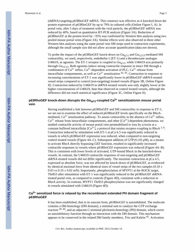
It has been established, that in its nascent form, p63RhoGEF is autoinhibited. The moleculecontains a Dbl-homology (DH domain), a minimal unit to catalyze the GTP exchangereaction 20, 44, and an adjacent C-terminal pleckstrin-homology (PH) domain, which exertsan autoinhibitory function through an interaction with the DH domain. This mechanismappears to be conserved in the related Dbl family members, Trio and Kalirin 45. Activation
Momotani et al. Page 5
Circ Res. Author manuscript; available in PMC 2012 October 14.
NIH
-PA Author Manuscript
NIH
-PA Author Manuscript
NIH
-PA Author Manuscript
occurs when the Gαq/11 subunit binds to specific amino acids in the PH domain plus itsadjacent conserved amino acid stretch (extended PH domain) of p63RhoGEF, thus causing astructural reorganization in p63RhoGEF and relief of autoinhibition. We hypothesized that ifthe Ca2+-sensitization effect is mediated by the same mechanism, then the isolated,recombinant extended PH domain (p63RhoGEF331–580) should interfere with the process,either by binding to the activated Gαq/11 and blocking downstream signaling, or by bindingto the transiently open, i.e. active conformation of endogenous p63RhoGEF. To test thishypothesis in intact blood vessels, we needed a vessel that responds to phenylephrine andcan be well permeabilized to proteins to allow for the introduction of the p63RhoGEFfragment into the SM cells, while retaining the receptor-coupling to RhoA. Because this wasnot possible using mouse portal vein, we used instead rabbit portal vein permeabilized withβ-escin46. Fortuitously, unlike the mouse vessel, rabbit portal vein responds tophenylephrine, reported to act purely through Gαq/11, and not Gα12/13 30, with Ca2+
sensitized force at constant intracellular [Ca2+] (pCa 6.3). Phenylephrine/Gαq/11 couplingwas established in mouse aorta30. Phenylephrine is a specificα1-adrenoreceptor agonist 47
shown to specifically mediate contractile responses through activation of Gαq/11 48. α1-adrenoreceptors have been shown to be present and functional in rabbit arteries including thecarotid aortic, iliac, mesenteric, renal, ear and resistance arteries 47. Therefore, we assumethat they are also present in rabbit portal vein, additionally this vessel contracts in responseto phenylephrine. When the contractile force induced by phenylephrine reached a plateau, acontrol filtrate (i.e. buffer from which recombinant p63RhoGEF331–580 protein wasremoved) was added, followed by the p63RhoGEF331–580 protein. The filtrate produced noeffect (Figure 5A), while the recombinant p63RhoGEF331–580 fragment induced relaxationof the SM strips (Figure 5A; Online Figure III). Both RhoA activity and RLC20phosphorylation level increased with phenylephrine-induced Ca2+ sensitized force and bothdecreased upon introduction of p63RhoGEF331–580 (Figure 5B). These findings in bloodvessels confirm the hypothesis that recombinant p63RhoGEF331–580 inhibits endogenousp63RhoGEF or interferes with the regulatory pathway by sequestering activated Gαq/11 aspredicted from in vitro experiments 45.
RhoA-GDP turnover is suppressed in MEF cells by over-expression of p63RhoGEF331–580
fragmentHaving established an inhibitory role for p63RhoGEF331–580 in blood vessels, weinvestigated if it acts in the same way in cells. We could not use SM cells for that purpose,because their transfection efficiency is generally only < 2–3% by traditional methods andabout 30 % by electroporation. Instead, we resorted to mouse embryonic fibroblasts (MEF)where we were able to achieve 80% transfection efficiency. Using MEF cells weinvestigated the changes in relative levels of RhoA•GTP upon phenylephrine stimulation inthe presence and absence of exogenous p63RhoGEF331–580. RhoA•GTP levels wereincreased when MEF cells were stimulated by phenylephrine (5 μM) (Figure 6A), anddropped in the presence of over-expressed p63RhoGEF331–580. A similar trend was alsoobserved upon ET-1 stimulation (data not shown).
p63RhoGEF331–580 associates with full-length p63RhoGEF and the constitutively activeGαq/11Q209L
We then asked if p63RhoGEF331–580 interferes with the RhoA activation pathway byinteracting with Gαq/11 or with endogenous p63RhoGEF. For this experiment, it wasnecessary to use cells that do not express endogenous p63RhoGEF, and we chose theHEK293 cells, which provide an additional benefit of easy transfection. Therefore, we over-expressed in HEK293 cells p63RhoGEF331–580 with and without full-length p63RhoGEF.Due to competitive co-expression of full-length p63RhoGEF and p63RhoGEF331–580, theexpression level of full-length p63RhoGEF was higher when it was expressed alone. In spite
Momotani et al. Page 6
Circ Res. Author manuscript; available in PMC 2012 October 14.
NIH
-PA Author Manuscript
NIH
-PA Author Manuscript
NIH
-PA Author Manuscript
of this, immunoprecipitation resulted in precipitation of p63RhoGEF331–580 only in thepresence of full-length p63RhoGEF (Figure 6B). This finding is consistent with structuraland in vitro studies suggesting that an intramolecular association of the PH domain with theDH domain results in autoinhibition 45. We then examined whether the p63RhoGEF331–580
interacts with Gαq/11. Full-length p63RhoGEF and p63RhoGEF331–580, both FLAG-tagged,were co-expressed with Gαq/11wild-type or constitutively active Gα11 Q209T mutant inHEK293 cells (Figure 6C). As expected, Gαq/11 Q209L showed higher affinity for full-length p63RhoGEF and p63RhoGEF331–580 than Gαq/11 wild-type, again, consistent withthe in vitro studies showing direct association of activated Gαq/11 and p63RhoGEF 45, 49.
DiscussionThe RhoA/ROCK pathway serves as a final signaling hub in all SM tissues, acceptingstimulatory signals initiated by diverse agonists, and activating Ca2+-sensitization viainhibition of myosin light chain phosphatase. This physiological phenomenon is of criticalsignificance in control of blood pressure, airway resistance, erectile dysfunction, gutmotility, bladder and ureter function, etc 3. The central role of the ROCK kinase prompted anumber of investigations into its potential as a drug target 50. However, the ubiquitousdistribution of ROCK reduces the clinical potential of this approach, as it lacks the desiredtissue specificity. On the other hand, downstream of ROCK, RhoA appears to be activatedvia diverse pathways, each utilizing a specific combination of GPCRs, trimeric G-proteinsand GEFs. This raises the exciting possibility of selective targeting of tissue-specific GEFsfor therapeutic use.
In order to better understand the intricate nature of the Ca2+-independent component ofcontractile stimulation in SM, it is necessary to dissect the molecular pathways and identifythe individual GPCRs as well as their partner G-proteins and coupled GEFs. Mostvasoconstrictors, such as angiotensin II, ET-1, phenylephrine and thromboxane A2 bind toGPCRs coupled either to Gαq/11 or Gα12/13 or both30. In mice, knockout of Gαq/11, but notGα12/13, results in a fall in basal blood pressure while both are needed for the developmentof salt-induced hypertension. Studies of contractility of aorta from these mice showed thatphenylephrine-induced force was completely abolished in the Gαq/11 deficient mice butunaffected in the absence of Gα12/13. On the other hand, contractions in response to ET-1 orU46619 are mediated by both G-proteins, with Gαq/11 dominating for ET-1 and Gα12/13 forU46619. The key question is what portion of Ca2+-sensitized contractile response istransduced by each of these G-proteins, and if their signaling pathways are routed throughdistinct GEFs.
While Gα12/13 was shown to activate RhoA through the three RGSL-containing, RhoAspecific GEFs, i.e. PDZRhoGEF, LARG and p115RhoGEF 24, the pathways involvingGαq/11 remained unclear. It has been established, that the RhoA-specific exchange factorp63RhoGEF is selectively activated by Gαq/11 45, 49, 51.. Recently, mRNA transcripts forp63RhoGEF were identified in rat aorta 52. In an unrelated study, p63RhoGEF couplingthrough Gαq/11 has been shown in cultured rat aortic SM cells to mediate angiotensin IIsignaling 35. It was found that angiotensin II first induces a rapid influx of intracellular[Ca2+] and activation of p63RhoGEF, followed by a significantly slower, [Ca2+]/Januskinase 2-dependent activation of p115RhoGEF to sustain RhoA signaling. However, the roleof this pathway in contractility was assessed only by a collagen contraction assay usingcultured rat aortic SM cells known to de-differentiate in culture. As expected, basalcontraction increased upon angiotensin II pathway stimulation, but not in p63RhoGEFdepleted cells 35.
Momotani et al. Page 7
Circ Res. Author manuscript; available in PMC 2012 October 14.
NIH
-PA Author Manuscript
NIH
-PA Author Manuscript
NIH
-PA Author Manuscript
The purpose of our investigation was to dissect the specific role of p63RhoGEF mediatedCa2+-sensitization in blood vessels in response to stimulation via Gαq/11, but throughreceptors other than that activated by angiotensin II. Our results confirm the presence ofp63RhoGEF mRNA transcripts and protein in SM tissues and cells. Importantly, we showthat expression profiles of upstream and downstream partners of p63RhoGEF (i.e. Gαq/11and RhoA) correlate well with p63RhoGEF across the tissues screened. We demonstrate forthe first time that p63RhoGEF plays an important part in vessel contractility induced byET-1, coupled primarily to Gαq/11 through the ETA receptors, but not by U46619, athromboxane A2 analogue, known to couple predominately by Gα12/13. The Gαq/11 couplingleads to PLCβ activation, production of IP3 (inositol 1,4,5-trisphosphate) and DAG(diacylglycerol) and consequently increased intracellular [Ca2+] as well as activation of theRhoA pathway. To evaluate if the Ca2+-sensitization pathway is affected by the knock-downof p63RhoGEF, we used α-toxin permeabilization, a method that retains agonist-GPCRcoupling to RhoA under conditions where intracellular [Ca2+] is clamped. Ca2+ sensitizationof contractile force induced by ET-1 but not by U46619 was significantly reduced whenp63RhoGEF expression was suppressed (Figure 4). Furthermore, this was accompanied by adecrease in MYPT1 phosphorylation indicative of decreased RhoA/ROCK activity. Theseand other findings 30 lead us to suggest that the Gαq/11 contribution to the regulation of basaland stimulated blood pressure may signal through p63RhoGEF rather than solely throughPLCβ-induced Ca2+ release. Consistent with this idea is the observation that ROCKinhibitor Y-27632 reduces hypertension53, 54 and that the Rho/ROCK signaling pathway inSM cells is an important contributor to peripheral resistance 55. Significantly, p63RhoGEFmRNA is expressed in the resistance vessels, thoracodorsal and mesenteric arteries (Figure1). P63RhoGEF protein was also detected in these vessels (data not shown). Thecontribution of this RhoGEF to the contractile behavior of resistance vessels will beexplored in the future.
We also wondered if the in vivo mechanism by which p63RhoGEF operates in SM isconsistent with a model derived from crystallographic studies of its complex with activatedGαq/11 49. We hypothesized, that if the Ca2+-sensitization effect is mediated by the samemechanism, then the isolated, recombinant extended PH domain (p63RhoGEF331–580)should interfere with the process, either by binding to the activated Gαq/11 and blockingdownstream signaling, or by binding to the transiently open, i.e. active conformation ofendogenous p63RhoGEF. To test this hypothesis in blood vessels, we performedcontractility assays using β-escin permeabilized rabbit portal vein to allow introduction ofthe p63RhoGEF fragment into the SM cells. As expected, p63RhoGEF331–580 inducedrelaxation following phenylephrine-induced contraction, which is known to be mediated byGαq/11
30. This was accompanied by a decrease in RhoA-GTP and phospho-RLC20suggesting suppression of Gαq/11-coupled RhoGEF activity. Reduction of RhoA activityupon stimulation by phenylephrine in the presence of exogenous p63RhoGEF331–580, actingas a dominant negative, was also observed in MEF cells. Overexpressed p63RhoGEF331–580
associated with the full-length p63RhoGEF and with activated Gαq/11. Our findings areconsistent with the DH domain of p63RhoGEF being auto-inhibited through its extended PHdomain under basal conditions with agonist activated Gαq/11 relieving this auto-inhibition asshown in compelling in vitro experiments 45, 49. A similar fragment of p63RhoGEF,comprising residues 295 to 580, was also shown to interfere with the angiotensin II mediatedRhoA activation pathway in SM cells 35. Taken together, these data strongly support thenotion that the molecular mechanism inferred from the crystallographic studies isrepresentative of the in vivo phenomenon.
In summary, the evidence presented in our paper shows that activation of p63RhoGEF byagonists that stimulate receptors coupled to Gαq/11 leads to Ca2+ sensitization in bloodvessels and cultured SM cells. We show that in addition to angiotensin II 35, both
Momotani et al. Page 8
Circ Res. Author manuscript; available in PMC 2012 October 14.
NIH
-PA Author Manuscript
NIH
-PA Author Manuscript
NIH
-PA Author Manuscript
phenylephrine and ET-1 also signal through p63RhoGEF to active RhoA and Ca2+
sensitized force. We conclude that different Gαq/11 coupled receptors can signal throughp63RhoGEF to contribute to vascular tone, and that it is not unique to angiotensin IIstimulation. Indeed, angiotensin II plays a major role in hypertension while the tonicdischarge of catecholamines by vasoconstricting sympathetic nerves maintains normotensiveblood pressure. Further identification of specific RhoGEFs and characterization of theirfunction in SM is a fertile ground for finding new targets that mediate specific functions ofnormal and diseased SM including the regulation of vascular tone.
Supplementary MaterialRefer to Web version on PubMed Central for supplementary material.
AcknowledgmentsSources of Funding
This work was supported by grants from the National Institutes of Health R01DK088905 and R01GM86457.
We thank Dr. John E. Sondek for productive discussions and the Ga11 wild-type and constitutively active Ga11Q209T mutant constructs and Dr Hideko Kasahara for her generosity and expertise in the preparation of theadenoviral constructs.
Non-standard Abbreviations and Acronyms
DH Dbl-homology
ET-1 Endothelin-1
GEF Guanine nucleotide exchange factor
GPCR G protein coupled receptor
HEK Human embryonic kidney
MEF Mouse embryonic fibroblast
MYPT1 Myosin targeting phosphatase subunit
PH Pleckstrin-homology
RLC20 Myosin regulatory light chain
ROCK Rho-kinase
shRNA short hairpin RA
SM Smooth muscle
References1. Ito M, Nakano T, Erdodi F, Hartshorne DJ. Myosin phosphatase: structure, regulation and function.
Mol Cell Biochem. 2004; 259(1–2):197–209. [PubMed: 15124925]2. Kimura K, Ito M, Amano M, Chihara K, Fukata Y, Nakafuku M, Yamamori B, Feng J, Nakano T,
Okawa K, Iwamatsu A, Kaibuchi K. Regulation of myosin phosphatase by Rho and Rho-associatedkinase (Rho-kinase). Science. 1996; 273(5272):245–248. [PubMed: 8662509]
3. Somlyo AP, Somlyo AV. Ca2+ sensitivity of smooth muscle and nonmuscle myosin II: modulatedby G proteins, kinases, and myosin phosphatase. Physiol Rev. 2003; 83(4):1325–1358. [PubMed:14506307]
4. Somlyo AP, Somlyo AV. Signal transduction and regulation in smooth muscle. Nature. 1994;372(6503):231–236. [PubMed: 7969467]
Momotani et al. Page 9
Circ Res. Author manuscript; available in PMC 2012 October 14.
NIH
-PA Author Manuscript
NIH
-PA Author Manuscript
NIH
-PA Author Manuscript
5. Gong MC, Iizuka K, Nixon G, Browne JP, Hall A, Eccleston JF, Sugai M, Kobayashi S, SomlyoAV, Somlyo AP. Role of guanine nucleotide-binding proteins--ras-family or trimeric proteins orboth--in Ca2+ sensitization of smooth muscle. Proc Natl Acad Sci U S A. 1996; 93(3):1340–1345.[PubMed: 8577766]
6. Kitazawa T, Kobayashi S, Horiuti K, Somlyo AV, Somlyo AP. Receptor-coupled, permeabilizedsmooth muscle. Role of the phosphatidylinositol cascade, G-proteins, and modulation of thecontractile response to Ca2+ J Biol Chem. 1989; 264(10):5339–5342. [PubMed: 2494163]
7. Somlyo AV, Phelps C, Dipierro C, Eto M, Read P, Barrett M, Gibson JJ, Burnitz MC, Myers C,Somlyo AP. Rho kinase and matrix metalloproteinase inhibitors cooperate to inhibit angiogenesisand growth of human prostate cancer xenotransplants. Faseb J. 2003; 17(2):223–234. [PubMed:12554701]
8. Kimura K, Ito M, Amano M, Chihara K, Fukata Y, Nakafuku M, Yamamori B, Feng J, Nakano T,Okawa K, Iwamatsu A, Kaibuchi K. Regulation of myosin phosphatase by Rho and Rho-associatedkinase (Rho-kinase) [see comments]. Science. 1996; 273(5272):245–248. [PubMed: 8662509]
9. Jaffe AB, Hall A. RHO GTPASES: Biochemistry and Biology. Annu Rev Cell Dev Biol. 2005;21:247–269. [PubMed: 16212495]
10. Etienne-Manneville S, Hall A. Rho GTPases in cell biology. Nature. 2002; 420(6916):629–635.[PubMed: 12478284]
11. Wei Y, Zhang Y, Derewenda U, Liu X, Minor W, Nakomoto RK, Somlyo AV, Somlyo AP,Derewenda ZS. Crystal structure of RhoA-GDP and its functional implications. Nature StructuralBiology. 1997; 4(9):699–702.
12. Ihara K, Muraguchi S, Kato M, Shimizu T, Shirakawa M, Kuroda S, Kaibuchi K, Hakoshima T.Crystal structure of human RhoA in a dominantly active form complexed with a GTP analogue. JBiol Chem. 1998; 273(16):9656–9666. [PubMed: 9545299]
13. Dvorsky R, Blumenstein L, Vetter IR, Ahmadian MR. Structural insights into the interaction ofROCKI with the switch regions of RhoA. J Biol Chem. 2004; 279(8):7098–7104. [PubMed:14660612]
14. Zhang B, Chernoff J, Zheng Y. Interaction of Rac1 with GTPase-activating proteins and putativeeffectors. A comparison with Cdc42 and RhoA. J Biol Chem. 1998; 273(15):8776–8782.[PubMed: 9535855]
15. Tcherkezian J, Lamarche-Vane N. Current knowledge of the large RhoGAP family of proteins.Biol Cell. 2007; 99(2):67–86. [PubMed: 17222083]
16. Scheffzek K, Ahmadian MR. GTPase activating proteins: structural and functional insights 18years after discovery. Cell Mol Life Sci. 2005; 62(24):3014–3038. [PubMed: 16314935]
17. Dovas A, Couchman JR. RhoGDI: multiple functions in the regulation of Rho family GTPaseactivities. Biochem J. 2005; 390(Pt 1):1–9. [PubMed: 16083425]
18. Boguski MS, McCormick F. Proteins regulating Ras and its relatives. Nature. 1993; 366:643–654.[PubMed: 8259209]
19. Longenecker K, Read P, Derewenda U, Dauter Z, Liu X, Garrard S, Walker L, Somlyo AV,Nakamoto RK, Somlyo AP, Derewenda ZS. How RhoGDI binds Rho. Acta Cryst D. 1999; 55(Pt9):1503–1515. [PubMed: 10489445]
20. Rossman KL, Der CJ, Sondek J. GEF means go: turning on RHO GTPases with guaninenucleotide-exchange factors. Nat Rev Mol Cell Biol. 2005; 6(2):167–180. [PubMed: 15688002]
21. Himpens B, Kitazawa T, Somlyo AP. Agonist-dependent modulation of Ca2+ sensitivity in rabbitpulmonary artery smooth muscle. Pflugers Arch. 1990; 417(1):21–28. [PubMed: 2127310]
22. Gohla A, Schultz G, Offermanns S. Role for G(12)/G(13) in agonist-induced vascular smoothmuscle cell contraction. Circ Res. 2000; 87(3):221–227. [PubMed: 10926873]
23. Maguire JJ, Davenport AP. Regulation of vascular reactivity by established and emerging GPCRs.Trends Pharmacol Sci. 2005; 26(9):448–454. [PubMed: 16054240]
24. Fukuhara S, Chikumi H, Gutkind JS. RGS-containing RhoGEFs: the missing link betweentransforming G proteins and Rho? Oncogene. 2001; 20(13):1661–1668. [PubMed: 11313914]
25. Aittaleb M, Boguth CA, Tesmer JJ. Structure and function of heterotrimeric G protein-regulatedRho guanine nucleotide exchange factors. Mol Pharmacol. 77(2):111–125. [PubMed: 19880753]
Momotani et al. Page 10
Circ Res. Author manuscript; available in PMC 2012 October 14.
NIH
-PA Author Manuscript
NIH
-PA Author Manuscript
NIH
-PA Author Manuscript
26. Kreutz B, Hajicek N, Yau DM, Nakamura S, Kozasa T. Distinct regions of Galpha13 participate inits regulatory interactions with RGS homology domain-containing RhoGEFs. Cell Signal. 2007;19(8):1681–1689. [PubMed: 17449226]
27. Longenecker KL, Lewis ME, Chikumi H, Gutkind JS, Derewenda ZS. Structure of the RGS-likedomain from PDZ-RhoGEF: linking heterotrimeric g protein-coupled signaling to Rho GTPases.Structure (Camb). 2001; 9(7):559–569. [PubMed: 11470431]
28. Chen Z, Wells CD, Sternweis PC, Sprang SR. Structure of the rgRGS domain of p115RhoGEF.Nat Struct Biol. 2001; 8(9):805–809. [PubMed: 11524686]
29. Fukuhara S, Murga C, Zohar M, Igishi T, Gutkind JS. A novel PDZ domain containing guaninenucleotide exchange factor links heterotrimeric G proteins to Rho. J Biol Chem. 1999; 274(9):5868–5879. [PubMed: 10026210]
30. Wirth A, Benyo Z, Lukasova M, Leutgeb B, Wettschureck N, Gorbey S, Orsy P, Horvath B,Maser-Gluth C, Greiner E, Lemmer B, Schutz G, Gutkind JS, Offermanns S. G12-G13-LARG-mediated signaling in vascular smooth muscle is required for salt-induced hypertension. Nat Med.2008; 14(1):64–68. [PubMed: 18084302]
31. Derewenda U, Oleksy A, Stevenson AS, Korczynska J, Dauter Z, Somlyo AP, Otlewski J, SomlyoAV, Derewenda ZS. The crystal structure of RhoA in complex with the DH/PH fragment ofPDZRhoGEF, an activator of the Ca(2+) sensitization pathway in smooth muscle. Structure. 2004;12(11):1955–1965. [PubMed: 15530360]
32. Hilgers RH, Todd J Jr, Webb RC. Increased PDZ-RhoGEF/RhoA/Rho kinase signaling in smallmesenteric arteries of angiotensin II-induced hypertensive rats. J Hypertens. 2007; 25(8):1687–1697. [PubMed: 17620967]
33. Ying Z, Jin L, Dorrance AM, Webb RC. Increased expression of mRNA for regulator of G proteinsignaling domain-containing Rho guanine nucleotide exchange factors in aorta from stroke-pronespontaneously hypertensive rats. Am J Hypertens. 2004; 17(10):981–985. [PubMed: 15485764]
34. Wirth A, Benyo Z, Lukasova M, Leutgeb B, Wettschureck N, Gorbey S, Orsy P, Horvath B,Maser-Gluth C, Greiner E, Lemmer B, Schutz G, Gutkind S, Offermanns S. G(12)-G(13)-LARG-mediated signaling in vascular smooth muscle is required for salt-induced hypertension. Nat Med.2007
35. Wuertz CM, Lorincz A, Vettel C, Thomas MA, Wieland T, Lutz S. p63RhoGEF--a key mediatorof angiotensin II-dependent signaling and processes in vascular smooth muscle cells. FASEB J.2010; 24(12):4865–4876. [PubMed: 20739613]
36. Chikumi H, Vazquez-Prado J, Servitja JM, Miyazaki H, Gutkind JS. Potent activation of RhoA byGalpha q and Gq-coupled receptors. J Biol Chem. 2002; 277(30):27130–27134. [PubMed:12016230]
37. Lutz S, Freichel-Blomquist A, Rumenapp U, Schmidt M, Jakobs KH, Wieland T. p63RhoGEF andGEFT are Rho-specific guanine nucleotide exchange factors encoded by the same gene. NaunynSchmiedebergs Arch Pharmacol. 2004; 369(5):540–546. [PubMed: 15069594]
38. Souchet M, Portales-Casamar E, Mazurais D, Schmidt S, Leger I, Javre JL, Robert P, Berrebi-Bertrand I, Bril A, Gout B, Debant A, Calmels TP. Human p63RhoGEF, a novel RhoA-specificguanine nucleotide exchange factor, is localized in cardiac sarcomere. J Cell Sci. 2002; 115(Pt 3):629–640. [PubMed: 11861769]
39. Sheffield P, Garrard S, Derewenda Z. Overcoming expression and purification problems ofRhoGDI using a family of “parallel” expression vectors. Protein Expr Purif. 1999; 15(1):34–39.[PubMed: 10024467]
40. Neppl RL, Lubomirov LT, Momotani K, Pfitzer G, Eto M, Somlyo AV. Thromboxane A2-inducedbi-directional regulation of cerebral arterial tone. J Biol Chem. 2009; 284(10):6348–6360.[PubMed: 19095646]
41. Guo X, Stafford LJ, Bryan B, Xia C, Ma W, Wu X, Liu D, Songyang Z, Liu M. A Rac/Cdc42-specific exchange factor, GEFT, induces cell proliferation, transformation, and migration. J BiolChem. 2003; 278(15):13207–13215. [PubMed: 12547822]
42. Ding X, Murray PA. Cellular mechanisms of thromboxane A2-mediated contraction in pulmonaryveins. Am J Physiol Lung Cell Mol Physiol. 2005; 289(5):L825–833. [PubMed: 15964897]
Momotani et al. Page 11
Circ Res. Author manuscript; available in PMC 2012 October 14.
NIH
-PA Author Manuscript
NIH
-PA Author Manuscript
NIH
-PA Author Manuscript
43. McNair LL, Salamanca DA, Khalil RA. Endothelin-1 promotes Ca2+ antagonist-insensitivecoronary smooth muscle contraction via activation of epsilon-protein kinase C. Hypertension.2004; 43(4):897–904. [PubMed: 14981072]
44. Garcia-Mata R, Burridge K. Catching a GEF by its tail. Trends Cell Biol. 2007; 17(1):36–43.[PubMed: 17126549]
45. Rojas RJ, Yohe ME, Gershburg S, Kawano T, Kozasa T, Sondek J. Galphaq directly activatesp63RhoGEF and Trio via a conserved extension of the Dbl homology-associated pleckstrinhomology domain. J Biol Chem. 2007; 282(40):29201–29210. [PubMed: 17606614]
46. Kobayashi S, Kitazawa T, Somlyo AV, Somlyo AP. Cytosolic heparin inhibits muscarinic andalpha-adrenergic Ca2+ release in smooth muscle. Physiological role of inositol 1,4,5-trisphosphatein pharmacomechanical coupling. J Biol Chem. 1989; 264(30):17997–18004. [PubMed: 2509451]
47. Guimaraes S, Moura D. Vascular adrenoceptors: an update. Pharmacol Rev. 2001; 53(2):319–356.[PubMed: 11356987]
48. Docherty JR. Subtypes of functional alpha1-adrenoceptor. Cell Mol Life Sci. 2010; 67(3):405–417.[PubMed: 19862476]
49. Lutz S, Shankaranarayanan A, Coco C, Ridilla M, Nance MR, Vettel C, Baltus D, Evelyn CR,Neubig RR, Wieland T, Tesmer JJ. Structure of Galphaq-p63RhoGEF-RhoA complex reveals apathway for the activation of RhoA by GPCRs. Science. 2007; 318(5858):1923–1927. [PubMed:18096806]
50. Hahmann C, Schroeter T. Rho-kinase inhibitors as therapeutics: from pan inhibition to isoformselectivity. Cell Mol Life Sci. 2010; 67(2):171–177. [PubMed: 19907920]
51. Lutz S, Freichel-Blomquist A, Yang Y, Rumenapp U, Jakobs KH, Schmidt M, Wieland T. Theguanine nucleotide exchange factor p63RhoGEF, a specific link between Gq/11-coupled receptorsignaling and RhoA. J Biol Chem. 2005; 280(12):11134–11139. [PubMed: 15632174]
52. Toumaniantz G, Ferland-McCollough D, Cario-Toumaniantz C, Pacaud P, Loirand G. The Rhoprotein exchange factor Vav3 regulates vascular smooth muscle cell proliferation and migration.Cardiovasc Res. 2010; 86(1):131–140. [PubMed: 19969623]
53. Seasholtz TM, Brown JH. RHO SIGNALING in vascular diseases. Mol Interv. 2004; 4(6):348–357. [PubMed: 15616164]
54. Uehata M, Ishizaki T, Satoh H, Ono T, Kawahara T, Morishita T, Tamakawa H, Yamagami K, InuiJ, Maekawa M, Narumiya S. Calcium sensitization of smooth muscle mediated by a Rho-associated protein kinase in hypertension. Nature. 1997; 389(6654):990–994. [PubMed: 9353125]
55. Schubert R, Lidington D, Bolz SS. The emerging role of Ca2+ sensitivity regulation in promotingmyogenic vasoconstriction. Cardiovasc Res. 2008; 77(1):8–18. [PubMed: 17764667]
Momotani et al. Page 12
Circ Res. Author manuscript; available in PMC 2012 October 14.
NIH
-PA Author Manuscript
NIH
-PA Author Manuscript
NIH
-PA Author Manuscript
Novelty and Significance
What is known?
• The small GTPase RhoA is activated by multiple agonists and significantlycontributes to vascular contractility under physiological as well aspathophysiological conditions such as hypertension.
• Multiple GTP exchange factors (GEFs) are expressed in smooth muscle raisingthe possibility that specific agonists of specific G-protein coupled receptors(GPCRs) may be associated with distinct RhoGEFs.
• Angiotensin II has been shown to signal through Gαq/11 in cultured smoothmuscle cells but the role of other agonists in intact blood vessels is unknown,particularly agonists that regulate basal vascular tone.
What new information does this article contribute?
• p63RhoGEF is selectively activated by agonists such as α-adrenergic andendothelin-1 that signal through Gαq/11 in blood vessels and maintainnormotensive blood pressure.
• Knock down of p63RhoGEF decreases RhoA activity leading to increasedmyosin phosphatase activity, decreased myosin phosphorylation and decreasedforce development in blood vessels.
• We demonstrate in vivo that the molecular mechanism of action of p63RhoGEFis consistent with a model derived from crystallographic studies.
• p63RhoGEF is a potential selective therapeutic target for decreasing peripheralresistance and blood pressure.
Multiple agonists signaling through GPCRs activate the RhoA pathway which plays acentral role in the regulation of basal tone, peripheral resistance, hypertension andcerebral vasospasm. RhoGEFs function upstream of RhoA, but it is unclear whether themultiple RhoGEFs present in smooth muscle (SM) couple selectively to specific agoniststhrough GPCRs. If so, this would allow for selective therapeutic targeting of specificRhoGEFs in vascular SM. p63RhoGEF selectively coupled Gαq/11, but not Gα12/13, toRhoA activation in blood vessels, mediating the physiologically important Ca2+
sensitization of force induced with Gαq/11 coupled agonists. Experiments using arecombinant extended PH domain fragment of p63RhoGEF show that this region servesas an autoinhibitory domain, interfering with the regulatory pathway by sequesteringactivated Gαq/11. Our study extends the reported findings on angiotensin II to otherGαq/11coupled agonists such as α-agonists and endothelin-1. Angiotensin II plays a majorrole in hypertension while catecholamines maintain normotensive blood pressure. Wesuggest that signaling through p63RhoGEF contributes to selective regulation of bloodpressure. Further identification of specific RhoGEFs and characterization of theirfunction in SM could identify new targets for regulating specific functions of normal anddiseased SM.
Momotani et al. Page 13
Circ Res. Author manuscript; available in PMC 2012 October 14.
NIH
-PA Author Manuscript
NIH
-PA Author Manuscript
NIH
-PA Author Manuscript
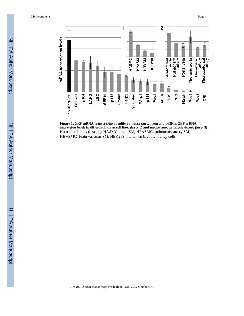
Figure 1. GEF mRNA transcription profile in mouse portal vein and p63RhoGEF mRNAexpression levels in different human cell lines (inset 1) and mouse smooth muscle tissues (inset 2)Human cell lines (inset 1): HASMC: aorta SM; HPASMC: pulmonary artery SM;HBVSMC: brain vascular SM; HEK293: human embryonic kidney cells.
Momotani et al. Page 14
Circ Res. Author manuscript; available in PMC 2012 October 14.
NIH
-PA Author Manuscript
NIH
-PA Author Manuscript
NIH
-PA Author Manuscript
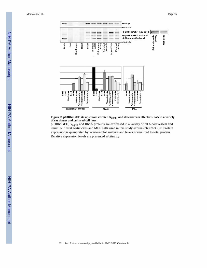
Figure 2. p63RhoGEF, its upstream effecter Gαq/11 and downstream effecter RhoA in a varietyof rat tissues and cultured cell linesp63RhoGEF, Gαq/11 and RhoA proteins are expressed in a variety of rat blood vessels andileum. R518 rat aortic cells and MEF cells used in this study express p63RhoGEF. Proteinexpression is quantitated by Western blot analysis and levels normalized to total protein.Relative expression levels are presented arbitrarily.
Momotani et al. Page 15
Circ Res. Author manuscript; available in PMC 2012 October 14.
NIH
-PA Author Manuscript
NIH
-PA Author Manuscript
NIH
-PA Author Manuscript
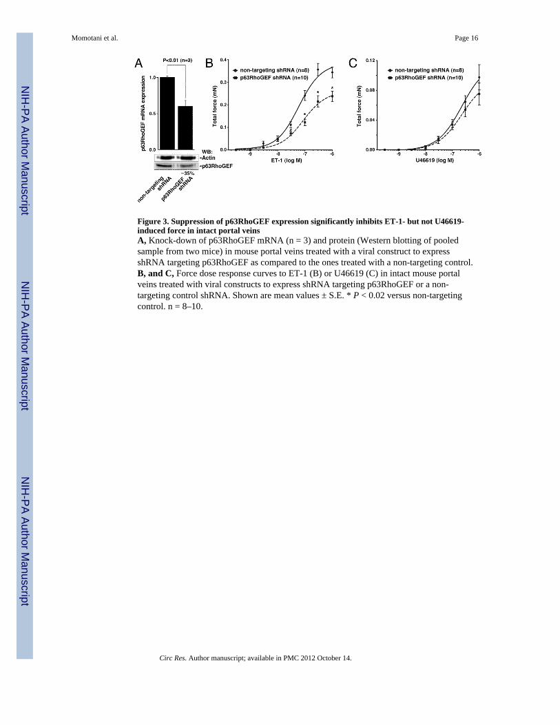
Figure 3. Suppression of p63RhoGEF expression significantly inhibits ET-1- but not U46619-induced force in intact portal veinsA, Knock-down of p63RhoGEF mRNA (n = 3) and protein (Western blotting of pooledsample from two mice) in mouse portal veins treated with a viral construct to expressshRNA targeting p63RhoGEF as compared to the ones treated with a non-targeting control.B, and C, Force dose response curves to ET-1 (B) or U46619 (C) in intact mouse portalveins treated with viral constructs to express shRNA targeting p63RhoGEF or a non-targeting control shRNA. Shown are mean values ± S.E. * P < 0.02 versus non-targetingcontrol. n = 8–10.
Momotani et al. Page 16
Circ Res. Author manuscript; available in PMC 2012 October 14.
NIH
-PA Author Manuscript
NIH
-PA Author Manuscript
NIH
-PA Author Manuscript
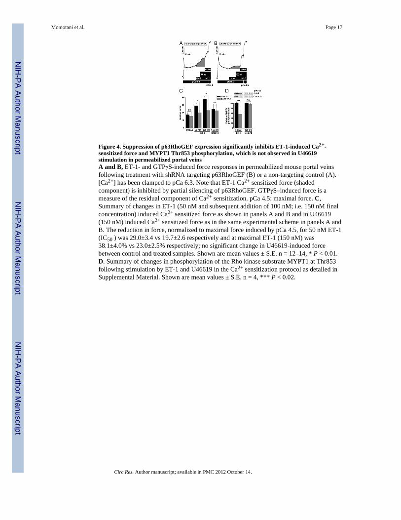
Figure 4. Suppression of p63RhoGEF expression significantly inhibits ET-1-induced Ca2+-sensitized force and MYPT1 Thr853 phosphorylation, which is not observed in U46619stimulation in permeabilized portal veinsA and B, ET-1- and GTPγS-induced force responses in permeabilized mouse portal veinsfollowing treatment with shRNA targeting p63RhoGEF (B) or a non-targeting control (A).[Ca2+] has been clamped to pCa 6.3. Note that ET-1 Ca2+ sensitized force (shadedcomponent) is inhibited by partial silencing of p63RhoGEF. GTPγS–induced force is ameasure of the residual component of Ca2+ sensitization. pCa 4.5: maximal force. C,Summary of changes in ET-1 (50 nM and subsequent addition of 100 nM; i.e. 150 nM finalconcentration) induced Ca2+ sensitized force as shown in panels A and B and in U46619(150 nM) induced Ca2+ sensitized force as in the same experimental scheme in panels A andB. The reduction in force, normalized to maximal force induced by pCa 4.5, for 50 nM ET-1(IC50 ) was 29.0±3.4 vs 19.7±2.6 respectively and at maximal ET-1 (150 nM) was38.1±4.0% vs 23.0±2.5% respectively; no significant change in U46619-induced forcebetween control and treated samples. Shown are mean values ± S.E. n = 12–14, * P < 0.01.D. Summary of changes in phosphorylation of the Rho kinase substrate MYPT1 at Thr853following stimulation by ET-1 and U46619 in the Ca2+ sensitization protocol as detailed inSupplemental Material. Shown are mean values ± S.E. n = 4, *** P < 0.02.
Momotani et al. Page 17
Circ Res. Author manuscript; available in PMC 2012 October 14.
NIH
-PA Author Manuscript
NIH
-PA Author Manuscript
NIH
-PA Author Manuscript
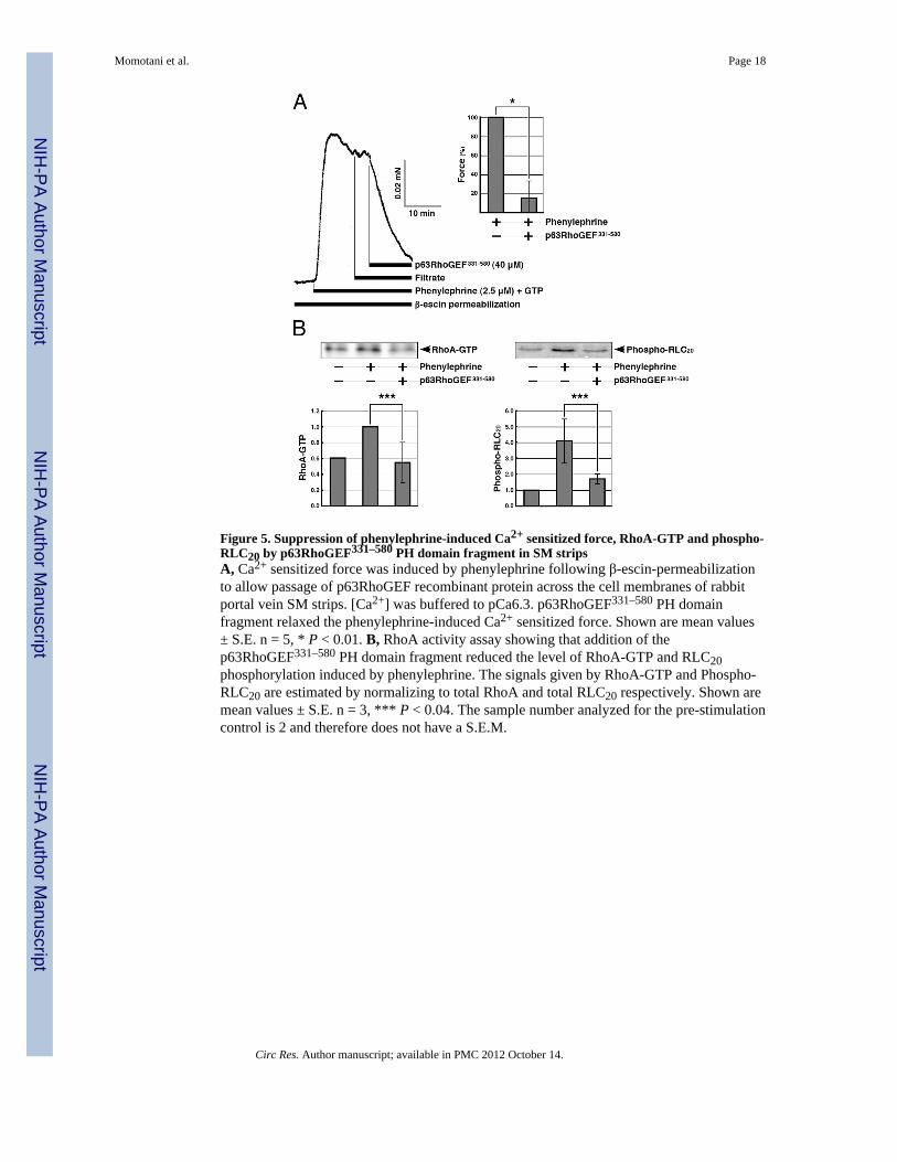
Figure 5. Suppression of phenylephrine-induced Ca2+ sensitized force, RhoA-GTP and phospho-RLC20 by p63RhoGEF331–580 PH domain fragment in SM stripsA, Ca2+ sensitized force was induced by phenylephrine following β-escin-permeabilizationto allow passage of p63RhoGEF recombinant protein across the cell membranes of rabbitportal vein SM strips. [Ca2+] was buffered to pCa6.3. p63RhoGEF331–580 PH domainfragment relaxed the phenylephrine-induced Ca2+ sensitized force. Shown are mean values± S.E. n = 5, * P < 0.01. B, RhoA activity assay showing that addition of thep63RhoGEF331–580 PH domain fragment reduced the level of RhoA-GTP and RLC20phosphorylation induced by phenylephrine. The signals given by RhoA-GTP and Phospho-RLC20 are estimated by normalizing to total RhoA and total RLC20 respectively. Shown aremean values ± S.E. n = 3, *** P < 0.04. The sample number analyzed for the pre-stimulationcontrol is 2 and therefore does not have a S.E.M.
Momotani et al. Page 18
Circ Res. Author manuscript; available in PMC 2012 October 14.
NIH
-PA Author Manuscript
NIH
-PA Author Manuscript
NIH
-PA Author Manuscript
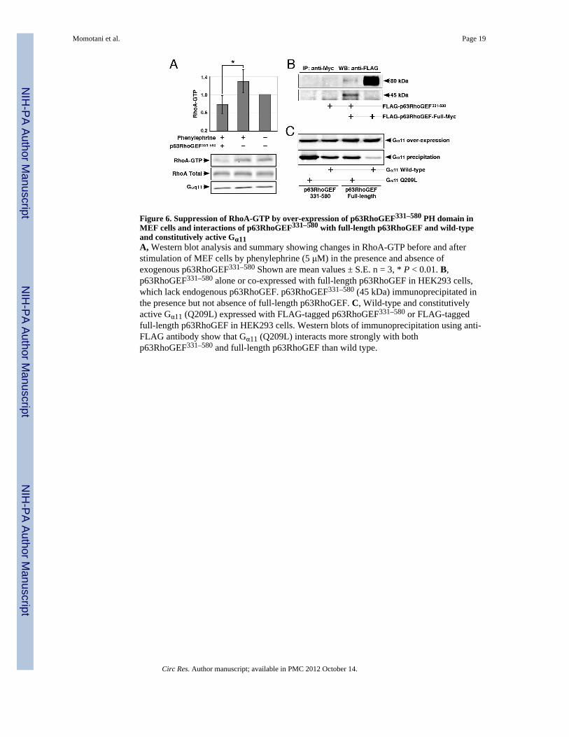
Figure 6. Suppression of RhoA-GTP by over-expression of p63RhoGEF331–580 PH domain inMEF cells and interactions of p63RhoGEF331–580 with full-length p63RhoGEF and wild-typeand constitutively active Gα11A, Western blot analysis and summary showing changes in RhoA-GTP before and afterstimulation of MEF cells by phenylephrine (5 μM) in the presence and absence ofexogenous p63RhoGEF331–580 Shown are mean values ± S.E. n = 3, * P < 0.01. B,p63RhoGEF331–580 alone or co-expressed with full-length p63RhoGEF in HEK293 cells,which lack endogenous p63RhoGEF. p63RhoGEF331–580 (45 kDa) immunoprecipitated inthe presence but not absence of full-length p63RhoGEF. C, Wild-type and constitutivelyactive Gα11 (Q209L) expressed with FLAG-tagged p63RhoGEF331–580 or FLAG-taggedfull-length p63RhoGEF in HEK293 cells. Western blots of immunoprecipitation using anti-FLAG antibody show that Gα11 (Q209L) interacts more strongly with bothp63RhoGEF331–580 and full-length p63RhoGEF than wild type.
Momotani et al. Page 19
Circ Res. Author manuscript; available in PMC 2012 October 14.
NIH
-PA Author Manuscript
NIH
-PA Author Manuscript
NIH
-PA Author Manuscript
Related Documents