INSTITUTE OF PHYSICS PUBLISHING PHYSICAL BIOLOGY Phys. Biol. 3 (2006) 130–137 doi:10.1088/1478-3975/3/2/005 Contractility and retrograde flow in lamellipodium motion K Kruse 1 , J F Joanny 2 ,FJ¨ ulicher 1 and J Prost 2,3 1 Max-Planck-Institut f ¨ ur Physik komplexer Systeme, N¨ othnitzerstr. 38, 01187 Dresden, Germany 2 Laboratoire Physico-Chimie Curie (CNRS-UMR168), Institut Curie recherche, 26, rue d’Ulm, 75248 Paris Cedex 05, France 3 ESPCI, 10 rue Vauquelin, 75231 Paris Cedex 05, France Received 9 September 2005 Accepted for publication 30 May 2006 Published 21 June 2006 Online at stacks.iop.org/PhysBio/3/130 Abstract We present a phenomenological description of cell locomotion on a solid substrate. The material properties of the actin cytoskeleton in the lamellipodium are described by the constitutive equations of a viscous polar gel with intrinsic activity. The polymerization of the gel takes place in a localized region near the leading edge. Using a simple two-dimensional description, we calculate in the steady state the thickness profile of the lamellipodium which at the rear connects to the cell body; we also calculate the flow profiles and the forces exerted on the substrate. The cell velocity is estimated as a function of externally applied forces. Our description is consistent with experimentally observed properties of motile cells such as the existence of a retrograde flow in the lamellipodium and a dipolar force distribution exerted by the cell on the substrate. Introduction Locomotion allows cells to change their environment and plays an important role in many biological processes such as wound healing, remodeling of connective tissues, rebuilding of damaged structures or formation of metastases in cancer. Under all these circumstances, the cells crawl on a surface coated by an extracellular matrix to which it adheres by specific adhesion proteins [1]. The motility is based on the active behavior of the actin cytoskeleton of the cell. It involves several steps [2, 3]: protrusion of a thin layer of cytoskeletal structure (the lamellipodium) at the leading edge of the cell, adhesion on the substrate and contraction of the cytoskeleton; eventually motility also involves the disassembly of the actin network at the rear of the cell. The contraction pulls the rear part of the cell forward. The energy consumed by the cell motion is provided by the hydrolysis of adenosinetriphosphate (ATP), which drives actin polymerization and depolymerization and the motion of myosin motor proteins. The interaction of myosin with actin filaments creates the contractile stresses in the cytoskeleton that drive the retraction processes. In the leading edge of the lamellipodium, new actin filaments are nucleated by branching off existing ones. Branching occurs when complexes of actin-related proteins (ARP) 2/3 bind to actin filaments close to the cell membrane [1]. This process together with filament growth by polymerization leads to the formation of a cross-linked and entangled filamental network or gel. The growth of the gel at the leading edge is controlled by the cell via signaling pathways and the activation of ARP 2/3 by proteins of the Wiskott– Aldrich syndrome family (WASP) which are localized in the cell membrane at the leading edge. Filament polymerization is also controlled by capping proteins which stabilize the ends of some filaments [4–6]. The localization of WASP proteins at the leading edge, in the cell membrane, is essential to ensure a controlled growth of the gel near the edge in the form of a thin layer. Below, we assume that WASP proteins are localized in a small region of size λ near the leading edge without describing the mechanisms that lead to this localization. In the following, we propose a theoretical analysis in which we correlate the lamellipodium velocity and shape to biophysical parameters which can be measured independently in suitably designed experiments. The most obvious parameters are the polymerization and depolymerization rates of actin filaments. Another parameter describes how the lamellipodium exerts forces on its substrate via adhesion molecules [7–9]. As long as the lamellipodium velocity is small compared to a/τ b where a is a molecular length and τ b is the average time during which an adhesion molecule is 1478-3975/06/020130+08$30.00 © 2006 IOP Publishing Ltd Printed in the UK 130

Welcome message from author
This document is posted to help you gain knowledge. Please leave a comment to let me know what you think about it! Share it to your friends and learn new things together.
Transcript
INSTITUTE OF PHYSICS PUBLISHING PHYSICAL BIOLOGY
Phys. Biol. 3 (2006) 130–137 doi:10.1088/1478-3975/3/2/005
Contractility and retrograde flow inlamellipodium motionK Kruse1, J F Joanny2, F Julicher1 and J Prost2,3
1 Max-Planck-Institut fur Physik komplexer Systeme, Nothnitzerstr. 38, 01187 Dresden, Germany2 Laboratoire Physico-Chimie Curie (CNRS-UMR168), Institut Curie recherche, 26, rue d’Ulm,75248 Paris Cedex 05, France3 ESPCI, 10 rue Vauquelin, 75231 Paris Cedex 05, France
Received 9 September 2005Accepted for publication 30 May 2006Published 21 June 2006Online at stacks.iop.org/PhysBio/3/130
AbstractWe present a phenomenological description of cell locomotion on a solid substrate. Thematerial properties of the actin cytoskeleton in the lamellipodium are described by theconstitutive equations of a viscous polar gel with intrinsic activity. The polymerization of thegel takes place in a localized region near the leading edge. Using a simple two-dimensionaldescription, we calculate in the steady state the thickness profile of the lamellipodium which atthe rear connects to the cell body; we also calculate the flow profiles and the forces exerted onthe substrate. The cell velocity is estimated as a function of externally applied forces. Ourdescription is consistent with experimentally observed properties of motile cells such as theexistence of a retrograde flow in the lamellipodium and a dipolar force distribution exerted bythe cell on the substrate.
Introduction
Locomotion allows cells to change their environment andplays an important role in many biological processes such aswound healing, remodeling of connective tissues, rebuildingof damaged structures or formation of metastases in cancer.Under all these circumstances, the cells crawl on a surfacecoated by an extracellular matrix to which it adheres byspecific adhesion proteins [1]. The motility is based onthe active behavior of the actin cytoskeleton of the cell.It involves several steps [2, 3]: protrusion of a thin layerof cytoskeletal structure (the lamellipodium) at the leadingedge of the cell, adhesion on the substrate and contractionof the cytoskeleton; eventually motility also involves thedisassembly of the actin network at the rear of the cell.The contraction pulls the rear part of the cell forward. Theenergy consumed by the cell motion is provided by thehydrolysis of adenosinetriphosphate (ATP), which drivesactin polymerization and depolymerization and the motion ofmyosin motor proteins. The interaction of myosin with actinfilaments creates the contractile stresses in the cytoskeletonthat drive the retraction processes.
In the leading edge of the lamellipodium, new actinfilaments are nucleated by branching off existing ones.Branching occurs when complexes of actin-related proteins
(ARP) 2/3 bind to actin filaments close to the cell membrane[1]. This process together with filament growth bypolymerization leads to the formation of a cross-linked andentangled filamental network or gel. The growth of the gel atthe leading edge is controlled by the cell via signaling pathwaysand the activation of ARP 2/3 by proteins of the Wiskott–Aldrich syndrome family (WASP) which are localized in thecell membrane at the leading edge. Filament polymerizationis also controlled by capping proteins which stabilize the endsof some filaments [4–6]. The localization of WASP proteins atthe leading edge, in the cell membrane, is essential to ensure acontrolled growth of the gel near the edge in the form of a thinlayer. Below, we assume that WASP proteins are localized in asmall region of size ! near the leading edge without describingthe mechanisms that lead to this localization.
In the following, we propose a theoretical analysis inwhich we correlate the lamellipodium velocity and shape tobiophysical parameters which can be measured independentlyin suitably designed experiments. The most obviousparameters are the polymerization and depolymerization ratesof actin filaments. Another parameter describes how thelamellipodium exerts forces on its substrate via adhesionmolecules [7–9]. As long as the lamellipodium velocity issmall compared to a/"b where a is a molecular length and"b is the average time during which an adhesion molecule is
1478-3975/06/020130+08$30.00 © 2006 IOP Publishing Ltd Printed in the UK 130
Contractility and retrograde flow in lamellipodium motion
bound to the substrate, this force is proportional to the velocityof the actin gel relative to the substrate and thus involvessimply a friction coefficient [10, 11]. This coefficient can beaccessible in well-designed experiments [31]. More complexbehaviors arise if the velocity is of the order a/"b but wepostpone for further analysis the study of the correspondingregimes. The total integrated force which acts on the cell atany given time is zero if no external forces are applied. Thisforce balance implies that if the front part of the cell pulls thecell forward, the rest of the cell pulls with exactly the sameforce in the backward direction. This balance of opposingforces can be characterized by a force dipole which canbe determined from experimentally measured traction maps[7, 8]. Such experiments show that these forces correspondto contractile behavior of the cell characterized by a negativeforce dipole in a one-dimensional description.
The description of the actin network is more challenging.Actin filaments are cross-linked in a structure which under theelectron microscope appears to be a gel [3]. This tells us that ontime scales such that the cross-links do not have time to move,and on length scales large compared to the average distancebetween cross-links, the actin network should behave like anelastic body. However, the cross-linking proteins statisticallyunbind after a characteristic time " , and on time scales longerthan " the actin network should behave like a fluid. Thesefeatures are well captured in the Maxwell description of whatis called a physical gel and have been measured experimentallyin cells [12].
Such a description is not sufficient for two reasons. First,actin filaments which have ‘barbed’ and ‘pointed’ ends arestructurally polar. Since they point on average with theirgrowing end toward the leading edge of the cell, the networkitself is polar. In the following, this polarity will mainly beimportant in the description of the lamellipodium surfaces(boundary conditions), but in general one has to keep in mindthat we are dealing with a polar gel. Secondly, cross-linkscan be redistributed in response to myosin motor activity.Myosin oligomers can even act as moving cross-links. Thisconfers a unique and original property to these biologicalsystems as compared to conventional physical gels [13, 14].We have recently derived a set of generic equations, basedon symmetries and conservation laws, precisely designedfor the description of actin gels in the presence of motoractivity [15]. This description involves a small number of newparameters that account for all effects of motor activity. Theseparameters can be measured in separate experiments. In thefollowing description, we will need only one such additionalparameter. It describes the tendency for molecular motors toanisotropically contract the gel. The main limitation of thisgeneric theory comes from the assumption of a system which isweakly out of equilibrium. In view of the fact that the force–velocity relation of motor proteins does not depart severelyfrom linear behavior, we think that it is a valid starting point.Thus with suitable polymerization/depolymerization rules, anappropriate substrate friction coefficient and a description ofthe actin/myosin gel, one is in a position to calculate principlesgoverning the shape and motion of cellular substructures suchas lamellipodia.
In order to keep the algebra tractable and to obtainclosed equations, we add into this first analysis a number ofreasonable assumptions. First, the polymerization is assumedto take place in the immediate vicinity of the plasma membranein a region of size !, corresponding to the localization ofthe WASP proteins. Second, while depolymerization is notlocalized, it occurs mainly in the rear of the cell. Weneglect depolymerization in the lamellipodium for simplicity,as this does not change the main features of our results.In order to describe the effects of the depolymerization inthe rear part of the cell, we impose boundary conditionsat a distance L from the leading edge, where L defines thelamellipodium length. These boundary conditions correspondto localized depolymerization in the rear and mimic the effectsof depolymerization in the cell body. A similar assumption oflocalized polymerization and depolymerization described viaboundary conditions on a gel has been used successfully andwith predictive power in actin growth geometries inspired bythe Listeria propulsion mechanism [16, 17]. A discussion ofthis assumption’s validity has been given in [18]. In the casewe discuss here it requires that the force f L
ext exerted by therest of the cell on the lamellipodium is large in a sense to bediscussed below.
Finally, we discuss the limit of an incompressible actingel and we neglect the effects of permeation of the solventthrough the gel. Actin gels have a finite compressibility.The limit of an incompressible gel is a simplification whichcaptures the features of deformations and force balances ina situation where shear deformations are large as comparedto compression. This is, in particular, the case for an elasticbody for which the ratio of shear and compression moduliis small. Perfectly incompressible gels have a Poisson ratioof exactly 1/2. Experiments on actin gels provide us withvalues ranging between 0.4 and 0.5, which shows that suchgels are only weakly compressible [19]. Our simple choiceof an incompressible gel captures the relevant physics of suchweakly compressible gels.
Within this framework, we can calculate in steady statethe lamellipodium shape and velocity, the forces exertedon the substrate and the velocity field of the gel, as afunction of a few independently measurable parameters. Weshow that there exists a new material parameter, namelythe contractile stress, which is as important as the gelelasticity and viscous relaxation time. We can estimate thecontractile stress and other key parameters by comparing oursolutions to experimentally observed lamellipodium motion ofkeratocytes. The only unknown which is not easily accessibleto experiments is the force exerted by the cell body onthe lamellipodium. One of our key results is that the netlamellipodium velocity is controlled by the depolymerizationrate and the contractile stress of the gel: the polymerizationrate is controlled by the depolymerization process via theactin monomer conservation. Another remarkable feature isthat a force which opposes the spontaneous motion exertedat the rear of the lamellipodium where the lamellipodiumconnects to the cell body can result either in a slowingdown or a speeding up of the motion! This depends ona subtle comparison of the contractile stress and the force
131
K Kruse et al
!(x)h(x)
vp(x)
L0
z
x
p
cellbody
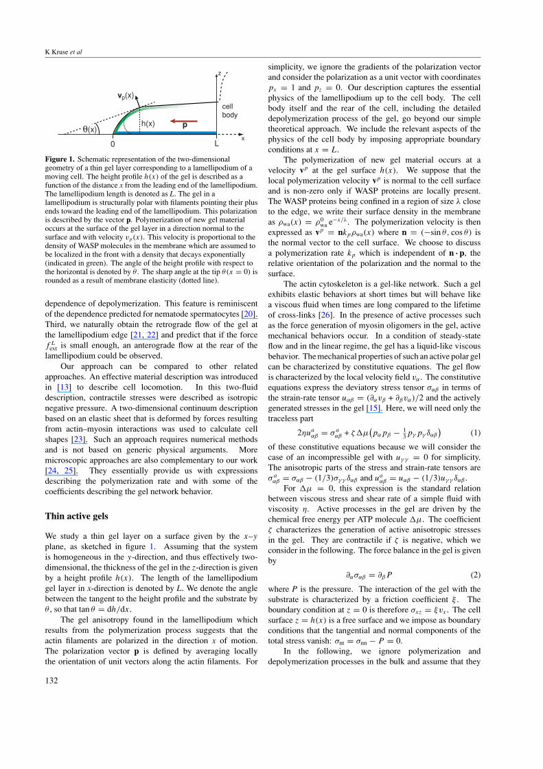
Figure 1. Schematic representation of the two-dimensionalgeometry of a thin gel layer corresponding to a lamellipodium of amoving cell. The height profile h(x) of the gel is described as afunction of the distance x from the leading end of the lamellipodium.The lamellipodium length is denoted as L. The gel in alamellipodium is structurally polar with filaments pointing their plusends toward the leading end of the lamellipodium. This polarizationis described by the vector p. Polymerization of new gel materialoccurs at the surface of the gel layer in a direction normal to thesurface and with velocity vp(x). This velocity is proportional to thedensity of WASP molecules in the membrane which are assumed tobe localized in the front with a density that decays exponentially(indicated in green). The angle of the height profile with respect tothe horizontal is denoted by # . The sharp angle at the tip #(x = 0) isrounded as a result of membrane elasticity (dotted line).
dependence of depolymerization. This feature is reminiscentof the dependence predicted for nematode spermatocytes [20].Third, we naturally obtain the retrograde flow of the gel atthe lamellipodium edge [21, 22] and predict that if the forcef L
ext is small enough, an anterograde flow at the rear of thelamellipodium could be observed.
Our approach can be compared to other relatedapproaches. An effective material description was introducedin [13] to describe cell locomotion. In this two-fluiddescription, contractile stresses were described as isotropicnegative pressure. A two-dimensional continuum descriptionbased on an elastic sheet that is deformed by forces resultingfrom actin–myosin interactions was used to calculate cellshapes [23]. Such an approach requires numerical methodsand is not based on generic physical arguments. Moremicroscopic approaches are also complementary to our work[24, 25]. They essentially provide us with expressionsdescribing the polymerization rate and with some of thecoefficients describing the gel network behavior.
Thin active gels
We study a thin gel layer on a surface given by the x–y
plane, as sketched in figure 1. Assuming that the systemis homogeneous in the y-direction, and thus effectively two-dimensional, the thickness of the gel in the z-direction is givenby a height profile h(x). The length of the lamellipodiumgel layer in x-direction is denoted by L. We denote the anglebetween the tangent to the height profile and the substrate by# , so that tan # = dh/dx.
The gel anisotropy found in the lamellipodium whichresults from the polymerization process suggests that theactin filaments are polarized in the direction x of motion.The polarization vector p is defined by averaging locallythe orientation of unit vectors along the actin filaments. For
simplicity, we ignore the gradients of the polarization vectorand consider the polarization as a unit vector with coordinatespx = 1 and pz = 0. Our description captures the essentialphysics of the lamellipodium up to the cell body. The cellbody itself and the rear of the cell, including the detaileddepolymerization process of the gel, go beyond our simpletheoretical approach. We include the relevant aspects of thephysics of the cell body by imposing appropriate boundaryconditions at x = L.
The polymerization of new gel material occurs at avelocity vp at the gel surface h(x). We suppose that thelocal polymerization velocity vp is normal to the cell surfaceand is non-zero only if WASP proteins are locally present.The WASP proteins being confined in a region of size ! closeto the edge, we write their surface density in the membraneas $wa(x) = $0
wa e!x/!. The polymerization velocity is thenexpressed as vp = nkp$wa(x) where n = (!sin #, cos #) isthe normal vector to the cell surface. We choose to discussa polymerization rate kp which is independent of n · p, therelative orientation of the polarization and the normal to thesurface.
The actin cytoskeleton is a gel-like network. Such a gelexhibits elastic behaviors at short times but will behave likea viscous fluid when times are long compared to the lifetimeof cross-links [26]. In the presence of active processes suchas the force generation of myosin oligomers in the gel, activemechanical behaviors occur. In a condition of steady-stateflow and in the linear regime, the gel has a liquid-like viscousbehavior. The mechanical properties of such an active polar gelcan be characterized by constitutive equations. The gel flowis characterized by the local velocity field v% . The constitutiveequations express the deviatory stress tensor &%' in terms ofthe strain-rate tensor u%' = ((%v' + ('v%)/2 and the activelygenerated stresses in the gel [15]. Here, we will need only thetraceless part
2)ua%' = & a
%' + *+µ!p%p' ! 1
3p,p, -%'"
(1)
of these constitutive equations because we will consider thecase of an incompressible gel with u, , = 0 for simplicity.The anisotropic parts of the stress and strain-rate tensors are& a%' = &%' ! (1/3)&,, -%' and ua
%' = u%' ! (1/3)u, , -%' .For +µ = 0, this expression is the standard relation
between viscous stress and shear rate of a simple fluid withviscosity ). Active processes in the gel are driven by thechemical free energy per ATP molecule +µ. The coefficient* characterizes the generation of active anisotropic stressesin the gel. They are contractile if * is negative, which weconsider in the following. The force balance in the gel is givenby
(%&%' = ('P (2)
where P is the pressure. The interaction of the gel with thesubstrate is characterized by a friction coefficient . . Theboundary condition at z = 0 is therefore &xz = .vx . The cellsurface z = h(x) is a free surface and we impose as boundaryconditions that the tangential and normal components of thetotal stress vanish: &nt = &nn ! P = 0.
In the following, we ignore polymerization anddepolymerization processes in the bulk and assume that they
132
Contractility and retrograde flow in lamellipodium motion
occur only at the surfaces of the gel. We assume furthermorefor simplicity that the gel is incompressible. With theseassumptions, the flow field satisfies " · v = 0, and the geldensity $ is constant. The pressure P is then a Lagrangemultiplier that is introduced to satisfy the incompressibilityconstraint.
In the limit of a thin gel layer, h # L, and slowly varyingthickness, dh/dx # 1, a thin-film approximation can beused to derive an effective one-dimensional description: weaverage the velocity, the stress and the pressure in the z-direction over the thickness of the gel v = $vx%, & = $&xx% andp = $P %. Note that here and in the following, the velocity v isalways measured with respect to the substrate. The total forceper unit length acting along the x-axis in a gel cross-section inthe y–z plane is F =
# h
0 dz(&xx ! P) = h(& ! p). The forcebalance on a slice of gel of size dx is obtained by balancingthis force with the friction force on the substrate:
dF
dx= .v. (3)
The boundary condition on the normal stress at the uppersurface of the gel imposes &zz = P . The force F can thereforebe written as F & h$&xx ! &zz%. Using the incompressibilitycondition, (xvx + (zvz = 0, the constitutive equations lead to
dv
dx= 1
4)
$F
h+ *+µ
%. (4)
We look for steady-state profiles of the lamellipodium whichmoves with constant velocity u in the negative x-direction,h = h(x + ut). In a reference frame moving at a velocity !u
with respect to the substrate, the height profile is stationary.The gel flux in the x-direction in this reference frame isj = $h(u+v). Mass conservation implies that the variation ofthis flux is compensated by the polymerization flux. To linearorder in dh/dx the steady-state height profile obeys
ddx
[(u + v)h] = kp$wa. (5)
The lamellipodium height can be expressed as a function ofthe velocity v by integrating this expression:
h(x) = kp
u + v(x)
& x
0dx '$wa(x
'). (6)
Equations (3) and (4) represent two first-order equations forthe force F and the gel flow v, the height profile being givenby equation (6). They can be solved by imposing boundaryconditions on the force at x = 0 (lamellipodium front) andx = L (connection of the lamellipodium to the cell body). Inorder to take into account the combined effects of adhesion andexternal forces, we impose at the front F(0) = !
!W + f 0
ext
"
and at the rear F(L) = W ' + f Lext. External forces fext
act on the lamellipodium at its front and rear. The forceat the rear includes that exerted by the cell body on thelamellipodium. We have ignored the effect of the membranetension and bending rigidity at the free surface of the cell,but we include the effects of the adhesion energies per unitarea of the lamellipodium (W) and the cell body (W ') on thesubstrate.
With these conditions, for a given length L there exists onesteady-state solution for any positive value of the velocity u. A
-1
-0.5
0
0.5
1
1.5
2
2.5
3
3.5
4
0 2 4 6 8 10 12 14 16position x [µm]
h Fv
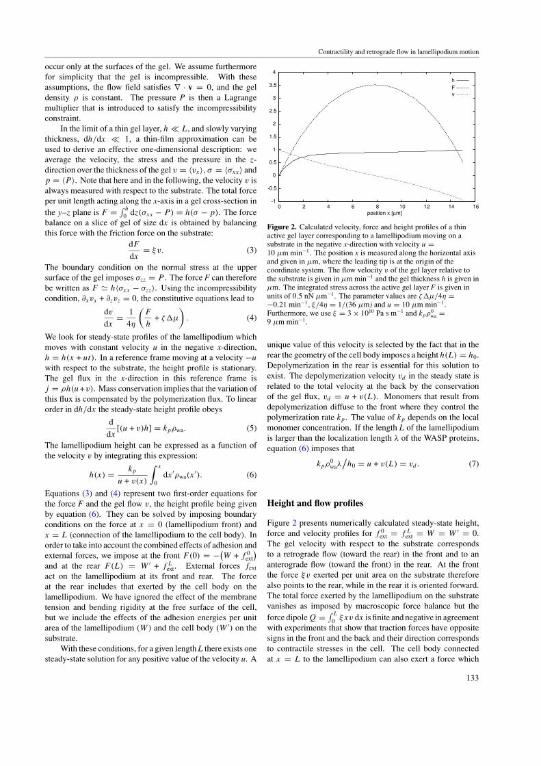
Figure 2. Calculated velocity, force and height profiles of a thinactive gel layer corresponding to a lamellipodium moving on asubstrate in the negative x-direction with velocity u =10 µm min!1. The position x is measured along the horizontal axisand given in µm, where the leading tip is at the origin of thecoordinate system. The flow velocity v of the gel layer relative tothe substrate is given in µm min!1 and the gel thickness h is given inµm. The integrated stress across the active gel layer F is given inunits of 0.5 nN µm!1. The parameter values are *+µ/4) =!0.21 min!1, ./4) = 1/(36 µm) and u = 10 µm min!1.Furthermore, we use . = 3 ( 1010 Pa s m!1 and kp$
0wa =
9 µm min!1.
unique value of this velocity is selected by the fact that in therear the geometry of the cell body imposes a height h(L) = h0.Depolymerization in the rear is essential for this solution toexist. The depolymerization velocity vd in the steady state isrelated to the total velocity at the back by the conservationof the gel flux, vd = u + v(L). Monomers that result fromdepolymerization diffuse to the front where they control thepolymerization rate kp. The value of kp depends on the localmonomer concentration. If the length L of the lamellipodiumis larger than the localization length ! of the WASP proteins,equation (6) imposes that
kp$0wa!
'h0 = u + v(L) = vd. (7)
Height and flow profiles
Figure 2 presents numerically calculated steady-state height,force and velocity profiles for f 0
ext = f Lext = W = W ' = 0.
The gel velocity with respect to the substrate correspondsto a retrograde flow (toward the rear) in the front and to ananterograde flow (toward the front) in the rear. At the frontthe force .v exerted per unit area on the substrate thereforealso points to the rear, while in the rear it is oriented forward.The total force exerted by the lamellipodium on the substratevanishes as imposed by macroscopic force balance but theforce dipole Q =
# L
0 .xv dx is finite and negative in agreementwith experiments that show that traction forces have oppositesigns in the front and the back and their direction correspondsto contractile stresses in the cell. The cell body connectedat x = L to the lamellipodium can also exert a force which
133
K Kruse et al
modifies the velocity u. In this case, the total force exerted bythe lamellipodium does not vanish but instead the total forceexerted by the cell, including the cell body, vanishes. Again,the force dipole of the whole cell is finite. If external forcesare applied in the front and the rear, both the height profile andthe advancing velocity of the lamellipodium are modified.
If the length L of the lamellipodium is large, a simplesolution of the steady-state profile can be found in the limitwhere the active stress *+µ is small. For simplicity, weconsider only the case where there is no applied force at thefront. The central region of size L ! 2d of the lamellipodiumhas approximately constant height h & h and vanishing gelvelocity v. In the front of this central region, the heightdecreases to zero over a length d and the flow is retrograde. Inthe rear of the central region, the height increases to the valueh0 imposed by the cell body and the flow is anterograde. Thesize d of these regions is given by d2 = 4)h/. . This analysisis valid if L ) d ) !.
At the front, there is a small region of size ! wherethe thickness h drops rapidly to zero while F and v areapproximately constant. Outside this edge region of size !,equation (6) for the height profile can be written as h(x) =kp!$
0wa
'[u + v(x)]. This determines the lamellipodium
thickness:
h = kp$0wa!/u = h0vd/u (8)
which is estimated as the height at a position within thelamellipodium for which v = 0 and thus dh/dx = 0 in theabsence of polymerization and where we have used equation(7). In the front region of size d, the velocity decreasesexponentially as v = !d*+µ/4) e!x/d . Note that retrogradeflow corresponds indeed to contractile active stress *+µ < 0.In the rear part of the lamellipodium, the velocity increasesexponentially as v(x) = v(L) exp(x ! L)/d . The velocityat the rear is obtained by imposing that the force F(L) beequal to the external force exerted by the cell body f L
ext:v(L) = d/4)
(f L
ext
'h + *+µ
). For a small external force
the velocity is negative, corresponding to an anterograde flow.The conservation of the gel flux at the rear of the
lamellipodium imposes u = vd ! v(L). This allows us toobtain an explicit expression of the velocity of motion of thelamellipodium
u & vd !$
h0
4).
%1/2 $*+µ +
f Lext
h0
%. (9)
If we ignore the force exerted by the cell body on thelamellipodium, the friction force exerted by the lamellipodiumon the substrate is indeed a force dipole. In the limit wherethe length L is large, the force dipole is Q & Lh*+µ < 0.
The lamellipodium length L in the steady state isdetermined by the transport of actin monomers to the front,where they are incorporated in the gel. Assuming that thetransport of actin in the gel can be described by an effectivediffusion coefficient D, and denoting the (two-dimensional)monomer concentration by $m, the monomer flux toward thefront is for L ) h given by jm & D($0 ! $m(0))/L where$0 = $m(L) is the actin monomer density at the cell body thatacts as a reservoir. We describe the gel polymerization by a
rate proportional to the monomer concentration: kp = %$m
where % is a kinetic coefficient. The steady-state conditionbecomes vdh0$ & %$m(0)$0
wa!$ & jmh where $ denotes themonomer concentration in the gel. This determines both thelength L and the actin monomer density $m(0) at the front ofthe lamellipodium:
$m(0) & vdh0
%!$0wa
(10)
L & D
u$($0 ! $m(0)). (11)
Here, we have assumed that the height h0, thedepolymerization velocity vd and the monomer concentration$0 are imposed by the cell body. Note that a stationarylamellipodium shape exists only if vd < %$0$
0wa!/h0.
Conclusion and outlook
In summary, we have proposed a theoretical description whichcaptures the main physical features of lamellipodium motionon a solid substrate. This approach allows us to introduce a newquantitative way to analyze experiments. A full comparison ofthis description with experiments would require measurementsof the height, flow and force profiles of a moving cell. Existingdata already provide an estimation of all the parameter valuesof our theory.
Taking the example of a crawling keratocyte, we estimateL & 10 µm and h & 1 µm. The advancing velocity isu & 10 µm min!1, and a retrograde flow exists at the frontwith v(0) & 1 µm min!1 [27, 28]. The elastic modulus ofan actin gel in a cell is of the order E & 104 Pa [11, 29, 30];the typical visco-elastic relaxation time " is of the order of10 s [12]. From existing measurements of both surface stress&xz & 4 ( 102 N m!2 [8] and the retrograde flow velocityv(0), we can deduce the friction coefficient of a lamellipodium. & &xz/v(0) & 1010 Pa s m!1. This value is very close tothe friction coefficient of an actin gel on a latex bead . & 3 (1010 Pa s m!1 [31]. We find a length d & 6 µm. These valuesused in figure 1 are consistent with the experimental results onkeratocytes[28]; d is of the order L and the cell body exerts afinite force on the lamellipodium.
The anisotropic active stress is obtained from theretrograde flow velocity as !*+µ & 4)v(0)/d & 103 Pa.Note that in this case the force transmitted by thelamellipodium does not vanish since it balances the force f L
extexerted by the cell body on the lamellipodium, where f L
ext &.v(0)d & !*+µh & 1 nN µm!1. For a lamellipodium witheffective lateral size Ly & 30 µm, the total force is therefore30 nN; this order of magnitude is consistent with the force of10 nN obtained in [8] by the integration of the stress. The forcedipole of the cell is QLy & f L
extLcLy & 6 ( 10!13 J, whereLc & 20 µm is the total length of the cell. The lamellipodiumlength is determined by L & D$0/u$. Using D & 3 (10!12 m2 s!1, $0 & 200 µM and $ & 300 µM [32, 17], weobtain L & 12 µm.
We have used for simplicity a one-dimensional descriptionwithin a thin-film approximation. A more realistic
134
Contractility and retrograde flow in lamellipodium motion
three-dimensional description is possible within the sameframework; however, it requires a precise analysis in a morecomplex geometry. The thin-film approximation requiresthat the localization length of the WASP proteins ! is smallenough and that the decay length of the retrograde flow d islarge enough, !, h < d . The second condition is realized if4)/(. h) > 1 which is the case for a keratocyte.
All the above considerations depend neither on theparticular distribution of WASP proteins nor on the detailsof the polymerization kinetics, provided they are localized.The localization of the WASP proteins in a small regionof size ! has been observed but the physical mechanismswhich are responsible for this localization are still underinvestigation. It has been suggested that the protein mightfavor the large membrane curvatures at the leading edge. Asecond possibility is that WASP in the membrane is transportedactively toward the leading edge by motor proteins whichadvance with velocity vm along actin filaments adjacent tothe membrane. Since the actin gel is polarized with the plusends pointing forward, such a transport would localize WASPproteins at the leading edge. Assuming that WASP diffusesin the membrane with diffusion coefficient Dm as it detachesfrom the transporting motors, the localization length is givenby ! & Dm/vm. A third possibility is that WASP interactsstrongly with the plus ends of the filaments and is transportedin the gel by diffusion or by motor proteins. We cannot,however, rule out other mechanisms as long as they lead to asufficiently low WASP concentration along the lamellipodiumsuch that the effects of their presence is negligible as comparedto controlled gel growth at the leading edge.
The active stress of the cytoskeleton mostly controls theretrograde flow at the leading edge. The retrograde flow islocalized over a length d and its amplitude increases with|*+µ|. In the limit |*+µ| = 0, there is no retrograde flowand the advancing velocity in a steady state is equal to thedepolymerization velocity u = vd . At a finite value of |*+µ|,the advancing velocity increases as a function of the contractilestress (equation (9)). The cytoskeleton is visco-elastic andbehaves at short times as an elastic solid and at long times asa viscous fluid. As a consequence, at the front the gel behavesas a solid over a length "vd & 1 µm. The fully viscousdescription is valid beyond this length and thus our theory isconsistent, if this length is smaller than d. In the same vein, wehave not described in detail the interaction of the growing gelwith the plasma membrane. We postpone a detailed discussionto future work. Yet one can foresee that the sharp angle of thegel shape at the leading edge which our analysis predicts willbe rounded up on a length scale (//W)1/2 as suggested by thedotted line in figure 1. Here, W denotes the total energy perunit surface of the membrane and / is the bending rigidity ofthe membrane.
We have assumed that the active stress is constantthroughout the lamellipodium. Experimentally, the surfacedensity of myosins increases toward the rear of thelamellipodium. Part of this effect is captured in our modelby the fact that the lamellipodium is thicker at the rear. Amore elaborate description would require keeping track ofboth the local myosin density and the gel elasticity at short
times. The fact that the myosin density becomes small at theleading tip can be captured in our description by assuming thatthe contractile stress *+µ is position dependent and vanishesat the front.
The depolymerization velocity vd influences theadvancing velocity u and the length of the lamellipodiumL. The advancing velocity increases with vd (equation (9))and the length of lamellipodium decreases, as expected, withthe depolymerization velocity (equation (10)). For smallexternal forces, the thickness of the lamellipodium h isinversely proportional to the advancing velocity; it increaseswith increasing depolymerization velocity. Lamellipodiumformation requires the depolymerization velocity to be smallerthan a critical value that increases with the actin monomerdensity $0 and the polymerization rate %.
Both the amplitude of the retrograde flow and theadvancing velocity decrease with increasing friction . .The length L and the thickness h of the lamellipodiumincrease for increasing friction . . We have consideredhere that the adhesion force is proportional to thelocal velocity on the substrate. Finally, an externallyapplied force on the lamellipodium modifies both thepolymerization/depolymerization rates and the stressdistribution. The effects of a force on the advancing velocitytherefore depends on its position of application [20]. In thecase of a pulling force f L
ext > 0 applied at the rear the twoeffects are competing. Such a force is naturally exerted by thecell body but could also be modified by external manipulation.If we ignore the force dependence of the depolymerization rate,then equation (9) predicts a decrease of u with increasing force.However, the depolymerization velocity can increase underthe action of this force, which tends to increase the velocity u(and could lead to a negative mobility as suggested in [20]).These two contributions could be separated experimentallyby their time dependence. The effect on the stress profileis almost instantaneous, while the velocity change due to avariation of the depolymerization rate is effective only after atime L2/D corresponding to the diffusion of actin monomersto the leading edge.
At small forces, the thickness h and the length L ofthe lamellipodium increase with increasing force. The forceexerted by the cell body on the lamellipodium results fromthe adhesion of the cell body to the substrate. On the timescales considered here, this force can be described by a viscousfriction force f L
ext. If the friction is linear, f Lext = .cbLcbu
is proportional to the advancing velocity, where .cb is asurface friction coefficient and Lcb is the size of the cellbody. The velocity u can be calculated self-consistently fromequation (9):
u &*
vd + |*+µ|$
h0
4).
%1/2+ , -
1 +.cbLcb
(4).h0)1/2
.. (12)
Here, we have expressed u in terms of the depolymerizationvelocity vd because it does not depend on the actinmonomer concentration. At steady state, the polymerizationvelocity adjusts to balance this depolymerization velocity byselecting the appropriate length of the lamellipodium. Thegel contractility provides an additional contribution which
135

K Kruse et al
increases the advancing velocity. For the parameter valuesused for keratocytes, we estimate this contribution to about10% of the total velocity. This implies that a myosin inhibitionshould reduce the advancing velocity while suppressing theretrograde flow. Note that such a retrograde flow could stillbe driven by the membrane tension exerting a force f 0
ext at theleading edge.
If the friction force f Lext(u) varies in a non-monotonic
way as a function of velocity such that a range of negativeslope exists, the resulting motion may exhibit a stick-slip orsaltatory behavior. This is the case if the friction force exertedby the cell body on the substrate is elastically coupled to thelamellipodium [18]. Such a mechanism could explain theinchworm motion observed for fibroblasts.
Glossary
Lamellipodium. The lamellipodium is the thin sheet-likefront part of a cell that is moving on a solid substrate. Thelamellipodium contains a network of actin filamentsassembled near the moving front. It is enclosed by theplasma membrane and linked to the substrate by specificadhesion molecules.
Force dipole. A cell moving on a solid substrate to which itadheres generates a distribution of traction forces withdensity *f (*r) acting on the substrate at position *r measuredfrom the cell center. Such traction maps show that forcesexerted at opposite sides of the cell largely cancel, such thatthe net total force
#d2r *f & 0. In such a situation the dipolar
structure of the force distribution can be characterized by thematrix Q%' =
#d2r r%f' , where % and ' = x, y. If the
y-dependence is unimportant, Q = Qxx describes the dipoleof forces projected on the x-axis. Q is negative if balancingforces are contractile.
Retrograde/anterograde flow. When a cell moves on asubstrate, the actin gel in the cell flows at a speed that isdifferent from the cell velocity of motion. If this actin flowmoves with respect to the substrate in a direction opposite tothe direction of motion of the cell, this flow is calledretrograde; movement in the same direction is anterogradeflow.
Visco-elastic gel. A gel is a network of polymers orfilaments which are cross-linked. A cross-linked gel withpermanent links is a solid elastic body. If the links betweenpolymers have a finite lifetime, the gel is still elastic ifdeformed only briefly, but exhibits liquid-like behavior ifstresses are applied for a sufficiently long time. Thiscombined behavior is called visco-elastic.
Active processes. Processes such as the force generation ofmotor proteins in the cytoskeleton are driven by the chemicalenergy of ATP hydrolysis. If such active processes take placein a system, thermodynamic equilibrium is not reached. Insuch a nonequilibrium situation, force and motion generationat the molecular scale modifies material properties of acytoskeletal gel as compared to the passive case.
References
[1] Alberts B et al 2002 Molecular Biology of the Cell 4th edn(New York: Garland)
Bray D 2001 Cell Movements 2nd edn (New York: Garland)Howard J 2001 Mechanics of Motor Proteins and the
Cytoskeleton (Sunderland: Sinauer Associates, Inc.)[2] Mitchison T J and Cramer L P 1996 Actin-based cell motility
and cell locomotion Cell 84 371–9[3] Verkhovsky A B, Svitkina T M and Borisy G G 1999
Self-polarization and directional motility of cytoplasmCurr. Biol. 9 11–21
[4] Pollard T D and Borisy G G 2003 Cellular motility driven byassembly and disassembly of actin filamentsCell 112 453–65
[5] Carlier M F and Pantaloni D 1997 Control of actin dynamicsin cell motility J. Mol. Biol. 269 459–67
[6] Rafelski S M and Theriot J A 2004 Crawling toward a unifiedmodel of cell mobility: spatial and temporal regulation ofactin dynamics Annu. Rev. Biochem. 73 209–39
[7] Dembo M, Oliver T, Ishigara A and Jacobson K 1996 Imagingthe traction stresses exerted by locomoting cells with theelastic substratum method Biophys. J. 70 2008–22
[8] Oliver T, Dembo M and Jacobson K 1999 Separation ofpropulsive and adhesive traction stresses in locomotingkeratocytes J. Cell Biol. 145 589–604
[9] Balaban N Q et al 2001 Force and focal adhesion assembly: aclose relationship studied using elastic micropatternedsubstrates Nat. Cell Biol. 3 466–72
[10] Tawada K and Sekimoto K 1991 Protein friction exerted bymotor enzymes through a weak-binding interactionJ. Theor. Biol. 150 193–200
[11] Gerbal F, Chaikin P, Rabin Y and Prost J 2000 An elasticanalysis of Listeria monocytogenes propulsion Biophys. J.79 2259–75
[12] Wottawah F et al 2005 Optical rheology of biological cellsPhys. Rev. Lett. 94 098103-1-4
[13] Alt W and Dembo M 1999 Cytoplasm dynamics and cellmotion: two-phase flow models Math. Biosci. 156 207–28
[14] Dembo M and Harris 1981 Motion of particles adhering to theleading lamella of crawling cells J. Cell Biol. 91 528–36
[15] Kruse K et al 2004 Asters, vortices, and rotating spirals inactive gels of polar filaments Phys. Rev. Lett. 92 078101
Kruse K et al 2005 Generic theory of active polar gels: aparadigm for cytoskeletal dynamics Eur. Phys. J. E 16 5–16
[16] Noireaux V et al 2000 Growing an actin gel on sphericalsurfaces Biophys. J. 78 1643–54
[17] Plastino J, Lelidis J, Prost J and Sykes C 2004 The effect ofdiffusion, depolymerization and nucleation promotingfactors on actin gel growth Eur. Biophys. J. 33 310–20
[18] Prost J 2001 Physics of Biomolecules and Cellsed H Flyvbjerg, F Julicher, P Ormos and F David (Les Ulis:EDP Sciences) p 215
[19] Schmidt F G, Ziemann F and Sackmann E 1996 Shear fieldmapping in actin networks by using magnetic tweezersEur. Biophys. J. 24 348–53
[20] Joanny J F, Julicher F and Prost J 2003 Motion of an adhesivegel in a swelling gradient: a mechanism for cell locomotionPhys. Rev. Lett. 90 168102-1-4
[21] Coussen F, Choquet D, Sheetz M P and Erickson H P 2002Trimers of the fibronectin cell adhesion domain localize toactin filament bundles and undergo rearward translocationJ. Cell Sci. 115 2581–90
[22] Jurado C, Haserick J R and Lee J 2005 Slipping or gripping?Fluorescent speckle microscopy in fish keratocytes revealstwo different mechanisms for generating a retrograde flowof actin Mol. Biol. Cell 16 507–18
[23] Rubinstein B, Jacobson K and Mogilner A 2005 Multiscaletwo-dimensional modeling of a motilde simple-shaped cellSIAM J. MMS 3 413–39
136
Contractility and retrograde flow in lamellipodium motion
Grimm H P, Verkhovsky A B, Mogilner A and Meister J-J2003 Analysis of actin dynamics at the leading edge ofcrawling cells: implications for the shape of keratocytelamellipodia Eur. Biophys. J. 32 563–77
[24] Mogilner A and Oster G 2003 Force generation by actinpolymerization: II. The elastic ratchet and tetheredfilaments Biophys. J. 84 1591–605
[25] Carlsson A 2001 Growth of branched actin networks againstobstacles Biophys. J. 81 1907–23
[26] Doi M and Edwards S F 1988 The Theory of PolymerDynamics (Oxford: Oxford University Press)
[27] Kucik D F, Kuo S C, Elson E L and Sheetz M P 1991Preferential attachment of membrane glycoproteins to thecytoskeleton at the leading edge of lamella J. CellBiol. 114 1029–36
[28] Vallotton P et al 2005 Tracking retrograde flow in keratocytes:news from the front Mol. Biol. Cell 16 1223–31
[29] MacKintosh F C, Kas J and Janmey P A 1995 Elasticity ofsemiflexible biopolymer networks Phys. Rev.Lett. 75 4425–8
[30] Gardel M L et al 2004 Elastic behavior of cross-linked andbundled actin networks Science 304 1301–5
[31] Marcy Y, Prost J, Carlier M F and Sykes C 2004 Forcesgenerated during actin-based propulsion: a directmeasurement by micromanipulation Proc. Natl Acad. Sci.USA 101 5992–7
[32] McGrath J L, Tardy Y, Dewey C F, Meister J J andHartwig J H 1998 Simultaneous measurements of actinfilament turnover, filament fraction, and monomer diffusionin endothelial cells Biophys. J. 75 2070–8
137
Related Documents