MOLECULAR AND CELLULAR BIOLOGY, July 2006, p. 4782–4793 Vol. 26, No. 13 0270-7306/06/$08.000 doi:10.1128/MCB.00069-06 Copyright © 2006, American Society for Microbiology. All Rights Reserved. p53 Downregulates Its Activating Vaccinia-Related Kinase 1, Forming a New Autoregulatory Loop Alberto Valbuena,† Francisco M. Vega,† Sandra Blanco, and Pedro A. Lazo* Instituto de Biologı ´a Molecular y Celular del Ca ´ncer, Consejo Superior de Investigaciones Cientı ´ficas, Universidad de Salamanca, Campus Miguel de Unamuno, E-37007 Salamanca, Spain Received 12 January 2006/Returned for modification 10 February 2006/Accepted 12 April 2006 The stable accumulation of p53 is detrimental to the cell because it blocks cell growth and division. Therefore, increases in p53 levels are tightly regulated, mainly by its transcriptional target, mdm2, that downregulates p53. Elucidation of new signaling pathways requires the characterization of the members and the nature of their connection. Vaccinia-related kinase 1 (VRK1) contributes to p53 stabilization by partly interfering with its mdm2-mediated degradation, among other mechanisms; therefore, it is likely that some form of autoregulation between VRK1 and p53 must occur. We report here the identification of an autoreg- ulatory loop between p53 and its stabilizing VRK1. There is an inverse correlation between VRK1 and p53 levels in cell lines, and induction of p53 by UV light downregulates VRK1 in fibroblasts. As the amount of p53 protein increases, there is a downregulation of the VRK1 protein level independent of its promoter. This effect is indirect but requires a transcriptionally active p53. The three most common transcriptionally inactive mutations detected in hereditary (Li-Fraumeni syndrome) and sporadic human cancer, p53(R175H), p53(R248W), and p53(R273H), as well as p53(R280K), are unable to induce downregulation of VRK1 protein. The p53 isoforms 40p53 and p53, lacking the transactivation and oligomerization domains, respectively, do not downregulate VRK1. VRK1 downregulation induced by p53 is independent of mdm2 activity and protea- some-mediated degradation since it occurs in the presence of proteasome inhibitors and in mdm2-deficient cells. The degradation of VRK1 is sensitive to chloroquine, an inhibitor of the late endosome-lysosome transport, and to serine protease inhibitors of the lysosomal pathway. The accumulation of the tumor suppressor protein p53 oc- curs in response to many different types of cellular stress or DNA damage. The levels of p53 in response to stress present a transient accumulation that may last a few hours (46). This transient p53 accumulation induces a variety of responses de- signed to protect the cell and allow for damage repair by stopping the cell cycle (1, 28, 38, 43, 57, 61) or inducing apop- tosis (41, 52, 62). If the level of p53 is maintained at a high level when unnecessary, there would be a permanent block to cell cycle progression or the cells will die. In any case, the stable and prolonged accumulation of p53 is incompatible with life. Therefore, the p53 molecule has evolved in a way that it is always present at low levels, and when it is stabilized, the accumulation occurs in a transient manner. The normal p53 levels are tightly regulated, and mechanisms aiming at its downregulation are highly developed. This is achieved in part by having a short half-life of about 20 min and, in case of an increase in p53 levels, by the induction of negative regulatory proteins for p53. The best-characterized mechanism is p53 downregulation by one of its transcriptional targets, hdm2/ mdm2 (34), so that the induction of mdm2 leads to p53 ubiq- uitination and degradation by the proteasome (32, 53). The p53 protein is held at low levels under normal growth condi- tions, and upon stress its level increases rapidly (41) with the subsequent transcriptional activation, including that of hdm2. Hdm2 is a ubiquitin ligase that promotes downregulation of p53, bringing its levels down to basal ones. Therefore, it can be considered that there are waves of p53 accumulation whose magnitude may vary depending on the type of stimulation or the cell type (24). The sequential expression of p53 and mdm2 might also contribute to oscillations in their levels (27). The same occurs with other p53 negative regulators that are also transcriptional targets, as is the case for other ubiquitin ligases such as COP1 (11). The downregulation of positive modulators by p53, such as chk1, has been also reported (15). The p53 mutant (R273H), present in sporadic cancers and in Li-Frau- meni syndrome patients, accumulates in human tumors and has its properties altered with oncogenic potential (25, 39). The p53 mutants detected in many tumors are likely to have altered these autoregulatory mechanisms, but these alterations have not been characterized. Recently, it has been shown that variant p53 proteins lacking either the transactivation (11) or the oligomerization domains (11) but retaining their ability to activate gene transcription can be generated by alternative splicing (8); these variant p53 proteins may have modifications in their regulatory mechanisms. The human vaccinia-related kinase 1 (VRK1) (26), a mem- ber of a new Ser-Thr kinase family in the human kinome (30) has autophosphorylation activity (29, 36) and also phosphoryl- ates different transcription factors such as p53 (4, 29, 58, 23), c-Jun (50), ATF2 (51), and nuclear factor BAF (37). VRK1 is able to induce p53 stabilization by a complex mechanism, a component of which is the phosphorylation of p53 in Thr18 (4, 29), partly preventing its interaction with mdm2 and promoting p300 recruitment that leads to p53 accumulation and transcrip- tional activation (58). p53 stabilization by VRK1 has been * Corresponding author. Mailing address: IBMCC-Centro de Inves- tigacio ´n del Ca ´ncer, CSIC-Universidad de Salamanca, Campus Miguel de Unamuno, E-37007 Salamanca, Spain. Phone: 34 923 294 804. Fax: 34 923 294 795. E-mail: [email protected]. † A.V. and F.M.V. contributed equally to the work. 4782

Welcome message from author
This document is posted to help you gain knowledge. Please leave a comment to let me know what you think about it! Share it to your friends and learn new things together.
Transcript
MOLECULAR AND CELLULAR BIOLOGY, July 2006, p. 4782–4793 Vol. 26, No. 130270-7306/06/$08.00�0 doi:10.1128/MCB.00069-06Copyright © 2006, American Society for Microbiology. All Rights Reserved.
p53 Downregulates Its Activating Vaccinia-Related Kinase 1, Forminga New Autoregulatory Loop
Alberto Valbuena,† Francisco M. Vega,† Sandra Blanco, and Pedro A. Lazo*Instituto de Biologıa Molecular y Celular del Cancer, Consejo Superior de Investigaciones Cientıficas,
Universidad de Salamanca, Campus Miguel de Unamuno, E-37007 Salamanca, Spain
Received 12 January 2006/Returned for modification 10 February 2006/Accepted 12 April 2006
The stable accumulation of p53 is detrimental to the cell because it blocks cell growth and division.Therefore, increases in p53 levels are tightly regulated, mainly by its transcriptional target, mdm2, thatdownregulates p53. Elucidation of new signaling pathways requires the characterization of the members andthe nature of their connection. Vaccinia-related kinase 1 (VRK1) contributes to p53 stabilization by partlyinterfering with its mdm2-mediated degradation, among other mechanisms; therefore, it is likely that someform of autoregulation between VRK1 and p53 must occur. We report here the identification of an autoreg-ulatory loop between p53 and its stabilizing VRK1. There is an inverse correlation between VRK1 and p53levels in cell lines, and induction of p53 by UV light downregulates VRK1 in fibroblasts. As the amount of p53protein increases, there is a downregulation of the VRK1 protein level independent of its promoter. This effectis indirect but requires a transcriptionally active p53. The three most common transcriptionally inactivemutations detected in hereditary (Li-Fraumeni syndrome) and sporadic human cancer, p53(R175H),p53(R248W), and p53(R273H), as well as p53(R280K), are unable to induce downregulation of VRK1 protein.The p53 isoforms �40p53 and p53�, lacking the transactivation and oligomerization domains, respectively, donot downregulate VRK1. VRK1 downregulation induced by p53 is independent of mdm2 activity and protea-some-mediated degradation since it occurs in the presence of proteasome inhibitors and in mdm2-deficientcells. The degradation of VRK1 is sensitive to chloroquine, an inhibitor of the late endosome-lysosometransport, and to serine protease inhibitors of the lysosomal pathway.
The accumulation of the tumor suppressor protein p53 oc-curs in response to many different types of cellular stress orDNA damage. The levels of p53 in response to stress presenta transient accumulation that may last a few hours (46). Thistransient p53 accumulation induces a variety of responses de-signed to protect the cell and allow for damage repair bystopping the cell cycle (1, 28, 38, 43, 57, 61) or inducing apop-tosis (41, 52, 62). If the level of p53 is maintained at a high levelwhen unnecessary, there would be a permanent block to cellcycle progression or the cells will die. In any case, the stableand prolonged accumulation of p53 is incompatible with life.Therefore, the p53 molecule has evolved in a way that it isalways present at low levels, and when it is stabilized, theaccumulation occurs in a transient manner. The normal p53levels are tightly regulated, and mechanisms aiming at itsdownregulation are highly developed. This is achieved in partby having a short half-life of about 20 min and, in case of anincrease in p53 levels, by the induction of negative regulatoryproteins for p53. The best-characterized mechanism is p53downregulation by one of its transcriptional targets, hdm2/mdm2 (34), so that the induction of mdm2 leads to p53 ubiq-uitination and degradation by the proteasome (32, 53). Thep53 protein is held at low levels under normal growth condi-tions, and upon stress its level increases rapidly (41) with thesubsequent transcriptional activation, including that of hdm2.
Hdm2 is a ubiquitin ligase that promotes downregulation ofp53, bringing its levels down to basal ones. Therefore, it can beconsidered that there are waves of p53 accumulation whosemagnitude may vary depending on the type of stimulation orthe cell type (24). The sequential expression of p53 and mdm2might also contribute to oscillations in their levels (27). Thesame occurs with other p53 negative regulators that are alsotranscriptional targets, as is the case for other ubiquitin ligasessuch as COP1 (11). The downregulation of positive modulatorsby p53, such as chk1, has been also reported (15). The p53mutant (R273H), present in sporadic cancers and in Li-Frau-meni syndrome patients, accumulates in human tumors andhas its properties altered with oncogenic potential (25, 39).The p53 mutants detected in many tumors are likely to havealtered these autoregulatory mechanisms, but these alterationshave not been characterized. Recently, it has been shown thatvariant p53 proteins lacking either the transactivation (11) orthe oligomerization domains (11) but retaining their ability toactivate gene transcription can be generated by alternativesplicing (8); these variant p53 proteins may have modificationsin their regulatory mechanisms.
The human vaccinia-related kinase 1 (VRK1) (26), a mem-ber of a new Ser-Thr kinase family in the human kinome (30)has autophosphorylation activity (29, 36) and also phosphoryl-ates different transcription factors such as p53 (4, 29, 58, 23),c-Jun (50), ATF2 (51), and nuclear factor BAF (37). VRK1 isable to induce p53 stabilization by a complex mechanism, acomponent of which is the phosphorylation of p53 in Thr18 (4,29), partly preventing its interaction with mdm2 and promotingp300 recruitment that leads to p53 accumulation and transcrip-tional activation (58). p53 stabilization by VRK1 has been
* Corresponding author. Mailing address: IBMCC-Centro de Inves-tigacion del Cancer, CSIC-Universidad de Salamanca, Campus Miguelde Unamuno, E-37007 Salamanca, Spain. Phone: 34 923 294 804. Fax:34 923 294 795. E-mail: [email protected].
† A.V. and F.M.V. contributed equally to the work.
4782
postulated to be a basic control process that operates in cellsunder normal growth conditions in the absence of or undersuboptimal stress and that permits p53 to remain in a readinessstate or operate in minor damage responses, such as duringreplication (58). VRK1 appears to be necessary for a basiccontrol mechanism in cell proliferation since its loss, inducedby small interfering RNA (siRNA) and in the absence of stressstimulation, leads to a retardation of cell division and celldeath by a mechanism not yet identified (58). In human headand neck squamous cell carcinomas, VRK1 correlates withestablished proliferation markers, which suggests that VRK1might be playing a role early in the G1 phase of the cell cycle(47).
We hypothesized that some cross-regulation between VRK1and p53 proteins must exist so that p53 stabilization can bereversed; this mechanism should involve the inactivation insome way of VRK1 so that its loss will permit the downregu-lation of p53 by mdm2 or any other mechanism. Elucidation ofsignaling networks implies the identification of interactingmolecules and the characterization of their interaction in orderto identify their contribution to different types of biologicaleffects (42). In this report we have identified that the accumu-lation of p53 is able to induce the downregulation of its stabi-lizing protein, VRK1, and this process requires the contribu-tion of different p53 domains, does not involve VRK1transcriptional regulation, and is mediated by the lysosomalpathway of protein degradation.
MATERIALS AND METHODS
Plasmids, antibodies, and reagents. The VRK1 constructs, pCEFL-HA-VRK1, pCEFL-HA-VRK1(K179E), and pCDNA3.1-VRK1-myc coding eitherfor the wild-type VRK1 or the VRK1(K179E) inactive mutant have been previ-ously described (58). The plasmid pCB6�p53, containing human p53 wild-typecDNA, plasmid pCB6�p53T18D (2), and plasmid pCOC-Mdm2-X2 coding forthe Mdm2 protein were from K. Vousden (The Beatson Institute, Glasgow,United Kingdom); the transcriptionally defective mutant p53(R280K) was fromA. J. Levine (Rockefeller University, New York) (12); the mutant plasmidspCMV-p53(R175H), pCMV-p53(R248W), and pCMV-p53(R273H) were fromBert Vogelstein (Johns Hopkins University, Baltimore, MD); plasmid pCMV-p53(L22Q, W23S) was from K. Roemer (7); plasmids expressing the p53 iso-forms p53� and �40p53 (p47) were from J.C. Bourdon (Dundee University,Scotland) (8); and pCMW-p53(L322A) and pCDNA3-p53C�60 were from S.Camus (Institute of Molecular and Cellular Biology, Singapore). The plasmidBRR12-ubiquitin-His (pUbiquitin-His) was from S. Lain and D. Lane (DundeeUniversity, Scotland). The p53 siRNA expression plasmid pSUPER.retro.p53(Oligoengine, Seattle, WA) was used where indicated to suppress the expressionof p53. All plasmids used for transfection were endotoxin free and purified witha JetStar Maxi kit from Genomed (Bad Oeynhausen, Germany).
The anti-�-actin antibody was from Sigma (St. Louis, MO). The hemagglutinin(HA) tag was detected with a mouse monoclonal antibody HA-probe (F7) fromCovance (Berkeley, Calif.). The p53 protein was detected with a mixture of DO1antibody (Santa Cruz, CA) and Pab1801 (Santa Cruz, CA) used at 1:500 and1:1,000, respectively. The p53 isoform, �40p53 (p47), lacking the transactivationdomain was detected with the CM1 polyclonal antibody at a dilution of 1:20,000(from A. Craig, Dundee University, Scotland). VRK1 was detected using a rabbitpolyclonal antibody (VE1) or a mouse monoclonal antibody (1F6 clone) madeagainst a VRK1 fusion protein. Poly(ADP-ribose) polymerase was determinedwith a monoclonal antibody from Enzyme Systems Products (Livermore, CA).As secondary antibodies, a goat anti-mouse-horseradish peroxidase or a goatanti-rabbit-horseradish peroxidase (Amersham Pharmacia Biotech) was used at1:5,000 in Western blotting.
The following protease inhibitors were used: pepstatin for aspartyl proteases;phenylmethylsulfonyl fluoride (PMSF), aprotinin, diisopropylfluorophosphate(DFP), soybean trypsin inhibitor (STI), and leupeptin for serine proteases;iodoacetic acid (IAA) and leupeptin for cysteine proteases; EDTA and 1,10-phenantroline for metaloproteasas; and ALLN, calpain inhibitor III, calpeptin,
EST, and PD150606 for calpain proteases (all from Sigma). Chloroquine wasfrom Sigma.
Cell lines and transfections. The human lung cancer cell line H1299 (p53�/�)was grown in RPMI medium supplemented with 10% fetal calf serum, glutamine,penicillin, and streptomycin in a humidified atmosphere and 5% CO2. HeLacells, U2OS (p16�/�), and the WS1 normal human fibroblast cell line (ATCCCRL-1502) were grown in Dulbecco’s modified Eagle’s medium with the samesupplements. For transfection experiments, H1299 cells were plated in 60- or100-mm dishes and transfected with the plasmid indicated in the specific exper-iments either by the calcium phosphate precipitation method or with JetPIreagent following the manufacturer’s instructions (Polytransfection; Illkirch,France). Unless otherwise indicated, the cells were lysed 36 h posttransfection inlysis buffer (50 mM Tris-HCl, pH 8, 150 mM NaCl, 5 mM EDTA, and 1%TritonX-100, plus protease and phosphatase inhibitors), and 40 �g of whole-cell extractwas processed for sodium dodecyl sulfate (SDS)-polyacrylamide gel electro-phoresis and subjected to immunoblotting with the indicated antibodies. Theimmortalized fibroblasts from double knockout p53/Mdm2 mice (p53�/�,mdm2�/�) were a gift of G. Lozano (MD Anderson Cancer Center, Houston,TX) and were grown in Dulbecco’s modified Eagle’s medium with 10% fetal calfserum and antibiotics. Where indicated, the proteasome inhibitor MG132 (Cal-biochem) was used at 50 �M for 6 h. Tetracycline was from Sigma (St. Louis,MO) and was used at a concentration of 2 �g/ml. Stimulation of the cells withUV light was done in a Stratalinker from Stratagene (San Diego, CA).
Immunoblotting. Total protein extracts were quantified using a Bio-Rad pro-tein assay kit. Protein was fractionated in an SDS-polyacrylamide gel and trans-ferred to a polyvinylidene difluoride Immobilon-P membrane (Millipore). Themembrane was blocked with TBS-T buffer (25 mM Tris, 50 mM NaCl, 2.5 mMKCl, 0.1% Tween-20) and 5% nonfat milk. Afterwards the membrane was rinsedwith TBS-T buffer; and the specific primary antibody (indicated in individualexperiments) was added, and the membrane was incubated for 90 min at roomtemperature. The membrane was rinsed and incubated with a secondary anti-body conjugated with peroxidase for 30 min. The membrane was develop forchemiluminescence with ECL reagent (Amersham) and exposed to X-ray films.Quantification was always performed in the linear response range.
Quantitative RT-PCR. Reverse transcription-PCR (RT-PCR) was performedas previously described (58). Briefly, H1299 cells were cotransfected as describedabove with the plasmids indicated in the experiment, and total RNA was ex-tracted using an RNAeasy extraction kit from QIAGEN (Hilden, Germany).One hundred nanograms of total RNA was used in a one-step reverse transcrip-tion real-time PCR amplification reaction using a Quantitec SYBR Green RT-PCR kit from QIAGEN in an iCycler (Bio-Rad, Hercules, CA). The reaction wasanalyzed with iCycler software (Bio-Rad). The primers used for VRK1 amplifi-cation detection were 5�-CCAACGAGCTGCAAAACCA-3� and 5�-TGTCATGTAGACCAGACCCCC-3�; for GAPDH amplification detection, the primerswere 5�-GGTCTTACTCCTTGGAGGCCATGT-3� and 5�-ACCTAACTACATGGTTTACATGTT-3�.
RESULTS
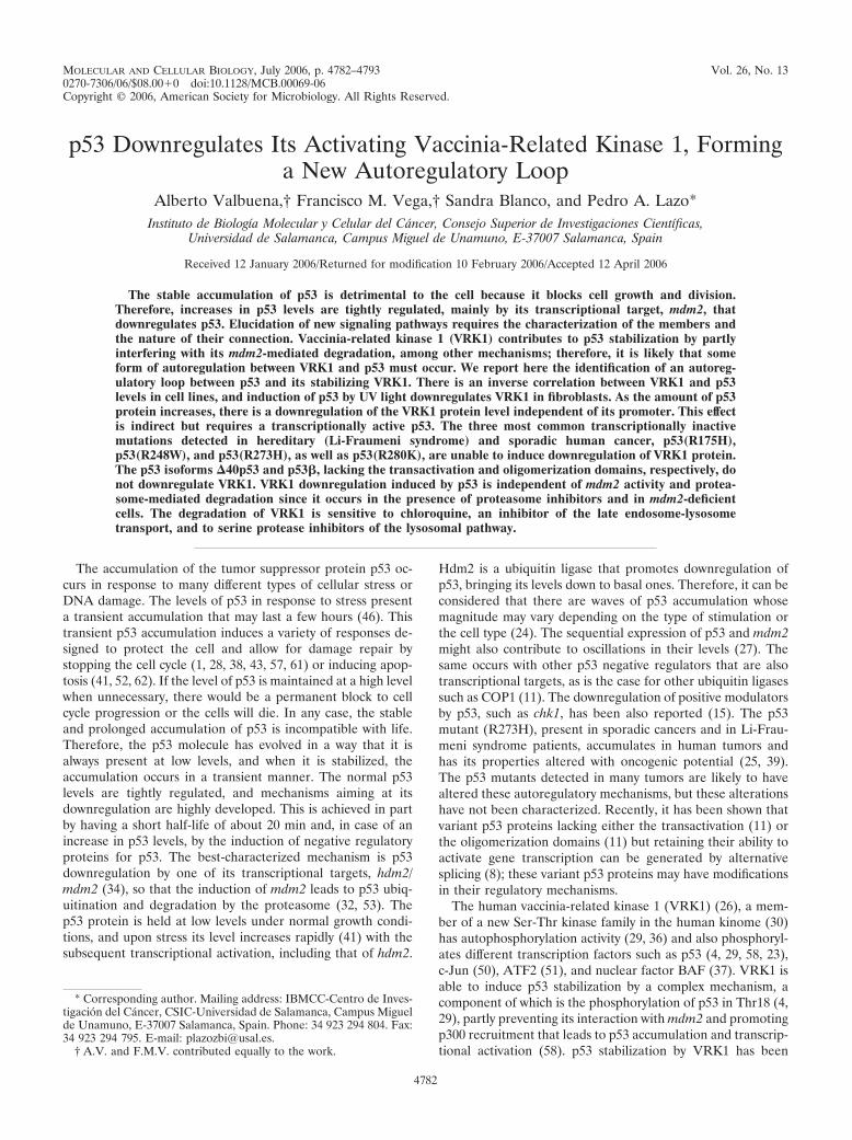
The level of VRK1 inversely correlates with p53 levels in celllines. During the course of the work, it was noticed that whena cell had p53 wild-type protein, the level of transfected VRK1detected was always lower than if the cell line lacked p53. Onceit was clear that this effect was not due to transfection effi-ciency, the correlation with the cell genotype was intriguingand suggested that the explanation might be related to theconnection of the two proteins in the pathway (58), with aninverse relation between them. To determine if that might bethe case, the level of the two endogenous proteins, VRK1 andp53, was determined in several cell lines (Fig. 1A). The celllines with wild-type p53 had lower levels of endogenous VRK1.But in the two cell lines lacking p53, such as H1299 or HCT116(p53 knockout), higher levels of endogenous VRK1 wereclearly detected (Fig. 1A). One of the effects of an activeVRK1 is to stabilize p53 by increasing its intracellular levels(58), and a stable accumulation of p53 is known to be detri-mental to the cell since it prevents cell division and might eventrigger an apoptotic response (3, 9, 43). Therefore, we consid-
VOL. 26, 2006 AUTOREGULATORY LOOP VRK1-p53 4783
ered the possibility that higher levels of p53 might promote adownregulation of its activator-stabilizer VRK1 protein, thusforming an autoregulatory loop between p53 and VRK1, whichmight be conceptually similar to the autoregulation betweenChk1 and p53 (15) or to the p53-mdm2 autoregulatory loop(34). That interpretation might account for the different levelsof VRK1 observed, depending on the p53 genotype of celllines.
p53 accumulation induced by UV downregulates VRK1. Todetermine if the level of p53 might indeed have an inversecorrelation with the level of VRK1, we studied the variation inlevels of both proteins in the cell response to stimulation withUV irradiation. Human diploid fibroblasts, the WS1 cell line(p53�/�), were treated with two doses of UV light, at 20 J/m2
or 40 J/m2, and cell extracts were analyzed by Western blotting8 h after irradiation. In both cases the endogenous p53 was
stabilized, and there was a reduction of VRK1 levels (Fig. 1B).Next, a time course of the response of WS1 cells irradiatedwith 20 J/m2 was performed for a period of 24 h. As expected,after UV treatment there was the expected fast stabilization ofp53 as a consequence of phosphorylation (46), but the accu-mulation of p53 continued to increase, reaching significantlymuch higher levels after 10 h (Fig. 1C), a response typical offibroblasts (16). After irradiation there was also a transientincrease in the level of VRK1 that reached a peak at 4 h, afterwhich it fell even below the initial level. The timing of thisreduction in VRK1 levels coincided with the time necessary forp53-dependent de novo gene expression and with the increasedlevel of p53, suggesting that perhaps the accumulated p53might be implicated in inducing the VRK1 downregulation.Next, we tried to determine if this down-regulatory effect couldbe reproduced by transfection of p53. To this end, the WS1
FIG. 1. Expression of endogenous p53 and VRK1 in tumor cell lines and in UV treated fibroblasts. (A) Expression of VRK1 and p53 in celllines that were p53 wild type (A549, U2OS, and WS1) or p53�/� (H1299 or HCT116-p53 knockout). VRK1 and p53 were determined by Westernblotting, using actin as internal control. (B) Levels of p53 and VRK1 in human fibroblasts treated with the indicated dose of UV light; the extractswere prepared 8 h after treatment and analyzed by Western blotting. (C) The levels of endogenous p53 and VRK1 in WS1 fibroblasts were detectedat different times after treatment with 20 J/m2 UV light. The proteins were identified by Western blotting with the corresponding antibodiesindicated in the methods section. (D) Normal human fibroblasts, cell line WS1, were transfected with increasing amounts of pCB6�p53 asindicated and 5 �g of pCEFL-HA-VRK1. Cell extracts were prepared 36 h after transfection. The proteins were detected with the correspondingantibodies by Western blotting, and to the right is shown the quantification of the normalized values.
4784 VALBUENA ET AL. MOL. CELL. BIOL.
fibroblast cell line was transfected with increasing amounts ofp53 (plasmid pCB6�p53) and a fixed amount of VRK1 (plas-mid pCEFL-HA-VRK1). As the amount of p53 increased, itwas accompanied by a reduction in the level of VRK1 proteinin these cells (Fig. 1D).
p53 downregulates VRK1 protein level. To reproduce thisdown-regulatory effect in an independent system, a new exper-iment was designed using a variant of the H1299 (p53�/�,p53Tet inducible) cell line that contains a p53 gene that is induc-ible by addition of tetracycline to the culture. This cell line hasa basal level of p53 expression and thus mimics a normal cellsituation. This cell line was transfected with a fixed amount ofpCEFL-HA-VRK1, p53 expression was induced by the addi-tion of tetracycline, and the level of VRK1 was determined atdifferent time points. After induction with tetracycline, as thelevel of p53 increased with time, it was accompanied by areduction in the level of the VRK1 protein detected with anantibody against the HA epitope tag (Fig. 2A).
To confirm that VRK1 protein downregulation was depen-dent on the accumulation of p53 protein, a time course exper-iment for VRK1 expression alone or together with p53 over-expression was performed (Fig. 2B). For this experiment,H1299 (p53�/�) cells were transfected with plasmids pCEFL-HA-VRK1 and pCB6�p53, and at different time points cellextracts were prepared and analyzed by immunoblotting. Inthe absence of p53, VRK1 protein levels increased constantly,as expected, given the high degree of VRK1 stability. However,in the presence of p53, at first VRK1 increased as a result of denovo protein synthesis; but when there was also a detectableaccumulation of p53, VRK1 accumulation slowed down andfinally came to a stop, which was particularly noticeable be-tween 24 and 36 h after transfection (Fig. 2B). These datasuggested that the effect depends on the concentration of p53in the cell and was probably due to the modification of genetranscription as a consequence.
To further determine whether such an effect was occurringin a p53 dose-dependent manner and was promoter indepen-dent, an experiment was designed to reproduce the previousinducible effect by transfection using the H1299 cell line (p53�/�).This cell line was transfected with increasing amounts ofpCB6�p53 and a fixed amount of pCEFL-HA-VRK1 (Fig.2C), which express VRK1 from a Moloney murine leukemiavirus promoter; with pCDNA3.1-VRK1-myc, which expressesVRK1 from a cytomegalovirus promoter; or with pEF1-VRK1,which uses the EF1 gene promoter. In this experiment it wasobserved that the higher the level of p53, the lower the amountof VRK1 protein detected in cell extracts (Fig. 2C). The effectof p53 on VRK1 was similar independent of the type of pro-moter from which VRK1 was expressed (not shown). Theeffect was also confirmed in other tumor cell lines, such asHT144, WS1, A549, and in U2OS (p16�/�), suggesting that theeffect is independent of cell type and that p16 is not involved inthis process (not shown). These results indicate that the effectis more general and does not depend on the genetic peculiar-ities of the particular tumor cell line or promoter used.
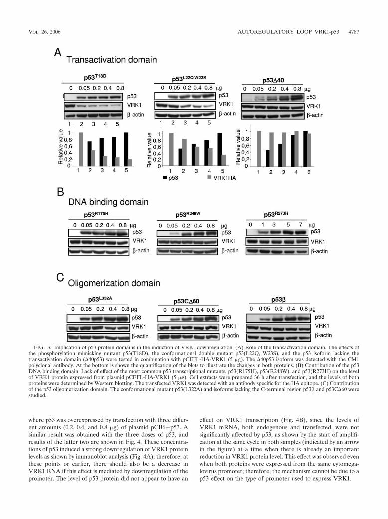
Contribution of p53 domains to VRK1 protein downregula-tion. To determine the requirements of the p53 protein re-quired to induce the downregulation of VRK1, the contribu-tions of three main p53 functional regions, the transactivation,DNA binding, and oligomerization domains, were analyzed.
The potential contribution of the N-terminal transactivationdomain of p53 was studied using three different types of mu-tants: a phosphorylation mutant that mimics phosphorylationin threonine-18, p53(T18D), the p53(L22Q, W23S) conforma-tional mutant than retains its transcriptional activation role buthas different effects on double-strand DNA repair and apop-tosis (7, 31), and the isoform �40p53, known as p47, that lacksthe first 40 amino acids of the transactivation domain (8). Thisisoform functions as a dominant negative of p53 growth sup-pression properties (10, 14). The phosphorylation and the con-formational mutants were able to induce the downregulationof VRK1 in a dose-dependent manner; however, the �40p53,lacking the first forty amino acids, has lost this role (Fig. 3A).These results indicate that the first 40 residues of the transac-tivation domain are required for this effect and reflect a func-tional difference in p53 isoforms. Different N-terminal phos-phorylation mutants were also assayed with same results aswith the p53(T18D) (not shown). p53 interaction with manyregulators is mediated by the N-terminal transactivation do-main, and it is possible that such interaction is necessary forp53-mediated VRK1 downregulation.
To ascertain if the DNA binding domain of p53 was a re-quirement for induction of VRK1 downregulation, several mu-tants in this domain were studied. The three most commonnaturally occurring p53 mutants in human cancer, both spo-radic and hereditary, were studied. The conformational mutantp53(R175H) and the DNA contact mutants p53(R248W)and p53(R273H) (55) account for approximately one-third ofall p53 mutations in human cancer (6, 56). A p53 mutant[p53(R280K)] engineered to prevent binding to DNA andwithout effects on other p53 properties was also studied (12).All of these p53 mutants were unable to induce a downregu-lation of VRK1 protein levels (Fig. 3B). This lack of effectindicated that the action of p53 on VRK1 levels requires theintegrity of the DNA binding domain and is therefore probablymediated by a DNA-bound form of p53. This result opens thepossibility of the effect being mediated by a p53-dependentinduction or repression of a gene not yet identified.
Finally, the potential contribution of the p53 oligomeriza-tion domain was analyzed. The C terminus is necessary forthe anti-growth arrest and anti-apoptotic effects of p53 (54).Three different p53 variants in this domain were used: thep53(L332A) conformational mutant that has a defective oli-gomerization, pCDNA3-p53C�60 (lacking the last 60 resi-dues), and the p53� isoform lacking the oligomerization do-main (8). The loss of this domain resulted in the loss of theeffect on VRK1, while the conformational mutant p53(L332A)was slightly less efficient in inducing it (Fig. 3C). These datasuggested that oligomerization of p53 is necessary for the in-duction of VRK1 protein downregulation and also indicated afunctional difference between p53 isoforms. This further sup-ports the idea of a transcriptionally active p53 role in VRK1downregulation.
p53 does not affect VRK1 transcription. There is the possi-bility that p53 might affect the expression of the VRK1 geneand contribute in that way to VRK1 regulation. However, theprevious observations in which the effect was detected by ex-pressing VRK1 from different promoters do not support thispossibility. To further rule out this possibility, mRNA levelswere determined by quantitative RT-PCR under conditions
VOL. 26, 2006 AUTOREGULATORY LOOP VRK1-p53 4785
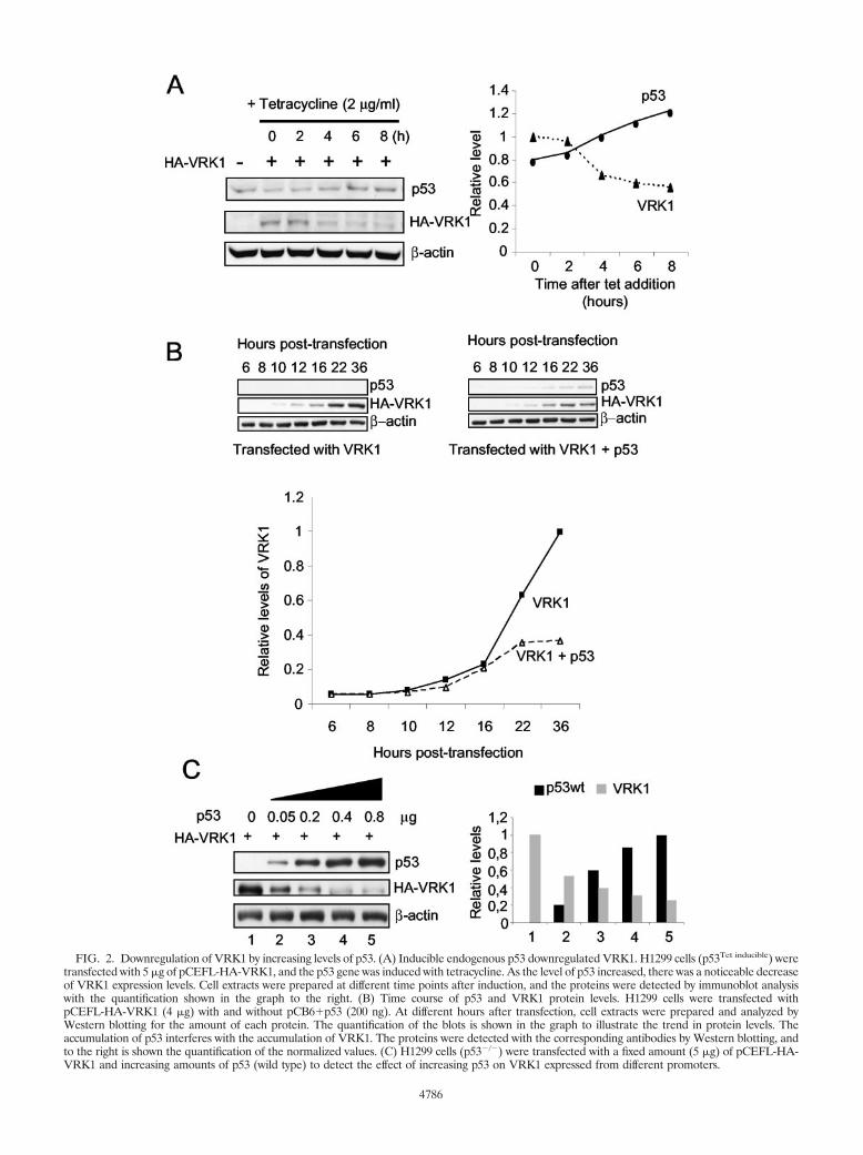
FIG. 2. Downregulation of VRK1 by increasing levels of p53. (A) Inducible endogenous p53 downregulated VRK1. H1299 cells (p53Tet inducible) weretransfected with 5 �g of pCEFL-HA-VRK1, and the p53 gene was induced with tetracycline. As the level of p53 increased, there was a noticeable decreaseof VRK1 expression levels. Cell extracts were prepared at different time points after induction, and the proteins were detected by immunoblot analysiswith the quantification shown in the graph to the right. (B) Time course of p53 and VRK1 protein levels. H1299 cells were transfected withpCEFL-HA-VRK1 (4 �g) with and without pCB6�p53 (200 ng). At different hours after transfection, cell extracts were prepared and analyzed byWestern blotting for the amount of each protein. The quantification of the blots is shown in the graph to illustrate the trend in protein levels. Theaccumulation of p53 interferes with the accumulation of VRK1. The proteins were detected with the corresponding antibodies by Western blotting, andto the right is shown the quantification of the normalized values. (C) H1299 cells (p53�/�) were transfected with a fixed amount (5 �g) of pCEFL-HA-VRK1 and increasing amounts of p53 (wild type) to detect the effect of increasing p53 on VRK1 expressed from different promoters.
4786
where p53 was overexpressed by transfection with three differ-ent amounts (0.2, 0.4, and 0.8 �g) of plasmid pCB6�p53. Asimilar result was obtained with the three doses of p53, andresults of the latter two are shown in Fig. 4. These concentra-tions of p53 induced a strong downregulation of VRK1 proteinlevels as shown by immunoblot analysis (Fig. 4A); therefore, atthese points or earlier, there should also be a decrease inVRK1 RNA if this effect is mediated by downregulation of thepromoter. The level of p53 protein did not appear to have an
effect on VRK1 transcription (Fig. 4B), since the levels ofVRK1 mRNA, both endogenous and transfected, were notsignificantly affected by p53, as shown by the start of amplifi-cation at the same cycle in both samples (indicated by an arrowin the figure) at a time when there is already an importantreduction in VRK1 protein level. This effect was observed evenwhen both proteins were expressed from the same cytomega-lovirus promoter; therefore, the mechanism cannot be due to ap53 effect on the type of promoter used to express VRK1.
FIG. 3. Implication of p53 protein domains in the induction of VRK1 downregulation. (A) Role of the transactivation domain. The effects ofthe phosphorylation mimicking mutant p53(T18D), the conformational double mutant p53(L22Q, W23S), and the p53 isoform lacking thetransactivation domain (�40p53) were tested in combination with pCEFL-HA-VRK1 (5 �g). The �40p53 isoform was detected with the CM1polyclonal antibody. At the bottom is shown the quantification of the blots to illustrate the changes in both proteins. (B) Contribution of the p53DNA binding domain. Lack of effect of the most common p53 transcriptional mutants, p53(R175H), p53(R248W), and p53(R273H) on the levelof VRK1 protein expressed from plasmid pCEFL-HA-VRK1 (5 �g). Cell extracts were prepared 36 h after transfection, and the levels of bothproteins were determined by Western blotting. The transfected VRK1 was detected with an antibody specific for the HA epitope. (C) Contributionof the p53 oligomerization domain. The conformational mutant p53(L322A) and isoforms lacking the C-terminal region p53� and p53C�60 werestudied.
VOL. 26, 2006 AUTOREGULATORY LOOP VRK1-p53 4787
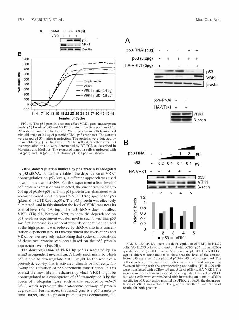
VRK1 downregulation induced by p53 protein is abrogatedby p53 siRNA. To further establish the dependence of VRK1downregulation on p53 levels, a different approach was usedbased on the use of siRNA. For this experiment a fixed level ofp53 protein expression was selected, the one corresponding to200 ng of pCB6�p53, and this p53 protein was eliminated withvector-delivered short hairpin RNA (shRNA) specific for p53(plasmid pSUPER.retro.p53). The p53 protein was effectivelyeliminated, and in this situation the level of VRK1 was near itscontrol level (Fig. 5A, top). The p53 shRNA does not affectVRK1 (Fig. 5A, bottom). Next, to show the dependence onp53 levels an experiment was designed in such a way that p53was first increased in a concentration-dependent manner, andat the high point, it was reduced by shRNA also in a concen-tration-dependent way. In this experiment the levels of p53 andVRK1 behave inversely, establishing that cycles of fluctuationsof these two proteins can occur based on the p53 proteinexpression levels (Fig. 5B).
The downregulation of VRK1 by p53 is mediated by anmdm2-independent mechanism. A likely mechanism by whichp53 is able to downregulate VRK1 might be the result of aproteolytic activity that is activated, directly or indirectly, fol-lowing the activation of p53-dependent transcription. In thiscontext the most likely mechanism by which VRK1 might bedownregulated as a consequence of p53 transcription is by theaction of a ubiquitin ligase, such as that encoded by mdm2/hdm2, which represents the proteasome pathway of proteindegradation. Furthermore, the mdm2 gene is a p53 transcrip-tional target, and this protein promotes p53 degradation, fol-
FIG. 4. The p53 protein does not affect VRK1 gene transcriptionlevels. (A) Levels of p53 and VRK1 protein at the time point used forRNA determination. The levels of VRK1 protein in cells transfectedwith either 0.4 or 0.8 �g of plasmid pCB6�p53 are shown. The extractswere prepared 36 h after transfection. The proteins were detected byimmunoblotting. (B) The levels of VRK1 mRNA, whether after p53overexpression or not, were determined by RT-PCR as described inMaterials and Methods. The results obtained in cells transfected with0.4 (p53) and 0.8 (p53) �g of plasmid pCB6�p53 are shown.
FIG. 5. p53 siRNA blocks the downregulation of VRK1 in H1299cells. (A) H1299 cells were transfected with pCB6�p53 and an siRNAspecific for p53 (pSUPER.retro.p53) as well as pCEFL-HA-VRK1 (5�g) in different combinations to show that the level of the cotrans-fected p53 expressed from plasmid pCB6�p53 is downregulated. Thecell extracts were prepared 36 h after transfection and analyzed byWestern blotting with the corresponding antibodies. (B) H1299 cellswere transfected with pCB6�p53 and 5 �g of pCEFL-HA-VRK1. Theincrease in p53 protein, as expected, downregulated the level of VRK1,but when cells were cotransfected with increasing amounts of siRNAspecific for p53, expression plasmid pSUPER.retro.p53, the downregu-lation of VRK1 was reduced. The graph shows the quantification ofresults for both proteins.
4788 VALBUENA ET AL. MOL. CELL. BIOL.
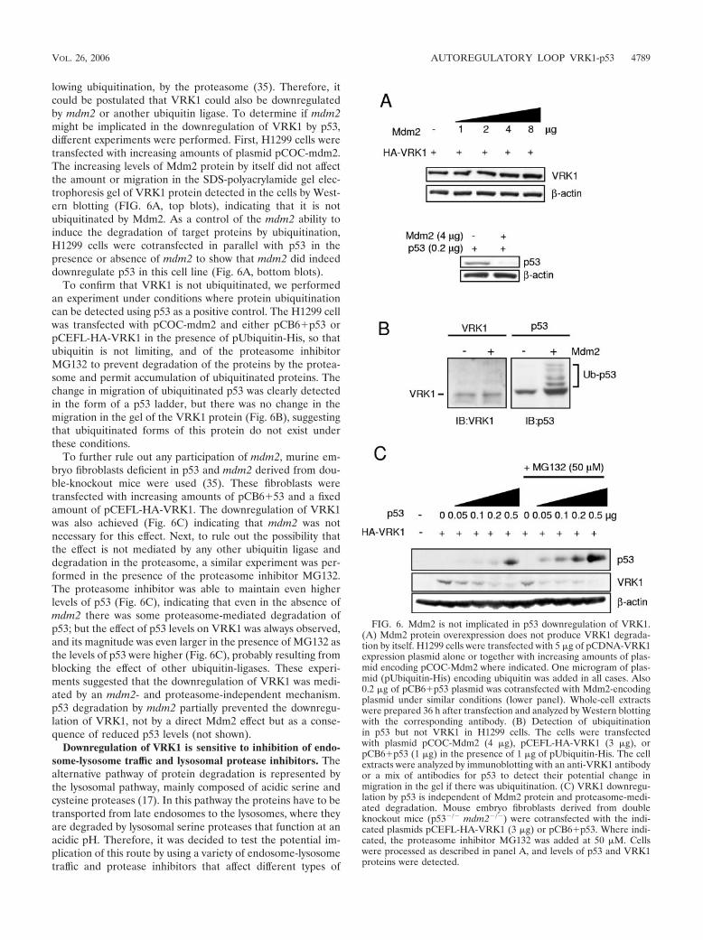
lowing ubiquitination, by the proteasome (35). Therefore, itcould be postulated that VRK1 could also be downregulatedby mdm2 or another ubiquitin ligase. To determine if mdm2might be implicated in the downregulation of VRK1 by p53,different experiments were performed. First, H1299 cells weretransfected with increasing amounts of plasmid pCOC-mdm2.The increasing levels of Mdm2 protein by itself did not affectthe amount or migration in the SDS-polyacrylamide gel elec-trophoresis gel of VRK1 protein detected in the cells by West-ern blotting (FIG. 6A, top blots), indicating that it is notubiquitinated by Mdm2. As a control of the mdm2 ability toinduce the degradation of target proteins by ubiquitination,H1299 cells were cotransfected in parallel with p53 in thepresence or absence of mdm2 to show that mdm2 did indeeddownregulate p53 in this cell line (Fig. 6A, bottom blots).
To confirm that VRK1 is not ubiquitinated, we performedan experiment under conditions where protein ubiquitinationcan be detected using p53 as a positive control. The H1299 cellwas transfected with pCOC-mdm2 and either pCB6�p53 orpCEFL-HA-VRK1 in the presence of pUbiquitin-His, so thatubiquitin is not limiting, and of the proteasome inhibitorMG132 to prevent degradation of the proteins by the protea-some and permit accumulation of ubiquitinated proteins. Thechange in migration of ubiquitinated p53 was clearly detectedin the form of a p53 ladder, but there was no change in themigration in the gel of the VRK1 protein (Fig. 6B), suggestingthat ubiquitinated forms of this protein do not exist underthese conditions.
To further rule out any participation of mdm2, murine em-bryo fibroblasts deficient in p53 and mdm2 derived from dou-ble-knockout mice were used (35). These fibroblasts weretransfected with increasing amounts of pCB6�53 and a fixedamount of pCEFL-HA-VRK1. The downregulation of VRK1was also achieved (Fig. 6C) indicating that mdm2 was notnecessary for this effect. Next, to rule out the possibility thatthe effect is not mediated by any other ubiquitin ligase anddegradation in the proteasome, a similar experiment was per-formed in the presence of the proteasome inhibitor MG132.The proteasome inhibitor was able to maintain even higherlevels of p53 (Fig. 6C), indicating that even in the absence ofmdm2 there was some proteasome-mediated degradation ofp53; but the effect of p53 levels on VRK1 was always observed,and its magnitude was even larger in the presence of MG132 asthe levels of p53 were higher (Fig. 6C), probably resulting fromblocking the effect of other ubiquitin-ligases. These experi-ments suggested that the downregulation of VRK1 was medi-ated by an mdm2- and proteasome-independent mechanism.p53 degradation by mdm2 partially prevented the downregu-lation of VRK1, not by a direct Mdm2 effect but as a conse-quence of reduced p53 levels (not shown).
Downregulation of VRK1 is sensitive to inhibition of endo-some-lysosome traffic and lysosomal protease inhibitors. Thealternative pathway of protein degradation is represented bythe lysosomal pathway, mainly composed of acidic serine andcysteine proteases (17). In this pathway the proteins have to betransported from late endosomes to the lysosomes, where theyare degraded by lysosomal serine proteases that function at anacidic pH. Therefore, it was decided to test the potential im-plication of this route by using a variety of endosome-lysosometraffic and protease inhibitors that affect different types of
FIG. 6. Mdm2 is not implicated in p53 downregulation of VRK1.(A) Mdm2 protein overexpression does not produce VRK1 degrada-tion by itself. H1299 cells were transfected with 5 �g of pCDNA-VRK1expression plasmid alone or together with increasing amounts of plas-mid encoding pCOC-Mdm2 where indicated. One microgram of plas-mid (pUbiquitin-His) encoding ubiquitin was added in all cases. Also0.2 �g of pCB6�p53 plasmid was cotransfected with Mdm2-encodingplasmid under similar conditions (lower panel). Whole-cell extractswere prepared 36 h after transfection and analyzed by Western blottingwith the corresponding antibody. (B) Detection of ubiquitinationin p53 but not VRK1 in H1299 cells. The cells were transfectedwith plasmid pCOC-Mdm2 (4 �g), pCEFL-HA-VRK1 (3 �g), orpCB6�p53 (1 �g) in the presence of 1 �g of pUbiquitin-His. The cellextracts were analyzed by immunoblotting with an anti-VRK1 antibodyor a mix of antibodies for p53 to detect their potential change inmigration in the gel if there was ubiquitination. (C) VRK1 downregu-lation by p53 is independent of Mdm2 protein and proteasome-medi-ated degradation. Mouse embryo fibroblasts derived from doubleknockout mice (p53�/� mdm2�/�) were cotransfected with the indi-cated plasmids pCEFL-HA-VRK1 (3 �g) or pCB6�p53. Where indi-cated, the proteasome inhibitor MG132 was added at 50 �M. Cellswere processed as described in panel A, and levels of p53 and VRK1proteins were detected.
VOL. 26, 2006 AUTOREGULATORY LOOP VRK1-p53 4789
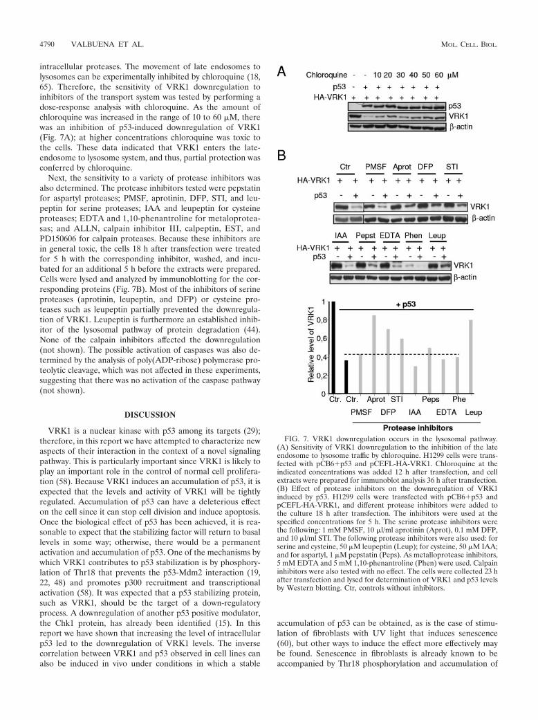
intracellular proteases. The movement of late endosomes tolysosomes can be experimentally inhibited by chloroquine (18,65). Therefore, the sensitivity of VRK1 downregulation toinhibitors of the transport system was tested by performing adose-response analysis with chloroquine. As the amount ofchloroquine was increased in the range of 10 to 60 �M, therewas an inhibition of p53-induced downregulation of VRK1(Fig. 7A); at higher concentrations chloroquine was toxic tothe cells. These data indicated that VRK1 enters the late-endosome to lysosome system, and thus, partial protection wasconferred by chloroquine.
Next, the sensitivity to a variety of protease inhibitors wasalso determined. The protease inhibitors tested were pepstatinfor aspartyl proteases; PMSF, aprotinin, DFP, STI, and leu-peptin for serine proteases; IAA and leupeptin for cysteineproteases; EDTA and 1,10-phenantroline for metaloprotea-sas; and ALLN, calpain inhibitor III, calpeptin, EST, andPD150606 for calpain proteases. Because these inhibitors arein general toxic, the cells 18 h after transfection were treatedfor 5 h with the corresponding inhibitor, washed, and incu-bated for an additional 5 h before the extracts were prepared.Cells were lysed and analyzed by immunoblotting for the cor-responding proteins (Fig. 7B). Most of the inhibitors of serineproteases (aprotinin, leupeptin, and DFP) or cysteine pro-teases such as leupeptin partially prevented the downregula-tion of VRK1. Leupeptin is furthermore an established inhib-itor of the lysosomal pathway of protein degradation (44).None of the calpain inhibitors affected the downregulation(not shown). The possible activation of caspases was also de-termined by the analysis of poly(ADP-ribose) polymerase pro-teolytic cleavage, which was not affected in these experiments,suggesting that there was no activation of the caspase pathway(not shown).
DISCUSSION
VRK1 is a nuclear kinase with p53 among its targets (29);therefore, in this report we have attempted to characterize newaspects of their interaction in the context of a novel signalingpathway. This is particularly important since VRK1 is likely toplay an important role in the control of normal cell prolifera-tion (58). Because VRK1 induces an accumulation of p53, it isexpected that the levels and activity of VRK1 will be tightlyregulated. Accumulation of p53 can have a deleterious effecton the cell since it can stop cell division and induce apoptosis.Once the biological effect of p53 has been achieved, it is rea-sonable to expect that the stabilizing factor will return to basallevels in some way; otherwise, there would be a permanentactivation and accumulation of p53. One of the mechanisms bywhich VRK1 contributes to p53 stabilization is by phosphory-lation of Thr18 that prevents the p53-Mdm2 interaction (19,22, 48) and promotes p300 recruitment and transcriptionalactivation (58). It was expected that a p53 stabilizing protein,such as VRK1, should be the target of a down-regulatoryprocess. A downregulation of another p53 positive modulator,the Chk1 protein, has already been identified (15). In thisreport we have shown that increasing the level of intracellularp53 led to the downregulation of VRK1 levels. The inversecorrelation between VRK1 and p53 observed in cell lines canalso be induced in vivo under conditions in which a stable
accumulation of p53 can be obtained, as is the case of stimu-lation of fibroblasts with UV light that induces senescence(60), but other ways to induce the effect more effectively maybe found. Senescence in fibroblasts is already known to beaccompanied by Thr18 phosphorylation and accumulation of
FIG. 7. VRK1 downregulation occurs in the lysosomal pathway.(A) Sensitivity of VRK1 downregulation to the inhibition of the lateendosome to lysosome traffic by chloroquine. H1299 cells were trans-fected with pCB6�p53 and pCEFL-HA-VRK1. Chloroquine at theindicated concentrations was added 12 h after transfection, and cellextracts were prepared for immunoblot analysis 36 h after transfection.(B) Effect of protease inhibitors on the downregulation of VRK1induced by p53. H1299 cells were transfected with pCB6�p53 andpCEFL-HA-VRK1, and different protease inhibitors were added tothe culture 18 h after transfection. The inhibitors were used at thespecified concentrations for 5 h. The serine protease inhibitors werethe following: 1 mM PMSF, 10 �l/ml aprotinin (Aprot), 0.1 mM DFP,and 10 �l/ml STI. The following protease inhibitors were also used: forserine and cysteine, 50 �M leupeptin (Leup); for cysteine, 50 �M IAA;and for aspartyl, 1 �M pepstatin (Peps). As metalloprotease inhibitors,5 mM EDTA and 5 mM 1,10-phenantroline (Phen) were used. Calpaininhibitors were also tested with no effect. The cells were collected 23 hafter transfection and lysed for determination of VRK1 and p53 levelsby Western blotting. Ctr, controls without inhibitors.
4790 VALBUENA ET AL. MOL. CELL. BIOL.
p53 (13). The inverse correlation can be reproduced by trans-fection in different cell types, making it more amenable for itscharacterization.
The contribution of p53 to VRK1 regulation is an interme-diate step in the process for which it is absolutely essential, thestructural maintenance of p53 molecules, since alterations ineach domain affect the downregulation of VRK1. This require-ment is important because it reflects a functional differencebetween normal p53 and p53 mutated in human cancer, eithersporadic or hereditary, or p53 isoforms. The response is af-fected by the transactivation domain of p53 since the effect islost in the case of �40p53, an isoform lacking the first 40 aminoacids, which have a dominant negative role and counteractgrowth suppression (10, 14). However, the conformational mu-tant p53(L22Q, W23S), which affects some response elementsbut not others (7, 31, 45, 59), still induces downregulation.Many p53 regulators interact with p53 through this N-terminaldomain. Therefore, it is likely that some p53 interaction not yetidentified could have a role in the VRK1 downregulation in-duced by p53. The contribution of the integrity of the DNA-binding domain is demonstrated by the loss of effect whencommon mutations, either conformational or with loss of DNAcontact, are present. This might have consequences in tumorsbearing these mutations. Some conformational mutations arenot active and contribute to tumor development by routescurrently under characterization since their pattern of tumorformation is different from that induced by structural mutantsaffecting contact with DNA (25, 39). It is important to notethat this lack of effect occurs with the most common p53mutations, detected in both sporadic tumors and hereditaryLi-Fraumeni syndrome (40). The dependence on the conser-vation of the oligomerization domain is further consistent withthe requirement for the integrity of p53 as the formation of afunctional tetramer is also important for the effect. All thesedata point to a potentially important functional consequencefor VRK1 downregulation since it does not occur when p53isoforms lacking transactivation or oligomerization domainsare expressed (8, 10). The p53 gene is mutated in more thanhalf of human tumors (63), and the mutations are concentratedin the DNA-binding domain affecting the transcriptional roleof p53. In tumors with p53 mutations, the mechanism respon-sible for VRK1 negative regulation is probably not induced,leading to a more stable VRK1 protein or even higher levels.Since VRK1 activity is correlated with cell proliferation, thesetumor cell lines are likely to have a higher potential to divide,but whether the process is successful will also depend on othercell properties. The exact implication, if any, of this mechanismin cancer progression has yet to be elucidated.
The results with the different p53 mutants indicate that themechanism appears to be either a direct consequence of a generegulated by p53 or an interaction of p53 with some regulatorthat includes binding to DNA. The regulated phenomenon isnot the expression of VRK1 itself, since the effect is indepen-dent of the type of promoter from which VRK1 was expressed,either endogenous or from different types of plasmids. Thegenes induced by p53 in the H1299 cell line have been partiallycharacterized by microarray analysis (20, 33, 49, 64), but a clearcandidate gene cannot be identified as a potential mediator ofthe effect reported. VRK1 and other p53 activators act byincreasing p53 stability, with the consequent rise in p53 levels.
Several mechanisms ensure that p53 accumulation is transient,and they include the transcriptional activation of negativemodulators, as is the case for mdm2, and the downregulationof positive modulators, as is the case of VRK1, through thetranscription of other p53-dependent genes as an intermediatestep. It may be either a protease that can degrade VRK1 or aprotein that somehow modifies VRK1 stability, either by in-teracting with it or by a covalent modification. Both may alsoaffect its kinase activity, which in some way can influenceVRK1 stability by making it more susceptible to enter theproteolytic degradation via the lysosome. The inactive kinaseVRK1(K179E), although less stable, was also equally down-regulated by p53 (unpublished results).
Intracellular proteins can be degraded by one of the twoalternative pathways, the proteasome or the lysosome. VRK1lacks PEST sequences that would make a protein susceptibleto proteasome-mediated degradation (5, 21). Mechanistically,the VRK1 downregulation induced by p53 is independent of aproteasome-mediated pathway, and mdm2 is not implicatedsince the downregulation was detected in mdm2-deficient cellsand was also insensitive to proteasome inhibitors. However,the downregulation is sensitive to some serine protease inhib-itors, suggesting that the final step is executed by a member thisprotease family. In this group is included leupeptin, the pro-totype inhibitor of the lysosome-mediated degradation path-way (44). Therefore, it is highly likely that this is the pathwayresponsible for the effect, but how VRK1 is targeted for deg-radation is not known. This would be the step that requiresp53-dependent transcription to control the targeting of VRK1for lysosomal degradation.
In conclusion, in this report we have identified and charac-terized a possible autoregulatory loop between the tumor sup-pressor p53 protein and its stabilizing protein, the VRK1 ki-nase; this regulation, as an intermediate step, depends on thep53 DNA-binding and transactivation domain and is not me-diated by mdm2. This downregulation as final step requires thetransport of VRK1 from late endosomes to lysosomes and thesubsequent degradation mediated by lysosomal serine pro-teases. The identification of this new regulatory circuit opensup new possibilities to better understand the regulation of cellproliferation in higher eukaryotes.
ACKNOWLEDGMENTS
This work was funded by grants from Ministerio de Educacion yCiencia (SAF2004-02900), Fundacion de Investigacion Medica MM,Junta de Castilla y Leon (SAN-SA04/05 and CSI05A05), and Funda-cion Memoria Samuel Solorzano Barruso. A.V. and S.B. were sup-ported by a fellowship from Ministerio de Educacion y Ciencia. F.M.V.was supported by fellowships from Fundacion Ramon Areces andAsociacion Espanola contra el Cancer.
REFERENCES
1. Agarwal, M. L., W. R. Taylor, M. V. Chernov, O. B. Chernova, and G. R.Stark. 1998. The p53 network. J. Biol. Chem. 273:1–4.
2. Ashcroft, M., M. H. Kubbutat, and K. H. Vousden. 1999. Regulation of p53function and stability by phosphorylation. Mol. Cell. Biol. 19:1751–1758.
3. Balint, E. E., and K. H. Vousden. 2001. Activation and activities of the p53tumour suppressor protein. Br. J. Cancer 85:1813–1823.
4. Barcia, R., S. Lopez-Borges, F. M. Vega, and P. A. Lazo. 2002. Kineticproperties of p53 phosphorylation by the human vaccinia-related kinase 1.Arch. Biochem. Biophys. 399:1–5.
5. Beinke, S., M. J. Robinson, M. Hugunin, and S. C. Ley. 2004. Lipopolysac-charide activation of the TPL-2/MEK/extracellular signal-regulated kinasemitogen-activated protein kinase cascade is regulated by I�B kinase-inducedproteolysis of NF-�B1 p105. Mol. Cell. Biol. 24:9658–9667.
VOL. 26, 2006 AUTOREGULATORY LOOP VRK1-p53 4791
6. Beroud, C., and T. Soussi. 2003. The UMD-p53 database: new mutationsand analysis tools. Hum. Mutation 21:176–181.
7. Boehden, G. S., N. Akyuz, K. Roemer, and L. Wiesmuller. 2003. p53 mutatedin the transactivation domain retains regulatory functions in homology-directed double-strand break repair. Oncogene 22:4111–4117.
8. Bourdon, J. C., K. Fernandes, F. Murray-Zmijewski, G. Liu, A. Diot, D. P.Xirodimas, M. K. Saville, and D. P. Lane. 2005. p53 isoforms can regulatep53 transcriptional activity. Genes Dev.
9. Canman, C., T. M. Gilmer, S. B. Coutts, and M. B. Kastan. 1995. Growthfactor modulation of p53-mediated growth arrest versus apoptosis. GenesDev. 9:600–611.
10. Courtois, S., G. Verhaegh, S. North, M. G. Luciani, P. Lassus, U. Hibner, M.Oren, and P. Hainaut. 2002. DeltaN-p53, a natural isoform of p53 lackingthe first transactivation domain, counteracts growth suppression by wild-typep53. Oncogene 21:6722–6728.
11. Dornan, D., I. Wertz, H. Shimizu, D. Arnott, G. D. Frantz, P. Dowd, K.O’Rourke, H. Koeppen, and V. M. Dixit. 2004. The ubiquitin ligase COP1 isa critical negative regulator of p53. Nature 429:86–92.
12. Epstein, C. B., E. F. Attiyeh, D. A. Hobson, A. L. Silver, J. R. Broach, andA. J. Levine. 1998. p53 mutations isolated in yeast based on loss of transcrip-tion factor activity: similarities and differences from p53 mutations detectedin human tumors. Oncogene 16:2115–2122.
13. Ferbeyre, G., E. de Stanchina, A. W. Lin, E. Querido, M. E. McCurrach, G. J.Hannon, and S. W. Lowe. 2002. Oncogenic ras and p53 cooperate to inducecellular senescence. Mol. Cell. Biol. 22:3497–3508.
14. Ghosh, A., D. Stewart, and G. Matlashewski. 2004. Regulation of human p53activity and cell localization by alternative splicing. Mol. Cell. Biol. 24:7987–7997.
15. Gottifredi, V., O. Karni-Schmidt, S.-Y. Shieh, and C. Prives. 2001. p53down-regulates CHK1 through p21 and the retinoblastoma protein. Mol.Cell. Biol. 21:1066–1076.
16. Gudkov, A. V., and E. A. Komarova. 2003. The role of p53 in determiningsensitivity to radiotherapy. Nat. Rev. Cancer 3:117–129.
17. Hunziker, W., and H. J. Geuze. 1996. Intracellular trafficking of lysosomalmembrane proteins. Bioessays 18:379–389.
18. Ignatiuk, A., J. P. Quickfall, A. D. Hawrysh, M. D. Chamberlain, and D. H.Anderson. 2006. The smaller isoforms of ankyrin 3 bind to the p85 subunit ofphosphatidylinositol 3�-kinase and enhance platelet-derived growth factorreceptor down-regulation. J. Biol. Chem. 281:5956–5964.
19. Jabbur, J. R., A. D. Tabor, X. Cheng, H. Wang, M. Uesugi, G. Lozano, andW. Zhang. 2002. Mdm-2 binding and TAF(II)31 recruitment is regulated byhydrogen bond disruption between the p53 residues Thr18 and Asp21. On-cogene 21:7100–7113.
20. Kannan, K., N. Amariglio, G. Rechavi, and D. Givol. 2000. Profile of geneexpression regulated by induced p53: connection to the TGF-� family. FEBSLett. 470:77–82.
21. Katagiri, C., K. Masuda, T. Urano, K. Yamashita, Y. Araki, K. Kikuchi, andH. Shima. 2005. Phosphorylation of Ser-446 determines stability of MKP-7.J. Biol. Chem. 280:14716–14722.
22. Kussie, P. H., S. Gorina, V. Marechal, B. Elenbaas, J. Moreau, A. J. Levine,and N. P. Pavletich. 1996. Structure of the MDM2 oncoprotein bound to thep53 tumor suppressor transactivation domain. Science 274:948–953.
23. Kwon, S. Y., Y. J. Choi, T. H. Kang, K. H. Lee, S. S. Cha, G. H. Kim, H. S.Lee, K. T. Kim, and K. J. Kim. 2005. Highly efficient protein expression andpurification using bacterial hemoglobin fusion vector. Plasmid 53:274–282.
24. Lahav, G., N. Rosenfeld, A. Sigal, N. Geva-Zatorsky, A. J. Levine, M. B.Elowitz, and U. Alon. 2004. Dynamics of the p53-Mdm2 feedback loop inindividual cells. Nat. Genet. 36:147–150.
25. Lang, G. A., T. Iwakuma, Y. A. Suh, G. Liu, V. A. Rao, J. M. Parant, Y. A.Valentin-Vega, T. Terzian, L. C. Caldwell, L. C. Strong, A. K. El-Naggar, andG. Lozano. 2004. Gain of function of a p53 hot spot mutation in a mousemodel of Li-Fraumeni syndrome. Cell 119:861–872.
26. Lazo, P. A., F. M. Vega, and A. Sevilla. 2005. Vaccinia-related kinase-1. AfCSNature Molecule Pages [Online.] doi:10.1038/mp.a003025.01.
27. Lev Bar-Or, R., R. Maya, L. A. Segel, U. Alon, A. J. Levine, and M. Oren.2000. Generation of oscillations by the p53-Mdm2 feedback loop: a theoret-ical and experimental study. Proc. Natl. Acad. Sci. USA 97:11250–11255.
28. Levine, A. 1997. p53, the cellular gatekeeper for growth and division. Cell88:323–331.
29. Lopez-Borges, S., and P. A. Lazo. 2000. The human vaccinia-related kinase1 (VRK1) phosphorylates threonine-18 within the mdm-2 binding site of thep53 tumour suppressor protein. Oncogene 19:3656–3664.
30. Manning, G., D. B. Whyte, R. Martinez, T. Hunter, and S. Sudarsanam.2002. The protein kinase complement of the human genome. Science 298:1912–1934.
31. Matas, D., A. Sigal, P. Stambolsky, M. Milyavsky, L. Weisz, D. Schwartz, N.Goldfinger, and V. Rotter. 2001. Integrity of the N-terminal transcriptiondomain of p53 is required for mutant p53 interference with drug-inducedapoptosis. EMBO J. 20:4163–4172.
32. Michael, D., and M. Oren. 2003. The p53-Mdm2 module and the ubiquitinsystem. Semin. Cancer Biol. 13:49–58.
33. Mirza, A., Q. Wu, L. Wang, T. McClanahan, W. R. Bishop, F. Gheyas, W.
Ding, B. Hutchins, T. Hockenberry, P. Kirschmeier, J. R. Greene, and S. Liu.2003. Global transcriptional program of p53 target genes during the processof apoptosis and cell cycle progression. Oncogene 22:3645–3654.
34. Moll, U. M., and O. Petrenko. 2003. The MDM2-p53 interaction. Mol.Cancer Res. 1:1001–1008.
35. Montes de Oca Luna, R., D. S. Wagner, and G. Lozano. 1995. Rescue ofearly embryonic lethality in mdm2-deficient mice by deletion of p53. Nature378:203–206.
36. Nichols, R. J., and P. Traktman. 2004. Characterization of three paralogousmembers of the mammalian vaccinia related kinase family. J. Biol. Chem.279:7934–7946.
37. Nichols, R. J., M. S. Wiebe, and P. Traktman. 2006. The vaccinia-relatedkinases phosphorylate the N terminus of BAF, regulating its interaction withDNA and its retention in the nucleus. Mol. Biol. Cell 17:2451–2464.
38. Okorokov, A. L. 2003. p53 in a crosstalk between DNA repair and cell cyclecheckpoints. Cell Cycle 2:233–235.
39. Olive, K. P., D. A. Tuveson, Z. C. Ruhe, B. Yin, N. A. Willis, R. T. Bronson,D. Crowley, and T. Jacks. 2004. Mutant p53 gain of function in two mousemodels of Li-Fraumeni syndrome. Cell 119:847–860.
40. Olivier, M., D. E. Goldgar, N. Sodha, H. Ohgaki, P. Kleihues, P. Hainaut,and R. A. Eeles. 2003. Li-Fraumeni and related syndromes: correlationbetween tumor type, family structure, and TP53 genotype. Cancer Res.63:6643–6650.
41. Oren, M. 2003. Decision making by p53: life, death and cancer. Cell DeathDiffer. 10:431–442.
42. Papin, J. A., T. Hunter, B. O. Palsson, and S. Subramaniam. 2005. Recon-struction of cellular signalling networks and analysis of their properties. Nat.Rev. Mol. Cell. Biol. 6:99–111.
43. Prives, C., and P. A. Hall. 1999. The p53 pathway. J. Pathol. 187:112–126.44. Qin, H., Q. Shao, S. A. Igdoura, M. A. Alaoui-Jamali, and D. W. Laird. 2003.
Lysosomal and proteasomal degradation play distinct roles in the life cycle ofCx43 in gap junctional intercellular communication-deficient and -compe-tent breast tumor cells. J. Biol. Chem. 278:30005–30014.
45. Roemer, K., and N. Mueller-Lantzsch. 1996. p53 transactivation domainmutant Q22, S23 is impaired for repression of promoters and mediation ofapoptosis. Oncogene 12:2069–2079.
46. Saito, S., H. Yamaguchi, Y. Higashimoto, C. Chao, Y. Xu, A. J. Fornace, Jr.,E. Appella, and C. W. Anderson. 2003. Phosphorylation site interdependenceof human p53 post-translational modifications in response to stress. J. Biol.Chem. 278:37536–37544.
47. Santos, C. R., M. Rodriguez-Pinilla, F. M. Vega, J. L. Rodriguez-Peralto, S.Blanco, A. Sevilla, A. Valbuena, T. Hernandez, A. J. van Wijnen, F. Li, E. deAlava, M. Sanchez-Cespedes, and P. A. Lazo. 2006. VRK1 signaling pathwayin the context of the proliferation phenotype in head and neck squamous cellcarcinoma. Mol. Cancer Res. 4:177–185.
48. Schon, O., A. Friedler, M. Bycroft, S. Freund, and A. Fersht. 2002. Molecularmechanism of the interaction between MDM2 and p53. J. Mol. Biol. 323:491–501.
49. Scian, M. J., K. E. R. Stagliano, M. A. Ellis, S. Hassan, M. Bowman, M. F.Miles, S. P. Deb, and S. Deb. 2004. Modulation of gene expression bytumor-derived p53 mutants. Cancer Res. 64:7447–7454.
50. Sevilla, A., C. R. Santos, R. Barcia, F. M. Vega, and P. A. Lazo. 2004. c-Junphosphorylation by the human vaccinia-related kinase 1 (VRK1) and itscooperation with the N-terminal kinase of c-Jun (JNK). Oncogene 23:8950–8958.
51. Sevilla, A., C. R. Santos, F. M. Vega, and P. A. Lazo. 2004. Human vaccinia-related kinase 1 (VRK1) activates the ATF2 transcriptional activity by novelphosphorylation on Thr-73 and Ser-62 and cooperates with JNK. J. Biol.Chem. 279:27458–27465.
52. Shen, Y., and E. White. 2001. p53-dependent apoptosis pathways. Adv.Cancer Res. 82:55–84.
53. Shirangi, T. R., A. Zaika, and U. M. Moll. 2002. Nuclear degradation of p53occurs during down-regulation of the p53 response after DNA damage.FASEB J. 16:420–422.
54. Sigal, A., D. Matas, N. Almog, N. Goldfinger, and V. Rotter. 2001. The Cterminus of mutant p53 is necessary for its ability to interfere with growtharrest or apoptosis. Oncogene 20:4891–4898.
55. Sigal, A., and V. Rotter. 2000. Oncogenic mutations of the p53 tumor sup-pressor: the demons of the guardian of the genome. Cancer Res. 60:6788–6793.
56. Soussi, T., C. Ishioka, M. Claustres, and C. Beroud. 2006. Locus-specificmutation databases: pitfalls and good practice based on the p53 experience.Nat. Rev. Cancer 6:83–90.
57. Taylor, W. R., and G. R. Stark. 2001. Regulation of the G2/M transition byp53. Oncogene 20:1803–1815.
58. Vega, F. M., A. Sevilla, and P. A. Lazo. 2004. p53 Stabilization and accumu-lation induced by human vaccinia-related kinase 1. Mol. Cell. Biol. 24:10366–10380.
59. Venot, C., M. Maratrat, V. Sierra, E. Conseiller, and L. Debussche. 1999.Definition of a p53 transactivation function-deficient mutant and character-ization of two independent p53 transactivation subdomains. Oncogene 18:2405–2410.
4792 VALBUENA ET AL. MOL. CELL. BIOL.

60. von Zglinicki, T., G. Saretzki, J. Ladhoff, F. d’Adda di Fagagna, and S. P.Jackson. 2005. Human cell senescence as a DNA damage response. Mech.Ageing Dev. 126:111–117.
61. Vousden, K. H. 2002. Activation of the p53 tumor suppressor protein. Bio-chim. Biophys. Acta 1602:47–59.
62. Wahl, G. M., and A. M. Carr. 2001. The evolution of diverse biologicalresponses to DNA damage: insights from yeast and p53. Nat. Cell Biol.3:E277–286.
63. Walker, D. R., J. P. Bond, R. E. Tarone, C. C. Harris, W. Makalowski, M. S.Boguski, and M. S. Greenblatt. 1999. Evolutionary conservation and somatic
mutation hotspot maps of p53: correlation with p53 protein structural andfunctional features. Oncogene 18:211–218.
64. Wang, L., Q. Wu, P. Qiu, A. Mirza, M. McGuirk, P. Kirschmeier, J. R.Greene, Y. Wang, C. B. Pickett, and S. Liu. 2001. Analyses of p53 targetgenes in the human genome by bioinformatic and microarray approaches.J. Biol. Chem. 276:43604–43610.
65. Xiao, K., D. F. Allison, M. D. Kottke, S. Summers, G. P. Sorescu, V. Faun-dez, and A. P. Kowalczyk. 2003. Mechanisms of VE-cadherin processing anddegradation in microvascular endothelial cells. J. Biol. Chem. 278:19199–19208.
VOL. 26, 2006 AUTOREGULATORY LOOP VRK1-p53 4793
Related Documents