ANNALS OF SURGERY Vol. 222, No. 3, 270-282 © 1995 Lippincott-Raven Publishers Outcome Analysis of 71 Clinical Intestinal Transplantations Satoru Todo, M.D., Jorge Reyes, M.D., Hiroyuki Furukawa, M.D., Kareem Abu-Elmagd, M.D., Randall G. Lee, M.D., Andreas Tzakis, M.D., Abdul S. Rao, M.D., D.Phil., and Thomas E. Starzl, M.D., Ph.D. From the Pittsburgh Transplant Institute, University of Pittsburgh Medical Center, Pittsburgh, Pennsylvania Objective The aim of the study was to determine risk factors associated with graft failure and mortality after transplantation of the intestine alone or as part of an organ complex. Summary Background Data Even with modern immunosuppressive therapies, clinical intestinal transplantation remains a difficult and unreliable procedure. Causes for this and solutions are needed. Methods Between May 1990 and February 1995, 71 intestinal transplantations were performed in 66 patients using tacrolimus and low-dose steroids. The first 63 patients, all but one treated 1 to 5 years ago, received either isolated grafts (n = 22), liver and intestinal grafts (n = 30), or multivisceral grafts (n = 1 1). Three more recipients of allografts who recently underwent surgery and one undergoing retransplantation were given unaltered donor bone marrow cells perioperatively as a biologic adjuvant. Results Of the first 63 recipients, 32 are alive: 28 have functioning primary grafts and 4 have resumed total parenteral nutrition after graft enterectomy. Thirty-five primary grafts were lost to technical and management errors (n = 10), rejection (n = 6), and infection (n = 19). Regression analysis revealed that duration of surgery, positive donor cytomegalovirus (CMV) serology, inclusion of graft colon, OKT3 use, steroid recycle, and high tacrolimus blood levels contributed to graft loss. All four intestine and bone marrow recipients are alive for 2-3 months without evidence of graft- versus-host disease. Conclusion To improve outcome after intestinal transplantation with previous management protocols, it will be necessary to avoid predictably difficult patients, CMV seropositive donors, and inclusion of the graft colon. Bone marrow transplantation may further improve outcome by ameliorating the biologic barriers of rejection and infection and allowing less restrictive selection criteria. 270

Welcome message from author
This document is posted to help you gain knowledge. Please leave a comment to let me know what you think about it! Share it to your friends and learn new things together.
Transcript

ANNALS OF SURGERYVol. 222, No. 3, 270-282© 1995 Lippincott-Raven Publishers
Outcome Analysis of 71 ClinicalIntestinal TransplantationsSatoru Todo, M.D., Jorge Reyes, M.D., Hiroyuki Furukawa, M.D., Kareem Abu-Elmagd, M.D.,Randall G. Lee, M.D., Andreas Tzakis, M.D., Abdul S. Rao, M.D., D.Phil.,and Thomas E. Starzl, M.D., Ph.D.
From the Pittsburgh Transplant Institute, University of Pittsburgh Medical Center,Pittsburgh, Pennsylvania
ObjectiveThe aim of the study was to determine risk factors associated with graft failure and mortality aftertransplantation of the intestine alone or as part of an organ complex.
Summary Background DataEven with modern immunosuppressive therapies, clinical intestinal transplantation remains adifficult and unreliable procedure. Causes for this and solutions are needed.
MethodsBetween May 1990 and February 1995, 71 intestinal transplantations were performed in 66patients using tacrolimus and low-dose steroids. The first 63 patients, all but one treated 1 to 5years ago, received either isolated grafts (n = 22), liver and intestinal grafts (n = 30), ormultivisceral grafts (n = 1 1). Three more recipients of allografts who recently underwent surgeryand one undergoing retransplantation were given unaltered donor bone marrow cellsperioperatively as a biologic adjuvant.
ResultsOf the first 63 recipients, 32 are alive: 28 have functioning primary grafts and 4 have resumed totalparenteral nutrition after graft enterectomy. Thirty-five primary grafts were lost to technical andmanagement errors (n = 10), rejection (n = 6), and infection (n = 19). Regression analysisrevealed that duration of surgery, positive donor cytomegalovirus (CMV) serology, inclusion ofgraft colon, OKT3 use, steroid recycle, and high tacrolimus blood levels contributed to graft loss.All four intestine and bone marrow recipients are alive for 2-3 months without evidence of graft-versus-host disease.
ConclusionTo improve outcome after intestinal transplantation with previous management protocols, it will benecessary to avoid predictably difficult patients, CMV seropositive donors, and inclusion of thegraft colon. Bone marrow transplantation may further improve outcome by ameliorating thebiologic barriers of rejection and infection and allowing less restrictive selection criteria.
270
Outcome Analysis of 71 Clinical Intestinal Transplantations 271
Two problems were predicted with intestinal trans-plantation for which simultaneous resolution was noteasily envisioned. By analogy with bone marrow trans-plantation",2 or with direct experimentation, 3 it ap-peared that either the recipient would reject a histoin-compatible intestine or the immunocytes in the leuko-cyte-laden bowel would gain ascendency and reject thehost (graft-versus-host disease [GVHD]). Recent labora-tory and clinical research with intestinal transplanta-tion7'10 played a critical role in overthrowing this falsedogma and in establishing a generic two-way (bidirec-tional) paradigm of transplantation immunology that isrelevant to all organs. 11,12 The reassessment began in No-vember 1987, when a 3-year-old multivisceral recipientdeveloped neither rejection nor GVHD.'3
In this first example of a functioning human intesti-nal allograft, the cadaveric organs were depleted of Tlymphocytes by infusing the donor with OKT3 treat-ment before procurement and by ex vivo irradiation af-ter their removal. The graft conditioning was suspectedto have contributed to the widespread B-cell lymphomathat caused death more than 6 months later.9" 3 Duringthe subsequent 2 years, four more patients achievedprolonged function of cadaveric intestine grafts, trans-planted alone'4 or as a component ofliver-intestine'5"6or multivisceral allografts. '7 Only the intestine-alonerecipient ofGoulet et al." is still alive, but the two liver-intestine recipients of Grant et al. and McAlister etal.'5",6 survived for 58 and 66 months. Additionally, a60-cm living donor jejunoileal segment transplanted byDeltz et al.'8 to an adult in February 1988 supportednutrition for 61 months and was the first successful iso-lated intestinal transplantation in the world. Cyclospor-ine-based immunosuppression was used in all of thesecases.
In 1989, the advent of the new immunosuppressantFK 506 (tacrolimus [Prograf, Fujisawa Pharmaceuti-cal Co., Osaka, Japan]) allowed more consistent sur-vival of rat7"19-2' and human intestinal and multivis-ceral recipients.'0'22-24 We report here the first 66 pa-tients treated with this drug, all but the last 4 withfollow-ups of 1 to 5 years. The last three patients andan additional one undergoing intestinal retransplanta-tion also were given unaltered adjuvant donor bonemarrow.
Presented at the 115th Annual Meeting ofthe American Surgical Asso-ciation, April 6-8, 1995, Chicago, Illinois.
Aided by Project Grant No. DK 29961 from the National Institutes ofHealth, Bethesda, Maryland.
Address reprint requests to Thomas E. Starzl, M.D., Ph.D., Depart-ment of Surgery, 3601 Fifth Avenue, SC Falk Clinic, University ofPittsburgh, Pittsburgh, PA 15213.
Accepted for publication April 10, 1995.
Table 1. CAUSES OF INTESTINAL FAILURE
Children Adults
Cause No. Cause No.
Volvulus 9 Thrombotic disorder 8Gastroschisis 8 Chron's disease 7Necrotizing enterocolitis 6 Desmoid tumor 5Intestinal atresia 6 Intestinal trauma 4Pseudo-obstruction 3 Intestinal adhesions 2Microvillus inclusion disease 3 Pseudo-obstruction 1Intestinal polyposis 1 Malignant gastrinoma 1Hirschsprung's disease 1 Volvulus 1
Total 37 Total 29
METHODSCase MaterialUnder immunosuppression with tacrolimus and low-
dose steroids, 71 intestinal transplantations were per-formed in 66 patients between May 1990 and February1995. There were 37 children, 16 male and 21 female,with a mean age of 4.3 ± 4.2 years. Twenty-nine werepatients were adults, 16 male and 13 female, with a meanage of 33.3 ± 9.5 years. The original diseases ultimatelyleading to transplantation are listed in Table 1. Twoadults had extensive thrombosis of the mesenteric ve-nous system, creating the most difficult technical prob-lems in our experience. Two other adults with this con-dition died ofmassive hemorrhage while the incision wasmade for preliminary dissection of the native organs;they were excluded from the analysis.
All but three of the recipients had been managed bytotal parenteral nutrition (TPN) for a mean duration of36 ± 35 months. They had experienced multiple epi-sodes of TPN-related complications, such as line sepsis,major vessel thrombosis, cholelithiasis, renal stones, andhepatic dysfunction. Sixty-two patients (94%) had un-dergone an average of 3.1 previous laparotomies, and 13patients had a history of more than 5 previous opera-tions. Of the 54 patients with short-bowel syndrome,only 5 retained the large bowel including the ileocecalvalve. The average length of the remaining proximalsmall bowel was 14 ± 13 cm in children and 26 ± 23 cmin adults.
All of the patients were followed until April 6, 1995.Median follow-up was 21 months, ranging from 1.5months to 57 months. Because of a moratoriumthroughout most of 1994, all but four ofthe patients havehad follow-up of at least 1 year (range, 1 to 5 years).
DonorsThe grafts were obtained from ABO blood type identi-
cal cadaveric donors. Lymphocytotoxic cross-match was
Vol. 222 V No. 3
272 Todo and Others
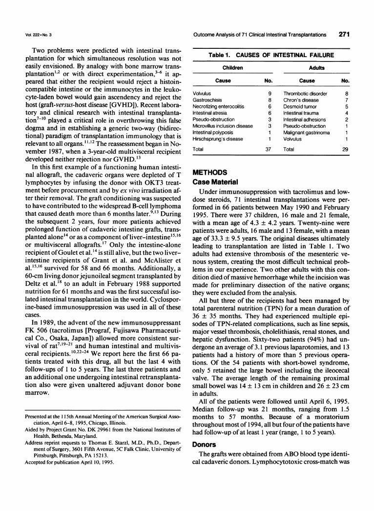
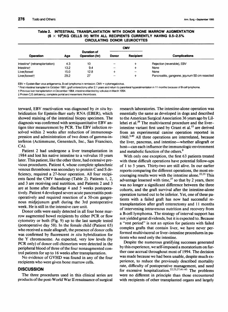
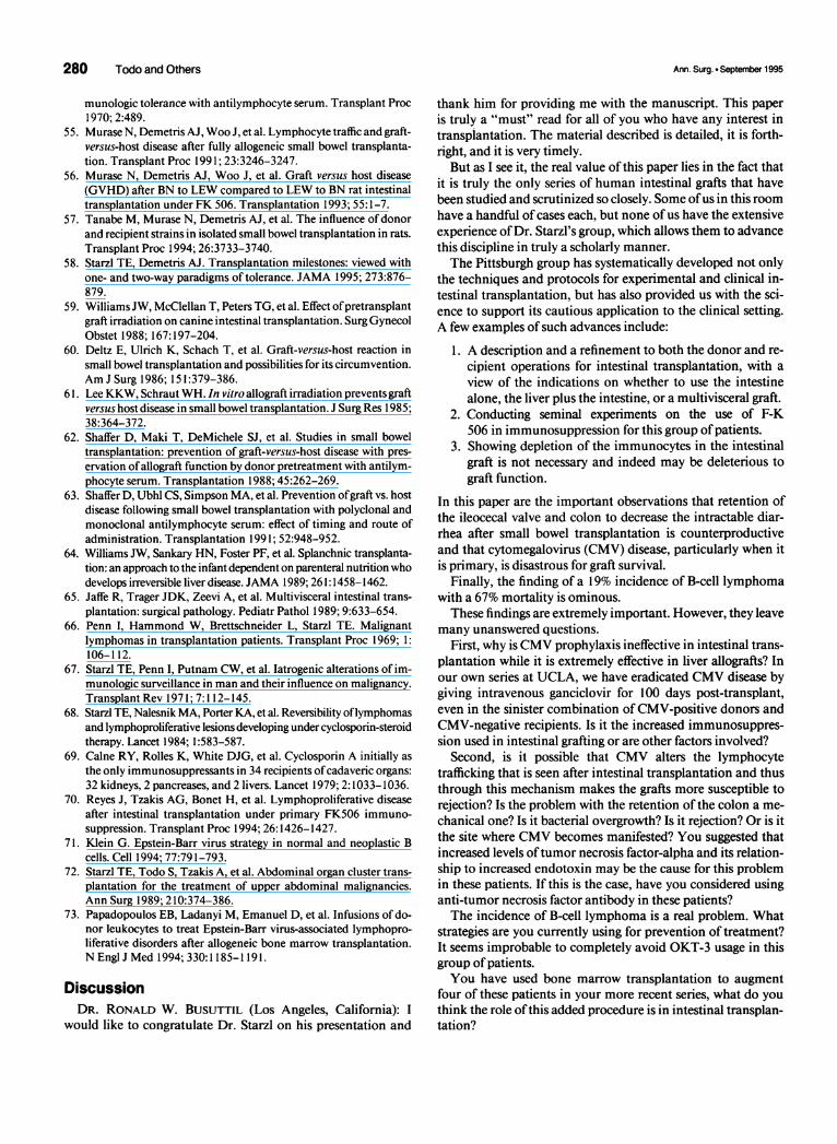
Figure 1. Three kinds of visceralallografts: intestine alone (right),liver and intestine (left), and multivis-ceral (middle). The number of pa-tients receiving each type of graft isindicated by "n." Colonic segments(shaded) were included in 29 recipi-ents scattered through the three co-horts. IVC: inferior vena cava; PV:portal vein; HA: hepatic artery; SMA:superior mesenteric artery; SMV:superior mesenteric vein; SA:splenic artery; LGA: left gastric ar-tery.
n = 11
positive in seven patients. Matching ofhuman leukocyteantigen (HLA) was random and poor. Donor manage-ment and operative procedures have been described else-where.9'25'26 The University of Wisconsin solution wasused for organ preservation in all but one case. Cold isch-emia time from aortic clamping until graft revasculari-zation averaged 7.7 ± 2.4 hours.
Transplantation OperationsThe three kinds of allografts are shown in Figure 1:
isolated intestinal (n = 23), combined intestine and liver(n = 32), and multivisceral (n = 1 1). Patients who stillhad good liver function received intestine only. Thosewho had inborn and/or TPN-related hepatic dysfunctionreceived liver and intestine. Abdominal multivisceralgrafts were reserved for patients who had extensive ab-normalities of the gastrointestinal tract caused by ab-sorptive, motility, or vascular disorders. The principlesand various modifications of the three generic proce-dures have been described elsewhere.922-2426-29 In allthree recipient cohorts, some of the grafts contained asegment of large bowel, whereas others did not. Use ofthe colon was prompted by the high postoperative sto-mal output and frequent readmissions for treatment ofdehydration that were frequently observed otherwise.23
Bone Marrow TransplantationBone marrow cells recovered from the same donor as
the intestine were infused intravenously into the recipi-ent during the operation in four recent cases (three pri-
mary and one retransplantation). The rationale andmethods ofsimultaneous bone marrow infusion in solidorgan transplantation have been reported elsewhere.30-32In brief, bone marrow cells were isolated from the thora-columbar vertebrae of the donor and suspended in 200mL of Dulbecco's Modified Eagle Medium (Gibco,Grand Island, NY) at a concentration of 6 X 10⁸ cells/kgbody weight. The infusions were given over 20 minutes,2 to 12 hours after revascularization of the intestinalgraft.
Postoperative ManagementImmunosuppression was achieved with tacrolimus
and low-dose steroids, to which prostaglandin E1 wasadded briefly during the early postoperative period. Ad-justment of tacrolimus dose and/or supplemental pred-nisone, OKT3, and azathioprine were given for episodesof rejection or for tacrolimus-related adverse effects, aspreviously described.22'27'33 In a few cases, azathioprinewas given as a third drug from the outset. Depression ofwhite blood cell count (<5000) was treated with 3 to 5,ug/kg granulocyte colony-stimulating factor (Neupogen,Amgen, Thousand Oaks, CA). The complex infectiousdisease and nutritional management was as described
2227,33,34elsewhere.222.33
Sampling ProtocolsRejection Surveillance
Histopathologic study of endoscope-guided biopsieswas done twice weekly until hospital discharge. The clin-
Ann. Surg. . Septernber 1995
Outcome Analysis of 71 Clinical Intestinal Transplantations 273
ical diagnosis of rejection was not accepted unless con-firmed histopathologically by criteria described else-where.2235 More than 1440 intestinal biopsy specimenshave been examined by a single pathologist (R.G.L.).
Graft-Versus-Host Disease Surveillance
Suspicious skin lesions were biopsied and studied byroutine histology, immunohistologic staining for donor-specific HLA antigens, and in situ hybridization tech-nique using the Y-chromosome-specific probe, as de-scribed elsewhere.'2'36'37CytokinesPlasma samples collected serially from patients who
did not receive colon (n = 9) and from patients receivingcolon segments (n = 7) were stored (-70 C) and analyzedfor tumor necrosis factorA, interleukin- 1 ,B, and interleu-kin-6 by the enzyme-linked immunosorbent assaymethod (R&D Systems, Minneapolis, MN).
Chimerism
Donor leukocyte chimerism was followed before andafter intestinal transplantation in four patients who alsowere given perioperative bone marrow and in nonmar-row control recipients of the same kinds of allografts.Leukocytes circulating in the recipient peripheral venousblood were identified as donor with donor-specific anti-HLA class I monoclonal antibodies and by fluorescent-activated cell sort analysis.30 The results were confirmedusing probes directed against HLA class II chromosomesby polymerase chain reaction (PCR) and by in situ hy-bridization technique with Y-chromosome-specificprobe.
StatisticsFigures are expressed as the mean plus or minus the
standard deviation of the mean. Differences in groupmeans were tested using the one-way analysis of vari-ance, and differences in proportions were examined us-ing Pearson's chi square test. Patient and graft survivalcurves were generated using the life table method andwere compared between the groups using the log-rank(Mantel-Cox) test. To analyze the risk factors for mor-bidity, graft loss, and mortality, Cox's proportional haz-ards model was used.
RESULTSCases Without Bone Marrow
Patient Survival
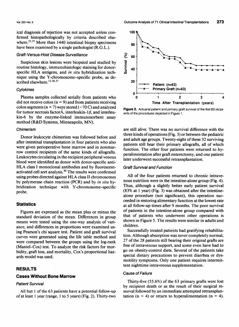
All but 1 of the 63 patients have a potential follow-upof at least 1 year (range, 1 to 5 years) (Fig. 2). Thirty-two
100
80
b-
._
2)
60
40
20
0
- Patient (n=63).... Primary Graft (n=63)
0 1 2 3 4Time After Transplantation (years)
Figure 2. Actuarial patient and primary graft survival of the first 63 recipi-ents of the procedures depicted in Figure 1.
are still alive. There was no survival difference with thethree kinds ofoperations (Fig. 3) or between the pediatricand adult age groups. Twenty-eight ofthese 32 survivingpatients still bear their primary allografts, all of whichfunction. The other four patients were returned to hy-peralimentation after graft enterectomy, and one patientlater underwent successful retransplantation.
Graft Survival and Function
All of the four patients returned to chronic intrave-nous nutrition were in the intestine-alone group (Fig. 4).Thus, although a slightly better early patient survival(83% at 1 year) (Fig. 3) was obtained after the intestine-alone procedure (not significant), this operation suc-ceeded in restoring alimentary function at the lowest rateat all follow-up times after 9 months. The poor survivalof patients in the intestine-alone group compared withthat of patients who underwent other operations isshown in Figure 5. The results were similar in adults andchildren.
Successfully treated patients had gratifying rehabilita-tion. Although absorption was never completely normal,27 of the 28 patients still bearing their original grafts arefree of intravenous support, and some even have had togo on obesity-control diets. Several of the patients takespecial dietary precautions to prevent diarrhea or dys-motility symptoms. Only one patient requires intermit-tent nighttime intravenous supplementation.
Cause of Failure
Thirty-five (55.6%) of the 63 primary grafts were lostby recipient death or as the result of their surgical re-moval followed by an immediate attempted retransplan-tation (n = 4) or return to hyperalimentation (n = 4).
Vol. 222 - No. 3
274 Todo and Others
1001
0
a<
'0
._
b-
cL
80
60
40
1001
0
._
4-
co
E._20- Isolated Intestine (n=22)
---.. Liver and Intestine (n=30)Multivisceral (n=11)
00 1 2 3 4
Time After Transplantation (years)Figure 3. Patient survival after the three types of allografts shown in Fig-ure 1.
The principal reasons for the primary graft losses aresummarized schematically in Figure 6.
In 10 cases, the failures could be traced back to surgicalor management errors. Eight ofthese 10 grafts were func-tioning at or shortly before the time of their loss. Thesubgroup of technical surgical errors, all in children, in-cluded intestinal anastomotic leakage, hepatic arterythrombosis, biliary anastomotic leakage, and cerebral in-farction associated with intraoperative cardiac arrest.One of the six grievous management errors (Fig. 6) wasnonrecognition of a pre-existing pneumonia. The graftwas removed immediately, immunosuppression wasstopped, and the patient was successfully returned to hy-peralimentation. Other management errors were over-dosing or underdosing oftacrolimus or other agents (n =4), including a lethal infusion ofhypertonic saline. Therewas one failure to diagnose drug noncompliance.The exact reason for loss of the other 25 grafts was
15
c
a)
0
.0Ez
10 -
5-
El On TPNE On Oral Diet
Intestine Liver/Intestine Multivisceral
Figure 4. Primary graft function in the 32 surviving patients after each ofthe three different transplantations. Note that 4 of the 12 intestine-alonerecipients are alive despite removal of their transplants.
80
60-
40
20 -
^---S.~~~~...A-- .
Isolated Intestine (n=22).A---^-- Liver and Intestine (n=30)
Multivisceral (n=11)
0 1 2 3 4Time After Transplantation (years)
Figure 5. Allograft survival with the three different kinds of operations.The differences are not statistically significant.
difficult to pinpoint because ofmultiple interlocking fac-tors (Fig. 6). The principal diagnosis was rejection inonly 6 cases, whereas an infectious complication attrib-utable to antirejection therapy was the major diagnosisin 19. The responsible microorganisms included bacteriaand fungi (n = 6), but among the viruses, cytomegalovi-rus (CMV) alone accounted for nearly as many losses (n= 5). Moreover, Epstein-Barr virus (EBV) associated B-cell lymphomas occurred in 12 (19%) of the 63 patients,ofwhom 8 lost their grafts and died ofthis complication.Eleven ofthe 12 patients who developed the lymphomashad been treated with OKT3.
Retransplantation
Four patients underwent retransplantation on thesame day as primary graft removal (two liver-intestine)or 1 to 2 months later (two intestine only). They died 47days, 57 days, 71 days, and 147 days after the second
Technical (n = 4)and
Management Errors (n = 6)
Rejection
*Infection
CMV (n=5)Lymphoma (n=8)
Fungal/Bacterial (n=6)Rejection (n=6)
Figure 6. Causes of the 35 primary graft losses.
Ann. Surg. . September 1995
.. .............
.........

Outcome Analysis of 71 Clinical Intestinal Transplantations 275
100 -
0
0)<Cu
0-
75 -
50-
25
0-
/7 ..
-....7, .:-
. = _ I
./ / '/A S'SSo;- I,o',:";. /#/S;"/;'/ ', I
;/ ;/// / w S ;/
N-N P-N N-PFigure 7. Effect on patient survival of donor and recipientogy. N-N: negative donor-negative recipient (26 patients);donor-negative recipient (13 patients); N-P: negative doncipient (17 patients); and P-P: positive donor-positive retients).
procedure, with the principal diagnoses of r(= 2), B-cell lymphoma (n = 1), and systemiinfection (n = 1). In addition, liver retransplarunsuccessful in a pediatric patient who devclated hepatic artery thrombosis 11 days afterintestine and liver transplantation.A fifth intestinal retransplantation was pei
an intestine-only recipient 11 months after grztomy. This patient, who is alive, also was gmarrow from the second intestine donor (see
Risk Factors
With univariate and multivariate analysis,cally significant risk factors for graft loss anamong 28 variables examined were high FKlevels, prednisone bolus therapy, adminisOKT3, length of operation (reflecting thedifficulty of the procedure), CMV plus statuand recipient, and inclusion of a segment ofthe graft. The first four of these risk factorsbeen described with transplantation of other (
will not be considered here except to mentionous relation to the infectious complicationsand sixth risk factors were more specific to thlrecipient.The CMV effect was pervasive. Serological
CMV recipients are surviving after 1 to 5 yeaof62% if their donors were CMV negative, btrate of 47% if their donors were CMV positi'An adverse effect on survival also was observerecipient already was serologically CMV posieration. Fifty-four percent survive to date ifa
E Alive ative donor was used, but only 14% with a CMV-positivedonor (Fig. 7). As recently reported in detail,38 therapy
-, ;that can regularly control this infection in other kinds ofallograft recipients was only marginally effective in theintestinal recipient. Once the clinical diagnosis ofCMVwas made, the predominant target in essentially all caseswas the intestinal allograft itself. The resulting ulcer-
.....-- ations were associated with bacterial translocation.Inclusion of a colonic segment in 29 patients was a
significant risk factor. Significantly better graft survivalwas observed in 34 patients without the colon than with
;.......it (Fig. 8). Although the negative colon influence was: .:::: ..::.::.. :::::::..
more pronounced in adults, it also was identified in the.. ... ...<
pediatric recipients.p-p A possible explanation was the postoperative increase
CMV+ serol- in plasma tumor necrosis factor-alpha (and by implica-P-N: positive tion, endotoxemia) in recipients given colon versus the
or-positive re- lower levels in nine patients whose grafts did not containcipient (7 pa- colon. Interleukin- 1 levels were barely detectable in both
cohorts, and interleukin-6 responses were comparable.
ejection (n Bone Marrow AugmentationIc bacterialitation was Two of the augmentation patients received intestineeloped iso- alone, and two were given liver plus intestine (Table 2).combined All were classified as high risk. Patient 1, an intestinal
retransplant recipient, had a monoclonal B-cell lym-rformed in phoma that had been in remission since discontinuanceaft enterec- ofimmunosuppression and removal ofhis primary graftOiven bone 11 months previously. Although the lymphocytotoxicbelow). cross-match was negative, he had a high antibody index
(plasma renin activity) before retransplantation and ex-perienced a severe rejection during the second postoper-
6 statisti- ative week, which was reversed with OKT3. Shortly af-d/or death506 blood,tration of 1001technical
is of donor 80- icolon withhave often '.organs and . 60-their obvi- c " e vThe fifth 40
e intestinal .0.
ly negativetrs at a rateat only at ave (Fig. 7).d when theitive at op-CMV-neg-
20No Colon (n=34)
-- Colon (n=29)
0 1 2 3 4Time After Transplantation (years)
Figure 8. Survival of 29 primary grafts containing a colon segment versus
34 grafts not including colon (p < 0.0313).
Vol. 222 9 No. 3
276 Todo and Others
Table 2. INTESTINAL TRANSPLANTATION WITH DONOR BONE MARROW AUGMENTATION(6 x 10⁸/KG CELLS IV) WITH ALL RECIPIENTS CURRENTLY HAVING 0.5-2.5%
CIRCULATING DONOR LEUKOCYTES
CMVDuration of
Operation Age Operation (hr) Donor Recipient Complications
Intestine* (retransplantation) 4.3 10 - + Rejection (reversible), EBVIntestinet 13.2 9.4 - + NoneLiver/bowel 15.0 12.8 + - NoneLiver/bowelt 29.2 27 - + Pancreatitis, gangrene, jejunum 50 cm resected
EBV = Epstein-Barr virus antigenemia, B-cell lymphoma in remission; CMV = cytomegalovirus.* First intestinal transplant in October 1991; graft enterectomy after 2.1 years and return to parenteral hyperalimentation in 11 months because of B-cell lymphoma.t Previous liver transplantation in December 1984; intestine infarcted by volvulus in March 1994.t Protein C/S deficiency; complete portal and mesenteric thrombosis.
terward, EBV reactivation was diagnosed by in situ hy-bridization for Epstein-Barr early RNA (EBER), whichshowed staining of the intestinal biopsy specimen. Thediagnosis was confirmed with semiquantitative EBV an-tigen titer measurement by PCR. The EBV infection re-solved within 2 weeks after reduction of immunosup-pression and administration of two doses of gamma-in-terferon (Actimmune, Genentech, Inc., San Francisco,CA).
Patient 2 had undergone a liver transplantation in1984 and lost his native intestine to a volvulus 10 yearslater. This patient, like the other three, had extensive pre-vious procedures. Patient 4, whose complete splanchnicvenous thrombosis was secondary to protein C and S de-ficiency, required a 27-hour operation. All four recipi-ents faced the CMV handicap (Table 2). Patients 1, 2,and 3 are receiving oral nutrition, and Patients 2 and 3are at home after discharge 4 and 5 weeks postopera-tively. Patient 4 developed severe acute pancreatitis post-operatively and required resection of a 50-cm gangre-nous midjejunum graft during the 3rd postoperativeweek. He is still in the intensive care unit.Donor cells were easily detected in all four bone mar-
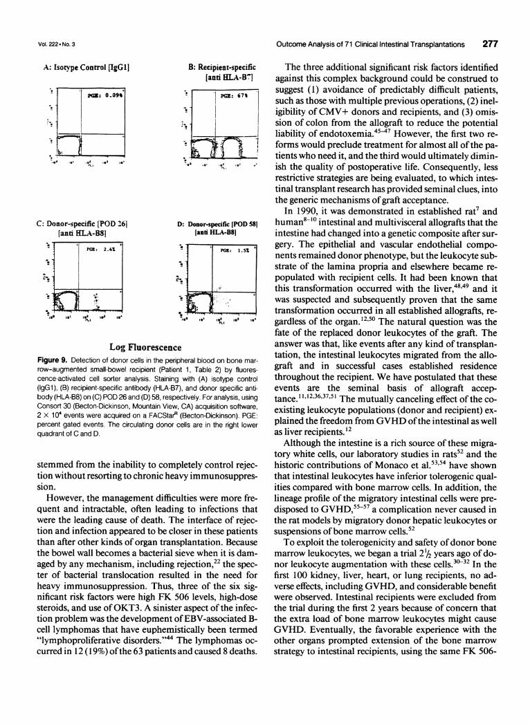
row augmented bowel recipients by either PCR or flowcytometry or both (Fig. 9) up to the last sample tested(postoperative day 58). In the female child (Patient 65)who received a male allograft, the presence ofdonor cellswas confirmed by fluorescent in situ hybridization forthe Y chromosome. As expected, very low levels (byPCR only) of donor cell chimerism were detected in theperipheral blood ofthree ofthe four nonaugmented con-trol patients for up to 16 weeks after transplantation.No evidence ofGVHD was found in any of the four
recipients who were given bone marrow cells.
DISCUSSIONThe three procedures used in this clinical series are
products ofthe post-World War II renaissance ofsurgical
research laboratories. The intestine-alone operation wasessentially the same as developed in dogs and describedto the American Surgical Association 36 years ago by Lil-lehei et al.39 The multivisceral procedure and the liver-intestine variant first used by Grant et al."5 are derivedfrom an experimental canine operation reported in1960.34' All three operations are interrelated, becausethe liver, pancreas, and intestine-whether allograft orhost-can each influence the immunologic environmentand metabolic function ofthe others.9With only one exception, the first 63 patients treated
with these difficult operations have potential follow-upsof 1 to 5 years. Thirty-two are still alive. In our earlierreports comparing the different operations, the most en-couraging results were with the intestine alone.22'23 Thisadvantage lessened with time,27 so that by 2 years, therewas no longer a significant difference between the threecohorts, and the graft survival after the intestine-aloneoperation turned out to be inferior. Yet, one ofthese pa-tients with a failed graft has now had successful re-transplantation after graft enterectomy and 11 monthsof intervening intravenous nutrition and recovery froma B-cell lymphoma. The strategy of interval support hasnot yielded great dividends, but it is expected to. Becausea "rest period" is not an option for patients with failedcomplex grafts that contain liver, we have never per-formed multivisceral or liver-intestine procedures in pa-tients who need only the intestine.
Despite the numerous gratifying successes generatedby this experience, we self-imposed a moratorium on fur-ther case accrual throughout most of 1994. The decisionwas made because we had been unable, despite much ex-perience, to reduce the previously described mortalityrate, difficulty of postoperative management, and needfor excessive hospitalization.222327'4 The problemswere no different in principle than those encounteredwith recipients of other transplanted organs and largely
Ann. Surg. . September 1995
Outcome Analysis of 71 Clinical Intestinal Transplantations 277
B: Recipient-specific[anti HLA-B71
.
D: Donor-specific [POD 581[anti HLA-B8j
.ts ,...I
Log FluorescenceFigure 9. Detection of donor cells in the peripheral blood on bone mar-
row-augmented small-bowel recipient (Patient 1, Table 2) by fluores-cence-activated cell sorter analysis. Staining with (A) isotype control(IgGl), (B) recipient-specific antibody (HLA-B7), and donor specific anti-body (HLA-B8) on (C) POD 26 and (D) 58, respectively. For analysis, usingConsort 30 (Becton-Dickinson, Mountain View, CA) acquisition software,2 X 104 events were acquired on a FACStarR (Becton-Dickinson). PGE:percent gated events. The circulating donor cells are in the right lowerquadrant of C and D.
stemmed from the inability to completely control rejec-tion without resorting to chronic heavy immunosuppres-sion.However, the management difficulties were more fre-
quent and intractable, often leading to infections thatwere the leading cause of death. The interface of rejec-tion and infection appeared to be closer in these patientsthan after other kinds of organ transplantation. Becausethe bowel wall becomes a bacterial sieve when it is dam-aged by any mechanism, including rejection,22 the spec-ter of bacterial translocation resulted in the need forheavy immunosuppression. Thus, three of the six sig-nificant risk factors were high FK 506 levels, high-dosesteroids, and use ofOKT3. A sinister aspect of the infec-tion problem was the development ofEBV-associated B-cell lymphomas that have euphemistically been termed"lymphoproliferative disorders."44 The lymphomas oc-
curred in 12 (19%) ofthe 63 patients and caused 8 deaths.
The three additional significant risk factors identifiedagainst this complex background could be construed tosuggest (1) avoidance of predictably difficult patients,such as those with multiple previous operations, (2) inel-igibility ofCMV+ donors and recipients, and (3) omis-sion of colon from the allograft to reduce the potentialliability of endotoxemia.45-47 However, the first two re-forms would preclude treatment for almost all ofthe pa-tients who need it, and the third would ultimately dimin-ish the quality of postoperative life. Consequently, lessrestrictive strategies are being evaluated, to which intes-tinal transplant research has provided seminal clues, intothe generic mechanisms of graft acceptance.
In 1990, it was demonstrated in established rat7 andhuman8-'0 intestinal and multivisceral allografts that theintestine had changed into a genetic composite after sur-gery. The epithelial and vascular endothelial compo-nents remained donor phenotype, but the leukocyte sub-strate of the lamina propria and elsewhere became re-populated with recipient cells. It had been known thatthis transformation occurred with the liver,48'49 and itwas suspected and subsequently proven that the sametransformation occurred in all established allografts, re-gardless of the organ.'250 The natural question was thefate of the replaced donor leukocytes of the graft. Theanswer was that, like events after any kind of transplan-tation, the intestinal leukocytes migrated from the allo-graft and in successful cases established residencethroughout the recipient. We have postulated that theseevents are the seminal basis of allograft accep-tance. '1'2,36,37, ' The mutually canceling effect ofthe co-
existing leukocyte populations (donor and recipient) ex-plained the freedom fromGVHD ofthe intestinal as wellas liver recipients.'2Although the intestine is a rich source of these migra-
tory white cells, our laboratory studies in rats52 and thehistoric contributions of Monaco et al.53'54 have shownthat intestinal leukocytes have inferior tolerogenic qual-ities compared with bone marrow cells. In addition, thelineage profile of the migratory intestinal cells were pre-disposed to GVHD,55-57 a complication never caused inthe rat models by migratory donor hepatic leukocytes orsuspensions ofbone marrow cells.52To exploit the tolerogenicity and safety ofdonor bone
marrow leukocytes, we began a trial 2/ years ago of do-nor leukocyte augmentation with these cells.3032 In thefirst 100 kidney, liver, heart, or lung recipients, no ad-verse effects, including GVHD, and considerable benefitwere observed. Intestinal recipients were excluded fromthe trial during the first 2 years because of concern thatthe extra load of bone marrow leukocytes might causeGVHD. Eventually, the favorable experience with theother organs prompted extension of the bone marrowstrategy to intestinal recipients, using the same FK 506-
A: Isotype Control [IgGli
PG: 0.09o
- 1 0.,-'ej.JM-
C: Donor-specific [POD 261[anti HLA-B88
PGE: 2.4X
a.-,.
I.1m ,. 1i 1e
r a
Vol. 222 X No. 3
a
III
278 Todo and Others
steroid immunosuppression as before. The first four in-testinal recipients treated in this way are alive after 2 to 3postoperative months. Donor cells account for approxi-mately 1% to 2.5% oftheir circulating mononuclear cells.There has been no evidence ofGVHD.
Aside from this direct therapeutic application, otherimplications of the recently evolved two-way paradigmof transplantation'' ,12,36,37,51,58 are relevant in assessingthe intestinal transplant experience. First, the unusualinfectivity ofCMV is not enigmatic. Because this virus islymphotropic and contained in the migratory cells, it isnot hard to envision its massive and efficient inoculationin either direction (graft to host or vice versa), propor-tional to the magnitude of cell migration that beginswithin a few minutes after graft revascularization.
Second, the etiology, prevention, and management ofthe EBV-associated B-cell lymphomas that have plaguedefforts at intestinal transplantation also become moreunderstandable. Because of the now obsolete belief thatintestinal grafts should be T-cell (or pan-leukocyte) de-pleted to avoid GVHD,4 6'59-63 most ofthe bowel donorsbefore 1990 were treated before organ procurement withantilymphoid preparations, or the allografts were irradi-ated.'3"15"16'64'65 Most ofthe patients who survived the op-eration developed B-cell lymphomas of recipient origin.The lymphomas were similar to those associated with ov-erimmunosuppression in recipients of other organs,66'67especially after the T-cell-directed agents cyclosporineand68'69 FK 50670 became available.
In a series of studies,7' Klein delineated the cellularand molecular mechanisms by which these iatrogenic in-terventions or inherited immunodeficiency diseases al-low an inherently self-limited EBV infection to proceedto a malignancy. In essence, surveillance of potentiallyneoplastic virus-infected B cells depends on the intact T-cell system that the various procedures used in transplan-tation disarm in the donor or recipient leukocyte popu-lations, or both. Suspicion that this was the reason for B-cell lymphomas in our first multivisceral recipientscaused us more than 6 years ago to recommend againsteither donor or allograft conditioning.9 72 When B-celllymphomas develop in organ recipients, restoration ofT-cell surveillance over EBV (whose antigenicity is ex-pressed during replication71) can be accomplished bylightening immunosuppression,68 but at the risk of rejec-tion.Bone marrow recipients who are cytoablated (with to-
tal body irradiation or drugs) and given T-cell-depletedallografts have an increased incidence of B-cell lympho-mas. These are of donor (not recipient) origin, and theoption of reducing maintenance immunosuppressionusually is not available. However, Papadopoulos et al.73reported a regression of these donor phenotype lympho-mas by infusing peripheral blood cytotoxic T lympho-
Ann. Surg. * September 1995
cytes collected from the original marrow donor. Thismimics the therapeutic effect achieved in organ recipi-ents by lightening immunosuppression and allowing re-cipient T-cell surveillance. Predictably, the penalty ofsuch treatment in the bone marrow recipients wasGVHD.
All of the B-cell lymphomas reported in intestinal re-cipients have been of recipient origin. It is possible to useimmunotherapy in such patients to treat tumors of thiskind that fail to respond to reduction immunosuppres-sion. Unaltered recipient buffy coat or bone marrowcould be collected preoperatively and stored for such anexigency, to be used as a last resort.A better understanding of the pathogenesis of B-cell
lymphomas and other problems of intestinal transplan-tation is the first step to solving them, particularly ifcom-plications are of a unifying etiology. However, progresswill depend on obtaining stable graft acceptance morequickly and completely, thereby decreasing the harmcaused by high-dose chronic immunosuppression. Theadministration of adjuvant donor bone marrow has thisobjective. Although there is a solid experimental base forthis approach, its clinical value is unproven.Even if the bone marrow initiative is effective, these
often difficult operations will not be made easier to per-form. Nevertheless, intestinal transplantation seems tous at least as far along as liver transplantation was littlemore than 12 years ago or even kidney transplantationin the early 1 970s.
References
1. Billingham RE. Reactions ofgrafts against their hosts: transplanta-tion immunity works both ways-hosts destroy grafts and graftsmay harm hosts. Science 1959; 130:947-953.
2. Vriesendorp HM. Engraftment of hemopoietic cells. In van Bek-kum DW, Lowenberg B, eds. Bone Marrow Transplantation: Bio-logical Mechanisms and Clinical Practice. New York: Marcel Dek-ker, Inc; 1985:76-78,114-119.
3. Starzl TE, Kaupp HA Jr., Brock DR, et al. Homotransplantationof multiple visceral organs. Am J Surg 1962; 103:219-229.
4. Monchik GJ, Russell PS. Transplantation ofthe small bowel in therat: technical and immunologic considerations. Surgery 1971; 70:693-702.
5. Lillehei RC, Manax WG, Lyona GW, Dietzman RH. Transplan-tation of gastrointestinal organs, including small intestine andstomach. Gastroenterology 1966; 51:966.
6. Cohen Z, MacGregor AB, Moore KTH, et al. Canine small boweltransplantation. Arch Surg 1976; 11 1:248.
7. Murase N, Demetris AJ, Matsuzaki T, et al. Long survival in ratsafter multivisceral versus isolated small bowel allotransplantationunder FK 506. Surgery 1991; 1 10:87-98.
8. Iwaki Y, Starzl TE, Yagihashi A, et al. Replacement ofdonor lym-phoid tissue in human small bowel transplants under FK 506 im-munosuppression. Lancet 1991; 337:818-819.
9. Starzl TE, Todo S, Tzakis A, et al. The many faces of multivisceraltransplantation. Surg Gynecol Obstet 1991; 172:335-344.
10. Todo S, Tzakis AG, Abu-Elmagd K, et al. Cadaveric small bowel
Vol. 222 . No. 3
and small bowel-liver transplantation in humans. Transplantation1992; 53:369-376.
11. Starzl TE, Demetris AJ, Murase N, et al. Cell migration, chime-rism, and graft acceptance. Lancet 1992; 339:1579-1582.
12. Starzl TE, Demetris AJ, Trucco M, et al. Cell migration and chi-merism after whole organ transplantation: the basis of graft accep-tance. Hepatology 1993; 17:1127-1152.
13. Starzl TE, Rowe MI, Todo S, et al. Transplantation of multipleabdominal viscera. JAMA 1989; 261:1449-1457.
14. Goulet 0, Revillon Y, Brousse N, et al. Successful small boweltransplantation in an infant. Transplantation 1992; 53:940-943.
15. Grant D, Wall W, Mimeault R, et al. Successful small-bowel/livertransplantation. Lancet 1990; 335:181-184.
16. McAlister V, Wall W, Ghent C, et al. Successful small intestinetransplantation. Transplant Proc 1992; 24:1236-1237.
17. Margreiter R, Konigsrainer A, Schmid T, et al. Successful multi-visceral transplantation. Transplant Proc 1992; 24:1226-1227.
18. Deltz E, Schroeder P, Gebhardt H, et al. Successful clinical smallbowel transplantation: report ofa case. Clin Transplantation 1989;3:89-91.
19. Murase N, Kim D, Todo S, et al. Induction of liver, heart andmultivisceral graft acceptance with a short course of FK 506.Transplant Proc 1990; 22:74-75.
20. Lee K, Stangl MJ, Todo S, et al. Successful orthotopic small boweltransplantation with short term FK 506 immunosuppressive ther-apy. Transplant Proc 1990; 22:78-79.
21. Hoffman AL, Makowka L, Banner B, et al. The use of FK 506for small intestine allotransplantation: inhibition ofacute rejectionand prevention of fatal graft-versus-host disease. Transplantation1990; 49:483-490.
22. Todo S, Tzakis AG, Abu-Elmagd K, et al. Intestinal transplanta-tion in composite visceral grafts or alone. Ann Surg 1992; 216:223-234.
23. Todo S, Tzakis A, Reyes J, et al. Small intestinal transplantationin humans with or without colon. Transplantation 1994; 57:840-848.
24. Todo S, Tzakis A, Abu-Elmagd K, et al. Abdominal multivisceraltransplantation. Transplantation 1995; 59:234-240.
25. Casavilla A, Selby R, Abu-Elmagd K, et al. Logistics and techniquefor combined hepatic-intestinal retrieval. Ann Surg 1992; 216:605-609.
26. Furukawa H, Abu-Elmagd K, Reyes J, et al. Technical aspects ofintestinal transplantation. In Braverman MH, Tawes RL, eds. Sur-gical Technology International. Vol 2. San Francisco, CA: Univer-sal Medical Press Inc; 1994:165-170.
27. Abu-Elmagd K, Todo S, Tzakis A, et al. Three years clinical expe-rience with intestinal transplantation. J Am Coll Surg 1994; 179:385-400.
28. Tzakis AG, Todo S, Reyes J, et al. Piggyback orthotopic intestinaltransplantation. Surg Gynecol Obstet 1993; 176:297-298.
29. Tzakis AG, Nour B, Reyes J, et al. Endorectal pull through oftransplanted colon as part of intestinal transplantation. Surgery1995; 117:451-453.
30. Fontes P, Rao A, Demetris AJ, et al. Augmentation with bone mar-row ofdonor leukocyte migration for kidney, liver, heart, and pan-creas islet transplantation. Lancet 1994; 344:151-155.
31. Starzl TE, Demetris AJ, Rao AS, et al. Spontaneous and iatrogeni-cally augmented leukocyte chimerism in organ transplant recipi-ents. Transplant Proc 1994; 26:3071-3076.
32. Rao AS, Fontes P, Zeevi A, et al. Augmentation of chimerism inwhole organ recipients by simultaneous infusion of donor bonemarrow cells. Transplant Proc 1995; 27:210-212.
33. Abu-Elmagd K, Fung JJ, Reyes J, et al. Management of intestinaltransplantation in humans. Transplant Proc 1992; 24:1243-1244.
Outcome Analysis of 71 Clinical Intestinal Transplantations 279
34. Reyes J, Tzakis AG, Todo S, et al. Nutritional management ofintestinal transplant recipients. Transplant Proc 1993; 25:1200-1201.
35. Nakamura K, Nalesnik M, Jaffe R, et al. Morphologic monitoringofhuman small bowel allografts. Transplant Proc 1993; 25:1212.
36. Starzl TE, Demetris AJ, Trucco M, et al. Systemic chimerism inhuman female recipients of male livers. Lancet 1992; 340:876-877.
37. Starzl TE, Demetris AJ, Trucco M, et al. Chimerism after livertransplantation for type IV glycogen storage disease and Type IGaucher's disease. N Engl J Med 1993; 328:745-749.
38. Manez R, Kusne S, Green M, et al. Incidence and risk factors as-sociated with the development ofcytomegalovirus disease after in-testinal transplantation. Transplantation 1995; 59:1010-1015.
39. Lillehei RC, Goott B, Miller FA. The physiologic response of thesmall bowel of the dog to ischemia including prolonged in vitropreservation ofthe bowel with successful replacement and survival.Ann Surg 1959; 150:543.
40. Starzl TE, Kaupp HA Jr. Mass homotransplantation ofabdominalorgans in dogs. Surg Forum 1960; 11:28-30.
41. Abu-Elmagd KM, Tzakis A, Todo S, et al. Monitoring and treat-ment of intestinal allograft rejection in humans. Transplant Proc1993; 25:1202-1203.
42. Tzakis AG, Todo S, Reyes J, et al. Clinical intestinal transplanta-tion: focus on complications. Transplant Proc 1992; 24:1238-1240.
43. Tzakis A, Starzl TE. Liver transplantation. In Ashcraft KW,Holder TM, eds. Pediatric Surgery. 2nd ed. Philadelphia: WBSaunders Co; 1992:505-524.
44. Hanto DW, Frizzera GI, Gajl-Peczalska KJ, Simmons RL. Ep-stein-Barr virus, immunodeficiency, and B cell lymphoprolifera-tion. Transplantation 1985; 39:461-472.
45. Yokoyama I, Todo S, Miyata T, et al. Endotoxemia and humanliver transplantation. Transplant Proc 1989; 21:3833-3841.
46. Miyata T, Yokoyama I, Todo S, et al. Endotoxemia, pulmonarycomplications and thrombocytopenia with clinical liver transplan-tation. Lancet 1989; 8656:189-191.
47. Miyata T, Todo S, Imventarza 0, et al. Endogenous endotoxemiaduring orthotopic liver transplantation in dogs. Transplant Proc1989; 21:3861-3862.
48. Kashiwagi N, Porter KA, Penn I, et al. Studies of homograft sexand of gamma globulin phenotypes after orthotopic homo-transplantation ofthe human liver. Surg Forum 1969; 20:374-376.
49. Porter KA. Pathology ofthe orthotopic homograft and heterograft.In Starzl TE, ed. Experience in Hepatic Transplantation. Philadel-phia: WB Saunders Co; 1969:464-465.
50. Randhawa PS, Starzl TE, Ramos H, et al. Allografts surviving for26-29 years following living related kidney transplantation: analy-sis by light microscopy, in situ hybridization for the Y chromo-some, and anti-HLA antibodies. Am J Kidney Dis 1994; 24:72-77.
51. Starzl TE, Demetris AJ, Trucco M, et al. Chimerism and donorspecific nonreactivity 27 to 29 years after kidney allotransplanta-tion. Transplantation 1993; 55:1272-1277.
52. Murase N, Tanabe M, Fujisaki S, et al. Variable chimerism, graftversus host disease, and tolerance after different kinds of cell andsolid organ transplantation from Lewis to Brown-Norway rats.Transplantation 1995 (in press).
53. Monaco AP, Wood ML, Maki T, Gozzo J. The use of donor-spe-cific bone marrow to induce specific unresponsiveness (tolerance)to tissue allografts. In Ildstad ST, ed. Chimerism and Tolerance.Austin, TX: RG Landes Co; 1994:99.
54. Monaco AP, Wood ML. Studies on heterologous anitlymphocyteserum in mice. VII. Optimal cellular antigen for induction of im-
280 Todo and Others
munologic tolerance with antilymphocyte serum. Transplant Proc1970; 2:489.
55. Murase N, Demetris AJ, Woo J, et al. Lymphocyte traffic and graft-versus-host disease after fully allogeneic small bowel transplanta-tion. Transplant Proc 1991; 23:3246-3247.
56. Murase N, Demetris AJ, Woo J, et al. Graft versus host disease(GVHD) after BN to LEW compared to LEW to BN rat intestinaltransplantation under FK 506. Transplantation 1993; 55:1-7.
57. Tanabe M, Murase N, Demetris AJ, et al. The influence of donorand recipient strains in isolated small bowel transplantation in rats.Transplant Proc 1994; 26:3733-3740.
58. Starzl TE, Demetris AJ. Transplantation milestones: viewed withone- and two-way paradigms of tolerance. JAMA 1995; 273:876-879.
59. Williams JW, McClellan T, Peters TG, et al. Effect ofpretransplantgraft irradiation on canine intestinal transplantation. Surg GynecolObstet 1988; 167:197-204.
60. Deltz E, Ulrich K, Schach T, et al. Graft-versus-host reaction insmall bowel transplantation and possibilities for its circumvention.Am J Surg 1986; 151:379-386.
61. Lee KKW, Schraut WH. In vitro allograft irradiation prevents graftversus host disease in small bowel transplantation. J Surg Res 1985;38:364-372.
62. Shaffer D, Maki T, DeMichele SJ, et al. Studies in small boweltransplantation: prevention of graft-versus-host disease with pres-ervation of allograft function by donor pretreatment with antilym-phocyte serum. Transplantation 1988; 45:262-269.
63. Shaffer D, Ubhl CS, Simpson MA, et al. Prevention ofgraft vs. hostdisease following small bowel transplantation with polyclonal andmonoclonal antilymphocyte serum: effect of timing and route ofadministration. Transplantation 1991; 52:948-952.
64. Williams JW, Sankary HN, Foster PF, et al. Splanchnic transplanta-tion: an approach to the infant dependent on parenteral nutrition whodevelops irreversible liver disease. JAMA 1989; 261:1458-1462.
65. Jaffe R, Trager JDK, Zeevi A, et al. Multivisceral intestinal trans-plantation: surgical pathology. Pediatr Pathol 1989; 9:633-654.
66. Penn I, Hammond W, Brettschneider L, Starzl TE. Malignantlymphomas in transplantation patients. Transplant Proc 1969; 1:106-112.
67. Starzl TE, Penn I, Putnam CW, et al. latrogenic alterations of im-munologic surveillance in man and their influence on malignancy.Transplant Rev 1971; 7:112-145.
68. Starzl TE, Nalesnik MA, Porter KA, et al. Reversibility oflymphomasand lymphoproliferative lesions developing under cyclosporin-steroidtherapy. Lancet 1984; 1:583-587.
69. Calne RY, Rolles K, White DJG, et al. Cyclosporin A initially asthe only immunosuppressants in 34 recipients ofcadaveric organs:32 kidneys, 2 pancreases, and 2 livers. Lancet 1979; 2:1033-1036.
70. Reyes J, Tzakis AG, Bonet H, et al. Lymphoproliferative diseaseafter intestinal transplantation under primary FK506 immuno-suppression. Transplant Proc 1994; 26:1426-1427.
71. Klein G. Epstein-Barr virus strategy in normal and neoplastic Bcells. Cell 1994; 77:791-793.
72. Starzl TE, Todo S, Tzakis A, et al. Abdominal organ cluster trans-plantation for the treatment of upper abdominal malignancies.Ann Surg 1989; 210:374-386.
73. Papadopoulos EB, Ladanyi M, Emanuel D, et al. Infusions of do-nor leukocytes to treat Epstein-Barr virus-associated lymphopro-liferative disorders after allogeneic bone marrow transplantation.N Engl J Med 1994; 330:1185-1191.
DiscussionDR. RONALD W. BUSUTTIL (Los Angeles, California): I
would like to congratulate Dr. Starzl on his presentation and
Ann. Surg. * September 1995
thank him for providing me with the manuscript. This paperis truly a "must" read for all of you who have any interest intransplantation. The material described is detailed, it is forth-right, and it is very timely.But as I see it, the real value of this paper lies in the fact that
it is truly the only series of human intestinal grafts that havebeen studied and scrutinized so closely. Some ofus in this roomhave a handful of cases each, but none ofus have the extensiveexperience ofDr. Starzl's group, which allows them to advancethis discipline in truly a scholarly manner.The Pittsburgh group has systematically developed not only
the techniques and protocols for experimental and clinical in-testinal transplantation, but has also provided us with the sci-ence to support its cautious application to the clinical setting.A few examples of such advances include:
1. A description and a refinement to both the donor and re-cipient operations for intestinal transplantation, with aview of the indications on whether to use the intestinealone, the liver plus the intestine, or a multivisceral graft.
2. Conducting seminal experiments on the use of F-K506 in immunosuppression for this group of patients.
3. Showing depletion of the immunocytes in the intestinalgraft is not necessary and indeed may be deleterious tograft function.
In this paper are the important observations that retention ofthe ileocecal valve and colon to decrease the intractable diar-rhea after small bowel transplantation is counterproductiveand that cytomegalovirus (CMV) disease, particularly when itis primary, is disastrous for graft survival.
Finally, the finding of a 19% incidence of B-cell lymphomawith a 67% mortality is ominous.
These findings are extremely important. However, they leavemany unanswered questions.
First, why isCMV prophylaxis ineffective in intestinal trans-plantation while it is extremely effective in liver allografts? Inour own series at UCLA, we have eradicated CMV disease bygiving intravenous ganciclovir for 100 days post-transplant,even in the sinister combination of CMV-positive donors andCMV-negative recipients. Is it the increased immunosuppres-sion used in intestinal grafting or are other factors involved?
Second, is it possible that CMV alters the lymphocytetrafficking that is seen after intestinal transplantation and thusthrough this mechanism makes the grafts more susceptible torejection? Is the problem with the retention of the colon a me-chanical one? Is it bacterial overgrowth? Is it rejection? Or is itthe site where CMV becomes manifested? You suggested thatincreased levels oftumor necrosis factor-alpha and its relation-ship to increased endotoxin may be the cause for this problemin these patients. If this is the case, have you considered usinganti-tumor necrosis factor antibody in these patients?The incidence of B-cell lymphoma is a real problem. What
strategies are you currently using for prevention of treatment?It seems improbable to completely avoid OKT-3 usage in thisgroup of patients.You have used bone marrow transplantation to augment
four of these patients in your more recent series, what do youthink the role ofthis added procedure is in intestinal transplan-tation?
Related Documents