A distinct cellular profile is seen in the human endocervix during Chlamydia trachomatis infection Mercedes Ficarra, MD 1 , Joyce S. A. Ibana, MS 1 , Constance Poretta, MS 2 , Liang Ma, PhD 3 , Leann Myers, PhD 4 , Stephanie N. Taylor, MD 1,3 , Sheila Greene, BS 1 , Barbara Smith, RN 3 , Michael Hagensee, MD, PhD 1,3 , David H. Martin, MD 1,3 , and Alison J. Quayle 1,* 1 Department of Microbiology, Immunology and Parasitology, Louisiana State University Health Sciences Center, New Orleans , Louisiana 70112, USA 2 Section of Pulmonary/Critical Care, Department of Medicine, Louisiana State University Health Sciences Center, New Orleans, Louisiana 70112, USA 3 Section of Infectious Diseases, Department of Medicine, Louisiana State University Health Sciences Center, New Orleans, Louisiana 70112, USA 4 School of Public Health and Tropical Medicine, Tulane University, Louisiana 70112, USA Abstract Problem—The endocervix is a major target of Chlamydia trachomatis (CT) infection, but little is known about the immune repertoire in this tissue, or its response to these common bacteria. Method of Study—Using a cytobrush, we isolated cells from the endocervix of 20 women during CT infection, and post-antibiotic treatment. Endocervical swabs and blood were taken in parallel. Endocervical cells were enumerated, and endocervical and blood T cells immunophenotyped. CT was genotyped by sequence analysis of the OmpA gene, and quantified by culture. Results—CT genotypes were D, E, F and Ia, and infectious burden varied considerably. Endocervical T cell and neutrophil numbers were highly elevated during infection, with both CD4 and CD8 T cell subsets accumulating. Regardless of the presence or absence of infection, the endocervical cell infiltrate was dominated by effector memory T cells, and the numbers of CCR5 and CD103 expressing T cells was significantly higher than in the blood. HLA-DR expression by endocervical T cells was significantly increased during infection. Conclusions—The human endocervix exhibits a distinct cellular response to C.trachomatis infection that can be longitudinally evaluated by cytobrush sampling. Infecting organisms can be sampled and analyzed in parallel. Keywords Chlamydia trachomatis; endocervix; human; T lymphocyte Introduction Chlamydia trachomatis serovars D-K are tropic for columnar epithelial cells of the genital tract, and the endocervix is the most commonly infected site in women.1 , 2 Over 80% of * Corresponding author: Alison J Quayle PhD, Department of Microbiology, Immunology and Parasitology, LSU Health Sciences Center, 1901 Perdido Street, Room 6G1, New Orleans, LA 70112-1393. Telephone (504) 568-4070. Fax: (504) 568-2918. [email protected]. NIH Public Access Author Manuscript Am J Reprod Immunol. Author manuscript; available in PMC 2009 November 1. Published in final edited form as: Am J Reprod Immunol. 2008 November ; 60(5): 415–425. doi:10.1111/j.1600-0897.2008.00639.x. NIH-PA Author Manuscript NIH-PA Author Manuscript NIH-PA Author Manuscript

Welcome message from author
This document is posted to help you gain knowledge. Please leave a comment to let me know what you think about it! Share it to your friends and learn new things together.
Transcript

A distinct cellular profile is seen in the human endocervix duringChlamydia trachomatis infection
Mercedes Ficarra, MD1, Joyce S. A. Ibana, MS1, Constance Poretta, MS2, Liang Ma, PhD3,Leann Myers, PhD4, Stephanie N. Taylor, MD1,3, Sheila Greene, BS1, Barbara Smith, RN3,Michael Hagensee, MD, PhD1,3, David H. Martin, MD1,3, and Alison J. Quayle1,*
1 Department of Microbiology, Immunology and Parasitology, Louisiana State University HealthSciences Center, New Orleans , Louisiana 70112, USA2 Section of Pulmonary/Critical Care, Department of Medicine, Louisiana State University HealthSciences Center, New Orleans, Louisiana 70112, USA3 Section of Infectious Diseases, Department of Medicine, Louisiana State University HealthSciences Center, New Orleans, Louisiana 70112, USA4 School of Public Health and Tropical Medicine, Tulane University, Louisiana 70112, USA
AbstractProblem—The endocervix is a major target of Chlamydia trachomatis (CT) infection, but little isknown about the immune repertoire in this tissue, or its response to these common bacteria.
Method of Study—Using a cytobrush, we isolated cells from the endocervix of 20 womenduring CT infection, and post-antibiotic treatment. Endocervical swabs and blood were taken inparallel. Endocervical cells were enumerated, and endocervical and blood T cellsimmunophenotyped. CT was genotyped by sequence analysis of the OmpA gene, and quantifiedby culture.
Results—CT genotypes were D, E, F and Ia, and infectious burden varied considerably.Endocervical T cell and neutrophil numbers were highly elevated during infection, with both CD4and CD8 T cell subsets accumulating. Regardless of the presence or absence of infection, theendocervical cell infiltrate was dominated by effector memory T cells, and the numbers of CCR5and CD103 expressing T cells was significantly higher than in the blood. HLA-DR expression byendocervical T cells was significantly increased during infection.
Conclusions—The human endocervix exhibits a distinct cellular response to C.trachomatisinfection that can be longitudinally evaluated by cytobrush sampling. Infecting organisms can besampled and analyzed in parallel.
KeywordsChlamydia trachomatis; endocervix; human; T lymphocyte
IntroductionChlamydia trachomatis serovars D-K are tropic for columnar epithelial cells of the genitaltract, and the endocervix is the most commonly infected site in women.1,2 Over 80% of
*Corresponding author: Alison J Quayle PhD, Department of Microbiology, Immunology and Parasitology, LSU Health SciencesCenter, 1901 Perdido Street, Room 6G1, New Orleans, LA 70112-1393. Telephone (504) 568-4070. Fax: (504) [email protected].
NIH Public AccessAuthor ManuscriptAm J Reprod Immunol. Author manuscript; available in PMC 2009 November 1.
Published in final edited form as:Am J Reprod Immunol. 2008 November ; 60(5): 415–425. doi:10.1111/j.1600-0897.2008.00639.x.
NIH
-PA Author Manuscript
NIH
-PA Author Manuscript
NIH
-PA Author Manuscript

women are asymptomatic, hence the majority of women who are infected remainundiagnosed and untreated. While many women are believed to resolve chlamydialinfection, this is thought to take several months to several years. 3–5 In women who arechronically infected, the bacteria are more likely to ascend into the endometrium andfallopian tubes, and this can result in pelvic inflammatory disease, tubal infertility andectopic pregnancy. 1,2
In the Chlamydia muridarum model of genital chlamydial infection, CD4 T cells are thepredominant T lymphocyte subpopulation that accumulate in the reproductive tract duringinfection,6,7 and interferon-gamma secreting cells, predominantly CD4 T cells, are themajor cell population involved in resolving the disease.8,9 We know much less about anti-chlamydial cellular immunity in the human genital tract where chlamydial infection is somuch more prolonged, and this is compounded by our limited knowledge of theendocervical immune repertoire. While studies have indicated that the cervix is a componentof the common mucosal system, at least with respect to B cell homing, it does have someunusual features, for example, there is a predominance of IgG rather than IgA. 10,11 Somestudies have also indicated that the human cervix may not share the same homing receptorsas classical immune sites. 12,13 The influence of female sex steroids on many aspects ofimmunity,14 the common existence of other genital coinfections, and exposure to mostantigens in the context of seminal plasma, a fluid rich in immunomodulatory cytokines,15clearly make this a unique and complex environment for the induction of immunity, effectorfunction and in the generation of T cell memory. This is almost certainly a result of the needto be tolerant to sperm and to paternal alloantigen. 15
By utilizing methodology that enables longitudinal sampling of cells in the humanendocervix, we were able to quantify and characterize the cellular response of theendocervix to Chlamydia trachomatis infection by comparing samples taken from womenduring infection, and one and 2 months after successful antibiotic treatment. These samplesalso enabled us to use multiparameter flow cytometry to phenotype and compareendocervical T cells sampled during and after infection, and to compare them withconcurrently collected peripheral blood T cells. These studies, and the associateddevelopment of techniques, should provide a basis to help develop our knowledge of cellularimmunity to C.trachomatis, and other sexually-transmitted pathogens, in the humanendocervix.
Materials and MethodsStudy population and Clinic Procedures
Institutional Review Board approval for this study was obtained from LSU Health SciencesCenter. Women aged 18–28 years and attending the Delgado STD Clinic or the NewOrleans Family Planning Clinic, or who were being screened for participation in a genitalherpes vaccine study, were asked to participate if they had a high probability of chlamydialinfection based on the following criteria: a recent positive screening test for Chlamydia,recent sexual contact with a male suspected of chlamydial infection, or a mucopurulentcervicitis (cervical friability and/or yellow discharge) (MPC) on routine examination. Theexclusion criteria were as follows: pregnancy, underlying chronic disease, use of steroids orantibiotics within the last 2 weeks, sexual intercourse within the last 12 hours, currentmenstrual bleeding, documented infection with human immunodeficiency virus, or a historyof genital herpes.
Women were asked to provide a baseline (infected, untreated), sample at the time ofenrollment. If they were C.trachomatis positive by a urine NAAT test and an endocervicalculture, they were also asked to return for a first follow-up sample approximately one month
Ficarra et al. Page 2
Am J Reprod Immunol. Author manuscript; available in PMC 2009 November 1.
NIH
-PA Author Manuscript
NIH
-PA Author Manuscript
NIH
-PA Author Manuscript

after the baseline visit. This was timed, wherever possible, to be at the same point in theirmenstrual cycle as the baseline sample. A second follow-up sample was also requested at 2months post-infection if the women provided a first follow-up sample. Women wereexcluded from the analyses if they were C.trachomatis negative or Neisseria gonorrhoeae orTrichomonas vaginalis positive at their baseline visit, if they failed to provide a first follow-up sample, or if they were C.trachomatis, N. gonorrhoeae or T. vaginalis positive at follow-up. All Chlamydia-infected women were treated with azithromycin or doxycycline on theday of the baseline visit, or as soon as their NAAT test was run.
The following samples were taken at each visit: urine for pregnancy testing and Chlamydiatrachomatis and N. gonorrhoeae screening, peripheral blood drawn into EDTA vacutainersfor isolation of mononuclear cells, and pelvic samples taken in the following order: twosequential cervical cytobrushes, each one a gentle 360° sweep of the cervical os, which wasimmediately placed in a 3.5ml of RPMI 1640 (Invitrogen, Carlsbad, CA) supplemented with30μg/ml Nystatin (Sigma, St. Louis, MO) and 100μg/mlGentamycin (Invitrogen), forisolation of endocervical cells; one endocervical swab placed immediately in 2ml of 0.2Msucrose phosphate transport medium16 for C. trachomatis culture and genotyping; onevaginal swab for a wet mount preparation and Gram stain preparation, and one vaginal swabimmediately placed in an InPouch (BioMed Diagnostics Inc., San Jose, CA) for T. vaginalisculture.
STD screeningUrine specimens were used to determine the presence of C.trachomatis and N.gonorrhoeaeDNA using the BD ProbeTec assay as instructed by the manufacturer (Beckton Dickinson,Franklin Lakes, NJ). A vaginal wet preparation was made in the clinic, and bacterialvaginosis (BV) was diagnosed by Gram stain, with a Nugent’s score of ≥ 7 being positive.17 InPouch culture for T. vaginalis screening was read at baseline, 48 and 72 hours.17
Swab specimens in endocervical medium were immediately frozen until required for semi-quantitative culture and genotyping of C.trachomatis. Semi-quantitative culture wasperformed using a modification of Barnes et al on near-confluent monolayers of McCoycells grown on cover slips in one dram vials. 16 In brief, immediately prior to infection, 30μlof 0.1% DEAE-Dextran was added to a vial, which was incubated at 35°C for approximately30 minutes. Medium was aspirated and 300μl of endocervical swab transport media (onesixth of the total volume) was inoculated onto the monolayer, and the vials were spun at1,200×g for 60 minutes at room temperature. The inoculum was then aspirated, replaced by1.5 ml of MEM-based maintenance medium containing 0.5 μg/ml cyclohexamide (Sigma),and vials were incubated for 72 hours at 35°C. The coverslip was then stained using an anti-Chlamydia LPS-FITC conjugated monoclonal antibody according to the manufacturersinstructions (Meriflour, Meridian Bioscience, Cincinnati, OH), and the number of inclusionforming units (IFU) in 20 high power fields (hpf) were counted. Total IFU per endocervicalswab were calculated from the following formula: Mean number of inclusions/hpf × numberof fields per coverslip × factor which was derived from the volume of the inoculum.
PCR amplification and sequencing of the C.trachomatis ompA geneA nested PCR approach was used that enabled sequencing to be performed directly fromclinical specimens. In brief, genomic DNA was extracted from 300–600μl endocervicalmedium by using the High Pure PCR Template Preparation Kit (Roche Applied Science,Indianapolis, IN) following the manufacturers’ instructions. All amplifications wereperformed with AmpliTaq Gold DNA polymerase (Applied Biosystems, Foster City, CA)and a touch-down protocol as described previously.18 Most specimens were amplified by asingle-round of PCR using primers 90UF (5′-GGACATCTTGTCTGGCTTTAACT-3′) and
Ficarra et al. Page 3
Am J Reprod Immunol. Author manuscript; available in PMC 2009 November 1.
NIH
-PA Author Manuscript
NIH
-PA Author Manuscript
NIH
-PA Author Manuscript

220DR (5′-GCGCTCAAGTAGACCGATATAGTA-3′). For several specimens that showedvery faint bands on agarose gels, a nested PCR was performed with primers 60UF (5′-CCGCCAGAAAAAGATAG-3′) and 80DR (5′-CCAGAAACACGGATAGTGTTATTA-3′)and a thermal cycling profile as follows: 1 cycle of 95°C for 9 min, 30 cycles of 94°C for 45sec, 52°C for 1 min, and 72°C for 1 min 30 s. All PCR products were purified by using theDNA Clean and Concentrator-5 Kit (Zymo Research, Orange, CA) and sequenced directlyfrom two directions. Each sequence was then aligned with those representing the knownompA sequences.
Processing of cytobrush and blood specimensCytobrushes were processed within 3 hrs of collection, as previously described.19 In brief,cytobrushes were vortexed, and if substantial mucus was present they were treated with2mM DTT (Sigma, St Louis, MO) in HBSS (Invitrogen) with 4% BSA (Sigma) for 10 minat room temperature, then washed twice and resuspended in 2ml of RPMI 1650supplemented with 10% FBS. If necessary, cells were then filtered through a 40μm Nyloncell strainer. Red blood cells (RBC), leukocytes and neutrophils (PMNs) were enumeratedby light microscopy, the latter using the Endtz test.20 Peripheral blood mononuclear cells(PBMNC) were isolated on a Ficoll gradient (Amersham Pharmacia Biotech, Uppsala,Sweden AB).
Flow cytometryT cell subpopulations in blood and endocervix were analyzed for expression of phenotypicmarkers by using fluorescent antibodies specific for the following: CD3, CD4, CD8, HLA-DR, β7, CD45RA, CD45RO, CCR5, CD103 (αE) and CCR7 (all BD BiosciencesPharmingen, San Diego, CA) and CD49d (α4) (Immunotech, Beckman Coulter, Marseille,France). Cells were stained and analyzed for expression of surface markers by flowcytometry on a FACSCalibur flow cytometer (Becton Dickinson) as previously described.19The antibody to CD3 was included in every tube, and T lymphocytes were gated based onforward and side scatter, and expression of this marker. PBMNC stained with only onespecific antibody were used to compensate for overlapping signals between differentfluorescence channels. Isotype controls were used to determine quadrant markers. A singleCD3 staining with 10% of the endocervical sample was always included for calculation ofCD3 numbers.
Data analysisDescriptive statistics were used to summarize the different cell populations and T cellsubpopulations in endocervical and blood samples of women during infection and afterresolution (treatment) of infection. Comparison of the frequency and phenotype of T cellsbetween endocervix and blood compartments and between infected and treated endocervixwere made by the non-parametric Wilcoxon Signed Rank test. Correlations were tested withthe Spearman correlation coefficient. All statistical analyses were performed using the SASsoftware (version 9.1, SAS Institute).
ResultsStudy population and characteristics of endocervical C.trachomatis infection
A total of 57 women were recruited into the study, and C.trachomatis infection wasdiagnosed by urine NAAT in 37 (64.5%) individuals. Twenty C. trachomatis positivewomen were included in the final analyses (54% of all infected women). The otherC.trachomatis-infected women were excluded for the following reasons: 1 (2.7%) waschlamydia NAAT positive, but culture negative, at the baseline visit; 3 (8.1%), were
Ficarra et al. Page 4
Am J Reprod Immunol. Author manuscript; available in PMC 2009 November 1.
NIH
-PA Author Manuscript
NIH
-PA Author Manuscript
NIH
-PA Author Manuscript

chlamydia positive at the first follow-up; 2 (5.4%) were co-infected with N. gonorrhoeaeand 5 (13.5%) with T. vaginalis at the baseline or first follow-up visits and 6 (16.2%)women did not return for a follow-up visit.
The median age of the twenty women was 22.4 years (range 19–28 years), 18/20 (90%)were African American and the remaining 10% were Caucasian. Six (30%) of these womenself-reported a previous history of chlamydial infection within the last 1–5 years, and 11 ofthe 20 participants (55%) were recruited into the study as they had tested positive forC.trachomatis by NAAT in the previous 6 weeks (median time from NAAT testing toenrollment/treatment 14 days, range 8–38 days). Eight (40%) presented with MPC at theirbaseline visit while none were MPC positive at first follow-up. Eight (40%) presented withBV at their baseline visit, and 6 (30%) at first follow-up. Asymptomatic carriage of yeastwas noted in 2 (10%) women at baseline and 3 (15%) at first follow-up. Four of the women(20%) were using Depoprovera for contraception and four (20%) were on OCPs when theywere enrolled in the study. These contraceptive practices remained consistent at the follow-up visits. The median time from the baseline, infected visit to the first follow-up visit was 35days (range 14–113 days).
Semi-quantitative culture of C.trachomatis indicated infectious burden in the endocervixvaried considerably, with the number of chlamydial inclusion forming units (IFU) culturedfrom the baseline visit swab ranging from 14 to 121,560 (median IFU 7,300) (Figure 1). TheC.trachomatis ompA genotypes infecting this cohort of women were those typically reportedin the US for genital infection,21 and were as follows: D (39%), E (11%), F (33%) and Ia(17%). The majority (80%) of the D genotype patients belonged to the subtype D/B-120(GenBank accession number X62918). Two patients could not be genotyped due to samplelosses because of Hurricane Katrina.
C. trachomatis infection induces a significant cellular infiltrate in the endocervixWhen we isolated and enumerated cells from endocervical cytobrushes, we observed adramatic difference between the baseline samples taken during active chlamydial infectionand the post-antibiotic treated samples taken at the first follow-up visit (Table I). Ofparticular note was the marked elevation in the number of T cells (CD3) cells at the baselinevisit (p=0.003). Neutrophil (p=0.05) and red blood cell numbers (p=0.05) were alsosignificantly higher at this visit.
To eliminate the possibility that higher cell numbers seen during infection actually reflectedan abnormally low cellularity in the first follow-up visit sample due to an effect of antibiotictreatment, we were able to follow-up on 9 individuals who returned for a second post-treatment visit approximately 1 month after the first (median time from the first to thesecond follow-up visit was 30 days, range 20 to 87 days). Sample inclusion criteria were thesame as for the first follow-up. No significant differences were seen in any cell populationbetween first and second follow-up visits, indicating that the cell counts observed in the firstfollow-up visit samples reflected a normal uninfected endocervix. In contrast to thesepatients, one woman was re-infected with C.trachomatis at the second follow-up, and thiswas accompanied by a concomitant dramatic increase in endocervical T cells. (Figure 2).OmpA sequencing revealed the infecting genotype at the second follow-up to be the same asthe baseline visit (Ia).
We next determined if there were any associations between the cell types retrieved from thecytobrush, clinical findings and bacterial counts (IFU). T cell numbers correlated very wellwith neutrophil numbers at the baseline visit (r=0.82, p<0.0001), but no other significantassociations between cell types were noted at baseline or the first follow-up visit.Surprisingly, a clinical diagnosis of MPC at baseline did not correlate with neutrophil count,
Ficarra et al. Page 5
Am J Reprod Immunol. Author manuscript; available in PMC 2009 November 1.
NIH
-PA Author Manuscript
NIH
-PA Author Manuscript
NIH
-PA Author Manuscript

or any other cell count. Neither was there an association between any cell type, or theclinical diagnosis of MPC, with IFU. Because BV is a condition that is highly prevalent inour population, we also determined whether BV impacted upon the cellularity of theendocervix at the time of infection, or at the subsequent first follow-up visit. No significantdifferences in cellular parameters were seen between BV-positive or BV-negative women inthe presence or absence of active chlamydial infection. Neither did BV appear to have animpact on the IFU number.
Both CD4 and CD8 T cells are recruited into the endocervix during C. trachomatisinfection, and are predominantly effector memory cells
Using flow cytometry, we next examined the relative ratio of CD4+ to CD8+ T cells inendocervix and blood, and found this to be similar at the two sites both during infection, andat the treated first follow-up visit (Figure 3 and Table I). No significant difference wereobserved in the CD4/CD8 ratio between the infected and treated peripheral blood T cellpopulations, and a similar observation was made when infected and treated endocervical Tcell populations were compared. Because T cell numbers are highly elevated in theendocervix during infection, this indicates that both subpopulations are recruited to and/orexpand in the endocervix during chlamydial infection.
In contrast to the CD4+/CD8+ ratio, there was a striking difference in the relative ratio ofCD45RO+/CD45RA+ T cells between blood and endocervix at both infected and treatedtimepoints, with the endocervix being entirely dominated by CD45RO-expressing cells,which is indicative of a memory phenotype (Table II). No significant difference was seen,however, in the ratio between infected and first follow-up endocervical T cell populations orbetween infected and first follow-up blood T cell populations. The lymph node homingreceptor CCR7 was almost completely absent from the endocervical T cell population bothduring infection, and after treatment, providing further evidence that the majority of the cellpopulation was comprised of effector memory cells (Table II).
Endocervical T cells express some the classical markers associated with mucosal homingand retention
We next examined expression of CD103 (αE), the alpha chain of the αEβ7 integrin thatmediates T cell adhesion to epithelial cells in the intestine via interactions with E-cadherin.22 This was separately determined in CD4 and CD8 T cells due to its differential expressionin these subpopulations in the intestine.23 We found that expression was 10-fold higher inboth cervical T cell subpopulations compared to blood, but there was no difference betweeninfected and treated samples. (Table II). We also looked at the number of T cells co-expressing α4 (CD49d) and β7, which together form the α4β7 integrin, and which isassociated with the homing of T cells to mucosal sites such as the gastrointestinal tract.24We found co-expression of α4 and β7was significantly lower in the cervical T cells than inperipheral T cells, and this was observed in both infected and treated cervices (Table II). Incontrast to this, CCR5, an inflammatory homing receptor, was significantly elevated in bothinfected and treated cervical T cell populations compared to blood (Table II).
Endocervical T cells become highly activated during C.trachomatis infectionsT cells situated in the Chlamydia-negative, treated, endocervix had a much higherexpression of HLA-DR compared to those in peripheral blood (Table II and Figure 4).Chlamydial infection also further elevated the expression of HLA-DR in the endocervical Tcell population, and this was the one marker surveyed in this study to show a differencebetween infected and first follow-up endocervix. The difference in T cell activation statusbetween infected and treated patient’s time-points was not seen in the peripheral blood.(Table II and Figure 4).
Ficarra et al. Page 6
Am J Reprod Immunol. Author manuscript; available in PMC 2009 November 1.
NIH
-PA Author Manuscript
NIH
-PA Author Manuscript
NIH
-PA Author Manuscript

DiscussionBy using an endocervical cytobrush, we were able to collect samples of endocervical cells toassess the changes that occur to the human endocervix during C. trachomatis infection, andafter resolution of infection by antibiotic treatment. These samples, taken concurrently withan endocervical swab that can be used for semi-quantitative assessment and genotyping ofthe infecting organism, create a very useful tool to longitudinally track the local hostresponse to infection by this complex organism.
The cohort investigated in this study represent is typical of most women infected withC.trachomatis. A number of these women had a previous self-reported history of chlamydialinfection, and the majority had been enrolled in the study when recalled to the clinic after apositive NAAT test. The observations we recorded were therefore unlikely to be early, acuteimmune responses to infection, but rather, ongoing acquired responses to infections of atleast 2 weeks duration, and in some cases, a secondary immune response to a re-infection.While neutrophil, RBC and T cell numbers were all significantly elevated in the infectedendocervix when compared to the treated endocervix, the greatest change was seen in the Tcell number. We also observed a correlation between T cell and neutrophil numbers, butonly during infection. Our findings suggest that at this stage of infection, the presence ofhigh numbers of T cells in the endocervical samples may be a very accurate cellularcorrelate of chlamydial infection. While neutrophil numbers were also increased duringinfection, they were less significantly so than T cells, possibly because the study subjectswere not seen in the first few days of a primary infection. It is also possible that neutrophils,as cellular components of the innate immune response, may be a more variable and lesspredictable indicator than T cells, as they can be transiently triggered by common eventssuch as minor physical trauma or challenge by vaginal bacteria, and which do not result inthe activation of an acquired immune response.
Chlamydiae were cultured from all the women included in the study, but an extremely widerange of infectious burden was estimated by IFU, and this did not correlate with numbers ofneutrophils or T cells isolated from the endocervix. Because this culture technique wassemi-quantitative, and many clinical strains may vary in their in vitro growth potential, itwould be of value to incorporate a quantitative PCR-based assay into these kind of studies inthe future. Infecting genotypes were D, E, F and Ia, which are the 4 most common reportedfor genital infections in the US. 21 Interestingly, however, the most common infectinggenotypes in our cohort of women was D, which was only the third most prevalent genotypereported in a large 3-city study, which included New Orleans, and was undertaken between1995–7.21 This may suggest a shift of genotypes in the New Orleans population.
Histological analyses in the mouse model have indicated that primary and secondaryC.muridarum infection is accompanied by an influx of T cells into the genital tract, and thatthese cells are predominantly CD4 positive.6,7 Elegant knockout studies have also shownthat Th1 CD4 cells are pivotal in clearing infection. 8,9 In contrast, both CD4 and CD8 Tcells accumulate in the cervix and upper genital tract of the guinea-pig during primary andsecondary C. muridarum infection,25 an observation also made in macaque fallopian tubesafter C.trachomatis infection.26,27 Historical studies of the human genital tract duringsexually transmitted infection have demonstrated influxes of leukocytes into the genitaltract28,29 and, more recently, several cross-sectional studies have demonstrated thepresence of enhanced T cell numbers in C.trachomatis infection.30,31 This is the first time,to our knowledge, of a longitudinal study showing that culture positive C.trachomatisinfection in the endocervix of young women is accompanied by a influx of both CD4 andCD8 T cells, and that antibiotic treatment of infection results in the disappearance of thesecells, presumably due to the reduction in bacteria by the antibiotic. This data strengthens a
Ficarra et al. Page 7
Am J Reprod Immunol. Author manuscript; available in PMC 2009 November 1.
NIH
-PA Author Manuscript
NIH
-PA Author Manuscript
NIH
-PA Author Manuscript

conclusion drawn from animal models of chlamydial infection, namely that the associationof T cells with infection indicate that is an important parameter in the response to infection.6,9,32,33 Furthermore, the absence of T cells when infection is no longer present may alsosupport the concept drawn from animal models that susceptibility to reinfection may relateloss of T cells in the genital tract.6,9,32,33 It may also be of relevance in a current debate asto whether antibiotic treatment might curtail the induction of natural immunity, and hencemight render women even more susceptible to re-infection.5
With respect to other phenotypic characteristics of the endocervical repertoire, we notedthat, similar to other mucosal sites and in contrast to systemic T cells, this tissue is almostexclusively populated by an experienced memory cell population.34,35 Though there is ahigh level of T cell activation in the uninfected endocervix, there is a marked increase in thenumber of such cells during culture positive C. trachomatis infection, suggesting furtheractivation by the infection. Also, similar to the GI tract, we noted that a significantproportion of T cells expressed CD103. In contrast, we found that few endocervical T cellsco-expressed α4 and β7, which are the integrins that combine to make the classical homingmarker which is used by GI homing cells, and which is retained by T cells in the tissue.34This observed lack of α4 and β7 co-expression may well be due to α4 down-regulation36,37by these cells once in the endocervix or, alternatively, may indicate the use of anunrecognized alternate dominant homing receptor. In contrast, the number of T cellsexpressing CCR5 in the endocervix was much higher than in the blood, thus supportingother studies that suggest that this is a generic homing receptor for inflamed tissue sites.38,39 The elevated number of T cells found in the endocervix during chlamydial infection,concomitant with their high CCR5 expression, is compatible with what is known of thesusceptibility of the endocervix to HIV infection. Our homing receptor observations shouldbe explored by focused homing studies as they have very important implications for thedesign of STD vaccines.
Studies of human immunity to an organism which is usually asymptomatic, and thereforethe time of the initiating event largely unknown, and that is cleared over a relatively longtime-period but may only generate short term immunity, are challenging. However, whileanimal models lead us to key concepts in immunity, there are major differences in the courseof the infection in humans, and we must also rise to the challenge of human research. In thisstudy, we chose to focus on the immune events that occur in the endocervix, the mostcommonly infected site in women, and where infection remains localized. While ourendocervical cytobrush technique is limited by the number of cells it can retrieve, with theadvent of multi-parameter flow cytometry, it can be used to ask very focused questions. Thepower is also amplified by the fact that it is relatively non-invasive and samples can be takenlongitudinally at every 3 weeks or so. In addition, concurrent swabs can be taken foranalysis and quantification of C.trachomatis, and for other sexually-transmitted organisms.
AcknowledgmentsThis study was supported by NIH grant 5U19AI061972 and a grant from the Louisiana Board of Regents (HEF[2001–04]-04). Flow cytometry was performed in the Immunology Core in the Section of Pulmonary Medicine/CCM, Department of Medicine, LSU HSC, which is in part supported by NIH grant HL76100. We thank JudyBurnett BS, Mary Welch BS and Cathy Cammarata BS for excellent technical assistance, Karen Lenzyck RN, JuliaMiller RN and Rebecca Lillis MD for help with patient recruitment and Priscilla Wyrick PhD and Paul Fidel PhDfor critical reading of the manuscript.
References1. Brunham, RC. Human Immunity to Chlamydiae. In: Stephens, RS., editor. Chlamydia: Intracellular
Biology, Pathogenesis and Immunity. Washington DC: American Society for Microbiology; 1999.p. 211-238.
Ficarra et al. Page 8
Am J Reprod Immunol. Author manuscript; available in PMC 2009 November 1.
NIH
-PA Author Manuscript
NIH
-PA Author Manuscript
NIH
-PA Author Manuscript

2. Brunham B, Rey-Ladino J. Immunology of Chlamydia infection: Implications for a Chlamydiatrachomatis vaccine. Nat Rev Immunol. 2005; 5:149–161. [PubMed: 15688042]
3. Golden MR, Schillinger JA, Markowitz L, St Louis ME. Duration of untreated genital infectionswith Chlamydia trachomatis: a review of the literature. Sex Transm Dis. 2000; 27:329–337.[PubMed: 10907908]
4. Molano M, Meijer CJLM, Weiderpass E, Arsian A, Posso H, Fransceschi F, et al. The naturalcourse of Chlamydia trachomatis infection in asymptomatic Colombian women: a five year follow-up study. J Infect Dis. 2005; 191:907–916. [PubMed: 15717266]
5. Rekart ML, Brunham RC. Debate. Epidemiology of chlamydial infection: Are we losing ground?Sex Transm Inf. 2008 Jan 23. Epub ahead of print.
6. Kelly KA, Rank RG. Identification of homing receptors that mediate the recruitment of CD4 T cellsto the genital tract following intravaginal infection with Chlamydia trachomatis. Infect Immunol.1997; 65:5198–5208. [PubMed: 9393816]
7. Morrison SG, Morrison RP. In situ analysis of the evolution of the primary response in murineChlamydia trachomatis infection. Infect Immunol. 2002; 70:2741–2751. [PubMed: 12010958]
8. Cotter TW, Ramsey KH, Miranpuri GS, Poulsen CE, Byrne GI. Dissemination of Chlamydiatrachomatis chronic genital tract infection in gamma interferon knockout mice. Infect Immunol.1997; 68:2145–2152. [PubMed: 9169744]
9. Rank, RG. The role of the CD4 T cell in the host response to Chlamydia. In: Bavoil, PM.; Wyrick,PB., editors. Chlamydia genomics and pathogenesis. Norfolk, UK: Horizon Bioscience; 2006. p.365-377.
10. Kutteh WH, et al. Secretory immune system of the female reproductive tract: I Immunoglobulinand secretory component containing cells. Obstet Gynecol. 1988; 71:56–60. [PubMed: 3336542]
11. Kutteh WH, et al. Variations in immunoglobulins and IgA subclasses of human uterine cervicalsecretions around the time of ovulation. Clin Exp Immunol. 1996; 104:538–542. [PubMed:9099941]
12. Johansson EL, Rudin A, Wassen L, Holmgren J. Distribution of lymphocytes and adhesionmolecules in human cervix and vagina. Immunology. 1999; 96:272–277. [PubMed: 10233705]
13. Johansen FE, Baekkevold ES, Carlsen HS, Farstad IN, Soler D, Brandtzaeg P. Regional inductionof adhesion molecules and chemokine receptors explains disparate homing of human B cells tosystemic and mucosal effector sites: dispersion from tonsils. Blood. 2005; 106:593–600. [PubMed:15827133]
14. Wira CR, Fahey JV, Sentman CL, Pioli PA, Shen L. Innate and adaptive immunity in femalegenital tract; cellular responses and interactions. J Acquir Immune Defic Syndr. 2005; 206:365–335.
15. Robertson SA, Ingman WV, O’Leary S, Sharkey DJ, Tremellen KP. Transforming growth factorbeta- a mediator of immune deviation in seminal plasma. J Reprod Immunol. 2002; 57:109–128.[PubMed: 12385837]
16. Barnes RC, Katz BP, Rolfs RT, Batteiger B, Caine V, Jones RB. Quantitative culture ofendocervical Chlamydia trachomatis. J Clin Microbiol. 1990; 28:774–780. [PubMed: 2332471]
17. Kissinger PJ, Dumestre J, Clark RA, Wenthold L, Mohammed H, Hagensee ME, Martin DH.Vaginal swabs versus lavage for detection of Trichomonas vaginalis and bacterial vaginosisamong HIV-positive women. Sex transm Dis. 2005; 32:227–230. [PubMed: 15788920]
18. Ma L, Kutty G, Jia Q, Kovacs JA. Characterization of variants of the gene encoding the p55antigen in Pneumocystis from rats and mice. J Med Microbiol. 2003; 52:955–960. [PubMed:14532339]
19. Quayle AJ, Shah M, CuUvin S, Politch J, Chou C, Anderson DJ, Tuomala R, Crowley-Nowick P,Duerr A. Implications of blood contamination for assessment of local cellular immunity in theendocervix. AIDS Res Human Retroviruses. 2004; 20:543–546. [PubMed: 15186529]
20. Endtz AW. A rapid staining method for differentiating granulocytes from ‘germinal cells’ inPapanicolaou-stained semen. Acta Cytol. 1974; 18:2–7. [PubMed: 4129934]
21. Millman K, Black CM, Stamm WE, Jones RB, Hook EW 3, Martin DH, Bolan G, Tavare S, DeanD. Population-based genetic epidemiologic analysis of Chlamydia trachomatis serotypes and lack
Ficarra et al. Page 9
Am J Reprod Immunol. Author manuscript; available in PMC 2009 November 1.
NIH
-PA Author Manuscript
NIH
-PA Author Manuscript
NIH
-PA Author Manuscript

of association between ompA polymorphisms and clinical phenotypes. Microbes Infect. 2006;8:604–611. [PubMed: 16527508]
22. Cepek KL, Shaw SK, Parker CM, Russell GJ, Morrow JS, Rimm DL, Brenner MB. Adhesionbetween epithelial cells and T lymphocytes mediated by E-cadherin and the alpha E beta 7integrin. Nature. 1994; 372:190–193. [PubMed: 7969453]
23. Schön MP, Arya A, Murphy EA, Adams CM, Strauch UG, Agace WW, Marsal J, Donohue JP, HerH, Beier DR, Olson S, Lefrancois L, Brenner MB, Grusby MJ, Parker CM. Mucosal T lymphocytenumbers are selectively reduced in integrin αE (CD103)-deficient mice. J Immunol. 1999;162:6641–6649. [PubMed: 10352281]
24. Erle DJ, Briskin MJ, Butcher EC, Garcia-Pardo A, Lazorovits AI, Tidswell M. Expression andfunction of the MAdCAM-1 receptor, integrin α4β7, on human leukocytes. J Immunol. 1994;153:517–528. [PubMed: 7517418]
25. Rank RG, Bowlin AK, Kelly KA. Characterization of the lymphocyte response in the femalegenital tract during ascending chlamydial genital infection in the guinea pig model. Infect Immun.2000; 68:5293–5298. [PubMed: 10948157]
26. Van Voorhis WC, Barrett LK, Sweeney YT, Kuo CC, Patton DL. Repeated Chlamydia trachomatisinfection of Macaca nemestrina fallopian tubes produces a Th1-like cytokine response associatedwith fibrosis and scarring. Infect Immun. 1997; 65:2175–2182. [PubMed: 9169748]
27. Van Voorhis WC, Barrett LK, Sweeney YT, Kuo CC, Patton DL. Analysis of lymphocytephenotype and cytokine activity in the inflammatory infiltrates of the upper genital tract of femalemacaques infected with Chlamydia trachomatis. J Infect Dis. 1996; 174:1142.
28. Kiviat NB, Wolner-Hanssen P, Eschenbach DA, Wasserheit JN, Paavonen JA, Bell TA, CritchlowCW, Stamm WE, Moore DE, Holmes KK. Endometrial histopathology in patients with culture-proved upper genital tract infection and laparoscopically diagnosed acute salpingitis. Am J SurgPathol. 1990; 14:167–175. [PubMed: 2137304]
29. Kiviat NB, Paavonen JA, Wolner-Hanssen P, Critchlow CW, Stamm WE, Douglas J, EschenbachDA, Corey LA, Holmes KK. Histopathology of endocervical infection caused by Chlamydiatrachomatis, herpes simplex virus, Trichomonas vaginalis, and Neisseria gonorrhoeae. HumPathol. 1990; 21:831–837. [PubMed: 2387574]
30. Levine WC, Pope V, Bhoomkar A, Tambe P, Lewis JS, Zaidi AA, Farshy CE, Mitchell S,Talkington DF. Increase in endocervical CD4 lymphocytes among women with non-ulcerativesexually transmitted diseases. J Infect Dis. 1998; 177:167–174. [PubMed: 9419184]
31. Mittal A, Rastogi S, Reddy BS, Verma S, Salhan S, Gupta E. Enhanced immunocompetent cells inchlamydial cervicitis. J Reprod Med. 2004; 49:671–677. [PubMed: 15457858]
32. Igietseme JU, Rank RG. Susceptibility to reinfection after a primary chlamydial genital infection isassociated with a decrease of antigen-specific T cells in the genital tract. Infect Immun. 1991;59:1346–1351. [PubMed: 1900810]
33. Hawkins RA, Rank RG, Kelly KA. Expression of mucosal homing receptor alpha4beta7 isassociated with enhanced migration to the Chlamydia-infected murine genital mucosa in vivo.Infect Immun. 68:5587–5594. [PubMed: 10992458]
34. Shacklett BL, Cox CA, Sandberg JK, Stollman NH, Jacobsen MA, Nixon DF. Trafficking ofhuman immunodeficiency virus type-1 specific CD8+ T cells to gut-associated lymphoid tissueduring chronic infection. J Virol. 2003; 77:5631–5631.
35. Kourtis AP, Ibegbu CC, Theiler R, Xu Y-X, Bansil P, Jamieson DJ, Lindsay M, Butera S, Duerr A.Breast milk CD4+ T cells express high levels of C chemokine receptor 5 and CXC chemokinereceptor 4 and are preserved in HIV-infected mothers receiving highly active antiretroviraltherapy. J Infect Dis. 195:965–972.
36. Hernández-Caselles T, Martinez-Esparza M, Lazarovits AI, Aparicio P. Specific regulation ofVLA-4 and α4β7 integrin expression on human activated lymphocytes. J Immunol. 1996;156:3668–3677. [PubMed: 8621901]
37. Meenan J, Spaans J, Grool TA, Pals ST, Tytgat GNJ, van Deventer SJH. Altered expression ofα4β7, a gut homing integrin, by circulating and mucosal T cells in colonic mucosal inflammation.Gut. 1997; 40:241–246. [PubMed: 9071939]
Ficarra et al. Page 10
Am J Reprod Immunol. Author manuscript; available in PMC 2009 November 1.
NIH
-PA Author Manuscript
NIH
-PA Author Manuscript
NIH
-PA Author Manuscript

38. Kunkel EJ, Boisvert J, Murphy K, Vierra MA, Genovese MC, Wardlaw AJ, Greenberg HB, HodgeMR, Wu L, Butcher EC, Campbell JJ. Expression of the chemokine receptors CCR4, CCR5, andCXCR3 by human tissue infiltrating lymphocytes. Am J Pathol. 2002:347–355. [PubMed:11786428]
39. Patterson BK, Landay A, Siegel JN, Flener Z, Pessis D, Chaviano A, Bailey RC. Susceptibility tohuman immunodeficiency virus-1 infection of human foreskin and cervical tissue grown in explantculture. Am J Pathol. 2002; 161:867–873. [PubMed: 12213715]
Ficarra et al. Page 11
Am J Reprod Immunol. Author manuscript; available in PMC 2009 November 1.
NIH
-PA Author Manuscript
NIH
-PA Author Manuscript
NIH
-PA Author Manuscript

Figure 1. C. trachomatis genotype and infectious burden in the endocervixSemi-quantitative culture of endocervical Chlamydia trachomatis was performed by 48-hourpassage of one sixth of each endocervical swab specimen on near-confluent layers of HeLacells treated with cycloheximide. The number of inclusion forming units (IFU) per swabwere calculated after immunostaining fixed cultures with a FITC-conjugated antibodyspecific chlamydial LPS. Genomic DNA was extracted from a portion of the remainingspecimen, and the C.trachomatis ompA gene amplified by PCR and sequenced.
Ficarra et al. Page 12
Am J Reprod Immunol. Author manuscript; available in PMC 2009 November 1.
NIH
-PA Author Manuscript
NIH
-PA Author Manuscript
NIH
-PA Author Manuscript

Figure 2. Longitudinal analysis of cellularity and infectious burden in the endocervix of patientswith resolved infection and reinfection in the endocervixEndocervical swabs were used for quantitating and genotyping C.trachomatis andcytobrushes were used to assess CD3 and neutrophil (PMN) numbers during infection (B),and at approximately 1 months (1) and 2 months (2) post-antibiotic treatment of theinfection.
Ficarra et al. Page 13
Am J Reprod Immunol. Author manuscript; available in PMC 2009 November 1.
NIH
-PA Author Manuscript
NIH
-PA Author Manuscript
NIH
-PA Author Manuscript

Figure 3. CD4 and CD8 expression by endocervical and peripheral blood T cellsExpression of CD4 and CD8 was assessed on cells isolated from endocervical cytobrushspecimens and peripheral blood by flow cytometry from women during C.trachomatisinfection, and again after successful antibiotic treatment. Numbers indicate the proportion ofCD3+ T cells expressing CD4 or CD8.
Ficarra et al. Page 14
Am J Reprod Immunol. Author manuscript; available in PMC 2009 November 1.
NIH
-PA Author Manuscript
NIH
-PA Author Manuscript
NIH
-PA Author Manuscript

Figure 4. HLA-DR expression by endocervical and peripheral blood T cellsExpression of the activation marker HLA-DR was assessed on cells isolated fromendocervical cytobrush specimens and peripheral blood by flow cytometry from womenduring C.trachomatis infection, and again after successful antibiotic treatment. Numbersindicate the proportion of CD3+ T cells expressing HLA-DR.
Ficarra et al. Page 15
Am J Reprod Immunol. Author manuscript; available in PMC 2009 November 1.
NIH
-PA Author Manuscript
NIH
-PA Author Manuscript
NIH
-PA Author Manuscript

NIH
-PA Author Manuscript
NIH
-PA Author Manuscript
NIH
-PA Author Manuscript
Ficarra et al. Page 16
Table I
Cellularity of the human endocervix sampled by cytobrush during C.trachomatis infection, and at follow-upafter antibiotic treatment.
Parameter Baseline visit a First follow-up visit a Pb
CD3 × 104 2.7(8.1 ± 11.5)
0.8(1.2 ± 1.7)
0.003
PMN × 106 1.8(2.8 ± 2.8)
0.4(1.0 ± 1.5)
0.05
RBC × 106 1.3(15.0 ± 35.1)
0.1(0.9 ± 1.8
0.05
Inclusion forming Units (IFU) × 103 7.0(25.0 ± 39.0)
0(0 ± 0)
0.0001
aData presented are median values (mean + standard deviation)
bSigned Rank test comparing baseline to follow-up, p> 0.05, not significant
Am J Reprod Immunol. Author manuscript; available in PMC 2009 November 1.
NIH
-PA Author Manuscript
NIH
-PA Author Manuscript
NIH
-PA Author Manuscript
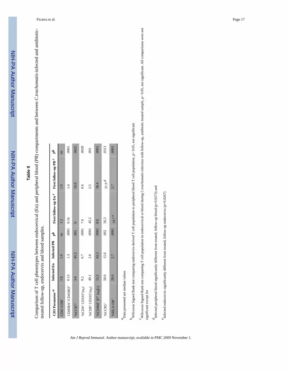
Ficarra et al. Page 17
Tabl
e II
Com
paris
on o
f T c
ell p
heno
type
s bet
wee
n en
doce
rvic
al (E
n) a
nd p
erip
hera
l blo
od (P
B) c
ompa
rtmen
ts a
nd b
etw
een
C.tr
acho
mat
is-in
fect
ed a
nd a
ntib
iotic
-tre
ated
follo
w-u
p, e
ndoc
ervi
x an
d bl
ood
sam
ples
.
CD
3 Pa
ram
eter
aIn
fect
ed E
nIn
fect
ed P
BPb
Firs
t fol
low
-up
En
cFi
rst f
ollo
w-u
p PB
cPb
CD
4+ /C
D8+
1.0
1.0
ns1.
21.
0ns
CD
45R
A+ /
CD
45R
O+
0.13
1.3
.000
10.
161.
4.0
001
%C
CR
7+3.
645
.3.0
010
16.9
.062
5
%C
D4+
CD
103+ (
α E)
9.2
0.7
.000
17.
60.
6.0
028
%C
D8+
CD
103+ (
α E)
49.1
2.6
.000
145
.22.
5.0
01
%C
D49
d+ β7
+ (α
4β7)
12.2
43.3
.000
18.
638
.4.0
001
%C
CR
5+58
.015
.6.0
0256
.321
.9 d
.031
3
%H
LA-D
R+
28.0
2.7
.000
114
.7 e
2.7
.000
1
a Dat
a pr
esen
ted
are
med
ian
valu
es
b Wilc
oxon
Sig
ned
Ran
k te
st c
ompa
ring
endo
cerv
ix-d
eriv
ed T
cel
l pop
ulat
ion
to p
erip
hera
l blo
od T
cel
l pop
ulat
ion,
p>
0.05
, not
sign
ifica
nt
c Wilc
oxon
Sig
ned
Ran
k te
st c
ompa
ring
T ce
ll po
pula
tion
in e
ndoc
ervi
cal o
r blo
od d
urin
g C
.trac
hom
atis
infe
ctio
n w
ith fo
llow
-up,
ant
ibio
tic tr
eate
d sa
mpl
e, p
> 0.
05, n
ot si
gnifi
cant
. All
com
paris
ons w
ere
not
sign
ifica
nt e
xcep
t for
d Infe
cted
per
iphe
ral b
lood
sign
ifica
ntly
diff
eren
t fro
m tr
eate
d, fo
llow
-up
bloo
d (p
=0.0
273)
and
e Infe
cted
end
ocer
vix
sign
ifica
ntly
diff
eren
t fro
m tr
eate
d, fo
llow
-up
endo
cerv
ix (p
=0.0
267)
Am J Reprod Immunol. Author manuscript; available in PMC 2009 November 1.
Related Documents