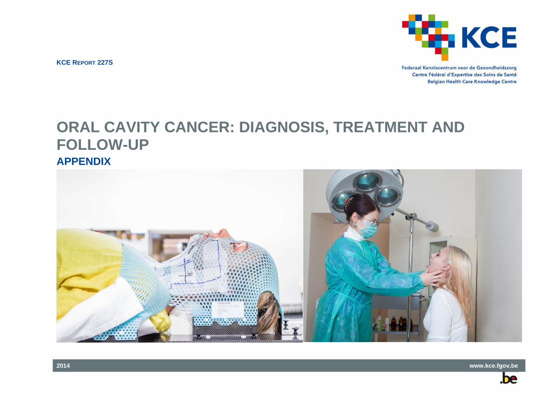
2014 www.kce.fgov.be KCE REPORT 227S ORAL CAVITY CANCER: DIAGNOSIS, TREATMENT AND FOLLOW-UP APPENDIX

Welcome message from author
This document is posted to help you gain knowledge. Please leave a comment to let me know what you think about it! Share it to your friends and learn new things together.
Transcript
2014 www.kce.fgov.be
KCE REPORT 227S
ORAL CAVITY CANCER: DIAGNOSIS, TREATMENT AND FOLLOW-UP APPENDIX

2014 www.kce.fgov.be
KCE REPORT 227S GOOD CLINICAL PRACTICE
ORAL CAVITY CANCER: DIAGNOSIS, TREATMENT AND FOLLOW-UP APPENDIX VINCENT GRÉGOIRE, ROOS LEROY, PAULINE HEUS, FLEUR VAN DE WETERING, LOTTY HOOFT, ROB J.P.M. SCHOLTEN, LEEN VERLEYE, LAURENS CARP, PAUL CLEMENT, PHILIPPE DERON, KAROLIEN GOFFIN, MARC HAMOIR, ESTHER HAUBEN, KRISTOF HENDRICKX, ROBERT HERMANS, SIDNEY KUNZ, OLIVIER LENSSEN, SANDRA NUYTS, CARL VAN LAER, JAN VERMORKEN, ELINE APPERMONT, ANNELIES DE PRINS, ELINE HEBBELINCK, GEERT HOMMEZ, CAROLINE VANDENBRUAENE, EVELINE VANHALEWYCK, JOAN VLAYEN
COLOPHON Title: Oral cavity cancer: diagnosis, treatment and follow-up –Appendix
Authors: Vincent Grégoire (UCL), Roos Leroy (KCE), Pauline Heus (Dutch Cochrane Center), Fleur van de Wetering (Dutch Cochrane Center), Lotty Hooft (Dutch Cochrane Center), Rob J.P.M. Scholten (Dutch Cochrane Center), Leen Verleye (KCE), Laurens Carp (UZA), Paul Clement (UZ Leuven), Philippe Deron (UZ Gent), Karolien Goffin (UZ Leuven), Marc Hamoir (UCL), Esther Hauben (UZ Leuven), Kristof Hendrickx (AZ Nikolaas), Robert Hermans (UZ Leuven), Sidney Kunz (AZ Groeninge), Olivier Lenssen (ZNA), Sandra Nuyts (UZ Leuven), Carl Van Laer (UZA), Jan Vermorken (UZA), Eline Appermont (UZ Leuven), Annelies De Prins (UZ Gent), Eline Hebbelinck (UZ Gent), Geert Hommez (UZ Gent), Caroline Vandenbruaene (AZ Sint Jan Brugge), Eveline Vanhalewyck (UZ Leuven), Joan Vlayen (KCE)
Project coordinator and senior supervisor:
Sabine Stordeur (KCE)
Reviewers: Anja Desomer (KCE), Sabine Stordeur (KCE), Raf Mertens (KCE)
Stakeholders: Jean-François Daisne (Association Belge de Radiothérapie-Oncologie), François-Xavier Hanin (Société Belge de Médecine Nucléaire), Peter Lemkens (Koninklijke Belgische Vereniging voor Oto-Rhino-Laryngologie, Gelaat- en Halschirurgie), Marc Lemort (Belgian Society of Radiology), Max Lonneux (Société Belge de Médecine Nucléaire), Pierre Mahy (Koninklijke Belgische Vereniging voor Stomatologie en Maxillo-Faciale Heelkunde), Myriam Remmelink (Société Belge d'Anatomopathologie), Ward Rommel (Vlaamse Liga tegen kanker), Joseph Schoenaers (Koninklijke Belgische Vereniging voor Stomatologie en Maxillo-Faciale Heelkunde), Pol Specenier (Belgische Vereniging voor Medische Oncologie), Geert Van Hemelen (Koninklijke Belgische Vereniging voor Stomatologie en Maxillo-Faciale Heelkunde), Pieter Van de Putte (Stichting Kankerregister), Vincent Vander Poorten (Domus Medica), Dirk Vangestel (Belgische Vereniging voor Radiotherapie-Oncologie), Tom Vauterin (Koninklijke Belgische Vereniging voor Oto-Rhino-Laryngologie, Gelaat- en Halschirurgie), Birgit Weynand (Société Belge d'Anatomopathologie), Karin Rondia (Fondation contre le Cancer), Elisabeth Van Eycken (Stichting Kankerregister)
External validators: Elisabeth Junor (NHS Scotland UK), Pierre Castadot (CHU Charleroi)
CEBAM validators: Dirk Ramaekers, Martine Goossens, Michel Martens
Other reported interests: Membership of a stakeholder group on which the results of this report could have an impact.: Paul Clement (BSMO, VWHHT, ASCO, ESMO), Sandra Nuyts (Vlaamse werkgroep Hoofd-en halstumoren), Elisabeth Van Eycken (BVRO) Holder of intellectual property (patent, product developer, copyrights, trademarks, etc.): Paul Clement (methods of inhibiting vascular proliferation) Participation in scientific or experimental research as an initiator, principal investigator or researcher: Paul
Clement, Jean-François Daisne (Boehringer Head and Neck Lux 2), Karolien Goffin (wetenschappelijk onderzoek hals- en hoodtumoren), Vincent Grégoire, Marc Hamoir (recherche clinique et tranfert dans les cancers de la tête et du cou, PI d’une etude académique international sur la valeur du bilan postradiochimiothérapie dans les cancers avancés), François-Xavier Hanin (Noichl EORTC study, GETTEC study), Dirk Van Gestel (PI 2 dose-paintingstudies: NKO recidieven en bot metastasen), Pol Specenier (Rage studie Merck), Geert Van Hemelen (3D surgery planning protocol), Vincent Vander Poorten (IKV), Sandra Nuyts (wetenschappelijk onderzoek FVVO, VLK, STK; PI klinische studie hoofd- en halsoncologie) A grant, fees or funds for a member of staff or another form of compensation for the execution of research: Karolien Goffin (Klinisch Onderzoeksfonds UZ Leuven), Sandra Nuyts Consultancy or employment for a company, an association or an organisation that may gain or lose financially due to the results of this report: Paul Clement (consultancy Merck Serono), Joseph Schoenaers (Hoogleraar KUL, UZ Leuven), Jan Baptist Vermorken (Advisory Board Meeting Merck Serono) Payments to speak, training remuneration, subsidised travel or payment for participation at a conference: Paul Clement (Merck Serono), Jean-François Daisne (Merck Serono), Karolien Goffin (cursus radiotherapie), François-Xavier Hanin (Forum Nucléaire), Dirk Van Gestel (Accuray), Sandra Nuyts Presidency or accountable function within an institution, association, department or other entity on which the results of this report could have an impact: Paul Clement (VWHHT), Jean-François Daisne (radiotherapie CMSE Namur), Vincent Grégoire (lid van ESTRO EORTC) , Peter Lemkens (Koninklijke Belgische Vereniging voor NKO Hoofd en Hals), Vincent Vander Poorten (secretaris VWHHT), Joseph Schoenaers (voorzitter KBVSMFH, lid International Board certification exam OMFP, secretaris generaal European Board Assessment)
Layout: Ine Verhulst
Disclaimer: The external experts were consulted about a (preliminary) version of the scientific report. Their comments were discussed during meetings. They did not co-author the scientific report and did not necessarily agree with its content.
Subsequently, a (final) version was submitted to the validators. The validation of the report results from a consensus or a voting process between the validators. The validators did not co-author the scientific report and did not necessarily all three agree with its content.
Finally, this report has been approved by common assent by the Executive Board. Only the KCE is responsible for errors or omissions that could persist. The policy recommendations
are also under the full responsibility of the KCE.
Publication date: 26 August 2014 (2nd print; 1st print: 08 July 2014)
Domain: Good Clinical Practice (GCP)
MeSH: Mouth Neoplasms; Head and Neck Neoplasms; Practice Guideline
NLM Classification: WE 707
Language: English
Format: Adobe® PDF™ (A4)
Legal depot: D/2013/10.273/59
Copyright: KCE reports are published under a “by/nc/nd” Creative Commons Licence http://kce.fgov.be/content/about-copyrights-for-kce-reports.
How to refer to this document? Grégoire V, Leroy R, Heus P, van de Wetering F, Hooft L, Scholten R, Verleye L, Carp L, Clement P, Deron P, Goffin K, Hamoir M, Hauben E, Hendrickx K, Hermans R, Kunz S, Lenssen O, Nuyts S, Van Laer C, Vermorken J, Appermont E, De Prins A, Hebbelinck E, Hommez G, Vandenbruaene C, Vanhalewyck E, Vlayen J. Oral cavity cancer: diagnosis, treatment and follow-up – Appendix. Good Clinical Practice (GCP) Brussels: Belgian Health Care Knowledge Centre (KCE). 2013. KCE Reports 227S. D/2013/10.273/59.
This document is available on the website of the Belgian Health Care Knowledge Centre.
KCE Report 227S Oral cavity cancer 1
APPENDIX REPORT TABLE OF CONTENTS 1. COMPOSITION OF THE GUIDELINE DEVELOPMENT GROUP ...................................................... 13
1.1. COMPOSITION OF THE GUIDELINE DEVELOPMENT GROUP ...................................................... 13 1.2. COMPOSITION OF THE KCE EXPERT TEAM ................................................................................... 14 1.3. EXTERNAL RESEARCHERS INVOLVED IN THE GUIDELINE DEVELOPMENT ............................. 15 2. SEARCH STRATEGIES ...................................................................................................................... 16 2.1. SEARCH STRATEGY FOR GUIDELINES .......................................................................................... 16 2.2. SEARCH STRATEGIES FOR OTHER PUBLICATIONS (SYSTEMATIC REVIEWS, META-
ANALYSES, INDIVIDUAL STUDIES) .................................................................................................. 18 2.2.1. RQ1: PET/CT in the staging of oral cavity cancer ................................................................. 18 2.2.2. RQ2: HPV testing in patients with oral cavity cancer ............................................................. 27 2.2.3. RQ3: elective lymph node dissection for patients with cN0 oral cavity cancer ...................... 29 2.2.4. RQ4: elective lymph node dissection for patients with cN+ oral cavity cancer ...................... 37 2.2.5. RQ5: elective lymph node dissection of contralateral neck ................................................... 37 2.2.6. RQ6: value of PET / MRI in the decision of neck dissection after CRT ................................. 40 2.2.7. RQ7: neck dissection after chemoradiotherapy in patients with oral cavity cancer ............... 45 2.2.8. RQ8: IMRT for patients with locally advanced HNSCC ......................................................... 49 2.2.9. RQ9: induction chemotherapy in patients with HNSCC......................................................... 53 2.2.10. RQ10: primary CRT for patients with non-resectable M0 HNSCC ........................................ 58 2.2.11. RQ11: interventions for M+ disease or recurrent disease not suitable for curative treatment69
3. QUALITY APPRAISAL ........................................................................................................................ 74 3.1. QUALITY APPRAISAL TOOLS ............................................................................................................ 74
3.1.1. Guidelines .............................................................................................................................. 74 3.1.2. Systematic reviews ................................................................................................................ 75 3.1.3. Diagnostic accuracy studies .................................................................................................. 77 3.1.4. Primary studies for therapeutic interventions ......................................................................... 79
3.2. GUIDELINES SELECTION AND QUALITY APPRAISAL .................................................................... 81 3.3. STUDY SELECTION AND QUALITY APPRAISAL .............................................................................. 83
2 Oral cavity cancer KCE Report 227S
3.3.1. RQ1: PET/CT in the staging of oral cavity cancer ................................................................. 83 3.3.2. RQ2: HPV testing in patients with oral cavity cancer ............................................................. 92 3.3.3. RQ3 & RQ4: elective lymph node dissection for patients with oral cavity cancer ................. 92 3.3.4. RQ5: elective lymph node dissection of contralateral neck ................................................. 105 3.3.5. RQ6: value of PET / MRI in the decision of neck dissection after CRT ............................... 111 3.3.6. RQ7: neck dissection after chemoradiotherapy in patients with oral cavity cancer ............. 126 3.3.7. RQ8: IMRT for patients with locally advanced HNSCC ....................................................... 133 3.3.8. RQ9: induction chemotherapy in patients with HNSCC....................................................... 144 3.3.9. RQ10: primary CRT for patients with non-resectable M0 HNSCC ...................................... 151 3.3.10. RQ11: interventions for M+ disease or recurrent disease not suitable for curative treatment ..
............................................................................................................................................. 160 4. EVIDENCE TABLES BY CLINICAL QUESTION .............................................................................. 169 4.1. RQ1: PET/CT FOR STAGING OF HNSCC ....................................................................................... 169
4.1.1. Nodal staging ....................................................................................................................... 169 4.1.2. M-staging ............................................................................................................................. 172
4.2. RQ3: ELECTIVE LYMPH NODE DISSECTION FOR PATIENTS WITH CN0 ORAL CAVITY CANCER ............................................................................................................................................ 177
4.3. RQ4: ELECTIVE LYMPH NODE DISSECTION FOR PATIENTS WITH CN+ ORAL CAVITY CANCER ............................................................................................................................................ 184
4.4. RQ5: ELECTIVE LYMPH NODE DISSECTION OF CONTRALATERAL NECK ............................... 191 4.5. RQ6: VALUE OF PET / MRI IN THE DECISION OF NECK DISSECTION AFTER CRT .................. 193 4.6. RQ7: NECK DISSECTION AFTER CHEMORADIOTHERAPY IN PATIENTS WITH ORAL
CAVITY CANCER .............................................................................................................................. 197 4.7. RQ8: IMRT FOR PATIENTS WITH LOCALLY ADVANCED HNSCC ............................................... 204 4.8. RQ9: INDUCTION CHEMOTHERAPY IN PATIENTS WITH HNSCC ............................................... 208 4.9. RQ10: PRIMARY CRT FOR PATIENTS WITH NON-RESECTABLE M0 HNSCC ........................... 220 4.10. RQ11: INTERVENTIONS FOR M+ DISEASE OR RECURRENT DISEASE NOT SUITABLE FOR
CURATIVE TREATMENT .................................................................................................................. 235 5. SUMMARY OF FINDINGS TABLES AND GRADE PROFILES ....................................................... 239 5.1. RQ1: PET/CT IN THE STAGING OF ORAL CAVITY CANCER ........................................................ 239
KCE Report 227S Oral cavity cancer 3
5.2. RQ3: ELECTIVE LYMPH NODE DISSECTION FOR PATIENTS WITH CN0 ORAL CAVITY CANCER ............................................................................................................................................ 249 5.2.1. Elective lymph node dissection versus watchful waiting for patients with cTanyN0M0 oral
cavity cancer ........................................................................................................................ 249 5.2.2. Elective lymph node dissection versus watchful waiting for patients with cT1-2N0M0
cancer of the tongue ............................................................................................................ 251 5.2.3. Elective lymph node dissection versus watchful waiting for patients with cT1-2N0M0
buccal cancer ....................................................................................................................... 255 5.3. RQ4: ELECTIVE LYMPH NODE DISSECTION FOR PATIENTS WITH CN+ ORAL CAVITY
CANCER ............................................................................................................................................ 256 5.3.1. Selective lymph node dissection versus modified radical lymph node dissection for
patients with cTanyN+M0 oral cavity cancer ....................................................................... 256 5.4. RQ5: ELECTIVE LYMPH NODE DISSECTION OF CONTRALATERAL NECK ............................... 259 5.5. RQ6: VALUE OF PET / MRI IN THE DECISION OF NECK DISSECTION AFTER CRT .................. 260 5.6. RQ7: NECK DISSECTION AFTER CHEMORADIOTHERAPY IN PATIENTS WITH ORAL
CAVITY CANCER .............................................................................................................................. 263 5.7. RQ8: IMRT FOR PATIENTS WITH LOCALLY ADVANCED HNSCC ............................................... 268 5.8. RQ9: INDUCTION CHEMOTHERAPY IN PATIENTS WITH HNSCC ............................................... 274
5.8.1. Induction chemotherapy with cisplatin and 5-fluorouracil for patients with stage 3 and 4 HNSCC ................................................................................................................................ 274
5.8.2. Induction chemotherapy with platin-containing combinations other than cisplatin and 5-fluorouracil in patients with stage 3 and 4 HNSCC ................................................... 278
5.8.3. Multi-agent induction chemotherapy without platin in patients with stage 3 and 4 HNSCC ................................................................................................................................ 281
5.9. RQ10: PRIMARY CRT FOR PATIENTS WITH NON-RESECTABLE M0 HNSCC ........................... 284 5.9.1. Primary CRT for patients with non-resectable (T4b) M0 HNSCC ....................................... 284 5.9.2. Combination of EGFR-inhibitors and radiotherapy for patients with non-resectable
(T4b) M0 HNSCC? ............................................................................................................... 291 5.10. RQ11: INTERVENTIONS FOR M+ DISEASE OR RECURRENT DISEASE NOT SUITABLE
FOR CURATIVE TREATMENT .......................................................................................................... 294 5.10.1. Chemoradiotherapy versus BSC for M+ HNSCC or recurrent HNSCC not suitable for
curative treatment ................................................................................................................ 294
4 Oral cavity cancer KCE Report 227S
5.10.2. Chemotherapy versus BSC for M+ HNSCC or recurrent HNSCC not suitable for curative treatment .............................................................................................................................. 295
5.10.3. Radiotherapy versus BSC for M+ HNSCC or recurrent HNSCC not suitable for curative treatment .............................................................................................................................. 296
5.10.4. Salvage surgery versus BSC for M+ HNSCC or recurrent HNSCC not suitable for curative treatment .............................................................................................................................. 297
5.10.5. Anti-EGFR plus BSC versus BSC alone for M+ HNSCC or recurrent HNSCC not suitable for curative treatment .............................................................................................. 298
6. FOREST PLOTS ................................................................................................................................ 299 6.1. RQ1: PET/CT IN THE STAGING OF ORAL CAVITY CANCER ........................................................ 299
6.1.1. Detection of cervical lymph nodes: PET (patient-based) ..................................................... 299 6.1.2. Detection of cervical lymph nodes: PET (neck-side-based) ................................................ 301 6.1.3. Detection of cervical lymph nodes: PET (node-based)........................................................ 303 6.1.4. Detection of cervical lymph nodes: non-enhanced PET/CT (patient-based) ....................... 303 6.1.5. Detection of cervical lymph nodes: non-enhanced PET/CT (neck-side-based) .................. 304 6.1.6. Detection of cervical lymph nodes: non-enhanced PET/CT (node-based).......................... 306 6.1.7. Detection of cervical lymph nodes: contrast-enhanced PET/CT (neck-side-based) ........... 308 6.1.8. Detection of cervical lymph nodes: contrast-enhanced PET/CT (node-based) ................... 308 6.1.9. Detection of cervical lymph nodes: conventional imaging ................................................... 309 6.1.10. Detection of cervical lymph nodes: comparison between PET or PET/CT with
conventional imaging ........................................................................................................... 310 6.1.11. Detection of distant metastases or second primaries .......................................................... 320 6.1.12. Detection of bone marrow invasion ...................................................................................... 322 6.1.13. Detection of bone metastases ............................................................................................. 323 6.1.14. Detection of lung metastases ............................................................................................... 323 6.1.15. Detection of liver metastases ............................................................................................... 324 6.1.16. Detection of head and neck metastases .............................................................................. 324 6.1.17. Detection of distant lymph node metastases ....................................................................... 325 6.1.18. Detection of other metastases of aerodigestive tract ........................................................... 325
6.2. RQ6: VALUE OF PET / MRI IN THE DECISION OF NECK DISSECTION AFTER CRT .................. 326 6.3. RQ8: IMRT ......................................................................................................................................... 327
KCE Report 227S Oral cavity cancer 5
6.4. RQ9: INDUCTION CHEMOTHERAPY IN PATIENTS WITH HNSCC ............................................... 329 6.5. RQ10: PRIMARY CRT FOR PATIENTS WITH NON-RESECTABLE M0 HNSCC ........................... 332 7. EXTERNAL REVIEW ......................................................................................................................... 335 7.1. EVALUATION OF THE RECOMMENDATIONS BY THE STAKEHOLDERS ................................... 335 8. TNM CLASSIFICATION .................................................................................................................... 344 8.1. CTNM CLINICAL CLASSIFICATION ................................................................................................. 344 8.2. PTNM PATHOLOGICAL CLASSIFICATION ..................................................................................... 345 8.3. STAGE GROUPING ........................................................................................................................... 346
6 Oral cavity cancer KCE Report 227S
LIST OF FIGURES Figure 1 – Study flow of selection of SRs regarding research question 3 and 4 ................................................ 93 Figure 2 – Study flow of selection of primary studies regarding research question 3 and 4 .............................. 95 Figure 3 – Risk of bias summary of RCTs for RQ3 and RQ4 adapted from Bessell 2011 ............................... 100 Figure 4 – Risk of bias summary per item of RCTs for RQ3 and 4 adapted from Bessell 2011 ...................... 101 Figure 5 – Risk of bias summary of the comparative observational studies RQ3 ............................................ 102 Figure 6 – Risk of bias summary per item of the comparative observational studies RQ3 .............................. 103 Figure 7 – Risk of bias summary of the comparative observational studies RQ4 ............................................ 104 Figure 8 – Risk of bias summary per item of the comparative observational studies RQ4 .............................. 105 Figure 9 – Study flow of selection of SRs ......................................................................................................... 106 Figure 10 – Study flow of selection of primary studies ..................................................................................... 108 Figure 11 – Risk of bias summary of included primary studies ........................................................................ 110 Figure 12 – Study flow of SR selection ............................................................................................................. 112 Figure 13 – Study flow of selection of primary studies evaluating the value of PET ........................................ 118 Figure 14 – Study flow of SR selection ............................................................................................................. 127 Figure 15 – Study flow of selection of primary studies ..................................................................................... 129 Figure 16 – Risk of bias summary of included primary studies ........................................................................ 132 Figure 17 – Study flow of selection of SRs regarding research question 8 ...................................................... 134 Figure 18 – Study flow of selection of SRs regarding research question 8 from The Cochrane Library ......... 135 Figure 19 – Study flow of selection of primary studies regarding research question 8 .................................... 137 Figure 20 – Risk of bias summary of comparative observational studies for RQ8 ........................................... 141 Figure 21 – Risk of bias summary per item of comparative observational studies for RQ8............................. 142 Figure 22 – Risk of bias summary of RCTs for RQ8 ........................................................................................ 143 Figure 23 – Risk of bias summary per item of RCTs for RQ8 .......................................................................... 144 Figure 24 – Study flow of selection of SRs regarding research question 9 ...................................................... 145 Figure 25 – Study flow of selection of primary studies regarding research question 9 .................................... 147 Figure 26 – Risk of bias summary of newly identified RCTs for RQ9 .............................................................. 150 Figure 27 – Risk of bias summary per item of newly identified RCTs for RQ9 ................................................ 151 Figure 28 – Study flow of selection of SRs regarding research question 10 .................................................... 152 Figure 29 – Study flow of selection of primary studies regarding research question 10 .................................. 155 Figure 30 – Risk of bias summary of comparative observational studies regarding RQ10.............................. 159
KCE Report 227S Oral cavity cancer 7
Figure 31 – Risk of bias summary per item of comparative observational studies regarding RQ10 ............... 160 Figure 32 – Study flow of selection of studies regarding research question 11 ............................................... 161 Figure 33 – Risk of bias summary of comparative observational studies regarding RQ11.............................. 165 Figure 34 – Risk of bias summary per item of comparative observational studies regarding RQ11 ............... 166 Figure 35 – Risk of bias summary of the RCT regarding RQ11 ....................................................................... 167 Figure 36 – Risk of bias summary per item of the RCT regarding RQ11 ......................................................... 168 Figure 37 – Forest plot: detection of cervical lymph nodes with PET (patient-based analysis) ....................... 299 Figure 38 – SROC curve: detection of cervical lymph nodes with PET (patient-based analysis) .................... 300 Figure 39 – Meta-analysis: detection of cervical lymph nodes with PET (patient-based analysis) .................. 301 Figure 40 – Forest plot: detection of cervical lymph nodes with PET (neck-side-based analysis) .................. 301 Figure 41 – HSROC curve: detection of cervical lymph nodes with PET (neck-side-based analysis) ............. 302 Figure 42 – Meta-analysis: detection of cervical lymph nodes with PET (neck-side-based analysis) ............. 303 Figure 43 – Forest plot: detection of cervical lymph nodes with PET (node-based analysis) .......................... 303 Figure 44 – Forest plot: detection of cervical lymph nodes with non-enhanced PET/CT (patient-based analysis) .................................................................................................................................... 303 Figure 45 – Forest plot: detection of cervical lymph nodes with non-enhanced PET/CT (neck-side-based analysis) ............................................................................................................................... 304 Figure 46 – HSROC curve: detection of cervical lymph nodes with non-enhanced PET/CT (neck-side-based analysis) ............................................................................................................................... 305 Figure 47 – Meta-analysis: detection of cervical lymph nodes with non-enhanced PET/CT (neck-side-based analysis) ............................................................................................................................... 306 Figure 48 – Forest plot: detection of cervical lymph nodes with non-enhanced PET/CT (node-based analysis) ...................................................................................................................................... 306 Figure 49 – HSROC curve: detection of cervical lymph nodes with non-enhanced PET/CT (node-based analysis) ...................................................................................................................................... 307 Figure 50 – Meta-analysis: detection of cervical lymph nodes with non-enhanced PET/CT (node-based analysis) ...................................................................................................................................... 308 Figure 51 – Forest plot: detection of cervical lymph nodes with contrast-enhanced PET/CT (neck-side-based analysis) ............................................................................................................................... 308 Figure 52 – Forest plot: detection of cervical lymph nodes with contrast-enhanced PET/CT (node-based analysis) ...................................................................................................................................... 308
8 Oral cavity cancer KCE Report 227S
Figure 53 – Forest plot: detection of cervical lymph nodes with conventional imaging (in studies comparing with PET or PET/CT) ..................................................................................................... 309 Figure 54 – Meta-analysis: detection of cervical lymph nodes, neck-side-based analysis, PET or PET/CT in comparative studies * ...................................................................................................................... 310 Figure 55 – Meta-analysis: detection of cervical lymph nodes, neck-side-based analysis, conventional imaging in comparative studies ........................................................................................................................ 311 Figure 56 – Meta-analysis (sensitivity analysis): detection of cervical lymph nodes, neck-side-based analysis, PET or PET/CT in comparative studies * ........................................................................................... 312 Figure 57 – Meta-analysis: detection of cervical lymph nodes, node-based analysis, PET or PET/CT in comparative studies * ...................................................................................................................... 313 Figure 58 – Meta-analysis: detection of cervical lymph nodes, node-based analysis, conventional imaging in comparative studies ........................................................................................................................ 314 Figure 59 – Meta-analysis (sensitivity analysis 1): detection of cervical lymph nodes, node-based analysis, PET or PET/CT in comparative studies * ........................................................................................... 315 Figure 60 – Meta-analysis (sensitivity analysis 2): detection of cervical lymph nodes, node-based analysis, PET or PET/CT in comparative studies * ........................................................................................... 316 Figure 61 – Meta-analysis (sensitivity analysis 2): detection of cervical lymph nodes, node-based analysis, conventional imaging in comparative studies * .................................................................................. 317 Figure 62 – Meta-analysis (sensitivity analysis 3): detection of cervical lymph nodes, node-based analysis, PET/CT in comparative studies with CT ............................................................................................ 318 Figure 63 – Meta-analysis (sensitivity analysis 3): detection of cervical lymph nodes, node-based analysis, CT in comparative studies with PET/CT ............................................................................................ 319 Figure 64 – Forest plot: detection of distant metastases or second primaries ................................................. 320 Figure 65 – SROC curve: detection of distant metastases or second primaries .............................................. 321 Figure 66 – Meta-analysis: detection of distant metastases or second primaries with PET or PET/CT .......... 322 Figure 67 – Forest plot: detection of bone marrow invasion ............................................................................ 322 Figure 68 – Forest plot: detection of bone metastases .................................................................................... 323 Figure 69 – Forest plot: detection of lung metastases...................................................................................... 323 Figure 70 – Forest plot: detection of liver metastases ...................................................................................... 324 Figure 71 – Forest plot: detection of head and neck metastases ..................................................................... 324 Figure 72 – Forest plot: detection of distant lymph node metastases .............................................................. 325 Figure 73 – Forest plot: detection of other metastases of aerodigestive tract ................................................. 325
KCE Report 227S Oral cavity cancer 9
Figure 74 – Forest plot: Detection of residual disease in cervical lymph nodes with PET/CT after (at least) CRT - Patient-based analysis ............................................................................................................ 326 Figure 75 – Forest plot: Detection of residual disease in cervical lymph nodes with PET/CT after (at least) CRT – Hemi-neck-based analysis ..................................................................................................... 326 Figure 76 – Forest plot: Detection of residual disease in cervical lymph nodes with PET after (at least) CRT – Patient-based analysis ........................................................................................................... 326 Figure 77 – Forest plot: Detection of residual disease in cervical lymph nodes with PET after (at least) CRT – Hemi-neck-based analysis ..................................................................................................... 327 Figure 78 – Forest plot: Detection of residual disease in cervical lymph nodes with PET after (at least) CRT – Node-based analysis .............................................................................................................. 327 Figure 79 – Forest plot: Pooled result for mucositis grade 2 or more of IMRT vs conventional RT ................. 327 Figure 80 – Forest plot: Pooled results for dysphagia grade 2 or more of IMRT vs conventional RT ............. 328 Figure 81 – Meta-analysis for different types of induction chemotherapy (PF, other platin-containing combinations, multi-agent chemotherapy combination without platin, and single-agent chemotherapy (methotrexate)) followed by locoregional treatment versus locoregional treatment alone for outcome overall survival for patients with locally advanced HNSCC (stage 3 and 4) ...... 329 Figure 82 – Meta-analysis for different types of induction chemotherapy (PF, other platin-containing combinations, multi-agent chemotherapy combination without platin, and single-agent chemotherapy (methotrexate)) followed by locoregional treatment versus locoregional treatment alone for outcome disease-free survival for patients with locally advanced HNSCC (stage 3 and 4) .................................................................................................................................................. 330 Figure 83 – Meta-analysis for PF induction chemotherapy followed by locoregional treatment versus locoregional treatment alone for outcome post-treatment mortality for patients with locally advanced HNSCC (stage 3 and 4) ................................................................................................................... 331 Figure 84 – Pooled results for grade 3-4 mucositis of primary CRT with RT alone ......................................... 332 Figure 85 – Pooled results for grade 3-4 dermatitis of primary CRT with RT alone ......................................... 332 Figure 86 – Pooled results for grade 3-4 anemia of primary CRT with RT alone ............................................ 333 Figure 87 – Pooled results for grade 3-4 leukopenia of primary CRT with RT alone ....................................... 333 Figure 88 – Pooled results for grade 3-4 thrombocytopenia of primary CRT with RT alone ........................... 334
10 Oral cavity cancer KCE Report 227S
LIST OF TABLES Table 1 – Search results - Guidelines on HNSCC .............................................................................................. 16 Table 2 – AGREE II instrument .......................................................................................................................... 74 Table 3 – AMSTAR checklist .............................................................................................................................. 75 Table 4 – The QUADAS tool ............................................................................................................................... 77 Table 5 – Cochrane Collaboration’s tool for assessing risk of bias .................................................................... 79 Table 6 – AGREE scores of identified guidelines ............................................................................................... 81 Table 7 – Reviews excluded based on full-text evaluation ................................................................................. 83 Table 8 – Methodological quality of the included systematic review (AMSTAR) ............................................... 84 Table 9 – Excluded primary studies based on full-text evaluation ..................................................................... 85 Table 10 – Methodological quality of the included primary studies for N-staging with PET-scan ...................... 86 Table 11 – Methodological quality of the included primary studies for M-staging with PET-scan ..................... 89 Table 12 – Methodological quality of the included systematic review ................................................................ 92 Table 13 – Included SRs regarding research question 3 and 4 ......................................................................... 94 Table 14 – Excluded SRs regarding research question 3 and 4 ........................................................................ 94 Table 15 – Included primary studies regarding research question 3 and 4 ....................................................... 96 Table 16 – Excluded primary studies regarding research question 3 and 4 ...................................................... 97 Table 17 – Methodological quality of the included systematic review (AMSTAR) Bessell 2011 ........................ 99 Table 18 – Reviews excluded based on full-text evaluation ............................................................................. 107 Table 19 – Excluded primary studies based on full-text evaluation ................................................................. 109 Table 20 – Reviews excluded based on full-text evaluation ............................................................................. 113 Table 21 – Methodological quality of the included systematic reviews (AMSTAR) .......................................... 113 Table 22 – Excluded primary studies cited in Gupta 2011 and/or Isles 2008 and the reasons for exclusion .. 114 Table 23 – Excluded primary PET(/CT) studies based on full-text evaluation ................................................. 119 Table 24 – Excluded primary MRI studies based on full-text evaluation .......................................................... 120 Table 25 – Methodological quality of selected primary PET(/CT) studies ........................................................ 122 Table 26 – Methodological quality of selected primary MRI studies ................................................................ 124 Table 27 – Reviews excluded based on full-text evaluation ............................................................................. 128 Table 28 – Excluded primary studies based on full-text evaluation ................................................................. 130 Table 29 – Included SRs regarding research question 8 ................................................................................. 136 Table 30 – Excluded SRs regarding research question 8 ................................................................................ 136
KCE Report 227S Oral cavity cancer 11
Table 31 – Included primary studies regarding research question 8 ................................................................ 138 Table 32 – Excluded primary studies regarding research question 8 .............................................................. 138 Table 33 – Methodological quality of the included systematic review (AMSTAR) 49 ........................................ 140 Table 34 – Included SRs regarding research question 9 (n=2) ........................................................................ 146 Table 35 – Excluded SRs regarding research question 9 (n=2) ...................................................................... 146 Table 36 – Included primary studies regarding research question 9 (n=5) ...................................................... 148 Table 37 – Excluded primary studies regarding research question 9 (n=15) ................................................... 148 Table 38 – Methodological quality of the included systematic reviews (AMSTAR) (n=2) ................................ 149 Table 39 – Excluded SRs regarding research question (n=18) ....................................................................... 153 Table 40 – Included primary studies regarding research question 10 (n=7) .................................................... 156 Table 41 – Excluded primary studies regarding research question 10 (n=46) ................................................. 156 Table 42 – Included studies regarding research question 11 ........................................................................... 162 Table 43 – Excluded studies regarding research question 11 ......................................................................... 162 Table 44 – N-staging of HNSCC with PET or PET/CT: systematic reviews..................................................... 169 Table 45 – N-staging of HNSCC with PET or PET/CT: primary studies .......................................................... 170 Table 46 – M-staging of HNSCC with PET or PET/CT: systematic reviews .................................................... 172 Table 47 – M-staging of HNSCC with PET or PET/CT: primary studies .......................................................... 174 Table 48 – Value of PET(/CT) in the decision of neck dissection after CRT: systematic reviews ................... 193 Table 49 – Value of PET(/CT) in the decision of neck dissection after CRT: primary studies ......................... 194 Table 50 – Value of MRI in the decision of neck dissection after CRT: primary studies .................................. 196 Table 51 – Induction chemotherapy: systematic reviews ................................................................................. 208 Table 52 – Induction chemotherapy: RCTs ...................................................................................................... 210 Table 53 – Evidence profile for diagnosis: PET for nodal staging .................................................................... 239 Table 54 – Evidence profile for diagnosis: non-enhanced PET/CT for nodal staging ...................................... 240 Table 55 – Evidence profile for diagnosis: contrast-enhanced PET/CT for nodal staging ............................... 241 Table 56 – SoF table for diagnosis: PET or PET/CT versus conventional imaging for nodal staging – neck-side-based analysis * ................................................................................................................................................ 242 Table 57 – SoF table for diagnosis: PET or PET/CT versus conventional imaging for nodal staging – node-based analysis * ................................................................................................................................................ 243 Table 58 – SoF table for diagnosis: PET/CT versus CT for nodal staging – node-based analysis *................ 244
12 Oral cavity cancer KCE Report 227S
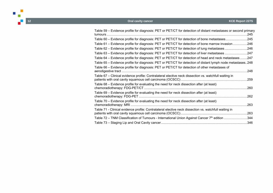
Table 59 – Evidence profile for diagnosis: PET or PET/CT for detection of distant metastases or second primary tumours ............................................................................................................................................................. 245 Table 60 – Evidence profile for diagnosis: PET or PET/CT for detection of bone metastases ........................ 245 Table 61 – Evidence profile for diagnosis: PET or PET/CT for detection of bone marrow invasion ................ 246 Table 62 – Evidence profile for diagnosis: PET or PET/CT for detection of lung metastases ......................... 246 Table 63 – Evidence profile for diagnosis: PET or PET/CT for detection of liver metastases ......................... 247 Table 64 – Evidence profile for diagnosis: PET or PET/CT for detection of head and neck metastases ........ 247 Table 65 – Evidence profile for diagnosis: PET or PET/CT for detection of distant lymph node metastases.. 248 Table 66 – Evidence profile for diagnosis: PET or PET/CT for detection of other metastases of aerodigestive tract ............................................................................................................................................ 248 Table 67 – Clinical evidence profile: Contralateral elective neck dissection vs. watchfull waiting in patients with oral cavity squamous cell carcinoma (OCSCC) .......................................................................... 259 Table 68 – Evidence profile for evaluating the need for neck dissection after (at least) chemoradiotherapy: FDG-PET/CT ................................................................................................................... 260 Table 69 – Evidence profile for evaluating the need for neck dissection after (at least) chemoradiotherapy: FDG-PET ......................................................................................................................... 262 Table 70 – Evidence profile for evaluating the need for neck dissection after (at least) chemoradiotherapy: MRI .................................................................................................................................. 263 Table 71 - Clinical evidence profile: Contralateral elective neck dissection vs. watchfull waiting in patients with oral cavity squamous cell carcinoma (OCSCC) .......................................................................... 263 Table 72 – TNM Classification of Tumours - International Union Against Cancer 7th edition .......................... 344 Table 73 – Staging Lip and Oral Cavity cancer ................................................................................................ 346
KCE Report 227S Oral cavity cancer 13
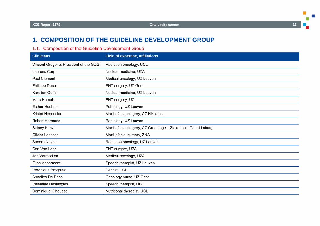
1. COMPOSITION OF THE GUIDELINE DEVELOPMENT GROUP 1.1. Composition of the Guideline Development Group Clinicians Field of expertise, affiliations
Vincent Grégoire, President of the GDG Radiation oncology, UCL
Laurens Carp Nuclear medicine, UZA
Paul Clement Medical oncology, UZ Leuven
Philippe Deron ENT surgery, UZ Gent
Karolien Goffin Nuclear medicine, UZ Leuven
Marc Hamoir ENT surgery, UCL
Esther Hauben Pathology, UZ Leuven
Kristof Hendrickx Maxillofacial surgery, AZ Nikolaas
Robert Hermans Radiology, UZ Leuven
Sidney Kunz Maxillofacial surgery, AZ Groeninge – Ziekenhuis Oost-Limburg
Olivier Lenssen Maxillofacial surgery, ZNA
Sandra Nuyts Radiation oncology, UZ Leuven
Carl Van Laer ENT surgery, UZA
Jan Vermorken Medical oncology, UZA
Eline Appermont Speech therapist, UZ Leuven
Véronique Brogniez Dentist, UCL
Annelies De Prins Oncology nurse, UZ Gent
Valentine Deslangles Speech therapist, UCL
Dominique Gihousse Nutritional therapist, UCL
14 Oral cavity cancer KCE Report 227S
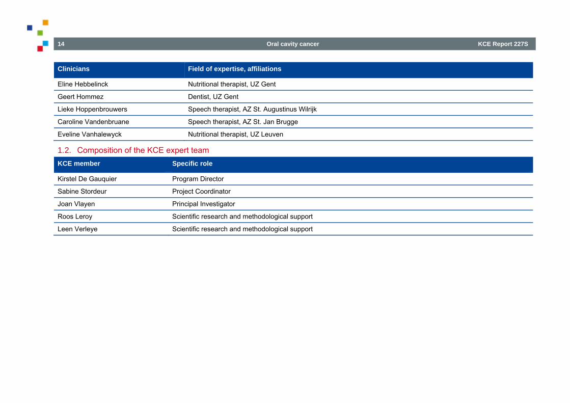
Clinicians Field of expertise, affiliations
Eline Hebbelinck Nutritional therapist, UZ Gent
Geert Hommez Dentist, UZ Gent
Lieke Hoppenbrouwers Speech therapist, AZ St. Augustinus Wilrijk
Caroline Vandenbruane Speech therapist, AZ St. Jan Brugge
Eveline Vanhalewyck Nutritional therapist, UZ Leuven
1.2. Composition of the KCE expert team KCE member Specific role
Kirstel De Gauquier Program Director
Sabine Stordeur Project Coordinator
Joan Vlayen Principal Investigator
Roos Leroy Scientific research and methodological support
Leen Verleye Scientific research and methodological support
KCE Report 227S Oral cavity cancer 15
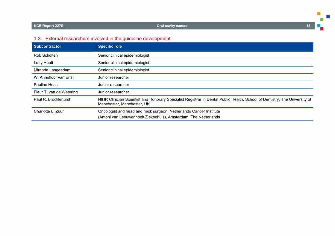
1.3. External researchers involved in the guideline development Subcontractor Specific role
Rob Scholten Senior clinical epidemiologist
Lotty Hooft Senior clinical epidemiologist
Miranda Langendam Senior clinical epidemiologist
W. Annefloor van Enst Junior researcher
Pauline Heus Junior researcher
Fleur T. van de Wetering Junior researcher
Paul R. Brocklehurst NIHR Clinician Scientist and Honorary Specialist Registrar in Dental Public Health, School of Dentistry, The University of Manchester, Manchester, UK
Charlotte L. Zuur Oncologist and head and neck surgeon, Netherlands Cancer Institute (Antoni van Leeuwenhoek Ziekenhuis), Amsterdam, The Netherlands
16 Oral cavity cancer KCE Report 227S
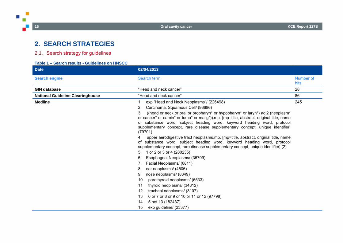
2. SEARCH STRATEGIES 2.1. Search strategy for guidelines
Table 1 – Search results - Guidelines on HNSCC Date 02/04/2013
Search engine Search term Number of hits
GIN database “Head and neck cancer” 28 National Guideline Clearinghouse “Head and neck cancer” 86 Medline 1 exp "Head and Neck Neoplasms"/ (226498)
2 Carcinoma, Squamous Cell/ (96686) 3 ((head or neck or oral or oropharyn* or hypopharyn* or laryn*) adj2 (neoplasm* or cancer* or carcin* or tumo* or malig*)).mp. [mp=title, abstract, original title, name of substance word, subject heading word, keyword heading word, protocol supplementary concept, rare disease supplementary concept, unique identifier] (79701) 4 upper aerodigestive tract neoplasms.mp. [mp=title, abstract, original title, name of substance word, subject heading word, keyword heading word, protocol supplementary concept, rare disease supplementary concept, unique identifier] (2) 5 1 or 2 or 3 or 4 (280235) 6 Esophageal Neoplasms/ (35709) 7 Facial Neoplasms/ (6811) 8 ear neoplasms/ (4506) 9 nose neoplasms/ (8349) 10 parathyroid neoplasms/ (6533) 11 thyroid neoplasms/ (34812) 12 tracheal neoplasms/ (3107) 13 6 or 7 or 8 or 9 or 10 or 11 or 12 (97798) 14 5 not 13 (182437) 15 exp guideline/ (23377)
245
KCE Report 227S Oral cavity cancer 17
16 "guideline*".ti. (42165) 17 recommendation*.ti. (20588) 18 standard*.ti. (58642) 19 15 or 16 or 17 or 18 (129130) 20 14 and 19 (655) 21 exp animals/ not humans.sh. (3784285) 22 20 not 21 (653) 23 limit 22 to (yr="2008 -Current" and (dutch or english or french or german)) (245)
After removal of duplicate guidelines, 32 guidelines were selected based on title and abstract and retained for full-text evaluation. Of these, 14 guidelines were excluded for the following reasons: 2 guidelines were out of scope 3 documents could not be considered as guideline 5 documents did not contain any recommendation 1 guideline had been replaced by a more recent version 2 guidelines were archived 1 guideline was based on another guideline Finally, 18 guidelines were retained for an evaluation of the methodological quality.
18 Oral cavity cancer KCE Report 227S
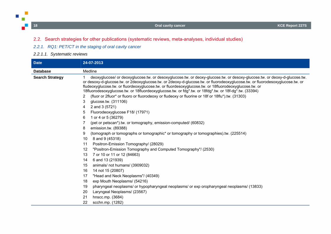
2.2. Search strategies for other publications (systematic reviews, meta-analyses, individual studies) 2.2.1. RQ1: PET/CT in the staging of oral cavity cancer
2.2.1.1. Systematic reviews
Date 24-07-2013
Database Medline Search Strategy 1 deoxyglucose/ or deoxyglucose.tw. or desoxyglucose.tw. or deoxy-glucose.tw. or desoxy-glucose.tw. or deoxy-d-glucose.tw.
or desoxy-d-glucose.tw. or 2deoxyglucose.tw. or 2deoxy-d-glucose.tw. or fluorodeoxyglucose.tw. or fluorodesoxyglucose.tw. or fludeoxyglucose.tw. or fluordeoxyglucose.tw. or fluordesoxyglucose.tw. or 18fluorodeoxyglucose.tw. or 18fluorodesoxyglucose.tw. or 18fluordeoxyglucose.tw. or fdg*.tw. or 18fdg*.tw. or 18f-dg*.tw. (33394) 2 (fluor or 2fluor* or fluoro or fluorodeoxy or fludeoxy or fluorine or 18f or 18flu*).tw. (31303) 3 glucose.tw. (311106) 4 2 and 3 (5721) 5 Fluorodeoxyglucose F18/ (17971) 6 1 or 4 or 5 (36279) 7 (pet or petscan*).tw. or tomography, emission-computed/ (60832) 8 emission.tw. (89388) 9 (tomograph or tomographs or tomographic* or tomography or tomographies).tw. (225514) 10 8 and 9 (45318) 11 Positron-Emission Tomography/ (28029) 12 "Positron-Emission Tomography and Computed Tomography"/ (2530) 13 7 or 10 or 11 or 12 (84663) 14 6 and 13 (21939) 15 animals/ not humans/ (3909032) 16 14 not 15 (20807) 17 "Head and Neck Neoplasms"/ (40349) 18 exp Mouth Neoplasms/ (54216) 19 pharyngeal neoplasms/ or hypopharyngeal neoplasms/ or exp oropharyngeal neoplasms/ (13833) 20 Laryngeal Neoplasms/ (23567) 21 hnscc.mp. (3684) 22 scchn.mp. (1282)
KCE Report 227S Oral cavity cancer 19
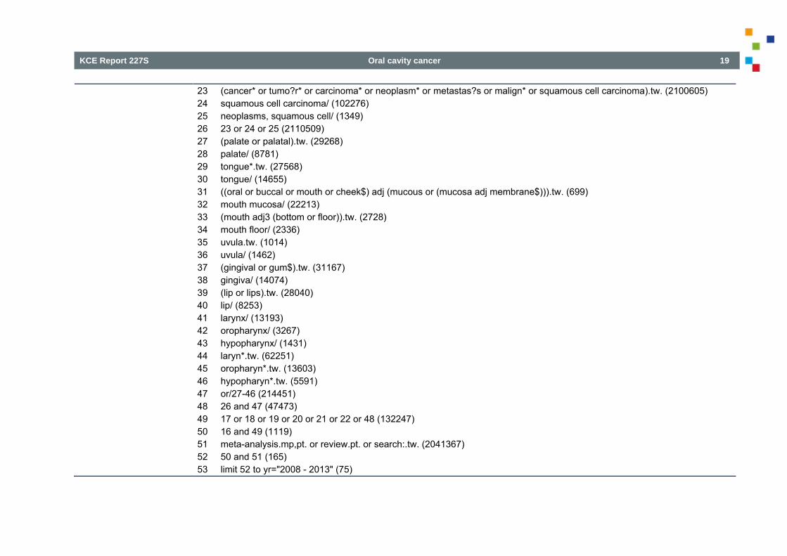
23 (cancer* or tumo?r* or carcinoma* or neoplasm* or metastas?s or malign* or squamous cell carcinoma).tw. (2100605) 24 squamous cell carcinoma/ (102276) 25 neoplasms, squamous cell/ (1349) 26 23 or 24 or 25 (2110509) 27 (palate or palatal).tw. (29268) 28 palate/ (8781) 29 tongue*.tw. (27568) 30 tongue/ (14655) 31 ((oral or buccal or mouth or cheek$) adj (mucous or (mucosa adj membrane$))).tw. (699) 32 mouth mucosa/ (22213) 33 (mouth adj3 (bottom or floor)).tw. (2728) 34 mouth floor/ (2336) 35 uvula.tw. (1014) 36 uvula/ (1462) 37 (gingival or gum$).tw. (31167) 38 gingiva/ (14074) 39 (lip or lips).tw. (28040) 40 lip/ (8253) 41 larynx/ (13193) 42 oropharynx/ (3267) 43 hypopharynx/ (1431) 44 laryn*.tw. (62251) 45 oropharyn*.tw. (13603) 46 hypopharyn*.tw. (5591) 47 or/27-46 (214451) 48 26 and 47 (47473) 49 17 or 18 or 19 or 20 or 21 or 22 or 48 (132247) 50 16 and 49 (1119) 51 meta-analysis.mp,pt. or review.pt. or search:.tw. (2041367) 52 50 and 51 (165) 53 limit 52 to yr="2008 - 2013" (75)
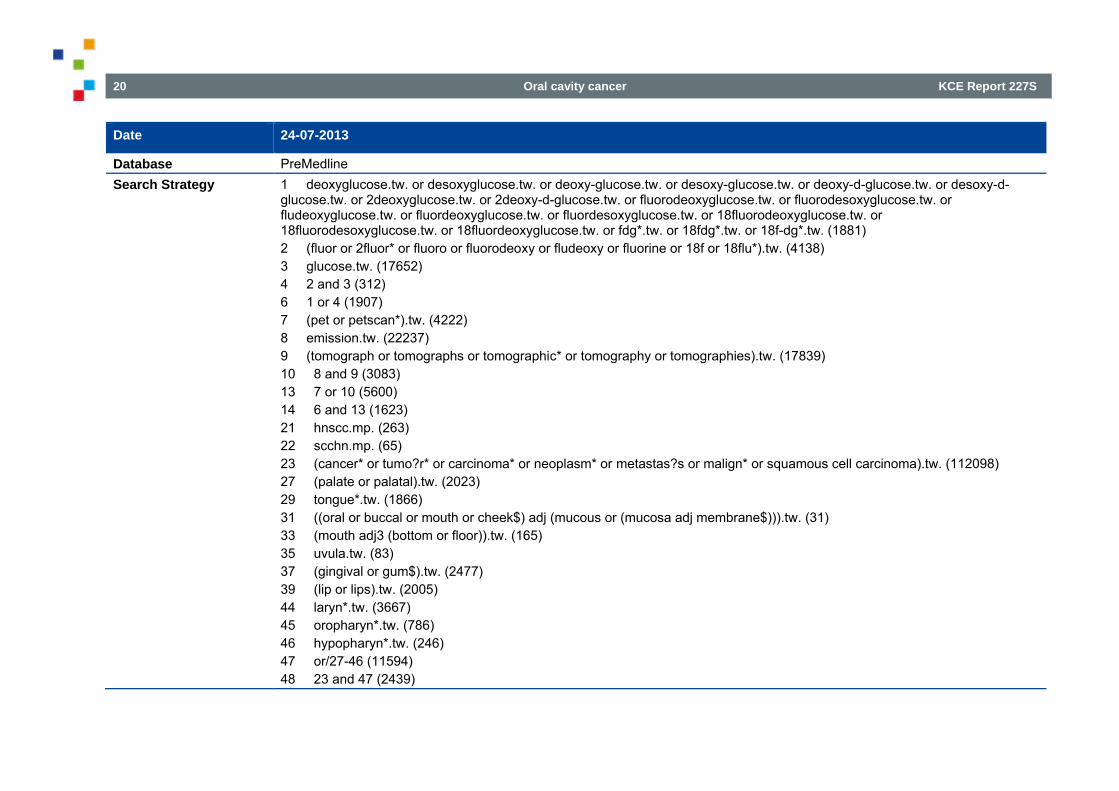
20 Oral cavity cancer KCE Report 227S
Date 24-07-2013
Database PreMedline Search Strategy 1 deoxyglucose.tw. or desoxyglucose.tw. or deoxy-glucose.tw. or desoxy-glucose.tw. or deoxy-d-glucose.tw. or desoxy-d-
glucose.tw. or 2deoxyglucose.tw. or 2deoxy-d-glucose.tw. or fluorodeoxyglucose.tw. or fluorodesoxyglucose.tw. or fludeoxyglucose.tw. or fluordeoxyglucose.tw. or fluordesoxyglucose.tw. or 18fluorodeoxyglucose.tw. or 18fluorodesoxyglucose.tw. or 18fluordeoxyglucose.tw. or fdg*.tw. or 18fdg*.tw. or 18f-dg*.tw. (1881) 2 (fluor or 2fluor* or fluoro or fluorodeoxy or fludeoxy or fluorine or 18f or 18flu*).tw. (4138) 3 glucose.tw. (17652) 4 2 and 3 (312) 6 1 or 4 (1907) 7 (pet or petscan*).tw. (4222) 8 emission.tw. (22237) 9 (tomograph or tomographs or tomographic* or tomography or tomographies).tw. (17839) 10 8 and 9 (3083) 13 7 or 10 (5600) 14 6 and 13 (1623) 21 hnscc.mp. (263) 22 scchn.mp. (65) 23 (cancer* or tumo?r* or carcinoma* or neoplasm* or metastas?s or malign* or squamous cell carcinoma).tw. (112098) 27 (palate or palatal).tw. (2023) 29 tongue*.tw. (1866) 31 ((oral or buccal or mouth or cheek$) adj (mucous or (mucosa adj membrane$))).tw. (31) 33 (mouth adj3 (bottom or floor)).tw. (165) 35 uvula.tw. (83) 37 (gingival or gum$).tw. (2477) 39 (lip or lips).tw. (2005) 44 laryn*.tw. (3667) 45 oropharyn*.tw. (786) 46 hypopharyn*.tw. (246) 47 or/27-46 (11594) 48 23 and 47 (2439)
KCE Report 227S Oral cavity cancer 21
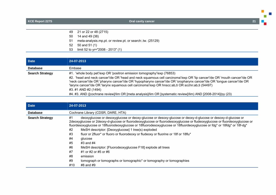
49 21 or 22 or 48 (2715) 50 14 and 49 (38) 51 meta-analysis.mp,pt. or review.pt. or search:.tw. (25129) 52 50 and 51 (1) 53 limit 52 to yr="2008 - 2013" (1)
Date 24-07-2013
Database Embase Search Strategy #1. 'whole body pet'/exp OR 'positron emission tomography'/exp (76853)
#2. 'head and neck cancer'/de OR 'head and neck squamous cell carcinoma'/exp OR 'lip cancer'/de OR 'mouth cancer'/de OR 'neck cancer'/de OR 'pharynx cancer'/de OR 'hypopharynx cancer'/de OR 'oropharynx cancer'/de OR 'tongue cancer'/de OR 'larynx cancer'/de OR 'larynx squamous cell carcinoma'/exp OR hnscc:ab,ti OR scchn:ab,ti (54497) #3. #1 AND #2 (1494) #4. #3. AND ([cochrane review]/lim OR [meta analysis]/lim OR [systematic review]/lim) AND [2008-2014]/py (23)
Date 24-07-2013
Database Cochrane Library (CDSR, DARE, HTA) Search Strategy #1 deoxyglucose or desoxyglucose or deoxy-glucose or desoxy-glucose or deoxy-d-glucose or desoxy-d-glucose or
2deoxyglucose or 2deoxy-d-glucose or fluorodeoxyglucose or fluorodesoxyglucose or fludeoxyglucose or fluordeoxyglucose or fluordesoxyglucose or 18fluorodeoxyglucose or 18fluorodesoxyglucose or 18fluordeoxyglucose or fdg* or 18fdg* or 18f-dg* #2 MeSH descriptor: [Deoxyglucose] 1 tree(s) exploded #3 fluor or 2fluor* or fluoro or fluorodeoxy or fludeoxy or fluorine or 18f or 18flu* #4 glucose #5 #3 and #4 #6 MeSH descriptor: [Fluorodeoxyglucose F18] explode all trees #7 #1 or #2 or #5 or #6 #8 emission #9 tomograph or tomographs or tomographic* or tomography or tomographies #10 #8 and #9
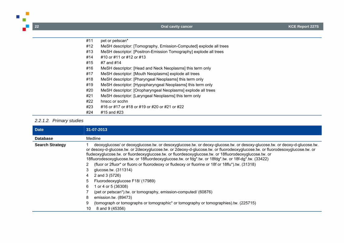
22 Oral cavity cancer KCE Report 227S
#11 pet or petscan* #12 MeSH descriptor: [Tomography, Emission-Computed] explode all trees #13 MeSH descriptor: [Positron-Emission Tomography] explode all trees #14 #10 or #11 or #12 or #13 #15 #7 and #14 #16 MeSH descriptor: [Head and Neck Neoplasms] this term only #17 MeSH descriptor: [Mouth Neoplasms] explode all trees #18 MeSH descriptor: [Pharyngeal Neoplasms] this term only #19 MeSH descriptor: [Hypopharyngeal Neoplasms] this term only #20 MeSH descriptor: [Oropharyngeal Neoplasms] explode all trees #21 MeSH descriptor: [Laryngeal Neoplasms] this term only #22 hnscc or scchn #23 #16 or #17 or #18 or #19 or #20 or #21 or #22 #24 #15 and #23
2.2.1.2. Primary studies
Date 31-07-2013
Database Medline Search Strategy 1 deoxyglucose/ or deoxyglucose.tw. or desoxyglucose.tw. or deoxy-glucose.tw. or desoxy-glucose.tw. or deoxy-d-glucose.tw.
or desoxy-d-glucose.tw. or 2deoxyglucose.tw. or 2deoxy-d-glucose.tw. or fluorodeoxyglucose.tw. or fluorodesoxyglucose.tw. or fludeoxyglucose.tw. or fluordeoxyglucose.tw. or fluordesoxyglucose.tw. or 18fluorodeoxyglucose.tw. or 18fluorodesoxyglucose.tw. or 18fluordeoxyglucose.tw. or fdg*.tw. or 18fdg*.tw. or 18f-dg*.tw. (33422) 2 (fluor or 2fluor* or fluoro or fluorodeoxy or fludeoxy or fluorine or 18f or 18flu*).tw. (31318) 3 glucose.tw. (311314) 4 2 and 3 (5726) 5 Fluorodeoxyglucose F18/ (17989) 6 1 or 4 or 5 (36308) 7 (pet or petscan*).tw. or tomography, emission-computed/ (60876) 8 emission.tw. (89473) 9 (tomograph or tomographs or tomographic* or tomography or tomographies).tw. (225715) 10 8 and 9 (45356)
KCE Report 227S Oral cavity cancer 23
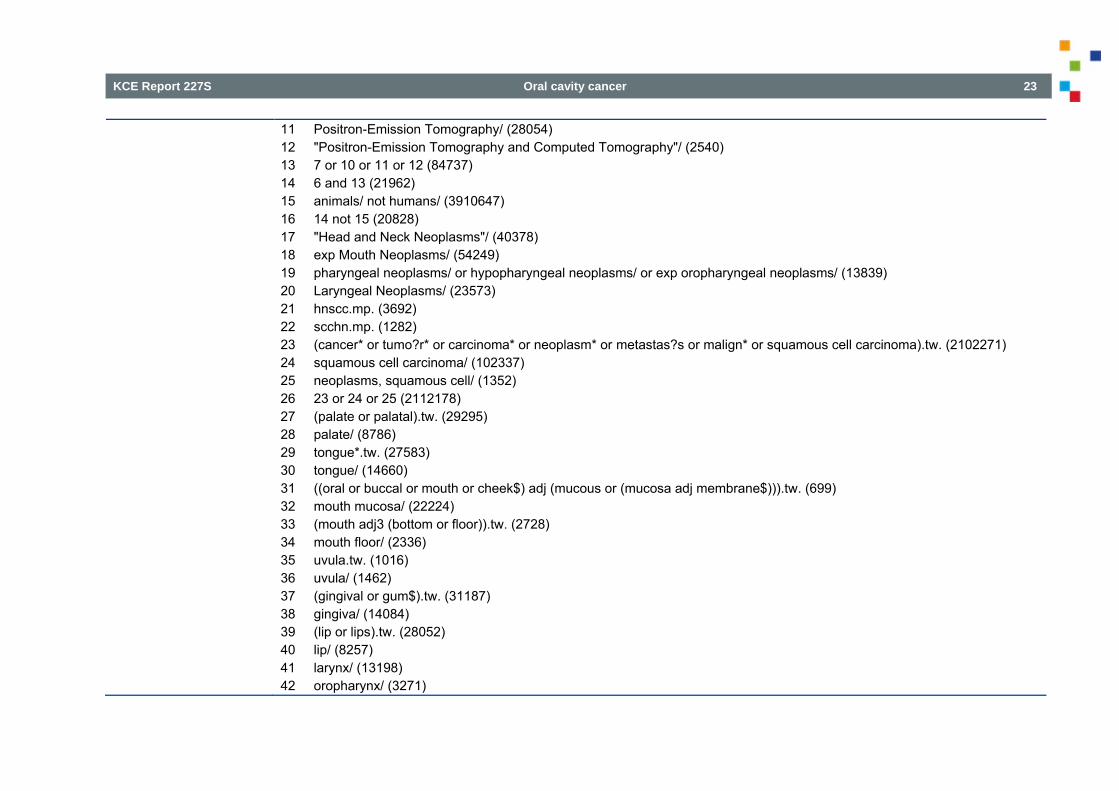
11 Positron-Emission Tomography/ (28054) 12 "Positron-Emission Tomography and Computed Tomography"/ (2540) 13 7 or 10 or 11 or 12 (84737) 14 6 and 13 (21962) 15 animals/ not humans/ (3910647) 16 14 not 15 (20828) 17 "Head and Neck Neoplasms"/ (40378) 18 exp Mouth Neoplasms/ (54249) 19 pharyngeal neoplasms/ or hypopharyngeal neoplasms/ or exp oropharyngeal neoplasms/ (13839) 20 Laryngeal Neoplasms/ (23573) 21 hnscc.mp. (3692) 22 scchn.mp. (1282) 23 (cancer* or tumo?r* or carcinoma* or neoplasm* or metastas?s or malign* or squamous cell carcinoma).tw. (2102271) 24 squamous cell carcinoma/ (102337) 25 neoplasms, squamous cell/ (1352) 26 23 or 24 or 25 (2112178) 27 (palate or palatal).tw. (29295) 28 palate/ (8786) 29 tongue*.tw. (27583) 30 tongue/ (14660) 31 ((oral or buccal or mouth or cheek$) adj (mucous or (mucosa adj membrane$))).tw. (699) 32 mouth mucosa/ (22224) 33 (mouth adj3 (bottom or floor)).tw. (2728) 34 mouth floor/ (2336) 35 uvula.tw. (1016) 36 uvula/ (1462) 37 (gingival or gum$).tw. (31187) 38 gingiva/ (14084) 39 (lip or lips).tw. (28052) 40 lip/ (8257) 41 larynx/ (13198) 42 oropharynx/ (3271)
24 Oral cavity cancer KCE Report 227S
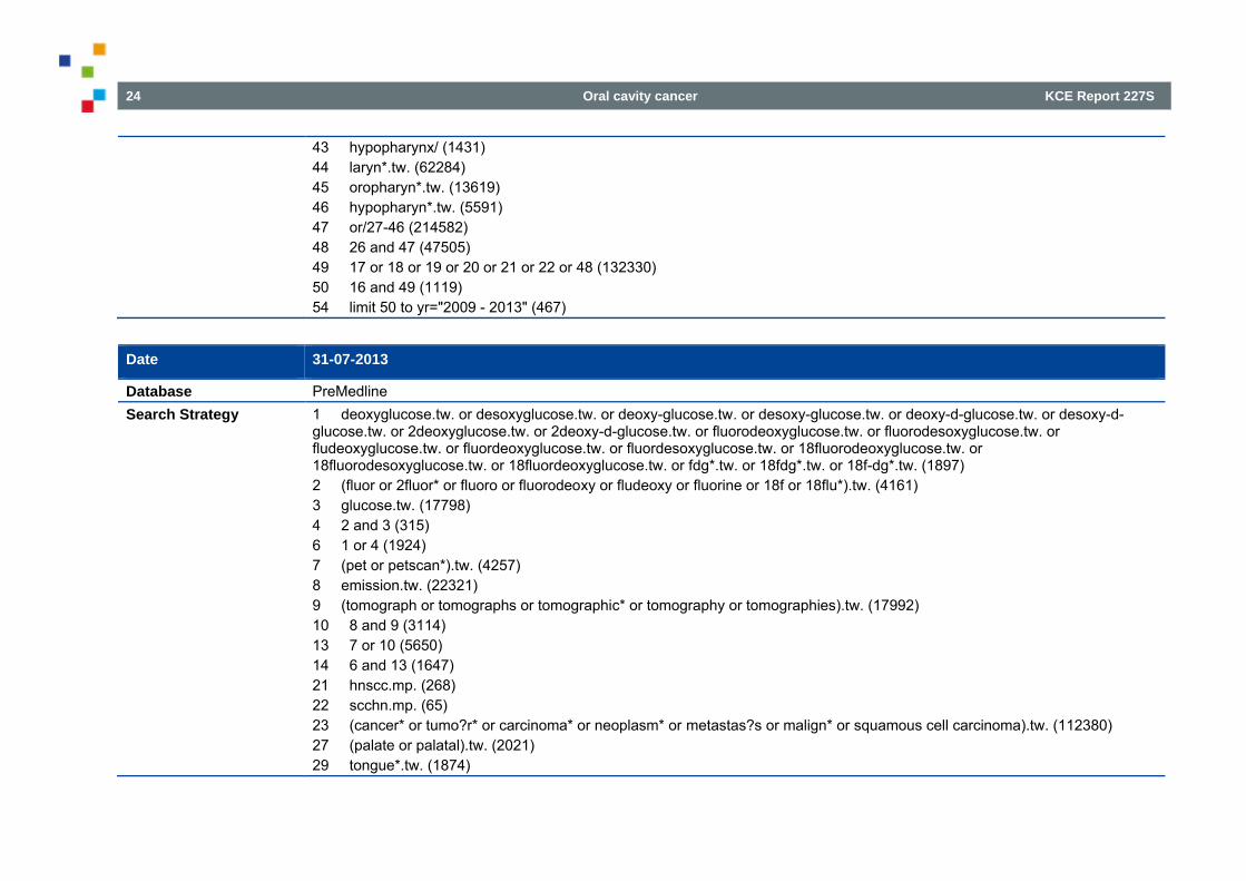
43 hypopharynx/ (1431) 44 laryn*.tw. (62284) 45 oropharyn*.tw. (13619) 46 hypopharyn*.tw. (5591) 47 or/27-46 (214582) 48 26 and 47 (47505) 49 17 or 18 or 19 or 20 or 21 or 22 or 48 (132330) 50 16 and 49 (1119) 54 limit 50 to yr="2009 - 2013" (467)
Date 31-07-2013
Database PreMedline Search Strategy 1 deoxyglucose.tw. or desoxyglucose.tw. or deoxy-glucose.tw. or desoxy-glucose.tw. or deoxy-d-glucose.tw. or desoxy-d-
glucose.tw. or 2deoxyglucose.tw. or 2deoxy-d-glucose.tw. or fluorodeoxyglucose.tw. or fluorodesoxyglucose.tw. or fludeoxyglucose.tw. or fluordeoxyglucose.tw. or fluordesoxyglucose.tw. or 18fluorodeoxyglucose.tw. or 18fluorodesoxyglucose.tw. or 18fluordeoxyglucose.tw. or fdg*.tw. or 18fdg*.tw. or 18f-dg*.tw. (1897) 2 (fluor or 2fluor* or fluoro or fluorodeoxy or fludeoxy or fluorine or 18f or 18flu*).tw. (4161) 3 glucose.tw. (17798) 4 2 and 3 (315) 6 1 or 4 (1924) 7 (pet or petscan*).tw. (4257) 8 emission.tw. (22321) 9 (tomograph or tomographs or tomographic* or tomography or tomographies).tw. (17992) 10 8 and 9 (3114) 13 7 or 10 (5650) 14 6 and 13 (1647) 21 hnscc.mp. (268) 22 scchn.mp. (65) 23 (cancer* or tumo?r* or carcinoma* or neoplasm* or metastas?s or malign* or squamous cell carcinoma).tw. (112380) 27 (palate or palatal).tw. (2021) 29 tongue*.tw. (1874)
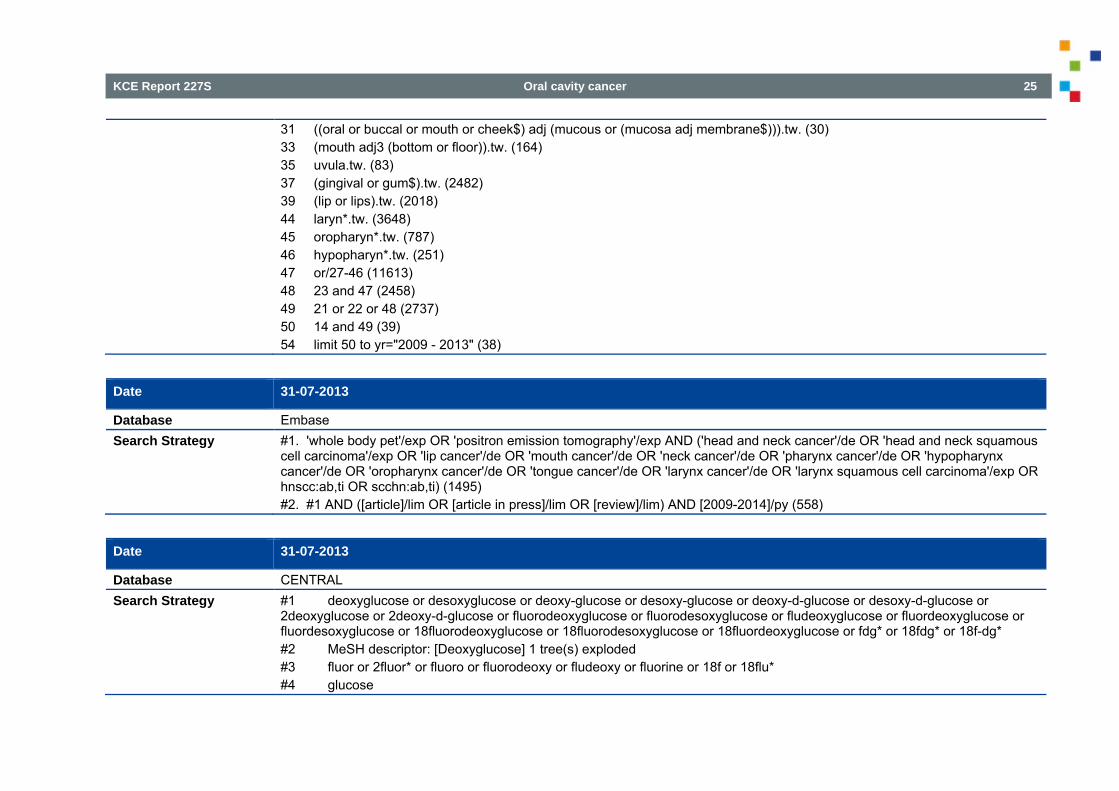
KCE Report 227S Oral cavity cancer 25
31 ((oral or buccal or mouth or cheek$) adj (mucous or (mucosa adj membrane$))).tw. (30) 33 (mouth adj3 (bottom or floor)).tw. (164) 35 uvula.tw. (83) 37 (gingival or gum$).tw. (2482) 39 (lip or lips).tw. (2018) 44 laryn*.tw. (3648) 45 oropharyn*.tw. (787) 46 hypopharyn*.tw. (251) 47 or/27-46 (11613) 48 23 and 47 (2458) 49 21 or 22 or 48 (2737) 50 14 and 49 (39) 54 limit 50 to yr="2009 - 2013" (38)
Date 31-07-2013
Database Embase Search Strategy #1. 'whole body pet'/exp OR 'positron emission tomography'/exp AND ('head and neck cancer'/de OR 'head and neck squamous
cell carcinoma'/exp OR 'lip cancer'/de OR 'mouth cancer'/de OR 'neck cancer'/de OR 'pharynx cancer'/de OR 'hypopharynx cancer'/de OR 'oropharynx cancer'/de OR 'tongue cancer'/de OR 'larynx cancer'/de OR 'larynx squamous cell carcinoma'/exp OR hnscc:ab,ti OR scchn:ab,ti) (1495) #2. #1 AND ([article]/lim OR [article in press]/lim OR [review]/lim) AND [2009-2014]/py (558)
Date 31-07-2013
Database CENTRAL Search Strategy #1 deoxyglucose or desoxyglucose or deoxy-glucose or desoxy-glucose or deoxy-d-glucose or desoxy-d-glucose or
2deoxyglucose or 2deoxy-d-glucose or fluorodeoxyglucose or fluorodesoxyglucose or fludeoxyglucose or fluordeoxyglucose or fluordesoxyglucose or 18fluorodeoxyglucose or 18fluorodesoxyglucose or 18fluordeoxyglucose or fdg* or 18fdg* or 18f-dg* #2 MeSH descriptor: [Deoxyglucose] 1 tree(s) exploded #3 fluor or 2fluor* or fluoro or fluorodeoxy or fludeoxy or fluorine or 18f or 18flu* #4 glucose
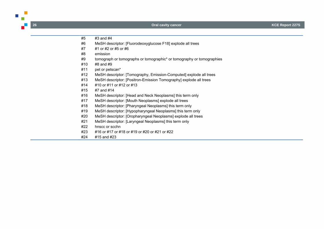
26 Oral cavity cancer KCE Report 227S
#5 #3 and #4 #6 MeSH descriptor: [Fluorodeoxyglucose F18] explode all trees #7 #1 or #2 or #5 or #6 #8 emission #9 tomograph or tomographs or tomographic* or tomography or tomographies #10 #8 and #9 #11 pet or petscan* #12 MeSH descriptor: [Tomography, Emission-Computed] explode all trees #13 MeSH descriptor: [Positron-Emission Tomography] explode all trees #14 #10 or #11 or #12 or #13 #15 #7 and #14 #16 MeSH descriptor: [Head and Neck Neoplasms] this term only #17 MeSH descriptor: [Mouth Neoplasms] explode all trees #18 MeSH descriptor: [Pharyngeal Neoplasms] this term only #19 MeSH descriptor: [Hypopharyngeal Neoplasms] this term only #20 MeSH descriptor: [Oropharyngeal Neoplasms] explode all trees #21 MeSH descriptor: [Laryngeal Neoplasms] this term only #22 hnscc or scchn #23 #16 or #17 or #18 or #19 or #20 or #21 or #22 #24 #15 and #23
KCE Report 227S Oral cavity cancer 27
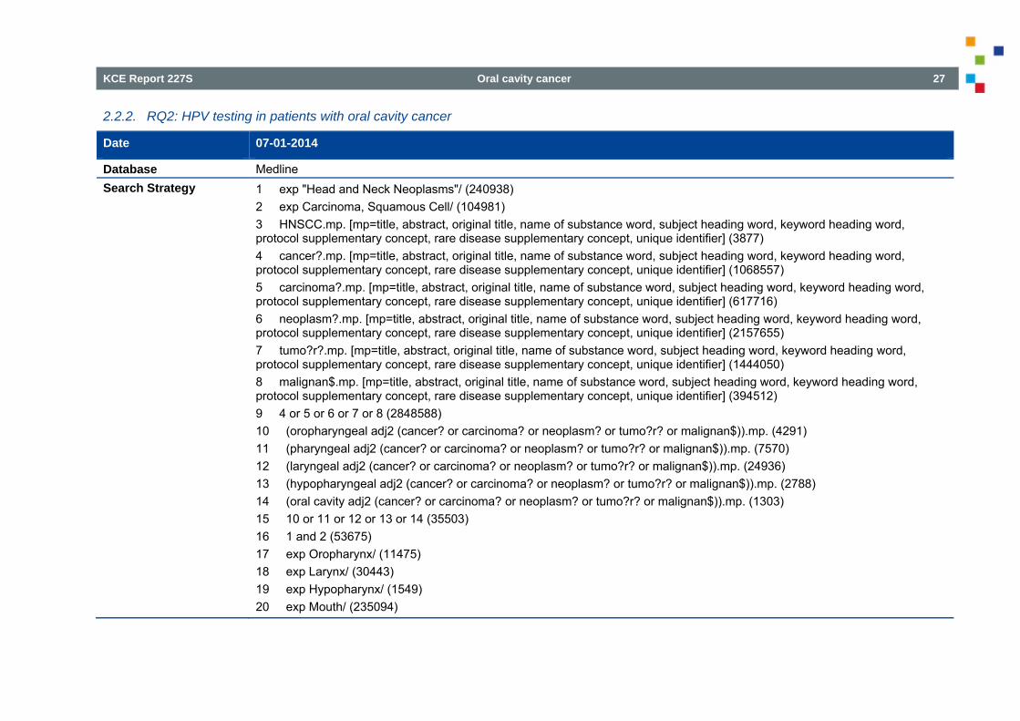
2.2.2. RQ2: HPV testing in patients with oral cavity cancer
Date 07-01-2014
Database Medline Search Strategy 1 exp "Head and Neck Neoplasms"/ (240938)
2 exp Carcinoma, Squamous Cell/ (104981) 3 HNSCC.mp. [mp=title, abstract, original title, name of substance word, subject heading word, keyword heading word, protocol supplementary concept, rare disease supplementary concept, unique identifier] (3877) 4 cancer?.mp. [mp=title, abstract, original title, name of substance word, subject heading word, keyword heading word, protocol supplementary concept, rare disease supplementary concept, unique identifier] (1068557) 5 carcinoma?.mp. [mp=title, abstract, original title, name of substance word, subject heading word, keyword heading word, protocol supplementary concept, rare disease supplementary concept, unique identifier] (617716) 6 neoplasm?.mp. [mp=title, abstract, original title, name of substance word, subject heading word, keyword heading word, protocol supplementary concept, rare disease supplementary concept, unique identifier] (2157655) 7 tumo?r?.mp. [mp=title, abstract, original title, name of substance word, subject heading word, keyword heading word, protocol supplementary concept, rare disease supplementary concept, unique identifier] (1444050) 8 malignan$.mp. [mp=title, abstract, original title, name of substance word, subject heading word, keyword heading word, protocol supplementary concept, rare disease supplementary concept, unique identifier] (394512) 9 4 or 5 or 6 or 7 or 8 (2848588) 10 (oropharyngeal adj2 (cancer? or carcinoma? or neoplasm? or tumo?r? or malignan$)).mp. (4291) 11 (pharyngeal adj2 (cancer? or carcinoma? or neoplasm? or tumo?r? or malignan$)).mp. (7570) 12 (laryngeal adj2 (cancer? or carcinoma? or neoplasm? or tumo?r? or malignan$)).mp. (24936) 13 (hypopharyngeal adj2 (cancer? or carcinoma? or neoplasm? or tumo?r? or malignan$)).mp. (2788) 14 (oral cavity adj2 (cancer? or carcinoma? or neoplasm? or tumo?r? or malignan$)).mp. (1303) 15 10 or 11 or 12 or 13 or 14 (35503) 16 1 and 2 (53675) 17 exp Oropharynx/ (11475) 18 exp Larynx/ (30443) 19 exp Hypopharynx/ (1549) 20 exp Mouth/ (235094)
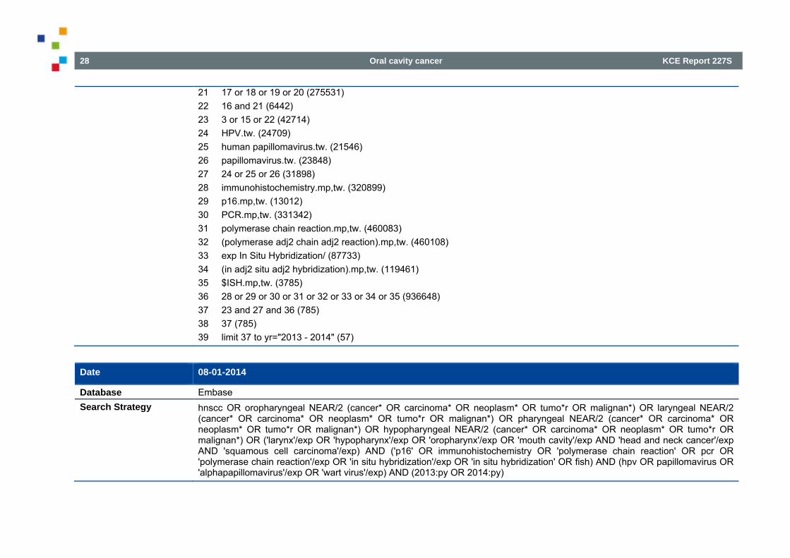
28 Oral cavity cancer KCE Report 227S
21 17 or 18 or 19 or 20 (275531) 22 16 and 21 (6442) 23 3 or 15 or 22 (42714) 24 HPV.tw. (24709) 25 human papillomavirus.tw. (21546) 26 papillomavirus.tw. (23848) 27 24 or 25 or 26 (31898) 28 immunohistochemistry.mp,tw. (320899) 29 p16.mp,tw. (13012) 30 PCR.mp,tw. (331342) 31 polymerase chain reaction.mp,tw. (460083) 32 (polymerase adj2 chain adj2 reaction).mp,tw. (460108) 33 exp In Situ Hybridization/ (87733) 34 (in adj2 situ adj2 hybridization).mp,tw. (119461) 35 $ISH.mp,tw. (3785) 36 28 or 29 or 30 or 31 or 32 or 33 or 34 or 35 (936648) 37 23 and 27 and 36 (785) 38 37 (785) 39 limit 37 to yr="2013 - 2014" (57)
Date 08-01-2014
Database Embase Search Strategy hnscc OR oropharyngeal NEAR/2 (cancer* OR carcinoma* OR neoplasm* OR tumo*r OR malignan*) OR laryngeal NEAR/2
(cancer* OR carcinoma* OR neoplasm* OR tumo*r OR malignan*) OR pharyngeal NEAR/2 (cancer* OR carcinoma* OR neoplasm* OR tumo*r OR malignan*) OR hypopharyngeal NEAR/2 (cancer* OR carcinoma* OR neoplasm* OR tumo*r OR malignan*) OR ('larynx'/exp OR 'hypopharynx'/exp OR 'oropharynx'/exp OR 'mouth cavity'/exp AND 'head and neck cancer'/exp AND 'squamous cell carcinoma'/exp) AND ('p16' OR immunohistochemistry OR 'polymerase chain reaction' OR pcr OR 'polymerase chain reaction'/exp OR 'in situ hybridization'/exp OR 'in situ hybridization' OR fish) AND (hpv OR papillomavirus OR 'alphapapillomavirus'/exp OR 'wart virus'/exp) AND (2013:py OR 2014:py)
KCE Report 227S Oral cavity cancer 29
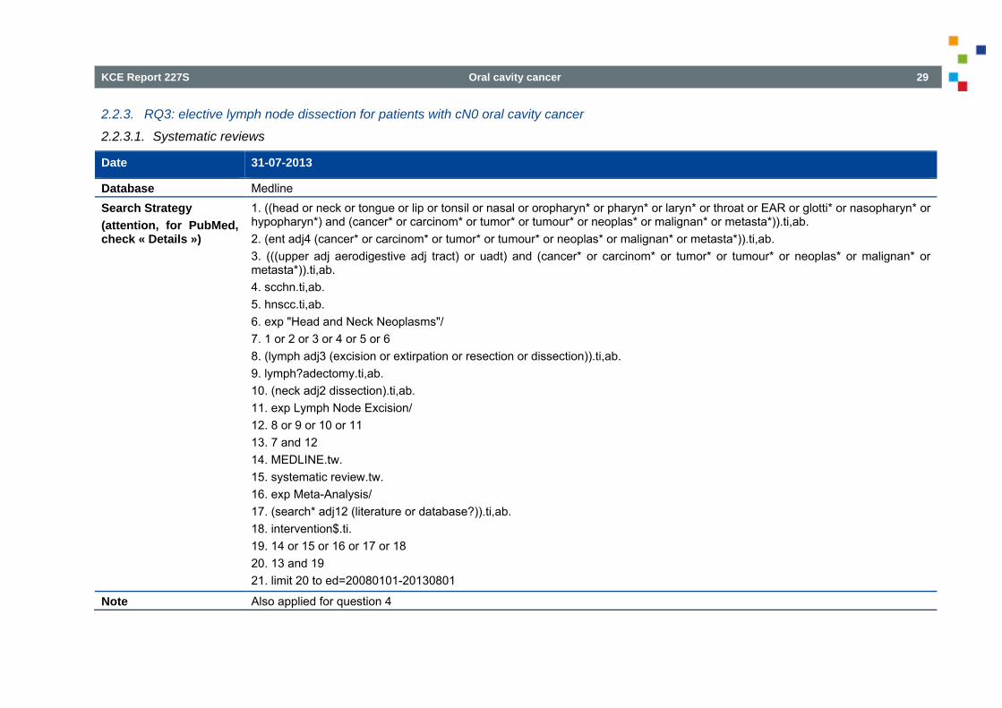
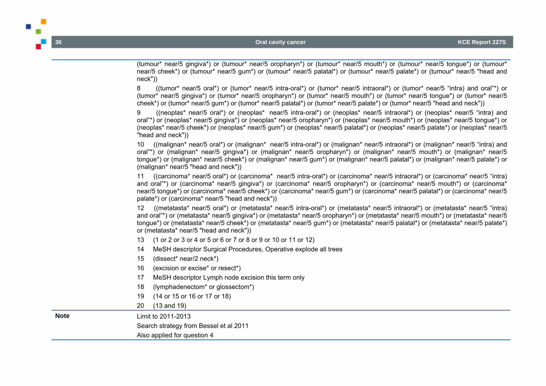
2.2.3. RQ3: elective lymph node dissection for patients with cN0 oral cavity cancer
2.2.3.1. Systematic reviews
Date 31-07-2013
Database Medline Search Strategy (attention, for PubMed, check « Details »)
1. ((head or neck or tongue or lip or tonsil or nasal or oropharyn* or pharyn* or laryn* or throat or EAR or glotti* or nasopharyn* or hypopharyn*) and (cancer* or carcinom* or tumor* or tumour* or neoplas* or malignan* or metasta*)).ti,ab. 2. (ent adj4 (cancer* or carcinom* or tumor* or tumour* or neoplas* or malignan* or metasta*)).ti,ab. 3. (((upper adj aerodigestive adj tract) or uadt) and (cancer* or carcinom* or tumor* or tumour* or neoplas* or malignan* or metasta*)).ti,ab. 4. scchn.ti,ab. 5. hnscc.ti,ab. 6. exp "Head and Neck Neoplasms"/ 7. 1 or 2 or 3 or 4 or 5 or 6 8. (lymph adj3 (excision or extirpation or resection or dissection)).ti,ab. 9. lymph?adectomy.ti,ab. 10. (neck adj2 dissection).ti,ab. 11. exp Lymph Node Excision/ 12. 8 or 9 or 10 or 11 13. 7 and 12 14. MEDLINE.tw. 15. systematic review.tw. 16. exp Meta-Analysis/ 17. (search* adj12 (literature or database?)).ti,ab. 18. intervention$.ti. 19. 14 or 15 or 16 or 17 or 18 20. 13 and 19 21. limit 20 to ed=20080101-20130801
Note Also applied for question 4
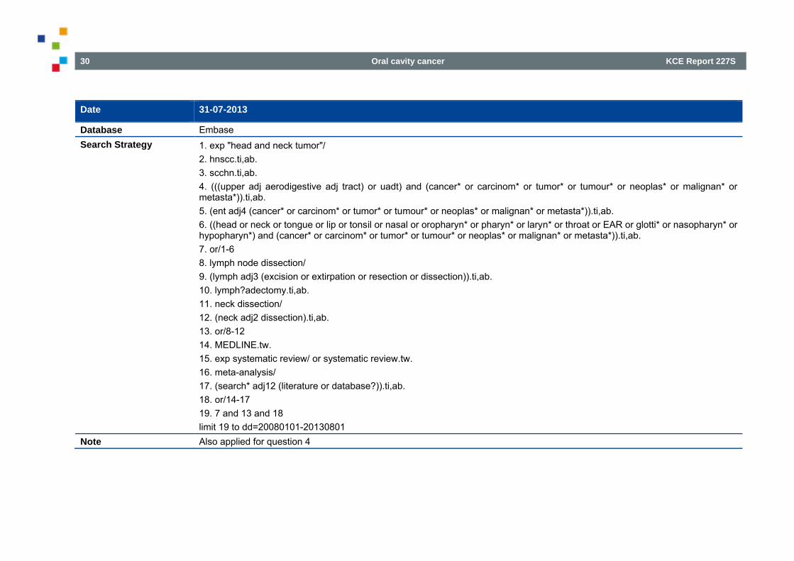
30 Oral cavity cancer KCE Report 227S
Date 31-07-2013
Database Embase Search Strategy 1. exp "head and neck tumor"/
2. hnscc.ti,ab. 3. scchn.ti,ab. 4. (((upper adj aerodigestive adj tract) or uadt) and (cancer* or carcinom* or tumor* or tumour* or neoplas* or malignan* or metasta*)).ti,ab. 5. (ent adj4 (cancer* or carcinom* or tumor* or tumour* or neoplas* or malignan* or metasta*)).ti,ab. 6. ((head or neck or tongue or lip or tonsil or nasal or oropharyn* or pharyn* or laryn* or throat or EAR or glotti* or nasopharyn* or hypopharyn*) and (cancer* or carcinom* or tumor* or tumour* or neoplas* or malignan* or metasta*)).ti,ab. 7. or/1-6 8. lymph node dissection/ 9. (lymph adj3 (excision or extirpation or resection or dissection)).ti,ab. 10. lymph?adectomy.ti,ab. 11. neck dissection/ 12. (neck adj2 dissection).ti,ab. 13. or/8-12 14. MEDLINE.tw. 15. exp systematic review/ or systematic review.tw. 16. meta-analysis/ 17. (search* adj12 (literature or database?)).ti,ab. 18. or/14-17 19. 7 and 13 and 18 limit 19 to dd=20080101-20130801
Note Also applied for question 4
KCE Report 227S Oral cavity cancer 31
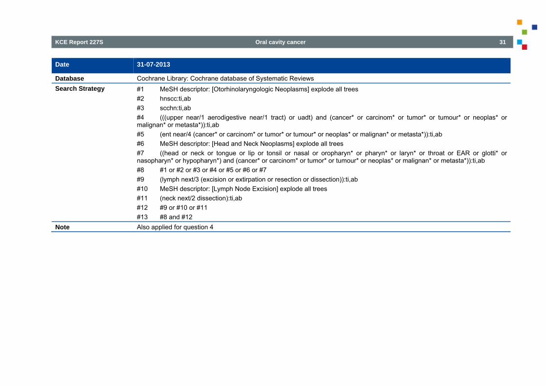
Date 31-07-2013
Database Cochrane Library: Cochrane database of Systematic Reviews Search Strategy #1 MeSH descriptor: [Otorhinolaryngologic Neoplasms] explode all trees
#2 hnscc:ti,ab #3 scchn:ti,ab #4 (((upper near/1 aerodigestive near/1 tract) or uadt) and (cancer* or carcinom* or tumor* or tumour* or neoplas* or malignan* or metasta*)):ti,ab #5 (ent near/4 (cancer* or carcinom* or tumor* or tumour* or neoplas* or malignan* or metasta*)):ti,ab #6 MeSH descriptor: [Head and Neck Neoplasms] explode all trees #7 ((head or neck or tongue or lip or tonsil or nasal or oropharyn* or pharyn* or laryn* or throat or EAR or glotti* or nasopharyn* or hypopharyn*) and (cancer* or carcinom* or tumor* or tumour* or neoplas* or malignan* or metasta*)):ti,ab #8 #1 or #2 or #3 or #4 or #5 or #6 or #7 #9 (lymph next/3 (excision or extirpation or resection or dissection)):ti,ab #10 MeSH descriptor: [Lymph Node Excision] explode all trees #11 (neck next/2 dissection):ti,ab #12 #9 or #10 or #11 #13 #8 and #12
Note Also applied for question 4
32 Oral cavity cancer KCE Report 227S
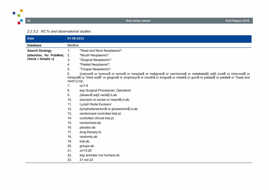
2.2.3.2. RCTs and observational studies
Date 07-08-2013
Database Medline Search Strategy (attention, for PubMed, check « Details »)
1. "Head and Neck Neoplasms"/ 2. "Mouth Neoplasms"/ 3. "Gingival Neoplasms"/ 4. "Palatal Neoplasms"/ 5. "Tongue Neoplasms"/ 6. ((cancer$ or tumour$ or tumor$ or neoplas$ or malignan$ or carcinoma$ or metatasta$) adj5 (oral$ or intra-oral$ or intraoral$ or "intra oral$" or gingiva$ or oropharyn$ or mouth$ or tongue$ or cheek$ or gum$ or palatal$ or palate$ or "head and neck")).mp. 7. or/1-6 8. exp Surgical Procedures, Operative/ 9. (dissect$ adj2 neck$).ti,ab. 10. (excision or excise or resect$).ti,ab. 11. Lymph Node Excision/ 12. (lymphadenectom$ or glossectom$).ti,ab. 13. randomized controlled trial.pt. 14. controlled clinical trial.pt. 15. randomized.ab. 16. placebo.ab. 17. drug therapy.fs. 18. randomly.ab. 19. trial.ab. 20. groups.ab. 21. or/13-20 22. exp animals/ not humans.sh. 23. 21 not 22
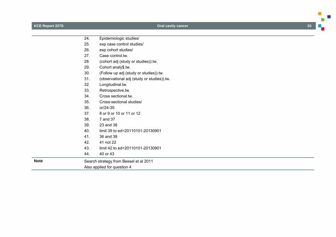
KCE Report 227S Oral cavity cancer 33
24. Epidemiologic studies/ 25. exp case control studies/ 26. exp cohort studies/ 27. Case control.tw. 28. (cohort adj (study or studies)).tw. 29. Cohort analy$.tw. 30. (Follow up adj (study or studies)).tw. 31. (observational adj (study or studies)).tw. 32. Longitudinal.tw. 33. Retrospective.tw. 34. Cross sectional.tw. 35. Cross-sectional studies/ 36. or/24-35 37. 8 or 9 or 10 or 11 or 12 38. 7 and 37 39. 23 and 38 40. limit 39 to ed=20110101-20130901 41. 36 and 38 42. 41 not 22 43. limit 42 to ed=20110101-20130901 44. 40 or 43
Note Search strategy from Bessel et al 2011 Also applied for question 4
34 Oral cavity cancer KCE Report 227S
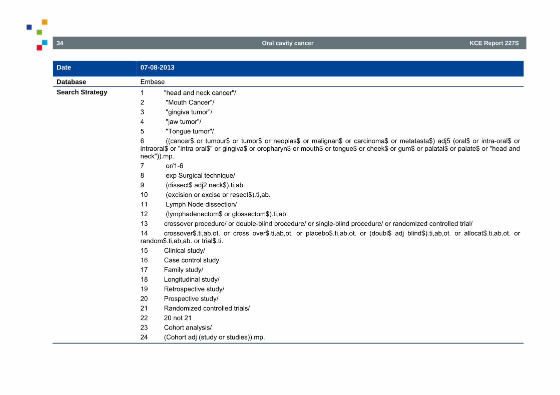
Date 07-08-2013
Database Embase Search Strategy 1 "head and neck cancer"/
2 "Mouth Cancer"/ 3 "gingiva tumor"/ 4 "jaw tumor"/ 5 "Tongue tumor"/ 6 ((cancer$ or tumour$ or tumor$ or neoplas$ or malignan$ or carcinoma$ or metatasta$) adj5 (oral$ or intra-oral$ or intraoral$ or "intra oral$" or gingiva$ or oropharyn$ or mouth$ or tongue$ or cheek$ or gum$ or palatal$ or palate$ or "head and neck")).mp. 7 or/1-6 8 exp Surgical technique/ 9 (dissect$ adj2 neck$).ti,ab. 10 (excision or excise or resect$).ti,ab. 11 Lymph Node dissection/ 12 (lymphadenectom$ or glossectom$).ti,ab. 13 crossover procedure/ or double-blind procedure/ or single-blind procedure/ or randomized controlled trial/ 14 crossover$.ti,ab,ot. or cross over$.ti,ab,ot. or placebo$.ti,ab,ot. or (doubl$ adj blind$).ti,ab,ot. or allocat$.ti,ab,ot. or random$.ti,ab,ab. or trial$.ti. 15 Clinical study/ 16 Case control study 17 Family study/ 18 Longitudinal study/ 19 Retrospective study/ 20 Prospective study/ 21 Randomized controlled trials/ 22 20 not 21 23 Cohort analysis/ 24 (Cohort adj (study or studies)).mp.
KCE Report 227S Oral cavity cancer 35
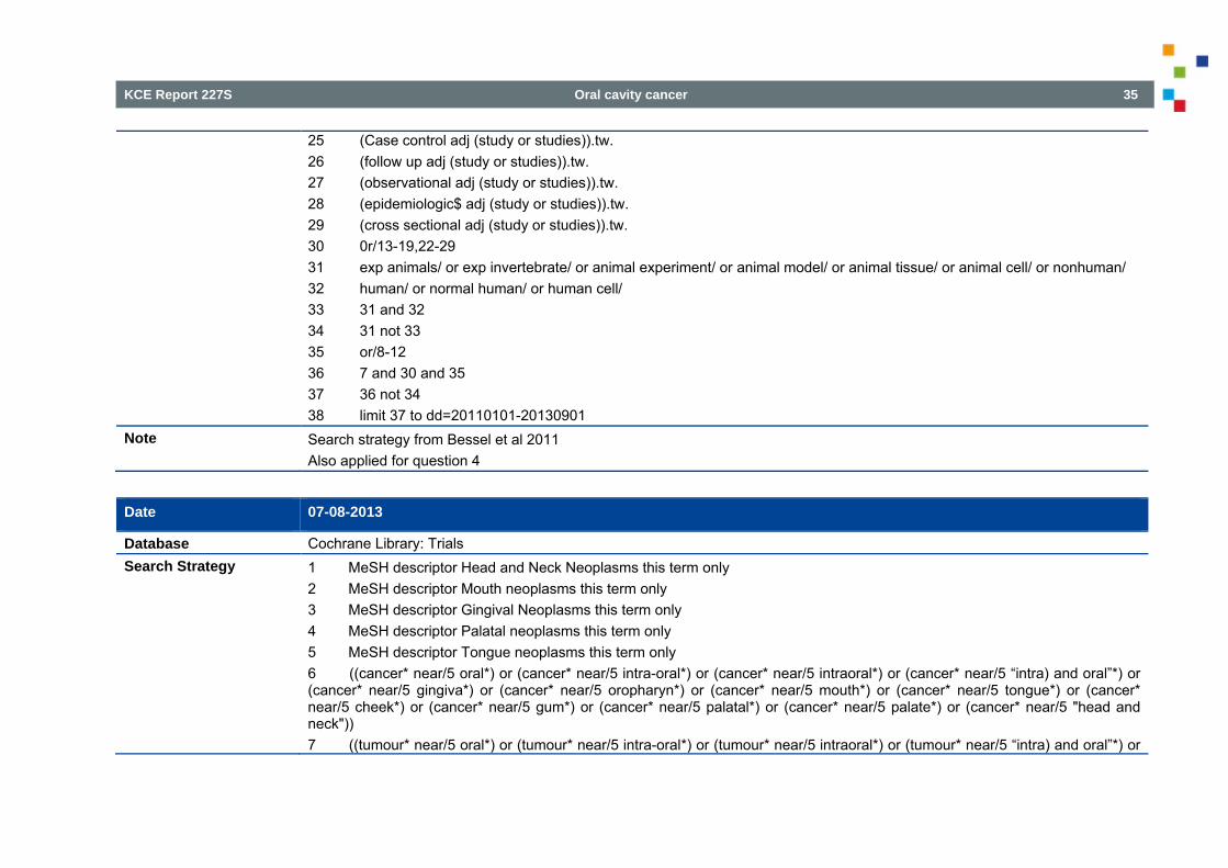
25 (Case control adj (study or studies)).tw. 26 (follow up adj (study or studies)).tw. 27 (observational adj (study or studies)).tw. 28 (epidemiologic$ adj (study or studies)).tw. 29 (cross sectional adj (study or studies)).tw. 30 0r/13-19,22-29 31 exp animals/ or exp invertebrate/ or animal experiment/ or animal model/ or animal tissue/ or animal cell/ or nonhuman/ 32 human/ or normal human/ or human cell/ 33 31 and 32 34 31 not 33 35 or/8-12 36 7 and 30 and 35 37 36 not 34 38 limit 37 to dd=20110101-20130901
Note Search strategy from Bessel et al 2011 Also applied for question 4
Date 07-08-2013
Database Cochrane Library: Trials Search Strategy 1 MeSH descriptor Head and Neck Neoplasms this term only
2 MeSH descriptor Mouth neoplasms this term only 3 MeSH descriptor Gingival Neoplasms this term only 4 MeSH descriptor Palatal neoplasms this term only 5 MeSH descriptor Tongue neoplasms this term only 6 ((cancer* near/5 oral*) or (cancer* near/5 intra-oral*) or (cancer* near/5 intraoral*) or (cancer* near/5 “intra) and oral”*) or (cancer* near/5 gingiva*) or (cancer* near/5 oropharyn*) or (cancer* near/5 mouth*) or (cancer* near/5 tongue*) or (cancer* near/5 cheek*) or (cancer* near/5 gum*) or (cancer* near/5 palatal*) or (cancer* near/5 palate*) or (cancer* near/5 "head and neck")) 7 ((tumour* near/5 oral*) or (tumour* near/5 intra-oral*) or (tumour* near/5 intraoral*) or (tumour* near/5 “intra) and oral”*) or
36 Oral cavity cancer KCE Report 227S
(tumour* near/5 gingiva*) or (tumour* near/5 oropharyn*) or (tumour* near/5 mouth*) or (tumour* near/5 tongue*) or (tumour* near/5 cheek*) or (tumour* near/5 gum*) or (tumour* near/5 palatal*) or (tumour* near/5 palate*) or (tumour* near/5 "head and neck")) 8 ((tumor* near/5 oral*) or (tumor* near/5 intra-oral*) or (tumor* near/5 intraoral*) or (tumor* near/5 “intra) and oral”*) or (tumor* near/5 gingiva*) or (tumor* near/5 oropharyn*) or (tumor* near/5 mouth*) or (tumor* near/5 tongue*) or (tumor* near/5 cheek*) or (tumor* near/5 gum*) or (tumor* near/5 palatal*) or (tumor* near/5 palate*) or (tumor* near/5 "head and neck")) 9 ((neoplas* near/5 oral*) or (neoplas* near/5 intra-oral*) or (neoplas* near/5 intraoral*) or (neoplas* near/5 “intra) and oral”*) or (neoplas* near/5 gingiva*) or (neoplas* near/5 oropharyn*) or (neoplas* near/5 mouth*) or (neoplas* near/5 tongue*) or (neoplas* near/5 cheek*) or (neoplas* near/5 gum*) or (neoplas* near/5 palatal*) or (neoplas* near/5 palate*) or (neoplas* near/5 "head and neck")) 10 ((malignan* near/5 oral*) or (malignan* near/5 intra-oral*) or (malignan* near/5 intraoral*) or (malignan* near/5 “intra) and oral”*) or (malignan* near/5 gingiva*) or (malignan* near/5 oropharyn*) or (malignan* near/5 mouth*) or (malignan* near/5 tongue*) or (malignan* near/5 cheek*) or (malignan* near/5 gum*) or (malignan* near/5 palatal*) or (malignan* near/5 palate*) or (malignan* near/5 "head and neck")) 11 ((carcinoma* near/5 oral*) or (carcinoma* near/5 intra-oral*) or (carcinoma* near/5 intraoral*) or (carcinoma* near/5 “intra) and oral”*) or (carcinoma* near/5 gingiva*) or (carcinoma* near/5 oropharyn*) or (carcinoma* near/5 mouth*) or (carcinoma* near/5 tongue*) or (carcinoma* near/5 cheek*) or (carcinoma* near/5 gum*) or (carcinoma* near/5 palatal*) or (carcinoma* near/5 palate*) or (carcinoma* near/5 "head and neck")) 12 ((metatasta* near/5 oral*) or (metatasta* near/5 intra-oral*) or (metatasta* near/5 intraoral*) or (metatasta* near/5 “intra) and oral”*) or (metatasta* near/5 gingiva*) or (metatasta* near/5 oropharyn*) or (metatasta* near/5 mouth*) or (metatasta* near/5 tongue*) or (metatasta* near/5 cheek*) or (metatasta* near/5 gum*) or (metatasta* near/5 palatal*) or (metatasta* near/5 palate*) or (metatasta* near/5 "head and neck")) 13 (1 or 2 or 3 or 4 or 5 or 6 or 7 or 8 or 9 or 10 or 11 or 12) 14 MeSH descriptor Surgical Procedures, Operative explode all trees 15 (dissect* near/2 neck*) 16 (excision or excise* or resect*) 17 MeSH descriptor Lymph node excision this term only 18 (lymphadenectom* or glossectom*) 19 (14 or 15 or 16 or 17 or 18) 20 (13 and 19)
Note Limit to 2011-2013 Search strategy from Bessel et al 2011 Also applied for question 4
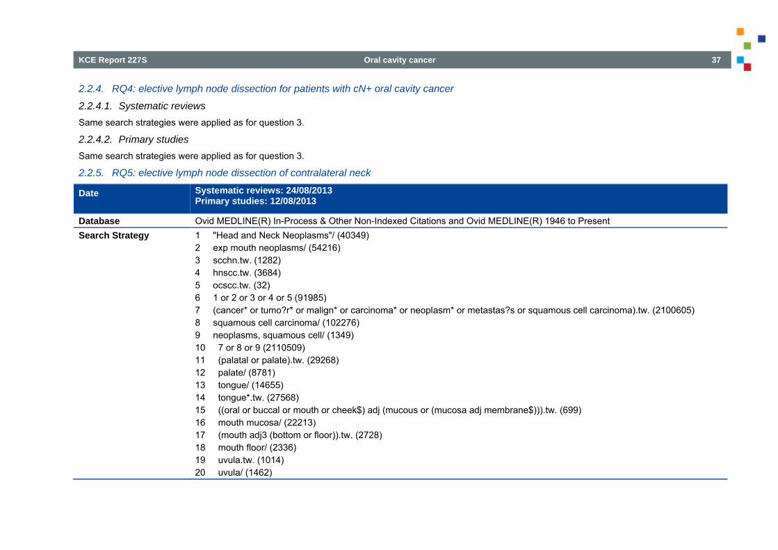
KCE Report 227S Oral cavity cancer 37
2.2.4. RQ4: elective lymph node dissection for patients with cN+ oral cavity cancer
2.2.4.1. Systematic reviews Same search strategies were applied as for question 3.
2.2.4.2. Primary studies Same search strategies were applied as for question 3.
2.2.5. RQ5: elective lymph node dissection of contralateral neck
Date Systematic reviews: 24/08/2013 Primary studies: 12/08/2013
Database Ovid MEDLINE(R) In-Process & Other Non-Indexed Citations and Ovid MEDLINE(R) 1946 to Present Search Strategy 1 "Head and Neck Neoplasms"/ (40349)
2 exp mouth neoplasms/ (54216) 3 scchn.tw. (1282) 4 hnscc.tw. (3684) 5 ocscc.tw. (32) 6 1 or 2 or 3 or 4 or 5 (91985) 7 (cancer* or tumo?r* or malign* or carcinoma* or neoplasm* or metastas?s or squamous cell carcinoma).tw. (2100605) 8 squamous cell carcinoma/ (102276) 9 neoplasms, squamous cell/ (1349) 10 7 or 8 or 9 (2110509) 11 (palatal or palate).tw. (29268) 12 palate/ (8781) 13 tongue/ (14655) 14 tongue*.tw. (27568) 15 ((oral or buccal or mouth or cheek$) adj (mucous or (mucosa adj membrane$))).tw. (699) 16 mouth mucosa/ (22213) 17 (mouth adj3 (bottom or floor)).tw. (2728) 18 mouth floor/ (2336) 19 uvula.tw. (1014) 20 uvula/ (1462)
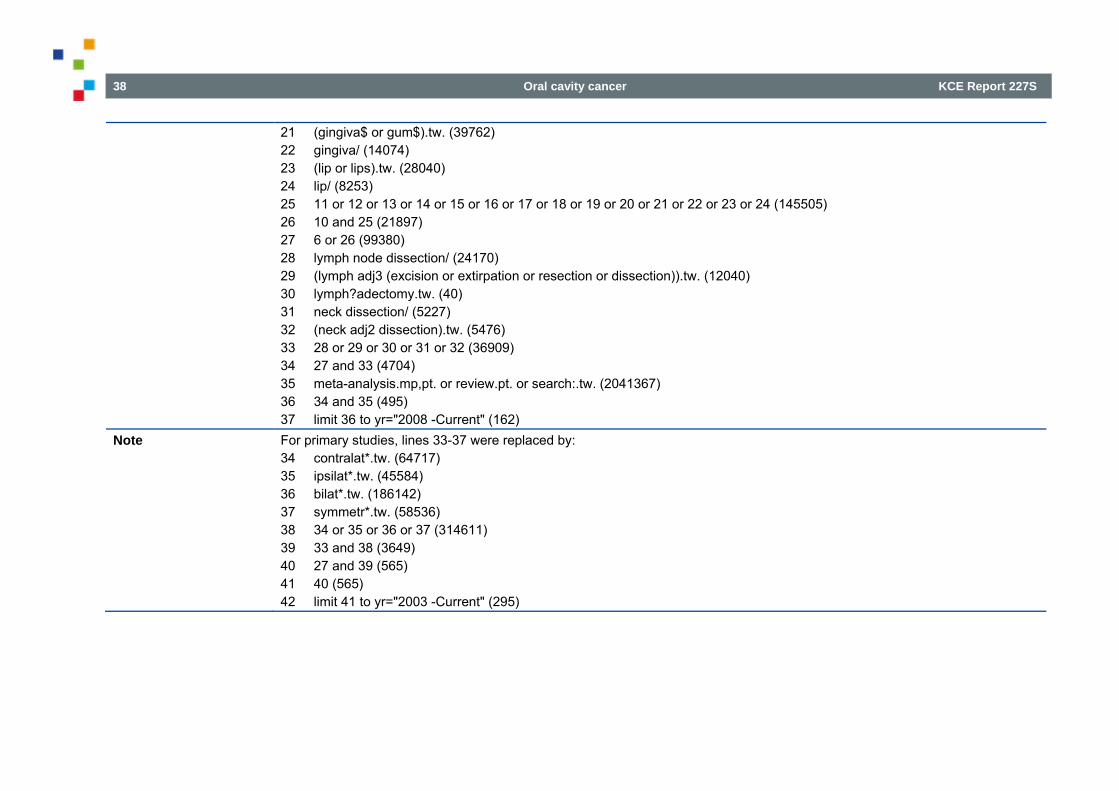
38 Oral cavity cancer KCE Report 227S
21 (gingiva$ or gum$).tw. (39762) 22 gingiva/ (14074) 23 (lip or lips).tw. (28040) 24 lip/ (8253) 25 11 or 12 or 13 or 14 or 15 or 16 or 17 or 18 or 19 or 20 or 21 or 22 or 23 or 24 (145505) 26 10 and 25 (21897) 27 6 or 26 (99380) 28 lymph node dissection/ (24170) 29 (lymph adj3 (excision or extirpation or resection or dissection)).tw. (12040) 30 lymph?adectomy.tw. (40) 31 neck dissection/ (5227) 32 (neck adj2 dissection).tw. (5476) 33 28 or 29 or 30 or 31 or 32 (36909) 34 27 and 33 (4704) 35 meta-analysis.mp,pt. or review.pt. or search:.tw. (2041367) 36 34 and 35 (495) 37 limit 36 to yr="2008 -Current" (162)
Note For primary studies, lines 33-37 were replaced by: 34 contralat*.tw. (64717) 35 ipsilat*.tw. (45584) 36 bilat*.tw. (186142) 37 symmetr*.tw. (58536) 38 34 or 35 or 36 or 37 (314611) 39 33 and 38 (3649) 40 27 and 39 (565) 41 40 (565) 42 limit 41 to yr="2003 -Current" (295)
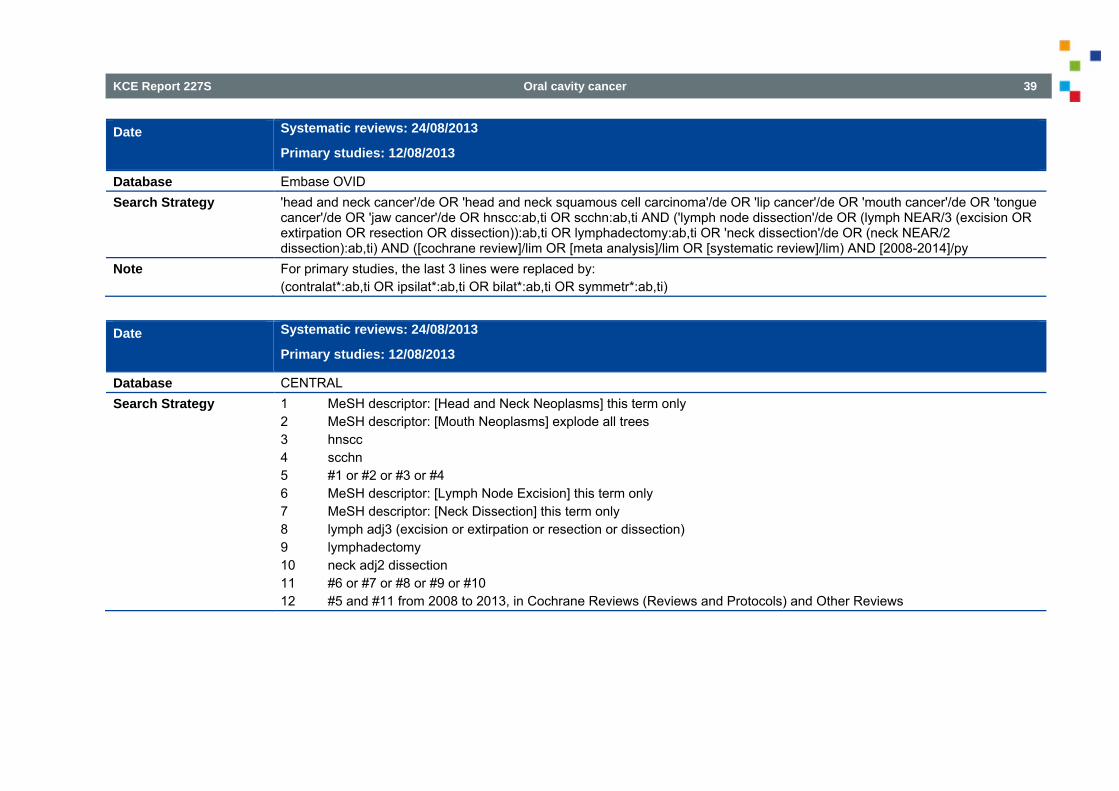
KCE Report 227S Oral cavity cancer 39
Date Systematic reviews: 24/08/2013
Primary studies: 12/08/2013
Database Embase OVID Search Strategy 'head and neck cancer'/de OR 'head and neck squamous cell carcinoma'/de OR 'lip cancer'/de OR 'mouth cancer'/de OR 'tongue
cancer'/de OR 'jaw cancer'/de OR hnscc:ab,ti OR scchn:ab,ti AND ('lymph node dissection'/de OR (lymph NEAR/3 (excision OR extirpation OR resection OR dissection)):ab,ti OR lymphadectomy:ab,ti OR 'neck dissection'/de OR (neck NEAR/2 dissection):ab,ti) AND ([cochrane review]/lim OR [meta analysis]/lim OR [systematic review]/lim) AND [2008-2014]/py
Note For primary studies, the last 3 lines were replaced by: (contralat*:ab,ti OR ipsilat*:ab,ti OR bilat*:ab,ti OR symmetr*:ab,ti)
Date Systematic reviews: 24/08/2013
Primary studies: 12/08/2013
Database CENTRAL Search Strategy 1 MeSH descriptor: [Head and Neck Neoplasms] this term only
2 MeSH descriptor: [Mouth Neoplasms] explode all trees 3 hnscc 4 scchn 5 #1 or #2 or #3 or #4 6 MeSH descriptor: [Lymph Node Excision] this term only 7 MeSH descriptor: [Neck Dissection] this term only 8 lymph adj3 (excision or extirpation or resection or dissection) 9 lymphadectomy 10 neck adj2 dissection 11 #6 or #7 or #8 or #9 or #10 12 #5 and #11 from 2008 to 2013, in Cochrane Reviews (Reviews and Protocols) and Other Reviews
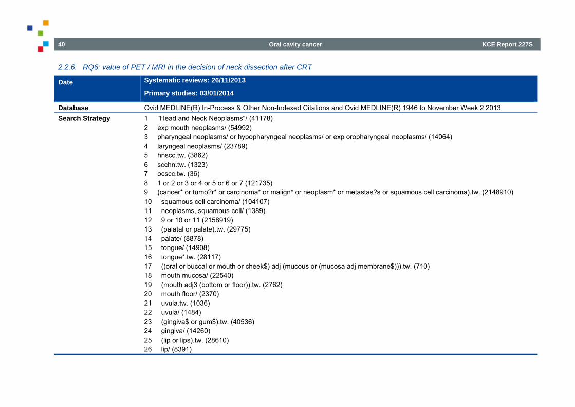
40 Oral cavity cancer KCE Report 227S
2.2.6. RQ6: value of PET / MRI in the decision of neck dissection after CRT
Date Systematic reviews: 26/11/2013
Primary studies: 03/01/2014
Database Ovid MEDLINE(R) In-Process & Other Non-Indexed Citations and Ovid MEDLINE(R) 1946 to November Week 2 2013 Search Strategy 1 "Head and Neck Neoplasms"/ (41178)
2 exp mouth neoplasms/ (54992) 3 pharyngeal neoplasms/ or hypopharyngeal neoplasms/ or exp oropharyngeal neoplasms/ (14064) 4 laryngeal neoplasms/ (23789) 5 hnscc.tw. (3862) 6 scchn.tw. (1323) 7 ocscc.tw. (36) 8 1 or 2 or 3 or 4 or 5 or 6 or 7 (121735) 9 (cancer* or tumo?r* or carcinoma* or malign* or neoplasm* or metastas?s or squamous cell carcinoma).tw. (2148910) 10 squamous cell carcinoma/ (104107) 11 neoplasms, squamous cell/ (1389) 12 9 or 10 or 11 (2158919) 13 (palatal or palate).tw. (29775) 14 palate/ (8878) 15 tongue/ (14908) 16 tongue*.tw. (28117) 17 ((oral or buccal or mouth or cheek$) adj (mucous or (mucosa adj membrane$))).tw. (710) 18 mouth mucosa/ (22540) 19 (mouth adj3 (bottom or floor)).tw. (2762) 20 mouth floor/ (2370) 21 uvula.tw. (1036) 22 uvula/ (1484) 23 (gingiva$ or gum$).tw. (40536) 24 gingiva/ (14260) 25 (lip or lips).tw. (28610) 26 lip/ (8391)
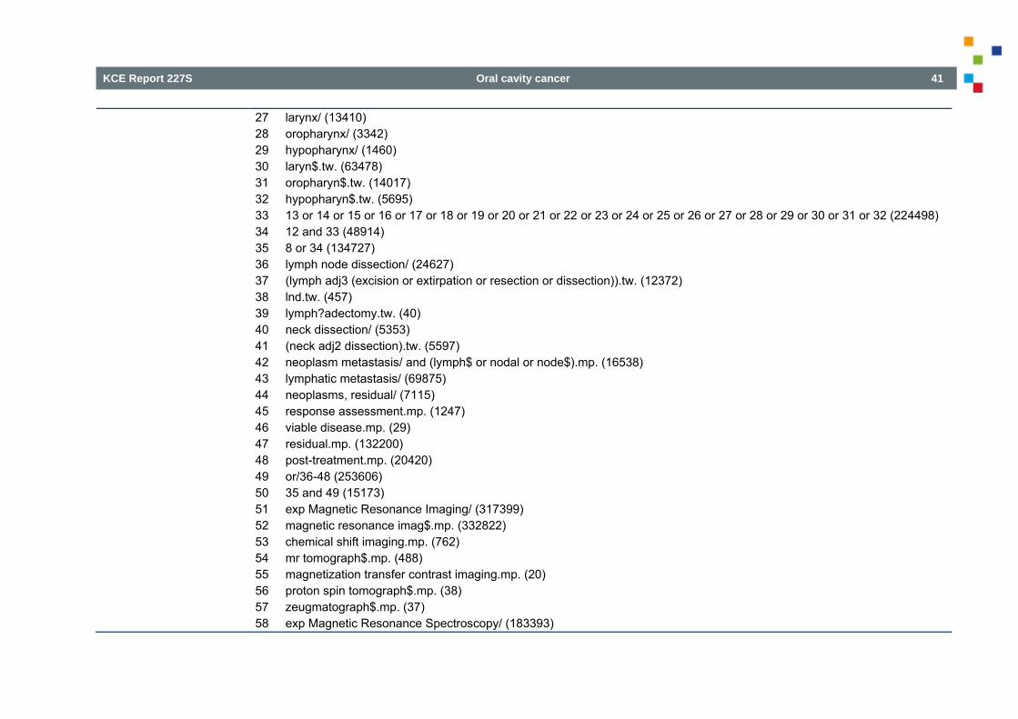
KCE Report 227S Oral cavity cancer 41
27 larynx/ (13410) 28 oropharynx/ (3342) 29 hypopharynx/ (1460) 30 laryn$.tw. (63478) 31 oropharyn$.tw. (14017) 32 hypopharyn$.tw. (5695) 33 13 or 14 or 15 or 16 or 17 or 18 or 19 or 20 or 21 or 22 or 23 or 24 or 25 or 26 or 27 or 28 or 29 or 30 or 31 or 32 (224498) 34 12 and 33 (48914) 35 8 or 34 (134727) 36 lymph node dissection/ (24627) 37 (lymph adj3 (excision or extirpation or resection or dissection)).tw. (12372) 38 lnd.tw. (457) 39 lymph?adectomy.tw. (40) 40 neck dissection/ (5353) 41 (neck adj2 dissection).tw. (5597) 42 neoplasm metastasis/ and (lymph$ or nodal or node$).mp. (16538) 43 lymphatic metastasis/ (69875) 44 neoplasms, residual/ (7115) 45 response assessment.mp. (1247) 46 viable disease.mp. (29) 47 residual.mp. (132200) 48 post-treatment.mp. (20420) 49 or/36-48 (253606) 50 35 and 49 (15173) 51 exp Magnetic Resonance Imaging/ (317399) 52 magnetic resonance imag$.mp. (332822) 53 chemical shift imaging.mp. (762) 54 mr tomograph$.mp. (488) 55 magnetization transfer contrast imaging.mp. (20) 56 proton spin tomograph$.mp. (38) 57 zeugmatograph$.mp. (37) 58 exp Magnetic Resonance Spectroscopy/ (183393)
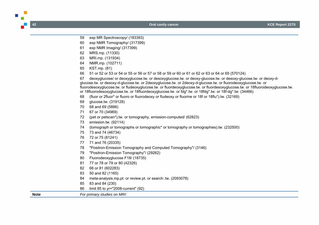
42 Oral cavity cancer KCE Report 227S
59 exp MR Spectroscopy/ (183393) 60 exp NMR Tomography/ (317399) 61 exp NMR Imaging/ (317399) 62 MRS.mp. (11330) 63 MRI.mp. (131934) 64 NMR.mp. (102711) 65 KST.mp. (81) 66 51 or 52 or 53 or 54 or 55 or 56 or 57 or 58 or 59 or 60 or 61 or 62 or 63 or 64 or 65 (570124) 67 deoxyglucose/ or deoxyglucose.tw. or desoxyglucose.tw. or deoxy-glucose.tw. or desoxy-glucose.tw. or deoxy-d-glucose.tw. or desoxy-d-glucose.tw. or 2deoxyglucose.tw. or 2deoxy-d-glucose.tw. or fluorodeoxyglucose.tw. or fluorodesoxyglucose.tw. or fludeoxyglucose.tw. or fluordeoxyglucose.tw. or fluordesoxyglucose.tw. or 18fluorodeoxyglucose.tw. or 18fluorodesoxyglucose.tw. or 18fluordeoxyglucose.tw. or fdg*.tw. or 18fdg*.tw. or 18f-dg*.tw. (34466) 68 (fluor or 2fluor* or fluoro or fluorodeoxy or fludeoxy or fluorine or 18f or 18flu*).tw. (32169) 69 glucose.tw. (319128) 70 68 and 69 (5886) 71 67 or 70 (34969) 72 (pet or petscan*).tw. or tomography, emission-computed/ (62823) 73 emission.tw. (92114) 74 (tomograph or tomographs or tomographic* or tomography or tomographies).tw. (232500) 75 73 and 74 (46734) 76 72 or 75 (81241) 77 71 and 76 (20335) 78 "Positron-Emission Tomography and Computed Tomography"/ (3146) 79 "Positron-Emission Tomography"/ (29262) 80 Fluorodeoxyglucose F18/ (18735) 81 77 or 78 or 79 or 80 (42326) 82 66 or 81 (602283) 83 50 and 82 (1165) 84 meta-analysis.mp,pt. or review.pt. or search:.tw. (2093078) 85 83 and 84 (230) 86 limit 85 to yr="2008-current" (92)
Note For primary studies on MRI:
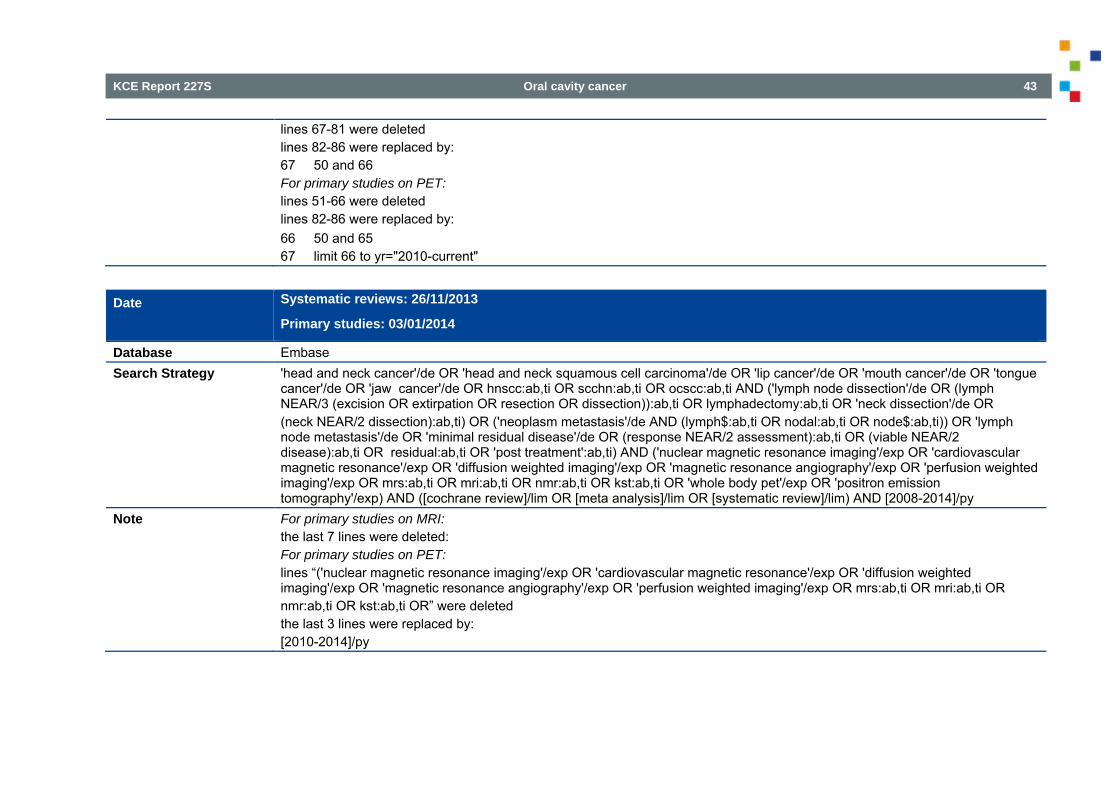
KCE Report 227S Oral cavity cancer 43
lines 67-81 were deleted lines 82-86 were replaced by: 67 50 and 66 For primary studies on PET: lines 51-66 were deleted lines 82-86 were replaced by: 66 50 and 65 67 limit 66 to yr="2010-current"
Date Systematic reviews: 26/11/2013
Primary studies: 03/01/2014
Database Embase Search Strategy 'head and neck cancer'/de OR 'head and neck squamous cell carcinoma'/de OR 'lip cancer'/de OR 'mouth cancer'/de OR 'tongue
cancer'/de OR 'jaw cancer'/de OR hnscc:ab,ti OR scchn:ab,ti OR ocscc:ab,ti AND ('lymph node dissection'/de OR (lymph NEAR/3 (excision OR extirpation OR resection OR dissection)):ab,ti OR lymphadectomy:ab,ti OR 'neck dissection'/de OR (neck NEAR/2 dissection):ab,ti) OR ('neoplasm metastasis'/de AND (lymph$:ab,ti OR nodal:ab,ti OR node$:ab,ti)) OR 'lymph node metastasis'/de OR 'minimal residual disease'/de OR (response NEAR/2 assessment):ab,ti OR (viable NEAR/2 disease):ab,ti OR residual:ab,ti OR 'post treatment':ab,ti) AND ('nuclear magnetic resonance imaging'/exp OR 'cardiovascular magnetic resonance'/exp OR 'diffusion weighted imaging'/exp OR 'magnetic resonance angiography'/exp OR 'perfusion weighted imaging'/exp OR mrs:ab,ti OR mri:ab,ti OR nmr:ab,ti OR kst:ab,ti OR 'whole body pet'/exp OR 'positron emission tomography'/exp) AND ([cochrane review]/lim OR [meta analysis]/lim OR [systematic review]/lim) AND [2008-2014]/py
Note For primary studies on MRI: the last 7 lines were deleted: For primary studies on PET: lines “('nuclear magnetic resonance imaging'/exp OR 'cardiovascular magnetic resonance'/exp OR 'diffusion weighted imaging'/exp OR 'magnetic resonance angiography'/exp OR 'perfusion weighted imaging'/exp OR mrs:ab,ti OR mri:ab,ti OR nmr:ab,ti OR kst:ab,ti OR” were deleted the last 3 lines were replaced by: [2010-2014]/py
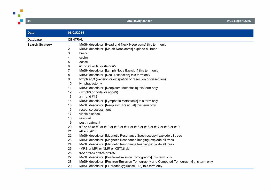
44 Oral cavity cancer KCE Report 227S
Date 06/01/2014
Database CENTRAL Search Strategy 1 MeSH descriptor: [Head and Neck Neoplasms] this term only
2 MeSH descriptor: [Mouth Neoplasms] explode all trees 3 hnscc 4 scchn 5 ocscc 6 #1 or #2 or #3 or #4 or #5 7 MeSH descriptor: [Lymph Node Excision] this term only 8 MeSH descriptor: [Neck Dissection] this term only 9 lymph adj3 (excision or extirpation or resection or dissection) 10 lymphadectomy 11 MeSH descriptor: [Neoplasm Metastasis] this term only 12 (lymph$ or nodal or node$) 13 #11 and #12 14 MeSH descriptor: [Lymphatic Metastasis] this term only 15 MeSH descriptor: [Neoplasm, Residual] this term only 16 response assessment 17 viable disease 18 residual 19 post-treatment 20 #7 or #8 or #9 or #10 or #13 or #14 or #15 or #16 or #17 or #18 or #19 21 #6 and #20 22 MeSH descriptor: [Magnetic Resonance Spectroscopy] explode all trees 23 MeSH descriptor: [Magnetic Resonance Imaging] explode all trees 24 MeSH descriptor: [Magnetic Resonance Imaging] explode all trees 25 (MRS or MRI or NMR or KST):ti,ab 26 #22 or #23 or #24 or #25 27 MeSH descriptor: [Positron-Emission Tomography] this term only 28 MeSH descriptor: [Positron-Emission Tomography and Computed Tomography] this term only 29 MeSH descriptor: [Fluorodeoxyglucose F18] this term only
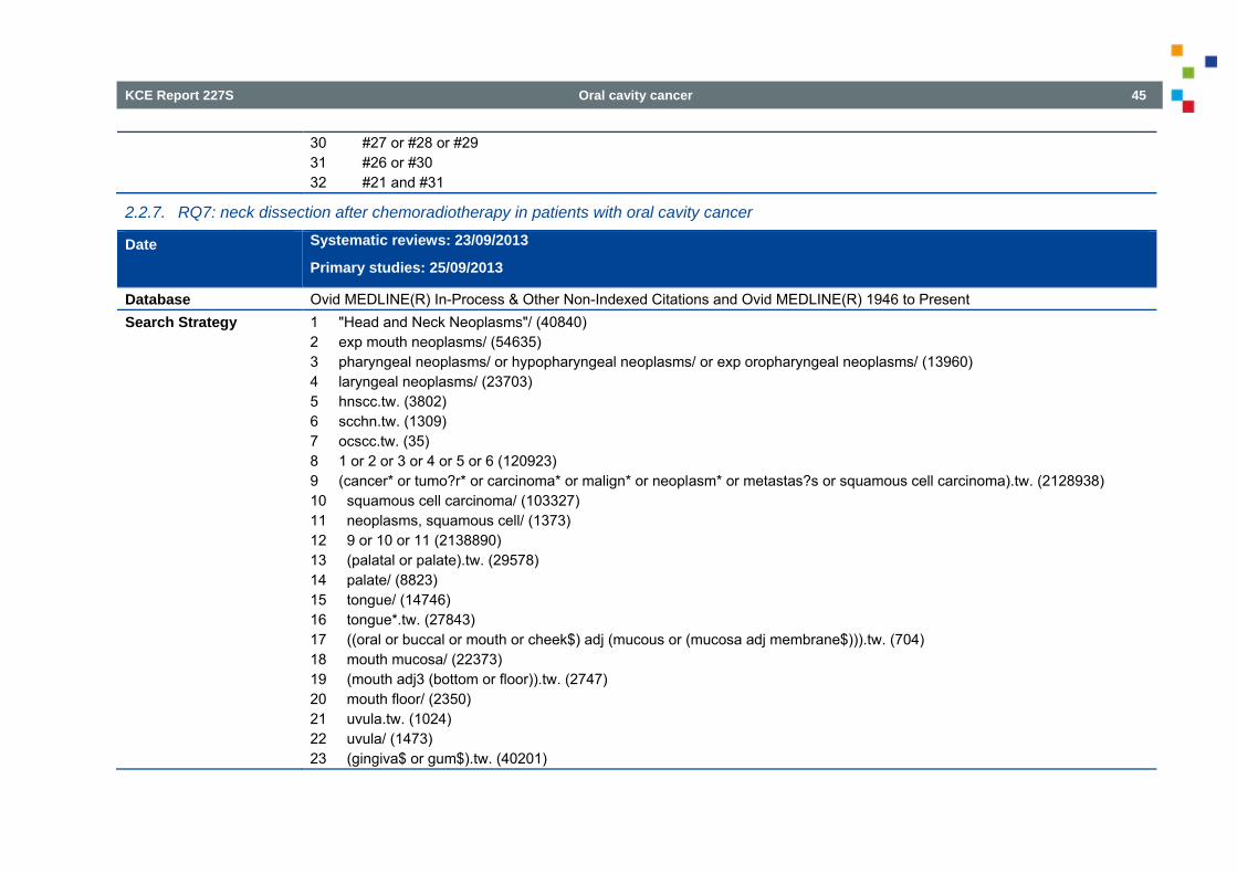
KCE Report 227S Oral cavity cancer 45
30 #27 or #28 or #29 31 #26 or #30 32 #21 and #31
2.2.7. RQ7: neck dissection after chemoradiotherapy in patients with oral cavity cancer
Date Systematic reviews: 23/09/2013
Primary studies: 25/09/2013
Database Ovid MEDLINE(R) In-Process & Other Non-Indexed Citations and Ovid MEDLINE(R) 1946 to Present Search Strategy 1 "Head and Neck Neoplasms"/ (40840)
2 exp mouth neoplasms/ (54635) 3 pharyngeal neoplasms/ or hypopharyngeal neoplasms/ or exp oropharyngeal neoplasms/ (13960) 4 laryngeal neoplasms/ (23703) 5 hnscc.tw. (3802) 6 scchn.tw. (1309) 7 ocscc.tw. (35) 8 1 or 2 or 3 or 4 or 5 or 6 (120923) 9 (cancer* or tumo?r* or carcinoma* or malign* or neoplasm* or metastas?s or squamous cell carcinoma).tw. (2128938) 10 squamous cell carcinoma/ (103327) 11 neoplasms, squamous cell/ (1373) 12 9 or 10 or 11 (2138890) 13 (palatal or palate).tw. (29578) 14 palate/ (8823) 15 tongue/ (14746) 16 tongue*.tw. (27843) 17 ((oral or buccal or mouth or cheek$) adj (mucous or (mucosa adj membrane$))).tw. (704) 18 mouth mucosa/ (22373) 19 (mouth adj3 (bottom or floor)).tw. (2747) 20 mouth floor/ (2350) 21 uvula.tw. (1024) 22 uvula/ (1473) 23 (gingiva$ or gum$).tw. (40201)
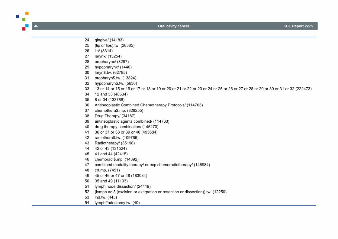
46 Oral cavity cancer KCE Report 227S
24 gingiva/ (14183) 25 (lip or lips).tw. (28385) 26 lip/ (8314) 27 larynx/ (13254) 28 oropharynx/ (3297) 29 hypopharynx/ (1440) 30 laryn$.tw. (62795) 31 oropharyn$.tw. (13824) 32 hypopharyn$.tw. (5636) 33 13 or 14 or 15 or 16 or 17 or 18 or 19 or 20 or 21 or 22 or 23 or 24 or 25 or 26 or 27 or 28 or 29 or 30 or 31 or 32 (222473) 34 12 and 33 (48534) 35 8 or 34 (133788) 36 Antineoplastic Combined Chemotherapy Protocols/ (114763) 37 chemothera$.mp. (328255) 38 Drug Therapy/ (34187) 39 antineoplastic agents combined/ (114763) 40 drug therapy combination/ (145270) 41 36 or 37 or 38 or 39 or 40 (493684) 42 radiothera$.tw. (109766) 43 Radiotherapy/ (35198) 44 42 or 43 (131524) 45 41 and 44 (42415) 46 chemoradi$.mp. (14392) 47 combined modality therapy/ or exp chemoradiotherapy/ (146984) 48 crt.mp. (7451) 49 45 or 46 or 47 or 48 (183034) 50 35 and 49 (11103) 51 lymph node dissection/ (24419) 52 (lymph adj3 (excision or extirpation or resection or dissection)).tw. (12250) 53 lnd.tw. (445) 54 lymph?adectomy.tw. (40)
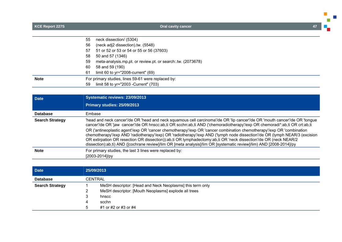
KCE Report 227S Oral cavity cancer 47
55 neck dissection/ (5304) 56 (neck adj2 dissection).tw. (5548) 57 51 or 52 or 53 or 54 or 55 or 56 (37603) 58 50 and 57 (1346) 59 meta-analysis.mp,pt. or review.pt. or search:.tw. (2073678) 60 58 and 59 (190) 61 limit 60 to yr="2008-current" (69)
Note For primary studies, lines 59-61 were replaced by: 59 limit 58 to yr="2003 -Current" (703)
Date Systematic reviews: 23/09/2013
Primary studies: 25/09/2013
Database Embase Search Strategy 'head and neck cancer'/de OR 'head and neck squamous cell carcinoma'/de OR 'lip cancer'/de OR 'mouth cancer'/de OR 'tongue
cancer'/de OR 'jaw cancer'/de OR hnscc:ab,ti OR scchn:ab,ti AND ('chemoradiotherapy'/exp OR chemoradi*:ab,ti OR crt:ab,ti OR ('antineoplastic agent'/exp OR 'cancer chemotherapy'/exp OR 'cancer combination chemotherapy'/exp OR 'combination chemotherapy'/exp AND 'radiotherapy'/exp) OR 'radiotherapy'/exp AND ('lymph node dissection'/de OR (lymph NEAR/3 (excision OR extirpation OR resection OR dissection)):ab,ti OR lymphadectomy:ab,ti OR 'neck dissection'/de OR (neck NEAR/2 dissection):ab,ti) AND ([cochrane review]/lim OR [meta analysis]/lim OR [systematic review]/lim) AND [2008-2014]/py
Note For primary studies, the last 3 lines were replaced by: [2003-2014]/py
Date 25/09/2013
Database CENTRAL Search Strategy 1 MeSH descriptor: [Head and Neck Neoplasms] this term only
2 MeSH descriptor: [Mouth Neoplasms] explode all trees 3 hnscc 4 scchn 5 #1 or #2 or #3 or #4
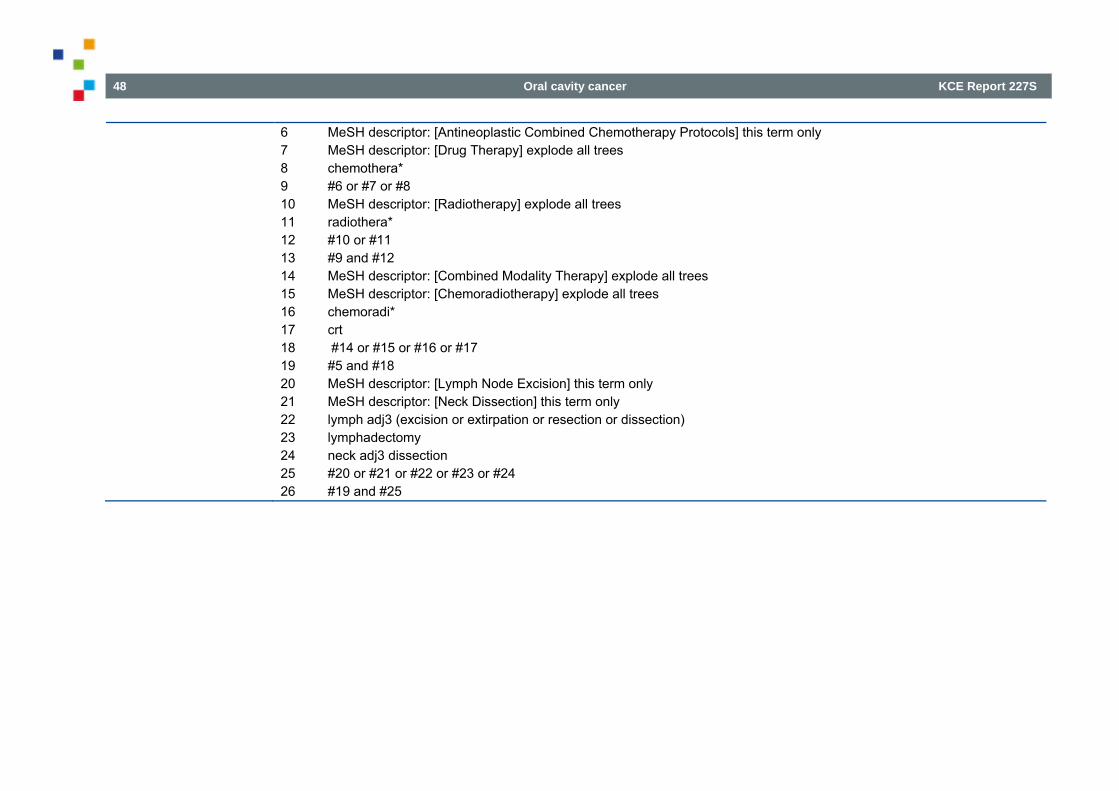
48 Oral cavity cancer KCE Report 227S
6 MeSH descriptor: [Antineoplastic Combined Chemotherapy Protocols] this term only 7 MeSH descriptor: [Drug Therapy] explode all trees 8 chemothera* 9 #6 or #7 or #8 10 MeSH descriptor: [Radiotherapy] explode all trees 11 radiothera* 12 #10 or #11 13 #9 and #12 14 MeSH descriptor: [Combined Modality Therapy] explode all trees 15 MeSH descriptor: [Chemoradiotherapy] explode all trees 16 chemoradi* 17 crt 18 #14 or #15 or #16 or #17 19 #5 and #18 20 MeSH descriptor: [Lymph Node Excision] this term only 21 MeSH descriptor: [Neck Dissection] this term only 22 lymph adj3 (excision or extirpation or resection or dissection) 23 lymphadectomy 24 neck adj3 dissection 25 #20 or #21 or #22 or #23 or #24 26 #19 and #25
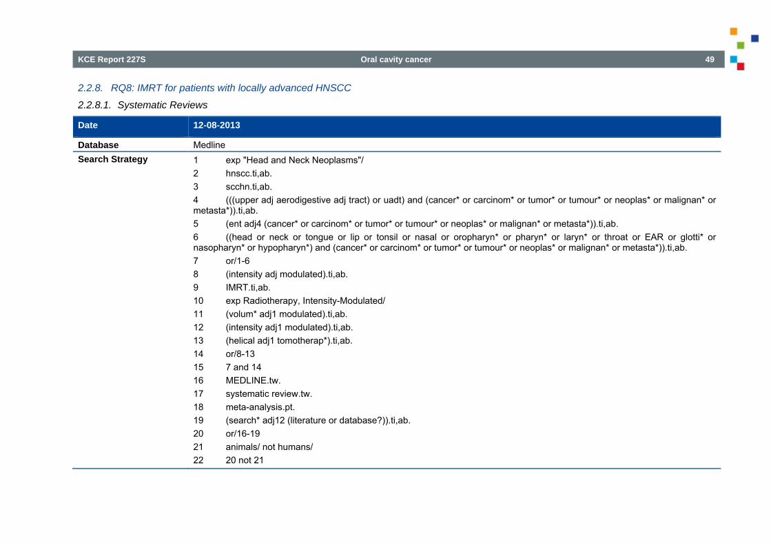
KCE Report 227S Oral cavity cancer 49
2.2.8. RQ8: IMRT for patients with locally advanced HNSCC
2.2.8.1. Systematic Reviews
Date 12-08-2013
Database Medline Search Strategy 1 exp "Head and Neck Neoplasms"/
2 hnscc.ti,ab. 3 scchn.ti,ab. 4 (((upper adj aerodigestive adj tract) or uadt) and (cancer* or carcinom* or tumor* or tumour* or neoplas* or malignan* or metasta*)).ti,ab. 5 (ent adj4 (cancer* or carcinom* or tumor* or tumour* or neoplas* or malignan* or metasta*)).ti,ab. 6 ((head or neck or tongue or lip or tonsil or nasal or oropharyn* or pharyn* or laryn* or throat or EAR or glotti* or nasopharyn* or hypopharyn*) and (cancer* or carcinom* or tumor* or tumour* or neoplas* or malignan* or metasta*)).ti,ab. 7 or/1-6 8 (intensity adj modulated).ti,ab. 9 IMRT.ti,ab. 10 exp Radiotherapy, Intensity-Modulated/ 11 (volum* adj1 modulated).ti,ab. 12 (intensity adj1 modulated).ti,ab. 13 (helical adj1 tomotherap*).ti,ab. 14 or/8-13 15 7 and 14 16 MEDLINE.tw. 17 systematic review.tw. 18 meta-analysis.pt. 19 (search* adj12 (literature or database?)).ti,ab. 20 or/16-19 21 animals/ not humans/ 22 20 not 21
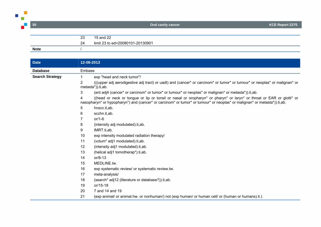
50 Oral cavity cancer KCE Report 227S
23 15 and 22 24 limit 23 to ed=20080101-20130901
Note /
Date 12-08-2013
Database Embase Search Strategy 1 exp "head and neck tumor"/
2 (((upper adj aerodigestive adj tract) or uadt) and (cancer* or carcinom* or tumor* or tumour* or neoplas* or malignan* or metasta*)).ti,ab. 3 (ent adj4 (cancer* or carcinom* or tumor* or tumour* or neoplas* or malignan* or metasta*)).ti,ab. 4 ((head or neck or tongue or lip or tonsil or nasal or oropharyn* or pharyn* or laryn* or throat or EAR or glotti* or nasopharyn* or hypopharyn*) and (cancer* or carcinom* or tumor* or tumour* or neoplas* or malignan* or metasta*)).ti,ab. 5 hnscc.ti,ab. 6 scchn.ti,ab. 7 or/1-6 8 (intensity adj modulated).ti,ab. 9 IMRT.ti,ab. 10 exp intensity modulated radiation therapy/ 11 (volum* adj1 modulated).ti,ab. 12 (intensity adj1 modulated).ti,ab. 13 (helical adj1 tomotherap*).ti,ab. 14 or/8-13 15 MEDLINE.tw. 16 exp systematic review/ or systematic review.tw. 17 meta-analysis/ 18 (search* adj12 (literature or database?)).ti,ab. 19 or/15-18 20 7 and 14 and 19 21 (exp animal/ or animal.hw. or nonhuman/) not (exp human/ or human cell/ or (human or humans).ti.)
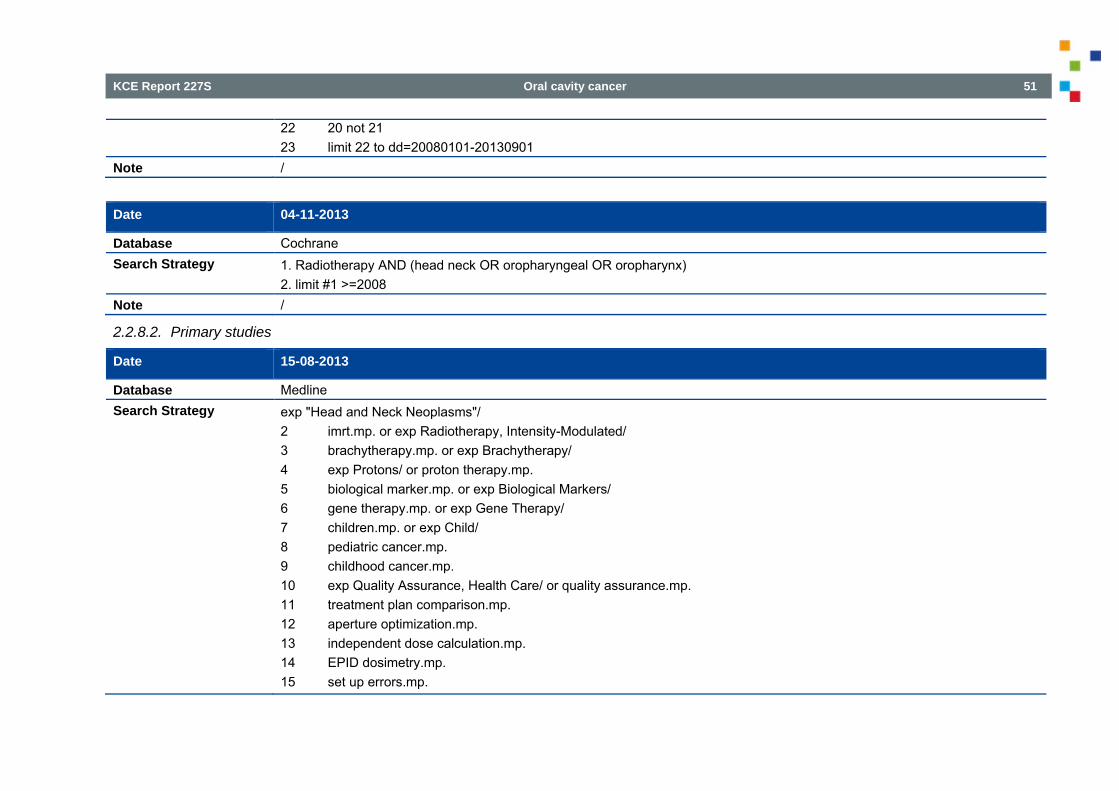
KCE Report 227S Oral cavity cancer 51
22 20 not 21 23 limit 22 to dd=20080101-20130901
Note /
Date 04-11-2013
Database Cochrane Search Strategy 1. Radiotherapy AND (head neck OR oropharyngeal OR oropharynx)
2. limit #1 >=2008 Note /
2.2.8.2. Primary studies
Date 15-08-2013
Database Medline Search Strategy exp "Head and Neck Neoplasms"/
2 imrt.mp. or exp Radiotherapy, Intensity-Modulated/ 3 brachytherapy.mp. or exp Brachytherapy/ 4 exp Protons/ or proton therapy.mp. 5 biological marker.mp. or exp Biological Markers/ 6 gene therapy.mp. or exp Gene Therapy/ 7 children.mp. or exp Child/ 8 pediatric cancer.mp. 9 childhood cancer.mp. 10 exp Quality Assurance, Health Care/ or quality assurance.mp. 11 treatment plan comparison.mp. 12 aperture optimization.mp. 13 independent dose calculation.mp. 14 EPID dosimetry.mp. 15 set up errors.mp.
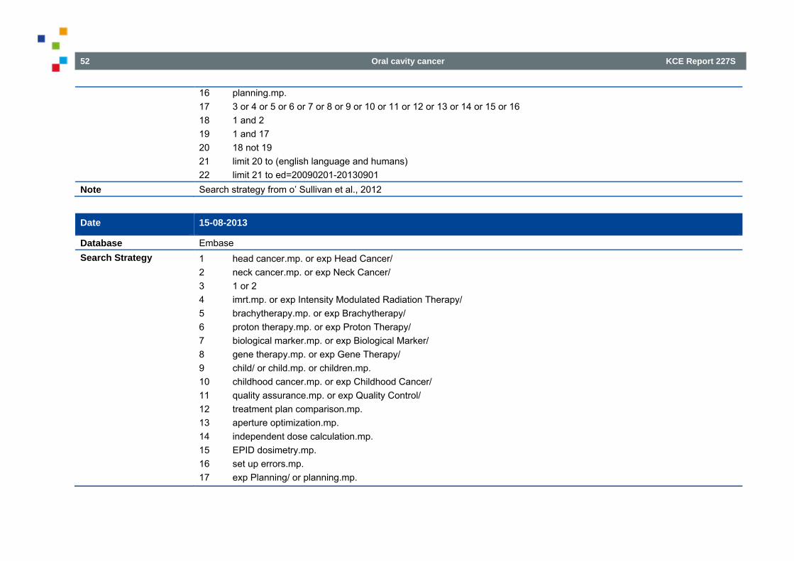
52 Oral cavity cancer KCE Report 227S
16 planning.mp. 17 3 or 4 or 5 or 6 or 7 or 8 or 9 or 10 or 11 or 12 or 13 or 14 or 15 or 16 18 1 and 2 19 1 and 17 20 18 not 19 21 limit 20 to (english language and humans) 22 limit 21 to ed=20090201-20130901
Note Search strategy from o’ Sullivan et al., 2012
Date 15-08-2013
Database Embase Search Strategy 1 head cancer.mp. or exp Head Cancer/
2 neck cancer.mp. or exp Neck Cancer/ 3 1 or 2 4 imrt.mp. or exp Intensity Modulated Radiation Therapy/ 5 brachytherapy.mp. or exp Brachytherapy/ 6 proton therapy.mp. or exp Proton Therapy/ 7 biological marker.mp. or exp Biological Marker/ 8 gene therapy.mp. or exp Gene Therapy/ 9 child/ or child.mp. or children.mp. 10 childhood cancer.mp. or exp Childhood Cancer/ 11 quality assurance.mp. or exp Quality Control/ 12 treatment plan comparison.mp. 13 aperture optimization.mp. 14 independent dose calculation.mp. 15 EPID dosimetry.mp. 16 set up errors.mp. 17 exp Planning/ or planning.mp.
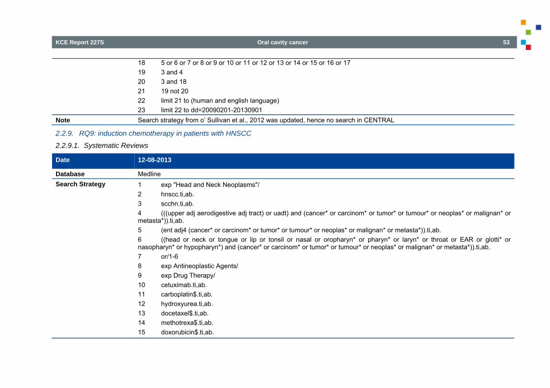
KCE Report 227S Oral cavity cancer 53
18 5 or 6 or 7 or 8 or 9 or 10 or 11 or 12 or 13 or 14 or 15 or 16 or 17 19 3 and 4 20 3 and 18 21 19 not 20 22 limit 21 to (human and english language) 23 limit 22 to dd=20090201-20130901
Note Search strategy from o’ Sullivan et al., 2012 was updated, hence no search in CENTRAL
2.2.9. RQ9: induction chemotherapy in patients with HNSCC
2.2.9.1. Systematic Reviews
Date 12-08-2013
Database Medline Search Strategy 1 exp "Head and Neck Neoplasms"/
2 hnscc.ti,ab. 3 scchn.ti,ab. 4 (((upper adj aerodigestive adj tract) or uadt) and (cancer* or carcinom* or tumor* or tumour* or neoplas* or malignan* or metasta*)).ti,ab. 5 (ent adj4 (cancer* or carcinom* or tumor* or tumour* or neoplas* or malignan* or metasta*)).ti,ab. 6 ((head or neck or tongue or lip or tonsil or nasal or oropharyn* or pharyn* or laryn* or throat or EAR or glotti* or nasopharyn* or hypopharyn*) and (cancer* or carcinom* or tumor* or tumour* or neoplas* or malignan* or metasta*)).ti,ab. 7 or/1-6 8 exp Antineoplastic Agents/ 9 exp Drug Therapy/ 10 cetuximab.ti,ab. 11 carboplatin$.ti,ab. 12 hydroxyurea.ti,ab. 13 docetaxel$.ti,ab. 14 methotrexa$.ti,ab. 15 doxorubicin$.ti,ab.
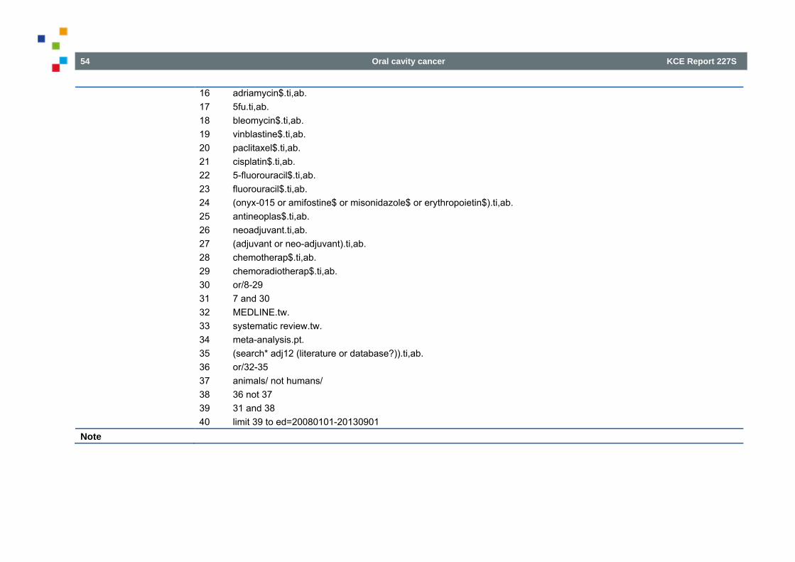
54 Oral cavity cancer KCE Report 227S
16 adriamycin$.ti,ab. 17 5fu.ti,ab. 18 bleomycin$.ti,ab. 19 vinblastine$.ti,ab. 20 paclitaxel$.ti,ab. 21 cisplatin$.ti,ab. 22 5-fluorouracil$.ti,ab. 23 fluorouracil$.ti,ab. 24 (onyx-015 or amifostine$ or misonidazole$ or erythropoietin$).ti,ab. 25 antineoplas$.ti,ab. 26 neoadjuvant.ti,ab. 27 (adjuvant or neo-adjuvant).ti,ab. 28 chemotherap$.ti,ab. 29 chemoradiotherap$.ti,ab. 30 or/8-29 31 7 and 30 32 MEDLINE.tw. 33 systematic review.tw. 34 meta-analysis.pt. 35 (search* adj12 (literature or database?)).ti,ab. 36 or/32-35 37 animals/ not humans/ 38 36 not 37 39 31 and 38 40 limit 39 to ed=20080101-20130901
Note
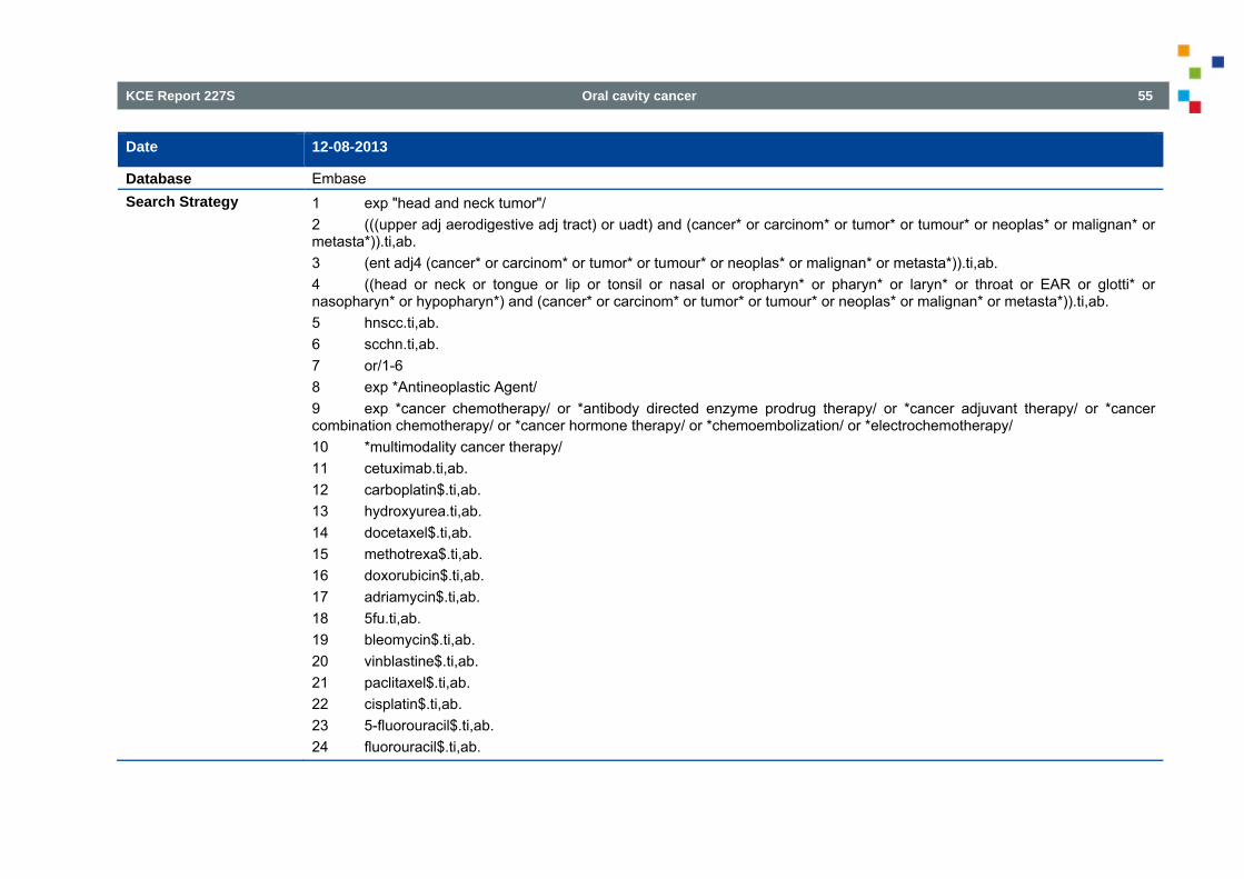
KCE Report 227S Oral cavity cancer 55
Date 12-08-2013
Database Embase Search Strategy 1 exp "head and neck tumor"/
2 (((upper adj aerodigestive adj tract) or uadt) and (cancer* or carcinom* or tumor* or tumour* or neoplas* or malignan* or metasta*)).ti,ab. 3 (ent adj4 (cancer* or carcinom* or tumor* or tumour* or neoplas* or malignan* or metasta*)).ti,ab. 4 ((head or neck or tongue or lip or tonsil or nasal or oropharyn* or pharyn* or laryn* or throat or EAR or glotti* or nasopharyn* or hypopharyn*) and (cancer* or carcinom* or tumor* or tumour* or neoplas* or malignan* or metasta*)).ti,ab. 5 hnscc.ti,ab. 6 scchn.ti,ab. 7 or/1-6 8 exp *Antineoplastic Agent/ 9 exp *cancer chemotherapy/ or *antibody directed enzyme prodrug therapy/ or *cancer adjuvant therapy/ or *cancer combination chemotherapy/ or *cancer hormone therapy/ or *chemoembolization/ or *electrochemotherapy/ 10 *multimodality cancer therapy/ 11 cetuximab.ti,ab. 12 carboplatin$.ti,ab. 13 hydroxyurea.ti,ab. 14 docetaxel$.ti,ab. 15 methotrexa$.ti,ab. 16 doxorubicin$.ti,ab. 17 adriamycin$.ti,ab. 18 5fu.ti,ab. 19 bleomycin$.ti,ab. 20 vinblastine$.ti,ab. 21 paclitaxel$.ti,ab. 22 cisplatin$.ti,ab. 23 5-fluorouracil$.ti,ab. 24 fluorouracil$.ti,ab.
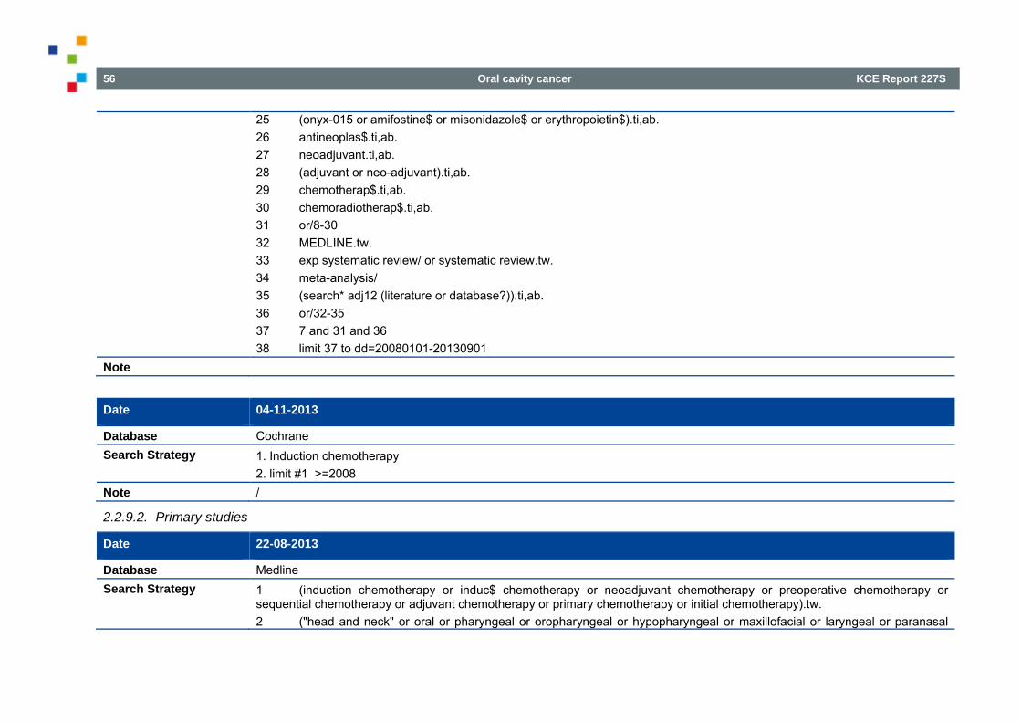
56 Oral cavity cancer KCE Report 227S
25 (onyx-015 or amifostine$ or misonidazole$ or erythropoietin$).ti,ab. 26 antineoplas$.ti,ab. 27 neoadjuvant.ti,ab. 28 (adjuvant or neo-adjuvant).ti,ab. 29 chemotherap$.ti,ab. 30 chemoradiotherap$.ti,ab. 31 or/8-30 32 MEDLINE.tw. 33 exp systematic review/ or systematic review.tw. 34 meta-analysis/ 35 (search* adj12 (literature or database?)).ti,ab. 36 or/32-35 37 7 and 31 and 36 38 limit 37 to dd=20080101-20130901
Note
Date 04-11-2013
Database Cochrane Search Strategy 1. Induction chemotherapy
2. limit #1 >=2008 Note /
2.2.9.2. Primary studies
Date 22-08-2013
Database Medline Search Strategy 1 (induction chemotherapy or induc$ chemotherapy or neoadjuvant chemotherapy or preoperative chemotherapy or
sequential chemotherapy or adjuvant chemotherapy or primary chemotherapy or initial chemotherapy).tw. 2 ("head and neck" or oral or pharyngeal or oropharyngeal or hypopharyngeal or maxillofacial or laryngeal or paranasal
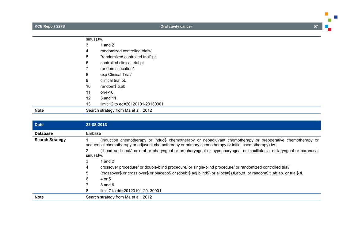
KCE Report 227S Oral cavity cancer 57
sinus).tw. 3 1 and 2 4 randomized controlled trials/ 5 "randomized controlled trial".pt. 6 controlled clinical trial.pt. 7 random allocation/ 8 exp Clinical Trial/ 9 clinical trial.pt. 10 random$.ti,ab. 11 or/4-10 12 3 and 11 13 limit 12 to ed=20120101-20130901
Note Search strategy from Ma et al., 2012
Date 22-08-2013
Database Embase Search Strategy 1 (induction chemotherapy or induc$ chemotherapy or neoadjuvant chemotherapy or preoperative chemotherapy or
sequential chemotherapy or adjuvant chemotherapy or primary chemotherapy or initial chemotherapy).tw. 2 ("head and neck" or oral or pharyngeal or oropharyngeal or hypopharyngeal or maxillofacial or laryngeal or paranasal sinus).tw. 3 1 and 2 4 crossover procedure/ or double-blind procedure/ or single-blind procedure/ or randomized controlled trial/ 5 (crossover$ or cross over$ or placebo$ or (doubl$ adj blind$) or allocat$).ti,ab,ot. or random$.ti,ab,ab. or trial$.ti. 6 4 or 5 7 3 and 6 8 limit 7 to dd=20120101-20130901
Note Search strategy from Ma et al., 2012
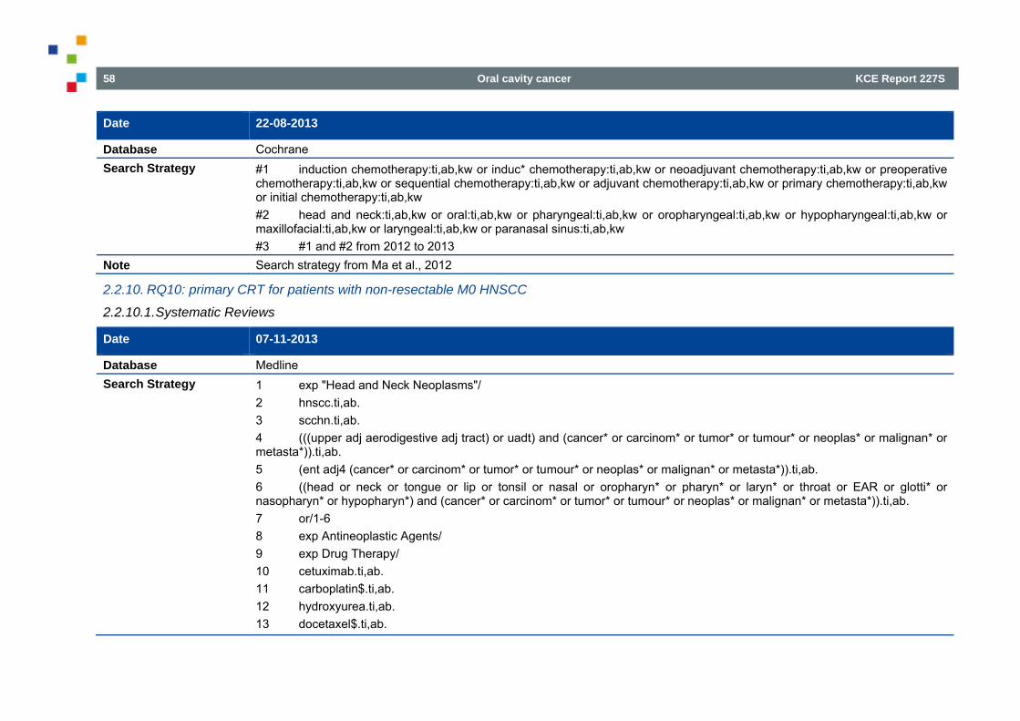
58 Oral cavity cancer KCE Report 227S
Date 22-08-2013
Database Cochrane Search Strategy #1 induction chemotherapy:ti,ab,kw or induc* chemotherapy:ti,ab,kw or neoadjuvant chemotherapy:ti,ab,kw or preoperative
chemotherapy:ti,ab,kw or sequential chemotherapy:ti,ab,kw or adjuvant chemotherapy:ti,ab,kw or primary chemotherapy:ti,ab,kw or initial chemotherapy:ti,ab,kw #2 head and neck:ti,ab,kw or oral:ti,ab,kw or pharyngeal:ti,ab,kw or oropharyngeal:ti,ab,kw or hypopharyngeal:ti,ab,kw or maxillofacial:ti,ab,kw or laryngeal:ti,ab,kw or paranasal sinus:ti,ab,kw #3 #1 and #2 from 2012 to 2013
Note Search strategy from Ma et al., 2012
2.2.10. RQ10: primary CRT for patients with non-resectable M0 HNSCC
2.2.10.1. Systematic Reviews
Date 07-11-2013
Database Medline Search Strategy 1 exp "Head and Neck Neoplasms"/
2 hnscc.ti,ab. 3 scchn.ti,ab. 4 (((upper adj aerodigestive adj tract) or uadt) and (cancer* or carcinom* or tumor* or tumour* or neoplas* or malignan* or metasta*)).ti,ab. 5 (ent adj4 (cancer* or carcinom* or tumor* or tumour* or neoplas* or malignan* or metasta*)).ti,ab. 6 ((head or neck or tongue or lip or tonsil or nasal or oropharyn* or pharyn* or laryn* or throat or EAR or glotti* or nasopharyn* or hypopharyn*) and (cancer* or carcinom* or tumor* or tumour* or neoplas* or malignan* or metasta*)).ti,ab. 7 or/1-6 8 exp Antineoplastic Agents/ 9 exp Drug Therapy/ 10 cetuximab.ti,ab. 11 carboplatin$.ti,ab. 12 hydroxyurea.ti,ab. 13 docetaxel$.ti,ab.
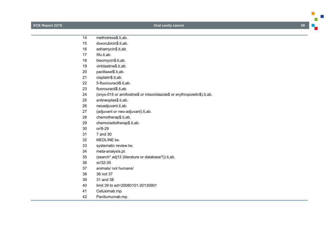
KCE Report 227S Oral cavity cancer 59
14 methotrexa$.ti,ab. 15 doxorubicin$.ti,ab. 16 adriamycin$.ti,ab. 17 5fu.ti,ab. 18 bleomycin$.ti,ab. 19 vinblastine$.ti,ab. 20 paclitaxel$.ti,ab. 21 cisplatin$.ti,ab. 22 5-fluorouracil$.ti,ab. 23 fluorouracil$.ti,ab. 24 (onyx-015 or amifostine$ or misonidazole$ or erythropoietin$).ti,ab. 25 antineoplas$.ti,ab. 26 neoadjuvant.ti,ab. 27 (adjuvant or neo-adjuvant).ti,ab. 28 chemotherap$.ti,ab. 29 chemoradiotherap$.ti,ab. 30 or/8-29 31 7 and 30 32 MEDLINE.tw. 33 systematic review.tw. 34 meta-analysis.pt. 35 (search* adj12 (literature or database?)).ti,ab. 36 or/32-35 37 animals/ not humans/ 38 36 not 37 39 31 and 38 40 limit 39 to ed=20080101-20130901 41 Cetuximab.mp. 42 Panitumumab.mp.
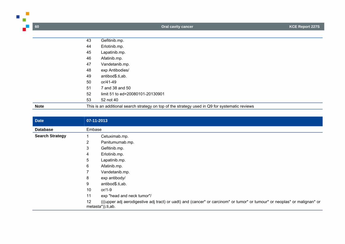
60 Oral cavity cancer KCE Report 227S
43 Gefitinib.mp. 44 Erlotinib.mp. 45 Lapatinib.mp. 46 Afatinib.mp. 47 Vandetanib.mp. 48 exp Antibodies/ 49 antibod$.ti,ab. 50 or/41-49 51 7 and 38 and 50 52 limit 51 to ed=20080101-20130901 53 52 not 40
Note This is an additional search strategy on top of the strategy used in Q9 for systematic reviews
Date 07-11-2013
Database Embase Search Strategy 1 Cetuximab.mp.
2 Panitumumab.mp. 3 Gefitinib.mp. 4 Erlotinib.mp. 5 Lapatinib.mp. 6 Afatinib.mp. 7 Vandetanib.mp. 8 exp antibody/ 9 antibod$.ti,ab. 10 or/1-9 11 exp "head and neck tumor"/ 12 (((upper adj aerodigestive adj tract) or uadt) and (cancer* or carcinom* or tumor* or tumour* or neoplas* or malignan* or metasta*)).ti,ab.
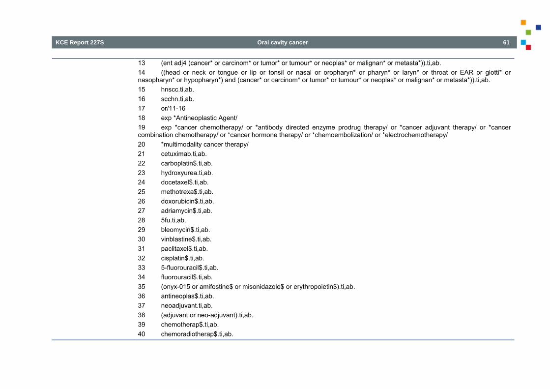
KCE Report 227S Oral cavity cancer 61
13 (ent adj4 (cancer* or carcinom* or tumor* or tumour* or neoplas* or malignan* or metasta*)).ti,ab. 14 ((head or neck or tongue or lip or tonsil or nasal or oropharyn* or pharyn* or laryn* or throat or EAR or glotti* or nasopharyn* or hypopharyn*) and (cancer* or carcinom* or tumor* or tumour* or neoplas* or malignan* or metasta*)).ti,ab. 15 hnscc.ti,ab. 16 scchn.ti,ab. 17 or/11-16 18 exp *Antineoplastic Agent/ 19 exp *cancer chemotherapy/ or *antibody directed enzyme prodrug therapy/ or *cancer adjuvant therapy/ or *cancer combination chemotherapy/ or *cancer hormone therapy/ or *chemoembolization/ or *electrochemotherapy/ 20 *multimodality cancer therapy/ 21 cetuximab.ti,ab. 22 carboplatin$.ti,ab. 23 hydroxyurea.ti,ab. 24 docetaxel$.ti,ab. 25 methotrexa$.ti,ab. 26 doxorubicin$.ti,ab. 27 adriamycin$.ti,ab. 28 5fu.ti,ab. 29 bleomycin$.ti,ab. 30 vinblastine$.ti,ab. 31 paclitaxel$.ti,ab. 32 cisplatin$.ti,ab. 33 5-fluorouracil$.ti,ab. 34 fluorouracil$.ti,ab. 35 (onyx-015 or amifostine$ or misonidazole$ or erythropoietin$).ti,ab. 36 antineoplas$.ti,ab. 37 neoadjuvant.ti,ab. 38 (adjuvant or neo-adjuvant).ti,ab. 39 chemotherap$.ti,ab. 40 chemoradiotherap$.ti,ab.
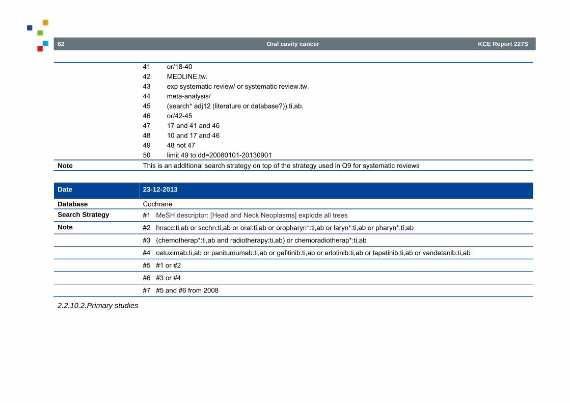
62 Oral cavity cancer KCE Report 227S
41 or/18-40 42 MEDLINE.tw. 43 exp systematic review/ or systematic review.tw. 44 meta-analysis/ 45 (search* adj12 (literature or database?)).ti,ab. 46 or/42-45 47 17 and 41 and 46 48 10 and 17 and 46 49 48 not 47 50 limit 49 to dd=20080101-20130901
Note This is an additional search strategy on top of the strategy used in Q9 for systematic reviews
Date 23-12-2013
Database Cochrane Search Strategy #1 MeSH descriptor: [Head and Neck Neoplasms] explode all trees
Note #2 hnscc:ti,ab or scchn:ti,ab or oral:ti,ab or oropharyn*:ti,ab or laryn*:ti,ab or pharyn*:ti,ab
#3 (chemotherap*:ti,ab and radiotherapy:ti,ab) or chemoradiotherap*:ti,ab
#4 cetuximab:ti,ab or panitumumab:ti,ab or gefitinib:ti,ab or erlotinib:ti,ab or lapatinib:ti,ab or vandetanib:ti,ab
#5 #1 or #2
#6 #3 or #4
#7 #5 and #6 from 2008
2.2.10.2. Primary studies
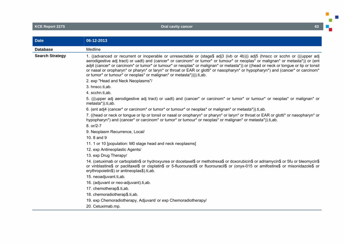
KCE Report 227S Oral cavity cancer 63
Date 06-12-2013
Database Medline Search Strategy 1. ((advanced or recurrent or inoperable or unresectable or (stage$ adj3 (ivb or 4b))) adj5 (hnscc or scchn or (((upper adj
aerodigestive adj tract) or uadt) and (cancer* or carcinom* or tumor* or tumour* or neoplas* or malignan* or metasta*)) or (ent adj4 (cancer* or carcinom* or tumor* or tumour* or neoplas* or malignan* or metasta*)) or ((head or neck or tongue or lip or tonsil or nasal or oropharyn* or pharyn* or laryn* or throat or EAR or glotti* or nasopharyn* or hypopharyn*) and (cancer* or carcinom* or tumor* or tumour* or neoplas* or malignan* or metasta*)))).ti,ab. 2. exp "Head and Neck Neoplasms"/ 3. hnscc.ti,ab. 4. scchn.ti,ab. 5. (((upper adj aerodigestive adj tract) or uadt) and (cancer* or carcinom* or tumor* or tumour* or neoplas* or malignan* or metasta*)).ti,ab. 6. (ent adj4 (cancer* or carcinom* or tumor* or tumour* or neoplas* or malignan* or metasta*)).ti,ab. 7. ((head or neck or tongue or lip or tonsil or nasal or oropharyn* or pharyn* or laryn* or throat or EAR or glotti* or nasopharyn* or hypopharyn*) and (cancer* or carcinom* or tumor* or tumour* or neoplas* or malignan* or metasta*)).ti,ab. 8. or/2-7 9. Neoplasm Recurrence, Local/ 10. 8 and 9 11. 1 or 10 [population: M0 stage head and neck neoplasms] 12. exp Antineoplastic Agents/ 13. exp Drug Therapy/ 14. (cetuximab or carboplatin$ or hydroxyurea or docetaxel$ or methotrexa$ or doxorubicin$ or adriamycin$ or 5fu or bleomycin$ or vinblastine$ or paclitaxel$ or cisplatin$ or 5-fluorouracil$ or fluorouracil$ or (onyx-015 or amifostine$ or misonidazole$ or erythropoietin$) or antineoplas$).ti,ab. 15. neoadjuvant.ti,ab. 16. (adjuvant or neo-adjuvant).ti,ab. 17. chemotherap$.ti,ab. 18. chemoradiotherap$.ti,ab. 19. exp Chemoradiotherapy, Adjuvant/ or exp Chemoradiotherapy/ 20. Cetuximab.mp.
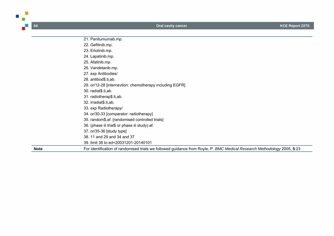
64 Oral cavity cancer KCE Report 227S
21. Panitumumab.mp. 22. Gefitinib.mp. 23. Erlotinib.mp. 24. Lapatinib.mp. 25. Afatinib.mp. 26. Vandetanib.mp. 27. exp Antibodies/ 28. antibod$.ti,ab. 29. or/12-28 [internevtion: chemotherapy including EGFR] 30. radiat$.ti,ab. 31. radiotherap$.ti,ab. 32. irradiat$.ti,ab. 33. exp Radiotherapy/ 34. or/30-33 [comparator: radiotherapy] 35. random$.af. [randomised controlled trials] 36. (phase iii trial$ or phase iii study).af. 37. or/35-36 [study type] 38. 11 and 29 and 34 and 37 39. limit 38 to ed=20031201-20140101
Note For identification of randomised trials we followed guidance from Royle, P. BMC Medical Research Methodology 2005, 5:23
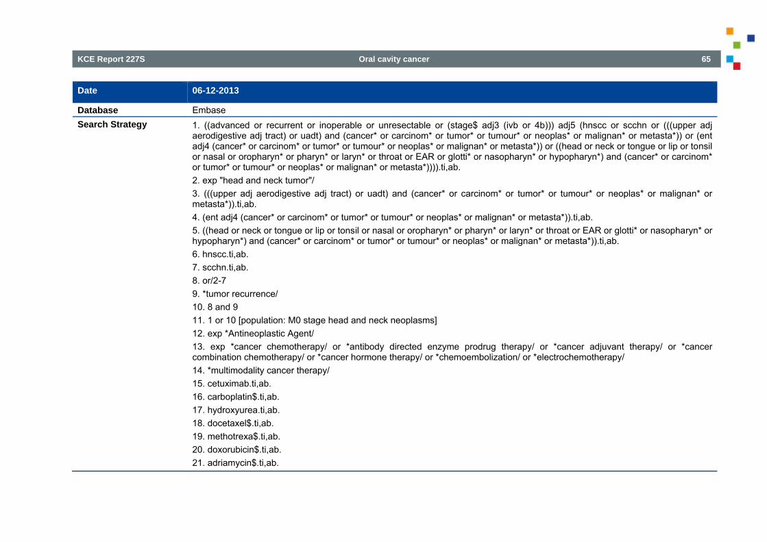
KCE Report 227S Oral cavity cancer 65
Date 06-12-2013
Database Embase Search Strategy 1. ((advanced or recurrent or inoperable or unresectable or (stage$ adj3 (ivb or 4b))) adj5 (hnscc or scchn or (((upper adj
aerodigestive adj tract) or uadt) and (cancer* or carcinom* or tumor* or tumour* or neoplas* or malignan* or metasta*)) or (ent adj4 (cancer* or carcinom* or tumor* or tumour* or neoplas* or malignan* or metasta*)) or ((head or neck or tongue or lip or tonsil or nasal or oropharyn* or pharyn* or laryn* or throat or EAR or glotti* or nasopharyn* or hypopharyn*) and (cancer* or carcinom* or tumor* or tumour* or neoplas* or malignan* or metasta*)))).ti,ab. 2. exp "head and neck tumor"/ 3. (((upper adj aerodigestive adj tract) or uadt) and (cancer* or carcinom* or tumor* or tumour* or neoplas* or malignan* or metasta*)).ti,ab. 4. (ent adj4 (cancer* or carcinom* or tumor* or tumour* or neoplas* or malignan* or metasta*)).ti,ab. 5. ((head or neck or tongue or lip or tonsil or nasal or oropharyn* or pharyn* or laryn* or throat or EAR or glotti* or nasopharyn* or hypopharyn*) and (cancer* or carcinom* or tumor* or tumour* or neoplas* or malignan* or metasta*)).ti,ab. 6. hnscc.ti,ab. 7. scchn.ti,ab. 8. or/2-7 9. *tumor recurrence/ 10. 8 and 9 11. 1 or 10 [population: M0 stage head and neck neoplasms] 12. exp *Antineoplastic Agent/ 13. exp *cancer chemotherapy/ or *antibody directed enzyme prodrug therapy/ or *cancer adjuvant therapy/ or *cancer combination chemotherapy/ or *cancer hormone therapy/ or *chemoembolization/ or *electrochemotherapy/ 14. *multimodality cancer therapy/ 15. cetuximab.ti,ab. 16. carboplatin$.ti,ab. 17. hydroxyurea.ti,ab. 18. docetaxel$.ti,ab. 19. methotrexa$.ti,ab. 20. doxorubicin$.ti,ab. 21. adriamycin$.ti,ab.
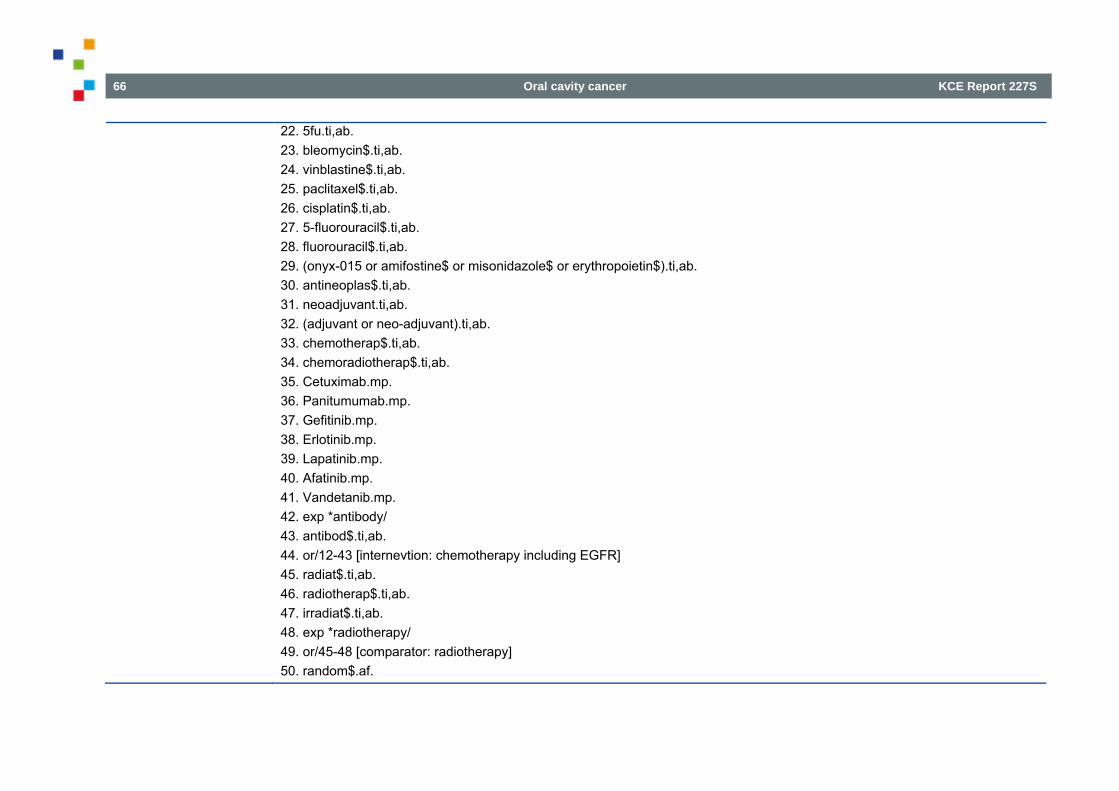
66 Oral cavity cancer KCE Report 227S
22. 5fu.ti,ab. 23. bleomycin$.ti,ab. 24. vinblastine$.ti,ab. 25. paclitaxel$.ti,ab. 26. cisplatin$.ti,ab. 27. 5-fluorouracil$.ti,ab. 28. fluorouracil$.ti,ab. 29. (onyx-015 or amifostine$ or misonidazole$ or erythropoietin$).ti,ab. 30. antineoplas$.ti,ab. 31. neoadjuvant.ti,ab. 32. (adjuvant or neo-adjuvant).ti,ab. 33. chemotherap$.ti,ab. 34. chemoradiotherap$.ti,ab. 35. Cetuximab.mp. 36. Panitumumab.mp. 37. Gefitinib.mp. 38. Erlotinib.mp. 39. Lapatinib.mp. 40. Afatinib.mp. 41. Vandetanib.mp. 42. exp *antibody/ 43. antibod$.ti,ab. 44. or/12-43 [internevtion: chemotherapy including EGFR] 45. radiat$.ti,ab. 46. radiotherap$.ti,ab. 47. irradiat$.ti,ab. 48. exp *radiotherapy/ 49. or/45-48 [comparator: radiotherapy] 50. random$.af.
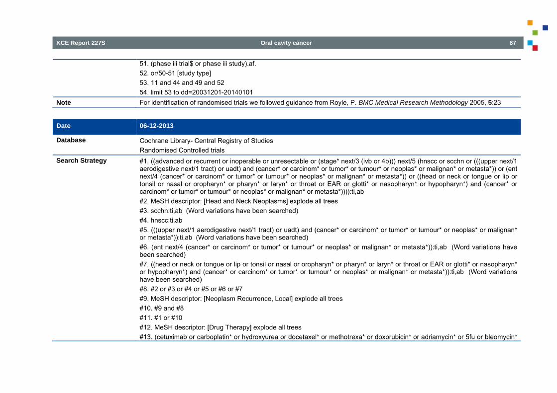
KCE Report 227S Oral cavity cancer 67
51. (phase iii trial$ or phase iii study).af. 52. or/50-51 [study type] 53. 11 and 44 and 49 and 52 54. limit 53 to dd=20031201-20140101
Note For identification of randomised trials we followed guidance from Royle, P. BMC Medical Research Methodology 2005, 5:23
Date 06-12-2013
Database Cochrane Library- Central Registry of Studies Randomised Controlled trials
Search Strategy #1. ((advanced or recurrent or inoperable or unresectable or (stage* next/3 (ivb or 4b))) next/5 (hnscc or scchn or (((upper next/1 aerodigestive next/1 tract) or uadt) and (cancer* or carcinom* or tumor* or tumour* or neoplas* or malignan* or metasta*)) or (ent next/4 (cancer* or carcinom* or tumor* or tumour* or neoplas* or malignan* or metasta*)) or ((head or neck or tongue or lip or tonsil or nasal or oropharyn* or pharyn* or laryn* or throat or EAR or glotti* or nasopharyn* or hypopharyn*) and (cancer* or carcinom* or tumor* or tumour* or neoplas* or malignan* or metasta*)))):ti,ab #2. MeSH descriptor: [Head and Neck Neoplasms] explode all trees #3. scchn:ti,ab (Word variations have been searched) #4. hnscc:ti,ab #5. (((upper next/1 aerodigestive next/1 tract) or uadt) and (cancer* or carcinom* or tumor* or tumour* or neoplas* or malignan* or metasta*)):ti,ab (Word variations have been searched) #6. (ent next/4 (cancer* or carcinom* or tumor* or tumour* or neoplas* or malignan* or metasta*)):ti,ab (Word variations have been searched) #7. ((head or neck or tongue or lip or tonsil or nasal or oropharyn* or pharyn* or laryn* or throat or EAR or glotti* or nasopharyn* or hypopharyn*) and (cancer* or carcinom* or tumor* or tumour* or neoplas* or malignan* or metasta*)):ti,ab (Word variations have been searched) #8. #2 or #3 or #4 or #5 or #6 or #7 #9. MeSH descriptor: [Neoplasm Recurrence, Local] explode all trees #10. #9 and #8 #11. #1 or #10 #12. MeSH descriptor: [Drug Therapy] explode all trees #13. (cetuximab or carboplatin* or hydroxyurea or docetaxel* or methotrexa* or doxorubicin* or adriamycin* or 5fu or bleomycin*
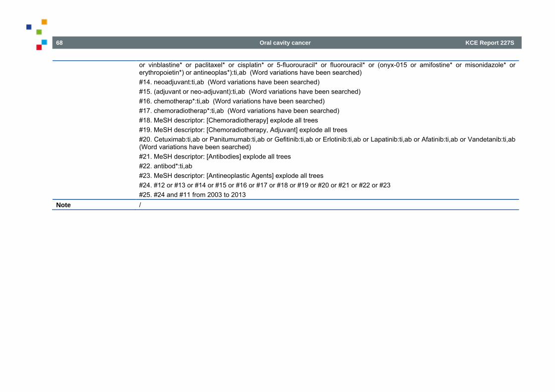
68 Oral cavity cancer KCE Report 227S
or vinblastine* or paclitaxel* or cisplatin* or 5-fluorouracil* or fluorouracil* or (onyx-015 or amifostine* or misonidazole* or erythropoietin*) or antineoplas*):ti,ab (Word variations have been searched) #14. neoadjuvant:ti,ab (Word variations have been searched) #15. (adjuvant or neo-adjuvant):ti,ab (Word variations have been searched) #16. chemotherap*:ti,ab (Word variations have been searched) #17. chemoradiotherap*:ti,ab (Word variations have been searched) #18. MeSH descriptor: [Chemoradiotherapy] explode all trees #19. MeSH descriptor: [Chemoradiotherapy, Adjuvant] explode all trees #20. Cetuximab:ti,ab or Panitumumab:ti,ab or Gefitinib:ti,ab or Erlotinib:ti,ab or Lapatinib:ti,ab or Afatinib:ti,ab or Vandetanib:ti,ab (Word variations have been searched) #21. MeSH descriptor: [Antibodies] explode all trees #22. antibod*:ti,ab #23. MeSH descriptor: [Antineoplastic Agents] explode all trees #24. #12 or #13 or #14 or #15 or #16 or #17 or #18 or #19 or #20 or #21 or #22 or #23 #25. #24 and #11 from 2003 to 2013
Note /
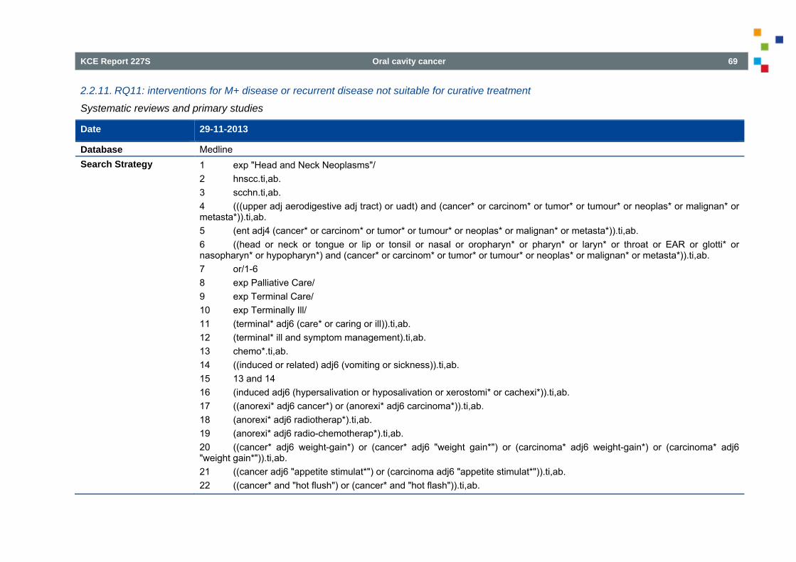
KCE Report 227S Oral cavity cancer 69
2.2.11. RQ11: interventions for M+ disease or recurrent disease not suitable for curative treatment
Systematic reviews and primary studies
Date 29-11-2013
Database Medline Search Strategy 1 exp "Head and Neck Neoplasms"/
2 hnscc.ti,ab. 3 scchn.ti,ab. 4 (((upper adj aerodigestive adj tract) or uadt) and (cancer* or carcinom* or tumor* or tumour* or neoplas* or malignan* or metasta*)).ti,ab. 5 (ent adj4 (cancer* or carcinom* or tumor* or tumour* or neoplas* or malignan* or metasta*)).ti,ab. 6 ((head or neck or tongue or lip or tonsil or nasal or oropharyn* or pharyn* or laryn* or throat or EAR or glotti* or nasopharyn* or hypopharyn*) and (cancer* or carcinom* or tumor* or tumour* or neoplas* or malignan* or metasta*)).ti,ab. 7 or/1-6 8 exp Palliative Care/ 9 exp Terminal Care/ 10 exp Terminally Ill/ 11 (terminal* adj6 (care* or caring or ill)).ti,ab. 12 (terminal* ill and symptom management).ti,ab. 13 chemo*.ti,ab. 14 ((induced or related) adj6 (vomiting or sickness)).ti,ab. 15 13 and 14 16 (induced adj6 (hypersalivation or hyposalivation or xerostomi* or cachexi*)).ti,ab. 17 ((anorexi* adj6 cancer*) or (anorexi* adj6 carcinoma*)).ti,ab. 18 (anorexi* adj6 radiotherap*).ti,ab. 19 (anorexi* adj6 radio-chemotherap*).ti,ab. 20 ((cancer* adj6 weight-gain*) or (cancer* adj6 "weight gain*") or (carcinoma* adj6 weight-gain*) or (carcinoma* adj6 "weight gain*")).ti,ab. 21 ((cancer adj6 "appetite stimulat*") or (carcinoma adj6 "appetite stimulat*")).ti,ab. 22 ((cancer* and "hot flush") or (cancer* and "hot flash")).ti,ab.
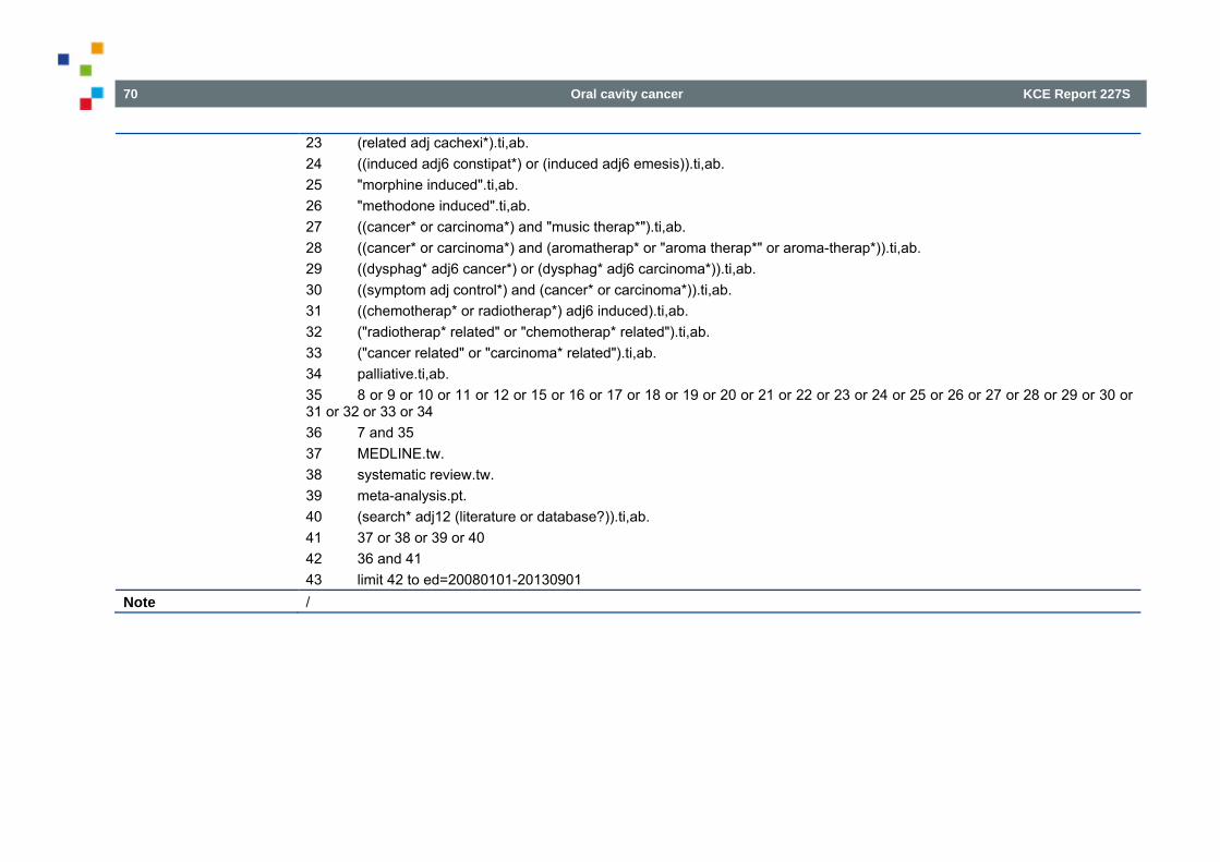
70 Oral cavity cancer KCE Report 227S
23 (related adj cachexi*).ti,ab. 24 ((induced adj6 constipat*) or (induced adj6 emesis)).ti,ab. 25 "morphine induced".ti,ab. 26 "methodone induced".ti,ab. 27 ((cancer* or carcinoma*) and "music therap*").ti,ab. 28 ((cancer* or carcinoma*) and (aromatherap* or "aroma therap*" or aroma-therap*)).ti,ab. 29 ((dysphag* adj6 cancer*) or (dysphag* adj6 carcinoma*)).ti,ab. 30 ((symptom adj control*) and (cancer* or carcinoma*)).ti,ab. 31 ((chemotherap* or radiotherap*) adj6 induced).ti,ab. 32 ("radiotherap* related" or "chemotherap* related").ti,ab. 33 ("cancer related" or "carcinoma* related").ti,ab. 34 palliative.ti,ab. 35 8 or 9 or 10 or 11 or 12 or 15 or 16 or 17 or 18 or 19 or 20 or 21 or 22 or 23 or 24 or 25 or 26 or 27 or 28 or 29 or 30 or 31 or 32 or 33 or 34 36 7 and 35 37 MEDLINE.tw. 38 systematic review.tw. 39 meta-analysis.pt. 40 (search* adj12 (literature or database?)).ti,ab. 41 37 or 38 or 39 or 40 42 36 and 41 43 limit 42 to ed=20080101-20130901
Note /
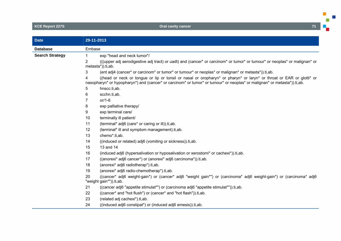
KCE Report 227S Oral cavity cancer 71
Date 29-11-2013
Database Embase Search Strategy 1 exp "head and neck tumor"/
2 (((upper adj aerodigestive adj tract) or uadt) and (cancer* or carcinom* or tumor* or tumour* or neoplas* or malignan* or metasta*)).ti,ab. 3 (ent adj4 (cancer* or carcinom* or tumor* or tumour* or neoplas* or malignan* or metasta*)).ti,ab. 4 ((head or neck or tongue or lip or tonsil or nasal or oropharyn* or pharyn* or laryn* or throat or EAR or glotti* or nasopharyn* or hypopharyn*) and (cancer* or carcinom* or tumor* or tumour* or neoplas* or malignan* or metasta*)).ti,ab. 5 hnscc.ti,ab. 6 scchn.ti,ab. 7 or/1-6 8 exp palliative therapy/ 9 exp terminal care/ 10 terminally ill patient/ 11 (terminal* adj6 (care* or caring or ill)).ti,ab. 12 (terminal* ill and symptom management).ti,ab. 13 chemo*.ti,ab. 14 ((induced or related) adj6 (vomiting or sickness)).ti,ab. 15 13 and 14 16 (induced adj6 (hypersalivation or hyposalivation or xerostomi* or cachexi*)).ti,ab. 17 ((anorexi* adj6 cancer*) or (anorexi* adj6 carcinoma*)).ti,ab. 18 (anorexi* adj6 radiotherap*).ti,ab. 19 (anorexi* adj6 radio-chemotherap*).ti,ab. 20 ((cancer* adj6 weight-gain*) or (cancer* adj6 "weight gain*") or (carcinoma* adj6 weight-gain*) or (carcinoma* adj6 "weight gain*")).ti,ab. 21 ((cancer adj6 "appetite stimulat*") or (carcinoma adj6 "appetite stimulat*")).ti,ab. 22 ((cancer* and "hot flush") or (cancer* and "hot flash")).ti,ab. 23 (related adj cachexi*).ti,ab. 24 ((induced adj6 constipat*) or (induced adj6 emesis)).ti,ab.
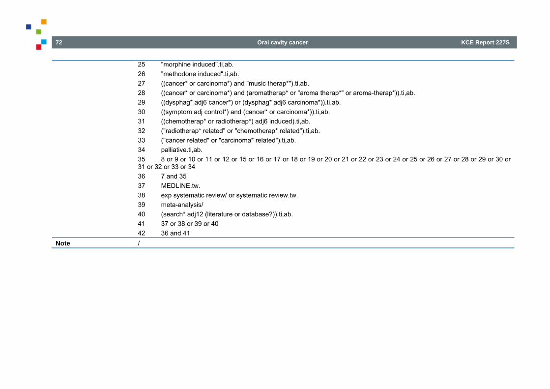
72 Oral cavity cancer KCE Report 227S
25 "morphine induced".ti,ab. 26 "methodone induced".ti,ab. 27 ((cancer* or carcinoma*) and "music therap*").ti,ab. 28 ((cancer* or carcinoma*) and (aromatherap* or "aroma therap*" or aroma-therap*)).ti,ab. 29 ((dysphag* adj6 cancer*) or (dysphag* adj6 carcinoma*)).ti,ab. 30 ((symptom adj control*) and (cancer* or carcinoma*)).ti,ab. 31 ((chemotherap* or radiotherap*) adj6 induced).ti,ab. 32 ("radiotherap* related" or "chemotherap* related").ti,ab. 33 ("cancer related" or "carcinoma* related").ti,ab. 34 palliative.ti,ab. 35 8 or 9 or 10 or 11 or 12 or 15 or 16 or 17 or 18 or 19 or 20 or 21 or 22 or 23 or 24 or 25 or 26 or 27 or 28 or 29 or 30 or 31 or 32 or 33 or 34 36 7 and 35 37 MEDLINE.tw. 38 exp systematic review/ or systematic review.tw. 39 meta-analysis/ 40 (search* adj12 (literature or database?)).ti,ab. 41 37 or 38 or 39 or 40 42 36 and 41
Note /
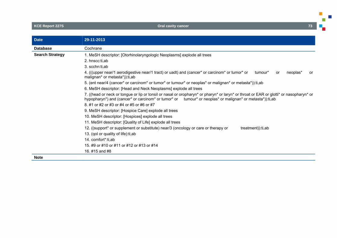
KCE Report 227S Oral cavity cancer 73
Date 29-11-2013
Database Cochrane Search Strategy 1. MeSH descriptor: [Otorhinolaryngologic Neoplasms] explode all trees
2. hnscc:ti,ab 3. scchn:ti,ab 4. (((upper near/1 aerodigestive near/1 tract) or uadt) and (cancer* or carcinom* or tumor* or tumour* or neoplas* or malignan* or metasta*)):ti,ab 5. (ent near/4 (cancer* or carcinom* or tumor* or tumour* or neoplas* or malignan* or metasta*)):ti,ab 6. MeSH descriptor: [Head and Neck Neoplasms] explode all trees 7. ((head or neck or tongue or lip or tonsil or nasal or oropharyn* or pharyn* or laryn* or throat or EAR or glotti* or nasopharyn* or hypopharyn*) and (cancer* or carcinom* or tumor* or tumour* or neoplas* or malignan* or metasta*)):ti,ab 8. #1 or #2 or #3 or #4 or #5 or #6 or #7 9. MeSH descriptor: [Hospice Care] explode all trees 10. MeSH descriptor: [Hospices] explode all trees 11. MeSH descriptor: [Quality of Life] explode all trees 12. ((support* or supplement or substitute) near/3 (oncology or care or therapy or treatment)):ti,ab 13. (qol or quality of life):ti,ab 14. comfort*:ti,ab 15. #9 or #10 or #11 or #12 or #13 or #14 16. #15 and #8
Note
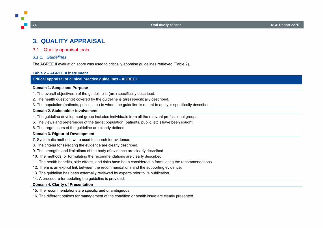
74 Oral cavity cancer KCE Report 227S
3. QUALITY APPRAISAL 3.1. Quality appraisal tools 3.1.1. Guidelines The AGREE II evaluation score was used to critically appraise guidelines retrieved (Table 2).
Table 2 – AGREE II instrument Critical appraisal of clinical practice guidelines - AGREE II
Domain 1. Scope and Purpose 1. The overall objective(s) of the guideline is (are) specifically described. 2. The health question(s) covered by the guideline is (are) specifically described. 3. The population (patients, public, etc.) to whom the guideline is meant to apply is specifically described. Domain 2. Stakeholder Involvement 4. The guideline development group includes individuals from all the relevant professional groups. 5. The views and preferences of the target population (patients, public, etc.) have been sought. 6. The target users of the guideline are clearly defined. Domain 3. Rigour of Development 7. Systematic methods were used to search for evidence. 8. The criteria for selecting the evidence are clearly described. 9. The strengths and limitations of the body of evidence are clearly described. 10. The methods for formulating the recommendations are clearly described. 11. The health benefits, side effects, and risks have been considered in formulating the recommendations. 12. There is an explicit link between the recommendations and the supporting evidence. 13. The guideline has been externally reviewed by experts prior to its publication. 14. A procedure for updating the guideline is provided. Domain 4. Clarity of Presentation 15. The recommendations are specific and unambiguous. 16. The different options for management of the condition or health issue are clearly presented.
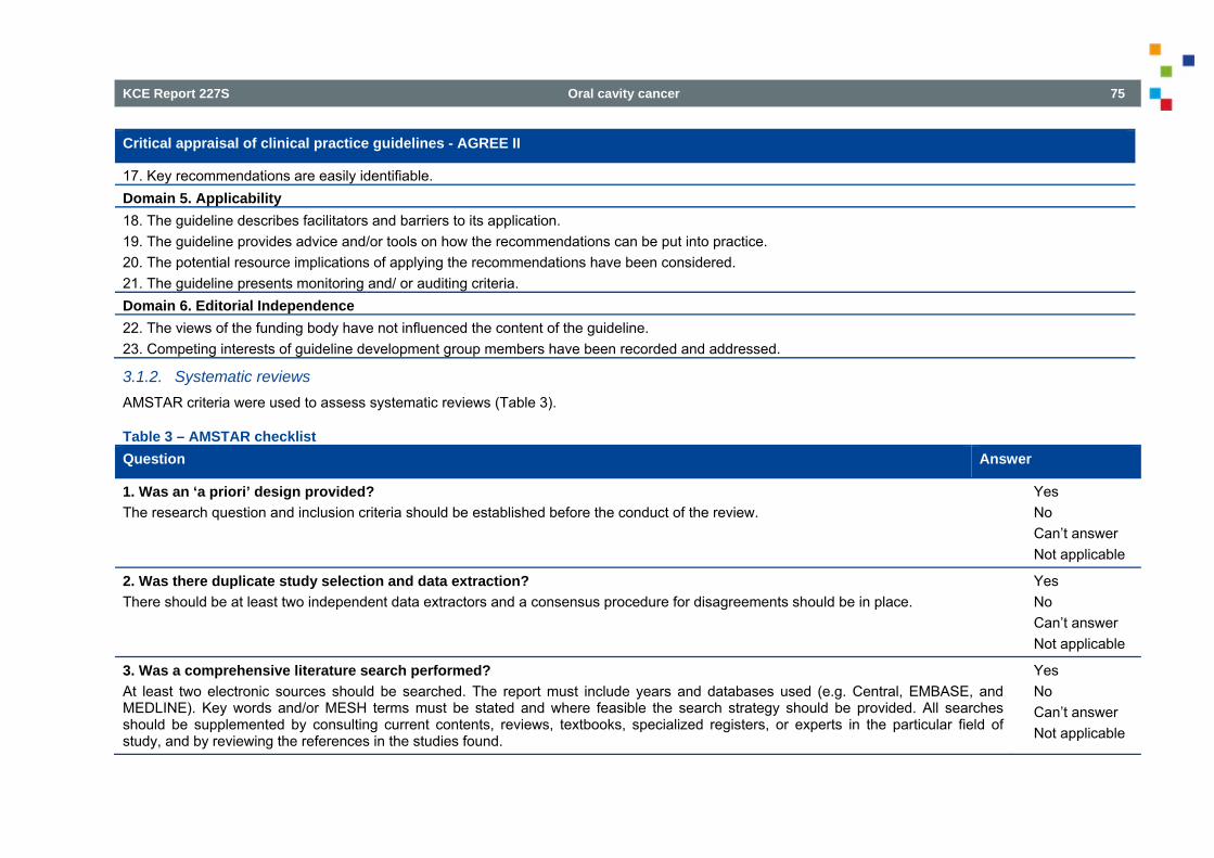
KCE Report 227S Oral cavity cancer 75
Critical appraisal of clinical practice guidelines - AGREE II
17. Key recommendations are easily identifiable. Domain 5. Applicability 18. The guideline describes facilitators and barriers to its application. 19. The guideline provides advice and/or tools on how the recommendations can be put into practice. 20. The potential resource implications of applying the recommendations have been considered. 21. The guideline presents monitoring and/ or auditing criteria. Domain 6. Editorial Independence 22. The views of the funding body have not influenced the content of the guideline. 23. Competing interests of guideline development group members have been recorded and addressed.
3.1.2. Systematic reviews AMSTAR criteria were used to assess systematic reviews (Table 3).
Table 3 – AMSTAR checklist Question Answer
1. Was an ‘a priori’ design provided? The research question and inclusion criteria should be established before the conduct of the review.
� Yes � No � Can’t answer � Not applicable
2. Was there duplicate study selection and data extraction? There should be at least two independent data extractors and a consensus procedure for disagreements should be in place.
� Yes � No � Can’t answer � Not applicable
3. Was a comprehensive literature search performed? At least two electronic sources should be searched. The report must include years and databases used (e.g. Central, EMBASE, and MEDLINE). Key words and/or MESH terms must be stated and where feasible the search strategy should be provided. All searches should be supplemented by consulting current contents, reviews, textbooks, specialized registers, or experts in the particular field of study, and by reviewing the references in the studies found.
� Yes � No � Can’t answer � Not applicable
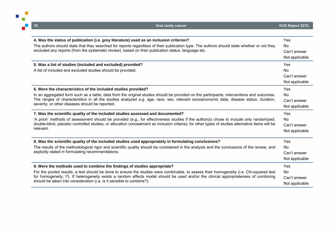
76 Oral cavity cancer KCE Report 227S
4. Was the status of publication (i.e. grey literature) used as an inclusion criterion? The authors should state that they searched for reports regardless of their publication type. The authors should state whether or not they excluded any reports (from the systematic review), based on their publication status, language etc.
� Yes � No � Can’t answer � Not applicable
5. Was a list of studies (included and excluded) provided? A list of included and excluded studies should be provided.
� Yes � No � Can’t answer � Not applicable
6. Were the characteristics of the included studies provided? In an aggregated form such as a table, data from the original studies should be provided on the participants, interventions and outcomes. The ranges of characteristics in all the studies analyzed e.g. age, race, sex, relevant socioeconomic data, disease status, duration, severity, or other diseases should be reported.
� Yes � No � Can’t answer � Not applicable
7. Was the scientific quality of the included studies assessed and documented? ‘A priori’ methods of assessment should be provided (e.g., for effectiveness studies if the author(s) chose to include only randomized, double-blind, placebo controlled studies, or allocation concealment as inclusion criteria); for other types of studies alternative items will be relevant.
� Yes � No � Can’t answer � Not applicable
8. Was the scientific quality of the included studies used appropriately in formulating conclusions? The results of the methodological rigor and scientific quality should be considered in the analysis and the conclusions of the review, and explicitly stated in formulating recommendations.
� Yes � No � Can’t answer � Not applicable
9. Were the methods used to combine the findings of studies appropriate? For the pooled results, a test should be done to ensure the studies were combinable, to assess their homogeneity (i.e. Chi-squared test for homogeneity, I²). If heterogeneity exists a random effects model should be used and/or the clinical appropriateness of combining should be taken into consideration (i.e. is it sensible to combine?).
� Yes � No � Can’t answer � Not applicable
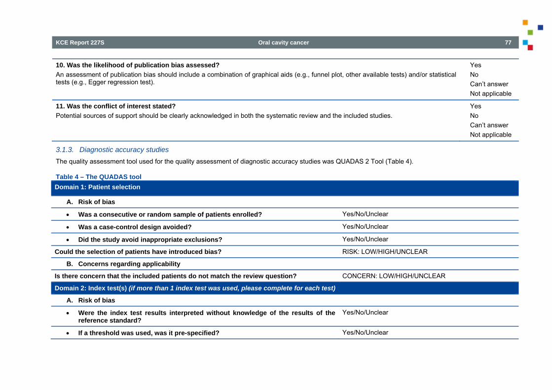
KCE Report 227S Oral cavity cancer 77
10. Was the likelihood of publication bias assessed? An assessment of publication bias should include a combination of graphical aids (e.g., funnel plot, other available tests) and/or statistical tests (e.g., Egger regression test).
� Yes � No � Can’t answer � Not applicable
11. Was the conflict of interest stated? Potential sources of support should be clearly acknowledged in both the systematic review and the included studies.
� Yes � No � Can’t answer � Not applicable
3.1.3. Diagnostic accuracy studies The quality assessment tool used for the quality assessment of diagnostic accuracy studies was QUADAS 2 Tool (Table 4).
Table 4 – The QUADAS tool Domain 1: Patient selection
A. Risk of bias
Was a consecutive or random sample of patients enrolled? Yes/No/Unclear
Was a case-control design avoided? Yes/No/Unclear
Did the study avoid inappropriate exclusions? Yes/No/Unclear
Could the selection of patients have introduced bias? RISK: LOW/HIGH/UNCLEAR
B. Concerns regarding applicability
Is there concern that the included patients do not match the review question? CONCERN: LOW/HIGH/UNCLEAR
Domain 2: Index test(s) (if more than 1 index test was used, please complete for each test)
A. Risk of bias
Were the index test results interpreted without knowledge of the results of the reference standard?
Yes/No/Unclear
If a threshold was used, was it pre-specified? Yes/No/Unclear
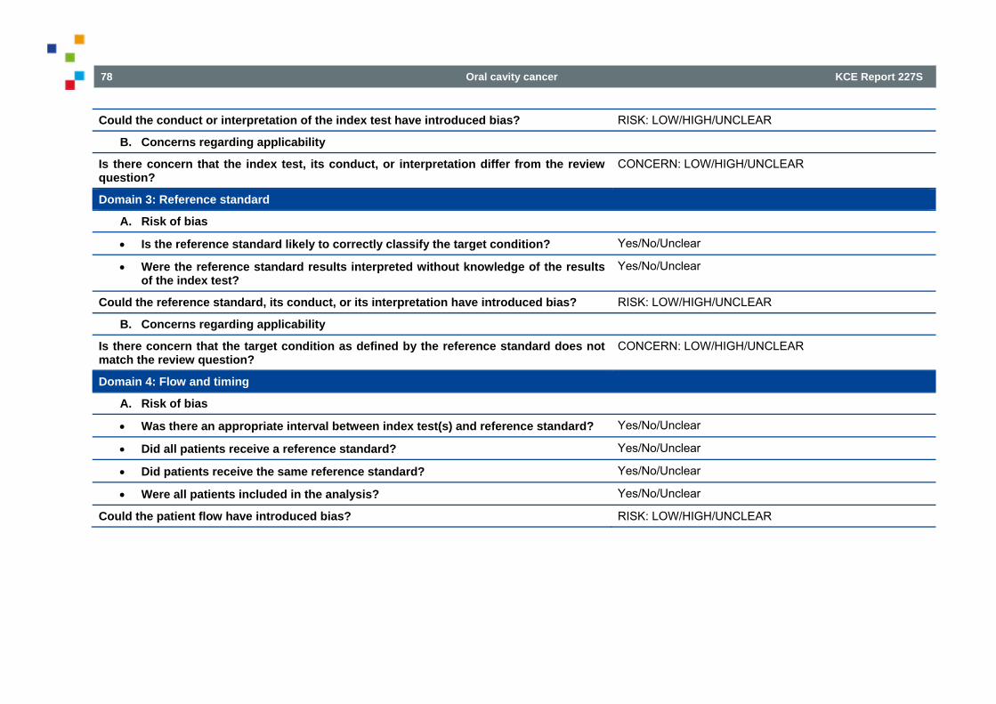
78 Oral cavity cancer KCE Report 227S
Could the conduct or interpretation of the index test have introduced bias? RISK: LOW/HIGH/UNCLEAR
B. Concerns regarding applicability
Is there concern that the index test, its conduct, or interpretation differ from the review question?
CONCERN: LOW/HIGH/UNCLEAR
Domain 3: Reference standard
A. Risk of bias
Is the reference standard likely to correctly classify the target condition? Yes/No/Unclear
Were the reference standard results interpreted without knowledge of the results of the index test?
Yes/No/Unclear
Could the reference standard, its conduct, or its interpretation have introduced bias? RISK: LOW/HIGH/UNCLEAR
B. Concerns regarding applicability
Is there concern that the target condition as defined by the reference standard does not match the review question?
CONCERN: LOW/HIGH/UNCLEAR
Domain 4: Flow and timing
A. Risk of bias
Was there an appropriate interval between index test(s) and reference standard? Yes/No/Unclear
Did all patients receive a reference standard? Yes/No/Unclear
Did patients receive the same reference standard? Yes/No/Unclear
Were all patients included in the analysis? Yes/No/Unclear
Could the patient flow have introduced bias? RISK: LOW/HIGH/UNCLEAR
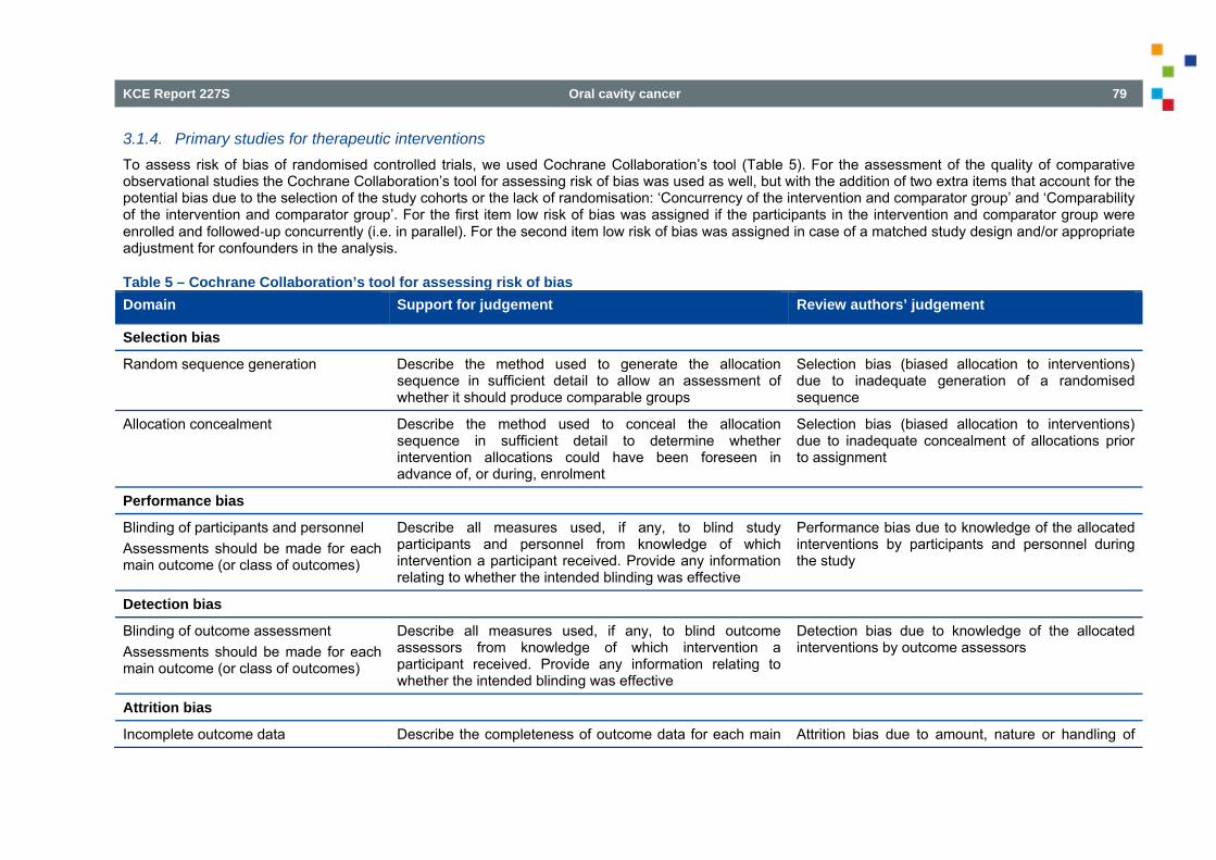
KCE Report 227S Oral cavity cancer 79
3.1.4. Primary studies for therapeutic interventions To assess risk of bias of randomised controlled trials, we used Cochrane Collaboration’s tool (Table 5). For the assessment of the quality of comparative observational studies the Cochrane Collaboration’s tool for assessing risk of bias was used as well, but with the addition of two extra items that account for the potential bias due to the selection of the study cohorts or the lack of randomisation: ‘Concurrency of the intervention and comparator group’ and ‘Comparability of the intervention and comparator group’. For the first item low risk of bias was assigned if the participants in the intervention and comparator group were enrolled and followed‐up concurrently (i.e. in parallel). For the second item low risk of bias was assigned in case of a matched study design and/or appropriate adjustment for confounders in the analysis.
Table 5 – Cochrane Collaboration’s tool for assessing risk of bias Domain Support for judgement Review authors’ judgement
Selection bias
Random sequence generation Describe the method used to generate the allocation sequence in sufficient detail to allow an assessment of whether it should produce comparable groups
Selection bias (biased allocation to interventions) due to inadequate generation of a randomised sequence
Allocation concealment Describe the method used to conceal the allocation sequence in sufficient detail to determine whether intervention allocations could have been foreseen in advance of, or during, enrolment
Selection bias (biased allocation to interventions) due to inadequate concealment of allocations prior to assignment
Performance bias
Blinding of participants and personnel Assessments should be made for each main outcome (or class of outcomes)
Describe all measures used, if any, to blind study participants and personnel from knowledge of which intervention a participant received. Provide any information relating to whether the intended blinding was effective
Performance bias due to knowledge of the allocated interventions by participants and personnel during the study
Detection bias
Blinding of outcome assessment Assessments should be made for each main outcome (or class of outcomes)
Describe all measures used, if any, to blind outcome assessors from knowledge of which intervention a participant received. Provide any information relating to whether the intended blinding was effective
Detection bias due to knowledge of the allocated interventions by outcome assessors
Attrition bias
Incomplete outcome data Describe the completeness of outcome data for each main Attrition bias due to amount, nature or handling of
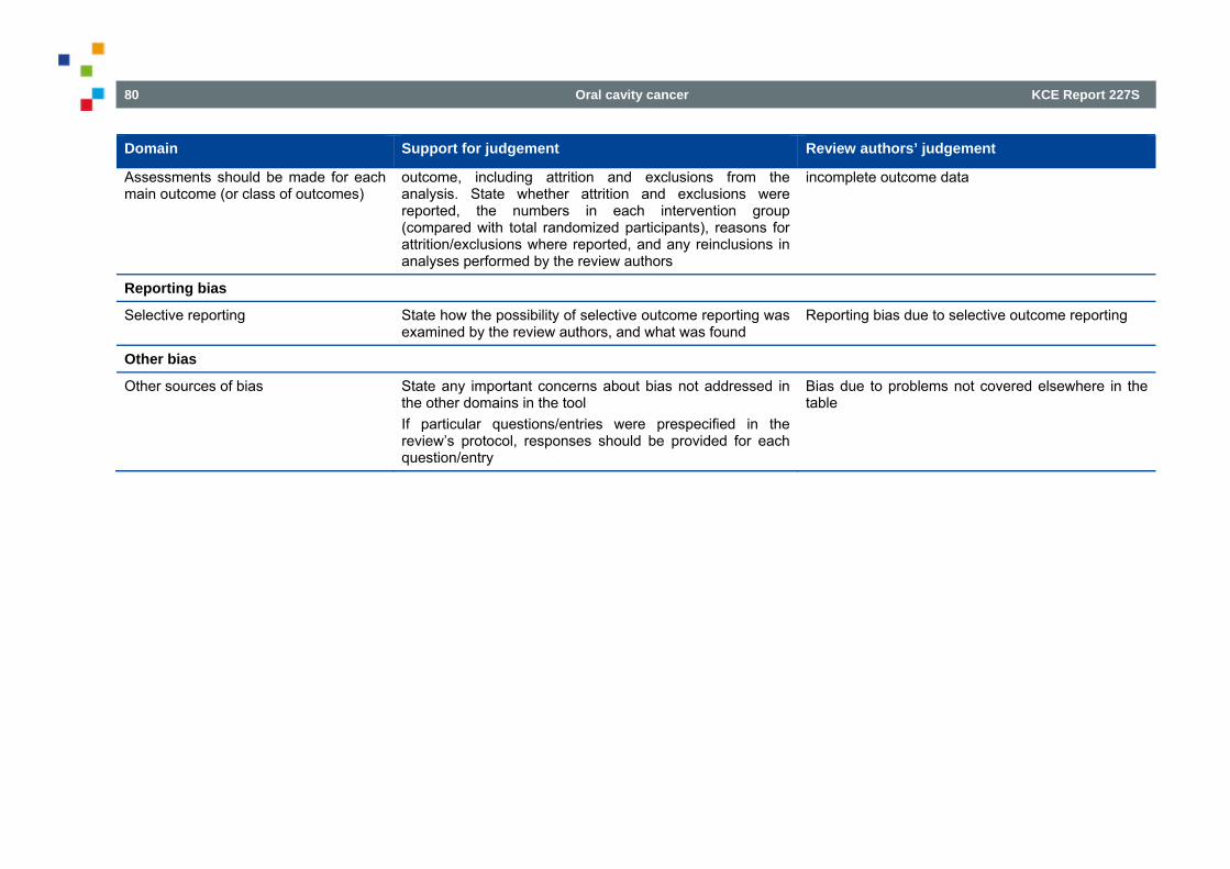
80 Oral cavity cancer KCE Report 227S
Domain Support for judgement Review authors’ judgement
Assessments should be made for each main outcome (or class of outcomes)
outcome, including attrition and exclusions from the analysis. State whether attrition and exclusions were reported, the numbers in each intervention group (compared with total randomized participants), reasons for attrition/exclusions where reported, and any reinclusions in analyses performed by the review authors
incomplete outcome data
Reporting bias
Selective reporting State how the possibility of selective outcome reporting was examined by the review authors, and what was found
Reporting bias due to selective outcome reporting
Other bias
Other sources of bias State any important concerns about bias not addressed in the other domains in the tool If particular questions/entries were prespecified in the review’s protocol, responses should be provided for each question/entry
Bias due to problems not covered elsewhere in the table
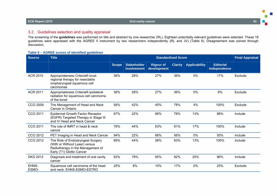
KCE Report 227S Oral cavity cancer 81
3.2. Guidelines selection and quality appraisal The screening of the guidelines was performed on title and abstract by one researcher (RL). Eighteen potentially relevant guidelines were selected. These 18 guidelines were appraised with the AGREE II instrument by two researchers independently (RL and JV) (Table 6). Disagreement was solved through discussion.
Table 6 – AGREE scores of identified guidelines Source Title Standardised Score Final Appraisal
Scope Stakeholder involvement
Rigour of development
Clarity Applicability Editorial Independence
ACR 2010 Appropriateness Criteria® local-regional therapy for resectable oropharyngeal squamous cell carcinomas
36% 28% 27% 36% 0% 17% Exclude
ACR 2011 Appropriateness Criteria® ipsilateral radiation for squamous cell carcinoma of the tonsil
36% 28% 27% 36% 0% 8% Exclude
CCO 2009 The Management of Head and Neck Cancer in Ontario
56% 42% 45% 78% 4% 100% Exclude
CCO 2011 Epidermal Growth Factor Receptor (EGFR) Targeted Therapy in Stage III and IV Head and Neck Cancer
67% 22% 68% 78% 13% 88% Include
CCO 2011 The role of IMRT in head & neck cancer
78% 44% 63% 81% 17% 100% Include
CCO 2012 PET Imaging in Head and Neck Cancer 94% 22% 68% 56% 0% 50% Include CCO 2012 The Role of Endolaryngeal Surgery
(With or Without Laser) versus Radiotherapy in the Management of Early (T1) Glottic Cancer
89% 44% 58% 83% 13% 100% Include
DKG 2012 Diagnosis and treatment of oral cavity cancer
83% 78% 65% 92% 25% 96% Include
EHNS-ESMO-
Squamous cell carcinoma of the head and neck: EHNS-ESMO-ESTRO
25% 8% 10% 17% 0% 25% Exclude
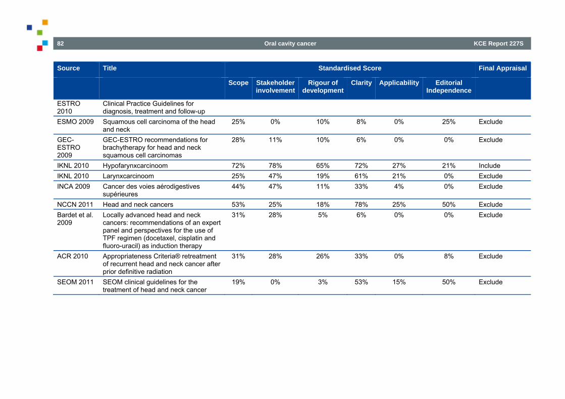
82 Oral cavity cancer KCE Report 227S
Source Title Standardised Score Final Appraisal
Scope Stakeholder involvement
Rigour of development
Clarity Applicability Editorial Independence
ESTRO 2010
Clinical Practice Guidelines for diagnosis, treatment and follow-up
ESMO 2009 Squamous cell carcinoma of the head and neck
25% 0% 10% 8% 0% 25% Exclude
GEC-ESTRO 2009
GEC-ESTRO recommendations for brachytherapy for head and neck squamous cell carcinomas
28% 11% 10% 6% 0% 0% Exclude
IKNL 2010 Hypofarynxcarcinoom 72% 78% 65% 72% 27% 21% Include IKNL 2010 Larynxcarcinoom 25% 47% 19% 61% 21% 0% Exclude INCA 2009 Cancer des voies aérodigestives
supérieures 44% 47% 11% 33% 4% 0% Exclude
NCCN 2011 Head and neck cancers 53% 25% 18% 78% 25% 50% Exclude Bardet et al. 2009
Locally advanced head and neck cancers: recommendations of an expert panel and perspectives for the use of TPF regimen (docetaxel, cisplatin and fluoro-uracil) as induction therapy
31% 28% 5% 6% 0% 0% Exclude
ACR 2010 Appropriateness Criteria® retreatment of recurrent head and neck cancer after prior definitive radiation
31% 28% 26% 33% 0% 8% Exclude
SEOM 2011 SEOM clinical guidelines for the treatment of head and neck cancer
19% 0% 3% 53% 15% 50% Exclude
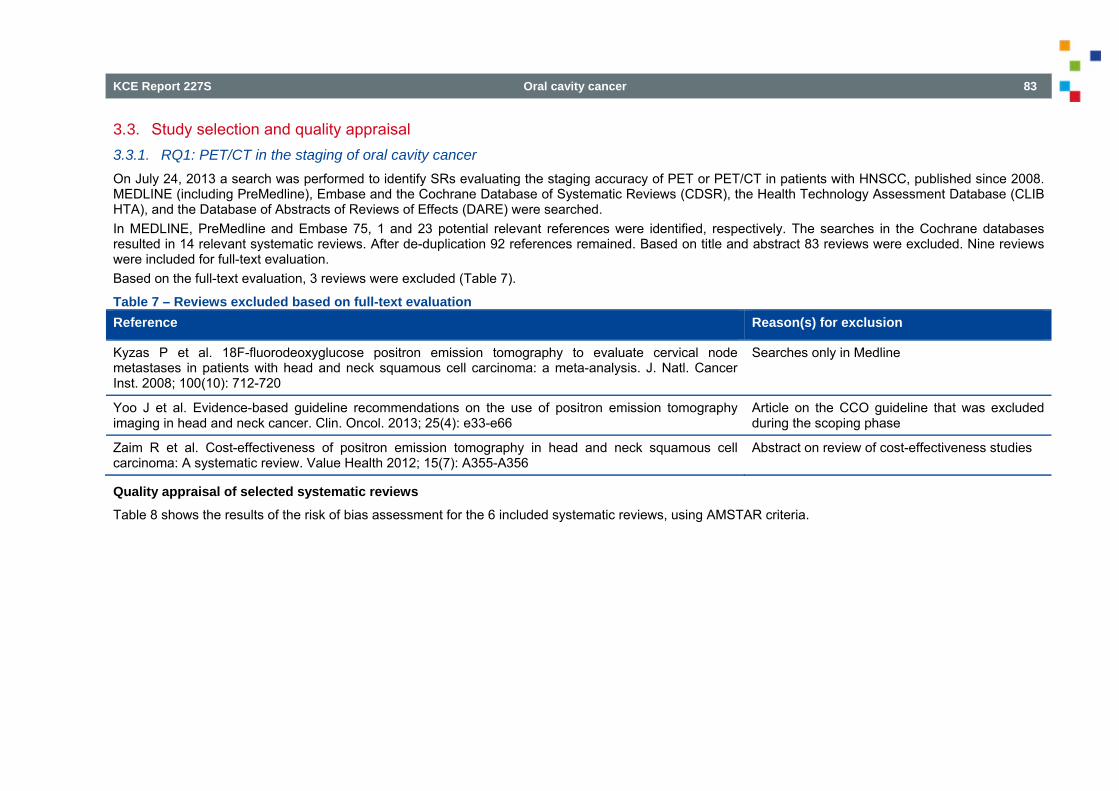
KCE Report 227S Oral cavity cancer 83
3.3. Study selection and quality appraisal 3.3.1. RQ1: PET/CT in the staging of oral cavity cancer On July 24, 2013 a search was performed to identify SRs evaluating the staging accuracy of PET or PET/CT in patients with HNSCC, published since 2008. MEDLINE (including PreMedline), Embase and the Cochrane Database of Systematic Reviews (CDSR), the Health Technology Assessment Database (CLIB HTA), and the Database of Abstracts of Reviews of Effects (DARE) were searched. In MEDLINE, PreMedline and Embase 75, 1 and 23 potential relevant references were identified, respectively. The searches in the Cochrane databases resulted in 14 relevant systematic reviews. After de-duplication 92 references remained. Based on title and abstract 83 reviews were excluded. Nine reviews were included for full-text evaluation. Based on the full-text evaluation, 3 reviews were excluded (Table 7).
Table 7 – Reviews excluded based on full-text evaluation Reference Reason(s) for exclusion
Kyzas P et al. 18F-fluorodeoxyglucose positron emission tomography to evaluate cervical node metastases in patients with head and neck squamous cell carcinoma: a meta-analysis. J. Natl. Cancer Inst. 2008; 100(10): 712-720
Searches only in Medline
Yoo J et al. Evidence-based guideline recommendations on the use of positron emission tomography imaging in head and neck cancer. Clin. Oncol. 2013; 25(4): e33-e66
Article on the CCO guideline that was excluded during the scoping phase
Zaim R et al. Cost-effectiveness of positron emission tomography in head and neck squamous cell carcinoma: A systematic review. Value Health 2012; 15(7): A355-A356
Abstract on review of cost-effectiveness studies
Quality appraisal of selected systematic reviews Table 8 shows the results of the risk of bias assessment for the 6 included systematic reviews, using AMSTAR criteria.
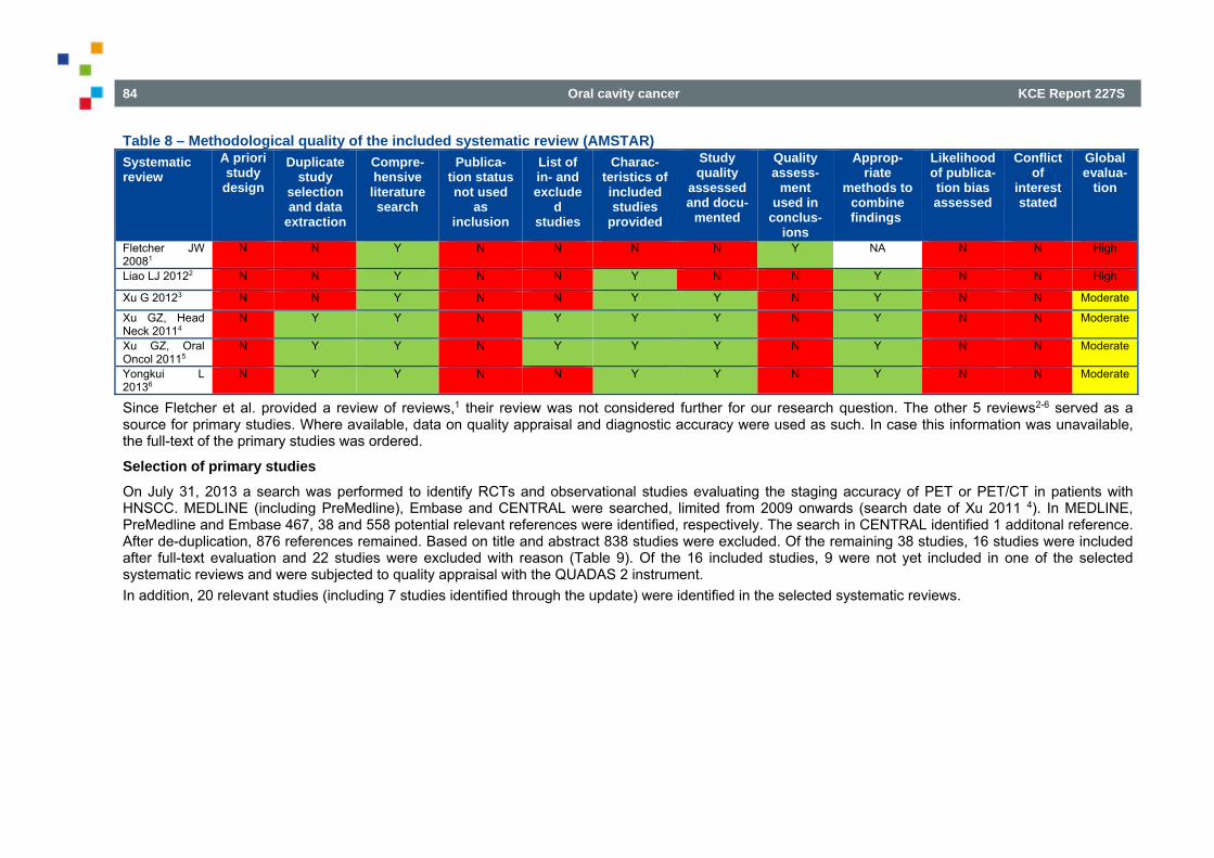
84 Oral cavity cancer KCE Report 227S
Table 8 – Methodological quality of the included systematic review (AMSTAR) Systematic review
A priori study
design
Duplicate study
selection and data
extraction
Compre-hensive
literature search
Publica-tion status not used
as inclusion
List of in- and exclude
d studies
Charac-teristics of included studies
provided
Study quality
assessed and docu-
mented
Quality assess-
ment used in
conclus-ions
Approp-riate
methods to combine findings
Likelihood of publica-tion bias assessed
Conflict of
interest stated
Global evalua-
tion
Fletcher JW 20081
N N Y N N N N Y NA N N High
Liao LJ 20122 N N Y N N Y N N Y N N High
Xu G 20123 N N Y N N Y Y N Y N N Moderate
Xu GZ, Head Neck 20114
N Y Y N Y Y Y N Y N N Moderate
Xu GZ, Oral Oncol 20115
N Y Y N Y Y Y N Y N N Moderate
Yongkui L 20136
N Y Y N N Y Y N Y N N Moderate
Since Fletcher et al. provided a review of reviews,1 their review was not considered further for our research question. The other 5 reviews2-6 served as a source for primary studies. Where available, data on quality appraisal and diagnostic accuracy were used as such. In case this information was unavailable, the full-text of the primary studies was ordered.
Selection of primary studies On July 31, 2013 a search was performed to identify RCTs and observational studies evaluating the staging accuracy of PET or PET/CT in patients with HNSCC. MEDLINE (including PreMedline), Embase and CENTRAL were searched, limited from 2009 onwards (search date of Xu 2011 4). In MEDLINE, PreMedline and Embase 467, 38 and 558 potential relevant references were identified, respectively. The search in CENTRAL identified 1 additonal reference. After de-duplication, 876 references remained. Based on title and abstract 838 studies were excluded. Of the remaining 38 studies, 16 studies were included after full-text evaluation and 22 studies were excluded with reason (Table 9). Of the 16 included studies, 9 were not yet included in one of the selected systematic reviews and were subjected to quality appraisal with the QUADAS 2 instrument. In addition, 20 relevant studies (including 7 studies identified through the update) were identified in the selected systematic reviews.
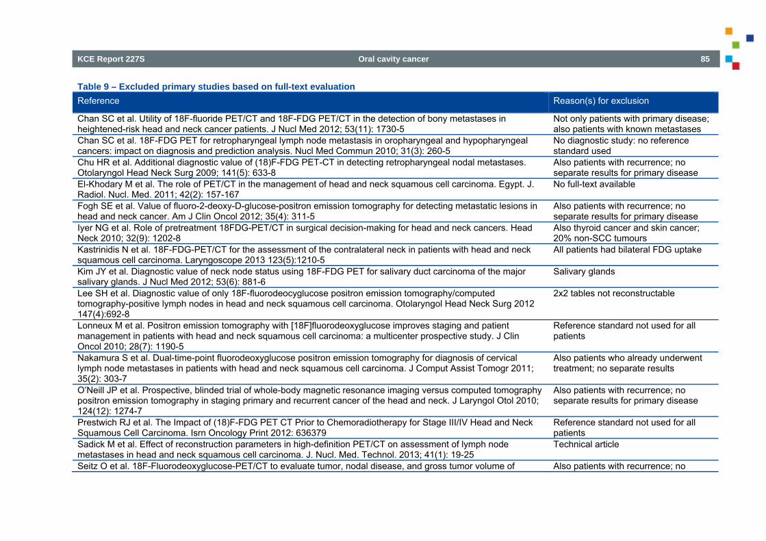
KCE Report 227S Oral cavity cancer 85
Table 9 – Excluded primary studies based on full-text evaluation Reference Reason(s) for exclusion
Chan SC et al. Utility of 18F-fluoride PET/CT and 18F-FDG PET/CT in the detection of bony metastases in heightened-risk head and neck cancer patients. J Nucl Med 2012; 53(11): 1730-5
Not only patients with primary disease; also patients with known metastases
Chan SC et al. 18F-FDG PET for retropharyngeal lymph node metastasis in oropharyngeal and hypopharyngeal cancers: impact on diagnosis and prediction analysis. Nucl Med Commun 2010; 31(3): 260-5
No diagnostic study: no reference standard used
Chu HR et al. Additional diagnostic value of (18)F-FDG PET-CT in detecting retropharyngeal nodal metastases. Otolaryngol Head Neck Surg 2009; 141(5): 633-8
Also patients with recurrence; no separate results for primary disease
El-Khodary M et al. The role of PET/CT in the management of head and neck squamous cell carcinoma. Egypt. J. Radiol. Nucl. Med. 2011; 42(2): 157-167
No full-text available
Fogh SE et al. Value of fluoro-2-deoxy-D-glucose-positron emission tomography for detecting metastatic lesions in head and neck cancer. Am J Clin Oncol 2012; 35(4): 311-5
Also patients with recurrence; no separate results for primary disease
Iyer NG et al. Role of pretreatment 18FDG-PET/CT in surgical decision-making for head and neck cancers. Head Neck 2010; 32(9): 1202-8
Also thyroid cancer and skin cancer; 20% non-SCC tumours
Kastrinidis N et al. 18F-FDG-PET/CT for the assessment of the contralateral neck in patients with head and neck squamous cell carcinoma. Laryngoscope 2013 123(5):1210-5
All patients had bilateral FDG uptake
Kim JY et al. Diagnostic value of neck node status using 18F-FDG PET for salivary duct carcinoma of the major salivary glands. J Nucl Med 2012; 53(6): 881-6
Salivary glands
Lee SH et al. Diagnostic value of only 18F-fluorodeocyglucose positron emission tomography/computed tomography-positive lymph nodes in head and neck squamous cell carcinoma. Otolaryngol Head Neck Surg 2012 147(4):692-8
2x2 tables not reconstructable
Lonneux M et al. Positron emission tomography with [18F]fluorodeoxyglucose improves staging and patient management in patients with head and neck squamous cell carcinoma: a multicenter prospective study. J Clin Oncol 2010; 28(7): 1190-5
Reference standard not used for all patients
Nakamura S et al. Dual-time-point fluorodeoxyglucose positron emission tomography for diagnosis of cervical lymph node metastases in patients with head and neck squamous cell carcinoma. J Comput Assist Tomogr 2011; 35(2): 303-7
Also patients who already underwent treatment; no separate results
O’Neill JP et al. Prospective, blinded trial of whole-body magnetic resonance imaging versus computed tomography positron emission tomography in staging primary and recurrent cancer of the head and neck. J Laryngol Otol 2010; 124(12): 1274-7
Also patients with recurrence; no separate results for primary disease
Prestwich RJ et al. The Impact of (18)F-FDG PET CT Prior to Chemoradiotherapy for Stage III/IV Head and Neck Squamous Cell Carcinoma. Isrn Oncology Print 2012: 636379
Reference standard not used for all patients
Sadick M et al. Effect of reconstruction parameters in high-definition PET/CT on assessment of lymph node metastases in head and neck squamous cell carcinoma. J. Nucl. Med. Technol. 2013; 41(1): 19-25
Technical article
Seitz O et al. 18F-Fluorodeoxyglucose-PET/CT to evaluate tumor, nodal disease, and gross tumor volume of Also patients with recurrence; no
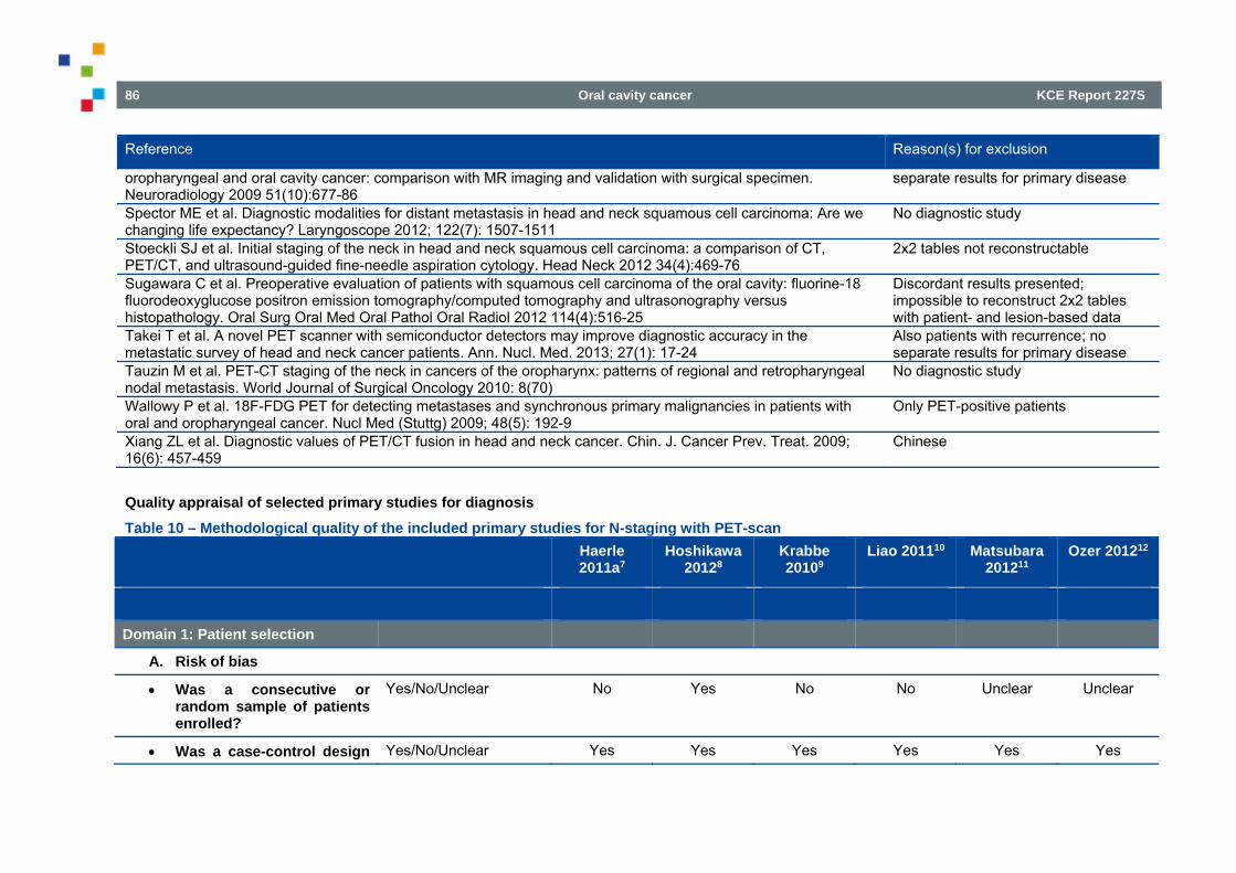
86 Oral cavity cancer KCE Report 227S
Reference Reason(s) for exclusion
oropharyngeal and oral cavity cancer: comparison with MR imaging and validation with surgical specimen. Neuroradiology 2009 51(10):677-86
separate results for primary disease
Spector ME et al. Diagnostic modalities for distant metastasis in head and neck squamous cell carcinoma: Are we changing life expectancy? Laryngoscope 2012; 122(7): 1507-1511
No diagnostic study
Stoeckli SJ et al. Initial staging of the neck in head and neck squamous cell carcinoma: a comparison of CT, PET/CT, and ultrasound-guided fine-needle aspiration cytology. Head Neck 2012 34(4):469-76
2x2 tables not reconstructable
Sugawara C et al. Preoperative evaluation of patients with squamous cell carcinoma of the oral cavity: fluorine-18 fluorodeoxyglucose positron emission tomography/computed tomography and ultrasonography versus histopathology. Oral Surg Oral Med Oral Pathol Oral Radiol 2012 114(4):516-25
Discordant results presented; impossible to reconstruct 2x2 tables with patient- and lesion-based data
Takei T et al. A novel PET scanner with semiconductor detectors may improve diagnostic accuracy in the metastatic survey of head and neck cancer patients. Ann. Nucl. Med. 2013; 27(1): 17-24
Also patients with recurrence; no separate results for primary disease
Tauzin M et al. PET-CT staging of the neck in cancers of the oropharynx: patterns of regional and retropharyngeal nodal metastasis. World Journal of Surgical Oncology 2010: 8(70)
No diagnostic study
Wallowy P et al. 18F-FDG PET for detecting metastases and synchronous primary malignancies in patients with oral and oropharyngeal cancer. Nucl Med (Stuttg) 2009; 48(5): 192-9
Only PET-positive patients
Xiang ZL et al. Diagnostic values of PET/CT fusion in head and neck cancer. Chin. J. Cancer Prev. Treat. 2009; 16(6): 457-459
Chinese
Quality appraisal of selected primary studies for diagnosis Table 10 – Methodological quality of the included primary studies for N-staging with PET-scan Haerle
2011a7 Hoshikawa
20128 Krabbe 20109
Liao 201110 Matsubara 201211
Ozer 201212
Domain 1: Patient selection
A. Risk of bias
Was a consecutive or random sample of patients enrolled?
Yes/No/Unclear No Yes No No Unclear Unclear
Was a case-control design Yes/No/Unclear Yes Yes Yes Yes Yes Yes
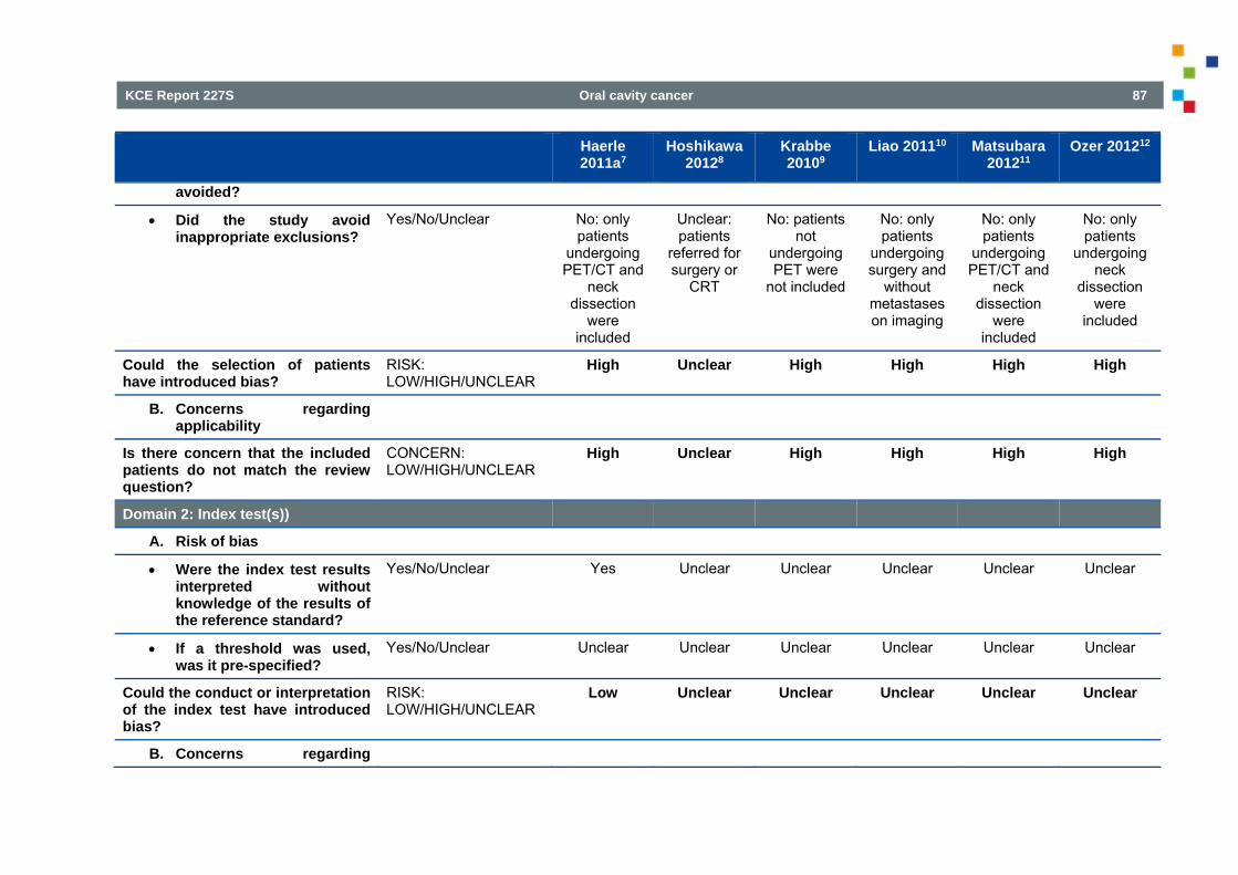
KCE Report 227S Oral cavity cancer 87
Haerle 2011a7
Hoshikawa 20128
Krabbe 20109
Liao 201110 Matsubara 201211
Ozer 201212
avoided?
Did the study avoid inappropriate exclusions?
Yes/No/Unclear No: only patients
undergoing PET/CT and
neck dissection
were included
Unclear: patients
referred for surgery or
CRT
No: patients not
undergoing PET were
not included
No: only patients
undergoing surgery and
without metastases on imaging
No: only patients
undergoing PET/CT and
neck dissection
were included
No: only patients
undergoing neck
dissection were
included
Could the selection of patients have introduced bias?
RISK: LOW/HIGH/UNCLEAR
High Unclear High High High High
B. Concerns regarding applicability
Is there concern that the included patients do not match the review question?
CONCERN: LOW/HIGH/UNCLEAR
High Unclear High High High High
Domain 2: Index test(s))
A. Risk of bias
Were the index test results interpreted without knowledge of the results of the reference standard?
Yes/No/Unclear Yes Unclear Unclear Unclear Unclear Unclear
If a threshold was used, was it pre-specified?
Yes/No/Unclear Unclear Unclear Unclear Unclear Unclear Unclear
Could the conduct or interpretation of the index test have introduced bias?
RISK: LOW/HIGH/UNCLEAR
Low Unclear Unclear Unclear Unclear Unclear
B. Concerns regarding
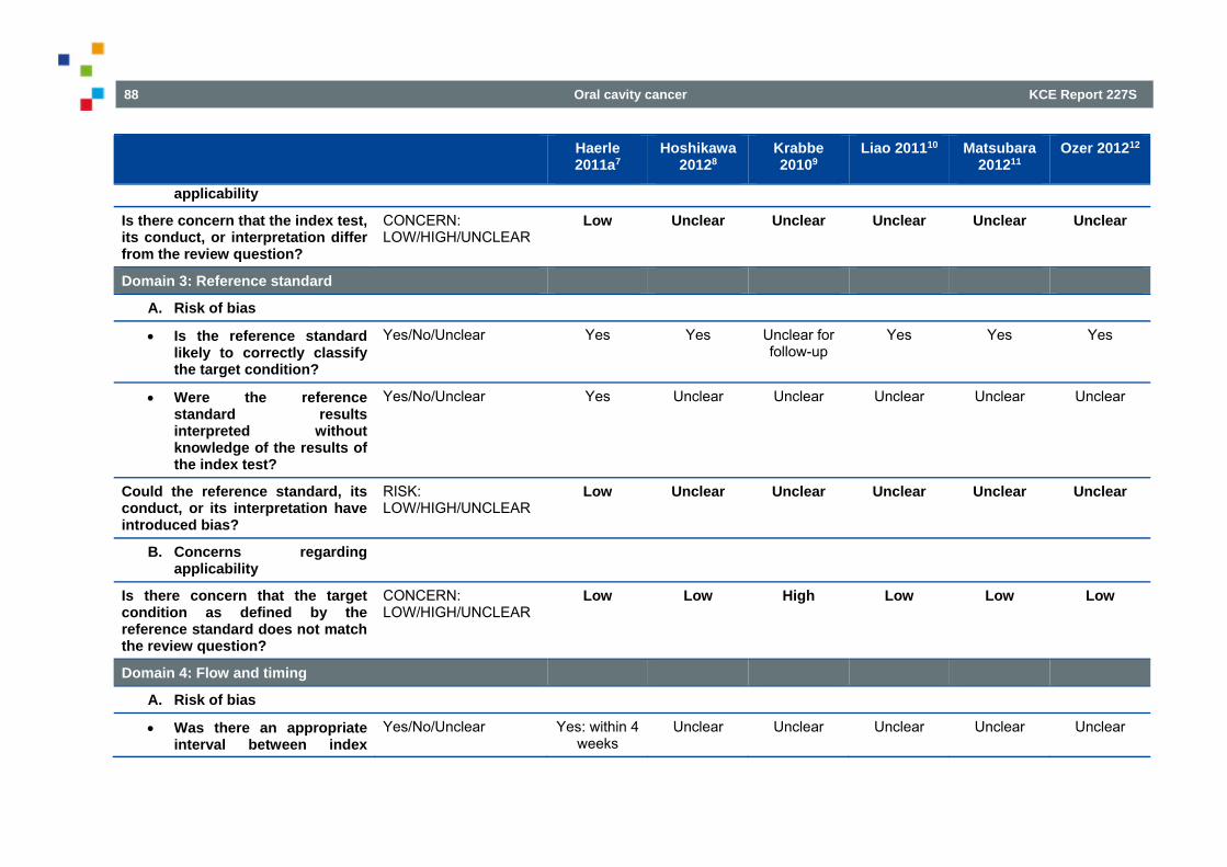
88 Oral cavity cancer KCE Report 227S
Haerle 2011a7
Hoshikawa 20128
Krabbe 20109
Liao 201110 Matsubara 201211
Ozer 201212
applicability
Is there concern that the index test, its conduct, or interpretation differ from the review question?
CONCERN: LOW/HIGH/UNCLEAR
Low Unclear Unclear Unclear Unclear Unclear
Domain 3: Reference standard
A. Risk of bias
Is the reference standard likely to correctly classify the target condition?
Yes/No/Unclear Yes Yes Unclear for follow-up
Yes Yes Yes
Were the reference standard results interpreted without knowledge of the results of the index test?
Yes/No/Unclear Yes Unclear Unclear Unclear Unclear Unclear
Could the reference standard, its conduct, or its interpretation have introduced bias?
RISK: LOW/HIGH/UNCLEAR
Low Unclear Unclear Unclear Unclear Unclear
B. Concerns regarding applicability
Is there concern that the target condition as defined by the reference standard does not match the review question?
CONCERN: LOW/HIGH/UNCLEAR
Low Low High Low Low Low
Domain 4: Flow and timing
A. Risk of bias
Was there an appropriate interval between index
Yes/No/Unclear Yes: within 4 weeks
Unclear Unclear Unclear Unclear Unclear
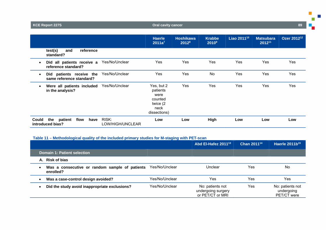
KCE Report 227S Oral cavity cancer 89
Haerle 2011a7
Hoshikawa 20128
Krabbe 20109
Liao 201110 Matsubara 201211
Ozer 201212
test(s) and reference standard?
Did all patients receive a reference standard?
Yes/No/Unclear Yes Yes Yes Yes Yes Yes
Did patients receive the same reference standard?
Yes/No/Unclear Yes Yes No Yes Yes Yes
Were all patients included in the analysis?
Yes/No/Unclear Yes, but 2 patients
were counted twice (2
neck dissections)
Yes Yes Yes Yes Yes
Could the patient flow have introduced bias?
RISK: LOW/HIGH/UNCLEAR
Low Low High Low Low Low
Table 11 – Methodological quality of the included primary studies for M-staging with PET-scan Abd El-Hafez 201113 Chan 201114 Haerle 2011b15
Domain 1: Patient selection
A. Risk of bias
Was a consecutive or random sample of patients enrolled?
Yes/No/Unclear Unclear Yes No
Was a case-control design avoided? Yes/No/Unclear Yes Yes Yes
Did the study avoid inappropriate exclusions? Yes/No/Unclear No: patients not undergoing surgery or PET/CT or MRI
Yes No: patients not undergoing
PET/CT were
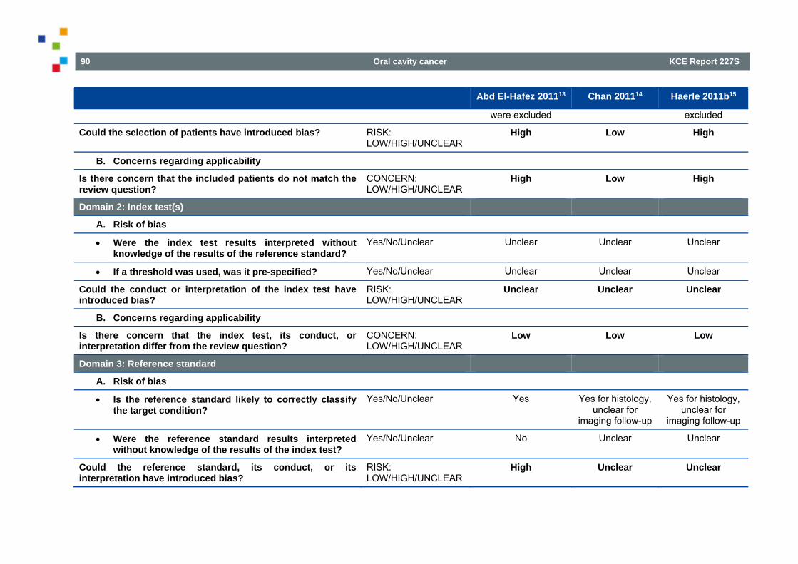
90 Oral cavity cancer KCE Report 227S
Abd El-Hafez 201113 Chan 201114 Haerle 2011b15
were excluded excluded
Could the selection of patients have introduced bias? RISK: LOW/HIGH/UNCLEAR
High Low High
B. Concerns regarding applicability
Is there concern that the included patients do not match the review question?
CONCERN: LOW/HIGH/UNCLEAR
High Low High
Domain 2: Index test(s)
A. Risk of bias
Were the index test results interpreted without knowledge of the results of the reference standard?
Yes/No/Unclear Unclear Unclear Unclear
If a threshold was used, was it pre-specified? Yes/No/Unclear Unclear Unclear Unclear
Could the conduct or interpretation of the index test have introduced bias?
RISK: LOW/HIGH/UNCLEAR
Unclear Unclear Unclear
B. Concerns regarding applicability
Is there concern that the index test, its conduct, or interpretation differ from the review question?
CONCERN: LOW/HIGH/UNCLEAR
Low Low Low
Domain 3: Reference standard
A. Risk of bias
Is the reference standard likely to correctly classify the target condition?
Yes/No/Unclear Yes Yes for histology, unclear for
imaging follow-up
Yes for histology, unclear for
imaging follow-up
Were the reference standard results interpreted without knowledge of the results of the index test?
Yes/No/Unclear No Unclear Unclear
Could the reference standard, its conduct, or its interpretation have introduced bias?
RISK: LOW/HIGH/UNCLEAR
High Unclear Unclear
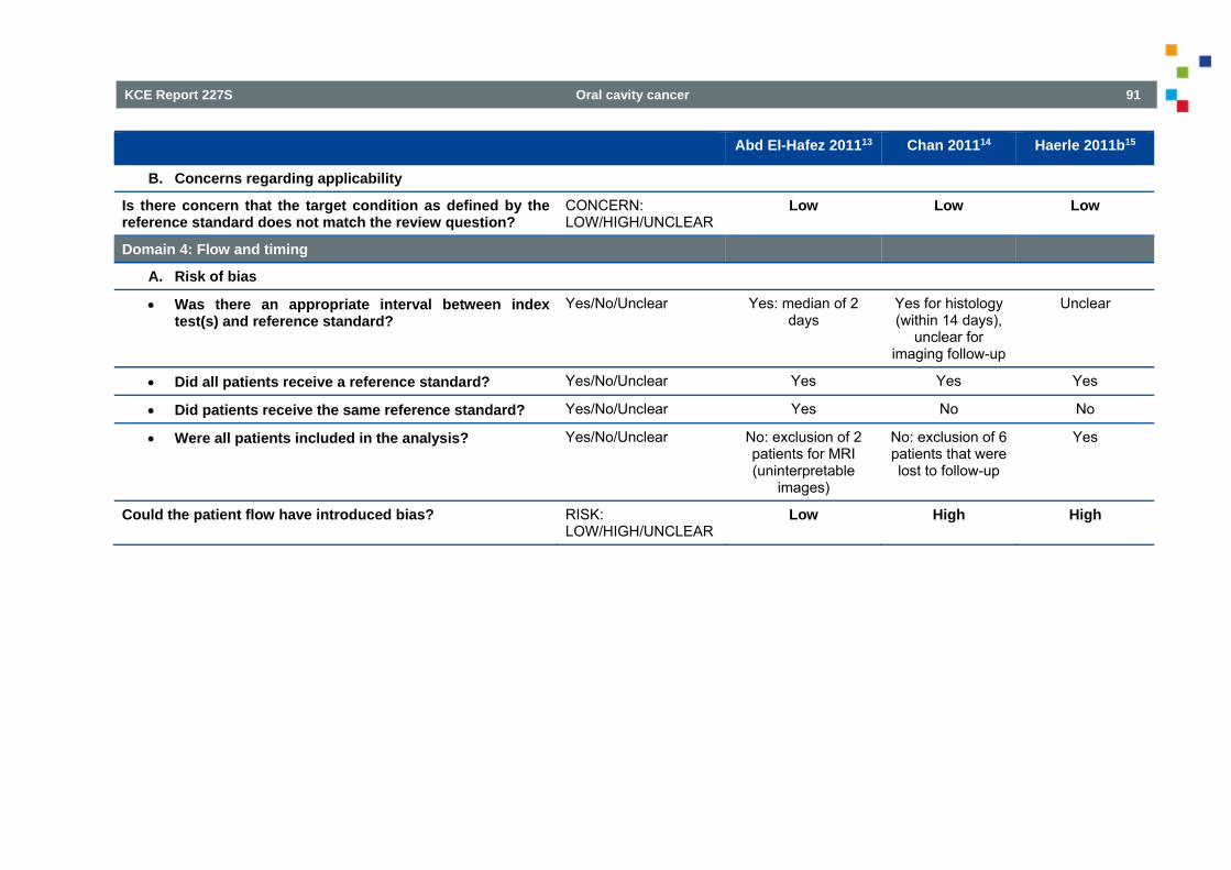
KCE Report 227S Oral cavity cancer 91
Abd El-Hafez 201113 Chan 201114 Haerle 2011b15
B. Concerns regarding applicability
Is there concern that the target condition as defined by the reference standard does not match the review question?
CONCERN: LOW/HIGH/UNCLEAR
Low Low Low
Domain 4: Flow and timing
A. Risk of bias
Was there an appropriate interval between index test(s) and reference standard?
Yes/No/Unclear Yes: median of 2 days
Yes for histology (within 14 days),
unclear for imaging follow-up
Unclear
Did all patients receive a reference standard? Yes/No/Unclear Yes Yes Yes
Did patients receive the same reference standard? Yes/No/Unclear Yes No No
Were all patients included in the analysis? Yes/No/Unclear No: exclusion of 2 patients for MRI (uninterpretable
images)
No: exclusion of 6 patients that were lost to follow-up
Yes
Could the patient flow have introduced bias? RISK: LOW/HIGH/UNCLEAR
Low High High
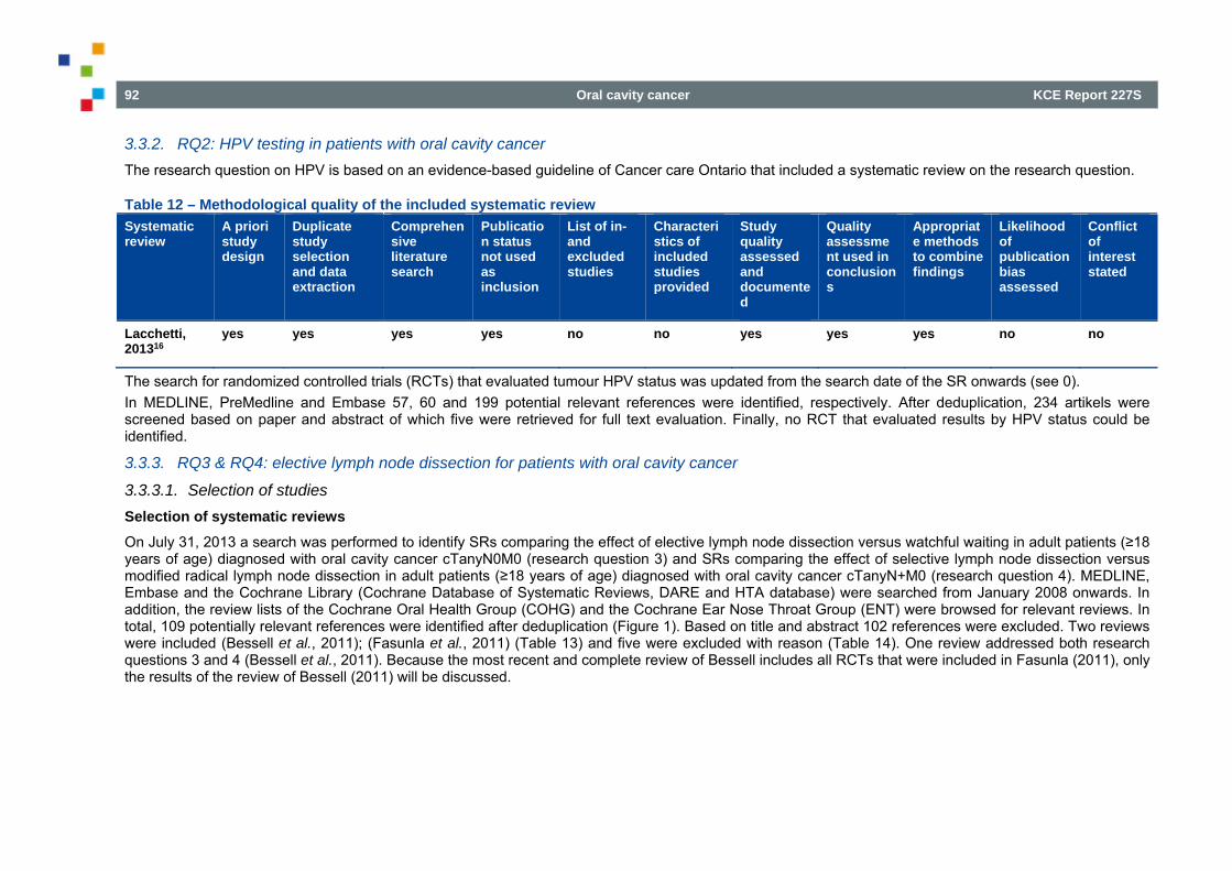
92 Oral cavity cancer KCE Report 227S
3.3.2. RQ2: HPV testing in patients with oral cavity cancer The research question on HPV is based on an evidence-based guideline of Cancer care Ontario that included a systematic review on the research question.
Table 12 – Methodological quality of the included systematic review Systematic review
A priori study design
Duplicate study selection and data extraction
Comprehensive literature search
Publication status not used as inclusion
List of in- and excluded studies
Characteristics of included studies provided
Study quality assessed and documented
Quality assessment used in conclusions
Appropriate methods to combine findings
Likelihood of publication bias assessed
Conflict of interest stated
Lacchetti, 201316
yes yes yes yes no no yes yes yes no no
The search for randomized controlled trials (RCTs) that evaluated tumour HPV status was updated from the search date of the SR onwards (see 0). In MEDLINE, PreMedline and Embase 57, 60 and 199 potential relevant references were identified, respectively. After deduplication, 234 artikels were screened based on paper and abstract of which five were retrieved for full text evaluation. Finally, no RCT that evaluated results by HPV status could be identified.
3.3.3. RQ3 & RQ4: elective lymph node dissection for patients with oral cavity cancer
3.3.3.1. Selection of studies Selection of systematic reviews On July 31, 2013 a search was performed to identify SRs comparing the effect of elective lymph node dissection versus watchful waiting in adult patients (≥18 years of age) diagnosed with oral cavity cancer cTanyN0M0 (research question 3) and SRs comparing the effect of selective lymph node dissection versus modified radical lymph node dissection in adult patients (≥18 years of age) diagnosed with oral cavity cancer cTanyN+M0 (research question 4). MEDLINE, Embase and the Cochrane Library (Cochrane Database of Systematic Reviews, DARE and HTA database) were searched from January 2008 onwards. In addition, the review lists of the Cochrane Oral Health Group (COHG) and the Cochrane Ear Nose Throat Group (ENT) were browsed for relevant reviews. In total, 109 potentially relevant references were identified after deduplication (Figure 1). Based on title and abstract 102 references were excluded. Two reviews were included (Bessell et al., 2011); (Fasunla et al., 2011) (Table 13) and five were excluded with reason (Table 14). One review addressed both research questions 3 and 4 (Bessell et al., 2011). Because the most recent and complete review of Bessell includes all RCTs that were included in Fasunla (2011), only the results of the review of Bessell (2011) will be discussed.
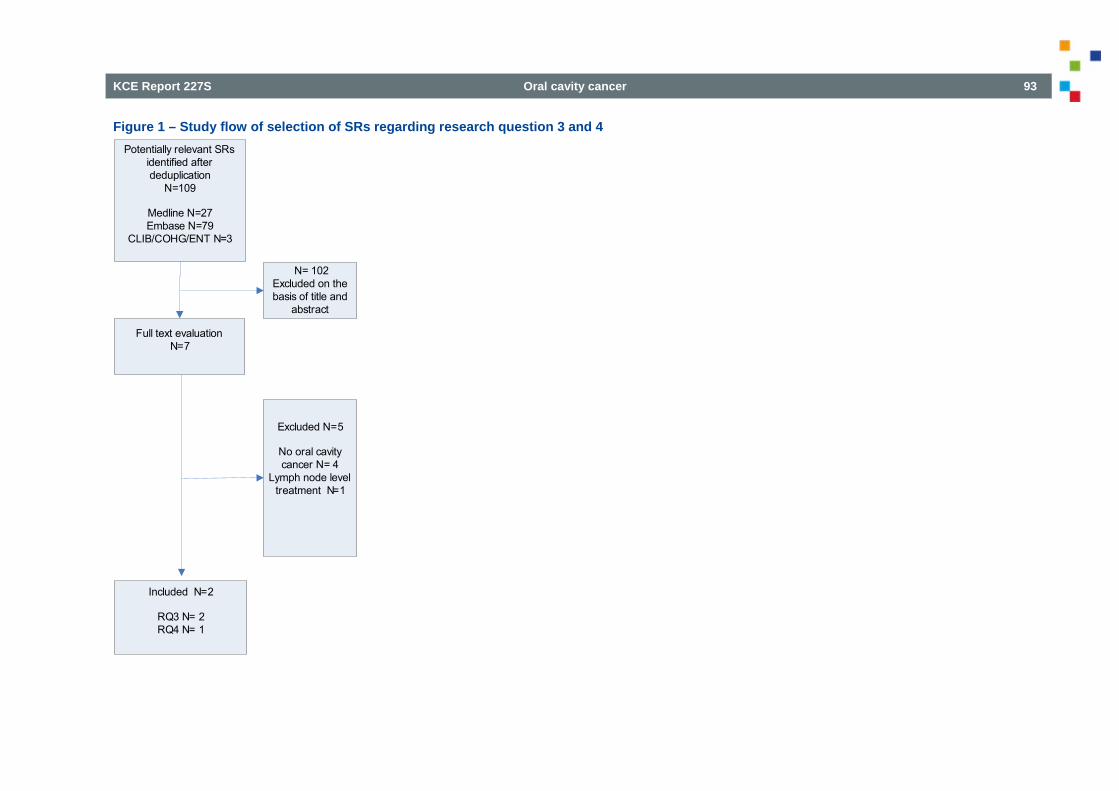
KCE Report 227S Oral cavity cancer 93
Figure 1 – Study flow of selection of SRs regarding research question 3 and 4 Potentially relevant SRs
identified after deduplication
N=109
Medline N=27Embase N=79
CLIB/COHG/ENT N=3
Full text evaluationN=7
N= 102Excluded on the basis of title and
abstract
Included N=2
RQ3 N= 2RQ4 N= 1
Excluded N=5
No oral cavity cancer N= 4
Lymph node level treatment N=1
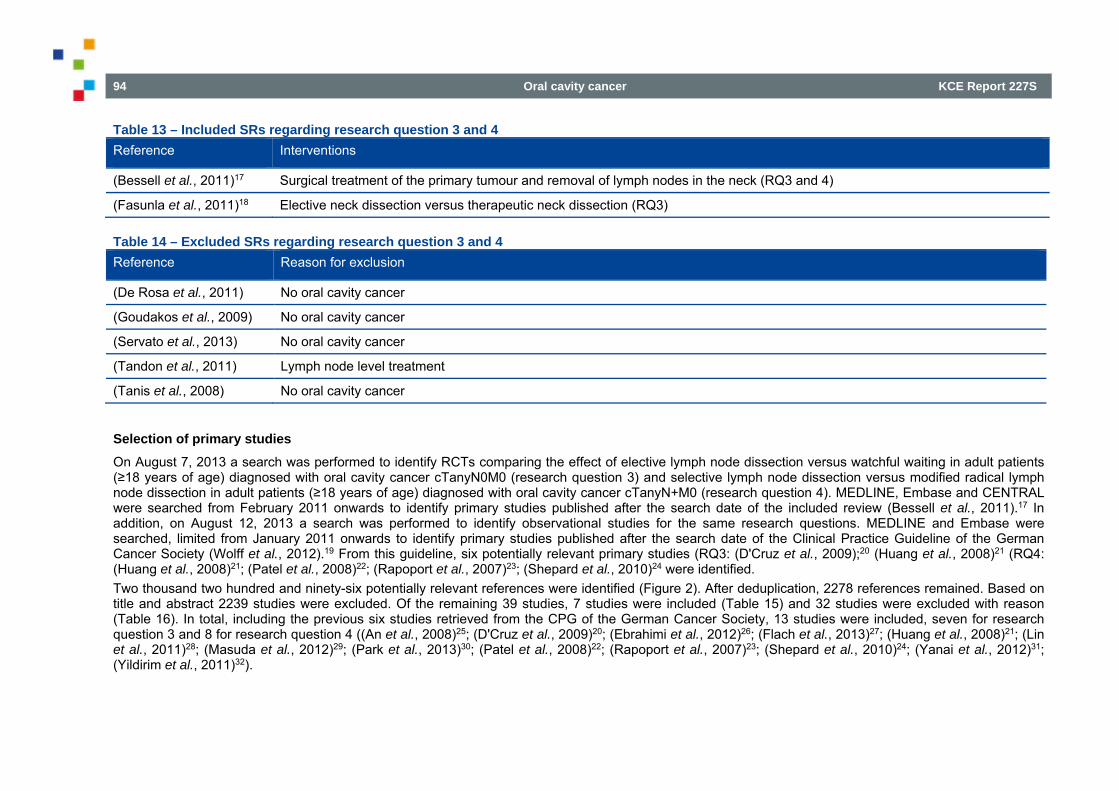
94 Oral cavity cancer KCE Report 227S
Table 13 – Included SRs regarding research question 3 and 4 Reference Interventions
(Bessell et al., 2011)17 Surgical treatment of the primary tumour and removal of lymph nodes in the neck (RQ3 and 4)
(Fasunla et al., 2011)18 Elective neck dissection versus therapeutic neck dissection (RQ3)
Table 14 – Excluded SRs regarding research question 3 and 4 Reference Reason for exclusion
(De Rosa et al., 2011) No oral cavity cancer
(Goudakos et al., 2009) No oral cavity cancer
(Servato et al., 2013) No oral cavity cancer
(Tandon et al., 2011) Lymph node level treatment
(Tanis et al., 2008) No oral cavity cancer
Selection of primary studies On August 7, 2013 a search was performed to identify RCTs comparing the effect of elective lymph node dissection versus watchful waiting in adult patients (≥18 years of age) diagnosed with oral cavity cancer cTanyN0M0 (research question 3) and selective lymph node dissection versus modified radical lymph node dissection in adult patients (≥18 years of age) diagnosed with oral cavity cancer cTanyN+M0 (research question 4). MEDLINE, Embase and CENTRAL were searched from February 2011 onwards to identify primary studies published after the search date of the included review (Bessell et al., 2011).17 In addition, on August 12, 2013 a search was performed to identify observational studies for the same research questions. MEDLINE and Embase were searched, limited from January 2011 onwards to identify primary studies published after the search date of the Clinical Practice Guideline of the German Cancer Society (Wolff et al., 2012).19 From this guideline, six potentially relevant primary studies (RQ3: (D'Cruz et al., 2009);20 (Huang et al., 2008)21 (RQ4: (Huang et al., 2008)21; (Patel et al., 2008)22; (Rapoport et al., 2007)23; (Shepard et al., 2010)24 were identified. Two thousand two hundred and ninety-six potentially relevant references were identified (Figure 2). After deduplication, 2278 references remained. Based on title and abstract 2239 studies were excluded. Of the remaining 39 studies, 7 studies were included (Table 15) and 32 studies were excluded with reason (Table 16). In total, including the previous six studies retrieved from the CPG of the German Cancer Society, 13 studies were included, seven for research question 3 and 8 for research question 4 ((An et al., 2008)25; (D'Cruz et al., 2009)20; (Ebrahimi et al., 2012)26; (Flach et al., 2013)27; (Huang et al., 2008)21; (Lin et al., 2011)28; (Masuda et al., 2012)29; (Park et al., 2013)30; (Patel et al., 2008)22; (Rapoport et al., 2007)23; (Shepard et al., 2010)24; (Yanai et al., 2012)31; (Yildirim et al., 2011)32).
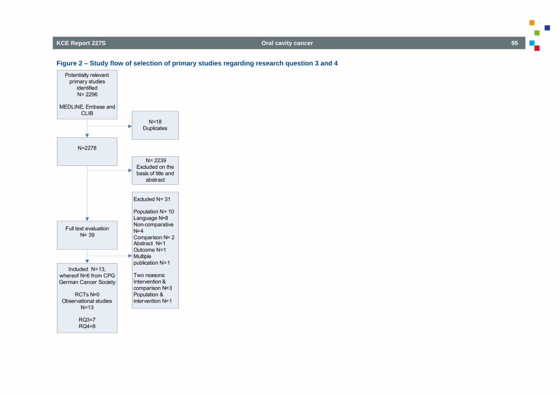
KCE Report 227S Oral cavity cancer 95
Figure 2 – Study flow of selection of primary studies regarding research question 3 and 4 Potentially relevant
primary studies identifiedN= 2296
MEDLINE, Embase and CLIB
N=2278
Full text evaluationN= 39
N=18Duplicates
N= 2239Excluded on the basis of title and
abstract
Included N=13,whereof N=6 from CPG German Cancer Society
RCTs N=0Observational studies
N=13
RQ3=7RQ4=8
Excluded N= 31
Population N= 10Language N=8Non-comparative N=4Comparison N= 2Abstract N=1Outcome N=1Multiple publication N=1
Two reasons: Intervention & comparison N=3Population & intervention N=1
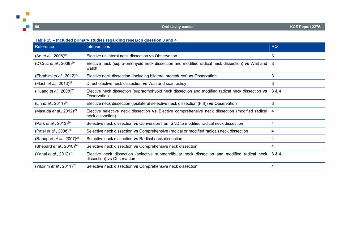
96 Oral cavity cancer KCE Report 227S
Table 15 – Included primary studies regarding research question 3 and 4 Reference Interventions RQ
(An et al., 2008)25 Elective unilateral neck dissection vs Observation 3
(D'Cruz et al., 2009)20 Elective neck (supra-omohyoid neck dissection and modified radical neck dissection) vs Wait and watch
3
(Ebrahimi et al., 2012)26 Elective neck dissection (including bilateral procedures) vs Observation 3
(Flach et al., 2013)27 Direct elective neck dissection vs Wait and scan policy 3
(Huang et al., 2008)21 Elective neck dissection (supraomohyoid neck dissection and modified radical neck dissection vs Observation
3 & 4
(Lin et al., 2011)28 Elective neck dissection (ipsilateral selective neck dissection (I-III)) vs Observation 3
(Masuda et al., 2012)29 Elective selective neck dissection vs Elective comprehensive neck dissection (modified radical neck dissection)
4
(Park et al., 2013)30 Selective neck dissection vs Conversion from SND to modified radical neck dissection 4
(Patel et al., 2008)22 Selective neck dissection vs Comprehensive (radical or modified radical) neck dissection 4
(Rapoport et al., 2007)23 Selective neck dissection vs Radical neck dissection 4
(Shepard et al., 2010)24 Selective neck dissection vs Comprehensive neck dissection 4
(Yanai et al., 2012)31 Elective neck dissection (selective submandibular neck dissection and modified radical neck dissection) vs Observation
3 & 4
(Yildirim et al., 2011)32 Selective neck dissection vs Comprehensive neck dissection 4
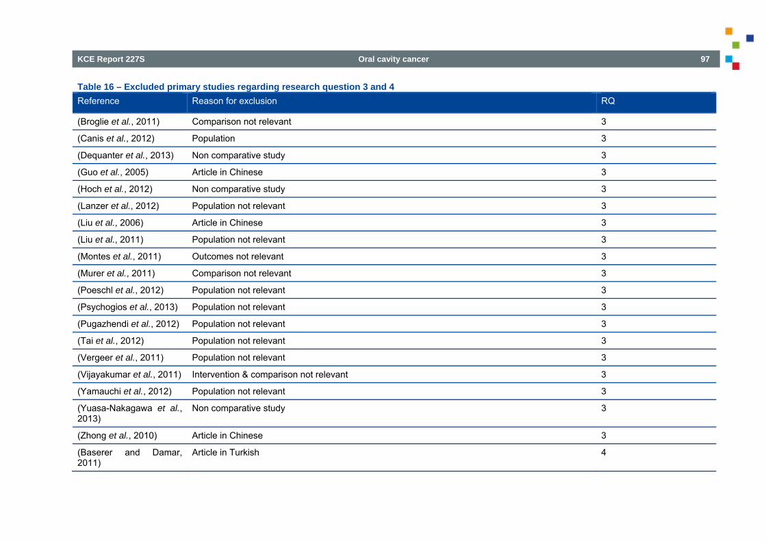
KCE Report 227S Oral cavity cancer 97
Table 16 – Excluded primary studies regarding research question 3 and 4 Reference Reason for exclusion RQ
(Broglie et al., 2011) Comparison not relevant 3
(Canis et al., 2012) Population 3
(Dequanter et al., 2013) Non comparative study 3
(Guo et al., 2005) Article in Chinese 3
(Hoch et al., 2012) Non comparative study 3
(Lanzer et al., 2012) Population not relevant 3
(Liu et al., 2006) Article in Chinese 3
(Liu et al., 2011) Population not relevant 3
(Montes et al., 2011) Outcomes not relevant 3
(Murer et al., 2011) Comparison not relevant 3
(Poeschl et al., 2012) Population not relevant 3
(Psychogios et al., 2013) Population not relevant 3
(Pugazhendi et al., 2012) Population not relevant 3
(Tai et al., 2012) Population not relevant 3
(Vergeer et al., 2011) Population not relevant 3
(Vijayakumar et al., 2011) Intervention & comparison not relevant 3
(Yamauchi et al., 2012) Population not relevant 3
(Yuasa-Nakagawa et al., 2013)
Non comparative study 3
(Zhong et al., 2010) Article in Chinese 3
(Baserer and Damar, 2011)
Article in Turkish 4
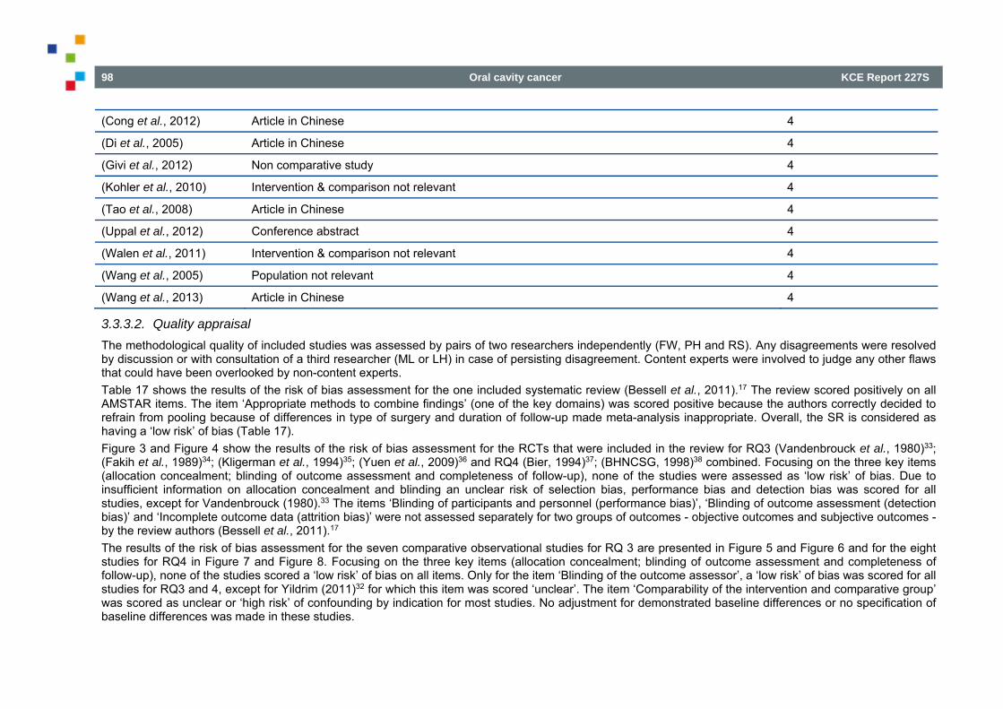
98 Oral cavity cancer KCE Report 227S
(Cong et al., 2012) Article in Chinese 4
(Di et al., 2005) Article in Chinese 4
(Givi et al., 2012) Non comparative study 4
(Kohler et al., 2010) Intervention & comparison not relevant 4
(Tao et al., 2008) Article in Chinese 4
(Uppal et al., 2012) Conference abstract 4
(Walen et al., 2011) Intervention & comparison not relevant 4
(Wang et al., 2005) Population not relevant 4
(Wang et al., 2013) Article in Chinese 4
3.3.3.2. Quality appraisal The methodological quality of included studies was assessed by pairs of two researchers independently (FW, PH and RS). Any disagreements were resolved by discussion or with consultation of a third researcher (ML or LH) in case of persisting disagreement. Content experts were involved to judge any other flaws that could have been overlooked by non-content experts. Table 17 shows the results of the risk of bias assessment for the one included systematic review (Bessell et al., 2011).17 The review scored positively on all AMSTAR items. The item ‘Appropriate methods to combine findings’ (one of the key domains) was scored positive because the authors correctly decided to refrain from pooling because of differences in type of surgery and duration of follow-up made meta-analysis inappropriate. Overall, the SR is considered as having a ‘low risk’ of bias (Table 17). Figure 3 and Figure 4 show the results of the risk of bias assessment for the RCTs that were included in the review for RQ3 (Vandenbrouck et al., 1980)33; (Fakih et al., 1989)34; (Kligerman et al., 1994)35; (Yuen et al., 2009)36 and RQ4 (Bier, 1994)37; (BHNCSG, 1998)38 combined. Focusing on the three key items (allocation concealment; blinding of outcome assessment and completeness of follow-up), none of the studies were assessed as ‘low risk’ of bias. Due to insufficient information on allocation concealment and blinding an unclear risk of selection bias, performance bias and detection bias was scored for all studies, except for Vandenbrouck (1980).33 The items ‘Blinding of participants and personnel (performance bias)’, ‘Blinding of outcome assessment (detection bias)’ and ‘Incomplete outcome data (attrition bias)’ were not assessed separately for two groups of outcomes - objective outcomes and subjective outcomes - by the review authors (Bessell et al., 2011).17 The results of the risk of bias assessment for the seven comparative observational studies for RQ 3 are presented in Figure 5 and Figure 6 and for the eight studies for RQ4 in Figure 7 and Figure 8. Focusing on the three key items (allocation concealment; blinding of outcome assessment and completeness of follow-up), none of the studies scored a ‘low risk’ of bias on all items. Only for the item ‘Blinding of the outcome assessor’, a ‘low risk’ of bias was scored for all studies for RQ3 and 4, except for Yildrim (2011)32 for which this item was scored ‘unclear’. The item ‘Comparability of the intervention and comparative group’ was scored as unclear or ‘high risk’ of confounding by indication for most studies. No adjustment for demonstrated baseline differences or no specification of baseline differences was made in these studies.
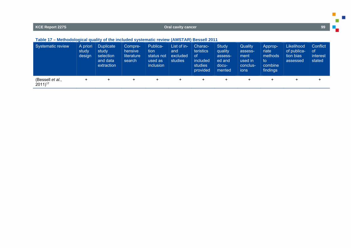
KCE Report 227S Oral cavity cancer 99
Table 17 – Methodological quality of the included systematic review (AMSTAR) Bessell 2011 Systematic review A priori
study design
Duplicate study selection and data extraction
Compre-hensive literature search
Publica-tion status not used as inclusion
List of in- and excluded studies
Charac-teristics of included studies provided
Study quality assess-ed and docu-mented
Quality assess-ment used in conclus-ions
Approp-riate methods to combine findings
Likelihood of publica-tion bias assessed
Conflict of interest stated
(Bessell et al., 2011)17
+ + + + + + + + + + +
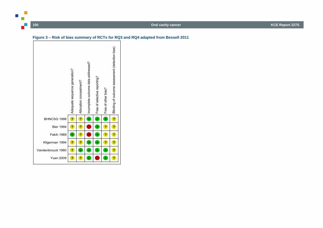
100 Oral cavity cancer KCE Report 227S
Figure 3 – Risk of bias summary of RCTs for RQ3 and RQ4 adapted from Bessell 2011
Adeq
uate
seq
uenc
e ge
nera
tion?
BHNCSG 1998 ?
Bier 1994 ?
Fakih 1989 +
Kligerman 1994 ?
Vandenbrouck 1980 ?
Yuen 2009 ?
Allo
catio
n co
ncea
lmen
t?
?
?
?
?
+
?
Inco
mpl
ete
outc
ome
data
add
ress
ed?
+
–
–
+
+
+
Free
of s
elec
tive
repo
rting
?
+
+
+
+
+
–
Free
of o
ther
bia
s?
+
?
?
?
+
+
Blin
ding
of o
utco
me
asse
ssm
ent (
dete
ctio
n bi
as)
?
?
?
?
?
?
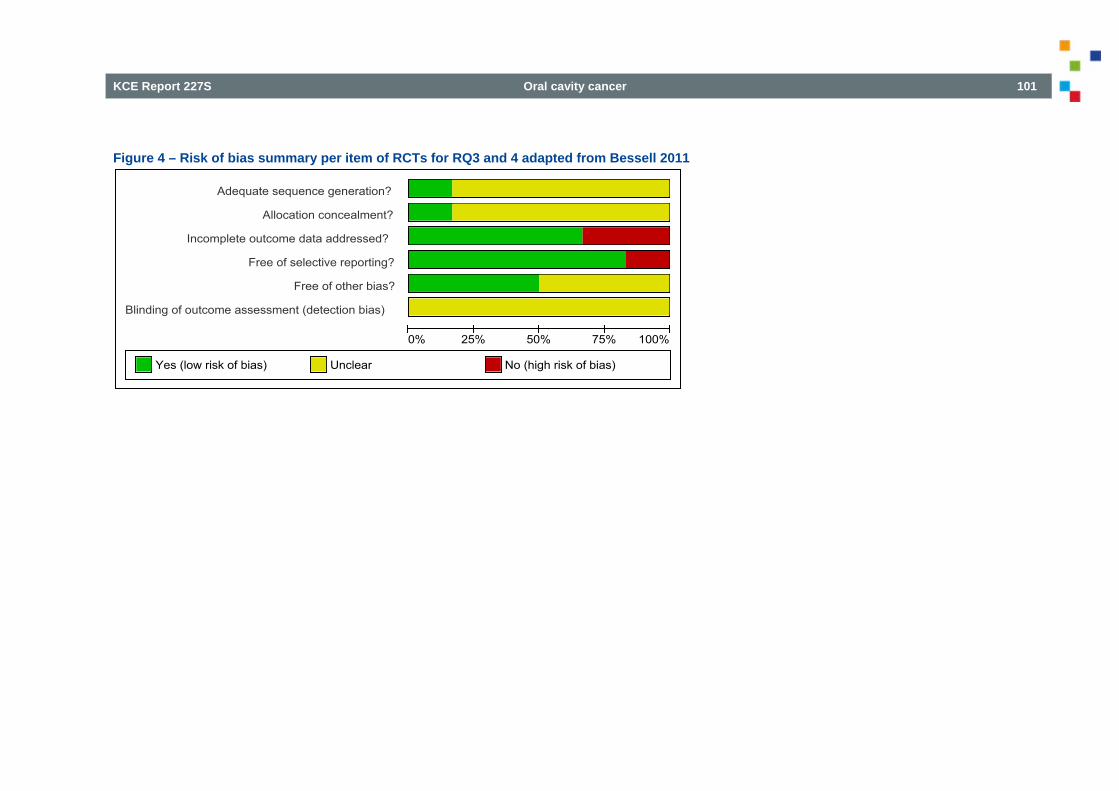
KCE Report 227S Oral cavity cancer 101
Figure 4 – Risk of bias summary per item of RCTs for RQ3 and 4 adapted from Bessell 2011
Adequate sequence generation?
Allocation concealment?
Incomplete outcome data addressed?
Free of selective reporting?
Free of other bias?
Blinding of outcome assessment (detection bias)
0% 25% 50% 75% 100%
Yes (low risk of bias) Unclear No (high risk of bias)
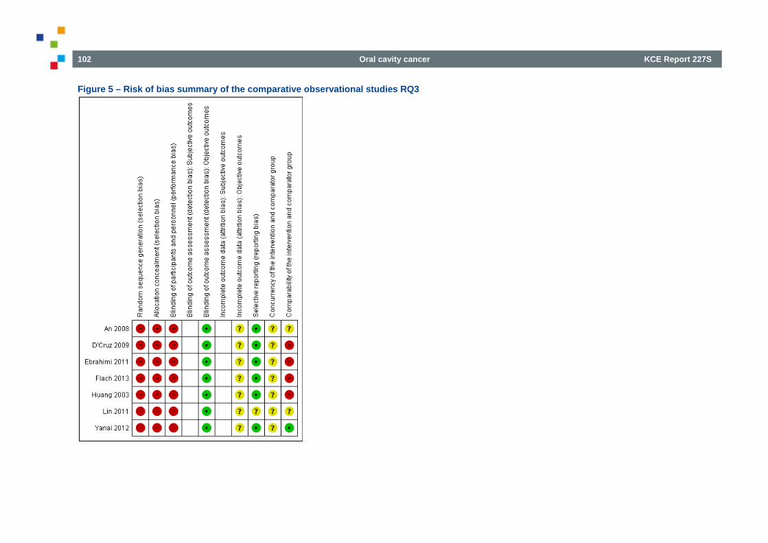
102 Oral cavity cancer KCE Report 227S
Figure 5 – Risk of bias summary of the comparative observational studies RQ3
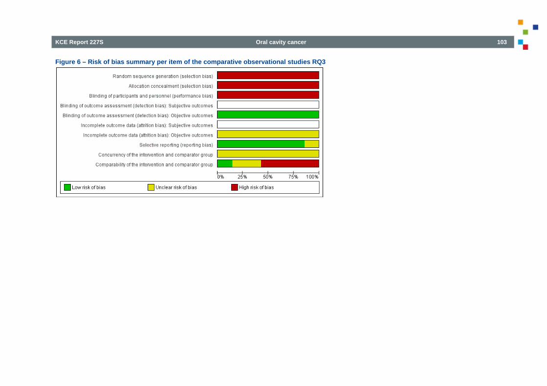
KCE Report 227S Oral cavity cancer 103
Figure 6 – Risk of bias summary per item of the comparative observational studies RQ3
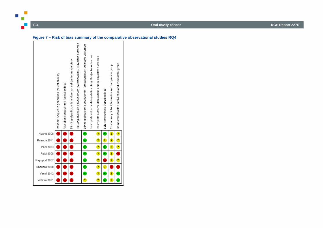
104 Oral cavity cancer KCE Report 227S
Figure 7 – Risk of bias summary of the comparative observational studies RQ4
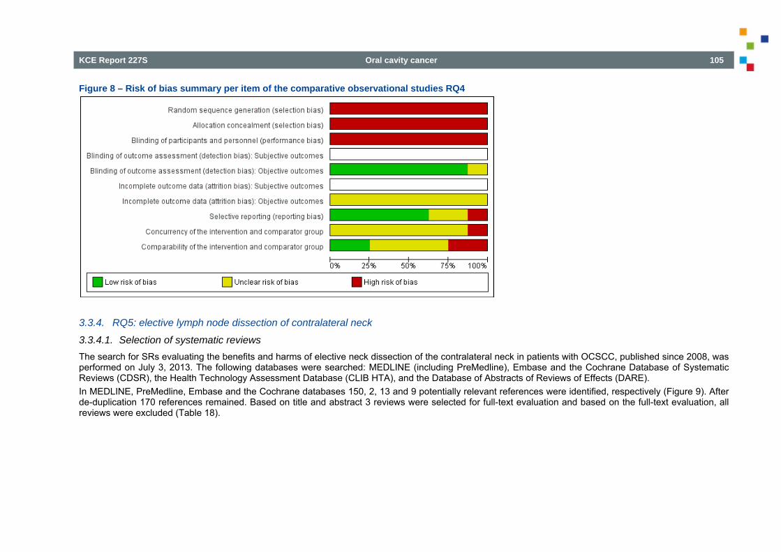
KCE Report 227S Oral cavity cancer 105
Figure 8 – Risk of bias summary per item of the comparative observational studies RQ4
3.3.4. RQ5: elective lymph node dissection of contralateral neck
3.3.4.1. Selection of systematic reviews The search for SRs evaluating the benefits and harms of elective neck dissection of the contralateral neck in patients with OCSCC, published since 2008, was performed on July 3, 2013. The following databases were searched: MEDLINE (including PreMedline), Embase and the Cochrane Database of Systematic Reviews (CDSR), the Health Technology Assessment Database (CLIB HTA), and the Database of Abstracts of Reviews of Effects (DARE). In MEDLINE, PreMedline, Embase and the Cochrane databases 150, 2, 13 and 9 potentially relevant references were identified, respectively (Figure 9). After de-duplication 170 references remained. Based on title and abstract 3 reviews were selected for full-text evaluation and based on the full-text evaluation, all reviews were excluded (Table 18).
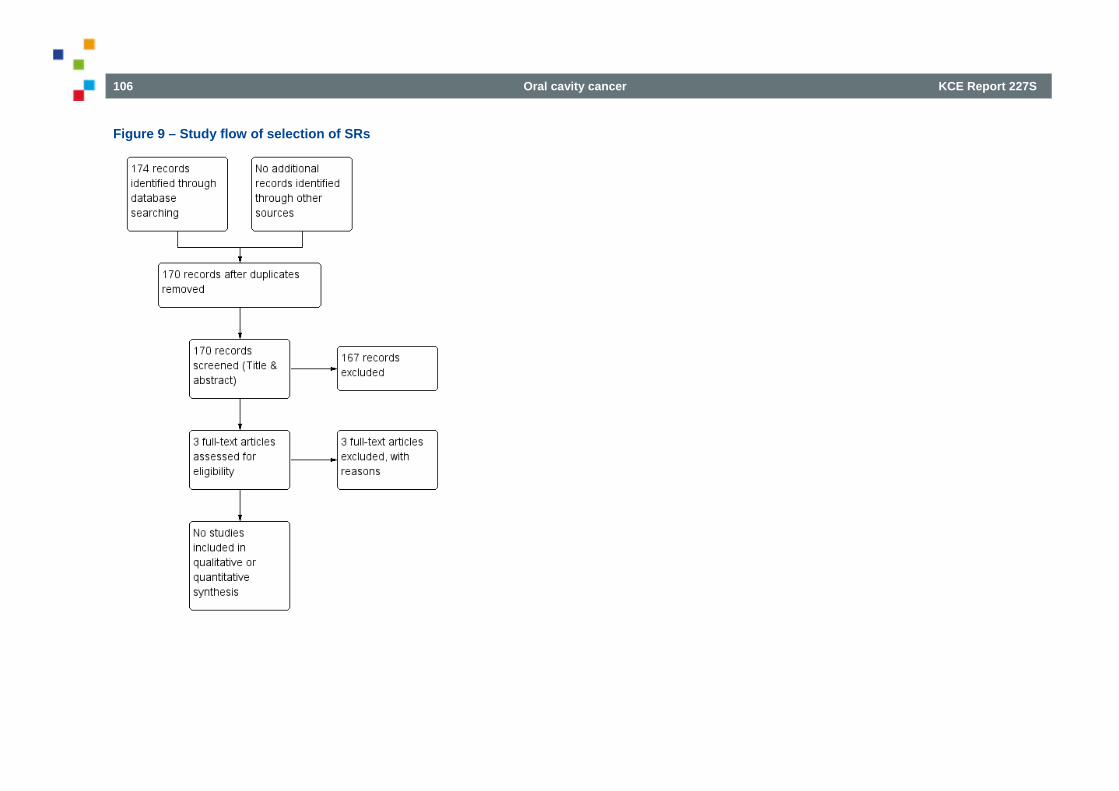
106 Oral cavity cancer KCE Report 227S
Figure 9 – Study flow of selection of SRs
KCE Report 227S Oral cavity cancer 107
Table 18 – Reviews excluded based on full-text evaluation Reference Reason(s) for exclusion
Bessell A et al. Interventions for the treatment of oral and oropharyngeal cancers: surgical treatment. Cochrane Database Syst Rev. 2011: CD006205.
Topic is not covered
Fan S et al. A review of clinical and histological parameters associated with contralateral neck metastases in oral squamous cell carcinoma. Int J Oral Sci 2011; 3(4): 180-91.
Narrative review
Fasunla A et al. A meta-analysis of the randomized controlled trials on elective neck dissection versus therapeutic neck dissection in oral cavity cancers with clinically node-negative neck. Oral Oncol 2011; 47(5): 320-4.
Topic is not covered
3.3.4.2. Selection of primary studies On August 12, 2013 a search was performed in MEDLINE (including PreMedline), Embase and CENTRAL to identify RCTs and observational studies evaluating the benefits and harms of elective neck dissection of the contralateral neck in patients with OCSCC, published from 2003 onwards. In MEDLINE, PreMedline and Embase 295, 8 and 161 potentially relevant references were identified, respectively (Figure 10); no references were found in CENTRAL. After de-duplication, 397 references remained. Based on title and abstract 391 articles were excluded. Of the remaining 6 studies, 3 studies were included after full-text evaluation; the rationale for exclusion of the other 3 articles is presented in Table 19.
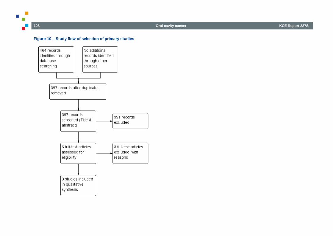
108 Oral cavity cancer KCE Report 227S
Figure 10 – Study flow of selection of primary studies
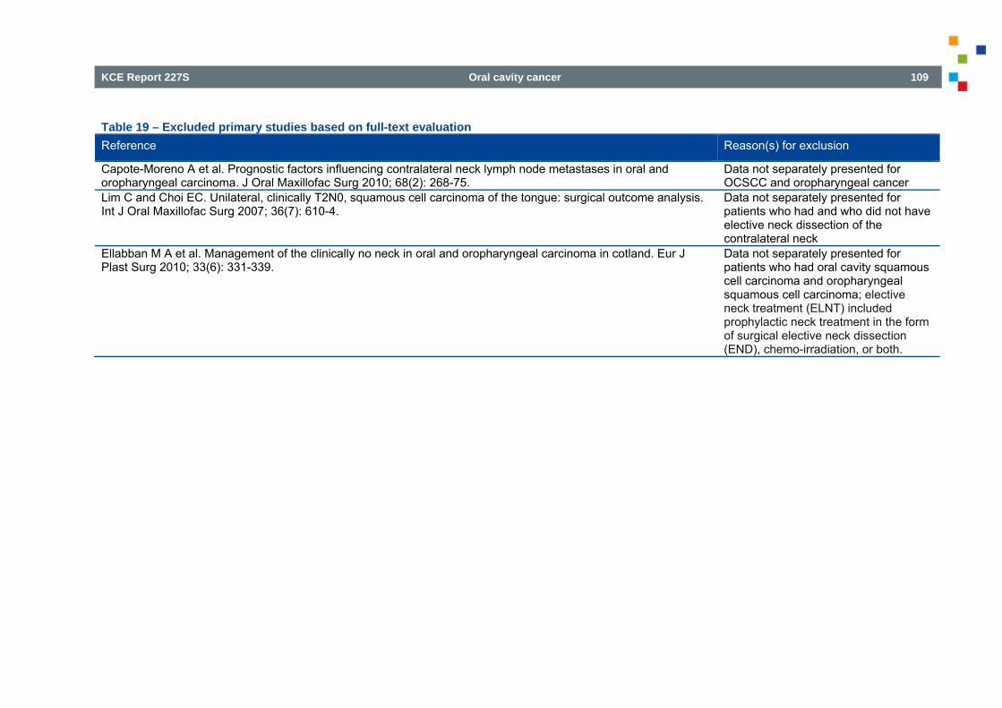
KCE Report 227S Oral cavity cancer 109
Table 19 – Excluded primary studies based on full-text evaluation Reference Reason(s) for exclusion
Capote-Moreno A et al. Prognostic factors influencing contralateral neck lymph node metastases in oral and oropharyngeal carcinoma. J Oral Maxillofac Surg 2010; 68(2): 268-75.
Data not separately presented for OCSCC and oropharyngeal cancer
Lim C and Choi EC. Unilateral, clinically T2N0, squamous cell carcinoma of the tongue: surgical outcome analysis. Int J Oral Maxillofac Surg 2007; 36(7): 610-4.
Data not separately presented for patients who had and who did not have elective neck dissection of the contralateral neck
Ellabban M A et al. Management of the clinically no neck in oral and oropharyngeal carcinoma in cotland. Eur J Plast Surg 2010; 33(6): 331-339.
Data not separately presented for patients who had oral cavity squamous cell carcinoma and oropharyngeal squamous cell carcinoma; elective neck treatment (ELNT) included prophylactic neck treatment in the form of surgical elective neck dissection (END), chemo-irradiation, or both.
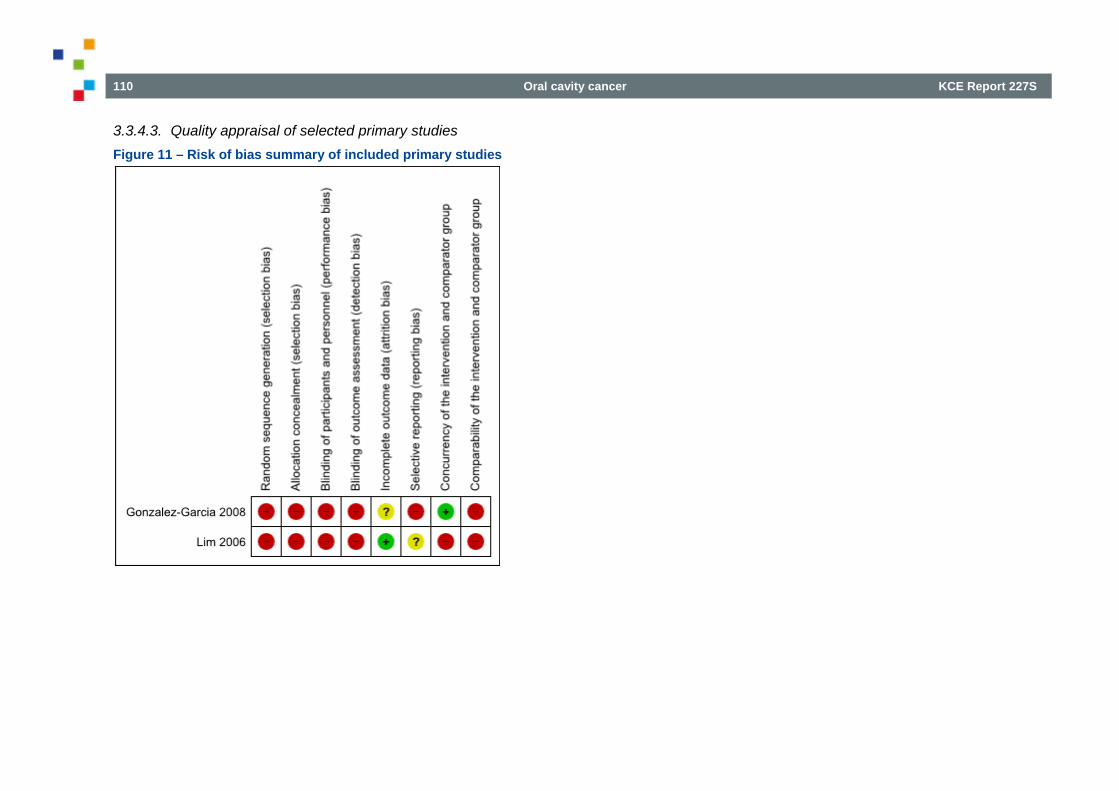
110 Oral cavity cancer KCE Report 227S
3.3.4.3. Quality appraisal of selected primary studies Figure 11 – Risk of bias summary of included primary studies
KCE Report 227S Oral cavity cancer 111
3.3.5. RQ6: value of PET / MRI in the decision of neck dissection after CRT
3.3.5.1. Selection of systematic reviews The search for SRs evaluating the value of PET and MRI in the decision of neck dissection after (at least) chemoradiotherapy (CRT) in patients with head & neck squamous cell carcinoma (HNSCC), was performed on November 26, 2013. The following databases were searched: MEDLINE (including PreMedline), Embase and the Cochrane Database of Systematic Reviews (CDSR), the Health Technology Assessment Database (CLIB HTA), and the Database of Abstracts of Reviews of Effects (DARE). In MEDLINE, Embase and the Cochrane databases 82 (after de-depulication), 13 and 10 potentially relevant references were identified, respectively (Figure 12); no additional systematic reviews were retrieved in pre-medline. After de-duplication 91 references remained. Based on title and abstract 4 reviews (all on the value of PET in the decision of neck dissection after CRT) were selected for full-text evaluation and based on the full-text evaluation, another 2 reviews were excluded (Table 20). No systematic reviews evaluated the diagnostic value of MRI in the decision of neck dissection after (at least) chemoradiotherapy in patients with head & neck squamous cell carcinoma.
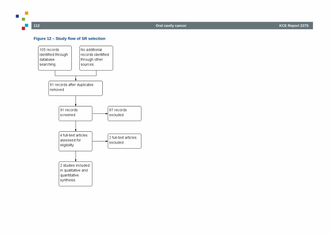
112 Oral cavity cancer KCE Report 227S
Figure 12 – Study flow of SR selection
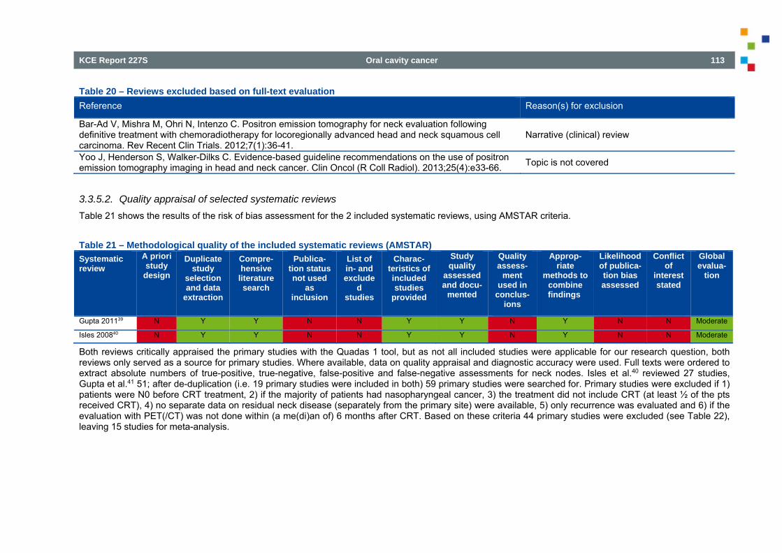
KCE Report 227S Oral cavity cancer 113
Table 20 – Reviews excluded based on full-text evaluation Reference Reason(s) for exclusion
Bar-Ad V, Mishra M, Ohri N, Intenzo C. Positron emission tomography for neck evaluation following definitive treatment with chemoradiotherapy for locoregionally advanced head and neck squamous cell carcinoma. Rev Recent Clin Trials. 2012;7(1):36-41.
Narrative (clinical) review
Yoo J, Henderson S, Walker-Dilks C. Evidence-based guideline recommendations on the use of positron emission tomography imaging in head and neck cancer. Clin Oncol (R Coll Radiol). 2013;25(4):e33-66. Topic is not covered
3.3.5.2. Quality appraisal of selected systematic reviews Table 21 shows the results of the risk of bias assessment for the 2 included systematic reviews, using AMSTAR criteria.
Table 21 – Methodological quality of the included systematic reviews (AMSTAR) Systematic review
A priori study
design
Duplicate study
selection and data
extraction
Compre-hensive
literature search
Publica-tion status not used
as inclusion
List of in- and exclude
d studies
Charac-teristics of included studies
provided
Study quality
assessed and docu-
mented
Quality assess-
ment used in
conclus-ions
Approp-riate
methods to combine findings
Likelihood of publica-tion bias assessed
Conflict of
interest stated
Global evalua-
tion
Gupta 201139 N Y Y N N Y Y N Y N N Moderate
Isles 200840 N Y Y N N Y Y N Y N N Moderate
Both reviews critically appraised the primary studies with the Quadas 1 tool, but as not all included studies were applicable for our research question, both reviews only served as a source for primary studies. Where available, data on quality appraisal and diagnostic accuracy were used. Full texts were ordered to extract absolute numbers of true-positive, true-negative, false-positive and false-negative assessments for neck nodes. Isles et al.40 reviewed 27 studies, Gupta et al.41 51; after de-duplication (i.e. 19 primary studies were included in both) 59 primary studies were searched for. Primary studies were excluded if 1) patients were N0 before CRT treatment, 2) if the majority of patients had nasopharyngeal cancer, 3) the treatment did not include CRT (at least ½ of the pts received CRT), 4) no separate data on residual neck disease (separately from the primary site) were available, 5) only recurrence was evaluated and 6) if the evaluation with PET(/CT) was not done within (a me(di)an of) 6 months after CRT. Based on these criteria 44 primary studies were excluded (see Table 22), leaving 15 studies for meta-analysis.
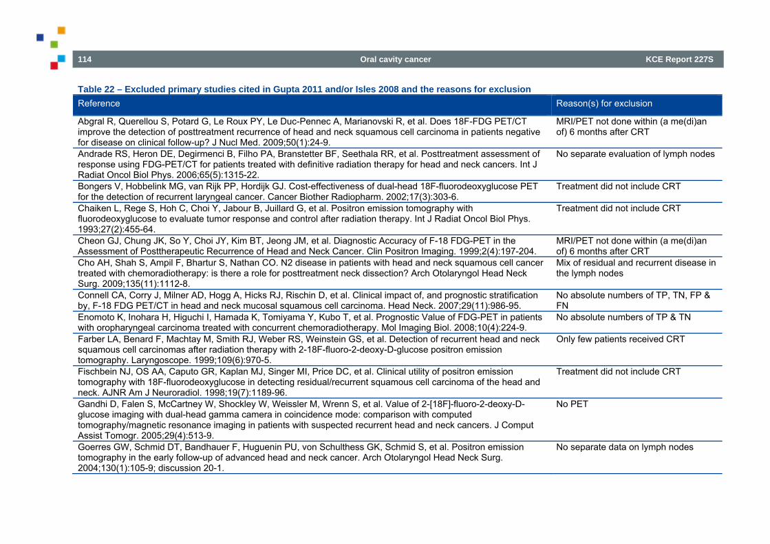
114 Oral cavity cancer KCE Report 227S
Table 22 – Excluded primary studies cited in Gupta 2011 and/or Isles 2008 and the reasons for exclusion Reference Reason(s) for exclusion
Abgral R, Querellou S, Potard G, Le Roux PY, Le Duc-Pennec A, Marianovski R, et al. Does 18F-FDG PET/CT improve the detection of posttreatment recurrence of head and neck squamous cell carcinoma in patients negative for disease on clinical follow-up? J Nucl Med. 2009;50(1):24-9.
MRI/PET not done within (a me(di)an of) 6 months after CRT
Andrade RS, Heron DE, Degirmenci B, Filho PA, Branstetter BF, Seethala RR, et al. Posttreatment assessment of response using FDG-PET/CT for patients treated with definitive radiation therapy for head and neck cancers. Int J Radiat Oncol Biol Phys. 2006;65(5):1315-22.
No separate evaluation of lymph nodes
Bongers V, Hobbelink MG, van Rijk PP, Hordijk GJ. Cost-effectiveness of dual-head 18F-fluorodeoxyglucose PET for the detection of recurrent laryngeal cancer. Cancer Biother Radiopharm. 2002;17(3):303-6.
Treatment did not include CRT
Chaiken L, Rege S, Hoh C, Choi Y, Jabour B, Juillard G, et al. Positron emission tomography with fluorodeoxyglucose to evaluate tumor response and control after radiation therapy. Int J Radiat Oncol Biol Phys. 1993;27(2):455-64.
Treatment did not include CRT
Cheon GJ, Chung JK, So Y, Choi JY, Kim BT, Jeong JM, et al. Diagnostic Accuracy of F-18 FDG-PET in the Assessment of Posttherapeutic Recurrence of Head and Neck Cancer. Clin Positron Imaging. 1999;2(4):197-204.
MRI/PET not done within (a me(di)an of) 6 months after CRT
Cho AH, Shah S, Ampil F, Bhartur S, Nathan CO. N2 disease in patients with head and neck squamous cell cancer treated with chemoradiotherapy: is there a role for posttreatment neck dissection? Arch Otolaryngol Head Neck Surg. 2009;135(11):1112-8.
Mix of residual and recurrent disease in the lymph nodes
Connell CA, Corry J, Milner AD, Hogg A, Hicks RJ, Rischin D, et al. Clinical impact of, and prognostic stratification by, F-18 FDG PET/CT in head and neck mucosal squamous cell carcinoma. Head Neck. 2007;29(11):986-95.
No absolute numbers of TP, TN, FP & FN
Enomoto K, Inohara H, Higuchi I, Hamada K, Tomiyama Y, Kubo T, et al. Prognostic Value of FDG-PET in patients with oropharyngeal carcinoma treated with concurrent chemoradiotherapy. Mol Imaging Biol. 2008;10(4):224-9.
No absolute numbers of TP & TN
Farber LA, Benard F, Machtay M, Smith RJ, Weber RS, Weinstein GS, et al. Detection of recurrent head and neck squamous cell carcinomas after radiation therapy with 2-18F-fluoro-2-deoxy-D-glucose positron emission tomography. Laryngoscope. 1999;109(6):970-5.
Only few patients received CRT
Fischbein NJ, OS AA, Caputo GR, Kaplan MJ, Singer MI, Price DC, et al. Clinical utility of positron emission tomography with 18F-fluorodeoxyglucose in detecting residual/recurrent squamous cell carcinoma of the head and neck. AJNR Am J Neuroradiol. 1998;19(7):1189-96.
Treatment did not include CRT
Gandhi D, Falen S, McCartney W, Shockley W, Weissler M, Wrenn S, et al. Value of 2-[18F]-fluoro-2-deoxy-D-glucose imaging with dual-head gamma camera in coincidence mode: comparison with computed tomography/magnetic resonance imaging in patients with suspected recurrent head and neck cancers. J Comput Assist Tomogr. 2005;29(4):513-9.
No PET
Goerres GW, Schmid DT, Bandhauer F, Huguenin PU, von Schulthess GK, Schmid S, et al. Positron emission tomography in the early follow-up of advanced head and neck cancer. Arch Otolaryngol Head Neck Surg. 2004;130(1):105-9; discussion 20-1.
No separate data on lymph nodes
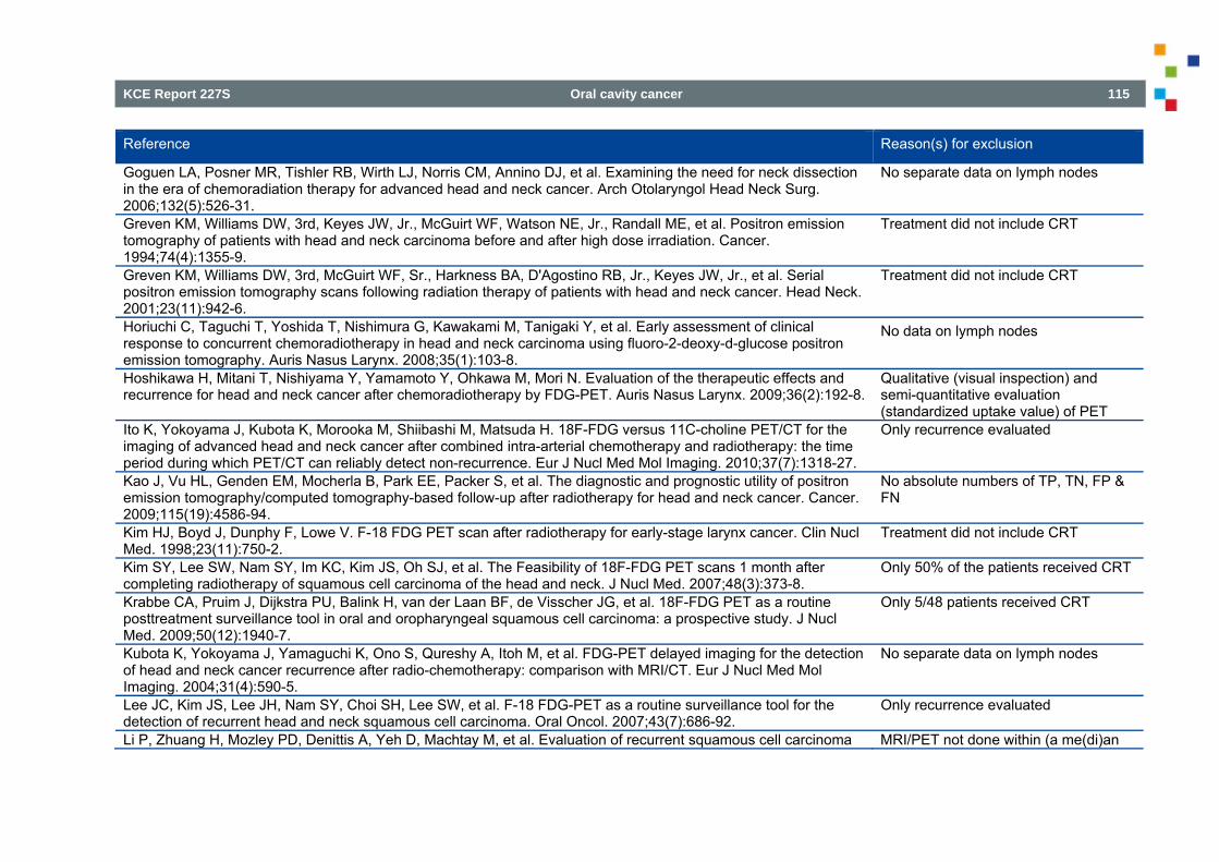
KCE Report 227S Oral cavity cancer 115
Reference Reason(s) for exclusion
Goguen LA, Posner MR, Tishler RB, Wirth LJ, Norris CM, Annino DJ, et al. Examining the need for neck dissection in the era of chemoradiation therapy for advanced head and neck cancer. Arch Otolaryngol Head Neck Surg. 2006;132(5):526-31.
No separate data on lymph nodes
Greven KM, Williams DW, 3rd, Keyes JW, Jr., McGuirt WF, Watson NE, Jr., Randall ME, et al. Positron emission tomography of patients with head and neck carcinoma before and after high dose irradiation. Cancer. 1994;74(4):1355-9.
Treatment did not include CRT
Greven KM, Williams DW, 3rd, McGuirt WF, Sr., Harkness BA, D'Agostino RB, Jr., Keyes JW, Jr., et al. Serial positron emission tomography scans following radiation therapy of patients with head and neck cancer. Head Neck. 2001;23(11):942-6.
Treatment did not include CRT
Horiuchi C, Taguchi T, Yoshida T, Nishimura G, Kawakami M, Tanigaki Y, et al. Early assessment of clinical response to concurrent chemoradiotherapy in head and neck carcinoma using fluoro-2-deoxy-d-glucose positron emission tomography. Auris Nasus Larynx. 2008;35(1):103-8.
No data on lymph nodes
Hoshikawa H, Mitani T, Nishiyama Y, Yamamoto Y, Ohkawa M, Mori N. Evaluation of the therapeutic effects and recurrence for head and neck cancer after chemoradiotherapy by FDG-PET. Auris Nasus Larynx. 2009;36(2):192-8.
Qualitative (visual inspection) and semi-quantitative evaluation (standardized uptake value) of PET
Ito K, Yokoyama J, Kubota K, Morooka M, Shiibashi M, Matsuda H. 18F-FDG versus 11C-choline PET/CT for the imaging of advanced head and neck cancer after combined intra-arterial chemotherapy and radiotherapy: the time period during which PET/CT can reliably detect non-recurrence. Eur J Nucl Med Mol Imaging. 2010;37(7):1318-27.
Only recurrence evaluated
Kao J, Vu HL, Genden EM, Mocherla B, Park EE, Packer S, et al. The diagnostic and prognostic utility of positron emission tomography/computed tomography-based follow-up after radiotherapy for head and neck cancer. Cancer. 2009;115(19):4586-94.
No absolute numbers of TP, TN, FP & FN
Kim HJ, Boyd J, Dunphy F, Lowe V. F-18 FDG PET scan after radiotherapy for early-stage larynx cancer. Clin Nucl Med. 1998;23(11):750-2.
Treatment did not include CRT
Kim SY, Lee SW, Nam SY, Im KC, Kim JS, Oh SJ, et al. The Feasibility of 18F-FDG PET scans 1 month after completing radiotherapy of squamous cell carcinoma of the head and neck. J Nucl Med. 2007;48(3):373-8.
Only 50% of the patients received CRT
Krabbe CA, Pruim J, Dijkstra PU, Balink H, van der Laan BF, de Visscher JG, et al. 18F-FDG PET as a routine posttreatment surveillance tool in oral and oropharyngeal squamous cell carcinoma: a prospective study. J Nucl Med. 2009;50(12):1940-7.
Only 5/48 patients received CRT
Kubota K, Yokoyama J, Yamaguchi K, Ono S, Qureshy A, Itoh M, et al. FDG-PET delayed imaging for the detection of head and neck cancer recurrence after radio-chemotherapy: comparison with MRI/CT. Eur J Nucl Med Mol Imaging. 2004;31(4):590-5.
No separate data on lymph nodes
Lee JC, Kim JS, Lee JH, Nam SY, Choi SH, Lee SW, et al. F-18 FDG-PET as a routine surveillance tool for the detection of recurrent head and neck squamous cell carcinoma. Oral Oncol. 2007;43(7):686-92.
Only recurrence evaluated
Li P, Zhuang H, Mozley PD, Denittis A, Yeh D, Machtay M, et al. Evaluation of recurrent squamous cell carcinoma MRI/PET not done within (a me(di)an
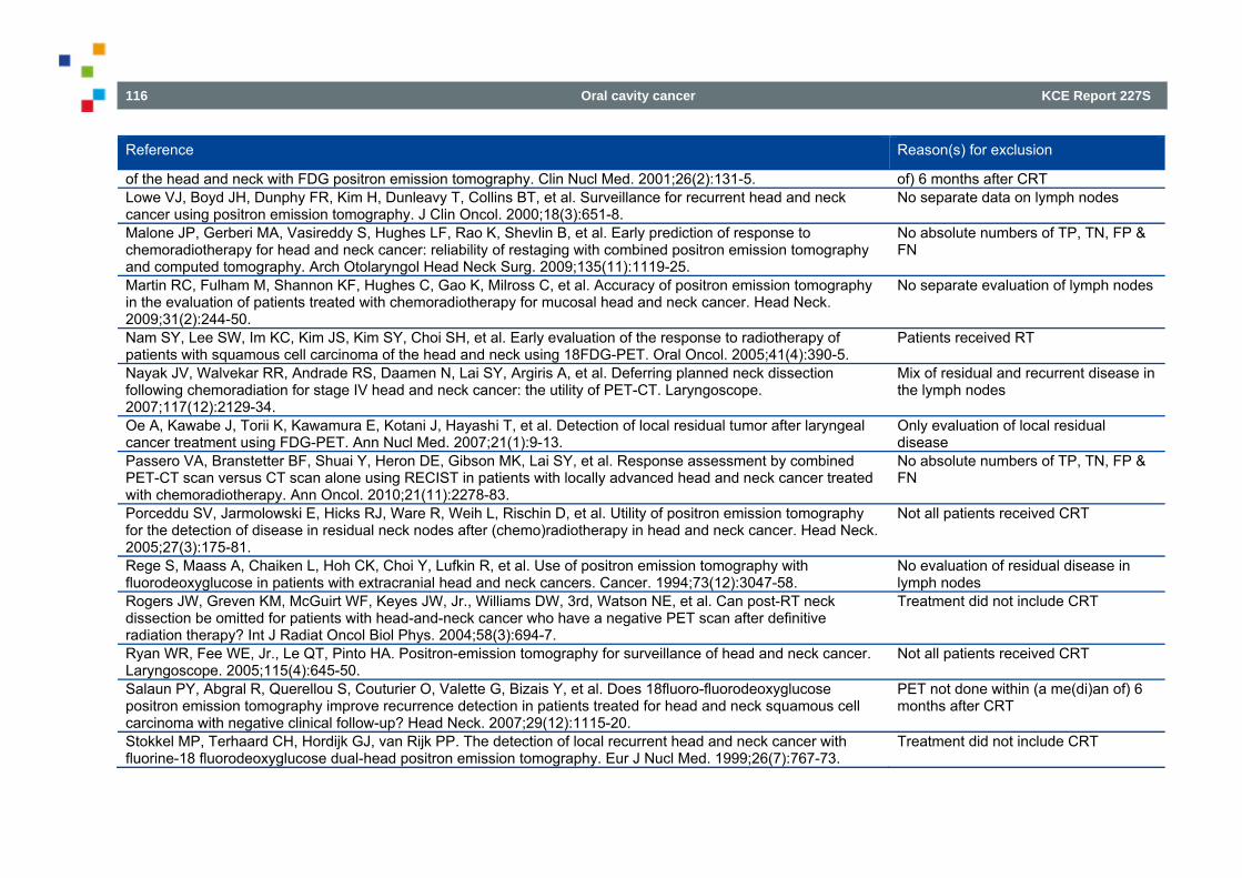
116 Oral cavity cancer KCE Report 227S
Reference Reason(s) for exclusion
of the head and neck with FDG positron emission tomography. Clin Nucl Med. 2001;26(2):131-5. of) 6 months after CRT Lowe VJ, Boyd JH, Dunphy FR, Kim H, Dunleavy T, Collins BT, et al. Surveillance for recurrent head and neck cancer using positron emission tomography. J Clin Oncol. 2000;18(3):651-8.
No separate data on lymph nodes
Malone JP, Gerberi MA, Vasireddy S, Hughes LF, Rao K, Shevlin B, et al. Early prediction of response to chemoradiotherapy for head and neck cancer: reliability of restaging with combined positron emission tomography and computed tomography. Arch Otolaryngol Head Neck Surg. 2009;135(11):1119-25.
No absolute numbers of TP, TN, FP & FN
Martin RC, Fulham M, Shannon KF, Hughes C, Gao K, Milross C, et al. Accuracy of positron emission tomography in the evaluation of patients treated with chemoradiotherapy for mucosal head and neck cancer. Head Neck. 2009;31(2):244-50.
No separate evaluation of lymph nodes
Nam SY, Lee SW, Im KC, Kim JS, Kim SY, Choi SH, et al. Early evaluation of the response to radiotherapy of patients with squamous cell carcinoma of the head and neck using 18FDG-PET. Oral Oncol. 2005;41(4):390-5.
Patients received RT
Nayak JV, Walvekar RR, Andrade RS, Daamen N, Lai SY, Argiris A, et al. Deferring planned neck dissection following chemoradiation for stage IV head and neck cancer: the utility of PET-CT. Laryngoscope. 2007;117(12):2129-34.
Mix of residual and recurrent disease in the lymph nodes
Oe A, Kawabe J, Torii K, Kawamura E, Kotani J, Hayashi T, et al. Detection of local residual tumor after laryngeal cancer treatment using FDG-PET. Ann Nucl Med. 2007;21(1):9-13.
Only evaluation of local residual disease
Passero VA, Branstetter BF, Shuai Y, Heron DE, Gibson MK, Lai SY, et al. Response assessment by combined PET-CT scan versus CT scan alone using RECIST in patients with locally advanced head and neck cancer treated with chemoradiotherapy. Ann Oncol. 2010;21(11):2278-83.
No absolute numbers of TP, TN, FP & FN
Porceddu SV, Jarmolowski E, Hicks RJ, Ware R, Weih L, Rischin D, et al. Utility of positron emission tomography for the detection of disease in residual neck nodes after (chemo)radiotherapy in head and neck cancer. Head Neck. 2005;27(3):175-81.
Not all patients received CRT
Rege S, Maass A, Chaiken L, Hoh CK, Choi Y, Lufkin R, et al. Use of positron emission tomography with fluorodeoxyglucose in patients with extracranial head and neck cancers. Cancer. 1994;73(12):3047-58.
No evaluation of residual disease in lymph nodes
Rogers JW, Greven KM, McGuirt WF, Keyes JW, Jr., Williams DW, 3rd, Watson NE, et al. Can post-RT neck dissection be omitted for patients with head-and-neck cancer who have a negative PET scan after definitive radiation therapy? Int J Radiat Oncol Biol Phys. 2004;58(3):694-7.
Treatment did not include CRT
Ryan WR, Fee WE, Jr., Le QT, Pinto HA. Positron-emission tomography for surveillance of head and neck cancer. Laryngoscope. 2005;115(4):645-50.
Not all patients received CRT
Salaun PY, Abgral R, Querellou S, Couturier O, Valette G, Bizais Y, et al. Does 18fluoro-fluorodeoxyglucose positron emission tomography improve recurrence detection in patients treated for head and neck squamous cell carcinoma with negative clinical follow-up? Head Neck. 2007;29(12):1115-20.
PET not done within (a me(di)an of) 6 months after CRT
Stokkel MP, Terhaard CH, Hordijk GJ, van Rijk PP. The detection of local recurrent head and neck cancer with fluorine-18 fluorodeoxyglucose dual-head positron emission tomography. Eur J Nucl Med. 1999;26(7):767-73.
Treatment did not include CRT
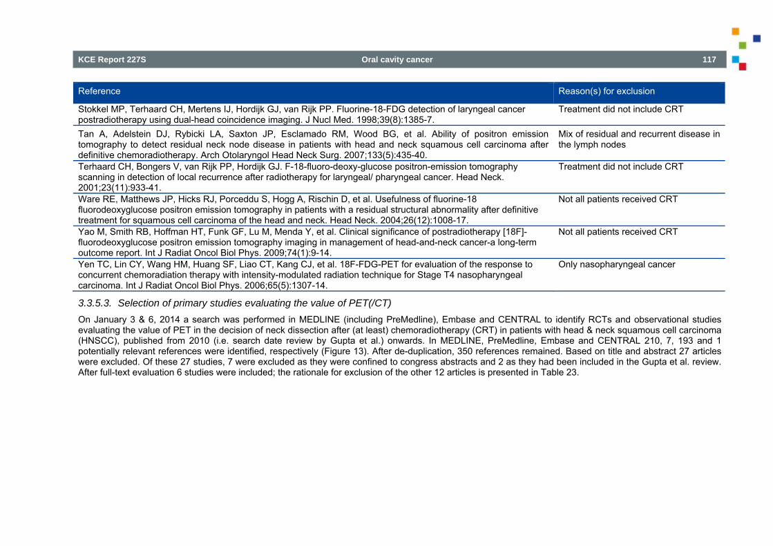
KCE Report 227S Oral cavity cancer 117
Reference Reason(s) for exclusion
Stokkel MP, Terhaard CH, Mertens IJ, Hordijk GJ, van Rijk PP. Fluorine-18-FDG detection of laryngeal cancer postradiotherapy using dual-head coincidence imaging. J Nucl Med. 1998;39(8):1385-7.
Treatment did not include CRT
Tan A, Adelstein DJ, Rybicki LA, Saxton JP, Esclamado RM, Wood BG, et al. Ability of positron emission tomography to detect residual neck node disease in patients with head and neck squamous cell carcinoma after definitive chemoradiotherapy. Arch Otolaryngol Head Neck Surg. 2007;133(5):435-40.
Mix of residual and recurrent disease in the lymph nodes
Terhaard CH, Bongers V, van Rijk PP, Hordijk GJ. F-18-fluoro-deoxy-glucose positron-emission tomography scanning in detection of local recurrence after radiotherapy for laryngeal/ pharyngeal cancer. Head Neck. 2001;23(11):933-41.
Treatment did not include CRT
Ware RE, Matthews JP, Hicks RJ, Porceddu S, Hogg A, Rischin D, et al. Usefulness of fluorine-18 fluorodeoxyglucose positron emission tomography in patients with a residual structural abnormality after definitive treatment for squamous cell carcinoma of the head and neck. Head Neck. 2004;26(12):1008-17.
Not all patients received CRT
Yao M, Smith RB, Hoffman HT, Funk GF, Lu M, Menda Y, et al. Clinical significance of postradiotherapy [18F]-fluorodeoxyglucose positron emission tomography imaging in management of head-and-neck cancer-a long-term outcome report. Int J Radiat Oncol Biol Phys. 2009;74(1):9-14.
Not all patients received CRT
Yen TC, Lin CY, Wang HM, Huang SF, Liao CT, Kang CJ, et al. 18F-FDG-PET for evaluation of the response to concurrent chemoradiation therapy with intensity-modulated radiation technique for Stage T4 nasopharyngeal carcinoma. Int J Radiat Oncol Biol Phys. 2006;65(5):1307-14.
Only nasopharyngeal cancer
3.3.5.3. Selection of primary studies evaluating the value of PET(/CT) On January 3 & 6, 2014 a search was performed in MEDLINE (including PreMedline), Embase and CENTRAL to identify RCTs and observational studies evaluating the value of PET in the decision of neck dissection after (at least) chemoradiotherapy (CRT) in patients with head & neck squamous cell carcinoma (HNSCC), published from 2010 (i.e. search date review by Gupta et al.) onwards. In MEDLINE, PreMedline, Embase and CENTRAL 210, 7, 193 and 1 potentially relevant references were identified, respectively (Figure 13). After de-duplication, 350 references remained. Based on title and abstract 27 articles were excluded. Of these 27 studies, 7 were excluded as they were confined to congress abstracts and 2 as they had been included in the Gupta et al. review. After full-text evaluation 6 studies were included; the rationale for exclusion of the other 12 articles is presented in Table 23.
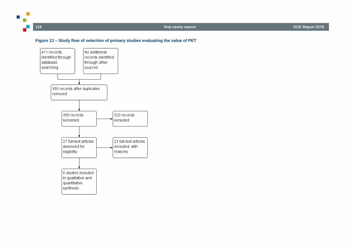
118 Oral cavity cancer KCE Report 227S
Figure 13 – Study flow of selection of primary studies evaluating the value of PET
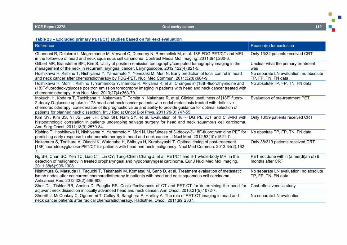
KCE Report 227S Oral cavity cancer 119
Table 23 – Excluded primary PET(/CT) studies based on full-text evaluation Reference Reason(s) for exclusion
Ghanooni R, Delpierre I, Magremanne M, Vervaet C, Dumarey N, Remmelink M, et al. 18F-FDG PET/CT and MRI in the follow-up of head and neck squamous cell carcinoma. Contrast Media Mol Imaging. 2011;6(4):260-6.
Only 13/32 patients received CRT
Gilbert MR, Branstetter BFt, Kim S. Utility of positron-emission tomography/computed tomography imaging in the management of the neck in recurrent laryngeal cancer. Laryngoscope. 2012;122(4):821-5.
Unclear what the primary treatment was
Hoshikawa H, Kishino T, Nishiyama Y, Yamamoto Y, Yonezaki M, Mori N. Early prediction of local control in head and neck cancer after chemoradiotherapy by FDG-PET. Nucl Med Commun. 2011;32(8):684-9.
No separate LN evaluation; no absolute TP, FP, TN, FN data
Hoshikawa H, Mori T, Kishino T, Yamamoto Y, Inamoto R, Akiyama K, et al. Changes in (18)F-fluorothymidine and (18)F-fluorodeoxyglucose positron emission tomography imaging in patients with head and neck cancer treated with chemoradiotherapy. Ann Nucl Med. 2013;27(4):363-70.
No absolute TP, FP, TN, FN data
Inokuchi H, Kodaira T, Tachibana H, Nakamura T, Tomita N, Nakahara R, et al. Clinical usefulness of [18F] fluoro-2-deoxy-D-glucose uptake in 178 head-and-neck cancer patients with nodal metastasis treated with definitive chemoradiotherapy: consideration of its prognostic value and ability to provide guidance for optimal selection of patients for planned neck dissection. Int J Radiat Oncol Biol Phys. 2011;79(3):747-55.
Evaluation of pre-treatment PET
Kim SY, Kim JS, Yi JS, Lee JH, Choi SH, Nam SY, et al. Evaluation of 18F-FDG PET/CT and CT/MRI with histopathologic correlation in patients undergoing salvage surgery for head and neck squamous cell carcinoma. Ann Surg Oncol. 2011;18(9):2579-84.
Only 13/39 patients received CRT
Kishino T, Hoshikawa H, Nishiyama Y, Yamamoto Y, Mori N. Usefulness of 3'-deoxy-3'-18F-fluorothymidine PET for predicting early response to chemoradiotherapy in head and neck cancer. J Nucl Med. 2012;53(10):1521-7.
No absolute TP, FP, TN, FN data
Nakamura S, Toriihara A, Okochi K, Watanabe H, Shibuya H, Kurabayashi T. Optimal timing of post-treatment [18F]fluorodeoxyglucose-PET/CT for patients with head and neck malignancy. Nucl Med Commun. 2013;34(2):162-7.
Only 38/319 patients received CRT
Ng SH, Chan SC, Yen TC, Liao CT, Lin CY, Tung-Chieh Chang J, et al. PET/CT and 3-T whole-body MRI in the detection of malignancy in treated oropharyngeal and hypopharyngeal carcinoma. Eur J Nucl Med Mol Imaging. 2011;38(6):996-1008.
PET not done within (a me(di)an of) 6 months after CRT
Nishimura G, Matsuda H, Taguchi T, Takahashi M, Komatsu M, Sano D, et al. Treatment evaluation of metastatic lymph nodes after concurrent chemoradiotherapy in patients with head and neck squamous cell carcinoma. Anticancer Res. 2012;32(2):595-600.
No separate LN evaluation; no absolute TP, FP, TN, FN data
Sher DJ, Tishler RB, Annino D, Punglia RS. Cost-effectiveness of CT and PET-CT for determining the need for adjuvant neck dissection in locally advanced head and neck cancer. Ann Oncol. 2010;21(5):1072-7.
Cost-effectiveness study
Sherriff J, McConkey C, Ogunremi T, Colley S, Sanghera P, Hartley A. The role of PET-CT imaging in head and neck cancer patients after radical chemoradiotherapy. Radiother. Oncol. 2011;99:S337.
No separate LN evaluation
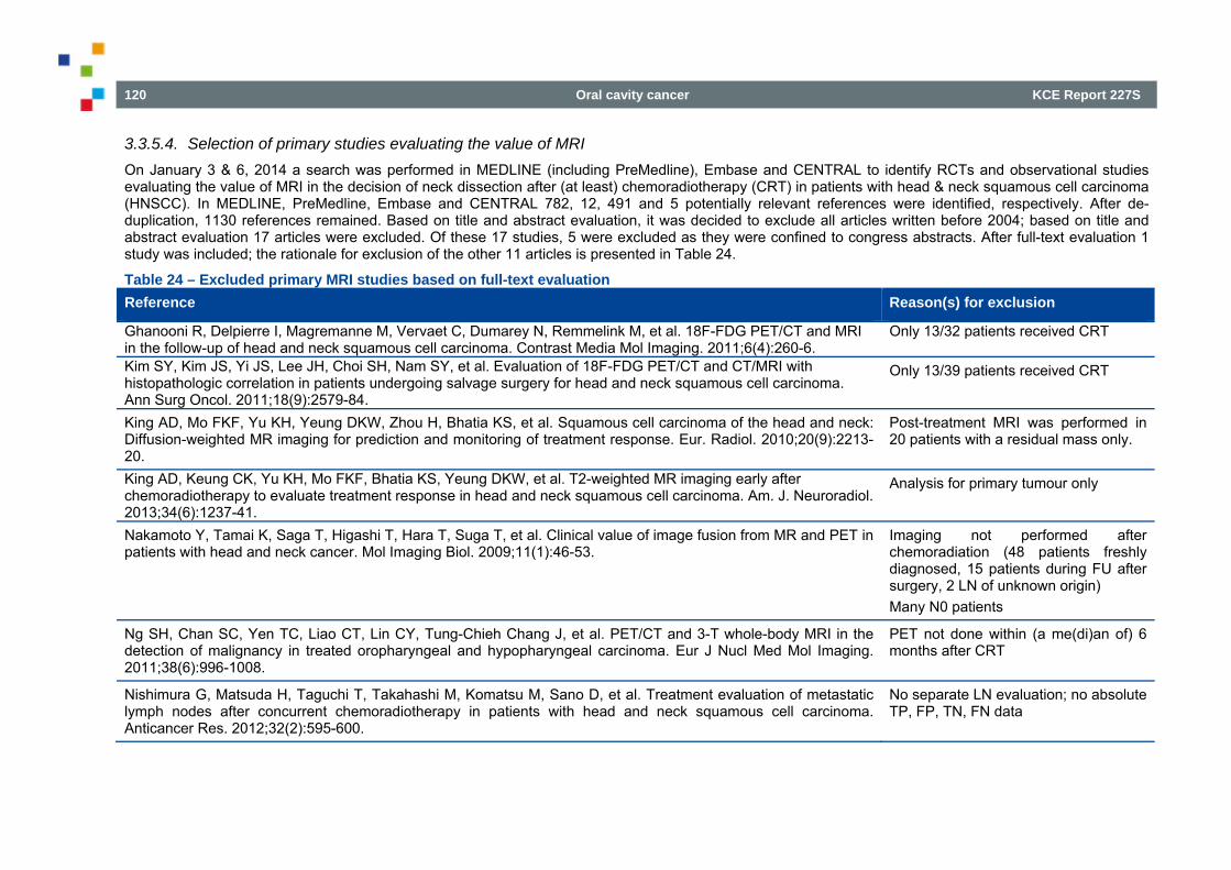
120 Oral cavity cancer KCE Report 227S
3.3.5.4. Selection of primary studies evaluating the value of MRI On January 3 & 6, 2014 a search was performed in MEDLINE (including PreMedline), Embase and CENTRAL to identify RCTs and observational studies evaluating the value of MRI in the decision of neck dissection after (at least) chemoradiotherapy (CRT) in patients with head & neck squamous cell carcinoma (HNSCC). In MEDLINE, PreMedline, Embase and CENTRAL 782, 12, 491 and 5 potentially relevant references were identified, respectively. After de-duplication, 1130 references remained. Based on title and abstract evaluation, it was decided to exclude all articles written before 2004; based on title and abstract evaluation 17 articles were excluded. Of these 17 studies, 5 were excluded as they were confined to congress abstracts. After full-text evaluation 1 study was included; the rationale for exclusion of the other 11 articles is presented in Table 24.
Table 24 – Excluded primary MRI studies based on full-text evaluation Reference Reason(s) for exclusion
Ghanooni R, Delpierre I, Magremanne M, Vervaet C, Dumarey N, Remmelink M, et al. 18F-FDG PET/CT and MRI in the follow-up of head and neck squamous cell carcinoma. Contrast Media Mol Imaging. 2011;6(4):260-6.
Only 13/32 patients received CRT
Kim SY, Kim JS, Yi JS, Lee JH, Choi SH, Nam SY, et al. Evaluation of 18F-FDG PET/CT and CT/MRI with histopathologic correlation in patients undergoing salvage surgery for head and neck squamous cell carcinoma. Ann Surg Oncol. 2011;18(9):2579-84.
Only 13/39 patients received CRT
King AD, Mo FKF, Yu KH, Yeung DKW, Zhou H, Bhatia KS, et al. Squamous cell carcinoma of the head and neck: Diffusion-weighted MR imaging for prediction and monitoring of treatment response. Eur. Radiol. 2010;20(9):2213-20.
Post-treatment MRI was performed in 20 patients with a residual mass only.
King AD, Keung CK, Yu KH, Mo FKF, Bhatia KS, Yeung DKW, et al. T2-weighted MR imaging early after chemoradiotherapy to evaluate treatment response in head and neck squamous cell carcinoma. Am. J. Neuroradiol. 2013;34(6):1237-41.
Analysis for primary tumour only
Nakamoto Y, Tamai K, Saga T, Higashi T, Hara T, Suga T, et al. Clinical value of image fusion from MR and PET in patients with head and neck cancer. Mol Imaging Biol. 2009;11(1):46-53.
Imaging not performed after chemoradiation (48 patients freshly diagnosed, 15 patients during FU after surgery, 2 LN of unknown origin) Many N0 patients
Ng SH, Chan SC, Yen TC, Liao CT, Lin CY, Tung-Chieh Chang J, et al. PET/CT and 3-T whole-body MRI in the detection of malignancy in treated oropharyngeal and hypopharyngeal carcinoma. Eur J Nucl Med Mol Imaging. 2011;38(6):996-1008.
PET not done within (a me(di)an of) 6 months after CRT
Nishimura G, Matsuda H, Taguchi T, Takahashi M, Komatsu M, Sano D, et al. Treatment evaluation of metastatic lymph nodes after concurrent chemoradiotherapy in patients with head and neck squamous cell carcinoma. Anticancer Res. 2012;32(2):595-600.
No separate LN evaluation; no absolute TP, FP, TN, FN data
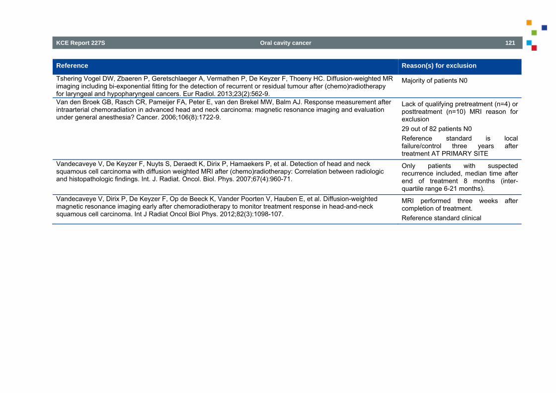
KCE Report 227S Oral cavity cancer 121
Reference Reason(s) for exclusion
Tshering Vogel DW, Zbaeren P, Geretschlaeger A, Vermathen P, De Keyzer F, Thoeny HC. Diffusion-weighted MR imaging including bi-exponential fitting for the detection of recurrent or residual tumour after (chemo)radiotherapy for laryngeal and hypopharyngeal cancers. Eur Radiol. 2013;23(2):562-9.
Majority of patients N0
Van den Broek GB, Rasch CR, Pameijer FA, Peter E, van den Brekel MW, Balm AJ. Response measurement after intraarterial chemoradiation in advanced head and neck carcinoma: magnetic resonance imaging and evaluation under general anesthesia? Cancer. 2006;106(8):1722-9.
Lack of qualifying pretreatment (n=4) or posttreatment (n=10) MRI reason for exclusion 29 out of 82 patients N0 Reference standard is local failure/control three years after treatment AT PRIMARY SITE
Vandecaveye V, De Keyzer F, Nuyts S, Deraedt K, Dirix P, Hamaekers P, et al. Detection of head and neck squamous cell carcinoma with diffusion weighted MRI after (chemo)radiotherapy: Correlation between radiologic and histopathologic findings. Int. J. Radiat. Oncol. Biol. Phys. 2007;67(4):960-71.
Only patients with suspected recurrence included, median time after end of treatment 8 months (inter-quartile range 6-21 months).
Vandecaveye V, Dirix P, De Keyzer F, Op de Beeck K, Vander Poorten V, Hauben E, et al. Diffusion-weighted magnetic resonance imaging early after chemoradiotherapy to monitor treatment response in head-and-neck squamous cell carcinoma. Int J Radiat Oncol Biol Phys. 2012;82(3):1098-107.
MRI performed three weeks after completion of treatment. Reference standard clinical
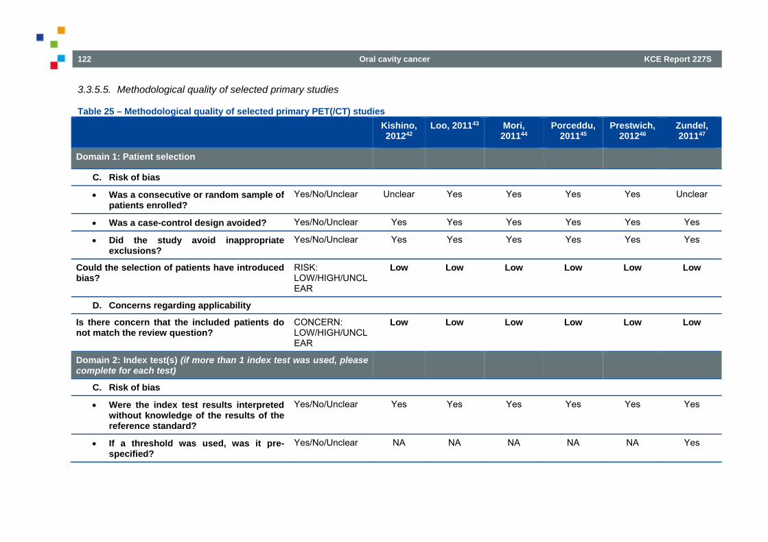
122 Oral cavity cancer KCE Report 227S
3.3.5.5. Methodological quality of selected primary studies
Table 25 – Methodological quality of selected primary PET(/CT) studies Kishino,
201242 Loo, 201143 Mori,
201144 Porceddu,
201145 Prestwich,
201246 Zundel, 201147
Domain 1: Patient selection
C. Risk of bias
Was a consecutive or random sample of patients enrolled?
Yes/No/Unclear Unclear Yes Yes Yes Yes Unclear
Was a case-control design avoided? Yes/No/Unclear Yes Yes Yes Yes Yes Yes
Did the study avoid inappropriate exclusions?
Yes/No/Unclear Yes Yes Yes Yes Yes Yes
Could the selection of patients have introduced bias?
RISK: LOW/HIGH/UNCLEAR
Low Low Low Low Low Low
D. Concerns regarding applicability
Is there concern that the included patients do not match the review question?
CONCERN: LOW/HIGH/UNCLEAR
Low Low Low Low Low Low
Domain 2: Index test(s) (if more than 1 index test was used, please complete for each test)
C. Risk of bias
Were the index test results interpreted without knowledge of the results of the reference standard?
Yes/No/Unclear Yes Yes Yes Yes Yes Yes
If a threshold was used, was it pre-specified?
Yes/No/Unclear NA NA NA NA NA Yes
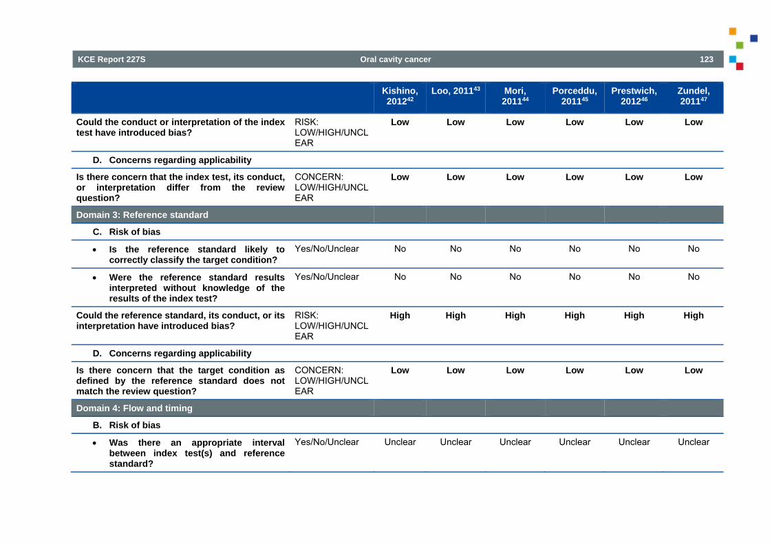
KCE Report 227S Oral cavity cancer 123
Kishino, 201242
Loo, 201143 Mori, 201144
Porceddu, 201145
Prestwich, 201246
Zundel, 201147
Could the conduct or interpretation of the index test have introduced bias?
RISK: LOW/HIGH/UNCLEAR
Low Low Low Low Low Low
D. Concerns regarding applicability
Is there concern that the index test, its conduct, or interpretation differ from the review question?
CONCERN: LOW/HIGH/UNCLEAR
Low Low Low Low Low Low
Domain 3: Reference standard
C. Risk of bias
Is the reference standard likely to correctly classify the target condition?
Yes/No/Unclear No No No No No No
Were the reference standard results interpreted without knowledge of the results of the index test?
Yes/No/Unclear No No No No No No
Could the reference standard, its conduct, or its interpretation have introduced bias?
RISK: LOW/HIGH/UNCLEAR
High High High High High High
D. Concerns regarding applicability
Is there concern that the target condition as defined by the reference standard does not match the review question?
CONCERN: LOW/HIGH/UNCLEAR
Low Low Low Low Low Low
Domain 4: Flow and timing
B. Risk of bias
Was there an appropriate interval between index test(s) and reference standard?
Yes/No/Unclear Unclear Unclear Unclear Unclear Unclear Unclear
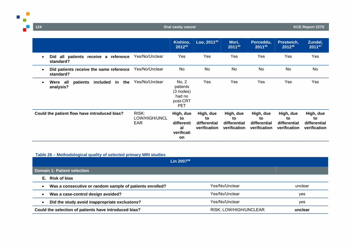
124 Oral cavity cancer KCE Report 227S
Kishino, 201242
Loo, 201143 Mori, 201144
Porceddu, 201145
Prestwich, 201246
Zundel, 201147
Did all patients receive a reference standard?
Yes/No/Unclear Yes Yes Yes Yes Yes Yes
Did patients receive the same reference standard?
Yes/No/Unclear No No No No No No
Were all patients included in the analysis?
Yes/No/Unclear No, 2 patients
(3 nodes) had no
post-CRT PET
Yes Yes Yes Yes Yes
Could the patient flow have introduced bias? RISK: LOW/HIGH/UNCLEAR
High, due to
differential
verification
High, due to
differential verification
High, due to
differential verification
High, due to
differential verification
High, due to
differential verification
High, due to
differential verification
Table 26 – Methodological quality of selected primary MRI studies Lin 200748
Domain 1: Patient selection
E. Risk of bias
Was a consecutive or random sample of patients enrolled? Yes/No/Unclear unclear
Was a case-control design avoided? Yes/No/Unclear yes
Did the study avoid inappropriate exclusions? Yes/No/Unclear yes
Could the selection of patients have introduced bias? RISK: LOW/HIGH/UNCLEAR unclear
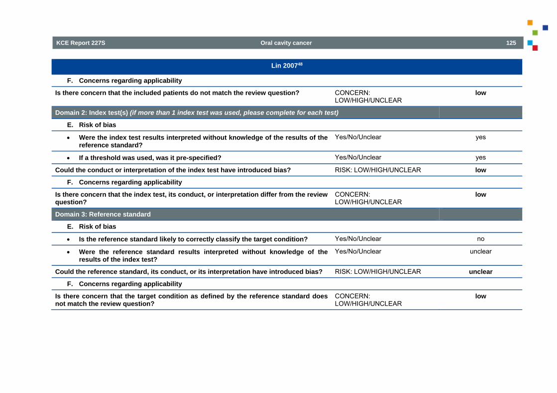
KCE Report 227S Oral cavity cancer 125
Lin 200748
F. Concerns regarding applicability
Is there concern that the included patients do not match the review question? CONCERN: LOW/HIGH/UNCLEAR
low
Domain 2: Index test(s) (if more than 1 index test was used, please complete for each test)
E. Risk of bias
Were the index test results interpreted without knowledge of the results of the reference standard?
Yes/No/Unclear yes
If a threshold was used, was it pre-specified? Yes/No/Unclear yes
Could the conduct or interpretation of the index test have introduced bias? RISK: LOW/HIGH/UNCLEAR low
F. Concerns regarding applicability
Is there concern that the index test, its conduct, or interpretation differ from the review question?
CONCERN: LOW/HIGH/UNCLEAR
low
Domain 3: Reference standard
E. Risk of bias
Is the reference standard likely to correctly classify the target condition? Yes/No/Unclear no
Were the reference standard results interpreted without knowledge of the results of the index test?
Yes/No/Unclear unclear
Could the reference standard, its conduct, or its interpretation have introduced bias? RISK: LOW/HIGH/UNCLEAR unclear
F. Concerns regarding applicability
Is there concern that the target condition as defined by the reference standard does not match the review question?
CONCERN: LOW/HIGH/UNCLEAR
low
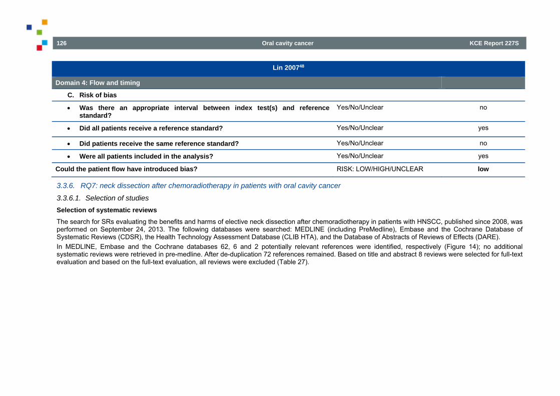
126 Oral cavity cancer KCE Report 227S
Lin 200748
Domain 4: Flow and timing
C. Risk of bias
Was there an appropriate interval between index test(s) and reference standard?
Yes/No/Unclear no
Did all patients receive a reference standard? Yes/No/Unclear yes
Did patients receive the same reference standard? Yes/No/Unclear no
Were all patients included in the analysis? Yes/No/Unclear yes
Could the patient flow have introduced bias? RISK: LOW/HIGH/UNCLEAR low
3.3.6. RQ7: neck dissection after chemoradiotherapy in patients with oral cavity cancer
3.3.6.1. Selection of studies Selection of systematic reviews The search for SRs evaluating the benefits and harms of elective neck dissection after chemoradiotherapy in patients with HNSCC, published since 2008, was performed on September 24, 2013. The following databases were searched: MEDLINE (including PreMedline), Embase and the Cochrane Database of Systematic Reviews (CDSR), the Health Technology Assessment Database (CLIB HTA), and the Database of Abstracts of Reviews of Effects (DARE). In MEDLINE, Embase and the Cochrane databases 62, 6 and 2 potentially relevant references were identified, respectively (Figure 14); no additional systematic reviews were retrieved in pre-medline. After de-duplication 72 references remained. Based on title and abstract 8 reviews were selected for full-text evaluation and based on the full-text evaluation, all reviews were excluded (Table 27).
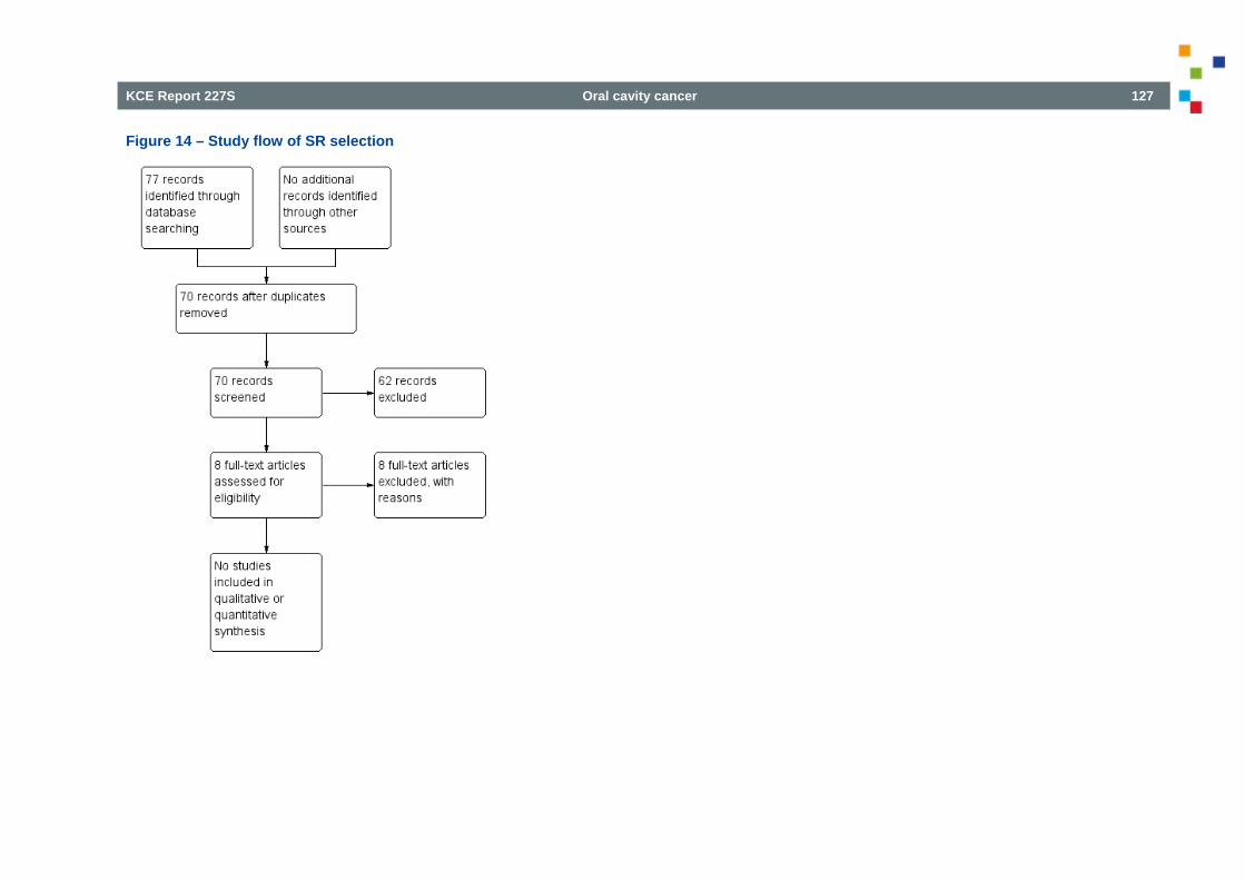
KCE Report 227S Oral cavity cancer 127
Figure 14 – Study flow of SR selection
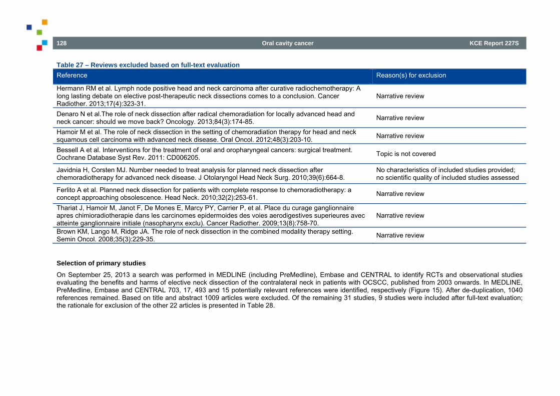
128 Oral cavity cancer KCE Report 227S
Table 27 – Reviews excluded based on full-text evaluation Reference Reason(s) for exclusion
Hermann RM et al. Lymph node positive head and neck carcinoma after curative radiochemotherapy: A long lasting debate on elective post-therapeutic neck dissections comes to a conclusion. Cancer Radiother. 2013;17(4):323-31.
Narrative review
Denaro N et al.The role of neck dissection after radical chemoradiation for locally advanced head and neck cancer: should we move back? Oncology. 2013;84(3):174-85. Narrative review
Hamoir M et al. The role of neck dissection in the setting of chemoradiation therapy for head and neck squamous cell carcinoma with advanced neck disease. Oral Oncol. 2012;48(3):203-10. Narrative review
Bessell A et al. Interventions for the treatment of oral and oropharyngeal cancers: surgical treatment. Cochrane Database Syst Rev. 2011: CD006205. Topic is not covered
Javidnia H, Corsten MJ. Number needed to treat analysis for planned neck dissection after chemoradiotherapy for advanced neck disease. J Otolaryngol Head Neck Surg. 2010;39(6):664-8.
No characteristics of included studies provided; no scientific quality of included studies assessed
Ferlito A et al. Planned neck dissection for patients with complete response to chemoradiotherapy: a concept approaching obsolescence. Head Neck. 2010;32(2):253-61. Narrative review
Thariat J, Hamoir M, Janot F, De Mones E, Marcy PY, Carrier P, et al. Place du curage ganglionnaire apres chimioradiotherapie dans les carcinomes epidermoides des voies aerodigestives superieures avec atteinte ganglionnaire initiale (nasopharynx exclu). Cancer Radiother. 2009;13(8):758-70.
Narrative review
Brown KM, Lango M, Ridge JA. The role of neck dissection in the combined modality therapy setting. Semin Oncol. 2008;35(3):229-35. Narrative review
Selection of primary studies On September 25, 2013 a search was performed in MEDLINE (including PreMedline), Embase and CENTRAL to identify RCTs and observational studies evaluating the benefits and harms of elective neck dissection of the contralateral neck in patients with OCSCC, published from 2003 onwards. In MEDLINE, PreMedline, Embase and CENTRAL 703, 17, 493 and 15 potentially relevant references were identified, respectively (Figure 15). After de-duplication, 1040 references remained. Based on title and abstract 1009 articles were excluded. Of the remaining 31 studies, 9 studies were included after full-text evaluation; the rationale for exclusion of the other 22 articles is presented in Table 28.
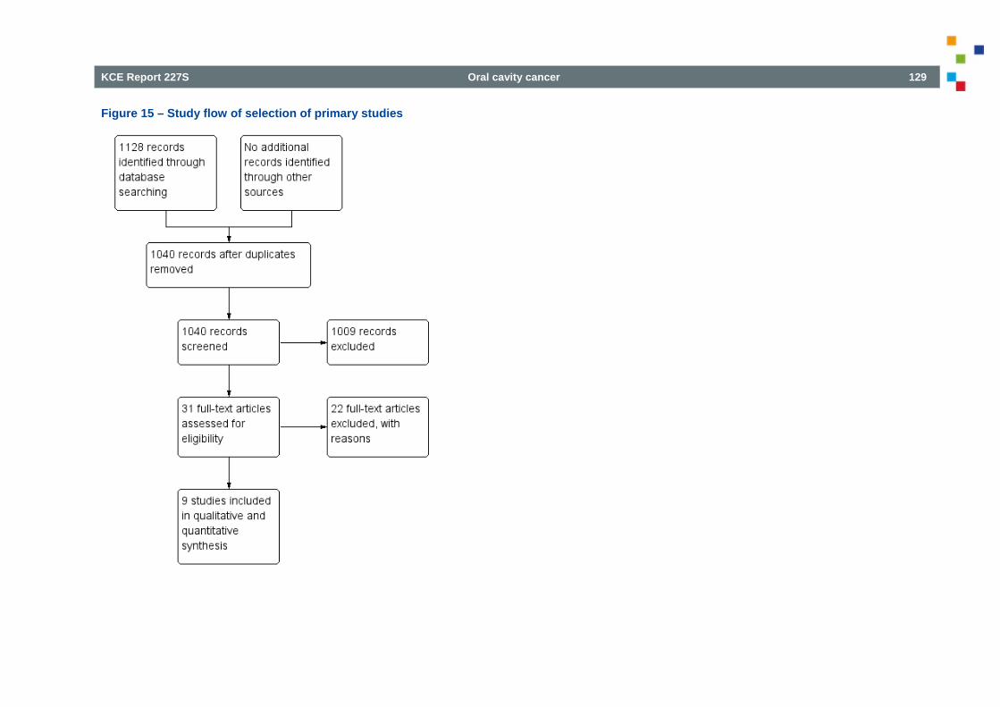
KCE Report 227S Oral cavity cancer 129
Figure 15 – Study flow of selection of primary studies
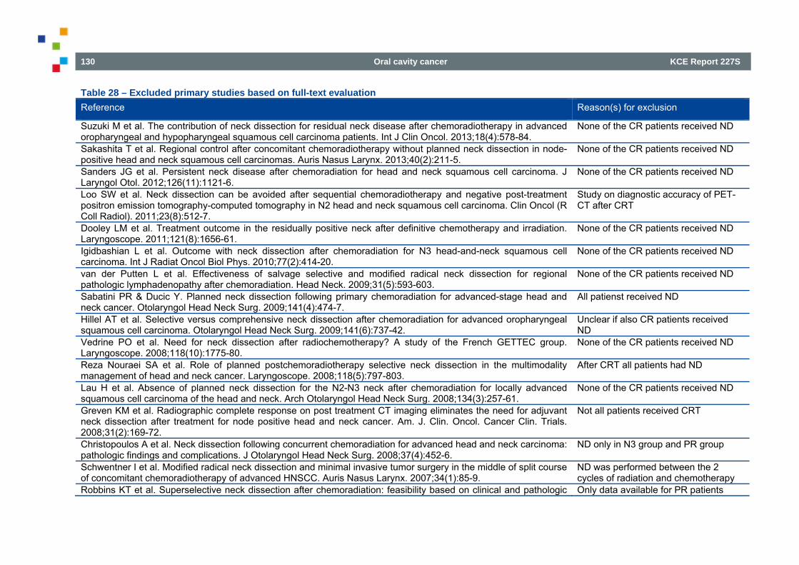
130 Oral cavity cancer KCE Report 227S
Table 28 – Excluded primary studies based on full-text evaluation Reference Reason(s) for exclusion
Suzuki M et al. The contribution of neck dissection for residual neck disease after chemoradiotherapy in advanced oropharyngeal and hypopharyngeal squamous cell carcinoma patients. Int J Clin Oncol. 2013;18(4):578-84.
None of the CR patients received ND
Sakashita T et al. Regional control after concomitant chemoradiotherapy without planned neck dissection in node-positive head and neck squamous cell carcinomas. Auris Nasus Larynx. 2013;40(2):211-5.
None of the CR patients received ND
Sanders JG et al. Persistent neck disease after chemoradiation for head and neck squamous cell carcinoma. J Laryngol Otol. 2012;126(11):1121-6.
None of the CR patients received ND
Loo SW et al. Neck dissection can be avoided after sequential chemoradiotherapy and negative post-treatment positron emission tomography-computed tomography in N2 head and neck squamous cell carcinoma. Clin Oncol (R Coll Radiol). 2011;23(8):512-7.
Study on diagnostic accuracy of PET-CT after CRT
Dooley LM et al. Treatment outcome in the residually positive neck after definitive chemotherapy and irradiation. Laryngoscope. 2011;121(8):1656-61.
None of the CR patients received ND
Igidbashian L et al. Outcome with neck dissection after chemoradiation for N3 head-and-neck squamous cell carcinoma. Int J Radiat Oncol Biol Phys. 2010;77(2):414-20.
None of the CR patients received ND
van der Putten L et al. Effectiveness of salvage selective and modified radical neck dissection for regional pathologic lymphadenopathy after chemoradiation. Head Neck. 2009;31(5):593-603.
None of the CR patients received ND
Sabatini PR & Ducic Y. Planned neck dissection following primary chemoradiation for advanced-stage head and neck cancer. Otolaryngol Head Neck Surg. 2009;141(4):474-7.
All patienst received ND
Hillel AT et al. Selective versus comprehensive neck dissection after chemoradiation for advanced oropharyngeal squamous cell carcinoma. Otolaryngol Head Neck Surg. 2009;141(6):737-42.
Unclear if also CR patients received ND
Vedrine PO et al. Need for neck dissection after radiochemotherapy? A study of the French GETTEC group. Laryngoscope. 2008;118(10):1775-80.
None of the CR patients received ND
Reza Nouraei SA et al. Role of planned postchemoradiotherapy selective neck dissection in the multimodality management of head and neck cancer. Laryngoscope. 2008;118(5):797-803.
After CRT all patients had ND
Lau H et al. Absence of planned neck dissection for the N2-N3 neck after chemoradiation for locally advanced squamous cell carcinoma of the head and neck. Arch Otolaryngol Head Neck Surg. 2008;134(3):257-61.
None of the CR patients received ND
Greven KM et al. Radiographic complete response on post treatment CT imaging eliminates the need for adjuvant neck dissection after treatment for node positive head and neck cancer. Am. J. Clin. Oncol. Cancer Clin. Trials. 2008;31(2):169-72.
Not all patients received CRT
Christopoulos A et al. Neck dissection following concurrent chemoradiation for advanced head and neck carcinoma: pathologic findings and complications. J Otolaryngol Head Neck Surg. 2008;37(4):452-6.
ND only in N3 group and PR group
Schwentner I et al. Modified radical neck dissection and minimal invasive tumor surgery in the middle of split course of concomitant chemoradiotherapy of advanced HNSCC. Auris Nasus Larynx. 2007;34(1):85-9.
ND was performed between the 2 cycles of radiation and chemotherapy
Robbins KT et al. Superselective neck dissection after chemoradiation: feasibility based on clinical and pathologic Only data available for PR patients
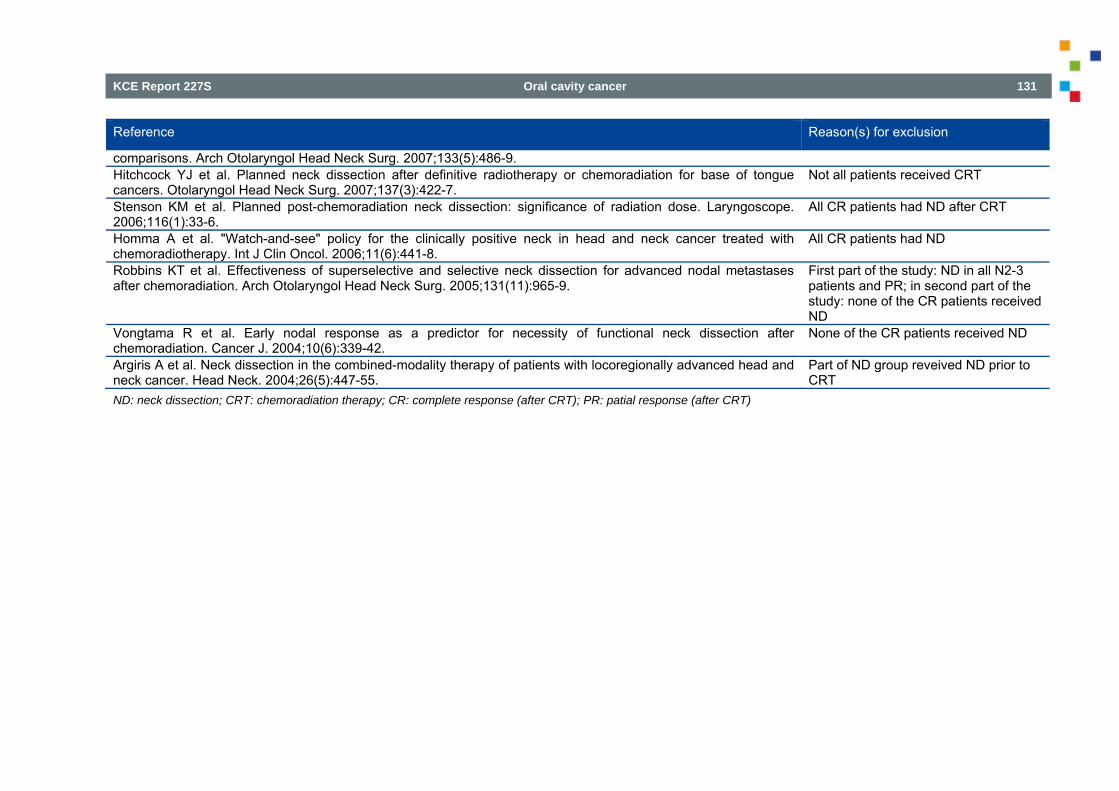
KCE Report 227S Oral cavity cancer 131
Reference Reason(s) for exclusion
comparisons. Arch Otolaryngol Head Neck Surg. 2007;133(5):486-9. Hitchcock YJ et al. Planned neck dissection after definitive radiotherapy or chemoradiation for base of tongue cancers. Otolaryngol Head Neck Surg. 2007;137(3):422-7.
Not all patients received CRT
Stenson KM et al. Planned post-chemoradiation neck dissection: significance of radiation dose. Laryngoscope. 2006;116(1):33-6.
All CR patients had ND after CRT
Homma A et al. "Watch-and-see" policy for the clinically positive neck in head and neck cancer treated with chemoradiotherapy. Int J Clin Oncol. 2006;11(6):441-8.
All CR patients had ND
Robbins KT et al. Effectiveness of superselective and selective neck dissection for advanced nodal metastases after chemoradiation. Arch Otolaryngol Head Neck Surg. 2005;131(11):965-9.
First part of the study: ND in all N2-3 patients and PR; in second part of the study: none of the CR patients received ND
Vongtama R et al. Early nodal response as a predictor for necessity of functional neck dissection after chemoradiation. Cancer J. 2004;10(6):339-42.
None of the CR patients received ND
Argiris A et al. Neck dissection in the combined-modality therapy of patients with locoregionally advanced head and neck cancer. Head Neck. 2004;26(5):447-55.
Part of ND group reveived ND prior to CRT
ND: neck dissection; CRT: chemoradiation therapy; CR: complete response (after CRT); PR: patial response (after CRT)
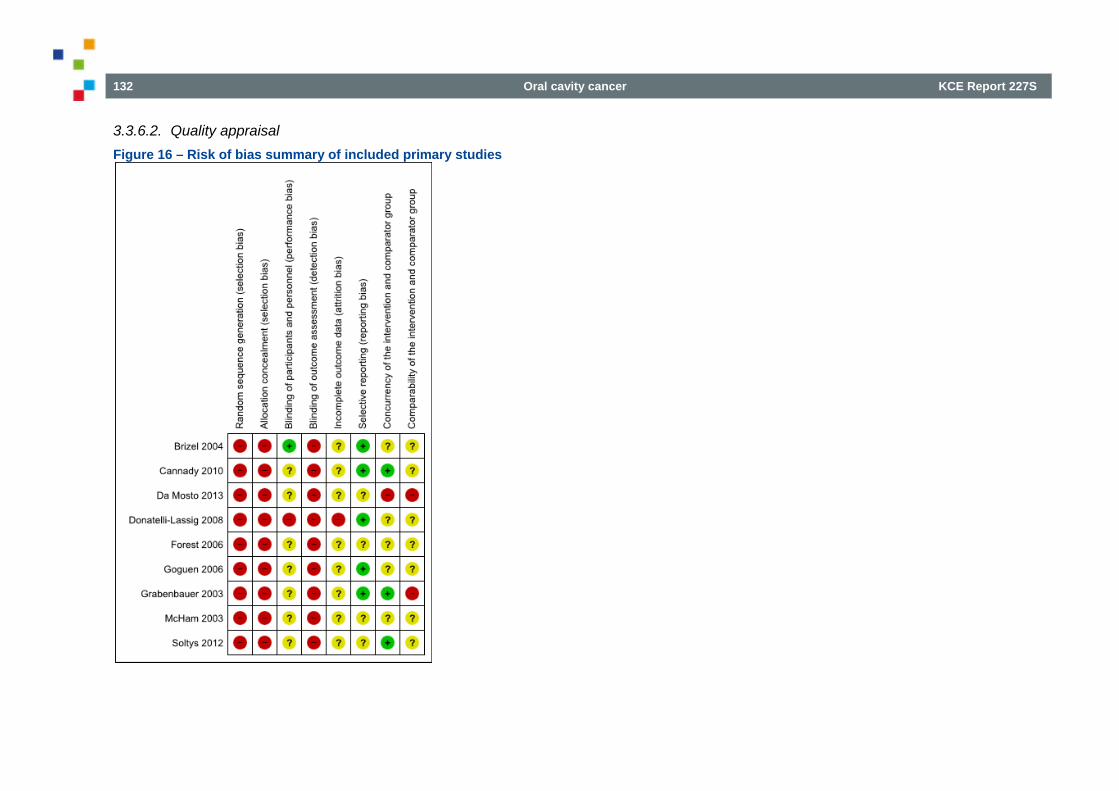
132 Oral cavity cancer KCE Report 227S
3.3.6.2. Quality appraisal Figure 16 – Risk of bias summary of included primary studies
KCE Report 227S Oral cavity cancer 133
3.3.7. RQ8: IMRT for patients with locally advanced HNSCC
3.3.7.1. Selection of studies Selection of systematic reviews On August 12, 2013 a search was performed to identify SRs assessing the clinical effectiveness of IMRT for adult patients (≥18 years of age) with locally advanced HNSCC (stage 3 and 4); the search in Cochrane was done on November 4, 2013. MEDLINE and Embase were searched from January 2008 onwards. In total, 52 potential relevant references were identified after deduplication (Figure 17). Based on title and abstract 42 references were excluded. Three reviews were included (49;49 50; 51) (Table 29) and seven were excluded with reason (Table 30). As the review of 49 was most recent and complete, only the results of this review are discussed. An additional search in the Cochrane Library did not result in the inclusion of any further systematic reviews (Figure 18).
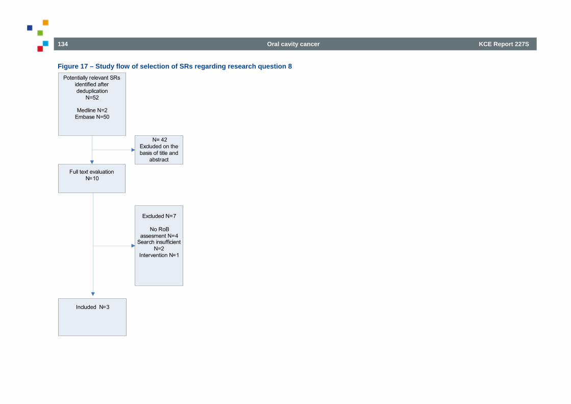
134 Oral cavity cancer KCE Report 227S
Figure 17 – Study flow of selection of SRs regarding research question 8 Potentially relevant SRs
identified after deduplication
N=52
Medline N=2Embase N=50
Full text evaluationN=10
N= 42Excluded on the basis of title and
abstract
Included N=3
Excluded N=7
No RoB assesment N=4
Search insufficient N=2
Intervention N=1
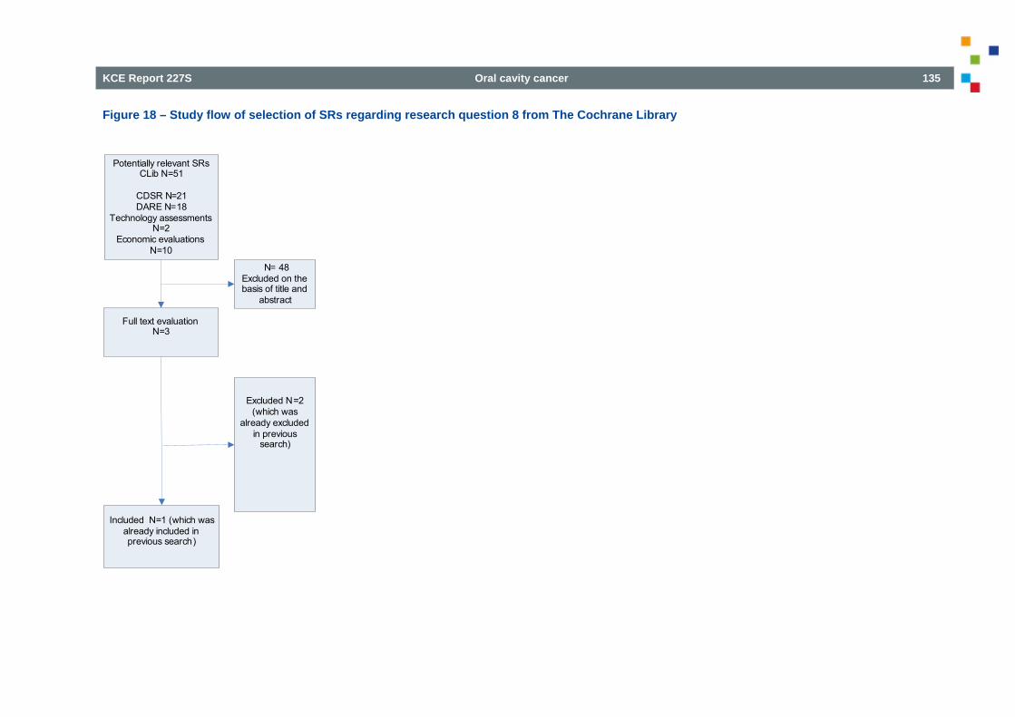
KCE Report 227S Oral cavity cancer 135
Figure 18 – Study flow of selection of SRs regarding research question 8 from The Cochrane Library
Potentially relevant SRs CLib N=51
CDSR N=21DARE N=18
Technology assessments N=2
Economic evaluationsN=10
Full text evaluationN=3
N= 48Excluded on the basis of title and
abstract
Included N=1 (which was already included in previous search)
Excluded N=2 (which was
already excluded in previous
search)
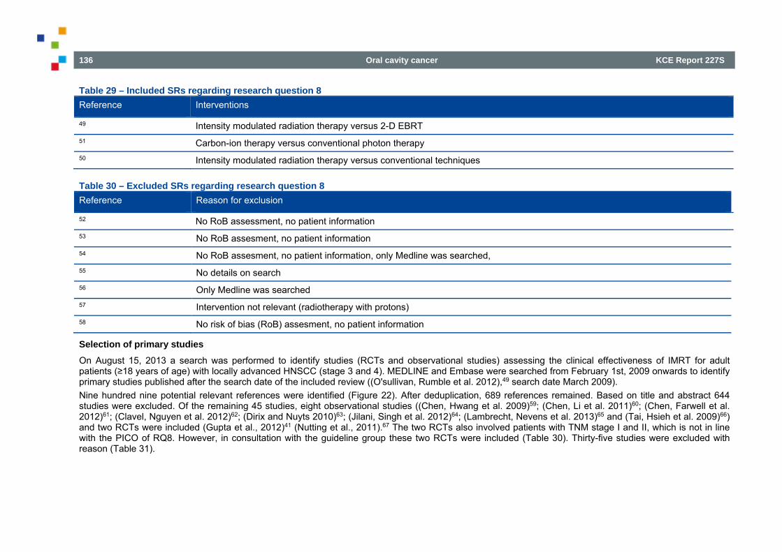
136 Oral cavity cancer KCE Report 227S
Table 29 – Included SRs regarding research question 8 Reference Interventions
49 Intensity modulated radiation therapy versus 2-D EBRT 51 Carbon-ion therapy versus conventional photon therapy 50 Intensity modulated radiation therapy versus conventional techniques
Table 30 – Excluded SRs regarding research question 8 Reference Reason for exclusion
52 No RoB assessment, no patient information 53 No RoB assesment, no patient information 54 No RoB assesment, no patient information, only Medline was searched, 55 No details on search 56 Only Medline was searched 57 Intervention not relevant (radiotherapy with protons) 58 No risk of bias (RoB) assesment, no patient information
Selection of primary studies On August 15, 2013 a search was performed to identify studies (RCTs and observational studies) assessing the clinical effectiveness of IMRT for adult patients (≥18 years of age) with locally advanced HNSCC (stage 3 and 4). MEDLINE and Embase were searched from February 1st, 2009 onwards to identify primary studies published after the search date of the included review ((O'sullivan, Rumble et al. 2012),49 search date March 2009). Nine hundred nine potential relevant references were identified (Figure 22). After deduplication, 689 references remained. Based on title and abstract 644 studies were excluded. Of the remaining 45 studies, eight observational studies ((Chen, Hwang et al. 2009)59; (Chen, Li et al. 2011)60; (Chen, Farwell et al. 2012)61; (Clavel, Nguyen et al. 2012)62; (Dirix and Nuyts 2010)63; (Jilani, Singh et al. 2012)64; (Lambrecht, Nevens et al. 2013)65 and (Tai, Hsieh et al. 2009)66) and two RCTs were included (Gupta et al., 2012)41 (Nutting et al., 2011).67 The two RCTs also involved patients with TNM stage I and II, which is not in line with the PICO of RQ8. However, in consultation with the guideline group these two RCTs were included (Table 30). Thirty-five studies were excluded with reason (Table 31).
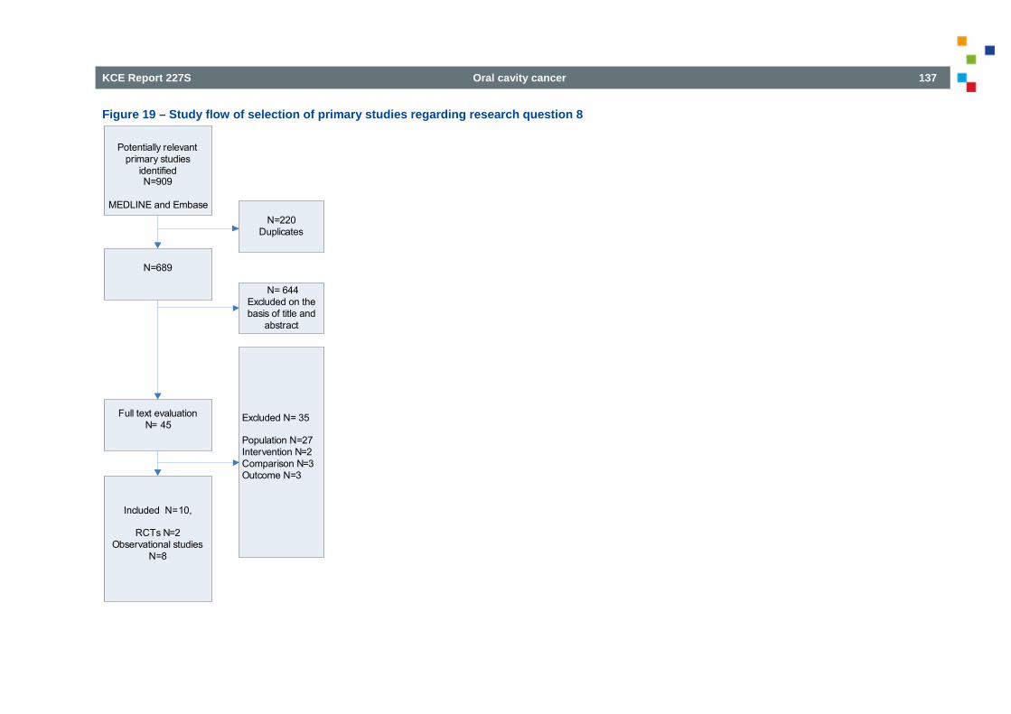
KCE Report 227S Oral cavity cancer 137
Figure 19 – Study flow of selection of primary studies regarding research question 8
Potentially relevant primary studies
identifiedN=909
MEDLINE and Embase
N=689
Full text evaluationN= 45
N=220Duplicates
N= 644Excluded on the basis of title and
abstract
Included N=10,
RCTs N=2Observational studies
N=8
Excluded N= 35
Population N=27Intervention N=2Comparison N=3 Outcome N=3
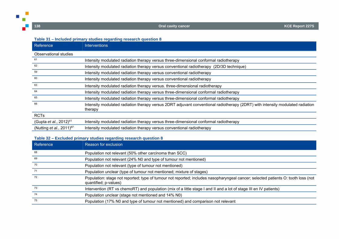
138 Oral cavity cancer KCE Report 227S
Table 31 – Included primary studies regarding research question 8 Reference Interventions
Observational studies 61 Intensity modulated radiation therapy versus three-dimensional conformal radiotherapy 62 Intensity modulated radiation therapy versus conventional radiotherapy (2D/3D technique) 59 Intensity modulated radiation therapy versus conventional radiotherapy 60 Intensity modulated radiation therapy versus conventional radiotherapy 63 Intensity modulated radiation therapy versus. three-dimensional radiotherapy 64 Intensity modulated radiation therapy versus three-dimensional conformal radiotherapy 65 Intensity modulated radiation therapy versus three-dimensional conformal radiotherapy 66 Intensity modulated radiation therapy versus 2DRT adjuvant conventional radiotherapy (2DRT) with intensity modulated radiation
therapy RCTs (Gupta et al., 2012)41 Intensity modulated radiation therapy versus three-dimensional conformal radiotherapy (Nutting et al., 2011)67 Intensity modulated radiation therapy versus conventional radiotherapy
Table 32 – Excluded primary studies regarding research question 8 Reference Reason for exclusion
68 Population not relevant (50% other carcinoma than SCC) 69 Population not relevant (24% N0 and type of tumour not mentioned) 70 Population not relevant (type of tumour not mentioned) 71 Population unclear (type of tumour not mentioned; mixture of stages) 72 Population: stage not reported; type of tumour not reported; includes nasopharyngeal cancer; selected patients O: tooth loss (not
quantified; p-values) 73 Intervention (RT vs chemoRT) and population (mix of a little stage I and II and a lot of stage III en IV patients) 74 Population unclear (stage not mentioned and 14% N0) 75 Population (17% N0 and type of tumour not mentioned) and comparison not relevant
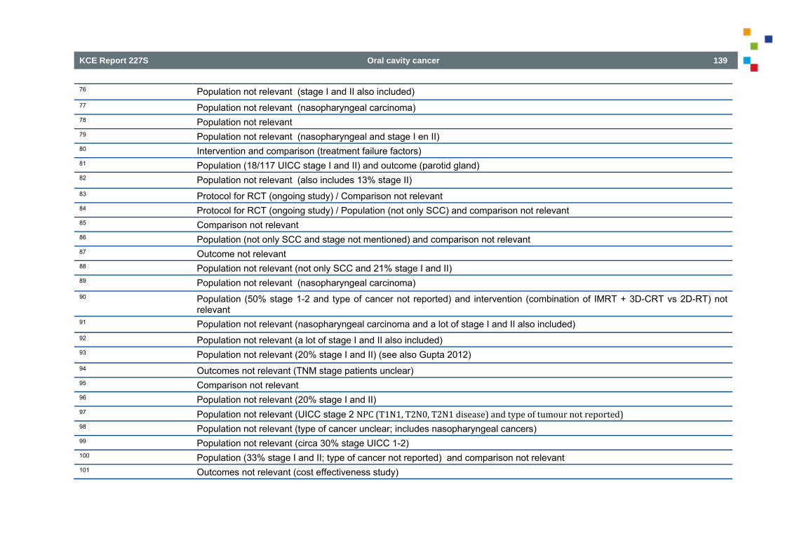
KCE Report 227S Oral cavity cancer 139
76 Population not relevant (stage I and II also included) 77 Population not relevant (nasopharyngeal carcinoma) 78 Population not relevant 79 Population not relevant (nasopharyngeal and stage I en II) 80 Intervention and comparison (treatment failure factors) 81 Population (18/117 UICC stage I and II) and outcome (parotid gland) 82 Population not relevant (also includes 13% stage II) 83 Protocol for RCT (ongoing study) / Comparison not relevant 84 Protocol for RCT (ongoing study) / Population (not only SCC) and comparison not relevant 85 Comparison not relevant 86 Population (not only SCC and stage not mentioned) and comparison not relevant 87 Outcome not relevant 88 Population not relevant (not only SCC and 21% stage I and II) 89 Population not relevant (nasopharyngeal carcinoma) 90 Population (50% stage 1-2 and type of cancer not reported) and intervention (combination of IMRT + 3D-CRT vs 2D-RT) not
relevant 91 Population not relevant (nasopharyngeal carcinoma and a lot of stage I and II also included) 92 Population not relevant (a lot of stage I and II also included) 93 Population not relevant (20% stage I and II) (see also Gupta 2012) 94 Outcomes not relevant (TNM stage patients unclear) 95 Comparison not relevant 96 Population not relevant (20% stage I and II) 97 Population not relevant (UICC stage 2 NPC(T1N1,T2N0,T2N1disease)andtypeoftumournotreported) 98 Population not relevant (type of cancer unclear; includes nasopharyngeal cancers) 99 Population not relevant (circa 30% stage UICC 1-2) 100 Population (33% stage I and II; type of cancer not reported) and comparison not relevant 101 Outcomes not relevant (cost effectiveness study)
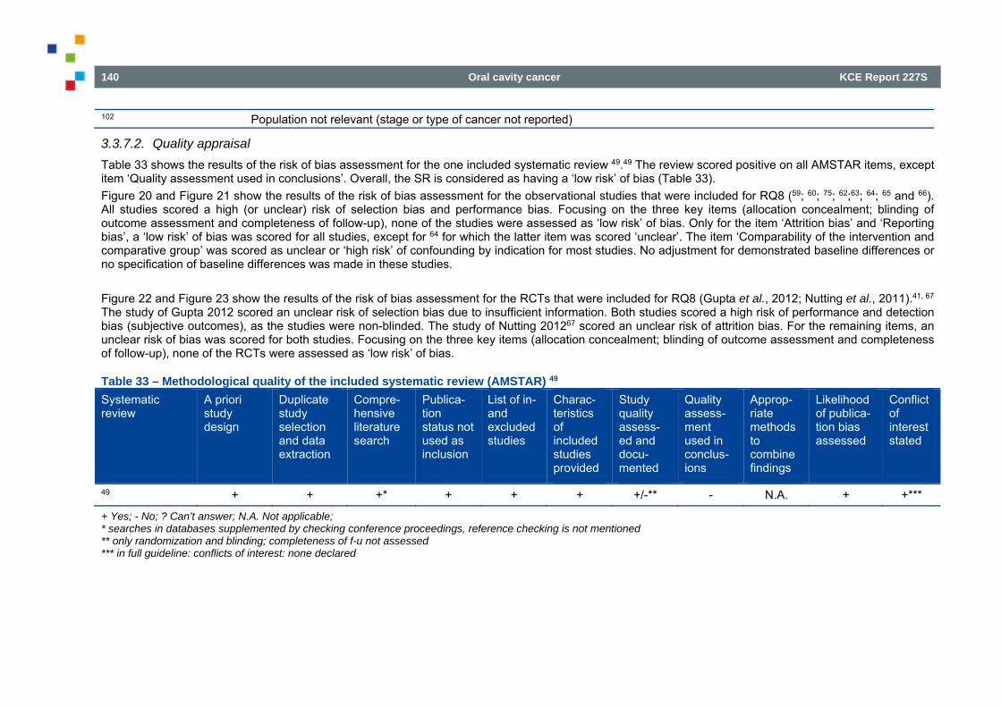
140 Oral cavity cancer KCE Report 227S
102 Population not relevant (stage or type of cancer not reported)
3.3.7.2. Quality appraisal Table 33 shows the results of the risk of bias assessment for the one included systematic review 49.49 The review scored positive on all AMSTAR items, except item ‘Quality assessment used in conclusions’. Overall, the SR is considered as having a ‘low risk’ of bias (Table 33). Figure 20 and Figure 21 show the results of the risk of bias assessment for the observational studies that were included for RQ8 (59; 60; 75; 62;63; 64; 65 and 66). All studies scored a high (or unclear) risk of selection bias and performance bias. Focusing on the three key items (allocation concealment; blinding of outcome assessment and completeness of follow-up), none of the studies were assessed as ‘low risk’ of bias. Only for the item ‘Attrition bias’ and ‘Reporting bias’, a ‘low risk’ of bias was scored for all studies, except for 64 for which the latter item was scored ‘unclear’. The item ‘Comparability of the intervention and comparative group’ was scored as unclear or ‘high risk’ of confounding by indication for most studies. No adjustment for demonstrated baseline differences or no specification of baseline differences was made in these studies. Figure 22 and Figure 23 show the results of the risk of bias assessment for the RCTs that were included for RQ8 (Gupta et al., 2012; Nutting et al., 2011).41, 67 The study of Gupta 2012 scored an unclear risk of selection bias due to insufficient information. Both studies scored a high risk of performance and detection bias (subjective outcomes), as the studies were non-blinded. The study of Nutting 201267 scored an unclear risk of attrition bias. For the remaining items, an unclear risk of bias was scored for both studies. Focusing on the three key items (allocation concealment; blinding of outcome assessment and completeness of follow-up), none of the RCTs were assessed as ‘low risk’ of bias.
Table 33 – Methodological quality of the included systematic review (AMSTAR) 49 Systematic review
A priori study design
Duplicate study selection and data extraction
Compre-hensive literature search
Publica-tion status not used as inclusion
List of in- and excluded studies
Charac-teristics of included studies provided
Study quality assess-ed and docu-mented
Quality assess-ment used in conclus-ions
Approp-riate methods to combine findings
Likelihood of publica-tion bias assessed
Conflict of interest stated
49 + + +* + + + +/-** - N.A. + +***
+ Yes; - No; ? Can’t answer; N.A. Not applicable; * searches in databases supplemented by checking conference proceedings, reference checking is not mentioned ** only randomization and blinding; completeness of f-u not assessed *** in full guideline: conflicts of interest: none declared
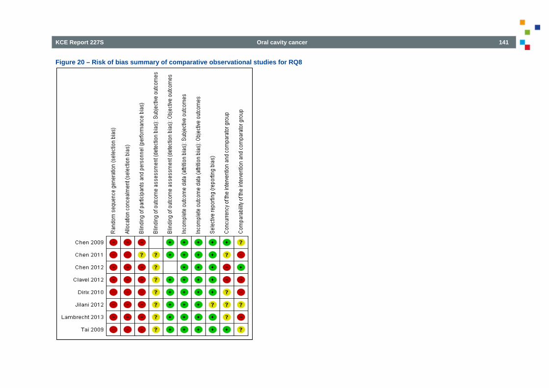
KCE Report 227S Oral cavity cancer 141
Figure 20 – Risk of bias summary of comparative observational studies for RQ8
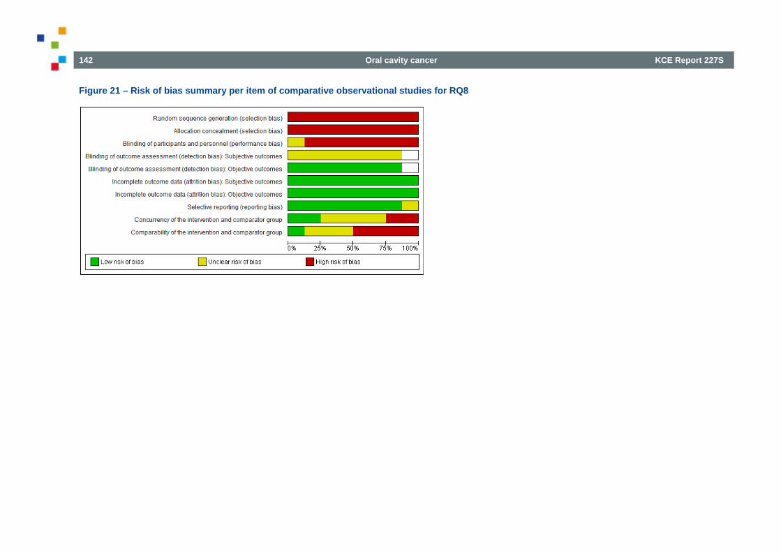
142 Oral cavity cancer KCE Report 227S
Figure 21 – Risk of bias summary per item of comparative observational studies for RQ8
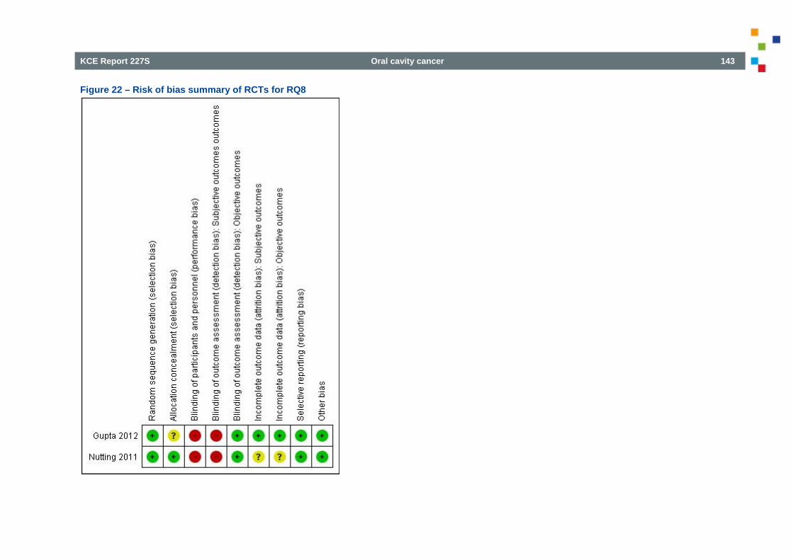
KCE Report 227S Oral cavity cancer 143
Figure 22 – Risk of bias summary of RCTs for RQ8

144 Oral cavity cancer KCE Report 227S
Figure 23 – Risk of bias summary per item of RCTs for RQ8
3.3.8. RQ9: induction chemotherapy in patients with HNSCC
3.3.8.1. Selection of studies Selection of systematic reviews On August 12, 2013 a search was performed to identify SRs comparing the effect of induction chemotherapy before locoregional therapy (i.e. RT, CRT or surgery) versus no induction chemotherapy (but identical locoregional therapy) in adult patients (≥18 years of age) diagnosed with stage 3 and 4 HNSCC (research question 9). MEDLINE and Embase were searched from January 2008 onwards. The Cochrane Library (Cochrane Database of Systematic Reviews, DARE and HTA database) was searched on November 4, 2013. In addition, the review lists of the Cochrane Oral Health Group (COHG) and the Cochrane Ear Nose Throat Group (ENT) were browsed for relevant reviews. In total, 529 potentially relevant references were identified after deduplication (Figure 24). Based on title and abstract 525 references were excluded. Two reviews were included (Furness et al., 2011)103; (Ma et al., 2012) (Table 34) and two were excluded with reason (Table 35). The reviews of Chen (Chen et al., 2011)104) and Ma (Ma et al., 2012)105 include the population as indicated by KCE. Because the most recent and complete review of Ma (Ma et al., 2012)105 includes all RCTs that were included in Chen (Chen et al., 2011), only the results of the review of (Ma et al., 2012)105 will be discussed. One review had smaller inclusion criteria regarding the study population (only oral cavity and oropharyngeal cancer) than indicated by KCE (Furness et al., 2011),103 but the results will be discussed as well, because only their searches attempted to identify all relevant trials irrespective of language.
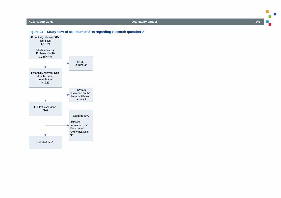
KCE Report 227S Oral cavity cancer 145
Figure 24 – Study flow of selection of SRs regarding research question 9
Potentially relevant SRs identified after deduplication
N=529
Full text evaluationN=4
N= 525Excluded on the basis of title and
abstract
Included N=2
Excluded N=2
Different population N=1More recent review available N=1
Potentially relevant SRs identified N= 746
Medline N=317Embase N=419
CLIB N=10
N= 217Duplicates
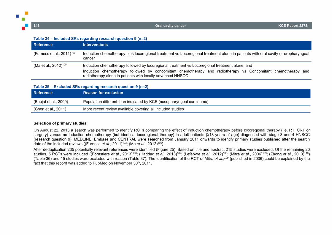
146 Oral cavity cancer KCE Report 227S
Table 34 – Included SRs regarding research question 9 (n=2) Reference Interventions
(Furness et al., 2011)103 Induction chemotherapy plus locoregional treatment vs Locoregional treatment alone in patients with oral cavity or oropharyngeal cancer
(Ma et al., 2012)105 Induction chemotherapy followed by locoregional treatment vs Locoregional treatment alone; and Induction chemotherapy followed by concomitant chemotherapy and radiotherapy vs Concomitant chemotherapy and radiotherapy alone in patients with locally advanced HNSCC
Table 35 – Excluded SRs regarding research question 9 (n=2) Reference Reason for exclusion
(Baujat et al., 2009) Population different than indicated by KCE (nasopharyngeal carcinoma)
(Chen et al., 2011) More recent review available covering all included studies
Selection of primary studies On August 22, 2013 a search was performed to identify RCTs comparing the effect of induction chemotherapy before locoregional therapy (i.e. RT, CRT or surgery) versus no induction chemotherapy (but identical locoregional therapy) in adult patients (≥18 years of age) diagnosed with stage 3 and 4 HNSCC (research question 9). MEDLINE, Embase and CENTRAL were searched from January 2011 onwards to identify primary studies published after the search date of the included reviews ((Furness et al., 2011)103; (Ma et al., 2012)105). After deduplication 235 potentially relevant references were identified (Figure 25). Based on title and abstract 215 studies were excluded. Of the remaining 20 studies, 5 RCTs were included ((Forastiere et al., 2013)106; (Haddad et al., 2013)107; (Lefebvre et al., 2012)108; (Mitra et al., 2006)109; (Zhong et al., 2013)110) (Table 36) and 15 studies were excluded with reason (Table 37). The identification of the RCT of Mitra et al.,109 (published in 2006) could be explained by the fact that this record was added to PubMed on November 30th, 2011.
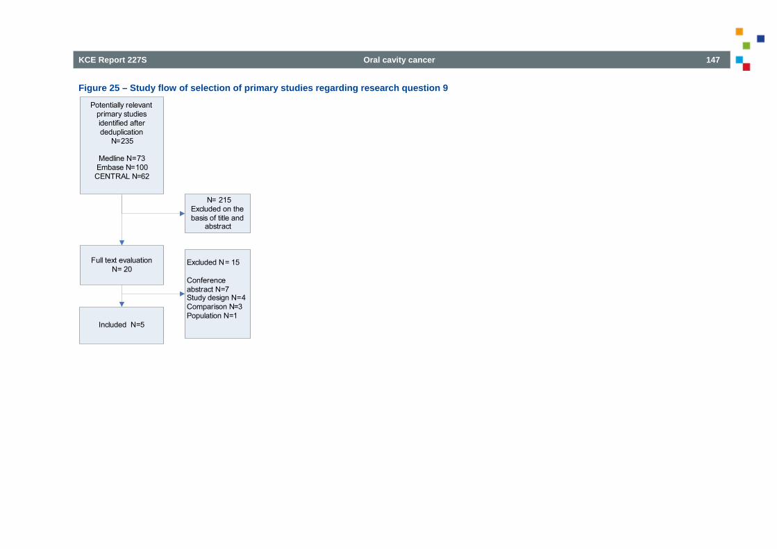
KCE Report 227S Oral cavity cancer 147
Figure 25 – Study flow of selection of primary studies regarding research question 9
Potentially relevant primary studies identified after deduplication
N=235
Medline N=73Embase N=100CENTRAL N=62
Full text evaluationN= 20
N= 215Excluded on the basis of title and
abstract
Included N=5
Excluded N= 15
Conference abstract N=7Study design N=4Comparison N=3Population N=1
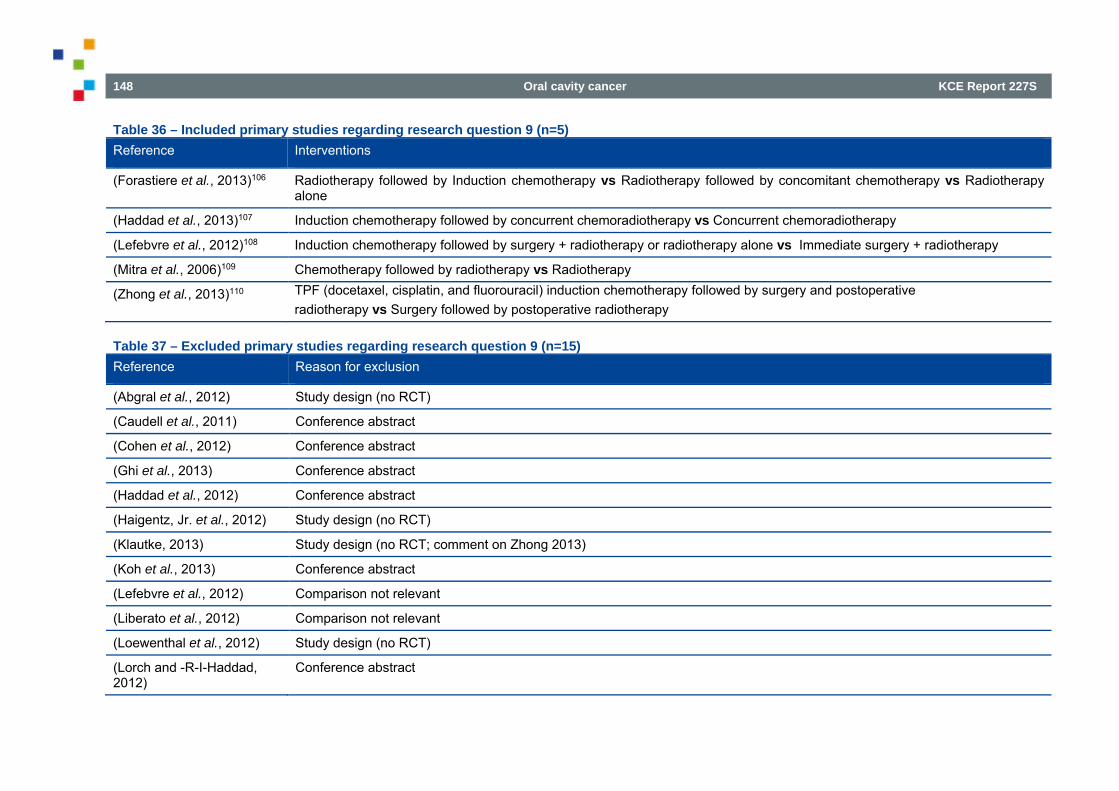
148 Oral cavity cancer KCE Report 227S
Table 36 – Included primary studies regarding research question 9 (n=5) Reference Interventions
(Forastiere et al., 2013)106 Radiotherapy followed by Induction chemotherapy vs Radiotherapy followed by concomitant chemotherapy vs Radiotherapy alone
(Haddad et al., 2013)107 Induction chemotherapy followed by concurrent chemoradiotherapy vs Concurrent chemoradiotherapy
(Lefebvre et al., 2012)108 Induction chemotherapy followed by surgery + radiotherapy or radiotherapy alone vs Immediate surgery + radiotherapy
(Mitra et al., 2006)109 Chemotherapy followed by radiotherapy vs Radiotherapy
(Zhong et al., 2013)110 TPF (docetaxel, cisplatin, and fluorouracil) induction chemotherapy followed by surgery and postoperative radiotherapy vs Surgery followed by postoperative radiotherapy
Table 37 – Excluded primary studies regarding research question 9 (n=15) Reference Reason for exclusion
(Abgral et al., 2012) Study design (no RCT)
(Caudell et al., 2011) Conference abstract
(Cohen et al., 2012) Conference abstract
(Ghi et al., 2013) Conference abstract
(Haddad et al., 2012) Conference abstract
(Haigentz, Jr. et al., 2012) Study design (no RCT)
(Klautke, 2013) Study design (no RCT; comment on Zhong 2013)
(Koh et al., 2013) Conference abstract
(Lefebvre et al., 2012) Comparison not relevant
(Liberato et al., 2012) Comparison not relevant
(Loewenthal et al., 2012) Study design (no RCT)
(Lorch and -R-I-Haddad, 2012)
Conference abstract
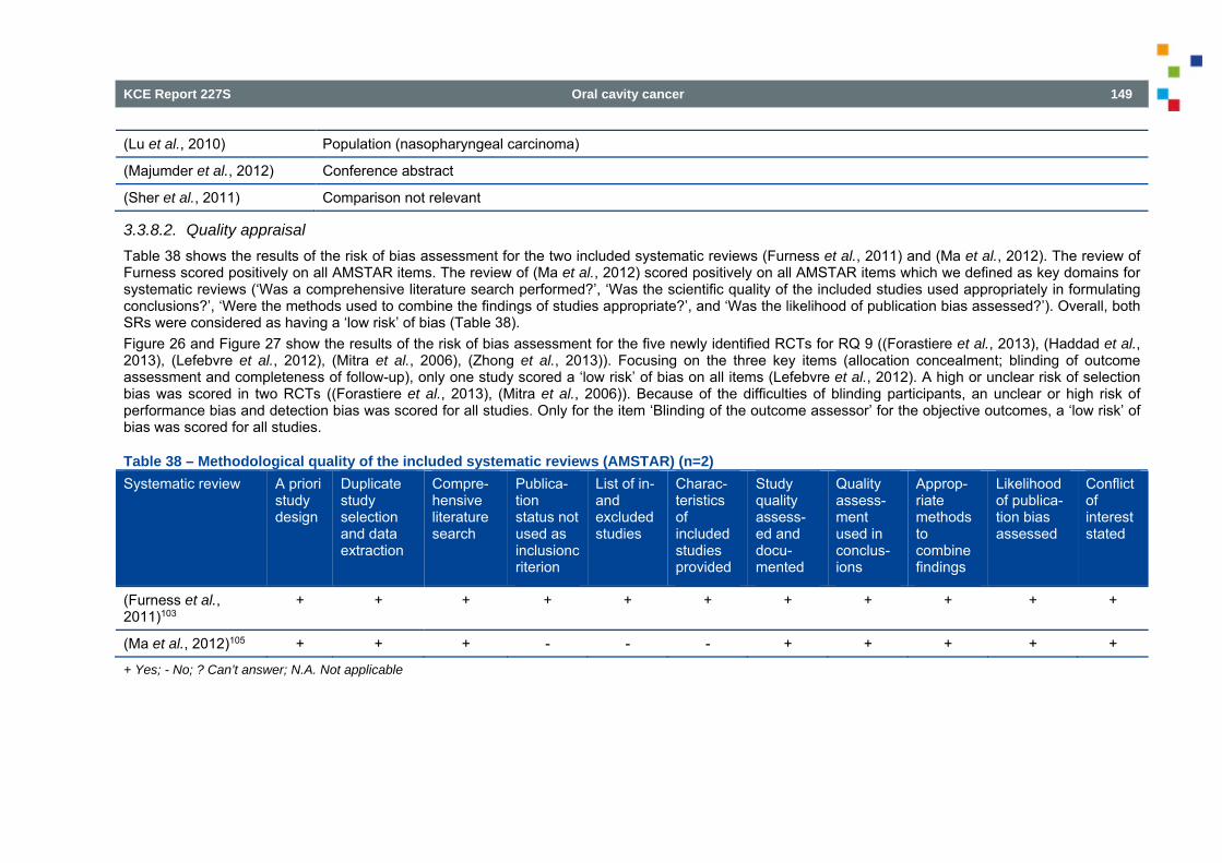
KCE Report 227S Oral cavity cancer 149
(Lu et al., 2010) Population (nasopharyngeal carcinoma)
(Majumder et al., 2012) Conference abstract
(Sher et al., 2011) Comparison not relevant
3.3.8.2. Quality appraisal Table 38 shows the results of the risk of bias assessment for the two included systematic reviews (Furness et al., 2011) and (Ma et al., 2012). The review of Furness scored positively on all AMSTAR items. The review of (Ma et al., 2012) scored positively on all AMSTAR items which we defined as key domains for systematic reviews (‘Was a comprehensive literature search performed?’, ‘Was the scientific quality of the included studies used appropriately in formulating conclusions?’, ‘Were the methods used to combine the findings of studies appropriate?’, and ‘Was the likelihood of publication bias assessed?’). Overall, both SRs were considered as having a ‘low risk’ of bias (Table 38). Figure 26 and Figure 27 show the results of the risk of bias assessment for the five newly identified RCTs for RQ 9 ((Forastiere et al., 2013), (Haddad et al., 2013), (Lefebvre et al., 2012), (Mitra et al., 2006), (Zhong et al., 2013)). Focusing on the three key items (allocation concealment; blinding of outcome assessment and completeness of follow-up), only one study scored a ‘low risk’ of bias on all items (Lefebvre et al., 2012). A high or unclear risk of selection bias was scored in two RCTs ((Forastiere et al., 2013), (Mitra et al., 2006)). Because of the difficulties of blinding participants, an unclear or high risk of performance bias and detection bias was scored for all studies. Only for the item ‘Blinding of the outcome assessor’ for the objective outcomes, a ‘low risk’ of bias was scored for all studies.
Table 38 – Methodological quality of the included systematic reviews (AMSTAR) (n=2) Systematic review A priori
study design
Duplicate study selection and data extraction
Compre-hensive literature search
Publica-tion status not used as inclusioncriterion
List of in- and excluded studies
Charac-teristics of included studies provided
Study quality assess-ed and docu-mented
Quality assess-ment used in conclus-ions
Approp-riate methods to combine findings
Likelihood of publica-tion bias assessed
Conflict of interest stated
(Furness et al., 2011)103
+ + + + + + + + + + +
(Ma et al., 2012)105 + + + - - - + + + + +
+ Yes; - No; ? Can’t answer; N.A. Not applicable
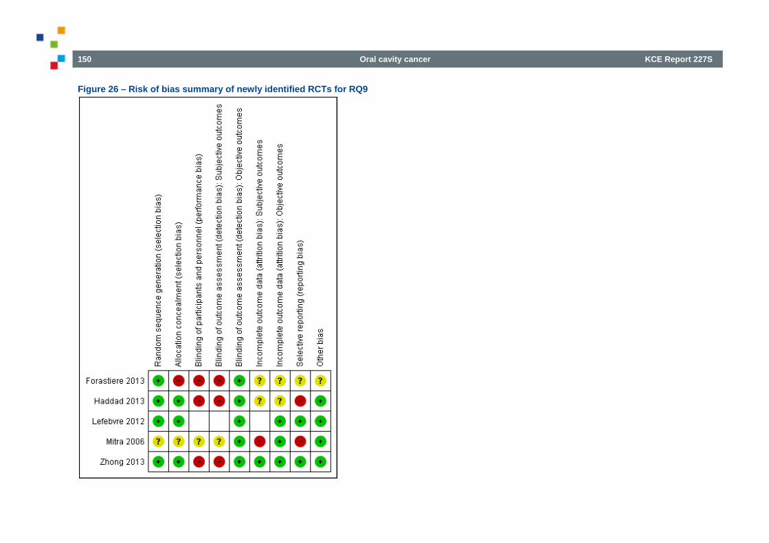
150 Oral cavity cancer KCE Report 227S
Figure 26 – Risk of bias summary of newly identified RCTs for RQ9
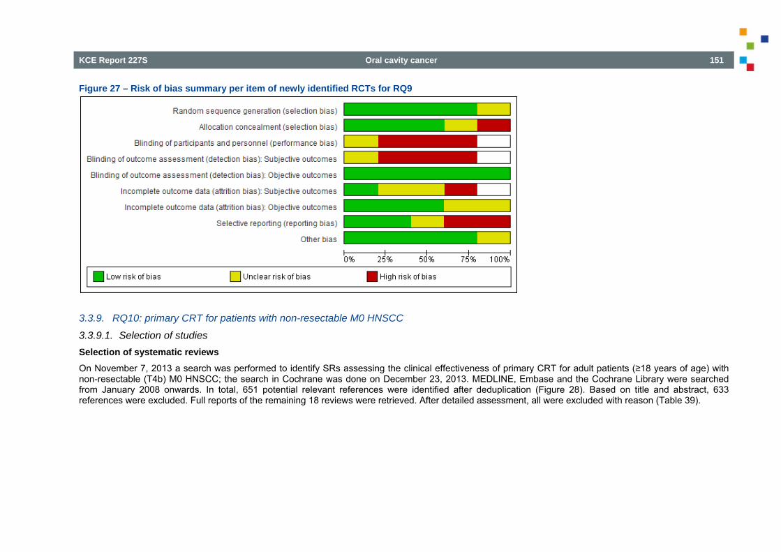
KCE Report 227S Oral cavity cancer 151
Figure 27 – Risk of bias summary per item of newly identified RCTs for RQ9
3.3.9. RQ10: primary CRT for patients with non-resectable M0 HNSCC
3.3.9.1. Selection of studies Selection of systematic reviews On November 7, 2013 a search was performed to identify SRs assessing the clinical effectiveness of primary CRT for adult patients (≥18 years of age) with non-resectable (T4b) M0 HNSCC; the search in Cochrane was done on December 23, 2013. MEDLINE, Embase and the Cochrane Library were searched from January 2008 onwards. In total, 651 potential relevant references were identified after deduplication (Figure 28). Based on title and abstract, 633 references were excluded. Full reports of the remaining 18 reviews were retrieved. After detailed assessment, all were excluded with reason (Table 39).
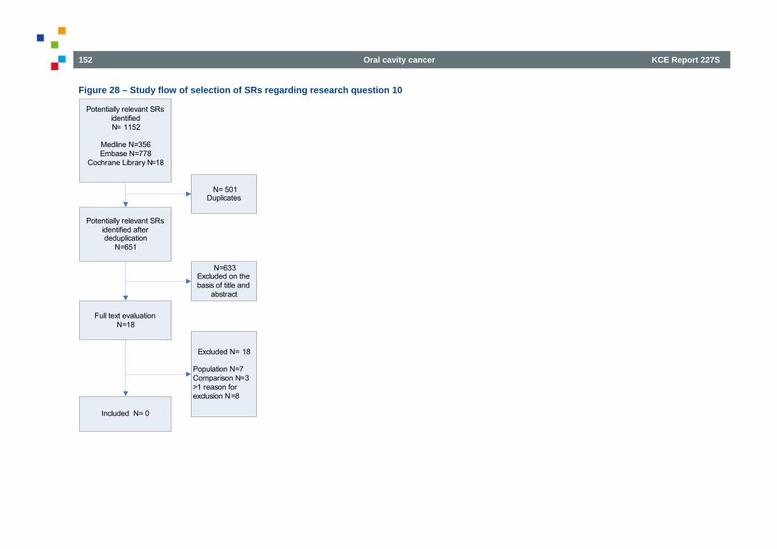
152 Oral cavity cancer KCE Report 227S
Figure 28 – Study flow of selection of SRs regarding research question 10
Potentially relevant SRs identified after deduplication
N=651
Full text evaluationN=18
N=633Excluded on the basis of title and
abstract
Included N= 0
Excluded N= 18
Population N=7 Comparison N=3>1 reason for exclusion N=8
Potentially relevant SRs identified N= 1152
Medline N=356Embase N=778
Cochrane Library N=18
N= 501Duplicates
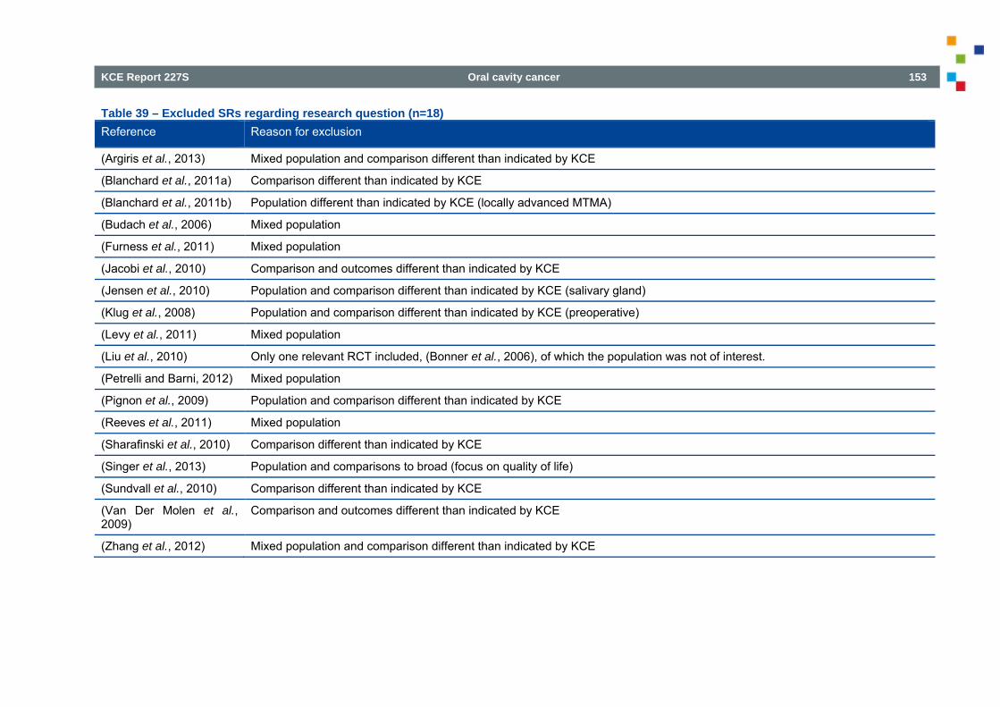
KCE Report 227S Oral cavity cancer 153
Table 39 – Excluded SRs regarding research question (n=18) Reference Reason for exclusion
(Argiris et al., 2013) Mixed population and comparison different than indicated by KCE
(Blanchard et al., 2011a) Comparison different than indicated by KCE
(Blanchard et al., 2011b) Population different than indicated by KCE (locally advanced MTMA)
(Budach et al., 2006) Mixed population
(Furness et al., 2011) Mixed population
(Jacobi et al., 2010) Comparison and outcomes different than indicated by KCE
(Jensen et al., 2010) Population and comparison different than indicated by KCE (salivary gland)
(Klug et al., 2008) Population and comparison different than indicated by KCE (preoperative)
(Levy et al., 2011) Mixed population
(Liu et al., 2010) Only one relevant RCT included, (Bonner et al., 2006), of which the population was not of interest.
(Petrelli and Barni, 2012) Mixed population
(Pignon et al., 2009) Population and comparison different than indicated by KCE
(Reeves et al., 2011) Mixed population
(Sharafinski et al., 2010) Comparison different than indicated by KCE
(Singer et al., 2013) Population and comparisons to broad (focus on quality of life)
(Sundvall et al., 2010) Comparison different than indicated by KCE
(Van Der Molen et al., 2009)
Comparison and outcomes different than indicated by KCE
(Zhang et al., 2012) Mixed population and comparison different than indicated by KCE
154 Oral cavity cancer KCE Report 227S
Selection of primary studies On December 6, 2013 a search was performed to identify RCTs assessing the clinical effectiveness of primary CRT for adult patients (≥18 years of age) with non-resectable (T4b) M0 HNSCC. As no systematic review was included, MEDLINE, Embase and CENTRAL were searched from 2003 onwards. One thousand six hundred and eighty-one potential relevant references were identified (Figure 29). After deduplication, 764 references remained. Based on title and abstract 711 studies were excluded. Of the remaining 53 studies, two studies were included that fully fulfilled the PICO for RQ10 [Bensadoun et al., 2006;Ruo Redda et al., 2010].111, 112 In consultation with KCE, another five studies which involved mixed populations (not solely stage T4b) were included [Budach et al., 2005;Chauhan et al., 2008;Rodriguez et al., 2010;Semrau et al., 2006;Quon et al., 2011]113-117 (Table 40). Forty-six studies were excluded with reason (Table 41).
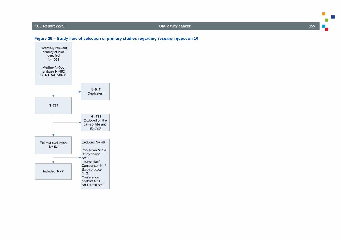
KCE Report 227S Oral cavity cancer 155
Figure 29 – Study flow of selection of primary studies regarding research question 10
N=764
Full text evaluationN= 53
N= 711Excluded on the basis of title and
abstract
Included N=7
Excluded N= 46
Population N=24Study design N=11Intervention/Comparison N=7Study protocol N=2Conference abstract N=1No full text N=1
Potentially relevant primary studies
identified N=1681
Medline N=553Embase N=692
CENTRAL N=436
N=917 Duplicates
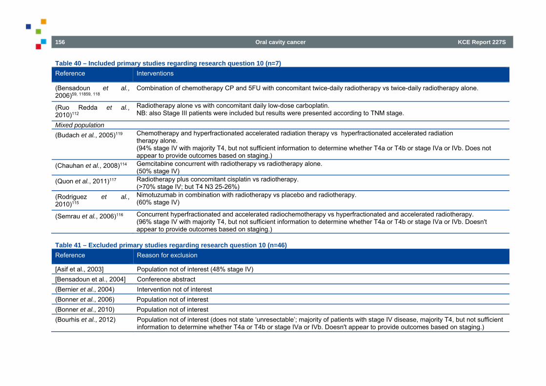
156 Oral cavity cancer KCE Report 227S
Table 40 – Included primary studies regarding research question 10 (n=7) Reference Interventions
(Bensadoun et al., 2006)59, 11859, 118
Combination of chemotherapy CP and 5FU with concomitant twice-daily radiotherapy vs twice-daily radiotherapy alone.
(Ruo Redda et al., 2010)112
Radiotherapy alone vs with concomitant daily low-dose carboplatin. NB: also Stage III patients were included but results were presented according to TNM stage.
Mixed population (Budach et al., 2005)119 Chemotherapy and hyperfractionated accelerated radiation therapy vs hyperfractionated accelerated radiation
therapy alone. (94% stage IV with majority T4, but not sufficient information to determine whether T4a or T4b or stage IVa or IVb. Does not appear to provide outcomes based on staging.)
(Chauhan et al., 2008)114 Gemcitabine concurrent with radiotherapy vs radiotherapy alone. (50% stage IV)
(Quon et al., 2011)117 Radiotherapy plus concomitant cisplatin vs radiotherapy. (>70% stage IV; but T4 N3 25-26%)
(Rodriguez et al., 2010)115
Nimotuzumab in combination with radiotherapy vs placebo and radiotherapy. (60% stage IV)
(Semrau et al., 2006)116 Concurrent hyperfractionated and accelerated radiochemotherapy vs hyperfractionated and accelerated radiotherapy. (96% stage IV with majority T4, but not sufficient information to determine whether T4a or T4b or stage IVa or IVb. Doesn't appear to provide outcomes based on staging.)
Table 41 – Excluded primary studies regarding research question 10 (n=46) Reference Reason for exclusion
[Asif et al., 2003] Population not of interest (48% stage IV) [Bensadoun et al., 2004] Conference abstract (Bernier et al., 2004) Intervention not of interest (Bonner et al., 2006) Population not of interest (Bonner et al., 2010) Population not of interest (Bourhis et al., 2012) Population not of interest (does not state ‘unresectable’; majority of patients with stage IV disease, majority T4, but not sufficient
information to determine whether T4a or T4b or stage IVa or IVb. Doesn't appear to provide outcomes based on staging.)
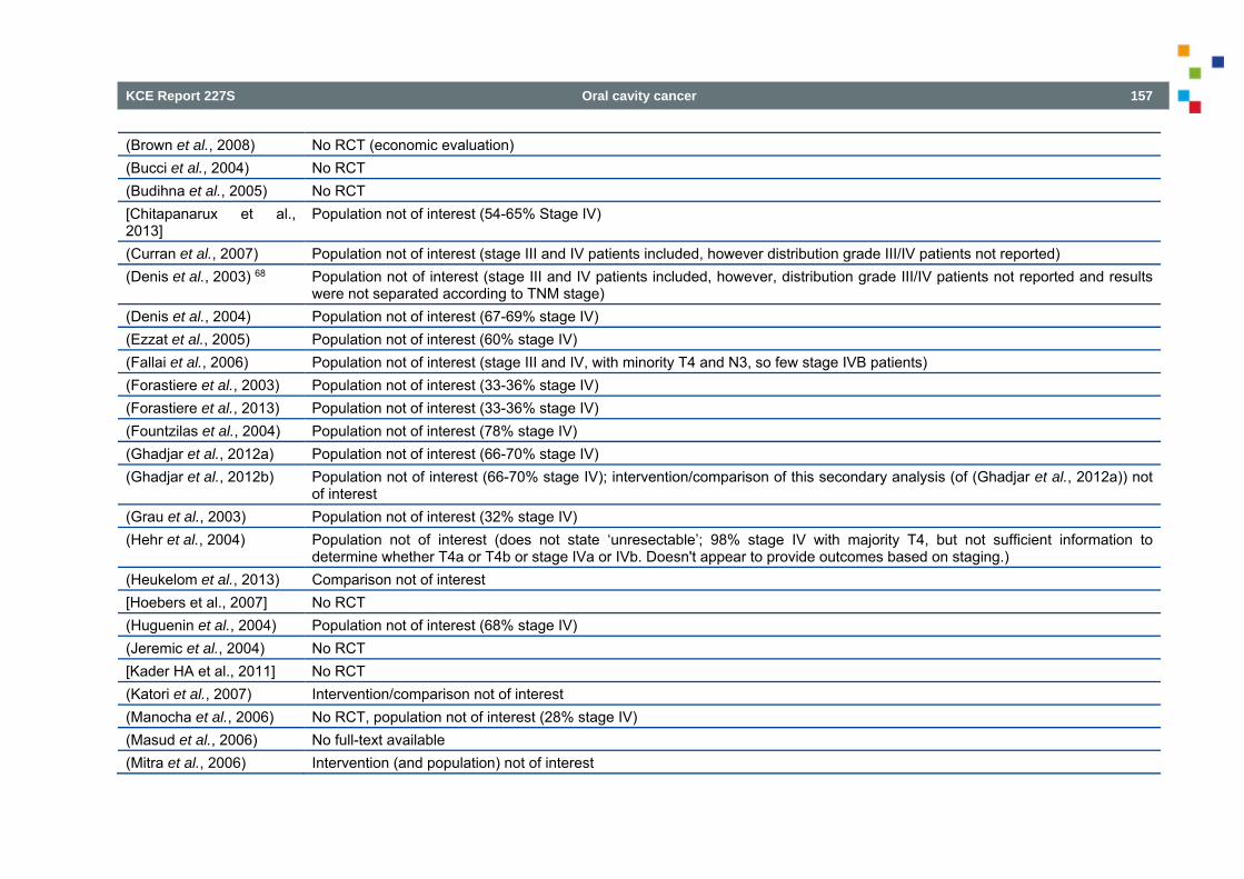
KCE Report 227S Oral cavity cancer 157
(Brown et al., 2008) No RCT (economic evaluation) (Bucci et al., 2004) No RCT (Budihna et al., 2005) No RCT [Chitapanarux et al., 2013]
Population not of interest (54-65% Stage IV)
(Curran et al., 2007) Population not of interest (stage III and IV patients included, however distribution grade III/IV patients not reported) (Denis et al., 2003) 68 Population not of interest (stage III and IV patients included, however, distribution grade III/IV patients not reported and results
were not separated according to TNM stage) (Denis et al., 2004) Population not of interest (67-69% stage IV) (Ezzat et al., 2005) Population not of interest (60% stage IV) (Fallai et al., 2006) Population not of interest (stage III and IV, with minority T4 and N3, so few stage IVB patients) (Forastiere et al., 2003) Population not of interest (33-36% stage IV) (Forastiere et al., 2013) Population not of interest (33-36% stage IV) (Fountzilas et al., 2004) Population not of interest (78% stage IV) (Ghadjar et al., 2012a) Population not of interest (66-70% stage IV) (Ghadjar et al., 2012b) Population not of interest (66-70% stage IV); intervention/comparison of this secondary analysis (of (Ghadjar et al., 2012a)) not
of interest (Grau et al., 2003) Population not of interest (32% stage IV) (Hehr et al., 2004) Population not of interest (does not state ‘unresectable’; 98% stage IV with majority T4, but not sufficient information to
determine whether T4a or T4b or stage IVa or IVb. Doesn't appear to provide outcomes based on staging.) (Heukelom et al., 2013) Comparison not of interest [Hoebers et al., 2007] No RCT (Huguenin et al., 2004) Population not of interest (68% stage IV) (Jeremic et al., 2004) No RCT [Kader HA et al., 2011] No RCT (Katori et al., 2007) Intervention/comparison not of interest (Manocha et al., 2006) No RCT, population not of interest (28% stage IV) (Masud et al., 2006) No full-text available (Mitra et al., 2006) Intervention (and population) not of interest
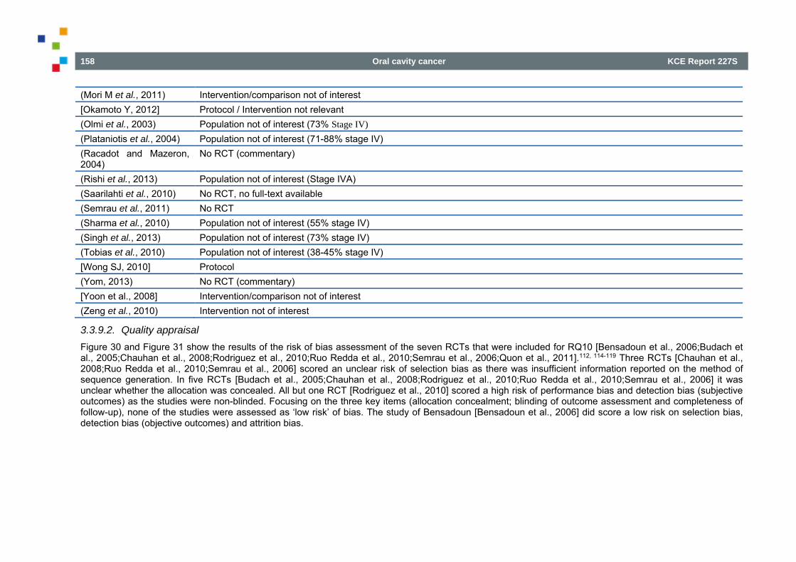
158 Oral cavity cancer KCE Report 227S
(Mori M et al., 2011) Intervention/comparison not of interest [Okamoto Y, 2012] Protocol / Intervention not relevant (Olmi et al., 2003) Population not of interest (73% Stage IV) (Plataniotis et al., 2004) Population not of interest (71-88% stage IV) (Racadot and Mazeron, 2004)
No RCT (commentary)
(Rishi et al., 2013) Population not of interest (Stage IVA) (Saarilahti et al., 2010) No RCT, no full-text available (Semrau et al., 2011) No RCT (Sharma et al., 2010) Population not of interest (55% stage IV) (Singh et al., 2013) Population not of interest (73% stage IV) (Tobias et al., 2010) Population not of interest (38-45% stage IV) [Wong SJ, 2010] Protocol (Yom, 2013) No RCT (commentary) [Yoon et al., 2008] Intervention/comparison not of interest (Zeng et al., 2010) Intervention not of interest
3.3.9.2. Quality appraisal Figure 30 and Figure 31 show the results of the risk of bias assessment of the seven RCTs that were included for RQ10 [Bensadoun et al., 2006;Budach et al., 2005;Chauhan et al., 2008;Rodriguez et al., 2010;Ruo Redda et al., 2010;Semrau et al., 2006;Quon et al., 2011].112, 114-119 Three RCTs [Chauhan et al., 2008;Ruo Redda et al., 2010;Semrau et al., 2006] scored an unclear risk of selection bias as there was insufficient information reported on the method of sequence generation. In five RCTs [Budach et al., 2005;Chauhan et al., 2008;Rodriguez et al., 2010;Ruo Redda et al., 2010;Semrau et al., 2006] it was unclear whether the allocation was concealed. All but one RCT [Rodriguez et al., 2010] scored a high risk of performance bias and detection bias (subjective outcomes) as the studies were non-blinded. Focusing on the three key items (allocation concealment; blinding of outcome assessment and completeness of follow-up), none of the studies were assessed as ‘low risk’ of bias. The study of Bensadoun [Bensadoun et al., 2006] did score a low risk on selection bias, detection bias (objective outcomes) and attrition bias.
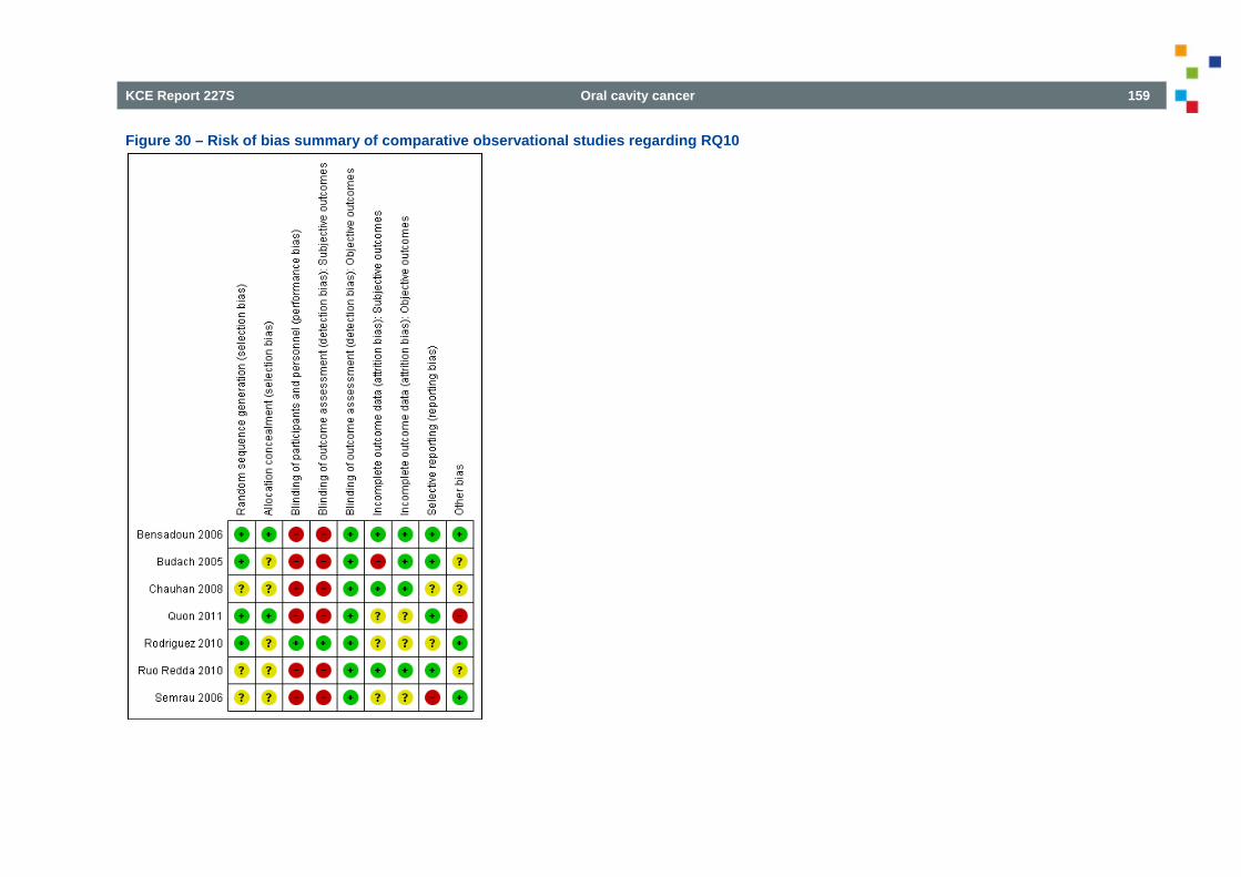
KCE Report 227S Oral cavity cancer 159
Figure 30 – Risk of bias summary of comparative observational studies regarding RQ10
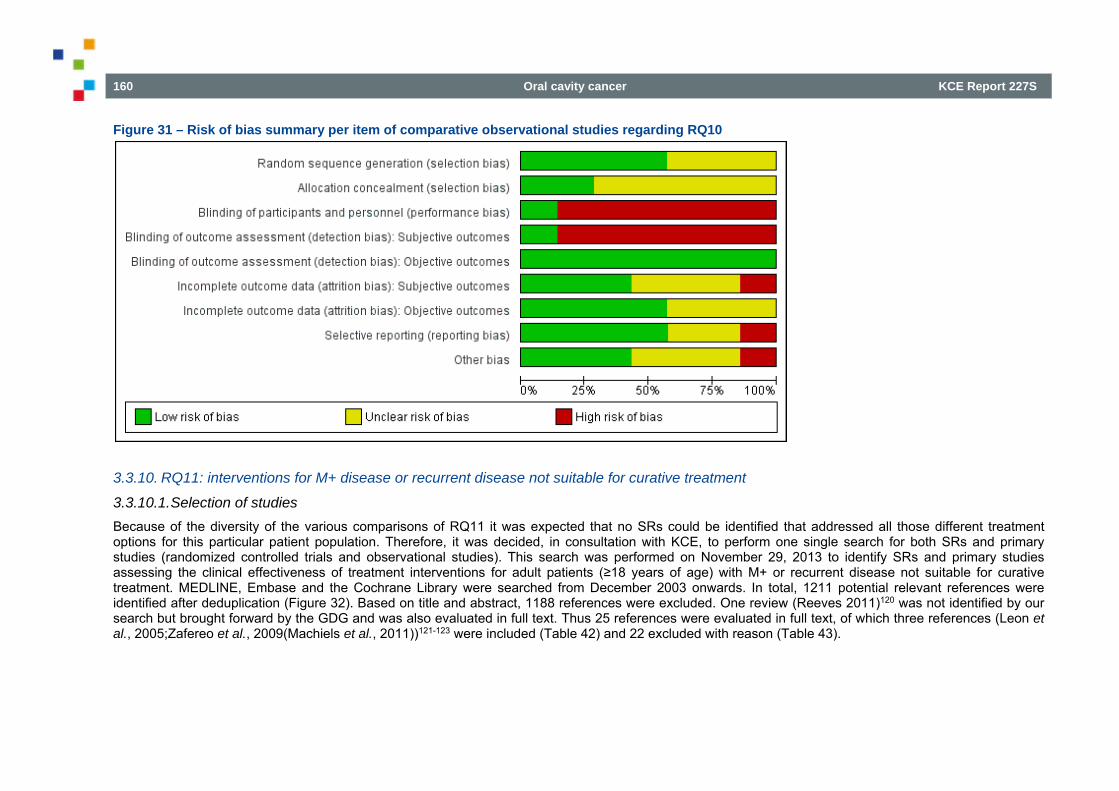
160 Oral cavity cancer KCE Report 227S
Figure 31 – Risk of bias summary per item of comparative observational studies regarding RQ10
3.3.10. RQ11: interventions for M+ disease or recurrent disease not suitable for curative treatment
3.3.10.1. Selection of studies Because of the diversity of the various comparisons of RQ11 it was expected that no SRs could be identified that addressed all those different treatment options for this particular patient population. Therefore, it was decided, in consultation with KCE, to perform one single search for both SRs and primary studies (randomized controlled trials and observational studies). This search was performed on November 29, 2013 to identify SRs and primary studies assessing the clinical effectiveness of treatment interventions for adult patients (≥18 years of age) with M+ or recurrent disease not suitable for curative treatment. MEDLINE, Embase and the Cochrane Library were searched from December 2003 onwards. In total, 1211 potential relevant references were identified after deduplication (Figure 32). Based on title and abstract, 1188 references were excluded. One review (Reeves 2011)120 was not identified by our search but brought forward by the GDG and was also evaluated in full text. Thus 25 references were evaluated in full text, of which three references (Leon et al., 2005;Zafereo et al., 2009(Machiels et al., 2011))121-123 were included (Table 42) and 22 excluded with reason (Table 43).
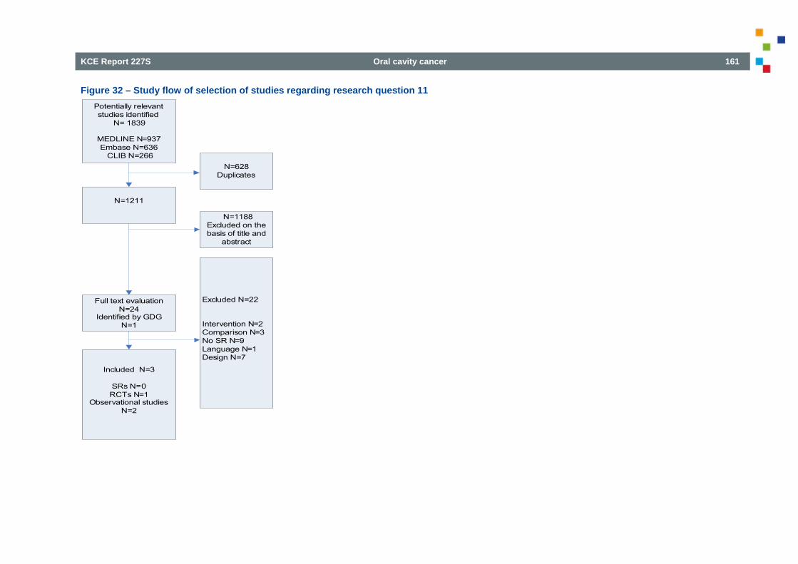
KCE Report 227S Oral cavity cancer 161
Figure 32 – Study flow of selection of studies regarding research question 11 Potentially relevant studies identified
N= 1839
MEDLINE N=937Embase N=636
CLIB N=266
N=1211
Full text evaluationN=24
Identified by GDGN=1
N=628Duplicates
N=1188 Excluded on the basis of title and
abstract
Included N=3
SRs N=0 RCTs N=1
Observational studies N=2
Excluded N=22
Intervention N=2Comparison N=3No SR N=9Language N=1Design N=7
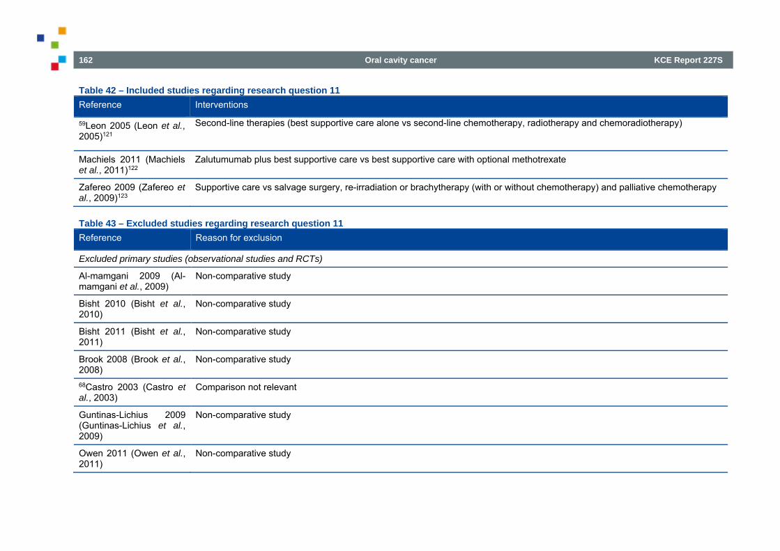
162 Oral cavity cancer KCE Report 227S
Table 42 – Included studies regarding research question 11 Reference Interventions
59Leon 2005 (Leon et al., 2005)121
Second-line therapies (best supportive care alone vs second-line chemotherapy, radiotherapy and chemoradiotherapy)
Machiels 2011 (Machiels et al., 2011)122
Zalutumumab plus best supportive care vs best supportive care with optional methotrexate
Zafereo 2009 (Zafereo et al., 2009)123
Supportive care vs salvage surgery, re-irradiation or brachytherapy (with or without chemotherapy) and palliative chemotherapy
Table 43 – Excluded studies regarding research question 11 Reference Reason for exclusion
Excluded primary studies (observational studies and RCTs)
Al-mamgani 2009 (Al-mamgani et al., 2009)
Non-comparative study
Bisht 2010 (Bisht et al., 2010)
Non-comparative study
Bisht 2011 (Bisht et al., 2011)
Non-comparative study
Brook 2008 (Brook et al., 2008)
Non-comparative study
68Castro 2003 (Castro et al., 2003)
Comparison not relevant
Guntinas-Lichius 2009 (Guntinas-Lichius et al., 2009)
Non-comparative study
Owen 2011 (Owen et al., 2011)
Non-comparative study
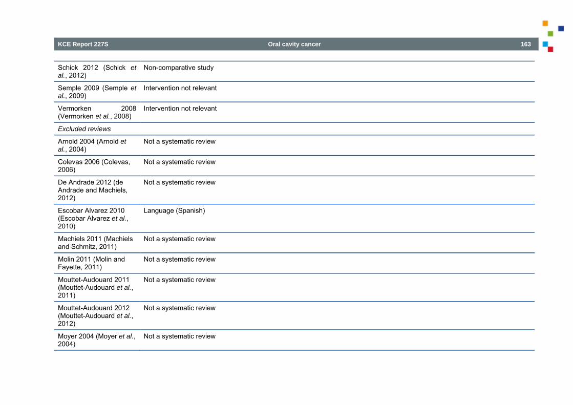
KCE Report 227S Oral cavity cancer 163
Schick 2012 (Schick et al., 2012)
Non-comparative study
Semple 2009 (Semple et al., 2009)
Intervention not relevant
Vermorken 2008 (Vermorken et al., 2008)
Intervention not relevant
Excluded reviews
Arnold 2004 (Arnold et al., 2004)
Not a systematic review
Colevas 2006 (Colevas, 2006)
Not a systematic review
De Andrade 2012 (de Andrade and Machiels, 2012)
Not a systematic review
Escobar Alvarez 2010 (Escobar Alvarez et al., 2010)
Language (Spanish)
Machiels 2011 (Machiels and Schmitz, 2011)
Not a systematic review
Molin 2011 (Molin and Fayette, 2011)
Not a systematic review
Mouttet-Audouard 2011 (Mouttet-Audouard et al., 2011)
Not a systematic review
Mouttet-Audouard 2012 (Mouttet-Audouard et al., 2012)
Not a systematic review
Moyer 2004 (Moyer et al., 2004)
Not a systematic review
164 Oral cavity cancer KCE Report 227S
Petrelli 2012 (Petrelli and Barni, 2012)
Comparison not of interest
Vermorken 2010 (Vermorken and Specenier, 2010)
Not a systematic review/comparator not of interest
Excluded review identified by the GDG
Reeves 2011 Comparison not of interest
3.3.10.2. Quality appraisal Figure 33 and Figure 34 show the results of the risk of bias assessment for the two observational studies that were included for RQ11 (Leon et al., 2005;Zafereo et al., 2009).121, 123 Focusing on the three key items (allocation concealment; blinding of outcome assessment and completeness of follow-up), none of the studies were assessed as ‘low risk’ of bias. The study of Leon (2005)121 did not address subjective outcomes, but did score a low risk of detection bias for objective outcomes and a low risk of reporting bias. The study of Zafereo (2009)123 did not score low risk of bias on any of the items. Figure 35 and Figure 36 show the results of the risk of bias assessment of the one included RCT (Machiels et al., 2011).122 The study scored a high risk of performance bias and detection bias for subjective outcomes as both participants and investigators were not blinded. The study was also judged as having a high risk of reporting and attrition bias (for subjective outcomes) as data for quality of life outcomes was not shown and the number of drop outs was substantial (no intention to treat analysis was performed for subjective outcomes). An unclear risk of other bias was scored as the sponsor of the study did the data management, statistical analyses, and interpreted the data. Focusing on the three key items (allocation concealment; blinding of outcome assessment and completeness of follow-up), the study was assessed as ‘high risk’ of bias.
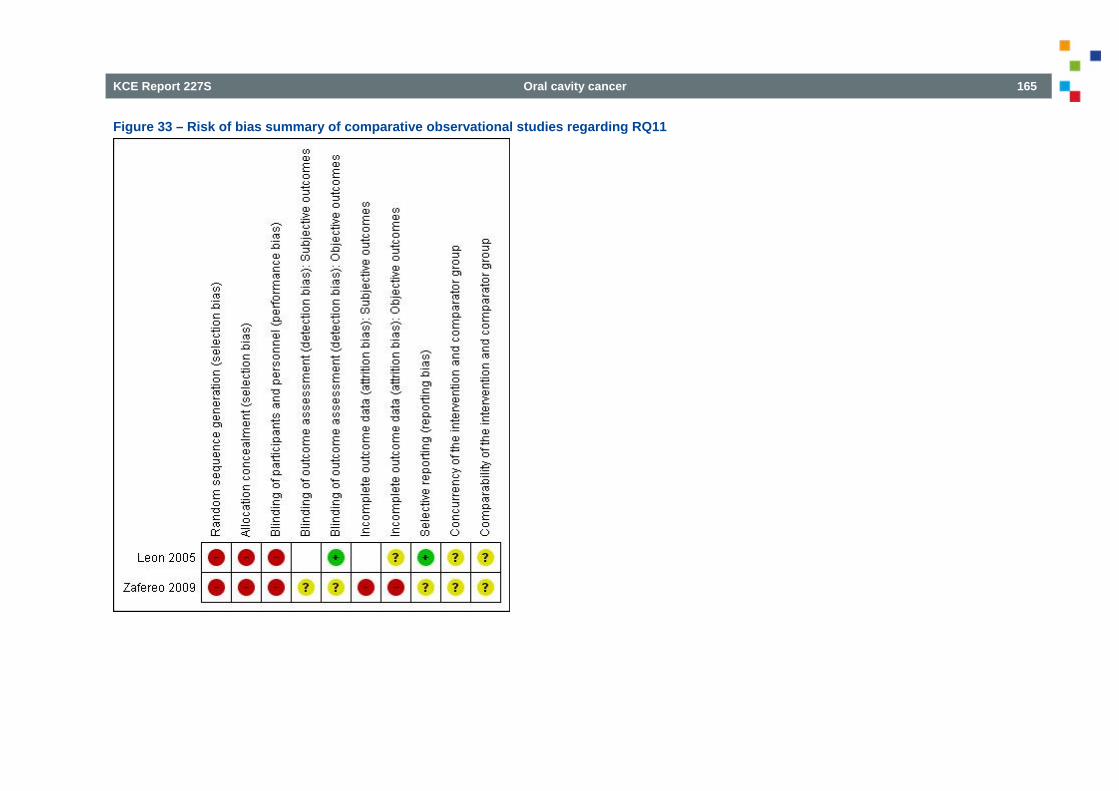
KCE Report 227S Oral cavity cancer 165
Figure 33 – Risk of bias summary of comparative observational studies regarding RQ11
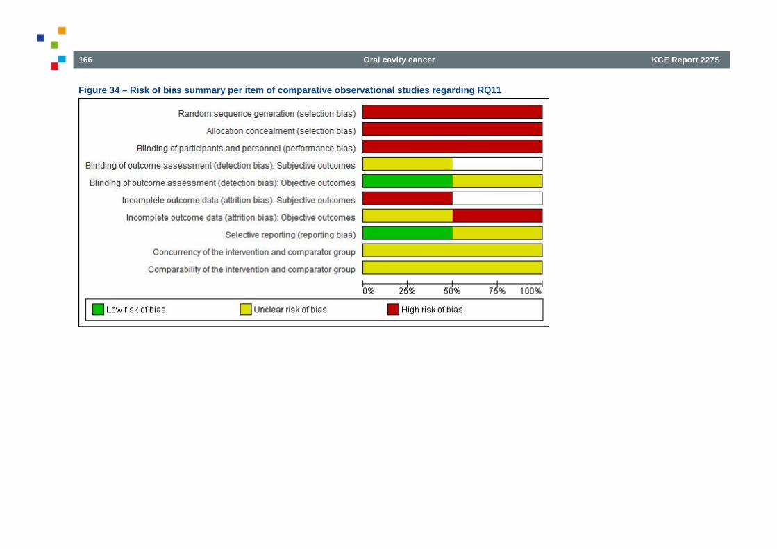
166 Oral cavity cancer KCE Report 227S
Figure 34 – Risk of bias summary per item of comparative observational studies regarding RQ11
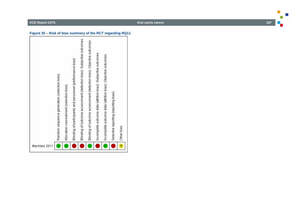
KCE Report 227S Oral cavity cancer 167
Figure 35 – Risk of bias summary of the RCT regarding RQ11
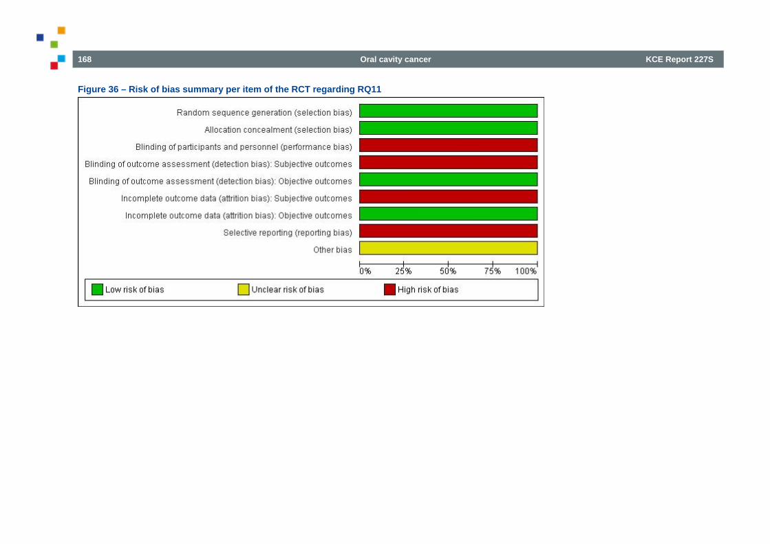
168 Oral cavity cancer KCE Report 227S
Figure 36 – Risk of bias summary per item of the RCT regarding RQ11
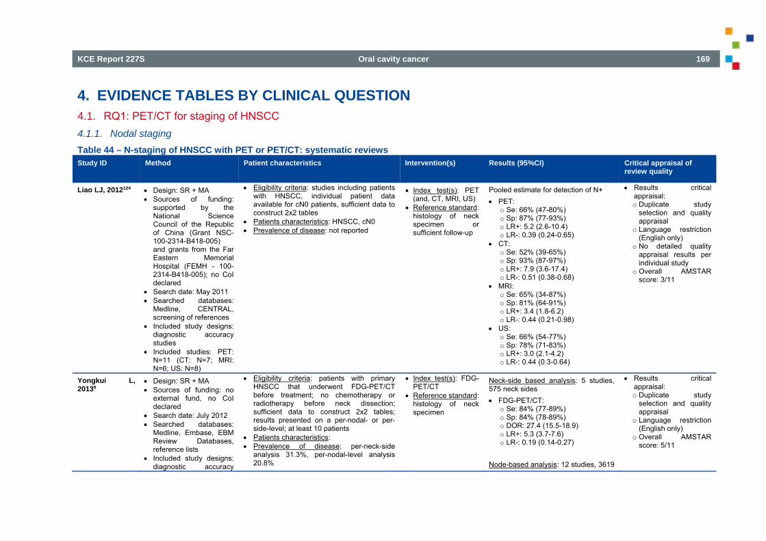
KCE Report 227S Oral cavity cancer 169
4. EVIDENCE TABLES BY CLINICAL QUESTION 4.1. RQ1: PET/CT for staging of HNSCC 4.1.1. Nodal staging Table 44 – N-staging of HNSCC with PET or PET/CT: systematic reviews Study ID Method Patient characteristics Intervention(s) Results (95%CI) Critical appraisal of
review quality
Liao LJ, 2012124 Design: SR + MA Sources of funding:
supported by the National Science Council of the Republic of China (Grant NSC-100-2314-B418-005) and grants from the Far Eastern Memorial Hospital (FEMH - 100-2314-B418-005); no CoI declared
Search date: May 2011 Searched databases:
Medline, CENTRAL, screening of references
Included study designs: diagnostic accuracy studies
Included studies: PET: N=11 (CT: N=7; MRI: N=6; US: N=8)
Eligibility criteria: studies including patients with HNSCC, individual patient data available for cN0 patients, sufficient data to construct 2x2 tables
Patients characteristics: HNSCC, cN0 Prevalence of disease: not reported
Index test(s): PET (and, CT, MRI, US)
Reference standard: histology of neck specimen or sufficient follow-up
Pooled estimate for detection of N+ PET:
o Se: 66% (47-80%) o Sp: 87% (77-93%) o LR+: 5.2 (2.6-10.4) o LR-: 0.39 (0.24-0.65)
CT: o Se: 52% (39-65%) o Sp: 93% (87-97%) o LR+: 7.9 (3.6-17.4) o LR-: 0.51 (0.38-0.68)
MRI: o Se: 65% (34-87%) o Sp: 81% (64-91%) o LR+: 3.4 (1.8-6.2) o LR-: 0.44 (0.21-0.98)
US: o Se: 66% (54-77%) o Sp: 78% (71-83%) o LR+: 3.0 (2.1-4.2) o LR-: 0.44 (0.3-0.64)
Results critical appraisal: o Duplicate study
selection and quality appraisal
o Language restriction (English only)
o No detailed quality appraisal results per individual study
o Overall AMSTAR score: 3/11
Yongkui L, 20136
Design: SR + MA Sources of funding: no
external fund, no CoI declared
Search date: July 2012 Searched databases:
Medline, Embase, EBM Review Databases, reference lists
Included study designs: diagnostic accuracy
Eligibility criteria: patients with primary HNSCC that underwent FDG-PET/CT before treatment; no chemotherapy or radiotherapy before neck dissection; sufficient data to construct 2x2 tables; results presented on a per-nodal- or per-side-level; at least 10 patients
Patients characteristics: Prevalence of disease: per-neck-side
analysis 31.3%, per-nodal-level analysis 20.8%
Index test(s): FDG-PET/CT
Reference standard: histology of neck specimen
Neck-side based analysis: 5 studies, 575 neck sides FDG-PET/CT:
o Se: 84% (77-89%) o Sp: 84% (78-89%) o DOR: 27.4 (15.5-18.9) o LR+: 5.3 (3.7-7.6) o LR-: 0.19 (0.14-0.27)
Node-based analysis: 12 studies, 3619
Results critical appraisal: o Duplicate study
selection and quality appraisal
o Language restriction (English only)
o Overall AMSTAR score: 5/11
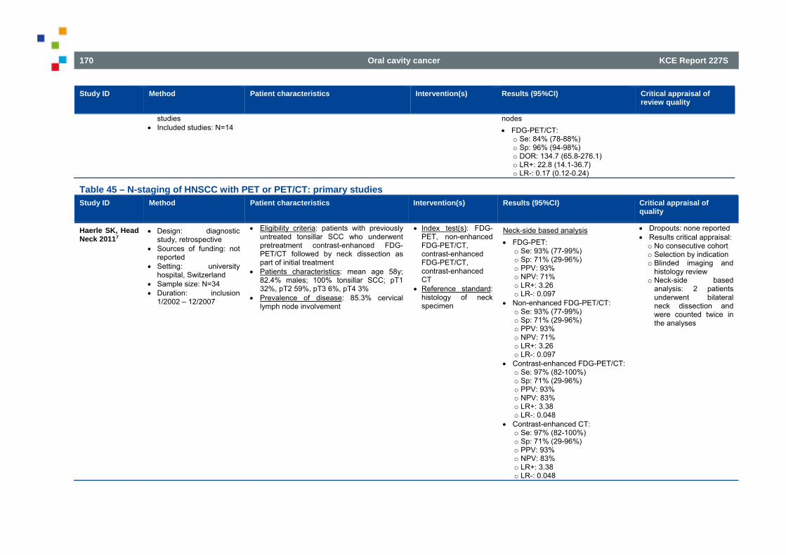
170 Oral cavity cancer KCE Report 227S
Study ID Method Patient characteristics Intervention(s) Results (95%CI) Critical appraisal of review quality
studies Included studies: N=14
nodes FDG-PET/CT:
o Se: 84% (78-88%) o Sp: 96% (94-98%) o DOR: 134.7 (65.8-276.1) o LR+: 22.8 (14.1-36.7) o LR-: 0.17 (0.12-0.24)
Table 45 – N-staging of HNSCC with PET or PET/CT: primary studies Study ID Method Patient characteristics Intervention(s) Results (95%CI) Critical appraisal of
quality
Haerle SK, Head Neck 20117
Design: diagnostic study, retrospective
Sources of funding: not reported
Setting: university hospital, Switzerland
Sample size: N=34 Duration: inclusion
1/2002 – 12/2007
Eligibility criteria: patients with previously untreated tonsillar SCC who underwent pretreatment contrast-enhanced FDG-PET/CT followed by neck dissection as part of initial treatment
Patients characteristics: mean age 58y; 82.4% males; 100% tonsillar SCC; pT1 32%, pT2 59%, pT3 6%, pT4 3%
Prevalence of disease: 85.3% cervical lymph node involvement
Index test(s): FDG-PET, non-enhanced FDG-PET/CT, contrast-enhanced FDG-PET/CT, contrast-enhanced CT
Reference standard: histology of neck specimen
Neck-side based analysis FDG-PET:
o Se: 93% (77-99%) o Sp: 71% (29-96%) o PPV: 93% o NPV: 71% o LR+: 3.26 o LR-: 0.097
Non-enhanced FDG-PET/CT: o Se: 93% (77-99%) o Sp: 71% (29-96%) o PPV: 93% o NPV: 71% o LR+: 3.26 o LR-: 0.097
Contrast-enhanced FDG-PET/CT: o Se: 97% (82-100%) o Sp: 71% (29-96%) o PPV: 93% o NPV: 83% o LR+: 3.38 o LR-: 0.048
Contrast-enhanced CT: o Se: 97% (82-100%) o Sp: 71% (29-96%) o PPV: 93% o NPV: 83% o LR+: 3.38 o LR-: 0.048
Dropouts: none reported Results critical appraisal: o No consecutive cohort o Selection by indication o Blinded imaging and
histology review o Neck-side based
analysis: 2 patients underwent bilateral neck dissection and were counted twice in the analyses
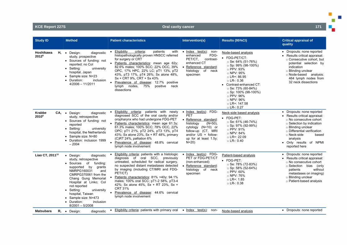
KCE Report 227S Oral cavity cancer 171
Study ID Method Patient characteristics Intervention(s) Results (95%CI) Critical appraisal of quality
Hoshikawa H, 20128
Design: diagnostic study, prospective
Sources of funding: not reported; no CoI
Setting: university hospital, Japan
Sample size: N=23 Duration: inclusion
4/2006 – 11/2011
Eligibility criteria: patients with histopathologically proven HNSCC referred for surgery or CRT
Patients characteristics: mean age 62y; 82.6% males; 100% SCC; 22% OCC, 39% OPC, 17% HPC, 22% LC; pT1 13%, pT2 43%, pT3 17%, pT4 26%; Sx alone 48%, Sx + CRT 9%, CRT + Sx 43%
Prevalence of disease: 12.7% positive lymph nodes, 75% positive neck dissections
Index test(s): non-enhanced FDG-PET/CT, contrast-enhanced CT
Reference standard: histology of neck specimen
Node-based analysis FDG-PET/CT:
o Se: 64% (51-76%) o Sp: 99% (98-100%) o PPV: 93% o NPV: 95% o LR+: 86.95 o LR-: 0.36
Contrast-enhanced CT: o Se: 73% (60-84%) o Sp: 100% (98-100%) o PPV: 96% o NPV: 96% o LR+: 147.58 o LR-: 0.27
Dropouts: none reported Results critical appraisal: o Consecutive cohort, but
potential selection by indication
o Blinding unclear o Node-based analysis:
464 lymph nodes from 32 neck dissections
Krabbe CA, 20109
Design: diagnostic study, retrospective
Sources of funding: not reported
Setting: university hospital, the Netherlands
Sample size: N=80 Duration: inclusion 1999
– 2004
Eligibility criteria: patients with newly diagnosed SCC of the oral cavity and/or oropharynx who had undergone FDG-PET
Patients characteristics: mean age 61.3y; 61.3% males; 100% SCC; 78% OCC, 22% OPC; pT1 21%, pT2 24%, pT3 13%, pT4 43%; Sx alone 23%, Sx + RT 48%, primary (C)RT 24%, palliation 6%
Prevalence of disease: 48.8% cervical lymph node involvement
Index test(s): FDG-PET
Reference standard: histology (N=50), cytology (N=10) or follow-up (CT, MRI and/or US + follow-up for at least 1.5y; N=20)
Neck-side based analysis FDG-PET:
o Se: 61% (46-74%) o Sp: 97% (92-99%) o PPV: 91% o NPV: 84% o LR+: 22.09 o LR-: 0.40
Dropouts: none reported Results critical appraisal: o No consecutive cohort o Selection by indication o Blinding unclear o Differential verification o Neck-side based
analysis Only results of NPMI
reported here
Liao CT, 201110 Design: diagnostic study, retrospective
Sources of funding: supported by grants NMRPG160031 and CMRPG370061 from the Chang Gung Memorial Hospital at Linko; CoI not reported
Setting: university hospital, Taiwan
Sample size: N=473 Duration: inclusion
8/2001 – 5/2008
Eligibility criteria: patients with a histologic diagnosis of oral SCC, previously untreated, scheduled for radical surgery, no suspected distant metastases detected by imaging (including CT/MRI and FDG-PET/CT)
Patients characteristics: 81% >40y; 94.1% males; 100% oral SCC; pT1-2 58%, pT3-4 42%; Sx alone 45%, Sx + RT 23%, Sx + CRT 31%
Prevalence of disease: 44.6% cervical lymph node involvement
Index test(s): FDG-PET or FDG-PET/CT (non-enhanced)
Reference standard: histology of neck specimen
Patient-based analysis FDG-PET:
o Se: 78% (72-83%) o Sp: 58% (52-64%) o PPV: 60% o NPV: 76% o LR+: 1.85 o LR-: 0.38
Dropouts: none reported Results critical appraisal: o No consecutive cohort o Selection bias (only
patients without metastases on imaging)
o Blinding unclear o Patient-based analysis
Matsubara R, Design: diagnostic Eligibility criteria: patients with primary oral Index test(s): non- Node-based analysis Dropouts: none reported
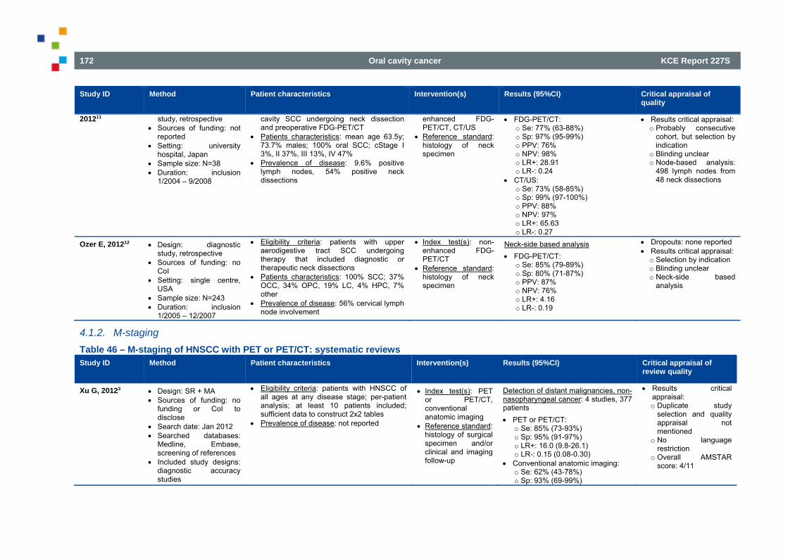
172 Oral cavity cancer KCE Report 227S
Study ID Method Patient characteristics Intervention(s) Results (95%CI) Critical appraisal of quality
201211 study, retrospective Sources of funding: not
reported Setting: university
hospital, Japan Sample size: N=38 Duration: inclusion
1/2004 – 9/2008
cavity SCC undergoing neck dissection and preoperative FDG-PET/CT
Patients characteristics: mean age 63.5y; 73.7% males; 100% oral SCC; cStage I 3%, II 37%, III 13%, IV 47%
Prevalence of disease: 9.6% positive lymph nodes, 54% positive neck dissections
enhanced FDG-PET/CT, CT/US
Reference standard: histology of neck specimen
FDG-PET/CT: o Se: 77% (63-88%) o Sp: 97% (95-99%) o PPV: 76% o NPV: 98% o LR+: 28.91 o LR-: 0.24
CT/US: o Se: 73% (58-85%) o Sp: 99% (97-100%) o PPV: 88% o NPV: 97% o LR+: 65.63 o LR-: 0.27
Results critical appraisal: o Probably consecutive
cohort, but selection by indication
o Blinding unclear o Node-based analysis:
498 lymph nodes from 48 neck dissections
Ozer E, 201212 Design: diagnostic study, retrospective
Sources of funding: no CoI
Setting: single centre, USA
Sample size: N=243 Duration: inclusion
1/2005 – 12/2007
Eligibility criteria: patients with upper aerodigestive tract SCC undergoing therapy that included diagnostic or therapeutic neck dissections
Patients characteristics: 100% SCC; 37% OCC, 34% OPC, 19% LC, 4% HPC, 7% other
Prevalence of disease: 56% cervical lymph node involvement
Index test(s): non-enhanced FDG-PET/CT
Reference standard: histology of neck specimen
Neck-side based analysis FDG-PET/CT:
o Se: 85% (79-89%) o Sp: 80% (71-87%) o PPV: 87% o NPV: 76% o LR+: 4.16 o LR-: 0.19
Dropouts: none reported Results critical appraisal: o Selection by indication o Blinding unclear o Neck-side based
analysis
4.1.2. M-staging Table 46 – M-staging of HNSCC with PET or PET/CT: systematic reviews Study ID Method Patient characteristics Intervention(s) Results (95%CI) Critical appraisal of
review quality
Xu G, 20123 Design: SR + MA Sources of funding: no
funding or CoI to disclose
Search date: Jan 2012 Searched databases:
Medline, Embase, screening of references
Included study designs: diagnostic accuracy studies
Eligibility criteria: patients with HNSCC of all ages at any disease stage; per-patient analysis; at least 10 patients included; sufficient data to construct 2x2 tables
Prevalence of disease: not reported
Index test(s): PET or PET/CT, conventional anatomic imaging
Reference standard: histology of surgical specimen and/or clinical and imaging follow-up
Detection of distant malignancies, non-nasopharyngeal cancer: 4 studies, 377 patients PET or PET/CT:
o Se: 85% (73-93%) o Sp: 95% (91-97%) o LR+: 16.0 (9.8-26.1) o LR-: 0.15 (0.08-0.30)
Conventional anatomic imaging: o Se: 62% (43-78%) o Sp: 93% (69-99%)
Results critical appraisal: o Duplicate study
selection and quality appraisal not mentioned
o No language restriction
o Overall AMSTAR score: 4/11
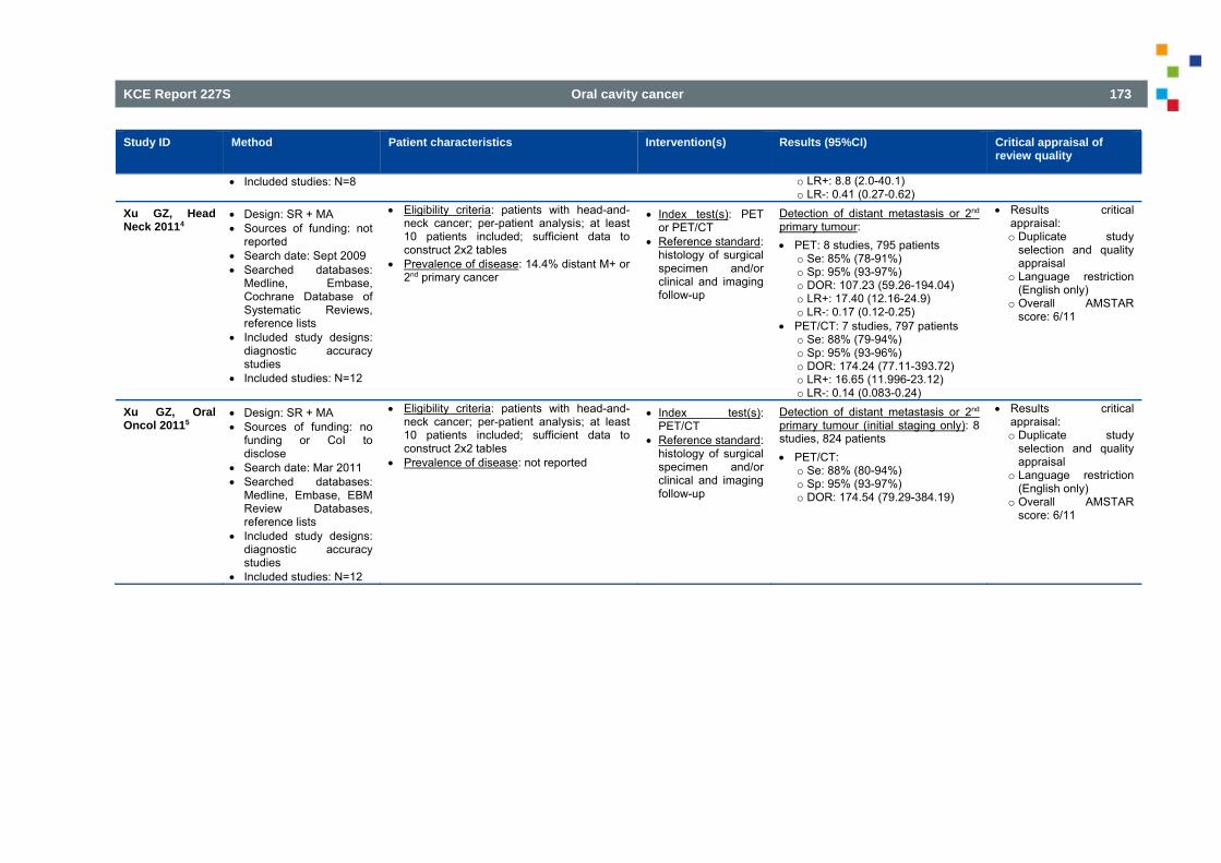
KCE Report 227S Oral cavity cancer 173
Study ID Method Patient characteristics Intervention(s) Results (95%CI) Critical appraisal of review quality
Included studies: N=8 o LR+: 8.8 (2.0-40.1) o LR-: 0.41 (0.27-0.62)
Xu GZ, Head Neck 20114
Design: SR + MA Sources of funding: not
reported Search date: Sept 2009 Searched databases:
Medline, Embase, Cochrane Database of Systematic Reviews, reference lists
Included study designs: diagnostic accuracy studies
Included studies: N=12
Eligibility criteria: patients with head-and-neck cancer; per-patient analysis; at least 10 patients included; sufficient data to construct 2x2 tables
Prevalence of disease: 14.4% distant M+ or 2nd primary cancer
Index test(s): PET or PET/CT
Reference standard: histology of surgical specimen and/or clinical and imaging follow-up
Detection of distant metastasis or 2nd primary tumour: PET: 8 studies, 795 patients
o Se: 85% (78-91%) o Sp: 95% (93-97%) o DOR: 107.23 (59.26-194.04) o LR+: 17.40 (12.16-24.9) o LR-: 0.17 (0.12-0.25)
PET/CT: 7 studies, 797 patients o Se: 88% (79-94%) o Sp: 95% (93-96%) o DOR: 174.24 (77.11-393.72) o LR+: 16.65 (11.996-23.12) o LR-: 0.14 (0.083-0.24)
Results critical appraisal: o Duplicate study
selection and quality appraisal
o Language restriction (English only)
o Overall AMSTAR score: 6/11
Xu GZ, Oral Oncol 20115
Design: SR + MA Sources of funding: no
funding or CoI to disclose
Search date: Mar 2011 Searched databases:
Medline, Embase, EBM Review Databases, reference lists
Included study designs: diagnostic accuracy studies
Included studies: N=12
Eligibility criteria: patients with head-and-neck cancer; per-patient analysis; at least 10 patients included; sufficient data to construct 2x2 tables
Prevalence of disease: not reported
Index test(s): PET/CT
Reference standard: histology of surgical specimen and/or clinical and imaging follow-up
Detection of distant metastasis or 2nd primary tumour (initial staging only): 8 studies, 824 patients PET/CT:
o Se: 88% (80-94%) o Sp: 95% (93-97%) o DOR: 174.54 (79.29-384.19)
Results critical appraisal: o Duplicate study
selection and quality appraisal
o Language restriction (English only)
o Overall AMSTAR score: 6/11
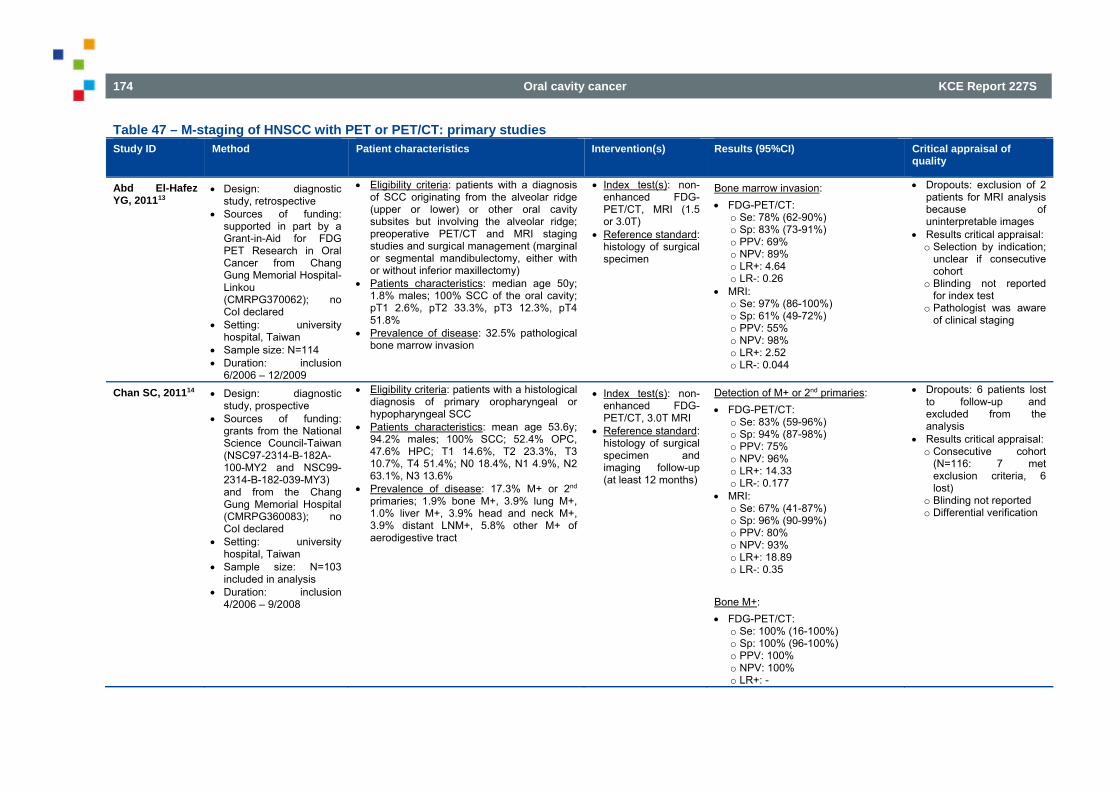
174 Oral cavity cancer KCE Report 227S
Table 47 – M-staging of HNSCC with PET or PET/CT: primary studies Study ID Method Patient characteristics Intervention(s) Results (95%CI) Critical appraisal of
quality
Abd El-Hafez YG, 201113
Design: diagnostic study, retrospective
Sources of funding: supported in part by a Grant-in-Aid for FDG PET Research in Oral Cancer from Chang Gung Memorial Hospital-Linkou (CMRPG370062); no CoI declared
Setting: university hospital, Taiwan
Sample size: N=114 Duration: inclusion
6/2006 – 12/2009
Eligibility criteria: patients with a diagnosis of SCC originating from the alveolar ridge (upper or lower) or other oral cavity subsites but involving the alveolar ridge; preoperative PET/CT and MRI staging studies and surgical management (marginal or segmental mandibulectomy, either with or without inferior maxillectomy)
Patients characteristics: median age 50y; 1.8% males; 100% SCC of the oral cavity; pT1 2.6%, pT2 33.3%, pT3 12.3%, pT4 51.8%
Prevalence of disease: 32.5% pathological bone marrow invasion
Index test(s): non-enhanced FDG-PET/CT, MRI (1.5 or 3.0T)
Reference standard: histology of surgical specimen
Bone marrow invasion: FDG-PET/CT:
o Se: 78% (62-90%) o Sp: 83% (73-91%) o PPV: 69% o NPV: 89% o LR+: 4.64 o LR-: 0.26
MRI: o Se: 97% (86-100%) o Sp: 61% (49-72%) o PPV: 55% o NPV: 98% o LR+: 2.52 o LR-: 0.044
Dropouts: exclusion of 2 patients for MRI analysis because of uninterpretable images
Results critical appraisal: o Selection by indication;
unclear if consecutive cohort
o Blinding not reported for index test
o Pathologist was aware of clinical staging
Chan SC, 201114 Design: diagnostic study, prospective
Sources of funding: grants from the National Science Council-Taiwan (NSC97-2314-B-182A-100-MY2 and NSC99-2314-B-182-039-MY3) and from the Chang Gung Memorial Hospital (CMRPG360083); no CoI declared
Setting: university hospital, Taiwan
Sample size: N=103 included in analysis
Duration: inclusion 4/2006 – 9/2008
Eligibility criteria: patients with a histological diagnosis of primary oropharyngeal or hypopharyngeal SCC
Patients characteristics: mean age 53.6y; 94.2% males; 100% SCC; 52.4% OPC, 47.6% HPC; T1 14.6%, T2 23.3%, T3 10.7%, T4 51.4%; N0 18.4%, N1 4.9%, N2 63.1%, N3 13.6%
Prevalence of disease: 17.3% M+ or 2nd primaries; 1.9% bone M+, 3.9% lung M+, 1.0% liver M+, 3.9% head and neck M+, 3.9% distant LNM+, 5.8% other M+ of aerodigestive tract
Index test(s): non-enhanced FDG-PET/CT, 3.0T MRI
Reference standard: histology of surgical specimen and imaging follow-up (at least 12 months)
Detection of M+ or 2nd primaries: FDG-PET/CT:
o Se: 83% (59-96%) o Sp: 94% (87-98%) o PPV: 75% o NPV: 96% o LR+: 14.33 o LR-: 0.177
MRI: o Se: 67% (41-87%) o Sp: 96% (90-99%) o PPV: 80% o NPV: 93% o LR+: 18.89 o LR-: 0.35
Bone M+: FDG-PET/CT:
o Se: 100% (16-100%) o Sp: 100% (96-100%) o PPV: 100% o NPV: 100% o LR+: -
Dropouts: 6 patients lost to follow-up and excluded from the analysis
Results critical appraisal: o Consecutive cohort
(N=116: 7 met exclusion criteria, 6 lost)
o Blinding not reported o Differential verification
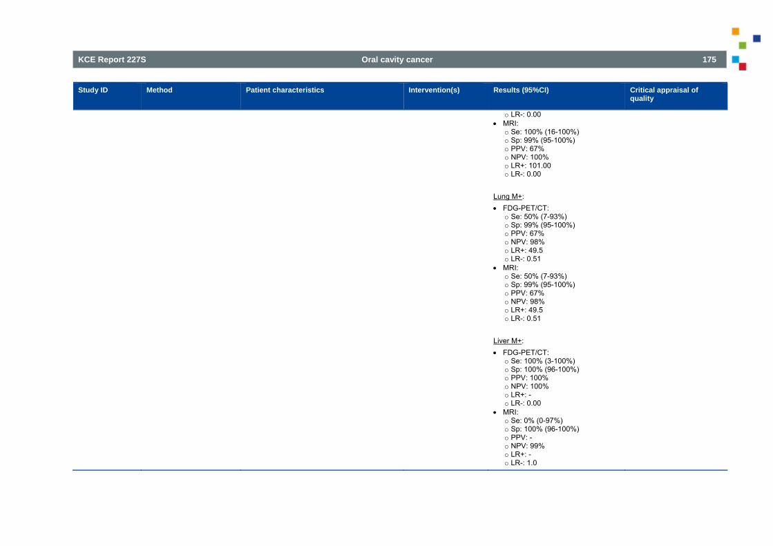
KCE Report 227S Oral cavity cancer 175
Study ID Method Patient characteristics Intervention(s) Results (95%CI) Critical appraisal of quality
o LR-: 0.00 MRI:
o Se: 100% (16-100%) o Sp: 99% (95-100%) o PPV: 67% o NPV: 100% o LR+: 101.00 o LR-: 0.00
Lung M+: FDG-PET/CT:
o Se: 50% (7-93%) o Sp: 99% (95-100%) o PPV: 67% o NPV: 98% o LR+: 49.5 o LR-: 0.51
MRI: o Se: 50% (7-93%) o Sp: 99% (95-100%) o PPV: 67% o NPV: 98% o LR+: 49.5 o LR-: 0.51
Liver M+: FDG-PET/CT:
o Se: 100% (3-100%) o Sp: 100% (96-100%) o PPV: 100% o NPV: 100% o LR+: - o LR-: 0.00
MRI: o Se: 0% (0-97%) o Sp: 100% (96-100%) o PPV: - o NPV: 99% o LR+: - o LR-: 1.0
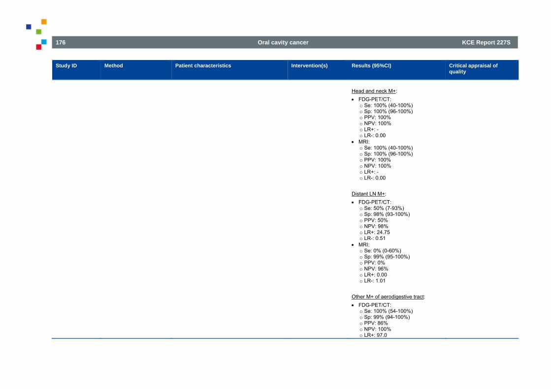
176 Oral cavity cancer KCE Report 227S
Study ID Method Patient characteristics Intervention(s) Results (95%CI) Critical appraisal of quality
Head and neck M+: FDG-PET/CT:
o Se: 100% (40-100%) o Sp: 100% (96-100%) o PPV: 100% o NPV: 100% o LR+: - o LR-: 0.00
MRI: o Se: 100% (40-100%) o Sp: 100% (96-100%) o PPV: 100% o NPV: 100% o LR+: - o LR-: 0.00
Distant LN M+: FDG-PET/CT:
o Se: 50% (7-93%) o Sp: 98% (93-100%) o PPV: 50% o NPV: 98% o LR+: 24.75 o LR-: 0.51
MRI: o Se: 0% (0-60%) o Sp: 99% (95-100%) o PPV: 0% o NPV: 96% o LR+: 0.00 o LR-: 1.01
Other M+ of aerodigestive tract: FDG-PET/CT:
o Se: 100% (54-100%) o Sp: 99% (94-100%) o PPV: 86% o NPV: 100% o LR+: 97.0
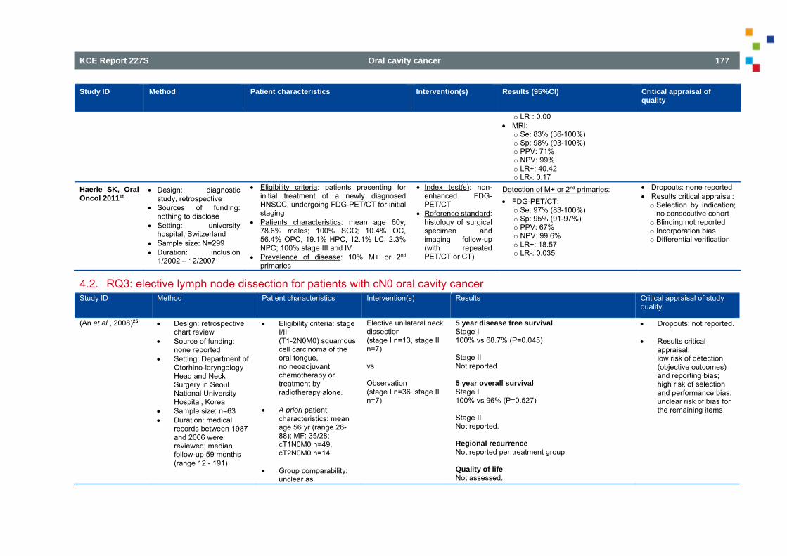
KCE Report 227S Oral cavity cancer 177
Study ID Method Patient characteristics Intervention(s) Results (95%CI) Critical appraisal of quality
o LR-: 0.00 MRI:
o Se: 83% (36-100%) o Sp: 98% (93-100%) o PPV: 71% o NPV: 99% o LR+: 40.42 o LR-: 0.17
Haerle SK, Oral Oncol 201115
Design: diagnostic study, retrospective
Sources of funding: nothing to disclose
Setting: university hospital, Switzerland
Sample size: N=299 Duration: inclusion
1/2002 – 12/2007
Eligibility criteria: patients presenting for initial treatment of a newly diagnosed HNSCC, undergoing FDG-PET/CT for initial staging
Patients characteristics: mean age 60y; 78.6% males; 100% SCC; 10.4% OC, 56.4% OPC, 19.1% HPC, 12.1% LC, 2.3% NPC; 100% stage III and IV
Prevalence of disease: 10% M+ or 2nd primaries
Index test(s): non-enhanced FDG-PET/CT
Reference standard: histology of surgical specimen and imaging follow-up (with repeated PET/CT or CT)
Detection of M+ or 2nd primaries: FDG-PET/CT:
o Se: 97% (83-100%) o Sp: 95% (91-97%) o PPV: 67% o NPV: 99.6% o LR+: 18.57 o LR-: 0.035
Dropouts: none reported Results critical appraisal: o Selection by indication;
no consecutive cohort o Blinding not reported o Incorporation bias o Differential verification
4.2. RQ3: elective lymph node dissection for patients with cN0 oral cavity cancer Study ID Method Patient characteristics Intervention(s) Results Critical appraisal of study
quality
(An et al., 2008)25 Design: retrospective chart review
Source of funding: none reported
Setting: Department of Otorhino-laryngology Head and Neck Surgery in Seoul National University Hospital, Korea
Sample size: n=63 Duration: medical
records between 1987 and 2006 were reviewed; median follow-up 59 months (range 12 - 191)
Eligibility criteria: stage I/II (T1-2N0M0) squamous cell carcinoma of the oral tongue, no neoadjuvant chemotherapy or treatment by radiotherapy alone.
A priori patient
characteristics: mean age 56 yr (range 26-88); MF: 35/28; cT1N0M0 n=49, cT2N0M0 n=14
Group comparability:
unclear as
Elective unilateral neck dissection (stage I n=13, stage II n=7) vs Observation (stage I n=36 stage II n=7)
5 year disease free survival Stage I 100% vs 68.7% (P=0.045) Stage II Not reported 5 year overall survival Stage I 100% vs 96% (P=0.527) Stage II Not reported. Regional recurrence Not reported per treatment group Quality of life Not assessed.
Dropouts: not reported. Results critical
appraisal: low risk of detection (objective outcomes) and reporting bias; high risk of selection and performance bias; unclear risk of bias for the remaining items
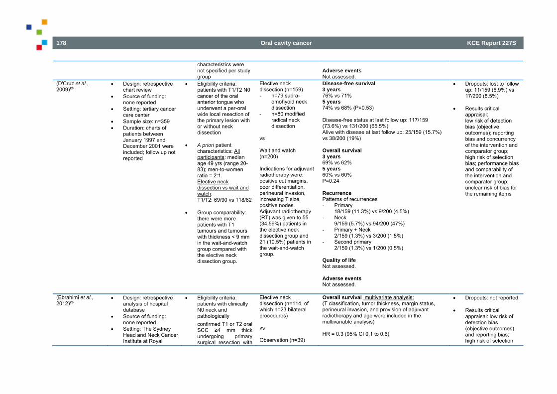
178 Oral cavity cancer KCE Report 227S
characteristics were not specified per study group
Adverse events Not assessed.
(D'Cruz et al., 2009)20
Design: retrospective chart review
Source of funding: none reported
Setting: tertiary cancer care center
Sample size: n=359 Duration: charts of
patients between January 1997 and December 2001 were included; follow up not reported
Eligibility criteria: patients with T1/T2 N0 cancer of the oral anterior tongue who underwent a per-oral wide local resection of the primary lesion with or without neck dissection
A priori patient
characteristics: All participants: median age 49 yrs (range 20-83); men-to-women ratio = 2:1. Elective neck dissection vs wait and watch: T1/T2: 69/90 vs 118/82
Group comparability: there were more patients with T1 tumours and tumours with thickness < 9 mm in the wait-and-watch group compared with the elective neck dissection group.
Elective neck dissection (n=159) - n=79 supra-
omohyoid neck dissection
- n=80 modified radical neck dissection
vs Wait and watch (n=200) Indications for adjuvant radiotherapy were: positive cut margins, poor differentiation, perineural invasion, increasing T size, positive nodes. Adjuvant radiotherapy (RT) was given to 55 (34.59%) patients in the elective neck dissection group and 21 (10.5%) patients in the wait-and-watch group.
Disease-free survival 3 years 76% vs 71% 5 years 74% vs 68% (P=0.53) Disease-free status at last follow up: 117/159 (73.6%) vs 131/200 (65.5%) Alive with disease at last follow up: 25/159 (15.7%) vs 38/200 (19%) Overall survival 3 years 69% vs 62% 5 years 60% vs 60% P=0.24 Recurrence Patterns of recurrences - Primary
18/159 (11.3%) vs 9/200 (4.5%) - Neck
9/159 (5.7%) vs 94/200 (47%) - Primary + Neck
2/159 (1.3%) vs 3/200 (1.5%) - Second primary
2/159 (1.3%) vs 1/200 (0.5%) Quality of life Not assessed. Adverse events Not assessed.
Dropouts: lost to follow up: 11/159 (6.9%) vs 17/200 (8.5%)
Results critical
appraisal: low risk of detection bias (objective outcomes); reporting bias and concurrency of the intervention and comparator group; high risk of selection bias; performance bias and comparability of the intervention and comparator group; unclear risk of bias for the remaining items
(Ebrahimi et al., 2012)26
Design: retrospective analysis of hospital database
Source of funding: none reported
Setting: The Sydney Head and Neck Cancer Institute at Royal
Eligibility criteria: patients with clinically N0 neck and pathologically confirmed T1 or T2 oral SCC ≥4 mm thick undergoing primary surgical resection with
Elective neck dissection (n=114, of which n=23 bilateral procedures) vs Observation (n=39)
Overall survival multivariate analysis: (T classification, tumor thickness, margin status, perineural invasion, and provision of adjuvant radiotherapy and age were included in the multivariable analysis) HR = 0.3 (95% CI 0.1 to 0.6)
Dropouts: not reported. Results critical
appraisal: low risk of detection bias (objective outcomes) and reporting bias; high risk of selection
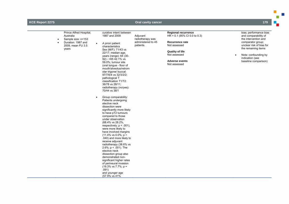
KCE Report 227S Oral cavity cancer 179
Prince Alfred Hospital, Australia
Sample size: n=153 Duration: 1987 and
2009, mean FU 3.5 years
curative intent between 1987 and 2009
A priori patient characteristics Sex (M/F): 71/43 vs 22/17; median age, years (range): 64 (30-92) - <65 42.1% vs. 59.0%; tumour site (oral tongue - floor of mouth/alveolus/retromolar trigone/ buccal: 97/7/6/4 vs 32/3/2/2; pathological T classification T1/T2: 36/78 vs 28/11; radiotherapy (no/yes): 70/44 vs 38/1
Group comparability: Patients undergoing elective neck dissection were significantly more likely to have pT2 tumours compared to those under observation (68.4% vs 28.2%, respectively; p < .001), were more likely to have involved margins (11.4% vs 0.0%; p = .040) and more likely to receive adjuvant radiotherapy (38.6% vs 2.6%; p < .001). The elective neck dissection group also demonstrated non-significant higher rates of perineural invasion (19.3% vs 7.7%; p = .091) and younger age (57.9% vs 41%
Adjuvant radiotherapy was administered to 45 patients.
Regional recurrence HR = 0.1 (95% CI 0.0 to 0.3) Recurrence rate Not assessed Quality of life Not assessed Adverse events Not assessed
bias; performance bias and comparability of the intervention and comparator group; unclear risk of bias for the remaining items
Note: confounding by
indication (see baseline comparison)
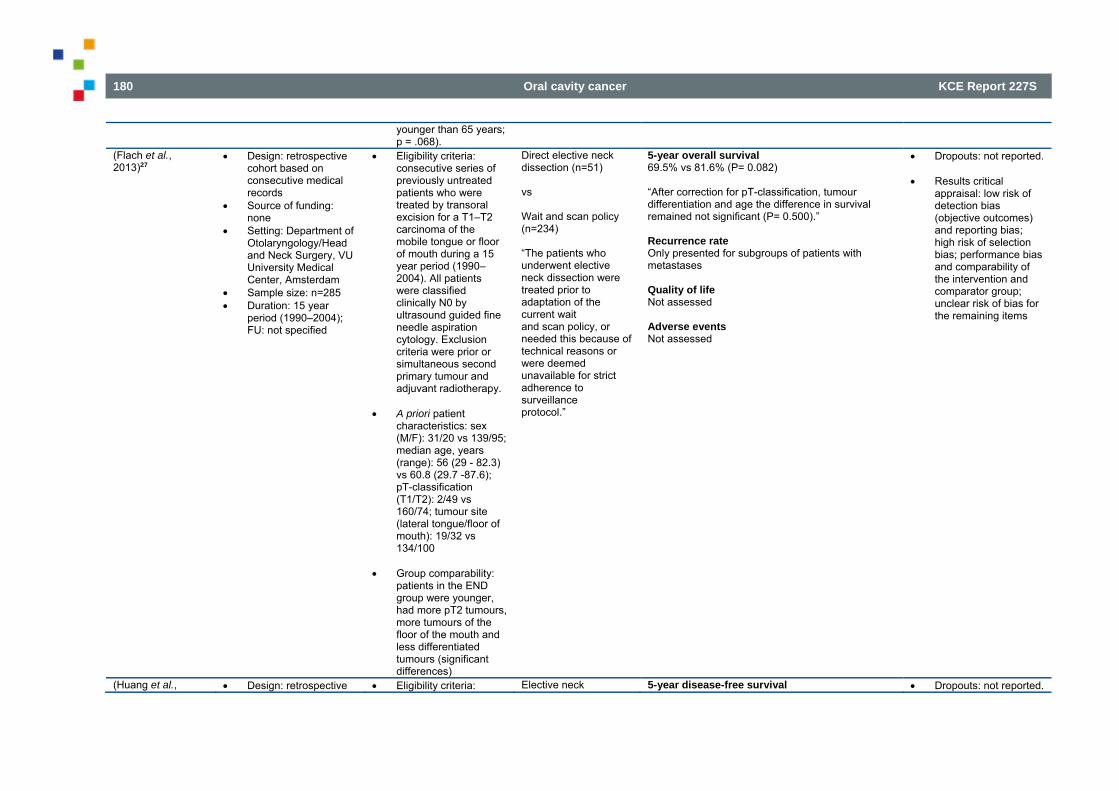
180 Oral cavity cancer KCE Report 227S
younger than 65 years; p = .068).
(Flach et al., 2013)27
Design: retrospective cohort based on consecutive medical records
Source of funding: none
Setting: Department of Otolaryngology/Head and Neck Surgery, VU University Medical Center, Amsterdam
Sample size: n=285 Duration: 15 year
period (1990–2004); FU: not specified
Eligibility criteria: consecutive series of previously untreated patients who were treated by transoral excision for a T1–T2 carcinoma of the mobile tongue or floor of mouth during a 15 year period (1990–2004). All patients were classified clinically N0 by ultrasound guided fine needle aspiration cytology. Exclusion criteria were prior or simultaneous second primary tumour and adjuvant radiotherapy.
A priori patient
characteristics: sex (M/F): 31/20 vs 139/95; median age, years (range): 56 (29 - 82.3) vs 60.8 (29.7 -87.6); pT-classification (T1/T2): 2/49 vs 160/74; tumour site (lateral tongue/floor of mouth): 19/32 vs 134/100
Group comparability:
patients in the END group were younger, had more pT2 tumours, more tumours of the floor of the mouth and less differentiated tumours (significant differences)
Direct elective neck dissection (n=51) vs Wait and scan policy (n=234) “The patients who underwent elective neck dissection were treated prior to adaptation of the current wait and scan policy, or needed this because of technical reasons or were deemed unavailable for strict adherence to surveillance protocol.”
5-year overall survival 69.5% vs 81.6% (P= 0.082) “After correction for pT-classification, tumour differentiation and age the difference in survival remained not significant (P= 0.500).” Recurrence rate Only presented for subgroups of patients with metastases Quality of life Not assessed Adverse events Not assessed
Dropouts: not reported. Results critical
appraisal: low risk of detection bias (objective outcomes) and reporting bias; high risk of selection bias; performance bias and comparability of the intervention and comparator group; unclear risk of bias for the remaining items
(Huang et al., Design: retrospective Eligibility criteria: Elective neck 5-year disease-free survival Dropouts: not reported.
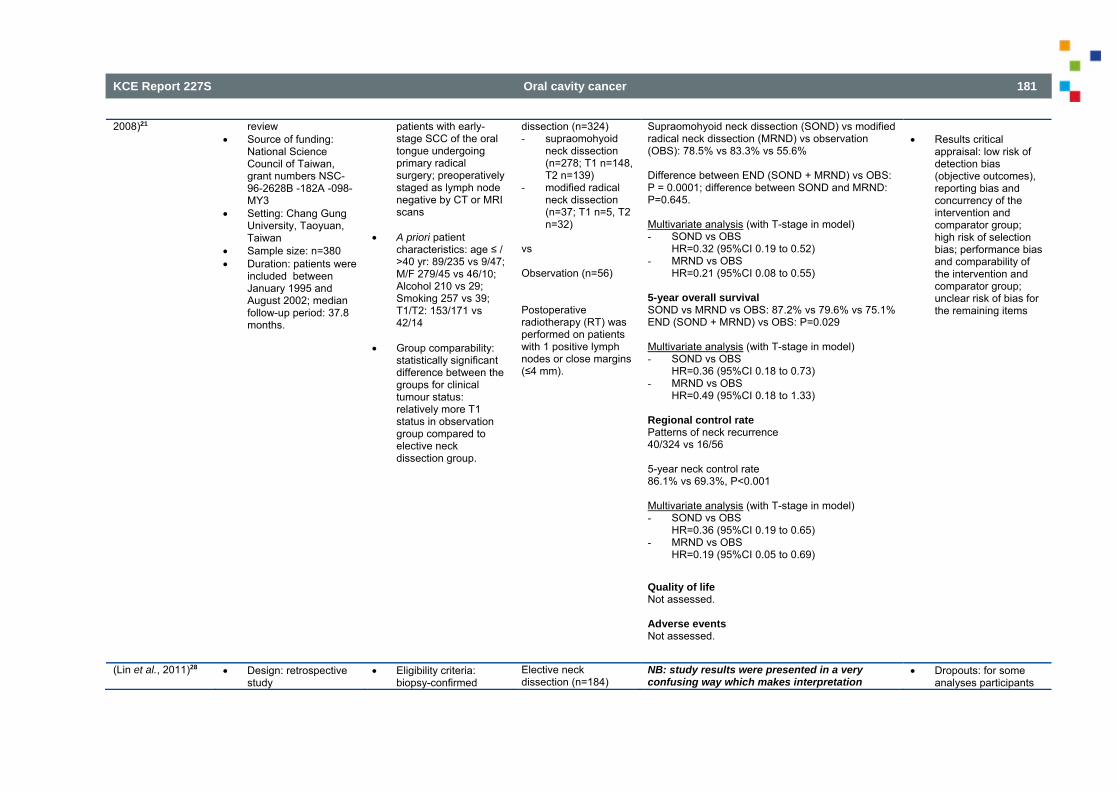
KCE Report 227S Oral cavity cancer 181
2008)21 review Source of funding:
National Science Council of Taiwan, grant numbers NSC-96-2628B -182A -098-MY3
Setting: Chang Gung University, Taoyuan, Taiwan
Sample size: n=380 Duration: patients were
included between January 1995 and August 2002; median follow-up period: 37.8 months.
patients with early-stage SCC of the oral tongue undergoing primary radical surgery; preoperatively staged as lymph node negative by CT or MRI scans
A priori patient
characteristics: age ≤ / >40 yr: 89/235 vs 9/47; M/F 279/45 vs 46/10; Alcohol 210 vs 29; Smoking 257 vs 39; T1/T2: 153/171 vs 42/14
Group comparability:
statistically significant difference between the groups for clinical tumour status: relatively more T1 status in observation group compared to elective neck dissection group.
dissection (n=324) - supraomohyoid
neck dissection (n=278; T1 n=148, T2 n=139)
- modified radical neck dissection (n=37; T1 n=5, T2 n=32)
vs Observation (n=56) Postoperative radiotherapy (RT) was performed on patients with 1 positive lymph nodes or close margins (≤4 mm).
Supraomohyoid neck dissection (SOND) vs modified radical neck dissection (MRND) vs observation (OBS): 78.5% vs 83.3% vs 55.6% Difference between END (SOND + MRND) vs OBS: P = 0.0001; difference between SOND and MRND: P=0.645. Multivariate analysis (with T-stage in model) - SOND vs OBS
HR=0.32 (95%CI 0.19 to 0.52) - MRND vs OBS
HR=0.21 (95%CI 0.08 to 0.55) 5-year overall survival SOND vs MRND vs OBS: 87.2% vs 79.6% vs 75.1% END (SOND + MRND) vs OBS: P=0.029 Multivariate analysis (with T-stage in model) - SOND vs OBS
HR=0.36 (95%CI 0.18 to 0.73) - MRND vs OBS
HR=0.49 (95%CI 0.18 to 1.33) Regional control rate Patterns of neck recurrence 40/324 vs 16/56 5-year neck control rate 86.1% vs 69.3%, P<0.001 Multivariate analysis (with T-stage in model) - SOND vs OBS
HR=0.36 (95%CI 0.19 to 0.65) - MRND vs OBS
HR=0.19 (95%CI 0.05 to 0.69) Quality of life Not assessed. Adverse events Not assessed.
Results critical
appraisal: low risk of detection bias (objective outcomes), reporting bias and concurrency of the intervention and comparator group; high risk of selection bias; performance bias and comparability of the intervention and comparator group; unclear risk of bias for the remaining items
(Lin et al., 2011)28 Design: retrospective study
Eligibility criteria: biopsy-confirmed
Elective neck dissection (n=184)
NB: study results were presented in a very confusing way which makes interpretation
Dropouts: for some analyses participants
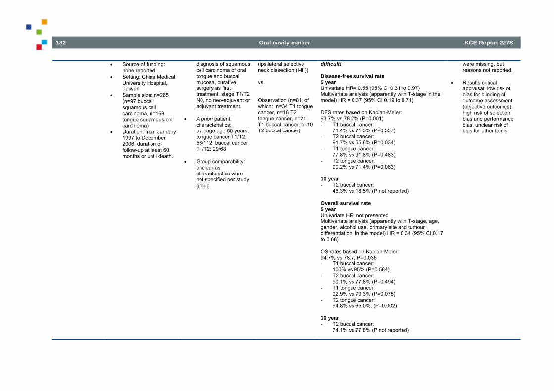
182 Oral cavity cancer KCE Report 227S
Source of funding: none reported
Setting: China Medical University Hospital, Taiwan
Sample size: n=265 (n=97 buccal squamous cell carcinoma, n=168 tongue squamous cell carcinoma)
Duration: from January 1997 to December 2006; duration of follow-up at least 60 months or until death.
diagnosis of squamous cell carcinoma of oral tongue and buccal mucosa, curative surgery as first treatment, stage T1/T2 N0, no neo-adjuvant or adjuvant treatment.
A priori patient
characteristics: average age 50 years; tongue cancer T1/T2: 56/112, buccal cancer T1/T2: 29/68
Group comparability:
unclear as characteristics were not specified per study group.
(ipsilateral selective neck dissection (I-III)) vs Observation (n=81; of which: n=34 T1 tongue cancer, n=16 T2 tongue cancer, n=21 T1 buccal cancer, n=10 T2 buccal cancer)
difficult! Disease-free survival rate 5 year Univariate HR= 0.55 (95% CI 0.31 to 0.97) Multivariate analysis (apparently with T-stage in the model) HR = 0.37 (95% CI 0.19 to 0.71) DFS rates based on Kaplan-Meier: 93.7% vs 78.2% (P=0.001) - T1 buccal cancer:
71.4% vs 71.3% (P=0.337) - T2 buccal cancer:
91.7% vs 55.6% (P=0.034) - T1 tongue cancer:
77.8% vs 91.8% (P=0.483) - T2 tongue cancer:
90.2% vs 71.4% (P=0.063) 10 year - T2 buccal cancer:
46.3% vs 18.5% (P not reported) Overall survival rate 5 year Univariate HR: not presented Multivariate analysis (apparently with T-stage, age, gender, alcohol use, primary site and tumour differentiation in the model) HR = 0.34 (95% CI 0.17 to 0.68) OS rates based on Kaplan-Meier: 94.7% vs 78.7, P=0.036 - T1 buccal cancer:
100% vs 95% (P=0.584) - T2 buccal cancer:
90.1% vs 77.8% (P=0.494) - T1 tongue cancer:
92.9% vs 79.3% (P=0.075) - T2 tongue cancer:
94.8% vs 65.0%, (P=0.002) 10 year - T2 buccal cancer:
74.1% vs 77.8% (P not reported)
were missing, but reasons not reported.
Results critical appraisal: low risk of bias for blinding of outcome assessment (objective outcomes), high risk of selection bias and performance bias, unclear risk of bias for other items.
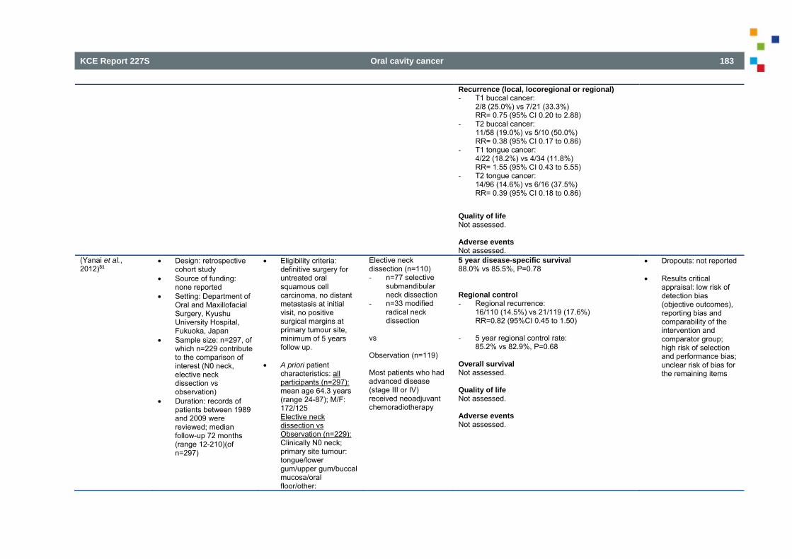
KCE Report 227S Oral cavity cancer 183
Recurrence (local, locoregional or regional) - T1 buccal cancer:
2/8 (25.0%) vs 7/21 (33.3%) RR= 0.75 (95% CI 0.20 to 2.88)
- T2 buccal cancer: 11/58 (19.0%) vs 5/10 (50.0%) RR= 0.38 (95% CI 0.17 to 0.86)
- T1 tongue cancer: 4/22 (18.2%) vs 4/34 (11.8%) RR= 1.55 (95% CI 0.43 to 5.55)
- T2 tongue cancer: 14/96 (14.6%) vs 6/16 (37.5%) RR= 0.39 (95% CI 0.18 to 0.86)
Quality of life Not assessed. Adverse events Not assessed.
(Yanai et al., 2012)31
Design: retrospective cohort study
Source of funding: none reported
Setting: Department of Oral and Maxillofacial Surgery, Kyushu University Hospital, Fukuoka, Japan
Sample size: n=297, of which n=229 contribute to the comparison of interest (N0 neck, elective neck dissection vs observation)
Duration: records of patients between 1989 and 2009 were reviewed; median follow-up 72 months (range 12-210)(of n=297)
Eligibility criteria: definitive surgery for untreated oral squamous cell carcinoma, no distant metastasis at initial visit, no positive surgical margins at primary tumour site, minimum of 5 years follow up.
A priori patient
characteristics: all participants (n=297): mean age 64.3 years (range 24-87); M/F: 172/125 Elective neck dissection vs Observation (n=229): Clinically N0 neck; primary site tumour: tongue/lower gum/upper gum/buccal mucosa/oral floor/other:
Elective neck dissection (n=110) - n=77 selective
submandibular neck dissection
- n=33 modified radical neck dissection
vs Observation (n=119) Most patients who had advanced disease (stage III or IV) received neoadjuvant chemoradiotherapy
5 year disease-specific survival 88.0% vs 85.5%, P=0.78 Regional control - Regional recurrence:
16/110 (14.5%) vs 21/119 (17.6%) RR=0.82 (95%CI 0.45 to 1.50)
- 5 year regional control rate:
85.2% vs 82.9%, P=0.68 Overall survival Not assessed. Quality of life Not assessed. Adverse events Not assessed.
Dropouts: not reported Results critical
appraisal: low risk of detection bias (objective outcomes), reporting bias and comparability of the intervention and comparator group; high risk of selection and performance bias; unclear risk of bias for the remaining items
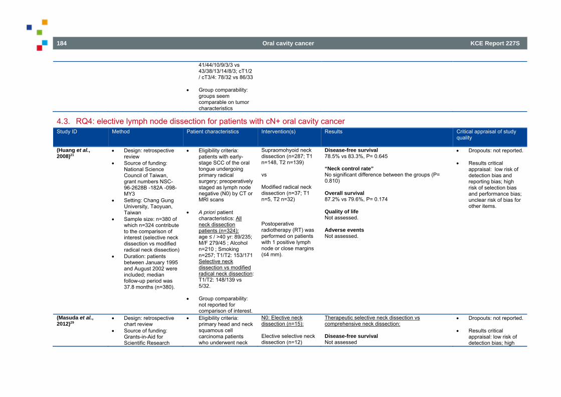
184 Oral cavity cancer KCE Report 227S
41/44/10/9/3/3 vs 43/38/13/14/8/3; cT1/2 / cT3/4: 78/32 vs 86/33
Group comparability: groups seem comparable on tumor characteristics
4.3. RQ4: elective lymph node dissection for patients with cN+ oral cavity cancer Study ID Method Patient characteristics Intervention(s) Results Critical appraisal of study
quality
(Huang et al., 2008)21
Design: retrospective review
Source of funding: National Science Council of Taiwan, grant numbers NSC-96-2628B -182A -098-MY3
Setting: Chang Gung University, Taoyuan, Taiwan
Sample size: n=380 of which n=324 contribute to the comparison of interest (selective neck dissection vs modified radical neck dissection)
Duration: patients between January 1995 and August 2002 were included; median follow-up period was 37.8 months (n=380).
Eligibility criteria: patients with early-stage SCC of the oral tongue undergoing primary radical surgery; preoperatively staged as lymph node negative (N0) by CT or MRI scans
A priori patient
characteristics: All neck dissection patients (n=324): age ≤ / >40 yr: 89/235; M/F 279/45 ; Alcohol n=210 ; Smoking n=257; T1/T2: 153/171 Selective neck dissection vs modified radical neck dissection: T1/T2: 148/139 vs 5/32.
Group comparability: not reported for comparison of interest.
Supraomohyoid neck dissection (n=287; T1 n=148, T2 n=139) vs Modified radical neck dissection (n=37; T1 n=5, T2 n=32) Postoperative radiotherapy (RT) was performed on patients with 1 positive lymph node or close margins (≤4 mm).
Disease-free survival 78.5% vs 83.3%, P= 0.645 “Neck control rate” No significant difference between the groups (P= 0.810) Overall survival 87.2% vs 79.6%, P= 0.174 Quality of life Not assessed. Adverse events Not assessed.
Dropouts: not reported. Results critical
appraisal: low risk of detection bias and reporting bias; high risk of selection bias and performance bias; unclear risk of bias for other items.
(Masuda et al., 2012)29
Design: retrospective chart review
Source of funding: Grants-in-Aid for Scientific Research
Eligibility criteria: primary head and neck squamous cell carcinoma patients who underwent neck
N0: Elective neck dissection (n=15): Elective selective neck dissection (n=12)
Therapeutic selective neck dissection vs comprehensive neck dissection: Disease-free survival Not assessed
Dropouts: not reported. Results critical
appraisal: low risk of detection bias; high
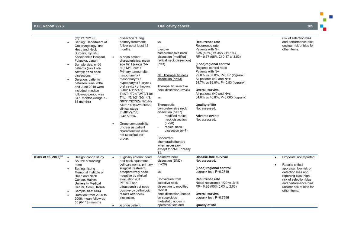
KCE Report 227S Oral cavity cancer 185
(C): 21592195 Setting: Department of
Otolaryngology, and Head and Neck Surgery, Kyushu Koseinenkin Hospital, Fukuoka, Japan
Sample size: n=66 patients (n=21 oral cavity); n=78 neck dissections
Duration: patients between June 2004 and June 2010 were included; median follow-up period was 34.1 months (range 7 - 85 months)
dissection during primary treatment; follow-up at least 12 months.
A priori patient
characteristics: mean age 62.1 (range 34-80); M/F: 55/11; Primary tumour site: nasopharynx / mesopharynx / hypopharynx / larynx / oral cavity / unknown: 3/16/14/11/21/1; T1a/T1/T2b/T2/T3/T4a/T4b: 1/5/1/21/20/14/3; N0/N1/N2/N2a/N2b/N2c/N3: 14/10/2/5/26/6/2; clinical stage I/II/III/IVa/IVb: 0/4/15/32/4
Group comparability:
unclear as patient characteristics were not specified per group.
vs Elective comprehensive neck dissection (modified radical neck dissection) (n=3) N+: Therapeutic neck dissection (n=63) Therapeutic selective neck dissection (n=36) vs Therapeutic comprehensive neck dissection (n=27) - modified radical
neck dissection (n=20)
- radical neck dissection (n=7)
Concurrent chemoradiotherapy when necessary, except for cN0 T1/early T2.
Recurrence rate Recurrence rate Patients with N+ 3/35 (8.3%) vs 3/27 (11.1%) RR= 0.77 (95% CI 0.17 to 3.53) (Loco)regional control Regional control rates Patients with N+ 92.0% vs 87.8%, P=0.57 (logrank) All patients (N0 and N+): 94.7% vs 89.9%, P= 0.53 (logrank) Overall survival All patients (N0 and N+): 64.0% vs 46.8%, P=0.065 (logrank) Quality of life Not assessed. Adverse events Not assessed.
risk of selection bias and performance bias; unclear risk of bias for other items.
(Park et al., 2013)30 Design: cohort study Source of funding:
none Setting: Ilsong
Memorial Institute of Head and Neck Cancer, Hallym University Medical Center, Seoul, Korea
Sample size: n=44 Duration: from 2000 to
2006; mean follow-up 55 (6-118) months
Eligibility criteria: head and neck squamous cell carcinoma, primary surgical treatment, preoperatively node negative by clinical evaluation (CT, PET/CT and ultrasound) but node positive by pathologic results after neck dissection.
A priori patient
Selective neck dissection (SND) (n=29) vs Conversion from selective neck dissection to modified radical neck dissection (based on suspicious metastatic nodes in operative field and
Disease-free survival Not assessed. (Loco) regional control Logrank test: P=0.2719 Recurrence rate Nodal recurrence 1/29 vs 2/15 RR= 0.26 (95% 0.03 to 2.63) Overall survival Logrank test: P=0.7596 Quality of life
Dropouts: not reported. Results critical
appraisal: low risk of detection bias and reporting bias; high risk of selection bias and performance bias; unclear risk of bias for other items.
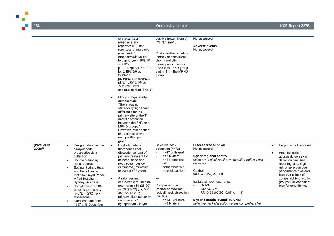
186 Oral cavity cancer KCE Report 227S
characteristics mean age: not reported; M/F: not reported; primary site (oral cavity/ oropharynx/laryn-go-hypopharynx): 16/3/10 vs 6/2/7; pT1/pT2/pT3/pT4a/pT4b/: 2/18/3/6/0 vs 2/8/4//1/0; pN1/pN2a/pN2b/pN2c/pN3: 16/0/12/1/0 vs 7/0/6/2/0; extra capsular spread: 8 vs 6
Group comparability: authors state “There was no statistically significant difference for the primary site or the T and N distribution between the SND and MRND groups.” However, other patient characteristics were not specified per group.
positive frozen biopsy) (MRND) (n=15) Postoperative radiation therapy or concurrent chemo-radiation therapy was done for n=20 in the SND group and n=11 in the MRND group
Not assessed. Adverse events Not assessed.
(Patel et al., 2008)22
Design: retrospective study/cohort; prospective data collection
Source of funding: none reported
Setting: Sydney Head and Neck Cancer Institute, Royal Prince Alfred Hospital, Sydney, Australia
Sample size: n=205 patients (oral cavity n=67), n=232 neck dissections
Duration: data from 1987 until December
Eligibility criteria: therapeutic neck dissection as part of primary treatment for mucosal head and neck squamous cell carcinoma,; minimum follow-up of 2 years.
A priori patient
characteristics: median age (range) 60 (28-99) vs 59 (23-89) yrs; M/F: 45/9 vs 123/27; primary site: oral cavity / oropharynx / hypopharynx / larynx:
Selective neck dissection (n=72) - n=47 unilateral - n=7 bilateral - n=11 combined
with comprehensive neck dissection
vs Comprehensive (radical or modified radical) neck dissection (n=160) - n=131 unilateral - n=9 bilateral
Disease free survival Not assessed 5-year regional control selective neck dissection vs modified radical neck dissection Control 96% vs 86%, P=0.06 Ipsilateral neck recurrence - cN1-3:
2/54 vs 8/71 RR=0.33 (95%CI 0.07 to 1.49)
5-year actuarial overall survival selective neck dissection versus comprehensive
Dropouts: not reported. Results critical
appraisal: low risk of detection bias and reporting bias; high risk of selection bias, performance bias and bias due to lack of comparability of study groups; unclear risk of bias for other items.
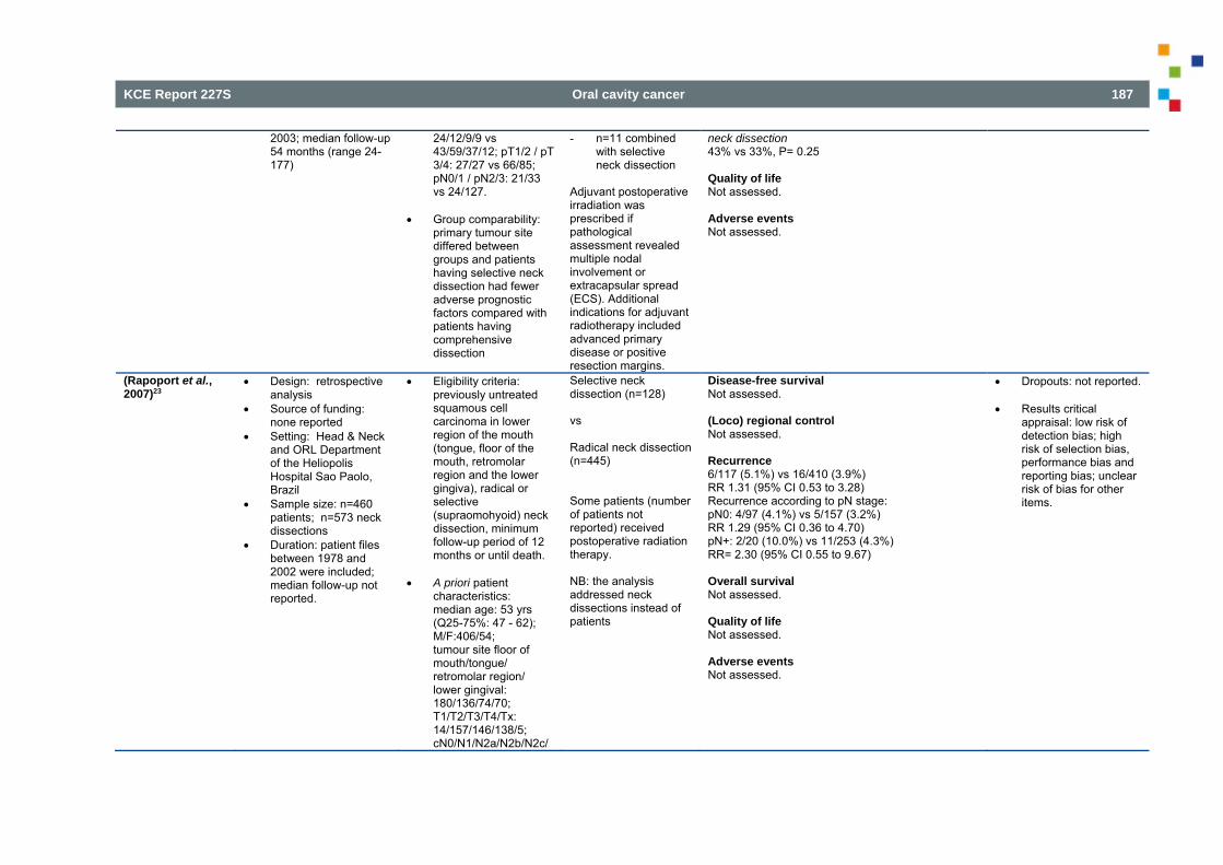
KCE Report 227S Oral cavity cancer 187
2003; median follow-up 54 months (range 24-177)
24/12/9/9 vs 43/59/37/12; pT1/2 / pT 3/4: 27/27 vs 66/85; pN0/1 / pN2/3: 21/33 vs 24/127.
Group comparability:
primary tumour site differed between groups and patients having selective neck dissection had fewer adverse prognostic factors compared with patients having comprehensive dissection
- n=11 combined with selective neck dissection
Adjuvant postoperative irradiation was prescribed if pathological assessment revealed multiple nodal involvement or extracapsular spread (ECS). Additional indications for adjuvant radiotherapy included advanced primary disease or positive resection margins.
neck dissection 43% vs 33%, P= 0.25 Quality of life Not assessed. Adverse events Not assessed.
(Rapoport et al., 2007)23
Design: retrospective analysis
Source of funding: none reported
Setting: Head & Neck and ORL Department of the Heliopolis Hospital Sao Paolo, Brazil
Sample size: n=460 patients; n=573 neck dissections
Duration: patient files between 1978 and 2002 were included; median follow-up not reported.
Eligibility criteria: previously untreated squamous cell carcinoma in lower region of the mouth (tongue, floor of the mouth, retromolar region and the lower gingiva), radical or selective (supraomohyoid) neck dissection, minimum follow-up period of 12 months or until death.
A priori patient
characteristics: median age: 53 yrs (Q25-75%: 47 - 62); M/F:406/54; tumour site floor of mouth/tongue/ retromolar region/ lower gingival: 180/136/74/70; T1/T2/T3/T4/Tx: 14/157/146/138/5; cN0/N1/N2a/N2b/N2c/
Selective neck dissection (n=128) vs Radical neck dissection (n=445) Some patients (number of patients not reported) received postoperative radiation therapy. NB: the analysis addressed neck dissections instead of patients
Disease-free survival Not assessed. (Loco) regional control Not assessed. Recurrence 6/117 (5.1%) vs 16/410 (3.9%) RR 1.31 (95% CI 0.53 to 3.28) Recurrence according to pN stage: pN0: 4/97 (4.1%) vs 5/157 (3.2%) RR 1.29 (95% CI 0.36 to 4.70) pN+: 2/20 (10.0%) vs 11/253 (4.3%) RR= 2.30 (95% CI 0.55 to 9.67) Overall survival Not assessed. Quality of life Not assessed. Adverse events Not assessed.
Dropouts: not reported. Results critical
appraisal: low risk of detection bias; high risk of selection bias, performance bias and reporting bias; unclear risk of bias for other items.
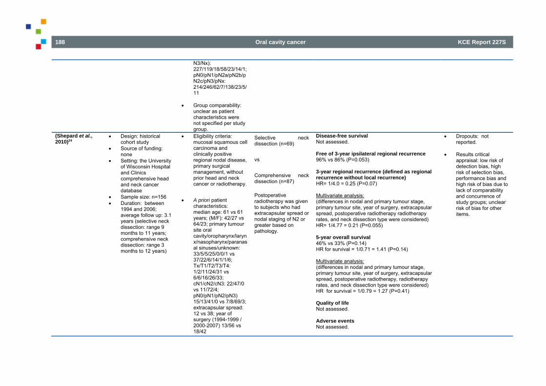
188 Oral cavity cancer KCE Report 227S
N3/Nx): 227/119/18/58/23/14/1; pN0/pN1/pN2a/pN2b/pN2c/pN3/pNx: 214/246/62/7/138/23/5/11
Group comparability: unclear as patient characteristics were not specified per study group.
(Shepard et al., 2010)24
Design: historical cohort study
Source of funding: none
Setting: the University of Wisconsin Hospital and Clinics comprehensive head and neck cancer database
Sample size: n=156 Duration: between
1994 and 2006; average follow up: 3.1 years (selective neck dissection: range 9 months to 11 years; comprehensive neck dissection: range 3 months to 12 years)
Eligibility criteria: mucosal squamous cell carcinoma and clinically positive regional nodal disease, primary surgical management, without prior head and neck cancer or radiotherapy.
A priori patient characteristics: median age: 61 vs 61 years; (M/F): 42/27 vs 64/23; primary tumour site oral cavity/oropharynx/larynx/nasopharynx/paranasal sinuses/unknown: 33/5/5/25/0/0/1 vs 37/22/6/14/1/1/6; Tx/T1/T2/T3/T4: 1/2/11/24/31 vs 6/6/16/26/33; cN1/cN2/cN3: 22/47/0 vs 11/72/4; pN0/pN1/pN2/pN3) 15/13/41/0 vs 7/8/69/3; extracapsular spread: 12 vs 38; year of surgery (1994-1999 / 2000-2007) 13/56 vs 18/42
Selective neck dissection (n=69) vs Comprehensive neck dissection (n=87) Postoperative radiotherapy was given to subjects who had extracapsular spread or nodal staging of N2 or greater based on pathology.
Disease-free survival Not assessed. Free of 3-year ipsilateral regional recurrence 96% vs 86% (P=0.053) 3-year regional recurrence (defined as regional recurrence without local recurrence) HR= 1/4.0 = 0.25 (P=0.07) Multivariate analysis: (differences in nodal and primary tumour stage, primary tumour site, year of surgery, extracapsular spread, postoperative radiotherapy radiotherapy rates, and neck dissection type were considered) HR= 1/4.77 = 0.21 (P=0.055) 5-year overall survival 46% vs 33% (P=0.14) HR for survival = 1/0.71 = 1.41 (P=0.14) Multivariate analysis: (differences in nodal and primary tumour stage, primary tumour site, year of surgery, extracapsular spread, postoperative radiotherapy, radiotherapy rates, and neck dissection type were considered) HR for survival = 1/0.79 = 1.27 (P=0.41) Quality of life Not assessed. Adverse events Not assessed.
Dropouts: not reported.
Results critical
appraisal: low risk of detection bias, high risk of selection bias, performance bias and high risk of bias due to lack of comparability and concurrence of study groups; unclear risk of bias for other items.
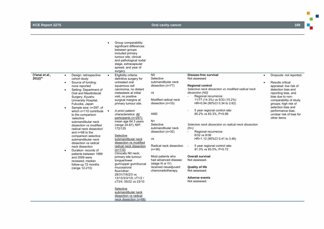
KCE Report 227S Oral cavity cancer 189
Group comparability:
significant differences between groups included primary tumour site, clinical and pathological nodal stage, extracapsular spread, and year of surgery.
(Yanai et al., 2012)31
Design: retrospective cohort study
Source of funding: none reported
Setting: Department of Oral and Maxillofacial Surgery, Kyushu University Hospital, Fukuoka, Japan
Sample size: n=297, of which n=110 contribute to the comparison ‘selective submandibular neck dissection vs modified radical neck dissection’ and n=68 to the comparison selective submandibular neck dissection vs radical neck dissection
Duration: records of patients between 1989 and 2009 were reviewed; median follow-up 72 months (range 12-210)
Eligibility criteria: definitive surgery for untreated oral squamous cell carcinoma, no distant metastasis at initial visit, no positive surgical margins at primary tumour site.
A priori patient
characteristics: All participants (n=297): mean age 64.3 years (range 24-87); M/F: 172/125 Selective submandibular neck dissection vs modified radical neck dissection (n=110) Clinically N0 neck; primary site tumour: tongue/lower gum/upper gum/buccal mucosa/oral floor/other: 28/31/7/6/2/3 vs 13/13/3/3/1/0; cT1/2 / cT3/4: 55/22 vs 23/10 Selective submandibular neck dissection vs radical neck dissection (n=68)
N0 Selective submandibular neck dissection (n=77) vs Modified radical neck dissection (n=33) AND N+ Selective submandibular neck dissection (n=32) vs Radical neck dissection (n=36) Most patients who had advanced disease (stage III or IV) received neoadjuvant chemoradiotherapy.
Disease-free survival Not assessed Regional control Selective neck dissection vs modified radical neck dissection (N0) - Regional recurrence:
11/77 (14.3%) vs 5/33 (15.2%) HR=0.94 (95%CI 0.34 to 2.62)
- 5 year regional control rate:
85.2% vs 83.3%, P=0.89 Selective neck dissection vs radical neck dissection (N+) - Regional recurrence:
6/32 vs 6/36 HR=1.12 (95%CI 0.41 to 3.46)
- 5 year regional control rate:
81.3% vs 83.0%, P=0.72 Overall survival Not assessed. Quality of life Not assessed. Adverse events Not assessed.
Dropouts: not reported. Results critical
appraisal: low risk of detection bias and reporting bias, and bias due to non-comparability of study groups; high risk of selection bias and performance bias; unclear risk of bias for other items.
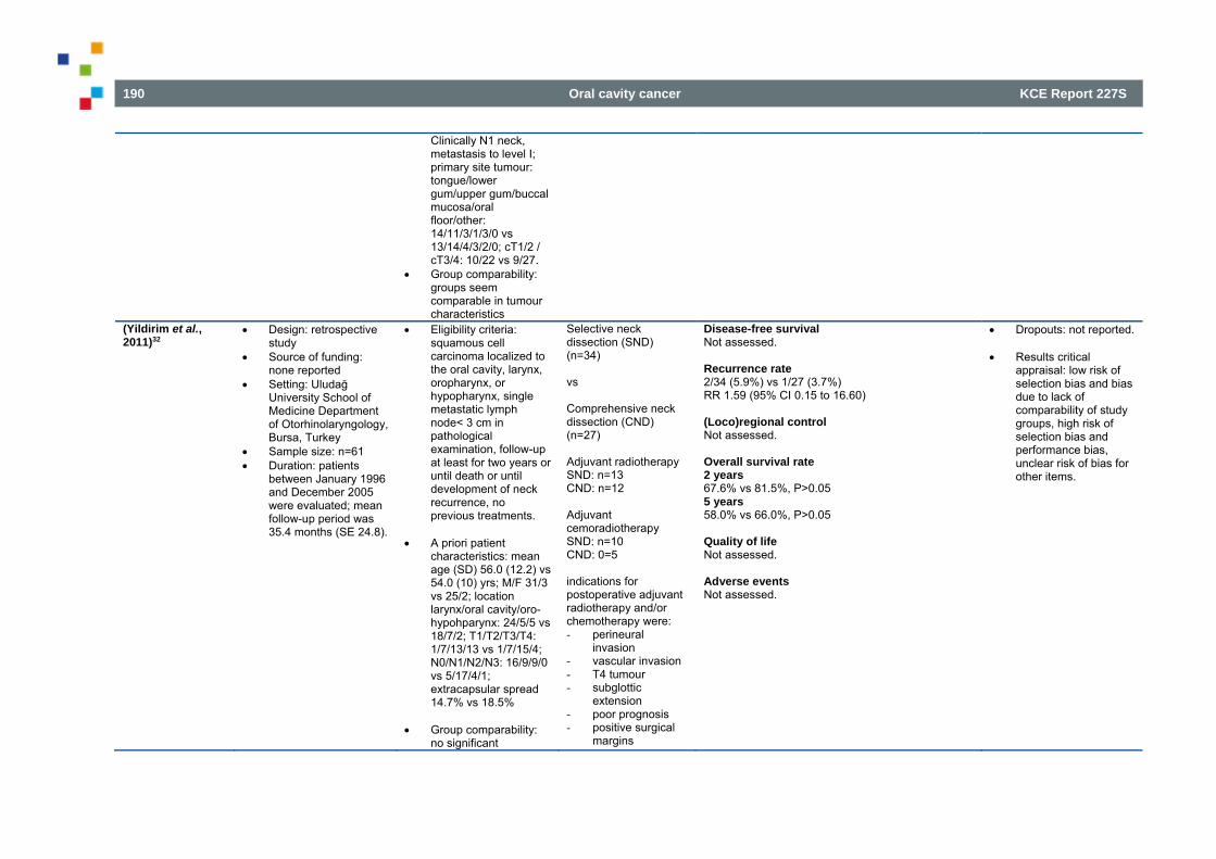
190 Oral cavity cancer KCE Report 227S
Clinically N1 neck, metastasis to level I; primary site tumour: tongue/lower gum/upper gum/buccal mucosa/oral floor/other: 14/11/3/1/3/0 vs 13/14/4/3/2/0; cT1/2 / cT3/4: 10/22 vs 9/27.
Group comparability: groups seem comparable in tumour characteristics
(Yildirim et al., 2011)32
Design: retrospective study
Source of funding: none reported
Setting: Uludağ University School of Medicine Department of Otorhinolaryngology, Bursa, Turkey
Sample size: n=61 Duration: patients
between January 1996 and December 2005 were evaluated; mean follow-up period was 35.4 months (SE 24.8).
Eligibility criteria: squamous cell carcinoma localized to the oral cavity, larynx, oropharynx, or hypopharynx, single metastatic lymph node< 3 cm in pathological examination, follow-up at least for two years or until death or until development of neck recurrence, no previous treatments.
A priori patient
characteristics: mean age (SD) 56.0 (12.2) vs 54.0 (10) yrs; M/F 31/3 vs 25/2; location larynx/oral cavity/oro-hypohparynx: 24/5/5 vs 18/7/2; T1/T2/T3/T4: 1/7/13/13 vs 1/7/15/4; N0/N1/N2/N3: 16/9/9/0 vs 5/17/4/1; extracapsular spread 14.7% vs 18.5%
Group comparability:
no significant
Selective neck dissection (SND) (n=34) vs Comprehensive neck dissection (CND) (n=27) Adjuvant radiotherapy SND: n=13 CND: n=12 Adjuvant cemoradiotherapy SND: n=10 CND: 0=5 indications for postoperative adjuvant radiotherapy and/or chemotherapy were: - perineural
invasion - vascular invasion - T4 tumour - subglottic
extension - poor prognosis - positive surgical
margins
Disease-free survival Not assessed. Recurrence rate 2/34 (5.9%) vs 1/27 (3.7%) RR 1.59 (95% CI 0.15 to 16.60) (Loco)regional control Not assessed. Overall survival rate 2 years 67.6% vs 81.5%, P>0.05 5 years 58.0% vs 66.0%, P>0.05 Quality of life Not assessed. Adverse events Not assessed.
Dropouts: not reported. Results critical
appraisal: low risk of selection bias and bias due to lack of comparability of study groups, high risk of selection bias and performance bias, unclear risk of bias for other items.
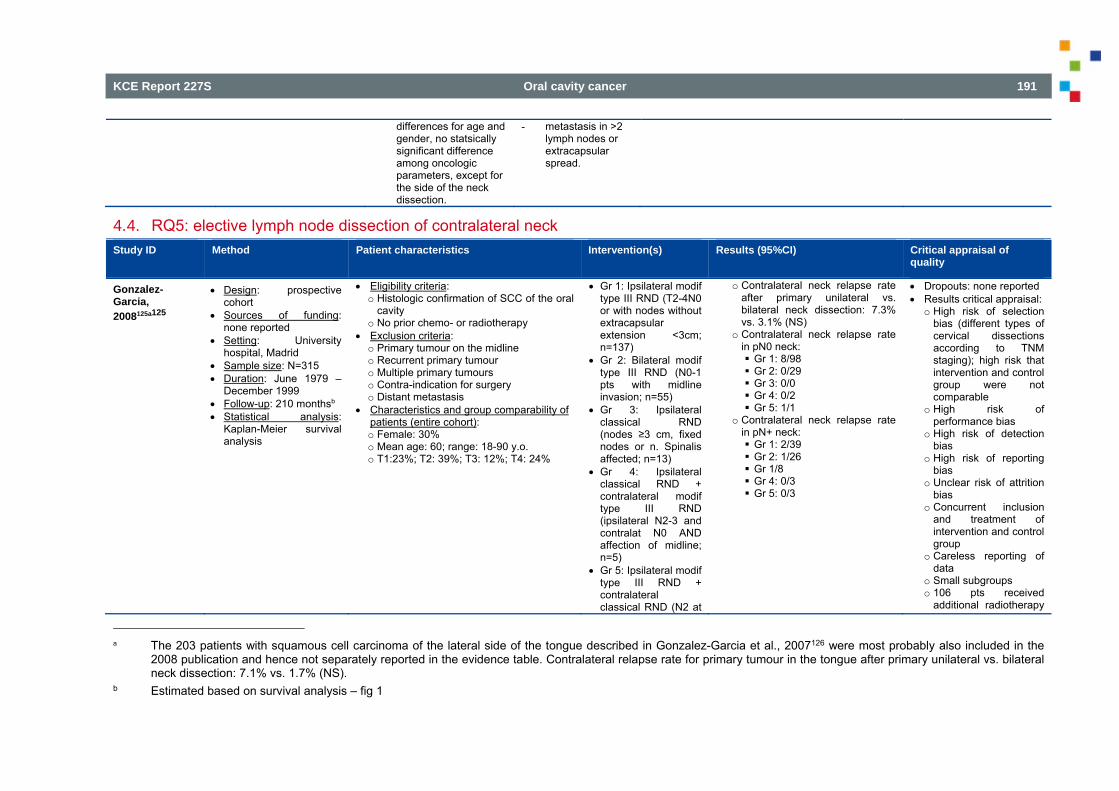
KCE Report 227S Oral cavity cancer 191
differences for age and gender, no statsically significant difference among oncologic parameters, except for the side of the neck dissection.
- metastasis in >2 lymph nodes or extracapsular spread.
4.4. RQ5: elective lymph node dissection of contralateral neck Study ID Method Patient characteristics Intervention(s) Results (95%CI) Critical appraisal of
quality
Gonzalez-Garcia, 2008125a125
Design: prospective cohort
Sources of funding: none reported
Setting: University hospital, Madrid
Sample size: N=315 Duration: June 1979 –
December 1999 Follow-up: 210 monthsb Statistical analysis:
Kaplan-Meier survival analysis
Eligibility criteria: o Histologic confirmation of SCC of the oral
cavity o No prior chemo- or radiotherapy
Exclusion criteria: o Primary tumour on the midline o Recurrent primary tumour o Multiple primary tumours o Contra-indication for surgery o Distant metastasis
Characteristics and group comparability of patients (entire cohort): o Female: 30% o Mean age: 60; range: 18-90 y.o. o T1:23%; T2: 39%; T3: 12%; T4: 24%
Gr 1: Ipsilateral modif type III RND (T2-4N0 or with nodes without extracapsular extension <3cm; n=137)
Gr 2: Bilateral modif type III RND (N0-1 pts with midline invasion; n=55)
Gr 3: Ipsilateral classical RND (nodes ≥3 cm, fixed nodes or n. Spinalis affected; n=13)
Gr 4: Ipsilateral classical RND + contralateral modif type III RND (ipsilateral N2-3 and contralat N0 AND affection of midline; n=5)
Gr 5: Ipsilateral modif type III RND + contralateral classical RND (N2 at
o Contralateral neck relapse rate after primary unilateral vs. bilateral neck dissection: 7.3% vs. 3.1% (NS)
o Contralateral neck relapse rate in pN0 neck: Gr 1: 8/98 Gr 2: 0/29 Gr 3: 0/0 Gr 4: 0/2 Gr 5: 1/1
o Contralateral neck relapse rate in pN+ neck: Gr 1: 2/39 Gr 2: 1/26 Gr 1/8 Gr 4: 0/3 Gr 5: 0/3
Dropouts: none reported Results critical appraisal:
o High risk of selection bias (different types of cervical dissections according to TNM staging); high risk that intervention and control group were not comparable
o High risk of performance bias
o High risk of detection bias
o High risk of reporting bias
o Unclear risk of attrition bias
o Concurrent inclusion and treatment of intervention and control group
o Careless reporting of data
o Small subgroups o 106 pts received
additional radiotherapy
a The 203 patients with squamous cell carcinoma of the lateral side of the tongue described in Gonzalez-Garcia et al., 2007126 were most probably also included in the
2008 publication and hence not separately reported in the evidence table. Contralateral relapse rate for primary tumour in the tongue after primary unilateral vs. bilateral neck dissection: 7.1% vs. 1.7% (NS).
b Estimated based on survival analysis – fig 1
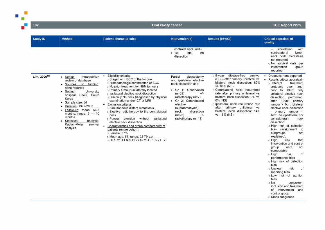
192 Oral cavity cancer KCE Report 227S
Study ID Method Patient characteristics Intervention(s) Results (95%CI) Critical appraisal of quality
contralat neck; n=4) 101 pts: no
dissection
– correlation with contralateral lymph neck node metastasis not reported
o No survival data per intervention group reported
Lim, 2006127 Design: retrospective review of database
Sources of funding: none reported
Setting: University hospital, Seoul, South Korea
Sample size: 54 Duration: 1992-2003 Follow-up: mean: 56.3
months; range: 3 – 110 months
Statistical analysis: Kaplan-Meier survival analysis
Eligibility criteria: o Stage I or II SCC of the tongue o Histopathologic confirmation of SCC o No prior treatment for H&N tumours o Primary tumour unilaterally located o Ipsilateral elective neck dissection o Clinically N0 neck (diagnosed by physical
examination and/or CT or MRI Exclusion criteria: o Simultaneous distant metastasis o Elective radiotherapy to the contralateral
neck o Peroral excision without ipsilateral
elective neck dissection Characteristics and group comparability of
patients (entire cohort): o Female: 37% o Mean age: 53; range: 22-79 y.o. o Gr 1: 21 T1 & 8 T2 vs Gr 2: 4 T1 & 21 T2
Partial glossectomy and ipsilateral elective neck dissection and:
Gr 1: Observation (n=29) +/- radiotherapy (n=7)
Gr 2: Contralateral elective (supraomohyoid) neck dissection (n=25) +/- radiotherapy (n=13)
o 5-year disease-free survival (DFS) after primary unilateral vs. bilateral neck dissection: 82% vs. 68% (NS)
o Contralateral neck recurrence rate after primary unilateral vs. bilateral neck dissection: 0% vs. 0% (NS)
o Ipsilateral neck recurrence rate after primary unilateral vs. bilateral neck dissection: 14% vs. 16% (NS)
Dropouts: none reported Results critical appraisal: o Different treatment
protocols over time: prior to 1998: only unilateral elective neck dissection performed; after 1998: primary tumour > 1cm: bilateral elective neck dissection - primary tumour < 1cm: no (ipsilateral nor contralateral) neck dissection
o High risk of selection bias (assignment to subgroups not explained);
o High risk that intervention and control group were not comparable
o High risk of performance bias
o High risk of detection bias
o Unclear risk of reporting bias
o Low risk of attrition bias
o No concurrent inclusion and treatment of intervention and control group
o Small subgroups
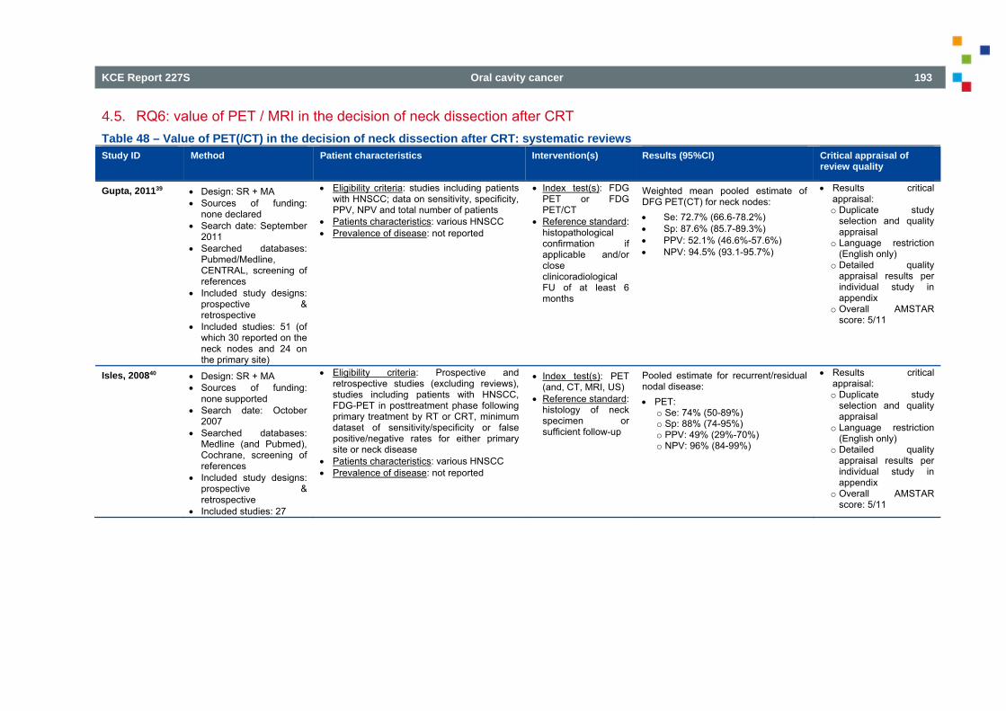
KCE Report 227S Oral cavity cancer 193
4.5. RQ6: value of PET / MRI in the decision of neck dissection after CRT Table 48 – Value of PET(/CT) in the decision of neck dissection after CRT: systematic reviews Study ID Method Patient characteristics Intervention(s) Results (95%CI) Critical appraisal of
review quality
Gupta, 201139 Design: SR + MA Sources of funding:
none declared Search date: September
2011 Searched databases:
Pubmed/Medline, CENTRAL, screening of references
Included study designs: prospective & retrospective
Included studies: 51 (of which 30 reported on the neck nodes and 24 on the primary site)
Eligibility criteria: studies including patients with HNSCC; data on sensitivity, specificity, PPV, NPV and total number of patients
Patients characteristics: various HNSCC Prevalence of disease: not reported
Index test(s): FDG PET or FDG PET/CT
Reference standard: histopathological confirmation if applicable and/or close clinicoradiological FU of at least 6 months
Weighted mean pooled estimate of DFG PET(CT) for neck nodes: Se: 72.7% (66.6-78.2%) Sp: 87.6% (85.7-89.3%) PPV: 52.1% (46.6%-57.6%) NPV: 94.5% (93.1-95.7%)
Results critical appraisal: o Duplicate study
selection and quality appraisal
o Language restriction (English only)
o Detailed quality appraisal results per individual study in appendix
o Overall AMSTAR score: 5/11
Isles, 200840 Design: SR + MA Sources of funding:
none supported Search date: October
2007 Searched databases:
Medline (and Pubmed), Cochrane, screening of references
Included study designs: prospective & retrospective
Included studies: 27
Eligibility criteria: Prospective and retrospective studies (excluding reviews), studies including patients with HNSCC, FDG-PET in posttreatment phase following primary treatment by RT or CRT, minimum dataset of sensitivity/specificity or false positive/negative rates for either primary site or neck disease
Patients characteristics: various HNSCC Prevalence of disease: not reported
Index test(s): PET (and, CT, MRI, US)
Reference standard: histology of neck specimen or sufficient follow-up
Pooled estimate for recurrent/residual nodal disease: PET:
o Se: 74% (50-89%) o Sp: 88% (74-95%) o PPV: 49% (29%-70%) o NPV: 96% (84-99%)
Results critical appraisal: o Duplicate study
selection and quality appraisal
o Language restriction (English only)
o Detailed quality appraisal results per individual study in appendix
o Overall AMSTAR score: 5/11
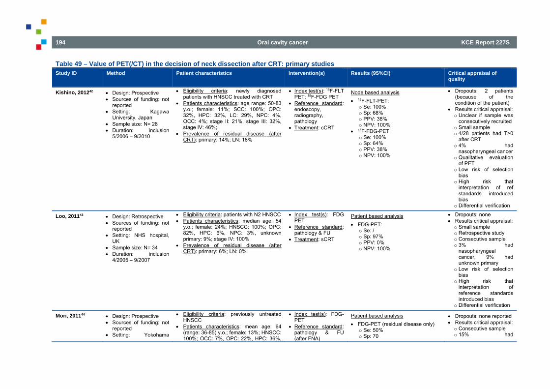
194 Oral cavity cancer KCE Report 227S
Table 49 – Value of PET(/CT) in the decision of neck dissection after CRT: primary studies Study ID Method Patient characteristics Intervention(s) Results (95%CI) Critical appraisal of
quality
Kishino, 201242 Design: Prospective Sources of funding: not
reported Setting: Kagawa
University, Japan Sample size: N= 28 Duration: inclusion
5/2006 – 9/2010
Eligibility criteria: newly diagnosed patients with HNSCC treated with CRT
Patients characteristics: age range: 50-83 y.o.; female: 11%; SCC: 100%; OPC: 32%, HPC: 32%, LC: 29%, NPC: 4%, OCC: 4%; stage II: 21%, stage III: 32%, stage IV: 46%;
Prevalence of residual disease (after CRT): primary: 14%; LN: 18%
Index test(s): 18F-FLT PET; 18F-FDG PET
Reference standard: endoscopy, radiography, pathology
Treatment: cCRT
Node based analysis 18F-FLT-PET:
o Se: 100% o Sp: 68% o PPV: 38% o NPV: 100%
18F-FDG-PET: o Se: 100% o Sp: 64% o PPV: 38% o NPV: 100%
Dropouts: 2 patients (because of the condition of the patient)
Results critical appraisal: o Unclear if sample was
consecutively recruited o Small sample o 4/28 patients had T>0
after CRT o 4% had
nasopharyngeal cancer o Qualitative evaluation
of PET o Low risk of selection
bias o High risk that
interpretation of ref standards introduced bias
o Differential verification
Loo, 201143 Design: Retrospective Sources of funding: not
reported Setting: NHS hospital,
UK Sample size: N= 34 Duration: inclusion
4/2005 – 9/2007
Eligibility criteria: patients with N2 HNSCC Patients characteristics: median age: 54
y.o.; female: 24%; HNSCC: 100%; OPC: 82%, HPC: 6%, NPC: 3%, unknown primary: 9%; stage IV: 100%
Prevalence of residual disease (after CRT): primary: 6%; LN: 0%
Index test(s): FDG PET
Reference standard: pathology & FU
Treatment: sCRT
Patient based analysis FDG-PET:
o Se: / o Sp: 97% o PPV: 0% o NPV: 100%
Dropouts: none Results critical appraisal: o Small sample o Retrospective study o Consecutive sample o 3% had
nasopharyngeal cancer, 9% had unknown primary
o Low risk of selection bias
o High risk that interpretation of reference standards introduced bias
o Differential verification
Mori, 201144 Design: Prospective Sources of funding: not
reported Setting: Yokohama
Eligibility criteria: previously untreated HNSCC
Patients characteristics: mean age: 64 (range: 36-85) y.o.; female: 13%; HNSCC: 100%; OCC: 7%, OPC: 22%, HPC: 36%,
Index test(s): FDG-PET
Reference standard: pathology & FU (after FNA)
Patient based analysis FDG-PET (residual disease only)
o Se: 50% o Sp: 70
Dropouts: none reported Results critical appraisal: o Consecutive sample o 15% had
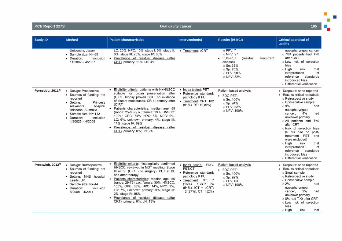
KCE Report 227S Oral cavity cancer 195
Study ID Method Patient characteristics Intervention(s) Results (95%CI) Critical appraisal of quality
University, Japan Sample size: N= 65 Duration: inclusion
11/2002 – 4/2007
LC: 20%, NPC: 15%; stage I: 5%, stage II: 6%, stage III: 23%, stage IV: 66%
Prevalence of residual disease (after CRT): primary: 11%; LN: 4%
Treatment: cCRT
o PPV: 7 o NPV: 97
FDG-PET (residual +recurrent disease) o Se: 33% o Sp: 70% o PPV: 20% o NPV: 82%
nasopharyngeal cancer o 7/64 patients had T>0
after CRT o Low risk of selection
bias o High risk that
interpretation of reference standards introduced bias
o Differential verification
Porceddu, 201145 Design: Prospective Sources of funding: not
reported Setting: Princess
Alexandra hospital Brisbane, Australia
Sample size: N= 112 Duration: inclusion
1/20025 – 4/2009
Eligibility criteria: patients with N+HNSCC suitable for organ preservation after (C)RT, biopsy proven SCC, no evidence of distant metastases, CR at primary after (C)RT
Patients characteristics: median age: 55 (range: 25-88) y.o.; female: 19%; HNSCC: 100%; OPC: 74%, HPC: 6%, NPC: 9%, LC: 6%, unknown primary: 4%; stage III: 11%, stage IV: 89%
Prevalence of residual disease (after CRT): primary: 0%; LN: 2%
Index test(s): PET Reference standard:
pathology & FU Treatment: CRT: 102
(91%), RT: 10 (9%)
Patient based analysis FDG-PET:
o Se: 100% o Sp: 94% o PPV: 22% o NPV: 100%
Dropouts: none reported Results critical appraisal: o Retrospective study o Consecutive sample o 9% had
nasopharyngeal cancer, 4% had unknown primary
o All patients had T=0 after CRT
o Risk of selection bias (5 pts had no post-treatment PET and were excluded)
o High risk that interpretation of reference standards introduced bias
o Differential verification
Prestwich, 201246 Design: Retrospective Sources of funding: not
reported Setting: NHS hospital
Leeds, UK Sample size: N= 44 Duration: inclusion
8/2008 – 4/2011
Eligibility criteria: histologically confirmed HNSCC, reviewed in MDT meeting, Stage III or IV, (C)RT (no surgery), PET at BL and after therapy
Patients characteristics: median age: 55 (range: 29-75) y.o.; female: 30%; HNSCC: 100%; OPC: 68%, HPC: 14%, NPC: 2%, LC: 7%, unknown primary: 9%; stage III: 2%, stage IV: 98%
Prevalence of residual disease (after CRT): primary: 8%; LN: 13%
Index test(s): FDG-PET/CT
Reference standard: pathology & FU
Treatment: RT: 7 (16%), cCRT: 24 (54%), ICT + cCRT: 12 (27%), CT: 1 (2%)
Patient based analysis FDG-PET:
o Se: 100% o Sp: 92% o PPV: 63 o NPV: 100%
Dropouts: none reported Results critical appraisal: o Small sample o Retrospective study o Consecutive sample o 2% had
nasopharyngeal cancer, 9% had unknown primary
o 8% had T>0 after CRT o Low risk of selection
bias o High risk that
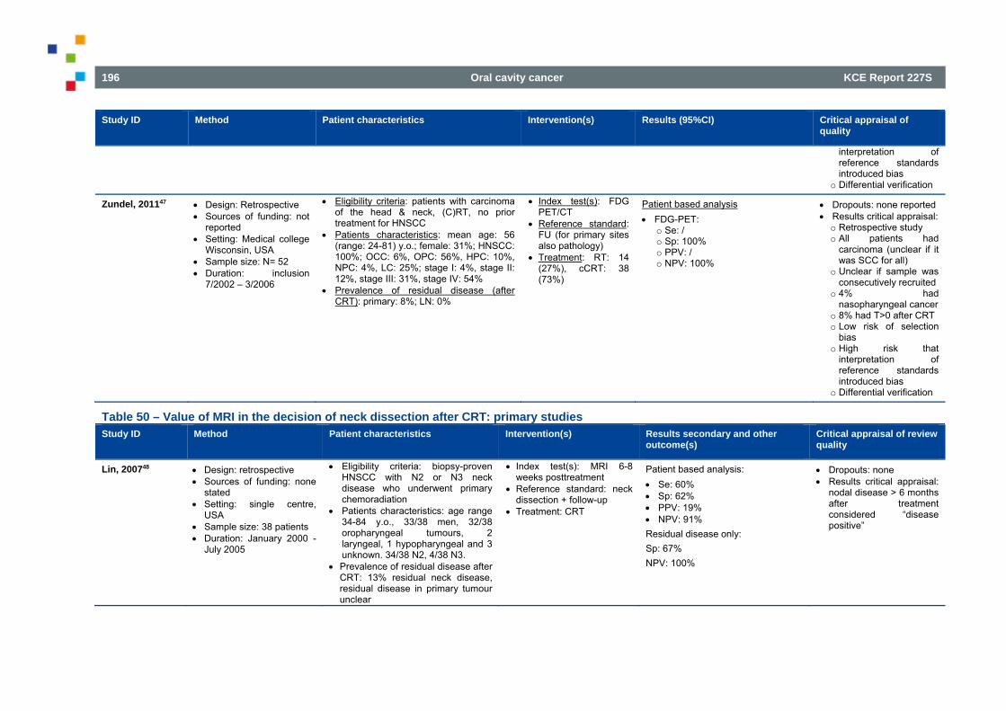
196 Oral cavity cancer KCE Report 227S
Study ID Method Patient characteristics Intervention(s) Results (95%CI) Critical appraisal of quality
interpretation of reference standards introduced bias
o Differential verification
Zundel, 201147 Design: Retrospective Sources of funding: not
reported Setting: Medical college
Wisconsin, USA Sample size: N= 52 Duration: inclusion
7/2002 – 3/2006
Eligibility criteria: patients with carcinoma of the head & neck, (C)RT, no prior treatment for HNSCC
Patients characteristics: mean age: 56 (range: 24-81) y.o.; female: 31%; HNSCC: 100%; OCC: 6%, OPC: 56%, HPC: 10%, NPC: 4%, LC: 25%; stage I: 4%, stage II: 12%, stage III: 31%, stage IV: 54%
Prevalence of residual disease (after CRT): primary: 8%; LN: 0%
Index test(s): FDG PET/CT
Reference standard: FU (for primary sites also pathology)
Treatment: RT: 14 (27%), cCRT: 38 (73%)
Patient based analysis FDG-PET:
o Se: / o Sp: 100% o PPV: / o NPV: 100%
Dropouts: none reported Results critical appraisal: o Retrospective study o All patients had
carcinoma (unclear if it was SCC for all)
o Unclear if sample was consecutively recruited
o 4% had nasopharyngeal cancer
o 8% had T>0 after CRT o Low risk of selection
bias o High risk that
interpretation of reference standards introduced bias
o Differential verification
Table 50 – Value of MRI in the decision of neck dissection after CRT: primary studies Study ID Method Patient characteristics Intervention(s) Results secondary and other
outcome(s) Critical appraisal of review quality
Lin, 200748 Design: retrospective Sources of funding: none
stated Setting: single centre,
USA Sample size: 38 patients Duration: January 2000 -
July 2005
Eligibility criteria: biopsy-proven HNSCC with N2 or N3 neck disease who underwent primary chemoradiation
Patients characteristics: age range 34-84 y.o., 33/38 men, 32/38 oropharyngeal tumours, 2 laryngeal, 1 hypopharyngeal and 3 unknown. 34/38 N2, 4/38 N3.
Prevalence of residual disease after CRT: 13% residual neck disease, residual disease in primary tumour unclear
Index test(s): MRI 6-8 weeks posttreatment
Reference standard: neck dissection + follow-up
Treatment: CRT
Patient based analysis: Se: 60% Sp: 62% PPV: 19% NPV: 91% Residual disease only: Sp: 67% NPV: 100%
Dropouts: none Results critical appraisal:
nodal disease > 6 months after treatment considered “disease positive”
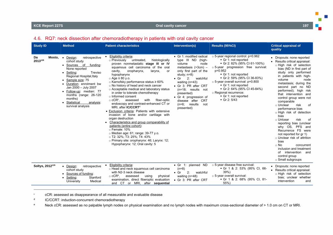
KCE Report 227S Oral cavity cancer 197
4.6. RQ7: neck dissection after chemoradiotherapy in patients with oral cavity cancer Study ID Method Patient characteristics Intervention(s) Results (95%CI) Critical appraisal of
quality
Da Mosto, 2013128
Design: retrospective cohort study
Sources of funding: None reported
Setting: Treviso Regional Hospital,Italy
Sample size: 75 Duration: enrolment bw
Jan 2000 – July 2007 Follow-up: median: 77
months (range: 26-120 months)
Statistical analysis: survival analysis
Eligibility criteria: o Previously untreated, histologically
proven nonmetastatic stage III or IV squamous cell carcinoma of the oral cavity, oropharynx, larynx, or hypopharynx
o Age ≤ 80 y.o. o Karnofsky performance status ≥ 60% o No history of head and neck cancer o Acceptable medical and laboratory status
in order to tolerate chemotherapy o Informed consent o cCRc, assessed with fiber-optic
endoscopy and contrast-enhanced CT or MRI, after IC/CCRTd
Exclusion criteria: Patients with extensive invasion of bone and/or cartilage with organ destruction
Characteristics and group comparability of patients (entire cohort): o Female: 10% o Median age: 61; range: 39-77 y.o. o T2: 32%; T3: 25%; T4: 43% o Primary site: oropharynx: 46; Larynx: 12;
Hypopharynx: 12; Oral cavity: 5
Gr 1: modified radical type III ND (high-volume node metastasis (>3cm) – only first part of the study; n=8)
Gr 2: watchful waiting (n=43)
Gr 3: PR after CRT (n=18; results not presented)
Gr 4: progression of disease after CRT (n=6; results not presented)
o 5-year regional control: p=0.962 Gr 1: not reported Gr 2: 82% (95% CI 61-100%)
o 5-year progression free survival: p=0.952 Gr 1: not reported Gr 2: 59% (95% CI 36-83%)
o 5-year overall survival: p=0.800 Gr 1: not reported Gr 2: 64% (95% CI 45-84%)
o Regional recurrence: Gr 1: not reported Gr 2: 5/43
Dropouts: none reported Results critical appraisal:
o High risk of selection bias (ND in first part of study only performed in patients with high-volume node metastasis; during the second part no ND performed); high risk that intervention and control group were not comparable
o Unclear risk of performance bias
o High risk of detection bias
o Unlcear risk of reporting bias (unclear why OS, PFS and Recurrence FS were not reported for gr 1)
o Unclear risk of attrition bias
o No concurrent inclusion and treatment of intervention and control group
o Small subgroups
Soltys, 2012129 Design: retrospective cohort study
Sources of funding: Setting: Stanford
University Medical
Eligibility criteria: o Head and neck squamous cell carcinoma
with N2-3 neck disease o cCRe, assessed using physical
examination, direct fiberoptic evaluation and CT or MRI, after sequential
Gr 1: planned ND (n=8)
Gr 2: watchful waiting (n=48)
Gr 3: PR after CRT
o 5-year disease free survival: Gr 1 & 2: 53% (95% CI, 66-
39%) o 5-year overall survival:
Gr 1 & 2: 68% (95% CI, 81-55%)
Dropouts: none reported Results critical appraisal:
o High risk of selection bias; unclear whether intervention and
c cCR: assessed as disappearance of all measurable and evaluable disease d IC/CCRT: induction-concurrent chemoradiotherapy e Neck cCR: assessed as no palpable lymph nodes on physical examination and no lymph nodes with maximum cross-sectional diameter of > 1.0 cm on CT or MRI.
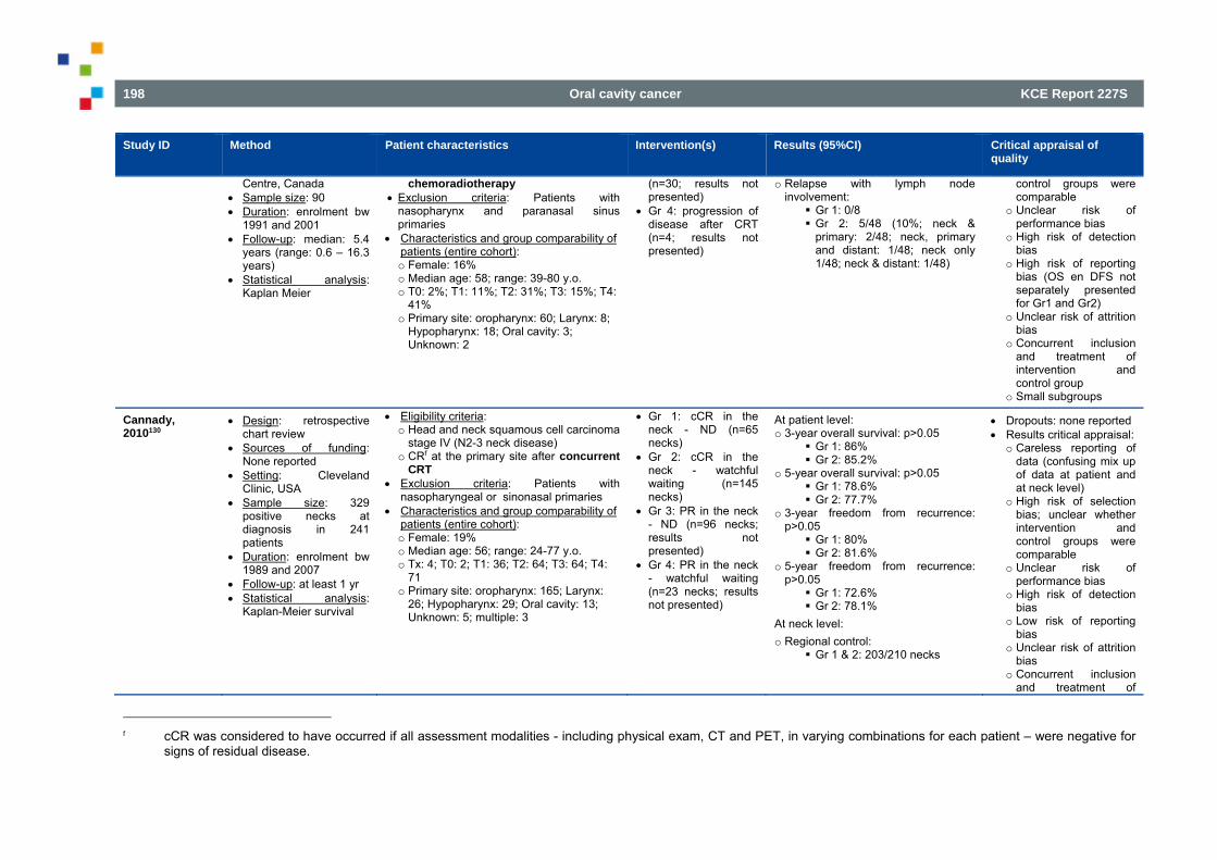
198 Oral cavity cancer KCE Report 227S
Study ID Method Patient characteristics Intervention(s) Results (95%CI) Critical appraisal of quality
Centre, Canada Sample size: 90 Duration: enrolment bw
1991 and 2001 Follow-up: median: 5.4
years (range: 0.6 – 16.3 years)
Statistical analysis: Kaplan Meier
chemoradiotherapy Exclusion criteria: Patients with
nasopharynx and paranasal sinus primaries
Characteristics and group comparability of patients (entire cohort): o Female: 16% o Median age: 58; range: 39-80 y.o. o T0: 2%; T1: 11%; T2: 31%; T3: 15%; T4:
41% o Primary site: oropharynx: 60; Larynx: 8;
Hypopharynx: 18; Oral cavity: 3; Unknown: 2
(n=30; results not presented)
Gr 4: progression of disease after CRT (n=4; results not presented)
o Relapse with lymph node involvement: Gr 1: 0/8 Gr 2: 5/48 (10%; neck &
primary: 2/48; neck, primary and distant: 1/48; neck only 1/48; neck & distant: 1/48)
control groups were comparable
o Unclear risk of performance bias
o High risk of detection bias
o High risk of reporting bias (OS en DFS not separately presented for Gr1 and Gr2)
o Unclear risk of attrition bias
o Concurrent inclusion and treatment of intervention and control group
o Small subgroups
Cannady, 2010130
Design: retrospective chart review
Sources of funding: None reported
Setting: Cleveland Clinic, USA
Sample size: 329 positive necks at diagnosis in 241 patients
Duration: enrolment bw 1989 and 2007
Follow-up: at least 1 yr Statistical analysis:
Kaplan-Meier survival
Eligibility criteria: o Head and neck squamous cell carcinoma
stage IV (N2-3 neck disease) o CRf at the primary site after concurrent
CRT Exclusion criteria: Patients with
nasopharyngeal or sinonasal primaries Characteristics and group comparability of
patients (entire cohort): o Female: 19% o Median age: 56; range: 24-77 y.o. o Tx: 4; T0: 2; T1: 36; T2: 64; T3: 64; T4:
71 o Primary site: oropharynx: 165; Larynx:
26; Hypopharynx: 29; Oral cavity: 13; Unknown: 5; multiple: 3
Gr 1: cCR in the neck - ND (n=65 necks)
Gr 2: cCR in the neck - watchful waiting (n=145 necks)
Gr 3: PR in the neck - ND (n=96 necks; results not presented)
Gr 4: PR in the neck - watchful waiting (n=23 necks; results not presented)
At patient level: o 3-year overall survival: p>0.05
Gr 1: 86% Gr 2: 85.2%
o 5-year overall survival: p>0.05 Gr 1: 78.6% Gr 2: 77.7%
o 3-year freedom from recurrence: p>0.05 Gr 1: 80% Gr 2: 81.6%
o 5-year freedom from recurrence: p>0.05 Gr 1: 72.6% Gr 2: 78.1%
At neck level: o Regional control:
Gr 1 & 2: 203/210 necks
Dropouts: none reported Results critical appraisal:
o Careless reporting of data (confusing mix up of data at patient and at neck level)
o High risk of selection bias; unclear whether intervention and control groups were comparable
o Unclear risk of performance bias
o High risk of detection bias
o Low risk of reporting bias
o Unclear risk of attrition bias
o Concurrent inclusion and treatment of
f cCR was considered to have occurred if all assessment modalities - including physical exam, CT and PET, in varying combinations for each patient – were negative for
signs of residual disease.
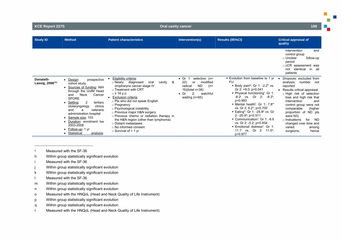
KCE Report 227S Oral cavity cancer 199
Study ID Method Patient characteristics Intervention(s) Results (95%CI) Critical appraisal of quality
intervention and control group
o Unclear follow-up period
o cCR assessment was not identical in all patients
Donatelli-Lassig, 2008131
Design: prospective cohort study
Sources of funding: NIH through the UofM Head and Neck Cancer SPORE
Setting: 2 tertiary otolaryngology clinics and a veterans administration hospital
Sample size: 103 Duration: enrolment bw
2003-2008 Follow-up: 1 yr Statistical analysis:
Eligibility criteria: o Newly diagnosed oral cavity &
oropharynx cancer stage IV o Treatment with CRT o ≥ 18 y.o.
Exclusion criteria: o Pts who did not speak English o Pregnancy o Psychological instability o Previous major H&N surgery o Previous chemo or radiation therapy in
the H&N region (other than lymphoma) o Distant metastases o No informed consent o Survival of < 1 yr
Gr 1: selective (n= 22) or modified radical ND (n= 16)(total n=38)
Gr 2: watchful waiting (n=65)
Evolution from baseline to 1 yr FU: Body paing: Gr 1: -2.2h vs.
Gr 2: +8.0; p=0.041 Physical functioningi: Gr 1:
-8.2j vs. Gr 2: -8.3k; p=0.993 Mental healthl: Gr 1: 7.8m
vs. Gr 2: 6.2n; p=0.700 Eatingo: Gr 1: -24.8p vs. Gr
2: -20.9q; p=0.511 Communicationr: Gr 1: -6.6
vs. Gr 2: -5.2; p=0.834 Emotional distresss: Gr 1:
11.1t vs. Gr 2: 11.0u; p=0.977
Dropouts: excluded from analysis; number not reported
Results critical appraisal: o High risk of selection
bias and high risk that intervention and control group were not comparable (higher proportion of ND pts were N3)
o Indications for ND changed over time and varied among surgeons, hence
g Measured with the SF-36 h Within group statistically significant evolution i Measured with the SF-36 j Within group statistically significant evolution k Within group statistically significant evolution l Measured with the SF-36 m Within group statistically significant evolution n Within group statistically significant evolution o Measured with the HNQoL (Head and Neck Quality of Life Instrument) p Within group statistically significant evolution q Within group statistically significant evolution r Measured with the HNQoL (Head and Neck Quality of Life Instrument)
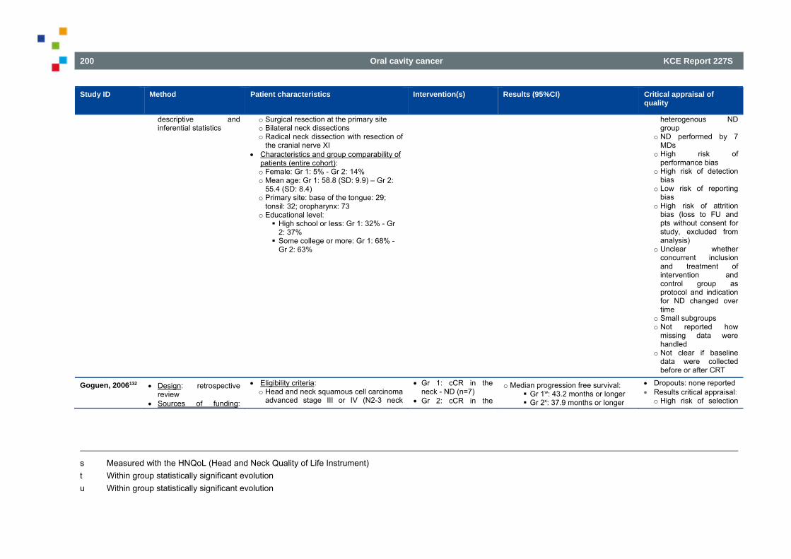
200 Oral cavity cancer KCE Report 227S
Study ID Method Patient characteristics Intervention(s) Results (95%CI) Critical appraisal of quality
descriptive and inferential statistics
o Surgical resection at the primary site o Bilateral neck dissections o Radical neck dissection with resection of
the cranial nerve XI Characteristics and group comparability of
patients (entire cohort): o Female: Gr 1: 5% - Gr 2: 14% o Mean age: Gr 1: 58.8 (SD: 9.9) – Gr 2:
55.4 (SD: 8.4) o Primary site: base of the tongue: 29;
tonsil: 32; oropharynx: 73 o Educational level: High school or less: Gr 1: 32% - Gr
2: 37% Some college or more: Gr 1: 68% -
Gr 2: 63%
heterogenous ND group
o ND performed by 7 MDs
o High risk of performance bias
o High risk of detection bias
o Low risk of reporting bias
o High risk of attrition bias (loss to FU and pts without consent for study, excluded from analysis)
o Unclear whether concurrent inclusion and treatment of intervention and control group as protocol and indication for ND changed over time
o Small subgroups o Not reported how
missing data were handled
o Not clear if baseline data were collected before or after CRT
Goguen, 2006132 Design: retrospective review
Sources of funding:
Eligibility criteria: o Head and neck squamous cell carcinoma
advanced stage III or IV (N2-3 neck
Gr 1: cCR in the neck - ND (n=7)
Gr 2: cCR in the
o Median progression free survival: Gr 1w: 43.2 months or longer Gr 2x: 37.9 months or longer
Dropouts: none reported Results critical appraisal:
o High risk of selection
s Measured with the HNQoL (Head and Neck Quality of Life Instrument) t Within group statistically significant evolution u Within group statistically significant evolution
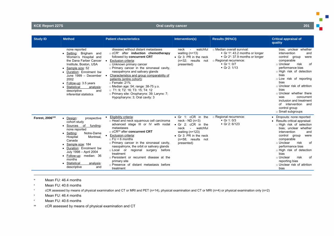
KCE Report 227S Oral cavity cancer 201
Study ID Method Patient characteristics Intervention(s) Results (95%CI) Critical appraisal of quality
none reported Setting: Brigham and
Women’s Hospital and the Dana Farber Cancer Institute, Boston, USA
Sample size: 52 Duration: Enrolment bw
June 1999 – December 2002
Follow-up: 3.5 years Statistical analysis:
descriptive and inferential statistics
disease) without distant metastases o cCRv after induction chemotherapy
followed by concurrent CRT Exclusion criteria: o Unknown primary cancer o Primary cancer in the sinonasal cavity,
nasopahrynx and salivary glands Characteristics and group comparability of
patients (entire cohort): o Female: 21% o Median age: 54; range: 38-75 y.o. o T1: 9; T2: 16; T3: 15; T4: 12 o Primary site: Oropharynx: 39; Larynx: 7;
Hypopharynx: 3; Oral cavity: 3
neck - watchful waiting (n=13)
Gr 3: PR in the neck (n=32; results not presented)
o Median overall survival: Gr 1y: 43.2 months or longer Gr 2z: 37.9 months or longer
o Regional recurrence: Gr 1: 0/7 Gr 2: 1/13
bias; unclear whether intervention and control group were comparable
o Unclear risk of performance bias
o High risk of detection bias
o Low risk of reporting bias
o Unclear risk of attrition bias
o Unclear whether there was concurrent inclusion and treatment of intervention and control group
o Small subgroups
Forest, 2006133 Design: prospective cohort study
Sources of funding: none reported
Setting: Notre-Dame Hospital Montreal, Canada
Sample size: 184 Duration: Enrolment bw
July 1998 – April 2004 Follow-up: median: 36
months Statistical analysis:
descriptive and
Eligibility criteria: o Head and neck squamous cell carcinoma
advanced stage III or IV with nodal metastases
o cCRaa after concurrent CRT Exclusion criteria: o FU < 6 months o Primary cancer in the sinonasal cavity,
nasopahrynx, the orbit or salivary glands o Local or regional surgery before
treatment o Persistent or recurrent disease at the
primary site o Presence of distant metastasis before
treatment
Gr 1: cCR in the neck - ND (n=3)
Gr 2: cCR in the neck - watchful waiting (n=123)
Gr 3: PR in the neck (n=58; results not presented)
o Regional recurrence: Gr 1: 0/3 Gr 2: 6/123
Dropouts: none reported Results critical appraisal:
o High risk of selection bias; unclear whether intervention and control group were comparable
o Unclear risk of performance bias
o High risk of detection bias
o Unclear risk of reporting bias
o Unclear risk of attrition bias
w Mean FU: 46.4 months x Mean FU: 40.6 months v cCR assessed by means of physical examination and CT or MRI and PET (n=14), physical examination and CT or MRI (n=4) or physical examination only (n=2) y Mean FU: 46.4 months z Mean FU: 40.6 months aa cCR assessed by means of physical examination and CT
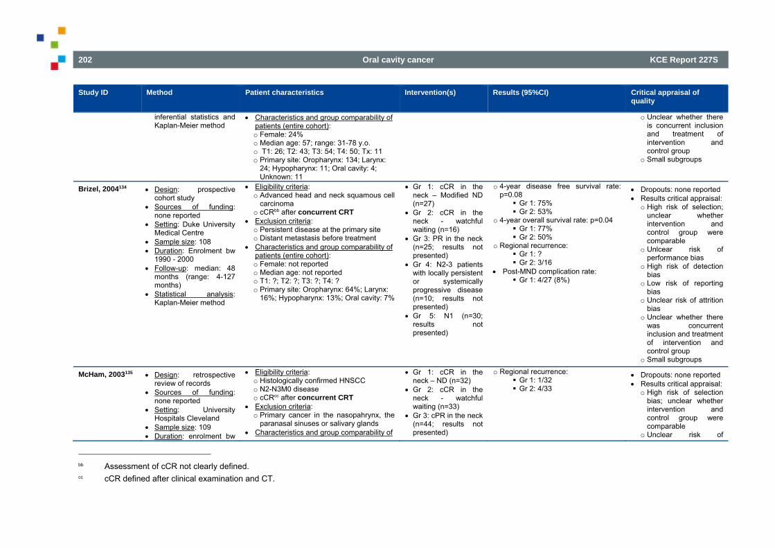
202 Oral cavity cancer KCE Report 227S
Study ID Method Patient characteristics Intervention(s) Results (95%CI) Critical appraisal of quality
inferential statistics and Kaplan-Meier method
Characteristics and group comparability of patients (entire cohort): o Female: 24% o Median age: 57; range: 31-78 y.o. o T1: 26; T2: 43; T3: 54; T4: 50; Tx: 11 o Primary site: Oropharynx: 134; Larynx:
24; Hypopharynx: 11; Oral cavity: 4; Unknown: 11
o Unclear whether there is concurrent inclusion and treatment of intervention and control group
o Small subgroups
Brizel, 2004134 Design: prospective cohort study
Sources of funding: none reported
Setting: Duke University Medical Centre
Sample size: 108 Duration: Enrolment bw
1990 - 2000 Follow-up: median: 48
months (range: 4-127 months)
Statistical analysis: Kaplan-Meier method
Eligibility criteria: o Advanced head and neck squamous cell
carcinoma o cCRbb after concurrent CRT
Exclusion criteria: o Persistent disease at the primary site o Distant metastasis before treatment
Characteristics and group comparability of patients (entire cohort): o Female: not reported o Median age: not reported o T1: ?; T2: ?; T3: ?; T4: ? o Primary site: Oropharynx: 64%; Larynx:
16%; Hypopharynx: 13%; Oral cavity: 7%
Gr 1: cCR in the neck – Modified ND (n=27)
Gr 2: cCR in the neck - watchful waiting (n=16)
Gr 3: PR in the neck (n=25; results not presented)
Gr 4: N2-3 patients with locally persistent or systemically progressive disease (n=10; results not presented)
Gr 5: N1 (n=30; results not presented)
o 4-year disease free survival rate: p=0.08 Gr 1: 75% Gr 2: 53%
o 4-year overall survival rate: p=0.04 Gr 1: 77% Gr 2: 50%
o Regional recurrence: Gr 1: ? Gr 2: 3/16
Post-MND complication rate: Gr 1: 4/27 (8%)
Dropouts: none reported Results critical appraisal:
o High risk of selection; unclear whether intervention and control group were comparable
o Unlcear risk of performance bias
o High risk of detection bias
o Low risk of reporting bias
o Unclear risk of attrition bias
o Unclear whether there was concurrent inclusion and treatment of intervention and control group
o Small subgroups
McHam, 2003135 Design: retrospective review of records
Sources of funding: none reported
Setting: University Hospitals Cleveland
Sample size: 109 Duration: enrolment bw
Eligibility criteria: o Histologically confirmed HNSCC o N2-N3M0 disease o cCRcc after concurrent CRT
Exclusion criteria: o Primary cancer in the nasopahrynx, the
paranasal sinuses or salivary glands Characteristics and group comparability of
Gr 1: cCR in the neck – ND (n=32)
Gr 2: cCR in the neck - watchful waiting (n=33)
Gr 3: cPR in the neck (n=44; results not presented)
o Regional recurrence: Gr 1: 1/32 Gr 2: 4/33
Dropouts: none reported Results critical appraisal:
o High risk of selection bias; unclear whether intervention and control group were comparable
o Unclear risk of
bb Assessment of cCR not clearly defined. cc cCR defined after clinical examination and CT.
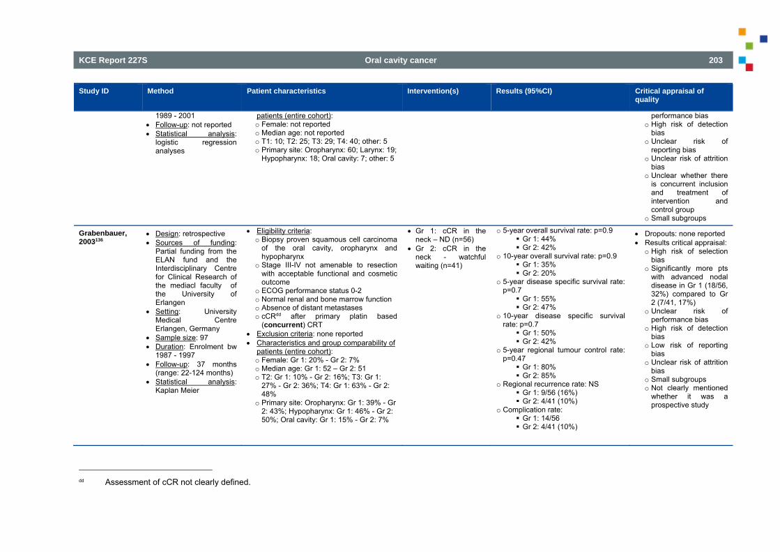
KCE Report 227S Oral cavity cancer 203
Study ID Method Patient characteristics Intervention(s) Results (95%CI) Critical appraisal of quality
1989 - 2001 Follow-up: not reported Statistical analysis:
logistic regression analyses
patients (entire cohort): o Female: not reported o Median age: not reported o T1: 10; T2: 25; T3: 29; T4: 40; other: 5 o Primary site: Oropharynx: 60; Larynx: 19;
Hypopharynx: 18; Oral cavity: 7; other: 5
performance bias o High risk of detection
bias o Unclear risk of
reporting bias o Unclear risk of attrition
bias o Unclear whether there
is concurrent inclusion and treatment of intervention and control group
o Small subgroups
Grabenbauer, 2003136
Design: retrospective Sources of funding:
Partial funding from the ELAN fund and the Interdisciplinary Centre for Clinical Research of the mediacl faculty of the University of Erlangen
Setting: University Medical Centre Erlangen, Germany
Sample size: 97 Duration: Enrolment bw
1987 - 1997 Follow-up: 37 months
(range: 22-124 months) Statistical analysis:
Kaplan Meier
Eligibility criteria: o Biopsy proven squamous cell carcinoma
of the oral cavity, oropharynx and hypopharynx
o Stage III-IV not amenable to resection with acceptable functional and cosmetic outcome
o ECOG performance status 0-2 o Normal renal and bone marrow function o Absence of distant metastases o cCRdd after primary platin based
(concurrent) CRT Exclusion criteria: none reported Characteristics and group comparability of
patients (entire cohort): o Female: Gr 1: 20% - Gr 2: 7% o Median age: Gr 1: 52 – Gr 2: 51 o T2: Gr 1: 10% - Gr 2: 16%; T3: Gr 1:
27% - Gr 2: 36%; T4: Gr 1: 63% - Gr 2: 48%
o Primary site: Oropharynx: Gr 1: 39% - Gr 2: 43%; Hypopharynx: Gr 1: 46% - Gr 2: 50%; Oral cavity: Gr 1: 15% - Gr 2: 7%
Gr 1: cCR in the neck – ND (n=56)
Gr 2: cCR in the neck - watchful waiting (n=41)
o 5-year overall survival rate: p=0.9 Gr 1: 44% Gr 2: 42%
o 10-year overall survival rate: p=0.9 Gr 1: 35% Gr 2: 20%
o 5-year disease specific survival rate: p=0.7 Gr 1: 55% Gr 2: 47%
o 10-year disease specific survival rate: p=0.7 Gr 1: 50% Gr 2: 42%
o 5-year regional tumour control rate: p=0.47 Gr 1: 80% Gr 2: 85%
o Regional recurrence rate: NS Gr 1: 9/56 (16%) Gr 2: 4/41 (10%)
o Complication rate: Gr 1: 14/56 Gr 2: 4/41 (10%)
Dropouts: none reported Results critical appraisal:
o High risk of selection bias
o Significantly more pts with advanced nodal disease in Gr 1 (18/56, 32%) compared to Gr 2 (7/41, 17%)
o Unclear risk of performance bias
o High risk of detection bias
o Low risk of reporting bias
o Unclear risk of attrition bias
o Small subgroups o Not clearly mentioned
whether it was a prospective study
dd Assessment of cCR not clearly defined.
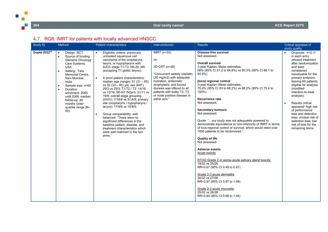
204 Oral cavity cancer KCE Report 227S
4.7. RQ8: IMRT for patients with locally advanced HNSCC Study ID Method Patient characteristics Intervention(s) Results
Critical appraisal of study quality
Gupta 201241 Design: RCT Source of funding:
Siemens Oncology Care Systems, USA
Setting: Tata Memorial Centre, Navi Mumbai, India
Sample size: n=62 Duration:
enrolment: 2005 until 2008; median follow-up: 40 months (inter-quartile range 26–50)
Eligibility criteria: previously untreated squamous cell carcinoma of the oropharynx, larynx, or hypopharynx with AJCC stage T1-T3, N0-2b, M0 (excepting T1 glottic larynx).
A priori patient characteristics: median age (range): 51 (31 – 65) vs 55 (33 – 65) yrs; sex (M/F): 29/3 vs 25/3; T1-T2 / T3: 14/18 vs 12/16; N0-N1 /N2a-b: 21/11 vs 19/9; overall stage grouping (I/II/IV): 7/16/9 vs 5/14/9; primary site (oropharynx / hypopharynx / larynx): 17/9/6 vs 15/8/5.
Group comparability: well balanced. “There were no significant differences in the baseline patient, disease, and treatment characteristics which were well matched in the two arms.”
IMRT (n=32)
vs
3D-CRT (n=28)
“Concurrent weekly cisplatin (30 mg/m2) with adequate hydration, antiemetic prophylaxis, and forced diuresis was offered to all patients with bulky T2, T3, or node positive disease in either arm.”
Disease-free survival Not assessed. Overall survival 3-year Kaplan–Meier estimates: 68% (95% CI 51.2 to 84.8%) vs 80.5% (95% CI 66.1 to 94.9%) (loco) regional control 3-year Kaplan–Meier estimates: 70.6% (95% CI 53 to 88.2%) vs 88.2% (95% CI 75.4 to 100%) Recurrence rate Not assessed. Secondary tumours Not assessed. Quote: “…our study was not adequately powered to demonstrate equivalence or non-inferiority of IMRT in terms of loco-regional control or survival, which would need over 1000 patients to be randomized.” Quality of life Not assessed. Adverse events Acute toxicity RTOG Grade 2 or worse acute salivary gland toxicity: 19/32 vs 25/28 RR=0.67 (95% CI 0.49 to 0.91) Grade 2-3 acute dermatitis 30/32 vs 27/28 RR=0.97 (95% CI 0.87 to 1.09) Grade 2-3 acute mucositis 25/32 vs 26/28 RR=0.84 (95% CI 0.68 to 1.04)
Dropouts: n=2 (1 in each arm) refused treatment after randomization and were considered inevaluable for the primary endpoint, leaving 60 patients eligible for analysis (modified intention-to-treat analysis).
Results critical appraisal: high risk of performance bias and detection bias; unclear risk of selection bias; low risk of bias for the remaining items.
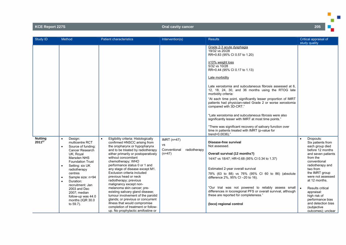
KCE Report 227S Oral cavity cancer 205
Study ID Method Patient characteristics Intervention(s) Results
Critical appraisal of study quality
Grade 2-3 acute dysphagia 19/32 vs 20/28 RR=0.83 (95% CI 0.57 to 1.20) ≥10% weight loss 5/32 vs 10/28 RR=0.44 (95% CI 0.17 to 1.13) Late morbidity Late xerostomia and subcutaneous fibrosis assessed at 6, 12, 18, 24, 30, and 36 months using the RTOG late morbidity criteria: “At each time point, significantly lesser proportion of IMRT patients had physician-rated Grade 2 or worse xerostomia compared with 3D-CRT.” “Late xerostomia and subcutaneous fibrosis were also significantly lesser with IMRT at most time points.” “There was significant recovery of salivary function over time in patients treated with IMRT (p-value for trend=0.0036).”
Nutting 201167
Design: multicentre RCT
Source of funding: Cancer Research UK; Royal Marsden NHS Foundation Trust
Setting: six UK radiotherapy centres
Sample size: n=94 Duration:
recruitment: Jan 2003 and Dec 2007; median follow-up was 44.0 months (IQR 30.0 to 59.7).
Eligibility criteria: Histologically confirmed HNSCC arising from the oropharynx or hypopharynx and to be treated by radiotherapy either primarily or postoperatively without concomitant chemotherapy; WHO performance status 0 or 1 and any stage of disease except M1. Exclusion criteria included previous head or neck radiotherapy; previous malignancy except non-melanoma skin cancer; pre-existing salivary gland disease; tumour involvement of the parotid glands; or previous or concurrent illness that would compromise completion of treatment or follow-up. No prophylactic amifostine or
IMRT (n=47) vs Conventional radiotherapy (n=47)
Disease-free survival Not assessed. Overall survival (12 months?) 14/47 vs 18/47; HR=0.68 (95% CI 0.34 to 1.37) Estimated 2-year overall survival 78% (63 to 88) vs 76% (95% CI 60 to 86) (absolute difference 2%, 95% CI –20 to 16). “Our trial was not powered to reliably assess small differences in locoregional PFS or overall survival, although these are reported for completeness.” (loco) regional control
Dropouts: Six patients from each group died before 12 months and seven patients from the conventional radiotherapy and two from the IMRT group were not assessed at 12 months.
Results critical
appraisal: high risk of performance bias and detection bias (subjective outcomes); unclear
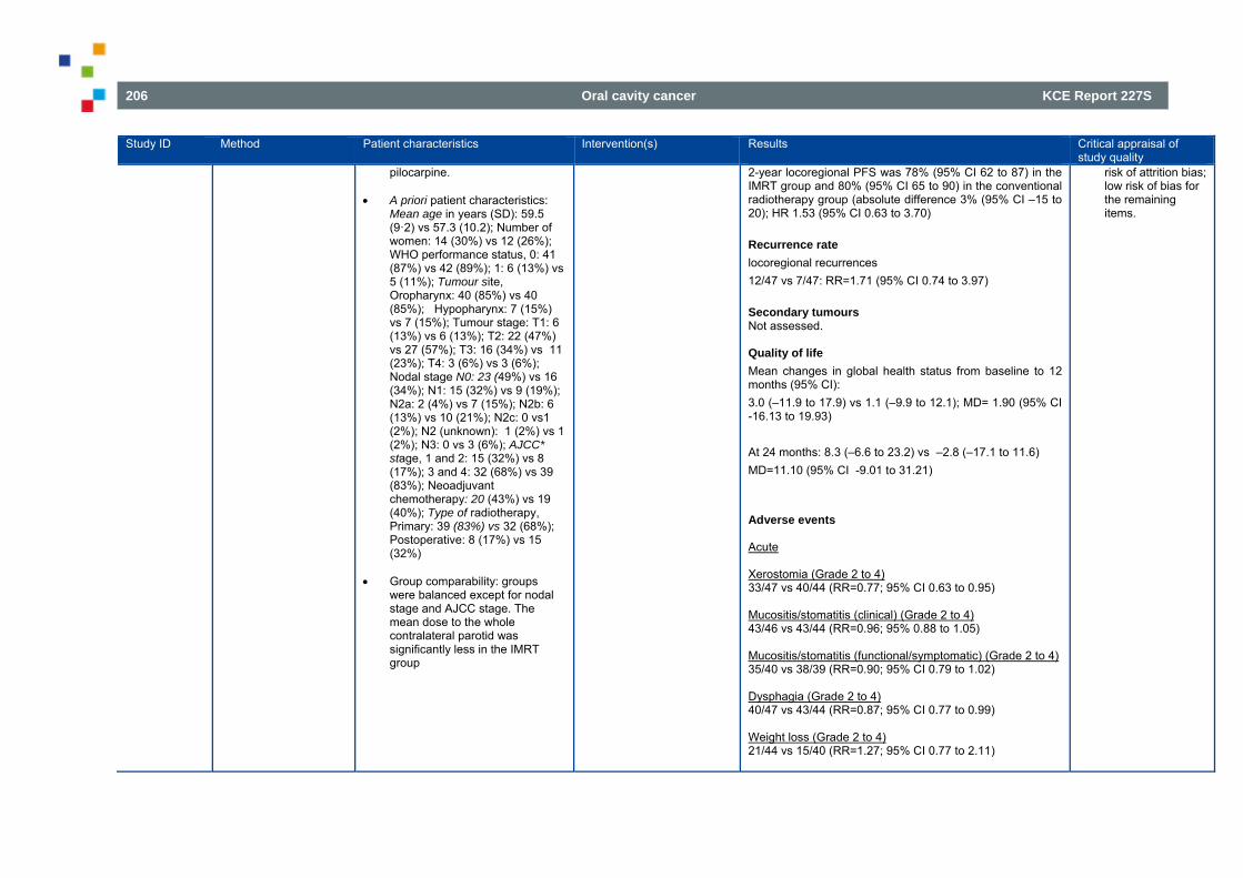
206 Oral cavity cancer KCE Report 227S
Study ID Method Patient characteristics Intervention(s) Results
Critical appraisal of study quality
pilocarpine. A priori patient characteristics:
Mean age in years (SD): 59.5 (9·2) vs 57.3 (10.2); Number of women: 14 (30%) vs 12 (26%); WHO performance status, 0: 41 (87%) vs 42 (89%); 1: 6 (13%) vs 5 (11%); Tumour site, Oropharynx: 40 (85%) vs 40 (85%); Hypopharynx: 7 (15%) vs 7 (15%); Tumour stage: T1: 6 (13%) vs 6 (13%); T2: 22 (47%) vs 27 (57%); T3: 16 (34%) vs 11 (23%); T4: 3 (6%) vs 3 (6%); Nodal stage N0: 23 (49%) vs 16 (34%); N1: 15 (32%) vs 9 (19%); N2a: 2 (4%) vs 7 (15%); N2b: 6 (13%) vs 10 (21%); N2c: 0 vs1 (2%); N2 (unknown): 1 (2%) vs 1 (2%); N3: 0 vs 3 (6%); AJCC* stage, 1 and 2: 15 (32%) vs 8 (17%); 3 and 4: 32 (68%) vs 39 (83%); Neoadjuvant chemotherapy: 20 (43%) vs 19 (40%); Type of radiotherapy, Primary: 39 (83%) vs 32 (68%); Postoperative: 8 (17%) vs 15 (32%)
Group comparability: groups
were balanced except for nodal stage and AJCC stage. The mean dose to the whole contralateral parotid was significantly less in the IMRT group
2-year locoregional PFS was 78% (95% CI 62 to 87) in the IMRT group and 80% (95% CI 65 to 90) in the conventional radiotherapy group (absolute difference 3% (95% CI –15 to 20); HR 1.53 (95% CI 0.63 to 3.70) Recurrence rate locoregional recurrences 12/47 vs 7/47: RR=1.71 (95% CI 0.74 to 3.97) Secondary tumours Not assessed. Quality of life Mean changes in global health status from baseline to 12 months (95% CI): 3.0 (–11.9 to 17.9) vs 1.1 (–9.9 to 12.1); MD= 1.90 (95% CI -16.13 to 19.93) At 24 months: 8.3 (–6.6 to 23.2) vs –2.8 (–17.1 to 11.6) MD=11.10 (95% CI -9.01 to 31.21) Adverse events Acute Xerostomia (Grade 2 to 4) 33/47 vs 40/44 (RR=0.77; 95% CI 0.63 to 0.95) Mucositis/stomatitis (clinical) (Grade 2 to 4) 43/46 vs 43/44 (RR=0.96; 95% 0.88 to 1.05) Mucositis/stomatitis (functional/symptomatic) (Grade 2 to 4) 35/40 vs 38/39 (RR=0.90; 95% CI 0.79 to 1.02) Dysphagia (Grade 2 to 4) 40/47 vs 43/44 (RR=0.87; 95% CI 0.77 to 0.99) Weight loss (Grade 2 to 4) 21/44 vs 15/40 (RR=1.27; 95% CI 0.77 to 2.11)
risk of attrition bias; low risk of bias for the remaining items.
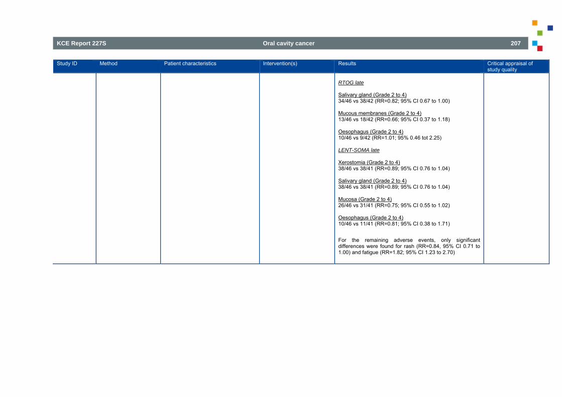
KCE Report 227S Oral cavity cancer 207
Study ID Method Patient characteristics Intervention(s) Results
Critical appraisal of study quality
RTOG late Salivary gland (Grade 2 to 4) 34/46 vs 38/42 (RR=0.82; 95% CI 0.67 to 1.00) Mucous membranes (Grade 2 to 4) 13/46 vs 18/42 (RR=0.66; 95% CI 0.37 to 1.18) Oesophagus (Grade 2 to 4) 10/46 vs 9/42 (RR=1.01; 95% 0.46 tot 2.25) LENT-SOMA late Xerostomia (Grade 2 to 4) 38/46 vs 38/41 (RR=0.89; 95% CI 0.76 to 1.04) Salivary gland (Grade 2 to 4) 38/46 vs 38/41 (RR=0.89; 95% CI 0.76 to 1.04) Mucosa (Grade 2 to 4) 26/46 vs 31/41 (RR=0.75; 95% CI 0.55 to 1.02) Oesophagus (Grade 2 to 4) 10/46 vs 11/41 (RR=0.81; 95% CI 0.38 to 1.71) For the remaining adverse events, only significant differences were found for rash (RR=0.84, 95% CI 0.71 to 1.00) and fatigue (RR=1.82; 95% CI 1.23 to 2.70)
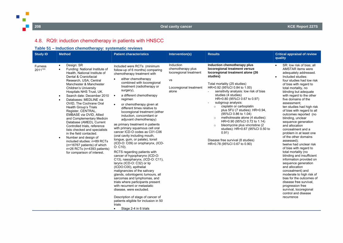
208 Oral cavity cancer KCE Report 227S
4.8. RQ9: induction chemotherapy in patients with HNSCC Table 51 – Induction chemotherapy: systematic reviews Study ID Method Patient characteristics Intervention(s) Results Critical appraisal of review
quality
Furness 2011103
Design: SR Funding: National Institute of
Health, National Institute of Dental & Craniofacial Research, USA; Central Manchester & Manchester Children’s University Hospitals NHS Trust, UK.
Search date: December 2010 Databases: MEDLINE via
OVID, The Cochrane Oral Health Group’s Trials Register, CENTRAL, EMBASE via OVID, Allied and Complementary Medicin Database (AMED), Current Controlled trials, reference lists checked and specialists in the field contacted.
Number and design of included studies: n=89 RCTs (n=16767 patients) of which n=26 RCTs (n=4393 patients) for comparison of interest.
Included were RCTs (minimum follow-up of 6 months) comparing chemotherapy treatment with either chemotherapy
combined with locoregional treatment (radiotherapy or surgery),
a different chemotherapy regimen
or chemotherapy given at different times relative to locoregional treatment (either induction, concomitant or adjuvant chemotherapy)
as primary treatment in patients with primary squamous cell oral cancer ICD-O codes as C01-C06 (oral cavity including mouth, tongue, gum, or palate), tonsil (ICD-O: C09) or oropharynx, (ICD-O: C10). RCTS regarding patients with cancer of hypopharynx (ICD-O: C13), nasopharynx, (ICD-O: C11), larynx (ICD-O: C32) or lip (ICDO:C00), epithelial malignancies of the salivary glands, odontogenic tumours, all sarcomas and lymphomas, and trials where participants present with recurrent or metastatic disease, were excluded. Description of stage of cancer of patients eligible for inclusion in 50 trials Stage 2-4 in 6 trials
Induction chemotherapy plus locoregional treatment vs Locoregional treatment alone
Induction chemotherapy plus locoregional treatment versus locoregional treatment alone (26 studies) Total mortality (25 studies) HR=0.92 (95%CI 0.84 to 1.00) - sensitivity analysis: low risk of bias
studies (4 studies) HR=0.80 (95%CI 0.67 to 0.97)
- subgroup analysis: o cisplatin or carboplatin
plus 5FU (7 studies): HR=0.94, (95%CI 0.86 to 1.04)
o methotrexate alone (4 studies): HR=0.90 (95%CI 0.72 to 1.14)
o bleomycine plus vincristine (2 studies): HR=0.67 (95%CI 0.50 to 0.91)
Disease free survival (8 studies) HR=0.78 (95%CI 0.67 to 0.90)
SR: low risk of bias; all AMSTAR items were adequately addressed.
Included studies: four studies had low risk of bias with regard to total mortality, no blinding but adequate with regard to the other five domains of the assessment; ten studies had high risk of bias with regard to all outcomes reported (no blinding, unclear sequence generation and allocation concealment and a problem in at least one of the other domains assessed); twelve had unclear risk of bias with regard to total mortality (no blinding and insufficient information provided on sequence generation and allocation concealment) and moderate to high risk of bias for the outcomes of disease free survival, progression free survival, locoregional control and disease recurrence
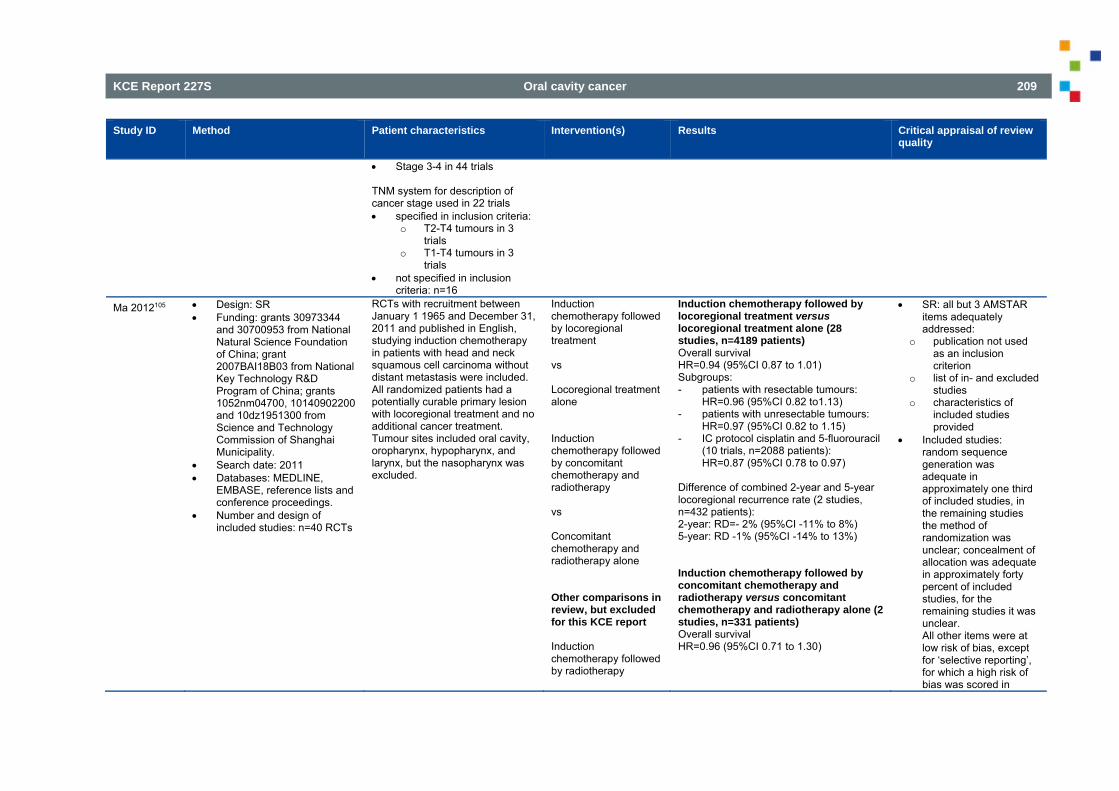
KCE Report 227S Oral cavity cancer 209
Study ID Method Patient characteristics Intervention(s) Results Critical appraisal of review quality
Stage 3-4 in 44 trials TNM system for description of cancer stage used in 22 trials specified in inclusion criteria:
o T2-T4 tumours in 3 trials
o T1-T4 tumours in 3 trials
not specified in inclusion criteria: n=16
Ma 2012105 Design: SR Funding: grants 30973344
and 30700953 from National Natural Science Foundation of China; grant 2007BAI18B03 from National Key Technology R&D Program of China; grants 1052nm04700, 10140902200 and 10dz1951300 from Science and Technology Commission of Shanghai Municipality.
Search date: 2011 Databases: MEDLINE,
EMBASE, reference lists and conference proceedings.
Number and design of included studies: n=40 RCTs
RCTs with recruitment between January 1 1965 and December 31, 2011 and published in English, studying induction chemotherapy in patients with head and neck squamous cell carcinoma without distant metastasis were included. All randomized patients had a potentially curable primary lesion with locoregional treatment and no additional cancer treatment. Tumour sites included oral cavity, oropharynx, hypopharynx, and larynx, but the nasopharynx was excluded.
Induction chemotherapy followed by locoregional treatment vs Locoregional treatment alone Induction chemotherapy followed by concomitant chemotherapy and radiotherapy vs Concomitant chemotherapy and radiotherapy alone Other comparisons in review, but excluded for this KCE report Induction chemotherapy followed by radiotherapy
Induction chemotherapy followed by locoregional treatment versus locoregional treatment alone (28 studies, n=4189 patients) Overall survival HR=0.94 (95%CI 0.87 to 1.01) Subgroups: - patients with resectable tumours:
HR=0.96 (95%CI 0.82 to1.13) - patients with unresectable tumours:
HR=0.97 (95%CI 0.82 to 1.15) - IC protocol cisplatin and 5-fluorouracil
(10 trials, n=2088 patients): HR=0.87 (95%CI 0.78 to 0.97)
Difference of combined 2-year and 5-year locoregional recurrence rate (2 studies, n=432 patients): 2-year: RD=- 2% (95%CI -11% to 8%) 5-year: RD -1% (95%CI -14% to 13%)
Induction chemotherapy followed by concomitant chemotherapy and radiotherapy versus concomitant chemotherapy and radiotherapy alone (2 studies, n=331 patients) Overall survival HR=0.96 (95%CI 0.71 to 1.30)
SR: all but 3 AMSTAR items adequately addressed:
o publication not used as an inclusion criterion
o list of in- and excluded studies
o characteristics of included studies provided
Included studies: random sequence generation was adequate in approximately one third of included studies, in the remaining studies the method of randomization was unclear; concealment of allocation was adequate in approximately forty percent of included studies, for the remaining studies it was unclear. All other items were at low risk of bias, except for ‘selective reporting’, for which a high risk of bias was scored in
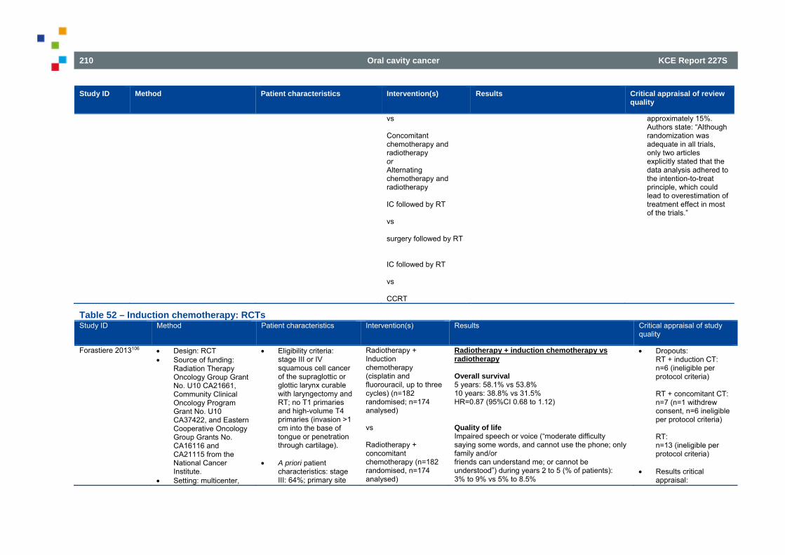
210 Oral cavity cancer KCE Report 227S
Study ID Method Patient characteristics Intervention(s) Results Critical appraisal of review quality
vs Concomitant chemotherapy and radiotherapy or Alternating chemotherapy and radiotherapy IC followed by RT vs surgery followed by RT IC followed by RT vs CCRT
approximately 15%. Authors state: “Although randomization was adequate in all trials, only two articles explicitly stated that the data analysis adhered to the intention-to-treat principle, which could lead to overestimation of treatment effect in most of the trials.”
Table 52 – Induction chemotherapy: RCTs Study ID Method Patient characteristics Intervention(s) Results Critical appraisal of study
quality
Forastiere 2013106 Design: RCT Source of funding:
Radiation Therapy Oncology Group Grant No. U10 CA21661, Community Clinical Oncology Program Grant No. U10 CA37422, and Eastern Cooperative Oncology Group Grants No. CA16116 and CA21115 from the National Cancer Institute.
Setting: multicenter,
Eligibility criteria: stage III or IV squamous cell cancer of the supraglottic or glottic larynx curable with laryngectomy and RT; no T1 primaries and high-volume T4 primaries (invasion >1 cm into the base of tongue or penetration through cartilage).
A priori patient
characteristics: stage III: 64%; primary site
Radiotherapy + Induction chemotherapy (cisplatin and fluorouracil, up to three cycles) (n=182 randomised; n=174 analysed) vs Radiotherapy + concomitant chemotherapy (n=182 randomised, n=174 analysed)
Radiotherapy + induction chemotherapy vs radiotherapy Overall survival 5 years: 58.1% vs 53.8% 10 years: 38.8% vs 31.5% HR=0.87 (95%CI 0.68 to 1.12) Quality of life Impaired speech or voice (“moderate difficulty saying some words, and cannot use the phone; only family and/or friends can understand me; or cannot be understood”) during years 2 to 5 (% of patients): 3% to 9% vs 5% to 8.5%
Dropouts: RT + induction CT: n=6 (ineligible per protocol criteria) RT + concomitant CT: n=7 (n=1 withdrew consent, n=6 ineligible per protocol criteria) RT: n=13 (ineligible per protocol criteria)
Results critical appraisal:
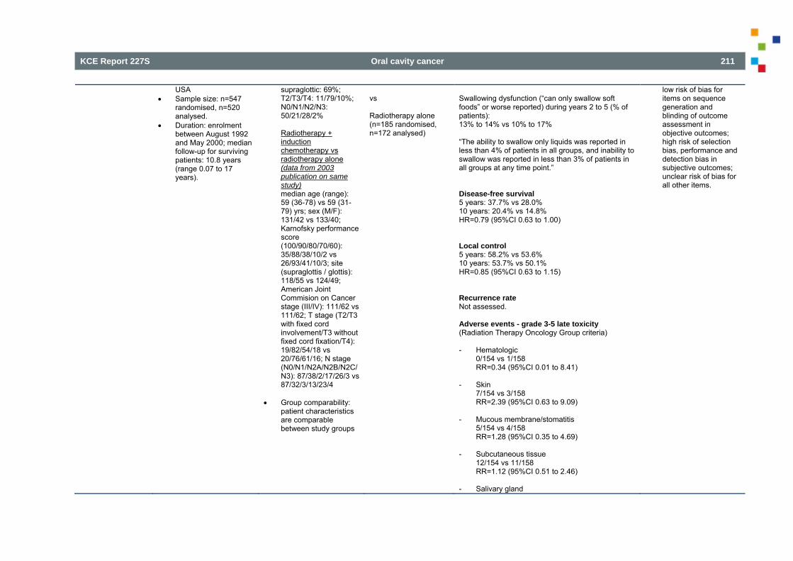
KCE Report 227S Oral cavity cancer 211
USA Sample size: n=547
randomised, n=520 analysed.
Duration: enrolment between August 1992 and May 2000; median follow-up for surviving patients: 10.8 years (range 0.07 to 17 years).
supraglottic: 69%; T2/T3/T4: 11/79/10%; N0/N1/N2/N3: 50/21/28/2%
Radiotherapy + induction chemotherapy vs radiotherapy alone (data from 2003 publication on same study) median age (range): 59 (36-78) vs 59 (31-79) yrs; sex (M/F): 131/42 vs 133/40; Karnofsky performance score (100/90/80/70/60): 35/88/38/10/2 vs 26/93/41/10/3; site (supraglottis / glottis): 118/55 vs 124/49; American Joint Commision on Cancer stage (III/IV): 111/62 vs 111/62; T stage (T2/T3 with fixed cord involvement/T3 without fixed cord fixation/T4): 19/82/54/18 vs 20/76/61/16; N stage (N0/N1/N2A/N2B/N2C/N3): 87/38/2/17/26/3 vs 87/32/3/13/23/4
Group comparability:
patient characteristics are comparable between study groups
vs Radiotherapy alone (n=185 randomised, n=172 analysed)
Swallowing dysfunction (“can only swallow soft foods” or worse reported) during years 2 to 5 (% of patients): 13% to 14% vs 10% to 17% “The ability to swallow only liquids was reported in less than 4% of patients in all groups, and inability to swallow was reported in less than 3% of patients in all groups at any time point.” Disease-free survival 5 years: 37.7% vs 28.0% 10 years: 20.4% vs 14.8% HR=0.79 (95%CI 0.63 to 1.00) Local control 5 years: 58.2% vs 53.6% 10 years: 53.7% vs 50.1% HR=0.85 (95%CI 0.63 to 1.15) Recurrence rate Not assessed. Adverse events - grade 3-5 late toxicity (Radiation Therapy Oncology Group criteria) - Hematologic
0/154 vs 1/158 RR=0.34 (95%CI 0.01 to 8.41)
- Skin
7/154 vs 3/158 RR=2.39 (95%CI 0.63 to 9.09)
- Mucous membrane/stomatitis
5/154 vs 4/158 RR=1.28 (95%CI 0.35 to 4.69)
- Subcutaneous tissue
12/154 vs 11/158 RR=1.12 (95%CI 0.51 to 2.46)
- Salivary gland
low risk of bias for items on sequence generation and blinding of outcome assessment in objective outcomes; high risk of selection bias, performance and detection bias in subjective outcomes; unclear risk of bias for all other items.
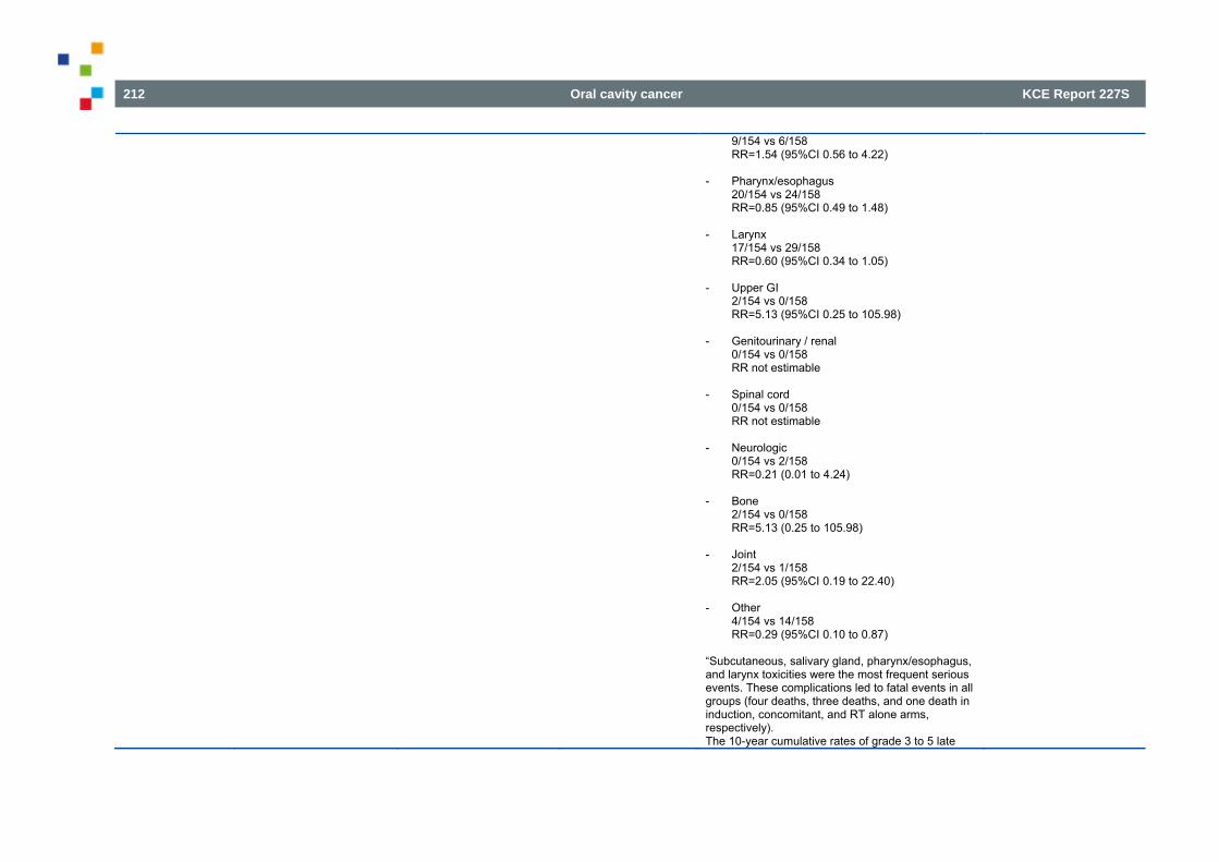
212 Oral cavity cancer KCE Report 227S
9/154 vs 6/158 RR=1.54 (95%CI 0.56 to 4.22)
- Pharynx/esophagus
20/154 vs 24/158 RR=0.85 (95%CI 0.49 to 1.48)
- Larynx
17/154 vs 29/158 RR=0.60 (95%CI 0.34 to 1.05)
- Upper GI
2/154 vs 0/158 RR=5.13 (95%CI 0.25 to 105.98)
- Genitourinary / renal
0/154 vs 0/158 RR not estimable
- Spinal cord
0/154 vs 0/158 RR not estimable
- Neurologic
0/154 vs 2/158 RR=0.21 (0.01 to 4.24)
- Bone
2/154 vs 0/158 RR=5.13 (0.25 to 105.98)
- Joint
2/154 vs 1/158 RR=2.05 (95%CI 0.19 to 22.40)
- Other
4/154 vs 14/158 RR=0.29 (95%CI 0.10 to 0.87)
“Subcutaneous, salivary gland, pharynx/esophagus, and larynx toxicities were the most frequent serious events. These complications led to fatal events in all groups (four deaths, three deaths, and one death in induction, concomitant, and RT alone arms, respectively). The 10-year cumulative rates of grade 3 to 5 late
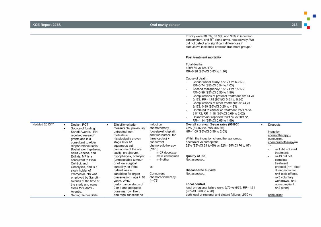
KCE Report 227S Oral cavity cancer 213
toxicity were 30.6%, 33.3%, and 38% in induction, concomitant, and RT alone arms, respectively. We did not detect any significant differences in cumulative incidence between treatment groups.” Post treatment mortality Total deaths 120/174 vs 124/172 RR=0.96 (95%CI 0.83 to 1.10) Cause of death: - Cancer under study: 45/174 vs 60/172,
RR=0.74 (95%CI 0.54 to 1.03) - Second malignancy: 15/174 vs 15/172,
RR=0.99 (95%CI 0.50 to 1.96) - Complications of protocol treatment: 9/174 vs
5/172, RR=1.78 (95%CI 0.61 to 5.20) - Complications of other treatment: 3/174 vs
3/172, 0.99 (95%CI 0.20 to 4.83) - Unrelated to cancer or treatment: 25/174 vs
21/172, RR=1.18 (95%CI 0.69 to 2.02) - Unknown/not reported: 23/174 vs 20/172,
RR=1.14 (95%CI 0.65 to 1.99) Haddad 2013107 Design: RCT
Source of funding: Sanofi-Aventis; RH received research grants and is a consultant to Alder Biopharmaceuticals, Boehringer Ingelheim, Astra Zeneca, and Exilixis. MP is a consultant to Eisai, Cel-Sci, and Oncolytics, and is a stock holder of Promedior. NS was employed by Sanofi -Aventis at the time of the study and owns stock for Sanofi -Aventis.
Setting:14 hospitals
Eligibility criteria: measurable, previously untreated, non-metastatic, histologically proven stage III or IV squamous-cell carcinoma of the oral cavity, oropharynx, hypopharynx, or larynx (unresectable tumour or of low surgical curability, or if the patient was a candidate for organ preservation); age ≥ 18 years, WHO performance status of 0 or 1 and adequate bone marrow, liver, and renal function; no
Induction chemotherapy (docetaxel, cisplatin and fluorouracil, for three cycles) + concurrent chemoradiotherapy (n=70) - n=27 docetaxel - n=37 carboplatin - n=6 other vs Concurrent chemoradiotherapy (n=75)
Overall survival, 3-year rates (95%CI) 73% (60-82) vs 78% (66-86) HR=1.09 (95%CI 0.59 to 2.03) Within the induction chemotherapy group: docetaxel vs carboplatin: 52% (95%CI 31 to 69) vs 92% (95%CI 76 to 97) Quality of life Not assessed. Disease-free survival Not assessed. Local control local or regional failure only: 9/70 vs 6/75, RR=1.61 (95%CI 0.60 to 4.28) both local or regional and distant failures: 2/70 vs
Dropouts: induction chermotherapy + concurrent chemoradiotherapyn= 14 - n=1 did not start
treatment; - n=13 did not
complete treatment protocol (n=1 died during induction, n=5 toxic effects, n=3 voluntary withdrawal, n=2 non-compliant n=2 other)
concurrent
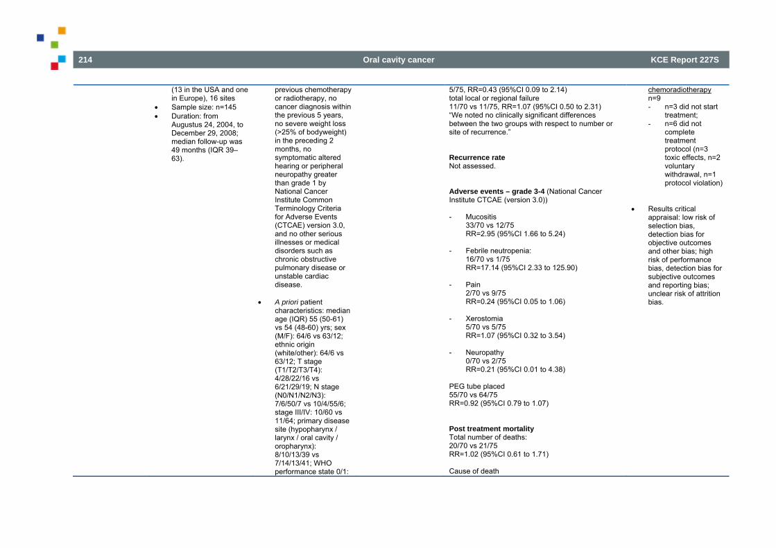
214 Oral cavity cancer KCE Report 227S
(13 in the USA and one in Europe), 16 sites
Sample size: n=145 Duration: from
Augustus 24, 2004, to December 29, 2008; median follow-up was 49 months (IQR 39–63).
previous chemotherapy or radiotherapy, no cancer diagnosis within the previous 5 years, no severe weight loss (>25% of bodyweight) in the preceding 2 months, no symptomatic altered hearing or peripheral neuropathy greater than grade 1 by National Cancer Institute Common Terminology Criteria for Adverse Events (CTCAE) version 3.0, and no other serious illnesses or medical disorders such as chronic obstructive pulmonary disease or unstable cardiac disease.
A priori patient
characteristics: median age (IQR) 55 (50-61) vs 54 (48-60) yrs; sex (M/F): 64/6 vs 63/12; ethnic origin (white/other): 64/6 vs 63/12; T stage (T1/T2/T3/T4): 4/28/22/16 vs 6/21/29/19; N stage (N0/N1/N2/N3): 7/6/50/7 vs 10/4/55/6; stage III/IV: 10/60 vs 11/64; primary disease site (hypopharynx / larynx / oral cavity / oropharynx): 8/10/13/39 vs 7/14/13/41; WHO performance state 0/1:
5/75, RR=0.43 (95%CI 0.09 to 2.14) total local or regional failure 11/70 vs 11/75, RR=1.07 (95%CI 0.50 to 2.31) “We noted no clinically significant differences between the two groups with respect to number or site of recurrence.” Recurrence rate Not assessed. Adverse events – grade 3-4 (National Cancer Institute CTCAE (version 3.0)) - Mucositis
33/70 vs 12/75 RR=2.95 (95%CI 1.66 to 5.24)
- Febrile neutropenia:
16/70 vs 1/75 RR=17.14 (95%CI 2.33 to 125.90)
- Pain
2/70 vs 9/75 RR=0.24 (95%CI 0.05 to 1.06)
- Xerostomia
5/70 vs 5/75 RR=1.07 (95%CI 0.32 to 3.54)
- Neuropathy
0/70 vs 2/75 RR=0.21 (95%CI 0.01 to 4.38)
PEG tube placed 55/70 vs 64/75 RR=0.92 (95%CI 0.79 to 1.07) Post treatment mortality Total number of deaths: 20/70 vs 21/75 RR=1.02 (95%CI 0.61 to 1.71) Cause of death
chemoradiotherapy n=9 - n=3 did not start
treatment; - n=6 did not
complete treatment protocol (n=3 toxic effects, n=2 voluntary withdrawal, n=1 protocol violation)
Results critical appraisal: low risk of selection bias, detection bias for objective outcomes and other bias; high risk of performance bias, detection bias for subjective outcomes and reporting bias; unclear risk of attrition bias.
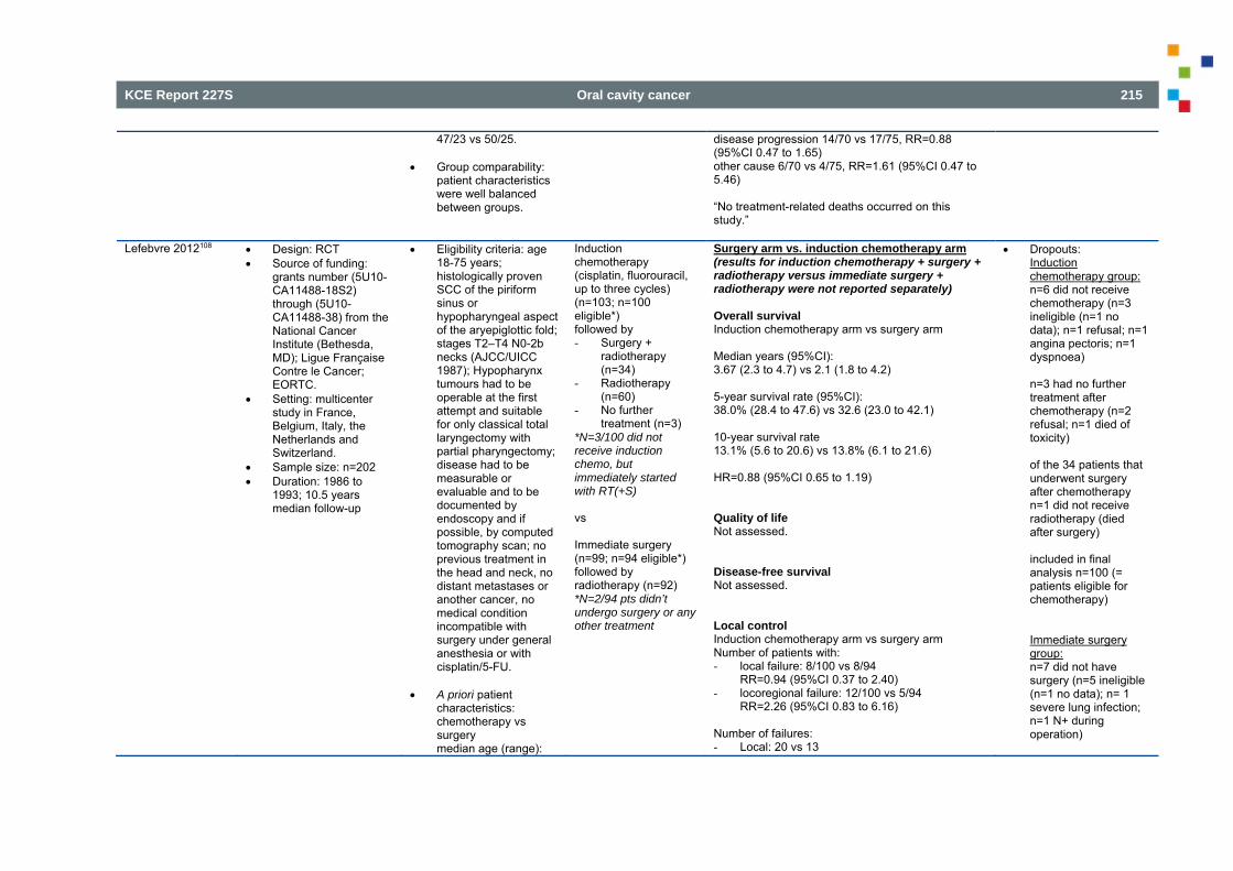
KCE Report 227S Oral cavity cancer 215
47/23 vs 50/25. Group comparability:
patient characteristics were well balanced between groups.
disease progression 14/70 vs 17/75, RR=0.88 (95%CI 0.47 to 1.65) other cause 6/70 vs 4/75, RR=1.61 (95%CI 0.47 to 5.46) “No treatment-related deaths occurred on this study.”
Lefebvre 2012108 Design: RCT Source of funding:
grants number (5U10-CA11488-18S2) through (5U10-CA11488-38) from the National Cancer Institute (Bethesda, MD); Ligue Française Contre le Cancer; EORTC.
Setting: multicenter study in France, Belgium, Italy, the Netherlands and Switzerland.
Sample size: n=202 Duration: 1986 to
1993; 10.5 years median follow-up
Eligibility criteria: age 18-75 years; histologically proven SCC of the piriform sinus or hypopharyngeal aspect of the aryepiglottic fold; stages T2–T4 N0-2b necks (AJCC/UICC 1987); Hypopharynx tumours had to be operable at the first attempt and suitable for only classical total laryngectomy with partial pharyngectomy; disease had to be measurable or evaluable and to be documented by endoscopy and if possible, by computed tomography scan; no previous treatment in the head and neck, no distant metastases or another cancer, no medical condition incompatible with surgery under general anesthesia or with cisplatin/5-FU.
A priori patient
characteristics: chemotherapy vs surgery median age (range):
Induction chemotherapy (cisplatin, fluorouracil, up to three cycles) (n=103; n=100 eligible*) followed by - Surgery +
radiotherapy (n=34)
- Radiotherapy (n=60)
- No further treatment (n=3)
*N=3/100 did not receive induction chemo, but immediately started with RT(+S) vs Immediate surgery (n=99; n=94 eligible*) followed by radiotherapy (n=92) *N=2/94 pts didn’t undergo surgery or any other treatment
Surgery arm vs. induction chemotherapy arm (results for induction chemotherapy + surgery + radiotherapy versus immediate surgery + radiotherapy were not reported separately) Overall survival Induction chemotherapy arm vs surgery arm Median years (95%CI): 3.67 (2.3 to 4.7) vs 2.1 (1.8 to 4.2) 5-year survival rate (95%CI): 38.0% (28.4 to 47.6) vs 32.6 (23.0 to 42.1) 10-year survival rate 13.1% (5.6 to 20.6) vs 13.8% (6.1 to 21.6) HR=0.88 (95%CI 0.65 to 1.19) Quality of life Not assessed. Disease-free survival Not assessed. Local control Induction chemotherapy arm vs surgery arm Number of patients with: - local failure: 8/100 vs 8/94
RR=0.94 (95%CI 0.37 to 2.40) - locoregional failure: 12/100 vs 5/94
RR=2.26 (95%CI 0.83 to 6.16) Number of failures: - Local: 20 vs 13
Dropouts: Induction chemotherapy group: n=6 did not receive chemotherapy (n=3 ineligible (n=1 no data); n=1 refusal; n=1 angina pectoris; n=1 dyspnoea) n=3 had no further treatment after chemotherapy (n=2 refusal; n=1 died of toxicity) of the 34 patients that underwent surgery after chemotherapy n=1 did not receive radiotherapy (died after surgery) included in final analysis n=100 (= patients eligible for chemotherapy)
Immediate surgery group: n=7 did not have surgery (n=5 ineligible (n=1 no data); n= 1 severe lung infection; n=1 N+ during operation)
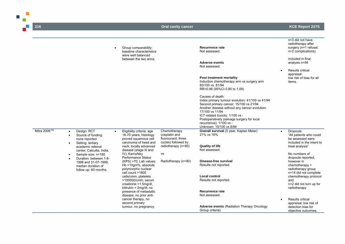
216 Oral cavity cancer KCE Report 227S
Group comparability:
baseline characteristics were well balanced between the two arms.
Recurrence rate Not assessed. Adverse events Not assessed. Post treatment mortality Induction chemotherapy arm vs surgery arm 83/100 vs. 81/94 RR=0.96 (95%CI 0.85 to 1.09) Causes of death: Index primary tumour evolution: 41/100 vs 41/94 Second primary cancer: 15/100 vs 21/94 Another disease without any cancer evolution: 17/100 vs 11/94 ICT-related toxicity: 1/100 vs - Postoperatively (salvage surgery for local recurrence): 1/100 vs - Unknown: 10/100 vs 8/94
n=3 did not have radiotherapy after surgery (n=1 refusal; n=2 complications) included in final analysis n=94
Results critical
appraisal: low risk of bias for all items.
Mitra 2006109 Design: RCT Source of funding:
none reported Setting: tertiary
academic referral center, Calcutta, India.
Sample size: n=180 Duration: between 1-8-
1998 and 31-07-1999; median duration of follow up: 60 months.
Eligibility criteria: age 18-70 years; histology proved squamous cell carcinoma of head and neck; locally advanced disease (stage III and IV); Karnofsky Performance Status (KPS) >70; Lab values: Hb >10gm%, absolute polymorphic nuclear cell count >1800 cells/cmm, platelets >100000/cmm, serum creatinine <1.5mg/dl, bilirubin < 2mg/dl; no presence of metastatic disease, no prior anti-cancer therapy, no second primary tumour, no pregnancy.
Chemotherapy (cisplatin and fluorouracil, three cycles) followed by radiotherapy (n=90)
vs Radiotherapy (n=90)
Overall survival (5 year, Kaplan Meier) 21% vs 16% Quality of life Not assessed. Disease-free survival Results not reported. Local control Results not reported. Recurrence rate Not assessed. Adverse events (Radiation Therapy Oncology Group criteria)
Dropouts: “All patients who could be assessed were included in the intent to treat analysis” No numbers of dropouts reported, however in chemotherapy + radiotherapy group n=14 did not complete chemotherapy protocol and n=2 did not turn up for radiotherapy
Results critical appraisal: low risk of detection bias for objective outcomes,
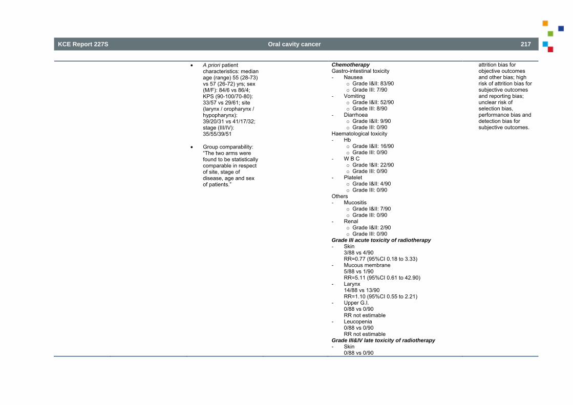
KCE Report 227S Oral cavity cancer 217
A priori patient characteristics: median age (range) 55 (28-73) vs 57 (26-72) yrs; sex (M/F): 84/6 vs 86/4; KPS (90-100/70-80): 33/57 vs 29/61; site (larynx / oropharynx / hypopharynx): 39/20/31 vs 41/17/32; stage (III/IV): 35/55/39/51
Group comparability: “The two arms were found to be statistically comparable in respect of site, stage of disease, age and sex of patients.”
Chemotherapy Gastro-intestinal toxicity - Nausea
o Grade I&II: 83/90 o Grade III: 7/90
- Vomiting o Grade I&II: 52/90 o Grade III: 8/90
- Diarrhoea o Grade I&II: 9/90 o Grade III: 0/90
Haematological toxicity - Hb
o Grade I&II: 16/90 o Grade III: 0/90
- W B C o Grade !&II: 22/90 o Grade III: 0/90
- Platelet o Grade I&II: 4/90 o Grade III: 0/90
Others - Mucositis
o Grade I&II: 7/90 o Grade III: 0/90
- Renal o Grade I&II: 2/90 o Grade III: 0/90
Grade III acute toxicity of radiotherapy - Skin
3/88 vs 4/90 RR=0.77 (95%CI 0.18 to 3.33)
- Mucous membrane 5/88 vs 1/90 RR=5.11 (95%CI 0.61 to 42.90)
- Larynx 14/88 vs 13/90 RR=1.10 (95%CI 0.55 to 2.21)
- Upper G.I. 0/88 vs 0/90 RR not estimable
- Leucopenia 0/88 vs 0/90 RR not estimable
Grade III&IV late toxicity of radiotherapy - Skin
0/88 vs 0/90
attrition bias for objective outcomes and other bias; high risk of attrition bias for subjective outcomes and reporting bias; unclear risk of selection bias, performance bias and detection bias for subjective outcomes.
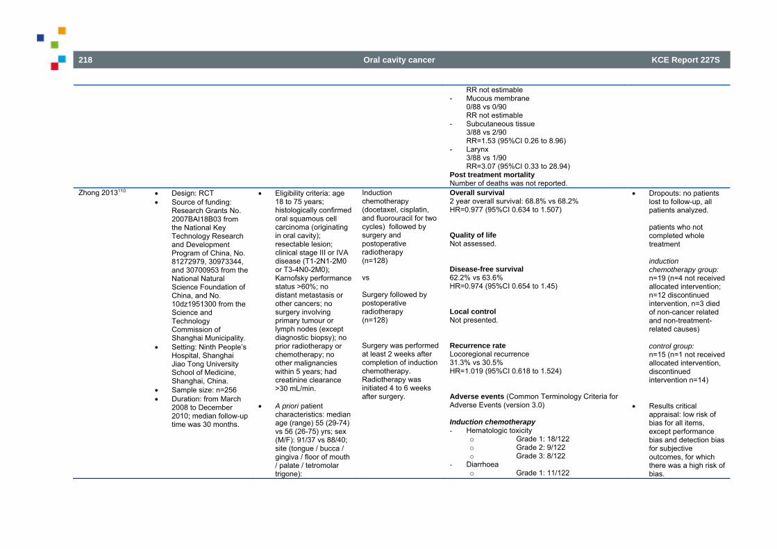
218 Oral cavity cancer KCE Report 227S
RR not estimable - Mucous membrane
0/88 vs 0/90 RR not estimable
- Subcutaneous tissue 3/88 vs 2/90 RR=1.53 (95%CI 0.26 to 8.96)
- Larynx 3/88 vs 1/90 RR=3.07 (95%CI 0.33 to 28.94)
Post treatment mortality Number of deaths was not reported.
Zhong 2013110 Design: RCT Source of funding:
Research Grants No. 2007BAI18B03 from the National Key Technology Research and Development Program of China, No. 81272979, 30973344, and 30700953 from the National Natural Science Foundation of China, and No. 10dz1951300 from the Science and Technology Commission of Shanghai Municipality.
Setting: Ninth People’s Hospital, Shanghai Jiao Tong University School of Medicine, Shanghai, China.
Sample size: n=256 Duration: from March
2008 to December 2010; median follow-up time was 30 months.
Eligibility criteria: age 18 to 75 years; histologically confirmed oral squamous cell carcinoma (originating in oral cavity); resectable lesion; clinical stage III or IVA disease (T1-2N1-2M0 or T3-4N0-2M0); Karnofsky performance status >60%; no distant metastasis or other cancers; no surgery involving primary tumour or lymph nodes (except diagnostic biopsy); no prior radiotherapy or chemotherapy; no other malignancies within 5 years; had creatinine clearance >30 mL/min.
A priori patient
characteristics: median age (range) 55 (29-74) vs 56 (26-75) yrs; sex (M/F): 91/37 vs 88/40; site (tongue / bucca / gingiva / floor of mouth / palate / tetromolar trigone):
Induction chemotherapy (docetaxel, cisplatin, and fluorouracil for two cycles) followed by surgery and postoperative radiotherapy (n=128) vs Surgery followed by postoperative radiotherapy (n=128) Surgery was performed at least 2 weeks after completion of induction chemotherapy. Radiotherapy was initiated 4 to 6 weeks after surgery.
Overall survival 2 year overall survival: 68.8% vs 68.2% HR=0.977 (95%CI 0.634 to 1.507) Quality of life Not assessed. Disease-free survival 62.2% vs 63.6% HR=0.974 (95%CI 0.654 to 1.45) Local control Not presented. Recurrence rate Locoregional recurrence 31.3% vs 30.5% HR=1.019 (95%CI 0.618 to 1.524) Adverse events (Common Terminology Criteria for Adverse Events (version 3.0) Induction chemotherapy - Hematologic toxicity
o Grade 1: 18/122 o Grade 2: 9/122 o Grade 3: 8/122
- Diarrhoea o Grade 1: 11/122
Dropouts: no patients lost to follow-up, all patients analyzed.
patients who not completed whole treatment induction chemotherapy group: n=19 (n=4 not received allocated intervention; n=12 discontinued intervention, n=3 died of non-cancer related and non-treatment-related causes) control group: n=15 (n=1 not received allocated intervention, discontinued intervention n=14)
Results critical appraisal: low risk of bias for all items, except performance bias and detection bias for subjective outcomes, for which there was a high risk of bias.
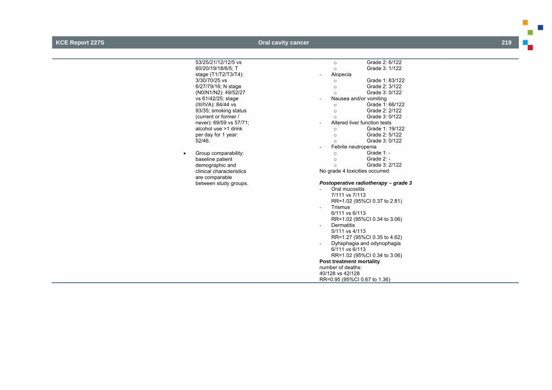
KCE Report 227S Oral cavity cancer 219
53/25/21/12/12/5 vs 60/20/19/18/6/5; T stage (T1/T2/T3/T4): 3/30/70/25 vs 6/27/79/16; N stage (N0/N1/N2): 49/52/27 vs 61/42/25; stage (III/IVA): 84/44 vs 93/35; smoking status (current or former / never): 69/59 vs 57/71; alcohol use >1 drink per day for 1 year: 52/46.
Group comparability: baseline patient demographic and clinical characteristics are comparable between study groups.
o Grade 2: 6/122 o Grade 3: 1/122
- Alopecia o Grade 1: 83/122 o Grade 2: 3/122 o Grade 3: 0/122
- Nausea and/or vomiting o Grade 1: 66/122 o Grade 2: 2/122 o Grade 3: 0/122
- Altered liver function tests o Grade 1: 19/122 o Grade 2: 5/122 o Grade 3: 0/122
- Febrile neutropenia o Grade 1: - o Grade 2: - o Grade 3: 2/122
No grade 4 toxicities occurred. Postoperative radiotherapy – grade 3 - Oral mucositis
7/111 vs 7/113 RR=1.02 (95%CI 0.37 to 2.81)
- Trismus 6/111 vs 6/113 RR=1.02 (95%CI 0.34 to 3.06)
- Dermatitis 5/111 vs 4/113 RR=1.27 (95%CI 0.35 to 4.62)
- Dyhsphagia and odynophagia 6/111 vs 6/113 RR=1.02 (95%CI 0.34 to 3.06)
Post treatment mortality number of deaths: 40/128 vs 42/128 RR=0.95 (95%CI 0.67 to 1.36)
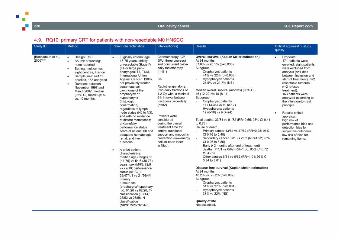
220 Oral cavity cancer KCE Report 227S
4.9. RQ10: primary CRT for patients with non-resectable M0 HNSCC Study ID Method Patient characteristics Intervention(s) Results Critical appraisal of study
quality
[Bensadoun et al., 2006]118
Design: RCT Source of funding:
none reported Setting: multicenter:
eight centres, France Sample size: n=171
enrolled, 163 analyzed Duration: between
November 1997 and March 2002; median (95% CI) follow-up: 50 vs. 40 months.
Eligibility criteria: age 18-75 years; strictly unresectable Stage IV (T4 or large pan-pharyngeal T3, TNM, International Union Against Cancer, 1988), not previously treated, squamous cell carcinoma of the oropharynx or hypopharynx (histologic confirmation), regardless of lymph node status (N0 to N3), and with no evidence of distant metastases; a Karnofsky performance status score of at least 60 and adequate hematologic, renal, and liver functions.
A priori patient
characteristics: median age (range) 53 (41-76) vs 54.6 (38-73) years; sex (M/F): 72/9 vs 72/10; performance status (0/1/2/-): 29/47/4/1 vs 21/56/4/1; primary tumour site (oropharynx/hypopharynx): 61/20 vs 62/20; T-classification (T3/T4): 28/53 vs 26/56; N-classification (N0/N1/N2b/N2c/N3)
Chemotherapy (CP-5FU, three courses) and concurrent twice-daily radiotherapy (n=81) vs Radiotherapy alone, (two daily fractions of 1.2 Gy with a minimal 6-h interval between fractions).twice-daily (n=82) Patients were considered during the overall treatment time for enteral nutritional support and mucositis prevention (low-energy helium-neon laser in Nice).
Overall survival (Kaplan Meier estimation) At 24 months 37.8% vs 20.1% (p=0.038) Subgroup: - Oropharynx patients
41% vs 22% (p=0.038) - Hypopharynx patients
21.5% vs 21.7% (NS)
Median overall survival (months) (95% CI) 16 (12-22) vs 10 (8-14) Subgroup: - Oropharynx patients
17 (13-36) vs 10 (8-17) - Hypopharynx patients
12 (6-50) vs 9 (7-24) Total deaths: 33/81 vs 61/82 (RR=0.55; 95% CI 0.41 to 0.73) Cause of death - Primary cancer 13/81 vs 47/82 (RR=0.28; 95%
CI 0.16 to 0.48) - Secondary cancer 3/81 vs 2/82 (RR=1.52; 95%
CI 0.26 to 8.85) - Early (<2 months after end of treatment)
deaths: 11/81 vs 6/82 (RR=1.86; 95% CI 0.72 to 4.78)
- Other causes 6/81 vs 6/82 (RR=1.01; 95% CI 0.34 to 3.01)
Disease-free survival (Kaplan Meier estimation) At 24 months 48.2% vs. 25.2% (p=0.002) Subgroup: - Oropharynx patients
51% vs 27% (p=0.001) - Hypopharynx patients
38% vs 22% (NS) Quality of life Not assessed.
Dropouts: 171 patients were enrolled, eight patients were excluded from analysis (n=4 died between inclusion and start of treatment), n=2 resectable tumours, n=2 refused treatment). 163 patients were analyzed according to the intention-to-treat principle.
Results critical appraisal: high risk of performance bias and detection bias for subjective outcomes; low risk of bias for remaining items.

KCE Report 227S Oral cavity cancer 221
7/10/16/31/17 vs 16/6/18/29/13.
Group comparability:
“Patients were evenly distributed between the two arms, as were patients within each investigating center.” Yet small (non-significant) differences between groups for performance status were found.
Local and regional control (at 2 years) Local control: 63.34% vs 34.48% Subgroup: - Oropharynx patients:
66.88% vs 34.4% - Hypopharynx patients:
50.7% vs 33.8% Regional control: 70.6% vs 53.02% - Oropharynx patients:
69.18% vs 55.32% - Hypopharynx patients:
71.4% vs 45.7% Rate of locoregional control (extrapolated by Kaplan-Meier method) 58.87% vs 27.5% (p=0.0003) Subgroup: - Oropharynx patients:
61.2% vs 28.23% (p=<0.0004) - Hypopharynx patients:
50.7% vs 24.3% (NS) Adverse events Grade 3-4 acute toxicity (World Health Organization criteria) - Mucositis
67/81 vs 57/82 (RR= 1.19; 95% CI 1.00 to 1.42)
- Dermatitis 31/81 vs 22/82 (RR=1.43; 95% CI 0.91 to 2.24)
- Nausea and diarrhoea 5/81 vs 0/82 (RR=11.13; 95% CI 0.6 to 198.13)
- Neutropenia 27/81 vs 2/82 (RR=13.67; 95% CI 3.36 to 55.59)
Early deaths (<2 months after end of treatment): 11/81 vs 6/82 (RR=1.99; 95% CI 0.70 to 5.67) Prevalence of gastrostomy tube Before treatment: 54/81 vs 38/82 (RR=1.44; 95% CI 1.09 to 1.90) 6 months: 10/49 vs 2/41 (RR=4.18; 95% CI 0.97 to 18.02)
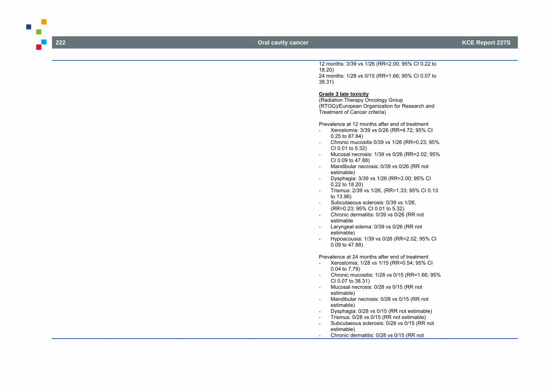
222 Oral cavity cancer KCE Report 227S
12 months: 3/39 vs 1/26 (RR=2.00; 95% CI 0.22 to 18.20) 24 months: 1/28 vs 0/15 (RR=1.66; 95% CI 0.07 to 38.31) Grade 3 late toxicity (Radiation Therapy Oncology Group (RTOG)/European Organization for Research and Treatment of Cancer criteria) Prevalence at 12 months after end of treatment - Xerostomia: 3/39 vs 0/26 (RR=4.72; 95% CI
0.25 to 87.84) - Chronic mucositis 0/39 vs 1/26 (RR=0.23; 95%
CI 0.01 to 5.32) - Mucosal necrosis: 1/39 vs 0/26 (RR=2.02; 95%
CI 0.09 to 47.88) - Mandibular necrosis: 0/39 vs 0/26 (RR not
estimable) - Dysphagia: 3/39 vs 1/26 (RR=2.00; 95% CI
0.22 to 18.20) - Trismus: 2/39 vs 1/26, (RR=1.33; 95% CI 0.13
to 13.96) - Subcutaeous sclerosis: 0/39 vs 1/26,
(RR=0.23; 95% CI 0.01 to 5.32) - Chronic dermatitis: 0/39 vs 0/26 (RR not
estimable - Laryngeal edema: 0/39 vs 0/26 (RR not
estimable) - Hypoacousia: 1/39 vs 0/26 (RR=2.02; 95% CI
0.09 to 47.88) Prevalence at 24 months after end of treatment - Xerostomia; 1/28 vs 1/15 (RR=0.54; 95% CI
0.04 to 7.79) - Chronic mucositis: 1/28 vs 0/15 (RR=1.66; 95%
CI 0.07 to 38.31) - Mucosal necrosis: 0/28 vs 0/15 (RR not
estimable) - Mandibular necrosis: 0/28 vs 0/15 (RR not
estimable) - Dysphagia: 0/28 vs 0/15 (RR not estimable) - Trismus: 0/28 vs 0/15 (RR not estimable) - Subcutaeous sclerosis: 0/28 vs 0/15 (RR not
estimable) - Chronic dermatitis: 0/28 vs 0/15 (RR not
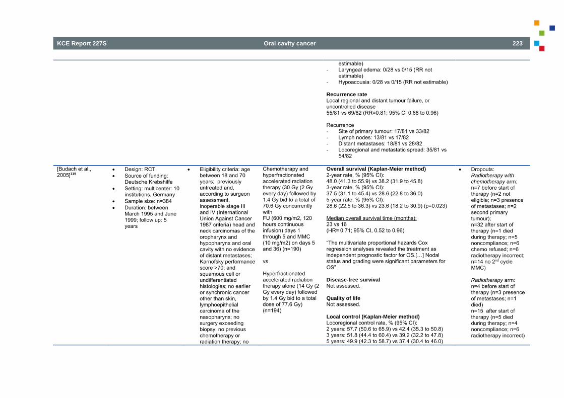
KCE Report 227S Oral cavity cancer 223
estimable) - Laryngeal edema: 0/28 vs 0/15 (RR not
estimable) - Hypoacousia: 0/28 vs 0/15 (RR not estimable) Recurrence rate Local regional and distant tumour failure, or uncontrolled disease 55/81 vs 69/82 (RR=0.81; 95% CI 0.68 to 0.96) Recurrence - Site of primary tumour: 17/81 vs 33/82 - Lymph nodes: 13/81 vs 17/82 - Distant metastases: 18/81 vs 28/82 - Locoregional and metastatic spread: 35/81 vs
54/82
[Budach et al., 2005]119
Design: RCT Source of funding:
Deutsche Krebshilfe Setting: multicenter: 10
institutions, Germany Sample size: n=384 Duration: between
March 1995 and June 1999; follow up: 5 years
Eligibility criteria: age between 18 and 70 years; previously untreated and, according to surgeon assessment, inoperable stage III and IV (International Union Against Cancer 1987 criteria) head and neck carcinomas of the oropharynx and hypopharynx and oral cavity with no evidence of distant metastases; Karnofsky performance score >70; and squamous cell or undifferentiated histologies; no earlier or synchronic cancer other than skin, lymphoepithelial carcinoma of the nasopharynx; no surgery exceeding biopsy; no previous chemotherapy or radiation therapy; no
Chemotherapy and hyperfractionated accelerated radiation therapy (30 Gy (2 Gy every day) followed by 1.4 Gy bid to a total of 70.6 Gy concurrently with FU (600 mg/m2, 120 hours continuous infusion) days 1 through 5 and MMC (10 mg/m2) on days 5 and 36) (n=190) vs Hyperfractionated accelerated radiation therapy alone (14 Gy (2 Gy every day) followed by 1.4 Gy bid to a total dose of 77.6 Gy) (n=194)
Overall survival (Kaplan-Meier method) 2-year rate, % (95% CI): 48.0 (41.3 to 55.9) vs 38.2 (31.9 to 45.8) 3-year rate, % (95% CI): 37.5 (31.1 to 45.4) vs 28.6 (22.8 to 36.0) 5-year rate, % (95% CI): 28.6 (22.5 to 36.3) vs 23.6 (18.2 to 30.9) (p=0.023) Median overall survival time (months): 23 vs 16 (HR=0.71; 95% CI, 0.52 to 0.96) “The multivariate proportional hazards Cox regression analyses revealed the treatment as independent prognostic factor for OS.[…] Nodal status and grading were significant parameters for OS” Disease-free survival Not assessed. Quality of life Not assessed. Local control (Kaplan-Meier method) Locoregional control rate, % (95% CI): 2 years: 57.7 (50.6 to 65.9) vs 42.4 (35.3 to 50.8) 3 years: 51.8 (44.4 to 60.4) vs 39.2 (32.2 to 47.8) 5 years: 49.9 (42.3 to 58.7) vs 37.4 (30.4 to 46.0)
Dropouts: Radiotherapy with chemotherapy arm: n=7 before start of therapy (n=2 not eligible; n=3 presence of metastases; n=2 second primary tumour); n=32 after start of therapy (n=1 died during therapy; n=5 noncompliance; n=6 chemo refused; n=6 radiotherapy incorrect; n=14 no 2nd cycle MMC) Radiotherapy arm: n=4 before start of therapy (n=3 presence of metastases; n=1 died) n=15 after start of therapy (n=5 died during therapy; n=4 noncompliance; n=6 radiotherapy incorrect)
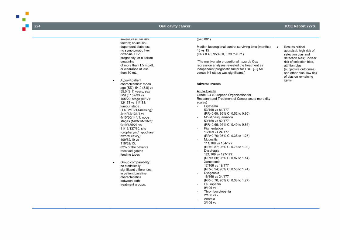
224 Oral cavity cancer KCE Report 227S
severe vascular risk factors; no insulin-dependent diabetes; no symptomatic liver cirrhosis, HIV, pregnancy, or a serum creatinine of more than 1.5 mg/dL or clearance of less than 80 mL
A priori patient
characteristics: mean age (SD): 54.0 (8.0) vs 55.0 (8.1) years; sex (M/F): 157/33 vs 165/29; stage (III/IV): 12/178 vs 11/183; tumour stage (T1/T2/T3/T4/missing): 2/14/42/131/1 vs 4/15/30/144/1; node stages (N0/N1/N2/N3): 9/19/135/27 vs 11/16/137/30; site (oropharynx/hypopharynx/oral cavity): 109/62/19 vs 119/62/13; 82% of the patients received gastric feeding tubes
Group comparability:
no statistically significant differences in patient baseline characteristics between both treatment groups.
(p=0.001) Median locoregional control surviving time (months): 48 vs 15 (HR=0.48; 95% CI, 0.33 to 0.71) “The multivariate proportional hazards Cox regression analyses revealed the treatment as independent prognostic factor for LRC.[…] N0 versus N3 status was significant.” Adverse events Acute toxicity Grade 3-4 (European Organisation for Research and Treatment of Cancer acute morbidity scales) - Erythema
53/169 vs 81/177 (RR=0.69; 95% CI 0.52 to 0.90)
- Moist desquamation 50/169 vs 82/177 (RR=0.65; 95% CI 0.49 to 0.86)
- Pigmentation 16/169 vs 24/177 (RR=0.70; 95% CI 0.38 to 1.27)
- Mucositis 111/169 vs 134/177 (RR=0.87; 95% CI 0.76 to 1.00)
- Dysphagia 121/169 vs 127/177 (RR=1.00; 95% CI 0.87 to 1.14)
- Xerostomia 17/169 vs 19/177 (RR=0.94; 95% CI 0.50 to 1.74)
- Dysgeusia 16/169 vs 24/177 (RR=0.70; 95% CI 0.38 to 1.27)
- Leukopenia 9/106 vs -
- Thrombocytopenia 2/106 vs -
- Anemia 3/106 vs -
Results critical appraisal: high risk of selection bias and detection bias; unclear risk of selection bias, attrition bias (subjective outcomes) and other bias; low risk of bias on remaining items.
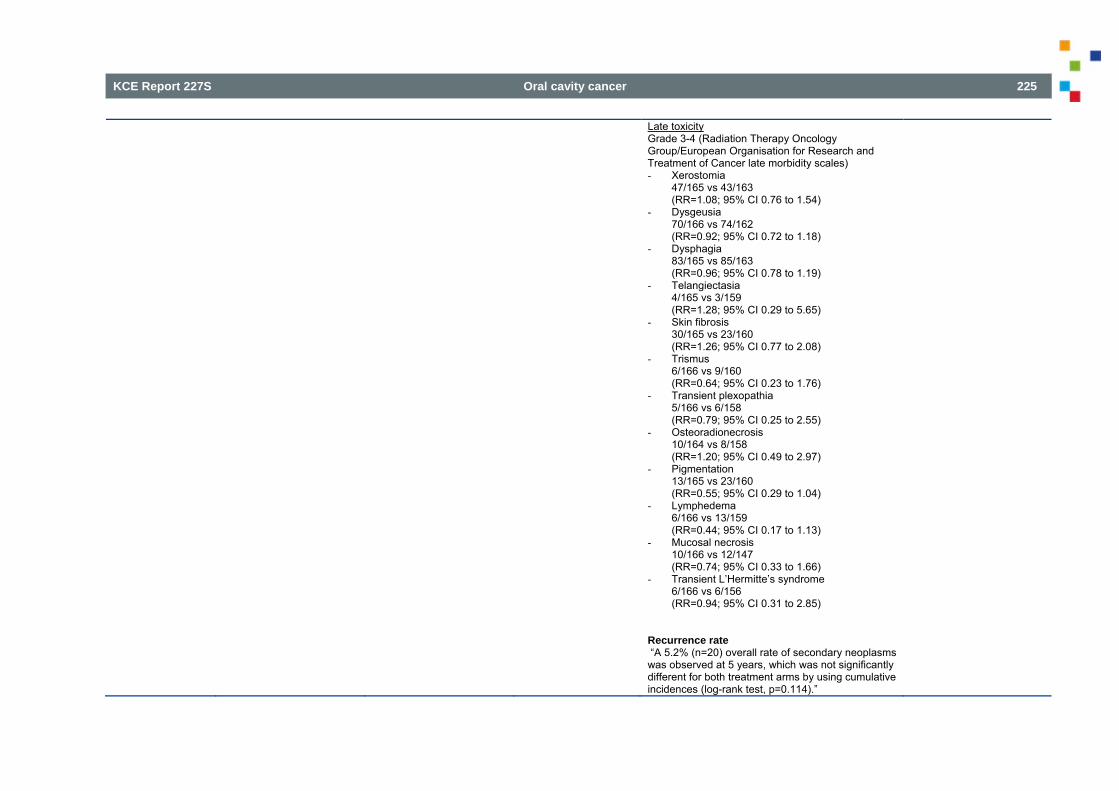
KCE Report 227S Oral cavity cancer 225
Late toxicity Grade 3-4 (Radiation Therapy Oncology Group/European Organisation for Research and Treatment of Cancer late morbidity scales) - Xerostomia
47/165 vs 43/163 (RR=1.08; 95% CI 0.76 to 1.54)
- Dysgeusia 70/166 vs 74/162 (RR=0.92; 95% CI 0.72 to 1.18)
- Dysphagia 83/165 vs 85/163 (RR=0.96; 95% CI 0.78 to 1.19)
- Telangiectasia 4/165 vs 3/159 (RR=1.28; 95% CI 0.29 to 5.65)
- Skin fibrosis 30/165 vs 23/160 (RR=1.26; 95% CI 0.77 to 2.08)
- Trismus 6/166 vs 9/160 (RR=0.64; 95% CI 0.23 to 1.76)
- Transient plexopathia 5/166 vs 6/158 (RR=0.79; 95% CI 0.25 to 2.55)
- Osteoradionecrosis 10/164 vs 8/158 (RR=1.20; 95% CI 0.49 to 2.97)
- Pigmentation 13/165 vs 23/160 (RR=0.55; 95% CI 0.29 to 1.04)
- Lymphedema 6/166 vs 13/159 (RR=0.44; 95% CI 0.17 to 1.13)
- Mucosal necrosis 10/166 vs 12/147 (RR=0.74; 95% CI 0.33 to 1.66)
- Transient L’Hermitte’s syndrome 6/166 vs 6/156 (RR=0.94; 95% CI 0.31 to 2.85)
Recurrence rate “A 5.2% (n=20) overall rate of secondary neoplasms was observed at 5 years, which was not significantly different for both treatment arms by using cumulative incidences (log-rank test, p=0.114).”
226 Oral cavity cancer KCE Report 227S
[Chauhan et al., 2008]114
Design: RCT Source of funding:
none described Setting: Department of
Radiotherapy, Pt. B.D. Sharma Post Graduate Institute of Medical Sciences, Rohtak, India
Sample size: n=80 Duration: from
November 2000 to March 2003; median follow-up: 9 months (range 6–52) in the RT group vs 11 months (range 5-51) in the CT/RT group
Eligibility criteria: locally advanced (T3, T4, any N, M0) previously untreated histopathologically proven squamous cell carcinoma of head and neck; unresectable disease or refusal of surgery; Karnofsky performance status score ≤70%, adequate liver function tests, bone marrow reserve and renal function.
A priori patient
characteristics: median age (range): 51.5 (30-69) vs 50 (28-72) years; sex (M/F): 38/2 vs 37/3; primary site (oral cavity / oropharynx/hypopharynx/larynx): 4/30/5/1 vs 3/30/6/1; stage (III/IV): 20/20 vs 18/22; tumour size (T3/T4): 37/3 vs 36/4; nodal status (N0/N1/N2/N3): 7/16/14/3 vs 6/16/19/0
Group comparability:
“There was good balance in the prognostic factors, including performance status, tumour and nodal stages, and histology, between the two groups”
Gemcitabine (intravenously over 30 minutes once weekly, 1–2 h before radiation, for 6 consecutive weeks at a dose of 100 mg/m2) concurrent with radiotherapy (once daily, 5 days a week as a single 2 Gy fraction to a total dose of 64 Gy) (n=40) vs Radiotherapy alone (once daily, 5 days a week as a single 2 Gy fraction to a total dose of 64 Gy) (n=40)
Overall survival Not assessed. Disease-free survival At 3 years follow-up: 63.3% vs 20% Quality of life Not assessed. Local control “Local control was good, none of the 19 patients with complete response developed relapse in the CT/RT group. Seven of the 13 patients with complete response in the radiation only group relapsed (3 at primary site, 3 at nodal and 1 distant):” Relapses: Primary: 0/30 vs 3/30 (RR=0.14; 95% CI 0.01 to 2.65) Nodal: 0/30 vs 3/30 (RR=0.14; 95% CI 0.01 to 2.65) Distant: 0/30 vs 1/30 (RR=0.33; 95% CI 0.01 to 7.87) Adverse events Acute reactions (WHO criteria) % of patients Haematological
Haemoglobin level: Grade I toxicity: 80% vs 47.5% Grade II toxicity: 20% vs 7.5% (p<0.05) “The leukocyte and platelet counts remained within normal limits during the treatment schedule in both the groups.”
Skin reactions Level 5: 50% vs 7.5% Level 6: 7.5% vs 2.5% (p<0.05)
Oral mucosal reactions Level 5: 67.5% vs 17.5 (p<0.05) Level 6: “During the 5th week in the CT/RT group two patients developed level-6 mucosal reactions due to which treatment had to be interrupted”
Nausea and vomiting: “In both the RT and
Dropouts: none: “All 80 patients were assessable for toxicity and response.”
Results critical
appraisal: high risk of performance bias and detection bias (subjective outcomes); unclear risk of selection, reporting and other bias; low risk of bias on remaining items.
KCE Report 227S Oral cavity cancer 227
CT/RT groups only mild nausea and transient vomiting, which did not require medication were observed.
Weight loss Grade I: 47.5% vs 85.7% Grade II: 52.5% vs 14.3% “Thus, there was significant loss of weight in the CT/RT group (p<0.05) as compared to the RT group during the later half of treatment which seems to be due to poor intake because of the debilitating oral mucosal reactions.”
Recurrence rate Not assessed.
[Quon et al., 2011]117
Design: RCT Source of funding:
Public Health Service, National Cancer Institute, National Institutes of Health and the Department of Health and Human Services
Setting: multicenter, USA
Sample size: n=371 randomized, n=308 analyzed
Duration: from November 1982 to June 1987; median follow-up period: 62 months
Eligibility criteria: patients with biopsy-proven head-and neck squamous cell or undifferentiated carcinoma, technically unresectable American Joint Committee on Cancer 1980 clinical Stage III or IV with no distant metastasis; all anatomic sites of the head and neck were included including nasopharyngeal carcinomas restricted to T3/4 or N2/3 disease; Eastern Cooperative Oncology Group (ECOG) performance status (PS) of 0–3; adequate hematologic, hepatic, and renal function; no existing cardiac conditions, pregnancy or lactation, prior treatment with RT or chemotherapy, and no prior or synchronous
RT Radiotherapy plus concomitant cisplatin dosed at 20 mg/m2 per week (n=149) vs Radiotherapy (n=159) Radiotherapy was given once daily, 5 days a week, prescribed dose to the primary lesion and involved nodal disease 68–76 Gy, daily fraction 1.8–2 Gy (radiation arm) or 1.8 (radiation+cisplatin arm)
Overall survival Median survival in months: 11.8 vs 13.3 (p =0.81) “ Also not significantly different with an intent-to-treat analysis (data not shown).” “Univariate analysis demonstrated that a poorer OS was associated with several factors: white race (p=0.04), greater smoking exposure (p=0.002), greater alcohol consumption (p<0.001), ECOG PS 2–4 (p<0.003), better cell differentiation (p=0.034), higher stage (p=0.021), and nonnasopharynx primary site (p=0.001). “ “Multivariate analysis demonstrated no significant treatment effect (p=0.60)” Disease-free survival Not assessed. Quality of life Not assessed. Local control Not assessed. Adverse events Acute adverse effects “The addition of weekly cisplatin significantly increased the frequency and severity of nausea/vomiting (p <0.001) and of neurologic (p=0.002), renal (p < 0.001), and hematologic
Dropouts: Of the 371 patients randomized, 63 were excluded from the primary analyses Radiotherapy + cisplatin: n=37 (n=27 ineligible; n=4 patient refusal; n=3 died before treatment; n=1 elected non-protocol treatment; n=1 decreased serum creatinine and creatinine clearance; n=1 unknown) Radiotherapy: n=26 (n=18 ineligible; n=5 patient refusal, n=1 died before treatment; n=1 received non-protocol treatment, n=1 could not be positioned for radiation therapy)
Results critical
appraisal: high risk of
228 Oral cavity cancer KCE Report 227S
malignancy. NB: “At the initiation of the trial, from November 1982 to August 1983, patients with an incomplete resection and gross residual tumor were permitted to enroll in the absence of documented distant disease. After August 1983, these patients were excluded.”
A priori patient characteristics: median age (range): 61y (20-81) vs 60y (19-85); sex (M/F): 117/32 vs 133/26; race (white/non-white): 115/34 vs 122/37; performance status (0/1/2/3): 33/87/23/6 vs 42/81/29/7;T-stage (0/1/2/3/4): 0/3/12/45/89 vs 2/5/14/49/89; N-stage (0/1/2/3): 32/24/27/66 vs 39/17/32/71; Stage (III / IV excluding T4 N3 / T4 N3): 21/89/39 vs 23/97/39; prior surgery (yes/no): 41/108 vs 38/121; primary site (nasopharynx/oral cavity/oropharynx/larynx/hypopharynx/other): 16/51/37/14/27/4 vs 25/43/48/7/30/6
Group comparability:
“There were some imbalances with
toxicities (p < 0.001).“ “Respiratory acute toxicities were increased in the RT + cisplatin group. The increased frequency of toxicities was primarily mild to moderate in severity. Toxicities within the radiation fields did not seem to be increased. Additional evaluation for laryngeal edema and nutritional toxicity was also evaluated with different grading schemas. The addition of weekly cisplatin also did not significantly increase the spectrum and the severity of any of these toxicities“ “When each patient was classified by the worst grade of any type of toxicity, the treatment groups were comparable (p=0.21).” Head and Neck Radiation Therapy Form. - Grade 3+ laryngeal edema: 5% vs 3% - Grade 3+ nutritional toxicity: 35% vs 31% Late toxicities (recorded on the Radiotherapy Long Term Follow-up Form) - Skin: 15% vs 21% (p=0.18) - Mucous membrane: 22% vs 28%, (p=0.29) - Subcutaneous tissue: 13% vs 11%, (p=0.60) - Esophagus: 9% vs 3% (p=0.03) - Larynx: 11% vs 4% (p=0.05) - Other: 16% vs 13% (p=0.52) Recurrence rate Not assessed.
performance bias, detection bias (subjective outcomes) and other bias; unclear risk of attrition bias; low risk on remaining items.
KCE Report 227S Oral cavity cancer 229
the RT + cisplatin group: a higher number of patients with age >65, weight loss 10% in the previous 6 months, >40 pack-years exposure to smoking, well or moderate cell differentiation, and nonnasopharyngeal primary tumors. These imbalances contributed to a bias against the RT + cisplatin treatment group with the as-treated analysis.”
[Rodriguez et al., 2010]115
Design: RCT Source of funding:
none reported Setting: multi-center,
most patients collected by the National Institute of Oncology and Radiobiology, Cuba
Sample size: n=106 Duration: from July
2002 to February 2007; median follow-up period: 45.2 months
Eligibility criteria: histologically documented advanced (stages III and IV) locoregional (unresectable) squamous-cell carcinoma of the head and neck; suitable for radiation therapy; measurable lesions; age ≥18y, ;ECOG performance status ≤2; life expectancy greater than 6 months; normal functioning of the organs and of te bone marrow; no prior radiotherapy or chemotherapy, concurrent active cancer, any uncontrolled illness and pregnancy or lactation.
A priori patient
characteristics:
Nimotuzumab in combination with radiotherapy (n=54) vs Placebo and radiotherapy (n=51) The mean monoclonal antibody (mAb) cumulative dose was 1,057 mg. The mean cumulative radiotherapy dose for the group receiving nimotuzumab was 6,030 cGy, whereas patients of the control group were treated with 5,931 cGy, as average.
Overall survival 9/54 vs 5/51 (RR=1.70; 95% CI 0.61 to 4.73) Intent to treat analysis (Kaplan-Meier) Mean survival (in months): 22.71 vs 17.71 Median survival (in months): 12.50 vs 9.47 Mean survival intent to treat analysis (in months) Kaplan-Meier method: 22.71 vs 17.71 Median survival intent to treat analysis (in months) Kaplan-Meier method: 12.50 vs 9.47 Death rate within 90 days post-randomization 4/54 vs 3/52 Disease-free survival Not assessed. Quality of life European Organization for Research and Treatment of Cancer (EORTC) QLQ-C30 (version 3.0) and the QLQ-H&N35 (head & neck cancer module) validated questionnaires. “Quality of life was evaluated in 42 patients, 21
Dropouts: n=86 received at least four doses of the investigational product (n=44 nimotuzumab, n=42 placebo), and n=77 completed six doses (n=39, nimotuzumab, n=37, placebo). The most frequent causes of treatment interruption were voluntary withdrawal, lost of follow up, severe adverse events (not attributed to nimotuzumab) and tumour progression. Exact numbers and reasons for lost to follow up were not reported per group.
Results critical
appraisal: unclear risk of selection bias and
230 Oral cavity cancer KCE Report 227S
median age: 59 vs 65 y; sex (M/F): 44/10 vs 37/14;; median weight (kg): 60 vs 57; stage (III/IV): 21/32 vs 21/30; ECOG performance status (Grade 0/1/2): 21/31/- vs 14/35/1; primary tumour site (tonsil/base of tongue/alveolar ridge/tetromolar trigone/hypopharynx/larynx/anterior tongue/oral mucosa/soft palate/hard/palate/pharyngeal wall/maxillary sinus/floor of mouth): 17/10/1/5/1/1/4/3/6/1/2/1/2 vs 24/10/-/5/-/-/2/-/5/3/-/1/2
Group comparability:
“No significant differ-ences were detected between the two arms regarding demography or tumor characteristics.” “Even though, no statistically significant unbalance was found between both treatment groups, 46.2% of tumors in the placebo arm were located at the tonsil while only 31.5% of patients in the nimotuzumab group had tonsil tumors.” Significant differences were found in relation with the global health status/QoL questionnaire The
treated with nimotuzumab and radiotherapy and 21 treated with a placebo and radiotherapy.” “Differences between the two groups were only found in relation with the general pain evaluation at month six. Patients treated with placebo referred less pain than patients treated with nimotuzumab. These differences were not found in the following evaluations (months 9 and 12). Notably, no differences were found either regarding pain or pain killer consumption between the two groups at any evaluation when applying the head and neck specific survey. Given that these differences were subtle and the results were not supported at other time points or the second scale, it is likely that this result occurred by chance.” “The remaining parameters of the global questionnaire did not show significant differences between the treatment groups at the 3, 6, 9 and 12 months. With regards to the specific head and neck questionnaire, no discernible differences were found between the monoclonal antibody and placebo groups for the 18 head and neck quality of life aspects at baseline and at 3, 6, 9 and 12 months” In summary, a quality of life increase and a reduction of the general and specific symptoms of the disease for both groups during the trial were detected. No negative impact of the use of nimotuzumab as compared to placebo was detected regarding quality of life.” Local control Not assessed. Adverse events Common Toxicity Criteria of the US National Cancer Institute, Version 2, April 30, 1999 (NCI-CTC, Version 2) Overall adverse events: 38/54 vs 30/52 (RR=1.22; 95% CI 0.91 to 1.63) Adverse events definitively, probably or possibly
reporting bias; low risk of bias on remaining items.
KCE Report 227S Oral cavity cancer 231
monoclonal antibody treated group showed the highest health score. For the rest of the five functional scales as well as for the individual symptoms of the general health scale, there were no differences at baseline between both groups.
related to the investigational drug Grade I or II adverse events: 17/54; mainly: Asthenia: 14.6% Fever: 9.8% Headache: 9.8% Chills: 7.8% Anorexia: 7.8% Skin rash: no skin rash Most frequent reactions due to radiotherapy Mucositis: 20.1% vs 16.8% Dry mouth: 17% vs 23% Dry radio-dermitis: 10.3% vs 12.1% Odynophagia: 8% vs 11.3 “There was no exacerbation of the adverse reactions related to irradiation after the administration of the monoclonal antibody.” Recurrence rate Not assessed.
[Ruo Redda et al., 2010]112
Design: RCT Source of funding:
none reported Setting: multicenter: six
centres, Italy Sample size:
randomized n=164, n=157 started treatment
Duration: November 1992 through December 1995; median follow-up period: 26.2 months (range, 6.2-169.5) with a median observation period for surviving patients of 154.3 months
Eligibility criteria: age >18 and ≤70 years; biopsy-proven diagnosis of locally advanced and unresectable stage III or IV non-metastatic HNSCC, using the criteria of the International Union Against Cancer by use of the 4th edition of the TNM classification of malignant tumours; no prior chemotherapy or radiotherapy for any kind of cancer (except for non-melanoma skin cancer or in situ cervical cancer); Eastern Cooperative Oncology Group (ECOG) performance
Radiotherapy with concurrent daily low-dose carboplatin (n=80) vs Radiotherapy alone (n=77)
Overall survival (Kaplan Meier method) 3 year rate: 28.9% vs 11.1% 5 year rate: 9.0% vs 6.9% 10 year rate: 5.5% vs 6.9% (p=0.02) Disease-free survival (Kaplan Meier method) 3 year rate: 16% vs 9.0% 5 year rate: 6.8% vs 5.5% 10 year rate: 6.8% vs 5.5% (p=0.09) Quality of life Not assessed. Local control (Kaplan Meier method) 3 year rate: 21.7% vs 15.0% 5 year rate: 15.1% vs 10.7% 10 year rate: 15.1% vs 10.7% (p=0.11)
Dropouts: Radiotherapy with chemotherapy arm: n=7 died during treatment (n=3 fatal bleeding; n=3 pneumonia; n=1 bowel perforation) Radiotherapy arm: n=5 died during treatment (n=3 fatal bleeding; n=2 bowel perforation)
Results critical
appraisal: Unclear risk of selection bias, performance bias and detection bias for subjective outcomes;
232 Oral cavity cancer KCE Report 227S
status of ≤2, without any serious concomitant diseases; adequate bone marrow reserve, renal function and liver function; adequate nutritional and liquid intake.
A priori patient
characteristics: median age (range) in years: 58 (39-70) vs 61 (40-71); sex (M/F): 66/7 vs 63/9; performance status (0/1/2/): 47/18/8 vs 30/33/9; site (oral cavity/oroparynx/larynx/hypopharynx): 14/42/9/8 vs 14/39/12/7; stage (III/IV): 18/55 vs 15/57
Group comparability:
“There were no differences between the two treatment arms as regard age, sex, primary tumour site and staging.”
Exploratory subgroup analysis “No significant difference in outcome when considering age, site of primary disease or nodal status.” Considering only stage IV patients: 3 year rate: 21.5% vs 12.8% 5 year rate: 15.9% vs 7.7% 10 year rate: 15.9% vs 7.7% (p=0.04) “However, the difference was not confirmed in multivariate analysis, possibly suggesting a possible imbalance in other prognostic factors in this subset of patients.” “Furthermore, multivariate analysis did not find any prognostic factor that was statistically significant.” Adverse events Acute toxicity Grade 3-4 (World Health Organization criteria) - Hemoglobin:
3/80 vs 0/77 (RR=6.74; 95% CI 0.35 to 128.38) - Leukocytes:
7/80 vs 0/77 (RR= 14.44; 95% CI 0.84 to 248.66)
- Thrombocytes: 1/80 vs 0/77 (RR=3.00; 95% CI 0.12 to 72.56)
- Mucositis: 10/80 vs 9/77 (RR=1.07; 95% CI 0.46 to 2.49)
Feeding tube required for nutritional support n=110 Late toxicity “The incidence of late toxicity in the combined arm was no higher than that observed in patients treated with radiotherapy alone, except for the observation of more severe neck fibrosis in patients who received both chemotherapy and radiotherapy”: 7/80 vs 3/77 (RR=2.25; 95% CI 0.60 to 8.37) “No radiation myelitis or toxic-related death was
low risk of bias for all other items.
KCE Report 227S Oral cavity cancer 233
observed in either treatment arm.” Recurrence rate Not assessed.
[Semrau et al., 2006]116
Design: RCT Source of funding:
none reported Setting: five
participating German centers (universities of Heidelberg, Wuerzburg, and Cologne, community hospitals of Kassel and Oldenburg)
Sample size: n=263 randomized, n=240 started treatment
Duration: between July 1995 and April 1999; median follow-up period (range): 57.3 (10.7-84.4) months
Eligibility criteria: histologically proven locoregionally advanced unresectable cancers of the head and neck, located in oropharynx or hypopharynx, International Union Against Cancer Stage III or IV; free of distant metastasis, no history of prior malignant neoplasm, no prior chemotherapy or radiation therapy; baseline routine laboratory tests had to be fulfilled; performance status between 0 and 2 (WHO scale).
A priori patient characteristics: median age (range) 57y (38–73) vs 56y (28–73); sex (M/F): 96/17 vs 108/19; tumour site (oropharynx/hypopharynx):: 87/26 vs 91/36;; T-stage (T1/T2/T3/T4): 0/1/19/91 vs 2/3/20/102 ;N-stage (N0/N1/N2/N3): 12/8/85/8 vs 11/7/92/17 ; Stage UICC III/IV: 4/109 vs 5/122
Concurrent hyperfractionated and accelerated radiochemotherapy (RCT) with two cycles 5-fluorouracil (600 mg/m2/day) and carboplatin (70 mg/m2/day) on Days 1–5 and 29–33 (n=113) vs Hyperfractionated and accelerated Radiotherapy (RT) (n=127) Total RT dose in both arms was 69.9 Gy in 38 days in concomitant boost technique. The majority of patients (138 of 240; 57.5%; no statistical difference between RCT and RT) received a gastric feeding tube, facilitating enteral nutrition during radiotherapy.
Overall survival “Patients treated with RCT have a statistically significant improved overall survival compared with patients treated with RT alone.” 5-year survival 25.6% (95% CI 15.8 to 35.4%) vs 15.8% (95% CI 9.1 to 22.4%) (p=0.016) Subgroup: Oropharyngeal:26.1% (95%CI 14.3 to 37.8) vs
13.0% (95%CI 5.3 to 20.6) (p=0.008) Hypopharyngeal: percentages not reported
(p=0.72) Disease-free survival 5-year rates of survival reported under ‘local control’ Quality of life Not reported Local control 5-year rates of survival with local control (Kaplan-Meier estimate) 22.7% (95% CI, 13.3–32.0%) vs 12.6% (95% CI, 6.6 –18.6%) (p=0.01) Subgroup: - Oropharyngeal: 22.9%, (95% CI 11.5 to 34.3%)
vs 10.0% (95% CI, 3.2 to 16.4%) (p=0.002) Hypopharyngeal: 19.2% vs 19.4%, (p=0.885) Adverse events Acute adverse effects, Grade 3–4 (according to CTC/RTOG-criteria) (reported in Staar 2001) Mucositis: 68% vs 52% (p=0.01) Dermatitis: 30% vs 28% WBC: 18% vs - Platelets: 5% vs - Anemia: - vs 1%
Dropouts: n=23 did not start treatment (n=1 died because of cardiac failure; n=7 missing qualification; n=7 refusals; n=3 infection of feeding tubes; n=2 alcohol excess; n=2 distant metastases; n=2 unknown reason). Drop outs were not specified per intervention group.
Results critical
appraisal: high risk of performance bias, detection bias (subjective outcomes) and reporting bias; unclear risk of selection bias; low risk of bias on remaining items.
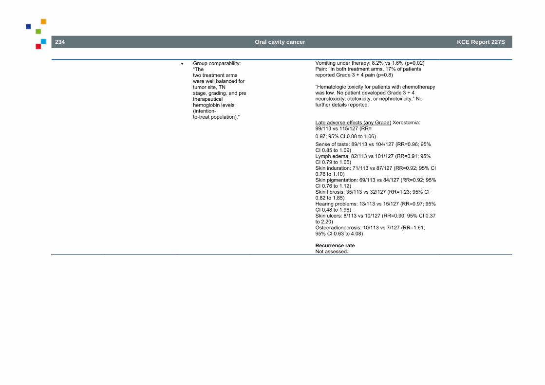
234 Oral cavity cancer KCE Report 227S
Group comparability: “The two treatment arms were well balanced for tumor site, TN stage, grading, and pre therapeutical hemoglobin levels (intention- to-treat population).”
Vomiting under therapy: 8.2% vs 1.6% (p=0.02) Pain: “In both treatment arms, 17% of patients reported Grade 3 + 4 pain (p=0.8) “Hematologic toxicity for patients with chemotherapy was low. No patient developed Grade 3 + 4 neurotoxicity, ototoxicity, or nephrotoxicity.” No further details reported. Late adverse effects (any Grade) Xerostomia: 99/113 vs 115/127 (RR= 0.97; 95% CI 0.88 to 1.06) Sense of taste: 89/113 vs 104/127 (RR=0.96; 95% CI 0.85 to 1.09) Lymph edema: 82/113 vs 101/127 (RR=0.91; 95% CI 0.79 to 1.05) Skin induration: 71/113 vs 87/127 (RR=0.92; 95% CI 0.76 to 1.10) Skin pigmentation: 69/113 vs 84/127 (RR=0.92; 95% CI 0.76 to 1.12) Skin fibrosis: 35/113 vs 32/127 (RR=1.23; 95% CI 0.82 to 1.85) Hearing problems: 13/113 vs 15/127 (RR=0.97; 95% CI 0.48 to 1.96) Skin ulcers: 8/113 vs 10/127 (RR=0.90; 95% CI 0.37 to 2.20) Osteoradionecrosis: 10/113 vs 7/127 (RR=1.61; 95% CI 0.63 to 4.08) Recurrence rate Not assessed.
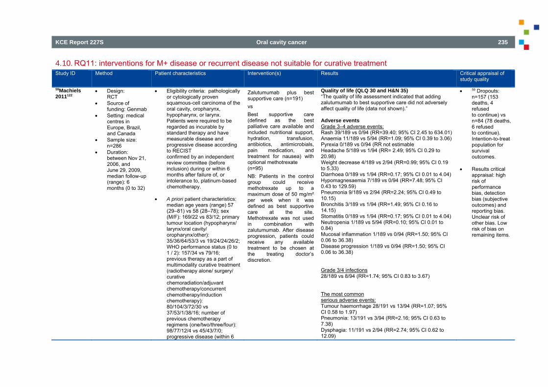
KCE Report 227S Oral cavity cancer 235
4.10. RQ11: interventions for M+ disease or recurrent disease not suitable for curative treatment Study ID Method Patient characteristics Intervention(s) Results Critical appraisal of
study quality
59Machiels 2011122
Design: RCT
Source of funding: Genmab
Setting: medical centres in Europe, Brazil, and Canada
Sample size: n=286
Duration: between Nov 21, 2006, and June 29, 2009, median follow-up (range): 6 months (0 to 32)
Eligibility criteria: pathologically or cytologically proven squamous-cell carcinoma of the oral cavity, oropharynx, hypopharynx, or larynx. Patients were required to be regarded as incurable by standard therapy and have measurable disease and progressive disease according to RECIST confirmed by an independent review committee (before inclusion) during or within 6 months after failure of, or intolerance to, platinum-based chemotherapy.
A priori patient characteristics:
median age years (range) 57 (29–81) vs 58 (28–78); sex (M/F): 169/22 vs 83/12; primary tumour location (hypopharynx/ larynx/oral cavity/ oropharynx/other): 35/36/64/53/3 vs 19/24/24/26/2; WHO performance status (0 to 1 / 2): 157/34 vs 79/16; previous therapy as a part of multimodality curative treatment (radiotherapy alone/ surgery/ curative chemoradiation/adjuvant chemotherapy/concurrent chemotherapy/induction chemotherapy): 80/104/3/72/30 vs 37/53/1/38/16; number of previous chemotherapy regimens (one/two/three/four): 98/77/12/4 vs 45/43/7/0; progressive disease (within 6
Zalutumumab plus best supportive care (n=191) vs Best supportive care (defined as the best palliative care available and included nutritional support, hydration, transfusion, antibiotics, antimicrobials, pain medication, and treatment for nausea) with optional methotrexate (n=95) NB: Patients in the control group could receive methotrexate up to a maximum dose of 50 mg/m² per week when it was defined as best supportive care at the site. Methotrexate was not used in combination with zalutumumab. After disease progression, patients could receive any available treatment to be chosen at the treating doctor’s discretion.
Quality of life (QLQ 30 and H&N 35) “The quality of life assessment indicated that adding zalutumumab to best supportive care did not adversely affect quality of life (data not shown).” Adverse events Grade 3–4 adverse events: Rash 39/189 vs 0/94 (RR=39.40; 95% CI 2.45 to 634.01) Anaemia 11/189 vs 5/94 (RR=1.09; 95% CI 0.39 to 3.06) Pyrexia 0/189 vs 0/94 (RR not estimable Headache 5/189 vs 1/94 (RR= 2.49; 95% CI 0.29 to 20.98) Weight decrease 4/189 vs 2/94 (RR=0.99; 95% CI 0.19 to 5.33) Diarrhoea 0/189 vs 1/94 (RR=0.17; 95% CI 0.01 to 4.04) Hypomagnesaemia 7/189 vs 0/94 (RR=7.48; 95% CI 0.43 to 129.59) Pneumonia 9/189 vs 2/94 (RR=2.24; 95% CI 0.49 to 10.15) Bronchitis 3/189 vs 1/94 (RR=1.49; 95% CI 0.16 to 14.15) Stomatitis 0/189 vs 1/94 (RR=0.17; 95% CI 0.01 to 4.04) Neutropenia 1/189 vs 5/94 (RR=0.10; 95% CI 0.01 to 0.84) Mucosal inflammation 1/189 vs 0/94 (RR=1.50; 95% CI 0.06 to 36.38) Disease progression 1/189 vs 0/94 (RR=1.50; 95% CI 0.06 to 36.38) Grade 3/4 infections 28/189 vs 8/94 (RR=1.74; 95% CI 0.83 to 3.67) The most common serious adverse events: Tumour haemorrhage 28/191 vs 13/94 (RR=1.07; 95% CI 0.58 to 1.97) Pneumonia: 13/191 vs 3/94 (RR=2.16; 95% CI 0.63 to 7.38) Dysphagia: 11/191 vs 2/94 (RR=2.74; 95% CI 0.62 to 12.09)
59 Dropouts: n=157 (153 deaths, 4 refused to continue) vs n=84 (78 deaths, 6 refused to continue). Intention-to-treat population for survival outcomes.
Results critical
appraisal: high risk of performance bias, detection bias (subjective outcomes) and reporting bias. Unclear risk of other bias. Low risk of bias on remaining items.
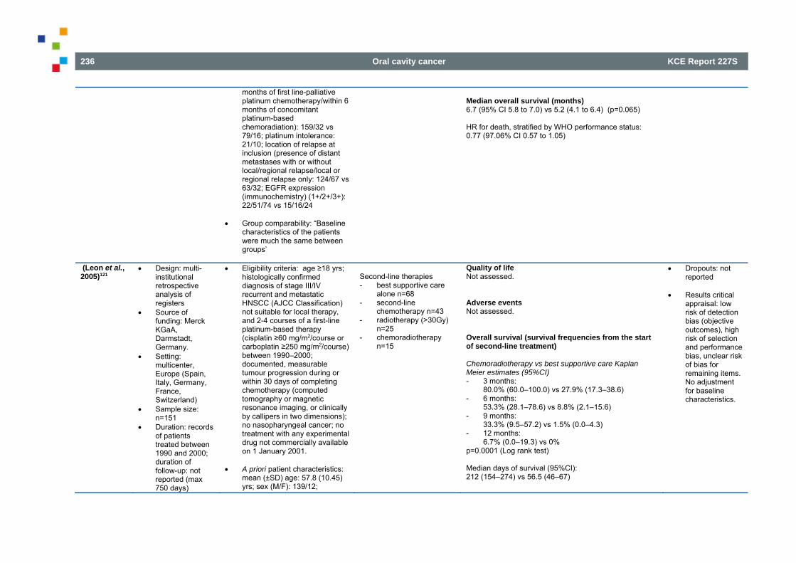
236 Oral cavity cancer KCE Report 227S
months of first line-palliative platinum chemotherapy/within 6 months of concomitant platinum-based chemoradiation): 159/32 vs 79/16; platinum intolerance: 21/10; location of relapse at inclusion (presence of distant metastases with or without local/regional relapse/local or regional relapse only: 124/67 vs 63/32; EGFR expression (immunochemistry) (1+/2+/3+): 22/51/74 vs 15/16/24
Group comparability: “Baseline
characteristics of the patients were much the same between groups’
Median overall survival (months) 6.7 (95% CI 5.8 to 7.0) vs 5.2 (4.1 to 6.4) (p=0.065) HR for death, stratified by WHO performance status: 0.77 (97.06% CI 0.57 to 1.05)
(Leon et al., 2005)121
Design: multi-institutional retrospective analysis of registers
Source of funding: Merck KGaA, Darmstadt, Germany.
Setting: multicenter, Europe (Spain, Italy, Germany, France, Switzerland)
Sample size: n=151
Duration: records of patients treated between 1990 and 2000; duration of follow-up: not reported (max 750 days)
Eligibility criteria: age ≥18 yrs; histologically confirmed diagnosis of stage III/IV recurrent and metastatic HNSCC (AJCC Classification) not suitable for local therapy, and 2-4 courses of a first-line platinum-based therapy (cisplatin ≥60 mg/m2/course or carboplatin ≥250 mg/m2/course) between 1990–2000; documented, measurable tumour progression during or within 30 days of completing chemotherapy (computed tomography or magnetic resonance imaging, or clinically by callipers in two dimensions); no nasopharyngeal cancer; no treatment with any experimental drug not commercially available on 1 January 2001.
A priori patient characteristics:
mean (±SD) age: 57.8 (10.45) yrs; sex (M/F): 139/12;
Second-line therapies - best supportive care
alone n=68 - second-line
chemotherapy n=43 - radiotherapy (>30Gy)
n=25 - chemoradiotherapy
n=15
Quality of life Not assessed. Adverse events Not assessed. Overall survival (survival frequencies from the start of second-line treatment) Chemoradiotherapy vs best supportive care Kaplan Meier estimates (95%CI) - 3 months:
80.0% (60.0–100.0) vs 27.9% (17.3–38.6) - 6 months:
53.3% (28.1–78.6) vs 8.8% (2.1–15.6) - 9 months:
33.3% (9.5–57.2) vs 1.5% (0.0–4.3) - 12 months:
6.7% (0.0–19.3) vs 0% p=0.0001 (Log rank test) Median days of survival (95%CI): 212 (154–274) vs 56.5 (46–67)
Dropouts: not reported
Results critical
appraisal: low risk of detection bias (objective outcomes), high risk of selection and performance bias, unclear risk of bias for remaining items. No adjustment for baseline characteristics.
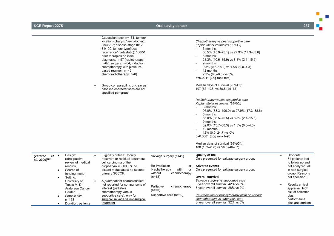
KCE Report 227S Oral cavity cancer 237
Caucasian race: n=151, tumour location (pharynx/larynx/other): 88/36/27; disease stage III/IV: 31/120; tumour type(local recurrence/ metastatic): 100/51; prior therapies on initial diagnosis: n=97 (radiotherapy: n=87, surgery: n=84, induction chemotherapy with platinum-based regimen: n=42, chemoradiotherapy: n=8)
Group comparability: unclear as
baseline characteristics are not specified per group
Chemotherapy vs best supportive care Kaplan Meier estimates (95%CI) - 3 months:
60.5% (45.9–75.1) vs 27.9% (17.3–38.6) - 6 months:
23.3% (10.6–35.9) vs 8.8% (2.1–15.6) - 9 months:
9.3% (0.6–18.0) vs 1.5% (0.0–4.3) - 12 months:
2.3% (0.0–6.8) vs 0% p=0.0011 (Log rank test) Median days of survival (95%CI): 107 (83–135) vs 56.5 (46–67) Radiotherapy vs best supportive care Kaplan Meier estimates (95%CI) - 3 months:
96.0% (88.3–100.0) vs 27.9% (17.3–38.6) - 6 months:
56.0% (36.5–75.5) vs 8.8% (2.1–15.6) - 9 months:
32.0% (13.7–50.3) vs 1.5% (0.0–4.3) - 12 months:
12% (0.0–24.7) vs 0% p=0.0001 (Log rank test) Median days of survival (95%CI): 188 (139–280) vs 56.5 (46–67)
(Zafereo et al., 2009)123
Design: retrospective review of medical records
Source of funding: none
Setting: University of Texas M. D. Anderson Cancer Center
Sample size: n=168
Duration: patients
Eligibility criteria: locally recurrent or residual squamous cell carcinoma of the oropharynx (SCCOP); no distant metastases; no second primary SCCOP.
A priori patient characteristics:
not reported for comparisons of interest (palliative chemotherapy versus supportive care), only for surgical salvage vs nonsurgical treatment
Salvage surgery (n=41) Re-irradiation or brachytherapy with or without chemotherapy (n=18) Palliative chemotherapy (n=70) Supportive care (n=39)
Quality of life Only presented for salvage surgery group. Adverse events Only presented for salvage surgery group. Overall survival Salvage surgery vs supportive care 3-year overall survival: 42% vs 5% 5-year overall survival: 28% vs 0% Re-irradiation or brachytherapy (with or without chemotherapy) vs supportive care 3-year overall survival: 32% vs 5%
Dropouts: 31 patients lost to follow up and not analyzed, all in non-surgical group. Reasons not specified.
Results critical
appraisal: high risk of selection bias, performance bias and attrition
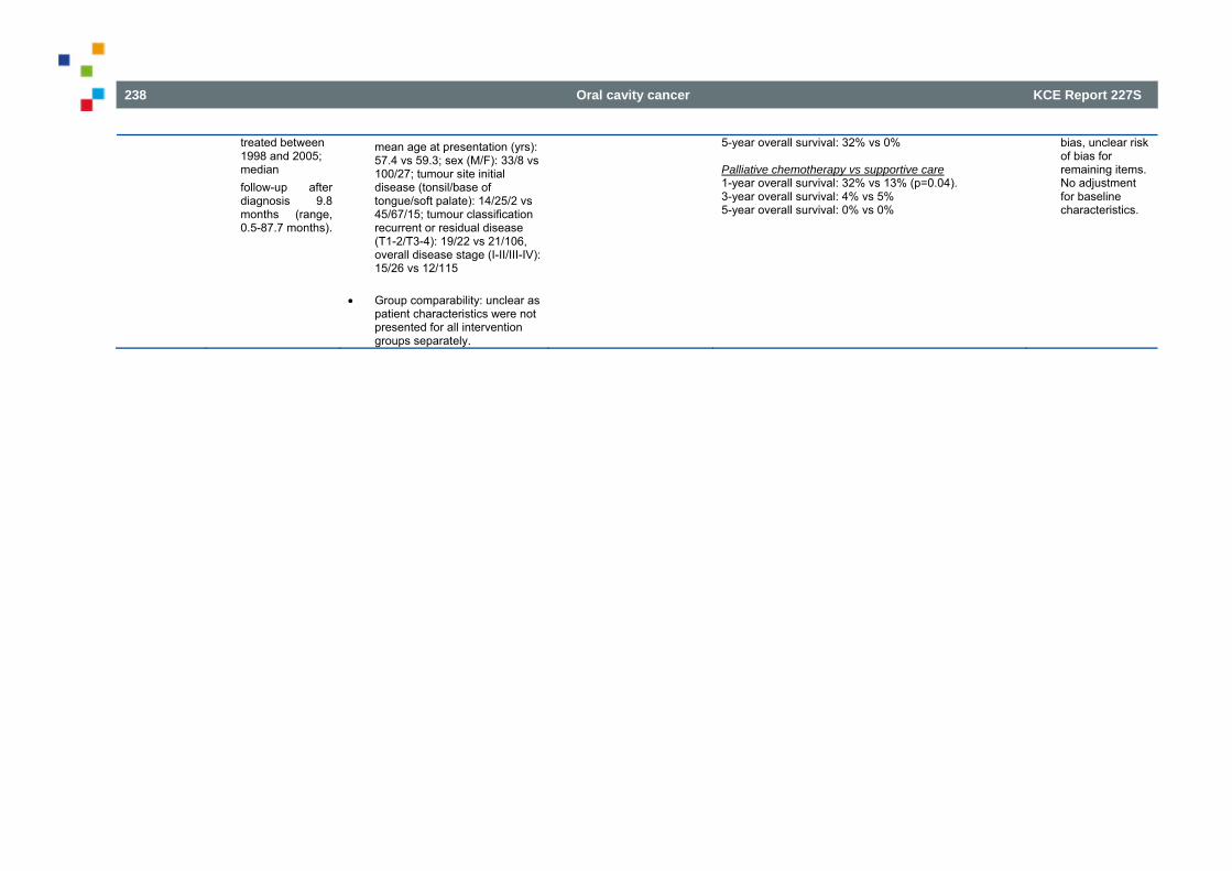
238 Oral cavity cancer KCE Report 227S
treated between 1998 and 2005; median follow-up after diagnosis 9.8 months (range, 0.5-87.7 months).
mean age at presentation (yrs): 57.4 vs 59.3; sex (M/F): 33/8 vs 100/27; tumour site initial disease (tonsil/base of tongue/soft palate): 14/25/2 vs 45/67/15; tumour classification recurrent or residual disease (T1-2/T3-4): 19/22 vs 21/106, overall disease stage (I-II/III-IV): 15/26 vs 12/115
Group comparability: unclear as
patient characteristics were not presented for all intervention groups separately.
5-year overall survival: 32% vs 0% Palliative chemotherapy vs supportive care 1-year overall survival: 32% vs 13% (p=0.04). 3-year overall survival: 4% vs 5% 5-year overall survival: 0% vs 0%
bias, unclear risk of bias for remaining items. No adjustment for baseline characteristics.
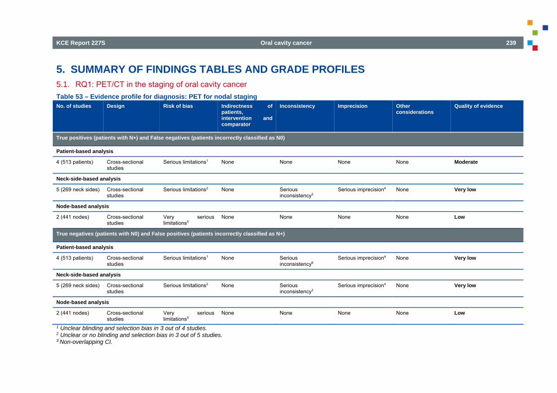
KCE Report 227S Oral cavity cancer 239
5. SUMMARY OF FINDINGS TABLES AND GRADE PROFILES 5.1. RQ1: PET/CT in the staging of oral cavity cancer Table 53 – Evidence profile for diagnosis: PET for nodal staging No. of studies Design Risk of bias Indirectness of
patients, intervention and comparator
Inconsistency Imprecision Other considerations
Quality of evidence
True positives (patients with N+) and False negatives (patients incorrectly classified as N0)
Patient-based analysis
4 (513 patients) Cross-sectional studies
Serious limitations1 None None None None Moderate
Neck-side-based analysis
5 (269 neck sides) Cross-sectional studies
Serious limitations2 None Serious inconsistency3
Serious imprecision4 None Very low
Node-based analysis
2 (441 nodes) Cross-sectional studies
Very serious limitations5
None None None None Low
True negatives (patients with N0) and False positives (patients incorrectly classified as N+)
Patient-based analysis
4 (513 patients) Cross-sectional studies
Serious limitations1 None Serious inconsistency6
Serious imprecision4 None Very low
Neck-side-based analysis
5 (269 neck sides) Cross-sectional studies
Serious limitations2 None Serious inconsistency3
Serious imprecision4 None Very low
Node-based analysis
2 (441 nodes) Cross-sectional studies
Very serious limitations5
None None None None Low
1 Unclear blinding and selection bias in 3 out of 4 studies. 2 Unclear or no blinding and selection bias in 3 out of 5 studies. 3 Non-overlapping CI.
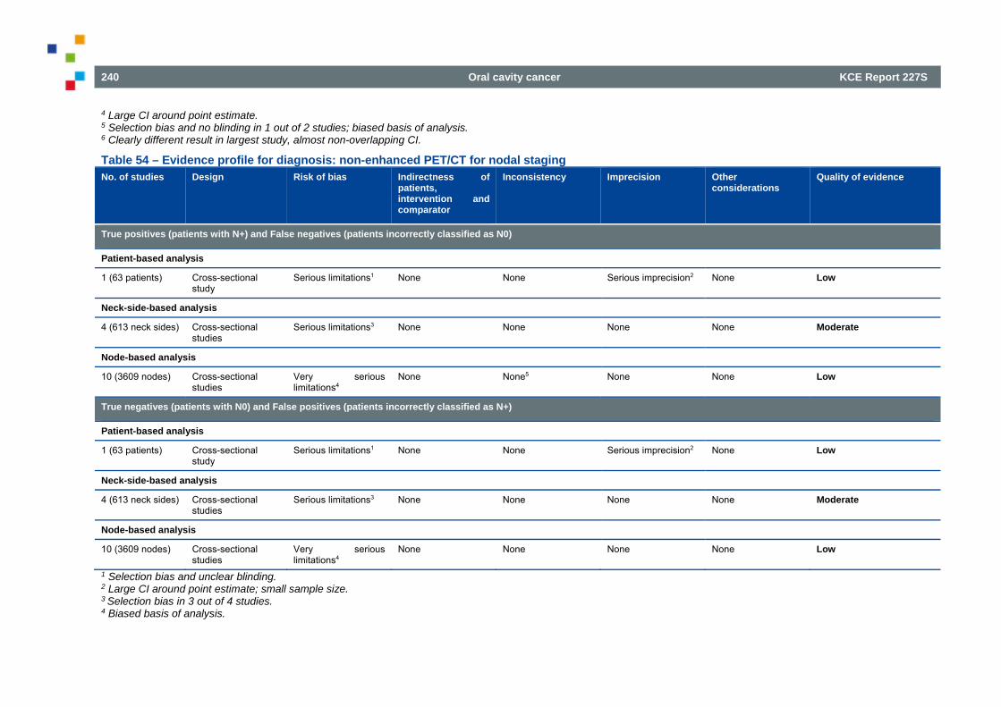
240 Oral cavity cancer KCE Report 227S
4 Large CI around point estimate. 5 Selection bias and no blinding in 1 out of 2 studies; biased basis of analysis. 6 Clearly different result in largest study, almost non-overlapping CI.
Table 54 – Evidence profile for diagnosis: non-enhanced PET/CT for nodal staging No. of studies Design Risk of bias Indirectness of
patients, intervention and comparator
Inconsistency Imprecision Other considerations
Quality of evidence
True positives (patients with N+) and False negatives (patients incorrectly classified as N0)
Patient-based analysis
1 (63 patients) Cross-sectional study
Serious limitations1 None None Serious imprecision2 None Low
Neck-side-based analysis
4 (613 neck sides) Cross-sectional studies
Serious limitations3 None None None None Moderate
Node-based analysis
10 (3609 nodes) Cross-sectional studies
Very serious limitations4
None None5 None None Low
True negatives (patients with N0) and False positives (patients incorrectly classified as N+)
Patient-based analysis
1 (63 patients) Cross-sectional study
Serious limitations1 None None Serious imprecision2 None Low
Neck-side-based analysis
4 (613 neck sides) Cross-sectional studies
Serious limitations3 None None None None Moderate
Node-based analysis
10 (3609 nodes) Cross-sectional studies
Very serious limitations4
None None None None Low
1 Selection bias and unclear blinding. 2 Large CI around point estimate; small sample size. 3 Selection bias in 3 out of 4 studies. 4 Biased basis of analysis.
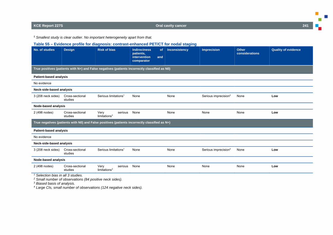
KCE Report 227S Oral cavity cancer 241
5 Smallest study is clear outlier. No important heterogeneity apart from that.
Table 55 – Evidence profile for diagnosis: contrast-enhanced PET/CT for nodal staging No. of studies Design Risk of bias Indirectness of
patients, intervention and comparator
Inconsistency Imprecision Other considerations
Quality of evidence
True positives (patients with N+) and False negatives (patients incorrectly classified as N0)
Patient-based analysis
No evidence
Neck-side-based analysis
3 (208 neck sides) Cross-sectional studies
Serious limitations1 None None Serious imprecision2 None Low
Node-based analysis
2 (498 nodes) Cross-sectional studies
Very serious limitations3
None None None None Low
True negatives (patients with N0) and False positives (patients incorrectly classified as N+)
Patient-based analysis
No evidence
Neck-side-based analysis
3 (208 neck sides) Cross-sectional studies
Serious limitations1 None None Serious imprecision4 None Low
Node-based analysis
2 (498 nodes) Cross-sectional studies
Very serious limitations3
None None None None Low
1 Selection bias in all 3 studies. 2 Small number of observations (84 positive neck sides). 3 Biased basis of analysis. 4 Large CIs, small number of observations (124 negative neck sides).
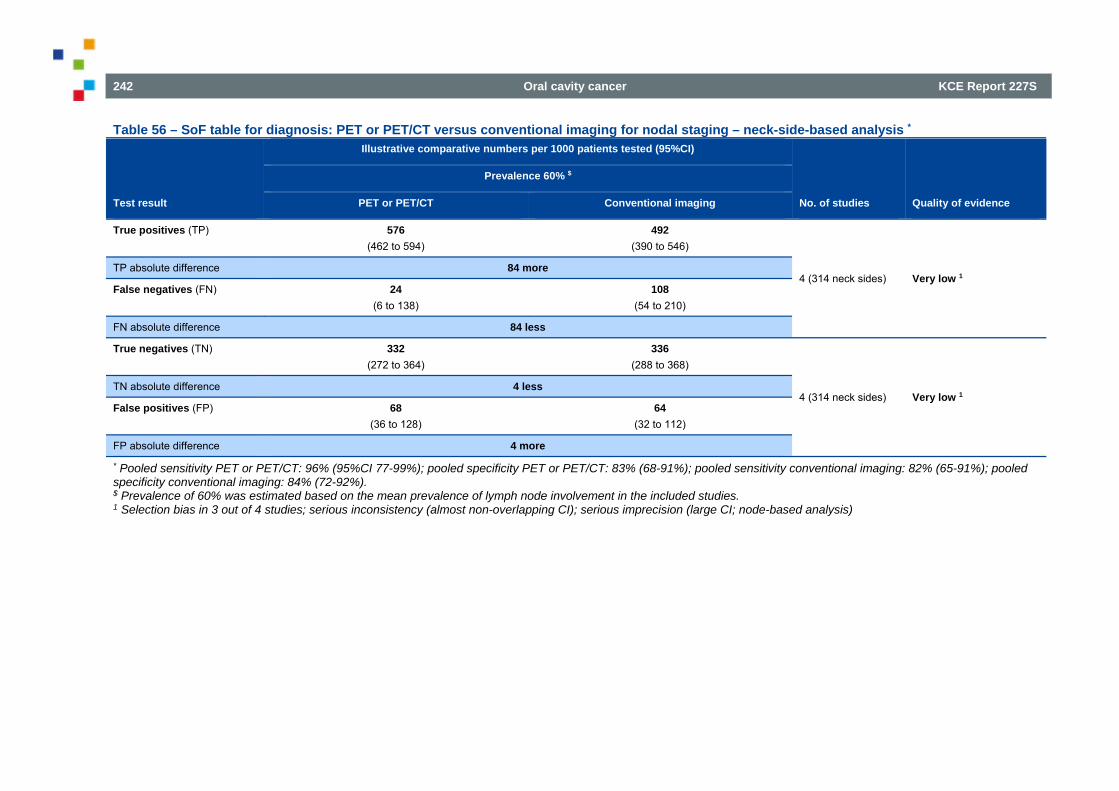
242 Oral cavity cancer KCE Report 227S
Table 56 – SoF table for diagnosis: PET or PET/CT versus conventional imaging for nodal staging – neck-side-based analysis *
Test result
Illustrative comparative numbers per 1000 patients tested (95%CI)
No. of studies Quality of evidence
Prevalence 60% $
PET or PET/CT Conventional imaging
True positives (TP) 576 (462 to 594)
492 (390 to 546)
4 (314 neck sides) Very low 1 TP absolute difference 84 more
False negatives (FN) 24 (6 to 138)
108 (54 to 210)
FN absolute difference 84 less
True negatives (TN) 332 (272 to 364)
336 (288 to 368)
4 (314 neck sides) Very low 1 TN absolute difference 4 less
False positives (FP) 68 (36 to 128)
64 (32 to 112)
FP absolute difference 4 more
* Pooled sensitivity PET or PET/CT: 96% (95%CI 77-99%); pooled specificity PET or PET/CT: 83% (68-91%); pooled sensitivity conventional imaging: 82% (65-91%); pooled specificity conventional imaging: 84% (72-92%). $ Prevalence of 60% was estimated based on the mean prevalence of lymph node involvement in the included studies. 1 Selection bias in 3 out of 4 studies; serious inconsistency (almost non-overlapping CI); serious imprecision (large CI; node-based analysis)
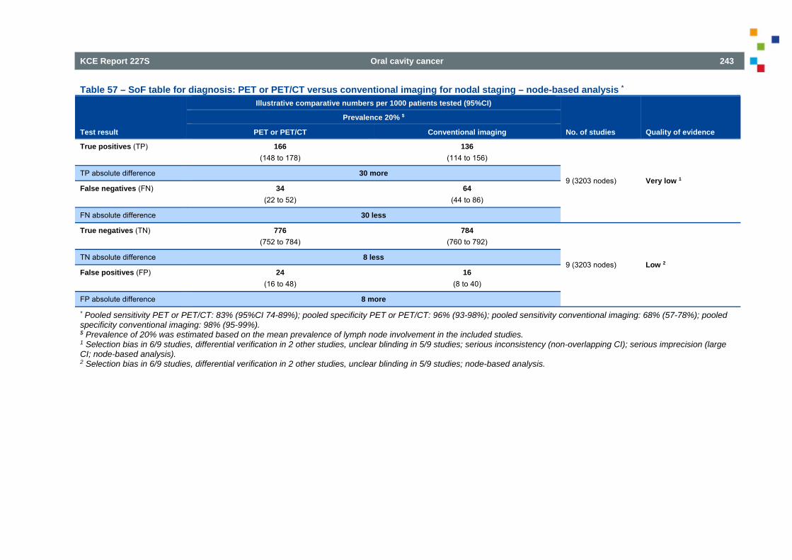
KCE Report 227S Oral cavity cancer 243
Table 57 – SoF table for diagnosis: PET or PET/CT versus conventional imaging for nodal staging – node-based analysis *
Test result
Illustrative comparative numbers per 1000 patients tested (95%CI)
No. of studies Quality of evidence
Prevalence 20% $
PET or PET/CT Conventional imaging
True positives (TP) 166 (148 to 178)
136 (114 to 156)
9 (3203 nodes) Very low 1 TP absolute difference 30 more
False negatives (FN) 34 (22 to 52)
64 (44 to 86)
FN absolute difference 30 less
True negatives (TN) 776 (752 to 784)
784 (760 to 792)
9 (3203 nodes) Low 2 TN absolute difference 8 less
False positives (FP) 24 (16 to 48)
16 (8 to 40)
FP absolute difference 8 more
* Pooled sensitivity PET or PET/CT: 83% (95%CI 74-89%); pooled specificity PET or PET/CT: 96% (93-98%); pooled sensitivity conventional imaging: 68% (57-78%); pooled specificity conventional imaging: 98% (95-99%). $ Prevalence of 20% was estimated based on the mean prevalence of lymph node involvement in the included studies. 1 Selection bias in 6/9 studies, differential verification in 2 other studies, unclear blinding in 5/9 studies; serious inconsistency (non-overlapping CI); serious imprecision (large CI; node-based analysis). 2 Selection bias in 6/9 studies, differential verification in 2 other studies, unclear blinding in 5/9 studies; node-based analysis.
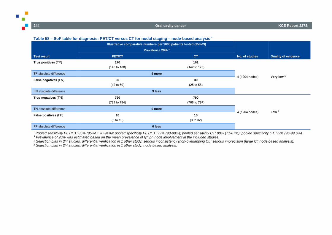
244 Oral cavity cancer KCE Report 227S
Table 58 – SoF table for diagnosis: PET/CT versus CT for nodal staging – node-based analysis *
Test result
Illustrative comparative numbers per 1000 patients tested (95%CI)
No. of studies Quality of evidence
Prevalence 20% $
PET/CT CT
True positives (TP) 170 (140 to 188)
161 (142 to 175)
4 (1204 nodes) Very low 1 TP absolute difference 9 more
False negatives (FN) 30 (12 to 60)
39 (25 to 58)
FN absolute difference 9 less
True negatives (TN) 790 (781 to 794)
790 (768 to 797)
4 (1204 nodes) Low 2 TN absolute difference 0 more
False positives (FP) 10 (6 to 19)
10 (3 to 32)
FP absolute difference 0 less
* Pooled sensitivity PET/CT: 85% (95%CI 70-94%); pooled specificity PET/CT: 99% (98-99%); pooled sensitivity CT: 80% (71-87%); pooled specificity CT: 99% (96-99.6%). $ Prevalence of 20% was estimated based on the mean prevalence of lymph node involvement in the included studies. 1 Selection bias in 3/4 studies, differential verification in 1 other study; serious inconsistency (non-overlapping CI); serious imprecision (large CI; node-based analysis). 2 Selection bias in 3/4 studies, differential verification in 1 other study; node-based analysis.
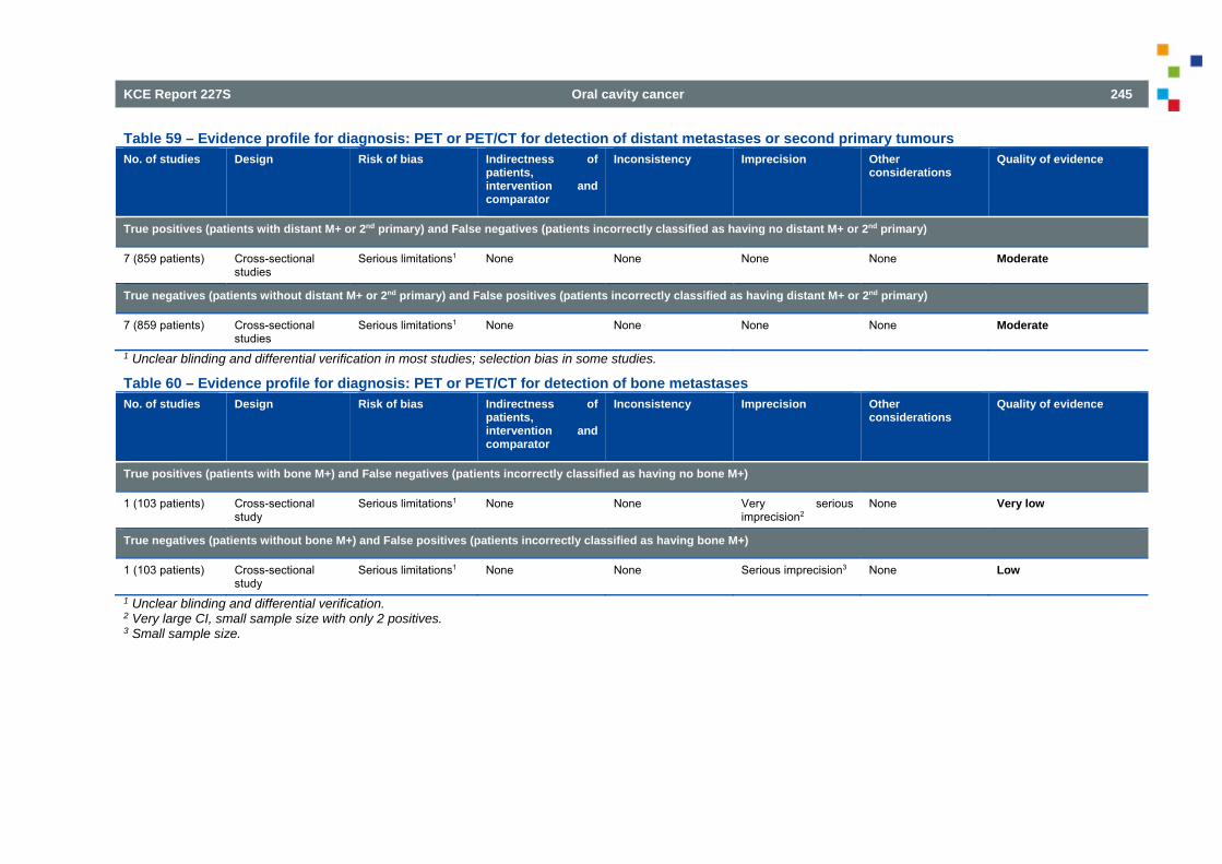
KCE Report 227S Oral cavity cancer 245
Table 59 – Evidence profile for diagnosis: PET or PET/CT for detection of distant metastases or second primary tumours No. of studies Design Risk of bias Indirectness of
patients, intervention and comparator
Inconsistency Imprecision Other considerations
Quality of evidence
True positives (patients with distant M+ or 2nd primary) and False negatives (patients incorrectly classified as having no distant M+ or 2nd primary)
7 (859 patients) Cross-sectional studies
Serious limitations1 None None None None Moderate
True negatives (patients without distant M+ or 2nd primary) and False positives (patients incorrectly classified as having distant M+ or 2nd primary)
7 (859 patients) Cross-sectional studies
Serious limitations1 None None None None Moderate
1 Unclear blinding and differential verification in most studies; selection bias in some studies.
Table 60 – Evidence profile for diagnosis: PET or PET/CT for detection of bone metastases No. of studies Design Risk of bias Indirectness of
patients, intervention and comparator
Inconsistency Imprecision Other considerations
Quality of evidence
True positives (patients with bone M+) and False negatives (patients incorrectly classified as having no bone M+)
1 (103 patients) Cross-sectional study
Serious limitations1 None None Very serious imprecision2
None Very low
True negatives (patients without bone M+) and False positives (patients incorrectly classified as having bone M+)
1 (103 patients) Cross-sectional study
Serious limitations1 None None Serious imprecision3 None Low
1 Unclear blinding and differential verification. 2 Very large CI, small sample size with only 2 positives. 3 Small sample size.
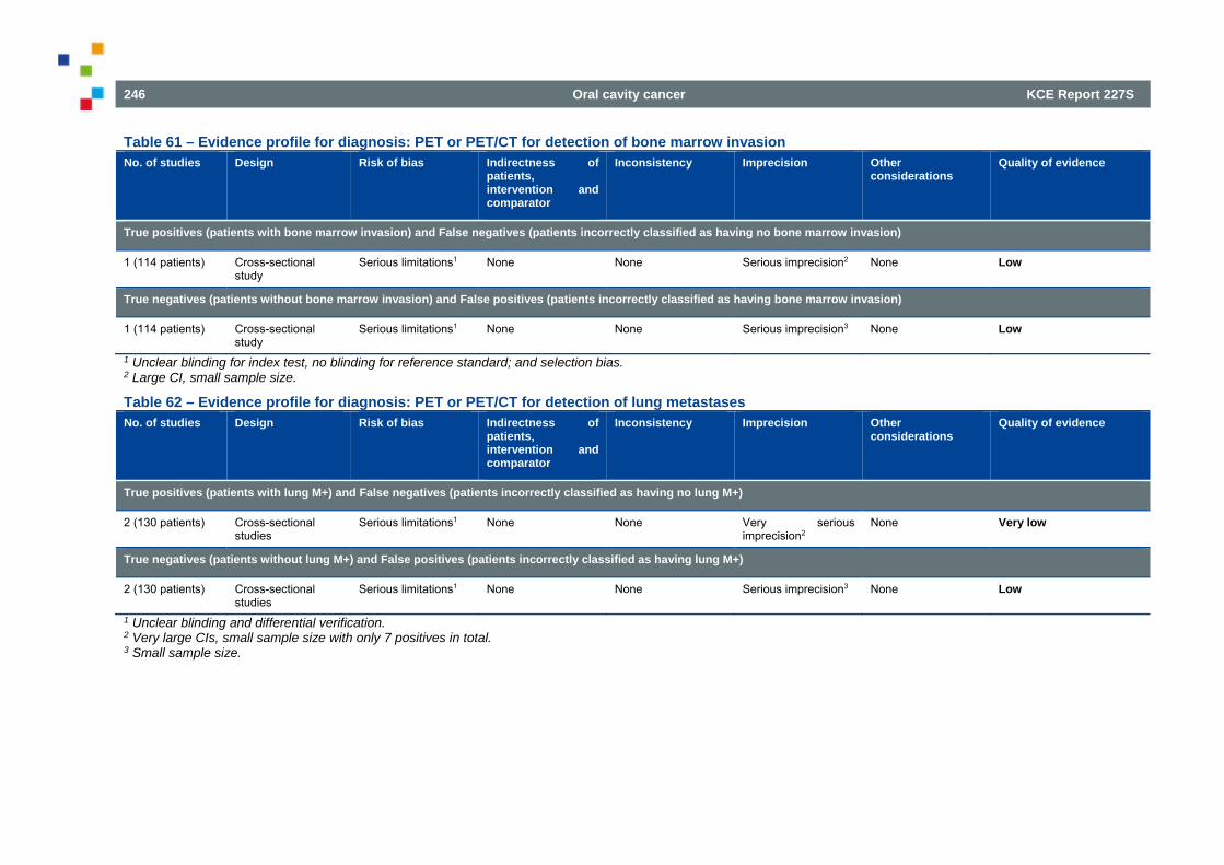
246 Oral cavity cancer KCE Report 227S
Table 61 – Evidence profile for diagnosis: PET or PET/CT for detection of bone marrow invasion No. of studies Design Risk of bias Indirectness of
patients, intervention and comparator
Inconsistency Imprecision Other considerations
Quality of evidence
True positives (patients with bone marrow invasion) and False negatives (patients incorrectly classified as having no bone marrow invasion)
1 (114 patients) Cross-sectional study
Serious limitations1 None None Serious imprecision2 None Low
True negatives (patients without bone marrow invasion) and False positives (patients incorrectly classified as having bone marrow invasion)
1 (114 patients) Cross-sectional study
Serious limitations1 None None Serious imprecision3 None Low
1 Unclear blinding for index test, no blinding for reference standard; and selection bias. 2 Large CI, small sample size.
Table 62 – Evidence profile for diagnosis: PET or PET/CT for detection of lung metastases No. of studies Design Risk of bias Indirectness of
patients, intervention and comparator
Inconsistency Imprecision Other considerations
Quality of evidence
True positives (patients with lung M+) and False negatives (patients incorrectly classified as having no lung M+)
2 (130 patients) Cross-sectional studies
Serious limitations1 None None Very serious imprecision2
None Very low
True negatives (patients without lung M+) and False positives (patients incorrectly classified as having lung M+)
2 (130 patients) Cross-sectional studies
Serious limitations1 None None Serious imprecision3 None Low
1 Unclear blinding and differential verification. 2 Very large CIs, small sample size with only 7 positives in total. 3 Small sample size.
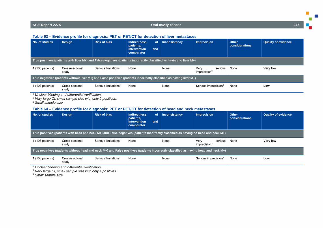
KCE Report 227S Oral cavity cancer 247
Table 63 – Evidence profile for diagnosis: PET or PET/CT for detection of liver metastases No. of studies Design Risk of bias Indirectness of
patients, intervention and comparator
Inconsistency Imprecision Other considerations
Quality of evidence
True positives (patients with liver M+) and False negatives (patients incorrectly classified as having no liver M+)
1 (103 patients) Cross-sectional study
Serious limitations1 None None Very serious imprecision2
None Very low
True negatives (patients without liver M+) and False positives (patients incorrectly classified as having liver M+)
1 (103 patients) Cross-sectional study
Serious limitations1 None None Serious imprecision3 None Low
1 Unclear blinding and differential verification. 2 Very large CI, small sample size with only 2 positives. 3 Small sample size.
Table 64 – Evidence profile for diagnosis: PET or PET/CT for detection of head and neck metastases No. of studies Design Risk of bias Indirectness of
patients, intervention and comparator
Inconsistency Imprecision Other considerations
Quality of evidence
True positives (patients with head and neck M+) and False negatives (patients incorrectly classified as having no head and neck M+)
1 (103 patients) Cross-sectional study
Serious limitations1 None None Very serious imprecision2
None Very low
True negatives (patients without head and neck M+) and False positives (patients incorrectly classified as having head and neck M+)
1 (103 patients) Cross-sectional study
Serious limitations1 None None Serious imprecision3 None Low
1 Unclear blinding and differential verification. 2 Very large CI, small sample size with only 4 positives. 3 Small sample size.
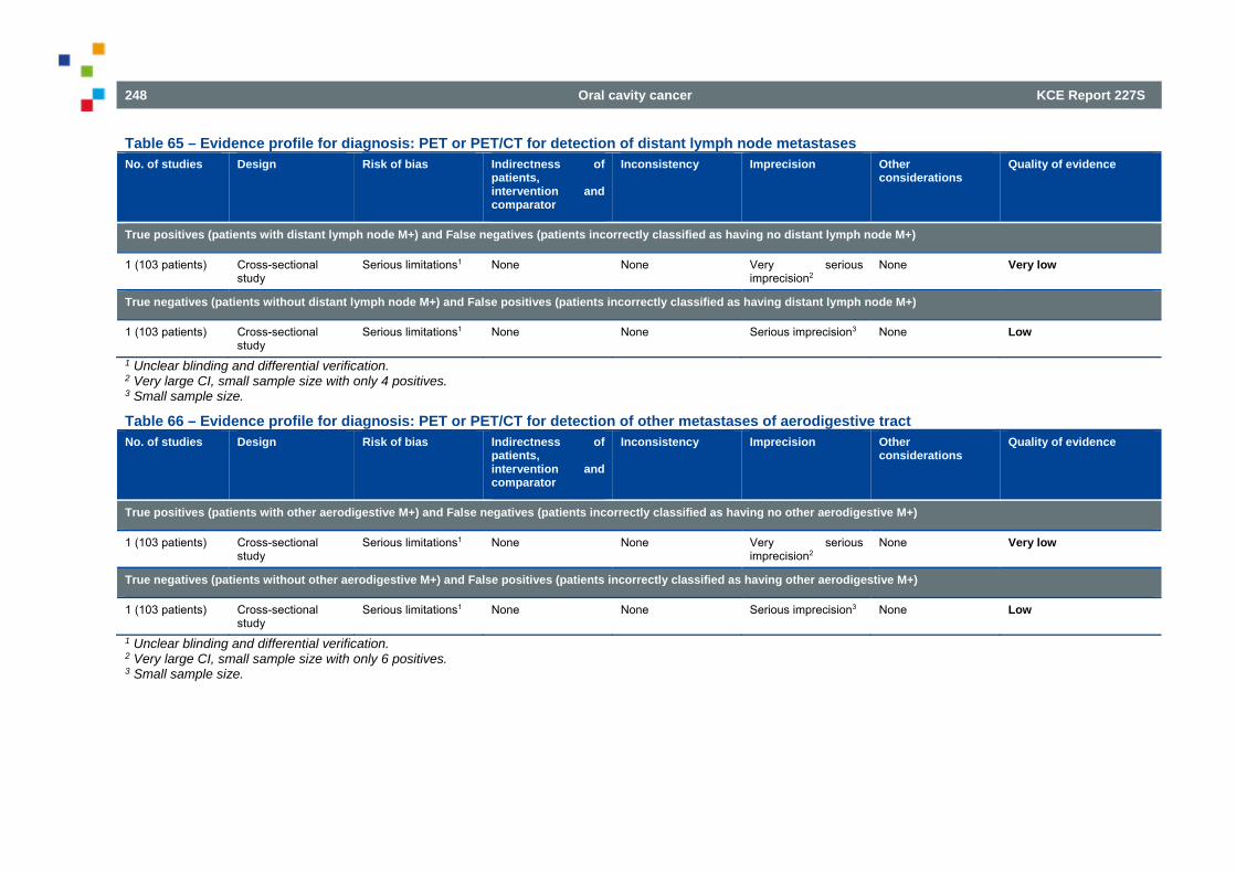
248 Oral cavity cancer KCE Report 227S
Table 65 – Evidence profile for diagnosis: PET or PET/CT for detection of distant lymph node metastases No. of studies Design Risk of bias Indirectness of
patients, intervention and comparator
Inconsistency Imprecision Other considerations
Quality of evidence
True positives (patients with distant lymph node M+) and False negatives (patients incorrectly classified as having no distant lymph node M+)
1 (103 patients) Cross-sectional study
Serious limitations1 None None Very serious imprecision2
None Very low
True negatives (patients without distant lymph node M+) and False positives (patients incorrectly classified as having distant lymph node M+)
1 (103 patients) Cross-sectional study
Serious limitations1 None None Serious imprecision3 None Low
1 Unclear blinding and differential verification. 2 Very large CI, small sample size with only 4 positives. 3 Small sample size.
Table 66 – Evidence profile for diagnosis: PET or PET/CT for detection of other metastases of aerodigestive tract No. of studies Design Risk of bias Indirectness of
patients, intervention and comparator
Inconsistency Imprecision Other considerations
Quality of evidence
True positives (patients with other aerodigestive M+) and False negatives (patients incorrectly classified as having no other aerodigestive M+)
1 (103 patients) Cross-sectional study
Serious limitations1 None None Very serious imprecision2
None Very low
True negatives (patients without other aerodigestive M+) and False positives (patients incorrectly classified as having other aerodigestive M+)
1 (103 patients) Cross-sectional study
Serious limitations1 None None Serious imprecision3 None Low
1 Unclear blinding and differential verification. 2 Very large CI, small sample size with only 6 positives. 3 Small sample size.
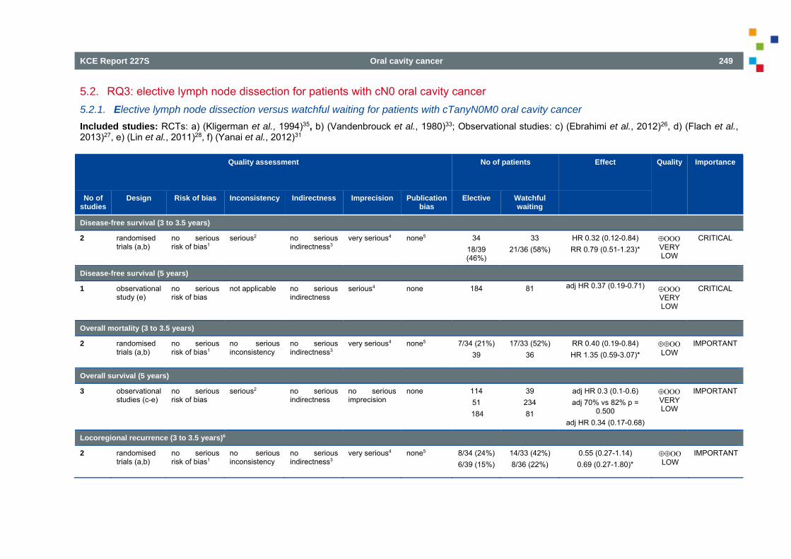
KCE Report 227S Oral cavity cancer 249
5.2. RQ3: elective lymph node dissection for patients with cN0 oral cavity cancer 5.2.1. Elective lymph node dissection versus watchful waiting for patients with cTanyN0M0 oral cavity cancer Included studies: RCTs: a) (Kligerman et al., 1994)35, b) (Vandenbrouck et al., 1980)33; Observational studies: c) (Ebrahimi et al., 2012)26, d) (Flach et al., 2013)27, e) (Lin et al., 2011)28, f) (Yanai et al., 2012)31
Quality assessment No of patients Effect Quality Importance
No of studies
Design Risk of bias Inconsistency Indirectness Imprecision Publication bias
Elective Watchful waiting
Disease-free survival (3 to 3.5 years)
2 randomised trials (a,b)
no serious risk of bias1
serious2 no serious indirectness3
very serious4 none5 34 18/39 (46%)
33 21/36 (58%)
HR 0.32 (0.12-0.84) RR 0.79 (0.51-1.23)*
VERY LOW
CRITICAL
Disease-free survival (5 years)
1
observational study (e)
no serious risk of bias
not applicable no serious indirectness
serious4 none 184 81 adj HR 0.37 (0.19-0.71)
VERY LOW
CRITICAL
Overall mortality (3 to 3.5 years)
2 randomised trials (a,b)
no serious risk of bias1
no serious inconsistency
no serious indirectness3
very serious4 none5 7/34 (21%) 39
17/33 (52%) 36
RR 0.40 (0.19-0.84) HR 1.35 (0.59-3.07)*
LOW
IMPORTANT
Overall survival (5 years)
3 observational studies (c-e)
no serious risk of bias
serious2 no serious indirectness
no serious imprecision
none 114 51
184
39 234 81
adj HR 0.3 (0.1-0.6) adj 70% vs 82% p =
0.500 adj HR 0.34 (0.17-0.68)
VERY LOW
IMPORTANT
Locoregional recurrence (3 to 3.5 years)6
2 randomised trials (a,b)
no serious risk of bias1
no serious inconsistency
no serious indirectness3
very serious4 none5 8/34 (24%) 6/39 (15%)
14/33 (42%) 8/36 (22%)
0.55 (0.27-1.14) 0.69 (0.27-1.80)*
LOW
IMPORTANT
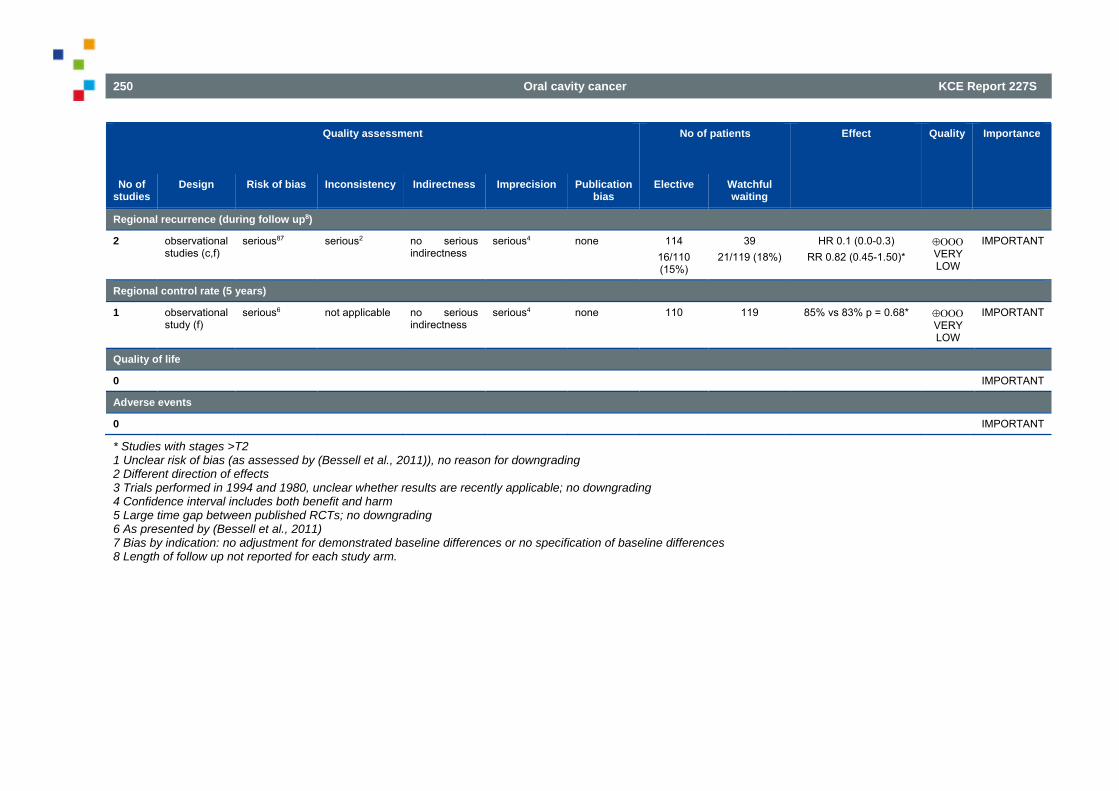
250 Oral cavity cancer KCE Report 227S
Quality assessment No of patients Effect Quality Importance
No of studies
Design Risk of bias Inconsistency Indirectness Imprecision Publication bias
Elective Watchful waiting
Regional recurrence (during follow up8)
2
observational studies (c,f)
serious87 serious2 no serious indirectness
serious4 none 114 16/110 (15%)
39 21/119 (18%)
HR 0.1 (0.0-0.3) RR 0.82 (0.45-1.50)*
VERY LOW
IMPORTANT
Regional control rate (5 years)
1 observational study (f)
serious6 not applicable no serious indirectness
serious4 none 110 119 85% vs 83% p = 0.68* VERY LOW
IMPORTANT
Quality of life
0 IMPORTANT Adverse events
0 IMPORTANT
* Studies with stages >T2 1 Unclear risk of bias (as assessed by (Bessell et al., 2011)), no reason for downgrading 2 Different direction of effects 3 Trials performed in 1994 and 1980, unclear whether results are recently applicable; no downgrading 4 Confidence interval includes both benefit and harm 5 Large time gap between published RCTs; no downgrading 6 As presented by (Bessell et al., 2011) 7 Bias by indication: no adjustment for demonstrated baseline differences or no specification of baseline differences 8 Length of follow up not reported for each study arm.
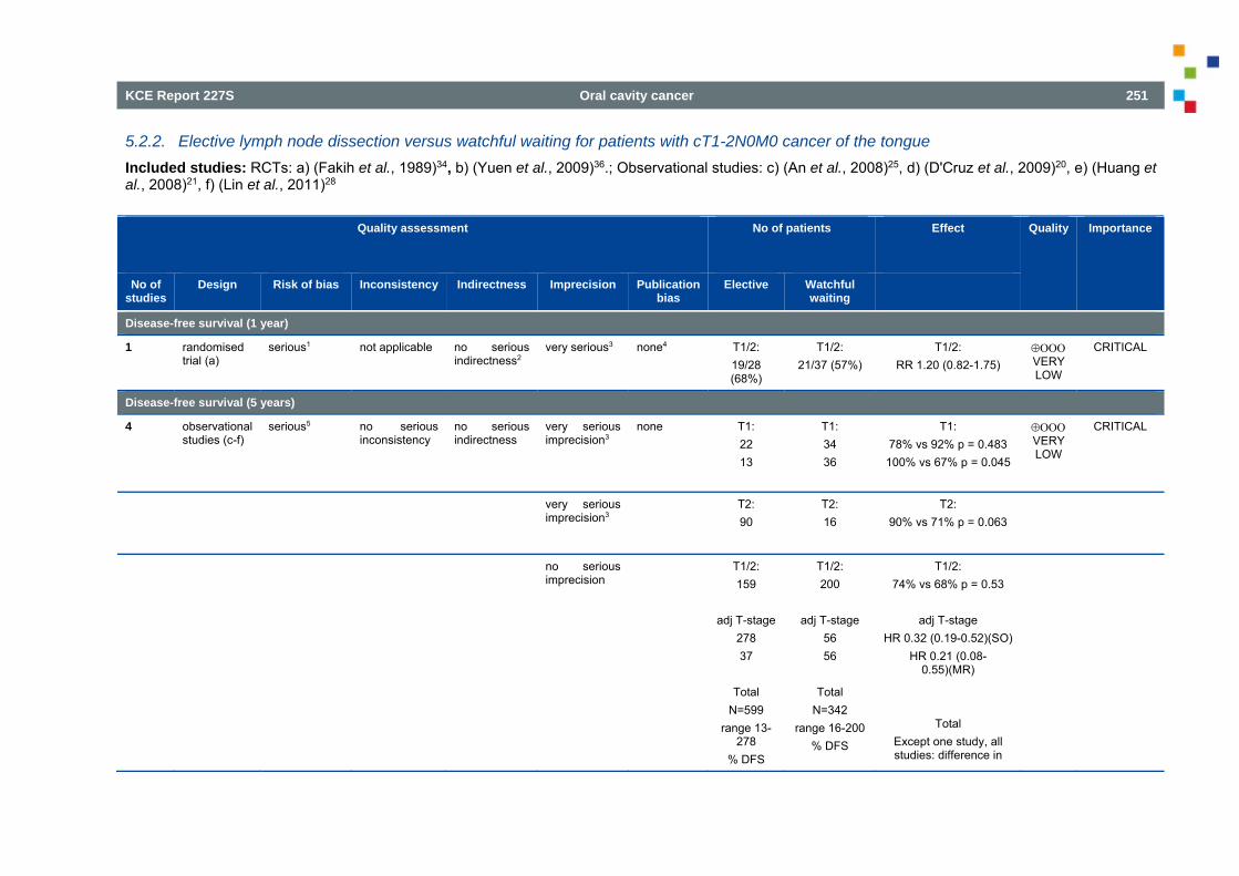
KCE Report 227S Oral cavity cancer 251
5.2.2. Elective lymph node dissection versus watchful waiting for patients with cT1-2N0M0 cancer of the tongue Included studies: RCTs: a) (Fakih et al., 1989)34, b) (Yuen et al., 2009)36.; Observational studies: c) (An et al., 2008)25, d) (D'Cruz et al., 2009)20, e) (Huang et al., 2008)21, f) (Lin et al., 2011)28
Quality assessment No of patients Effect Quality Importance
No of studies
Design Risk of bias Inconsistency Indirectness Imprecision Publication bias
Elective Watchful waiting
Disease-free survival (1 year)
1 randomised trial (a)
serious1 not applicable no serious indirectness2
very serious3 none4 T1/2: 19/28 (68%)
T1/2: 21/37 (57%)
T1/2: RR 1.20 (0.82-1.75)
VERY LOW
CRITICAL
Disease-free survival (5 years)
4
observational studies (c-f)
serious5 no serious inconsistency
no serious indirectness
very serious imprecision3
none T1: 22 13
T1: 34 36
T1: 78% vs 92% p = 0.483
100% vs 67% p = 0.045
VERY LOW
CRITICAL
very serious imprecision3
T2: 90
T2: 16
T2: 90% vs 71% p = 0.063
no serious imprecision
T1/2: 159
adj T-stage
278 37
Total
N=599 range 13-
278 % DFS
T1/2: 200
adj T-stage
56 56
Total
N=342 range 16-200
% DFS
T1/2: 74% vs 68% p = 0.53
adj T-stage
HR 0.32 (0.19-0.52)(SO) HR 0.21 (0.08-
0.55)(MR)
Total Except one study, all studies: difference in
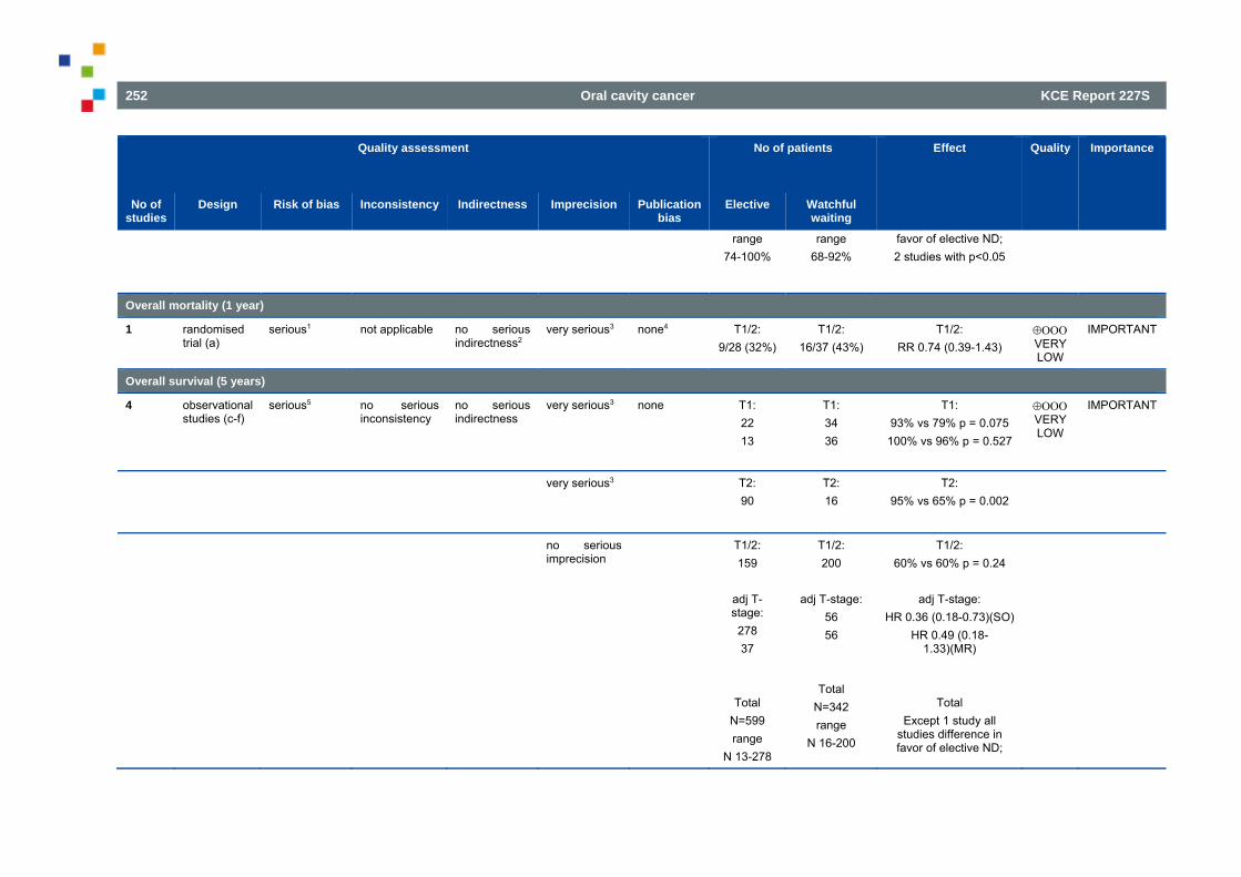
252 Oral cavity cancer KCE Report 227S
Quality assessment No of patients Effect Quality Importance
No of studies
Design Risk of bias Inconsistency Indirectness Imprecision Publication bias
Elective Watchful waiting
range 74-100%
range 68-92%
favor of elective ND; 2 studies with p<0.05
Overall mortality (1 year)
1 randomised trial (a)
serious1 not applicable no serious indirectness2
very serious3 none4 T1/2: 9/28 (32%)
T1/2: 16/37 (43%)
T1/2: RR 0.74 (0.39-1.43)
VERY LOW
IMPORTANT
Overall survival (5 years)
4
observational studies (c-f)
serious5 no serious inconsistency
no serious indirectness
very serious3 none T1: 22 13
T1: 34 36
T1: 93% vs 79% p = 0.075
100% vs 96% p = 0.527
VERY LOW
IMPORTANT
very serious3 T2: 90
T2: 16
T2: 95% vs 65% p = 0.002
no serious imprecision
T1/2: 159
adj T-stage: 278 37
Total N=599 range
N 13-278
T1/2: 200
adj T-stage:
56 56
Total N=342 range
N 16-200
T1/2: 60% vs 60% p = 0.24
adj T-stage:
HR 0.36 (0.18-0.73)(SO) HR 0.49 (0.18-
1.33)(MR)
Total Except 1 study all
studies difference in favor of elective ND;
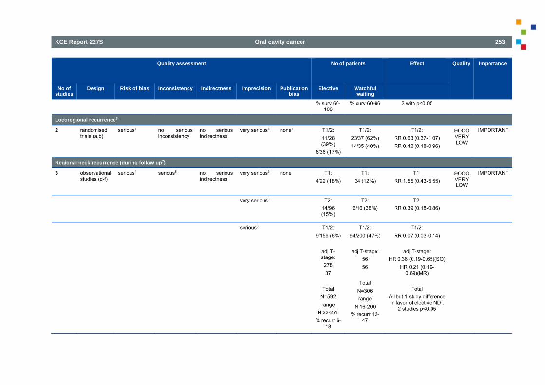
KCE Report 227S Oral cavity cancer 253
Quality assessment No of patients Effect Quality Importance
No of studies
Design Risk of bias Inconsistency Indirectness Imprecision Publication bias
Elective Watchful waiting
% surv 60-100
% surv 60-96 2 with p<0.05
Locoregional recurrence6
2 randomised trials (a,b)
serious1 no serious inconsistency
no serious indirectness
very serious3 none4 T1/2: 11/28 (39%)
6/36 (17%)
T1/2: 23/37 (62%) 14/35 (40%)
T1/2: RR 0.63 (0.37-1.07) RR 0.42 (0.18-0.96)
VERY LOW
IMPORTANT
Regional neck recurrence (during follow up7)
3 observational studies (d-f)
serious4 serious8
no serious indirectness
very serious3 none T1: 4/22 (18%)
T1: 34 (12%)
T1: RR 1.55 (0.43-5.55)
VERY LOW
IMPORTANT
very serious3 T2: 14/96 (15%)
T2: 6/16 (38%)
T2: RR 0.39 (0.18-0.86)
serious3 T1/2: 9/159 (6%)
adj T-stage: 278 37
Total
N=592 range
N 22-278 % recurr 6-
18
T1/2: 94/200 (47%)
adj T-stage:
56 56
Total
N=306 range
N 16-200 % recurr 12-
47
T1/2: RR 0.07 (0.03-0.14)
adj T-stage:
HR 0.36 (0.19-0.65)(SO) HR 0.21 (0.19-
0.69)(MR)
Total All but 1 study difference in favor of elective ND ;
2 studies p<0.05
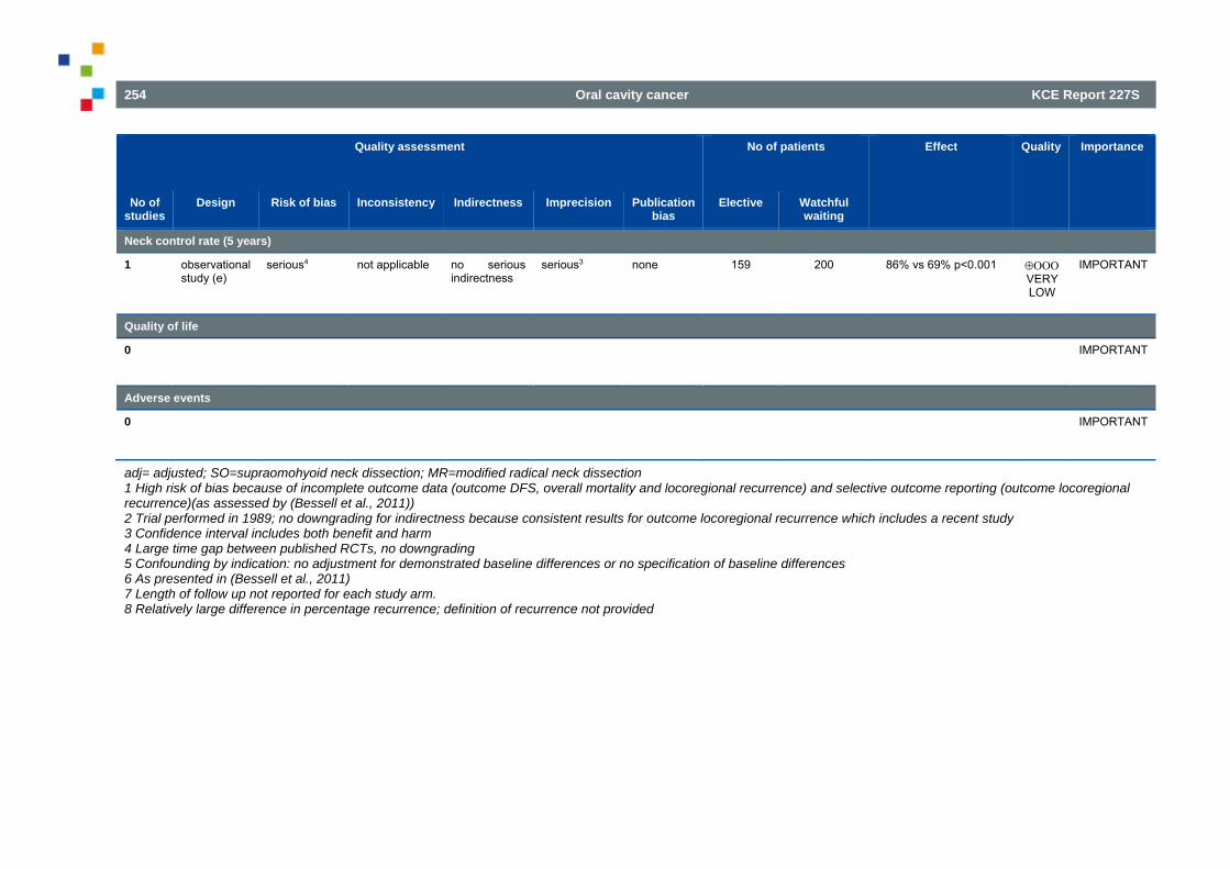
254 Oral cavity cancer KCE Report 227S
Quality assessment No of patients Effect Quality Importance
No of studies
Design Risk of bias Inconsistency Indirectness Imprecision Publication bias
Elective Watchful waiting
Neck control rate (5 years)
1
observational study (e)
serious4 not applicable no serious indirectness
serious3 none 159 200 86% vs 69% p<0.001 VERY LOW
IMPORTANT
Quality of life
0 IMPORTANT
Adverse events
0 IMPORTANT
adj= adjusted; SO=supraomohyoid neck dissection; MR=modified radical neck dissection 1 High risk of bias because of incomplete outcome data (outcome DFS, overall mortality and locoregional recurrence) and selective outcome reporting (outcome locoregional recurrence)(as assessed by (Bessell et al., 2011)) 2 Trial performed in 1989; no downgrading for indirectness because consistent results for outcome locoregional recurrence which includes a recent study 3 Confidence interval includes both benefit and harm 4 Large time gap between published RCTs, no downgrading 5 Confounding by indication: no adjustment for demonstrated baseline differences or no specification of baseline differences 6 As presented in (Bessell et al., 2011) 7 Length of follow up not reported for each study arm. 8 Relatively large difference in percentage recurrence; definition of recurrence not provided
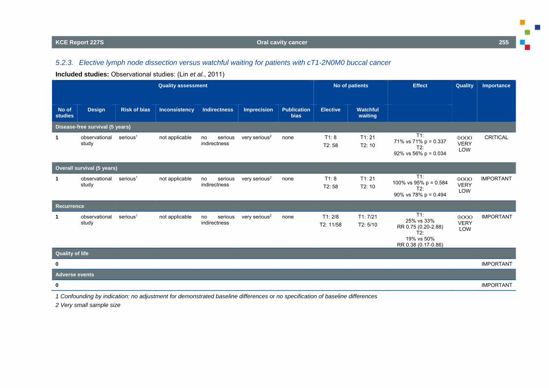
KCE Report 227S Oral cavity cancer 255
5.2.3. Elective lymph node dissection versus watchful waiting for patients with cT1-2N0M0 buccal cancer Included studies: Observational studies: (Lin et al., 2011)
Quality assessment No of patients Effect Quality Importance
No of studies
Design Risk of bias Inconsistency Indirectness Imprecision Publication bias
Elective Watchful waiting
Disease-free survival (5 years)
1
observational study
serious1 not applicable no serious indirectness
very serious2 none T1: 8 T2: 58
T1: 21 T2: 10
T1: 71% vs 71% p = 0.337
T2: 92% vs 56% p = 0.034
VERY LOW
CRITICAL
Overall survival (5 years)
1
observational study
serious1 not applicable no serious indirectness
very serious2 none T1: 8 T2: 58
T1: 21 T2: 10
T1: 100% vs 95% p = 0.584
T2: 90% vs 78% p = 0.494
VERY LOW
IMPORTANT
Recurrence
1 observational study
serious1 not applicable no serious indirectness
very serious2 none T1: 2/8 T2: 11/58
T1: 7/21 T2: 5/10
T1: 25% vs 33%
RR 0.75 (0.20-2.88) T2:
19% vs 50% RR 0.38 (0.17-0.86)
VERY LOW
IMPORTANT
Quality of life
0 IMPORTANT Adverse events
0 IMPORTANT
1 Confounding by indication: no adjustment for demonstrated baseline differences or no specification of baseline differences 2 Very small sample size
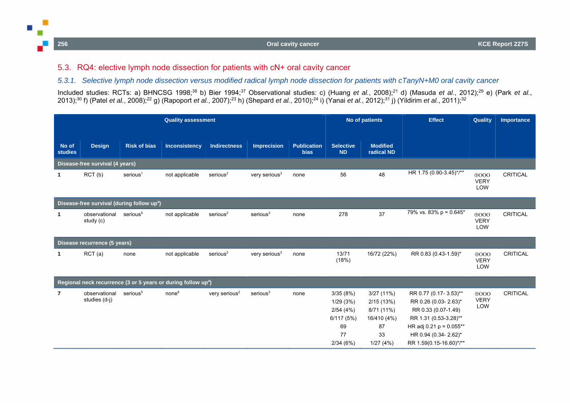
256 Oral cavity cancer KCE Report 227S
5.3. RQ4: elective lymph node dissection for patients with cN+ oral cavity cancer 5.3.1. Selective lymph node dissection versus modified radical lymph node dissection for patients with cTanyN+M0 oral cavity cancer Included studies: RCTs: a) BHNCSG 1998;38 b) Bier 1994;37 Observational studies: c) (Huang et al., 2008);21 d) (Masuda et al., 2012);29 e) (Park et al., 2013);30 f) (Patel et al., 2008);22 g) (Rapoport et al., 2007);23 h) (Shepard et al., 2010);24 i) (Yanai et al., 2012);31 j) (Yildirim et al., 2011);32
Quality assessment No of patients Effect Quality Importance
No of studies
Design Risk of bias Inconsistency Indirectness Imprecision Publication bias
Selective ND
Modified radical ND
Disease-free survival (4 years)
1
RCT (b) serious1 not applicable serious2 very serious3 none 56 48
HR 1.75 (0.90-3.45)*/**
VERY LOW
CRITICAL
Disease-free survival (during follow up4)
1
observational study (c)
serious5 not applicable serious2 serious3 none 278 37
79% vs. 83% p = 0.645*
VERY LOW
CRITICAL
Disease recurrence (5 years)
1 RCT (a) none not applicable serious2 very serious3 none 13/71 (18%)
16/72 (22%) RR 0.83 (0.43-1.59)* VERY LOW
CRITICAL
Regional neck recurrence (3 or 5 years or during follow up4)
7
observational studies (d-j)
serious5 none6 very serious2 serious3 none 3/35 (8%) 1/29 (3%) 2/54 (4%)
6/117 (5%) 69 77
2/34 (6%)
3/27 (11%) 2/15 (13%) 8/71 (11%)
16/410 (4%) 87 33
1/27 (4%)
RR 0.77 (0.17- 3.53)** RR 0.26 (0.03- 2.63)* RR 0.33 (0.07-1.49)
RR 1.31 (0.53-3.28)** HR adj 0.21 p = 0.055** HR 0.94 (0.34- 2.62)*
RR 1.59(0.15-16.60)*/**
VERY LOW
CRITICAL
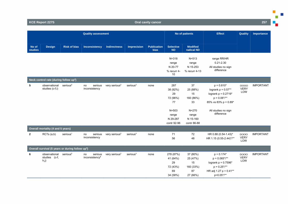
KCE Report 227S Oral cavity cancer 257
Quality assessment No of patients Effect Quality Importance
No of studies
Design Risk of bias Inconsistency Indirectness Imprecision Publication bias
Selective ND
Modified radical ND
N=318 range
N 20-77 % recurr 4-
10
N=513 range
N 15-253 % recurr 4-13
range RR/HR
0.21-2.30 All studies no sign
difference
Neck control rate (during follow up4)
5
observational studies (c-f,i)
serious5 no serious inconsistency
very serious2 serious3 none 287 38 (92%)
29 72 (96%)
77
N=503 range
N 29-287 contr 92-96
37 25 (88%)
15 160 (86%)
33
N=270 range
N 15-160 contr 86-88
p = 0.810* logrank p = 0.57**
logrank p = 0.2719* p = 0.06*/**
85% vs 83% p = 0.89*
All studies no sign difference
VERY LOW
IMPORTANT
Overall mortality (4 and 5 years)
2
RCTs (a,b) serious1 no serious inconsistency
very serious2 serious3 none 71 56
72 48
HR 0.88 (0.54-1.43)* HR 1.15 (0.55-2.44)*/**
VERY LOW
IMPORTANT
Overall survival (5 years or during follow up4)
6
observational studies (c-f, h,j)
serious5 no serious inconsistency6
very serious2 serious3 none 278 (87%) 41 (64%)
29 72 (43%)
69 34 (58%)
37 (80%) 25 (47%)
15 160 (33%)
87 27 (66%)
p = 0.174* p = 0.065*/**
logrank p = 0.7596* p = 0.25*/**
HR adj 1.27 p = 0.41** p>0.05*/**
VERY LOW
IMPORTANT
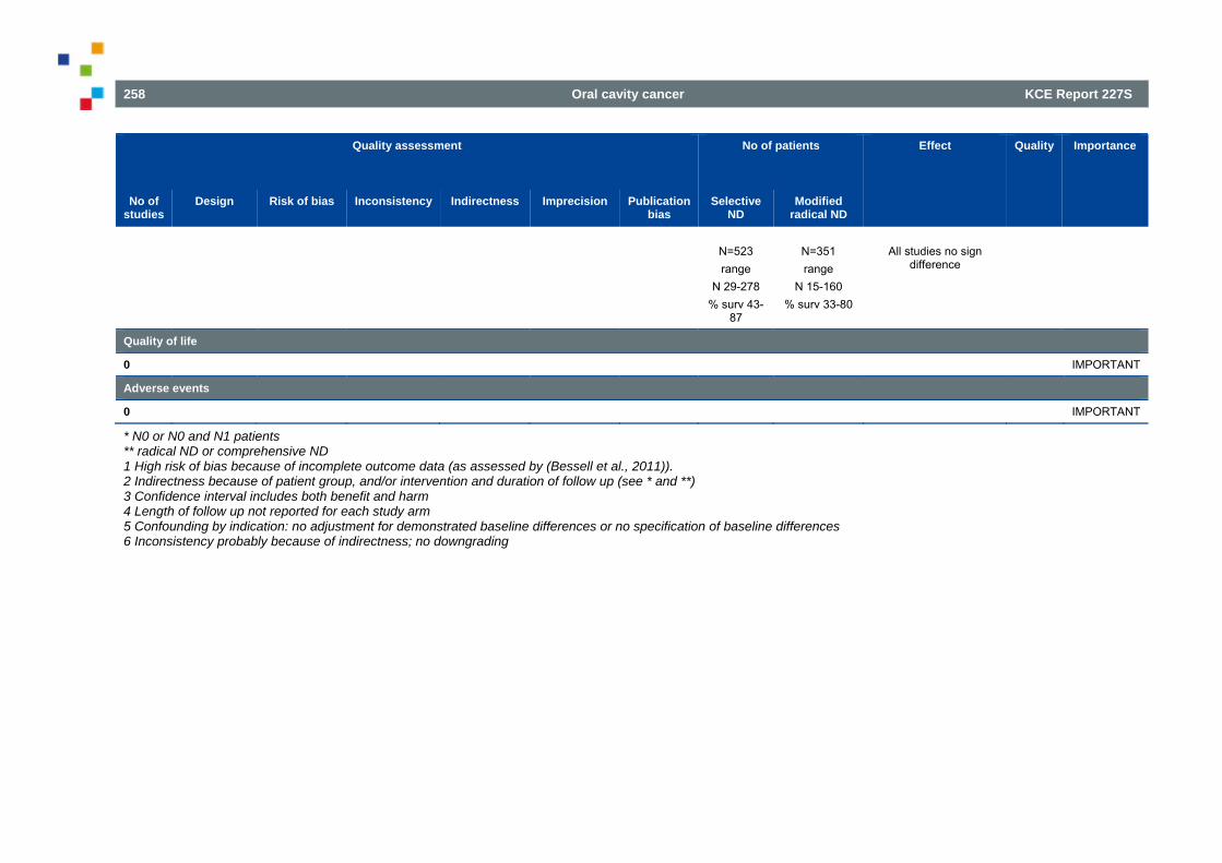
258 Oral cavity cancer KCE Report 227S
Quality assessment No of patients Effect Quality Importance
No of studies
Design Risk of bias Inconsistency Indirectness Imprecision Publication bias
Selective ND
Modified radical ND
N=523 range
N 29-278 % surv 43-
87
N=351 range
N 15-160 % surv 33-80
All studies no sign
difference
Quality of life
0 IMPORTANT Adverse events
0 IMPORTANT
* N0 or N0 and N1 patients ** radical ND or comprehensive ND 1 High risk of bias because of incomplete outcome data (as assessed by (Bessell et al., 2011)). 2 Indirectness because of patient group, and/or intervention and duration of follow up (see * and **) 3 Confidence interval includes both benefit and harm 4 Length of follow up not reported for each study arm 5 Confounding by indication: no adjustment for demonstrated baseline differences or no specification of baseline differences 6 Inconsistency probably because of indirectness; no downgrading
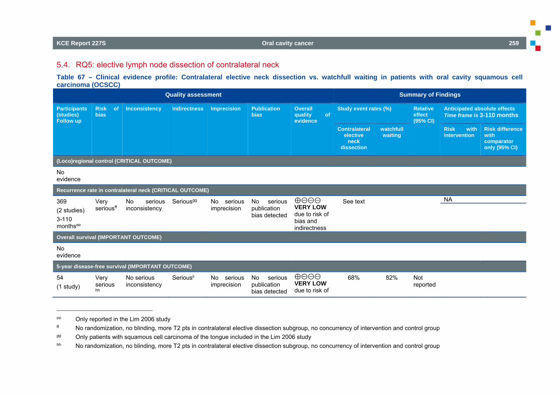
KCE Report 227S Oral cavity cancer 259
5.4. RQ5: elective lymph node dissection of contralateral neck Table 67 – Clinical evidence profile: Contralateral elective neck dissection vs. watchfull waiting in patients with oral cavity squamous cell carcinoma (OCSCC)
Quality assessment Summary of Findings
Participants(studies) Follow up
Risk of bias
Inconsistency Indirectness Imprecision Publication bias
Overall quality of evidence
Study event rates (%) Relative effect (95% CI)
Anticipated absolute effects Time frame is 3-110 months
Contralateral elective
neck dissection
watchfull waiting
Risk with intervention
Risk difference with comparator only (95% CI)
(Loco)regional control (CRITICAL OUTCOME)
No evidence
Recurrence rate in contralateral neck (CRITICAL OUTCOME)
369 (2 studies) 3-110 monthsee
Very seriousff
No serious inconsistency
Seriousgg No serious
imprecision No serious publication bias detected
⊕⊝⊝⊝ VERY LOW due to risk of bias and indirectness
See text NA
Overall survival (IMPORTANT OUTCOME)
No evidence
5-year disease-free survival (IMPORTANT OUTCOME)
54 (1 study)
Very serious
hh
No serious inconsistency
Seriousii No serious
imprecision No serious publication bias detected
⊕⊝⊝⊝ VERY LOW due to risk of
68% 82% Not reported
ee Only reported in the Lim 2006 study ff No randomization, no blinding, more T2 pts in contralateral elective dissection subgroup, no concurrency of intervention and control group gg Only patients with squamous cell carcinoma of the tongue included in the Lim 2006 study hh No randomization, no blinding, more T2 pts in contralateral elective dissection subgroup, no concurrency of intervention and control group
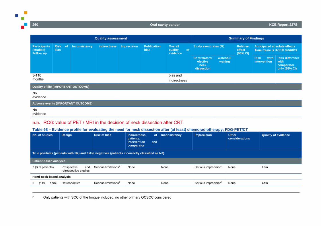
260 Oral cavity cancer KCE Report 227S
Quality assessment Summary of Findings
Participants(studies) Follow up
Risk of bias
Inconsistency Indirectness Imprecision Publication bias
Overall quality of evidence
Study event rates (%) Relative effect (95% CI)
Anticipated absolute effects Time frame is 3-110 months
Contralateral elective
neck dissection
watchfull waiting
Risk with intervention
Risk difference with comparator only (95% CI)
3-110 months
bias and indirectness
Quality of life (IMPORTANT OUTCOME)
No evidence
Adverse events (IMPORTANT OUTCOME)
No evidence
5.5. RQ6: value of PET / MRI in the decision of neck dissection after CRT Table 68 – Evidence profile for evaluating the need for neck dissection after (at least) chemoradiotherapy: FDG-PET/CT No. of studies Design Risk of bias Indirectness of
patients, intervention and comparator
Inconsistency Imprecision Other considerations
Quality of evidence
True positives (patients with N+) and False negatives (patients incorrectly classified as N0)
Patient-based analysis
7 (339 patients) Prospective and retrospective studies
Serious limitations1 None None Serious imprecision2 None Low
Hemi-neck-based analysis
2 (119 hemi- Retrospective Serious limitations1 None None Serious imprecision3 None Low
ii Only patients with SCC of the tongue included, no other primary OCSCC considered
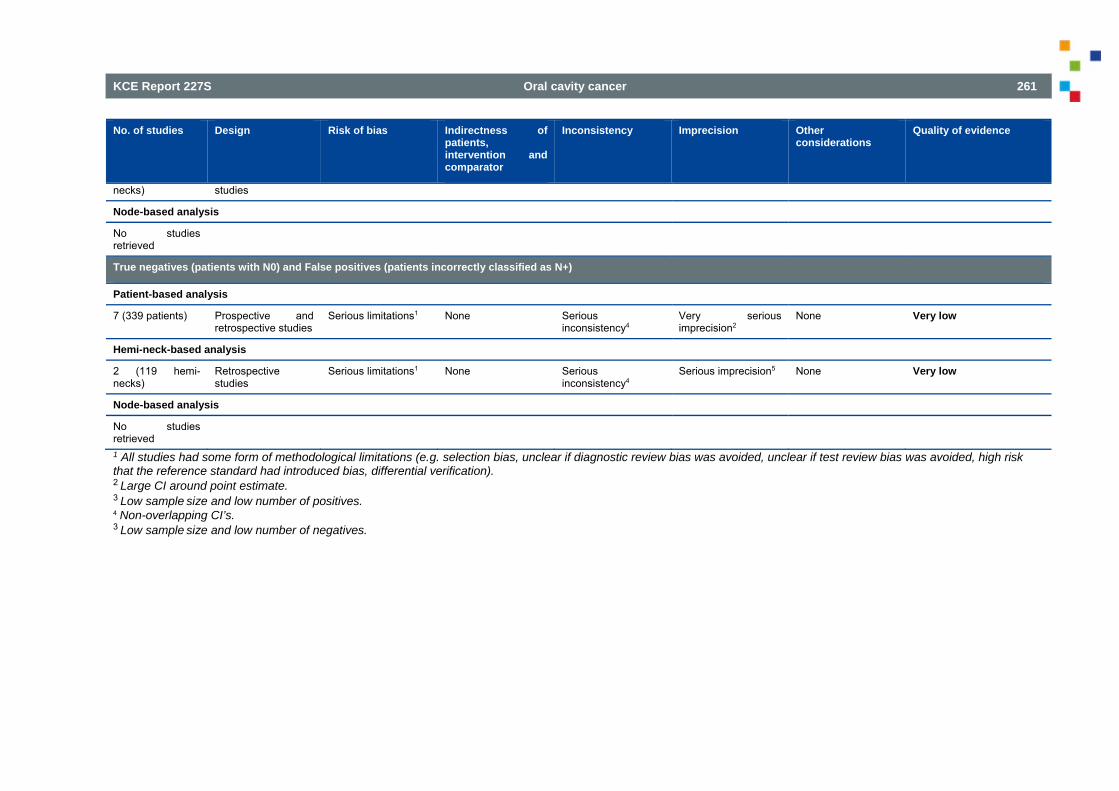
KCE Report 227S Oral cavity cancer 261
No. of studies Design Risk of bias Indirectness of patients, intervention and comparator
Inconsistency Imprecision Other considerations
Quality of evidence
necks) studies
Node-based analysis
No studies retrieved
True negatives (patients with N0) and False positives (patients incorrectly classified as N+)
Patient-based analysis
7 (339 patients) Prospective and retrospective studies
Serious limitations1 None Serious inconsistency4
Very serious imprecision2
None Very low
Hemi-neck-based analysis
2 (119 hemi-necks)
Retrospective studies
Serious limitations1 None Serious inconsistency4
Serious imprecision5 None Very low
Node-based analysis
No studies retrieved
1 All studies had some form of methodological limitations (e.g. selection bias, unclear if diagnostic review bias was avoided, unclear if test review bias was avoided, high risk that the reference standard had introduced bias, differential verification). 2 Large CI around point estimate. 3 Low sample size and low number of positives. 4 Non-overlapping CI’s. 3 Low sample size and low number of negatives.
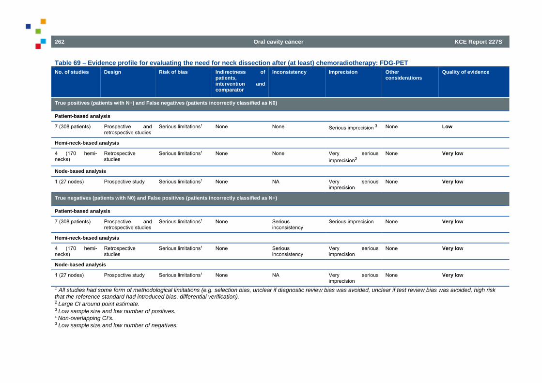
262 Oral cavity cancer KCE Report 227S
Table 69 – Evidence profile for evaluating the need for neck dissection after (at least) chemoradiotherapy: FDG-PET No. of studies Design Risk of bias Indirectness of
patients, intervention and comparator
Inconsistency Imprecision Other considerations
Quality of evidence
True positives (patients with N+) and False negatives (patients incorrectly classified as N0)
Patient-based analysis
7 (308 patients) Prospective and retrospective studies
Serious limitations1 None None Serious imprecision 3 None Low
Hemi-neck-based analysis
4 (170 hemi-necks)
Retrospective studies
Serious limitations1 None None Very serious imprecision2
None Very low
Node-based analysis
1 (27 nodes) Prospective study Serious limitations1 None NA Very serious imprecision
None Very low
True negatives (patients with N0) and False positives (patients incorrectly classified as N+)
Patient-based analysis
7 (308 patients) Prospective and retrospective studies
Serious limitations1 None Serious inconsistency
Serious imprecision None Very low
Hemi-neck-based analysis
4 (170 hemi-necks)
Retrospective studies
Serious limitations1 None Serious inconsistency
Very serious imprecision
None Very low
Node-based analysis
1 (27 nodes) Prospective study Serious limitations1 None NA Very serious imprecision
None Very low
1 All studies had some form of methodological limitations (e.g. selection bias, unclear if diagnostic review bias was avoided, unclear if test review bias was avoided, high risk that the reference standard had introduced bias, differential verification). 2 Large CI around point estimate. 3 Low sample size and low number of positives. 4 Non-overlapping CI’s. 3 Low sample size and low number of negatives.
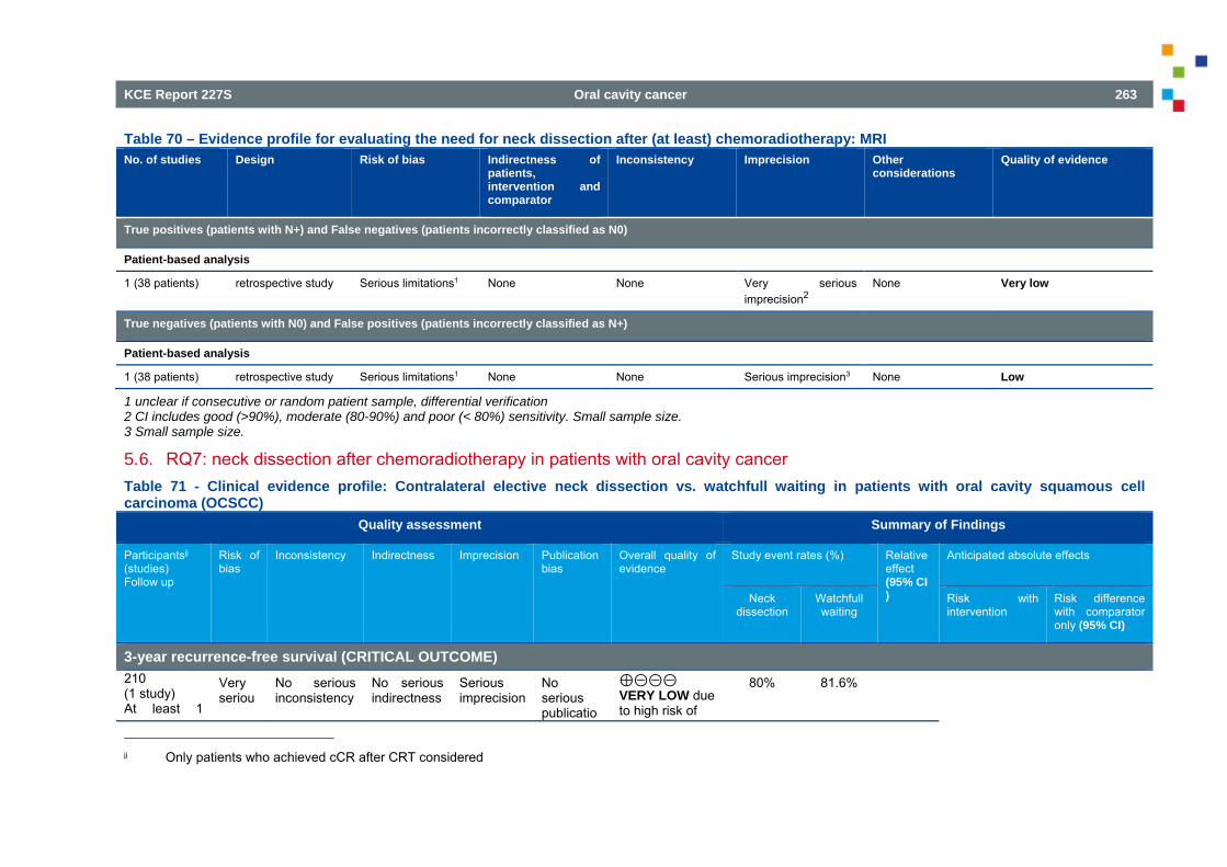
KCE Report 227S Oral cavity cancer 263
Table 70 – Evidence profile for evaluating the need for neck dissection after (at least) chemoradiotherapy: MRI No. of studies Design Risk of bias Indirectness of
patients, intervention and comparator
Inconsistency Imprecision Other considerations
Quality of evidence
True positives (patients with N+) and False negatives (patients incorrectly classified as N0)
Patient-based analysis
1 (38 patients) retrospective study Serious limitations1 None None Very serious imprecision2
None Very low
True negatives (patients with N0) and False positives (patients incorrectly classified as N+)
Patient-based analysis
1 (38 patients) retrospective study Serious limitations1 None None Serious imprecision3 None Low
1 unclear if consecutive or random patient sample, differential verification 2 CI includes good (>90%), moderate (80-90%) and poor (< 80%) sensitivity. Small sample size. 3 Small sample size.
5.6. RQ7: neck dissection after chemoradiotherapy in patients with oral cavity cancer Table 71 - Clinical evidence profile: Contralateral elective neck dissection vs. watchfull waiting in patients with oral cavity squamous cell carcinoma (OCSCC)
Quality assessment Summary of Findings
Participantsjj (studies) Follow up
Risk of bias
Inconsistency Indirectness Imprecision Publication bias
Overall quality of evidence
Study event rates (%) Relative effect (95% CI)
Anticipated absolute effects
Neck dissection
Watchfull waiting
Risk with intervention
Risk difference with comparator only (95% CI)
3-year recurrence-free survival (CRITICAL OUTCOME) 210 (1 study) At least 1
Very seriou
No serious inconsistency
No serious indirectness
Serious
imprecision No serious publicatio
⊕⊝⊝⊝ VERY LOW due to high risk of
80% 81.6%
jj Only patients who achieved cCR after CRT considered
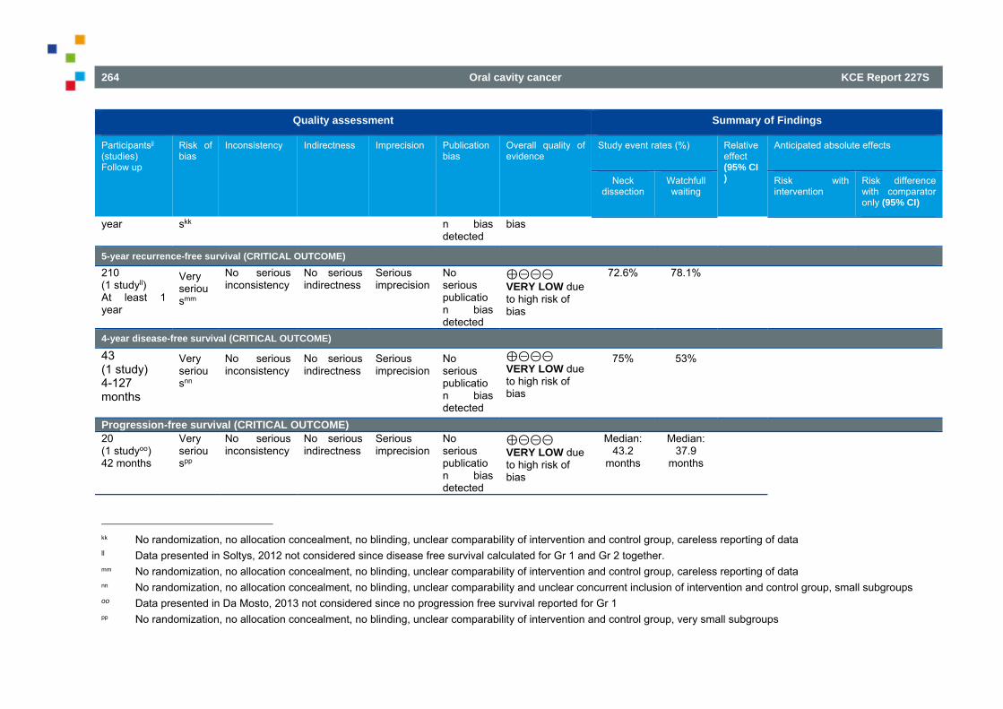
264 Oral cavity cancer KCE Report 227S
Quality assessment Summary of Findings
Participantsjj (studies) Follow up
Risk of bias
Inconsistency Indirectness Imprecision Publication bias
Overall quality of evidence
Study event rates (%) Relative effect (95% CI)
Anticipated absolute effects
Neck dissection
Watchfull waiting
Risk with intervention
Risk difference with comparator only (95% CI)
year skk n bias detected
bias
5-year recurrence-free survival (CRITICAL OUTCOME)
210 (1 studyll) At least 1 year
Very seriousmm
No serious inconsistency
No serious indirectness
Seriousimprecision
No serious publication bias detected
⊕⊝⊝⊝ VERY LOW due to high risk of bias
72.6% 78.1%
4-year disease-free survival (CRITICAL OUTCOME)
43 (1 study) 4-127 months
Very seriousnn
No serious inconsistency
No serious indirectness
Serious
imprecision No serious publication bias detected
⊕⊝⊝⊝ VERY LOW due to high risk of bias
75% 53%
Progression-free survival (CRITICAL OUTCOME)20 (1 studyoo) 42 months
Very seriouspp
No serious inconsistency
No serious indirectness
Seriousimprecision
No serious publication bias detected
⊕⊝⊝⊝ VERY LOW due to high risk of bias
Median: 43.2
months
Median: 37.9
months
kk No randomization, no allocation concealment, no blinding, unclear comparability of intervention and control group, careless reporting of data ll Data presented in Soltys, 2012 not considered since disease free survival calculated for Gr 1 and Gr 2 together. mm No randomization, no allocation concealment, no blinding, unclear comparability of intervention and control group, careless reporting of data nn No randomization, no allocation concealment, no blinding, unclear comparability and unclear concurrent inclusion of intervention and control group, small subgroups oo Data presented in Da Mosto, 2013 not considered since no progression free survival reported for Gr 1 pp No randomization, no allocation concealment, no blinding, unclear comparability of intervention and control group, very small subgroups
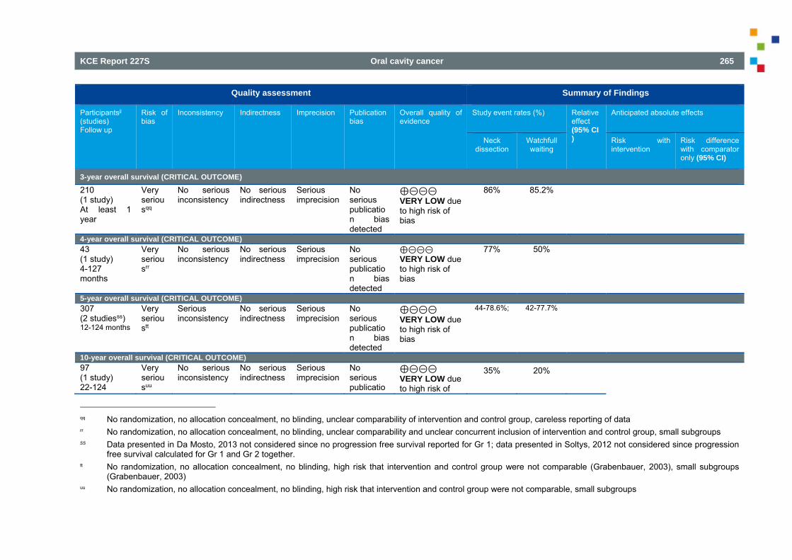
KCE Report 227S Oral cavity cancer 265
Quality assessment Summary of Findings
Participantsjj (studies) Follow up
Risk of bias
Inconsistency Indirectness Imprecision Publication bias
Overall quality of evidence
Study event rates (%) Relative effect (95% CI)
Anticipated absolute effects
Neck dissection
Watchfull waiting
Risk with intervention
Risk difference with comparator only (95% CI)
3-year overall survival (CRITICAL OUTCOME)
210 (1 study) At least 1 year
Very seriousqq
No serious inconsistency
No serious indirectness
Seriousimprecision
No serious publication bias detected
⊕⊝⊝⊝ VERY LOW due to high risk of bias
86% 85.2%
4-year overall survival (CRITICAL OUTCOME) 43 (1 study) 4-127 months
Very seriousrr
No serious inconsistency
No serious indirectness
Seriousimprecision
No serious publication bias detected
⊕⊝⊝⊝ VERY LOW due to high risk of bias
77% 50%
5-year overall survival (CRITICAL OUTCOME) 307 (2 studiesss) 12-124 months
Very serioustt
Serious inconsistency
No serious indirectness
Seriousimprecision
No serious publication bias detected
⊕⊝⊝⊝ VERY LOW due to high risk of bias
44-78.6%;
42-77.7%
10-year overall survival (CRITICAL OUTCOME) 97 (1 study) 22-124
Very seriousuu
No serious inconsistency
No serious indirectness
Seriousimprecision
No serious publicatio
⊕⊝⊝⊝ VERY LOW due to high risk of
35% 20%
qq No randomization, no allocation concealment, no blinding, unclear comparability of intervention and control group, careless reporting of data rr No randomization, no allocation concealment, no blinding, unclear comparability and unclear concurrent inclusion of intervention and control group, small subgroups ss Data presented in Da Mosto, 2013 not considered since no progression free survival reported for Gr 1; data presented in Soltys, 2012 not considered since progression
free survival calculated for Gr 1 and Gr 2 together. tt No randomization, no allocation concealment, no blinding, high risk that intervention and control group were not comparable (Grabenbauer, 2003), small subgroups
(Grabenbauer, 2003) uu No randomization, no allocation concealment, no blinding, high risk that intervention and control group were not comparable, small subgroups
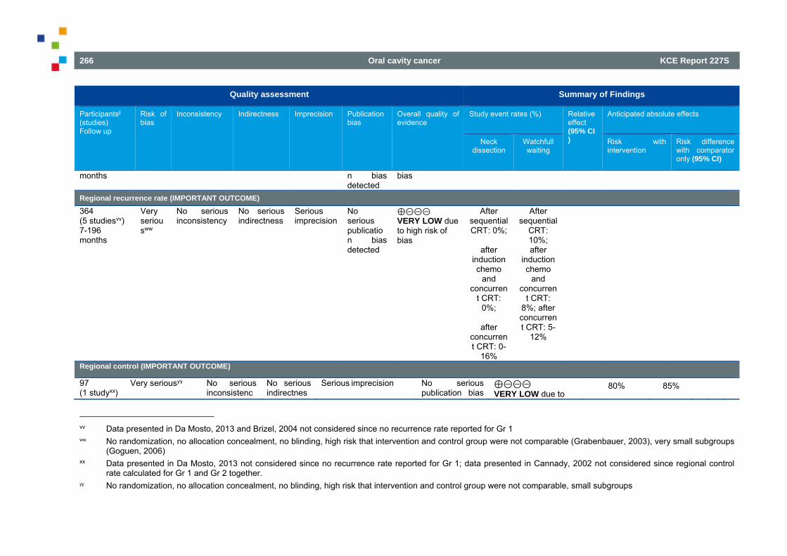
266 Oral cavity cancer KCE Report 227S
Quality assessment Summary of Findings
Participantsjj (studies) Follow up
Risk of bias
Inconsistency Indirectness Imprecision Publication bias
Overall quality of evidence
Study event rates (%) Relative effect (95% CI)
Anticipated absolute effects
Neck dissection
Watchfull waiting
Risk with intervention
Risk difference with comparator only (95% CI)
months n bias detected
bias
Regional recurrence rate (IMPORTANT OUTCOME)
364 (5 studiesvv) 7-196 months
Very seriousww
No serious inconsistency
No serious indirectness
Seriousimprecision
No serious publication bias detected
⊕⊝⊝⊝ VERY LOW due to high risk of bias
After sequential CRT: 0%;
after
induction chemo
and concurren
t CRT: 0%;
after
concurrent CRT: 0-
16%
After sequential
CRT: 10%; after
induction chemo
and concurren
t CRT: 8%; after concurrent CRT: 5-
12%
Regional control (IMPORTANT OUTCOME)
97 (1 studyxx)
Very seriousyy No serious inconsistenc
No serious indirectnes
Serious imprecision No serious publication bias
⊕⊝⊝⊝ VERY LOW due to
80% 85%
vv Data presented in Da Mosto, 2013 and Brizel, 2004 not considered since no recurrence rate reported for Gr 1 ww No randomization, no allocation concealment, no blinding, high risk that intervention and control group were not comparable (Grabenbauer, 2003), very small subgroups
(Goguen, 2006) xx Data presented in Da Mosto, 2013 not considered since no recurrence rate reported for Gr 1; data presented in Cannady, 2002 not considered since regional control
rate calculated for Gr 1 and Gr 2 together. yy No randomization, no allocation concealment, no blinding, high risk that intervention and control group were not comparable, small subgroups
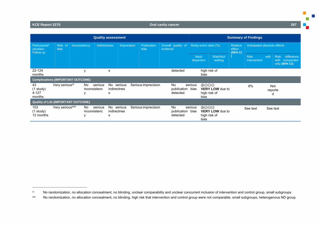
KCE Report 227S Oral cavity cancer 267
Quality assessment Summary of Findings
Participantsjj (studies) Follow up
Risk of bias
Inconsistency Indirectness Imprecision Publication bias
Overall quality of evidence
Study event rates (%) Relative effect (95% CI)
Anticipated absolute effects
Neck dissection
Watchfull waiting
Risk with intervention
Risk difference with comparator only (95% CI)
22-124 months
y s detected high risk ofbias
Complications (IMPORTANT OUTCOME)
43 (1 study) 4-127 months
Very seriouszz No serious inconsistency
No serious indirectness
Serious imprecision No serious publication bias detected
⊕⊝⊝⊝ VERY LOW due to high risk of bias
8% Not reporte
d
Quality of Life (IMPORTANT OUTCOME)
103 (1 study) 12 months
Very seriousaaa No serious inconsistency
No serious indirectness
Serious imprecision No serious publication bias detected
⊕⊝⊝⊝ VERY LOW due to high risk of bias
See text See text
zz No randomization, no allocation concealment, no blinding, unclear comparability and unclear concurrent inclusion of intervention and control group, small subgroups aaa No randomization, no allocation concealment, no blinding, high risk that intervention and control group were not comparable, small subgroups, heterogenous ND group
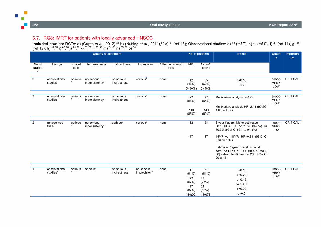
268 Oral cavity cancer KCE Report 227S
5.7. RQ8: IMRT for patients with locally advanced HNSCC Included studies: RCTs: a) (Gupta et al., 2012),41 b) (Nutting et al., 2011),67 c) 49 (ref 16); Observational studies: d) 49 (ref 7), e) 49 (ref 9), f) 49 (ref 11), g) 49 (ref 12); h) 59,59 i) 60,60 j) 75,75 k) 62,62 l) 63,63 m) 64,64 n) 65,65 o) 66
Quality assessment No of patients Effect Quality
Importance
No of studie
s
Design Risk of bias
Inconsistency Indirectness Imprecision Otherconsiderations
IMRT Conv/ConfRT
Disease-free survival: 2 year 2 observational
studies serious1
no serious inconsistency
no serious indirectness
serious2 none 42 (48%)
5 (80%)
55 (60%)
8 (50%)
p=0.18 NS
VERY LOW
CRITICAL
Disease-free survival: 3 year 2 observational
studies serious1
no serious inconsistency
no serious indirectness
serious3 none 22 (64%)
110
(85%)
27 (66%)
149
(69%)
Multivariate analysis p=0.73 Multivariate analysis HR=2.11 (95%CI 1.06 to 4.17)
VERY LOW
CRITICAL
Overall survival: 1-3 year 2 randomised
trials serious1
no serious inconsistency
serious5 serious6 none 32
47
28
47
3-year Kaplan–Meier estimates: 68% (95% CI 51.2 to 84.8%) vs 80.5% (95% CI 66.1 to 94.9%) 14/47 vs 18/47; HR=0.68 (95% CI 0.34 to 1.37) Estimated 2-year overall survival 78% (63 to 88) vs 76% (95% CI 60 to 86) (absolute difference 2%, 95% CI 20 to 16)
VERY LOW
CRITICAL
Overall survival: 2-3 year 7 observational
studies7 serious1
serious4 no serious indirectness
no serious imprecision8
none 41 (91%)
22 (67%)
27 (87%)
110(92
71 (81%)
27 (77%) 24 (86%) 149(75
p=0.10 p=0.70 p=0.43
p<0.001 p=0.29 p=0.5
VERY LOW
CRITICAL
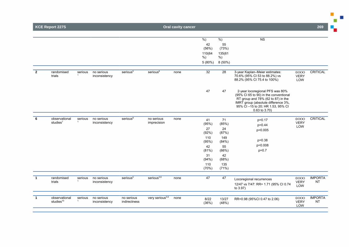
KCE Report 227S Oral cavity cancer 269
%)42
(56%) 110(64%) 5 (80%)
%)55
(73%) 135(61%) 8 (50%)
NS
(Loco-)regional control: 2-3 year 2 randomised
trials serious1
no serious inconsistency
serious5 serious6 none 32
47
28
47
3-year Kaplan–Meier estimates: 70.6% (95% CI 53 to 88.2%) vs 88.2% (95% CI 75.4 to 100%)
2-year locoregional PFS was 80% (95% CI 65 to 90) in the conventional RT group and 78% (62 to 87) in the
IMRT group (absolute difference 3%, 95% CI –15 to 20; HR 1.53, 95% CI
0.63 to 3.70)
VERY LOW
CRITICAL
(Loco-)regional control: 2-3 year 6 observational
studies7 serious1
no serious inconsistency
serious9 no serious imprecision
none 41 (95%)
27 (92%) 110
(95%) 42
(81%) 31
(94%) 110
(70%)
71 (85%)
24 (87%) 149
(84%) 55
(66%) 42
(68%) 135
(71%)
p=0.17 p=0.44
p=0.005
p=0.38 p=0.008
p=0.7
VERY LOW
CRITICAL
Recurrence rate (follow-up median 44 months) 1 randomised
trials serious1
no serious inconsistency
serious5 serious3,6 none 47 47 Locoregional recurrences 12/47 vs 7/47: RR= 1.71 (95% CI 0.74 to 3.97)
VERY LOW
IMPORTANT
Recurrence rate (follow-up median 3.3 years) 1 observational
studies10 serious1
no serious inconsistency
no serious indirectness
very serious3,6 none 8/22 (36%)
13/27 (48%)
RR=0.98 (95%CI 0.47 to 2.06) VERY LOW
IMPORTANT
Secondary tumours (follow-up median 3.3 years)
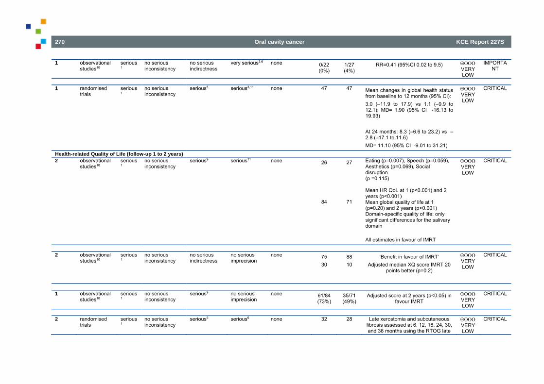
270 Oral cavity cancer KCE Report 227S
1 observational studies10
serious1
no serious inconsistency
no serious indirectness
very serious3,6 none 0/22 (0%)
1/27 (4%)
RR=0.41 (95%CI 0.02 to 9.5) VERY LOW
IMPORTANT
Quality of Life (mean changes in global health status) (follow-up median 44 months) 1 randomised
trials serious1
no serious inconsistency
serious5 serious3,11 none 47 47 Mean changes in global health status from baseline to 12 months (95% CI): 3.0 (–11.9 to 17.9) vs 1.1 (–9.9 to 12.1); MD= 1.90 (95% CI -16.13 to 19.93) At 24 months: 8.3 (–6.6 to 23.2) vs –2.8 (–17.1 to 11.6) MD= 11.10 (95% CI -9.01 to 31.21)
VERY LOW
CRITICAL
Health-related Quality of Life (follow-up 1 to 2 years) 2 observational
studies10 serious1
no serious inconsistency
serious9 serious11 none 26
84
27
71
Eating (p=0.007), Speech (p=0.059), Aesthetics (p=0.069), Social disruption (p =0.115) Mean HR QoL at 1 (p<0.001) and 2 years (p<0.001) Mean global quality of life at 1 (p=0.20) and 2 years (p<0.001) Domain-specific quality of life: only significant differences for the salivary domain All estimates in favour of IMRT
VERY LOW
CRITICAL
Xerostomia-related Quality of Life (follow-up 12 to median 31.2 months) 2 observational
studies10 serious1
no serious inconsistency
no serious indirectness
no serious imprecision
none 75 30
88 10
‘Benefit in favour of IMRT’ Adjusted median XQ score IMRT 20
points better (p=0.2)
VERY LOW
CRITICAL
Very good or Outstanding QoL (follow-up 2 years) 1 observational
studies10 serious1
no serious inconsistency
serious9 no serious imprecision
none 61/84 (73%)
35/71 (49%)
Adjusted score at 2 years (p<0.05) in favour IMRT
VERY LOW
CRITICAL
Adverse events: xerostomia (follow-up median 40-44 months) 2 randomised
trials serious1
no serious inconsistency
serious5 serious6 none 32
28
Late xerostomia and subcutaneous fibrosis assessed at 6, 12, 18, 24, 30, and 36 months using the RTOG late
VERY LOW
CRITICAL
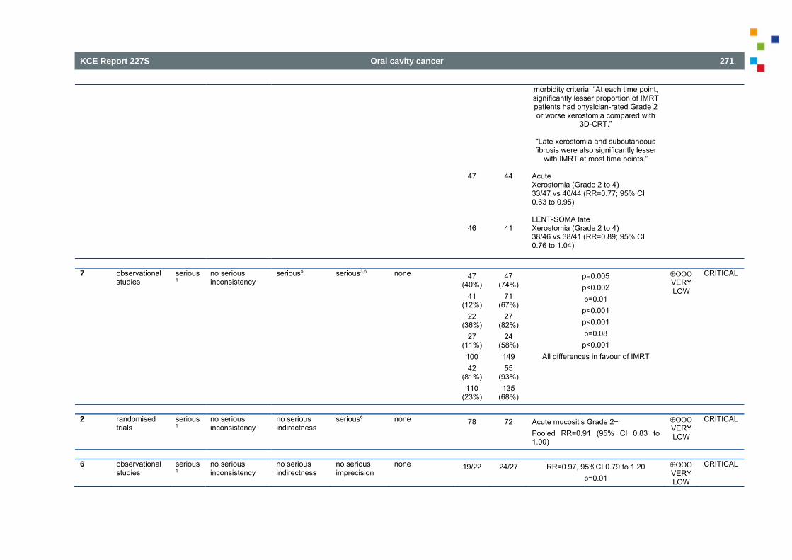
KCE Report 227S Oral cavity cancer 271
47
46
44
41
morbidity criteria: “At each time point, significantly lesser proportion of IMRT patients had physician-rated Grade 2 or worse xerostomia compared with
3D-CRT.”
“Late xerostomia and subcutaneous fibrosis were also significantly lesser
with IMRT at most time points.”
Acute Xerostomia (Grade 2 to 4) 33/47 vs 40/44 (RR=0.77; 95% CI 0.63 to 0.95)
LENT-SOMA late Xerostomia (Grade 2 to 4) 38/46 vs 38/41 (RR=0.89; 95% CI 0.76 to 1.04)
Adverse events: xerostomia 7 observational
studies serious1
no serious inconsistency
serious5 serious3,6 none 47 (40%)
41 (12%)
22 (36%)
27 (11%) 100 42
(81%) 110
(23%)
47 (74%)
71 (67%)
27 (82%)
24 (58%) 149 55
(93%) 135
(68%)
p=0.005 p<0.002 p=0.01
p<0.001 p<0.001 p=0.08
p<0.001 All differences in favour of IMRT
VERY LOW
CRITICAL
Adverse events: mucositis (follow-up median 40-44 months) 2 randomised
trials serious1
no serious inconsistency
no serious indirectness
serious6 none 78 72 Acute mucositis Grade 2+ Pooled RR=0.91 (95% CI 0.83 to 1.00)
VERY LOW
CRITICAL
Adverse events: mucositis 6 observational
studies serious1
no serious inconsistency
no serious indirectness
no serious imprecision
none 19/22
24/27
RR=0.97, 95%CI 0.79 to 1.20 p=0.01
VERY LOW
CRITICAL
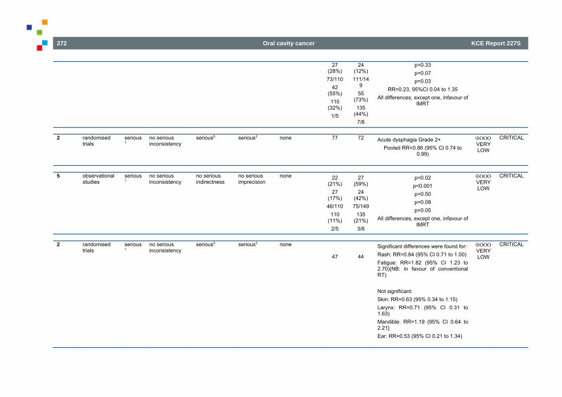
272 Oral cavity cancer KCE Report 227S
27 (28%) 73/110
42 (55%) 110
(32%) 1/5
24 (12%) 111/14
9 55
(73%) 135
(44%) 7/8
p=0.33 p=0.07 p=0.03
RR=0.23, 95%CI 0.04 to 1.35 All differences, except one, infavour of
IMRT
Adverse events: dysphagia (follow-up median 40-44 months) 2 randomised
trials serious1
no serious inconsistency
serious5 serious3 none 77 72 Acute dysphagia Grade 2+ Pooled RR=0.86 (95% CI 0.74 to
0.99)
VERY LOW
CRITICAL
Adverse events: swallowing, dysphagia, need for enteral feeding or liquid diet 5 observational
studies serious1
no serious inconsistency
no serious indirectness
no serious imprecision
none 22 (21%)
27 (17%) 46/110
110 (11%)
2/5
27 (59%)
24 (42%) 75/149
135 (21%)
3/8
p=0.02 p<0.001 p=0.50 p=0.08 p>0.05
All differences, except one, infavour of IMRT
VERY LOW
CRITICAL
Adverse events: other (follow-up median 40-44 months) 2 randomised
trials serious1
no serious inconsistency
serious5 serious3 none
47
44
Significant differences were found for: Rash: RR=0.84 (95% CI 0.71 to 1.00) Fatigue: RR=1.82 (95% CI 1.23 to 2.70)(NB: in favour of conventional RT)
Not significant: Skin: RR=0.63 (95% 0.34 to 1.15) Larynx: RR=0.71 (95% CI 0.31 to 1.63) Mandible: RR=1.19 (95% CI 0.64 to 2.21] Ear: RR=0.53 (95% CI 0.21 to 1.34)
VERY LOW
CRITICAL
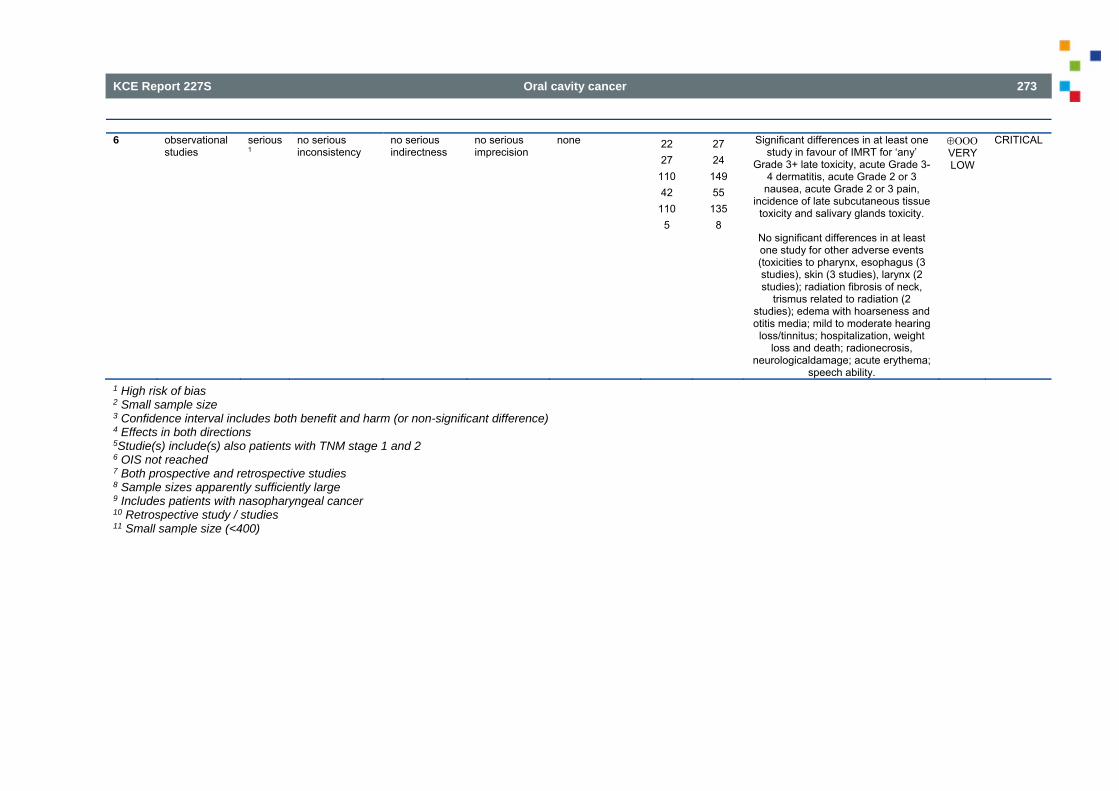
KCE Report 227S Oral cavity cancer 273
Adverse events: other 6 observational
studies serious1
no serious inconsistency
no serious indirectness
no serious imprecision
none 22 27
110 42
110 5
27 24
149 55
135 8
Significant differences in at least one study in favour of IMRT for ‘any’
Grade 3+ late toxicity, acute Grade 3-4 dermatitis, acute Grade 2 or 3
nausea, acute Grade 2 or 3 pain, incidence of late subcutaneous tissue
toxicity and salivary glands toxicity.
No significant differences in at least one study for other adverse events (toxicities to pharynx, esophagus (3 studies), skin (3 studies), larynx (2 studies); radiation fibrosis of neck,
trismus related to radiation (2 studies); edema with hoarseness and otitis media; mild to moderate hearing
loss/tinnitus; hospitalization, weight loss and death; radionecrosis,
neurologicaldamage; acute erythema; speech ability.
VERY LOW
CRITICAL
1 High risk of bias 2 Small sample size 3 Confidence interval includes both benefit and harm (or non-significant difference) 4 Effects in both directions 5Studie(s) include(s) also patients with TNM stage 1 and 2 6 OIS not reached 7 Both prospective and retrospective studies 8 Sample sizes apparently sufficiently large 9 Includes patients with nasopharyngeal cancer 10 Retrospective study / studies 11 Small sample size (<400)
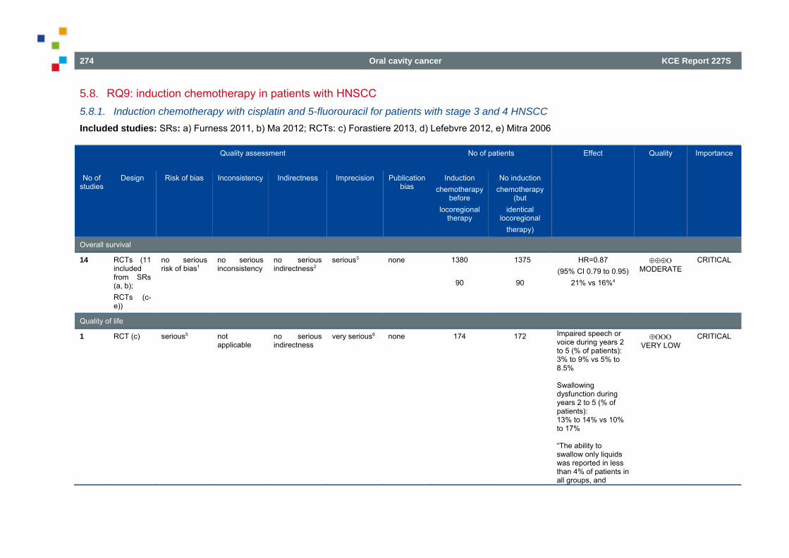
274 Oral cavity cancer KCE Report 227S
5.8. RQ9: induction chemotherapy in patients with HNSCC 5.8.1. Induction chemotherapy with cisplatin and 5-fluorouracil for patients with stage 3 and 4 HNSCC Included studies: SRs: a) Furness 2011, b) Ma 2012; RCTs: c) Forastiere 2013, d) Lefebvre 2012, e) Mitra 2006
Quality assessment No of patients Effect Quality Importance
No of studies
Design Risk of bias Inconsistency Indirectness Imprecision Publication bias
Induction chemotherapy
before locoregional
therapy
No induction chemotherapy
(but identical
locoregional therapy)
Overall survival
14 RCTs (11 included from SRs (a, b); RCTs (c-e))
no serious risk of bias1
no serious inconsistency
no serious indirectness2
serious3 none 1380
90
1375
90
HR=0.87 (95% CI 0.79 to 0.95)
21% vs 16%4
MODERATE
CRITICAL
Quality of life
1
RCT (c) serious5 not applicable
no serious indirectness
very serious6 none 174 172 Impaired speech or voice during years 2 to 5 (% of patients): 3% to 9% vs 5% to 8.5% Swallowing dysfunction during years 2 to 5 (% of patients): 13% to 14% vs 10% to 17% “The ability to swallow only liquids was reported in less than 4% of patients in all groups, and
VERY LOW
CRITICAL
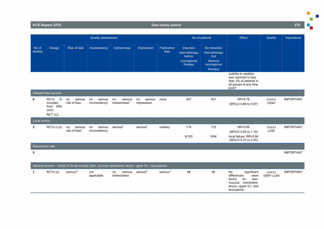
KCE Report 227S Oral cavity cancer 275
Quality assessment No of patients Effect Quality Importance
No of studies
Design Risk of bias Inconsistency Indirectness Imprecision Publication bias
Induction chemotherapy
before locoregional
therapy
No induction chemotherapy
(but identical
locoregional therapy)
inability to swallow was reported in less than 3% of patients in all groups at any time point"
Disease free survival
6 RCTs (5 included from SRs (a,b); RCT (c))
no serious risk of bias
no serious inconsistency
no serious indirectness2
no serious imprecision
none 507 501 HR=0.76 (95%CI 0.66 to 0.87)
HIGH
IMPORTANT
Local control
2
RCTs (c,d) no serious risk of bias7
no serious inconsistency
serious8 serious9
unlikely 174
8/100
172
8/94
HR=0.85 (95%CI 0.63 to 1.15) local failure: RR=0.94 (95%CI 0.37 to 2.40)
LOW
IMPORTANT
Recurrence rate
0 IMPORTANT
Adverse events – Grade III Acute toxicity (skin, mucous membrane, larynx, upper G.I., leucopenia)
1 RCTs (e) serious10 not applicable
no serious indirectness
serious9
serious11 88
90
No significant differences were found for skin, mucous membrane, larynx, upper G.I. and leucopenia.
VERY LOW
IMPORTANT
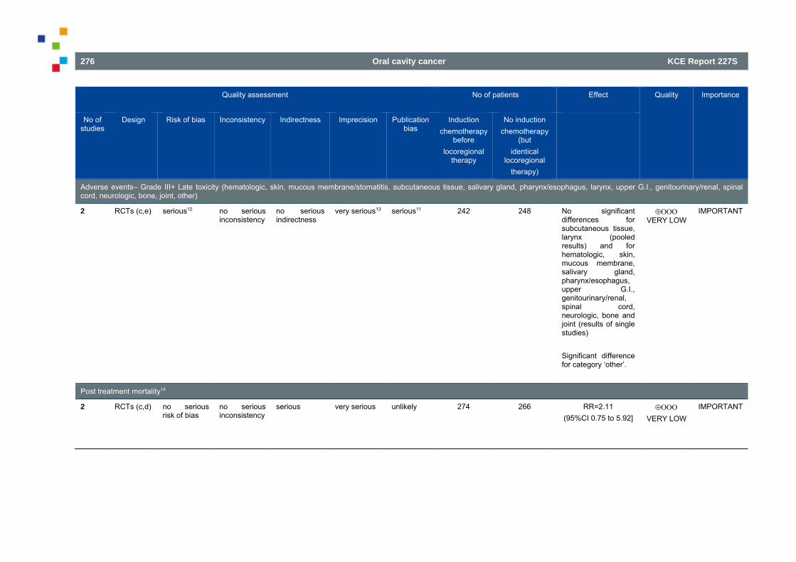
276 Oral cavity cancer KCE Report 227S
Quality assessment No of patients Effect Quality Importance
No of studies
Design Risk of bias Inconsistency Indirectness Imprecision Publication bias
Induction chemotherapy
before locoregional
therapy
No induction chemotherapy
(but identical
locoregional therapy)
Adverse events– Grade III+ Late toxicity (hematologic, skin, mucous membrane/stomatitis, subcutaneous tissue, salivary gland, pharynx/esophagus, larynx, upper G.I., genitourinary/renal, spinal cord, neurologic, bone, joint, other)
2 RCTs (c,e) serious12 no serious inconsistency
no serious indirectness
very serious13
serious11 242
248
No significant differences for subcutaneous tissue, larynx (pooled results) and for hematologic, skin, mucous membrane, salivary gland, pharynx/esophagus, upper G.I., genitourinary/renal, spinal cord, neurologic, bone and joint (results of single studies) Significant difference for category ‘other’.
VERY LOW
IMPORTANT
Post treatment mortality14
2 RCTs (c,d) no serious risk of bias
no serious inconsistency
serious very serious
unlikely 274 266 RR=2.11 (95%CI 0.75 to 5.92]
VERY LOW
IMPORTANT
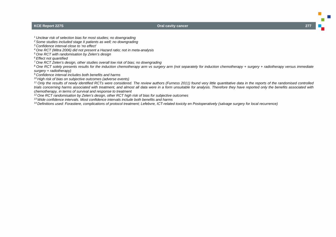
KCE Report 227S Oral cavity cancer 277
1 Unclear risk of selection bias for most studies; no downgrading 2 Some studies included stage II patients as well; no downgrading 3 Confidence interval close to ‘no effect’ 4 One RCT (Mitra 2006) did not present a Hazard ratio; not in meta-analysis 5 One RCT with randomisation by Zelen’s design 6 Effect not quantified 7 One RCT Zelen’s design, other studies overall low risk of bias; no downgrading 8 One RCT solely presents results for the induction chemotherapy arm vs surgery arm (not separately for induction chemotherapy + surgery + radiotherapy versus immediate surgery + radiotherapy) 9 Confidence interval includes both benefits and harms 10 High risk of bias on subjective outcomes (adverse events) 11 Only the results of newly identified RCTs were considered. The review authors (Furness 2011) found very little quantitative data in the reports of the randomised controlled trials concerning harms associated with treatment, and almost all data were in a form unsuitable for analysis. Therefore they have reported only the benefits associated with chemotherapy, in terms of survival and response to treatment 12 One RCT randomisation by Zelen’s design, other RCT high risk of bias for subjective outcomes 13 Wide confidence intervals. Most confidence intervals include both benefits and harms 14 Definitions used: Forastiere, complications of protocol treatment; Lefebvre, ICT-related toxicity en Postoperatively (salvage surgery for local recurrence)
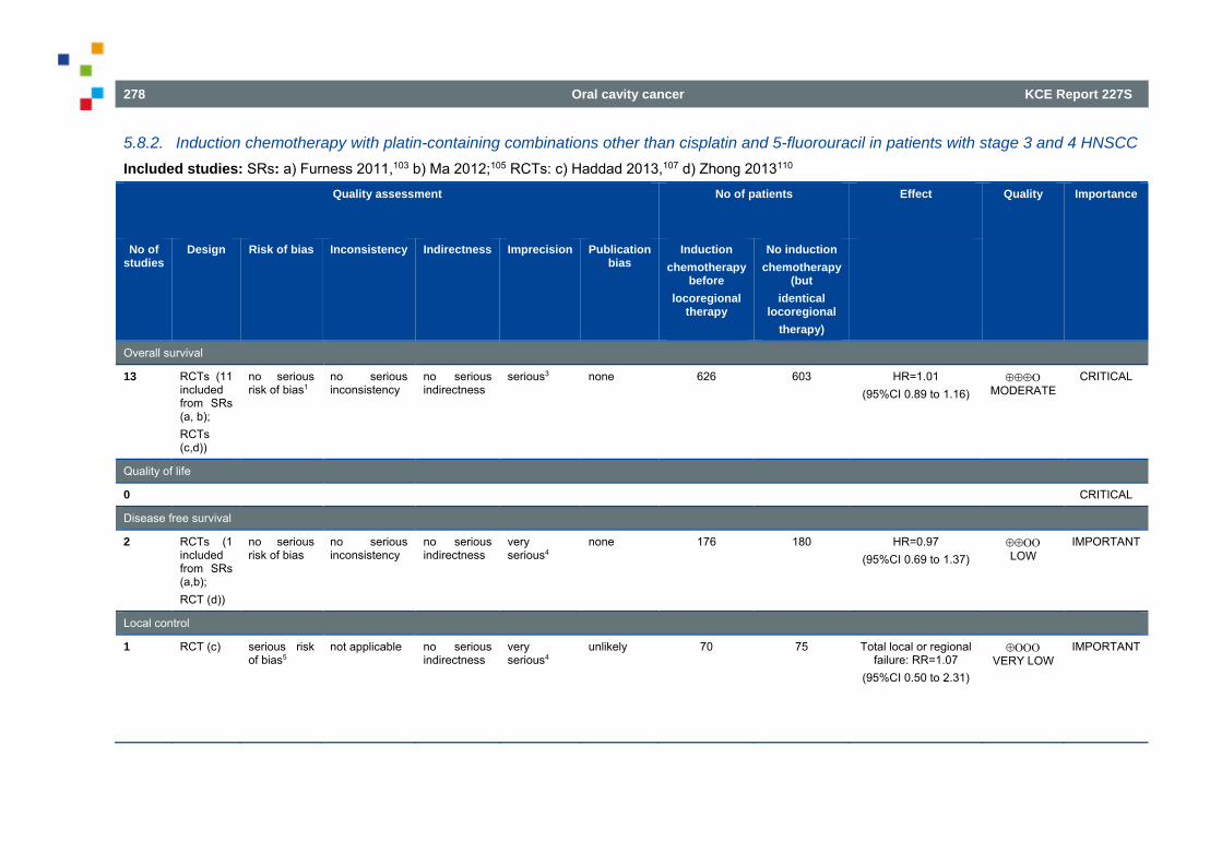
278 Oral cavity cancer KCE Report 227S
5.8.2. Induction chemotherapy with platin-containing combinations other than cisplatin and 5-fluorouracil in patients with stage 3 and 4 HNSCC Included studies: SRs: a) Furness 2011,103 b) Ma 2012;105 RCTs: c) Haddad 2013,107 d) Zhong 2013110
Quality assessment No of patients Effect Quality Importance
No of studies
Design Risk of bias Inconsistency Indirectness Imprecision Publication bias
Induction chemotherapy
before locoregional
therapy
No induction chemotherapy
(but identical
locoregional therapy)
Overall survival
13 RCTs (11 included from SRs (a, b); RCTs (c,d))
no serious risk of bias1
no serious inconsistency
no serious indirectness
serious3
none 626 603 HR=1.01 (95%CI 0.89 to 1.16)
MODERATE
CRITICAL
Quality of life
0 CRITICAL Disease free survival
2 RCTs (1 included from SRs (a,b); RCT (d))
no serious risk of bias
no serious inconsistency
no serious indirectness
very serious4
none 176 180 HR=0.97 (95%CI 0.69 to 1.37)
LOW
IMPORTANT
Local control
1 RCT (c) serious risk of bias5
not applicable no serious indirectness
very serious4
unlikely 70 75 Total local or regional failure: RR=1.07
(95%CI 0.50 to 2.31)
VERY LOW
IMPORTANT
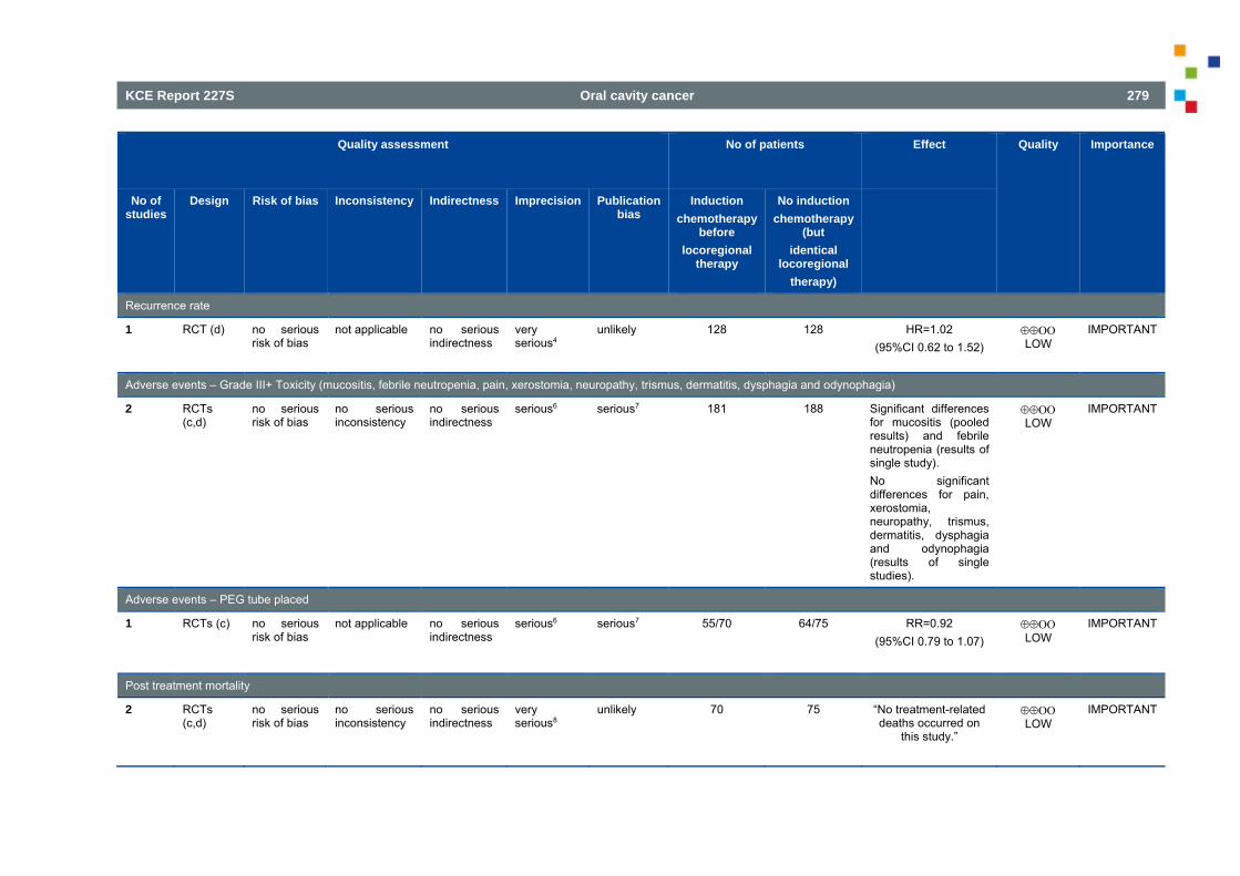
KCE Report 227S Oral cavity cancer 279
Quality assessment No of patients Effect Quality Importance
No of studies
Design Risk of bias Inconsistency Indirectness Imprecision Publication bias
Induction chemotherapy
before locoregional
therapy
No induction chemotherapy
(but identical
locoregional therapy)
Recurrence rate
1 RCT (d) no serious risk of bias
not applicable no serious indirectness
very serious4
unlikely 128 128 HR=1.02 (95%CI 0.62 to 1.52)
LOW
IMPORTANT
Adverse events – Grade III+ Toxicity (mucositis, febrile neutropenia, pain, xerostomia, neuropathy, trismus, dermatitis, dysphagia and odynophagia)
2 RCTs (c,d)
no serious risk of bias
no serious inconsistency
no serious indirectness
serious6 serious7 181
188
Significant differences for mucositis (pooled results) and febrile neutropenia (results of single study). No significant differences for pain, xerostomia, neuropathy, trismus, dermatitis, dysphagia and odynophagia (results of single studies).
LOW
IMPORTANT
Adverse events – PEG tube placed
1 RCTs (c) no serious risk of bias
not applicable no serious indirectness
serious6 serious7 55/70 64/75 RR=0.92 (95%CI 0.79 to 1.07)
LOW
IMPORTANT
Post treatment mortality
2 RCTs (c,d)
no serious risk of bias
no serious inconsistency
no serious indirectness
very serious8
unlikely 70
75
“No treatment-related deaths occurred on
this study.”
LOW
IMPORTANT
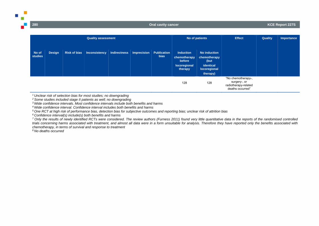
280 Oral cavity cancer KCE Report 227S
Quality assessment No of patients Effect Quality Importance
No of studies
Design Risk of bias Inconsistency Indirectness Imprecision Publication bias
Induction chemotherapy
before locoregional
therapy
No induction chemotherapy
(but identical
locoregional therapy)
128
128
“No chemotherapy-, surgery-, or
radiotherapy-related deaths occurred”
1 Unclear risk of selection bias for most studies; no downgrading 2 Some studies included stage II patients as well; no downgrading 3 Wide confidence intervals. Most confidence intervals include both benefits and harms 4 Wide confidence interval. Confidence interval includes both benefits and harms 5 One RCT at high risk of performance bias, detection bias for subjective outcomes and reporting bias; unclear risk of attrition bias 6 Confidence interval(s) include(s) both benefits and harms 7 Only the results of newly identified RCTs were considered. The review authors (Furness 2011) found very little quantitative data in the reports of the randomised controlled trials concerning harms associated with treatment, and almost all data were in a form unsuitable for analysis. Therefore they have reported only the benefits associated with chemotherapy, in terms of survival and response to treatment 8 No deaths occurred
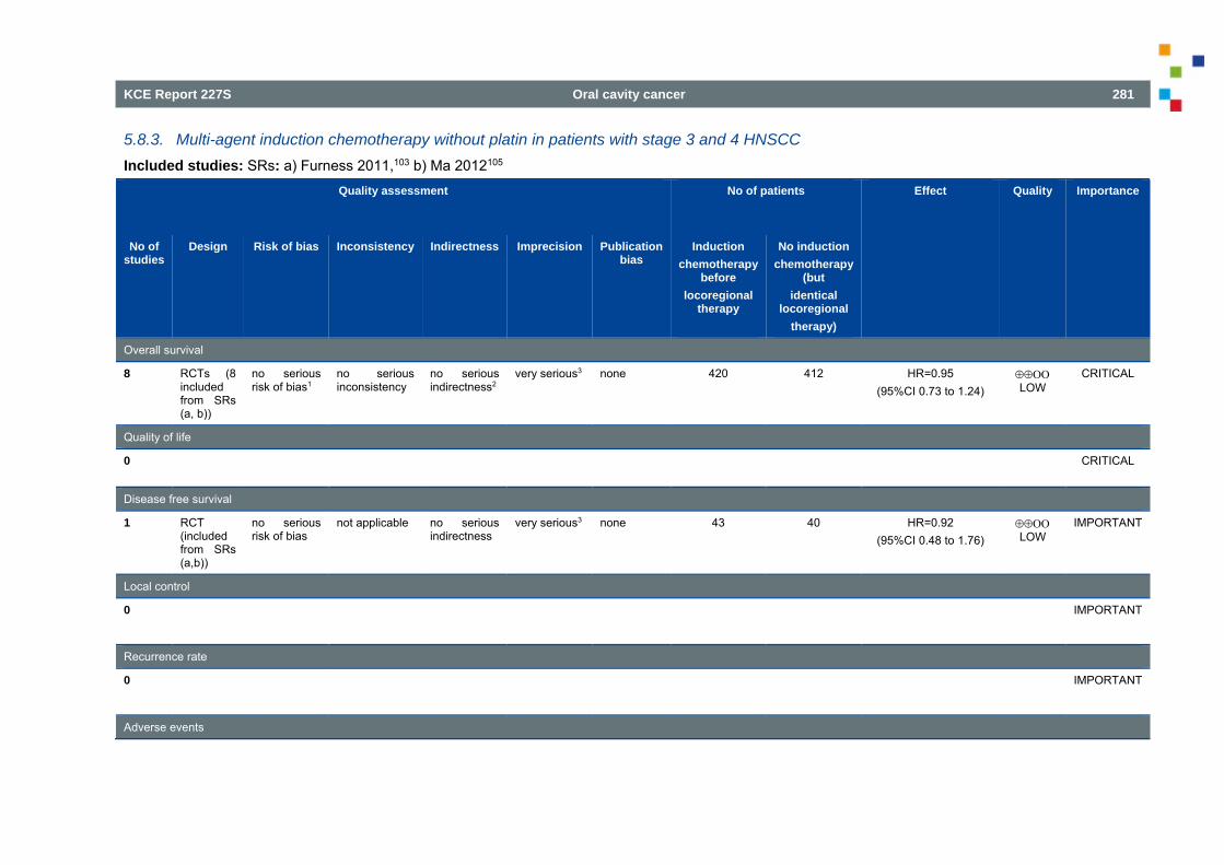
KCE Report 227S Oral cavity cancer 281
5.8.3. Multi-agent induction chemotherapy without platin in patients with stage 3 and 4 HNSCC Included studies: SRs: a) Furness 2011,103 b) Ma 2012105
Quality assessment No of patients Effect Quality Importance
No of studies
Design Risk of bias Inconsistency Indirectness Imprecision Publication bias
Induction chemotherapy
before locoregional
therapy
No induction chemotherapy
(but identical
locoregional therapy)
Overall survival
8 RCTs (8 included from SRs (a, b))
no serious risk of bias1
no serious inconsistency
no serious indirectness2
very serious3
none 420 412 HR=0.95
(95%CI 0.73 to 1.24)
LOW
CRITICAL
Quality of life
0 CRITICAL
Disease free survival
1 RCT (included from SRs (a,b))
no serious risk of bias
not applicable no serious indirectness
very serious3 none 43 40 HR=0.92 (95%CI 0.48 to 1.76)
LOW
IMPORTANT
Local control
0 IMPORTANT
Recurrence rate
0 IMPORTANT
Adverse events
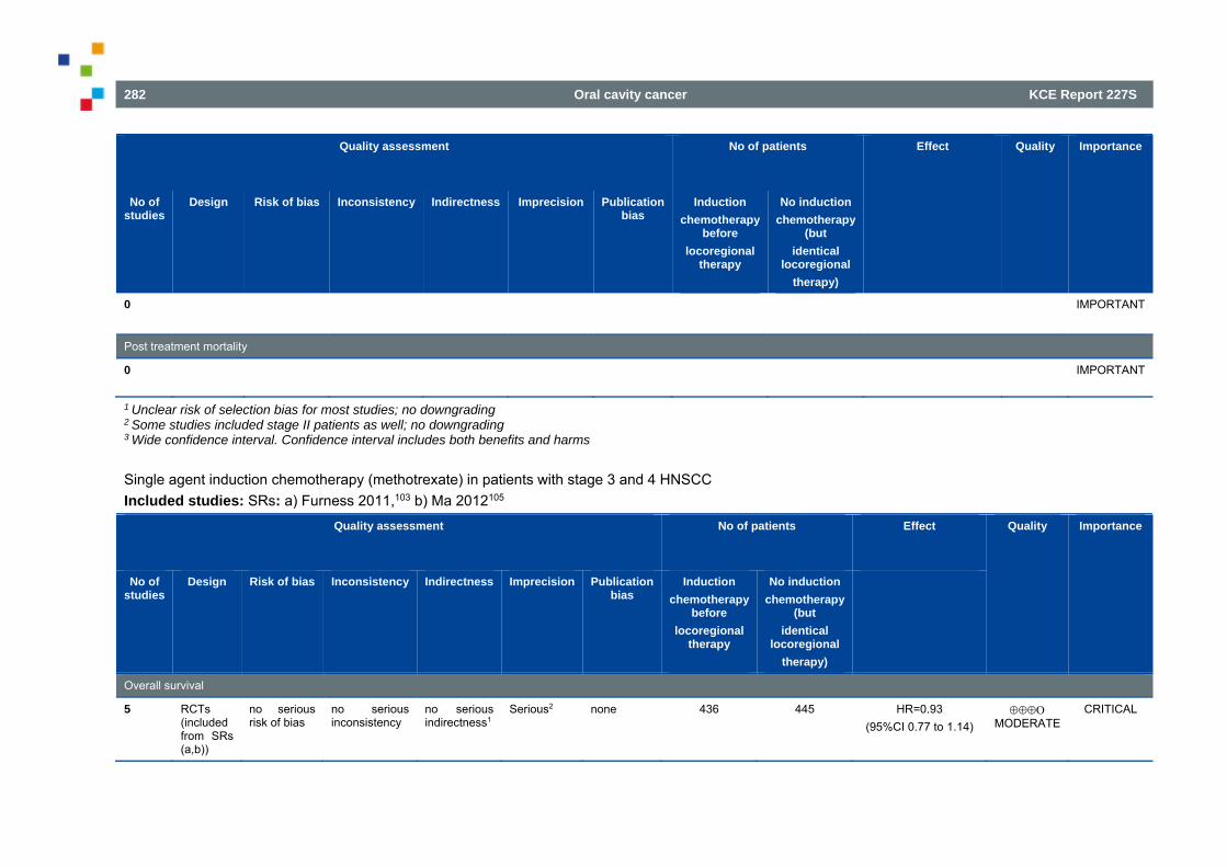
282 Oral cavity cancer KCE Report 227S
Quality assessment No of patients Effect Quality Importance
No of studies
Design Risk of bias Inconsistency Indirectness Imprecision Publication bias
Induction chemotherapy
before locoregional
therapy
No induction chemotherapy
(but identical
locoregional therapy)
0 IMPORTANT
Post treatment mortality
0 IMPORTANT
1 Unclear risk of selection bias for most studies; no downgrading 2 Some studies included stage II patients as well; no downgrading 3 Wide confidence interval. Confidence interval includes both benefits and harms Single agent induction chemotherapy (methotrexate) in patients with stage 3 and 4 HNSCC Included studies: SRs: a) Furness 2011,103 b) Ma 2012105
Quality assessment No of patients Effect Quality Importance
No of studies
Design Risk of bias Inconsistency Indirectness Imprecision Publication bias
Induction chemotherapy
before locoregional
therapy
No induction chemotherapy
(but identical
locoregional therapy)
Overall survival
5 RCTs (included from SRs (a,b))
no serious risk of bias
no serious inconsistency
no serious indirectness1
Serious2 none 436 445 HR=0.93 (95%CI 0.77 to 1.14)
MODERATE
CRITICAL
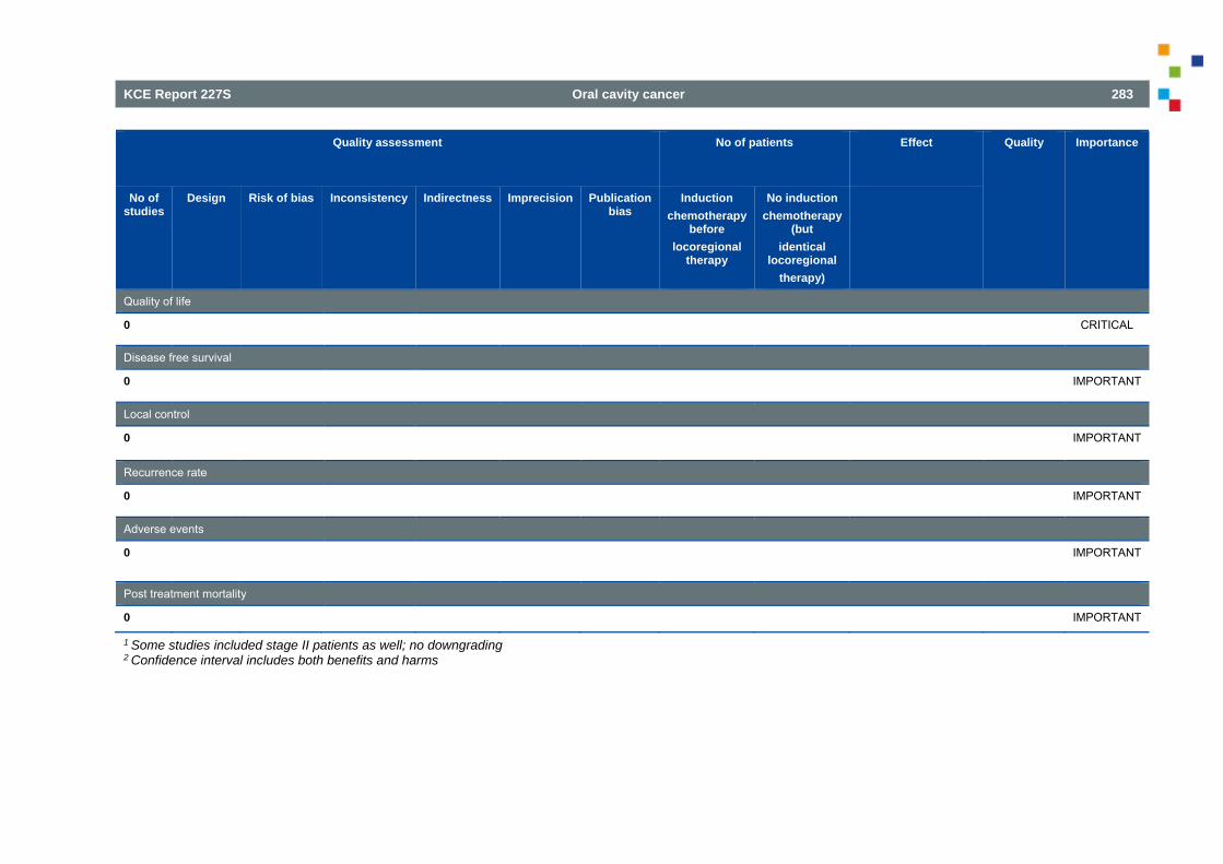
KCE Report 227S Oral cavity cancer 283
Quality assessment No of patients Effect Quality Importance
No of studies
Design Risk of bias Inconsistency Indirectness Imprecision Publication bias
Induction chemotherapy
before locoregional
therapy
No induction chemotherapy
(but identical
locoregional therapy)
Quality of life
0 CRITICAL
Disease free survival
0 IMPORTANT
Local control
0 IMPORTANT
Recurrence rate
0 IMPORTANT
Adverse events
0
IMPORTANT
Post treatment mortality
0 IMPORTANT 1 Some studies included stage II patients as well; no downgrading 2 Confidence interval includes both benefits and harms
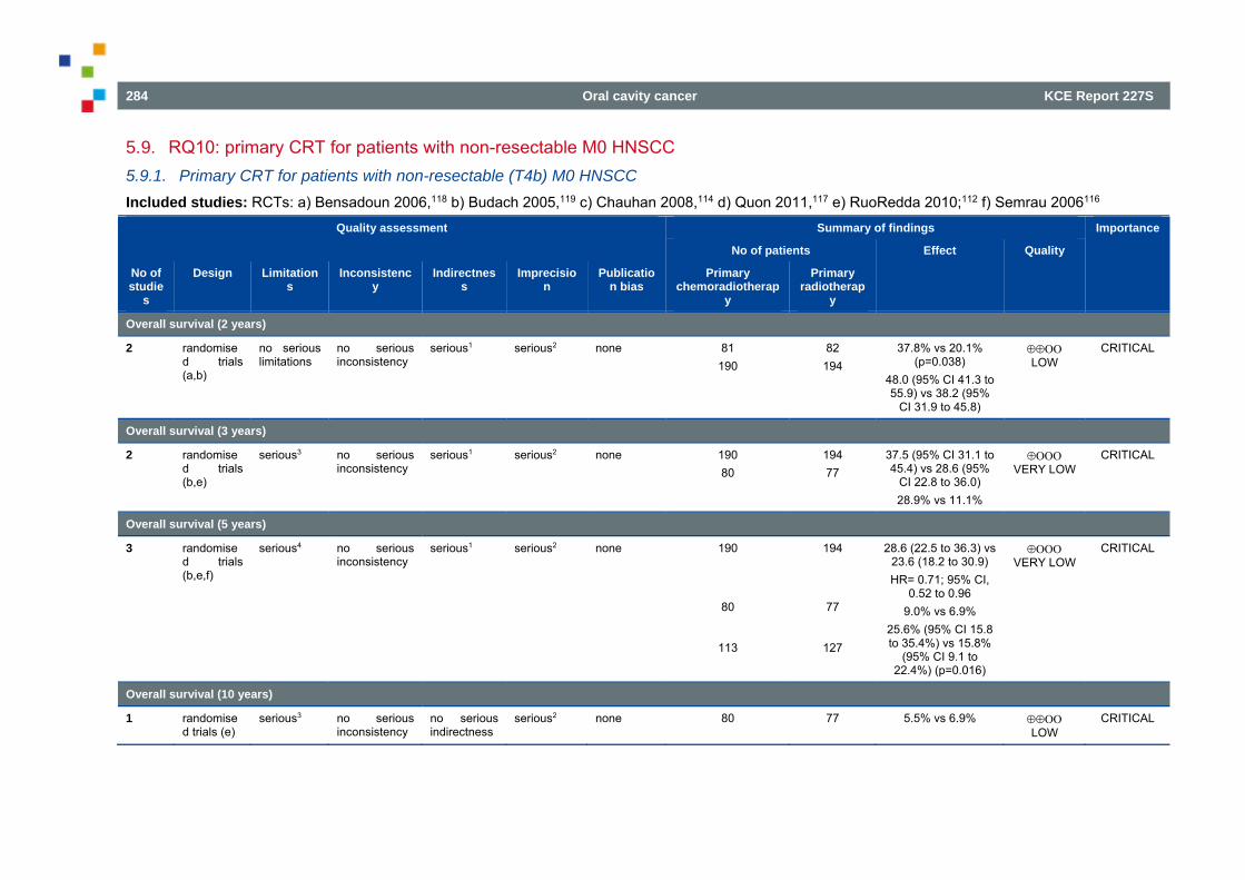
284 Oral cavity cancer KCE Report 227S
5.9. RQ10: primary CRT for patients with non-resectable M0 HNSCC 5.9.1. Primary CRT for patients with non-resectable (T4b) M0 HNSCC Included studies: RCTs: a) Bensadoun 2006,118 b) Budach 2005,119 c) Chauhan 2008,114 d) Quon 2011,117 e) RuoRedda 2010;112 f) Semrau 2006116
Quality assessment Summary of findings Importance
No of patients Effect Quality
No of studie
s
Design Limitations
Inconsistency
Indirectness
Imprecision
Publication bias
Primary chemoradiotherap
y
Primary radiotherap
y
Overall survival (2 years)
2 randomised trials (a,b)
no serious limitations
no serious inconsistency
serious1 serious2 none 81 190
82 194
37.8% vs 20.1% (p=0.038)
48.0 (95% CI 41.3 to 55.9) vs 38.2 (95%
CI 31.9 to 45.8)
LOW
CRITICAL
Overall survival (3 years)
2 randomised trials (b,e)
serious3 no serious inconsistency
serious1 serious2 none 190 80
194 77
37.5 (95% CI 31.1 to 45.4) vs 28.6 (95%
CI 22.8 to 36.0) 28.9% vs 11.1%
VERY LOW
CRITICAL
Overall survival (5 years)
3 randomised trials (b,e,f)
serious4 no serious inconsistency
serious1 serious2 none 190
80
113
194
77
127
28.6 (22.5 to 36.3) vs 23.6 (18.2 to 30.9) HR= 0.71; 95% CI,
0.52 to 0.96 9.0% vs 6.9%
25.6% (95% CI 15.8 to 35.4%) vs 15.8%
(95% CI 9.1 to 22.4%) (p=0.016)
VERY LOW
CRITICAL
Overall survival (10 years)
1 randomised trials (e)
serious3 no serious inconsistency
no serious indirectness
serious2 none 80 77 5.5% vs 6.9% LOW
CRITICAL
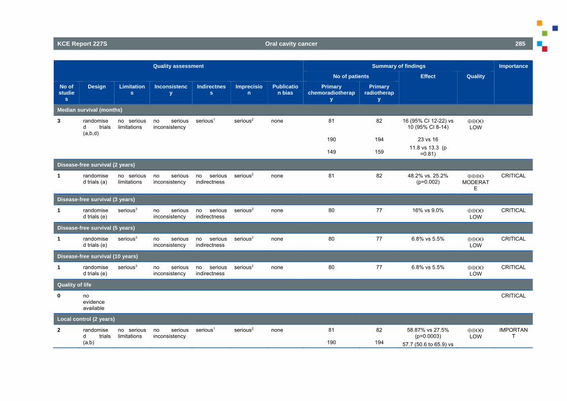
KCE Report 227S Oral cavity cancer 285
Quality assessment Summary of findings Importance
No of patients Effect Quality
No of studie
s
Design Limitations
Inconsistency
Indirectness
Imprecision
Publication bias
Primary chemoradiotherap
y
Primary radiotherap
y
Median survival (months)
3 randomised trials (a,b,d)
no serious limitations
no serious inconsistency
serious1 serious2 none 81
190
149
82
194
159
16 (95% CI 12-22) vs 10 (95% CI 8-14)
23 vs 16
11.8 vs 13.3 (p =0.81)
LOW
Disease-free survival (2 years)
1 randomised trials (a)
no serious limitations
no serious inconsistency
no serious indirectness
serious2 none 81 82 48.2% vs. 25.2% (p=0.002)
MODERAT
E
CRITICAL
Disease-free survival (3 years)
1 randomised trials (e)
serious3 no serious inconsistency
no serious indirectness
serious2 none 80 77 16% vs 9.0% LOW
CRITICAL
Disease-free survival (5 years)
1 randomised trials (e)
serious3 no serious inconsistency
no serious indirectness
serious2 none 80 77 6.8% vs 5.5% LOW
CRITICAL
Disease-free survival (10 years)
1 randomised trials (e)
serious3 no serious inconsistency
no serious indirectness
serious2 none 80 77 6.8% vs 5.5% LOW
CRITICAL
Quality of life
0 no evidence available
CRITICAL
Local control (2 years)
2 randomised trials (a,b)
no serious limitations
no serious inconsistency
serious1 serious2 none 81
190
82
194
58.87% vs 27.5% (p=0.0003)
57.7 (50.6 to 65.9) vs
LOW
IMPORTANT
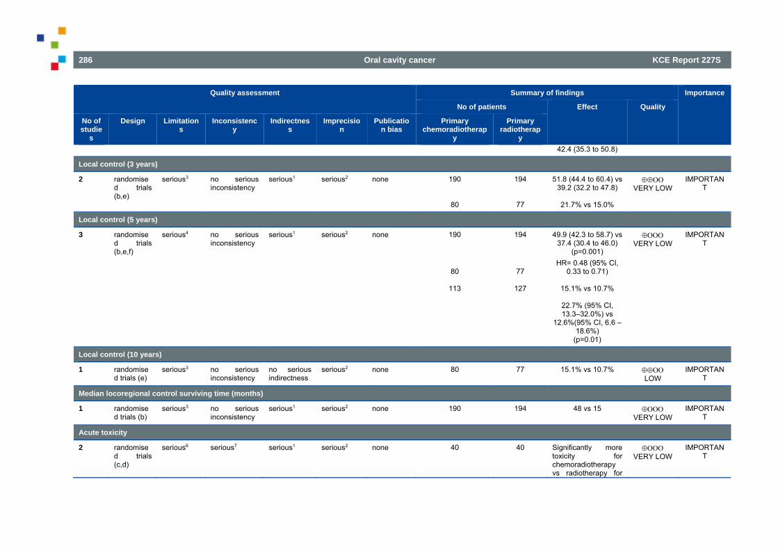
286 Oral cavity cancer KCE Report 227S
Quality assessment Summary of findings Importance
No of patients Effect Quality
No of studie
s
Design Limitations
Inconsistency
Indirectness
Imprecision
Publication bias
Primary chemoradiotherap
y
Primary radiotherap
y 42.4 (35.3 to 50.8)
Local control (3 years)
2 randomised trials (b,e)
serious3 no serious inconsistency
serious1 serious2 none 190
80
194
77
51.8 (44.4 to 60.4) vs 39.2 (32.2 to 47.8)
21.7% vs 15.0%
VERY LOW
IMPORTANT
Local control (5 years)
3 randomised trials (b,e,f)
serious4 no serious inconsistency
serious1 serious2 none 190
80
113
194
77
127
49.9 (42.3 to 58.7) vs 37.4 (30.4 to 46.0)
(p=0.001) HR= 0.48 (95% CI,
0.33 to 0.71)
15.1% vs 10.7%
22.7% (95% CI, 13.3–32.0%) vs
12.6%(95% CI, 6.6 –18.6%)
(p=0.01)
VERY LOW
IMPORTANT
Local control (10 years)
1 randomised trials (e)
serious3 no serious inconsistency
no serious indirectness
serious2 none 80 77 15.1% vs 10.7% LOW
IMPORTANT
Median locoregional control surviving time (months)
1 randomised trials (b)
serious3 no serious inconsistency
serious1 serious2 none 190 194 48 vs 15 VERY LOW
IMPORTANT
Acute toxicity
2 randomised trials (c,d)
serious6 serious7 serious1 serious2 none 40
40
Significantly more toxicity for chemoradiotherapy vs radiotherapy for
VERY LOW
IMPORTANT
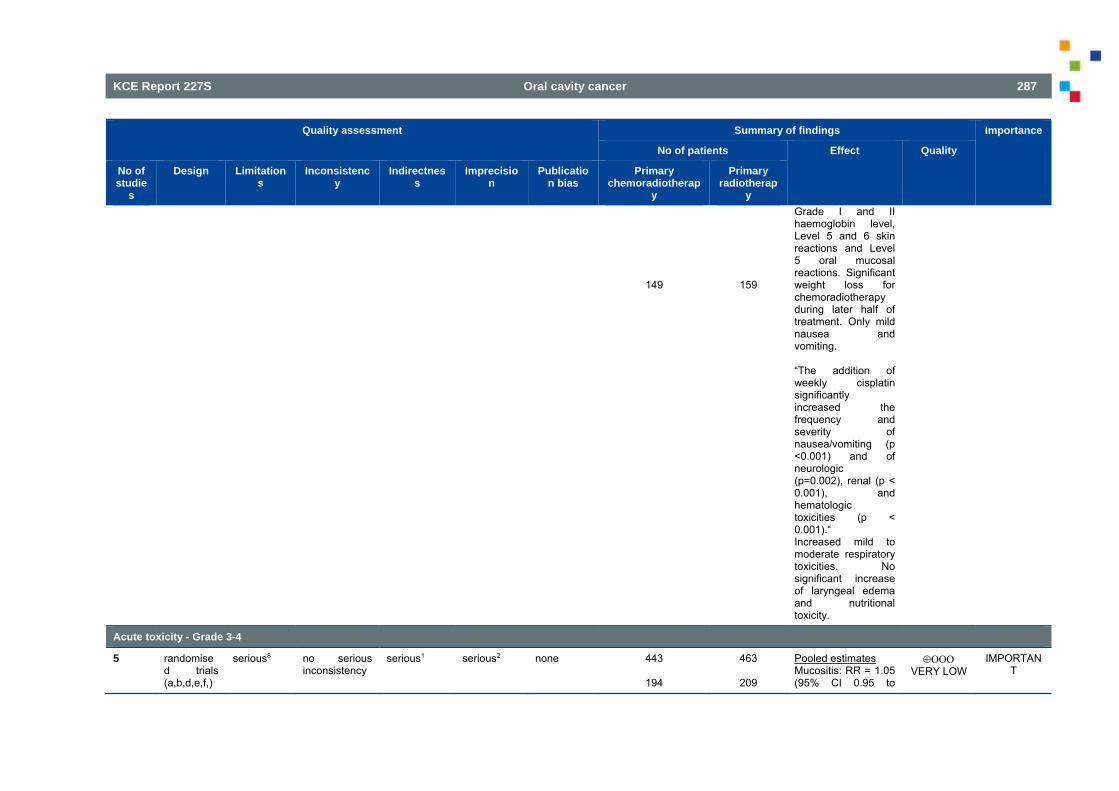
KCE Report 227S Oral cavity cancer 287
Quality assessment Summary of findings Importance
No of patients Effect Quality
No of studie
s
Design Limitations
Inconsistency
Indirectness
Imprecision
Publication bias
Primary chemoradiotherap
y
Primary radiotherap
y
149
159
Grade I and II haemoglobin level, Level 5 and 6 skin reactions and Level 5 oral mucosal reactions. Significant weight loss for chemoradiotherapy during later half of treatment. Only mild nausea and vomiting. “The addition of weekly cisplatin significantly increased the frequency and severity of nausea/vomiting (p <0.001) and of neurologic (p=0.002), renal (p < 0.001), and hematologic toxicities (p < 0.001).“ Increased mild to moderate respiratory toxicities. No significant increase of laryngeal edema and nutritional toxicity.
Acute toxicity - Grade 3-4
5 randomised trials (a,b,d,e,f,)
serious8 no serious inconsistency
serious1 serious2 none 443
194
463
209
Pooled estimates Mucositis: RR = 1.05 (95% CI 0.95 to
VERY LOW
IMPORTANT
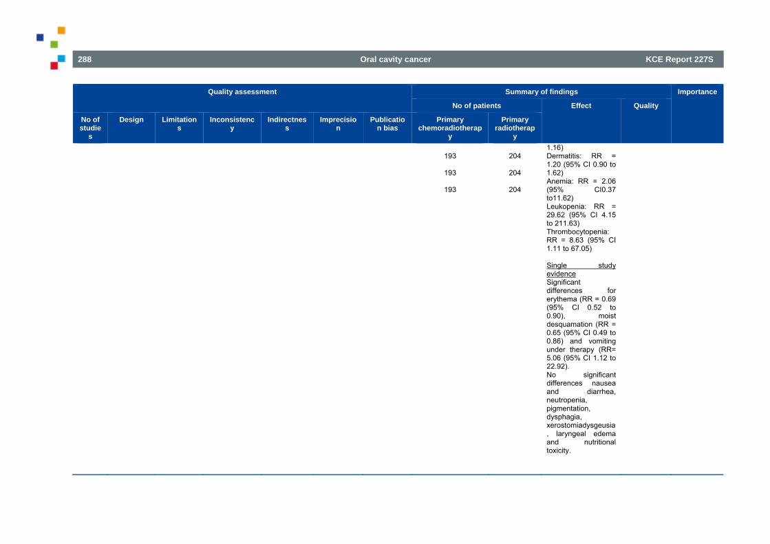
288 Oral cavity cancer KCE Report 227S
Quality assessment Summary of findings Importance
No of patients Effect Quality
No of studie
s
Design Limitations
Inconsistency
Indirectness
Imprecision
Publication bias
Primary chemoradiotherap
y
Primary radiotherap
y
193
193
193
204
204
204
1.16) Dermatitis: RR = 1.20 (95% CI 0.90 to 1.62) Anemia: RR = 2.06 (95% CI0.37 to11.62) Leukopenia: RR = 29.62 (95% CI 4.15 to 211.63) Thrombocytopenia: RR = 8.63 (95% CI 1.11 to 67.05) Single study evidence Significant differences for erythema (RR = 0.69 (95% CI 0.52 to 0.90), moist desquamation (RR = 0.65 (95% CI 0.49 to 0.86) and vomiting under therapy (RR= 5.06 (95% CI 1.12 to 22.92). No significant differences nausea and diarrhea, neutropenia, pigmentation, dysphagia, xerostomiadysgeusia, laryngeal edema and nutritional toxicity.
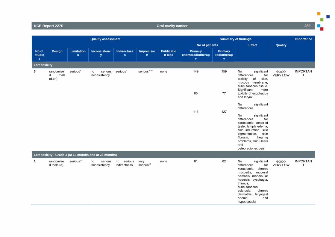
KCE Report 227S Oral cavity cancer 289
Quality assessment Summary of findings Importance
No of patients Effect Quality
No of studie
s
Design Limitations
Inconsistency
Indirectness
Imprecision
Publication bias
Primary chemoradiotherap
y
Primary radiotherap
y
Late toxicity
3 randomised trials (d,e,f)
serious9 no serious inconsistency
serious1 serious2,10 none 149
80
113
159
77
127
No significant differences for toxicity of skin, mucous membrane, subcutaneous tissue. Significant more toxicity of esophagus and larynx. No significant differences No significant differences for xerostomia, sense of taste, lymph edema, skin induration, skin pigmentation, skin fibrosis, hearing problems, skin ulcers and osteoradionecrosis.
VERY LOW
IMPORTANT
Late toxicity - Grade 3 (at 12 months and at 24 months)
1 randomised trials (a)
serious11 no serious inconsistency
no serious indirectness
very serious10
none 81 82 No significant differences for xerostomia, chronic mucositis, mucosal necrosis, mandibular necrosis, dysphagia, trismus, subcutaneous sclerosis, chronic dermatitis, laryngeal edema and hypoacousia.
VERY LOW
IMPORTANT
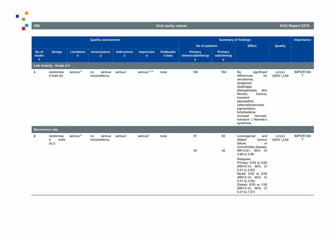
290 Oral cavity cancer KCE Report 227S
Quality assessment Summary of findings Importance
No of patients Effect Quality
No of studie
s
Design Limitations
Inconsistency
Indirectness
Imprecision
Publication bias
Primary chemoradiotherap
y
Primary radiotherap
y
Late toxicity - Grade 3-4
1 randomised trials (b)
serious12 no serious inconsistency
serious1 serious13,14 none 190 194 No significant differences for xerostomia, dysgeusia, dysphagia, telangiectasia, skin fibrosis, trismus, transient plexopathia, osteoradionecrosis, pigmentation, lymphedema, mucosal necrosis, transient L’Hermite’s syndrome.
VERY LOW
IMPORTANT
Recurrence rate
2 randomised trials (a,c)
serious14 no serious inconsistency
serious1 serious2 none 81
40
82
40
Locoregional and distant tumour failure, or uncontrolled disease: RR=0.81; 95% CI 0.68 to 0.96 Relapses: Primary: 0/30 vs 3/30 (RR=0.14; 95% CI 0.01 to 2.65) Nodal: 0/30 vs 3/30 (RR=0.14; 95% CI 0.01 to 2.65) Distant: 0/30 vs 1/30 (RR=0.33; 95% CI 0.01 to 7.87)
VERY LOW
IMPORTANT
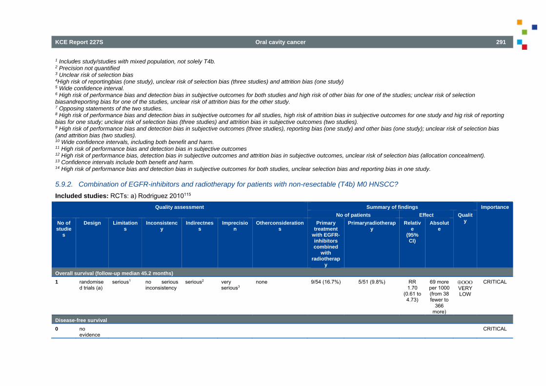
KCE Report 227S Oral cavity cancer 291
1 Includes study/studies with mixed population, not solely T4b. 2 Precision not quantified 3 Unclear risk of selection bias 4High risk of reportingbias (one study), unclear risk of selection bias (three studies) and attrition bias (one study) 5 Wide confidence interval. 6 High risk of performance bias and detection bias in subjective outcomes for both studies and high risk of other bias for one of the studies; unclear risk of selection biasandreporting bias for one of the studies, unclear risk of attrition bias for the other study. 7 Opposing statements of the two studies. 8 High risk of performance bias and detection bias in subjective outcomes for all studies, high risk of attrition bias in subjective outcomes for one study and hig risk of reporting bias for one study; unclear risk of selection bias (three studies) and attrition bias in subjective outcomes (two studies). 9 High risk of performance bias and detection bias in subjective outcomes (three studies), reporting bias (one study) and other bias (one study); unclear risk of selection bias (and attrition bias (two studies). 10 Wide confidence intervals, including both benefit and harm. 11 High risk of performance bias and detection bias in subjective outcomes 12 High risk of performance bias, detection bias in subjective outcomes and attrition bias in subjective outcomes, unclear risk of selection bias (allocation concealment). 13 Confidence intervals include both benefit and harm. 14 High risk of performance bias and detection bias in subjective outcomes for both studies, unclear selection bias and reporting bias in one study.
5.9.2. Combination of EGFR-inhibitors and radiotherapy for patients with non-resectable (T4b) M0 HNSCC? Included studies: RCTs: a) Rodriguez 2010115
Quality assessment Summary of findings Importance No of patients Effect Qualit
y No of studie
s
Design Limitations
Inconsistency
Indirectness
Imprecision
Otherconsiderations
Primary treatment
with EGFR-inhibitors combined
with radiotherap
y
Primaryradiotherapy
Relative
(95% CI)
Absolute
Overall survival (follow-up median 45.2 months) 1 randomise
d trials (a) serious1 no serious
inconsistency serious2 very
serious3 none 9/54 (16.7%) 5/51 (9.8%) RR
1.70 (0.61 to 4.73)
69 more per 1000 (from 38 fewer to
366 more)
VERY LOW
CRITICAL
Disease-free survival 0 no
evidence CRITICAL
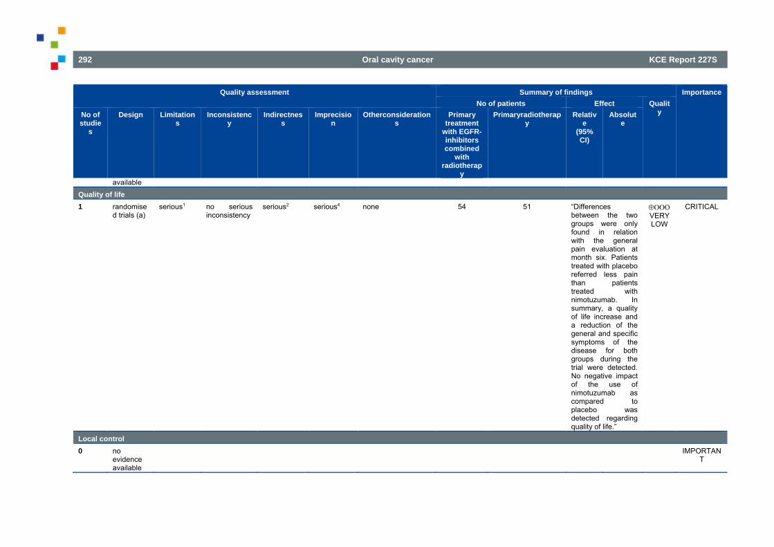
292 Oral cavity cancer KCE Report 227S
Quality assessment Summary of findings Importance No of patients Effect Qualit
y No of studie
s
Design Limitations
Inconsistency
Indirectness
Imprecision
Otherconsiderations
Primary treatment
with EGFR-inhibitors combined
with radiotherap
y
Primaryradiotherapy
Relative
(95% CI)
Absolute
available Quality of life 1 randomise
d trials (a) serious1 no serious
inconsistency serious2 serious4 none 54 51 “Differences
between the two groups were only found in relation with the general pain evaluation at month six. Patients treated with placebo referred less pain than patients treated with nimotuzumab. In summary, a quality of life increase and a reduction of the general and specific symptoms of the disease for both groups during the trial were detected. No negative impact of the use of nimotuzumab as compared to placebo was detected regarding quality of life.”
VERY LOW
CRITICAL
Local control 0 no
evidence available
IMPORTANT

KCE Report 227S Oral cavity cancer 293
Quality assessment Summary of findings Importance No of patients Effect Qualit
y No of studie
s
Design Limitations
Inconsistency
Indirectness
Imprecision
Otherconsiderations
Primary treatment
with EGFR-inhibitors combined
with radiotherap
y
Primaryradiotherapy
Relative
(95% CI)
Absolute
Adverse events (Common Toxicity Criteria of the US National Cancer Institute, Version 2, April 30, 1999 (NCI-CTC, Version 2)) 1 randomise
d trials (a) serious1 no serious
inconsistency serious2 serious5 none 38/54
(70.4%) 30/52 (57.7%) RR
1.22 (0.91 to 1.63)
127 more per
1000 (from 52 fewer to
363 more)
VERY LOW
IMPORTANT
Recurrencerate 0 no
evidence available
IMPORTANT
1 Unclear risk of selection, attrition and reporting bias 2 Population consists of grade III/IV HNSCC; unclear how many T4b patients. 3 Large confidence interval, includes both benefit and harm. 4 No quantification of results 5 Confidence interval includes both benefit and harm.
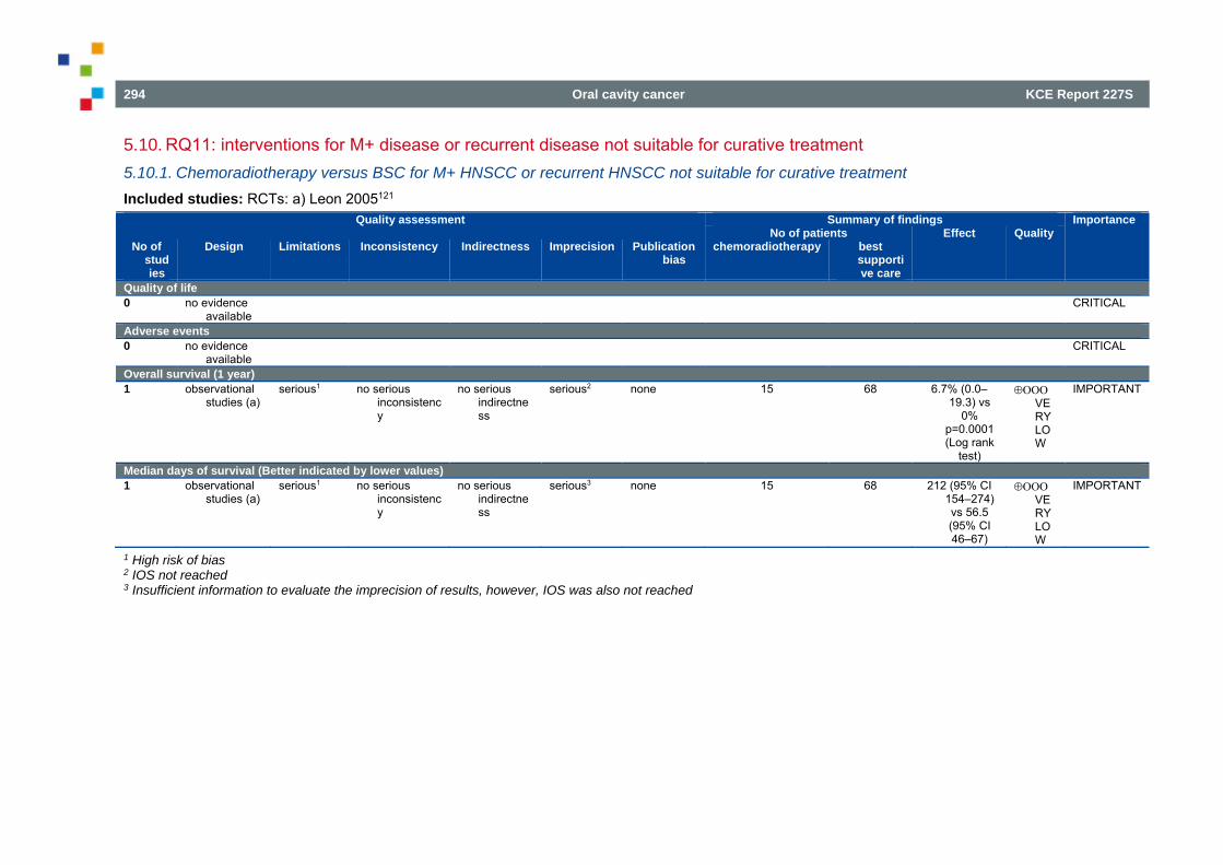
294 Oral cavity cancer KCE Report 227S
5.10. RQ11: interventions for M+ disease or recurrent disease not suitable for curative treatment 5.10.1. Chemoradiotherapy versus BSC for M+ HNSCC or recurrent HNSCC not suitable for curative treatment Included studies: RCTs: a) Leon 2005121
Quality assessment Summary of findings Importance No of patients Effect Quality
No of studies
Design Limitations Inconsistency Indirectness Imprecision Publication bias
chemoradiotherapy best supportive care
Quality of life 0 no evidence
available CRITICAL
Adverse events 0 no evidence
available CRITICAL
Overall survival (1 year) 1 observational
studies (a) serious1 no serious
inconsistency
no serious indirectness
serious2 none 15 68 6.7% (0.0–19.3) vs
0% p=0.0001 (Log rank
test)
VERY LOW
IMPORTANT
Median days of survival (Better indicated by lower values) 1 observational
studies (a) serious1 no serious
inconsistency
no serious indirectness
serious3 none 15 68 212 (95% CI 154–274) vs 56.5 (95% CI 46–67)
VERY LOW
IMPORTANT
1 High risk of bias 2 IOS not reached 3 Insufficient information to evaluate the imprecision of results, however, IOS was also not reached
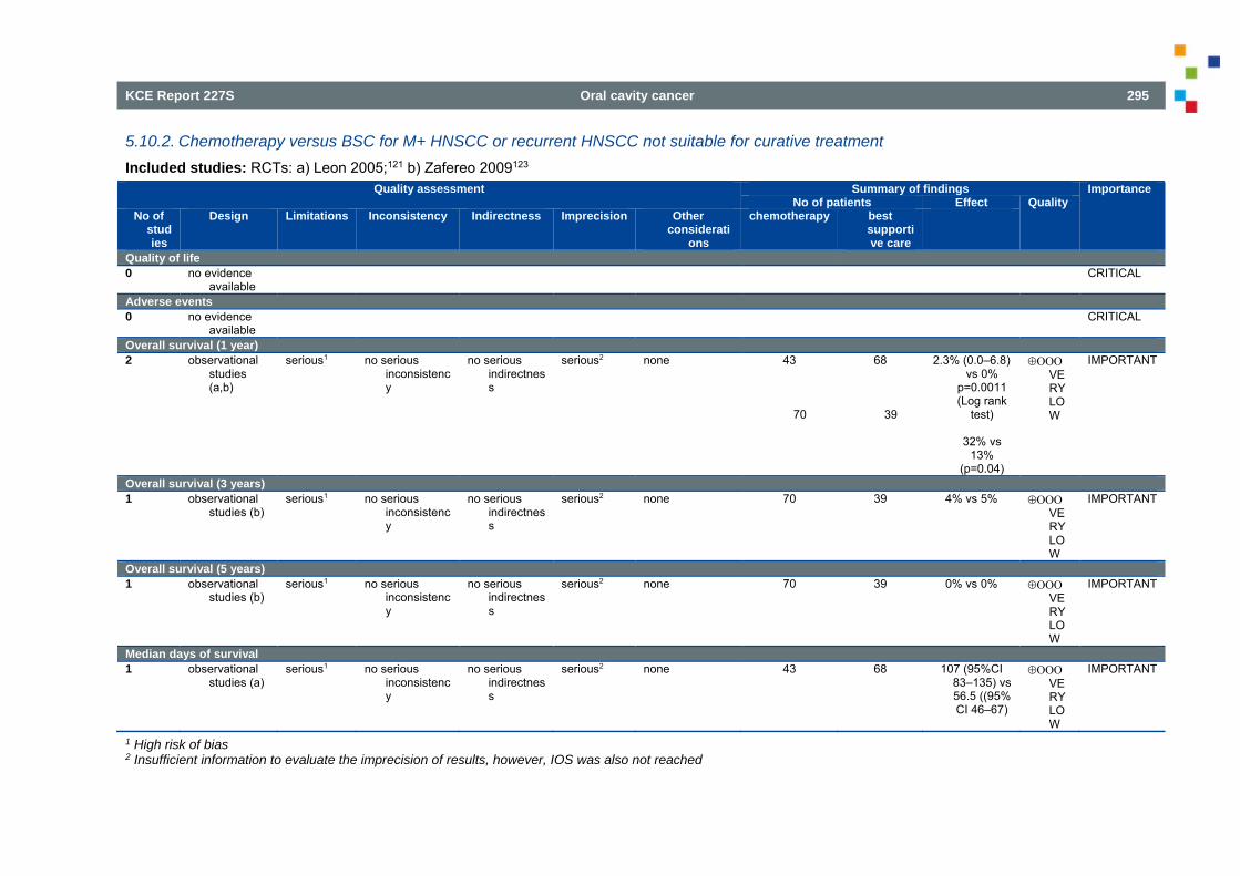
KCE Report 227S Oral cavity cancer 295
5.10.2. Chemotherapy versus BSC for M+ HNSCC or recurrent HNSCC not suitable for curative treatment Included studies: RCTs: a) Leon 2005;121 b) Zafereo 2009123
Quality assessment Summary of findings Importance No of patients Effect Quality
No of studies
Design Limitations Inconsistency Indirectness Imprecision Other considerati
ons
chemotherapy best supportive care
Quality of life 0 no evidence
available CRITICAL
Adverse events 0 no evidence
available CRITICAL
Overall survival (1 year) 2 observational
studies (a,b)
serious1 no serious inconsistency
no serious indirectness
serious2 none 43
70
68
39
2.3% (0.0–6.8) vs 0%
p=0.0011 (Log rank
test)
32% vs 13%
(p=0.04)
VERY LOW
IMPORTANT
Overall survival (3 years) 1 observational
studies (b) serious1 no serious
inconsistency
no serious indirectness
serious2 none 70 39 4% vs 5% VERY LOW
IMPORTANT
Overall survival (5 years) 1 observational
studies (b) serious1 no serious
inconsistency
no serious indirectness
serious2 none 70 39 0% vs 0% VERY LOW
IMPORTANT
Median days of survival 1 observational
studies (a) serious1 no serious
inconsistency
no serious indirectness
serious2 none 43 68 107 (95%CI 83–135) vs 56.5 ((95% CI 46–67)
VERY LOW
IMPORTANT
1 High risk of bias 2 Insufficient information to evaluate the imprecision of results, however, IOS was also not reached
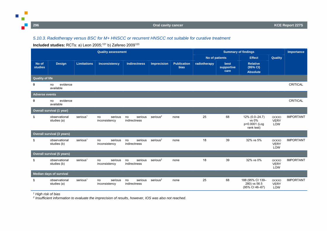
296 Oral cavity cancer KCE Report 227S
5.10.3. Radiotherapy versus BSC for M+ HNSCC or recurrent HNSCC not suitable for curative treatment Included studies: RCTs: a) Leon 2005;121 b) Zafereo 2009123
Quality assessment Summary of findings Importance
No of patients Effect Quality
No of studies
Design Limitations Inconsistency Indirectness Imprecision Publication bias
radiotherapy best supportive
care
Relative (95% CI) Absolute
Quality of life
0 no evidence available
CRITICAL
Adverse events
0 no evidence available
CRITICAL
Overall survival (1 year)
1 observational studies (a)
serious1 no serious inconsistency
no serious indirectness
serious2 none 25
68
12% (0.0–24.7) vs 0%
p=0.0001 (Log rank test)
VERY LOW
IMPORTANT
Overall survival (3 years)
1 observational studies (b)
serious1 no serious inconsistency
no serious indirectness
serious2 none 18 39 32% vs 5% VERY LOW
IMPORTANT
Overall survival (5 years)
1 observational studies (b)
serious1 no serious inconsistency
no serious indirectness
serious2 none 18 39 32% vs 0% VERY LOW
IMPORTANT
Median days of survival
1 observational studies (a)
serious1 no serious inconsistency
no serious indirectness
serious2 none 25 68 188 (95% CI 139–280) vs 56.5
(95% CI 46–67)
VERY LOW
IMPORTANT
1 High risk of bias 2 Insufficient information to evaluate the imprecision of results, however, IOS was also not reached.
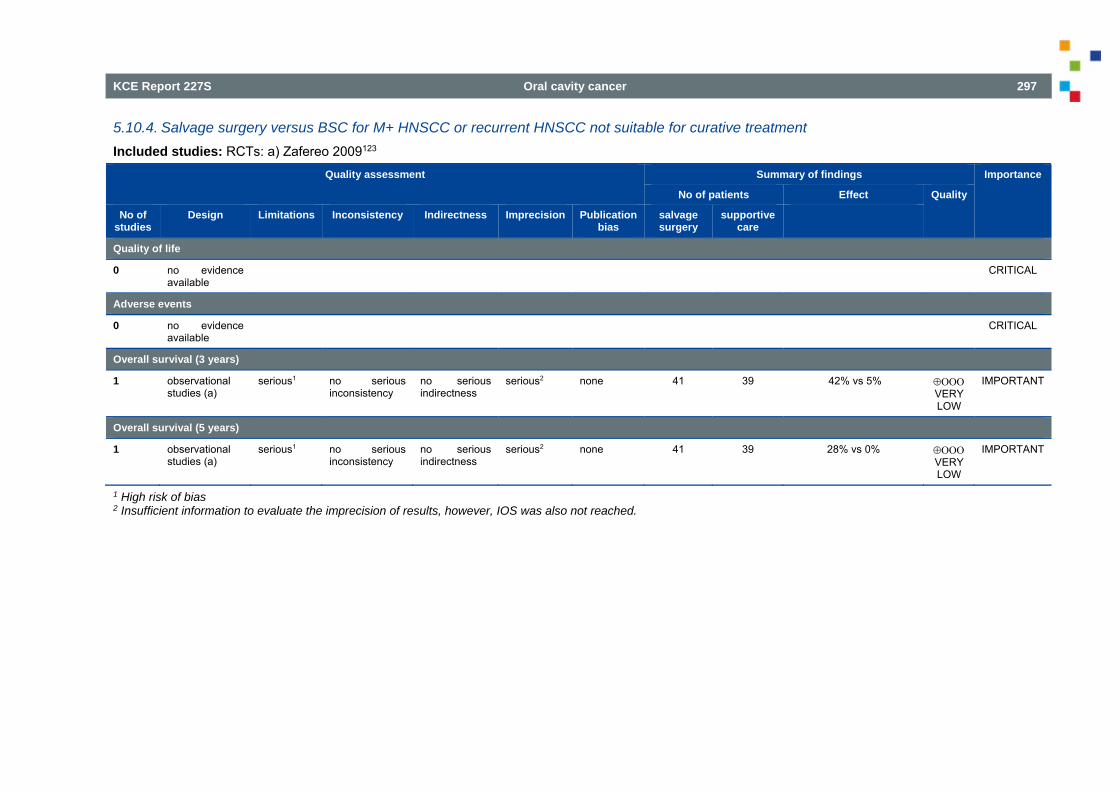
KCE Report 227S Oral cavity cancer 297
5.10.4. Salvage surgery versus BSC for M+ HNSCC or recurrent HNSCC not suitable for curative treatment Included studies: RCTs: a) Zafereo 2009123
Quality assessment Summary of findings Importance
No of patients Effect Quality
No of studies
Design Limitations Inconsistency Indirectness Imprecision Publication bias
salvage surgery
supportive care
Quality of life
0 no evidence available
CRITICAL
Adverse events
0 no evidence available
CRITICAL
Overall survival (3 years)
1 observational studies (a)
serious1 no serious inconsistency
no serious indirectness
serious2 none 41 39 42% vs 5% VERY LOW
IMPORTANT
Overall survival (5 years)
1 observational studies (a)
serious1 no serious inconsistency
no serious indirectness
serious2 none 41 39 28% vs 0% VERY LOW
IMPORTANT
1 High risk of bias 2 Insufficient information to evaluate the imprecision of results, however, IOS was also not reached.
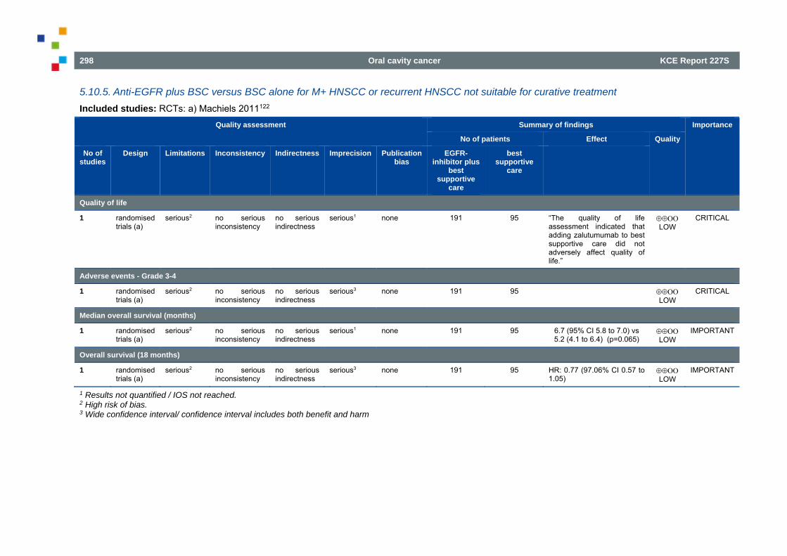
298 Oral cavity cancer KCE Report 227S
5.10.5. Anti-EGFR plus BSC versus BSC alone for M+ HNSCC or recurrent HNSCC not suitable for curative treatment Included studies: RCTs: a) Machiels 2011122
Quality assessment Summary of findings Importance
No of patients Effect Quality
No of studies
Design Limitations Inconsistency Indirectness Imprecision Publication bias
EGFR-inhibitor plus
best supportive
care
best supportive
care
Quality of life
1 randomised trials (a)
serious2 no serious inconsistency
no serious indirectness
serious1 none 191 95 “The quality of life assessment indicated that adding zalutumumab to best supportive care did not adversely affect quality of life.”
LOW
CRITICAL
Adverse events - Grade 3-4
1 randomised trials (a)
serious2 no serious inconsistency
no serious indirectness
serious3 none 191 95 LOW
CRITICAL
Median overall survival (months)
1 randomised trials (a)
serious2 no serious inconsistency
no serious indirectness
serious1 none 191 95 6.7 (95% CI 5.8 to 7.0) vs 5.2 (4.1 to 6.4) (p=0.065)
LOW
IMPORTANT
Overall survival (18 months)
1 randomised trials (a)
serious2 no serious inconsistency
no serious indirectness
serious3 none 191 95 HR: 0.77 (97.06% CI 0.57 to 1.05)
LOW
IMPORTANT
1 Results not quantified / IOS not reached. 2 High risk of bias. 3 Wide confidence interval/ confidence interval includes both benefit and harm
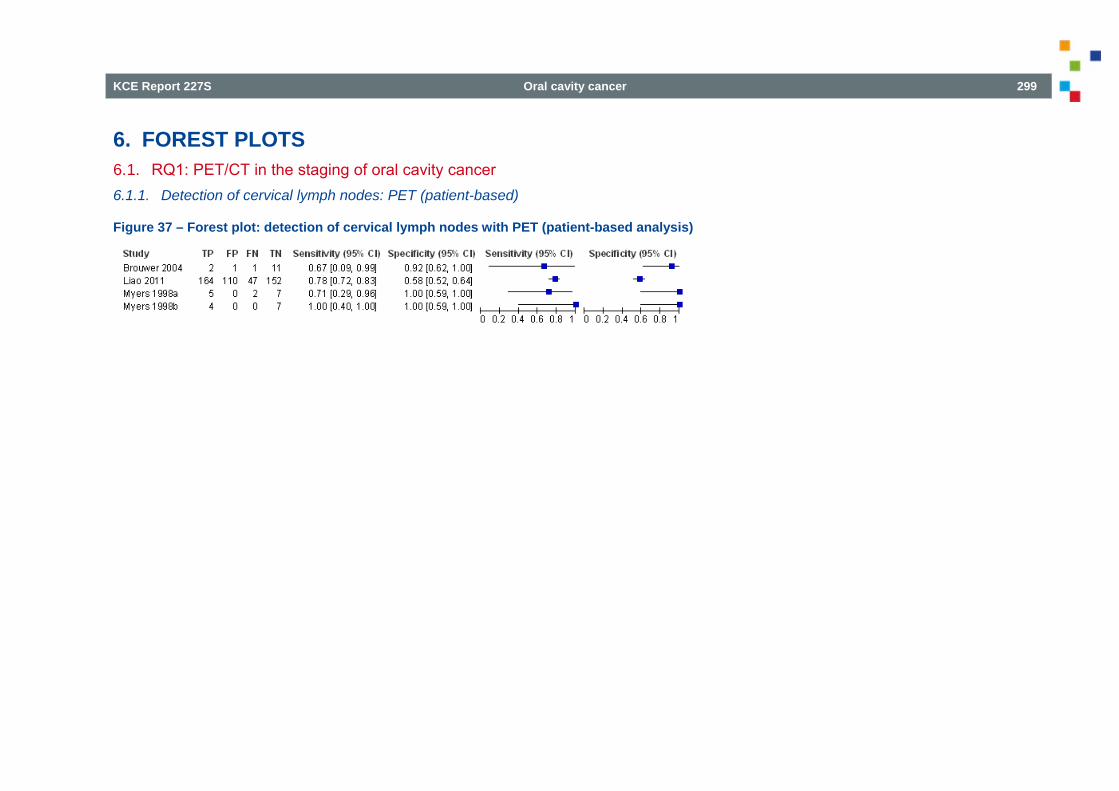
KCE Report 227S Oral cavity cancer 299
6. FOREST PLOTS 6.1. RQ1: PET/CT in the staging of oral cavity cancer 6.1.1. Detection of cervical lymph nodes: PET (patient-based)
Figure 37 – Forest plot: detection of cervical lymph nodes with PET (patient-based analysis)
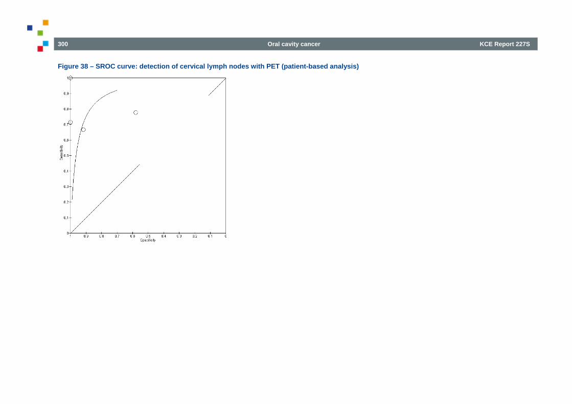
300 Oral cavity cancer KCE Report 227S
Figure 38 – SROC curve: detection of cervical lymph nodes with PET (patient-based analysis)
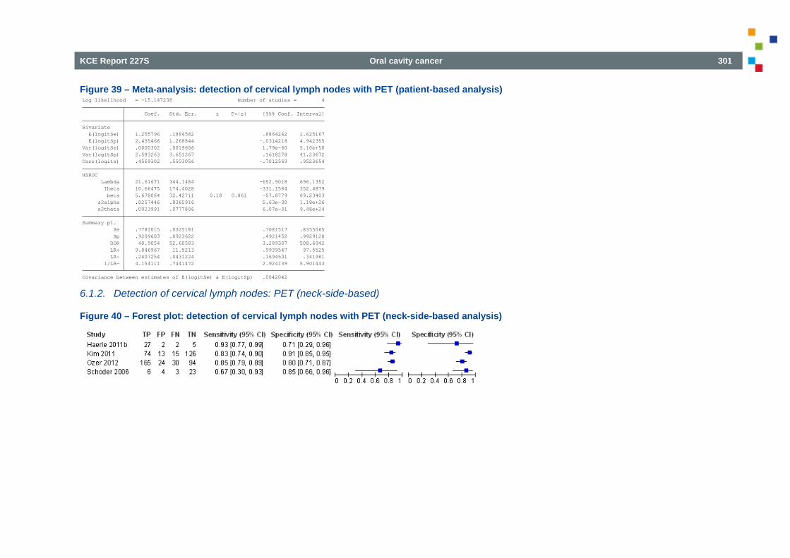
KCE Report 227S Oral cavity cancer 301
Figure 39 – Meta-analysis: detection of cervical lymph nodes with PET (patient-based analysis)
6.1.2. Detection of cervical lymph nodes: PET (neck-side-based)
Figure 40 – Forest plot: detection of cervical lymph nodes with PET (neck-side-based analysis)
Covariance between estimates of E(logitSe) & E(logitSp) .0042042 1/LR- 4.154111 .7441472 2.924139 5.901443 LR- .2407254 .0431224 .1694501 .341981 LR+ 9.846967 11.5213 .9939547 97.5525 DOR 40.9054 52.60583 3.289307 508.6942 Sp .9209603 .0923622 .4921452 .9929128 Se .7783015 .0325181 .7081517 .8355065Summary pt. s2theta .0023991 .0777806 6.07e-31 9.48e+24 s2alpha .0257446 .8365916 5.63e-30 1.18e+26 beta 5.678064 32.42711 0.18 0.861 -57.8779 69.23403 Theta 10.66475 174.4028 -331.1584 352.4879 Lambda 21.61671 344.1484 -652.9018 696.1352HSROC Corr(logits) .4569302 .5503056 -.7012569 .9523654Var(logitSp) 2.583263 3.651267 .1618278 41.23672Var(logitSe) .0000302 .0019606 1.79e-60 5.10e+50 E(logitSp) 2.455466 1.268844 -.0314218 4.942355 E(logitSe) 1.255796 .1884582 .8864242 1.625167Bivariate Coef. Std. Err. z P>|z| [95% Conf. Interval] Log likelihood = -15.147238 Number of studies = 4
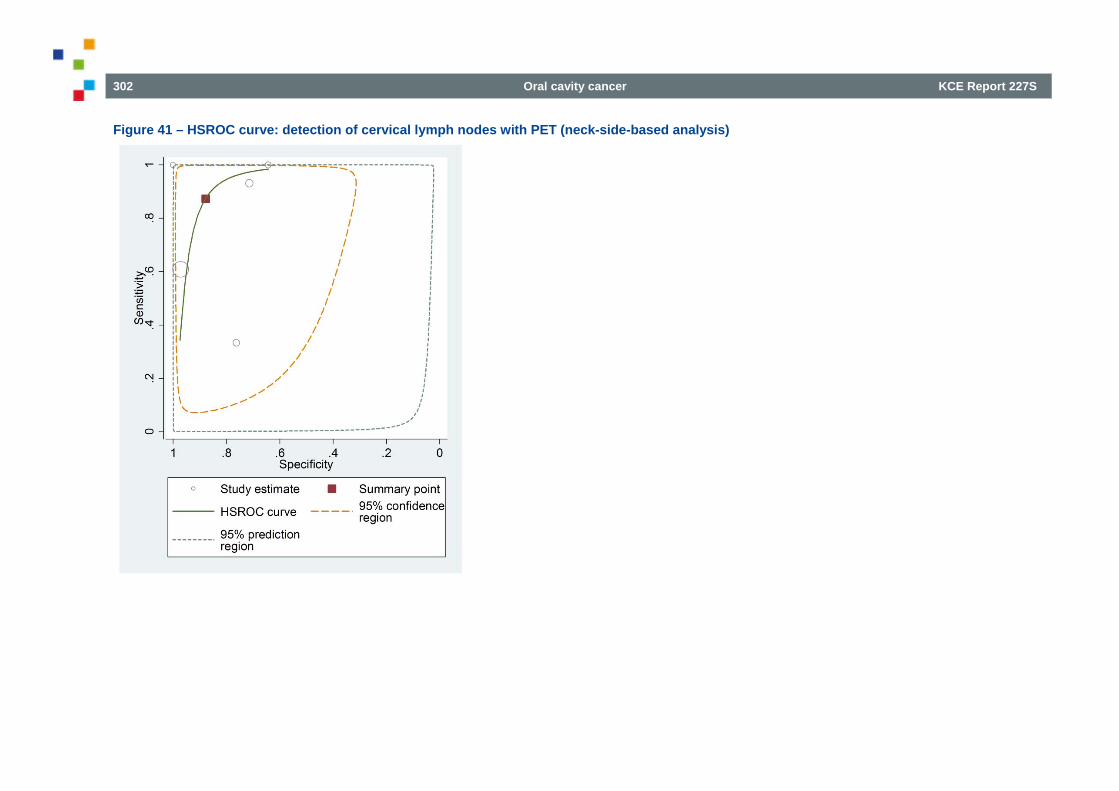
302 Oral cavity cancer KCE Report 227S
Figure 41 – HSROC curve: detection of cervical lymph nodes with PET (neck-side-based analysis)
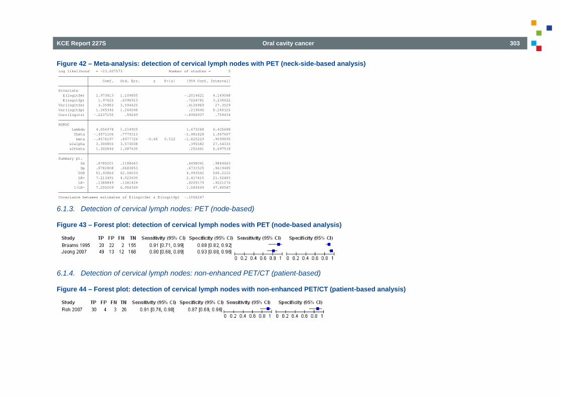
KCE Report 227S Oral cavity cancer 303
Figure 42 – Meta-analysis: detection of cervical lymph nodes with PET (neck-side-based analysis)
6.1.3. Detection of cervical lymph nodes: PET (node-based)
Figure 43 – Forest plot: detection of cervical lymph nodes with PET (node-based analysis)
6.1.4. Detection of cervical lymph nodes: non-enhanced PET/CT (patient-based)
Figure 44 – Forest plot: detection of cervical lymph nodes with non-enhanced PET/CT (patient-based analysis)
Covariance between estimates of E(logitSe) & E(logitSp) -.1066247 1/LR- 7.200209 6.954349 1.084449 47.80587 LR- .1388849 .1341424 .0209179 .9221276 LR+ 7.213491 4.023635 2.417415 21.52483 DOR 51.93864 62.06033 4.993542 540.2222 Sp .8782808 .0683853 .6731525 .9619485 Se .8780201 .1188663 .4498041 .9844663Summary pt. s2theta 1.300846 1.087635 .252661 6.697518 s2alpha 3.300855 3.573038 .395582 27.54333 beta -.4576197 .6977726 -0.66 0.512 -1.825229 .9099895 Theta -.4571106 .7779313 -1.981828 1.067607 Lambda 4.054478 1.214925 1.673268 6.435688HSROC Corr(logits) -.2237155 .59249 -.8956937 .759434Var(logitSp) 1.345346 1.244048 .219646 8.240326Var(logitSe) 3.35983 3.594625 .4126969 27.3529 E(logitSp) 1.97625 .6396913 .7224781 3.230022 E(logitSe) 1.973813 1.109855 -.2014621 4.149088Bivariate Coef. Std. Err. z P>|z| [95% Conf. Interval] Log likelihood = -23.607573 Number of studies = 5
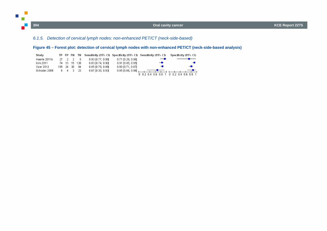
304 Oral cavity cancer KCE Report 227S
6.1.5. Detection of cervical lymph nodes: non-enhanced PET/CT (neck-side-based)
Figure 45 – Forest plot: detection of cervical lymph nodes with non-enhanced PET/CT (neck-side-based analysis)
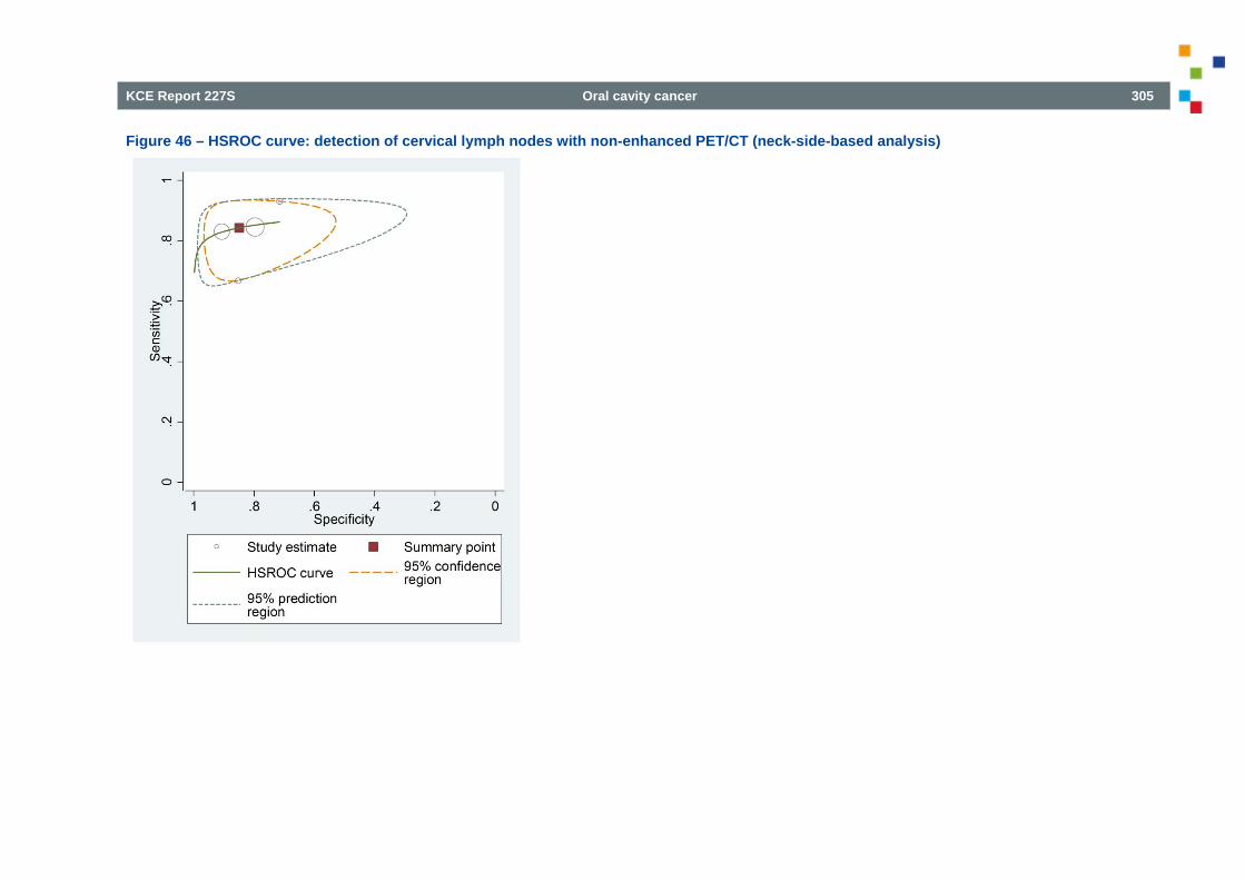
KCE Report 227S Oral cavity cancer 305
Figure 46 – HSROC curve: detection of cervical lymph nodes with non-enhanced PET/CT (neck-side-based analysis)
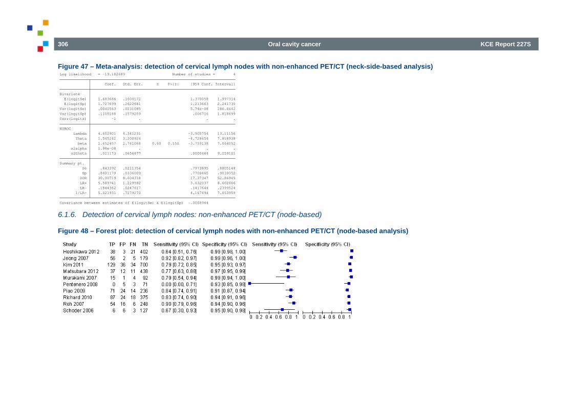
306 Oral cavity cancer KCE Report 227S
Figure 47 – Meta-analysis: detection of cervical lymph nodes with non-enhanced PET/CT (neck-side-based analysis)
6.1.6. Detection of cervical lymph nodes: non-enhanced PET/CT (node-based)
Figure 48 – Forest plot: detection of cervical lymph nodes with non-enhanced PET/CT (node-based analysis)
Covariance between estimates of E(logitSe) & E(logitSp) -.0068944 1/LR- 5.421931 .7279273 4.167494 7.053959 LR- .1844362 .0247617 .1417644 .2399524 LR+ 5.589741 1.229592 3.632037 8.602666 DOR 30.30719 8.604318 17.37347 52.86945 Sp .8491179 .0336009 .7709465 .9039352 Se .843392 .0211354 .7973895 .8805148Summary pt. s2theta .021173 .0656877 .0000484 9.259101 s2alpha 1.98e-08 . . . beta 1.652457 2.761068 0.60 0.550 -3.759138 7.064052 Theta 1.545242 3.200924 -4.728454 7.818938 Lambda 4.602901 4.341231 -3.905754 13.11156HSROC Corr(logits) -1 . . .Var(logitSp) .1105188 .1579259 .006716 1.818699Var(logitSe) .0040563 .0231085 5.74e-08 286.6642 E(logitSp) 1.727699 .2622681 1.213663 2.241735 E(logitSe) 1.683686 .1600172 1.370058 1.997314Bivariate Coef. Std. Err. z P>|z| [95% Conf. Interval] Log likelihood = -19.182689 Number of studies = 4
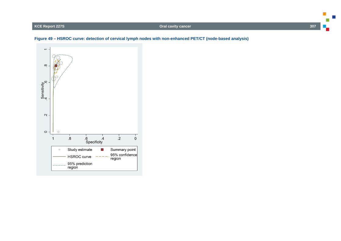
KCE Report 227S Oral cavity cancer 307
Figure 49 – HSROC curve: detection of cervical lymph nodes with non-enhanced PET/CT (node-based analysis)
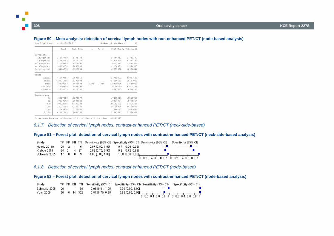
308 Oral cavity cancer KCE Report 227S
Figure 50 – Meta-analysis: detection of cervical lymph nodes with non-enhanced PET/CT (node-based analysis)
6.1.7. Detection of cervical lymph nodes: contrast-enhanced PET/CT (neck-side-based)
Figure 51 – Forest plot: detection of cervical lymph nodes with contrast-enhanced PET/CT (neck-side-based analysis)
6.1.8. Detection of cervical lymph nodes: contrast-enhanced PET/CT (node-based)
Figure 52 – Forest plot: detection of cervical lymph nodes with contrast-enhanced PET/CT (node-based analysis)
Covariance between estimates of E(logitSe) & E(logitSp) -.0141577 1/LR- 4.887743 .6663769 3.741612 6.384958 LR- .2045934 .0278935 .1566181 .2672645 LR+ 22.27114 5.122355 14.18948 34.95574 DOR 108.8556 27.35334 66.52122 178.1318 Sp .9639542 .0086146 .9426934 .9775154 Se .8027813 .0274177 .7435213 .8510914Summary pt. s2theta .1958703 .1213741 .0581445 .6598253 s2alpha .2500825 .3538293 .0156229 4.003186 beta .5330183 .5688884 0.94 0.349 -.5819826 1.648019 Theta -.3424756 .6398974 -1.596651 .9117003 Lambda 4.349911 .2896519 3.782203 4.917618HSROC Corr(logits) -.5160773 .5316181 -.9633992 .6906644Var(logitSp) .4403158 .2863248 .1230983 1.574985Var(logitSe) .1516319 .1519995 .0212581 1.081573 E(logitSp) 3.286253 .2479272 2.800325 3.772182 E(logitSe) 1.403769 .1731753 1.064352 1.743187Bivariate Coef. Std. Err. z P>|z| [95% Conf. Interval] Log likelihood = -62.901803 Number of studies = 10
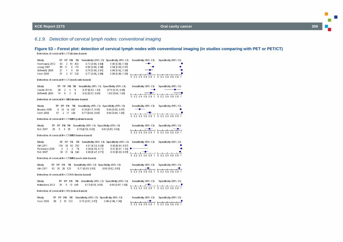
KCE Report 227S Oral cavity cancer 309
6.1.9. Detection of cervical lymph nodes: conventional imaging
Figure 53 – Forest plot: detection of cervical lymph nodes with conventional imaging (in studies comparing with PET or PET/CT)
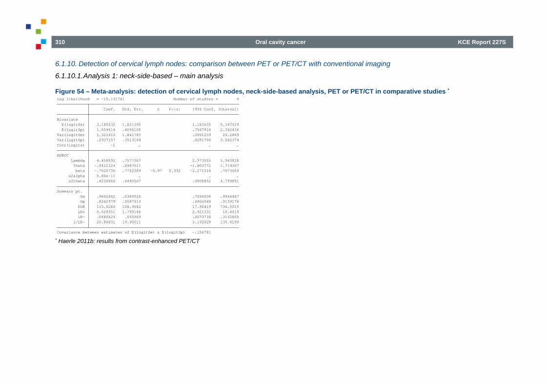
310 Oral cavity cancer KCE Report 227S
6.1.10. Detection of cervical lymph nodes: comparison between PET or PET/CT with conventional imaging
6.1.10.1. Analysis 1: neck-side-based – main analysis
Figure 54 – Meta-analysis: detection of cervical lymph nodes, neck-side-based analysis, PET or PET/CT in comparative studies *
* Haerle 2011b: results from contrast-enhanced PET/CT Covariance between estimates of E(logitSe) & E(logitSp) -.156781 1/LR- 20.80631 19.90011 3.192028 135.6199 LR- .0480624 .045969 .0073736 .3132805 LR+ 5.528351 1.799146 2.921331 10.4619 DOR 115.0246 108.9682 17.96419 736.5015 Sp .8262979 .0587913 .6806568 .9139176 Se .9602862 .0389526 .7656008 .9944447Summary pt. s2theta .6230866 .6490567 .0808852 4.799851 s2alpha 5.80e-11 . . . beta -.7520735 .7752389 -0.97 0.332 -2.271514 .7673669 Theta -.0422324 .8987611 -1.803772 1.719307 Lambda 4.458692 .7577367 2.973555 5.943828HSROC Corr(logits) -1 . . .Var(logitSp) .2937157 .3513164 .0281706 3.062374Var(logitSe) 1.321812 1.841785 .0861239 20.2869 E(logitSp) 1.559614 .4096105 .7567918 2.362436 E(logitSe) 3.185532 1.021395 1.183635 5.187429Bivariate Coef. Std. Err. z P>|z| [95% Conf. Interval] Log likelihood = -15.131761 Number of studies = 4
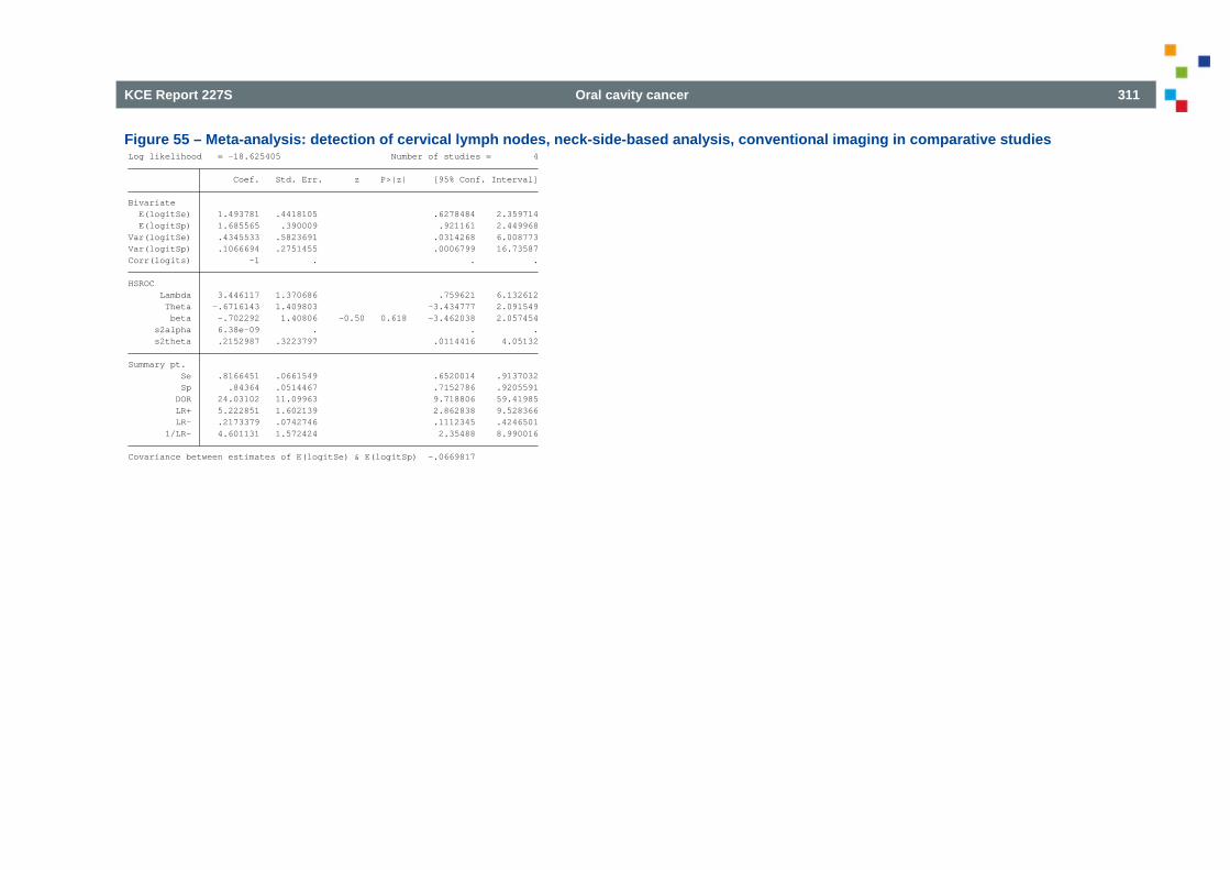
KCE Report 227S Oral cavity cancer 311
Figure 55 – Meta-analysis: detection of cervical lymph nodes, neck-side-based analysis, conventional imaging in comparative studies
Covariance between estimates of E(logitSe) & E(logitSp) -.0669817 1/LR- 4.601131 1.572424 2.35488 8.990016 LR- .2173379 .0742746 .1112345 .4246501 LR+ 5.222851 1.602139 2.862838 9.528366 DOR 24.03102 11.09963 9.718806 59.41985 Sp .84364 .0514467 .7152786 .9205591 Se .8166451 .0661549 .6520014 .9137032Summary pt. s2theta .2152987 .3223797 .0114416 4.05132 s2alpha 6.38e-09 . . . beta -.702292 1.40806 -0.50 0.618 -3.462038 2.057454 Theta -.6716143 1.409803 -3.434777 2.091549 Lambda 3.446117 1.370686 .759621 6.132612HSROC Corr(logits) -1 . . .Var(logitSp) .1066694 .2751455 .0006799 16.73587Var(logitSe) .4345533 .5823691 .0314268 6.008773 E(logitSp) 1.685565 .390009 .921161 2.449968 E(logitSe) 1.493781 .4418105 .6278484 2.359714Bivariate Coef. Std. Err. z P>|z| [95% Conf. Interval] Log likelihood = -18.625405 Number of studies = 4
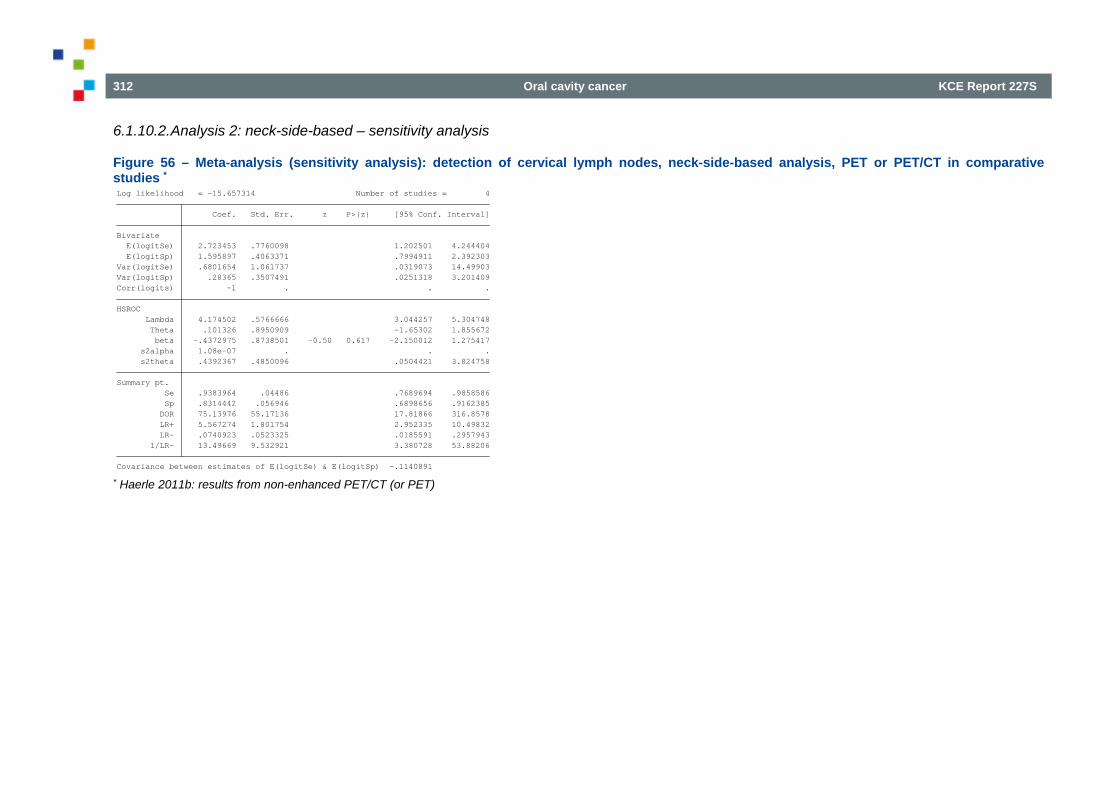
312 Oral cavity cancer KCE Report 227S
6.1.10.2. Analysis 2: neck-side-based – sensitivity analysis
Figure 56 – Meta-analysis (sensitivity analysis): detection of cervical lymph nodes, neck-side-based analysis, PET or PET/CT in comparative studies *
* Haerle 2011b: results from non-enhanced PET/CT (or PET) Covariance between estimates of E(logitSe) & E(logitSp) -.1140891 1/LR- 13.49669 9.532921 3.380728 53.88206 LR- .0740923 .0523325 .0185591 .2957943 LR+ 5.567274 1.801754 2.952335 10.49832 DOR 75.13976 55.17136 17.81866 316.8578 Sp .8314442 .056946 .6898656 .9162385 Se .9383964 .04486 .7689694 .9858586Summary pt. s2theta .4392367 .4850096 .0504421 3.824758 s2alpha 1.08e-07 . . . beta -.4372975 .8738501 -0.50 0.617 -2.150012 1.275417 Theta .101326 .8950909 -1.65302 1.855672 Lambda 4.174502 .5766666 3.044257 5.304748HSROC Corr(logits) -1 . . .Var(logitSp) .28365 .3507491 .0251318 3.201409Var(logitSe) .6801654 1.061737 .0319073 14.49903 E(logitSp) 1.595897 .4063371 .7994911 2.392303 E(logitSe) 2.723453 .7760098 1.202501 4.244404Bivariate Coef. Std. Err. z P>|z| [95% Conf. Interval] Log likelihood = -15.657314 Number of studies = 4
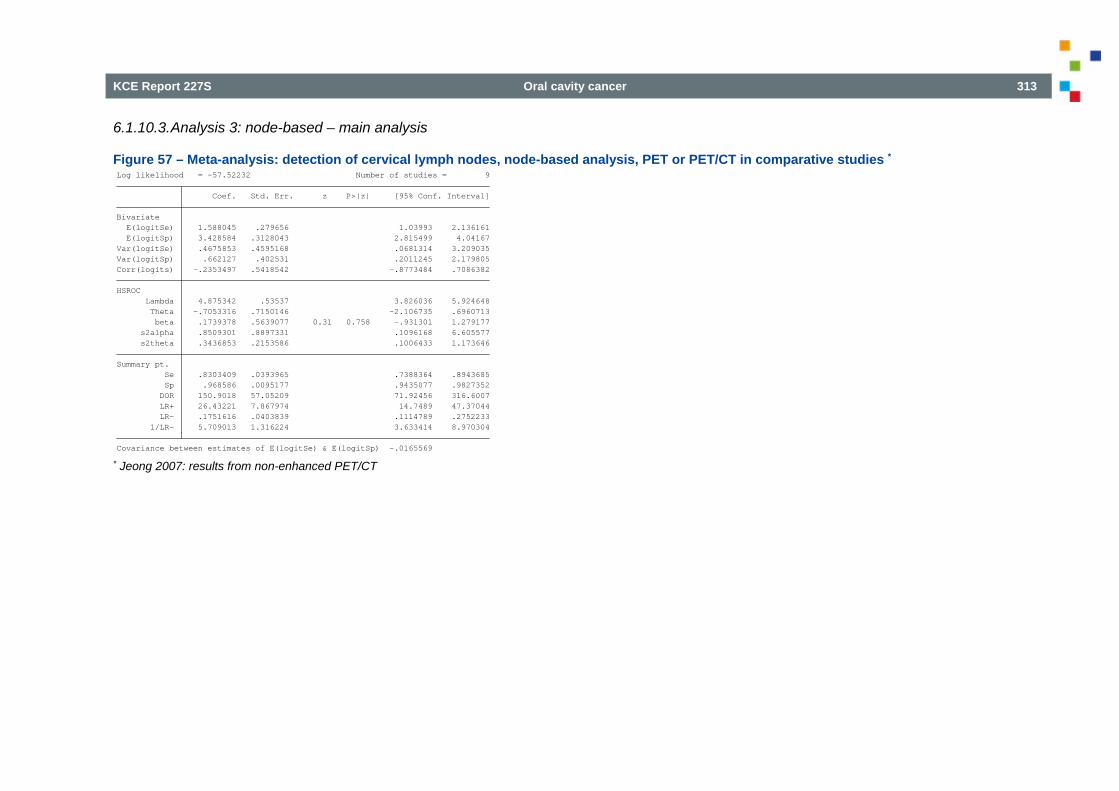
KCE Report 227S Oral cavity cancer 313
6.1.10.3. Analysis 3: node-based – main analysis
Figure 57 – Meta-analysis: detection of cervical lymph nodes, node-based analysis, PET or PET/CT in comparative studies *
* Jeong 2007: results from non-enhanced PET/CT Covariance between estimates of E(logitSe) & E(logitSp) -.0165569 1/LR- 5.709013 1.316224 3.633414 8.970304 LR- .1751616 .0403839 .1114789 .2752233 LR+ 26.43221 7.867974 14.7489 47.37044 DOR 150.9018 57.05209 71.92456 316.6007 Sp .968586 .0095177 .9435077 .9827352 Se .8303409 .0393965 .7388364 .8943685Summary pt. s2theta .3436853 .2153586 .1006433 1.173646 s2alpha .8509301 .8897331 .1096168 6.605577 beta .1739378 .5639077 0.31 0.758 -.931301 1.279177 Theta -.7053316 .7150146 -2.106735 .6960713 Lambda 4.875342 .53537 3.826036 5.924648HSROC Corr(logits) -.2353497 .5418542 -.8773484 .7086382Var(logitSp) .662127 .402531 .2011245 2.179805Var(logitSe) .4675853 .4595168 .0681314 3.209035 E(logitSp) 3.428584 .3128043 2.815499 4.04167 E(logitSe) 1.588045 .279656 1.03993 2.136161Bivariate Coef. Std. Err. z P>|z| [95% Conf. Interval] Log likelihood = -57.52232 Number of studies = 9
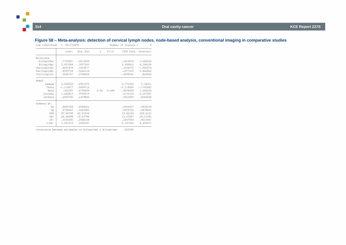
314 Oral cavity cancer KCE Report 227S
Figure 58 – Meta-analysis: detection of cervical lymph nodes, node-based analysis, conventional imaging in comparative studies
Covariance between estimates of E(logitSe) & E(logitSp) .022995 1/LR- 3.091676 .5602287 2.167462 4.409977 LR- .3234492 .0586108 .2267585 .4613691 LR+ 28.28498 10.63788 13.53387 59.11391 DOR 87.44798 42.91534 33.42106 228.8123 Sp .9758042 .0083983 .9525702 .9878025 Se .6843769 .0564602 .5650437 .7835139Summary pt. s2theta .2093782 .1479886 .0523965 .8366828 s2alpha 1.642812 .9750419 .5133132 5.257667 beta .322387 .4708438 0.68 0.494 -.6004499 1.245224 Theta -1.118677 .5099116 -2.118085 -.1192687 Lambda 4.056025 .6551575 2.771939 5.34011HSROC Corr(logits) .3246747 .3748916 -.4498341 .820459Var(logitSp) .8559718 .5242116 .2577325 2.842822Var(logitSe) .4491978 .3355877 .1038772 1.942474 E(logitSp) 3.697084 .3557063 2.999912 4.394256 E(logitSe) .7739601 .2613836 .2616576 1.286263Bivariate Coef. Std. Err. z P>|z| [95% Conf. Interval] Log likelihood = -58.233878 Number of studies = 9

KCE Report 227S Oral cavity cancer 315
6.1.10.4. Analysis 4: node-based – sensitivity analysis 1
Figure 59 – Meta-analysis (sensitivity analysis 1): detection of cervical lymph nodes, node-based analysis, PET or PET/CT in comparative studies *
* Jeong 2007: results from PET Covariance between estimates of E(logitSe) & E(logitSp) -.0293323 1/LR- 5.085917 .7394791 3.824782 6.762881 LR- .1966214 .0285882 .147866 .2614528 LR+ 20.50367 5.06193 12.63827 33.26409 DOR 104.28 23.8725 66.57909 163.3292 Sp .9604384 .010493 .9339045 .9765874 Se .8111573 .0285794 .7487341 .8609523Summary pt. s2theta .2670167 .1686329 .0774402 .9206829 s2alpha .0515063 .2067727 .0000197 134.5921 beta .6492004 .5391621 1.20 0.229 -.4075379 1.705939 Theta -.1444809 .5710648 -1.263747 .9747856 Lambda 4.321929 .2539563 3.824184 4.819675HSROC Corr(logits) -.9079894 .3440609 -.9999556 .9810681Var(logitSp) .5357184 .3239143 .1637834 1.752278Var(logitSe) .146234 .1541411 .0185277 1.154186 E(logitSp) 3.189532 .2761574 2.648273 3.73079 E(logitSe) 1.457547 .1865723 1.091872 1.823222Bivariate Coef. Std. Err. z P>|z| [95% Conf. Interval] Log likelihood = -57.454343 Number of studies = 9
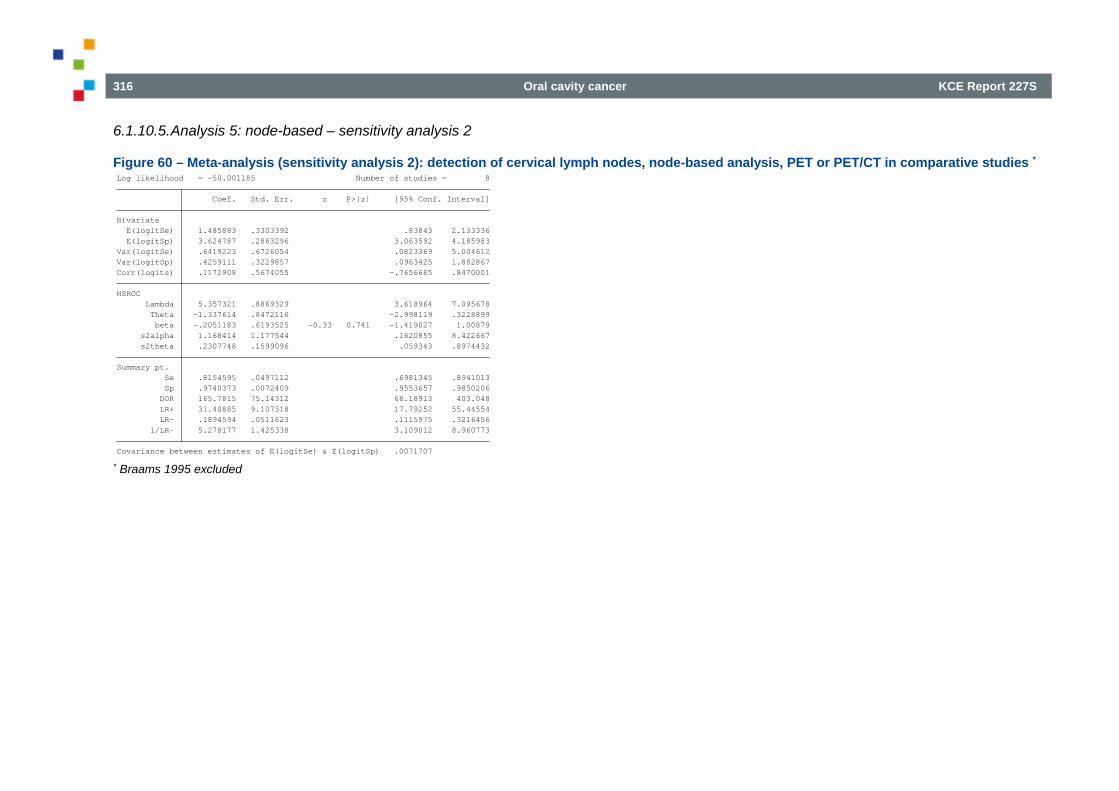
316 Oral cavity cancer KCE Report 227S
6.1.10.5. Analysis 5: node-based – sensitivity analysis 2
Figure 60 – Meta-analysis (sensitivity analysis 2): detection of cervical lymph nodes, node-based analysis, PET or PET/CT in comparative studies *
* Braams 1995 excluded Covariance between estimates of E(logitSe) & E(logitSp) .0071707 1/LR- 5.278177 1.425338 3.109012 8.960773 LR- .1894594 .0511623 .1115975 .3216456 LR+ 31.40885 9.107318 17.79252 55.44554 DOR 165.7815 75.14312 68.18913 403.048 Sp .9740373 .0072409 .9553657 .9850206 Se .8154595 .0497112 .6981345 .8941013Summary pt. s2theta .2307748 .1599096 .059343 .8974432 s2alpha 1.168414 1.177544 .1620855 8.422667 beta -.2051183 .6193525 -0.33 0.741 -1.419027 1.00879 Theta -1.337614 .8472116 -2.998119 .3228899 Lambda 5.357321 .8869329 3.618964 7.095678HSROC Corr(logits) .1172908 .5674055 -.7656685 .8470001Var(logitSp) .4259111 .3229857 .0963425 1.882867Var(logitSe) .6419223 .6726054 .0823369 5.004612 E(logitSp) 3.624787 .2863296 3.063592 4.185983 E(logitSe) 1.485883 .3303392 .83843 2.133336Bivariate Coef. Std. Err. z P>|z| [95% Conf. Interval] Log likelihood = -50.001185 Number of studies = 8
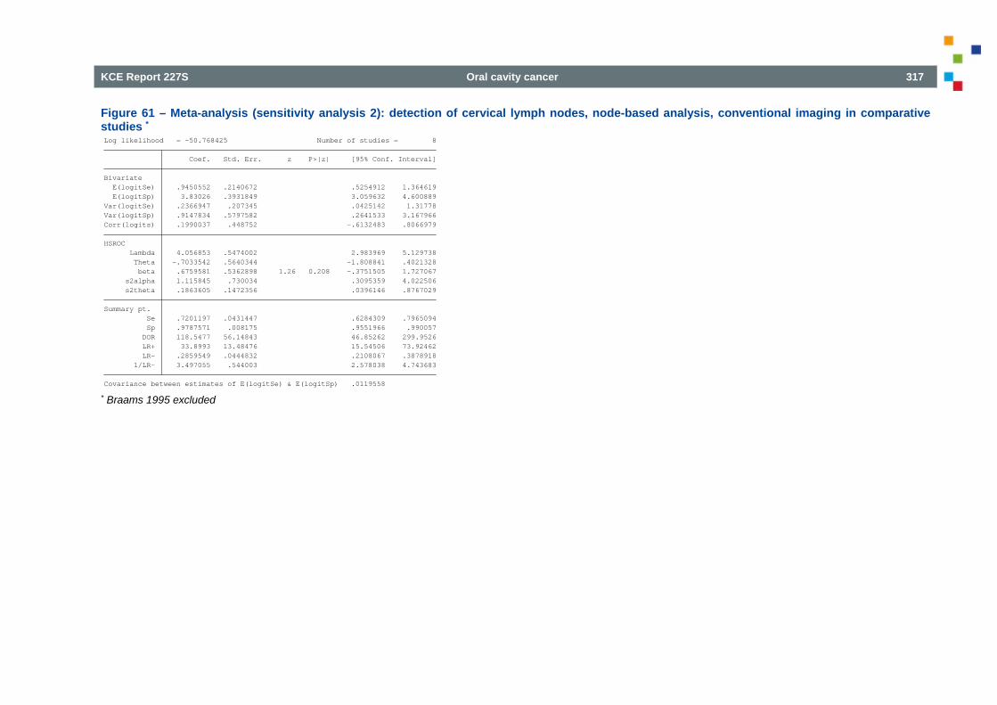
KCE Report 227S Oral cavity cancer 317
Figure 61 – Meta-analysis (sensitivity analysis 2): detection of cervical lymph nodes, node-based analysis, conventional imaging in comparative studies *
* Braams 1995 excluded Covariance between estimates of E(logitSe) & E(logitSp) .0119558 1/LR- 3.497055 .544003 2.578038 4.743683 LR- .2859549 .0444832 .2108067 .3878918 LR+ 33.8993 13.48476 15.54506 73.92462 DOR 118.5477 56.14843 46.85262 299.9526 Sp .9787571 .008175 .9551966 .990057 Se .7201197 .0431447 .6284309 .7965094Summary pt. s2theta .1863605 .1472356 .0396146 .8767029 s2alpha 1.115845 .730034 .3095359 4.022506 beta .6759581 .5362898 1.26 0.208 -.3751505 1.727067 Theta -.7033542 .5640344 -1.808841 .4021328 Lambda 4.056853 .5474002 2.983969 5.129738HSROC Corr(logits) .1990037 .448752 -.6132483 .8066979Var(logitSp) .9147834 .5797582 .2641533 3.167966Var(logitSe) .2366947 .207345 .0425142 1.31778 E(logitSp) 3.83026 .3931849 3.059632 4.600889 E(logitSe) .9450552 .2140672 .5254912 1.364619Bivariate Coef. Std. Err. z P>|z| [95% Conf. Interval] Log likelihood = -50.768425 Number of studies = 8
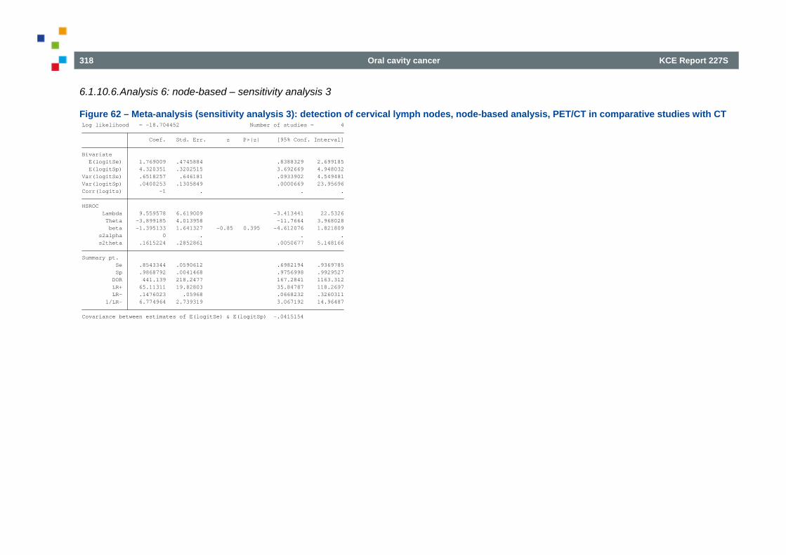
318 Oral cavity cancer KCE Report 227S
6.1.10.6. Analysis 6: node-based – sensitivity analysis 3
Figure 62 – Meta-analysis (sensitivity analysis 3): detection of cervical lymph nodes, node-based analysis, PET/CT in comparative studies with CT
Covariance between estimates of E(logitSe) & E(logitSp) -.0415154 1/LR- 6.774964 2.739319 3.067192 14.96487 LR- .1476023 .05968 .0668232 .3260311 LR+ 65.11311 19.82803 35.84787 118.2697 DOR 441.139 218.2477 167.2841 1163.312 Sp .9868792 .0041468 .9756998 .9929527 Se .8543344 .0590612 .6982194 .9369785Summary pt. s2theta .1615224 .2852861 .0050677 5.148166 s2alpha 0 . . . beta -1.395133 1.641327 -0.85 0.395 -4.612076 1.821809 Theta -3.899185 4.013958 -11.7664 3.968028 Lambda 9.559578 6.619009 -3.413441 22.5326HSROC Corr(logits) -1 . . .Var(logitSp) .0400253 .1305849 .0000669 23.95696Var(logitSe) .6518257 .646181 .0933902 4.549481 E(logitSp) 4.320351 .3202515 3.692669 4.948032 E(logitSe) 1.769009 .4745884 .8388329 2.699185Bivariate Coef. Std. Err. z P>|z| [95% Conf. Interval] Log likelihood = -18.704452 Number of studies = 4
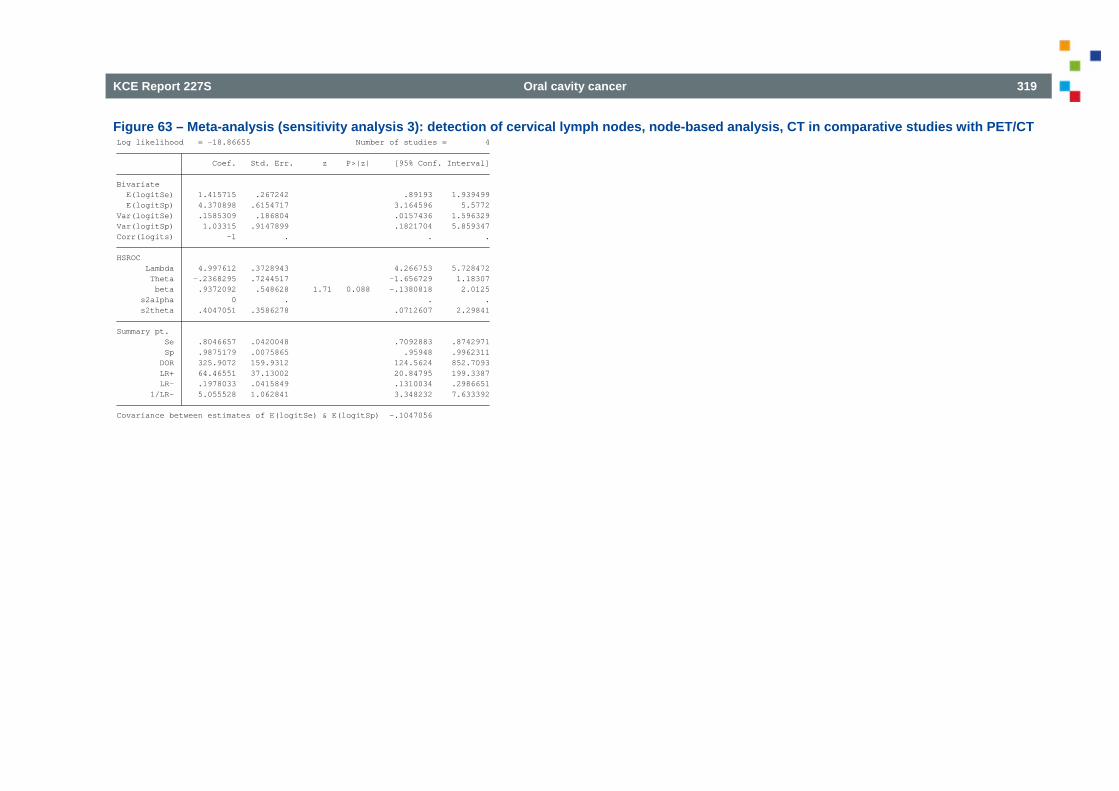
KCE Report 227S Oral cavity cancer 319
Figure 63 – Meta-analysis (sensitivity analysis 3): detection of cervical lymph nodes, node-based analysis, CT in comparative studies with PET/CT
Covariance between estimates of E(logitSe) & E(logitSp) -.1047056 1/LR- 5.055528 1.062841 3.348232 7.633392 LR- .1978033 .0415849 .1310034 .2986651 LR+ 64.46551 37.13002 20.84795 199.3387 DOR 325.9072 159.9312 124.5624 852.7093 Sp .9875179 .0075865 .95948 .9962311 Se .8046657 .0420048 .7092883 .8742971Summary pt. s2theta .4047051 .3586278 .0712607 2.29841 s2alpha 0 . . . beta .9372092 .548628 1.71 0.088 -.1380818 2.0125 Theta -.2368295 .7244517 -1.656729 1.18307 Lambda 4.997612 .3728943 4.266753 5.728472HSROC Corr(logits) -1 . . .Var(logitSp) 1.03315 .9147899 .1821704 5.859347Var(logitSe) .1585309 .186804 .0157436 1.596329 E(logitSp) 4.370898 .6154717 3.164596 5.5772 E(logitSe) 1.415715 .267242 .89193 1.939499Bivariate Coef. Std. Err. z P>|z| [95% Conf. Interval] Log likelihood = -18.86655 Number of studies = 4
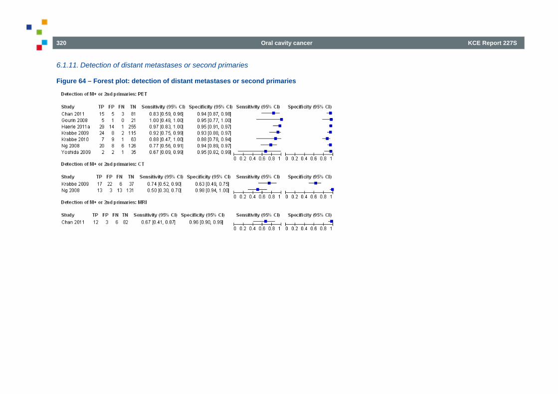
320 Oral cavity cancer KCE Report 227S
6.1.11. Detection of distant metastases or second primaries
Figure 64 – Forest plot: detection of distant metastases or second primaries
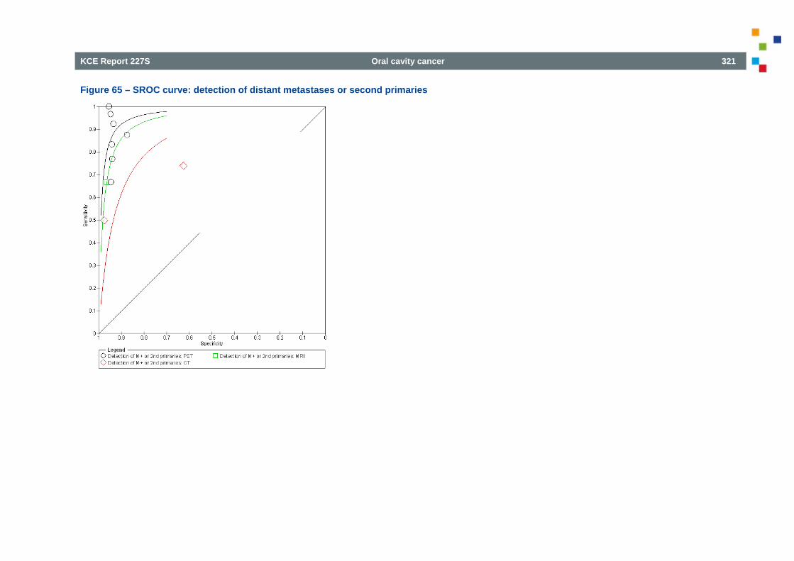
KCE Report 227S Oral cavity cancer 321
Figure 65 – SROC curve: detection of distant metastases or second primaries
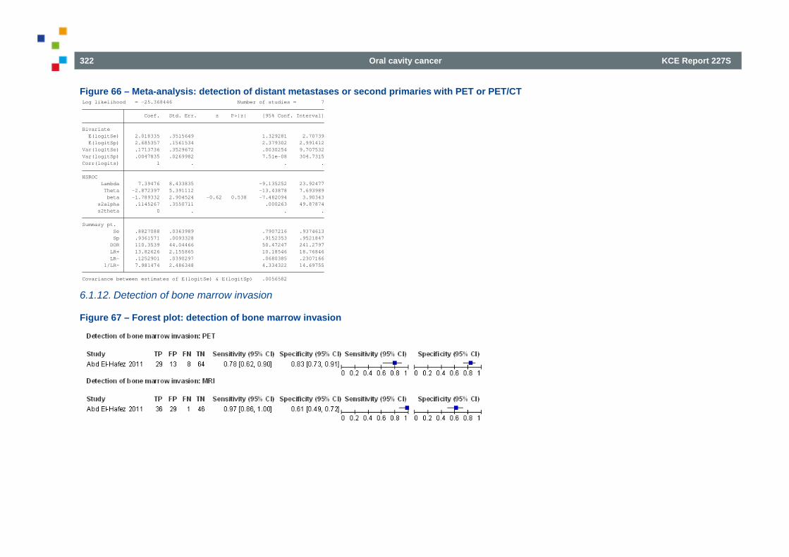
322 Oral cavity cancer KCE Report 227S
Figure 66 – Meta-analysis: detection of distant metastases or second primaries with PET or PET/CT
6.1.12. Detection of bone marrow invasion
Figure 67 – Forest plot: detection of bone marrow invasion
Covariance between estimates of E(logitSe) & E(logitSp) .0056582 1/LR- 7.981474 2.486348 4.334322 14.69755 LR- .1252901 .0390297 .0680385 .2307166 LR+ 13.82626 2.155865 10.18546 18.76846 DOR 110.3539 44.04466 50.47247 241.2797 Sp .9361571 .0093328 .9152353 .9521847 Se .8827088 .0363989 .7907216 .9374613Summary pt. s2theta 0 . . . s2alpha .1145267 .3550711 .000263 49.87874 beta -1.789332 2.904524 -0.62 0.538 -7.482094 3.90343 Theta -2.872397 5.391112 -13.43878 7.693989 Lambda 7.39476 8.433835 -9.135252 23.92477HSROC Corr(logits) 1 . . .Var(logitSp) .0047835 .0269982 7.51e-08 304.7315Var(logitSe) .1713736 .3529672 .0030254 9.707532 E(logitSp) 2.685357 .1561534 2.379302 2.991412 E(logitSe) 2.018335 .3515649 1.329281 2.70739Bivariate Coef. Std. Err. z P>|z| [95% Conf. Interval] Log likelihood = -25.368446 Number of studies = 7
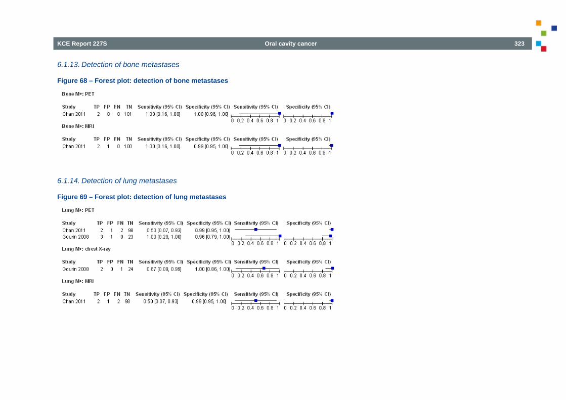
KCE Report 227S Oral cavity cancer 323
6.1.13. Detection of bone metastases
Figure 68 – Forest plot: detection of bone metastases
6.1.14. Detection of lung metastases
Figure 69 – Forest plot: detection of lung metastases
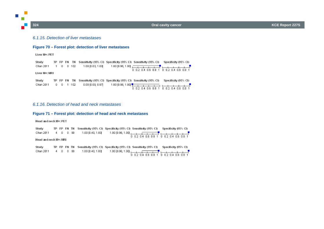
324 Oral cavity cancer KCE Report 227S
6.1.15. Detection of liver metastases
Figure 70 – Forest plot: detection of liver metastases
6.1.16. Detection of head and neck metastases
Figure 71 – Forest plot: detection of head and neck metastases
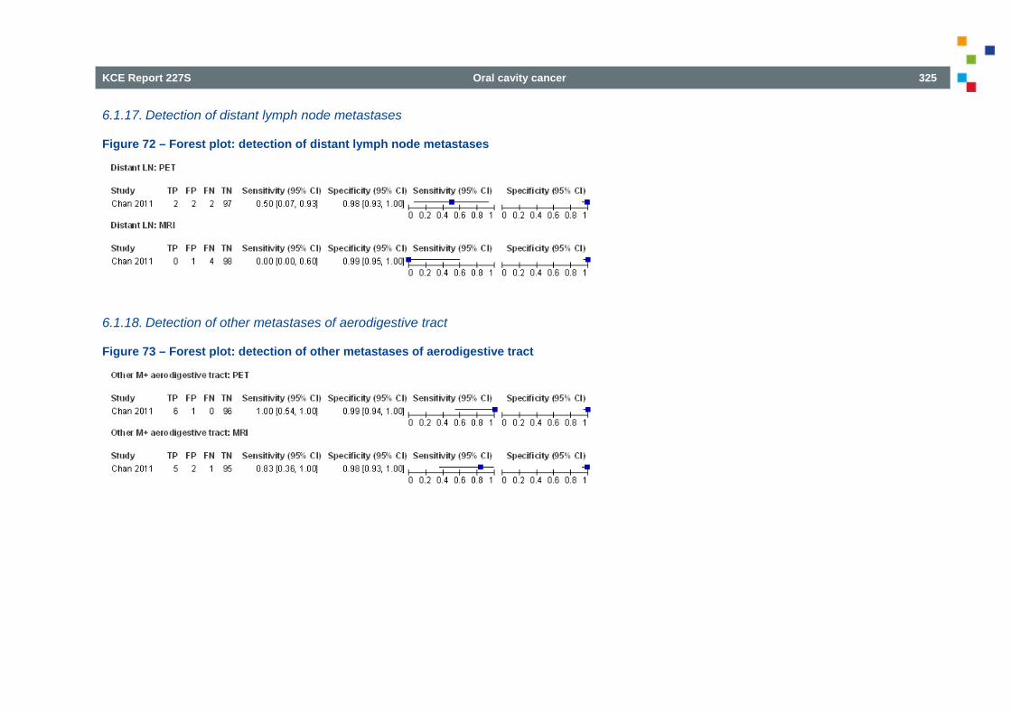
KCE Report 227S Oral cavity cancer 325
6.1.17. Detection of distant lymph node metastases
Figure 72 – Forest plot: detection of distant lymph node metastases
6.1.18. Detection of other metastases of aerodigestive tract
Figure 73 – Forest plot: detection of other metastases of aerodigestive tract
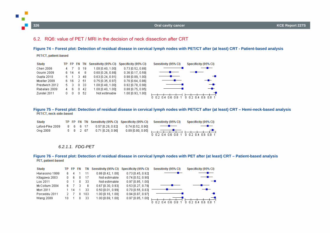
326 Oral cavity cancer KCE Report 227S
6.2. RQ6: value of PET / MRI in the decision of neck dissection after CRT
Figure 74 – Forest plot: Detection of residual disease in cervical lymph nodes with PET/CT after (at least) CRT - Patient-based analysis
Figure 75 – Forest plot: Detection of residual disease in cervical lymph nodes with PET/CT after (at least) CRT – Hemi-neck-based analysis
6.2.1.1. FDG-PET
Figure 76 – Forest plot: Detection of residual disease in cervical lymph nodes with PET after (at least) CRT – Patient-based analysis
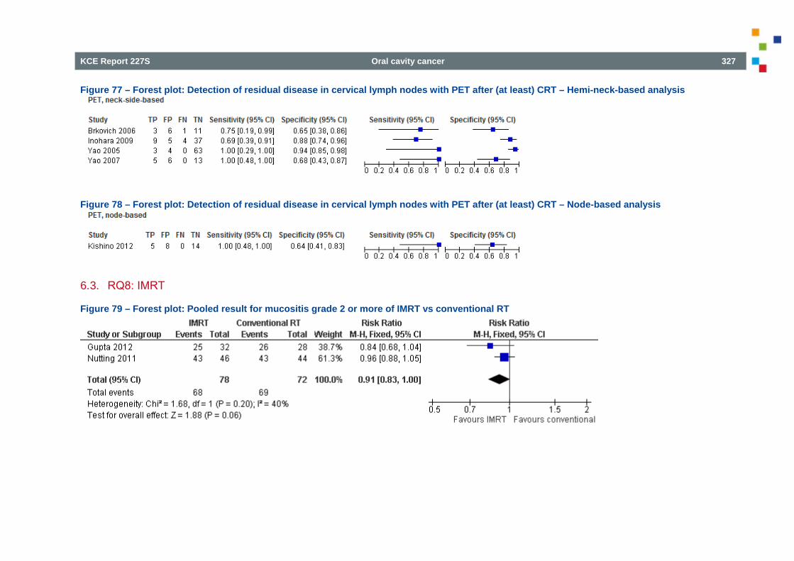
KCE Report 227S Oral cavity cancer 327
Figure 77 – Forest plot: Detection of residual disease in cervical lymph nodes with PET after (at least) CRT – Hemi-neck-based analysis
Figure 78 – Forest plot: Detection of residual disease in cervical lymph nodes with PET after (at least) CRT – Node-based analysis
6.3. RQ8: IMRT
Figure 79 – Forest plot: Pooled result for mucositis grade 2 or more of IMRT vs conventional RT
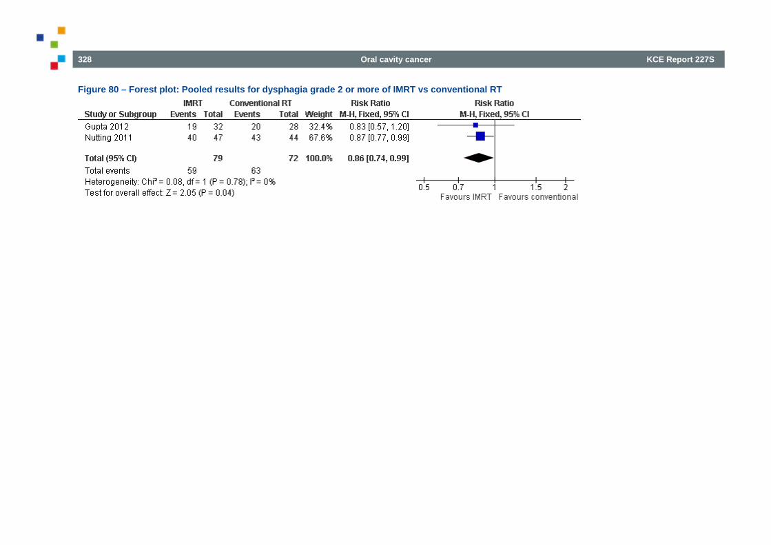
328 Oral cavity cancer KCE Report 227S
Figure 80 – Forest plot: Pooled results for dysphagia grade 2 or more of IMRT vs conventional RT
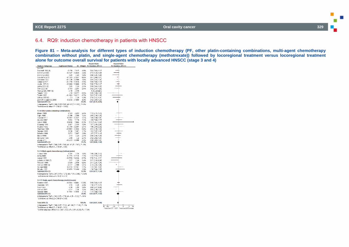
KCE Report 227S Oral cavity cancer 329
6.4. RQ9: induction chemotherapy in patients with HNSCC
Figure 81 – Meta-analysis for different types of induction chemotherapy (PF, other platin-containing combinations, multi-agent chemotherapy combination without platin, and single-agent chemotherapy (methotrexate)) followed by locoregional treatment versus locoregional treatment alone for outcome overall survival for patients with locally advanced HNSCC (stage 3 and 4)
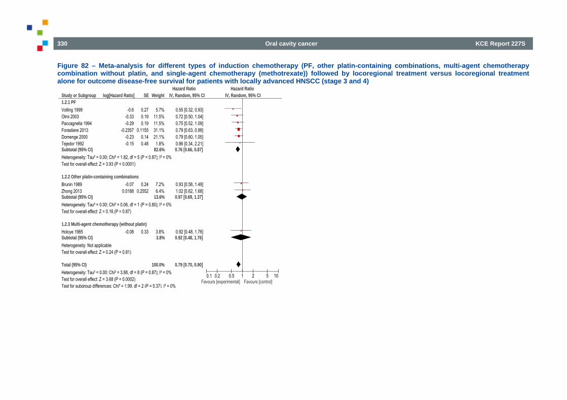
330 Oral cavity cancer KCE Report 227S
Figure 82 – Meta-analysis for different types of induction chemotherapy (PF, other platin-containing combinations, multi-agent chemotherapy combination without platin, and single-agent chemotherapy (methotrexate)) followed by locoregional treatment versus locoregional treatment alone for outcome disease-free survival for patients with locally advanced HNSCC (stage 3 and 4)
Study or Subgroup1.2.1 PFVolling 1999Olmi 2003Paccagnella 1994Forastiere 2013Domenge 2000Tejedor 1992Subtotal (95% CI)Heterogeneity: Tau² = 0.00; Chi² = 1.82, df = 5 (P = 0.87); I² = 0%Test for overall effect: Z = 3.93 (P < 0.0001)
1.2.2 Other platin-containing combinationsBrunin 1989Zhong 2013Subtotal (95% CI)Heterogeneity: Tau² = 0.00; Chi² = 0.06, df = 1 (P = 0.80); I² = 0%Test for overall effect: Z = 0.16 (P = 0.87)
1.2.3 Multi-agent chemotherapy (without platin)Holoye 1985Subtotal (95% CI)Heterogeneity: Not applicableTest for overall effect: Z = 0.24 (P = 0.81)
Total (95% CI)Heterogeneity: Tau² = 0.00; Chi² = 3.88, df = 8 (P = 0.87); I² = 0%Test for overall effect: Z = 3.68 (P = 0.0002)Test for subgroup differences: Chi² = 1.99, df = 2 (P = 0.37), I² = 0%
log[Hazard Ratio]
-0.6-0.33-0.29
-0.2357-0.23-0.15
-0.070.0188
-0.08
SE
0.270.190.19
0.11550.140.48
0.240.2552
0.33
Weight
5.7%11.5%11.5%31.1%21.1%
1.8%82.6%
7.2%6.4%
13.6%
3.8%3.8%
100.0%
IV, Random, 95% CI
0.55 [0.32, 0.93]0.72 [0.50, 1.04]0.75 [0.52, 1.09]0.79 [0.63, 0.99]0.79 [0.60, 1.05]0.86 [0.34, 2.21]0.76 [0.66, 0.87]
0.93 [0.58, 1.49]1.02 [0.62, 1.68]0.97 [0.69, 1.37]
0.92 [0.48, 1.76]0.92 [0.48, 1.76]
0.79 [0.70, 0.90]
Hazard Ratio Hazard RatioIV, Random, 95% CI
0.1 0.2 0.5 1 2 5 10Favours [experimental] Favours [control]
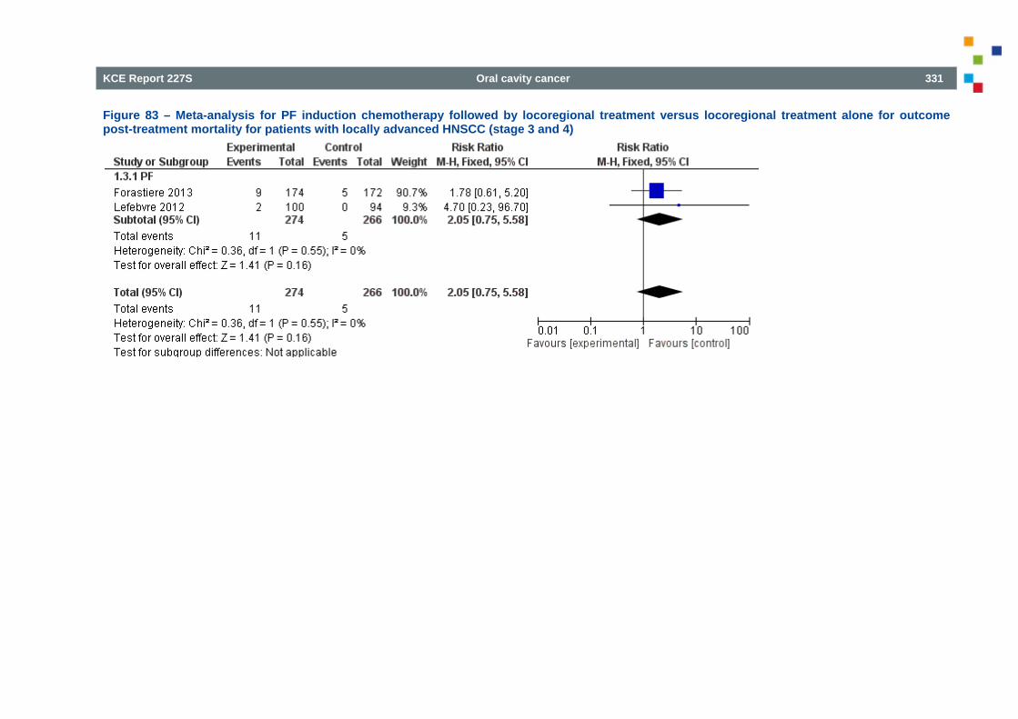
KCE Report 227S Oral cavity cancer 331
Figure 83 – Meta-analysis for PF induction chemotherapy followed by locoregional treatment versus locoregional treatment alone for outcome post-treatment mortality for patients with locally advanced HNSCC (stage 3 and 4)
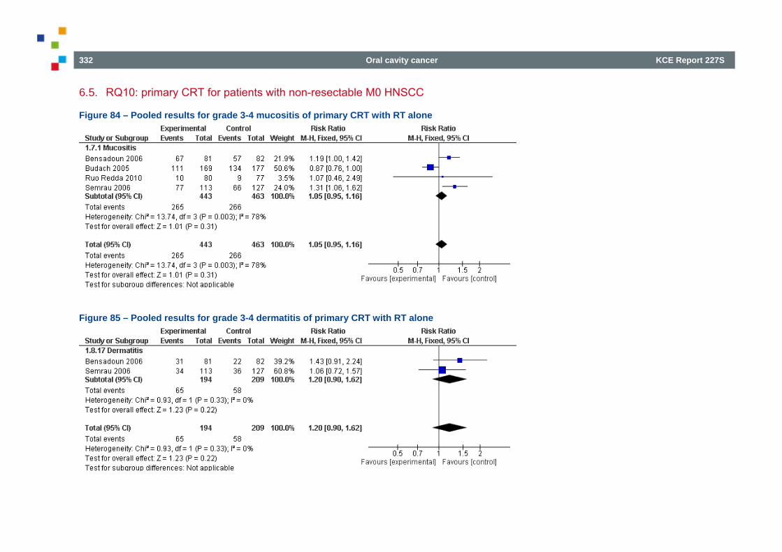
332 Oral cavity cancer KCE Report 227S
6.5. RQ10: primary CRT for patients with non-resectable M0 HNSCC
Figure 84 – Pooled results for grade 3-4 mucositis of primary CRT with RT alone
Figure 85 – Pooled results for grade 3-4 dermatitis of primary CRT with RT alone
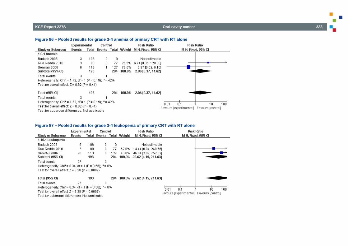
KCE Report 227S Oral cavity cancer 333
Figure 86 – Pooled results for grade 3-4 anemia of primary CRT with RT alone
Figure 87 – Pooled results for grade 3-4 leukopenia of primary CRT with RT alone
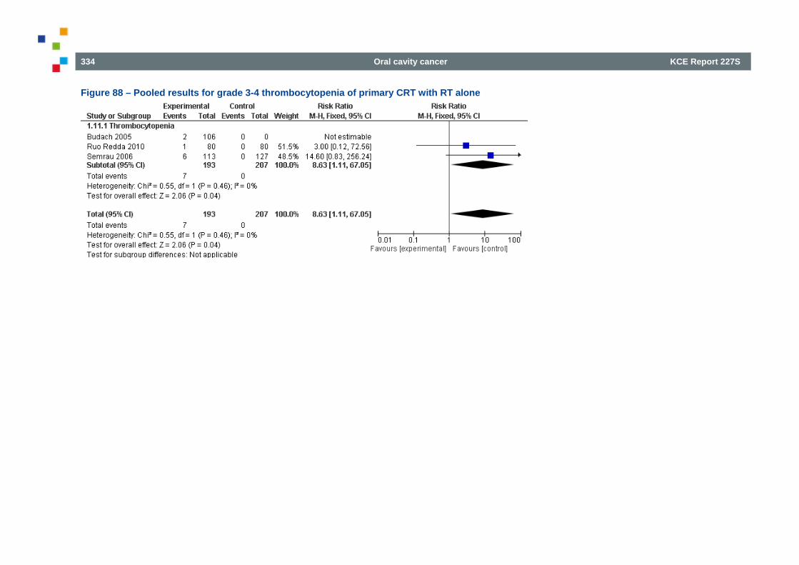
334 Oral cavity cancer KCE Report 227S
Figure 88 – Pooled results for grade 3-4 thrombocytopenia of primary CRT with RT alone
KCE Report 227S Oral cavity cancer 335
7. EXTERNAL REVIEW 7.1. Evaluation of the recommendations by the stakeholders
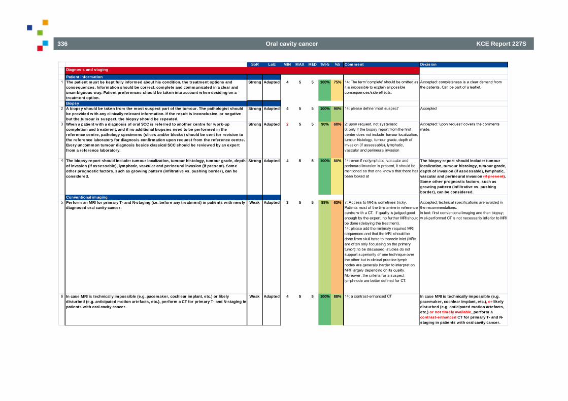
336 Oral cavity cancer KCE Report 227S
SoR LoE MIN MAX MED %4-5 %5 Comment DecisionDiagnosis and staging
Patient information1 The patient must be kept fully informed about his condition, the treatment options and
consequences. Information should be correct, complete and communicated in a clear and unambiguous way. Patient preferences should be taken into account when deciding on a treatment option.
Strong Adapted 4 5 5 100% 75% 14: The term 'complete' should be omitted as it is impossible to explain all possible consequences/side effects.
Accepted: completeness is a clear demand from the patients. Can be part of a leaflet.
Biopsy2 A biopsy should be taken from the most suspect part of the tumour. The pathologist should
be provided with any clinically relevant information. If the result is inconclusive, or negative but the tumour is suspect, the biopsy should be repeated.
Strong Adapted 4 5 5 100% 90% 14: please define 'most suspect' Accepted
3 When a patient w ith a diagnosis of oral SCC is referred to another centre for work-up completion and treatment, and if no additional biopsies need to be performed in the reference centre, pathology specimens (slices and/or blocks) should be sent for revision to the reference laboratory for diagnosis confirmation upon request from the reference centre. Every uncommon tumour diagnosis beside classical SCC should be reviewed by an expert from a reference laboratory.
Strong Adapted 2 5 5 90% 60% 2: upon request, not systematic6: only if the biopsy report from the first center does not include tumour localization, tumour histology, tumour grade, depth of invasion (if assessable), lymphatic, vascular and perineural invasion
Accepted: 'upon request' covers the comments made.
4 The biopsy report should include: tumour localization, tumour histology, tumour grade, depth of invasion (if assessable), lymphatic, vascular and perineural invasion (if present). Some other prognostic factors, such as growing pattern (infiltrative vs. pushing border), can be considered.
Strong Adapted 4 5 5 100% 80% 14: even if no lymphatic, vascular and perineural invasion is present, it should be mentioned so that one know s that there has been looked at
The biopsy report should include: tumour localization, tumour histology, tumour grade, depth of invasion (if assessable), lymphatic, vascular and perineural invasion (if present). Some other prognostic factors, such as growing pattern (infiltrative vs. pushing border), can be considered.
Conventional imaging5 Perform an MRI for primary T- and N-staging (i.e. before any treatment) in patients w ith newly
diagnosed oral cavity cancer.Weak Adapted 3 5 5 88% 63% 7: Access to MRI is sometimes tricky.
Patients most of the time arrive in reference centre w ith a CT. If quality is judged good enough by the expert, no further MRI should be done (delaying the treatment).14: please add the minimally required MRI sequences and that the MRI should be done from skull base to thoracic inlet (MRIs are often only focussing on the primary tumor); to be discussed: studies do not support superiority of one technique over the other but in clinical practice lymph nodes are generally harder to interpret on MRI, largely depending on its quality. Moreover, the criteria for a suspect lymphnode are better def ined for CT.
Accepted; technical specif ications are avoided in the recommendations.In text: f irst conventional imaging and than biopsy; w ell-performed CT is not necessarily inferior to MRI
6 In case MRI is technically impossible (e.g. pacemaker, cochlear implant, etc.) or likely disturbed (e.g. anticipated motion artefacts, etc.), perform a CT for primary T- and N-staging in patients w ith oral cavity cancer.
Weak Adapted 4 5 5 100% 88% 14: a contrast-enhanced CT In case MRI is technically impossible (e.g. pacemaker, cochlear implant, etc.), or likely disturbed (e.g. anticipated motion artefacts, etc.) or not timely available, perform a contrast-enhanced CT for primary T- and N-staging in patients w ith oral cavity cancer.
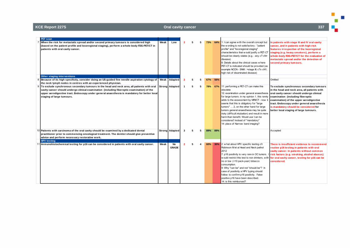
KCE Report 227S Oral cavity cancer 337
PET scan7 When the risk for metastatic spread and/or second primary tumours is considered high
(based on the patient profile and locoregional staging), perform a whole-body FDG-PET/CT in patients w ith oral cavity cancer.
Weak Low 2 5 5 73% 64% 1: I can agree w ith the overall concept but the w ording is not satisfactory : "patient profile" and "locoregional staging" charactieristics that w ould justify a PET-CT should be clearly states (e.g. : any cT cN+ disease)8: Details about the clinical cases w here PET-CT is indicated should be provided (as example NCCN - SNM : >stage III; cTx cN1; high risk of disaminated disease)
In patients w ith stage III and IV oral cavity cancer, and in patients w ith high-risk features irrespective of the locoregional staging (e.g. heavy smokers), perform a whole-body FDG-PET/CT for the evaluation of metastatic spread and/or the detection of second primary tumours.
Other staging interventions8 Because of its high specificity, consider doing an US-guided fine needle aspiration cytology of
the neck lymph nodes in centres w ith an experienced physician. Weak Adapted 2 5 5 67% 56% Omitted
9 To exclude synchronous secondary tumours in the head and neck area, all patients with oral cavity cancer should undergo clinical examination (including fiberoptic examination) of the upper aerodigestive tract. Endoscopy under general anaesthesia is mandatory for better local staging of large tumours.
Strong Adapted 1 5 5 78% 67% 11: performing a PET-CT can make this obsolete12: examination under general anaesthesia for large tumors: in my opinion 1. this rarely adds to the assessment by MRI/CT - now it seems that this is obligatory for "large tumors" … 2, on the other hand for large tumors general anaesthesia may be quite risky (diff icult intubation) and result in more harm than benefit. Would use 'can be considered' instead of "mandatory"14: place of Narrow band imaging?
To exclude synchronous secondary tumours in the head and neck area, all patients w ith oral cavity cancer should undergo clinical examination (including fiberoptic examination) of the upper aerodigestive tract. Endoscopy under general anaesthesia is mandatory should be considered for better local staging of large tumours.
10 Patients w ith carcinoma of the oral cavity should be examined by a dedicated dental practitioner prior to commencing oncological treatment. The dentist should give preventive advice and perform necessary restorative work.
Strong Adapted 3 5 5 89% 89% Accepted
HPV testing11 Immunohistochemical testing for p16 can be considered in patients with oral cavity cancer. Weak No
GRADE2 5 4 60% 30% 2: w hat about HPV specif ic testing cfr
Robinson M et al Head and Neck pathol 20127: p16 positivity is very rare in OC tumors. I w ould restrict this test to non drinkers, w ith no or low (<10 pack-year) tobacco consumption.9: Why "can be" and not "should be"? In case of positivity a HPV typing should follow to confirm p16 positivity. False positive p16 have been described.14: is this reimbursed?
There is insufficient evidence to recommend routine p16 testing in patients w ith oral cavity cancer. In patients without common risk factors (e.g. smoking, alcohol abusus) for oral cavity cancer, testing for p16 can be considered.
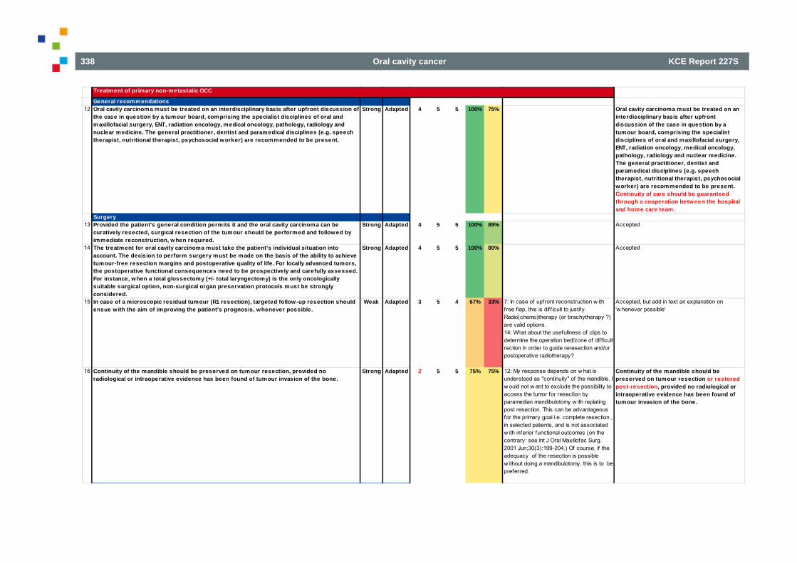
338 Oral cavity cancer KCE Report 227S
Treatment of primary non-metastatic OCC
General recommendations12 Oral cavity carcinoma must be treated on an interdisciplinary basis after upfront discussion of
the case in question by a tumour board, comprising the specialist disciplines of oral and maxillofacial surgery, ENT, radiation oncology, medical oncology, pathology, radiology and nuclear medicine. The general practitioner, dentist and paramedical disciplines (e.g. speech therapist, nutritional therapist, psychosocial worker) are recommended to be present.
Strong Adapted 4 5 5 100% 75% Oral cavity carcinoma must be treated on an interdisciplinary basis after upfront discussion of the case in question by a tumour board, comprising the specialist disciplines of oral and maxillofacial surgery, ENT, radiation oncology, medical oncology, pathology, radiology and nuclear medicine. The general practitioner, dentist and paramedical disciplines (e.g. speech therapist, nutritional therapist, psychosocial worker) are recommended to be present. Continuity of care should be guaranteed through a cooperation between the hospital and home care team.
Surgery13 Provided the patient's general condition permits it and the oral cavity carcinoma can be
curatively resected, surgical resection of the tumour should be performed and followed by immediate reconstruction, when required.
Strong Adapted 4 5 5 100% 89% Accepted
14 The treatment for oral cavity carcinoma must take the patient's individual situation into account. The decision to perform surgery must be made on the basis of the ability to achieve tumour-free resection margins and postoperative quality of life. For locally advanced tumors, the postoperative functional consequences need to be prospectively and carefully assessed. For instance, when a total glossectomy (+/- total laryngectomy) is the only oncologically suitable surgical option, non-surgical organ preservation protocols must be strongly considered.
Strong Adapted 4 5 5 100% 80% Accepted
15 In case of a microscopic residual tumour (R1 resection), targeted follow-up resection should ensue with the aim of improving the patient's prognosis, whenever possible.
Weak Adapted 3 5 4 67% 33% 7: In case of upfront reconstruction w ith free f lap, this is diff icult to justify. Radio(chemo)therapy (or brachytherapy ?) are valid options.14: What about the usefullness of clips to determine the operation bed/zone of dif f icult rection in order to guide reresection and/or postoperative radiotherapy?
Accepted, but add in text an explanation on 'w henever possible'
16 Continuity of the mandible should be preserved on tumour resection, provided no radiological or intraoperative evidence has been found of tumour invasion of the bone.
Strong Adapted 2 5 5 75% 75% 12: My response depends on w hat is understood as "continuity" of the mandible. I w ould not w ant to exclude the possibility to access the tumor for resection by paramedian mandibulotomy w ith replating post resection. This can be advantageous for the primary goal i.e. complete resection , in selected patients, and is not associated w ith inferior functional outcomes (on the contrary: see Int J Oral Maxillofac Surg. 2001 Jun;30(3):199-204.) Of course, if the adequacy of the resection is possible w ithout doing a mandibulotomy, this is to be preferred.
Continuity of the mandible should be preserved on tumour resection or restored post-resection, provided no radiological or intraoperative evidence has been found of tumour invasion of the bone.
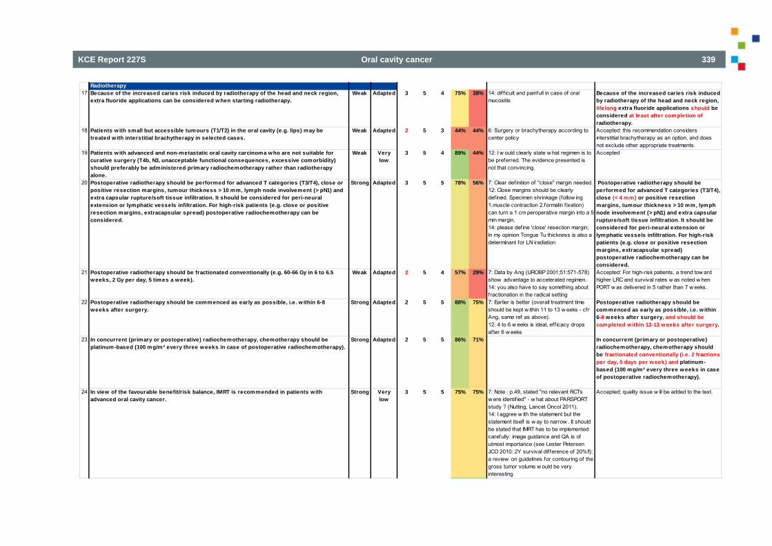
KCE Report 227S Oral cavity cancer 339
Radiotherapy17 Because of the increased caries risk induced by radiotherapy of the head and neck region,
extra fluoride applications can be considered when starting radiotherapy.Weak Adapted 3 5 4 75% 38% 14: dif f icult and painfull in case of oral
mucositisBecause of the increased caries risk induced by radiotherapy of the head and neck region, lifelong extra fluoride applications should be considered at least after completion of radiotherapy.
18 Patients w ith small but accessible tumours (T1/T2) in the oral cavity (e.g. lips) may be treated with interstitial brachytherapy in selected cases.
Weak Adapted 2 5 3 44% 44% 6: Surgery or brachytherapy according to center policy
Accepted: this recommendation considers interstitial brachytherapy as an option, and does not exclude other appropriate treatments.
19 Patients w ith advanced and non-metastatic oral cavity carcinoma who are not suitable for curative surgery (T4b, N3, unacceptable functional consequences, excessive comorbidity) should preferably be administered primary radiochemotherapy rather than radiotherapy alone.
Weak Very low
3 5 4 89% 44% 12: I w ould clearly state w hat regimen is to be preferred. The evidence presented is not that convincing.
Accepted
20 Postoperative radiotherapy should be performed for advanced T categories (T3/T4), close or positive resection margins, tumour thickness > 10 mm, lymph node involvement (> pN1) and extra capsular rupture/soft tissue infiltration. It should be considered for peri-neural extension or lymphatic vessels infiltration. For high-risk patients (e.g. close or positive resection margins, extracapsular spread) postoperative radiochemotherapy can be considered.
Strong Adapted 3 5 5 78% 56% 7: Clear definition of "close" margin needed.12: Close margins should be clearly defined. Specimen shrinkage (follow ing 1.muscle contraction 2.formalin f ixation) can turn a 1 cm peroperative margin into a 5 mm margin,14: please define 'close' resection margin; In my opinion Tongue Tu thickness is also a determinant for LN iradiation
Postoperative radiotherapy should be performed for advanced T categories (T3/T4), close (< 4 mm) or positive resection margins, tumour thickness > 10 mm, lymph node involvement (> pN1) and extra capsular rupture/soft tissue infiltration. It should be considered for peri-neural extension or lymphatic vessels infiltration. For high-risk patients (e.g. close or positive resection margins, extracapsular spread) postoperative radiochemotherapy can be considered.
21 Postoperative radiotherapy should be fractionated conventionally (e.g. 60-66 Gy in 6 to 6.5 weeks, 2 Gy per day, 5 times a week).
Weak Adapted 2 5 4 57% 29% 7: Data by Ang (IJROBP 2001;51:571-578) show advantage to accelerated regimen.14: you also have to say something about fractionation in the radical setting
Accepted: For high-risk patients, a trend tow ard higher LRC and survival rates w as noted w hen PORT w as delivered in 5 rather than 7 w eeks.
22 Postoperative radiotherapy should be commenced as early as possible, i.e. w ithin 6-8 weeks after surgery.
Strong Adapted 2 5 5 88% 75% 7: Earlier is better (overall treatment time should be kept w ithin 11 to 13 w eeks - cfr Ang, same ref as above).12: 4 to 6 w eeks is ideal, eff icacy drops after 6 w eeks
Postoperative radiotherapy should be commenced as early as possible, i.e. w ithin 6-8 weeks after surgery, and should be completed within 12-13 weeks after surgery.
23 In concurrent (primary or postoperative) radiochemotherapy, chemotherapy should be platinum-based (100 mg/m² every three weeks in case of postoperative radiochemotherapy).
Strong Adapted 2 5 5 86% 71% In concurrent (primary or postoperative) radiochemotherapy, chemotherapy should be fractionated conventionally (i.e. 2 fractions per day, 5 days per week) and platinum-based (100 mg/m² every three weeks in case of postoperative radiochemotherapy).
24 In view of the favourable benefit/risk balance, IMRT is recommended in patients with advanced oral cavity cancer.
Strong Very low
3 5 5 75% 75% 7: Note : p.49, stated "no relevant RCTs w ere identif ied" - w hat about PARSPORT study ? (Nutting, Lancet Oncol 2011).14: I aggree w ith the statement but the statement itself is w ay to narrow . It should be stated that IMRT has to be implemented carefully: image guidance and QA is of utmost importance (see Lester Petersen JCO 2010: 2Y survival difference of 20%!!); a review on guidelines for contouring of the gross tumor volume w ould be very interesting
Accepted; quality issue w ill be added to the text.
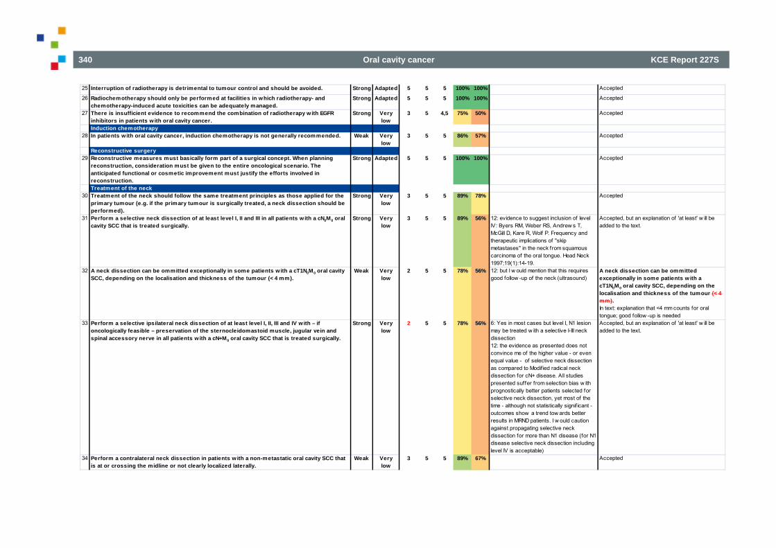
340 Oral cavity cancer KCE Report 227S
25 Interruption of radiotherapy is detrimental to tumour control and should be avoided. Strong Adapted 5 5 5 100% 100% Accepted
26 Radiochemotherapy should only be performed at facilities in which radiotherapy- and chemotherapy-induced acute toxicities can be adequately managed.
Strong Adapted 5 5 5 100% 100% Accepted
27 There is insufficient evidence to recommend the combination of radiotherapy w ith EGFR inhibitors in patients w ith oral cavity cancer.
Strong Very low
3 5 4,5 75% 50% Accepted
Induction chemotherapy28 In patients with oral cavity cancer, induction chemotherapy is not generally recommended. Weak Very
low3 5 5 86% 57% Accepted
Reconstructive surgery29 Reconstructive measures must basically form part of a surgical concept. When planning
reconstruction, consideration must be given to the entire oncological scenario. The anticipated functional or cosmetic improvement must justify the efforts involved in reconstruction.
Strong Adapted 5 5 5 100% 100% Accepted
Treatment of the neck30 Treatment of the neck should follow the same treatment principles as those applied for the
primary tumour (e.g. if the primary tumour is surgically treated, a neck dissection should be performed).
Strong Very low
3 5 5 89% 78% Accepted
31 Perform a selective neck dissection of at least level I, II and III in all patients w ith a cN0M0 oral cavity SCC that is treated surgically.
Strong Very low
3 5 5 89% 56% 12: evidence to suggest inclusion of level IV: Byers RM, Weber RS, Andrew s T, McGill D, Kare R, Wolf P. Frequency and therapeutic implications of "skip metastases" in the neck from squamous carcinoma of the oral tongue. Head Neck 1997;19(1):14-19.
Accepted, but an explanation of 'at least' w ill be added to the text.
32 A neck dissection can be ommitted exceptionally in some patients w ith a cT1N0M0 oral cavity SCC, depending on the localisation and thickness of the tumour (< 4 mm).
Weak Very low
2 5 5 78% 56% 12: but I w ould mention that this requires good follow -up of the neck (ultrasound)
A neck dissection can be ommitted exceptionally in some patients w ith a cT1N0M0 oral cavity SCC, depending on the localisation and thickness of the tumour (< 4 mm). In text: explanation that <4 mm counts for oral tongue; good follow -up is needed
33 Perform a selective ipsilateral neck dissection of at least level I, II, III and IV w ith – if oncologically feasible – preservation of the sternocleidomastoid muscle, jugular vein and spinal accessory nerve in all patients w ith a cN+M0 oral cavity SCC that is treated surgically.
Strong Very low
2 5 5 78% 56% 6: Yes in most cases but level I, N1 lesion may be treated w ith a selective I-III neck dissection12: the evidence as presented does not convince me of the higher value - or even equal value - of selective neck dissection as compared to Modif ied radical neck dissection for cN+ disease. All studies presented suffer from selection bias w ith prognostically better patients selected for selective neck dissection, yet most of the time - although not statistically signif icant - outcomes show a trend tow ards better results in MRND patients. I w ould caution against propagating selective neck dissection for more than N1 disease (for N1 disease selective neck dissection including level IV is acceptable)
Accepted, but an explanation of 'at least' w ill be added to the text.
34 Perform a contralateral neck dissection in patients w ith a non-metastatic oral cavity SCC that is at or crossing the midline or not clearly localized laterally.
Weak Very low
3 5 5 89% 67% Accepted
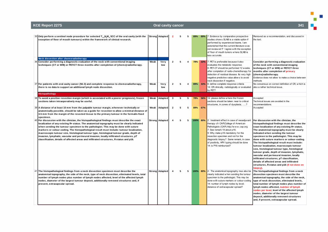
KCE Report 227S Oral cavity cancer 341
35 Only perform a sentinel node procedure for selected T1-2N0M0 SCC of the oral cavity (w ith the exception of floor of mouth tumours) w ithin the framework of clinical research.
Strong Adapted 2 5 5 89% 89% 7: Evidence by comparative prospective studies show s SLNB is a viable option if performed by experienced teams. I am astonished that the current literature w as not review ed !? I agree w ith the exception of f loor of mouth tumors w here SLNB is less accurate.
Removed as a recommendation, and discussed in the text.
Neck dissection after chemoradiotherapy36 Consider performing a diagnostic evaluation of the neck with conventional imaging
techniques (CT or MRI) or PET/CT three months after completion of (chemo)radiotherapy.Weak Very
low2 5 4 78% 22% 7: PET is preferable because it also
evaluates the metabolic response.8: PET-CT should be performed 12 w eeks after completion of radio-chemotherapy for detection of residual disease. Its very high negative predictive value allow s to avoid neck dissection if negative.
Consider performing a diagnostic evaluation of the neck with conventional imaging techniques (CT or MRI) or PET/CT three months after completion of primary (chemo)radiotherapy.Evidence does not allow to make a choice betw een methods
37 For patients w ith oral cavity cancer (N1-3) and complete response to chemoradiotherapy, there is no data to support an additional lymph node dissection.
Weak Very low
2 5 4 89% 33% 8: def ine complete response criteria14: CR clinically, radiologically or evaluated by PET?
No consensus on correct definition of CR, w hich is also a rather technical issue.
Histopathology
38 To avoid a positive resection margin (which is associated w ith a poorer prognosis), frozen sections taken intraoperatively may be useful.
Weak Adapted 3 5 5 78% 56% 14: please define w here the frozen sections should be taken: near to critical structures, in zones of dysplasia, …..?
AcceptedTechnical issues are avoided in the recommendations.
39 A distance of at least 10 mm from the palpable tumour margin, whenever technically or anatomically possible, should be taken as a guide for resection to allow a minimal distance of 3-5 mm from the margin of the resected tissue to the primary tumour in the formalin-fixed specimen.
Weak Adapted 2 5 5 89% 67% Accepted
40 For discussion with the clinician, the histopathological findings must describe the exact localization of any existing R+ status. The anatomical topography must be clearly indicated when sending the tumour specimen to the pathologist. This may be done w ith suture markers or colour-coding. The histopathological result must include: tumour localization, macroscopic tumour size, histological tumour type, histological tumour grade, depth of invasion, lymphatic, vascular and perineural invasion, locally infiltrated structures, pT classification, details of affected areas and infiltrated structures, R status and p16.
Strong Adapted 4 5 5 100% 80% 2: treatment effect in case of neoadjuvant therapy cfr CAP(College of American Pathologists (CAP).http://w w w .cap.org7: See remark 19 about p16.9: Why make p16 mandatory for the resection specimen and not for the diagnostic biopsy? Same remark, in case of positivity, HPV typing should be done14: is P16 reimbursed?
For discussion with the clinician, the histopathological findings must describe the exact localization of any existing R+ status. The anatomical topography must be clearly indicated when sending the tumour specimen to the pathologist. This may be done w ith suture markers or colour-coding. The histopathological result must include: tumour localization, macroscopic tumour size, histological tumour type, histological tumour grade, depth of invasion, lymphatic, vascular and perineural invasion, locally infiltrated structures, pT classification, details of affected areas and infiltrated structures, R status and p16 (if not done on biopsy).
41 The histopathological findings from a neck dissection specimen must describe the anatomical topography, the side of the neck, type of neck dissection, eliminated levels, total number of lymph nodes plus number of lymph nodes affected, level of the affected lymph nodes, diameter of the largest tumour deposit, additionally removed structures and, if present, extracapsular spread.
Strong Adapted 4 5 5 100% 80% 2: The anatomical topography mus also be clearly indicated w hen sending the tumour specimen to the pathologist. This may be done w ith suture markers or colour-coding14: number of lymph nodes by level; distance of extracapsular spread?
The histopathological findings from a neck dissection specimen must describe the anatomical topography, the side of the neck, type of neck dissection, eliminated levels, total number of lymph nodes plus number of lymph nodes affected, number of lymph nodes per level, level of the affected lymph nodes, diameter of the largest tumour deposit, additionally removed structures and, if present, extracapsular spread.
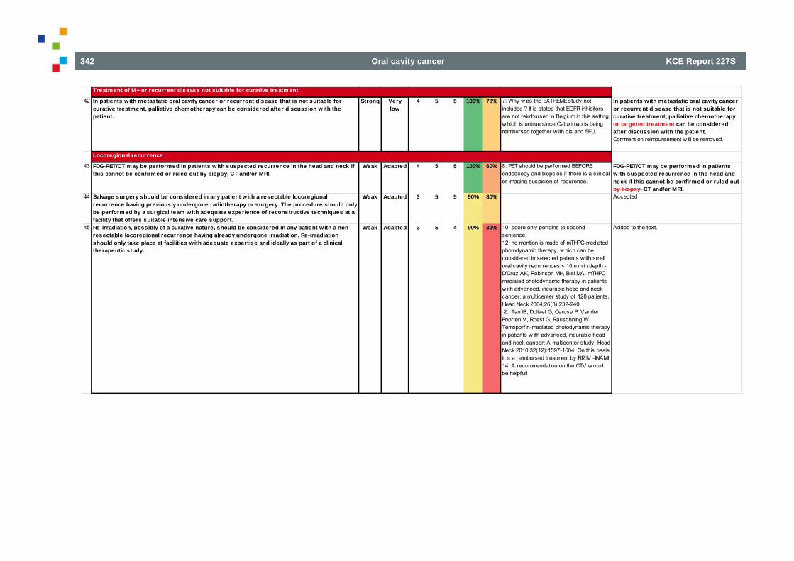
342 Oral cavity cancer KCE Report 227S
Treatment of M+ or recurrent disease not suitable for curative treatment
42 In patients with metastatic oral cavity cancer or recurrent disease that is not suitable for curative treatment, palliative chemotherapy can be considered after discussion with the patient.
Strong Very low
4 5 5 100% 78% 7: Why w as the EXTREME study not included ? It is stated that EGFR inhibitors are not reimbursed in Belgium in this setting, w hich is untrue since Cetuximab is being reimbursed together w ith cis and 5FU.
In patients with metastatic oral cavity cancer or recurrent disease that is not suitable for curative treatment, palliative chemotherapy or targeted treatment can be considered after discussion w ith the patient. Comment on reimbursement w ill be removed.
Locoregional recurrence
43 FDG-PET/CT may be performed in patients w ith suspected recurrence in the head and neck if this cannot be confirmed or ruled out by biopsy, CT and/or MRI.
Weak Adapted 4 5 5 100% 60% 8: PET should be performed BEFORE endoscopy and biopsies if there is a clinical or imaging suspicion of recurence.
FDG-PET/CT may be performed in patients w ith suspected recurrence in the head and neck if this cannot be confirmed or ruled out by biopsy, CT and/or MRI.
44 Salvage surgery should be considered in any patient with a resectable locoregional recurrence having previously undergone radiotherapy or surgery. The procedure should only be performed by a surgical team with adequate experience of reconstructive techniques at a facility that offers suitable intensive care support.
Weak Adapted 3 5 5 90% 80% Accepted
45 Re-irradiation, possibly of a curative nature, should be considered in any patient w ith a non-resectable locoregional recurrence having already undergone irradiation. Re-irradiation should only take place at facilities w ith adequate expertise and ideally as part of a clinical therapeutic study.
Weak Adapted 3 5 4 90% 30% 10: score only pertains to second sentence. 12: no mention is made of mTHPC-mediated photodynamic therapy, w hich can be considered in selected patients w ith small oral cavity recurrences < 10 mm in depth - D'Cruz AK, Robinson MH, Biel MA. mTHPC-mediated photodynamic therapy in patients w ith advanced, incurable head and neck cancer: a multicenter study of 128 patients. Head Neck 2004;26(3):232-240. 2. Tan IB, Dolivet G, Ceruse P, Vander Poorten V, Roest G, Rauschning W. Temoporf in-mediated photodynamic therapy in patients w ith advanced, incurable head and neck cancer: A multicenter study. Head Neck 2010;32(12):1597-1604. On this basis it is a reimbursed treatment by RIZIV -INAMI14: A recommendation on the CTV w ould be helpfull
Added to the text.
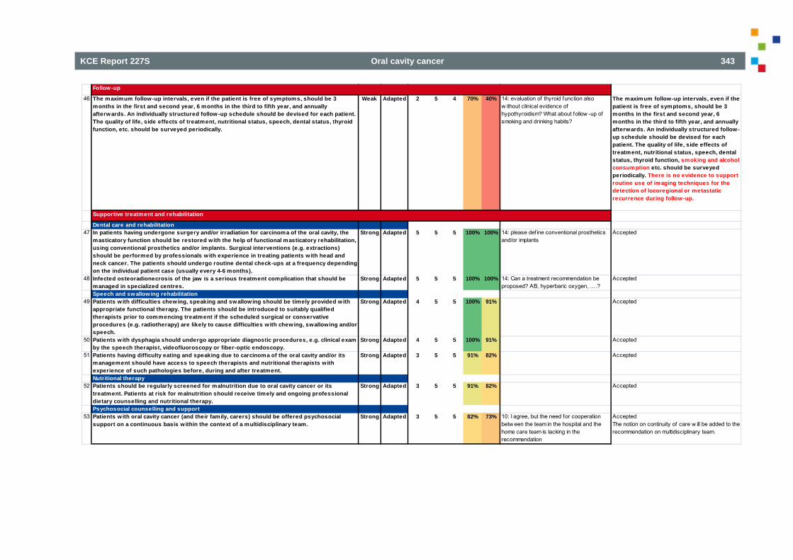
KCE Report 227S Oral cavity cancer 343
Follow-up
46 The maximum follow-up intervals, even if the patient is free of symptoms, should be 3 months in the first and second year, 6 months in the third to fifth year, and annually afterwards. An individually structured follow-up schedule should be devised for each patient. The quality of life, side effects of treatment, nutritional status, speech, dental status, thyroid function, etc. should be surveyed periodically.
Weak Adapted 2 5 4 70% 40% 14: evaluation of thyroid function also w ithout clinical evidence of hypothyroidism? What about follow -up of smoking and drinking habits?
The maximum follow-up intervals, even if the patient is free of symptoms, should be 3 months in the first and second year, 6 months in the third to fifth year, and annually afterwards. An individually structured follow-up schedule should be devised for each patient. The quality of life, side effects of treatment, nutritional status, speech, dental status, thyroid function, smoking and alcohol consumption etc. should be surveyed periodically. There is no evidence to support routine use of imaging techniques for the detection of locoregional or metastatic recurrence during follow-up.
Supportive treatment and rehabilitation
Dental care and rehabilitation47 In patients having undergone surgery and/or irradiation for carcinoma of the oral cavity, the
masticatory function should be restored with the help of functional masticatory rehabilitation, using conventional prosthetics and/or implants. Surgical interventions (e.g. extractions) should be performed by professionals with experience in treating patients w ith head and neck cancer. The patients should undergo routine dental check-ups at a frequency depending on the individual patient case (usually every 4-6 months).
Strong Adapted 5 5 5 100% 100% 14: please define conventional prosthetics and/or implants
Accepted
48 Infected osteoradionecrosis of the jaw is a serious treatment complication that should be managed in specialized centres.
Strong Adapted 5 5 5 100% 100% 14: Can a treatment recommendation be proposed? AB, hyperbaric oxygen, ….?
Accepted
Speech and swallowing rehabilitation49 Patients w ith difficulties chewing, speaking and swallow ing should be timely provided w ith
appropriate functional therapy. The patients should be introduced to suitably qualified therapists prior to commencing treatment if the scheduled surgical or conservative procedures (e.g. radiotherapy) are likely to cause difficulties with chewing, swallowing and/or speech.
Strong Adapted 4 5 5 100% 91% Accepted
50 Patients w ith dysphagia should undergo appropriate diagnostic procedures, e.g. clinical exam by the speech therapist, videofluoroscopy or fiber-optic endoscopy.
Strong Adapted 4 5 5 100% 91% Accepted
51 Patients having difficulty eating and speaking due to carcinoma of the oral cavity and/or its management should have access to speech therapists and nutritional therapists with experience of such pathologies before, during and after treatment.
Strong Adapted 3 5 5 91% 82% Accepted
Nutritional therapy52 Patients should be regularly screened for malnutrition due to oral cavity cancer or its
treatment. Patients at risk for malnutrition should receive timely and ongoing professional dietary counselling and nutritional therapy.
Strong Adapted 3 5 5 91% 82% Accepted
Psychosocial counselling and support53 Patients w ith oral cavity cancer (and their family, carers) should be offered psychosocial
support on a continuous basis within the context of a multidisciplinary team.Strong Adapted 3 5 5 82% 73% 10: I agree, but the need for cooperation
betw een the team in the hospital and the home care team is lacking in the recommendation
AcceptedThe notion on continuity of care w ill be added to the recommendation on multidisciplinary team.
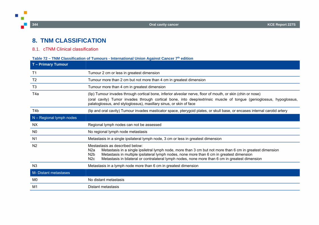
344 Oral cavity cancer KCE Report 227S
8. TNM CLASSIFICATION 8.1. cTNM Clinical classification
Table 72 – TNM Classification of Tumours - International Union Against Cancer 7th edition T – Primary Tumour
T1 Tumour 2 cm or less in greatest dimension
T2 Tumour more than 2 cm but not more than 4 cm in greatest dimension
T3 Tumour more than 4 cm in greatest dimension
T4a (lip) Tumour invades through cortical bone, inferior alveolar nerve, floor of mouth, or skin (chin or nose) (oral cavity) Tumor invades through cortical bone, into deep/extrinsic muscle of tongue (genioglossus, hypoglossus, palatoglossus, and styloglossus), maxillary sinus, or skin of face
T4b (lip and oral cavity) Tumour invades masticator space, pterygoid plates, or skull base, or encases internal carotid artery
N – Regional lymph nodes
NX Regional lymph nodes can not be assessed
N0 No regional lymph node metastasis
N1 Metastasis in a single ipsilateral lymph node, 3 cm or less in greatest dimension
N2 Mestastasis as described below: N2a Metastasis in a single ipsiletral lymph node, more than 3 cm but not more than 6 cm in greatest dimension N2b Metastasis in multiple ipsilateral lymph nodes, none more than 6 cm in greatest dimension N2c Metastasis in bilateral or contralateral lymph nodes, none more than 6 cm in greatest dimension
N3 Metastasis in a lymph node more than 6 cm in greatest dimension
M- Distant metastases
M0 No distant metastasis
M1 Distant metastasis
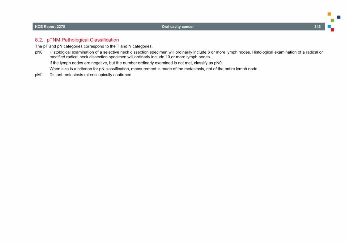
KCE Report 227S Oral cavity cancer 345
8.2. pTNM Pathological Classification The pT and pN categories correspond to the T and N categories. pN0 Histological examination of a selective neck dissection specimen will ordinarily include 6 or more lymph nodes. Histological examination of a radical or modified radical neck dissection specimen will ordinarly include 10 or more lymph nodes. If the lymph nodes are negative, but the number ordinarly examined is not met, classify as pN0. When size is a criterion for pN classification, measurement is made of the metastasis, not of the entire lymph node. pM1 Distant metastasis microscopically confirmed
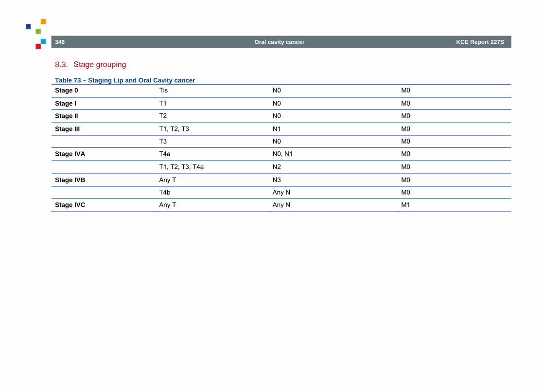
346 Oral cavity cancer KCE Report 227S
8.3. Stage grouping
Table 73 – Staging Lip and Oral Cavity cancer Stage 0 Tis N0 M0
Stage I T1 N0 M0
Stage II T2 N0 M0
Stage III T1, T2, T3 N1 M0
T3 N0 M0
Stage IVA T4a N0, N1 M0
T1, T2, T3, T4a N2 M0
Stage IVB Any T N3 M0
T4b Any N M0
Stage IVC Any T Any N M1
KCE Report 227S Oral cavity cancer 347
REFERENCES 1. Fletcher JW, Djulbegovic B, Soares HP, Siegel BA, Lowe VJ, Lyman GH, et al. Recommendations on the use of 18F-FDG PET in oncology. J. Nucl.
Med. 2008;49(3):480-508. 2. Liao L-J, Lo W-C, Hsu W-L, Wang C-T, Lai M-S. Detection of cervical lymph node metastasis in head and neck cancer patients with clinically N0
neck-a meta-analysis comparing different imaging modalities. BMC Cancer. 2012;12 , 2012. Article Number: 236. Date of Publication: 12 Jun 2012. 3. Xu G, Li J, Zuo X, Li C. Comparison of whole body positron emission tomography (PET)/PET-computed tomography and conventional anatomic
imaging for detecting distant malignancies in patients with head and neck cancer: A meta-analysis. Laryngoscope. 2012;122(9):1974-8. 4. Xu GZ, Zhu XD, Li MY. Accuracy of whole-body PET and PET-CT in initial M staging of head and neck cancer: A meta-analysis. Head Neck.
2011;33(1):87-94. 5. Xu GZ, Guan DJ, He ZY. 18FDG-PET/CT for detecting distant metastases and second primary cancers in patients with head and neck cancer. A
meta-analysis. Oral Oncol. 2011;47(7):560-5. 6. Yongkui L, Jian L, Wanghan, Jingui L. 18FDG-PET/CT for the detection of regional nodal metastasis in patients with primary head and neck cancer
before treatment: A meta-analysis. Surg. Oncol. 2013;22(2):e11-e6. 7. Haerle SK, Strobel K, Ahmad N, Soltermann A, Schmid DT, Stoeckli SJ. Contrast-enhanced 18F-FDG-PET/CT for the assessment of necrotic lymph
node metastases. Head Neck. 2011;33(3):324-9. 8. Hoshikawa H, Kishino T, Mori T, Nishiyama Y, Yamamoto Y, Inamoto R, et al. Comparison of 18 F-FLT PET and 18 F-FDG PET for detection of
cervical lymph node metastases in head and neck cancers. Acta Oto-Laryngol. 2012;132(12):1347-54. 9. Krabbe CA, Pruim J, Scholtens AM, Roodenburg JL, Brouwers AH, Phan TT, et al. 18F-FDG PET in squamous cell carcinoma of the oral cavity and
oropharynx: a study on inter- and intraobserver agreement. J Oral Maxillofac Surg. 2010;68(1):21-7. 10. Liao CT, Wang HM, Huang SF, Chen IH, Kang CJ, Lin CY, et al. PET and PET/CT of the neck lymph nodes improves risk prediction in patients with
squamous cell carcinoma of the oral cavity. J Nucl Med. 2011;52(2):180-7. 11. Matsubara R, Kawano S, Chikui T, Kiyosue T, Goto Y, Hirano M, et al. Clinical significance of combined assessment of the maximum standardized
uptake value of F-18 FDG PET with nodal size in the diagnosis of cervical lymph node metastasis of oral squamous cell carcinoma. Acad Radiol. 2012;19(6):708-17.
12. Ozer E, Naiboglu B, Meacham R, Ryoo C, Agrawal A, Schuller DE. The value of PET/CT to assess clinically negative necks. Eur Arch Otorhinolaryngol. 2012;269(11):2411-4.
13. Abd El-Hafez YG, Chen CC, Ng SH, Lin CY, Wang HM, Chan SC, et al. Comparison of PET/CT and MRI for the detection of bone marrow invasion in patients with squamous cell carcinoma of the oral cavity. Oral Oncol. 2011;47(4):288-95.
14. Chan SC, Wang HM, Yen TC, Lin CY, Chin SC, Liao CT, et al. 18F-FDG PET/CT and 3.0-T whole-body MRI for the detection of distant metastases and second primary tumours in patients with untreated oropharyngeal/hypopharyngeal carcinoma: a comparative study. Eur J Nucl Med Mol Imaging. 2011;38(9):1607-19.
348 Oral cavity cancer KCE Report 227S
15. Haerle SK, Schmid DT, Ahmad N, Hany TF, Stoeckli SJ. The value of (18)F-FDG PET/CT for the detection of distant metastases in high-risk patients with head and neck squamous cell carcinoma. Oral Oncol. 2011;47(7):653-9.
16. Lacchetti CW, J.; Perez-Ordonez, B.; Kamel-Reid, C.; Cripps, C.; Gilbert, R.; Haed and Neck Cancer DSG. Routine HPV Testing in Head and Neck Squamous cell Carcinoma. Cancer Care Ontario Evidence-Based Series 5-9. 2013.
17. Bessell A, Glenny AM, Furness S, Clarkson JE, Oliver R, Conway D, I, et al. Interventions for the treatment of oral and oropharyngeal cancers: surgical treatment. Cochrane Database of Systematic Reviews. 2011.
18. Fasunla AJ, Greene BH, Timmesfeld N, Wiegand S, Werner JA, Sesterhenn AM. A meta-analysis of the randomized controlled trials on elective neck dissection versus therapeutic neck dissection in oral cavity cancers with clinically node-negative neck. Oral Oncology. 2011;47:320-4.
19. Wolff K-D. Mundhöhlenkarzinom - Diagnostik und Therapie des Mundhöhlenkarzinoms. 2012. 20. D'Cruz AK, Siddachari RC, Walvekar RR, Pantvaidya GH, Chaukar DA, Deshpande MS, et al. Elective neck dissection for the management of the N0
neck in early cancer of the oral tongue: need for a randomized controlled trial. Head & neck. 2009;31:618-24. 21. Huang S-F, Chang C-J, Lin C-Y, Fan K-H, Yen T-C, Wang H-M, et al. Neck treatment of patients with early stage oral tongue cancer: comparison
between observation, supraomohyoid dissection, and extended dissection. Cancer. 2008;112:1066-75. 22. Patel RS, Clark JR, Gao K, O'Brien CJ. Effectiveness of selective neck dissection in the treatment of the clinicaly positive neck. Head Neck.
2008(9):1231-6. 23. Rapoport A, Ortellado DK, Amar A, Lehn CN, Dedivitis RA, Perez RS, et al. Radical versus supraomohyoid neck dissection in the treatment of
squamous cell carcinoma of the inferiour level of the mouth. Brazilian Journal of Otorhinolaryngology. 2007;73(5):641-6. 24. Shepard PM, Olson J, Harari PM, Leverson G, Hartig GK. Therapeutic selective neck dissection outcomes. Otolaryngology - Head and Neck Surgery
(United States). 2010;142(5). 25. An S-Y, Jung E-J, Lee M, Kwon T-K, Sung M-W, Jeon YK, et al. Factors related to regional recurrence in early stage squamous cell carcinoma of the
oral tongue. In: Clinical & Experimental Otorhinolaryngology Clin; 2008. 26. Ebrahimi A, Ashford BG, Clark JR. Improved survival with elective neck dissection in thick early-stage oral squamous cell carcinoma. Head and Neck.
2012;34(5). 27. Flach GB, Tenhagen M, de Bree R, Brakenhoff RH, van der Waal I, Bloemena E, et al. Outcome of patients with early stage oral cancer managed by
an observation strategy towards the N0 neck using ultrasound guided fine needle aspiration cytology: No survival difference as compared to elective neck dissection. In: Oral Oncology Oral Oncol; 2013.
28. Lin TC, Tsou YA, Lin MH, Hua CH, Tseng HC, Bau DT, et al. Impact of neck dissection in early tongue and buccal cancer without neck extension. In: B-Ent; 2011.
29. Masuda M, Kamizono KI, Uryu H, Fujimura A, Uchi R. Roles of Therapeutic Selective Neck Dissection in Multidisciplinairy Treatment, Neck Dissection - Clinical Application and Recent Advantages. In Tech: http://www.intechopen.com/books/neck-dissection-clinical-application-and-recent-advances/roles-oftherapeutic-selective-neck-dissection-in-multidisciplinary-treatment. 2012.
30. Park SM, Lee DJ, Chung EJ, Kim JH, Park IS, Lee MJ, et al. Conversion from selective to comprehensive neck dissection: is it necessary for occult nodal metastasis? 5-year observational study. In: Clinical & Experimental Otorhinolaryngology Clin; 2013.
KCE Report 227S Oral cavity cancer 349
31. Yanai Y, Sugiura T, Imajyo I, Yoshihama N, Akimoto N, Kobayashi Y, et al. Retrospective study of selective submandibular neck dissection versus radical neck dissection for N0 or N1 necks in level i patients with oral squamous cell carcinoma. Journal of Oncology. 2012(634183).
32. Yildirim T, Ozmen OA, Erisen L, Kasapoglu F, Coskun H, Basut O, et al. The role of selective neck dissection in pathological N1 squamous cell carcinomas of the head and neck. Kulak burun bogaz ihtisas dergisi : KBB = Journal of ear, nose, and throat. 2011;21(2).
33. Vandenbrouck C, Sancho-Garnier H, Chassagne D, Saravane D, Cachin Y, Micheau C. Elective versus therapeutic radical neck dissection in epidermoid carcinoma of the oral cavity: results of a randomized clinical trial. Cancer. 1980;46(2):386-90.
34. Fakih AR, Rao RS, Borges AM, Patel AR. Elective versus therapeutic neck dissection in early carcinoma of the oral tongue. Am.J.Surg. 1989;158(4):309-13.
35. Kligerman J, Lima RA, Soares JR, Prado L, Dias FL, Freitas EQ, et al. Supraomohyoid neck dissection in the treatment of T1/T2 squamous cell carcinoma of oral cavity. Am.J.Surg. 1994;168(5):391-4.
36. Yuen AP, Ho CM, Chow TL, Tang LC, Cheung WY, Ng RW, et al. Prospective randomized study of selective neck dissection versus observation for N0 neck of early tongue carcinoma. Head Neck. 2009;31(6):765-72.
37. Bier J. Radical neck dissection versus conservative neck dissection for squamous cell carcinoma of the oral cavity. Recent Results Cancer Res. 1994;134:57-62.
38. BHNCSG. Results of a prospective trial on elective modified radical classical versus supraomohyoid neck dissection in the management of oral squamous carcinoma. Brazilian Head and Neck Cancer Study Group. Am.J.Surg. 1998;176(5):422-7.
39. Gupta T, Master Z, Kannan S, Agarwal JP, Ghsoh-Laskar S, Rangarajan V, et al. Diagnostic performance of post-treatment FDG PET or FDG PET/CT imaging in head and neck cancer: a systematic review and meta-analysis (Provisional abstract). European Journal of Nuclear Medicine and Molecular Imaging. 2011;38(11):2083-95.
40. Isles MG, McConkey C, Mehanna HM. A systematic review and meta-analysis of the role of positron emission tomography in the follow up of head and neck squamous cell carcinoma following radiotherapy or chemoradiotherapy (Structured abstract). Clin Otolaryngol. 2008;33(3):210-22.
41. Gupta T, Agarwal J, Jain S, Phurailatpam R, Kannan S, Ghosh-Laskar S, et al. Three-dimensional conformal radiotherapy (3D-CRT) versus intensity modulated radiation therapy (IMRT) in squamous cell carcinoma of the head and neck: A randomized controlled trial. Radiotherapy and Oncology. 2012;104:343-8.
42. Kishino T, Hoshikawa H, Nishiyama Y, Yamamoto Y, Mori N. Usefulness of 3'-deoxy-3'-18F-fluorothymidine PET for predicting early response to chemoradiotherapy in head and neck cancer. J Nucl Med. 2012;53(10):1521-7.
43. Loo SW, Geropantas K, Beadsmoore C, Montgomery PQ, Martin WMC, Roques TW. Neck dissection can be avoided after sequential chemoradiotherapy and negative post-treatment positron emission tomography-computed tomography in N2 Head and neck squamous cell carcinoma. Clin. Oncol. 2011;23(8):512-7.
44. Mori M, Tsukuda M, Horiuchi C, Matsuda H, Taguchi T, Takahashi M, et al. Efficacy of fluoro-2-deoxy-d-glucose positron emission tomography to evaluate responses to concurrent chemoradiotherapy for head and neck squamous cell carcinoma. Auris Nasus Larynx. 2011;38(6):724-9.
45. Porceddu SV, Pryor DI, Burmeister E, Burmeister BH, Poulsen MG, Foote MC, et al. Results of a prospective study of positron emission tomography-directed management of residual nodal abnormalities in node-positive head and neck cancer after definitive radiotherapy with or without systemic therapy. Head Neck. 2011;33(12):1675-82.
350 Oral cavity cancer KCE Report 227S
46. Prestwich RJ, Subesinghe M, Gilbert A, Chowdhury FU, Sen M, Scarsbrook AF. Delayed response assessment with FDG-PET-CT following (chemo) radiotherapy for locally advanced head and neck squamous cell carcinoma. Clin Radiol. 2012;67(10):966-75.
47. Zundel MT, Michel MA, Schultz CJ, Maheshwari M, Wong SJ, Campbell BH, et al. Comparison of physical examination and fluorodeoxyglucose positron emission tomography/computed tomography 4-6 months after radiotherapy to assess residual head-and-neck cancer. Int J Radiat Oncol Biol Phys. 2011;81(5):e825-32.
48. Lin D, Glastonbury CM, Rafaelian O, Eisele DW, Wang SJ. Management of advanced nodal disease following chemoradiation for head and neck squamous cell carcinoma: role of magnetic resonance imaging. J Otolaryngol. 2007;36(6):350-6.
49. O'sullivan B, Rumble RB, Warde P. Intensity-modulated Radiotherapy in the Treatment of Head and Neck Cancer. Clinical Oncology. 2012;24:474-87. 50. Roe JWG, Carding PN, Dwivedi RC, Kazi RA, Rhys-Evans PH, Harrington KJ, et al. Swallowing outcomes following Intensity Modulated Radiation
Therapy (IMRT) for head & neck cancer - A systematic review. Oral Oncology. 2010;46:727-33. 51. Ramaekers BLT, Pijls-Johannesma M, Joore MA, Van Den Ende P, Langendijk JA, Lambin P, et al. Systematic review and meta-analysis of
radiotherapy in various head and neck cancers: Comparing photons, carbon-ions and protons. Cancer Treatment Reviews. 2011;37:185-201. 52. Guha S, Kelly CG, Guha R, Achari R, Mallick I, Paleri V, et al. Intensity modulated radiation therapy (IMRT) in the treatment of squamous carcinoma
of the oropharynx: An overview. Journal of Cancer Science and Therapy. 2012;4:77-83. 53. Jensen SB, Pedersen AML, Vissink A, Andersen E, Brown CG, Davies AN, et al. A systematic review of salivary gland hypofunction and xerostomia
induced by cancer therapies: Prevalence, severity and impact on quality of life. Supportive Care in Cancer. 2010;18:1039-60. 54. Staffurth J. A review of the clinical evidence for intensity-modulated radiotherapy. Clinical Oncology. 2010;22:643-57. 55. Thariat J, Bolle S, Demizu Y, Marcy P-Y, Hu Y, Santini J, et al. New techniques in radiation therapy for head and neck cancer: IMRT, CyberKnife,
protons, and carbon ions. Improved effectiveness and safety? Impact on survival? Anti-Cancer Drugs. 2011;22:596-606. 56. Tribius S, Bergelt C. Intensity-modulated radiotherapy versus conventional and 3D conformal radiotherapy in patients with head and neck cancer: Is
there a worthwhile quality of life gain? Cancer Treatment Reviews. 2011;37:511-9. 57. van de Water TA, Bijl HP, Schilstra C, Pijls-Johannesma M, Langendijk JA. The potential benefit of radiotherapy with protons in head and neck cancer
with respect to normal tissue sparing: A systematic review of literature. Oncologist. 2011;16:366-77. 58. Veldeman L, Madani I, Hulstaert F, De MG, Mareel M, De NW. Evidence behind use of intensity-modulated radiotherapy: a systematic review of
comparative clinical studies. The Lancet Oncology. 2008;9:367-75. 59. Chen WC, Hwang TZ, Wang WH, Lu CH, Chen CC, Chen CM, et al. Comparison between conventional and intensity-modulated post-operative
radiotherapy for stage III and IV oral cavity cancer in terms of treatment results and toxicity. Oral Oncology. 2009;45:(6):505-10. 60. Chen AM, Li BQ, Farwell DG, Marsano J, Vijayakumar S, Purdy JA. Improved dosimetric and clinical outcomes with intensity-modulated radiotherapy
for head-and-neck cancer of unknown primary origin. International Journal of Radiation Oncology, 2011 Mar 1. 2011;79(3):756-62. 61. Chen AM, Farwell DG, Luu Q, Vazquez EG, Lau DH, Purdy JA. Intensity-modulated radiotherapy is associated with improved global quality of life
among long-term survivors of head-and-neck cancer. International Journal of Radiation Oncology Biology Physics. 2012;84:170-5.
KCE Report 227S Oral cavity cancer 351
62. Clavel S, Nguyen DHA, Fortin B, Despres P, Khaouam N, Donath D, et al. Simultaneous integrated boost using intensity-modulated radiotherapy compared with conventional radiotherapy in patients treated with concurrent carboplatin and 5-fluorouracil for locally advanced oropharyngeal carcinoma. International Journal of Radiation Oncology Biology Physics. 2012;82:582-9.
63. Dirix P, Nuyts S. Value of intensity-modulated radiotherapy in stage IV head-and-neck squamous cell carcinoma. International Journal of Radiation Oncology Biology Physics. 2010;78:1373-80.
64. Jilani OK, Singh P, Wernicke AG, Kutler DI, Kuhel W, Christos P, et al. Radiation therapy is well tolerated and produces excellent control rates in elderly patients with locally advanced head and neck cancers. Journal of Geriatric Oncology. 2012;3:337-43.
65. Lambrecht M, Nevens D, Nuyts S. Intensity-modulated radiotherapy vs. parotid-sparing 3D conformal radiotherapy. Effect on outcome and toxicity in locally advanced head and neck cancer. Strahlentherapie und Onkologie : Organ der Deutschen Rontgengesellschaft. 2013;.. [et al]. 189:223-9.
66. Tai HC, Hsieh CH, Chao KS, Liu SH, Leu YS, Chang YF, et al. Comparison of radiotherapy strategies for locally advanced hypopharyngeal cancer after resection and ileocolic flap reconstruction. Acta Oto-Laryngologica. 2009;129:(3):311-7.
67. Nutting CM, Morden JP, Harrington KJ, Urbano TG, Bhide SA, Clark C, et al. Parotid-sparing intensity modulated versus conventional radiotherapy in head and neck cancer (PARSPORT): A phase 3 multicentre randomised controlled trial. The Lancet Oncology. 2011;12:127-36.
68. Al-Mamgani A, Monserez D, Rooij PV, Verduijn GM, Hardillo JAU, Levendag PC. Highly-conformal intensity-modulated radiotherapy reduced toxicity without jeopardizing outcome in patients with paranasal sinus cancer treated by surgery and radiotherapy or (chemo)radiation. Oral Oncology. 2012;48:905-11.
69. Al-Mamgani A, Mehilal R, van Rooij PH, Tans L, Sewnaik A, Levendag PC. Toxicity, quality of life, and functional outcomes of 176 hypopharyngeal cancer patients treated by (chemo)radiation: the impact of treatment modality and radiation technique. Laryngoscope. 2012;122:(8):1789-95.
70. Al-Mamgani A, Van RP, Verduijn GM, Mehilal R, Kerrebijn JD, Levendag PC. The impact of treatment modality and radiation technique on outcomes and toxicity of patients with locally advanced oropharyngeal cancer. Laryngoscope. 2013;123:386-93.
71. Al-Mamgani A, Van RP, Tans L, Teguh DN, Levendag PC. Toxicity and outcome of intensity-modulated radiotherapy versus 3-dimensional conformal radiotherapy for oropharyngeal cancer: a matched-pair analysis. Technology in Cancer Research & Treatment. 2013;12:(2):123-30.
72. Beesley R, Rieger J, Compton S, Parliament M, Seikaly H, Wolfaardt J. Comparison of tooth loss between intensity-modulated and conventional radiotherapy in head and neck cancer patients. Journal of Otolaryngology - Head and Neck Surgery. 2012;41:389-95.
73. Chan SH, Ng WT, Kam KL, Lee MC, Choi CW, Yau TK, et al. Sensorineural hearing loss after treatment of nasopharyngeal carcinoma: a longitudinal analysis. International Journal of Radiation Oncology, 2009 Apr 1. 2009;73(5):1335-42.
74. Chen AM, Farwell DG, Luu Q, Chen LM, Vijayakumar S, Purdy JA. Misses and near-misses after postoperative radiation therapy for head and neck cancer: Comparison of IMRT and non-IMRT techniques in the CT-simulation era. Head and Neck. 2010;32:1452-9.
75. Chen AM, Hall WH, Li J, Beckett L, Farwell DG, Lau DH, et al. Brachial plexus-associated neuropathy after high-dose radiation therapy for head-and-neck cancer. International Journal of Radiation Oncology Biology Physics. 2012;84:165-9.
76. Chen PY, Chen HH, Hsiao JR, Yang MW, Hsueh WT, Tasi ST, et al. Intensity-modulated radiotherapy improves outcomes in postoperative patients with squamous cell carcinoma of the oral cavity. Oral Oncology. 2012;48:(8):747-52.
352 Oral cavity cancer KCE Report 227S
77. Cheng SC, Ying MT, Kwong DL, Wu VW. Sonographic appearance of parotid glands in patients treated with intensity-modulated radiotherapy or conventional radiotherapy for nasopharyngeal carcinoma. Ultrasound in Medicine & Biology. 2011;37:(2):220-30.
78. Dirix P, Vanstraelen B, Jorissen M, Vander P, V, Nuyts S. Intensity-modulated radiotherapy for sinonasal cancer: improved outcome compared to conventional radiotherapy. International Journal of Radiation Oncology, 2010 Nov 15. 2010;78(4):998-1004.
79. Fang FM, Tsai WL, Lee TF, Liao KC, Chen HC, Hsu HC. Multivariate analysis of quality of life outcome for nasopharyngeal carcinoma patients after treatment. Radiotherapy & Oncology. 2010;97:(2):263-9.
80. Guan X, Wang X, Liu Y, Hu C, Zhu G. Lymph node metastasis in sinonasal squamous cell carcinoma treated with IMRT/3D-CRT. Oral Oncology. 2013;49:(1):60-5.
81. Hey J, Setz J, Gerlach R, Janich M, Hildebrandt G, Vordermark D, et al. Parotid gland-recovery after radiotherapy in the head and neck region--36 months follow-up of a prospective clinical study. Radiation oncology (London, England). 2011;6:125.
82. Huang TL, Tsai WL, Chien CY, Lee TF, Fang FM. Quality of life for head and neck cancer patients treated by combined modality therapy: the therapeutic benefit of technological advances in radiotherapy. Quality of life research : an international journal of quality of life aspects of treatment, care and rehabilitation. 2010;19:1243-54.
83. Jensen AD, Nikoghosyan A, Windemuth-Kieselbach C, Debus J, Munter MW. Combined treatment of malignant salivary gland tumours with intensity-modulated radiation therapy (IMRT) and carbon ions: COSMIC. BMC cancer. 2010;10:546, 2010.:546, 2010.
84. Jensen AD, Nikoghosyan AV, Windemuth-Kieselbach C, Debus J, Munter MW. Treatment of malignant sinonasal tumours with intensity-modulated radiotherapy (IMRT) and carbon ion boost (C12). BMC cancer. 2011;11:190, 2011.:190, 2011.
85. Jensen AD, Nikoghosyan AV, Ecker S, Ellerbrock M, Debus J, Herfarth KK, et al. Raster-scanned carbon ion therapy for malignant salivary gland tumors: Acute toxicity and initial treatment response. Radiation Oncology. 2011;6.
86. Jensen AD, Nikoghosyan AV, Lossner K, Herfarth KK, Debus J, Munter MW. IMRT and carbon ion boost for malignant salivary gland tumors: interim analysis of the COSMIC trial. BMC cancer. 2012;12:163, 2012.:163, 2012.
87. Kataoka SH, Setzer FC, Fregnani ER, Pessoa OF, Jr GE, Caldeira CL. Effects of 3-dimensional conformal or intensity-modulated radiotherapy on dental pulp sensitivity during and after the treatment of oral or oropharyngeal malignancies. Journal of Endodontics. 2012;38:(2):148-52.
88. Kong M, Hong SE, Choi J, Kim Y. Comparison of survival rates between patients treated with conventional radiotherapy and helical tomotherapy for head and neck cancer. Radiation Oncology Journal. 2013;31:1-11.
89. Kuang WL, Zhou Q, Shen LF. Outcomes and prognostic factors of conformal radiotherapy versus intensity-modulated radiotherapy for nasopharyngeal carcinoma. Clinical & Translational Oncology: Official Publication of the Federation of Spanish Oncology Societes & of the National Cancer Institute of Mexico. 2012;14:(10):783-90.
90. Ou X, Shen C, Kong L, Wang X, Ding J, Gao Y, et al. Treatment outcome of nasopharyngeal carcinoma with retropharyngeal lymph nodes metastasis only and the feasibility of elective neck irradiation. Oral Oncology. 2012;48:(10):1045-50.
91. Peng G, Wang T, Yang KY, Zhang S, Zhang T, Li Q, et al. A prospective, randomized study comparing outcomes and toxicities of intensity-modulated radiotherapy vs. conventional two-dimensional radiotherapy for the treatment of nasopharyngeal carcinoma. Radiotherapy & Oncology. 2012;104:(3):286-93.
KCE Report 227S Oral cavity cancer 353
92. Popovtzer A, Gluck I, Chepeha DB, Teknos TN, Moyer JS, Prince ME, et al. The Pattern of Failure After Reirradiation of Recurrent Squamous Cell Head and Neck Cancer: Implications for Defining the Targets. International Journal of Radiation Oncology Biology Physics. 2009;74:1342-7.
93. Rathod S, Gupta T, Ghosh-Laskar S, Murthy V, Budrukkar A, Agarwal J. Quality-of-life (QOL) outcomes in patients with head and neck squamous cell carcinoma (HNSCC) treated with intensity-modulated radiation therapy (IMRT) compared to three-dimensional conformal radiotherapy (3D-CRT): Evidence from a prospective randomized study. Oral Oncology. 2013;49:634-42.
94. Shao Z-Y, Tang Z-S, Yan C, Jiang Y-T, Ma R, Liu Z, et al. Effects of intensity-modulated radiotherapy on human oral microflora. Journal of Radiation Research. 2011;52:834-9.
95. Studer G, Furrer K, Davis BJ, Stoeckli SS, Zwahlen RA, Luetolf UM, et al. Postoperative IMRT in head and neck cancer. Radiation oncology (London, England). 2006;1:40.
96. Studer G, Zwahlen RA, Graetz KW, Davis BJ, Glanzmann C. IMRT in oral cavity cancer. Radiation Oncology. 2007;2:16, 2007.:16, 2007. 97. Tsang RK, Kwong DL, Ho AC, To VS, Ho WK, Wei WI. Long-term hearing results and otological complications of nasopharyngeal carcinoma patients:
comparison between treatment with conventional two-dimensional radiotherapy and intensity-modulated radiotherapy. Orl; Journal of Oto-Rhino-Laryngology & its Related Specialties. 2012;74:(4):228-33.
98. van Rij CM, Oughlane-Heemsbergen WD, Ackerstaff AH, Lamers EA, Balm AJ, Rasch CR. Parotid gland sparing IMRT for head and neck cancer improves xerostomia related quality of life. Radiation oncology (London, England). 2008;3:41.
99. Vergeer MR, Doornaert PAH, Rietveld DHF, Leemans CR, Slotman BJ, Langendijk JA. Intensity-Modulated Radiotherapy Reduces Radiation-Induced Morbidity and Improves Health-Related Quality of Life: Results of a Nonrandomized Prospective Study Using a Standardized Follow-Up Program. International Journal of Radiation Oncology Biology Physics. 2009;74:1-8.
100. Wan LS, Lee TF, Chien CY, Chao PJ, Tsai WL, Fang FM. Health-related quality of life in 640 head and neck cancer survivors after radiotherapy using EORTC QLQ-C30 and QLQ-H&N35 questionnaires. BMC cancer. 2011;11:128.
101. Yong JH, Beca J, O'sullivan B, Huang SH, McGowan T, Warde P, et al. Cost-effectiveness of intensity-modulated radiotherapy in oropharyngeal cancer. Clinical Oncology (Royal College of Radiologists). 2012;24:(7):532-8.
102. Yu JB, Soulos PR, Sharma R, Makarov DV, Decker RH, Smith BD, et al. Patterns of care and outcomes associated with intensity-modulated radiation therapy versus conventional radiation therapy for older patients with head-and-neck cancer. International Journal of Radiation Oncology Biology Physics. 2012;83:e101-e7.
103. Furness S, Glenny AM, Worthington HV, Pavitt S, Oliver R, Clarkson JE, et al. Interventions for the treatment of oral cavity and oropharyngeal cancer: chemotherapy. Cochrane database of systematic reviews (Online). 2011;4:CD006386.
104. Chen H, Zhou L, Chen D, Luo J. Clinical efficacy of neoadjuvant chemotherapy with platinum-based regimen for patients with locoregionally advanced head and neck squamous cell carcinoma: An evidence-based meta-analysis. Annals of Saudi Medicine. 2011;31:502-12.
105. Ma J, Liu Y, Huang X-L, Zhang Z-Y, Myers JN, Neskey DM, et al. Induction chemotherapy decreases the rate of distant metastasis in patients with head and neck squamous cell carcinoma but does not improve survival or locoregional control: A meta-analysis. Oral Oncology. 2012;48:1076-84.
106. Forastiere AA, Zhang Q, Weber RS, Maor MH, Goepfert H, Pajak TF, et al. Long-term results of RTOG 91-11: a comparison of three nonsurgical treatment strategies to preserve the larynx in patients with locally advanced larynx cancer. Journal of clinical oncology : official journal of the American Society of Clinical Oncology. 2013;31:845-52.
354 Oral cavity cancer KCE Report 227S
107. Haddad R, O'Neill A, Rabinowits G, Tishler R, Khuri F, Adkins D, et al. Induction chemotherapy followed by concurrent chemoradiotherapy (sequential chemoradiotherapy) versus concurrent chemoradiotherapy alone in locally advanced head and neck cancer (PARADIGM): a randomised phase 3 trial. Lancet Oncology. 2013;14:(3):257-64.
108. Lefebvre JL, Andry G, Chevalier D, Luboinski B, Collette L, Traissac L, et al. Laryngeal preservation with induction chemotherapy for hypopharyngeal squamous cell carcinoma: 10-year results of EORTC trial 24891. Annals of Oncology. 2012;23:(10):2708-14.
109. Mitra D, Basu S, Deb AR, Rashid MA, Sur PK. Chemoradiotherapy for advanced head and neck cancer - Analysis of a prospective, randomized trial. Indian Journal of Otolaryngology & Head & Neck Surgery. 2006;58:(4):360-3.
110. Zhong LP, Zhang CP, Ren GX, Guo W, William WN, Jr., Sun J, et al. Randomized phase III trial of induction chemotherapy with docetaxel, cisplatin, and fluorouracil followed by surgery versus up-front surgery in locally advanced resectable oral squamous cell carcinoma. Journal of Clinical Oncology. 2013;31:(6):744-51.
111. Bensadoun RJ, Dassonville O, Ramaioli A, Magne N, Benezery K, Marcy P. Phase III multicenter randomized study of concurrent twice-a-day radiotherapy with and without cisplatin (BiRCF) in unresectable pharyngeal carcinoma. Results at 18 months (FNCLCC-GORTEC): 5504. Journal of Clinical Oncology. 2004;22(Suppl 14).
112. Ruo Redda MG, Ragona R, Ricardi U, Beltramo G, Rampino M, Gabriele P, et al. Radiotherapy alone or with concomitant daily low-dose carboplatin in locally advanced, unresectable head and neck cancer: definitive results of a phase III study with a follow-up period of up to ten years. In: Tumori; 2010.
113. Budihna M, Soba E, Smid L, Zakotnik B, Strojan P, Cemazar M, et al. Inoperable oropharyngeal carcinoma treated with concomitant irradiation, mitomycin C and bleomycin - long term results. In: Neoplasma; 2005.
114. Chauhan A, Singh H, Sharma T, Manocha KK. Gemcitabine concurrent with radiation therapy for locally advanced hecarcinomasad and neck. In: African Health Sciences Afr Health Sci; 2008.
115. Rodriguez MO, Rivero TC, del Castillo Bahi R, Muchuli CR, Bilbao MA, Vinageras EN, et al. Nimotuzumab plus radiotherapy for unresectable squamous-cell carcinoma of the head and neck. In: Cancer Biology & Therapy Cancer Biol Ther; 2010.
116. Semrau R, Mueller RP, Stuetzer H, Staar S, Schroeder U, Guntinas-Lichius O, et al. Efficacy of intensified hyperfractionated and accelerated radiotherapy and concurrent chemotherapy with carboplatin and 5-fluorouracil: updated results of a randomized multicentric trial in advanced head-and-neck cancer. In: International Journal of Radiation Oncology, Biology, Physics Int J Radiat Oncol Biol Phys; 2006.
117. Quon H, Leong T, Haselow R, Leipzig B, Cooper J, Forastiere A. Phase III study of radiation therapy with or without cis-platinum in patients with unresectable squamous or undifferentiated carcinoma of the head and neck: an intergroup trial of the Eastern Cooperative Oncology Group (E2382). In: International Journal of Radiation Oncology, Biology, Physics Int J Radiat Oncol Biol Phys; 2011.
118. Bensadoun RJ, Benezery K, Dassonville O, Magne N, Poissonnet G, Ramaioli A, et al. French multicenter phase III randomized study testing concurrent twice-a-day radiotherapy and cisplatin/5-fluorouracil chemotherapy (BiRCF) in unresectable pharyngeal carcinoma: Results at 2 years (FNCLCC-GORTEC). In: International Journal of Radiation Oncology, Biology, Physics Int J Radiat Oncol Biol Phys; 2006.
KCE Report 227S Oral cavity cancer 355
119. Budach V, Stuschke M, Budach W, Baumann M, Geismar D, Grabenbauer G, et al. Hyperfractionated accelerated chemoradiation with concurrent fluorouracil-mitomycin is more effective than dose-escalated hyperfractionated accelerated radiation therapy alone in locally advanced head and neck cancer: final results of the radiotherapy cooperative clinical trials group of the German Cancer Society 95-06 Prospective Randomized Trial. In: Journal of Clinical Oncology J Clin Oncol; 2005.
120. Reeves TD, Hill EG, Armeson KE, Gillespie MB. Cetuximab therapy for head and neck squamous cell carcinoma: A systematic review of the data. Otolaryngology - Head and Neck Surgery. 2011;144:676-84.
121. Leon X, Hitt R, Constenla M, Rocca A, Stupp R, Kovacs AF, et al. A retrospective analysis of the outcome of patients with recurrent and/or metastatic squamous cell carcinoma of the head and neck refractory to a platinum-based chemotherapy. In: Clinical Oncology (Royal College of Radiologists) Clin Oncol (R Coll Radiol); 2005.
122. Machiels JP, Subramanian S, Ruzsa A, Repassy G, Lifirenko I, Flygare A, et al. Zalutumumab plus best supportive care versus best supportive care alone in patients with recurrent or metastatic squamous-cell carcinoma of the head and neck after failure of platinum-based chemotherapy: an open-label, randomised phase 3 trial. In: Lancet Oncology Lancet Oncol; 2011.
123. Zafereo ME, Hanasono MM, Rosenthal DI, Sturgis EM, Lewin JS, Roberts DB, et al. The role of salvage surgery in patients with recurrent squamous cell carcinoma of the oropharynx. In: Cancer; 2009.
124. Liao LJ, Lo WC, Hsu WL, Wang CT, Lai MS. Detection of cervical lymph node metastasis in head and neck cancer patients with clinically N0 neck-a meta-analysis comparing different imaging modalities. BMC Cancer. 2012;12.
125. Gonzalez-Garcia R, Naval-Gias L, Rodriguez-Campo FJ, Sastre-Perez J, Munoz-Guerra MF, Gil-Diez Usandizaga JL. Contralateral lymph neck node metastasis of squamous cell carcinoma of the oral cavity: a retrospective analytic study in 315 patients. J Oral Maxillofac Surg. 2008;66(7):1390-8.
126. Gonzalez-Garcia R, Naval-Gias L, Sastre-Perez J, Rodriguez-Campo FJ, Munoz-Guerra MF, Usandizaga JL, et al. Contralateral lymph neck node metastasis of primary squamous cell carcinoma of the tongue: a retrospective analytic study of 203 patients. Int J Oral Maxillofac Surg. 2007;36(6):507-13.
127. Lim YC, Lee JS, Koo BS, Kim SH, Kim YH, Choi EC. Treatment of contralateral N0 neck in early squamous cell carcinoma of the oral tongue: elective neck dissection versus observation. Laryngoscope. 2006;116(3):461-5.
128. Da Mosto MC, Lupato V, Romeo S, Spinato G, Addonisio G, Baggio V, et al. Is neck dissection necessary after induction plus concurrent chemoradiotherapy in complete responder head and neck cancer patients with pretherapy advanced nodal disease? Ann Surg Oncol. 2013;20(1):250-6.
129. Soltys SG, Choi CY, Fee WE, Pinto HA, Le QT. A planned neck dissection is not necessary in all patients with N2-3 head-and-neck cancer after sequential chemoradiotherapy. Int J Radiat Oncol Biol Phys. 2012;83(3):994-9.
130. Cannady SB, Lee WT, Scharpf J, Lorenz RR, Wood BG, Strome M, et al. Extent of neck dissection required after concurrent chemoradiation for stage IV head and neck squamous cell carcinoma. Head Neck. 2010;32(3):348-56.
131. Donatelli-Lassig AA, Duffy SA, Fowler KE, Ronis DL, Chepeha DB, Terrell JE. The effect of neck dissection on quality of life after chemoradiation. Otolaryngol Head Neck Surg. 2008;139(4):511-8.
132. Goguen LA, Posner MR, Tishler RB, Wirth LJ, Norris CM, Annino DJ, et al. Examining the need for neck dissection in the era of chemoradiation therapy for advanced head and neck cancer. Arch Otolaryngol Head Neck Surg. 2006;132(5):526-31.
356 Oral cavity cancer KCE Report 227S
133. Forest VI, Nguyen-Tan PF, Tabet JC, Olivier MJ, Larochelle D, Fortin B, et al. Role of neck dissection following concurrent chemoradiation for advanced head and neck carcinoma. Head Neck. 2006;28(12):1099-105.
134. Brizel DM, Prosnitz RG, Hunter S, Fisher SR, Clough RL, Downey MA, et al. Necessity for adjuvant neck dissection in setting of concurrent chemoradiation for advanced head-and-neck cancer. Int J Radiat Oncol Biol Phys. 2004;58(5):1418-23.
135. McHam SA, Adelstein DJ, Rybicki LA, Lavertu P, Esclamado RM, Wood BG, et al. Who merits a neck dissection after definitive chemoradiotherapy for N2-N3 squamous cell head and neck cancer? Head Neck. 2003;25(10):791-8.
136. Grabenbauer GG, Rodel C, Ernst-Stecken A, Brunner T, Hornung J, Kittel K, et al. Neck dissection following radiochemotherapy of advanced head and neck cancer--for selected cases only? Radiother Oncol. 2003;66(1):57-63.


Related Documents