Optimization of Topical Therapy for Leishmania major Localized Cutaneous Leishmaniasis Using a Reliable C57BL/6 Model Herve ´ Lecoeur 1 , Pierre Buffet 2 , Gloria Morizot 2 , Sophie Goyard 1 , Ghislaine Guigon 3 , Genevie ` ve Milon 1 , Thierry Lang 1 * 1 Unite ´ d’Immunophysiologie et Parasitisme Intracellulaire, Institut Pasteur, Paris, France, 2 Unite ´ de Recherche Clinique, Institut Pasteur, Paris, France, 3 Plate-forme Sante ´ Publique, Institut Pasteur, Paris, France Abstract Background: Because topical therapy is easy and usually painless, it is an attractive first-line option for the treatment of localized cutaneous leishmaniasis (LCL). Promising ointments are in the final stages of development. One main objective was to help optimize the treatment modalities of human LCL with WR279396, a topical formulation of aminoglycosides that was recently proven to be efficient and safe for use in humans. Methodology/Principal Findings: C57BL/6 mice were inoculated in the ear with luciferase transgenic L. major and then treated with WR279396. The treatment period spanned lesion onset, and the evolution of clinical signs and bioluminescent parasite loads could be followed for several months without killing the mice. As judged by clinical healing and a 1.5-3 log parasite load decrease in less than 2 weeks, the 94% efficacy of 10 daily applications of WR279396 in mice was very similar to what had been previously observed in clinical trials. When WR279396 was applied with an occlusive dressing, parasitological and clinical efficacy was significantly increased and no rebound of parasite load was observed. In addition, 5 applications under occlusion were more efficient when done every other day for 10 days than daily for 5 days, showing that length of therapy is a more important determinant of treatment efficacy than the total dose topically applied. Conclusions/Significance: Occlusion has a significant adjuvant effect on aminoglycoside ointment therapy of experimental cutaneaous leishmaniasis (CL), a concept that might apply to other antileishmanial or antimicrobial ointments. Generated in a laboratory mouse-based model that closely mimics the course of LCL in humans, our results support a schedule based on discontinuous applications for a few weeks rather than several daily applications for a few days. Citation: Lecoeur H, Buffet P, Morizot G, Goyard S, Guigon G, et al. (2007) Optimization of Topical Therapy for Leishmania major Localized Cutaneous Leishmaniasis Using a Reliable C57BL/6 Model. PLoS Negl Trop Dis 1(2): e34. doi:10.1371/journal.pntd.0000034 Editor: Charles Jaffe, Hebrew University, Israel Received: February 9, 2007; Accepted: June 12, 2007; Published: November 28, 2007 Copyright: ß 2007 Lecoeur et al. This is an open-access article distributed under the terms of the Creative Commons Attribution License, which permits unrestricted use, distribution, and reproduction in any medium, provided the original author and source are credited. Funding: This work was supported by the Institut Pasteur and by ANRS 62570 (GMi).The funders had no role in study design, data collection and analysis, decision to publish, or preparation of the manuscript Competing Interests: HL, SG, GG, GMi and TL declare that no competing interests exist. PB and GMo were co-investigators in a Phase 2 clinical trial of WR279396 conducted under the co-sponsorship of the Office of The Surgeon General, Department of the Army, FDA IND number 50,098, HSRRB Protocol number A-97-68.1, and the Institut Pasteur, Paris, France. Their institution (Institut Pasteur) was awarded a grant from US Army Medical Research and Materiel Command (MRMC) for the execution of this clinical trial. MRMC is responsible for the development of WR279396. *E-mail: [email protected] Introduction Of the 350 million people exposed to the risk of Leishmania parasite inoculation and further development, 2 million each year experience the discomfort and potential complications of cutane- ous leishmaniasis (CL). Many active lesions are disfiguring, and remain so when healing as inesthetic scars that expose patients to social stigma, sometimes for life [1,2]. The demand for improved CL therapy has been fueled for decades by the lack of an efficient, affordable, easy-to-apply drug/schedule, as well as by the risks associated with the use of parenteral antiparasitic drugs such as pentavalent antimonial drugs or pentamidine [3,4]. Topical therapy of CL is a promising approach [5,6]. The aminoglycoside paromomycin is the most well studied compound as a potential topical treatment for CL [7]. First and second generation paromomycin-based ointments were either reasonably efficient [8,9] but too irritant (first generation paromomycin-Methyl benzo chloride, ‘‘Leshcutan’’) [10,11] or well-tolerated but not efficient enough when first tested in humans (second generation paromo- mycin-urea ‘‘WHO formulation’’) [12,13]. WR279396, a third- generation aminoglycoside ointment that contains 15% paromo- mycin formulated in a hydrophilic vehicle as well as a second aminoglycoside, 0.5% gentamicin, was designed to be effective but non-irritative. This new formulation was recently shown to be efficient and safe for the treatment of L. major localized cutaneous leishmaniasis (LCL) (Ben Salah, Buffet et al.,submitted and [14]). Although very encouraging, this result is only one step toward a simple and easily applicable therapy for this neglected disease. Various parameters such as frequency and duration of application or application in the presence or absence of an occlusive dressing- may markedly influence the efficacy or safety of topically applied formulations [12,13,15,16]. For example, once-a-day applications of Leshcutan are associated with less frequent and less severe local PLoS Neglected Tropical Diseases | www.plosntds.org 1 2007 | Volume 1 | Issue 2 | e34

Welcome message from author
This document is posted to help you gain knowledge. Please leave a comment to let me know what you think about it! Share it to your friends and learn new things together.
Transcript

Optimization of Topical Therapy for Leishmania majorLocalized Cutaneous Leishmaniasis Using a ReliableC57BL/6 ModelHerve Lecoeur1, Pierre Buffet2, Gloria Morizot2, Sophie Goyard1, Ghislaine Guigon3, Genevieve Milon1,
Thierry Lang1*
1 Unite d’Immunophysiologie et Parasitisme Intracellulaire, Institut Pasteur, Paris, France, 2 Unite de Recherche Clinique, Institut Pasteur, Paris, France, 3 Plate-forme Sante
Publique, Institut Pasteur, Paris, France
Abstract
Background: Because topical therapy is easy and usually painless, it is an attractive first-line option for the treatment oflocalized cutaneous leishmaniasis (LCL). Promising ointments are in the final stages of development. One main objectivewas to help optimize the treatment modalities of human LCL with WR279396, a topical formulation of aminoglycosides thatwas recently proven to be efficient and safe for use in humans.
Methodology/Principal Findings: C57BL/6 mice were inoculated in the ear with luciferase transgenic L. major and thentreated with WR279396. The treatment period spanned lesion onset, and the evolution of clinical signs and bioluminescentparasite loads could be followed for several months without killing the mice. As judged by clinical healing and a 1.5-3 logparasite load decrease in less than 2 weeks, the 94% efficacy of 10 daily applications of WR279396 in mice was very similarto what had been previously observed in clinical trials. When WR279396 was applied with an occlusive dressing,parasitological and clinical efficacy was significantly increased and no rebound of parasite load was observed. In addition, 5applications under occlusion were more efficient when done every other day for 10 days than daily for 5 days, showing thatlength of therapy is a more important determinant of treatment efficacy than the total dose topically applied.
Conclusions/Significance: Occlusion has a significant adjuvant effect on aminoglycoside ointment therapy of experimentalcutaneaous leishmaniasis (CL), a concept that might apply to other antileishmanial or antimicrobial ointments. Generated ina laboratory mouse-based model that closely mimics the course of LCL in humans, our results support a schedule based ondiscontinuous applications for a few weeks rather than several daily applications for a few days.
Citation: Lecoeur H, Buffet P, Morizot G, Goyard S, Guigon G, et al. (2007) Optimization of Topical Therapy for Leishmania major Localized CutaneousLeishmaniasis Using a Reliable C57BL/6 Model. PLoS Negl Trop Dis 1(2): e34. doi:10.1371/journal.pntd.0000034
Editor: Charles Jaffe, Hebrew University, Israel
Received: February 9, 2007; Accepted: June 12, 2007; Published: November 28, 2007
Copyright: � 2007 Lecoeur et al. This is an open-access article distributed under the terms of the Creative Commons Attribution License, which permitsunrestricted use, distribution, and reproduction in any medium, provided the original author and source are credited.
Funding: This work was supported by the Institut Pasteur and by ANRS 62570 (GMi).The funders had no role in study design, data collection and analysis,decision to publish, or preparation of the manuscript
Competing Interests: HL, SG, GG, GMi and TL declare that no competing interests exist. PB and GMo were co-investigators in a Phase 2 clinical trial ofWR279396 conducted under the co-sponsorship of the Office of The Surgeon General, Department of the Army, FDA IND number 50,098, HSRRB Protocol numberA-97-68.1, and the Institut Pasteur, Paris, France. Their institution (Institut Pasteur) was awarded a grant from US Army Medical Research and Materiel Command(MRMC) for the execution of this clinical trial. MRMC is responsible for the development of WR279396.
*E-mail: [email protected]
Introduction
Of the 350 million people exposed to the risk of Leishmania
parasite inoculation and further development, 2 million each yearexperience the discomfort and potential complications of cutane-ous leishmaniasis (CL). Many active lesions are disfiguring, andremain so when healing as inesthetic scars that expose patients tosocial stigma, sometimes for life [1,2]. The demand for improvedCL therapy has been fueled for decades by the lack of an efficient,affordable, easy-to-apply drug/schedule, as well as by the risksassociated with the use of parenteral antiparasitic drugs such aspentavalent antimonial drugs or pentamidine [3,4]. Topicaltherapy of CL is a promising approach [5,6]. The aminoglycosideparomomycin is the most well studied compound as a potentialtopical treatment for CL [7]. First and second generationparomomycin-based ointments were either reasonably efficient
[8,9] but too irritant (first generation paromomycin-Methyl benzo
chloride, ‘‘Leshcutan’’) [10,11] or well-tolerated but not efficient
enough when first tested in humans (second generation paromo-
mycin-urea ‘‘WHO formulation’’) [12,13]. WR279396, a third-
generation aminoglycoside ointment that contains 15% paromo-
mycin formulated in a hydrophilic vehicle as well as a second
aminoglycoside, 0.5% gentamicin, was designed to be effective but
non-irritative. This new formulation was recently shown to be
efficient and safe for the treatment of L. major localized cutaneous
leishmaniasis (LCL) (Ben Salah, Buffet et al.,submitted and [14]).
Although very encouraging, this result is only one step toward
a simple and easily applicable therapy for this neglected disease.
Various parameters such as frequency and duration of application
or application in the presence or absence of an occlusive dressing-
may markedly influence the efficacy or safety of topically applied
formulations [12,13,15,16]. For example, once-a-day applications
of Leshcutan are associated with less frequent and less severe local
PLoS Neglected Tropical Diseases | www.plosntds.org 1 2007 | Volume 1 | Issue 2 | e34
reactions than a twice-a-day application schedule [17]. Though
still suboptimal, a 28-day schedule of paromomycin-urea (WHO
formulation) is significantly more efficient than a 14-day schedule
[12]. These 2 examples show that optimizing application para-
meters through clinical trials, the most reliable approach, takes
years. Also, for obvious ethical reasons, there is usually no
untreated control group in clinical trials, making interpretation of
the mechanisms of drug action more difficult.
In order to more rapidly and accurately identify important
parameters that influence the efficacy of WR279396, we designed
and used a mouse model of CL that mimics important features of
the natural sand fly dependent-transmission of parasites to
mammal. A relatively low (104) inoculum of L. major metacyclic
promastigotes was injected in the C57BL/6 ear dermis [18]. As in
a majority of patients with L. major CL [19], the development of
localized dermal lesions in C57Bl6 mice is followed by
spontaneous healing over the course of weeks to months [18].
Because luciferase transgenic parasites were used in this model, the
kinetics of parasite load could be established without killing the
mice: indeed, a linear correlation between bioluminescence values
and parasite loads assessed by the reference limiting dilution
technique has been previously established [20].
Materials and Methods
MiceFemale C57BL/6 (5 week old) and Swiss nu/nu mice were
purchased from Charles River (Saint Germain-sur-l’Arbresle,
France) and were housed under institutional guidelines of the A3
Animal facility at Institut Pasteur (Paris, France).
Generation of bioluminescent Leishmania majorA 1.66 kbp firefly luciferase coding region was cut from pGL3
basic (Promega, Madison WI) using NcoI/EagI and subsequently
cloned into the Leishmania expression vector pF4X1.HYG
(Jenabioscience, Jena, Germany) with a marker gene for selection
with Hygromycin B (Cayla, Toulouse, France) which was
previously cut with NcoI/NotI. In this construct, 18s rRNA
flanked the luciferase and HYG genes. Following linearization
with SwaI, luciferase and HYG genes were integrated into the 18s
rRNA locus of the nuclear DNA of Leishmania. Transfections
were realized by electroporation with the following conditions:
25 mF, 1500 V, in 4 mm cuvette; 3.75 kV/cm [21]. Following
electroporation, cells were incubated 24 h in media without
drug and plated on semisolid media containing 100 mg/ml of
hygromycin B [20].
Parasite preparation, inoculation in the ear dermis andparasite-loaded ear feature monitoring
Transgenic luciferase L. major strain NIH173 (MHOM/IR/-/
173) amastigotes were isolated from infected Swiss nude mice.
Briefly, the promastigote developmental stage was grown at 26uCin M199 media supplemented with 10% FBS, 25 mM Hepes
pH 6.9, 12 mM NaHCO3, 1 mM glutamine, 16RPMI 1640
vitamin mix, 10 mM folic acid, 100 mM adenosine, 7.6 mM
hemin, 50 U/ml of penicillin and 50 mg/ml of streptomycin [21].
Infective-stage metacyclic promastigotes were isolated from
stationary phase cultures (6 day old) using density gradient
centrifugation, as previously described [22].
C57BL/6 mice were anaesthetised by intraperitoneal adminis-
tration of a mixture of Ketamine (120 mg/kg21 Imalgene 1000,
Merial, France) and Xylazine (4 mg kg21; Rompun 2%, Bayer,
Leverkusen, Germany). Ten thousand metacyclic promastigotes
per 10 ml of Dulbecco’s phosphate buffered saline (PBS) were
injected in the right ear dermis. Images of ketamine-xylasine
anaesthetised mice were captured each day bioluminescence
analyses were performed. The clinical features of parasite-loaded
ear were examined based upon three phases: 1) early, leucocyte
infiltrate-free inflammatory, processes, 2) leucocyte infiltrates-
positive inflammatory processes and 3) late repair processes
could be distinguished. Only one name, ‘‘lesion’’, was used to
designate these different processes. The ‘‘lesion’’ size measure-
ment (mm2) was approximated from the picture by fit within
a rectangle.
In vivo bioluminescence imaging of the luciferase-transgenic Leishmania population size fluctuations in theear dermis
Luciferin (D-Luciferin potassium salt, Xenogen, California), the
luciferase substrate, was intra-peritonealy inoculated into mice at
a concentration of 150 mg/kg 25 minutes before bioluminescence
analysis. Mice were anaesthetised in a 2.5% isoflurane atmosphere
(Aerane, Baxter SA, Maurepas, France) for 5 minutes and
maintained in the imaging chamber for analysis. Emitted photons
were collected by 1 minute acquisition with a charge couple device
(CCD) camera (IVIS Imaging System 100 Series) using the high
resolution (small bining) mode. Analysis was performed after
defining a region of interest (ROI) that delimited the surface of the
entire ear. The same ROI was applied to every animal at every
time point. Total photon emission from the ventral image of each
mouse ear was quantified with Living Image software (Xenogen
Corporation, Almeda, California), and results are expressed in
number of photons/sec/ROI. The photon signal from the ear is
presented as a pseudocolor image representing light intensity
(red = most intense and blue = least intense) and superimposed on
the grey scale reference image. Of note, the lower threshold
bioluminescence value indicates a parasite load of $5000 parasites
per ear, precluding any detection of persisting parasite population
that oscillates between 500 and 1000 parasites.
Author Summary
When initiating the cutaneous disease named cutaneousleishmaniasis (CL), Leishmania parasites develop within theparasitophorous vacuoles of phagocytes residing in and/orrecruited to the dermis, a process leading to more or lesschronic dermis and epidermis-damaging inflammatoryprocesses. Topical treatment of CL could be a mainstayin its management. Any improvements of topicals, such asnew vehicles and shorter optimal contact regimes, couldfacilitate their use as an ambulatory treatment. Recently,WR279396, a third-generation aminoglycoside ointment,was designed with the aim to provide stability and optimalbioavailability for the molecules expected to targetintracellular Leishmania. Two endpoints were expected tobe reached: i) accelerated clearance of the maximalnumber of parasites, and ii) accelerated and stable repairprocesses without scars. A mouse model of CL wasdesigned: it relies on the intradermal inoculation ofluciferase-expressing Leishmania, allowing for in vivobioluminescence imaging of the parasite load fluctuation,which can then be quantified simultaneously with theonset and resolution of clinical signs. These quantitativereadout assays, deployed in real time, provide robustmethods to rapidly assess efficacy of drugs/compounds i)to screen treatment modalities and ii) allow standardizedcomparison of different therapeutic agents.
Topical Therapy of L. major Cutaneous Lesions
PLoS Neglected Tropical Diseases | www.plosntds.org 2 2007 | Volume 1 | Issue 2 | e34

Distribution of mice within groups on the basis ofmedian bioluminescence values
Forty to 60 animals per experiment were inoculated with
transfected L. major and the total photon emission of each ear
was quantified 11 days later. Mice were monitored and
distributed in groups according to an equal median biolumines-
cence value (16106–56106 photons/sec/ROI) and standard
deviation. Each experimental group contained 7 to 10 mice, each
individually ear-tagged (the contralateral ear with respect to the
inoculation site).
Topical formulationTopical formulations were prepared at the Walter Reed Army
Institute of Research (Washington DC). WR279396 consists of
paromomycin sulphate (15%) plus gentamicin (0.5%) in a vehicle
as previously described [23].
Topical ointment regimen designFrom day eleven post-L. major inoculation, topical ointments
were applied to parasite-loaded ears once every two days for
10 days or once everyday for 5 days. Each formulation was
applied using a sterile tip directly onto the ears to form a thin
layer. Control groups were treated with the vehicle used in the
medication without any of the active ingredients, i.e., the
paromomycin and gentamicin. The ointment was either left open
without dressing or covered with an occlusive dressing. The
occlusive dressing was an adhesive polyurethane membrane
(Tegaderm; 3M Health Care, St Paul, USA) that keeps water
but is permeable to both water vapour and oxygen. Then two
independent leaflets of 3M Micropore Surgical Tape (3M Health
Care) were directly applied to the Tegaderm. This tape permitted
maintenance of Tegaderm and formulation in contact with the ear
during the two days.
Quantification of parasitesWe estimated the number of parasites present in parasite-loaded
ears as previously described [24]. Ears were cut off. The dorsal ear
half was separated from the cartilage-containing ventral ear half,
cut into small pieces and ground in HOSMEM-II culture medium
using a glass tissue homogenizer. Tissue/organ homogenates were
serially diluted in HOSMEM-II culture medium and then
dispensed into 96-well plates containing semi-solid agar (Bacto-
Agar, Difco, Detroit, MI) supplemented with 10% sterile rabbit
blood collected on heparin. Plates were incubated for ten days and
each well was then examined and classified as positive or negative
according to whether or not viable promastigotes were present.
Limiting dilution analysis was then applied to the data to estimate
the number of viable parasites, expressed in limiting dilution assay
units (LDAU) [25]. Statistical analysis of the results was based on
the maximal likelihood method [26,27].
StatisticsLesion size or log transformed parasite loads were analyzed with
a two-way analysis of variance (ANOVA). The two factors
examined were the treatment (untreated, vehicle, drug, drug with
occlusion) and the period of observation (treatment, post-
treatment and final) in the statistical environment R. The
assumption of homoscedasticity and normality were tested with
the Bartlett and Kolmogorov-Smirnov test, respectively. If the
interaction term was significant, pair wise comparisons using t tests
were realized for each combination of factors. A probability level
of p,0.05 was accepted for the purpose of declaring statistically
significant treatment effects.
Results
Design of a reliable mouse model of LCLThe first objective of this study was to design and validate
standardized readout assays for assessing different drug regimens
using C57BL/6 mice inoculated with Leishmania major. To carry
out these experiments, 104 luciferase-expressing L. major metacyclic
promastigotes were inoculated intradermally into the mouse ear.
Parasites produced a significant bioluminescent signal in situ
allowing parasite load expansion and reduction to be monitored
non-invasively. The development and outcome of parasite burden
and parasite-loaded ear features were examined simultaneously
over a period of 3 months. The relationship between biolumines-
cence and the clinical features of the ear were respectively assessed
by quantifying the number of photons per second per ear and
measuring the ‘‘lesion’’ area.
Figures 1A and 1B illustrate the real-time bioluminescent
images and clinical signs displayed by the L. major-inoculated ear
from a representative C57BL/6 mouse (untreated group). The first
post-inoculation phase (days 0–11) was characterized by a sharp
increase of the bioluminescent signal at the inoculation site (from
76103 to 4.46106 photons/sec/ear at day 11; Figure 1B, C). By
day 7, mouse ears displayed no clinically detectable sign
(Figure 1A). However, a leukocyte infiltrate-free tiny red spot
(5 mm2) was observed at day 11 (Figure 1A, C). Thus, during the
first stage of parasite development no significant correlation was
found between the bioluminescence value at the inoculation site
and the clinically detectable features. By day 22, the parasite load
peaked (Figure 1B and 1C) with a median value of 1.56107
photons/sec/ear which was associated with the first bona fide
cutaneous clinical signs (Figure 1A and 1C). The next phase of
L. major-driven processes was characterized by a relatively sharp
decrease in bioluminescence followed by healing of the ear lesion
(Figure 1A, 1B and 1C). Following the complete and stable healing
of this dermal lesion, no more bioluminescent signal was detected
in the ear tissue (Figure 1B and 1C). We are aware that any
persisting parasite load with a population size value #5000 per ear
is not detectable using bioluminescence: thus, between days 80–96
post inoculation at the time of mouse sacrifice, mouse ears were
recovered in the control and treated group. Using the LDA
readout assay, these ears were monitored for the presence of
persisting parasites. 40% of the ears were positive in all groups
(#500 parasites per ear) and these percentages were obtained from
two independent experiments (data not shown).
In contrast, the persistent presence of a low number of parasites
as measured by LDAU was noted in the inoculation site (3 positive
mice out of 7) for up to 80 days post-inoculation.
These measurements helped us to define the onset of the first
topical ointment application (WR279396 vehicle or WR279396
ointment with or without dressing). We decided to initiate
treatment at day 11 post-inoculation for three reasons. First, at
this time point, a high parasite load (bioluminescence va-
lues.16106 photons/sec/ear) was reproducibly measured. Sec-
ondly, these values were observed in the median part of acute-
phase load, allowing for monitoring of either an increase in
parasite load in the absence of any topical application or a decrease
in treated groups. Thirdly, the last topical ointment application in
the group of mice treated with WR279396 was coincident with the
highest parasite load measured in the control group.
Efficiency of WR279396 ointment under an occlusivedressing
By day 11 post-parasite inoculation, C57BL/6 mice were
distributed in different groups on the basis of equal median
Topical Therapy of L. major Cutaneous Lesions
PLoS Neglected Tropical Diseases | www.plosntds.org 3 2007 | Volume 1 | Issue 2 | e34

bioluminescence values. WR279396 was applied topically to the L.
major-inoculated ear every two days for 10 days. Occlusive dressing
was performed by covering the L. major-loaded ears with adhesive
polyurethane dressing (Tegaderm) and a surgical tape to maintain
the formulation for 2 days (Figure 2). An evaluation of the effect of
WR279396 with an occlusive dressing was monitored by
measuring the bioluminescence and ear ‘‘lesion’’ area (Figures 3B
and 3C). Three periods of observation have been defined i) the 10-
day treatment period ii) the post-treatment period, which ends
with the absence of any bioluminescence signals in the control
group and iii) the late period. As controls, three groups were
analysed. In the first group, ears were left untreated. In the second
group, the paromomycin- and gentamicin-free vehicle was applied
to L. major-inoculated ears that remained uncovered after
application. In the third group, the WR279396 vehicle was
applied and ears were immediately covered with an occlusive
dressing. In all control groups, parasite load as well as lesion onset
development and healing were simultaneously assessed. No
statistical differences in parasite loads and lesion area were
observed in any period between untreated and ointment vehicle-
treated groups regardless of the period under study with respect to
the measurement (not shown).
Monitoring of bioluminescence values showed that topical
treatment with WR279396 (without a dressing) accelerated the
decrease of both the parasite load (Figure 3A) and ‘‘lesion’’ area
(Figure 3B and 3C). Two-way ANOVA analysis indicated
a significant effect of treatment (P-value,9.261026) and period
(p-value,2.261026) on parasite load for the whole experimental
group, and there was a significant interaction between treatment
and period effect (p-value,0.008). The parasite load (grey line;
Figure 3A) decreased rapidly after the fourth application (day 18;
Figure 3A). Median values of bioluminescence indicated that
parasite loads in the group of mice left without dressing were
significantly lower than the control group during the treatment
period (Figure 3A and 3D; grey line vs blue line and box plot-:
p-value = 0.0033).Of note, during the post-treatment period,
a rebound pattern of parasite load was observed in mice
treated with ointment without occlusive dressing (grey line;
Figure 1. In vivo bioluminescence imaging of Leishmania in the C57BL/6 ear model. Simultaneous follow up of parasite load in the eardermis and of lesion onset, features and cure in mice inoculated with luciferase-transgenic Leishmania major. 104 luciferase-expressing NIH 173metacyclic promastigotes were inoculated into the dermis of the right ear of C57BL/6 mice (day 0) and followed for more than 80 days. Thebioluminescent signal is displayed as a pseudo-colour image representing light intensities over the body surface area. Red represents the mostintense signal while blue corresponds to the weakest one. A, B: Individual follow up of a representative mouse left without any ointment application.Clinical features (A) were simultaneously monitored with parasite load fluctuations assessed by the bioluminescence of luciferase-expressingparasites (B). (C) Bioluminescence quantification of the parasite load (left panel; photons/sec/ear; grey area = background measurement) and ‘‘lesion’’area (right panel; mm2) were followed for 7 mice left without any ointment and depicted as medians +/2sd. Note the detection of a bioluminescencesignal before any significant clinically detectable features. Of note, the so called ‘‘lesion’’ area measured between day 11 and day 15 was still made upof inflammatory processes free of any leukocyte infiltrates.doi:10.1371/journal.pntd.0000034.g001
Topical Therapy of L. major Cutaneous Lesions
PLoS Neglected Tropical Diseases | www.plosntds.org 4 2007 | Volume 1 | Issue 2 | e34
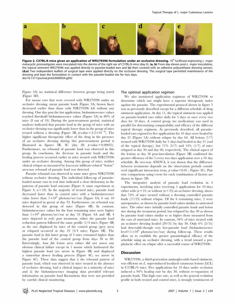
Figure 3A) no statistical difference between groups being noted
(Figure 3D).
For mouse ears that were covered with WR279396 under an
occlusive dressing, mean parasite loads (Figure 3A; brown lines)
decreased earlier than those with WR279396 left without any
dressing. One day post the last application, bioluminescence values
reached threshold bioluminescence values (Figure 3A) in 80% of
mice (8 out of 10). During the post-treatment period, statistical
analyses indicated that parasite load in the group of mice with an
occlusive dressing was significantly lower than in the group of mice
treated without a dressing (Figure 3B, p-value = 2.261028). The
higher significant therapeutic effect of the drug in the presence
of an occlusive dressing during this post-treatment period is
illustrated in figures 3B, 3C (day 28: p-value = 0.00055).
Furthermore, no rebound of parasite load was observed in this
group. In conclusion, the decrease in parasite loads and the
healing process occurred earlier in mice treated with WR279396
under an occlusive dressing. Among this group of mice, neither
clinical relapse-as measured by leucocyte infiltrate-related ‘‘lesion’’
area-nor rebound of parasite load was detected.
Parasite rebound was observed in some mice given WR279396
without occlusive dressing. The individual follow-up of parasite-
loaded mouse ears in real time indicated a clear dichotomy in the
patterns of parasite load outcome (Figure 4; same experiment as
Figure 3; n = 10). In the majority of treated mice, parasite load
decreased faster than in control mice with a bioluminescence
value lower than 16106 photons/sec/ear (Figure 4A; 6 out 10
mice depicted in green) at day 33. Furthermore, no rebound was
detected in this group of mice (Figure 4B). In contrast,
bioluminescence values for the four remaining mice were higher
than 16106 photons/sec/ear at day 33 (Figure 4A and 4B; 4
mice depicted in red): post treatment, either the parasite load
reduction pattern followed the same profile (1/4 mice; Figure 4B)
as the one displayed by mice of the control group (grey area)
or relapsed occurred at day 22 (3/4 mice; Figure 4B). The
parasite load in this latter group of 3 mice remained higher than
the parasite load of the control group from days 30 to 60.
Interestingly, bona fide lesion area values did not assess any
obvious clinical failure except in 1 mouse which harboured the
highest parasite load (see arrow in Figure 4B) and displayed
a somewhat slower healing process (Figure 4C; see arrow in
Figure 4C). These data suggest that i) the rebound pattern of
parasite load, which was observed in mice treated in the absence
of occlusive dressing, had a clinical impact in a minority of mice
and ii) the bioluminescence imaging data provided relevant
information on parasite load fluctuations that were not provided
by careful clinical monitoring.
The optimal application regimenWe also monitored application regimens of WR279396 to
determine which one might have a superior therapeutic index
against the parasite. The experimental protocol shown in figure 5
was as previously described except for a different schedule of drug
ointment application. At day 11, the topical ointment was applied
on parasite-loaded ears either daily for 5 days or once every two
days for 10 days. A control group (no medication) was used in
parallel for determining comparability and efficacy of the different
topical therapy regimens. As previously described, all parasite-
loaded ears exposed to five applications for 10 days were healed by
day 21 (Figure 5A) without relapse by day 64. All lesions (7/7)
treated with WR279396 daily for 5 days had healed at day 21 (end
of the topical therapy), but 71% (5/7) and 14% (1/7) of mice
relapsed at day 50 and day 60, respectively. The clinical aspect of
the lesions at day 36 post-inoculation (Figure 5B) confirmed the
greater efficiency of the 5 every two days application over a 10 day
schedule. By two-way ANOVA, it was shown that the difference
between treatments depends on the observation periods consid-
ered (significant interaction term, p-value,0.05 ; Figure 5C). Pair
wise comparisons using t-tests for each combination of factors are
shown in figure 5D.
The integrative analysis of parasite load evolution in 4
experiments, involving mice receiving 5 applications for 10 days
either with (n = 31) or without (n = 23) an occlusive dressing, shows
that 74% of mice treated without a dressing controlled parasite
loads (17/23) without relapse. Of the 6 remaining mice, 2 were
unresponsive, as shown by parasite load values similar to untreated
mice. The other mice initially controlled parasite loads and lesion
size during the treatment period, but relapsed by day 30 as shown
by parasite load values similar to or higher those measured from
the ears of untreated mice. In contrast, 94% of mice treated with
an occlusive dressing healed (29/31) by day 30. Only 6% (2/31)
had detectable-though very low-parasite load (bioluminescence
level,16106 photons/sec/ear) during follow-up. These results
allow us to establish the greater parasitological efficacy of the
schedule using an occlusive dressing, with a trend toward a pro-
phylactic effect on relapse after a successful course of WR279396.
Discussion
WR279396, a third-generation aminoglycoside-based ointment,
was efficient on L. major-induced localized cutaneous lesions (LCL)
in C57BL/6 mice. Five applications for 10 days under occlusion
induced a 94% healing rate by day 30, without re-expansion of
parasite loads. This high cure rate, as well as the general evolution
profile in both treated and control mice, is strongly reminiscent of
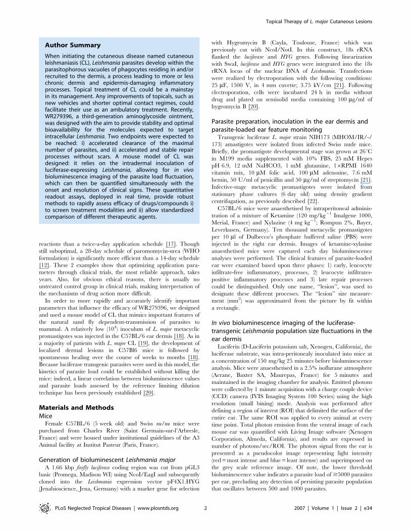
Figure 2. C57BL/6 mice given an application of WR279396 formulation under an occlusive dressing. 104 luciferase-expressing L. majormetacyclic promastigotes were inoculated into the dermis of the right ear of C57BL/6 mice (day 0). (a, b) From day eleven post-L. major inoculation,the topical ointment WR279396 was applied directly to parasite-loaded ears and (c) then covered with an adhesive polyurethane dressing (arrow).(d,e) Two independent leaflets of surgical tape were applied directly on the occlusive dressing. This surgical tape permitted maintenance of thedressing and kept the formulation in contact with the parasite-loaded site for two days.doi:10.1371/journal.pntd.0000034.g002
Topical Therapy of L. major Cutaneous Lesions
PLoS Neglected Tropical Diseases | www.plosntds.org 5 2007 | Volume 1 | Issue 2 | e34

what has been observed in clinical trials (Ben Salah, Buffet et al.-
submitted and [14]), providing a strong validation of this new
model for drug-testing purposes.
The adjuvant use of an occlusive dressing significantly enhanced
control of parasite loads. Several non-mutually exclusive mech-
anisms may account for these effects. First, the dressing prevented
removal of the ointment from the lesion by protecting the skin
from scratching, rubbing and scraping. These latter observations
have been made in patients treated without occlusion, an
important proportion of the ointment being wiped off by clothes
during the day, sheets during the night or even attracted to
a ‘‘protective’’ gauze put on the top of ulcerated lesions. Second,
occlusion on burns or wounds favours epidermal regeneration (ie,
ulceration closure). Finally, water retention by semi-permeable
occlusive dressing (like the polyurethane film used here) results in
hydration of the ointment application zone [28] and likely
improves the penetration and diffusion of hydrophilic antiparasitic
compounds into the dermis [15], where intracellular amastigotes
multiply. The aminoglycosides paromomycin and gentamicin, the
active ingredients in the WR279396 ointment, are OH-rich
hydrophilic compounds. Whether the dominant mechanism of the
adjuvant effect is merely mechanical (enough ointment maintained
on the lesion) or linked to dermal diffusion issues, the occlusive
dressing enhanced the healing process induced by active
Figure 3. Earlier and sustained control of both parasite load and lesion healing post topical applications of WR 279396. 104
luciferase-expressing L. major metacyclic promastigotes were inoculated into the dermis of the right ear of C57BL/6 mice (day 0) and mice werefollowed for 3 months. We compared parasite load and clinical feature outcome in mice left without any ointment (group 1, blue line) or receivingthe WR279396 ointment (one application ( q ) every two days, over ten days either without dressing (group 2, grey line) or with an occlusive dressing(group 3, brown line) at 11 day post-inoculation of promastigotes. Total photon emission from the L. major-loaded site (A, photons/sec/ear) isdepicted as medians 6sd. Lesion area (B; mm2) and representative pictures of C57BL/6 L. major loaded-ears (C), at day 28 post-inoculation, arepresented. Significant differences are indicated as follows: * for P,0.001. White squares delimit the ‘‘lesion’’ area of groups 1, 2 and 3 respectively. Thethree periods of observation (treatment, post-treatment and final) are represented by a colour code. (D) Comparison of bioluminescence (naturallogarithm (ln) of photons/sec/ear) for each group of mice during the period of observation. The box plot for each group, assessed by a two-wayANOVA, graphs the percentile and median of parasite loads. The ends of the box define the 25th and 75th percentiles, with a line at the median anderrors bars defining the 10th and 90th percentiles. The dots outside the ends of the whiskers are outliers. The p-values are displayed on the top ofeach box with the following colour code: -blue indicates that the group under study is compared to the blue group-grey indicates that the groupunder study is compared to the grey group.doi:10.1371/journal.pntd.0000034.g003
Topical Therapy of L. major Cutaneous Lesions
PLoS Neglected Tropical Diseases | www.plosntds.org 6 2007 | Volume 1 | Issue 2 | e34

Figure 4. Individual follow up of bioluminescence (parasite load) and ‘‘lesion’’ area in mice treated with WR279396 without anydressing. 104 luciferase-expressing L. major metacyclic promastigotes were inoculated into the dermis of the right ear of C57BL/6 mice (n = 10; day0) treated with WR279396 (5 applications (q) for 10 days) at day 11 post-inoculation. The parasite load (photons/sec/ear) in individual mice (A, B) andthe area (mm2; C) of the lesion displayed by the same mice were followed for 3 months. (A, B, C) Green colour assesses the profile in mice thatcontrolled their ear parasite load, i.e. exhibiting a bioluminescence value,16106 photons/sec/ear at day 33 (green points in panel A and green linesin panels B and C). In contrast, red colour corresponds to mouse ears that display a high bioluminescence value at day 33 (.16106 photons/sec/ear; red points in panel A and red lines in panels B and C). Note that lesion area values (C) did not assess any clinical failure except in 1 mouse(arrow). Values obtained for control mice are shown in the grey areas indicated in each graph.doi:10.1371/journal.pntd.0000034.g004
Topical Therapy of L. major Cutaneous Lesions
PLoS Neglected Tropical Diseases | www.plosntds.org 7 2007 | Volume 1 | Issue 2 | e34
ingredients, and prevented persisting parasite loads to re-expand.
Now that this adjuvant effect is established, future studies should
be set up for dissecting its fine mechanisms.
Apart from a mild difference in thickness, LCL lesions in mice
resemble human lesions both clinically and histologically. As
opposed to systemic drug testing, topical drug testing in mice will
be relevant since potential pharmacokinetic differences between
mouse and human skin are expected to be minor and easily
tractable to further analyses. These observations, along with the
careful validation of the model, support the assumption that our
results are likely to apply to human therapy. To our knowledge,
occlusion has never been fully validated as an adjuvant for the
topical therapy of human cutaneous lesions driven by invasive
microorganisms, but several case reports have proposed this
approach both in CL [29,30] and non-infectious dermatologic
conditions [15]. We provide here a strong validation of a concept
that might apply to other antileishmanial or antimicrobial
ointments. Ointments are usually painless, their application
requires no sophisticated expensive device or local anaesthesia
and they can be applied easily to both children and adults by
a primary care health provider with minimal training. Excluding
those L. braziliensis. foci where the incidence of mucosal extension
can be high, ointment therapy of cutaneous lesions otherwise
declared as neglected diseases should be favoured since the
potential adjuvant effect of occlusion might help some ointment
formulations to reach the required efficacy for development.
The duration of treatment is another important determinant of
reaching a stable cure. A very short 5-day daily application
Figure 5. Determination of the most suitable regimen for WR279396 application under an occlusive dressing. Eleven days post-inoculation of 104 luciferase-transfected L. major parasites, three groups of 7 mice were constituted: In the control group, mouse ears were leftwithout any ointment (blue plain line), and in the other group WR279396 was applied on the mouse ear under an occlusive dressing with differentapplication frequencies; either 5 applications/10 days (brown plain line) or 5 applications/5 days (brown dotted line). Bioluminescence evaluation ofparasite load was performed over the course of 64 days post-inoculation (A). Note that the rebound of the parasite load in the every day-applicationgroup can be associated to a more severe lesion as illustrated by a representative mouse at day 36 post-inoculation (B/5 days). White squares delimitthe ‘‘lesion’’ area. (C, D) Parasites loads were analysed with ANOVA whose two factors were the treatment (WR279396 10 days or 5 days) and theperiod of observation. Pair wise comparisons using t-tests were realized for each combination of factors.doi:10.1371/journal.pntd.0000034.g005
Topical Therapy of L. major Cutaneous Lesions
PLoS Neglected Tropical Diseases | www.plosntds.org 8 2007 | Volume 1 | Issue 2 | e34
schedule under occlusion led to a ‘‘rebound’’ pattern similar to
that displayed in mice treated for 10 days without occlusion. In
other words, too short of an application period may lead to
parasite load rebound, this latter risk being partially controlled by
an occlusive dressing. The ability to perform individual mouse
follow-up revealed a dichotomic pattern of parasite load evolution
(‘‘sustained control’’ versus ‘‘unstable control with parasite
rebound’’), pervasive over many weeks post transient topical
ointment application. Interestingly, these patterns were displayed
over several weeks, i.e., well beyond the treatment application
period. Parasite load level at the end of applications was not a good
predictor of further evolution (Figure 4). So, not only parasite
killing but also some modification of parasite environment
determined the long-term outcome of tissue damage and repair
processes. It is then very likely that, during the treatment
application period, some integrated programs are triggered that
will be the dominant determinant of evolution. Those results fit
well with observations in human CL, such as the low prognosis
value of parasitological tests at the end of therapy or the efficacy of
therapeutic schedules stopped before lesion healing [4,31]. Taken
together, these experiments show that parasites must be exposed to
the drug for.5 days to drive evolution toward long term sustained
control of parasite loads and clinical healing. Duration of drug
exposure was a stronger determinant of outcome than the total
amount of drug used. Intracellular pharmacokinetics of aminogly-
cosides helps understand the mechanism leading to this observa-
tion. In eukaryotic cells exposed to aminoglycosides in vitro, a slow
(2–4 days) lysosomal accumulation is observed, followed, when
aminoglycosides are removed from the extracellular medium, by
an even slower (2–5 days) release [32]. Interestingly, the lysosome is
the only subcellular compartment in which aminoglycosides
accumulate, an important feature of their antileishmanial efficacy
[32,33]. So, provided that appropriate concentrations of aminogly-
cosides are reached in the dermal intercellular space, relatively
discontinuous applications would probably suffice to allow in-
tracellular killing of replicating amastigotes and long term sustained
control of parasite loads. Taken together, our observations will help
select the most efficient ointment application schedules for
implementation, in the context of the therapy of this neglected
disease, by health care providers with little resources and heavy
duties. Even relatively discontinuous applications for a few weeks
should be preferred to many daily applications for a few days.
Our model offers relevant preclinical readout assays i) of the
efficacy of a topical ointment delivered under occlusion or not ii)
for establishing the proper regimen/schedule that allows sustained
parasite load reduction and lesion healing during post-treatment
period features. This luciferase-based imaging study might be
useful for pre-clinical evaluation of novel formulations containing
molecules that target parasite-loaded cells residing in the dermis as
well as molecules that contribute to damaged skin-repair processes.
Next challenges will be to screen molecules expected to act on the
amastigote population that persist in the dermis or in distant sites
[34] and to investigate the acquisition in real time of long-term
protective immunity.
Acknowledgments
The authors would like to thank Marie-Anne Nicola for her appreciated
assistance with the IVIS Imaging System 100 (Xenogen Corporation) in
the Plate-Forme d’Imagerie Dynamique. The authors are extremely
grateful to Dr Max Grogl and Dr. Philip Smith (US Army Medical
Materiel Development Activity; Fort Detrick, MD) for kindly providing the
WR279396 ointment. We would like also to thank Christine Maillet for her
excellent technical assistance and Serge Hedan for his help in taking care of
our mice.
Author Contributions
Conceived and designed the experiments: GM PB TL HL. Performed the
experiments: PB TL HL SG. Analyzed the data: PB TL HL. Contributed
reagents/materials/analysis tools: TL HL GM SG. Wrote the paper: GM
GG PB TL HL. Statistical analysis: GG.
References
1. Desjeux P (2001) Worldwide increasing risk factors for leishmaniasis. MedicalMicrobiology&Immunology 190: 77–79.
2. Alvar J, Yactayo S, Bern C (2006) Leishmaniasis and poverty. Trends in
Parasitology 22: 552–557.
3. Buffet P, Morizot G (2003) Cutaneous leishmaniasis in France: towards the end
of injectable therapy? Bull Soc Pathol Exot 96: 383–388.
4. Magill AJ (2005) Cutaneous leishmaniasis in the returning traveler. Infect DisClin North Am 19: 241–266.
5. Bryceson A (1987) Therapy in man. In: Peters W, Killick-Kendrick R, eds. The
leishmaniases. London: Academic Press. pp 847–869.
6. Garnier T, Croft SL (2002) Topical treatment for cutaneous leishmaniasis.Current opinion in investigational drugs 3: 538–544.
7. Asilian A, Sadeghinia A, Faghihi G, Momeni A, Harandi AA (2003) The efficacy
of treatment with intralesional meglumine antimoniate alone, compared withthat of cryotherapy combined with the meglumine antimoniate or intralesional
sodium stibogluconate, in the treatment of cutaneous leishmaniasis. Ann Trop
Med Parasitol 97: 493–498.
8. El-On J, Livshin R, Even-Paz Z, Hamburger D, Weinrauch L (1986) Topicaltreatment of cutaneous leishmaniasis. J Invest Dermatol 87: 284–288.
9. El-On J, Halevy S, Grunwald MH, Weinrauch L (1992) Topical treatment of
Old World cutaneous leishmaniasis caused by Leishmania major: a double-blindcontrol study. J Am Acad Dermatol 27: 227–231.
10. el-Safi SH, Murphy AG, Bryceson AD, Neal RA (1990) A double-blind clinical
trial of the treatment of cutaneous leishmaniasis with paromomycin ointment.Trans R Soc Trop Med Hyg 84: 690–691.
11. Bryceson ADM, Murphy A, Moody AH (1994) Treatment of Old-World
Cutaneous Leishmaniasis with Aminosidine Ointment-Results of an Open Study
in London. Trans R Soc Trop Med Hyg 88: 226–228.
12. Asilian A, Jalayer T, Whitworth JA, Ghasemi RL, Nilforooshzadeh M, et al.(1995) A randomized, placebo-controlled trial of a two-week regimen of
aminosidine (paromomycin) ointment for treatment of cutaneous leishmaniasisin Iran. Am J Trop Med Hyg 53: 648–651.
13. Ben Salah A, Zakraoui H, Zaatour A, Ftaiti A, Zaafouri B, et al. (1995) A
randomized, placebo-controlled trial in Tunisia treating cutaneous leishmaniasiswith paromomycin ointment. Am J Trop Med Hyg 53: 162–166.
14. Soto JM, Toledo JT, Gutierrez P, Arboleda M, Nicholls RS, et al. (2002)
Treatment of cutaneous leishmaniasis with a topical antileishmanial drug
(WR279396): Phase 2 pilot study. Am J Trop Med Hyg 66: 147–151.
15. Mrowietz U, Graeber M, Brautigam M, Thurston M, Wagenaar A, et al. (1998)
The novel ascomycin derivative SDZ ASM 981 is effective for psoriasis when
used topically under occlusion. Br J Dermatol 139: 992–996.
16. Iraji F, Sadeghinia A (2005) Efficacy of paromomycin ointment in the treatment
of cutaneous leishmaniasis: results of a double-blind, randomized trial in Isfahan,
Iran. Ann Trop Med Parasitol 99: 3–9.
17. Krause G, Kroeger A (1994) Topical treatment of American cutaneous
leishmaniasis with paramomycin and methylbenzethonium chloride: a clinical
study under field conditions in Ecuador. Trans R Soc Trop Med Hyg 88: 92–94.
18. Belkaid Y, Mendez S, Lira R, Kadambi N, Milon G, et al. (2000) A natural
model of Leishmania major infection reveals a prolonged ‘‘silent’’ phase of parasite
amplification in the skin before the onset of lesion formation and immunity.
J Immunol 165: 969–977.
19. Belazzoug S, Neal RA (1986) Failure of Meglumine Antimoniate to Cure
Cutaneous Lesions Due to Leishmania-Major in Algeria. Trans R Soc Trop
Med Hyg 80: 670–671.
20. Lang T, Goyard S, Lebastard M, Milon G (2005) Bioluminescent Leishmania
expressing luciferase for rapid and high throughput of drugs acting on
amastigote-harbouring macrophages and for quantitative real-time monitoring
of parasitism features in living mice. Cellular Microbiology 7: 383–392.
21. Goyard S, Segawa H, Gordon J, Showalter M, Duncan R, et al. (2003) An in
vitro system for developmental and genetic studies of Leishmania donovani
phosphoglycans. Mol Biochem Parasitol 130: 31–42.
22. Spath GF, Beverley SM (2001) A lipophosphoglycan-independent method for
isolation of infective Leishmania metacyclic promastigotes by density gradient
centrifugation. Exp Parasitol 99: 97–103.
23. Grogl M, Schuster BG, Ellis WY, Berman JD (1999) Successful topical treatment
of murine cutaneous leishmaniasis with a combination of paromomycin
(Aminosidine) and gentamicin. J Parasitol 85: 354–359.
24. Leclercq V, Lebastard M, Belkaid Y, Louis J, Milon G (1996) The outcome of
the parasitic process initiated by Leishmania infantum in laboratory mice: a tissue-
Topical Therapy of L. major Cutaneous Lesions
PLoS Neglected Tropical Diseases | www.plosntds.org 9 2007 | Volume 1 | Issue 2 | e34

dependent pattern controlled by the Lsh and MHC loci. J Immunol 157:
4537–4545.
25. Sunderkotter C, Kunz M, Steinbrink K, Meinardus-Hager G, Goebeler M, et al.
(1993) Resistance of mice to experimental leishmaniasis is associated with more
rapid appearance of mature macrophages in vitro and in vivo. J Immunol 151:
4891–4901.
26. Strijbosch LW, Buurman WA, Does RJ, Zinken PH, Groenewegen G (1987)
Limiting dilution assays. Experimental design and statistical analysis. J Immunol
Methods 97: 133–140.
27. Taswell C (1981) Limiting dilution assays for the determination of immuno-
competent cell frequencies. I. Data analysis. J Immunol 126: 1614–1619.
28. Berardesca E, Vignoli GP, Fideli D, Maibach H (1992) Effect of Occlusive
Dressings on the Stratum-Corneum Water Holding Capacity. Am J Med Sci
304: 25–28.
29. Bell SA, Schaller M, Rocken M (1997) Occlusive paromomycin for cutaneous
leishmaniasis [see comments]. Lancet 349: 29.30. Saurat JH (1993) Cutaneous Leishmaniasis Treated with Controlled Localized
Heating. Arch Dermatol 129: 510–510.
31. Berman J (2003) Current treatment approaches to leishmaniasis. CurrentOpinion in Infectious Diseases 16: 397–401.
32. Tulkens P, Trouet A (1978) Uptake and Intracellular Accumulation ofAminoglycoside Antibiotics in Lysosomes of Cultured Rat Fibroblasts. Biochem
Pharmacol 27: 415–424.
33. Tulkens PM (1991) Intracellular-Distribution and Activity of Antibiotics.European Journal of Clinical Microbiology&Infectious Diseases 10: 100–106.
34. Nicolas L, Sidjanski S, Colle J-H, Milon G (2000) Leishmania major reaches distantcutaneous sites where it persists transiently while persisting durably in the
primary dermal site and its draining lymph node: a study with laboratory mice.Infect Immun 68: 6561–6566.
Topical Therapy of L. major Cutaneous Lesions
PLoS Neglected Tropical Diseases | www.plosntds.org 10 2007 | Volume 1 | Issue 2 | e34
Related Documents