18/08/2011 1 Principles of Flow Cytometry (Practised in a Clinical Laboratory) Noel Williams Immunobiology Division of Immunology Optical Measurement Principles Cytometer Components Reagents Cytometer Setup Cytometer Daily Setup and Quality Control Sample Preparation Automation Networked Components Quality Assurance Data Management Limitations Standardisation Clinical Laboratory Applications Data Analysis Software Cases Optical Measurement Principles

Welcome message from author
This document is posted to help you gain knowledge. Please leave a comment to let me know what you think about it! Share it to your friends and learn new things together.
Transcript
18/08/2011
1
Principles of Flow Cytometry(Practised in a Clinical Laboratory)
Noel Williams Immunobiology
Division of Immunology
Optical Measurement Principles
Cytometer Components
Reagents
Cytometer Setup
Cytometer Daily Setup and Quality Control
Sample Preparation
Automation
Networked Components
Quality Assurance
Data Management
Limitations
Standardisation
Clinical Laboratory Applications
Data Analysis Software
Cases
Optical Measurement Principles
18/08/2011
2
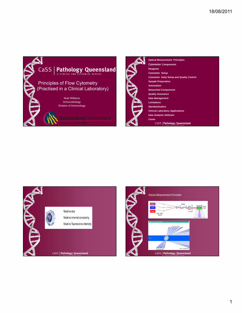
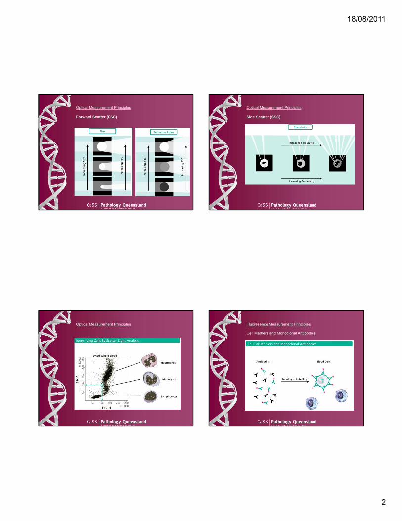
Optical Measurement Principles
Forward Scatter (FSC)
Optical Measurement Principles
Side Scatter (SSC)
Optical Measurement Principles Fluoresence Measurement Principles
Cell Markers and Monoclonal Antibodies

18/08/2011
3
Fluoresence Measurement Principles
Cell Markers and Monoclonal Antibodies
18/08/2011
4
Cytometer Components
Fluidics
Optics
Electronics
Ancillary Computer Software Network Connections
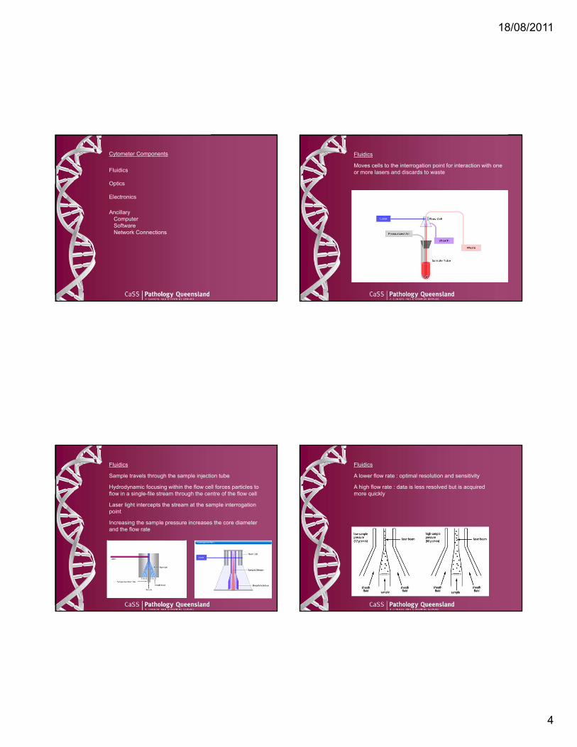
Fluidics
Moves cells to the interrogation point for interaction with one or more lasers and discards to waste
Fluidics
Sample travels through the sample injection tube
Hydrodynamic focusing within the flow cell forces particles to flow in a single-file stream through the centre of the flow cell
Laser light intercepts the stream at the sample interrogation point
Increasing the sample pressure increases the core diameter and the flow rate
Fluidics
A lower flow rate : optimal resolution and sensitivity
A high flow rate : data is less resolved but is acquired more quickly
18/08/2011
5
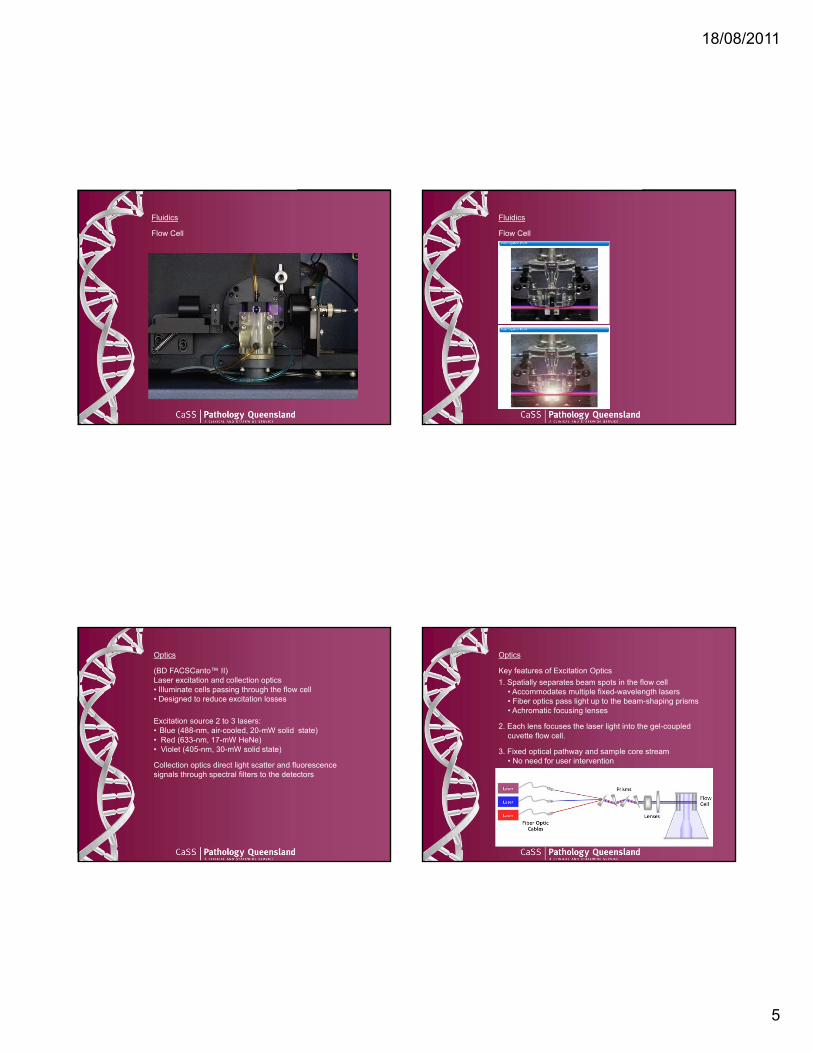
Fluidics
Flow Cell
Fluidics
Flow Cell
Optics
(BD FACSCanto™ II)Laser excitation and collection optics • Illuminate cells passing through the flow cell• Designed to reduce excitation losses
Excitation source 2 to 3 lasers: • Blue (488-nm, air-cooled, 20-mW solid state)• Red (633-nm, 17-mW HeNe)• Violet (405-nm, 30-mW solid state)
Collection optics direct light scatter and fluorescence signals through spectral filters to the detectors
Optics
Key features of Excitation Optics1. Spatially separates beam spots in the flow cell
• Accommodates multiple fixed-wavelength lasers• Fiber optics pass light up to the beam-shaping prisms• Achromatic focusing lenses
2. Each lens focuses the laser light into the gel-coupled cuvette flow cell.
3. Fixed optical pathway and sample core stream • No need for user intervention

18/08/2011
6
Excitation Optics
Menu of lasers
Working SelectionBlue laser 488nm 4 / Red laser 633nm 2 / Violet laser 405nm 2
OpticsOptics
Fluorochrome Excitation / Emmission
Optics
Collection Optics
The emission signals are transmitted from the flow cell to the detector arrays
Optics
Collection Optics
Detector arrays• an Octagon for the blue laser
• the octagon contains five PMTs and detects light from the 488-nm blue laser
• a PMT in the octagon collects side scatter signals
• a Trigon each for the red and the violet lasers• each trigon contains two PMTs and detect light from
the 633-nm (red) and the 405-nm (violet) lasers

18/08/2011
7
Optics
Collection Optics
The Octagon and Trigon detector arrays (BD-patented)use serial light reflections to guide signals to their target detectors• resulting in efficient light collection and signal retention at the detector level
• enhanced instrument sensitivity by collecting the dimmest emission signals first
Optics
Lasers
Collection Optics
Fibre optic cables
Detector Arrays
• Labelled filters• Long pass filter directs highest λ to first detector (PMT)
and progressively lower λ to successive detectors
• PMTs

18/08/2011
8
Electronic system
Main functions
Electronically remove debris
Correctly assign different datacollected from multiple lasers for each cell
Digitise light signals
Electronic system
Laser Delay
Electronic systemElectronic system
Converts optical signals to electronic signals and digitizes

18/08/2011
9
Electronic system Reagents
Sample Preparation•RBC lysis buffer•Monoclonal & Polyclonal Antibodies•DNA dye
•Propidium Iodide / 7-AAD•Fixative•Balanced Salt Solution
Instrument•Sheath Fluid•Instrument Tracking Beads•DI Water / Bleach
Monoclonal Antibody ManagementExpensive $300 at 500ul – 1000ulCatalogue of 100• Titred• Aliquotted• Light & temperature sensitive• Mixed into ‘Cocktails’• Cocktails verified before use
Cytometer Setup
Immediately Post Installation → Almost useless• Exceptions include Predefined Programming
• Lymphocyte Subset
Useful After :Setting Instrument Baseline (One baseline per configuration)
Cytometer Setup & Tracking Beads (CS&T)
Dilute suspension of beads analysed on the cytometer• Consist of fluoresence dim (2um), mid and bright (3um) polystyrene beads dyed with a mixture of fluorochromes which emit fluoresence measured in the detectors
• Establish baseline reference values• optical / electrical noise• detector efficiency• resolution of fluorescent populations
Cytometer Setup
Definition and linkage• Experiment = Test with
• Sample panel e.g. Acute Leukaemia Panel• Application settings
• Include PMT voltages / Threshold (Debris Cutoff)• Adjust (optimise) FSC / SSC / PMT (fluoresence)
voltages while acquiring positively stained control cells
• saved for subsequent use • Compensation settings
• spillover corrections determined using stained cells• mean MFI of positive and negative populations
aligned
18/08/2011
10
Cytometer Setup Cytometer Setup
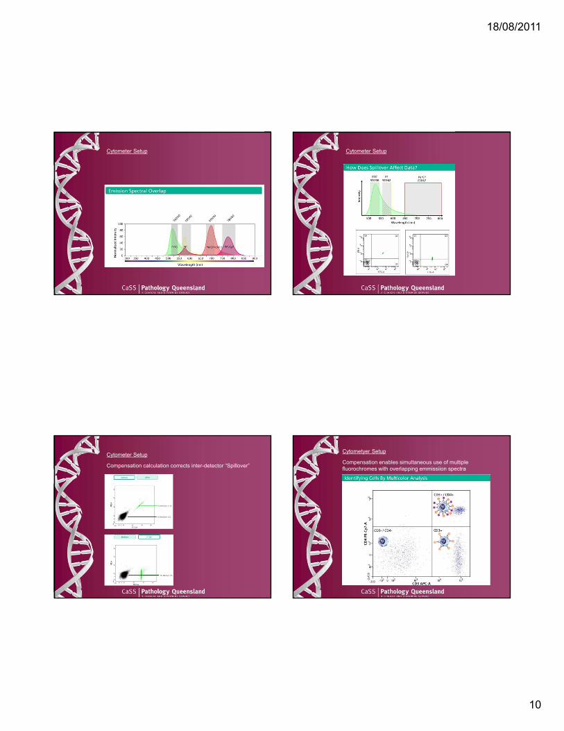
Cytometer Setup
Compensation calculation corrects inter-detector “Spillover”
Cytometyer Setup
Compensation enables simultaneous use of multiple fluorochromes with overlapping emmissiion spectra
18/08/2011
11
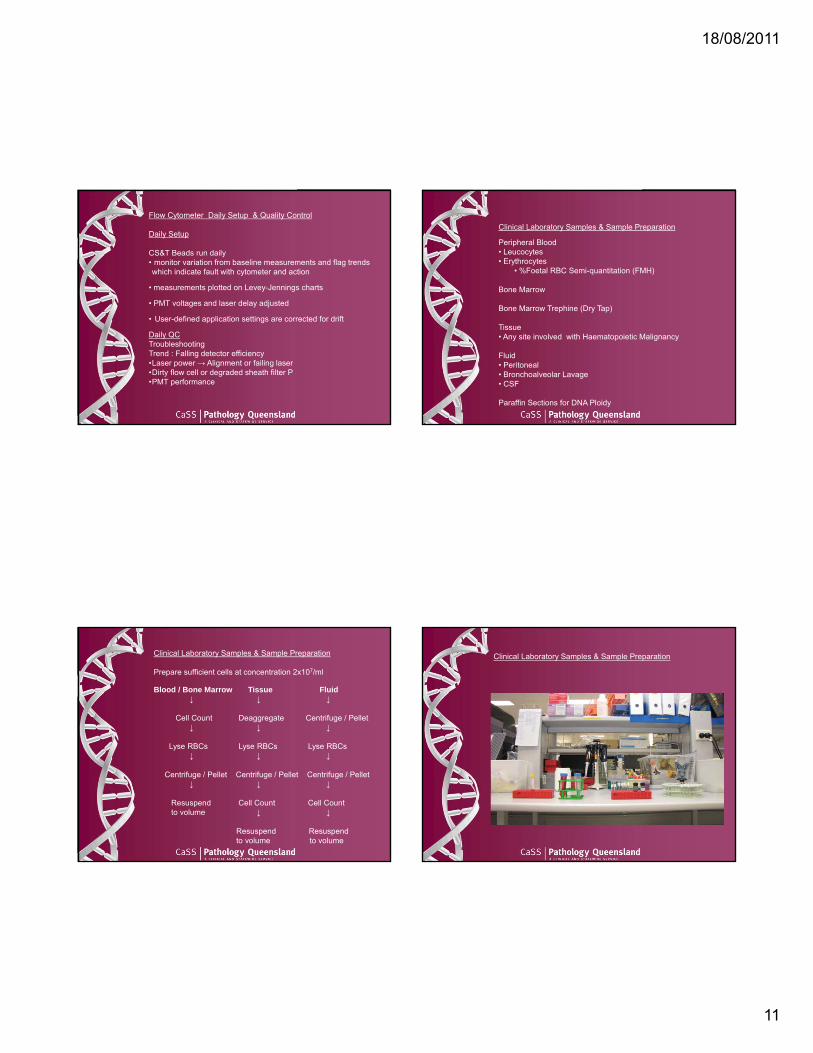
Flow Cytometer Daily Setup & Quality Control
Daily Setup
CS&T Beads run daily • monitor variation from baseline measurements and flag trends which indicate fault with cytometer and action
• measurements plotted on Levey-Jennings charts
• PMT voltages and laser delay adjusted
• User-defined application settings are corrected for drift
Daily QCTroubleshooting Trend : Falling detector efficiency •Laser power → Alignment or failing laser•Dirty flow cell or degraded sheath filter P•PMT performance
Clinical Laboratory Samples & Sample Preparation
Peripheral Blood• Leucocytes • Erythrocytes
• %Foetal RBC Semi-quantitation (FMH)
Bone Marrow
Bone Marrow Trephine (Dry Tap)
Tissue • Any site involved with Haematopoietic Malignancy
Fluid • Peritoneal • Bronchoalveolar Lavage• CSF
Paraffin Sections for DNA Ploidy
Clinical Laboratory Samples & Sample Preparation
Prepare sufficient cells at concentration 2x10⁷/ml
Blood / Bone Marrow Tissue Fluid↓ ↓ ↓
Cell Count Deaggregate Centrifuge / Pellet ↓ ↓ ↓
Lyse RBCs Lyse RBCs Lyse RBCs ↓ ↓ ↓
Centrifuge / Pellet Centrifuge / Pellet Centrifuge / Pellet↓ ↓ ↓
Resuspend Cell Count Cell Countto volume ↓ ↓
Resuspend Resuspendto volume to volume
Clinical Laboratory Samples & Sample Preparation
18/08/2011
12
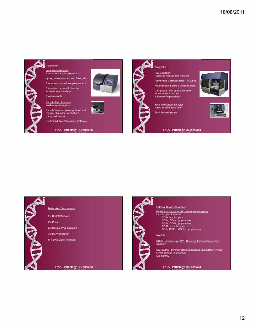
Automation
Lyse Wash AssistantAutomates sample preparation
Lyses, mixes, washes, and fixes cells
Processes up to 40 samples per atch
Eliminates the need to transfer samples to a centrifuge
Programmable
Sample Prep AssistantWalkaway automation
Sample tube cap piercing, blood and reagent aliquoting, incubations, lysing and mixing
Predefined & customizable protocols
Automation
FACS LoaderReplaces manual tube handling
Removable Carousel holds1-40 tubes
Automatically Loads & Unloads tubes
Compatible with other automation• Lyse Wash Assistant • Sample Prep Assistant
High Throughput SamplerRapid sample acquisition
96 & 384 well plates
Networked Components
2 x BD FACS Canto
2 x Printer
2 x Sample Prep Assistant
3 x PC Workstation
(1 x Lyse Wash Assistant)
External Quality Assurance
RCPA Immunology QAP : ImmunophenotypingLymphocyte Subset %
CD3+ lymphocytesCD3+, CD4+ LymphocytesCD3+, CD8+ LymphocytesCD19+ Lymphocytes CD3-, CD16+, CD56+ Lymphocytes
Monthly
RCPA Haematology QAP : Oncology ImmunophenotypingQuarterly
UK NEQAS : Minimal Residual Disease Quantitation (Acute Lymphoblastic Leukaemia)Six monthly
18/08/2011
13
Data Management
Quarterly backup to DVD • FCS files
• Immunophenotyping• Lymphocyte Subset
Electronic storage of Immunophenotyping Report Sheets
Standardisation
CLSIEnumeration of Immunologically Defined Cell Populations by Flow Cytometry; Approved Guideline - Second EditionLymphocyte subsets and CD34+ (hematopoietic) stem cells
Clinical Flow Cytometric Analysis of Neoplastic Hematolymphoid Cells; Approved Guideline - Second EditionPerformance guidelines for the immunophenotypic analysis of neoplastic hematolymphoid cells
AFCG
International Clinical Cytometry SocietyJuly/August Flow cytometry immunophenotyping for the evaluation of bone marrow dysplasia
Optimizing antibody panels for efficient and cost-effective flow cytometric diagnosis of acute leukemia
Standardisation
Across three laboratories of Pathology Queensland
• Screening Panels
• Core technical documents
• Reporting
• Specimen Rejection & Acceptance
Clinical Laboratory Applications
Diagnostic HaematologyRapid diagnosis & subclassification of Acute Leukaemia & Lymphoma by expression
• surface markers (B, T or NK-cell and myeloid markers),• cytoplasmic markers (MPO, CD3, CD22, CD79a etc) • nuclear markers (TdT)• forward & side light scatter• WHO Classification “Tumors of Haematopoietic &
Lymphoid Tissues”
Assessment of Minimal Residual Disease • Detect low levels of cells with aberrant immunophenotype• B and T-ALL and AML
Diagnosis of PNH
Primary Immunodeficiency
Enumeration CD34+ Stem Cells
DNA Ploidy• Triploidy in partial hydatiform mole • Aneuploidy in paediatric B-ALL
18/08/2011
14
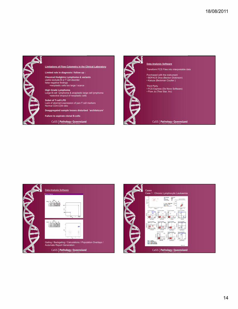
Limitations of Flow Cytometry in the Clinical Laboratory
Limited role in diagnosis / follow up :
Classical Hodgkins Lymphoma & variants useful exclude B or T cell disorderfalse negative findings
•neoplastic cells too large / scarce
High Grade LymphomaLarge B-cell lymphoma & anaplastic large cell lymphoma
•selective dropout of neoplastic cells
Subet of T-cell LPDLack of aberrant expression of pan-T cell markersNormal CD4:CD8 ratio
Deaggregated sample looses disturbed ‘architetcure’
Failure to aspirate clonal B-cells
Data Analysis Software
Transform FCS Files into interpretable data
Purchased with the instrument• BDFACS Diva (Becton Dickinson)• Kaluza (Beckman Coulter )
Third Party• FCS Express (De Novo Software)• Flow Jo (Tree Star, Inc)
Data Analysis Software
Gating / Backgating / Calculations / Population Overlays / Automate Report Generation
CasesCase 1 : Chronic Lymphocytic Leukaemia

18/08/2011
15
CasesCase 1 : Chronic Lymphocytic LeukaemiaApproximately 64% of total cells express CD5 weak, CD19, CD20, CD23 and kappa surface light chains
CasesCase 2 : Chronic Lymphocytic Leukaemia
Case 2Approximately 19% of total cells express CD5 weak, CD19, CD20, CD23 and indeterminate surface light chains
Case 2

18/08/2011
16
AcknowledgementsStaff ColleaguesAdam McKinlay
Staff ImmunopathologistsDr David Gillis
Becton Dickinson
Beckman Coulter
Related Documents