Cronicon OPEN ACCESS EC DENTAL SCIENCE Review Article Diabetes Mellitus as a Risk Factor for Periodontitis Samba Siva Reddy 1 , Krishna Kripal 1 *, Anuradha 2 and Ravi Varma Prasad 2 1 Department of Periodontology, Rajarajeswari Dental College and Hospital, Bangalore, Karnataka, India 2 Department of Periodontology, M.N.R. Dental College and Hospital, Sangareddy, Telangana State, India *Corresponding Author: Krishna Kripal, Department of Periodontology, Rajarajeswari Dental College and Hospital, Bangalore, Karnataka, India. Citation: Krishna Kripal., et al. “Diabetes Mellitus as a Risk Factor for Periodontitis”. EC Dental Science 1.S2 (2017): S12-S24. Received: February 23, 2017; Published: May 22, 2017 Abstract In cases of diabetes the function of immune cells, including neutrophils, monocytes and macrophages is often altered. Neutro- philic adherence, chemotaxis and phagocytosis are changed, inhibiting an adequate defense against bacteria in the periodontal pouch and significantly increasing the destruction of the periodontal membrane. In diabetics, even though the function of neutrophils is decreased, the monocyte/macrophage cell line may be hyper-responding when faced with the bacterial antigenic contact. As a result, the production of pro-inflammatory cytokines is increased. In diabetes patients, there is production of high levels of tumour necrosis factor-α (TNF- α) by peripheral monocytes. Inflammatory cytokines level in the gingival crevicular fluid is also related to glycemic control. Keywords: Diabetes Mellitus; Periodontal Diseases; Gingival Crevicular Fluid; Collagen Metabolism Mechanism of Diabetic Influence on Periodontium The possible mechanisms by which DM may have its effect on periodontium can be understood by evaluating the following [1]: 1. Sub-gingival microbiota 2. GCF glucose levels 3. Periodontal vasculature 4. Host response 5. Collagen metabolism 6. Genetic predisposition Changes in Sub Gingival Microbiota Earlier studies showed possible differences in sub-gingival microbiota between diabetic and non- diabetic patients with periodontitis but more recent research has demonstrated few differences in periodontally diseased sites of diabetic patients harbour similar species comparable to that in non- diabetic individuals [2,3]. So this lack of significance in the primary bacterial etiologic agents of periodontal disease suggests that increased prevalence and severity of periodontitis in diabetes may be due to differences in host response factors. Bacterial Associations Experimental diabetes in rats demonstrated that the shift in subgingival bacteria to a periodontopathic flora (Gram-negative rods and filaments). Capnocytophaga, Aggregatibacter actinomycetemcomitans species predominate in most periodontal lesions of young IDDM

Welcome message from author
This document is posted to help you gain knowledge. Please leave a comment to let me know what you think about it! Share it to your friends and learn new things together.
Transcript

CroniconO P E N A C C E S S EC DENTAL SCIENCE
Review Article
Diabetes Mellitus as a Risk Factor for Periodontitis
Samba Siva Reddy1, Krishna Kripal1*, Anuradha2 and Ravi Varma Prasad2
1Department of Periodontology, Rajarajeswari Dental College and Hospital, Bangalore, Karnataka, India2Department of Periodontology, M.N.R. Dental College and Hospital, Sangareddy, Telangana State, India
*Corresponding Author: Krishna Kripal, Department of Periodontology, Rajarajeswari Dental College and Hospital, Bangalore, Karnataka, India.
Citation: Krishna Kripal., et al. “Diabetes Mellitus as a Risk Factor for Periodontitis”. EC Dental Science 1.S2 (2017): S12-S24.
Received: February 23, 2017; Published: May 22, 2017
Abstract
In cases of diabetes the function of immune cells, including neutrophils, monocytes and macrophages is often altered. Neutro-philic adherence, chemotaxis and phagocytosis are changed, inhibiting an adequate defense against bacteria in the periodontal pouch and significantly increasing the destruction of the periodontal membrane. In diabetics, even though the function of neutrophils is decreased, the monocyte/macrophage cell line may be hyper-responding when faced with the bacterial antigenic contact. As a result, the production of pro-inflammatory cytokines is increased. In diabetes patients, there is production of high levels of tumour necrosis factor-α (TNF- α) by peripheral monocytes. Inflammatory cytokines level in the gingival crevicular fluid is also related to glycemic control.
Keywords: Diabetes Mellitus; Periodontal Diseases; Gingival Crevicular Fluid; Collagen Metabolism
Mechanism of Diabetic Influence on Periodontium
The possible mechanisms by which DM may have its effect on periodontium can be understood by evaluating the following [1]:
1. Sub-gingival microbiota
2. GCF glucose levels
3. Periodontal vasculature
4. Host response
5. Collagen metabolism
6. Genetic predisposition
Changes in Sub Gingival Microbiota
Earlier studies showed possible differences in sub-gingival microbiota between diabetic and non- diabetic patients with periodontitis but more recent research has demonstrated few differences in periodontally diseased sites of diabetic patients harbour similar species comparable to that in non- diabetic individuals [2,3]. So this lack of significance in the primary bacterial etiologic agents of periodontal disease suggests that increased prevalence and severity of periodontitis in diabetes may be due to differences in host response factors.
Bacterial Associations
Experimental diabetes in rats demonstrated that the shift in subgingival bacteria to a periodontopathic flora (Gram-negative rods and filaments). Capnocytophaga, Aggregatibacter actinomycetemcomitans species predominate in most periodontal lesions of young IDDM
S13
Diabetes Mellitus as a Risk Factor for Periodontitis
Citation: Krishna Kripal., et al. “Diabetes Mellitus as a Risk Factor for Periodontitis”. EC Dental Science 1.S2 (2017): S12-S24.
patients [4]. Higher levels of P. intermedia have been reported in diseased versus healthy periodontal sites in IDDM.
In diabetic patients, the percentage of streptococci (healthy colonizers), increased after improvement in the metabolic control of the diabetic state. The composition of the periodontal microflora of NIDDM patients appears to be similar to that found in chronic adult periodontitis. The three common predominant pathogens found in the subgingival dental plaque of NIDDM are Porphyromonas gingivalis, Prevotella intermedia, and Campylobacter rectus [2]. The presence of P. gingivalis and Aggregatibacter actinomycetemcomitans was similar to that found in chronic adult periodontitis.
Increased GCF Glucose Levels
Increased blood glucose levels in diabetes are reflected in increase levels of GCF glucose [5]. Invitro studies show decreased chemotax-is of periodontal ligament fibroblasts to PDGF when placed in hyperglycemic environment compared with normoglycemic conditions [6].
Thus, elevated glucose levels in diabetes may adversely affect periodontal wound healing events and the local host response to micro-bial challenge.
Periodontal Vasculature
Changes affecting the renal, retinal and perineural vasculature in diabetes also occur in the periodontium. Increased thickness of gin-gival capillary endothelial cell basement membranes and walls of small blood vessels may be seen in diabetic individuals [7].
This thickening may impair oxygen diffusion and nutrient provision across basement membranes. Increased thickness of small vessel walls results in narrowing of lumen, altering normal periodontal tissue homeostasis which occurs due to AGE’s in periodontal tissues. The increase in AGE accumulation in diabetic gingiva is twice when compared with gingiva of non- diabetic subjects. AGEs formation not only stimulates arterial smooth muscle cell proliferation, but also increases the thickness of vessel walls. In capillaries, enhanced cross linking of AGE modified collagen in the basement membrane inhibits the normal degradation of these proteins, increasing the thickness of basement membrane. Increased oxidant stress has been targeted as the underlying mechanism responsible for the widespread vascular injury associated with diabetes [8].
Elevated LDL levels especially common in type 2 diabetes may cause change in gingival vasculature. The AGE modified collagen in the gingival blood vessel walls can bind circulating LDL, resulting in atheroma formation and narrowing of vessel lumen. These events may play a role in increasing the severity and periodontitis progression by altering the tissue response to periodontopathic bacteria.
Alteration in Host Defence
Defects in PMN adherence, chemotaxis, and phagocytosis have been observed in some individuals with diabetes. Mc Mullen., et al. 1981 [9] stated that many of these defects are improved with glycemic control. Thus, due to these defects in first line of defence, increased tissue destruction has been shown in such patients. However, the tissue destruction in poorly controlled diabetic patients could be ex-plained due to another possible mechanism in whom hyper responsive PMNs or increased number of PMNs within the gingival crevice has been found as indicated by PMN derived beta-glucuronidase enzyme. Oliver., et al. 1993 [10] found the hyper responsive monocyte/macrophage phenotype which upon stimulation by bacterial antigens results in dramatically increased cytokine production. Offenbacher., et al. 1996 [11] stated that it may be genetically related also (Figure 1).

S14
Diabetes Mellitus as a Risk Factor for Periodontitis
Citation: Krishna Kripal., et al. “Diabetes Mellitus as a Risk Factor for Periodontitis”. EC Dental Science 1.S2 (2017): S12-S24.
Figure 1: Hypothetical mechanism indicating exaggerated innate immune system is a key element in inducing more severe forms of periodontal disease in type 1 and type 2 diabetic patients, and in obese
subjects.
Increased production of pro-inflammatory cytokines by monocytes derived from patients with diabetes. For example, in response to LPS of P. gingivalis, diabetic monocytes produced 24 to 32 times the level of TNFα, 4fold increase in PGE2 and IL-1β [12]. Formation of AGEs also play an important role in up-regulation of monocyte/macrophage cell line accumulation of AGEs in periodontium stimulated influx of monocytes.
Once in tissue, AGEs interact with receptor RAGE on monocyte cell surfaces. This halts migration of monocytes; fixing them at local site. AGE-RAGE interaction induces change in phenotype of monocytes and up-regulating the cell and cytokine production thus increasing cytokines TNF, PGE, IL-1B in diabetics [13]. However, there is a great deal of variability in AGE formation in different individuals, so there is variance in risk of periodontal complications in diabetics (Figure 2).
Figure 2: Diabetes, periodontal disease and inflammation.
Altered Collagen Metabolism and Changes in Wound Healing
Collagen is primary constituent of gingival connective tissue and organic matrix of alveolar bone. A change in collagen metabolism contributes to alterations in wound healing and to periodontal disease and its progression.
S15
Diabetes Mellitus as a Risk Factor for Periodontitis
Citation: Krishna Kripal., et al. “Diabetes Mellitus as a Risk Factor for Periodontitis”. EC Dental Science 1.S2 (2017): S12-S24.
In periodontium, MMPs including collagenase, gelatinase, elastases etc, produced by various major cell types when activated by cyto-kines and growth factors, are responsible for breakdown of bone and connective tissue during periodontal diseases. Increased collagen breakdown through stimulation of collagenase activity has been observed in periodontium of diabetics [13].
Collagenases degrade more newly formed collagen macro molecules. Sustained hyperglycemia causes AGE modification of existing collagen by increasing the cross linking. The net effect is rapid degradation of newly formed collagen by host collagenase and predomi-nance of older cross linked AGE modified collagen, there by result in altered wound healing in response to physical or microbial injury of periodontium.
Genetic Predisposition
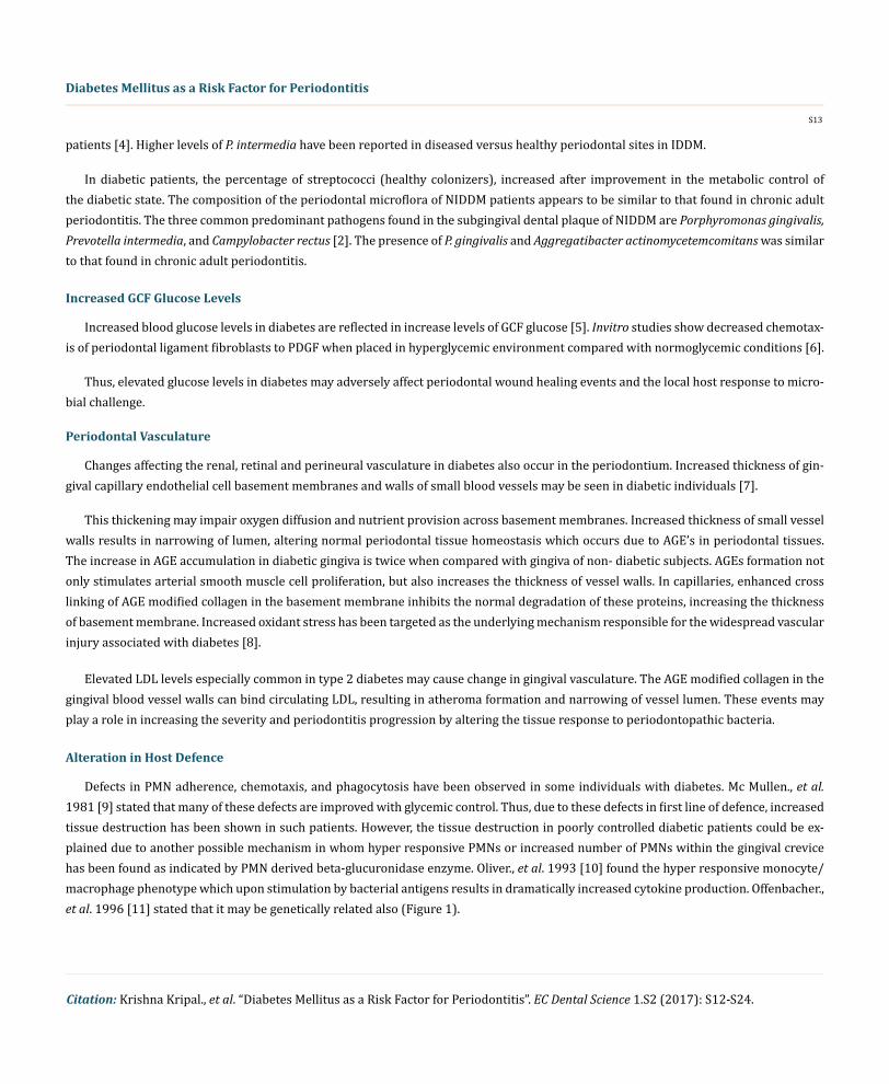
Antoni., et al. [14] reviewed and reported that both diabetes and periodontal disease are considered to have a hereditary component and a large number of cases have been related to a given family pattern. Hence both diseases can therefore be described as polygenic, because it has not yet proved possible to relate these dis-eases to any specific genetic mutation or disorder.
Genetic factors evidently play a major role in suscep¬tibility to these diseases. However, the complex inter-actions in periodontal dis-ease between host response mechanisms and the action of pathogenic microorgan-isms hamper clarification of the role of genetic factors. Nevertheless, an association has been identified between both diseases and an HLA genotype. The HLA molecule (human major histocom-patibility locus mol-ecule) is genetically determined on chromosome 6, and disorders in this chromosome appear to predispose the host to both diabetes and periodontitis by altering antigen presentation to T cells and therefore the specific immune response of the patient.
The 1704/G variant of the RAGE gene could be involved in the genetic background, determining a protection against chronic periodon-titis.
Figure 3: Risk factors of periodontitis in type 2 diabetes mellitus.
S16
Diabetes Mellitus as a Risk Factor for Periodontitis
Citation: Krishna Kripal., et al. “Diabetes Mellitus as a Risk Factor for Periodontitis”. EC Dental Science 1.S2 (2017): S12-S24.
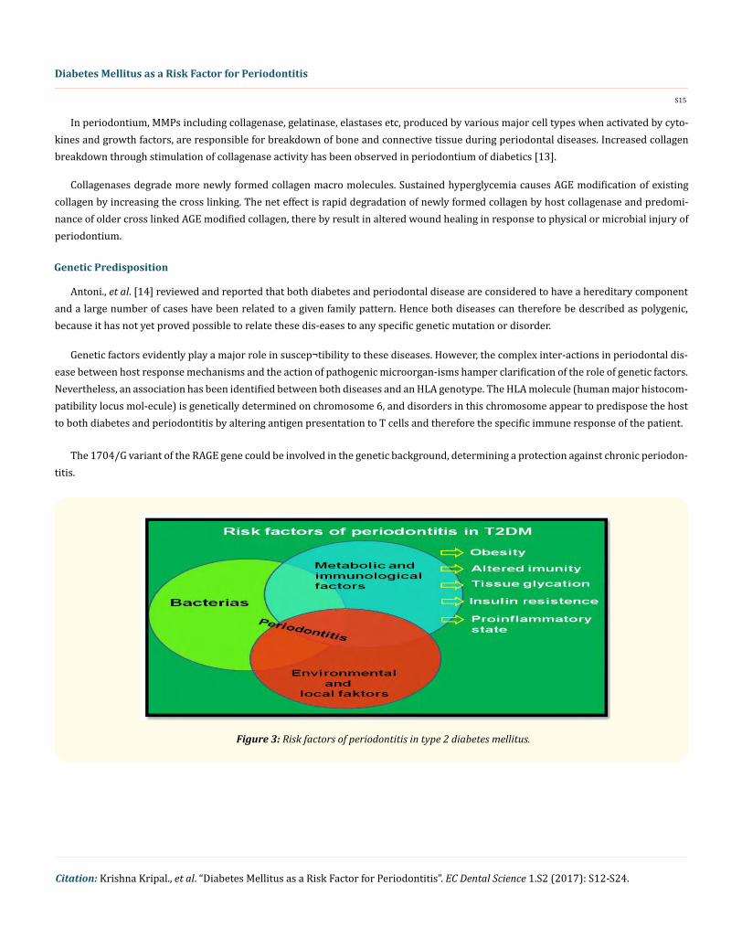
Figure 4: Consequences of non-enzymatic glycation in periodontal tissues.
Impact of Diabetes Mellitus on the Periodontium
Antoni., et al. [14] reviewed and reported that both diabetes and periodontal disease are considered to have a hereditary component and a large number of cases have been related to a given family pattern. Hence both diseases can therefore be described as polygenic, because it has not yet proved possible to relate these dis-eases to any specific genetic mutation or disorder.
Conversely, in diabetic people the monocyte–macrophage cell line may be hyperresponsive to bacterial antigens, as a result there is significant increase in production of pro-inflammatory cytokines and mediators. For example, peripheral blood monocytes from individu-als with diabetes demonstrate upregulated production of TNF-α in response to antigens from the Gram-negative, anaerobic periodontal pathogen Porphyromonas gingivalis, when compared to monocytes from people without diabetes. This hyperinflammatory monocyte/macrophage response results in elevated levels of pro-inflammatory cytokines in the gingival crevicular fluid that is present in the peri-odontal pocket adjacent to the teeth.
Furthermore, poorly controlled diabetes is associated with a two-fold elevation in crevicular fluid IL-1β levels compared to well con-trol diabetes. In diabetics because of host defence alterations there is increased periodontal inflammation and destruction of the support-ing teeth structures [16] (Figure 4).
Examination of the available data reveals strong evidence that diabetes is a risk factor for gingivitis and periodontitis, and the level of glycemic control appears to be an important determinant in this relationship. One must use caution in evaluating the research because studies examined diverse populations, often lacked controls or had small numbers of subjects, defined diabetes and glycemic control in various ways, and used different periodontal parameters to describe the clinical conditions present. Although some authors have not found a significant association between diabetes and gingival inflammation, in many studies, the prevalence and severity of gingivitis has been demonstrated to be higher in individuals with diabetes [17].
S17
Diabetes Mellitus as a Risk Factor for Periodontitis
Citation: Krishna Kripal., et al. “Diabetes Mellitus as a Risk Factor for Periodontitis”. EC Dental Science 1.S2 (2017): S12-S24.
In type 1 diabetic children, the prevalence of gingivitis was more than in non-diabetic children with same plaque levels. Number of sites with gingival inflammation in children with diabetes is twice as compared to non-diabetic control children with same plaque levels. Poor metabolic control can increase the severity of gingival inflammation in diabetic children, whereas improvement in glycemic control may be associated with decreased gingival inflammation [18].
In type 1 diabetic adults, the overall degree of gingival inflammation between diabetic subjects as a whole and non-diabetic control subjects with same plaque accumulation was similar. However, when diabetic patients in this study were stratified according to their level of glycemic control, significantly greater gingival bleeding was seen in poorly controlled diabetic patients than in either well-controlled diabetic subjects or non-diabetic controls. The number of bleeding sites decreased as glycemic control improved [19].
Gingival inflammation was also seen to be increased in adults with type 2 diabetes than in non-diabetic controls, with the highest level of inflammation in poor glycemic control subjects. A longitudinal experimental gingivitis study showed more rapid and pronounced devel-opment of gingival inflammation in relatively well controlled adult type 1 diabetic subjects than in non-diabetic controls, despite similar levels of plaque accumulation and similar bacterial composition of plaque, suggesting a hyperinflammatory gingival response in diabetes [20]. Above studies indicate that the presence of diabetes is often, but not always, associated with increased gingival inflammation. In ad-dition, in diabetic people the level of glycemic control may also play a role in the response of gingiva to bacterial plaque.
The preponderance of evidence indicate that the risk of periodontitis is increased by diabetes. A thorough meta-analysis concluded that the most of the Studies demonstrated an increase in severity of periodontal condition in diabetic adults than in adults without dia-betes. These studies were conducted on 3,500 diabetic adults, which clearly demonstrated a significant relation between periodontitis and diabetes.
Even in young age groups diabetes has been associated with an greater risk of periodontitis. In a group of 263 type 1 diabetic patients compared to 59 non-diabetic siblings and 149 non-diabetic unrelated controls, subjects below the age of 12 did not show periodontitis, but 13.6% of the diabetic individuals between 13 and 18 years of age had periodontitis, and the prevalence increased to 39% between 19 to 32 years. On comparison, the prevalence in non-diabetic control subjects was found to be < 3%. Epidemiologic studies in diabetic adults have often shown an increase in extent and severity of periodontitis [21].
In the Pima Indians of Arizona, a population with the highest occurrence of type 2 diabetes in the world, the prevalence and severity of attachment loss and bone loss was greater among diabetic subjects than among non-diabetic control subjects in all age groups. In a multivariate risk analysis, when compared to non-diabetic subjects, diabetic subjects showed 2.8- to 3.4-fold increased odds of having periodontitis after adjusting for the effects of confounding variables such as age, gender, and oral hygiene measures. In diabetic adults there is increased risk of attachment loss and bone loss as confirmed by Smaller cross-sectional and case-control studies. Longitudinal re-search has also shown an greater risk of progressive periodontal destruction in diabetic people. In a study conducted on the Pima Indians, the incidence and prevalence of periodontal disease were determined in 2,273 subjects 15 years of age or older [22].
The periodontitis prevalence was 60% in diabetic subjects and 36% in non-diabetics. The incidence was determined at baseline in a total of 701 subjects who were 15 to 54 years old, with little or no evidence of periodontitis. Subjects were followed for 2.5 years and it was found that the incidence of periodontitis was 2.6-fold higher in diabetic subjects than in non-diabetic patients. In another 2-year longitudinal study, type 2 diabetes subjects showed a fourfold greater risk of progressive alveolar bone loss compared to non-diabetic subjects [23].
It is tough to determine the relationship between metabolic control of diabetes and periodontal disease. Research indicate that this association is similar to the association between glycemic control and the classic complications of diabetes such as retinopathy and ne-

S18
Diabetes Mellitus as a Risk Factor for Periodontitis
Citation: Krishna Kripal., et al. “Diabetes Mellitus as a Risk Factor for Periodontitis”. EC Dental Science 1.S2 (2017): S12-S24.
phropathy; namely, in the diabetic population, there is significant heterogeneity. Even though the risk of diabetic complications increases with poor control of diabetes, there are many poorly controlled diabetic individuals without major complications. Conversely, the risk of diabetic complications decreases with good diabetic control, but there are people with well-controlled diabetes who suffer major dia-betic complications nonetheless. In same fashion, the evidence suggests that there is extensive periodontal destruction in some diabetic patients with poor glycemic control, whereas others do not. On the other hand, many well-controlled diabetic patients have excellent periodontal health, but others develop periodontitis (Figure 5 and 6).
Figure 5: Intense gingival response to dental plaque in a 38 year old female.
Figure 6: Localized gingival swelling and inflammation extending through the attached gingiva of teeth 23 and 24 in a 32 year old uncontrolled diabetic patient.
S19
Diabetes Mellitus as a Risk Factor for Periodontitis
Citation: Krishna Kripal., et al. “Diabetes Mellitus as a Risk Factor for Periodontitis”. EC Dental Science 1.S2 (2017): S12-S24.
In a large epidemiologic study conducted in the United States, there is 2.9 fold greater risk of having periodontitis in adults with poorly controlled diabetes compared to non-diabetic adult subjects; conversely, well-controlled diabetic subjects had no significant increase in the risk of periodontitis. In a cross-sectional study of patients who had type 1 diabetes for a mean duration of over 16 years, subjects with poor glycemic control had more interproximal attachment loss and bone loss than well-controlled subjects [24].
Same results have been found in other studies where the percentage of deep periodontal pockets and the prevalence of severe at-tachment loss increased as the glycemic control worsened. Type 1 diabetic subjects with poor metabolic control over the preceding 2 to 5 years had a significantly greater prevalence of deep probing depths and advanced attachment loss than subjects with good glycemic control. In the same way, there is significantly greater bone loss and attachment loss in poorly controlled diabetic subjects than well-controlled diabetic subjects over a 2- to 3-year follow-up period.
In longitudinal Pima Indian studies, poor glycemic control of type 2 diabetes was associated with an 11-fold increased risk of progres-sive bone loss compared to non-diabetic controls, whereas well-controlled diabetic subjects had no significant increase in risk. Thus, in the onset and progression of periodontal disease, metabolic control of diabetes may play an important variable [23].
Some studies gave only peripheral support to the relationship between glycemic control and the extent or severity of periodontitis, whereas some have shown no relationship. In a study of 118 diabetic subjects and 115 healthy controls, deeper probing depths and greater gingival inflammation, bleeding on probing, and attachment loss were seen in those with diabetes; however, the level of glycemic control among the diabetic subjects did not correlate to the periodontal parameters measured. Other study showed a trend toward an in-creasing alveolar bone loss as glycemic control worsened. The mean percentage of sites with > 15% bone loss increased from 28% in well-controlled type 1 diabetic subjects to 44% in poorly controlled subjects. But, the difference did not reach statistical significance, due to the small size of the study population. Some studies found no evidence of a relationship between glycemic control and periodontal status [25].
Extensive research has examined the mechanisms by which periodontal diseases might impact glycemic control in diabetes. From the discussion above, it is clear that inflammatory periodontal diseases may have significant effects on systemic health. Elevations in serum inflammatory and thrombotic mediators such as TNF-a, IL-6, fibrinogen and CRP in individuals with periodontitis may have a major im-pact on glycemic control, especially through their effect on insulin resistance. These mediators are significantly elevated in the presence of obesity, insulin resistance, hyperglycemia and diabetes. Obesity and insulin resistance are linked to the actions of IL-6 and TNF-a, and their resultant stimulation of acute phase reactant production in the liver [26]. In individuals with type 2 diabetes and periodontitis, el-evated serum inflammatory mediators resulting from the periodontal disease may add to existing insulin resistance, worsening glycemic control.
Inflammation
Cell Apoptosis and Periodontitis
Diabetes impaired immune function and increased risk of bacterial, viral and fungal infections. A study [27], with poor glycaemic control, showed decreased chemotactic activity and bactericidal activity leading to decreased release of ROS and lysosomal enzyme. Im-paired inflammatory response with hyperglycemia mediated alteration in lipid and protein function can result in AGEs and methylglyoxal formation in cell culture and human ex- vivo experiment. Occurrence of Apoptotic lymphocytes in diabetic decreased numbers of plasma lymphocytes in the patients. In patients with poorly controlled diabetes expanded apoptosis of lymphocytes may elucidate the impaired immune function. Neutrophil apoptosis is an integral component of inflammation and its resolution particularly. The enhanced loss of fibroblasts and osteoblasts through apoptosis in diabetics could contribute to limited repair of injured tissue, and influent the wound healing of periodontal tissue [27].

S20
Diabetes Mellitus as a Risk Factor for Periodontitis
Citation: Krishna Kripal., et al. “Diabetes Mellitus as a Risk Factor for Periodontitis”. EC Dental Science 1.S2 (2017): S12-S24.
Chen TL reported the molecular mechanisms of apoptosis on the onset of periodontitis and investigated the molecular control mecha-nism of apoptosis on periodontitis. The finding showed that the control gene for apoptosis are mainly p53, Bcl-2, c-mys. Reciprocity of Fas and Fasl showed relation to cell apoptosis. Two major pathways are involved in the process of apoptosis in periodontitis. One is the intrin-sic pathway for apoptosis induced by mitochondria, known as the intrinsic pathway. The death receptor (Fas/FasL) which is involved in the second pathway, is also known as the extrinsic pathway. The apoptosis of cell is related to lipid peroxide. Tetracycline and Vitamin C has the therapeutic effect on periodontitis by inhibiting apoptosis [28].
Bacterial Association
Bacteria residing in biofilms cause periodontal diseases along the tooth surface at interface of the gingival tissues and the tooth, most of which are Gram-negative and anaerobic. At this site, persistent microbial wound is present, a wound which is contaminated by any of the more than 400 bacterial species present in the mouth. An intact wound healing response is necessary to prevent local tissue destruc-tion or systemic dissemination of bacterial products (Figure 7). Biofilm-related products released into the periodontal pocket include bacterial endotoxins, chemotactic peptides and organic acids. Inflammation results in ulceration of the epithelial pocket lining, providing ready access of these compounds into the gingival tissues. This results in further stimulation of the host response, activation of host en-zymes including matrix metalloproteinases, and release of pro-inflammatory cytokines such as IL-1, TNF-a, IL-6, IL-17, and prostaglandin E2 (PGE2), among others. This cascade of events leads to eventual destruction of periodontal tissues [29].
Figure 7: Bacteria are necessary agents in the pathogenesis of periodontitis.
Diabetes Induced Periodontitis and Bone Loss
Anaerobes are the predominant pathological bacteria of periodontitis. Inflammatory response caused by periodontal bacteria and secretion resulted in periodontal tissue breakdown. Alveolar bone has the ability for bone remodelling and regeneration. Bacterial plaque accumulated on the tooth surface can stimulate the host response in the adjacent gingival and resulted in the destruction of periodontal tissue, and periodontal bone loss is the critical characters of periodontitis. When the absorption of bone exceeds new bone formation there is periodontal bone loss seen. Hyperglycemia is responsible for inflammatory response in diabetics.

S21
Diabetes Mellitus as a Risk Factor for Periodontitis
Citation: Krishna Kripal., et al. “Diabetes Mellitus as a Risk Factor for Periodontitis”. EC Dental Science 1.S2 (2017): S12-S24.
A study showed that T1DM decreased new bone formation and decreased bone mineral density leading to osteopenia. The effect of T1DM on bone is shown by a significant delay in fracture healing. Both T1DM and T2DM shoed 3 - 4 times increased the risk of periodon-titis. There is decreased fracture healing or osseous repair after marrow ablation in diabetics when compared with normals.
Bacterial insuling can induce the apoptosis of bone-lining cells on which diabetes had an intense effect. Diabetic mice bone surface has lesser cells than bone in normal and the increased apoptosis of bone-lining cells decreased the bone formation. Soft tissue wounds indicated diabetic mice had increased levels of apoptosis, and diabetes influence on apoptosis of matrix-producing cells and limit the repair of injured tissue [30].
Tuominen indicated that the reduced bone mass in T1DM had a higher bone loss and a profound effect on bone remodelling than T2DM. An inflammatory stimulus in animal model of T2DM showed the inhibition of osteoclastogenesis represented reducing new bone formation [30].
The mechanisms of hyperglycemia on periodontitis described as following. Firstly, hyperglycemia leading to increase gingival crevicu-lar fluid influences the microbial flora such as biofilm and accelerates the inflammatory processes in the mouth and alters the immune response of the periodontal bacteria infectious leading to the breakdown of periodontium. Secondly, hyperglycemia increases the sensi-tivity of bacteria to diabetes patients and alters the chemotaxis and adherence to neutrophil resulting in producing much inflammatory cytokine.
Increased levels of AGEs due to Hyperglycemia leads to pathological biochemical processes such as glycation of protein-like collagens or lipids and non- enzymatic oxidative destruction. AGEs has the ability to show influence on normal protein functions directly or act by reacting with receptors indirectly on the different cell membrane. The glycated products had the potential to create molecular complexes reducing the solubility of the target protein- like collagens and alter the functional properties of type 1 collagen and lamina. The expres-sion of cytokines and growth factors that occur by Interactions between AGEs and receptors are mediated by macrophages. In diabetic patients inflammatory responses induced by AGEs contribute to systemic degradation of periodontal tissue.
Blockade of receptors for the AGEs reduced alveolar bone loss and the effects of oxidative stress by blocking the activation of innate immunity may be used to treat periodontitis. Thirdly, increased inflammatory cytokines and secretion resulted in insulin resistance and in turn caused periodontal infection stimulate immune activity cell to release a number of inflammatory cytokine TNF1α and IL-1. TNF1α inhibited phosphorylation of insulin receptor and lessen the sensitivity of insulin leading to insulin resistance [31].
Diabetes mellitus are associated with altered collagen metabolism and increased bacteria pathogenic to periodontal tissue and there-by increased the severity of periodontitis. Matrix metalloproteinase involved in a number of physiological events and as the major option in collagen breakdown and periodontal tissue destruction. Increased levels of matrix metalloproteinase 8 and 9 in the gingival tissue of diabetic with periodontitis suggested that expression of matrix metalloproteinases contributes to failure of the healing in the diabetic. In chronic periodontitis, Periodontal therapy improves healing of tissue by inhibition of matrix metalloproteinase.
Periodontitis with diabetics enhanced susceptibility to infection due to diminished neutrophil recruitment and function and increased formation of inflammatory cytokines and delayed wound healing after bacterial inbreak. Bone loss increased because the effects of diabe-tes inhibited new bone formation and the apoptosis of bone lining cells increased. Enhanced expression of cytokines in vitro is capable of stimulating bone absorption in diabetics. And enhanced inflammation and bone absorption may increase risk and severity of periodon-titis with diabetes [32].

S22
Diabetes Mellitus as a Risk Factor for Periodontitis
Citation: Krishna Kripal., et al. “Diabetes Mellitus as a Risk Factor for Periodontitis”. EC Dental Science 1.S2 (2017): S12-S24.
Wound Healing
Impaired wound healing is one of the common complication associated with diabetes mellitus. In patients with diabetes mellitus heal-ing is characterized by decreased tensile strength of wounds when compared with controls, suggesting either defective matrix production or deposition. In the human mammal, healing is impaired due to diminished perfusion resulting from the presence of peripheral arterial disease as well as reduced sensory nerve function caused by peripheral neuropathy. It is thought that diabetic complications result due to periods of poor glycemic control. However, there may be involvement of aberrant growth factor expression or factors secondary to dia-betes, such as advanced glycation and cross-linking of matrix protein. Growth factor involvement has been implicated not only in diabetic wounds but also in other diabetic complications, such as diabetic retinopathy and nephropathy [33].
It is also stated that that in the normal process of wound healing, apoptosis is involved in the loss of granulation tissue, including fibroblasts and small vessels, occurring late as scars. By comparison, in a diabetic wound, throughout the healing process apoptosis is increased. This indicate aberrant control of cell survival. In contrast to person without diabetes, it is seen mainly in the late phase of heal-ing during scar formation. It has been shown that high ambient glucose concentration, linked to vascular complications in diabetes, in vivo modulates messenger RNA expression of fibronectin, collagen, tissue-type plasminogen activator and plasminogen activator inhibitor and induces delayed replication followed by excess cell death in cultured vascular endothelial cells.
Increased production of reactive oxygen species (ROS) in the case of hyperglycemia leads to ROS-mediated mitochondrial release of cytochrome C followed by activation of caspase-3, leading to hyperglycemia induced myocardial cell apoptosis. Partial inhibition of increased glucose levels by insulin almost completely prevents myocardial cell death; otherwise, it could be argued that there is a signifi-cant increase in apoptosis with an increase in the levels of blood sugar. Dysregulation of apoptosis in response to hyperglycemia is seen throughout the body, leading to impaired wound healing along with the involvement of other target organs.
Contrary to popular opinion that diabetic foot is caused by neuropathy and peripheral vascular disease, it now appears that dysregu-lated apoptosis is emerging as a major cause of the diabetic foot wound. It shows association with other microvascular complications of diabetes mellitus, thus indicating generalized increased apoptosis all over the body. The effect of hyperglycemia is independent of the hyperosmolar effects of glucose, as experiments with mannitol did not increase apoptosis significantly [33].
Conclusion
In conclusion, it is uncertain which of the hypothesised mechanisms or combinations of mechanisms is directly responsible for the detrimental effects of diabetes on periodontal health or vice versa. Prospective clinical studies with a larger scale are required to better clarify the mechanisms of possible interactions between these two entities. It is quite clear that especially poorly-controlled DM increases the risk for periodontitis, whereas there is ever-increasing evidence which shows adverse effects of periodontal disease on DM onset and progression. Existing evidence suggests that improvement of patients’ awareness on oral health should be an integral part of the routine prevention and treatment protocol of DM. This can be best achieved by a closer collaboration between dentists and physicians and referral to a dentist is highly suggested after diagnosis of DM.
Bibliography
1. Rose Genco., et al. “Periodontal medicine. 3rd edition”. Elsevier, St. Louis (2008): 134.
2. Zambon JJ., et al. “Microbiological and immunological studies of adult periodontitis in patients with non-insulin dependent diabetes mellitus”. Journal of Periodontology 59.1 (1988): 23-31.
3. Sastrowijoto S., et al. “Periodontal condition and microbiology of healthy and diseased periodontal pockets in type 1 diabetes mel-litus patients”. Journal of Clinical Periodontology 16.5 (1989): 316-322.

S23
Diabetes Mellitus as a Risk Factor for Periodontitis
Citation: Krishna Kripal., et al. “Diabetes Mellitus as a Risk Factor for Periodontitis”. EC Dental Science 1.S2 (2017): S12-S24.
4. Mealey B. “Diabetes and periodontal diseases”. Journal of Periodontology 8 (1999): 935-949.
5. Ficara AJ., et al. “A comparison of the glucose and protein content of gingival crevicular fluid from diabetics and non diabetics”. Journal of Periodontal Research 10.3 (1975): 171-175.
6. Kjellman O. “The presence of glucose in gingival exudate and resting saliva of subjects with insulin treated diabetes mellitus”. Swedish Dental Journal 63.1 (1970): 11-19.
7. Frantzis TG., et al. “The Ultrastructure of capillary basement membranes in the attached gingiva of diabetic and non-diabetic patients with periodontal disease”. Journal of Periodontology 42.7 (1971): 406-411.
8. Seppala B., et al. “Morphometric analysis of cellular and vascular changes in gingival connective tissue in long-term insulin-depen-dent diabetes”. Journal of Periodontology 68.12 (1997): 1237-1245.
9. McMullen JA., et al. “Neutrophil chemotaxis inindividuals with advanced periodontal disease and a genetic predisposition to diabetes mellitus”. Journal of Periodontology 52.4 (1981): 167-173.
10. Oliver RC and Tervonen T. “Enzyme activity in crevicular fluid in relation to metabolic control of diabetes and other periodontal risk factors”. Journal of Periodontology 64.5 (1993): 358-362.
11. Offenbacher S., et al. “World Workshop in Clinical Periodontics. Periodontal diseases: pathogenesis”. Annals of Periodontology 22 (1996): 821-878.
12. Salvi GE., et al. “Monocytic TNF-a secretion patterns in IDDM patients with periodontal diseases”. Journal of Clinical Periodontology 24.1 (1997): 8-16.
13. Schmidt AM., et al. “RAGE: A novel cellular receptor for advanced glycation end products”. Diabetes 45.3 (1996): S77-S80.
14. Antonio Bascones-Martinez., et al. “Periodontal disease and diabetes-Review of the literature”. Medicina Oral Patologia Oral y Cirugia Bucal 16.6 (2011): e722-e729.
15. Mealey BL. “Periodontal disease and diabetes: A two-way street”. Journal of the American Dental Association 137 (2006): 26S-31S.
16. Engebretson SP., et al. “Gingival crevicular fluid levels of interleukin-1b and glycemic control in patients with chronic periodontitis and type 2 diabetes”. Journal of Periodontology 75.9 (2004): 1203-1208.
17. Sbordone L., et al. “Periodontal status and subgingival microbiota of insulin-dependent juvenile diabetics: A 3-year longitudinal study”. Journal of Periodontology 69.2 (1998): 120-128.
18. Sastrowijoto S., et al. “Improved metabolic control, clinical periodontal status, and subgingival microbiology in insulin dependent diabetes mellitus. A prospective study”. Journal of Clinical Periodontology 17.4 (1990): 233-242.
19. Ervasti T., et al. “Relation between control of diabetes and gingival bleeding”. Journal of Periodontology 56.3 (1985): 154-157.
20. Bacic M., et al. “CPITN assessment of periodontal status in diabetic patients”. Journal of Periodontology 59.12 (1988): 816-822.
21. Salvi GE., et al. “Experimental gingivitis in type 1 diabetics: A controlled clinical and microbiological study”. Journal of Clinical Peri-odontology 32.3 (2005): 310-316.
22. Nelson RG., et al. “Periodontal disease and NIDDM in Pima Indians”. Diabetes Care 13.8 (1990): 836-840.
23. Taylor GW., et al. “Non-insulin dependent diabetes mellitus and alveolar bone loss progression over 2 years”. Journal of Periodontol-ogy 69.1 (1998): 76-83.
S24
Diabetes Mellitus as a Risk Factor for Periodontitis
Citation: Krishna Kripal., et al. “Diabetes Mellitus as a Risk Factor for Periodontitis”. EC Dental Science 1.S2 (2017): S12-S24.
24. Safkan-Seppala B and Ainamo J. “Periodontal conditions in insulin-dependent diabetes mellitus”. Journal of Clinical Periodontology 19.1 (1992): 24-29.
25. Sastrowijoto S., et al. “Periodontal condition and microbiology of healthy and diseased periodontal pockets in type 1 diabetes mel-litus patients”. Journal of Clinical Periodontology 16.5 (1989): 316-322.
26. Pickup JC and Crook MA. “Is type 2 diabetes mellitus a disease of the innate immune system?” Diabetologia 41.10 (1998): 1241-1248.
27. Graves DT., et al. “Diabetes- enhanced inflammation and apoptosis impact on periodontal pathology”. Journal of Dental Research 85.1 (2006): 15-21.
28. Chen and Wu ZF. “The molecular mechanisms of apoptosis on the onset of periodontitis”. Journal of Tongji University 32 (2011): 116-119.
29. Paster BJ., et al. “Bacterial diversity in human subgingival plaque”. Journal of Bacteriology 183.12 (2001): 3770-3783.
30. Tuominen J., et al. “Bone mineral density in patients with type 1 and type 2 diabetes”. Diabetes Care 22.7 (1999): 1196-1200.
31. Ryan ME., et al. “The influence of diabetes on the periodontal tissues”. Journal of the American Dental Association 134 (2003): 34S-40S.
32. Kurtis B., et al. “IL-6 levels in gingival crecicular fluid (GCF) from patients with non-insulin dependent diabetes mellitus (NIDDM), adult periodontitis and healthy subjects”. Journal of Oral Science 41.4 (1999): 163-167.
33. Manish Mishra., et al. “Diabetic delayed wound healing and the role of salivary nano particles”. Digest Journal of Nanomaterials and Biostructures 3.2 (2008): 49-54.
Volume 1 Issue S2 May 2017© All rights are reserved by Krishna Kripal., et al.
Related Documents