Maturitas 59 (2008) 293–303 Available online at www.sciencedirect.com Clinical trial paper One-year therapy with 10 mg/day DHEA alone or in combination with HRT in postmenopausal women: Effects on hormonal milieu Nicola Pluchino a , Filippo Ninni a , Massimo Stomati b , Letizia Freschi a , Elena Casarosa a , Valeria Valentino a , Stefano Luisi c , Alessandro D. Genazzani d , Elena Pot` ı b , Andrea R. Genazzani a,∗ a Department of Reproductive Medicine and Child Development, Division of Gynecology and Obstetrics, University of Pisa, Via Roma 35, 56100 Pisa, Italy b Operative Unit of Obstetrics and Gynecology, “D. Camberlingo” Hospital, Francavilla Fontana, Brindisi, Italy c Department of Pediatric Obstetrics and Reproductive Medicine, Division of Obstetrics and Gynecology, University of Siena, Siena, Italy d Department of Obstetrics and Gynecology, University of Modena, Modena, Italy Received 28 November 2007; received in revised form 2 February 2008; accepted 10 February 2008 Abstract The purpose of this study was to evaluate the effects on hormonal milieu of 1-year therapy with 10mg/day oral dehy- droepiandrosterone (DHEA) or 50 g transdermal estradiol plus 100 mg/day oral micronized progesterone in a group of 20 healthy postmenopausal women (age = 50–58 and years since menopause (ysm) = 1–6) and also the effects observed by combin- ing these two therapies in a group of 12 postmenopausal women (age = 54–61 and ysm = 6–10) characterized by lower baseline DHEA and DHEAS levels (<2.40 and <0.55 g/ml, respectively). DHEA produced a significant rise in androgens levels, whereas HRT did not. Moreover, DHEA alone induced a significantly lower increase in estrogens and beta-endorphin levels and a higher decrease in cortisol levels than HRT. DHEA and HRT also produced a significant similar increase in allopregnanolone levels. DHEA plus HRT induced a significantly higher increase in testosterone and estradiol and a lower increase in allopregnanolone and beta-endorphin levels and a significantly lower decrease in cortisol levels than HRT alone treated group. A similar increase was observed in progesterone and SHBG levels in all groups. These results suggest that 10-mg DHEA seems to be the proper dose to replace androgen deficiency in subjects with reduced Delta-5 androgens plasma levels. However, the aging process and the number of years since menopause may further modulate the effects of hormone therapy on hormonal milieu. © 2008 Elsevier Ireland Ltd. All rights reserved. Keywords: DHEA replacement therapy; HRT; Postmenopausal women; Sex steroids; Androgenic deficiency; Neurosteroids ∗ Corresponding author. Tel.: +39 050 503985; fax: +39 050 553410. E-mail address: [email protected] (A.R. Genazzani). 0378-5122/$ – see front matter © 2008 Elsevier Ireland Ltd. All rights reserved. doi:10.1016/j.maturitas.2008.02.004

Welcome message from author
This document is posted to help you gain knowledge. Please leave a comment to let me know what you think about it! Share it to your friends and learn new things together.
Transcript

A
dhiD
lp
aw
Dt©
K
0
Maturitas 59 (2008) 293–303
Available online at www.sciencedirect.com
Clinical trial paper
One-year therapy with 10 mg/day DHEA alone or in combinationwith HRT in postmenopausal women: Effects on hormonal milieu
Nicola Pluchino a, Filippo Ninni a, Massimo Stomati b, Letizia Freschi a, ElenaCasarosa a, Valeria Valentino a, Stefano Luisi c, Alessandro D. Genazzani d,
Elena Potı b, Andrea R. Genazzani a,∗
a Department of Reproductive Medicine and Child Development, Division of Gynecology and Obstetrics, University of Pisa,Via Roma 35, 56100 Pisa, Italy
b Operative Unit of Obstetrics and Gynecology, “D. Camberlingo” Hospital, Francavilla Fontana, Brindisi, Italyc Department of Pediatric Obstetrics and Reproductive Medicine, Division of Obstetrics and Gynecology, University of Siena, Siena, Italy
d Department of Obstetrics and Gynecology, University of Modena, Modena, Italy
Received 28 November 2007; received in revised form 2 February 2008; accepted 10 February 2008
bstract
The purpose of this study was to evaluate the effects on hormonal milieu of 1-year therapy with 10 mg/day oral dehy-roepiandrosterone (DHEA) or 50 �g transdermal estradiol plus 100 mg/day oral micronized progesterone in a group of 20ealthy postmenopausal women (age = 50–58 and years since menopause (ysm) = 1–6) and also the effects observed by combin-ng these two therapies in a group of 12 postmenopausal women (age = 54–61 and ysm = 6–10) characterized by lower baselineHEA and DHEAS levels (<2.40 and <0.55 �g/ml, respectively).DHEA produced a significant rise in androgens levels, whereas HRT did not. Moreover, DHEA alone induced a significantly
ower increase in estrogens and beta-endorphin levels and a higher decrease in cortisol levels than HRT. DHEA and HRT alsoroduced a significant similar increase in allopregnanolone levels.
DHEA plus HRT induced a significantly higher increase in testosterone and estradiol and a lower increase in allopregnanolonend beta-endorphin levels and a significantly lower decrease in cortisol levels than HRT alone treated group. A similar increase
as observed in progesterone and SHBG levels in all groups.These results suggest that 10-mg DHEA seems to be the proper dose to replace androgen deficiency in subjects with reducedelta-5 androgens plasma levels. However, the aging process and the number of years since menopause may further modulatehe effects of hormone therapy on hormonal milieu.
2008 Elsevier Ireland Ltd. All rights reserved.
eywords: DHEA replacement therapy; HRT; Postmenopausal women; Sex
∗ Corresponding author. Tel.: +39 050 503985; fax: +39 050 553410.E-mail address: [email protected] (A.R. Genazzani).
378-5122/$ – see front matter © 2008 Elsevier Ireland Ltd. All rights resedoi:10.1016/j.maturitas.2008.02.004
steroids; Androgenic deficiency; Neurosteroids
rved.

2 aturitas
1
eadsdagm[mar
l[pdc
aaoeImwr3ncw
ato[cbo[
b(ew
gm
2
2
oaiwwtceoeemsa
poas
aea
t[
DtaaRaH5
94 N. Pluchino et al. / M
. Introduction
Dehydroepiandrosterone (DHEA) and its sulphatester (DHEAS) are steroid hormones secreted bydrenal glands, totalling 90–95% of the overall pro-uction, with a characteristic age-associated pattern ofecretion [1], reaching maximum levels during the thirdecade of life and steadily declining to 10–20% byround the age of 70 years [2–4]. In contrast, the adrenallucorticoid and mineralcorticoid secretion rates areaintained unchanged throughout the human lifespan
5]. This process commonly named “adrenopause” isainly attributed to impairment in 17, 20 desmolase
ctivity within the adrenal zona reticularis [6] and to aeduction in the size of the zona reticularis itself [7].
The age-related decreases in DHEA and DHEASevels and the resulting increased cortisol/DHEA ratio8] have been hypothesized to be at the basis of theathophysiology of the so-called “cortisol-potentiated”iseases, such as diabetes, obesity, osteoporosis, glau-oma and neurodegenerative disorders [9].
Recently, renewed interest has arisen in DHEA asnew strategic tool for hormonal replacement ther-
py also in postmenopausal women [10] as a resultf its ability to exert a positive modulation on severalndocrine [11–15] and metabolic [11–19] parameters.n addition, we demonstrated that DHEA supple-entation in postmenopausal women (6–12 months)as effective in stimulating the synthesis of neu-
oactive steroids, in particular allopregnanolone, a�–5�-reduced metabolite of progesterone, and someeuropeptides such as beta-endorphin, which are cru-ial in the modulation of mood, memory and feeling ofell being, during the reproductive aging [20,21,13].However, controversial results has been reported
bout the effects of DHEA therapy on some symp-oms observed in postmenopausal women, such as lossf libido and/or sexual desire, arousal and excitability18–22], but the addition of an androgen molecule toonventional estroprogestin therapies seems to be moreeneficial than HRT alone to improve some aspectsf the psychosexual sphere and thus of quality of life23–25].
On these bases, we evaluated the effects exerted
y 1-year low-dose oral DHEA supplementation10 mg/day) alone or in addition to a conventionalstroprogestin replacement regimen in postmenopausalomen, in order to assess the DHEA impact on estro-p(sa
59 (2008) 293–303
enic, progestogenic, glucocorticoid and androgenicilieu respect to standard HRT.
. Materials and methods
.1. Subjects and study protocol
32 postmenopausal women (50–61 years) from theutpatients referred from the Department of Obstetricsnd Gynecology, University of Pisa, Italy were enrolledn the study. All subjects had natural menopause andere healthy. Excluding criteria for patient enrolmentere: previous or current endocrine disorders, such as
hyroid or adrenal dysfunction and altered prolactinirculating levels; treatments for cardiovascular dis-ases, hypertension or psychiatric disorders; previousr current hormonal treatments known to influencendocrine function (including HRT); smoking; pres-nce of any kind of pelvic and breast disease. Naturalenopause was defined retrospectively after 12 con-
ecutive months without natural menstrual periods andge at menopause was the age at last menstruation.
Each patient was followed monthly by the samehysician throughout the study. The Local Committeef the University of Pisa approved the study protocolnd written informed consent was obtained from eachubject before beginning the study.
On these bases patients were divided in two groupsccording only to their DHEA and DHEAS plasma lev-ls, i.e. higher than or lower than 2.40 ng/ml for DHEAnd 0.55 �g/ml for DHEAS.
These values were chosen arbitrarily, according tohe age-related decrease of DHEA and DHEAS levels26].
The first group (n = 20), characterized by levels ofHEA and DHEAS above this cut-off value, was fur-
her divided randomly in two subgroups, groups And B. Group A (n = 10; age = 50–55 years) receivedn oral 10 mg/day DHEA supplementation (10 mg,ottapharm, Monza, Italy) and group B (n = 10;ge = 52–58 years) received a continuous combinedRT regimen consisting of a twice weekly transdermal0 �g estradiol (TE) patch (Dermestril 50, Rottapharm)
lus oral micronized progesterone (mP) 100 mg/dayPrometrium 100, Rottapharm). The second group ofubjects (n = 12; age = 54–61 years), presenting DHEAnd DHEAS plasma levels under the cut-off value,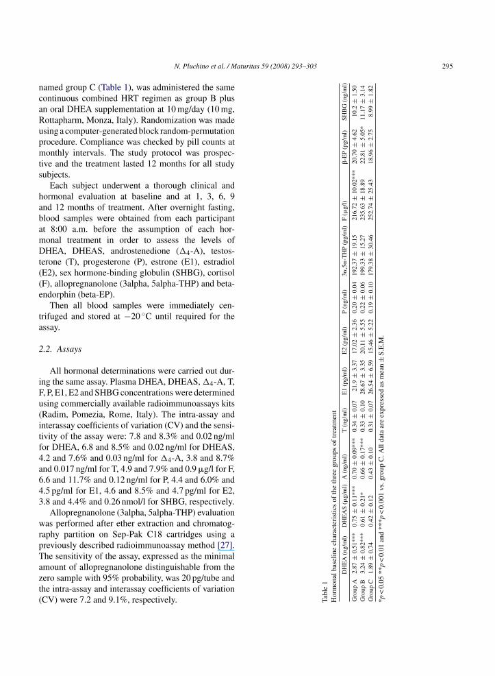
aturitas 59 (2008) 293–303 295
ncaRupmts
habamDt((e
ta
2
iFu(itf4a643
wrpTazt(
line
char
acte
rist
ics
ofth
eth
ree
grou
psof
trea
tmen
t
(ng/
ml)
DH
EA
S(�
g/m
l)A
(ng/
ml)
T(n
g/m
l)E
1(p
g/m
l)E
2(p
g/m
l)P
(ng/
ml)
3�,5
�-T
HP
(pg/
ml)
F(�
g/l)
�-E
P(p
g/m
l)SH
BG
(ng/
ml)
0.51
***
0.75
±0.
11**
*0.
70±
0.09
***
0.34
±0.
0721
.9±
3.37
17.0
2±
2.36
0.20
±0.
0419
2.37
±19
.15
216.
72±
10.0
2***
20.7
0±
4.62
10.2
±1.
500.
82**
*0.
61±
0.21
*0.
66±
0.17
***
0.33
±0.
1028
.67
±3.
3520
.11
±5.
550.
22±
0.06
199.
33±
15.2
723
5.63
±18
.89
22.8
1±
5.05
*11
.17
±3.
140.
740.
42±
0.12
0.43
±0.
100.
31±
0.07
26.5
4±
6.59
15.4
6±
5.22
0.19
±0.
1017
9.38
±30
.46
252.
74±
25.4
318
.96
±2.
758.
99±
1.82
0.01
and
***p
<0.
001
vs.g
roup
C.A
llda
taar
eex
pres
sed
asm
ean±
S.E
.M.
N. Pluchino et al. / M
amed group C (Table 1), was administered the sameontinuous combined HRT regimen as group B plusn oral DHEA supplementation at 10 mg/day (10 mg,ottapharm, Monza, Italy). Randomization was madesing a computer-generated block random-permutationrocedure. Compliance was checked by pill counts atonthly intervals. The study protocol was prospec-
ive and the treatment lasted 12 months for all studyubjects.
Each subject underwent a thorough clinical andormonal evaluation at baseline and at 1, 3, 6, 9nd 12 months of treatment. After overnight fasting,lood samples were obtained from each participantt 8:00 a.m. before the assumption of each hor-onal treatment in order to assess the levels ofHEA, DHEAS, androstenedione (�4-A), testos-
erone (T), progesterone (P), estrone (E1), estradiolE2), sex hormone-binding globulin (SHBG), cortisolF), allopregnanolone (3alpha, 5alpha-THP) and beta-ndorphin (beta-EP).
Then all blood samples were immediately cen-rifuged and stored at −20 ◦C until required for thessay.
.2. Assays
All hormonal determinations were carried out dur-ng the same assay. Plasma DHEA, DHEAS, �4-A, T,, P, E1, E2 and SHBG concentrations were determinedsing commercially available radioimmunoassays kitsRadim, Pomezia, Rome, Italy). The intra-assay andnterassay coefficients of variation (CV) and the sensi-ivity of the assay were: 7.8 and 8.3% and 0.02 ng/mlor DHEA, 6.8 and 8.5% and 0.02 ng/ml for DHEAS,.2 and 7.6% and 0.03 ng/ml for �4-A, 3.8 and 8.7%nd 0.017 ng/ml for T, 4.9 and 7.9% and 0.9 �g/l for F,.6 and 11.7% and 0.12 ng/ml for P, 4.4 and 6.0% and.5 pg/ml for E1, 4.6 and 8.5% and 4.7 pg/ml for E2,.8 and 4.4% and 0.26 nmol/l for SHBG, respectively.
Allopregnanolone (3alpha, 5alpha-THP) evaluationas performed after ether extraction and chromatog-
aphy partition on Sep-Pak C18 cartridges using areviously described radioimmunoassay method [27].he sensitivity of the assay, expressed as the minimal
mount of allopregnanolone distinguishable from theero sample with 95% probability, was 20 pg/tube andhe intra-assay and interassay coefficients of variationCV) were 7.2 and 9.1%, respectively. Table
1H
orm
onal
base
DH
EA
Gro
upA
2.87
±G
roup
B3.
24±
Gro
upC
1.89
±*p
<0.
05**
p<

2 aturitas
eCrasr
2
iSDb
wummoa2K
3
3
fiimgpc5
i2
3
3
aloAbBsw(
33TDis6boeggo
toC
TP
GGG
*
96 N. Pluchino et al. / M
The beta-EP concentrations were determined afterxtraction and chromatography partition on Sep-Pak18 cartridges and using a previously described
adioimmunoassay method [28]. The sensitivity of thessay was 2.5 pg/ml and the intra-assay and interas-ay coefficients of variation (CV) were 6.0 and 9.0%,espectively.
.3. Statistical analysis
Data are expressed as mean ± S.E.M. and as delta ofncrease or decrease in comparison to baseline values.tatistical between group comparison of basal levels ofHEA, DHEAS, Delta4-A and beta-EP was achievedy using a Student’s t-test for paired data.
One- and two-way analysis of variance (ANOVA)as used to compare hormonal levels during follow-p among the three groups of patients. Bonferroni’sultiple comparison test was used to compare the hor-onal levels at all the follow-up times for all groups
f therapy. Alpha value for Bonferroni’s test was sett 0.05. Statistical analysis was performed with NCSS001 software (Number Cruncher Statistical Systems,aysville, UT, USA).
. Results
.1. Patient characteristics
All the patients enrolled in the study completed theollow-up, without any adverse events. The subjectsn group C had a greater menopausal age than thosen groups A and B (p < 0.01) (7.50 ± 0.60 years since
enopause (ysm) for group C vs. 3.70 ± 0.67 ysm for
roup A and 4.20 ± 0.64 ysm for group B). Group Catients also presented a greater chronological age inomparison to group A (p < 0.001) (56.70 ± 2.17 vs.2.70 ± 1.95). No significant difference was observedpsap
able 2atients characteristics of the three groups of treatment
Age (years) BMI (kg/m2) Years since m
roup A 52.70 ± 1.95*** 27.65 ± 1.84 3.70 ± 0.67roup B 54.83 ± 1.85 28.65 ± 2.04 4.20 ± 0.64roup C 56.70 ± 2.17 27.97 ± 0.82 7.50 ± 0.60
p < 0.05 **p < 0.01 and ***p < 0.001 vs. group C. All data are expressed a
59 (2008) 293–303
n terms of BMI (27.65 ± 1.84 vs. 28.65 ± 2.04 vs.7.97 ± 0.82) (Table 1).
.2. Endocrine evaluation
.2.1. Baseline statusBesides the lower circulating levels of DHEA
nd DHEAS, group C patients showed significantlyower plasma levels of �4-A (0.43 ± 0.10) than thosebserved in group B (0.66 ± 0.17) and in group
(0.70 ± 0.09) and also lower plasma levels ofeta-EP (18.96 ± 2.75) than those observed in group
(22.81 ± 5.05). Furthermore, the baseline corti-ol levels observed in group C (252.74 ± 25.43)ere significantly higher than those in group A
216.72 ± 10.02) (p < 0.001) (Table 2).
.2.2. Post-treatment evaluation
.2.2.1. Androgens (DHEA, DHEAS, Delta4-A and) and estrogens. In both the DHEA-treated groups,HEA serum levels showed a progressive increase dur-
ng the entire treatment period with a significant risetarting from the 3rd month for group A and from theth month for group C (p < 005 for both groups vs.aseline). No difference of DHEA serum levels wasbserved in HRT group throughout all 12 months ofvaluation (group B). This progressive rise observed inroup C abolished DHEA baseline difference betweenroups B and C from the 6th month of treatment, with-ut affecting the difference between groups A and C.
Similarly, DHEAS levels increased only in DHEA-reated groups (groups A and C). Baseline differencef DHEAS levels disappeared between groups B andafter 6 months of treatment (Fig. 1).Delta4-androstenedione levels increased slowly and
rogressively in both DHEA-treated groups, with aignificant rise from the 3rd month for group And from the 6th month for group C (p < 0.01 and< 0.001, respectively) maintaining the baseline dif-
enopause Smokers/non-smokers Natural menopause
*** No Yes*** No Yes
No Yes
s mean ± S.E.M.

N. Pluchino et al. / Maturitas 59 (2008) 293–303 297
Fig. 1. Mean ± S.D. plasma DHEA, DHEAS, androstenedione, testosterone, E1 and E2 levels at the baseline and at 1, 3, 6, 9 and 12 months.White column: group A (50–55 years). Grey column: group B (52–58 years). Black column: group C (54–61 years) (*) differences betweenthe three groups (*p < 0.05; **p < 0.005; ***p < 0.001); (◦) steroid concentration at each time point vs. baseline levels in group A (◦p < 0.05;◦◦p < 0.005; ◦◦◦p < 0.001); (§) steroid concentration at each time point vs. baseline levels in group B (§p < 0.05; §§p < 0.005; §§§p < 0.001);(+) steroid concentration at each time point vs. baseline levels in group C (+p < 0.05; ++p < 0.005; +++p < 0.001).
298 N. Pluchino et al. / Maturitas 59 (2008) 293–303
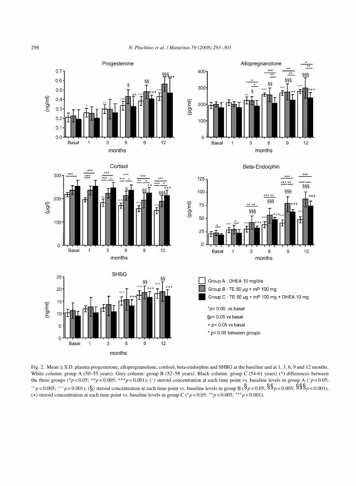
Fig. 2. Mean ± S.D. plasma progesterone, allopregnanolone, cortisol, beta-endorphin and SHBG at the baseline and at 1, 3, 6, 9 and 12 months.White column: group A (50–55 years). Grey column: group B (52–58 years). Black column: group C (54-61 years) (*) differences betweenthe three groups (*p < 0.05; **p < 0.005; ***p < 0.001); (◦) steroid concentration at each time point vs. baseline levels in group A (◦p < 0.05;◦◦p < 0.005; ◦◦◦p < 0.001); (§) steroid concentration at each time point vs. baseline levels in group B (§p < 0.05; §§p < 0.005; §§§p < 0.001);(+) steroid concentration at each time point vs. baseline levels in group C (+p < 0.05; ++p < 0.005; +++p < 0.001).

N. Pluchino et al. / Maturitas 59 (2008) 293–303 299
Fig. 3. Delta of increase and decrease of allopregnanolone, beta-endorphin and cortisol plasma levels at 1, 3, 6, 9 and 12 months. White column:group A (50–55 years). Grey column: group B (52–58 years). Black column: group C (54–61 years) (*) differences between the three groups(*p < 0.05; **p < 0.005; ***p < 0.001).

3 aturitas
ftab(
tDgBifjft
ahDvd(
sg(fvh
3asf6rg
i3pilcCtnr
gfamaots1l(hov
iftwt(gfv
it((ep
4
nif(ipl
00 N. Pluchino et al. / M
erence between these two groups throughout allhe study period (p < 0.001). The increase in Delta4-ndrostenedione levels observed in group C removedaseline differences versus group B from the 9th monthFig. 1).
Testosterone levels increased progressively fromhe 1st month of treatment in patients receiving onlyHEA and since the 3rd month in DHEA–HRT treatedroup (p < 0.05 vs. baseline for both groups). Group
receiving conventional HRT showed no changesn circulating testosterone levels throughout all theollow-up period (Fig. 1). Testosterone levels in sub-ects receiving only DHEA were higher than group Brom the 1st month (p < 0.05) and than group C fromhe 3rd month of therapy (p < 0.001).
There was also a progressive rise in E1 levels inll groups; subjects treated with DHEA–HRT showedigher E1 levels in comparison to those receivingHEA alone from the 6th month of therapy (p < 0.001s. group B and p < 0.01 vs. group C); no significantifference was observed between HRT-treated groupsgroups B and C) during all period of analysis (Fig. 1)
E2 levels showed a significant increase in all groupstarting from the 3rd month in group A (p < 0.01) and inroup C (p < 0.001) and from the 6th month in group Bp < 0.001) with significant between group differencesrom the 9th month (p < 0.001 A vs. C and p < 0.05 Bs. C) whereas patients receiving DHEA–HRT showedighest levels of E2 (Fig. 1).
.2.2.2. Progesterone, allopregnanolone, cortisolnd beta-endorphin and SHBG. Progesterone levelshowed a significant increase in all groups, startingrom the 1st month for group A (p < 0.05) and from theth month for groups B and C (p < 0.05 and p < 0.01,espectively), without significant differences betweenroups throughout the whole study period.
Allopregnanolone levels showed a progressivencrease in all groups, which became significant at therd month in group A and in group B (p < 0.01 and< 0.05 vs. baseline, respectively) and at the 9th month
n group C (p < 0.01 vs. baseline). Allopregnanoloneevels in subjects receiving only DHEA (group A) andonventional HRT (group B) were higher than group
from the 3rd month (p < 0.05) (Fig. 2). Furthermore,he delta of increase in group A and in group B was sig-ificantly higher than that reached in group C (p < 0.05,espectively) (Fig. 3).
evne
59 (2008) 293–303
Cortisol levels showed a progressive reduction in allroups of treatment, reaching statistical significanceor group A at the 3rd month (p < 0.01), for group Bt the 6th month (p < 0.05) and for group C at the 9thonth (p < 0.01). Baseline difference between group A
nd group C remained unchanged throughout the entirebservation period (p < 0.001). We observed lower cor-isol levels in subjects receiving only DHEA than inubjects receiving only HRT (group B) already from thest month (p < 0.001) and subjects of group B showedower cortisol levels than group C from the 3rd monthp < 0.05) (Fig. 2). Delta decrease (�) was significantlyigher in group A versus the other two groups at the endf the study period (p < 0.01 vs. group B and p < 0.001s. group C) (Fig. 3).
Beta-endorphin levels showed a progressivencrease in all groups, reaching statistical significanceor group A already at the 1st month and for the otherwo groups at the 3rd month (p < 0.001). In addition,e observed significantly higher levels in group B
han in groups A and C from the 3rd month (p < 0.01)Fig. 2). Delta increase (�) was significantly higher inroup B versus the other two groups at the end of theollow-up period (p < 0.001 vs. group A and p < 0.01s. group C) (Fig. 3).
SHBG levels increased slowly and progressivelyn all groups, with a significant rise in both DHEA-reated groups from the 6th month (groups A and C)p < 0.001) and from the 9th month for HRT groupp < 0.01). No significant differences in SHBG lev-ls resulted between groups throughout the follow-uperiod.
. Discussion
In the past years, several trials have reported sig-ificant hormonal changes after DHEA administrationn postmenopausal women [11–15,29]. However, shortollow-ups and supraphysiological doses of DHEA50 mg/day or higher) hampered these studies. Admin-stration of a lower dose (25 mg/day) of DHEA produceositive effects on hormonal milieu and on quality ofife in early and late postmenopausal women, restoring
strogenic, progestogenic and androgenic tone. Con-entional HRT (estrogen or estrogen plus progestin) didot affect [30] or even decrease androgen plasma lev-ls [31,32]. This is probably the reason why HRT fails
aturitas
twgd
aise1ltactpmDtdr(tp(ist[asl
atomd[atmayit
H
at
gootDdicoa
spo[oHnwBpacaaMnDssaohDt
btHme
N. Pluchino et al. / M
o improve some domains of female sexual functionhich are associated to the menopause-related andro-en deficiency, such as loss of libido and/or sexualesire, arousal and excitability [24,25].
The different impact of HRT and DHEA therapy onndrogenic tone was confirmed in the present studyn which DHEA supplementation at 10 mg/day washown to increase plasma androgen levels whereas thestrogen–progestin therapy alone did not. In addition,0 mg/day of DHEA was sufficient to enhance plasmaevel of estrone, estradiol and progesterone, to posi-ively modify adrenal synthesis of cortisol (reduction)nd allopregnanolone (increase) and to increase plasmaoncentration of beta-endorphin. Moreover, the addi-ion of 10 mg/day of DHEA to the estrogen–progestinreparation in �5-androgens deficient and older post-enopausal women was able to restore circulatingHEA/S to levels in the range of younger subjects
hat received only HRT, abolishing any age-relatedifference of �5-androgens levels. Similarly, age-elated baseline difference of Delta4-androstenedionebetween groups B and C) resulted abolished withhe addition of 10-mg DHEA to a standard HRT. Wereviously evidenced that 1-year therapy with DHEA25 mg/day) was able to affect adrenal steroidogenesis,nducing an increase in baseline and ACTH-stimulatedecretion of Delta-4, Delta-5 androgens and proges-erone, both in early and late postmenopausal women14]. Thus, the present results suggest that DHEA ther-py, even at 10 mg/day, might enhance the adrenalynthesis of Delta-4 and androgens, since DHEA/Sevels continued increasing over the study time.
In addition, conversion of DHEA into morective steroids, such as androstenedione, testos-erone, dihydrotestosterone and estrogens dependsn the activity of tissue-specific steroidogenic andetabolizing systems such as 3�-hydroxysteroid
ehydroidrogenase/�5-�4-isomerase and aromatase33]. However, the aging process and the concomitantdministration of estroprogestin treatment may modifyhe metabolism and the effect of DHEA therapy on hor-
onal milieu. In fact, the increase of testosterone levelsfter 12 months of DHEA treatment result higher inounger women with elevated baseline DHEA/S than
n older women with lower baseline DHEA/S receivinghe combination DHEA–HRT.On the contrary, an additive effect of DHEA onRT was evaluated for circulating estrogen levels (E1
tdta
59 (2008) 293–303 301
nd E2) that resulted higher in subjects receiving bothreatments.
Cortisol plasma levels decreased in all treatmentroups throughout the 12 months, confirming previ-us data [12–14,20,21,30]. Furthermore, 10 mg/dayf DHEA are much more effective in reducing cor-isol than HRT in young postmenopausal women.HEA–HRT regimen determined a lower cortisolecrease with respect to HRT alone, suggesting thatn older postmenopausal women with DHEA/S defi-iency, the metabolic pathway leading to this reductionf cortisol is probably less responsive to DHEA ther-py.
In previous studies, we observed that both DHEAupplementation and estrogen–progestin treatments inostmenopausal women could increase plasma levelsf progesterone and its metabolite, allopregnanolone34,35]. On these bases, we expected the addictionf a low-dose of DHEA to a continuous combinedRT regimen to have a synergistic effect on allopreg-anolone plasma levels. However, in group C patients,ho received the same dosage of progestin as group, we observed a significantly lower increase in allo-regnanolone plasma levels. This suggests that theddition of DHEA to an estrogen–progestin treatmentannot further modulate the biosynthetic activity of thedrenal gland, which is the main source of circulatingllopregnanolone in postmenopausal women [34–36].oreover, we observed significantly higher allopreg-
anolone levels in younger subjects who receivedHEA supplementation at 10 mg/day than in older
ubjects treated with HRT in combination with theame dose of DHEA. Aging seems to negatively affectdrenal DHEA synthesis/release as well as the responsef adrenal steroidogenesis to hormonal treatments,ighlighting the importance of timing for HRT and/orHEA therapy to reverse adrenal ageing and to obtain
he most positive hormonal results.Similar considerations may apply to the increase in
eta-endorphin plasma levels throughout the observa-ion period. The higher response of beta-endorphin toRT therapies (groups B and C) respect to group Aight be related to the higher increase of circulating
strogens (E1 and E2) of groups B and C, since cen-
ral and peripheral content of this opioid are strictlyependent on the estrogen level [37,38]. However,he additive effect of DHEA–HRT respect to HRTlone, evaluated for estrogens, did not induce a sim-
3 aturitas
its
Fiatodmtatas
A
tS
R
[
[
[
[
[
[
[
[
[
[
[
[
[
02 N. Pluchino et al. / M
lar response for beta-endorphin, suggesting again thathe aging process might negatively affect the synthe-is/release of this opioid to estrogen stimuli.
In conclusion, our data lead to some considerations.irst, a daily dosage of only 10 mg of DHEA, alone or
n combination to a HRT regimen is able to restore thendrogenic milieu and also has a positive impact onhe estrogenic tone in postmenopausal women. Sec-nd, menopausal age and “adrenal age” might explainifferent results of these three treatments on hormonalilieu, evidencing that timing of therapy deeply affects
he results of HRT and/or DHEA treatment. Finally,dditional studies, using a larger population, are neededo evaluate the combination of estrogen–progestin ther-pies plus DHEA as a new additional therapeuticaltrategy for postmenopausal women.
cknowledgement
This work was partially supported by a grant fromhe Fondazione Cassa Di Risparmio Di San Miniato,an Miniato, Pisa, Italy.
eferences
[1] Orentreich N, Brind JL, Rizer RL, Vogelman JH. Age changesand sex differences in serum dehydroepiandrosterone sulphateconcentration throughout adulthood. J Clin Endocrinol Metab1984;59(3):551–5.
[2] Arlt W. Dehydroepiandrosterone and ageing. Best Pract ResClin Endocrinol Metab 2004;18(3):363–80.
[3] Labrie F, Belanger A, Cusan L, Gomez JL, Candas B. Markeddecline in serum concentrations of adrenal C19 sex steroidsprecursors and conjugated androgen metabolites during aging.J Clin Endocrinol Metab 1997;82(8):2396–402.
[4] Harper AJ, Buster JE, Casson PR. Changes in adrenocorticalfunction with aging and therapeutic implications. Semin ReprodEndocrinol 1999;17(4):327–38.
[5] Laughlin GA, Barrett-Connor E. Sexual dimorphism inthe influence of advanced aging on adrenal hormone lev-els: the Brancho Bernardo study. J Clin Endocrinol Metab2000;85(10):3561–8.
[6] Liu CH, Laughlin GA, Fischer UG, Yen SS. Marked attenuationof ultradian and circadian rhythms of dehydroepiandrosterone
in postmenopausal women: evidence for a reduced 17, 20desmolase activity. J Clin Endocrinol Metab 1990;71(4):900–6.[7] Parker Jr CR, Mixon RL, Brissie EM, Grizzle WE. Aging alterszonation in the adrenal cortex of men. J Clin Endocrinol Metab1997;82(11):3898–901.
[
59 (2008) 293–303
[8] Davison SL, Bell R, Donath S, Montalto JG, Davis SR. Andro-gen levels in adult females: changes with age, menopause andoophorectomy. J Clin Endocrinol Metab 2005;90(7):3847–53.
[9] Swaab DF, Bao AM, Lucassen PJ. The stress system in thehuman brain in depression and neurodegeneration. Ageing ResRev 2005;4(2):141–94.
10] Genazzani AD, Lanzoni C, Genazzani AR. Might DHEA bea beneficial replacement therapy in the elderly? Drugs Aging2007;24(3):173–85.
11] Mortola JF, Yen SCC. The effects of oral dehydroepiandros-terone on endocrine-metabolic in postmenopausal women. JClin Endocrinol Metab 1990;71(3):696–704.
12] Morales AJ, Nolan JJ, Nelson JC, Yen SCC. Effects of replace-ment dose of dehydroepiandrosterone in men and in women ofadvancing age. J Clin Endocrinol Metab 1994;78(6):1360–7.
13] Genazzani AR, Pluchino N, Begliuomini S, et al. Long-term low-dose oral administration of dehydroepiandrosteronemodulates response to adrenocorticotropic hormone inearly and late postmenopausal women. Gynecol Endocrinol2006;22(11):627–35.
14] Genazzani AR, Inglese S, Lombardi I, et al. Long termlow-dose dehydroepiandrosterone replacement therapy inaging males with partial androgen deficiency. Aging Male2004;7(2):133–43.
15] Nair KS, Rizza RA, O’Brien P, et al. DHEA in elderly womenand DHEA or testosterone in elderly men. N Engl J Med2006;355(16):1647–59.
16] Simoncini T, Genazzani AR. Dehydroepiandrosterone, theendothelium and cardiovascular protection. Endocrinology2007;148(7):3065–7.
17] Libe R, Barbetta L, Dall’Asta C, Salvaggio F, Gala C,Beck-Peccoz P. Effects of dehydroepiandrosterone (DHEA)supplementation on hormonal, metabolic and behavioural sta-tus in patients with hypoadrenalism. J Endocrinol Invest2004;27(8):736–41.
18] Barnhart KT, Freeman E, Grisso JA, et al. The effect ofdehydroepiandrosterone supplementation to symptomatic per-imenopausal women on serum profiles, lipid parameters,and health related quality of life. J Clin Endocrinol Metab1999;84(11):3896–902.
19] Buvat J. Androgen therapy with dehydroepiandrosterone.World J Urol 2003;21(5):346–55.
20] Stomati M, Monteleone P, Casarosa E, et al. Six oral monthsdehydroepiandrosterone supplementation in early and latemenopause. Gynecol Endocrinol 2000;14(5):342–63.
21] Genazzani AD, Stomati S, Bernardi F, Pieri M, Rovati L,Genazzani AR. Long term low-dose dehydroepiandrosteroneoral supplementation in early and late postmenopausal womenmodulates endocrine parameters and synthesis of neuroactivesteroids. Fertil Steril 2003;80(6):1495–501.
22] Panjari M, Davis S. DHEA therapy for women: effecton sexual function and wellbeing. Hum Reprod Update
2007;13(3):239–48.23] Sarrel P, Dobay B, Wiita B. Estrogen and estrogen–androgenreplacement in postmenopausal women dissatisfied withestrogen-only therapy. Sexual behaviour and neuroendocrineresponses. J Reprod Med 1998;43(10):847–56.
aturitas
[
[
[
[
[
[
[
[
[
[
[
[
[
[
N. Pluchino et al. / M
24] Dobs AS, Nguyen T, Pace C, Roberts CP. Differential effects oforal estrogen versus oral estrogen–androgen replacement ther-apy on body composition in postmenopausal women. J ClinEndocrinol Metab 2002;87:1509–16.
25] Denise von Muhlen GA, Laughlin, Kritz-Silverstein D, Barrett-Connor E. The dehydroepiandrosterone and wellness (DAWN)study: research design and methods. Contemp Clin Trials2007;28:153–68.
26] Davison SL, Bell R, Donath S, Montalto JG, Davis SR. Andro-gen levels in adult females: changes with age, menopause, andoophorectomy. J Clin Endocrinol Metab 2005;90(7):3847–53.
27] Genazzani AR, Bernardi F, Petraglia F, et al. Circulating lev-els of allopregnanolone in humans: gender, age and endocrineinfluences. J Clin Endocrinol Metab 1998;83(6):1–5.
28] Stomati M, Bersi C, Rubino S, et al. Neuroendocrine effectsof different oestradiol-progestin regimens in postmenopausalwomen. Maturitas 1997;28(2):127–35.
29] Labrie F, Luu-The V, Belanger A, et al. Is dehydroepiandros-terone a hormone? J Endocrinol 2005;187(2):169–96.
30] Castelo-Blanco C, Martinez De Osaba MJ, Fortuny A, Igle-sias X, Gonzalez-Merlo J. Circulating hormone levels inmenopausal women receiving different hormone replace-ment therapy regimens. A comparison. J Reprod Med1995;40(8):556–60.
[
59 (2008) 293–303 303
31] Doren M, Rubig A, Coelingh Bennink HJ, Holzgreve W. Dif-ferential effects on the androgen status of postmenopausalwomen treated with tibolone and continuous combined estra-diol and norethindrone acetate replacement therapy. Fertil Steril2001;75(3):554–9.
32] Casson PR, Elkind-Hirsch KE, Buster JE, Hornsby PJ,Carson SA, Snabes MC. Effect of postmenopausal estro-gen replacement on circulating androgens. Obstet Gynecol1997;90(6):995–8.
33] Labrie F, Luu-The V, Labrie C, Simard J. DHEA and its transfor-mation into androgens and estrogens in peripheral target tissues:intracrinology. Front Neuroendocrinol 2001;22(3):185–212.
34] Paul SM, Purdy RH. Neuroactive steroids. FASEB J1992;6(6):2311–22.
35] Robel P, Baulieu EE. Neurosteroids: biosynthesis and function.Crit Rev Neurobiol 1995;9(4):383–94.
36] Stoffel-Wagner B. Neurosteroid metabolism in the brain. Eur JEndocrinol 2001;145(6):669–79.
37] Piva F, Limonta P, Dondi D, Pimpinelli F, Martini L, Maggi
R. Effects of sex steroids on brain opioid system. J SteroidBiochem Mol Biol 1995;53(6):343–8.38] Genazzani AR, Pluchino N, Luisi S, Luisi M. Estro-gen, cognition and female ageing. Hum Reprod Update2007;13(2):175–87.
Related Documents