OFD | Syndrome and Mental Retardation EARLE H. YEAMANS, D.D.S. Hartford, Connecticut 06112 In 1954, Papillon-Léage and Psaume (20, 21) described eight patients with similar congenital anomalies involving abnormalities of the oral frenula, digits, and dentition, clefts of the tongue, lip, and palate, mental retardation, hypoplastic nasal cartilages, and several other defects. Simi- lar cases first appeared in the English literature in 1961 when Gorlin, Anderson, and Scott (12) presented four affected females in one family and a forme fruste in another. They attributed the mode of inheritance to a dominant pattern with lethality in males. The name oral-facial-digital syndrome, or OFD syndrome, was first used by Ruess, Pruzansky, Lis, and Patau (25) in their report of twelve cases. Recently, Rimoin and Edgerton (23) described a second type of oral-facial-digital syndrome. They suggested referring to the two types as OFD I and OFD II. The purpose of this paper is to review the syndrome and discuss the variable expressivity with regard to mental retardation. Physical Characteristics Many case reports have been published which describe in detail the physical characteristics of OFD I syndrome (1, 5, 7, 1 3, 25, 26). The most striking oral finding is the hypertrophied frenula which extend from the buccal mucosa to the alveolus. The frenula, often seen in both arches, form numerous fibrous bands which may actually create a cleft of the alveolar ridge (Figures 1 and 2). Hypertrophy of the lingual frenum may cause severe ankyloglossia. True clefts of the secondary palate are often seen. There may be a congenital absence of lateral incisors, especially where clefts are present (Figures 1 and 2). Supernumerary primary and/ or permanent cuspids may be present (Figure 3). Enamel hypoplasia has been noted (1, 5, 138, 25, 27). Lateral palatal tori are often reported (1, 7, 12, 13, 27), but these may actually be pseudoclefts of the palate where there has been a failure of the lateral palatal shelves to fuse and the intervening area is covered only by soft tissue (Figure 3). A bifid, trifid, or multilobed tongue is commonly seen. Frequently observed facial malformations include hypoplasia of the nasal cartilages, dystopia canthorum, discrepancy in size of the nostrils, milia of the ear pinnae, and varying degrees of hair loss. Another facial feature is a pseudocleft of the upper lip in the midline (Figure 4). Earle H. Yeamans, D.D.S. is affiliated with the Department of Pediatric Dentistry, University of Connecticut Health Center, Hartford, Connecticut 06112. 84

Welcome message from author
This document is posted to help you gain knowledge. Please leave a comment to let me know what you think about it! Share it to your friends and learn new things together.
Transcript

OFD | Syndrome and Mental Retardation
EARLE H. YEAMANS, D.D.S.
Hartford, Connecticut 06112
In 1954, Papillon-Léage and Psaume (20, 21) described eight patientswith similar congenital anomalies involving abnormalities of the oralfrenula, digits, and dentition, clefts of the tongue, lip, and palate, mentalretardation, hypoplastic nasal cartilages, and several other defects. Simi-lar cases first appeared in the English literature in 1961 when Gorlin,Anderson, and Scott (12) presented four affected females in one familyand a forme fruste in another. They attributed the mode of inheritance toa dominant pattern with lethality in males. The name oral-facial-digitalsyndrome, or OFD syndrome, was first used by Ruess, Pruzansky, Lis,and Patau (25) in their report of twelve cases. Recently, Rimoin andEdgerton (23) described a second type of oral-facial-digital syndrome.They suggested referring to the two types as OFD I and OFD II. Thepurpose of this paper is to review the syndrome and discuss the variableexpressivity with regard to mental retardation.
Physical Characteristics
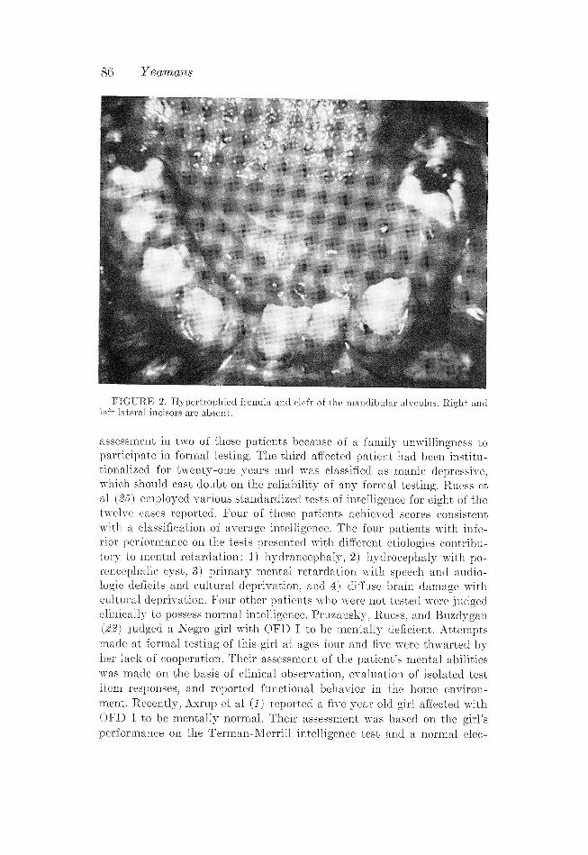
Many case reports have been published which describe in detail thephysical characteristics of OFD I syndrome (1, 5, 7, 1 3, 25, 26). The moststriking oral finding is the hypertrophied frenula which extend from thebuccal mucosa to the alveolus. The frenula, often seen in both arches,form numerous fibrous bands which may actually create a cleft of thealveolar ridge (Figures 1 and 2). Hypertrophy of the lingual frenum maycause severe ankyloglossia. True clefts of the secondary palate are oftenseen. There may be a congenital absence of lateral incisors, especiallywhere clefts are present (Figures 1 and 2). Supernumerary primary and/or permanent cuspids may be present (Figure 3). Enamel hypoplasia hasbeen noted (1, 5, 138, 25, 27). Lateral palatal tori are often reported (1, 7,12, 13, 27), but these may actually be pseudoclefts of the palate wherethere has been a failure of the lateral palatal shelves to fuse and theintervening area is covered only by soft tissue (Figure 3). A bifid, trifid,
or multilobed tongue is commonly seen.
Frequently observed facial malformations include hypoplasia of thenasal cartilages, dystopia canthorum, discrepancy in size of the nostrils,milia of the ear pinnae, and varying degrees of hair loss. Another facialfeature is a pseudocleft of the upper lip in the midline (Figure 4).
Earle H. Yeamans, D.D.S. is affiliated with the Department of Pediatric Dentistry,University of Connecticut Health Center, Hartford, Connecticut 06112.
84

OFD I, MENTAL RETARDATION 85
FIGURE 1. Hypertrophied frenula and cleft of the maxillary alveolus. The rightlateral incisor is absent.
Digital anomalies include characteristic dermatoglyphic patterns (4, 6),
brachydactyly, syndactyly, and clinodactyly. Hands are most often af-
fected, but anomalies do occur on the feet.
Other defects associated with OFD I syndrome may include an in-
creased incidence of mental retardation and central nervous system disor-
ders, speech impairment, hearing loss, polycystic kidneys, and cutancous
dysplasia with decreased or absent sebaceous glands (26).
Mental Retardation
Mental retardation is not a consistant finding in patients with OFD I
syndrome. The incidence of OFD I has been estimated at about 2 per
100,000 Caucasian births (27), and it has been estimated that thirty to
fifty per cent of patients exhibiting this syndrome have a subnormal level
of intelligence (5, 12, 138, 21, 28, 25). In general, the etiology of mental
retardation is extremely complex, and a direct cause and effect relation-
ship is difficult to establish. The methods of assessment of mental retarda-
tion in case reports of OFD I vary. Papillon-Léage and Psaume (20, 21)
did not state the manner in which they judged half of their reported
patients to be mentally retarded, nor did they state the degree of retarda-
tion. Gorlin et al (12) found that three of the four patients they reported
were mentally retarded. Clinical observation was the only method of
86 Yeamans
FIGURE 2. Hypertrophied frenula and cleft of the mandibular alveolus. Right andleft lateral incisors are absent.
assessment in two of these patients because of a family unwillingness to
participate in formal testing. The third affected patient had been institu-
tionalized for twenty-one years and was classified as manic depressive,
which should cast doubt on the reliability of any formal testing. Ruess ct
al (25) employed various standardized tests of intelligence for cight of the
twelve cases reported. Four of these patients achieved scores consistent
with a classification of average intelligence. The four patients with infc-
rior performance on the tests presented with different ctiologics contribu-
tory to mental retardation: 1) hydrancephaly, 2) hydrocephaly with po-
rencephalie cyst, 3) primary mental retardation with speech and audio-
logic deficits and cultural deprivation, and 4) diffuse brain damage with
cultural deprivation. Four other patients who were not tested were judged
clinically to possess normal intelligence. Pruzansky, Rucess, and Buszdygan
(22) judged a Negro girl with OFD I to be mentally deficient. Attempts
made at formal testing of this girl at ages four and five were thwarted by
her lack of cooperation. Their assessment of the patient's mental abilities
was made on the basis of clinical observation, evaluation of isolated test
item responses, and reported functional behavior in the home environ-
ment. Recently, Axrup et al (1) reported a five year old girl affected with
OFD I to be mentally normal. Their assessment was based on the girl's
performance on the Terman-Merrill intelligence test and a normal elec-
OFD I, MENTAL RETARDATION 87
FIGURE 3. Plaster model showing lateral palatal shelf. Note the supernumerary
right cuspid.
troencephalogram. Wahrman et al ($7) reported mental retardation in a
sixteen month old male with 47/XXY chromosomes (Klinefelter's syn-
drome} and OFD I syndrome. This patient was reported to have partial
agenesis of the corpus callosum with a lipoma in the rostral portion of the
corpus callosum. Doege and co-workers (6) made the first attempt at a
controlled study of mental retardation in OFD I syndrome. They endeav-
ored to eliminate the effect of culture and environment by comparing
affected and non-affected sisters in two generations of a large family with
seventeen OFD I females. In addition, they compared another OFD I
female from this same family and her non-affected first cousin. The paired
sisters were consccutive in birth order and were of the same parental
origin. Formal testing of the affected mother indicated that she possessed
normal intelligence. The results of their study indicate that all affected
offspring who were tested were mentally retarded, ranging from borderline
to profound; however, two non-affected offspring were also classified as
being mentally retarded on the basis of their test scores. It was suggested
that there may be a direct relationship between the level of intelligence
and the severity of OFD I anomalies. There is some indication from this
study that OFD I females tend to score lower on tests of verbal skills
while showing higher scores in performance areas.

88 Yeamans
FIGURE 4. Midline pseudocleft of the upper lip.
With tests of intelligence relying more and more on verbal skills, onemight consider the interaction between hearing loss, speech impairment,cleft palate, and mental retardation in OFD I patients. There may be anincreased incidence of hearing loss, speech impairment and intellectualimpairment in cleft palate poulations (8, 10, 16), and there is a high
incidence of cleft palate in OFD I populations.
Gorlin (11) states, "The intelligence quotient in these individuals issomewhere around 70." The evidence does, in fact, indicate a high inci-dence of mental retardation in OFD I patients, but retardation is by nomeans a cardinal feature of the syndrome. Until more is known about themultifactorial etiology of mental retardation in general, broad, all-encom-passing statements about the mentality of OFD I patients should beavoided.
Although substantive data is scanty, clinical observations by experi-enced professionals cannot be discounted in assessing relative levels ofmental function. When a patient presents with OFD I syndrome, everyeffort must be made to evaluate her mental abilities on an individual basisin order to maximize her potential for adapting to society. Ruess, Pruzan-sky and Lis (24) have emphasized the need to analyze critically thedevelopmental, environmental, and situational factors of each OFD Ipatient individually. In this way, the highest degree of success may beattained in the long range planning for the habilitation of these patients.

OFD I, MENTAL RETARDATION 89
Ruess et al. have stressed that a normal level of intelligence is seen in
many patients with OFD I syndrome.
Genetics
It has been suggested that OFD I is transmitted as an X-linked domi-
nant trait with lethality in homozygous males (6, 11, 19, 27). One excep-
tion to this pattern was reported in 1967 where OFD I syndrome appeared
in a chromosomally normal male (17). Prior to the recognition of OFD II
syndrome, there were two reports in the English literature of OFD in
males (15, 27). One of these males, however, appears to have OFD II
syndrome with bilateral polysyndactyly of the large toe. Cytogenetic
studies of the other male revealed 47/XXY chromosomes. The presence of
the X chromosomes may serve to explain the viability of this male with
OFD I syndrome. There have been other reportes of OFD I in males (2, 3,
18), however, close evaluation of the physical findings would indicate that
the diagnosis of OFD I syndrome may be open to discussion.
In a dominant pattern with lethality in males, a theoretical sex ratio of
2:1, female to male, would be expected. Analysis of OFD I pedigrees
indicates that this ratio is closely approximated (0).
From the viewpoint of genetic counseling, an affected female has a fifty
per cent chance of passing the trait to her daughters. Any sons that
survive will not be affected. It should be stressed that a non-affected
female offspring is not likely to pass the trait to her daughters. A non-af-
fected female offspring has the same probability of having an affected
daughter as does a non-affected female in a normal population. A high
incidence of abortions and stillbirths may be seen in an affected family.
Although the greatest majority of OFD I patients appears to inherit the
trait in an X-linked dominant pattern with lethality in males, the possi-
bility of other modes of transmission or new mutation should not be
overlooked (17).
In 1967, Rimoin and Edgerton (238) presented evidence to indicate the
existence of a second type of oral-facial-digital syndrome. It was referred
to as OFD II, or Mohr syndrome. Anomalies of the tongue, lip, palate,
frenula, and digits are similar in both syndromes; however, there are
distinct differences which serve to distinguish the two. Bilateral polysyn-
dactyly of the large toe is the most consistent characteristic finding in
patients with OFD II. This bilateral feature has not been reported in
patients with OFD I. Although hypertrophied frenula are seen in both
syndromes, these are not associated with clefts of the alveolar ridges in
OFD II, and the distribution is often limited to the midline. The lack of
clefting of the alveolus may account for the fact that the dentition in
OFD II is more often normal in number. Hair and skin defects have not
been noted in OFD II, and mental retardation is apparently not a com-
mon feature. '
OFD II may occur in either males or females and appears to be trans-

90 Yeamans
mitted as an autosomal recessive trait. The pedigree presented by Rimoin
and Edgerton showed affected siblings of both sexes with apparently nor-
mal parents. They also cited another study of a similarly affected male
where a history of consanguinity was revealed.
Every effort should be made to distinguish OFD I from OFD II because
of the implications in genetic counseling. A patient with OFD II whose
mate is not similarly affected will not transmit the trait; however, all
offspring will be carriers.
Summary
Although a high incidence of mental retardation is seen in OFD I
populations, it is stressed that the syndrome is not incompatible with
normal intelligence. Each patient's mental abilities should be assessed
individually. Effective long range planning of the education and training
of these patients will permit adaptation to the demands of society. Clini-
cal manifestations of the syndrome include hypertrophied frenula, clefts
of the palate, alveolus, lip, and tongue, hypoplastic nasal cartilages,
brachydactyly, and syndactyly. OFD I appears to be transmitted as an
X-linked dominant trait with lethality in the hemizygous male. OFD II
appears to be transmitted as an autosomal recessive trait. There are other
distinct differences between OFD I and OFD II. Differentiation between
these two syndromes is important because of the implications in genetic |
counseling.
Acknowledgment: The author is grateful to Dr. D. R. Porter of the
University of Oregon Dental School, and Dr. R. Stewart of the UCLA
School of Dentistry for their assistance during the preparation of the
manuscript.
References
1. Axrup, K., B. Linvgqvist, and G. SAmuzuson, Oral-facial-digital syndrome. Odont.Revy., 22, 137, 1971.
2. BELL, R. C., A child with two tongues (oral-facial-digital syndrome). Brit. J. Plast.Surg., 24, 198, 1971.
3. Boon, W. H. and C. T. Sena, The oral-facial-digital syndrome in a Chinese male.J. Singapore Pediat. Soc., 8, 75, 1966.
4. Co-T's, P., C. L. Dommax, B. TiscHmurr, and R. B. Lowry, Oral-facial-digital syn-drome: a case with necropsy findings. Amer. J. Dis. Child., 119, 280, 1970.
5. Doner, J. A., and D. C. KErnonax, Oral-facial-digital syndrome. Arch. Dis. Child.,48, 214, 1967.
6. Dores, T. C., M. M. Campsrur, J. S8. Bryant, and H. C. Turing, Mental retarda-tion and dermatoglyphics in a family with the oral-facial-digital syndrome. Amer.J. Dis. Chald., 116, 615, 1968.
7. Dores, T. C., H. C. Truum®, J. H. Prisst, D. E. NorBYv, and J. S. BRYANT, Studiesof a family with the oral-facial-digital syndrome. New Engl. J. Med., 271, 1073,1964.
8. EstBs, R. E., and H. L. MorrIs, Relationships among intelligence, speech profi-ciency, and hearing sensitivity in children with cleft palates. Cleft Pal. J., 9, 763,1970.
9. Gitmmors, S., and S. Horman, Clefts in Wisconsin: incidence and related factors.Cleft Pal. J., 8, 186, 1966.
10.
11.
12.
18.
14.
15.
16.
17.
18.
19.
20.
21.
22.
23.
24.
25.
26.
27.
OFD I, MENTAL RETARDATION 91
GoopsTEIN, L., Intellectual impairment in children with cleft palates. J. Sp. Hear.Res., 4, 287, 1961.CGoRLIN, R. J., Some facial syndromes. Birth defects: original article series, 5, 69,No. 2, 1969.GorLn, R. J., V. E. and C. R. Scott, Hypertrophied frenuli, oligophre-nia, familial trembling and anomalies of the hand. New Engl. J. Med., 264, 486,1961.GorLIn, R. J., and J. Psavm®, Orodigitofacial dysostosis: a new syndrome. J. Pe-diat., 61, 520, 1962. AGrEENE, J., J. VEermiLuion, S. Hay, S. GtsBENS, and S. KerscnBaum, Epidemiologicstudy of cleft lip and cleftpalate in four states. J. Am. Dent. A., 68, 387, 1964.Kusunicx, T., T. P. Massa, and R. Orofaciodigital syndrome in a male.J. Pediat., 63, 1130, 1968. 7LEwIis, Rutz, Survey of the intelligence of cleft-lip and cleft-palate children inOntario. Br. J. of Disorders of Communication, 6, 17, 1971.ManpELL, F., P. L. Ocra, S. L. Horowitz, and K. Horscnnorxn, Oral-facial-digitalsyndrome in a chromosomally normal male. Pediatrics, 40, 63, 1967.
WaunEmar and Danuta, IrzyNsKA-ForFAara, Zespol Ustno-T'warzowo-Palcowy (O.F.D.) U Noworodka. Pediat. Pol., 46, 343-5, 1971.McKusick, V. A., Mendelian inheritance in man. 2nd Ed. Baltimore: The JohnsHopkins Press, 1968. p. 420.Papitron-LfAagE and J. Psaumg, Dysmorphie des friens buccaux: huit observations.Actualités Ondonto-Stomatol., 25, 't, 1954.
and J. Une malformation héréditaire de la muqueuse buc-cale : brides et freins anormaux. Rev. Stomatol., 55, 209, 1954.Pruzansky, S., A. and D. Buzovycaxn, Oral-facial-digital syndrome in a Ne-gro female. Plast. Reconstr. Surg., 87, 221, 1966.Rimmotx, D. L., and M. T. Enagrrton, Genetic and clinical heterogeneity in the oral-facial-digital syndromes. J. Pediat., 71, 94, 1967.Ruzrss, A. L., S. Pruzans®y, and E. F. Lts, Intellectual development and OFD syn-drome: a review. Cleft Pal. J., 2,350, 1965.Rurss, A. L., S. Pruzansxy, E. F. Lis, and K. Parav, The oral-facial-digital syn-drome: a multiple congenital condition of females with associated chromosomalabnormalities. Pediatrics, 29, 985, 1962.Soromon, L. M., D. FrEtzin, and S. Pruzansxy, Pilosebaceous dysplasia in theoral-facial-digital syndrome.Waurman, J., M. BeErant, J. JacomBs, I. Aviap, and N. BrEn-Hur, The oral-facial-digital syndrome: a male-lethal condition in a boy with 47/XXY chromosomes.Pediatrics, 87, 812, 1966.
Related Documents