OBJECTIVE: 1. Understanding the external and internal structure of udder 2. Where and how milk to be secreted ANATOMY AND PHYSIOLOGY OF UDDER

Welcome message from author
This document is posted to help you gain knowledge. Please leave a comment to let me know what you think about it! Share it to your friends and learn new things together.
Transcript
OBJECTIVE:
1. Understanding the external and internal structure of udder
2. Where and how milk to be secreted
ANATOMY AND PHYSIOLOGY OF UDDER
• 1. Anatomy of the udder• 2. Internal structure of the udder• 3. Physiology of the udder
CORE SUBJECTS:
Introduction
• Milking is the process of persuading the cow to let down its milk and allow dairy farmer to remove it for his or her own consumption or for sale.
• It is therefore not entirely a natural process.
• The dairy farmer must manipulate the natural process so that he receives the maximum benefit.
• It is therefore essential that one understands the natural process in order to manipulate it.

Mammary glands are the major features that distinguish mammals from other kinds of
animals.
Cow Goat Sheep
Mammary glands are the organs that, in mammals, produce milk for the sustenance of the young.
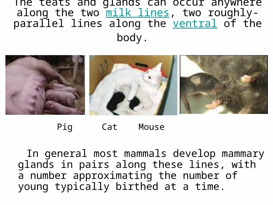
The teats and glands can occur anywhere along the two milk lines, two roughly-parallel lines along the
ventral of the body.
Pig Cat Mouse
In general most mammals develop mammary glands
in pairs along these lines, with a number approximating the number of young typically birthed at a time.

The number and positioning of complex and simple mammary glands varies widely in different mammals.
Elephant Human
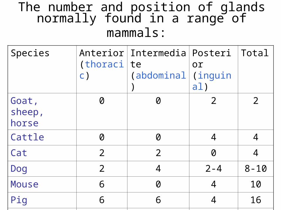
The number and position of glands normally found in a range of mammals:
Species Anterior(thoracic)
Intermediate(abdominal)
Posterior(inguinal)
Total
Goat, sheep, horse
0 0 2 2
Cattle 0 0 4 4
Cat 2 2 0 4
Dog 2 4 2-4 8-10
Mouse 6 0 4 10
Pig 6 6 4 16
Elephants, primates
2 0 0 2
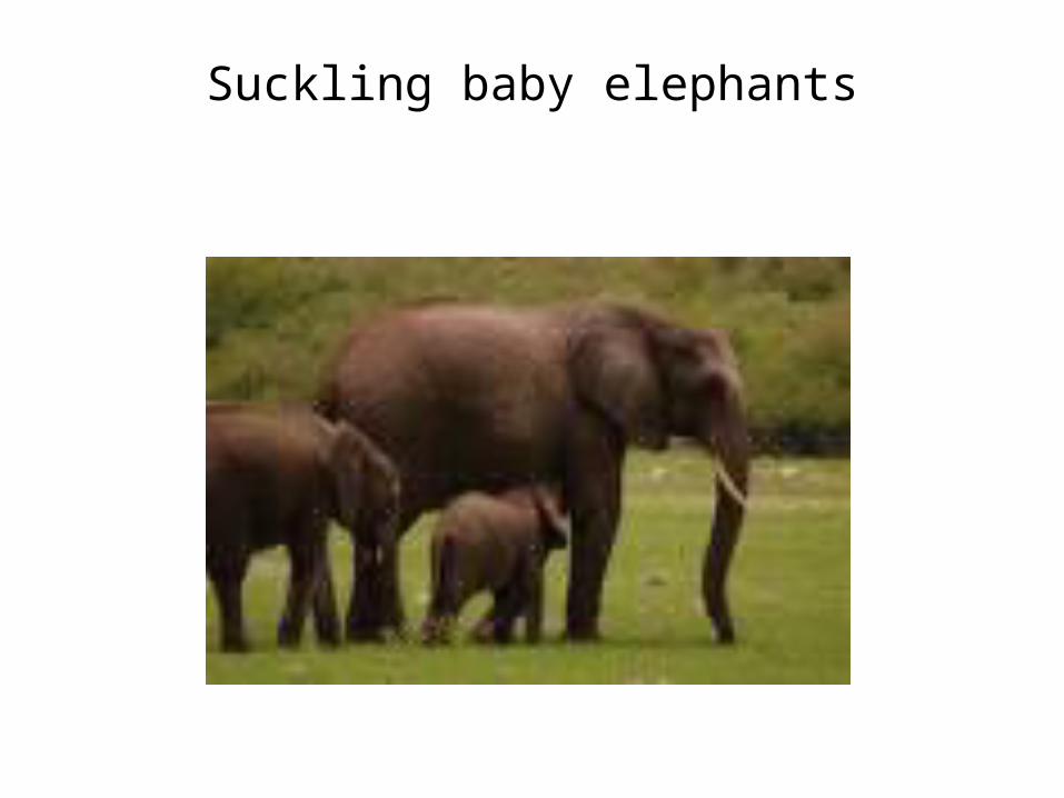
Suckling baby elephants
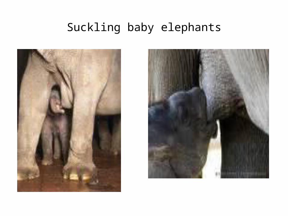
Suckling baby elephants
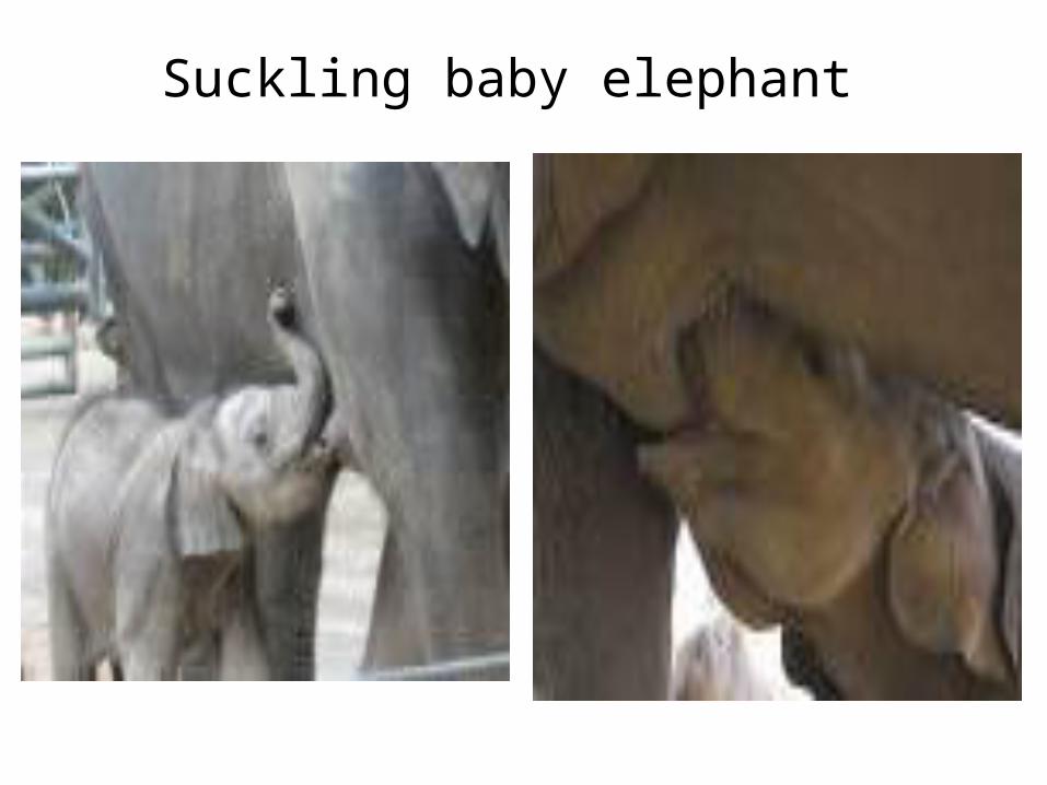
Suckling baby elephant

Several examples of species that reflect the range of anatomical location and number of glands or teats.
Species Location (region) Num of glands
Num of teats
Cow inguinal (groin) 4 4
Ewe, doe inguinal (groin) 2 2
Mare inguinal (groin) 4 2
Sow abdomen 10-14 10-14
Cat abdomen 10-14 10-14
Dog abdomen 10-14 10-14
Human thoracic (pectoral) 2 2
Elephant thoracic (pectoral) 2 2
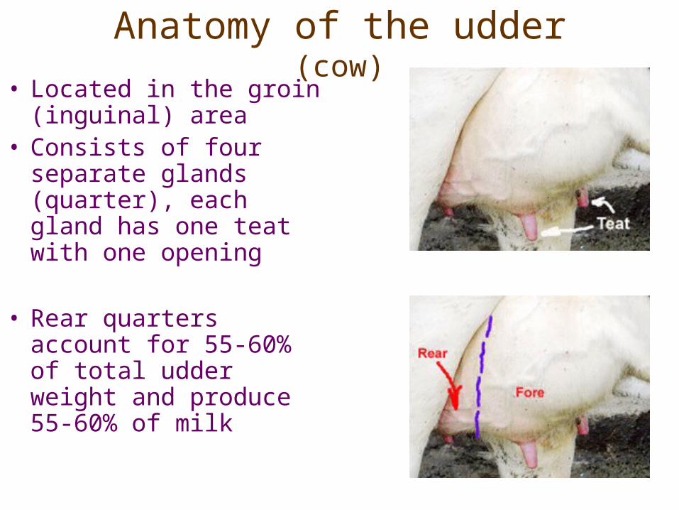
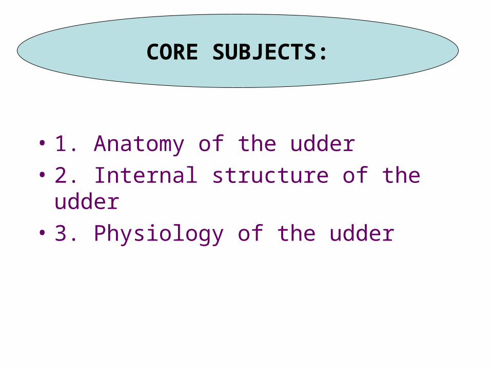
Anatomy of the udder(cow)
• Located in the groin (inguinal) area
• Consists of four separate glands (quarter), each gland has one teat with one opening
• Rear quarters account for 55-60% of total udder weight and produce 55-60% of milk
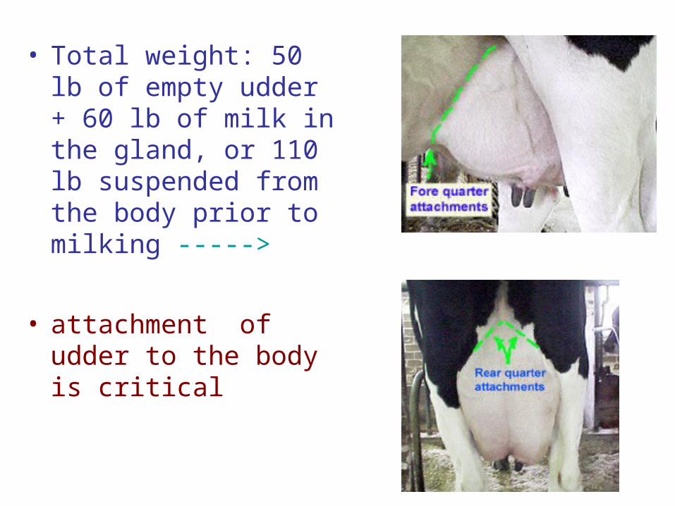
• Total weight: 50 lb of empty udder + 60 lb of milk in the gland, or 110 lb suspended from the body prior to milking ----->
• attachment of udder to the body is critical
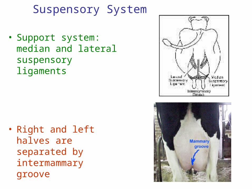
Suspensory System
• Support system: median and lateral suspensory ligaments
• Right and left halves are separated by intermammary groove
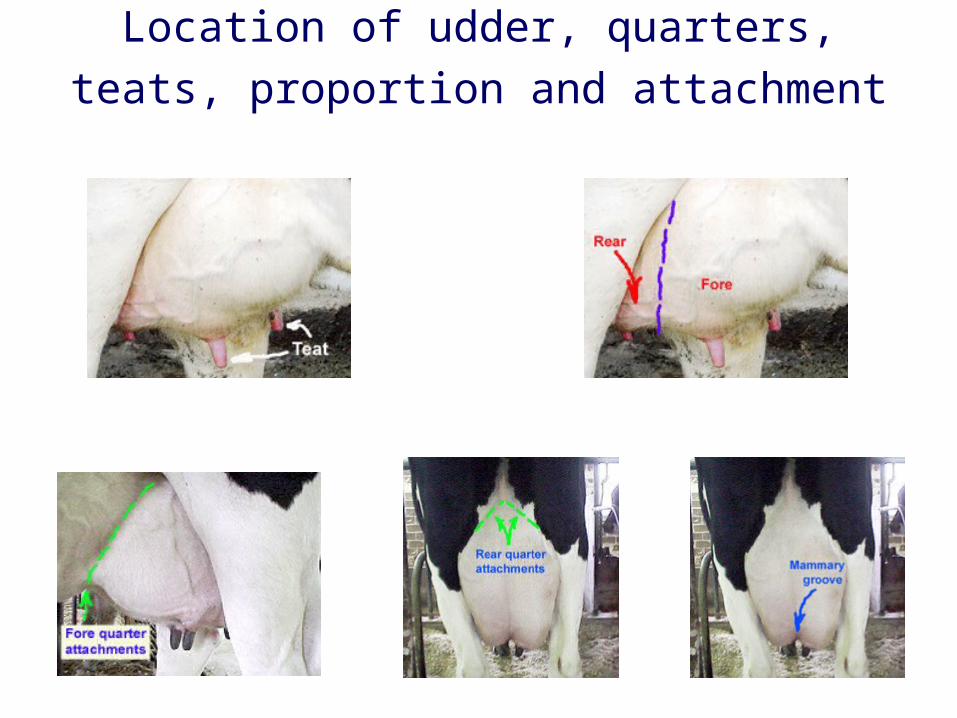
Location of udder, quarters, teats, proportion and attachment
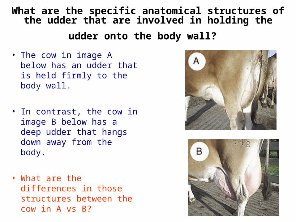
What are the specific anatomical structures of the udder
that are involved in holding the udder onto the body wall? • The cow in image A below
has an udder that is held firmly to the body wall.
• In contrast, the cow in image B below has a deep udder that hangs down away from the body.
• What are the differences in those structures between the cow in A vs B?
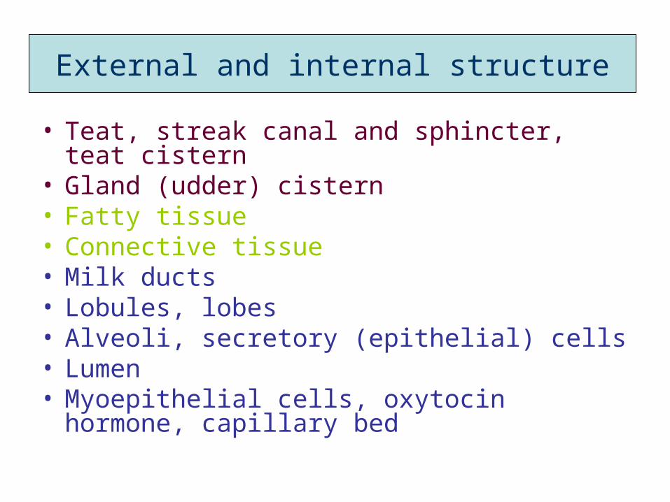
• Teat, streak canal and sphincter, teat cistern
• Gland (udder) cistern • Fatty tissue• Connective tissue• Milk ducts • Lobules, lobes• Alveoli, secretory (epithelial) cells• Lumen• Myoepithelial cells, oxytocin hormone,
capillary bed
External and internal structure
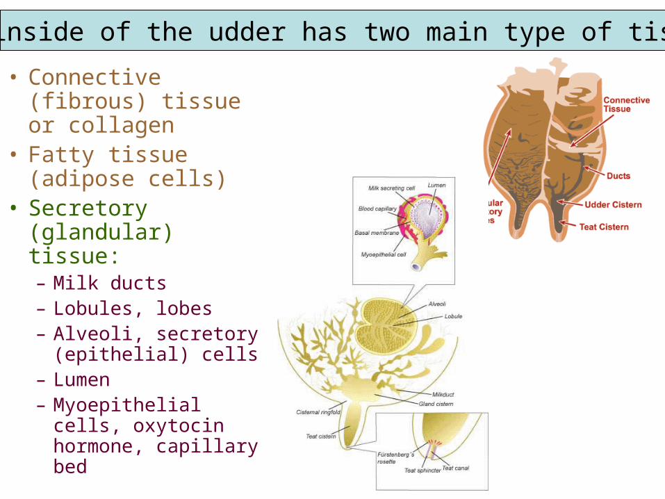
• Connective (fibrous) tissue or collagen
• Fatty tissue (adipose cells)
• Secretory (glandular) tissue:– Milk ducts – Lobules, lobes– Alveoli, secretory
(epithelial) cells– Lumen– Myoepithelial
cells, oxytocin hormone, capillary bed
The inside of the udder has two main type of tissue:
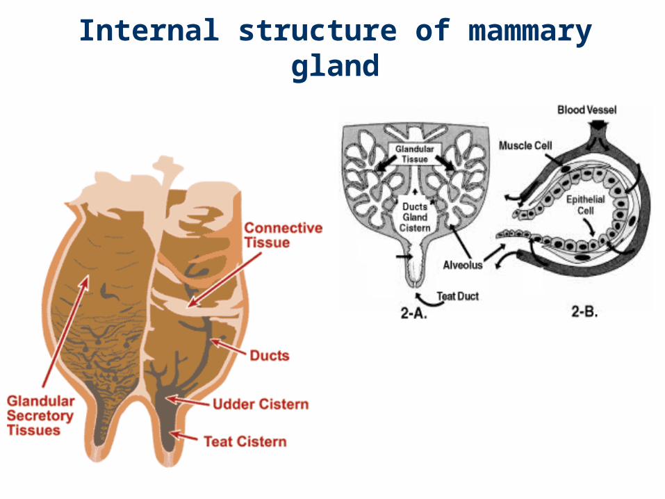
Internal structure of mammary gland
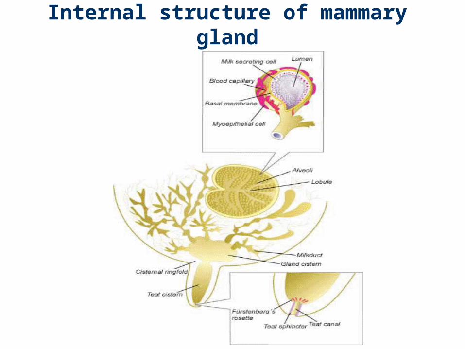
Internal structure of mammary gland
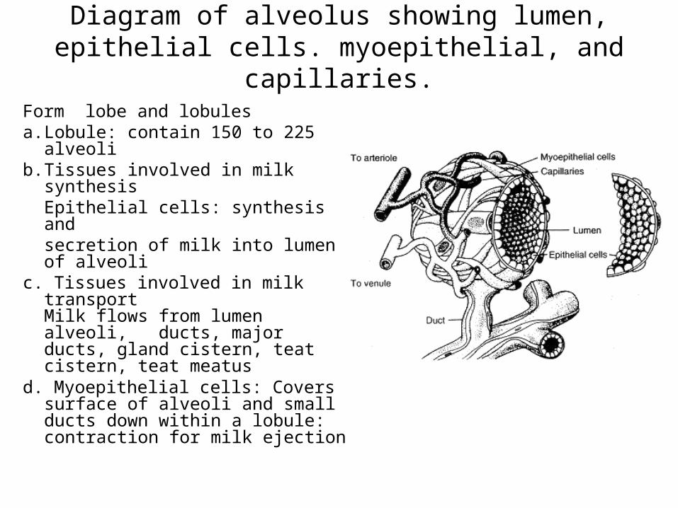
Diagram of alveolus showing lumen, epithelial cells. myoepithelial, and capillaries.
Form lobe and lobulesa. Lobule: contain 150 to 225
alveoli b. Tissues involved in milk
synthesisEpithelial cells: synthesis andsecretion of milk into lumen of alveoli
c. Tissues involved in milk transport Milk flows from lumen alveoli, ducts, major ducts, gland cistern, teat cistern, teat meatus
d. Myoepithelial cells: Covers surface of alveoli and small ducts down within a lobule: contraction for milk ejection
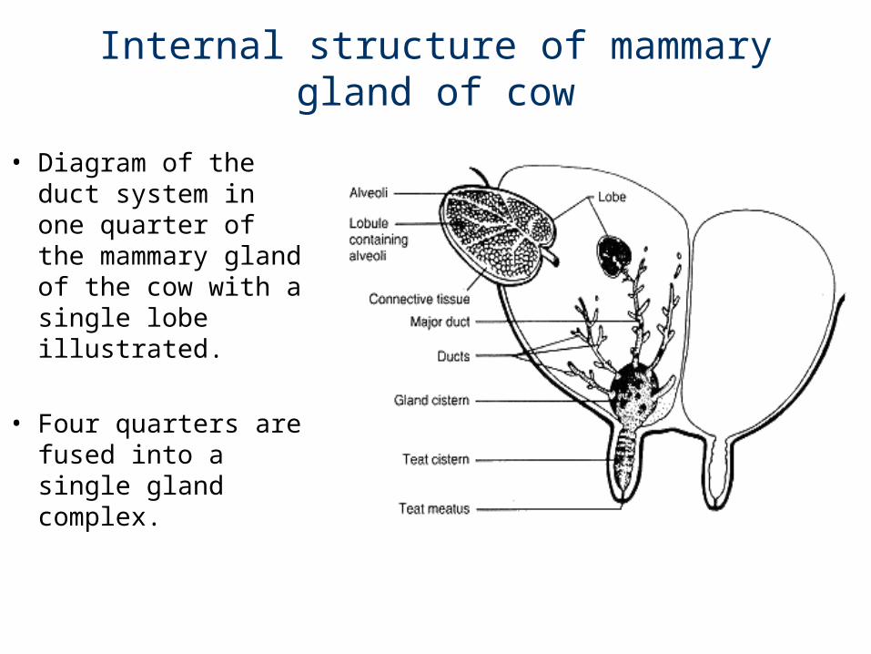
Internal structure of mammary gland of cow
• Diagram of the duct system in one quarter of the mammary gland of the cow with a single lobe illustrated.
• Four quarters are fused into a single gland complex.
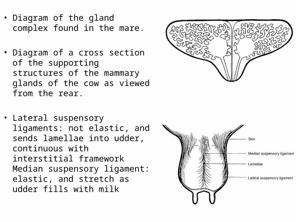
• Diagram of the gland complex found in the mare.
• Diagram of a cross section of the supporting structures of the mammary glands of the cow as viewed from the rear.
• Lateral suspensory ligaments:
not elastic, and sends lamellae into udder, continuous with interstitial framework Median suspensory ligament: elastic, and stretch as udder fills with milk
Blood vascular system
• The blood supply to the mammary gland is extremely important for mammary function! All of the milk precursors come from blood.
• On avg. 400 - 500 units of blood passes through the udder for each unit of milk synthesized by a high producing dairy cow; that is ~280 ml per sec.
• High producing dairy goats have a lower (460:1) ratio of blood flow through the gland:milk produced, compared with low producers (1000:1).
• This means that the amount of blood flow through the mammary gland may by similar for the high and low producing goats, but the efficiency of extraction of the components from the blood while it passes through the udder is very important. This principle is probably similar for cows.

• Total udder blood volume for lactating cows about 8% of total body blood volume, while for a non-lactating cow it is about 7.4%.
• There is a 2-6 fold increase in blood flow in the mammary gland starting 2-3 days prepartum.
• The decrease in production with advancing lactation is not due to decreased blood flow, but it is due to the loss of secretory (epithelial) cells through a process programmed cell death (apoptosis).
Related Documents