OBJECT AND ACTION NAMING IN APHASIC STROKE PATIENTS: LESION CHARACTERISTICS RELATED TO TREATMENT IMPROVEMENT By ROBERT BRUCE PARKINSON A DISSERTATION PRESENTED TO THE GRADUATE SCHOOL OF THE UNIVERSITY OF FLORIDA IN PARTIAL FULFILLMENT OF THE REQUIREMENTS FOR THE DEGREE OF DOCTOR OF PHILOSOPHY UNIVERSITY OF FLORIDA 2006

Welcome message from author
This document is posted to help you gain knowledge. Please leave a comment to let me know what you think about it! Share it to your friends and learn new things together.
Transcript
OBJECT AND ACTION NAMING IN APHASIC STROKE PATIENTS:
LESION CHARACTERISTICS RELATED TO TREATMENT IMPROVEMENT
By
ROBERT BRUCE PARKINSON
A DISSERTATION PRESENTED TO THE GRADUATE SCHOOL OF THE UNIVERSITY OF FLORIDA IN PARTIAL FULFILLMENT
OF THE REQUIREMENTS FOR THE DEGREE OF DOCTOR OF PHILOSOPHY
UNIVERSITY OF FLORIDA
2006

Copyright 2006
by
Robert Bruce Parkinson
This document is dedicated to my wife Faith and my daughter Alice, who was born the same week this dissertation was defended.
iv
ACKNOWLEDGMENTS
I wish to thank my dissertation committee chair and research mentor, Bruce
Crosson, for his excellent guidance throughout the dissertation process, and his example
of intellectual integrity and rigor. I also wish to thank Anastasia Raymer, who was
generous in allowing me to use data from her treatment study as the basis for this
dissertation. I also thank Yu-Ling Chang for her many hours spent rating brain scans,
and her kind insistence that it was she who benefited from the experience by learning a
new skill. Thanks also go to Faith Parkinson, for her proof-reading and encouragement,
but most of all for helping me stay grounded through what has proved to be an absorbing
enterprise.

v
TABLE OF CONTENTS page
ACKNOWLEDGMENTS ................................................................................................. iv
LIST OF TABLES........................................................................................................... viii
LIST OF FIGURES ........................................................................................................... ix
ABSTRACT.........................................................................................................................x
CHAPTER
1 INTRODUCTION AND BACKGROUND LITERATURE........................................1
Predictors of Improvement in Aphasia .........................................................................1 General Predictors of Aphasia Improvement ........................................................2 Lesion Predictors of Aphasia Improvement ..........................................................3
Improvement in general language functioning...............................................4 Improvement in comprehension.....................................................................4 Improvement in language expression and fluency.........................................6 Improvement in naming .................................................................................8
Basal Ganglia Lesions in Aphasia ..............................................................................11 Naming of Objects and Actions..................................................................................15 Background Summary ................................................................................................26
2 AIMS AND HYPOTHESES ......................................................................................29
Aims............................................................................................................................29 Hypotheses..................................................................................................................30
Hypothesis I. Effects of Treatment.....................................................................30 A. Improvement in all treatments................................................................30 B. No treatment differences ........................................................................31
Hypothesis II. Naming and Cortical Lesions .....................................................31 A. Pre-treatment functioning.......................................................................31 B. Improvement during treatment ...............................................................31
Hypothesis III. Naming and Basal Ganglia Lesions ..........................................32 A. Pre-treatment functioning.......................................................................32 B. Improvement during treatment ...............................................................32
Hypothesis IV. Comprehension and Improvement ............................................32
vi
3 METHODS.................................................................................................................34
Subjects.......................................................................................................................34 Aphasia Treatment......................................................................................................35 Imaging and Lesion Analysis .....................................................................................38
Structural Images.................................................................................................38 Lesion Analysis ...................................................................................................39
4 RESULTS...................................................................................................................43
Hypothesis I. Effects of Treatment............................................................................45 Hypothesis II. Naming and Cortical Lesions.............................................................48
A. Pre-treatment functioning ..............................................................................48 B. Improvement during treatment ......................................................................52
Hypothesis III. Naming and Basal Ganglia Lesions..................................................55 A. Pre-treatment functioning ..............................................................................55 B. Improvement during treatment ......................................................................55
Hypothesis IV. Comprehension and Improvement ..................................................58
5 DISCUSSION.............................................................................................................60
Effectiveness of Treatments .......................................................................................60 Basal Ganglia and Anterior Cortical Lesions .............................................................61
Proposed Explanation..........................................................................................64 Addressing an Alternative Explanation...............................................................66
Posterior Lesions and Naming....................................................................................67 Pre-treatment Naming .........................................................................................67 Naming Treatment Improvement ........................................................................69 Controlling for Anterior Lesion Extent ...............................................................70
Other Subcortical Findings .........................................................................................71 Pre-treatment Language Predictors of Naming Improvement....................................72 Object and Action Naming .........................................................................................74 Future Directions ........................................................................................................77 Conclusions.................................................................................................................82
APPENDIX
A SEMANTIC + PHONOLOGIC TREATMENT ........................................................85
B VERBAL + GESTURAL TREATMENT..................................................................87
C IMAGE PROCESSING INSTRUCTIONS................................................................89
Step 1 ..........................................................................................................................89 Alternative Method for Step 1 ....................................................................................92 Step 2 ..........................................................................................................................94 Step 3 ..........................................................................................................................96

vii
D RATING SHEET........................................................................................................99
E SLICE DESCRIPTIONS ACCORDING TO MATSUI 15 DEGREES...................100
F PROCEDURE FOR RATING LESIONS ................................................................103
G ROI DEFINITIONS..................................................................................................109
REFERENCES ................................................................................................................117
BIOGRAPHICAL SKETCH ...........................................................................................123
viii
LIST OF TABLES
Table page 3-1: ROI names and abbreviations....................................................................................40
4-1: Mean and Standard Deviation for Ratings of each ROI............................................45
4-2: Frequency of each Possible Final ROI Rating. .........................................................46
4-3: Anterior, posterior, and basal ganglia ratings for each subject. ................................47
4-4: Descriptive statistics for language measures. ............................................................47
4-5: Correlations between pre-treatment naming and cortical lesions extent...................50
4-6: Correlations between pre-treatment naming and individual ROI ratings..................52
4-7: Correlations between naming improvement and cortical lesion extent.....................54
4-8: Correlations between naming improvement and individual ROI ratings. .................56
4-9: Correlations between pre-treatment naming and basal ganglia lesion extent. ..........57
4-10: Correlations between naming improvement and basal ganglia lesion extent..........57
4-11: Partial correlations between naming measures and subcortical ROIs.....................58
4-12: Correlations between naming improvement and pre-treatment language measures. ..................................................................................................................59
5-1: Summary of significant correlations between ROIs and naming. .............................84
ix
FIGURE
Figure page F-1: Lesion rating guide..................................................................................................106
x
Abstract of Dissertation Presented to the Graduate School of the University of Florida in Partial Fulfillment of the Requirements for the Degree of Doctor of Philosophy
OBJECT AND ACTION NAMING IN APHASIC STROKE PATIENTS: LESION CHARACTERISTICS RELATED TO TREATMENT IMPROVEMENT
By
Robert Bruce Parkinson
August 2006
Chair: Bruce Crosson Major Department: Clinical and Health Psychology
Few studies have examined the relationship between lesion location and naming
treatment improvement in chronic aphasic stroke patients. The purpose of this study was
to determine whether degree of lesion in certain brain regions was related to degree of
treatment improvement demonstrated over the course of object and action naming
treatments. Participants and Methods: Fifteen aphasic left hemisphere stroke patients
underwent naming treatments for object and/or action naming. Two raters assessed
extent of lesion in 29 cortical and subcortical regions of interest (ROIs) on CT or MRI
scans. Correlations were calculated between composite basal ganglia and anterior
cortical lesion ratings and both pre-treatment and treatment-improvement measures for
both object and action naming. Results: Greater total basal ganglia lesion extent was
highly correlated with worse scores on all four naming measures when partial
correlations controlled for total anterior lesion extent (r ranging from -.623 to -.785).
Also, unexpectedly, greater anterior cortical lesion extent was highly correlated with
xi
better scores on all four naming measures when partial correlations controlled for total
basal ganglia lesion extent (r ranging from .730 to .858). No consistent differences were
found between the correlations of ROI ratings with object naming versus action naming
scores. Conclusion: Large anterior cortical lesions and an intact basal ganglia may both
contribute to more efficient re-organization of language functions. Since in this group of
patients lesion size in these two areas appears to affect naming in opposite directions,
controlling for the effects of one is needed to more clearly observe the effects of the
other.

1
CHAPTER 1 INTRODUCTION AND BACKGROUND LITERATURE
The current study investigated the relationship between brain lesion characteristics
in aphasic stroke patients and improvement in object and action naming over the course
of naming treatment. The introductory chapter of this document reviews literature
relevant to this subject, including (1) predictors of language improvement in stroke
patients, (2) the role of the basal ganglia in language functioning and language recovery,
and (3) neurocognitive differences in the naming of objects versus actions.
Predictors of Improvement in Aphasia
The mechanisms driving improvements in the language functioning of aphasic
stroke patients are not well understood, and are the topic of recent research. Factors
influencing these mechanisms such as patient characteristics and lesion characteristics are
of interest for several reasons. In a practical sense, by understanding how these factors
influence language recovery, clinicians may be better able to tailor language
rehabilitation programs to individual patients, thereby increasing the quality of life for
aphasic patients as well as decreasing the cost of their care. Also, from a scientific
perspective, such knowledge would increase our understanding of principles underlying
language processing in the human brain both in normal and brain-injured patients. Past
studies have examined a variety of factors for how they may affect language recovery in
stroke patients. Some studies have focused on the importance of factors such as aphasia
type, initial aphasia severity, and age of patient, and how these factors influence recovery
of language functioning in general. Other studies have looked at how lesion
2
characteristics such as size and location influence improvement in groups of patients with
specific aphasia types or symptoms. The following two sections will review the literature
related to these areas – general predictors and lesion characteristic predictors.
General Predictors of Aphasia Improvement
Past studies have examined a variety of possible factors which may explain why
some aphasic stroke patients improve more than others. Several studies have reported
that certain types of aphasic syndromes are more likely to show improvement than other
types. For example, there are reports that stroke patients are likely to show more
improvement from Broca’s and Wernicke’s aphasia than from global aphasia, and that
expressive impairments are more likely to improve than comprehension impairments (for
a review see Demeurisse, Capon & Verhas, 1985; and Lomas & Kertesz, 1978). Some
studies have found that older patients are likely to show less improvement compared to
younger patients (Sands, Sarno & Shankweiler, 1969), while other studies have found no
such correlation (for review see Demeurisse et al., 1985). Initial general severity of the
aphasia has often been cited as a predictor of improvement, with less severe patients
likely to experience greater improvement (Demeurisse et al., 1985; Kenin & Swisher,
1972; Naeser et al., 1998; and Sands et al., 1969). More specifically, Naeser et al. (1998)
reported that initial scores on measures of comprehension were predictive of later success
in a language treatment.
At least one study has examined the relationship between quantitative brain
measures on CT, and language improvement in stroke patients. Pieniadz, Naeser, Koff
and Levine (1983) reported that structural asymmetries of the cerebral hemispheres
predicted greater improvement on a number of language measures, including
comprehension, repetition, and naming, in a group of 14 global aphasic subjects.
3
Specifically, they found that subjects with right occipital asymmetries (longer and wider
occipital lobes on the right side) demonstrated greater recovery on those measures. The
authors hypothesize that these hemispheric asymmetries reflect unusually large right
hemisphere areas corresponding to left language areas, which are better able to take over
language functioning following injury to the left hemisphere.
Lesion Predictors of Aphasia Improvement
Although much work has been done in studying the relationship between lesion
characteristics and aphasia type (Cappa & Vignolo, 1999; Damasio, 1998; Kertesz, 1979;
Kreisler et al., 2000), fewer studies have examined whether lesion characteristics are
related to improvement in aphasia. The studies that have examined possible relationships
between lesion location and aphasia improvement differ on a number of important
factors. For example, while some studies include a wide variety of aphasic subjects in
their subject groups, others may focus only on subjects with a specific aphasic syndrome,
such as Wernicke’s aphasia, or a specific aphasic symptom, such as a naming deficit.
Also, while some studies examine improvement on one or more specific language
measures, other studies examine improvement based on measures of general language
functioning. In other words, studies vary on both the specificity of the inclusion criteria
of their subjects and the specificity of their outcome measures. With so few studies
analyzing lesion predictors of language recovery, and with such a wide variety of
inclusion criteria and outcome measures represented in those studies, it is difficult to
draw any firm conclusions from the existing literature. However, the following sections
will briefly review the major findings of available studies. Considering that the
relationship between lesion characteristics and associated language improvements likely
depend on the particular language function being measured, the following sections will
4
be organized according to the language outcome variable examined, namely: general
language improvement, comprehension improvement, improvement in expression and
fluency, and naming improvement.
Improvement in general language functioning
One study examined the language improvement of a group of stroke patients based
on a general language impairment index score. Goldenberg and Spatt (1994) looked at
the general language recovery of 18 aphasic stroke patients during both treatment and
non-treatment phases of recovery. They found that damage to Wernicke’s area was
related to poorer spontaneous improvement, while damage to the left basal temporal lobe
was related to poorer treatment improvement. These findings lead the authors to
hypothesize that treatment improvement relies on structures associated with explicit
learning, specifically the connections between perisylvian language structures and the
hippocampus. The authors also found that larger overall lesion size, irrespective of its
location within the left hemisphere, was related to both the initial severity of the aphasia
and to the degree of recovery during both treatment and non-treatment phases.
Improvement in comprehension
Lesion characteristics related to improvement in comprehension have been
examined by more studies than any other aspect of language improvement. Naeser,
Gaddie, Palumbo, and Stiassny-Eder (1990) examined the recovery of single word
comprehension in 14 globally aphasic stroke patients from the acute to chronic stages.
They found that recovery of comprehension ability was greater in patients whose lesion
spared Wernicke’s area. Since Wernicke’s area lesions also often include the temporal
isthmus, a subcortical white matter track deep to Wernicke’s area, the authors also
divided the subjects into two groups: those whose lesions included both Wernicke’s area
5
and the temporal isthmus, and those whose lesion included only the temporal isthmus.
All of the subjects had lesions in frontal and parietal areas, but subjects who had temporal
isthmus plus Wernicke’s area lesions had less recovery of comprehension than subjects
with lesions of just the temporal isthmus.
Other studies have also reported on the importance of Wernicke’s area for
improvements in comprehension. Selnes, Niccum, Knopman and Rubens (1984)
reported on the recovery of 11 aphasic patients with initial single-word comprehension
deficits. Six of the seven patients who demonstrated poor recovery had lesions involving
Wernicke’s area. However, two of the four patients who demonstrated good recovery
also had lesions in Wernicke’s area. They concluded that although lesions of Wernicke’s
area often lead to poor recovery of comprehension, Wernicke’s area lesions do not
preclude good recovery.
Naeser, Helm-Estabrooks, Haas, Auerbach, and Srinivasan (1987) examined
whether the extent of lesion within Wernicke’s area was related to the degree of
comprehension recovery in 10 subjects with Wernicke’s aphasia. They found that while
overall lesion size (including temporal and parietal areas) did not predict recovery,
subjects with less than half of Wernicke’s area lesioned demonstrated greater recovery
than subjects with greater than half of Wernicke’s area lesioned. Moreover, those
subjects whose lesions extended anteriorly and inferiorly into the middle temporal gyrus
had particularly poor recovery.
Kertesz, Lau and Polk (1993) studied a group of 22 patients with Wernicke’s
aphasia to determine factors predicting overall spontaneous recovery at 3 and 12 months
post-stroke. They found that initial severity, lesion size, and whether the lesions affected
6
the supramarginal gyrus and angular gyrus were the best predictors of improvement.
They hypothesized that those two areas, being posteriorly adjacent to Wernicke’s area,
were important in compensating for comprehension deficits. In a separate study of a
group of consecutive left hemisphere stroke patients with no particular aphasic syndrome,
Wernicke’s area lesions and supramarginal gyrus lesions were also found to be predictive
of less comprehension recovery (Selnes, Knopman, Niccum, Rubens & Larson, 1983).
Overall, available research indicates that Wernicke’s area lesions, as well as
lesions extending anteriorly, inferiorly, and posteriorly from Wernicke’s area, are usually
related to worse recovery of language comprehension. While one study (Kertesz, Lau &
Polk, 1993) found that overall lesion size was also a good predictor of improvement,
others either disagreed (Selnes, Knopman, Niccum, Rubens & Larson, 1983) or found
that only the lesion size within Wernicke’s area was important (Naeser, Helm-
Estabrooks, Haas, Auerbach & Srinivasan, 1987).
Improvement in language expression and fluency
Several studies have examined predictors of improvement on measures of language
production, including general expressive language measures and fluency ratings.
Demeurisse, Capon, and Verhas (1985) found that in subjects with cortical-subcortical
lesions, overall lesion size was predictive of the rate of improvement of expressive
language abilities. However, size of lesions in subjects with only subcortical lesions was
not predictive of improvement. They also found that subjects with lower baseline
language abilities tended to improve less during the course of language treatment. A
second study found that baseline comprehension scores were particularly good predictors
of improvement in language production following stroke (Lomas & Kertesz, 1978).
7
Specific lesion sites have been associated with persistent non-fluency. In one study
(Knopman, Selnes, Niccum, Rubens, Yock & Larson, 1983), subjects were found to have
better recovery of fluency when lesions spared the rolandic cortical region, including
underlying white matter. The authors also reported that greater overall lesion size
predicted persistent non-fluency. Another study (Naeser, Palumbo, Helm-Estabrooks,
Stiassny-Eder & Albert, 1989) specifically identified lesions in two white matter
pathways as being particularly predictive of severe non-fluency. The authors found that
lesions involving significant portions of both the medial subcallosal fasciculus and the
middle one-third of the periventricular white matter produced severe and persistent
nonfluency. They proposed that lesions affecting the medial subcallosal fasciculus
interrupted pathways from the cingulate gyrus and supplementary motor area to the basal
ganglia, which affects intentional aspects of speech production. The authors also noted
that lesions of the middle one-third of the periventricular white matter, which contain
white matter tracks deep to the motor and sensory cortex for the mouth, would likely
affect motor execution and sensory feedback necessary for speech. In addition to the
author’s explanation, it may be important to note that lesions affecting the middle third of
the periventricular white matter may also affect the dorsal caudate. Although the authors
did not measure lesions in the dorsal caudate, lesions to this structure may also effect
speech production.
The findings from Naeser’s study may account for the earlier findings of Knopman
et al. (1983) implicating rolandic cortical regions in persistent non-fluency. A more
detailed analysis of the persistently non-fluent subjects’ lesions described by Knopman et
al. may have shown that they also included both the medial subcallosal fasciculus and the
8
middle third of the periventricular white matter, as predicted by Naeser et al. (1989), and
possibly also the dorsal caudate, as noted above. Naeser et al. suggested that lesions of
white matter pathways, which cause disconnections between cortical and subcortical
regions, are more important predictors than cortical or basal ganglia lesions alone.
Another study examined predictors of language treatment success for a computer
assisted language treatment program (Naeser et al., 1998). This study differs from the
studies cited above in that subjects were trained to produce language via input into a
computer rather than verbally. The authors found that subjects with lesions in specific
areas tended to achieve a lower level of success in treatment, namely, Wernicke’s area,
the temporal isthmus, supraventricular frontal lobe structures (including the
supplementary motor area and cingulate gyrus) and the subcortical medial subcallosal
fasciculus.
Improvement in naming
Few studies have specifically examined predictors of improvement in naming
ability in aphasic stroke patients. Knopman, Selnes, Niccum, and Rubens (1984)
examined a group of 54 left hemisphere stroke patients with mild to severe naming
deficits at one month and six months post-stroke. Forty brain regions were assessed for
presence of lesion, and subjects were administered measures of naming, single word
comprehension, and verbal fluency at both time points. It was unclear whether the
patients participated in formal language therapy. The authors report that initial scores on
measures of naming and single word comprehension at one month post-stroke were good
predictors of naming recovery by six months. They also found that subjects with larger
overall lesions showed less recovery of naming. Subjects with lesions in Wernicke’s
area and inferior parietal cortex were noted to demonstrate the most severe naming

9
impairments at six months. Subjects with lesions of the insula and putamen, extending
into areas deep to the supramarginal gyrus, were noted to have significant but less severe
naming impairments by six months. It should be noted that although this study examined
a relatively large number of subjects, the degree of precision of its measures was small.
For example, language recovery for each subjects was categorized simply as being poor
or good, and lesion ROI analysis consisted of categorizing regions as being either
lesioned or not. Given the simplicity of the measures used, the authors used descriptive
rather than inferential statistics to arrive at their conclusions.
Using more precise measures of but fewer subjects, Cato, Parkinson, Wierenga and
Crosson (2004a) and Cato, Parkinson, Wierenga and Crosson (2004b) examined
improvement of naming ability in nine non-fluent subjects over the course of a novel
naming treatment. Unlike the Knopman et al. (1984) study, the above two studies used
range corrected gain scores of a naming measure to characterize naming improvement.
Cato et al. (2004b) also characterized lesions using a 7 point rating scale for ROIs
(Naeser et al., 1998) rather than using a dichotomous rating system. Cato et al. (2004a)
found that, like previous studies involving language production and naming (Knopman et
al., 1984; Lomas & Kertesz, 1978), improvement in naming was associated with initial
comprehension scores. However, unlike Knopman et al., the authors found that naming
improvement was not correlated with overall lesion size Subjects with both large or
small lesions could show significant improvement. Cato et al. (2004b) found that while
overall size of lesion did not predict recovery, location of the lesion did, with poorer
recovery being correlated with greater lesion extent ratings in Wernicke’s area, the
supramarginal gyrus, and the posterior one-third of the periventricular white matter. The
10
authors point out the convergence of these behavioral and lesion predictors of
improvement, because comprehension abilities have long been associated with posterior
lesions.
It should be pointed out that the above-mentioned naming studies focus mainly on
the improvement of object naming rather than action naming. For example, Knopman et
al. (1984) used the Boston Diagnostic Aphasia Examination (BDAE) confrontation
naming subtest and the Boston Naming Test (BNT) to assess naming improvement. The
BNT is comprised exclusively of object naming items, while the 35 items of the BDAE
naming subtest include objects, numbers, letters, actions, forms, body parts, and colors.
Neither of these tests specifically addresses a subject’s ability to name objects versus
actions. Likewise, in the Cato et al. (2004a) and Cato et al. (2004b) studies, naming was
assessed exclusively by object picture naming measures. As will be discussed in greater
detail in a later section of this document, previous studies have indicated that distinct
neural systems may be involved in the naming of objects versus actions. However, no
studies have actually looked at how lesion characteristics are related to improvements in
object versus action naming.
Part of the difficulty in using lesion characteristics to predict improvement in
aphasia may be that standard CT and MRI images (T1 or T2 weighted scans) only allow
for the quantification of areas of necrosis; however, there may be other areas which
appear healthy on CT and MRI but are actually hypoperfused and dysfunctional. Recent
studies (Hillis, Barker, et al., 2004; Hillis, Wityk, et al., 2002; Love, Swinney, Wong, &
Buxton, 2002) have shown that perfusion weighted imaging (PWI) techniques, which
allow the visualization and quantification of hypoperfused areas of brain tissue (i.e.,

11
functional lesions), tend to be better predictors of aphasia symptoms than standard
structural imaging methods. However, PWI is not yet as widely available as CT or
standard MRI, and studies examining the relationship between these “functional lesions”
and aphasia treatment improvement have yet to be done.
The studies reviewed in the above sections have helped identify several potential
predictors of language recovery. The most common findings are that worse recovery is
related to initial severity of the aphasia (particularly initial comprehension ability), lesion
size, and the presence of posterior lesions, particularly in Wernicke’s area. However, it
should be noted that relatively few studies have examined this area of research, and there
is still debate over how useful these predictors are in individual cases. For example, there
are individual cases reported which seem to contradict all of these basic findings (Basso
& Farabola, 1997).
The literature reviewed in the previous sections has focused almost exclusively on
the effects of lesions involving cortical areas and subcortical white matter pathways.
However, there is also evidence that the basal ganglia are involved in language
functioning. In fact, there are indications that the basal ganglia may play a specific role
in the recovery of language functioning after stroke. The following section will briefly
review these issues.
Basal Ganglia Lesions in Aphasia
The role of subcortical lesions in aphasia has long been debated (Nadeau &
Crosson, 1997). Many case studies have reported the occurrence of aphasia following
lesions restricted to subcortical areas, including the basal ganglia (Alexander, Naeser &
Palumbo, 1987; D’Esposito & Alexander, 1995). Recently, perfusion weighted imaging
studies (Hillis, Barker, et al., 2004; Hillis, Wityk, et al., 2002) have shown that aphasia
12
following lesions restricted to the basal ganglia is more likely caused by the
hypoperfusion of language cortex than the basal ganglia lesion itself. Nevertheless, there
is also evidence that when combined with cortical lesions, left basal ganglia lesions may
decrease an individual’s ability to recover language functioning.
Brunner, Kornhuber, Seemuller, Suger, and Wallesch (1982) studied a group of
40 patients with vascular lesions in the language-dominant hemisphere. They found that
patients with restricted subcortical lesions (including the basal ganglia) experienced
language disturbance, but usually with good spontaneous recovery. Patients with both
cortical and basal ganglia lesions experienced relatively more severe and longer lasting
aphasia. The authors also found that most patients with cortical damage alone (unless the
lesion involved Wernicke’s area, which in some cases produced longer lasting aphasia)
generally displayed more transient aphasic syndromes. In other words, when combined
with lesions to cortical language areas, subcortical lesions appear to decrease recovery
potential in aphasia.
A recent series of functional imaging studies may help explain the role of the basal
ganglia in language production. In an fMRI study with normal subjects, Crosson et al.
(2003) found that the left “pre-SMA-dorsal caudate nucleus-ventral anterior thalamic”
loop was active during real-word generation tasks, but not during nonsense syllable
generation tasks, indicating that this loop likely plays a role in the retrieval of words from
pre-existing lexical stores. Activity also was seen in the right basal ganglia during real-
word generation tasks but not during nonsense syllable generation tasks. However, this
right basal ganglia activity was not accompanied by right frontal activity, leading the
authors to hypothesize that the right basal ganglia activity was serving to suppress right
13
frontal activity, and prevented the right frontal areas from interfering with language
production in the left hemisphere.
In another fMRI, study reported by Kim, Ko, Parrish, and Kim, (2002) different
activation patterns were observed in aphasic patients with restricted left frontal lesions
compared to aphasic patients with left frontal plus subcortical lesions. During an
auditory sentence completion task, patients with restricted left frontal lesions displayed
activation mainly in the right inferior frontal lobes, while patients with left frontal plus
subcortical lesions displayed bilateral frontal and temporal activation. It is possible that
Crosson’s hypothesis (Crosson et al., 2003) may be extended to help explain the findings
of Kim et al. (2002). Unilateral right activation may have been observed in patients with
left cortical damage in the patients described by Kim et al. because the intact left basal
ganglia was used to suppress any peri-lesional left cortical activity which would interfere
with the newly established right hemisphere language centers. Bilateral frontal and
temporal activation was observed in stoke patients with left frontal plus subcortical
lesions, because the lesioned left basal ganglia could not inhibit left cortical activity.
The 2003 Crosson hypothesis may also help explain the finding of Brunner et al.
(1982) that patients with combined basal ganglia and cortical damage experience more
severe and long lasting aphasia than patients with comparably sized lesions involving
only the left cortex. Given a large lesion circumscribed to only left language cortex, the
left basal ganglia would inhibit any noise originating from the remaining peri-lesional
areas, thereby allowing right hemisphere areas to begin to take over language production.
However, given the same large left hemisphere lesion plus a lesion to the left basal
14
ganglia, noise originating from the left peri-lesional areas could not be inhibited and
would interfere with attempts by the right hemisphere to produce language.
A second study by Crosson et al. (2005), supports these hypotheses. The authors
describe two left hemisphere aphasic stroke patients who received a novel naming
treatment aimed at priming right hemisphere intentional mechanisms to support
reorganization of language production to the right lateral frontal cortex by initiating
naming trials with a complex left-hand movement. fMRI results indicated right
hemisphere language lateralization one patient whose lesion encompassed only cortical
areas before treatment had even begun. This right lateralization continued through
treatment. However, in a second patient whose lesion included cortical areas plus the left
basal ganglia and thalamus, left lateralization was observed pre-treatment, and only after
treatment was a shift noted towards right medial and lateral frontal cortex activation. The
authors suggest that if damage to left hemisphere language production areas reaches a
critical amount, rehabilitation success is greater with increased right hemisphere
participation. In the case of the patient with intact left basal ganglia functioning, the left
basal ganglia facilitated reorganization of language production in the right hemisphere by
suppressing the noise generated by dysfunctional left lateral frontal areas, even prior to
any type of specialized treatment. But in the case of the patient with a left basal ganglia
lesion, the intentional treatment was thought to have allowed a more successful right
hemisphere reorganization by priming right pre-SMA, which uses its crossed connections
with the left basal ganglia to suppress the inefficient attempts of the left frontal cortex to
generate language.

15
The functional imaging studies discussed above appear to indicate that when
combined with large enough left cortical lesions, left basal ganglia lesions may decrease a
patient’s ability to recover language functioning following stroke. Further structural
imaging studies using quantitative lesion analysis techniques are also needed to test this
hypothesis and further elucidate the role of the basal ganglia in language recovery. Thus,
one of the aims of the current study is to determine whether lesions involving both cortex
and basal ganglia are associated with less improvement in naming treatments than lesions
involving cortical areas alone.
Naming of Objects and Actions
Another aim of the current study is to examine whether lesion characteristics in
aphasic stroke patients are related to their degree of improvement in object versus action
naming treatments. As mentioned previously, past studies that have examined potential
predictors of naming improvement have focused on the naming of nouns, or objects, with
no studies specifically addressing improvement of verb, or action, naming. Although the
literature examining predictors of language improvement does not differentiate between
object and action naming, studies with a broader focus (i.e., investigating naming ability
in general, rather than improvements in naming ability) have found that objects and
actions may be processed by different brain regions. In fact, distinct lesion sites have
been reported to be related to specific object versus action naming deficits. This section
will briefly review key studies related to specific naming deficits of objects and actions.
It should be noted that some of the studies reviewed in this section use the terms
“noun” and “verb”, while others use the terms “object” and “action.” In the current
study, the terms “object” and “action” are preferred because they represent the specific
categories of nouns and verbs which are normally tested in naming paradigms. For the
16
purposes of this study, an “object” refers to a concrete noun which can be identified by
means of a picture (such as “dog” or “brick”), and does not refer to abstract nouns (such
as “freedom” or “negotiation”). The term “action” will refer to action verbs which can be
identified by means of a picture (such as “walking” or “throwing”), and does not refer to
helping verbs (such as “will”), or state of being verbs (such as “was” or “is”). Although
some studies cited in this section may use the terms “noun” and “verb,” they are
generally only referring to those nouns and verbs which are objects and actions. Thus,
the terms “object” and “action” are preferred, and will be used except when describing
specific studies which use the terms “noun” and “verb.”
Individual case reports have been published describing stroke patients with
differential deficits in object versus action naming. Caramazza and Hillis (1991) reported
on two stroke patients with specific naming deficits relative to nouns and verbs and
different production modalities. One subject performed normally in naming both nouns
and verbs orally, and in producing written nouns, but showed a deficit for producing
written verbs. The other subject performed normally in producing written nouns and
verbs, but was impaired in oral naming, especially with verbs. The authors interpreted
these findings as an indication that brain mechanisms involved in word production are
organized not only by output modality (written versus oral), but also by grammatical
class. In a later study Hillis and Caramazza (1995) reported the case of a third subject
who demonstrated worse naming of nouns than verbs when speaking, but worse verbs
than nouns when writing. Taken together with their first study, the authors conceptualize
their findings as a double dissociation, and as evidence of separate neural systems for the
phonologic and orthographic representations of nouns and verbs.

17
The idea of speech output mechanisms being supported by separate systems
depending on grammatical class has since received much attention from researchers. In a
seminal paper in 1993, Damasio and Tranel described the naming deficits of three
patients, and presented their findings as evidence of a double dissociation between
anatomical regions supporting noun versus verb production. All three of the subjects had
normal performance in grammar, morphology, phonetic implementation, prosody,
reading and writing, but with specific selective retrieval deficits. Two of the subjects
were able to perform as well as normal control subjects on naming verbs, however, they
were impaired in naming concrete nouns. On the other hand, a third subject performed as
well as normal control subjects when naming nouns, but was impaired at naming verbs.
The subjects with noun naming impairments had lesions in the left anterior and middle
temporal lobe while the subject with verb naming impairment had a lesion in the left
premotor cortex. Although the subjects in this study showed deficits in retrieving certain
classes of words based on visual stimuli, it appeared that representations of these words
still existed, as the subjects were able to produce the words under different conditions,
such as in phonetic cueing tasks and running speech. The authors concluded that there
are separate systems involved in the retrieval of nouns and verbs which are located in
distinct brain regions.
More recently, Tranel, Adolphs, Damasio, and Damasio (2001) reported further
evidence supporting the double dissociation proposed in their 1993 paper. Lesions of 75
subjects with stable focal lesions were analyzed. The authors found that lesions related to
action naming impairment had maximum overlap in the left frontal operculum, its
underlying white matter, and in the anterior insula. As predicted by their previous paper,

18
lesions in the left anterior temporal regions were associated with deficits in naming
concrete entities, but not with deficits in naming actions. In subjects with
disproportionately greater action naming deficits compared to object naming deficits,
lesions were most common in premotor/prefrontal cortex, left mesial occipital cortex, and
periventricular white matter deep to the supramarginal gyrus and posterior temporal
cortex.
Whereas the previous studies examining differences in object/action naming
systems focused on chronic stroke patients, Hillis, Tuffiash, Wityk, and Barker (2002),
addressed the object/action naming dissociation issue with a group of acute stroke
patients using diffusion weighted and perfusion weighted MRI. Unlike standard MRI
techniques (T1 or T2 acquisition protocols) or CT imaging, which allow for the
visualization of structural lesions only, perfusion weighted imaging (PWI) also delineates
functional lesions, or areas of hypoperfusion. Furthermore, these new imaging
techniques can define the areas of cerebral dysfunction at the acute stage of stroke. In
previous studies, such as those mentioned above, patients had been examined at the
chronic stage, when brain lesion borders and language functioning were thought to be
mostly stabilized. However, by the chronic stage, those subjects’ functional
neuroanatomy may have already undergone plastic changes in response to the injury. By
examining functional lesions of acute patients, rather than structural lesions of chronic
patients, the relationships between lesion location and cognitive deficits more likely
reflect the patient’s normal functional neuroanatomy. The Hillis and Tuffiash et al.
(2002) study included 33 patients with acute left hemisphere stroke who were tested for
oral naming and comprehension of both nouns and verbs at the time of the imaging.

19
Similar to previous studies with chronic patients, left temporal cortex lesions were
associated with object naming deficits, whereas left posterior frontal cortex lesions were
associated with action naming deficits.
Reports describing specific naming deficits for objects or actions in dementia
patients also support the anatomical correlates proposed in the above cited stroke studies.
For example, several studies have assessed the specific naming deficits of patients with
primary progressive aphasia (Bak, O’Donovan, Xuereb, Boniface & Hodges, 2001;
Daniele, Guistolisi, Silver, Colosimo & Gainotti, 1994; Hillis, Oh & Ken, 2004).
Primary progressive aphasia (PPA), is a degenerative condition characterized by at least
two years of progressive language deficits without cognitive decline in other areas, and is
a type of fronto-temporal dementia. Both fluent and non-fluent types of PPA have been
described in the literature, with fluent PPA being associated with atrophy of the left
posterior superior temporal lobe and angular gyrus, while non-fluent PPA is associated
with atrophy of the left posterior inferior frontal lobe, left premotor cortex, and insula
(Hillis, Oh, et al., 2004).
Daniele et al. (1994) reported findings from a group of three patients, two of which
displayed frontal lobe atrophy and non-fluent aphasia, and one patient with temporal lobe
atrophy and fluent aphasia. The non-fluent frontal patients displayed impaired naming
and comprehension of verbs while the fluent temporal patient displayed disproportionate
difficulties in naming and comprehending nouns. Bak et al. (2001) examined a set of six
non-fluent patients with amyotrophic lateral sclerosis or ALS (a disease related to non-
fluent PPA in that non-fluent PPA patients are sometimes later diagnosed with ALS) and
found a greater deficit for comprehension and naming of verbs than nouns. In a post-
20
mortem examination of three of these patients, pathological involvement was observed in
several frontal areas, including the motor and premotor cortex and Brodmann’s area 44
and 45. In a larger scale study, Hillis, Oh, et al. (2004) confirmed the results of these
previous 2 studies with a group of 15 non-fluent PPA patients, seven fluent PPA patients,
and six ALS patients. Again, non-fluent PPA and ALS patients were more impaired in
verb naming , while fluent PPA patients were more impaired in noun naming.
Like PPA patients, Alzheimer’s disease (AD) patients also tend to display both a
specific pattern of language impairment and a specific distribution of brain pathology.
Anomia is one of the earliest signs of AD, and worsens as the disease progresses.
Williamson, Adair, Raymer, and Heilman (1998) found that although AD patients
performed worse on both object and action naming tasks compared to normals, they were
especially impaired on naming objects. This effect was seen even when the authors
controlled for the frequency of occurrence of their target words. The authors reported
that in post-mortem studies, neurofibrillary tangles and neuritic plaques, which define
AD, are much more likely to be found in limbic, temporal and occipital areas compared
to frontal regions. They conclude that the pathological changes to these regions impair
retrieval systems for objects more than actions.
Cappa, Sandrini, Rossini, Sosta and Miniussi (2002) provide additional evidence
that neural substrates supporting action naming are distinct from those supporting object
naming. Using repetitive transcranial magnetic stimulation (rTMS), the authors
examined whether stimulation to the dorsolateral frontal cortex differentially affected
normal subjects in object and action naming tasks. They found that subjects were able to
name actions significantly faster during left hemisphere stimulation compared to right
21
hemisphere stimulation or the sham condition. On the other hand, subjects showed no
difference in object naming speed for any of the three conditions. Taken together, the
stroke, dementia, and rTMS studies cited above argue that temporal regions are
particularly important for object naming, while frontal regions are particularly important
for naming actions.
Although the studies cited above support the hypothesis that there are distinct
neural systems supporting the naming of objects and actions, it should be noted that there
is debate over whether the systems responsible for the processing of individual words are
truly organized by grammatical function, or other defining features, such as semantic
attributes. For example, Lu et al. (2002) provided evidence that the naming deficits of a
group of left anterior temporal lobectomy subjects were better defined by a semantic
dichotomy than a grammatical one. The authors found that although there was no
difference between the subjects’ performance on object naming versus action naming,
these subjects were specifically impaired on both objects and actions that were associated
with human actions. Nouns representing objects used in human actions such as tools and
implements (i.e., shovel, fork) and verbs which described human actions (i.e., cutting,
saluting) were more impaired than nouns representing living objects (i.e., rabbit,
broccoli) and verbs representing non-human actions (i.e., blooming, dripping). Thus, it
may be that representations of objects and actions may actually be organized according
to multiple critical attributes.
Several functional imaging studies have indicated that neural systems required to
perform semantic decision making tasks may not follow the object/action division
proposed in naming studies such as Damasio and Tranel (1993), and some suggest that
22
there may be other organizational principles at work. For example, one study (Grossman
et al., 2002) showed that semantic representations of verbs appear to be differentially
represented in the brain depending on whether they are verbs of motion or verbs of
cognition. The authors reported that during a semantic rating task using fMRI, motion
verbs were associated with activation of the left ventral temporal-occipital cortex,
bilateral prefrontal cortex, and caudate nucleus, while verbs of cognition were associated
with left posterolateral temporal activation. In another fMRI study involving normal
subjects in a passive verb reading task (Hauk, Johnsrude & Pulvermuller, 2004),
activation appeared to be organized somatotopically along the motor strip. In other
words, areas of the motor strip were activated according to the body part used to produce
the action represented by the verb. For example, ‘kick’ is associated with the foot, while
‘lick’ is associated with the tongue. Both of these studies provide evidence for a
semantic influence in the organization of verbs in the brain. Another fMRI study in
normal subjects (Kraut, Moo, Segal & Hart, 2002) showed that semantic representation
of some objects, such as fruits and tools, may also be associated with regions of the
motor strip, ostensibly because there are characteristic actions associated with these
objects.
While lesion studies and dementia studies both provide evidence that neural
systems involved in object and action production are likely distinct, some functional
imaging studies have failed to show differences between areas activated during object
versus action tasks. Some may take this as evidence against the hypotheses drawn from
the stroke and dementia studies (such as Damasio & Tranel, 1993). However, before
discussing these studies, it should be pointed out that the tasks used in the lesion and

23
dementia studies cited above often differed from tasks used in the functional imaging
studies. While the stroke and dementia studies used simple picture naming tasks, most
tasks in the functional imaging studies involved word comprehension or had other
semantic processing components. As pointed out by Hillis, Tuffiash, Wityk, and Barker
(2002), the claim that the naming of objects and actions is supported by anatomically
differentiated systems, does not mean that all aspects of object/action processing, such as
comprehension, are necessarily distributed in the same way. The following functional
imaging studies have provided evidence for an extensive semantic network that is highly
distributed and overlapping, rather than localized for objects or actions.
In a positron emission tomography (PET) study with normal subjects (Tyler,
Russell, Fadili & Moss, 2001), no differences were found between areas of activation for
nouns and verbs during tasks involving lexical decision making or semantic
categorization. Instead, these tasks resulted in activation patterns extending from the left
inferior frontal cortex to the inferior temporal lobe. As the authors note, this finding does
not preclude the possibility that specific aspects of noun versus verb processing, such as
naming, are differentially distributed. They suggest that lesion studies which report
regional specialization of nouns and verbs reflect either damage to phonological
processes or damage to processes involved in mapping semantic with phonological
aspects of nouns and verbs, rather than damage to the semantic representations only.
Another interpretation of the findings of Tyler et al. (2001) could be that some of the
nouns which were tested had significant action components strongly associated with
them, thereby inducing similar activation patterns as the verbs (Kraut et al, 2002). In
other PET studies involving lexical processing tasks, some report distinct activation

24
regions for noun and verb processing (Perani et al., 1999), but others do not (Warburton
et al., 1996).
In a PET study focusing more specifically on verb generation (Herholz et al.,
1996), activation was reported in Brodmann area 45 for all subjects, as predicted by the
above lesion studies. However, there was also significant activation in the superior and
middle temporal gyri, and the paracingulate gyrus. The activation of these other areas
may be explained by the nature of the task, which required subjects to produce verbs that
were semantically related to certain nouns that they were given verbally. In other words,
not only did the task involve verb retrieval and production, but it also involved object
comprehension and the activation of semantic networks involving both nouns and verbs.
In models of lexical processing, abstract lexical representations and
phonologic/orthographic output processing are generally thought to be independent levels
of processing (Berndt, Mitchum, Haendiges & Sandson, 1997; Ellis & Young, 1988).
Kraut et al. (2002) point out that naming tasks may not require access to category
information of the items being named (unlike semantic tasks). Therefore, tasks requiring
semantic decision-making are not comparable to tasks involving picture naming, and do
not represent direct evidence for or against the hypothesis put forth by Damasio and
Tranel (1993). The current study focuses on the ability of patients to name objects and
actions, and does not involve comprehension of words or semantic decision making.
To address whether subjects with specific noun or verb production deficits showed
parallel comprehension deficits, Berndt et al. (1997) assessed both the production and
comprehension of nouns and verbs in a study consisting of two patients with specific
noun production impairments and five patients with specific verb production
25
impairments. They reported finding no evidence that single word comprehension
abilities of these patients were necessarily consistent with their production deficits vis-à-
vis nouns and verbs.
On the other hand, this finding does not preclude the possibility that certain
patients may show parallel deficits of production and comprehension for nouns or verbs.
For example, McCarthy and Warrington (1985) reported on the language deficits of a 42
year old subject with an unknown progressive degenerative disease. This subject
displayed extremely abnormal verb phrase construction in his spontaneous speech and
was impaired in both production and comprehension of verbs, in contrast to excellent
comprehension and production of nouns. Taken together with previously discussed
studies, this case indicates that patients demonstrating specific deficits in production of
nouns or verbs may or may not display a parallel deficit in their comprehension abilities.
Besides differing lesion location, patients reported with differences in noun versus
verb production have sometimes been found to differ on other dimensions as well. For
example, in the dementia studies cited above (Bak et al., 2001; Daniele et al., 1994;
Hillis, Oh, et al., 2004), subjects with specific verb production deficits were described as
non-fluent aphasics, while subjects with specific noun production deficits were described
as fluent aphasics. However, this distinction does not appear to always be reliable. For
example, some patients who have been reported in the literature display specific verb
production deficits but are characterized as having fluent aphasia (Berndt et al., 1997).
Another dimension along which subjects with specific noun or verb production
deficits are described is that of agrammatic versus anomic aphasia. Subjects with specific
verb production deficits have been described in several studies as having agrammatic
26
aphasia (Berndt et al., 1997; Miceli, Silveri, Villa & Caramazza, 1984; McCarthy &
Warrington, 1985; and Zingeser & Berndt, 1990), while subjects with specific noun
production deficits are generally described as having anomic aphasia (Berndt et al., 1997;
Miceli, 1984; and Zingeser & Berndt, 1990). Zingeser and Berndt (1990) also noted that
while some agrammatic subjects may be more impaired on the production of isolated
verbs, other agrammatic subjects may be more impaired in the production of verbs in
connected speech. Although some of the studies cited above provide evidence that
subjects with specific noun or verb production deficits may also be categorized as anomic
versus agrammatic, or fluent versus non-fluent, these studies all provide evidence that
grammatical classes can be selectively impaired in aphasia.
The above section has reviewed a wide variety of studies which have focused on
the representation of objects and actions in the cortex. Taken together, there appears to
be good evidence that objects and actions are diffusely represented throughout the cortex.
It also appears that there may be a variety of organizing principles at work, including
those based on semantic and grammatical principles. However, studies which have
focused on the effects of lesion location on naming appear to indicate that objects and
action naming are affected differently depending on the lesion site. Specifically, anterior
cortical lesion appear to particularly affect action naming tasks while posterior
perisylvian and temporal cortices are appear to have a more particular affect on object
naming.
Background Summary
As reviewed in the previous sections, although many studies have addressed the
issue of selective impairment of object versus action naming, these studies have not
examined how lesion site may produce differential improvement during the course of
27
language therapy for object and action naming. Likewise, although some studies have
looked at predictors of improvement on naming over the course of a language treatment,
no studies have specifically addressed differences in the improvement of naming objects
versus actions. The intersection of these two lines of research has yet to be explored.
Combining the findings of studies examining object naming (from the object versus
action naming literature) with the studies of object naming improvement (from the
language improvement literature), we learn that posterior perisylvian and temporal
lesions are often associated with both object naming deficits and worse improvement in
treatment of object naming. However, while the object versus action naming literature
also suggests that frontal lesions involving Broca’s area and pre-motor and motor cortex
are often associated with action naming deficits, no known studies have addressed
whether lesions in the frontal cortex are related to lesser improvement in action naming
treatments. One might interpret the naming improvement studies as evidence that
superior posterior temporal and posterior perisylvian lesions affect mechanisms required
for subjects to benefit from naming treatments of any word class. In this case, one would
expect these lesions to be related to less improvement in both object naming treatments
and action naming treatments. On the other hand, based on the lesion literature of
object/action naming, one might predict that just as separate lesion sites seem to affect a
subject’s ability to name objects and actions pretreatment, the same distinct lesions may
also affect a subject’s ability to benefit from naming treatments for objects versus actions.
In other words, previous naming improvement studies have left unanswered the question
of whether superior posterior temporal/posterior perisylvian lesions affect treatment

28
success of naming tasks in general, or only for naming objects, but not actions. One of
the purposes of the current study is to examine this issue.
Furthermore, the literature reviewed above examining the effects of basal ganglia
lesions combined with cortical lesions suggests that left basal ganglia involvement has an
added deleterious effect on language functioning, and that the left basal ganglia may play
a special role in facilitating reorganization of language in the brain and recovery of
language functioning. However, few studies have addressed the effects of basal ganglia
lesions as predictors of language treatment improvement. The current study will attempt
to examine this issue in the context of the treatment of object and action naming deficits.

29
CHAPTER 2 AIMS AND HYPOTHESES
Aims
The primary purpose of the current study was to determine whether pre-treatment
naming and naming treatment improvement for objects and actions by aphasic stroke
patients is related to lesion characteristics. The cost and time required to recruit and
provide treatment and imaging for aphasic stroke patients is high, and studies involving
large groups of these patients are uncommon. The current study used an already-existing
data set in which 15 aphasic stroke patients received naming treatments for objects and/or
actions. Four subjects out of this group received “semantic” naming treatments for
objects and actions, while six subjects received “gestural” naming treatments for objects
and actions, and five subjects received both treatments for either objects or actions. The
frequency and duration of the two treatments were identical, and preliminary analysis
showed that both treatments were effective in improving naming scores. Given the rare
opportunity of conducting a detailed lesion analysis on a group of patients receiving both
object and action naming treatments, the similarities between the two treatments, and the
importance of larger sample sizes in correlational research, the current study was
designed to include these two subgroups into a single treatment group.
Before examining the relationships between language functioning and lesion
location, the first aim was be to determine whether the treatments administered (i.e.,
semantic and gestural) were comparably effective, or in other words, whether patients’
naming scores improved about the same amount over the course of each treatment.
30
Should both treatments be found to be effective, with no difference found between the
effectiveness of the two treatments, it was felt that this would justify combining the 15
subjects into a single “treatment” group. Once the justification for the single group was
established, the second aim would be to examine whether anterior versus
posterior/temporal lesion locations were more related to (a) subjects’ pre-treatment
performance on action naming and object naming tasks, and (b) subjects’ improvement in
object and action naming tasks. The third aim was to examine whether subjects with
more extensive lesions in the basal ganglia were (a) more impaired on pre-treatment
naming tasks, and (b) showed less improvement over the course of treatment. The fourth
and final aim was to examine whether pre-treatment comprehension scores were related
to degree of improvement over the course of therapy, as has been found in past studies
(Knopman et al., 1984; Lomas & Kertesz, 1978, Cato et al., 2004a).
Hypotheses
As stated in the study aims, the main purpose of the study was to determine
whether pre-treatment performance and improvement in the naming of objects and
actions by aphasic stroke patients following treatment was related to specific lesion
location patterns. Before addressing this question, it was first necessary to address
whether the two treatments administered to the 15 subjects were indeed effective in
improving the patient’s naming scores.
Hypothesis I. Effects of Treatment
A. Improvement in all treatments
Subject groups which received the semantic and gestural treatments will show
improvement in naming scores for both objects and actions over the course of treatment.
31
B. No treatment differences
Semantic and gestural treatments will not show significant differences in
treatment effectiveness.
Hypothesis II. Naming and Cortical Lesions
A. Pre-treatment functioning
The literature presented in the background section of this document indicated that
anterior/frontal brain regions are more closely associated with action naming, whereas
temporal and posterior perisylvian brain regions are more closely associated with object
naming (see Damasio & Tranel, 1993). Based on this literature, it was hypothesized that
patients will demonstrate a double dissociation between pre-treatment object versus
action naming abilities and anterior versus posterior/temporal lesion extent. Specifically,
the degree of lesion extent in the anterior regions would be significantly correlated with
pre-treatment action naming ability but not pre-treatment object naming ability. The
degree of lesion extent in posterior/temporal regions would be significantly correlated
with pre-treatment object naming ability, but not pre-treatment action naming ability.
B. Improvement during treatment
Previous studies have not examined whether the degree of improvement in object
and action naming are related to the same lesion sites as pre-treatment naming. It was
hypothesized that as with pre-treatment naming, patients would demonstrate a double
dissociation between improvement in object versus action naming abilities and anterior
versus posterior/temporal lesion extent. Specifically, degree of lesion extent in the
anterior regions would be significantly correlated with improvement in action naming
ability but not improvement in object naming ability. Degree of lesion extent in

32
posterior/temporal regions would be significantly correlated with improvement in object
naming ability, but not improvement in action naming ability.
As discussed in the background summary, an alternative hypothesis supported by
previous language improvement studies (Cato et al., 2004b; Naeser et al., 1990), would
be that larger posterior language area lesions would be correlated with worse
improvement on both object naming and action naming.
Hypothesis III. Naming and Basal Ganglia Lesions
A. Pre-treatment functioning
Research discussed in the background section (see Brunner et al., 1982) indicates
that in aphasic stroke patients with lesions to cortical language areas, basal ganglia
involvement was related to more severe and longer lasting language deficits. It was
therefore hypothesized that in the current study subjects’ basal ganglia lesion extent
would be significantly correlated with both pre-treatment object naming and action
naming abilities.
B. Improvement during treatment
Research has suggested that the left basal ganglia may play an important role in the
reorganization of language functioning in stroke patients with large cortical lesions
(Crosson et al., 2005). It was hypothesized that basal ganglia lesion extent would be
significantly correlated with less improvement on both object and action naming probes.
Hypothesis IV. Comprehension and Improvement
Past studies have found that pre-treatment auditory comprehension scores are
related to the degree of naming improvement in treatment (Knopman et al., 1984; Lomas
& Kertesz, 1978, Cato et al., 2004a). It was hypothesized that this same relationship
would also be observed in the current study. It was not expected that pre-treatment

33
measures of fluency, naming, or overall language functioning would be correlated with
treatment improvement.

34
CHAPTER 3 METHODS
Treatment data was obtained from a previous study conducted by Dr. Anastasia
Raymer of Old Dominion University (Raymer et al., 2004). Permission was granted by
Dr. Raymer to conduct the current study using this data.
Subjects
Twenty-three subjects with left unilateral stroke completed object and action
naming treatments in the above mentioned study. Of those 23 subjects, 16 subjects had
chronic MRI or CT brain scans available for analysis and were included in the current
study. One of these subjects was later dropped because the subject had a brain shunt and
clip which interfered with the raters’ ability to confidently rate the subject’s lesion.
Therefore, data from 15 subjects were used in this study. Subjects were recruited by
investigators at medical and clinical facilities for neurology and speech-language
pathology affiliated with Old Dominion University in Norfolk, Virginia, the Brain
Rehabilitation Research Center of the Malcom Randall VA Medical Center in
Gainesville, Florida, and the Brooks Rehabilitation Hospital in Jacksonville, Florida. All
subjects had documented left hemisphere brain lesions on CT or MRI due to stroke.
Subjects had no history of right hemisphere stroke, other neurological illness, or
developmental learning disabilities, and all were native English speakers. All subjects
were right-handed, and demonstrated difficulties in naming objects and actions (<75%
accuracy on the Action Naming Test and the Boston Naming Test), with 10 of the
subjects being classified as having non-fluent aphasia and 5 as having fluent aphasia. Of
35
the 15 subjects, 12 were Caucasian and 3 were African American. There were 10 men
and 5 women, with an average age of 65.2 years (SD = 10.6, range = 49 to 81 years) and
average education of 13.4 years (SD = 2.0, range = 10 to 18 years). Subjects varied in
the time between their stroke and treatment, with a mean of 30.7 months (SD = 35.6;
range = 5 to 128 months). Participants gave informed consent according to guidelines
approved by the Internal Review Board (IRB) of the University of Florida or Old
Dominion University.
Aphasia Treatment
Subjects each participated in a naming treatment composed of two phases.
Treatment phases differed by the type of treatment technique used (either semantic or
gestural) or by the type of word being trained (either objects or actions). Some subjects
received treatments for both objects and actions using either the semantic or gestural
technique. For these subjects, data from both object and action treatments were used in
the analysis. Other subjects received both semantic and gestural treatments for either
objects or actions. For these subjects, only the data from the first treatment was used in
the analysis. Treatment order was counterbalanced across all subjects.
In the semantic treatment, the therapist used semantic and phonological information
to cue the subject in naming pictures. In the gestural treatment, subjects were trained to
use a pantomime paired with the target word when naming pictures. See Appendix A and
Appendix B for more detailed descriptions of the semantic and gestural treatments
respectively. Among the 15 subjects, the data were used from six semantic treatments for
objects, four semantic treatments for actions, seven gestural treatments for objects, and
eight gestural treatments for actions. Seven trained therapists from three sites provided
the treatments based on the same established protocol.
36
It should be noted that although one of the purposes of the original treatment study
was to compare the efficacy of the semantic and gestural treatments, the focus of the
current study was to examine differences in treatment effects for object and action
naming without regard to treatment type. Although for the purposes of the current study
it would be preferable to compare these differences in a group of subjects all receiving
the same standardized treatment, large datasets of this type are rare. Therefore, in order
to increase the statistical power of the current study, subjects receiving either semantic or
gestural treatments have been included. Fortunately, the two treatments are similar in
several important ways. For example, they contained the same frequency of sessions, the
same number of sessions, the same treatment providers, the same outcome measures, and
all subjects were recruited using the same methods and criteria and are from the same
geographical areas. However, it should also be acknowledged that the two treatments
differ in the manner in which they attempt to treat naming deficits. This difference may
be substantive since the success of these treatments may depend upon the integrity of
different underlying neural mechanisms and brain regions, and therefore conceivably
may be differentially affected by different lesion patterns. However, considering (1) the
lack of studies examining the effects of lesion location on treatment improvement of
object and action naming, (2) the difficulty of obtaining large datasets involving a single
standardized treatment, and (3) the similarities of the two treatments in question, it was
felt that combining the treatment groups was justified as long as no obvious differences in
effectiveness of treatments could be found.
Prior to beginning treatment, individualized picture/word sets were developed for
each subject consisting of pictures that subject had difficulty naming. This was done by
37
administering an oral picture naming task comprised of black and white line drawings of
340 objects and 234 actions to each subject. The pictures were administered three times
and when a subject was unable to name a given picture at least two out of the three times
that picture was added to a pool of potential stimuli from which to draw treatment sets
and untrained probe sets for that subject. A set of 20 object stimuli was then chosen from
this pool for the treatment word set and a set of 20 different object stimuli was chosen as
the untrained word set, to measure generalized improvement of naming abilities.
Likewise, 20 action words were selected for inclusion in an untrained action set, and 20
action words were selected for a trained action set. Because a previous analysis of the
treatment data (Raymer et al., 2004) showed that subjects generally only experienced
improvement on the naming of trained stimuli, only the naming scores from the trained
stimuli were analyzed in the current study.
Prior to each treatment phase, subjects were administered (asked to name) all
stimuli from their individual word sets for between 4 and 10 sessions, or until their
baseline performance appeared stable. The treatment phase then began, with each session
being initiated by the administration of the two probes sets (comprised of trained and
untrained words), with no feedback from the therapist, followed by the administration of
the treatment word set under treatment conditions. Treatment phases lasted for 10
sessions. Subjects received between three and five treatment sessions per week and had a
month break between treatments. The Boston Naming Test (BNT; Kaplan, Goodglass &
Weintraub, 1983), the Action Naming Test (ANT; Nicolas, Obler, Albert & Goodglass,
1985), and the Western Aphasia Battery (Kertesz, 1982) were administered to subjects at
baseline and after each treatment.

38
Imaging and Lesion Analysis
Structural Images
Brain images for characterizing stroke lesions were acquired from each of the 15
subjects. The average length of time between the stroke and scan was 32.6 months (SD =
35.3, range = 6 to 131 months). Seven of the 15 subjects underwent functional MRI
scans as part of the original treatment study. For these subjects, three dimensional
anatomical T1 weighted scans were acquired as part of the scanning protocol. These
structural scans were imported into ANALYZE AVW format, and using ANALYZE 5.0,
the axial slice angle was adjusted to 15 degrees from the canthomeatal line (as depicted in
Matsui & Hirano, 1978). Axial slices were then screen-captured into tif files for later
analysis. The method used to realign these scans was developed by the author in a
previous study (Cato et al., 2004b). See Appendix C and Appendix E for a more detailed
description of this method.
Eight of the 15 subjects were unable to be scanned as part of the original study, but
were scanned as part of their clinical protocols following their strokes. Either CT or MRI
films were obtained from these subjects. Scan type depended on scanning preferences of
the medical facility at which that subject had been treated as well as the subject’s
individual limitations. For example, subjects with metal implants would be administered
a CT rather than MRI due to the danger of introducing metal into the magnetic field of an
MRI scanner. Of the eight clinical scans obtained, seven were CT, and one was an MRI
(FLAIR sequence). These films were digitized using either a Microtek 9800XL flatbed
scanner with a transparent media adapter or a Nikon Dx1 5 MP digital camera mounted
over a standard clinical light box. All images were stored and analyzed as high resolution
uncompressed tif files.

39
Lesion Analysis
Lesions were analyzed using a modified version of a protocol developed by
Naeser and colleagues (Naeser et al., 1998; Naeser & Hayward, 1978; Naeser et al.,
1989). According to the Naeser method, lesions are localized with the aid of a set of
axial templates representing the angle at which CT scans are typically acquired (15
degrees from the canthomeatal line). Regions of interest (ROIs) drawn on the templates
are visually compared with a subject’s brain scan in order to determine the extent to
which each ROI may have been lesioned. Trained raters estimate the degree to which a
given ROI is lesioned using these templates, and record a rating for each ROI. According
to the Naeser scale, 0 = no lesion; 1 = equivocal lesion; 2 = small, patchy or partial
lesion; 2.5 = patchy, less that half of area has lesion; 3 = half of area has lesion; 4 = more
than half of area has solid lesion; 5 = total area has solid lesion (Naeser et al., 1989).
This method has been validated by Naeser and colleagues in several published studies.
In the current study the Naeser templates were used to rate most subcortical ROIs
whereas cortical ROIs were rated with the aid of a detailed photographic atlas of the brain
(Matsui & Hirano, 1978). The dorsal caudate and thalamus, which were not represented
on Naeser’s templates, were identified and rated with the aid of the Matsui atlas. Names
and abbreviations for the 29 ROIs rated in this study are listed in Table 3-1. Definitions
of each ROI are found in Appendix G.
Although it is preferable to use consistent imaging types (i.e., either CT or MRI)
when analyzing the lesions in a group of subjects, it is generally acknowledged that this is
not always possible due to the expense of the procedure and individual limitations of
subjects (Dronkers & Ludy, 1998). Given that the method used in this study to analyze
lesion size and location is based to some extent on the judgment of the rater, and ROIs
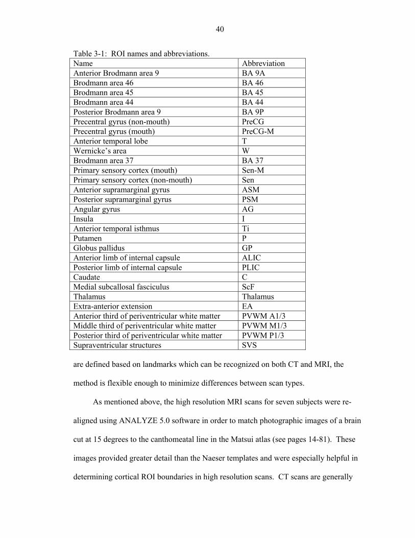
40
Table 3-1: ROI names and abbreviations. Name Abbreviation Anterior Brodmann area 9 BA 9A Brodmann area 46 BA 46 Brodmann area 45 BA 45 Brodmann area 44 BA 44 Posterior Brodmann area 9 BA 9P Precentral gyrus (non-mouth) PreCG Precentral gyrus (mouth) PreCG-M Anterior temporal lobe T Wernicke’s area W Brodmann area 37 BA 37 Primary sensory cortex (mouth) Sen-M Primary sensory cortex (non-mouth) Sen Anterior supramarginal gyrus ASM Posterior supramarginal gyrus PSM Angular gyrus AG Insula I Anterior temporal isthmus Ti Putamen P Globus pallidus GP Anterior limb of internal capsule ALIC Posterior limb of internal capsule PLIC Caudate C Medial subcallosal fasciculus ScF Thalamus Thalamus Extra-anterior extension EA Anterior third of periventricular white matter PVWM A1/3 Middle third of periventricular white matter PVWM M1/3 Posterior third of periventricular white matter PVWM P1/3 Supraventricular structures SVS are defined based on landmarks which can be recognized on both CT and MRI, the
method is flexible enough to minimize differences between scan types.
As mentioned above, the high resolution MRI scans for seven subjects were re-
aligned using ANALYZE 5.0 software in order to match photographic images of a brain
cut at 15 degrees to the canthomeatal line in the Matsui atlas (see pages 14-81). These
images provided greater detail than the Naeser templates and were especially helpful in
determining cortical ROI boundaries in high resolution scans. CT scans are generally
41
acquired at an angle of about 15 degrees to the canthomeatal line, which matches the
Naeser templates and Matsui atlas, and can usually be rated using these as guides as well.
However, the angle of acquisition for three of the CT films and the MRI film did not
match the Naeser templates or the set of 15 degree slices in the Matsui atlas. Due to the
fact that these scans contained fewer slices compared to the high resolution MRI scans, it
was not possible to realign these scans. Therefore, templates developed by Damasio and
Damasio (1989) representing alternate acquisition angles were also consulted in order to
ensure accurate identification of important sulci and ROIs.
The first step in rating the ROIs for a given scan was to identify which slices on the
scan best corresponded with the 15 degree slices on the Matsui atlas (using the guidelines
in Appendix E), and to record the subject scan slice numbers on the rating sheet (see
Appendix D for an example rating sheet). The rating sheet was then used as a guide to
indicate which ROIs the rater could expect to see on each of the subject scan slices, by
noting which cells were shaded on the rating sheet. Ratings for each ROI appearing on
each slice were recorded on the ratings sheet. After ROIs on each slice were rated, raters
estimated and recorded an overall rating for each ROI based on the slice by slice ratings
and the relative size of the ROI on each slice. Only this overall ROI rating was used in
the statistical analysis. A more detailed description of the rating method can be found in
Appendix F. Raters were blind to the subjects’ treatment data until after the scans were
rated.
The author and one other rater (Yu-Ling Chang) rated all scans. In order to train
the raters and establish inter-rater reliability, a set of five practice scans was rated. The
practice set consisted scans from left hemisphere stroke patients who had participated in a

42
separate language treatment study. These scans were high resolution T1 weighted MRI
scans acquired on the same scanner and with the same parameters as the subject scans for
the current study. Practice scans were aligned, prepared, and rated using the same
methods described above. After rating each practice scan independently, the raters met
and discussed any discrepancies before rating the next scan. The correlation between the
two sets of ratings for all five scans was r = .887.

43
CHAPTER 4 RESULTS
As discussed in the introduction, few studies have examined the relationship
between lesion characteristics and improvement during the course of a naming treatment
in aphasic stroke subjects. No known studies have examined this relationship as it
applies to the naming improvement of objects versus actions. The current study is unique
in its aims and is made possible by an existing dataset that includes baseline (pre-
treatment) functioning, treatment scores, and neuroimaging data, and, therefore, has the
potential for examining the effects of several possible predictor variables. However, the
current study also has a relatively small sample size (N = 15), which calls for caution
when making multiple statistical comparisons. Increasing the number of comparisons
made with a small sample size also increases the risk of making Type 1 errors in any
study. On the other hand, restricting the number of statistical comparisons would
increase the chance of discarding potentially useful data. It is therefore important to
balance the tradeoffs between specificity and sensitivity when determining how many
statistical comparison to conduct in a study. In other words, one must ask whether it is
more useful to err on the side of specificity and decrease the chances of making spurious
conclusions, or whether it is more useful to optimize sensitivity so as to not overlook
potentially important relationships. If the current study had been aimed at examining
questions within the framework of a well developed literature, or confirming slightly
different aspects of previously tested hypotheses, it would be important to maximize the
specificity of the study. However, considering the novelty of this area of research, this
44
study has purposefully conducted more comparisons than one would ordinarily make
with such a small sample size, in order to explore potential predictors of naming
improvement. By exploring a wider range of hypotheses in the current study, it was
hoped that future research would be in a better position to focus on only those variables
found to be important.
In order to provide context for the statistical analyses which were conducted,
Tables 4-1 through 4-4 describe the distribution of lesion and language variables
measured. Table 4-1 shows the mean and standard deviation for ratings of each ROI.
Table 4-2 shows the frequency of each possible ROI rating. It should be noted
that final ROI ratings are the average of the ratings from two raters. Thus some values
such as 4.5 were arrived at as the average of two ratings (i.e., 4 and 5).
Table 4-3 shows the anterior, posterior, and basal ganglia ratings for each of the 15
subjects. Anterior lesion extent was calculated as the sum of the following ROI ratings:
BA 9A, BA 46, BA 45, BA 44, BA 9P, PreCG, and PreCG-M. The mean anterior lesion
extent across subjects was 13.58 (SD = 10.42). Posterior lesion extent was calculated as
the sum of the following ROI ratings: T, W, BA 37, Sen-M, Sen, ASM, PSM, and AG.
The mean posterior lesion extent across subjects was 17.98 (SD = 9.15). Basal ganglia
lesion extent was calculated as the sum of the following ROI ratings: P, GP, and C. The
mean basal ganglia lesion extent across subjects was 6.38 (SD = 4.29).
Table 4-4 shows the means and standard deviations for the language measures used
in the analyses. Scores on the BNT and ANT were expressed as the percent correct.
Improvements in the naming of objects and actions were calculated as the range corrected
gain score for the naming probes for the words on which the subject was trained [(mean
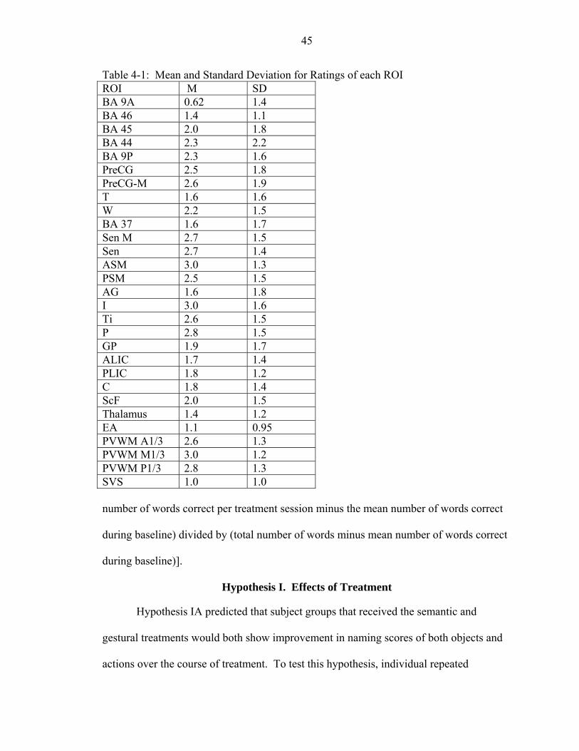
45
Table 4-1: Mean and Standard Deviation for Ratings of each ROI ROI M SD BA 9A 0.62 1.4 BA 46 1.4 1.1 BA 45 2.0 1.8 BA 44 2.3 2.2 BA 9P 2.3 1.6 PreCG 2.5 1.8 PreCG-M 2.6 1.9 T 1.6 1.6 W 2.2 1.5 BA 37 1.6 1.7 Sen M 2.7 1.5 Sen 2.7 1.4 ASM 3.0 1.3 PSM 2.5 1.5 AG 1.6 1.8 I 3.0 1.6 Ti 2.6 1.5 P 2.8 1.5 GP 1.9 1.7 ALIC 1.7 1.4 PLIC 1.8 1.2 C 1.8 1.4 ScF 2.0 1.5 Thalamus 1.4 1.2 EA 1.1 0.95 PVWM A1/3 2.6 1.3 PVWM M1/3 3.0 1.2 PVWM P1/3 2.8 1.3 SVS 1.0 1.0 number of words correct per treatment session minus the mean number of words correct
during baseline) divided by (total number of words minus mean number of words correct
during baseline)].
Hypothesis I. Effects of Treatment
Hypothesis IA predicted that subject groups that received the semantic and
gestural treatments would both show improvement in naming scores of both objects and
actions over the course of treatment. To test this hypothesis, individual repeated

46
Table 4-2: Frequency of each Possible Final ROI Rating.
Ratings
ROI
0 .5
1 1.5
2 2.25
2.5
2.75
3 3.5
4 4.5
5 BA 9A 6 6 2 1 BA 46 4 2 1 1 1 2 3 1 BA 45 3 4 2 1 1 2 1 1 BA 44 6 1 1 1 2 2 2 BA 9P 2 3 1 2 1 2 3 1 PreCG 3 2 1 1 2 3 3 PreCG-M 3 2 1 2 1 1 5 T 4 3 1 3 2 1 1 W 3 1 1 1 2 3 1 3 BA 37 7 1 1 1 2 2 1 Sen M 2 1 1 2 2 1 4 2 Sen 2 1 4 1 2 4 1 ASM 1 1 1 1 2 2 1 4 2 PSM 1 2 1 1 1 1 3 1 1 1 3 AG 6 2 1 2 1 2 1 I 2 1 1 2 2 1 3 1 2 Ti 1 2 1 1 2 1 1 3 1 1 1 P 1 1 1 2 1 2 1 4 2 GP 4 2 1 1 1 1 1 1 2 1 ALIC 4 1 2 1 1 3 1 2 PLIC 1 3 1 3 1 1 1 1 1 1 1 C 4 1 2 2 2 1 1 2 ScF 3 2 2 2 2 1 1 1 1 Thalamus 2 5 1 1 1 2 2 1 EA 3 4 2 1 2 3 PVWM A1/3 1 1 1 1 1 3 1 1 5 PVWM M1/3 1 3 1 2 1 1 5 1 PVWM P1/3 1 2 1 2 3 1 4 1 SVS 6 2 5 2 measures ANOVAs were conducted for each of the four treatments, with the dependent
variable being the subject’s average naming score during the baseline and treatment
phases. The following treatments showed a significant increase in naming score from
baseline to treatment: semantic treatment for objects (F = 7.79, p = .038., partial eta
squared = .609), gestural treatment for objects (F = 11.187, p = .016, partial eta squared =
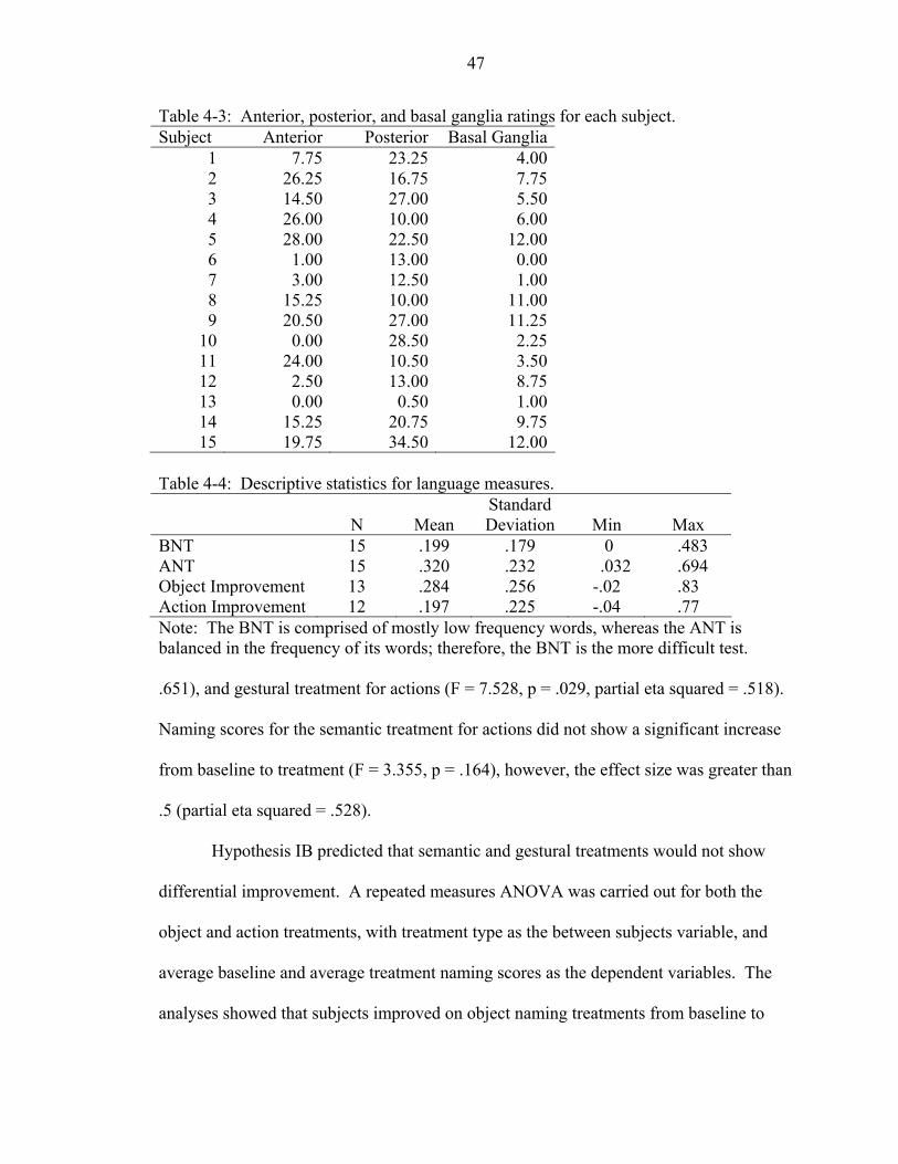
47
Table 4-3: Anterior, posterior, and basal ganglia ratings for each subject. Subject Anterior Posterior Basal Ganglia
1 7.75 23.25 4.002 26.25 16.75 7.753 14.50 27.00 5.504 26.00 10.00 6.005 28.00 22.50 12.006 1.00 13.00 0.007 3.00 12.50 1.008 15.25 10.00 11.009 20.50 27.00 11.25
10 0.00 28.50 2.2511 24.00 10.50 3.5012 2.50 13.00 8.7513 0.00 0.50 1.0014 15.25 20.75 9.7515 19.75 34.50 12.00
Table 4-4: Descriptive statistics for language measures.
N
Mean Standard Deviation
Min
Max
BNT 15 .199 .179 0 .483 ANT 15 .320 .232 .032 .694 Object Improvement 13 .284 .256 -.02 .83 Action Improvement 12 .197 .225 -.04 .77 Note: The BNT is comprised of mostly low frequency words, whereas the ANT is balanced in the frequency of its words; therefore, the BNT is the more difficult test. .651), and gestural treatment for actions (F = 7.528, p = .029, partial eta squared = .518).
Naming scores for the semantic treatment for actions did not show a significant increase
from baseline to treatment (F = 3.355, p = .164), however, the effect size was greater than
.5 (partial eta squared = .528).
Hypothesis IB predicted that semantic and gestural treatments would not show
differential improvement. A repeated measures ANOVA was carried out for both the
object and action treatments, with treatment type as the between subjects variable, and
average baseline and average treatment naming scores as the dependent variables. The
analyses showed that subjects improved on object naming treatments from baseline to
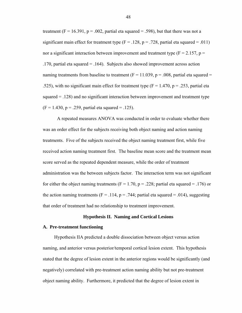
48
treatment (F = 16.391, p = .002, partial eta squared = .598), but that there was not a
significant main effect for treatment type (F = .128, p = .728, partial eta squared = .011)
nor a significant interaction between improvement and treatment type (F = 2.157, p =
.170, partial eta squared = .164). Subjects also showed improvement across action
naming treatments from baseline to treatment (F = 11.039, p = .008, partial eta squared =
.525), with no significant main effect for treatment type (F = 1.470, p = .253, partial eta
squared = .128) and no significant interaction between improvement and treatment type
(F = 1.430, p = .259, partial eta squared = .125).
A repeated measures ANOVA was conducted in order to evaluate whether there
was an order effect for the subjects receiving both object naming and action naming
treatments. Five of the subjects received the object naming treatment first, while five
received action naming treatment first. The baseline mean score and the treatment mean
score served as the repeated dependent measure, while the order of treatment
administration was the between subjects factor. The interaction term was not significant
for either the object naming treatments (F = 1.70, p = .228; partial eta squared = .176) or
the action naming treatments (F = .114, p = .744; partial eta squared = .014), suggesting
that order of treatment had no relationship to treatment improvement.
Hypothesis II. Naming and Cortical Lesions
A. Pre-treatment functioning
Hypothesis IIA predicted a double dissociation between object versus action
naming, and anterior versus posterior/temporal cortical lesion extent. This hypothesis
stated that the degree of lesion extent in the anterior regions would be significantly (and
negatively) correlated with pre-treatment action naming ability but not pre-treatment
object naming ability. Furthermore, it predicted that the degree of lesion extent in
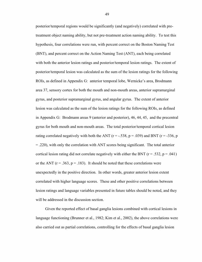
49
posterior/temporal regions would be significantly (and negatively) correlated with pre-
treatment object naming ability, but not pre-treatment action naming ability. To test this
hypothesis, four correlations were run, with percent correct on the Boston Naming Test
(BNT), and percent correct on the Action Naming Test (ANT), each being correlated
with both the anterior lesion ratings and posterior/temporal lesion ratings. The extent of
posterior/temporal lesion was calculated as the sum of the lesion ratings for the following
ROIs, as defined in Appendix G: anterior temporal lobe, Wernicke’s area, Brodmann
area 37, sensory cortex for both the mouth and non-mouth areas, anterior supramarginal
gyrus, and posterior supramarginal gyrus, and angular gyrus. The extent of anterior
lesion was calculated as the sum of the lesion ratings for the following ROIs, as defined
in Appendix G: Brodmann areas 9 (anterior and posterior), 46, 44, 45, and the precentral
gyrus for both mouth and non-mouth areas. The total posterior/temporal cortical lesion
rating correlated negatively with both the ANT (r = -.538, p = .039) and BNT (r = -336, p
= .220), with only the correlation with ANT scores being significant. The total anterior
cortical lesion rating did not correlate negatively with either the BNT (r = .532, p = .041)
or the ANT (r = .363, p = .183). It should be noted that these correlations were
unexpectedly in the positive direction. In other words, greater anterior lesion extent
correlated with higher language scores. These and other positive correlations between
lesion ratings and language variables presented in future tables should be noted, and they
will be addressed in the discussion section.
Given the reported effect of basal ganglia lesions combined with cortical lesions in
language functioning (Brunner et al., 1982; Kim et al., 2002), the above correlations were
also carried out as partial correlations, controlling for the effects of basal ganglia lesion
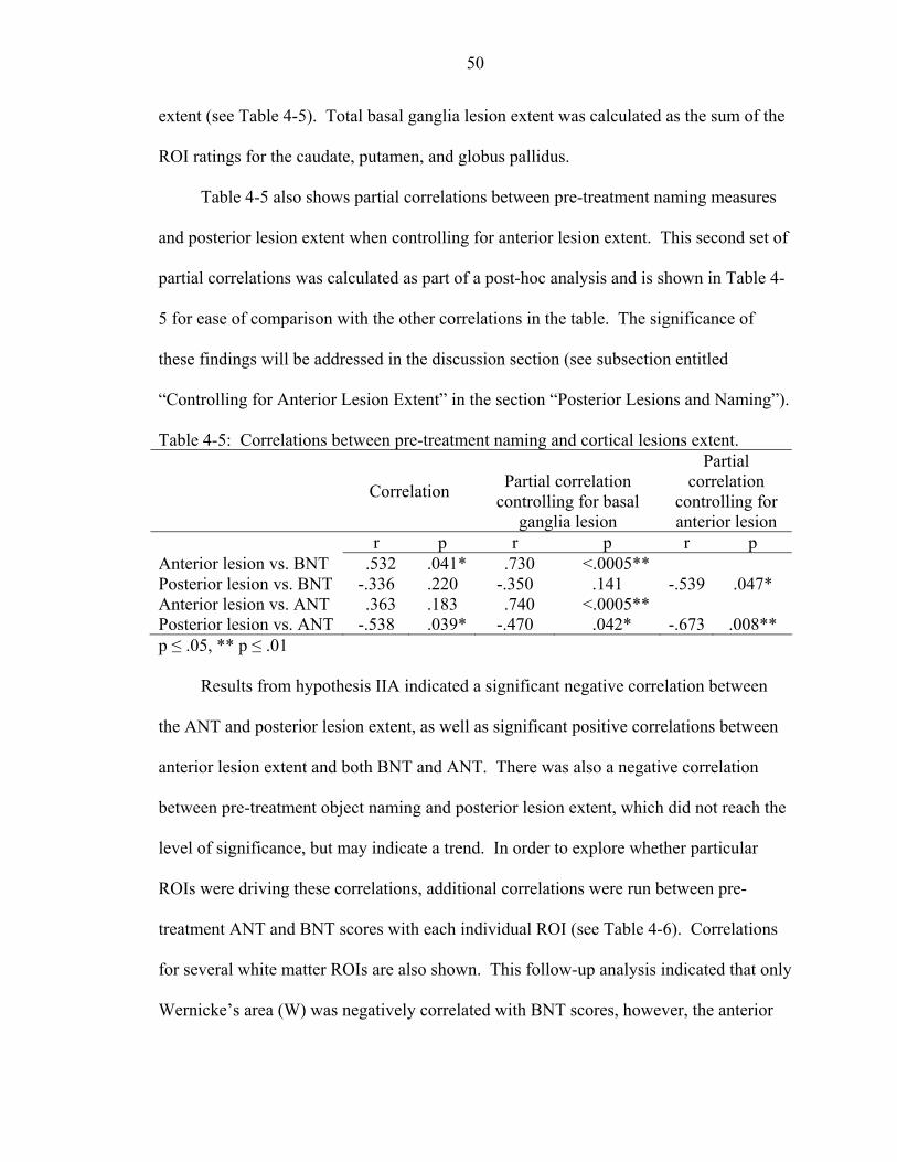
50
extent (see Table 4-5). Total basal ganglia lesion extent was calculated as the sum of the
ROI ratings for the caudate, putamen, and globus pallidus.
Table 4-5 also shows partial correlations between pre-treatment naming measures
and posterior lesion extent when controlling for anterior lesion extent. This second set of
partial correlations was calculated as part of a post-hoc analysis and is shown in Table 4-
5 for ease of comparison with the other correlations in the table. The significance of
these findings will be addressed in the discussion section (see subsection entitled
“Controlling for Anterior Lesion Extent” in the section “Posterior Lesions and Naming”).
Table 4-5: Correlations between pre-treatment naming and cortical lesions extent.
Correlation
Partial correlation
controlling for basal ganglia lesion
Partial correlation
controlling for anterior lesion
r p r p r p Anterior lesion vs. BNT .532 .041* .730 <.0005** Posterior lesion vs. BNT -.336 .220 -.350 .141 -.539 .047* Anterior lesion vs. ANT .363 .183 .740 <.0005** Posterior lesion vs. ANT -.538 .039* -.470 .042* -.673 .008** p ≤ .05, ** p ≤ .01
Results from hypothesis IIA indicated a significant negative correlation between
the ANT and posterior lesion extent, as well as significant positive correlations between
anterior lesion extent and both BNT and ANT. There was also a negative correlation
between pre-treatment object naming and posterior lesion extent, which did not reach the
level of significance, but may indicate a trend. In order to explore whether particular
ROIs were driving these correlations, additional correlations were run between pre-
treatment ANT and BNT scores with each individual ROI (see Table 4-6). Correlations
for several white matter ROIs are also shown. This follow-up analysis indicated that only
Wernicke’s area (W) was negatively correlated with BNT scores, however, the anterior
51
temporal lobe (T), and the anterior portion of the temporal isthmus (Ti) also became
correlated when basal ganglia lesion extent was controlled for. Several anterior ROI
ratings were positively correlated with BNT scores, including anterior and posterior
Brodmann area 9 (BA 9A, BA 9P), Brodmann area 46 (BA 46), and supraventricular
structures (SVS). All anterior ROIs became significantly correlated with BNT scores
when controlling for basal ganglia lesion extent.
Lesions involving the anterior temporal lobe (T), Wernicke’s area (W), Brodmann
area 37 (BA 37), posterior supramarginal gyrus (PSM), anterior temporal isthmus (Ti),
and the posterior third of the periventricular white matter (PVWM P1/3), were
significantly correlated with lower ANT scores. In general, the strength of these
correlations did not change much when basal ganglia lesion extent was controlled for. In
general, although not all were significant, all posterior cortical regions listed in Table 4-6
correlated with lower pre-treatment naming except for the primary sensory cortex. In
contrast, all individual anterior cortical ROIs correlated with higher pre-treatment
naming. Although none of the anterior correlations were significant, partial correlations
controlling for basal ganglia lesion extent were significant for all anterior ROIs. Neither
the insula or middle third of the periventricular white matter exhibited significant
correlations in either direction.
Similar to Table 4-5, Table 4-6 also shows partial correlations between pre-
treatment naming measures and non-anterior lesion ratings when controlling for anterior
lesion extent. This second set of partial correlations was calculated as part of a post-hoc
analysis and is shown in Table 4-6 for ease of comparison with the other correlations in
the table. The significance of these findings will be addressed in the discussion section.

52
Table 4-6: Correlations between pre-treatment naming and individual ROI ratings. BNT ANT
Region Correlation Partial BG
Partial Anterior
Correlation Partial BG
Partial Anterior
BA 9A .723** .784** .499 .638** BA 46 .556* .690** .323 .563* BA 45 .495 .644** .350 .633* BA 44 .414 .586* .293 .620* BA 9P .556* .717** .401 .694** PreCG .460 .654** .319 .667** PreCG-M .427 .623* .278 .629* EA .488 .630* .296 .557* PVWM A1/3 .394 .624* .235 .628*
Ant
erio
r
SVS .587* .628* .477 .591* I .042 .183 -.693** -.179 .103 -.782** PVWM M1/3 .068 .133 -.290 -.245 -.107 -.546* T -.402 -.630* -.726** -.572* -.650* -.789** W -.517* -.516 -.529* -.672** -.660** -.674** BA 37 -.478 -.476 -.518 -.548* -.543* -.560* Sen-M .230 .312 -.207 .013 .194 -.339 Sen .218 .296 -.246 .029 .211 -.331 ASM -.198 -.190 -.359 -.416 -.365 -.529* PSM -.392 -.389 -.365 -.571* -.569* -.557* AG -.383 -.382 -.347 -.427 -.433 -.396 Ti -.452 -.541* -.626* -.672** -.679** -.783**
Post
erio
r
PVWM P1/3 -.388 -.387 -.417 -.628* -.601* -.650** Note: “Partial BG” refers to partial correlations controlling for total basal ganglia lesion extent and “Partial Anterior” refers to partial correlations controlling for anterior lesion extent.. * p ≤ .05, ** p ≤ .01 B. Improvement during treatment
Hypothesis IIB predicted a double dissociation between improvement in object
versus action naming scores and anterior versus posterior lesion extent. Specifically, the
hypothesis predicted that the degree of lesion extent in the anterior regions would be
significantly (and negatively) correlated with improvement in action naming ability but
not with improvement in object naming ability. It also predicted that the degree of lesion
extent in posterior regions would be significantly (and negatively) correlated with
improvement in object naming ability but not with improvement in action naming ability.
53
Anterior and posterior regions were defined as stated in Hypothesis IIA above.
Treatment “improvement” was calculated as range corrected gain scores, as previously
described.
Posterior lesion totals were negatively correlated with both improvements in action
naming (r = -.295, p = .351) and object naming (r = -.296, p = .327), however, neither of
these correlations were statistically significant (p < .05). Anterior lesion totals were
positively correlated with improvements in action naming (r = .437, p = .156) and object
naming (r = .634, p = .020). It should be noted again that these positive correlations were
unexpected, as they indicate that greater anterior lesion ratings were related to greater
naming improvement. This unexpected result will be addressed in the discussion section.
As with hypothesis IA, partial correlations were also carried out, controlling for basal
ganglia lesion extent (see Table 4-7). Correlations with posterior lesion extent remained
non-significant, however, correlations with anterior lesion extent became very strong and
highly significant.
Similar to Tables 4-5 and 4-6, Table 4-7 also shows partial correlations between
naming measures and posterior lesion extent when controlling for anterior lesion extent.
The significance of these findings will be addressed in the discussion section.
Results did not indicate any significant correlations between naming improvement
and posterior lesion extent. However, there were small negative correlations between
posterior lesion extent and both action naming (r = -.264) and object naming
improvement (r = -.368). There were also highly significant positive correlations
between anterior lesion extent and both object and action naming when controlling for
basal ganglia lesion extent. An additional set of correlations were conducted to explore

54
Table 4-7: Correlations between naming improvement and cortical lesion extent.
Correlation Partial correlation
controlling for basal ganglia lesion extent
Partial correlation controlling for anterior
lesion extent
r p r p r p Anterior lesion vs. object naming improvement .634 .020* .858
<.0005**
Posterior lesion vs. object naming improvement -.296 .327 -.373 .232 -.530 .076 Anterior lesion vs. action naming improvement .437 .156 .821 .002** Posterior lesion vs. action naming improvement -.295 .351 -.256 .448 -.428 .190 * p < .05, ** p < .01 which of the individual ROIs were most strongly correlated with naming improvement
and whether any of these correlations reached a level of statistical significance (see Table
4-8). Some correlations with white matter ROIs are also shown.
None of the individual posterior ROIs correlated significantly with object naming
improvement, however, several approached a significant level, such as Wernicke’s area
(W) and the posterior supramarginal gyrus (PSM). When basal ganglia extent was
controlled for, the partial correlation with the anterior temporal isthmus (Ti) was
significant. All but one anterior ROI (BA 9A) was strongly and significantly correlated
with object naming improvement in the positive direction. These correlations became
even stronger when basal ganglia extent was controlled for.
Although correlations with posterior ROIs were in a consistently negative direction,
none were significantly correlated with action naming improvement, even when
controlling for basal ganglia lesion extent. The anterior temporal isthmus (Ti)
approached significance. Anterior lesion ratings all correlated with action naming
55
improvement in a positive direction. Although none of these correlations were
statistically significant, all but two partial correlations were highly significant when
controlling for basal ganglia lesion extent.
Similar to Tables 4-5 through 4-7, Table 4-8 also shows partial correlations
between naming measures and posterior lesion extent when controlling for anterior lesion
extent, and the significance of these findings will be addressed in the discussion section.
Hypothesis III. Naming and Basal Ganglia Lesions
A. Pre-treatment functioning
Hypothesis IIIA stated that basal ganglia lesion extent would be significantly correlated
with both pre-treatment object naming and action naming abilities. To test this
hypothesis, correlations were carried out between total basal ganglia lesion extent ratings
and pre-treatment BNT scores, as well as between total basal ganglia lesion extent ratings
and pre-treatment ANT scores. Basal ganglia extent was calculated as the sum of the
ROI ratings for the caudate, putamen, and globus pallidus. Neither of the correlations
were significant (see Table 4-9). Given the reported effect of basal ganglia lesions
combined with cortical lesions in language functioning (Kim et al., 2002), the above
correlations were also carried out as partial correlations, controlling for the effects of
anterior and posterior cortical lesions. These partial correlations are also shown in Table
4-9. Partial correlations with both the BNT and ANT became strong and highly
significant when controlling for anterior lesion extent, however, correlations were
relatively unaffected when posterior lesion extent was controlled for.
B. Improvement during treatment
Hypothesis IIIB stated that basal ganglia lesion extent would be negatively correlated
with improvement on both object and action naming probes. To test this

56
Table 4-8: Correlations between naming improvement and individual ROI ratings. Object Naming Improvement
Action Naming Improvement
Region Correlation Partial BG
Partial Anterior
Correlation Partial BG
Partial Anterior
BA 9A .431 .439 .561 .672* BA 46 .632* .738** .449 .686* BA 45 .589* .701** .384 .757** BA 44 .561* .743** .361 .744** BA 9P .601* .746** .345 .602* PreCG .627* .886** .412 .791** PreCG-M .611* .878** .415 .794** EA .563* .657* .444 .650* PVWM A1/3 .544* .734** .166 .449
Ant
erio
r
SVS .548* .567* .410 .458 I -.234 .355 -.617* -.204 -.019 -.892** PVWM M1/3 .139 .119 -.286 .041 .153 -.276 T -.170 -.398 -.594* -.399 -.369 -.622* W -.496 -.515 -.527 -.362 -.365 -.351 BA 37 -.332 -.336 -.284 -.088 -.094 -.082 Sen-M .305 .324 -.193 -.096 .002 -.479 Sen .336 .357 -.181 -.030 .070 -.415 ASM -.406 -.460 -.667* -.173 -.151 -.318 PSM -.503 -.527 -.509 -.264 -.303 -.237 AG -.471 -.487 -.423 -.287 -.323 -.240 Ti -.444 -.630* -.708** -.460 -.417 -.626*
Post
erio
r
PVWM P1/3 -.463 -.512 -.517 -.106 -.126 -.092 Note: “Partial BG” refers to partial correlations controlling for total basal ganglia lesion extent and “Partial Anterior” refers to partial correlations controlling for anterior lesion extent; p ≤ .05, ** p ≤ .01.
hypothesis, correlations were carried out between total basal ganglia lesion extent ratings
and object naming improvement scores, as well as between total basal ganglia lesion
extent ratings and action naming improvement scores. Improvement scores were
calculated as previously described, and total basal ganglia lesion was calculated as the
sum of caudate, putamen, and globus pallidus ratings. Neither measure of naming
improvement was significantly correlated with basal ganglia lesion extent (see Table 4-
10). However, when controlling for anterior lesion extent, basal ganglia lesion extent

57
was strongly correlated with both object and action naming improvement. No significant
correlations where found when controlling for posterior lesion extent.
Table 4-9: Correlations between pre-treatment naming and basal ganglia lesion extent.
Correlations
Partial correlations controlling for cortical
lesion extent
r p Control variable r p Anterior lesion -.623 .004** BNT vs. basal
ganglia -.056 .842 Posterior lesion .040 .870 Anterior lesion -.750 <.0005** ANT vs.
basal ganglia -.263 .343 Posterior lesion -.154 .530 ** p ≤ .01 Table 4-10: Correlations between naming improvement and basal ganglia lesion extent.
Correlations
Partial correlations controlling for cortical
lesion extent
r p Control variable r p Anterior lesion -.749 .005** Object naming
improvement vs. basal ganglia
.075 .808 Posterior lesion .249 .434
Anterior lesion -.785 .004** Action naming improvement vs. basal ganglia
-.219 .494 Posterior lesion -.159 .641
** p < .01
Results from hypothesis IIIA and IIIB show that a combined basal ganglia rating
was not correlated with pre-treatment or improvement naming measures unless anterior
lesion extent was controlled, in which case the correlations became strong and highly
significant. In order to determine if certain basal ganglia structures were more correlated
than others to pre-treatment and improvement naming measures, individual partial
correlations were run for each basal ganglia structure, controlling for anterior lesion
extent. Results are shown in Table 4-11. Partial correlations with the thalamus and deep
white matter ROIs are also shown. Both the BNT and ANT were significantly correlated
with the putamen, globus pallidus, and anterior subcallosal fasciculus. Partial

58
Table 4-11: Partial correlations between naming measures and subcortical ROIs. Pre-treatment Improvement
BNT ANT Object Action Putamen -.658** -.731** -.637* -.851** Globus Pallidus -.532* -.629* -.708** -.650* Caudate -.474 -.528 -.661* -.637* Thalamus -.125 -.184 -.110 -.110 ALIC -.420 -.468 -.620* -.581 PLIC -.403 -.535* -.712** -.725** ScF -.713** -.698** -.392 -.477 Note: Partial correlations are controlling for anterior lesion extent. * p ≤ .05, ** p ≤ .01 correlations with pre-treatment measures and the caudate, ALIC, and PLIC approached
significance, with only the partial correlation between the ANT and PLIC being
statistically significant. Measures of both object and action naming improvement had
significant partial correlations with the putamen, globus pallidus, caudate, and PLIC. The
partial correlation with object naming improvement and ALIC was also significant.
Other partial correlations with ALIC and the subcallosal fasciculus approached
significance. The thalamus lesion ratings did not correlate even moderately with any
language measure.
Hypothesis IV. Comprehension and Improvement
Hypothesis IV predicted that pre-treatment auditory comprehension scores would
be related to the degree of treatment improvement. This hypothesis was tested by
carrying out correlations between object naming improvement and action naming
improvement scores (as previously described) and the subjects’ pre-treatment WAB
Comprehension Index. The analysis indicated that neither object naming improvement
(r = .314, p = .376) or action naming improvement (r = .139, p = .722) were significantly
correlated with pre-treatment comprehension scores. However, as shown in Table 4-12,

59
improvement in naming of objects and actions was correlated with pre-treatment
measures of naming, including the BNT, ANT, and the WAB naming index.
Table 4-12: Correlations between naming improvement and pre-treatment language measures.
WAB Overall
WAB Fluency
WAB Comp.
WAB Repetition
WAB Naming ANT BNT
Object naming improvement
.475 -.498 .294 .361 .667* .721** .711**
Action naming improvement
.528 -.314 .211 .153 .626 .658* .669*
* p ≤ .05, ** p ≤ .01
60
CHAPTER 5 DISCUSSION
Before discussing the major findings of this study, it will be necessary to first
briefly address the issue of treatment effectiveness. Once this is addressed, the major
finding of the study will be discussed, namely, the relationship between frontal and basal
ganglia damage in predicting naming performance and treatment improvement. In the
course of this discussion, a possible explanation will be offered for the counterintuitive
finding that greater anterior lesion size was related to better language performance and
greater treatment improvement. Next, findings related to other lesion and cognitive
predictors of naming and naming improvement will be discussed, followed by an
examination of findings related to object versus action naming. Directions for future
research, including several methodological considerations will then be outlined, followed
by a summary of study findings.
Effectiveness of Treatments
Since the purpose of this study was to determine predictors of treatment
improvement, it was important to first establish that improvement did indeed take place
over the course of each treatment. This improvement was demonstrated in two ways.
First, as reported in the results section, combined treatment groups for both action
naming and object naming showed significant improvement, with no main effect or
significant interaction for treatment type. Second, individual repeated measures
ANOVAs indicated that statistically significant improvement took place in three of the
four treatments (semantic for objects, gestural for objects, and gestural for actions). The

61
fourth treatment, semantic treatment for actions, did not reach a statistically significant
level, however, this was likely due at least in part to the fact that this treatment was only
given to four subjects. It should also be noted that the effect size for the semantic action
treatment was just as large as the effect size for the gestural action treatment, indicating
that the lack of significance for the improvement in the semantic action treatment was
due to the small number of subjects who were administered that treatment.
As discussed earlier, the fact that the two treatments did not result in quantitatively
different improvements does not necessarily indicate that the treatments are comparable
in terms of the neurological systems and/or brain regions that support them. However,
given the exploratory nature of this study and the advantage of improving statistical
power by combining groups, establishing that the two treatments did not differ
substantially in terms of the quantitative improvements resulting from them was felt to be
sufficient rationale for combining the semantic and gestural treatment groups. In
summary, these preliminary analyses verified that, all treatment types resulted in
improved naming, and that the improvements associated with the semantic and gestural
treatments were not quantitatively different and could reasonably be combined into a
single treatment group for further analysis.
Basal Ganglia and Anterior Cortical Lesions
Basal ganglia lesion extent was not significantly correlated with pre-treatment
naming measures or naming improvement measures except when anterior lesion extent
was controlled for (see Table 4-9 and Table 4-10). This finding agrees with previous
literature that has suggested basal ganglia lesions produce significant and lasting aphasia
only when combined with cortical lesions (Brunner et al, 1982; Hillis, Wityk, et al, 2002;
Hillis, Barker, et al., 2004). It indicates that in the presence of anterior cortical lesion,

62
basal ganglia lesion extent is highly correlated with naming abilities in chronic aphasics,
as well as their ability to improve during treatment.
Analyses of individual ROIs showed that ratings for the putamen and globus
pallidus were highly correlated with all four of the naming measures, when controlling
for anterior lesion extent (see Table 4-11). Caudate ratings were also significantly
correlated with both naming improvement measures, but caudate correlations with pre-
treatment naming measures only approached significance. Due to the small sample size
of this study this finding should not be this should not be taken as strong evidence for
whether or not lesions of the putamen and globus pallidus affect recovery of language
function more than lesions of the caudate. Future studies with larger subject groups
would help elucidate whether lesions of certain structures within the basal ganglia affect
language functioning more than others.
The fact that controlling for anterior lesion extent was so influential on the
correlations, while controlling for posterior lesion extent had little if any effect, suggests
a special relationship between anterior cortical and basal ganglia lesions. Indeed,
Middleton and Strick (2000) have previously described several basal ganglia-thalamo-
cortical circuits or loops involving anterior cortical regions, including Brodmann areas 46
and 9. Similar loops involving other anterior cortical regions such as Brodmann areas 44
and 45 may also exist. These loops were hypothesized by Mink (1996) to facilitate the
generation of competing motor programs in the cortex that are then subject to
enhancement or inhibition by the basal ganglia based upon their relevance to a desired
movement. The basal ganglia’s role of enhancing selected programs or suppressing non-
useful competing programs may apply to language production processes as well.

63
In particular, the relationship between language improvement and basal ganglia
lesion extent in the presence of anterior cortical lesion may be explained by the
hypothesis expressed by Crosson et al. (2005). This hypothesis suggests that in patients
with significant damage to left hemisphere cortical language areas, the left basal ganglia
serves to inhibit dysfunctional neural activity (noise) produced by the lesioned areas.
This proposed mechanism would thus allows for more successful reorganization of
language function in non-lesioned areas, including the right hemisphere. The idea that
language function may be reorganized to right hemisphere areas in some patients with
left hemisphere damage is not new. For example, Kinsborne (1971) reported that
aphasia patients undergoing Wada testing experienced decreased language functioning
when the right but not left hemisphere was anesthetized. Right hemisphere
reorganization of language has also been supported by reports of decreased language
functioning in aphasia patients following lesions to the right hemisphere (Barlow, 1877;
Gowers, 1887; and Basso, Gardelli, Grassi & Mariotti, 1989). According to the Crosson
hypothesis, individuals with significant lesions to both the left cortical language areas and
left basal ganglia, the basal ganglia is less able to inhibit neural activity from the
damaged cortical areas or enhance activity from healthy cortex, resulting in decreased
recovery of language function.
In general, the current study showed that larger lesion ratings in almost every
anterior cortical ROI in the current study were correlated with greater naming
improvement. These correlations were only statistically significant for object naming
improvement, but approached significance for several ROIs when correlated with action

64
naming improvement. These correlations became very strong for both action naming and
object naming improvement when basal ganglia lesion extent was controlled.
Although any theory claiming that a larger brain lesion leads to better cognitive
functioning is likely to be controversial, the idea is not without precedent. For example,
reports from the epilepsy literature have indicated that surgical lesions in the left anterior
temporal lobe can be associated with improved complex language comprehension
(Hermann, Wyler & Somes, 1991). Given the strength of the correlations found in the
present study between anterior lesion extent and better naming, it is important to explore
the possibility that the current findings are reflective of previously undescribed neural
processes occurring in stroke patients. The following section describes a possible
explanation for the observed correlations between anterior lesions, basal ganglia lesions,
and language functioning.
Proposed Explanation
The following explanation for the observed effects of anterior and basal ganglia
lesions is based on the Crosson hypothesis (2005) described above. To begin with, it
should be noted that the subjects in this study do not represent the general population of
stroke patients with aphasia. This group includes only subjects with chronic naming
deficits, and does not include patients with transient aphasia who have recovered normal
language function or patients with only minor aphasic symptoms. Given that each
subject in the current study had significant and lasting language production deficits which
could be measured by standard language testing, it can be inferred that each of these
subjects suffered significant damage to whatever left hemisphere neural networks are
necessary for supporting language production. Such damage, although perhaps brought

65
about by damage to a variety of lesion patterns, resulted in the generation of “noisy”
output by the remaining left frontal cortex.
Assuming this to be the case, the results of this study there appear to reflect the
presence of two relatively orthogonal principles or mechanisms which are able to
suppress the “noisy” or non-functional left frontal output in this group of patients. The
first principle is that when left frontal areas are sufficiently damaged, right hemisphere
structures (such as the pre-SMA) will use an intact left basal ganglia to suppress “noisy”
left frontal output, thereby preventing it from interfering or competing with reorganized
language activity in the right frontal cortex. This first principle is simply a reversal of
what happens in normal language, where left pre-SMA uses the right basal ganglia to
suppress right frontal activity that might compete or interfere with left frontal activity
during word generation (see Crosson et al., 2003). The second principle is that larger left
frontal lesions will inflict greater damage to noise producing frontal structures, thereby
eliminating a potential source of competition with the newly reorganized right
hemisphere language areas.
These principles appear to operate in a somewhat orthogonal fashion, making it
difficult to detect the effects of one unless one controls for the effects of the other. As
seen in the current study, one must control for the size of frontal lesions to detect the
basal ganglia lesion effect because large frontal lesions have relatively the same effect as
having an intact basal ganglia. Similarly, in order to better observe that larger frontal
lesions lead to better outcome, one should control for the amount of basal ganglia
damage, because a lesioned basal ganglia would have relatively the same effect as a small
frontal lesion. As mentioned previously, any subjects whose left frontal cortex had
66
maintained significant potential to contribute to recovery would have been excluded from
this study. After all, these patients would have experienced greater spontaneous
improvement and their residual impairments would not great enough to meet inclusion
criteria.
Addressing an Alternative Explanation
Due to the unexpected, and perhaps controversial nature of the finding that larger
anterior lesions were related to better language functioning, it is important to explore and
test other possible explanations for the relationship. One such explanation will be
discussed here: Could it be possible that this relationship is simply an artifact of the
lesion distribution pattern of this group of subjects? For example, what would happen if
the majority of subjects in the study had either a significantly larger anterior lesion
compared to posterior lesion or a significantly larger posterior lesion compared to
anterior lesion. If this were the case, a subject who had a large posterior lesion may be
statistically more likely to have a smaller anterior lesion. Likewise, a subject with a large
anterior lesion may be more likely to have a smaller posterior lesion. In this situation,
even if it were the smaller posterior lesions that were the actual physiological cause
driving correlations with better language functioning, it may spuriously appear that the
larger anterior lesions were also meaningfully related to better language functioning.
In order to test whether this scenario could account for the above finding, a
correlation was carried out between posterior lesion extent and anterior lesion extent. A
significant negative correlation would lend support to the possibility that the above
scenario may account for the finding. However, instead, it was found that posterior
lesion extent and anterior lesion extent were in fact positively (although not significantly)
correlated (r = .207). In other words, if anything, patients with larger anterior lesions
67
tended to have larger (not smaller) posterior lesions. Thus, it appears unlikely that the
correlations between greater anterior lesion extent and better language functioning are
simply a statistical artifact of the lesion distribution pattern described in the scenario
above.
Posterior Lesions and Naming
Pre-treatment Naming
Overall, greater posterior lesion extent was weakly to moderately correlated with
worse pre-treatment and improvement scores for both objects and actions. Although only
the correlation with pre-treatment action naming was statistically significant, the lack of
significance for the other correlations may be explained at least in part by the small
number of subjects in the study, as well as the limited variability of the BNT. Although
the ANT and BNT are similar in structure, and both measures were expressed as percent
correct, subjects varied less in their performance on the BNT, which may have decreased
the relative potential for stronger correlations with lesion measures relative to the ANT
(ANT variance = .054, BNT variance = .032). The BNT is also the more difficult test
because it includes mostly low frequency words, whereas the ANT includes words with a
greater range of frequencies (e.g., see Lu et al., 2002). Given the small sample size and
these differences between the ANT and BNT, it is unclear whether the differences
between posterior ROI correlations with BNT and ANT should be interpreted as truly
representing differences in object and action naming. The number of subjects for all the
pre-treatment language measures was small (n = 15), but the number of subjects
measured for object naming improvement (n = 13) and action naming improvement (n =
12) was even smaller. Based on the fact that weak to moderate correlations were found
68
for all four language measures, future studies with a greater number of subjects should
continue to examine these relationships.
In follow-up correlations, the individual posterior ROIs which appeared to be most
important for naming either objects or actions were the anterior superior temporal gyrus
(T), Wernicke’s area (W), Brodmann area 37 (BA 37), the posterior supramarginal gyrus
(PSM), the anterior temporal isthmus (Ti), and the posterior third of the periventricular
white matter (PVWM P1/3). This finding coincides with previous studies examining
language recovery (Brunner et al., 1982; Raymer et al., 1997; Naeser et al., 1990;
Kertesz, Lau and Polk, 1993; Knopman et al., 1984), and in particular, recovery of
naming (Cato et al., 2004b).
While a correlation between greater posterior lesion extent and lower object
naming scores was predicted in the hypotheses, the correlation between greater posterior
lesion extent and lower action naming scores was not. However, the fact that posterior
lesion extent was significantly correlated with pre-treatment action naming scores does
make sense in the context of certain studies which have shown posterior region
involvement in the naming of actions. For example, in a recent PET study, Tranel,
Martin, Damasio, Grabowski, and Hichwa (2005) found that action naming was related
not only to left frontal activation, but also activation of left posterior middle temporal
regions. Indeed, in the current study, one of the lesion sites most highly correlated with
worse action naming was Brodmann’s area 37 (BA 37), which includes portions of the
posterior middle temporal gyrus.
It may be that the posterior language cortex plays a special role in the
reorganization of naming ability in anterior regions post-stroke. For example, when left
69
anterior cortex (ostensibly containing neural networks representing the word production
lexicon) is lesioned, perhaps the preservation of networks in the left posterior cortex
representing aspects of a receptive lexicon could facilitate the reorganization of a new
production lexicon in the right anterior cortex. Therefore, when significant lesions affect
the left posterior cortex, it would be less likely that such reorganization could take place.
Controlling for basal ganglia lesion extent did not appear to have a consistent effect
on correlations between posterior ROIs and BNT and ANT scores, except in a few cases.
Namely, in the case of correlations between ANT and BNT scores and the anterior
superior temporal gyrus (T), and between BNT scores and the anterior temporal isthmus
(Ti), partial correlations were notably higher than correlations (see Table 4-6). It is
unclear why lesions in these specific ROIs may be particularly affected by basal ganglia
lesion extent. Perhaps there is a cortico-striatal loop serving the anterior temporal cortex
via the temporal isthmus in addition to the anterior cortico-striatal loops previously
discussed. Middleton and Strick (2000) have in fact suggested the existence of such
temporal and parietal basal ganglia loops.
Naming Treatment Improvement
The pattern of correlations between object naming improvement and posterior
lesion ratings was similar to the pattern between pre-treatment measures and posterior
lesion ratings discussed above, however, the correlations were weaker. Only the
correlation between the anterior temporal isthmus (Ti) and object naming improvement
was significant (when controlling for basal ganglia lesion extent). Similar to the
correlations with pre-treatment measures, object naming improvement was correlated
most strongly (albeit non-significantly) with Wernicke’s area (W), anterior supramarginal
gyrus (ASM), posterior supramarginal gyrus (PSM), angular gyrus (AG), anterior
70
temporal isthmus (Ti), and the posterior third of the periventricular white matter (PVWM
P1/3). Action naming improvement had moderate but non-significant correlations with
the anterior superior temporal gyrus (T) and the anterior temporal isthmus (Ti).
An informal comparison between object naming improvement and action naming
improvement correlation strengths suggests that posterior lesions correlated more highly
with measures of object naming improvement compared to action naming improvement.
This could be viewed as support for the original hypothesis that posterior regions are
more important to object naming than action naming. However, such a conclusion would
be tenuous at best since only one correlation reached a statistically significant level. As
with the correlations between pre-treatment naming measures and posterior lesion extent,
correlations between naming improvement and posterior lesion extent did not appear to
be consistently or significantly affected by controlling for basal ganglia lesion extent.
Controlling for Anterior Lesion Extent
In the previous section, the relationship between extent of lesion in the anterior
cortex and basal ganglia was discussed. It was proposed that since the effects of lesions
in these two areas appear to affect naming in opposite directions, in order to more clearly
observe the effects of lesions in one area it was necessary to control for the effects of
lesions in the other area. In the case of observing the effects of basal ganglia lesion
extent, which was negatively correlated with naming measures, it was important to
control for the effects of anterior lesion extent, which were positively correlated with
naming measures. This observation leads to the question of whether the effects of
anterior lesion extent may also be obscuring the effects of lesions in areas other than just
the basal ganglia. A post-hoc analysis was conducted to examine whether correlations
between posterior regions might also become stronger when anterior lesion extent was
71
controlled for. As shown in Tables 4-5, controlling for the effects of anterior lesion
extent did in fact strengthen the correlations between total posterior lesion extent and pre-
treatment naming (both for the ANT and BNT). Correlations with individual ROIs which
had previously been significant remained significant (see Table 4-6). In the case of two
ROIs, the anterior temporal isthmus (Ti) and the anterior temporal lobe (T), the
previously significant correlations became even stronger. In the case of the insula (I)
previously non-significant correlations became highly significant.
A similar pattern was noted regarding correlations with lesion site and treatment
improvement scores. Correlations with total posterior lesion extent became stronger,
although still remained non-significant (see Table 4-7). Correlations with the insula (I),
anterior temporal isthmus (Ti) and the anterior temporal lobe (T), became notably
stronger. In general, it appears that the technique of controlling for anterior lesion extent
using partial correlations was productive in further distinguishing ROIs of particular
importance to naming. It also supported previous findings that lesions to the insula may
be related to impaired naming (Knopman et al., 1984; Tranel et al., 2001).
Other Subcortical Findings
When controlling for anterior lesion extent, lesion ratings for the medial subcallosal
fasciculus (ScF) were strongly correlated with pre-treatment naming scores, however,
correlations with naming improvement measures were moderate and non-significant.
Lesions to the ScF have been previously reported as predictive of severe non-fluency
(Naeser et al., 1989). This structure contains fibers connecting the anterior cingulate and
supplementary motor area (SMA) to the caudate, and is thought to be important in
maintaining intentional control of language production. Although any strong conclusions
regarding any direct comparison of correlation strength between pre-treatment and
72
improvement scores with the ScF would be inappropriate (especially considering the
small N of this study) it is interesting to note the difference. This finding may indicate
that compared to a subject’s ability to improve over the course of treatment, pre-treatment
naming abilities may rely more on left hemisphere intentional mechanisms, because that
was the original pathway used when the word was first learned. When the original
connections become damaged, spontaneous recovery is limited. However, during
language treatment, when a subject is re-trained to name objects or actions, new
pathways are developed, linking the speech programs with intact intentional structures
(perhaps in the non-lesioned right hemisphere). The new learning may be less dependent
on the old pathways than spontaneous recovery was, and hence, lesions to the ScF are
more highly correlated with lower pre-treatment scores than lower improvement scores.
There was no significant relationship found between thalamic lesion extent and the
four language measures. However, this is not surprising since thalamic lesions were not
common in the current subject group (only one subject had a thalamic lesion with a rating
greater than three). Subject groups with a greater number of thalamic lesions would
provide a more rigorous test of how thalamic involvement may affect naming.
Pre-treatment Language Predictors of Naming Improvement
Pre-treatment measures of naming (including ANT, BNT, and WAB naming index)
were found to be strongly correlated with both object and action naming improvement.
This finding is similar to that of a previous naming study which found that naming scores
of aphasic stroke patients at one month were predictive of naming scores at six months
(Knopman et al., 1984). However, unlike several studies (Knopman et al.,1984; Cato et
al., 2004a; Lomas & Kertesz, 1978), pre-treatment comprehension scores in the current
study were not significantly correlated with naming improvements scores. One
73
explanation for this may be that in two of these studies (Knopman et al., 1984; Lomas &
Kertesz, 1978), pre-treatment scores were measured during the acute stage of recovery
rather than chronic, and improvement was following spontaneous improvement rather
than treatment.
It is not clear why Cato et al. (2004a) found a relationship between pre-treatment
comprehension and naming improvement, while the current study did not. However, it
should be noted that Cato et al. also found significant correlations between posterior
language areas and treatment improvement, while the present study generally did not
(except for the temporal isthmus), and posterior lesions are often associated with
comprehension deficits. It is worth considering whether these differences could be due to
the nature of the treatments given, since the treatments appear to differ in several
significant ways. For example, the intentional treatment was specifically aimed at
encouraging reorganization of language mechanisms to the right hemisphere, while the
semantic and gestural treatments were not. As discussed earlier, in subjects with large
frontal lesions the neural mechanisms supporting the shift of language production to the
right hemisphere may depend on the intactness of the left posterior language cortex.
While the semantic and gestural treatment subjects may continue to rely more heavily on
left hemisphere anterior cortical-basal ganglia loops, the intentional treatment subjects
may rely more heavily on posterior regions (as a source of receptive lexical information
which may be used to facilitate reorganization of a production lexicon in right anterior
regions). Given that the intentional treatment is specifically designed to facilitate this
shift while the semantic and gestural treatments were not, it makes sense that posterior
lesion extent (and one of its primary functions – comprehension) would be more highly
74
correlated with worse improvement in the intentional treatment than in the semantic and
gestural treatments.
It should be noted, however, that both of these studies relied on relatively small
groups of subjects, and correlation strengths and significance levels may change when
more subjects are added to these studies. At this point, comparisons between the two
studies may lead to useful hypotheses, but strong claims are unwarranted until larger
numbers of subjects are included in the analyses.
Similar to the Cato et al. (2004a) study, overall WAB scores were not significantly
correlated with naming improvements.
Object and Action Naming
Based on previous research (Damasio & Tranel., 1993; Hillis, Tuffiash, et al.,
2002; Tranel et al., 2001), Hypothesis IIA predicted a double dissociation between pre-
treatment object versus action naming and anterior versus posterior cortical lesion extent.
In other words, greater anterior lesion extent was predicted to be related to lower action
naming scores but not lower object naming scores, and greater posterior lesion extent was
predicted to be related to lower object naming scores but not lower action naming scores.
However, as previously discussed, the results of the current study indicated that greater
anterior lesion extent was in fact related to higher scores in both object naming and action
naming, and greater posterior lesion extent was significantly correlated with lower action
naming scores but not lower object naming scores. A similar double dissociation was
predicted regarding the relationship between naming improvement and anterior/posterior
lesion extent. However, again, the predicted double dissociation was not observed.
Instead, greater anterior lesion extent was again correlated with greater naming
improvement (for both objects and actions), and greater posterior lesion extent was only

75
mildly and non-significantly correlated with worse naming improvement (for both objects
and actions).
In other words, the present study did not find any compelling evidence that
particular brain regions were more related to action naming or object naming as
hypothesized. One reason for this may have been that the majority of subjects in the
present study were non-fluent aphasics (of the 15 subjects, 10 were classified as non-
fluent). Some research has indicated that non-fluency is usually associated more with
specific verb production deficits, while fluency is more associated with noun production
deficits (Bak et al., 2001; Daniele et al., 1994; Hillis, Oh, et al., 2004). A more balanced
representation of fluent and non-fluent subjects might have resulted in clearer findings
regarding object/action naming differences.
There are also several methodological reasons why the present study may not have
found the relationships reported in past studies. Differences between the current study
and two previous group studies (Tranel et al., 2001 and Hillis, Tuffiash, et al., 2002)
which may have led to this discrepancy will be examined one by one.
First, while all of the subjects in the current study were chronically aphasic (some
severely), many of the subjects with left hemisphere lesions in the Tranel et al. (2001)
study were described as “recovered aphasics,” and presumably many of the other subjects
had never been aphasic. It is possible that the more subtle differences in naming
observed in the Tranel et al. group may have been obscured in the current group because
the of their more extensive language deficits.
Second, in the Tranel et al. (2001) study, both right and left hemisphere stroke
patients were included in the subject group, whereas the current study only included left
76
hemisphere lesioned subjects. This difference likely resulted in much greater variability
in lesion site, and a greater potential for Tranel et al. to find differences between impaired
and non-impaired groups.
Thirdly, unlike the present study, 14 of the 75 subjects in the Tranel (2001) study
were not stroke patients, but had lesions due to temporal lobectomy or herpes simplex
encephalitis. The neuropathological changes following stroke may result in different
reorganizational mechanisms as compared to temporal lobectomy or herpes simplex
encephalitis.
Fourth, the two studies relied on fundamentally different lesion analysis methods.
While the current study used ratings of individual ROIs, the Tranel (2001) study relied on
a lesion overlap method. The two methods may differ substantially in their abilities to
detect certain types of behavioral/lesion relationships. For example, the ROI method
used in the current study may be better able to detect relationships that are due to a
certain threshold of lesion existing in a certain region. This is because the overlap
method requires that all tissue be characterized as either lesion or non-lesion, with no
middle ground. The overlap method also does not recognize the existence of regions
within the brain. On the other hand, the overlap method may be better able to detect
cases in which areas smaller than an ROI (or areas cutting across more than one ROI) are
related to a given deficit. Another major difference between the two methods is that the
ROI method can make use of continuous behavioral data, whereas, the overlap method
used by Tranel et al., relies on the dichotomization behavioral variables (i.e., either a
subject is impaired or not). With such stark differences, even if one were to use these
two methods on the same dataset, it might lead to different results. Unfortunately, no

77
known published studies have examined these methodological differences using the same
dataset.
Any of the four major differences listed above between the current study and the
Tranel et al. study may have contributed to the failure of the current study to find
significant differences between brain regions important for action versus object naming.
The present study also differed from the Hillis, Tuffiash, et al. (2002) study in
several important ways. First, the Hillis et al. study included consecutive patients with
left hemisphere lesion who may or may not have suffered from lasting aphasia. Like the
Tranel et al. (2001) study, this circumstance may have resulted in a vastly different range
and variability in naming ability among subjects, as well as a difference in the average
severity of aphasia. Secondly, the Hillis et al. study used diffusion weighted MRI to
detect areas of ischemic or infarcted tissue, and perfusion weighted MRI (PWI) to detect
areas of hypoperfusion. The sensitivity to tissue damage of these techniques would likely
be quite different from the T1 weighted MRI and CT scans which were used in the
current study. Some levels of tissue damage detected by PWI may go undetected by the
imaging methods used in the current study. Thirdly, the subjects in the Hillis et al. study
were tested acutely, whereas, in the present study, all subjects were in the chronic stage
of recovery. Consequently, subjects in the present study had likely undergone a greater
degree of reorganization of function than the acute subjects.
Future Directions
Results from the present study may present several implications for future research.
However, before discussing these it should be kept in mind that the results of this study
were obtained with a unique group of subjects. These subjects had suffered from stokes
at least five months prior to the study, and were still experiencing significant aphasic
78
symptoms. Attempts to replicate the findings of this study, particularly the correlation
between larger anterior lesions and improved naming, should include only chronically
aphasic stroke patients with significant naming deficits. This being said, studies with
other types of subject groups would also be valuable. For example, based on the
hypotheses put forth by this study, one would not necessarily expect that a group of
stroke patients who had demonstrated good spontaneous recovery by five months post-
stroke would demonstrate the same correlation between better naming and larger anterior
lesions. For this subject group, improved naming may be more related to smaller
posterior lesions. In order to conduct such a study, naming measures would have to be
taken at an acute stage, since naming at the chronic stage would be near ceiling levels,
and only subjects who later demonstrated good recovery would be included in the
analysis.
Functional imaging studies also could help elucidate findings from the present
study. For example, functional imaging could characterize changes in lateralization of
neural activity during naming tasks from the acute stage to the chronic pre-treatment
stage to the post-treatment stage of recovery. Patients with greater left basal ganglia
damage may be expected to show less right hemisphere shift in activation than patients
with no basal ganglia lesion. Patients with small anterior lesions and poor recovery
might also show less right hemisphere shift in activation than patients with small anterior
lesions and good recovery. Future studies may also benefit from the use of perfusion-
weighted imaging (PWI). This technique may be used to help explore the unexpected
finding that greater anterior lesion extent was correlated with better language
performance. For example, it may be found that in subjects with poor recovery but

79
relatively smaller anterior lesions, their remaining anterior cortex which is actually
hypoperfused and noise-producing. However, failure to find hypoperfused anterior
regions may not necessarily contradict the hypothesis since cortex may not have to be
hypoperfused to be noise-producing. Such areas may therefore be difficult to detect
using PWI or functional imaging techniques, but future studies should begin to address
these issues.
It should be noted that the technical methods used to measure lesions as well as the
statistical approach used to describe relationships between lesions and behavior will
likely affect results of any study. Thus, variations in methodology may lead to different
conclusions when results are interpreted. For this reason, a few methodological issues
will be addressed. The first issue deals with how lesions are quantified in group lesion
studies. There are two major approaches to group comparisons in lesion analysis. Using
the lesion overlap technique, the lesions of multiple subjects with a given deficit are
overlaid on a common set of templates to find which regions are most frequently
effected. Usually subject groups must be divided into separate comparison groups,
those with the deficit and those without it; however, newer methods also exist which
allow for continuous behavioral data (Bates et al., 2003). The second approach, and the
one used in this study, is the region of interest (ROI) approach, in which the brain is
divided into individual regions, and the amount of lesion affecting each region is
somehow quantified. Quantification may be done in many ways. The current study used
a visual rating method, but measures could also be obtained through more detailed
tracings of the ROIs. The ROI approach may be more sensitive in cases where function
80
does not depend so much upon whether a certain cubic millimeter of brain is lesioned, but
upon whether a certain threshold of damage is done to a particular region.
Describing all of the possible advantages or disadvantages of these approaches is
beyond the scope of this study; however, it is important to note that the findings of the
current study may not have been evident had the ROI method not been used. The ability
to measure the degree of lesion to particular regions was critical to the outcome of this
study. For example, the ability to independently identify the influence of damage to the
basal ganglia vs. the frontal cortex (using partial correlations) was dependent on the
ability to specify the degree of damage to these specific structures. It is possible that
other important relationships may have been found had the lesion overlap method been
used instead of the ROI method. However, it is unclear how complex relationships
between particular lesion sites could examined without the use of ROIs and statistical
methods such as partial correlations which can control for multiple lesion affects.
One important principle which is regarded as a fundamental limitation of lesion
analysis should be acknowledged. Namely, the mere fact that an area of the brain is
damaged does not necessarily imply that any particular part of that lesion is directly
related to any particular impairment which is observed. The damage may be coincidental
to the impairment of interest. However, if the degree of damage to a structure also
correlates highly with the degree of impairment, then argument that the two are related is
strengthened. With an ROI approach to lesion analysis such correlations can be
performed.
Although there are important advantages to the ROI method, there are also
advantages to using a lesion overlap method, for example, increased spatial resolution. It

81
will be important for future studies to explore the relative advantages of the ROI and
overlap methods, and perhaps develop ways to implement the advantages of both into
single studies.
Another methodological issue that could be an important consideration in future
studies is the way in which basal ganglia lesions are measured. In the current study, care
was taken to discriminate between cases of basal ganglia lesion versus basal ganglia
atrophy. It is possible that the distinct processes producing these two conditions are
related to distinct cognitive patterns. For example, when large anterior cortical lesions
are present, the basal ganglia may experience significant and visible atrophy because the
basal ganglia are no longer receiving input from those cortical regions, making those
loops defunct. Therefore, the lesioned cortex may be the actual driving force behind any
language deficits, not the atrophied basal ganglia. In such cases, an atrophied basal
ganglia may border the lesion without being directly damaged itself, so care should be
taken not to mistake atrophy for lesion.
This scenario may be very different from one in which the basal ganglia is in fact
damaged as a direct result of the stroke. In this case, the basal ganglia lesion may be part
of the driving force behind any language deficits. For example, there may be spared
cortical regions whose function would normally be modulated the basal ganglia (through
inhibition or enhancement) but become dysfunctional due to basal ganglia lesion. Thus,
discriminating between primary basal ganglia lesion and secondary basal ganglia atrophy
in stroke patients may be crucial in cognitive and behavioral research. Even with high
resolution MRI, differentiating between these two conditions can sometimes be difficult,
especially when lesion borders are close to the borders of the basal ganglia. Future
82
studies should use high resolution scans when possible, and care should be taken in
interpreting whether smaller than expected basal ganglia regions are due to direct infarct
or to atrophy.
Another methodological issue involves the use of partial correlations in lesion
analysis. In the present study, few individual regions were strongly correlated with
language measures by themselves. However, by conducting partial correlations
controlling for other regions, strong effects were found. For example, the present study
found that basal ganglia and frontal lesions both impact language and improvement
during treatment in such a way that the effects of one lesion appeared to obscure the
effects of the other. In past studies, researchers have usually attempted to correlate
language measures with single regions one at a time. Such a methodology assumes that
each separate brain region has unique effects on the cognitive variables of interest, and
that those effects are independent of the effects of lesions to other regions. Findings from
the current study appear to indicate that such an assumption cannot be made. Thus,
future studies in lesion analysis may benefit from using statistical or methodological
techniques that allow for multiple regions to be considered in a single analysis.
Conclusions
The results of the current study may be summarized by four major findings. First, when
controlling for anterior lesion extent, greater basal ganglia lesion extent was strongly
correlated with both worse pre-treatment naming and less improvement during treatment.
Second, when controlling for basal ganglia lesion extent, greater anterior lesion extent
correlated strongly with better pre-treatment and improvement naming scores. A
conceptual model to explain these results was proposed. Third, larger lesions to several
posterior ROIs were correlated with worse pre-treatment naming ability in chronic stroke
83
patients. Finally, pre-treatment naming performance was correlated with naming
improvements during treatment. Table 5-1 summarizes the relationships found in the
present study between naming and lesion extent in particular ROIs.
Two potentially important issues related to cognition, lesion analysis, and brain
plasticity were also discussed. Namely, the present study has shown that the cognitive
effects of one lesion site may obscure the independent effects of a separate lesion site.
Therefore, rather that only looking for ways in which lesions to discrete regions affect
cognitive functioning it may also be important to consider the effects of multiple lesion
sites in a single analysis. This study also introduced the idea that larger lesions to certain
cortical areas may ironically be related to better functioning in certain groups of stroke
patients. Further research addressing these two issues may lead to a greater
understanding of how cortical and subcortical lesions affect language functioning as well
as how they may predict and influence a patient’s response to treatment. Ultimately, it is
hoped that such knowledge will help guide language treatments for aphasic stroke
patients and help contribute to their increased quality of life.
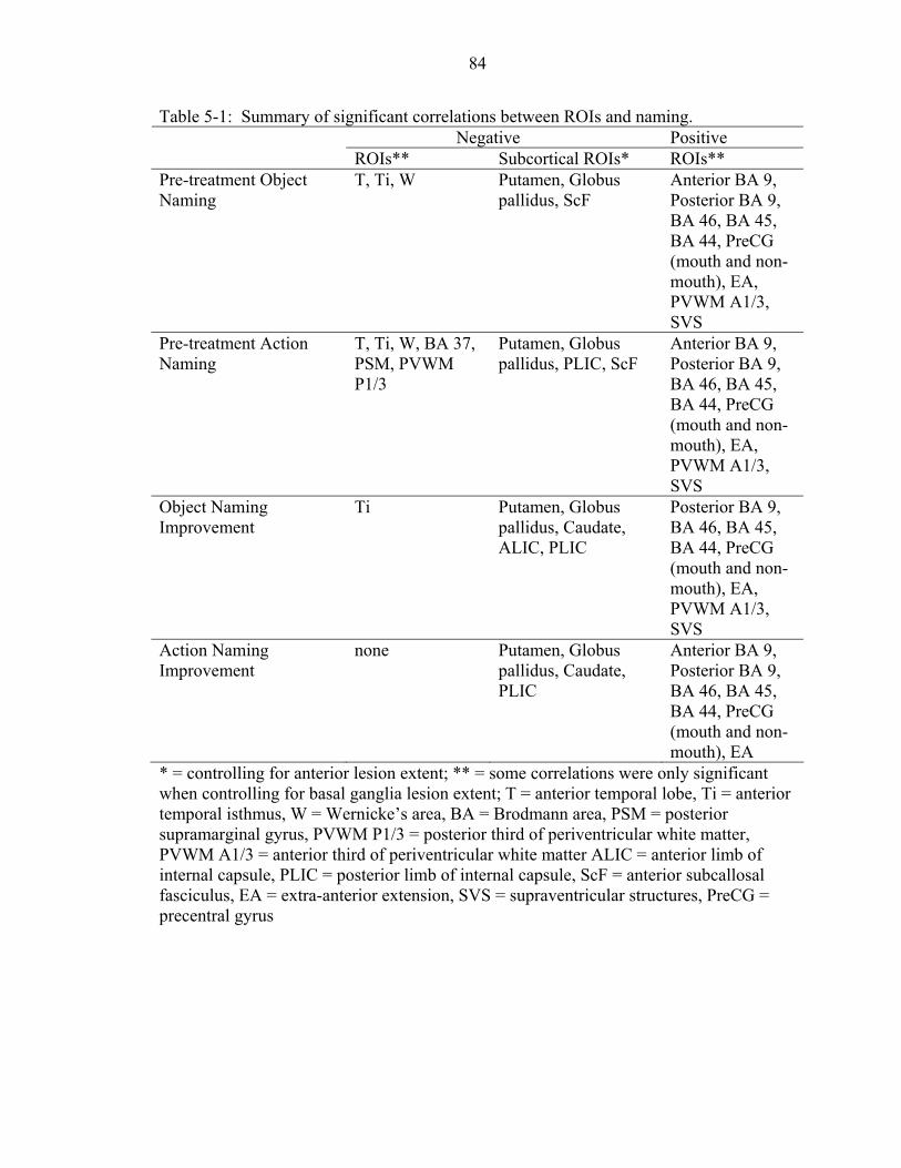
84
Table 5-1: Summary of significant correlations between ROIs and naming. Negative Positive ROIs** Subcortical ROIs* ROIs**
Pre-treatment Object Naming
T, Ti, W Putamen, Globus pallidus, ScF
Anterior BA 9, Posterior BA 9, BA 46, BA 45, BA 44, PreCG (mouth and non-mouth), EA, PVWM A1/3, SVS
Pre-treatment Action Naming
T, Ti, W, BA 37, PSM, PVWM P1/3
Putamen, Globus pallidus, PLIC, ScF
Anterior BA 9, Posterior BA 9, BA 46, BA 45, BA 44, PreCG (mouth and non-mouth), EA, PVWM A1/3, SVS
Object Naming Improvement
Ti Putamen, Globus pallidus, Caudate, ALIC, PLIC
Posterior BA 9, BA 46, BA 45, BA 44, PreCG (mouth and non-mouth), EA, PVWM A1/3, SVS
Action Naming Improvement
none Putamen, Globus pallidus, Caudate, PLIC
Anterior BA 9, Posterior BA 9, BA 46, BA 45, BA 44, PreCG (mouth and non-mouth), EA
* = controlling for anterior lesion extent; ** = some correlations were only significant when controlling for basal ganglia lesion extent; T = anterior temporal lobe, Ti = anterior temporal isthmus, W = Wernicke’s area, BA = Brodmann area, PSM = posterior supramarginal gyrus, PVWM P1/3 = posterior third of periventricular white matter, PVWM A1/3 = anterior third of periventricular white matter ALIC = anterior limb of internal capsule, PLIC = posterior limb of internal capsule, ScF = anterior subcallosal fasciculus, EA = extra-anterior extension, SVS = supraventricular structures, PreCG = precentral gyrus

85
APPENDIX A SEMANTIC + PHONOLOGIC TREATMENT
Treatment Procedure: “I’m going to show you some pictures and ask you to name
them. Then we’ll practice some ways to help you remember the words using questions
about what the word means and how it sounds. Maybe these cues will help you
remember this word or another word like it the next time you try to say it.”
1. What is this? (the examiner displays a picture: e.g. cat, catch)
Subject attempts to name. If correct, proceed to next steps.
If incorrect, examiner says target word: This is a cat/catching.
2. Semantic Coordinate question:
Is a cat similar to a dog/bear?
Is catching similar to throwing/talking?
3. Semantic Associate question:
Does a cat meow/bark?
Do you catch a ball/tree?
4. Initial Phoneme question:
Does cat/catch start with kuh/puh?
5. Rhyme question:
Does cat/catch rhyme with rat/match?
6. What is this?
7. Repeat this word after me three times.
8. Now keep saying it silently. (5 seconds)

86
9. What is this again?
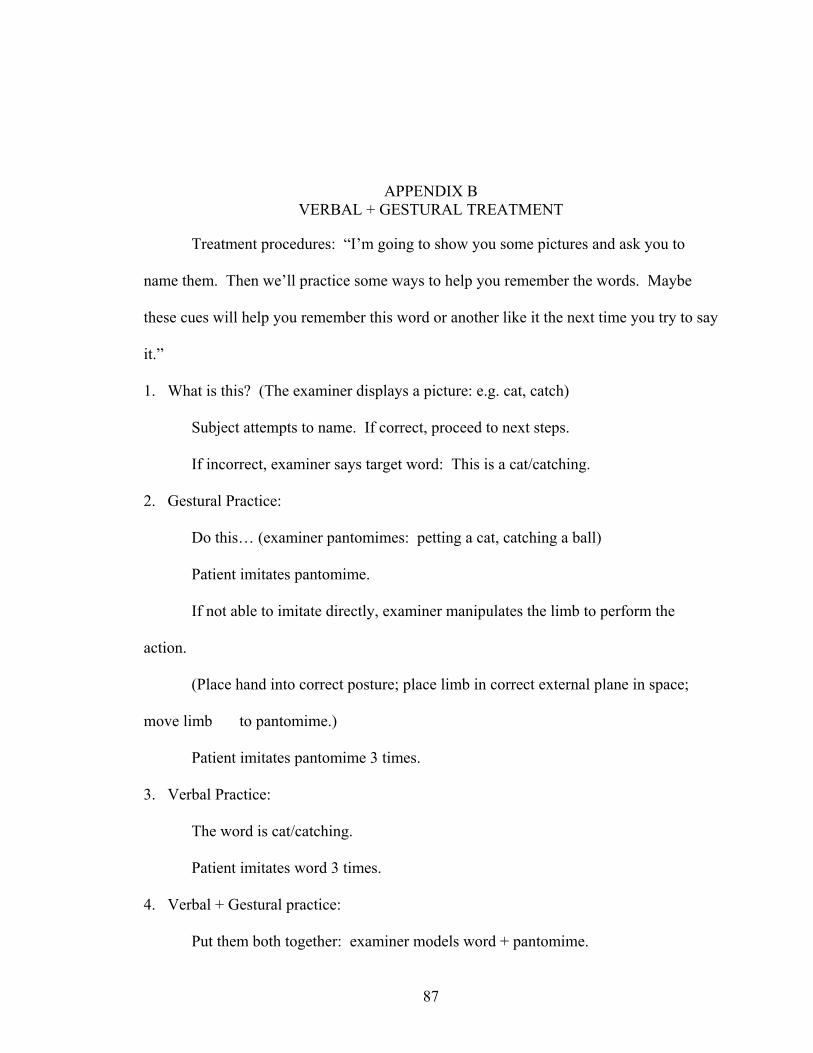
87
APPENDIX B VERBAL + GESTURAL TREATMENT
Treatment procedures: “I’m going to show you some pictures and ask you to
name them. Then we’ll practice some ways to help you remember the words. Maybe
these cues will help you remember this word or another like it the next time you try to say
it.”
1. What is this? (The examiner displays a picture: e.g. cat, catch)
Subject attempts to name. If correct, proceed to next steps.
If incorrect, examiner says target word: This is a cat/catching.
2. Gestural Practice:
Do this… (examiner pantomimes: petting a cat, catching a ball)
Patient imitates pantomime.
If not able to imitate directly, examiner manipulates the limb to perform the
action.
(Place hand into correct posture; place limb in correct external plane in space;
move limb to pantomime.)
Patient imitates pantomime 3 times.
3. Verbal Practice:
The word is cat/catching.
Patient imitates word 3 times.
4. Verbal + Gestural practice:
Put them both together: examiner models word + pantomime.

88
Patient imitates 3 times.
5. What is this?
6. Now keep thinking of it silently. (5 seconds)
7. What is this again?
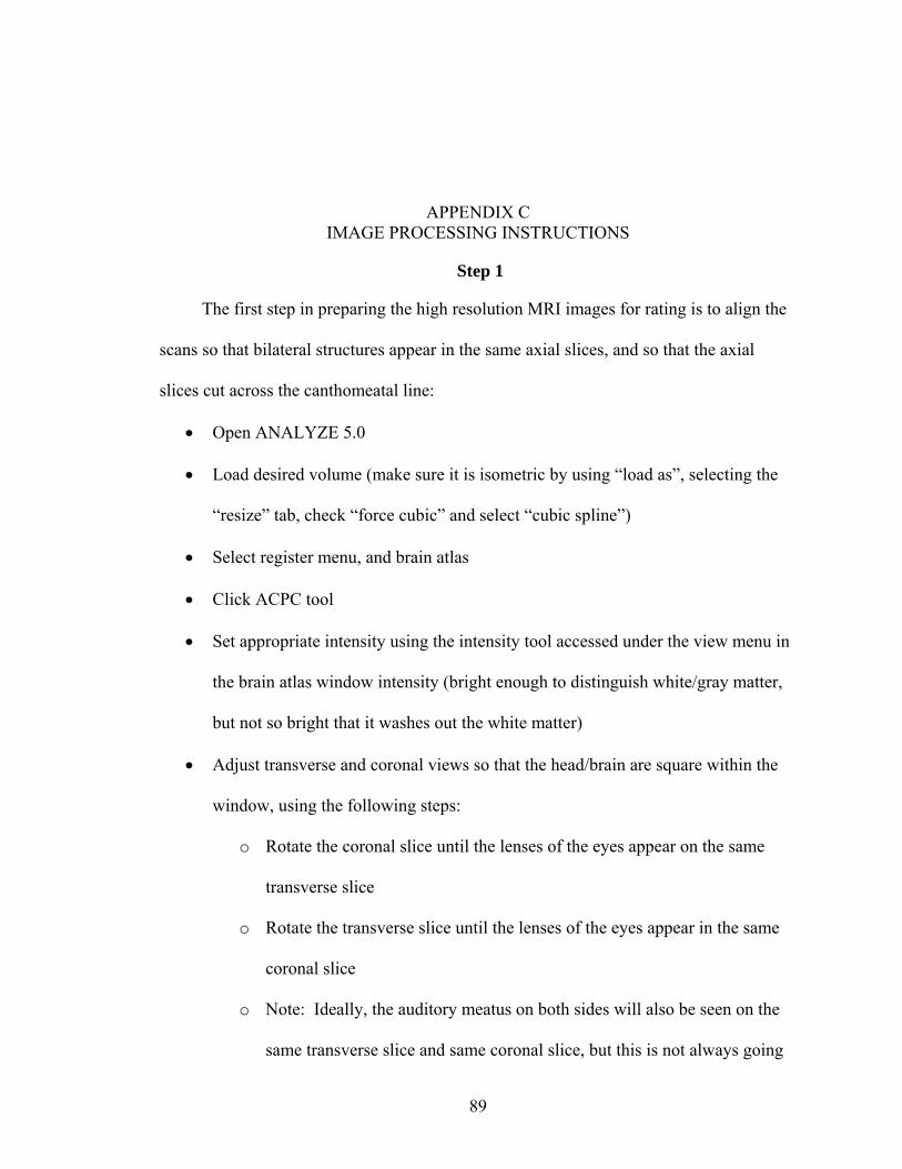
89
APPENDIX C IMAGE PROCESSING INSTRUCTIONS
Step 1
The first step in preparing the high resolution MRI images for rating is to align the
scans so that bilateral structures appear in the same axial slices, and so that the axial
slices cut across the canthomeatal line:
• Open ANALYZE 5.0
• Load desired volume (make sure it is isometric by using “load as”, selecting the
“resize” tab, check “force cubic” and select “cubic spline”)
• Select register menu, and brain atlas
• Click ACPC tool
• Set appropriate intensity using the intensity tool accessed under the view menu in
the brain atlas window intensity (bright enough to distinguish white/gray matter,
but not so bright that it washes out the white matter)
• Adjust transverse and coronal views so that the head/brain are square within the
window, using the following steps:
o Rotate the coronal slice until the lenses of the eyes appear on the same
transverse slice
o Rotate the transverse slice until the lenses of the eyes appear in the same
coronal slice
o Note: Ideally, the auditory meatus on both sides will also be seen on the
same transverse slice and same coronal slice, but this is not always going

90
to line up perfectly, given individual variation. If the lens is missing, line
up the head based on relative size and location of 2 eyeballs in slices.
Also, occasionally a patient will have external features which are not
square with brain orientation. In such case, it is best to find brain
landmarks by which to align the scan, such as the temporal pole, cerebral
peduncle, cerebellum, etc.
o Adjust the colored midline indicator with the arrows until it runs through
the longitudinal fissure. Due to lesion-induced midline shift, it may not be
possible to have the midline run through the fissure perfectly, but try to get
it as close as possible, considering the anterior, posterior, and middle
regions, and minimizing cortex seen in the midsagital slice.
• Select transform interpolation option to be “cubic spline” from the pull down
menu
• Click the “align AC-PC” button and then “done”
• In the Brain Atlas window, select Generate, Interpolation and select Cubic Spline
• In the Brain Atlas window, select file, output
• Select “volume” for “output what”
• Select “AC-PC aligned volume” for “space”
• Select “disk” for “where”
• Name the file (for example: “BP square” and select appropriate folder, and
“Analyze AVW” format
• Click generate output
• Click “done”
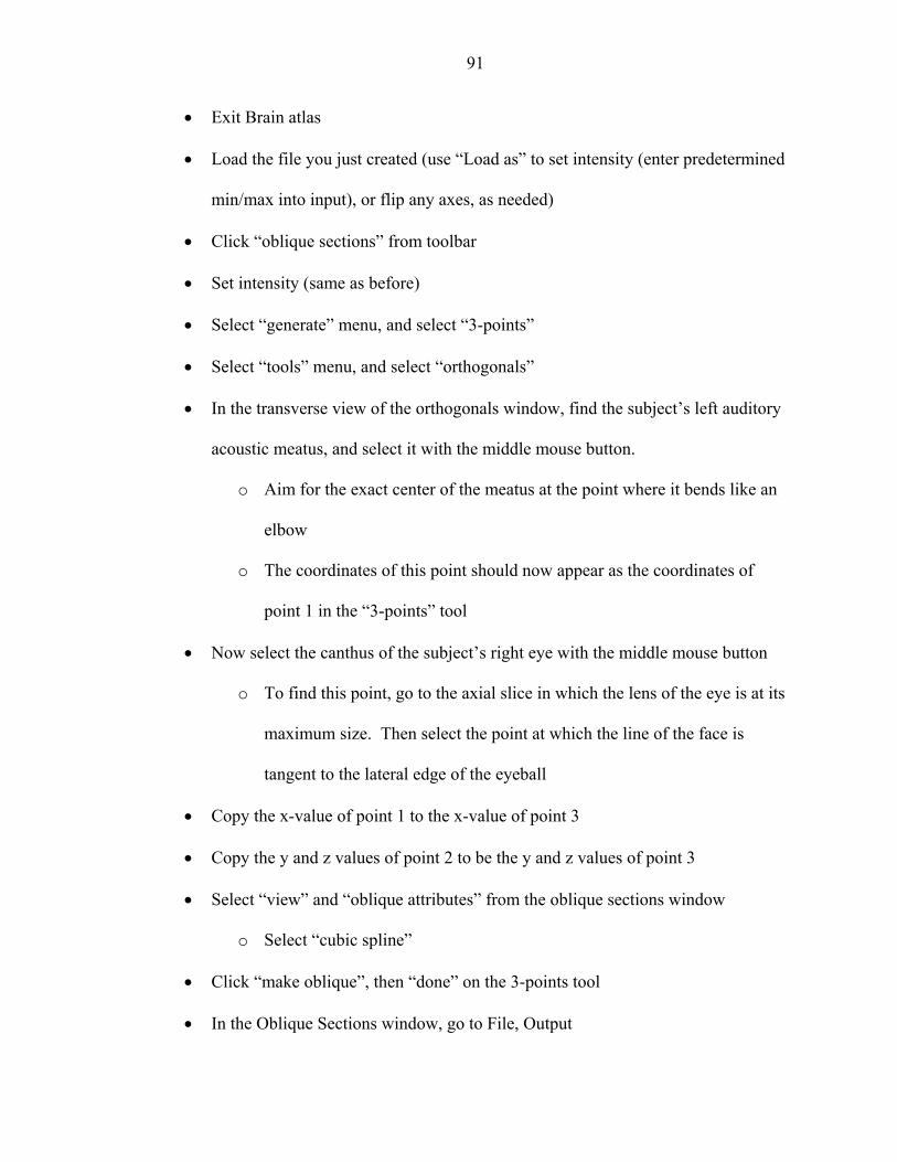
91
• Exit Brain atlas
• Load the file you just created (use “Load as” to set intensity (enter predetermined
min/max into input), or flip any axes, as needed)
• Click “oblique sections” from toolbar
• Set intensity (same as before)
• Select “generate” menu, and select “3-points”
• Select “tools” menu, and select “orthogonals”
• In the transverse view of the orthogonals window, find the subject’s left auditory
acoustic meatus, and select it with the middle mouse button.
o Aim for the exact center of the meatus at the point where it bends like an
elbow
o The coordinates of this point should now appear as the coordinates of
point 1 in the “3-points” tool
• Now select the canthus of the subject’s right eye with the middle mouse button
o To find this point, go to the axial slice in which the lens of the eye is at its
maximum size. Then select the point at which the line of the face is
tangent to the lateral edge of the eyeball
• Copy the x-value of point 1 to the x-value of point 3
• Copy the y and z values of point 2 to be the y and z values of point 3
• Select “view” and “oblique attributes” from the oblique sections window
o Select “cubic spline”
• Click “make oblique”, then “done” on the 3-points tool
• In the Oblique Sections window, go to File, Output
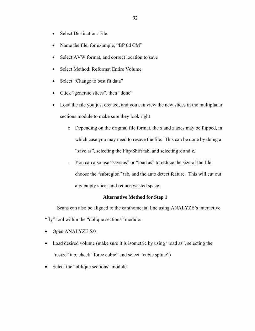
92
• Select Destination: File
• Name the file, for example, “BP 0d CM”
• Select AVW format, and correct location to save
• Select Method: Reformat Entire Volume
• Select “Change to best fit data”
• Click “generate slices”, then “done”
• Load the file you just created, and you can view the new slices in the multiplanar
sections module to make sure they look right
o Depending on the original file format, the x and z axes may be flipped, in
which case you may need to resave the file. This can be done by doing a
“save as”, selecting the Flip/Shift tab, and selecting x and z.
o You can also use “save as” or “load as” to reduce the size of the file:
choose the “subregion” tab, and the auto detect feature. This will cut out
any empty slices and reduce wasted space.
Alternative Method for Step 1
Scans can also be aligned to the canthomeatal line using ANALYZE’s interactive
“fly” tool within the “oblique sections” module.
• Open ANALYZE 5.0
• Load desired volume (make sure it is isometric by using “load as”, selecting the
“resize” tab, check “force cubic” and select “cubic spline”)
• Select the “oblique sections” module
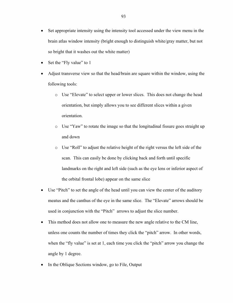
93
• Set appropriate intensity using the intensity tool accessed under the view menu in the
brain atlas window intensity (bright enough to distinguish white/gray matter, but not
so bright that it washes out the white matter)
• Set the “Fly value” to 1
• Adjust transverse view so that the head/brain are square within the window, using the
following tools:
o Use “Elevate” to select upper or lower slices. This does not change the head
orientation, but simply allows you to see different slices within a given
orientation.
o Use “Yaw” to rotate the image so that the longitudinal fissure goes straight up
and down
o Use “Roll” to adjust the relative height of the right versus the left side of the
scan. This can easily be done by clicking back and forth until specific
landmarks on the right and left side (such as the eye lens or inferior aspect of
the orbital frontal lobe) appear on the same slice
• Use “Pitch” to set the angle of the head until you can view the center of the auditory
meatus and the canthus of the eye in the same slice. The “Elevate” arrows should be
used in conjunction with the “Pitch” arrows to adjust the slice number.
• This method does not allow one to measure the new angle relative to the CM line,
unless one counts the number of times they click the “pitch” arrow. In other words,
when the “fly value” is set at 1, each time you click the “pitch” arrow you change the
angle by 1 degree.
• In the Oblique Sections window, go to File, Output
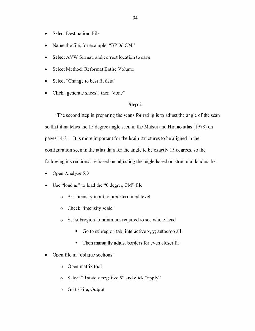
94
• Select Destination: File
• Name the file, for example, “BP 0d CM”
• Select AVW format, and correct location to save
• Select Method: Reformat Entire Volume
• Select “Change to best fit data”
• Click “generate slices”, then “done”
Step 2
The second step in preparing the scans for rating is to adjust the angle of the scan
so that it matches the 15 degree angle seen in the Matsui and Hirano atlas (1978) on
pages 14-81. It is more important for the brain structures to be aligned in the
configuration seen in the atlas than for the angle to be exactly 15 degrees, so the
following instructions are based on adjusting the angle based on structural landmarks.
• Open Analyze 5.0
• Use “load as” to load the “0 degree CM” file
o Set intensity input to predetermined level
o Check “intensity scale”
o Set subregion to minimum required to see whole head
Go to subregion tab; interactive x, y; autocrop all
Then manually adjust borders for even closer fit
• Open file in “oblique sections”
o Open matrix tool
o Select “Rotate x negative 5” and click “apply”
o Go to File, Output
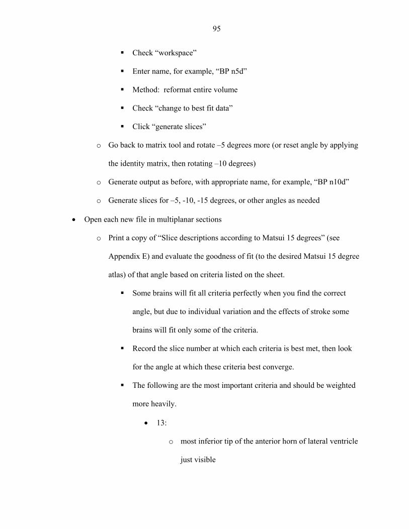
95
Check “workspace”
Enter name, for example, “BP n5d”
Method: reformat entire volume
Check “change to best fit data”
Click “generate slices”
o Go back to matrix tool and rotate –5 degrees more (or reset angle by applying
the identity matrix, then rotating –10 degrees)
o Generate output as before, with appropriate name, for example, “BP n10d”
o Generate slices for –5, -10, -15 degrees, or other angles as needed
• Open each new file in multiplanar sections
o Print a copy of “Slice descriptions according to Matsui 15 degrees” (see
Appendix E) and evaluate the goodness of fit (to the desired Matsui 15 degree
atlas) of that angle based on criteria listed on the sheet.
Some brains will fit all criteria perfectly when you find the correct
angle, but due to individual variation and the effects of stroke some
brains will fit only some of the criteria.
Record the slice number at which each criteria is best met, then look
for the angle at which these criteria best converge.
The following are the most important criteria and should be weighted
more heavily.
• 13:
o most inferior tip of the anterior horn of lateral ventricle
just visible
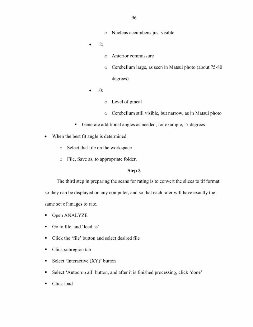
96
o Nucleus accumbens just visible
• 12:
o Anterior commissure
o Cerebellum large, as seen in Matsui photo (about 75-80
degrees)
• 10:
o Level of pineal
o Cerebellum still visible, but narrow, as in Matsui photo
Generate additional angles as needed, for example, -7 degrees
• When the best fit angle is determined:
o Select that file on the workspace
o File, Save as, to appropriate folder.
Step 3
The third step in preparing the scans for rating is to convert the slices to tif format
so they can be displayed on any computer, and so that each rater will have exactly the
same set of images to rate.
Open ANALYZE
Go to file, and ‘load as’
Click the ‘file’ button and select desired file
Click subregion tab
Select ‘Interactive (XY)’ button
Select ‘Autocrop all’ button, and after it is finished processing, click ‘done’
Click load

97
Open multiplanar sections module and maximize the window
Open slice tool and drag it to the bottom right corner of screen
Open intensity tool and set to appropriate intensity (bright enough to distinguish
white/gray matter, but not so bright that it washes out the white matter)
Choose page mode on slice tool
Enter the value of the first slice you want to capture
Hit “display sections”
o The number of slices appearing on the screen depends on how big your screen
is, and how big the sections are
o Make sure that neither the slice tool nor cursor is not blocking your view of
any of the sections
On keyboard, hit control-print screen to copy the contents of the screen to the
clipboard
Open Photoshop 7.0
Hit control-n to make a new file
Name the file according to brain number and slice numbers, and hit return, for
example, “B4 158-163”
Hit control-v to paste in captured screen from the clipboard
Select rectangular marquee tool and frame the area you want to keep
Select Image menu, and crop
Select Image menu, and mode, and select grayscale
When prompted, select “flatten”
Close window of image, and click yes when prompted to save

98
In “save” window, select tif format and make sure it is saving to desired folder
Hit “save”, and when prompted, make sure image compression is set to “none”
Go back to ANALYZE to select next set of slices and repeat the process

99
APPENDIX D RATING SHEET
Lesion Rating SheetSubject: Rating systemRater: 0 = no lesion
1 = equivocal lesion2 = small, patchy or partial lesion2.5 = patchy, less than half of the area has lesion3 = half of area has lesion4 = more than half of area has solid lesion5 = total area has solid lesion
Matsui slice: 13 12 11 10 9 8 7 6 - ROISubject slice:
ROI'sBA 9BA 46BA 45BA 44P-MMM-MTWBA 37Sen-MSenASMPSMAGITiPGPALICPLICCScFThalamusEAPVWM A1/3PVWM M1/3PVWM P1/3SVS
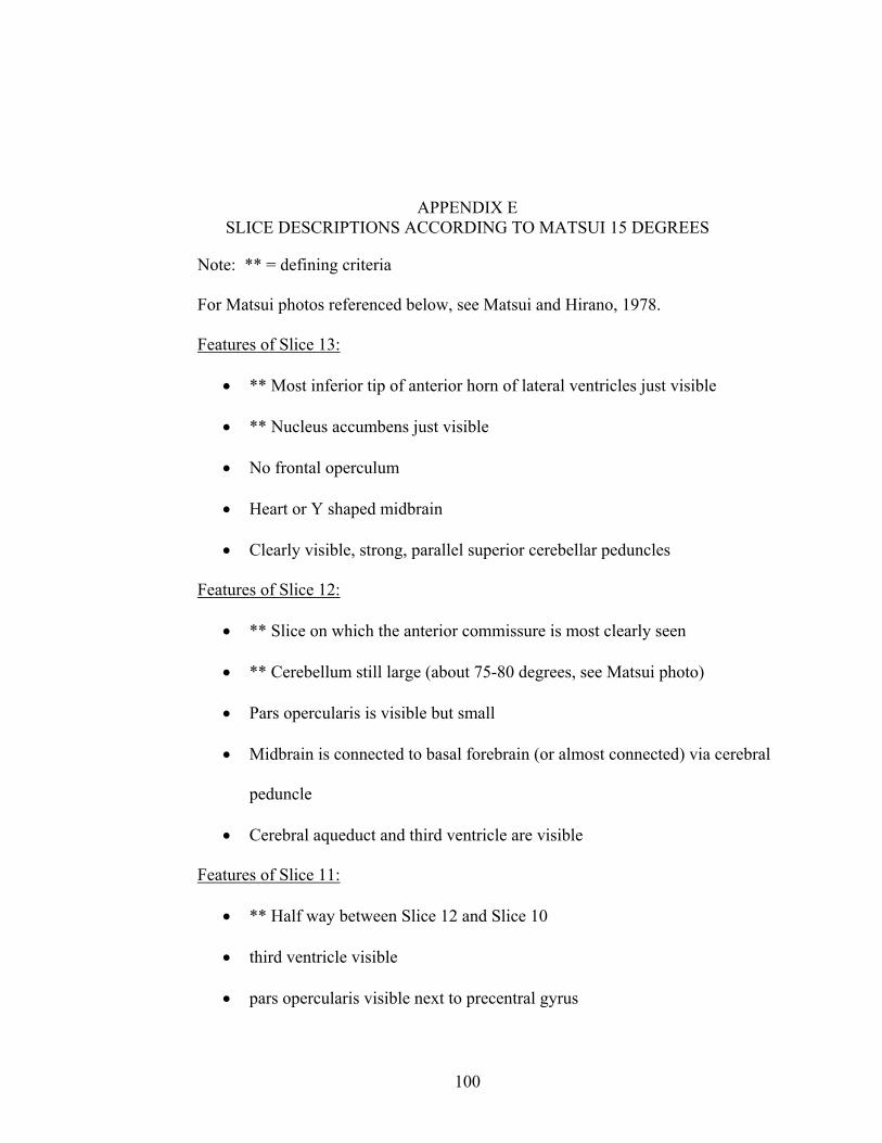
100
APPENDIX E SLICE DESCRIPTIONS ACCORDING TO MATSUI 15 DEGREES
Note: ** = defining criteria
For Matsui photos referenced below, see Matsui and Hirano, 1978.
Features of Slice 13:
• ** Most inferior tip of anterior horn of lateral ventricles just visible
• ** Nucleus accumbens just visible
• No frontal operculum
• Heart or Y shaped midbrain
• Clearly visible, strong, parallel superior cerebellar peduncles
Features of Slice 12:
• ** Slice on which the anterior commissure is most clearly seen
• ** Cerebellum still large (about 75-80 degrees, see Matsui photo)
• Pars opercularis is visible but small
• Midbrain is connected to basal forebrain (or almost connected) via cerebral
peduncle
• Cerebral aqueduct and third ventricle are visible
Features of Slice 11:
• ** Half way between Slice 12 and Slice 10
• third ventricle visible
• pars opercularis visible next to precentral gyrus
101
• inferior colliculus visible
• quadrigeminal cistern shaped like upside-down mustache
• cerebellum size as seen in Matsui photo
Features of Slice 10:
• ** about level of pineal
• cerebellum still visible but narrowing, as in Matsui photo
• above the level of the posterior commissure
Features of Slice 9:
• ** inferior aspect of body of lateral ventricle forming, connecting anterior
horn and atrium (in brains where the lesioned hemisphere has much larger
ventricles, extending much lower than in the non-lesioned hemisphere, use
the non-lesioned hemisphere for reference)
• splenium of corpus callosum beginning to be formed or formed
• fornix may be visible running along the base of the body of the 3rd ventricle
• insula has almost disappeared
Features of Slice 8:
• ** half way between Slice 9 and Slice 7
• body of corpus callosum formed in normal brains (in stroke brains the body
may not be formed yet, or is very thin)
Features of Slice 7:
• ** longitudinal fissure is complete
Features of Slice 6:

102
• ** lateral ventricles have disappeared (if the lesioned hemisphere’s lateral
ventricle is significantly enlarged, and extends much higher than the non-
lesioned hemisphere’s lateral ventricle, use the non-lesioned hemisphere for
reference)
103
APPENDIX F PROCEDURE FOR RATING LESIONS
1. Raters will rate each subject scan independently, and will be blind to the treatment
scores of each subject until after that rating is complete.
2. The first step in rating a subject’s scan is to determine which slices in the subject’s
scan best correspond to the Matsui and Hirano (1978) atlas slices 6-13, using the
criteria and instructions listed in the Matsui Slice Descriptions (see Appendix E). The
slice number of the subject slice which best corresponds to each Matsui slice should
be recorded in the cell immediately below the Matsui slice number on the rating sheet
(Appendix D). Once one subject slice has been chosen which best represents each
Matsui slice, the remaining subject slice numbers should be recorded in the
appropriate columns, adding columns if needed.
3. Major sulci should then be identified using the following guidelines. Begin at the
axial slice in which the operculum begins to appear, and keep moving superiorly,
identifying new sulci as they branch off from the Sylvian fissure. The process
becomes more difficult with brains which are lesioned in these areas. For brains
which are lesioned so severely that these guidelines cannot be used, locations of sulci
may be estimated based on their locations relative to other landmarks on the Matsui
atlas (for example, on slice 10, the central sulcus begins to emerge straight across
from the posterior border of the lateral ventricles).
In slice 13 the Sylvian fissure is easily found, dividing the frontal and
temporal lobes.

104
Around slice 12, the pars opercularis (BA 44) begins to appear. It is bounded
posteriorly by the Sylvian fissure (SF), and anteriorly by the anterior
ascending ramus (AAR).
As the operculum gets larger, in slice 11, the precentral sulcus (PreC) begins
to appear, which divides the operculum into an anterior part (BA 44) and a
posterior part, the precentral gyrus (PreCG).
In slice 10, the central sulcus (C) begins to appear between SF and PreC.
Also in slice 10, notice that as BA 44 gets smaller, AAR and PreC get closer
and may merge.
In slice 9, the postcentral sulcus (PostC) begins to emerge between SF and C.
Follow each of these sulci (SF, PreC, C, PostC) up through succeeding slices.
It may be useful to mark these sulci on the images (by printing hardcopies of
the images and marking with a pencil, or by drawing arrows using
Photoshop). One may also choose to mark the inferior and superior frontal
sulci, or the superior and middle temporal sulci, however, this may not be
necessary, as they may be fairly obvious once the above mentioned sulci are
marked.
4. Once the major sulci are identified, ROI’s should be identified based on their
descriptions listed in Appendix G. Raters can use the rating sheet as a guide for
which ROI’s to expect on each slice because those cells are shaded. However, due to
the effects of the stroke or individual variation, if a structure appears on slices other
than those indicated by the shading on the worksheet, raters should still rate the
105
structure. Likewise, if a cell is shaded, but the structure does not appear on that slice,
that cell can be crossed out.
5. In order to locate cortical ROI’s, the Matsui atlas should be used. Subcortical ROI’s
can be located using both the Matsui atlas and the Naeser templates. Some CT or
MRI scans which were copied from clinical films may be tilted at a somewhat
different angle than the Matsui atlas and Naeser templates. In those cases, it may be
useful to consult alternatively angled templates found in Damasio and Damasio
(1989) to ensure accurate identification of cortical ROI’s and the sulci which bound
them.
6. The depth (medial border) of cortical ROI’s is the approximate depth of the Sylvian
fissure or the deepest sulcus. This is illustrated in the Naeser templates.
7. To aid in the rating process, ROI’s are listed in order on the rating sheet, with more
anterior cortical areas listed first followed by posterior cortical areas, and then
subcortical structures. For scans with many slices, raters may find it easiest to rate an
ROI on each slice on which it appears before moving to the next ROI. However, on
scans with very few slices, a rater may find it easier to rate all the ROI’s on each slice
before moving on to the next slice.
8. The scale for rating each ROI is listed on the rating sheet. The following guidelines
should also be taken into account.
It may be helpful to conceptualize a lesion as consisting of 2 parts, the dark area
(solid lesion which is similar in intensity to the ventricles) and the gray area
which often surrounds it (patchy lesion). A patchy area is definitely darker than
the non-lesioned tissue surrounding it, but represents tissue which is not totally
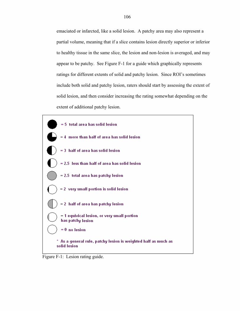
106
emaciated or infarcted, like a solid lesion. A patchy area may also represent a
partial volume, meaning that if a slice contains lesion directly superior or inferior
to healthy tissue in the same slice, the lesion and non-lesion is averaged, and may
appear to be patchy. See Figure F-1 for a guide which graphically represents
ratings for different extents of solid and patchy lesion. Since ROI’s sometimes
include both solid and patchy lesion, raters should start by assessing the extent of
solid lesion, and then consider increasing the rating somewhat depending on the
extent of additional patchy lesion.
Figure F-1: Lesion rating guide.
107
For the purposes of this study, the term “lesion” refers to infarcted tissue, or areas
of encephalomalacia associated with the patient’s stroke. Therefore, although we
will also see signs of atrophy and other tissue change secondary to the lesion or
age, our purpose is not to characterize these secondary changes.
It is important to keep in mind that very subtle changes seen in a high resolution
MRI may not be visible on the lower resolution CT scans. Therefore, unless an
area is clearly lesioned, we would only give it a rating of 0 or 1 (equivocal lesion).
For the most part, areas of lesion should be contiguous. It is possible for a subject
to have more than one clear lesion, in which case all lesions should be rated.
However, if an area is slightly darker than expected, but is not obviously lesioned
and is not contiguous with an obvious lesion, then it should be rated a 0 or 1.
In determining whether an area is truly lesioned, it is sometimes useful to refer to
the non-lesioned hemisphere for comparison. For example, sometimes the globus
pallidus may appear dark like a lesion, however, if this feature is bilateral and
relatively symmetrical, it may not be appropriate to characterize the area as being
lesion, but may be due to other causes, such as iron deposits or calcification.
Therefore, subcortical areas which resemble lesion, but occur bilaterally and are
symmetrical, should not be considered lesion.
Areas of possible lesion should be examined several slices above and below the
slice in question. It may be determined in some cases that a dark area is simply
caused by a partial deep sulcus which becomes more visible on adjacent slices.
9. After individual slice ratings are complete, an overall rating will be assessed for each
ROI, and recorded in the column “ROI.” Instead of mathematically averaging the
108
individual slice ratings of an ROI, raters will estimate an overall average by taking
into account the fact that a given ROI may be larger on one slice than another, so
some individual slice ratings should be weighted more heavily than others. Only this
overall rating for the ROI will be used in the analysis. Therefore, raters should focus
more on the accuracy of this overall ROI rating, rather than obsess over the accuracy
of individual slice ratings. In other words, the process of recording ratings for
individual slices should be seen only as a tool for arriving at the overall ROI rating.
10. After both raters have independently determined overall ratings for each ROI,
overall ratings will be compared. Ratings which differ by one point or less will be
averaged. Ratings which differ by more than one point will be discussed and
modified by the raters until the two ratings differ by one point or less and can be
averaged. In order to facilitate this discussion, raters may choose to refer to their
individual slice ratings to see how they arrived at their overall rating for that ROI.
109
APPENDIX G ROI DEFINITIONS
The following is a list of regions of interest (ROI’s) rated in the study. Each
description includes the ROI name, abbreviation, and working definition. ROI’s are
listed in the same order as on the rating sheet (see Appendix D). Given the variety of
scan types used in this study, it was important that ROI’s be defined using clearly
definable anatomic landmarks, such as major sulci, which would be consistently
identifiable across subjects. Thus, although some ROI’s listed are named after
Brodmann areas, their borders may not correspond exactly to Brodmann areas as defined
by other sources. Any discrepancies should not be construed as an attempt to redefine
Brodmann areas. Rather, it should be kept in mind that the names and definitions for the
following ROI’s were designed as a means of consistently identifying areas of interest
(which sometimes roughly correspond to established Brodmann areas) in a way that
could be applied to a variety of scan types. Mentions of “Matsui slices” refer to the set
of slices in the Matsui and Hirano (1978) atlas which were cut at 15 degrees to the
canthomeatal line (p. 14-81).
1. Anterior Brodmann area 9 (BA 9A). This area refers only to the anterior portion of
Brodmann area 9. It is defined as the gyrus anterior to the superior frontal sulcus, and
rated on slices corresponding to Matsui slices 7, 8 and 9.
2. Brodmann area 46 (BA 46). This area refers to the middle frontal gyrus. It is defined
as the gyrus bounded by the superior and inferior frontal sulci and is rated on slices
corresponding to Matsui slices 7-12.
110
3. Brodmann area 45 (BA 45). Also known as pars triangularis of the inferior frontal
gyrus, it is bounded posteriorly by the anterior ascending ramus of the Sylvian
fissure, and anteriorly by the inferior frontal sulcus. It was rated on slices
corresponding to Matsui slices 10-12, and on the superior half of the slices
corresponding to Matsui slice 13.
4. Brodmann area 44 (BA 44). Also known as pars opercularis of the inferior frontal
gyrus, it is bounded anteriorly by the anterior ascending ramus on slices 10-12. It is
bounded posteriorly by the Sylvian fissure on slices corresponding to Matsui slice 12
and by the precentral sulcus on slices corresponding to Matsui slices 11 and 10.
5. Posterior Brodmann area 9 (BA 9P). This area refers to the posterior portion of
Brodmann area 9. It is defined as the cortex bound anteriorly by the inferior frontal
sulcus, and posteriorly by the precentral sulcus, and is rated on slices corresponding
to Matsui slices 7-9.
6. Pre-central gyrus (PreCG). This area includes portions of Brodmann areas 4 (motor)
and 6 (premotor), and is rated only on slices corresponding to Matsui slices 11, 10,
and 7. It is defined as the gyrus bounded anteriorly by the precentral sulcus on all
slices, and posteriorly by the Sylvian fissure on slice 11 and the central sulcus on
slices 10 and 7.
7. Pre-central gyrus for mouth region (PreCG-M) This area is rated only on slices
corresponding to Matsui slices 8 and 9, and is bounded anteriorly by the precentral
sulcus and posteriorly by the central sulcus. It is comprised of portions of Brodmann
areas 4 and 6.
111
8. Anterior temporal lobe (T). This area is defined as the portions of the superior
temporal gyrus visible in slices corresponding to Matsui slices 12 and 13. It is
bounded anteriorly by the Sylvian fissure and posteriorly by the superior temporal
sulcus.
9. Wernicke’s area (W). This area refers to the posterior two thirds of the left superior
temporal gyrus (Brodmann area 22). Besides the cortex traditionally known as
Wernicke’s area, this area also includes the primary auditory reception area
(Brodmann areas 41 and 42) because of the difficulty in distinguishing between the
two areas on lower resolution scans (Naeser et al, 1987). It is defined as the cortex
bounded by the Sylvian fissure anteriorly and the superior temporal sulcus
posteriorly, and is rated on slices corresponding to Matsui slices 9-11.
10. Brodmann area 37 (BA 37). This area includes portions of the middle and inferior
temporal gyri. On slices corresponding to Matsui slices 12 and 13 the ROI is
bounded by the inferior and middle temporal sulci. On slices corresponding to
Matsui slice 11, it includes the posterior half of the middle temporal gyrus and the
anterior half of the lateral aspect of the inferior temporal gyrus. On slices
corresponding to Matsui slices 9 and 10, it is bounded by the middle and superior
temporal sulci.
11. Primary sensory cortex for the mouth (Sen-M). This region is defined as the cortical
area bounded by the central and postcentral sulci on slices corresponding to Matsui
slices 8 and 9. It is comprised of portions of Brodmann areas 1, 2, and 3.
12. Primary sensory cortex other than for the mouth (Sen). On slices corresponding to
Matsui slice 10, this region is bounded by the central sulcus and the Sylvian fissure.
112
On slices corresponding to Matsui slice 7 this region is bounded by the central sulcus
and the postcentral sulcus. It is comprised of portions of Brodmann areas 1, 2, and 3.
13. Anterior supramarginal gyrus (ASM). Otherwise known as the anterior half of
Brodmann area 40, this region is defined on slices corresponding to Matsui slice 9 as
the area bounded anteriorly by the postcentral sulcus and posteriorly by the Sylvian
fissure. On slices corresponding to Matsui slices 7 and 8, ASM is bounded anteriorly
by the postcentral sulcus and posteriorly by the posterior ascending limb of the
Sylvian fissure, or approximately half way to the angular gyrus.
14. Posterior supramarginal gyrus (PSM). The posterior half of Brodmann area 40, this
region is bounded anteriorly by the posterior ascending limb of the lateral sulcus, and
posteriorly by the angular gyrus. It is rated on slices corresponding to Matsui slices 7
and 8.
15. Angular gyrus (AG). This region begins to emerge on slices corresponding to Matsui
slice 8. On slices corresponding to Matsui slice 7, if the posterior half of the left
hemisphere is divided into four equal portions, the AG is approximately the second
quarter from the occipital pole. The third and fourth quarters from the occipital pole
are the PSM and ASM respectively. This area is meant to generally correspond to
Brodmann area 39.
16. Insula (I). Besides the insula, which is easily identified on the Matsui atlas and the
Naeser templates, this area also includes the claustrum, external capsule, and extreme
capsule. It is rated on all slices in which it was visible, which is usually the slices
corresponding to Matsui slices 10-13.
113
17. Anterior temporal isthmus (Ti). This region is defined as the anterior half of the
white matter lying between the inferior edge of the Sylvian fissure/insula and the
temporal horn of the lateral ventricle. Only the anterior half is rated since the anterior
half contains afferent auditory pathways from the medial geniculate body to Heschl’s
gyrus, whereas the posterior half contains visual pathways (Naeser et al., 1990). It is
rated on slices corresponding to Matsui slices 9-12, or where visible.
18. Putamen (P). The putamen is rated on all slices where it is visible. When needed, the
intact right hemisphere can be used as a guide for estimating the location of the
putamen in the left hemisphere. Generally the putamen is rated on slices
corresponding to Matsui slices 10-13. The putamen is recognizable by its unique
shape, as seen in the Matsui atlas. It is rated starting in slice 13 where the nucleus
accumbens splits into the putamen and caudate nucleus, until about slice 9 or 10
where it tapers in size until it disappears in the surrounding white matter. It is
bounded medially by the globus pallidus and the anterior limb of the internal capsule
until the globus pallidus ends in superior slices, after which it is bounded medially by
the posterior limb of the internal capsule. It is bounded laterally by the external
capsule.
19. Globus pallidus (GP). The globus pallidus recognized by its characteristic shape,
medial to the putamen as seen in the Matsui atlas, and it is rated on all slices where it
is visible. It is generally a lighter grey than the putamen, but darker than white
matter. When needed, the intact right hemisphere can be used as a guide for
estimating the location of the globus pallidus in the left hemisphere. Generally the
114
globus pallidus is rated on slices corresponding to Matsui slices 11 and 12. It is
bounded laterally by the putamen, and medially by the internal capsule.
20. Anterior limb of the internal capsule (ALIC). This region is defined as the white
matter strip lying between the caudate and putamen. It is generally rated on slices
corresponding to Matsui slices 10-13.
21. Posterior limb of the internal capsule (PLIC). This region is defined as the white
matter strip lying between the putamen or globus pallidus and the thalamus. It is
generally rated on slices corresponding to Matsui slices 9-12.
22. Caudate (C). While only the head of the caudate is shown on the Naeser templates,
both the head and body of the caudate were rated for this study. Both the head and
body of the caudate can be identified using the Matsui atlas. It is rated on slices
corresponding to Matsui slices 8-13.
23. Medial subcallosal fasciculus (ScF). As described in Naeser et al. (1989), this region
is the “narrow white matter area surrounding the lateral angle of the frontal horn
containing a pathway through which fibers pass from the cingulate gyrus and
supplementary motor area to the caudate. . .[it] is only one-tenth of the distance from
the lateral border of the frontal horn to the cortical mantle.” The ScF is rated from the
most inferior slice on which the caudate and frontal horn appear to the most superior
slice on which Brodmann areas 44 and 45 appear, which generally corresponds to
Matsui slices 10-13.
24. Thalamus. This region includes the major thalamic nuclei as seen in the Matsui atlas.
In general, the thalamus is bounded laterally by the posterior limb of the internal
capsule, medially by the third ventricle, superiorly by the body of the lateral ventricle,
115
and anteriorly by the genu of the internal capsule. It is generally rated in slices
corresponding to Matsui slices 9-11.
25. Extra-anterior extension (EA). This region includes a white matter strip immediately
anterior to the frontal horn of the lateral ventricle as shown on the Naeser templates.
It is rated on each slice in which the frontal horn is visible.
26. Anterior third of the periventricular white matter (PVWM A1/3). This region is rated
on all slices where the body of the lateral ventricle is seen, and contains the anterior
portion of the white matter adjacent to the caudate and/or body of the lateral ventricle.
Although the Naeser templates do not show the body of the caudate, which is
immediately lateral to the lateral ventricle, the Matsui atlas can be used to help
identify the border between the caudate and PVWM.
27. Middle third of the periventricular white matter (PVWM M1/3). This region is rated
on all slices where the body of the lateral ventricle is seen, and contains the middle
portion of the white matter adjacent to the caudate and/or body of the lateral ventricle.
Although the Naeser templates do not show the body of the caudate, which is
immediately lateral to the lateral ventricle, the Matsui atlas can be used to help
identify the border between the caudate and PVWM.
28. Posterior third of the periventricular white matter (PVWM P1/3). This region
contains the posterior portion of the white matter adjacent to the body of the lateral
ventricle, as shown in the Naeser templates. It is rated on all slices where the body of
the lateral ventricle is seen.
29. Supraventricular frontal lobe structures (SVS). This region includes portions of
Brodmann areas 32, 24, 8, 6, and 4, and white matter deep to these structures. It is
116
rated on all slices corresponding to Matsui slice 6 or superior to Matsui slice 6, and is
operationally defined as all medial cortex anterior to the midpoint of the longitudinal
fissure, including underlying white matter extending halfway to the lateral cortex.

117
REFERENCES
Alexander, M. P., Naeser, M. A. & Palumbo, C. L. (1987). Correlations of subcortical CT lesion sites and aphasia profiles. Brain, 110, 961-991.
Bak, T. H., O’Donovan, D. G., Xuereb, J. H., Boniface, S. & Hodges, J. R. (2001). Selective impairment of verb processing associated with pathological changes in Brodmann areas 44 and 45 in the motor neuron disease-dementia-aphasia syndrome. Brain 124, 103-120.
Barlow T. (1877). On a case of double cerebral hemiplegia, with cerebral symmetrical lesions. British Medical Journal, 2, 103-104.
Basso, A. & Farabola, M. (1997). Comparison of improvement of aphasia in three patients with lesions in anterior, posterior, and antero-posterior language areas. Neuropsychological Rehabilitation, 7(3), 215-230.
Basso A., Gardelli M., Grassi M.P. & Mariotti M. (1989). The role of the right hemisphere in recovery from aphasia. Two case studies. Cortex 25, 555-566.
Bates, E., Wilson, S. M., Saygin, A. P., Dick, F., Sereno, M. I., Knight, R. T. & Dronkers, N. F. (2003). Voxel-based lesion-symptom mapping. Nature Neuroscience, 6(5), 448-450.
Berndt, R. S., Mitchum, C. C., Haendiges, A. N. & Sandson, J. (1997). Verb retrieval in aphasia: Characterizing single word impairments. Brain and Language, 56, 68-106.
Brunner, R. J., Kornhuber, H. H., Seemuller, E., Suger, G. & Wallesch, C. W. (1982). Basal ganglia participation in language pathology. Brain and Language, 16, 281-299.
Caramazza, A. & Hillis, A. E. (1991). Lexical organization of nouns and verbs in the brain. Nature, 349, 788-790.
Cappa, S. F., Sandrini, M., Rossini, P. M., Sosta, K. & Miniussi, C. (2002). The role of the left frontal lobe in action naming: rTMS evidence. Neurology, 59, 720-723.
Cappa, S. F. & Vignolo, L. A. (1999). The neurological foundations of language. In G. Denes & L. Pizzamiglio (Eds.) Handbook of clinical and experimental neuropsychology (pp. 155-179). Hove, East Sussex, UK: Psychology Press.
118
Cato, M., Parkinson, R. B., Wierenga, C. & Crosson, B. (2004a, February). Predicting rehabilitative treatment success in chronic nonfluent aphasia: Lesion and performance characteristics. Poster presented at the annual meeting of the International Neuropsychological Society, Baltimore, MD.
Cato, M. A., Parkinson, R. B., Wierenga, C. E. & Crosson, B. (2004b, October). Lesion pattern relates to rehabilitative treatment success in chronic nonfluent aphasia. Poster presented at the annual meeting of the Society for Neuroscience, San Diego, CA.
Crosson, B., Benefield, H., Cato, M. A., Sadek, J. R., Moore, A. B., Wierenga, C. E., Gopinath, K., Soltysik, D., Bauer, R. M., Auerbach, E. J., Gokcay, D., Leonard, C. M. & Briggs, R. W. (2003). Left and right basal ganglia and frontal activity during language generation: Contributions to lexical, semantic, and phonological processes. Journal of the International Neuropsychological Society, 9, 1061-1077.
Crosson, B., Moore, A. B., Gopinath, K., White, K. D., Wierenga, C. E., Gaiefsky, M. E., Fabrizio, K. R., Peck, K. K., Soltysik, D., Milstead, C., Briggs, R. W., Conway, T. W. & Gonzalez-Rothi, L. J. (2005). Role of the right and left hemispheres in recovery of function during treatment of intention in aphasia. Journal of Cognitive Neuroscience, 17(3), 392-406.
Damasio, H. (1998). Neuroanatomical correlates of the aphasias. In M. T. Sarno (Ed.), Acquired aphasia (3rd edition, pp. 43-70). San Diego: Academic Press.
Damasio, H. & Damasio, A. R. (1989). Lesion analysis in neuropsychology. New York: Oxford University Press.
Damasio, A. R. & Tranel, D. (1993). Nouns and verbs are retrieved with differently distributed neural systems. Proceedings of the National Academy of Sciences of the United States of America, 90, 4957-4960.
Daniele, A., Guistolisi, L., Silveri, M. C., Colosimo, C. & Gainotti, G. (1994). Evidence for a possible neuroanatomical basis for lexical processing of nouns and verbs. Neuropsychologia, 32(11), 1325-1341.
Demeurisse, G., Capon, A. & Verhas, M. (1985). Prognostic value of computed tomography in aphasic stroke patients. European Neurology 24, 134-139.
D’Esposito, M. & Alexander, P. (1995). Subcortical aphasia: Distinct profiles following left putaminal hemorrhage. Neurology, 45(1), 38-41
Dronkers, N. F. & Ludy, C. A. (1998). Brain lesion analysis in clinical research. In B. Stemmer & H. A. Whitaker (Eds.), Handbook of neurolinguistics (pp. 173-187). San Diego: Academic Press.
Ellis, A. W. & Young A. W. (1988). Human Cognitive Neuropsychology. London: Lawrence Erlbaum.
119
Goldenberg, G. & Spatt, J. (1994). Influence of size and site of cerebral lesions on spontaneous recovery of aphasia and on success of language therapy. Brain and Language, 47, 684-698.
Gowers W.R. (1887). Lectures on the diagnosis of diseases of the brain. London: Churchill.
Grossman, M., Koenig, P., DeVita, C., Glosser, G., Alsop, D., Detre, J. & Gee, J. (2002). Neural representation of verb meaning: An fMRI study. Human Brain Mapping, 15, 124-134.
Hauk, O., Johnsrude, I. & Pulvermuller, F. (2004). Somatotopic representation of action words in human motor and premotor cortex. Neuron, 41, 301-307.
Herholz, K., Thiel, A., Wienhard, K., Pietrzyk, U., von Stockhausen, H. M., Karbe, H., Kessler, J., Bruckbauer, T., Halber, M. & Heiss, W. D. (1996). Individual functional anatomy of verb generation. Neuroimage, 3, 185-194.
Hermann, B. P., Wyler, A. R. & Somes, G. (1991). Language function following anterior temporal lobectomy. Journal of Neurosurgery, 74, 560-566.
Hillis, A. E., Barker, P. B., Wityk, R. J., Aldrich, E. M., Restrepo, L., Breese, E. L., et al. (2004). Variability in subcortical aphasia is due to variable sites of cortical hypoperfusion. Brain and Language, 89, 524-530.
Hillis, A. E. & Caramazza, A. (1995). Representation of grammatical categories or words in the brain. Journal of Cognitive Neuroscience, 7(3), 396-407.
Hillis, A. E., Oh, S. & Ken, L. (2004). Deteriorization of naming nouns versus verbs in primary progressive aphasia. Annals of Neurology, 55, 268-275.
Hillis, A. E., Tuffiash, E., Wityk, R. J. & Barker, P. B. (2002). Regions of neural dysfunction associated with impaired naming of actions and objects in acute stroke. Cognitive Neuropsychology, 19(6), 523-534.
Hillis, A. E., Wityk, R. J., Barker, P. B., Beauchamp, N. J., Gailloud, P., Murphy, K., et al. (2002). Subcortical aphasia and neglect in acute stroke: The role of cortical hypoperfusion. Brain, 125, 1094-1104.
Kaplan, E., Goodglass, H. & Weintraub, S. (1983). Boston Naming Test. Philadelphia: Lea & Febiger.
Kenin, M. & Swisher, L. P. (1972). A study of pattern of recovery in aphasia. Cortex, 8(1), 56-68.
Kertesz, A. (1979). Computerized tomography in aphasia. In A. Kertesz, Aphasia and associated disorders (pp. 169-190). New York: Grune & Stratton.
120
Kertesz, A. (1982). Western aphasia battery. Orlando, FL: Grune & Stratton.
Kertesz, A., Lau, W. K.& Polk, M. (1993). Structural determinants of recovery in Wernicke’s aphasia. Brain and Language, 44, 153-164.
Kim, Y. H., Ko, M. H., Parrish, T. B. & Kim, H. G. (2002). Reorganization of cortical language areas in patients with aphasia: A functional MRI study. Yonsei Medical Journal, 43(4), 441-445.
Kinsborne M. (1971). The minor cerebral hemisphere as a source of aphasic speech. Archives of Neurology, 25, 302-306.
Knopman, D. S., Selnes, O. A., Niccum, N. & Rubens, A. B. (1984). Recovery of naming in aphasia: Relationship to fluency, comprehension and CT findings. Neurology, 34, 1461-1470.
Knopman, D. S., Selnes, O. A., Niccum, N., Rubens, A. B., Yock, D. & Larson, D. A. (1983). A longitudinal study of speech fluency in aphasia: CT correlates of recovery and persistent nonfluency. Neurology, 33, 1170-1178.
Kraut, M. A., Moo, L. R., Segal, J. B. & Hart, J. (2002). Neural activation during an explicit categorization task: category- or feature-specific effects? Cognitive Brain Research, 13, 213-220.
Kreisler, A, Godefroy, O., Delmaire, C., Debachy, B., Leclercq, M., Pruvo, J. –P. et al. (2000). The anatomy of aphasia revisited. Neurology, 54, 1117-1123.
Lu, L. H., Crosson, B., Nadeau, S. E., Heilman, K. M., Gonzalez-Rothi, L. J., Raymer, A., Gilmore, R. L., Bauer, R. M. & Roper, S. N. (2002). Category-specific naming deficits for objects and actions: semantic attribute and grammatical role hypotheses. Neuropsychologia, 40, 1608-1621.
Lomas, J. & Kertesz, A. (1978). Patterns of spontaneous recovery in aphasic groups: A study of adult stroke patients. Brain and Language, 5, 388-401.
Love, T., Swinney, D., Wong, E. & Buxton, R. (2002). Perfusion imaging and stroke: A more sensitive measure of the brain bases of cognitive deficits. Aphasiology, 16(9), 873-883.
Matsui, T. & Hirano, A. (1978). An atlas of the human brain for computerized tomography. Tokyo: Igaku-shoin
McCarthy, R & Warrington, E. K. (1985). Category specificity in an agrammatic patient: The relative impairment of verb retrieval and comprehension. Neuropsychologia, 23(6), 709-727.
Miceli, G., Silveri, M. C., Villa, G. & Caramazza, A. (1984). On the basis for the agrammatic’s difficulty in producing main verbs. Cortex, 20(2), 207-220.

121
Middleton, F. A. & Strick, P. L. (2000). Basal ganglia and cerebellar loops: Motor and cognitive circuits. Brain Research Reviews, 31, 236-250.
Mink, J. W. (1996). The basal ganglia: Focused selection and inhibition of competing motor programs. Progress in Neurobiology, 50(4), 381-425.
Nadeau, S. & Crosson, B. (1997). Subcortical aphasia. Brain and Language, 58, 355-402.
Naeser, M. A., Baker, E. H., Palumbo, C. L., Nicholas, M., Alexander, M. P., Samaraweera, R., et al. (1998). Lesion site patterns in severe, nonverbal aphasia to predict outcome with a computer-assisted treatment program. Archives of Neurology, 55, 1438-1448.
Naeser, M. A., Gaddie, A., Palumbo, C. L. & Stiassny-Eder, D. (1990). Late recovery of auditory comprehension in global aphasia: Improved recovery observed with subcortical temporal isthmus lesion vs. Wernicke’s cortical area lesion. Archives of Neurology, 47, 425-432.
Naeser, M. A. & Hayward, R. W. (1978). Lesion localization with cranial computed tomography and the Boston Diagnostic Aphasia Exam. Neurology, 28, 545-551.
Naeser, M. A., Helm-Estabrooks, N., Haas, G., Auerbach, S. & Srinivasan, M. (1987). Relationship between lesion extent in ‘Wernicke’s area’ on computed tomographic scan and predicting recovery of comprehension in Wernicke’s aphasia. Archives of Neurology, 44, 73-82.
Naeser, M. A., Palumbo, C. L. Helm-Estabrooks, N. Stiassny-Eder, D. & Albert, M. L. (1989). Severe nonfluency in aphasia: Role of the medial subcallosal fasciculus and other white matter pathways in recovery of spontaneous speech. Brain, 112, 1-38.
Nicolas, M., Obler, L., Albert, M. & Goodglass, H. (1985). Lexical retrieval in healthy aging. Cortex, 21, 595-606.
Perani, D., Cappa, S. F., Schnur, T., Tettamanti, M., Collina, S., Rosa, M. M. & Fazio, F. (1999). The neural correlates of verb and noun processing: A PET study. Brain, 122, 2337-2344.
Pieniadz, J. M., Naeser, M. A., Koff, E. & Levine, H. L. (1983). CT scan cerebral hemispheric asymmetry measurements in stroke cases with global aphasia: Atypical asymmetries associated with improved recovery. Cortex, 19, 371-391.
Raymer, A. M., Ciampitti, M., Fuller, R., Holliway, B., Singletary, F., Blonder, L. X., Ketterson, T., Heilman, K. M. & Gonzalez-Rothi, L. J. (2004). Lexical-semantic treatment for noun and verb retrieval impairments in aphasia. Manuscript submitted for publication, Old Dominion University, Norfolk, Virginia.
122
Raymer, A. M., Foundas, A.L., Maher, L. M., Greenwald, M. L. Morris, M., Rothi, L. J. G. & Heilman, K. M. (1997). Cognitive neuropsychological analysis and neuroanatomic correlates in a case of acute anomia. Brain and Language, 58, 137-156.
Sands, E., Sarno, M. T. & Shankweiler, D. (1969). Long-term assessment of language function in aphasia due to stroke. Archives of Physical Medicine and Rehabilitation, 50, 202-206.
Selnes, O. A., Knopman, D. S., Niccum, N., Rubens, A. B. & Larson, D. (1983). Computed tomographic scan correlates of auditory comprehension deficits in aphasia: A prospective recovery study. Annals of Neurology, 13, 558-566.
Selnes, O. A., Niccum, N., Knopman, D. S. & Rubens, A. B. (1984). Recovery of single word comprehension: CT-scan correlates. Brain and Language, 21, 72-84.
Tranel, D., Adolphs, R., Damasio, H. & Damasio, A. R. (2001). A neural basis for the retrieval of words for actions. Cognitive Neuropsychology, 18(7), 655-674.
Tranel, D., Martin, C., Damasio, H., Grabowski, T. J. & Hichwa, R. (2005) Effects of noun-verb homonymy on the neural correlates of naming concrete entities and actions. Brain and Language, 92, 288-299.
Tyler, L. K., Russell, R., Fadili, J. & Moss, H. (2001). The neural representation of nouns and verbs: PET studies. Brain, 124, 1619-1634.
Warburton, E., Wise, R. J., Price, C. J., Weiller, C., Hadar, U., Ramsay, S. & Frackowiak, R. S. (1996). Noun and verb retrieval by normal subjects: Studies with PET. Brain, 119, 159-179.
Williamson, D. J. G, Adair, J. C., Raymer, A. M. & Heilman, K. M. (1998). Object and action naming in Alzheimer’s disease. Cortex, 34, 601-610.
Zingeser, L. B. & Berndt, R. S. (1990). Retrieval of nouns and verbs in agrammatism and anomia. Brain and Language, 39, 14-32.
123
BIOGRAPHICAL SKETCH
Robert Bruce Parkinson graduated from Brigham Young University in 1997 with
a Bachelor of Arts degree in linguistics. After spending a year teaching English in
Taegu, South Korea, he returned to Brigham Young University where he earned his
Master of Science in psychology, with his thesis entitled “White Matter Hyperintensities
and Neuropsychological Outcome following Carbon Monoxide Poisoning.” In 2001 he
began his doctoral training as a Presidential Fellow at the University of Florida in clinical
and health psychology, with an emphasis in neuropsychology. Through his graduate
training he developed a particular interest in the areas of language, rehabilitation, and
structural neuroimaging. While a graduate student in Gainesville, he met and married his
wife, Faith Fearing Parkinson, and their daughter Alice was born three days following his
dissertation defense. Mr. Parkinson is currently completing his internship in clinical
psychology at the University of Alabama at Birmingham Psychology Training
Consortium, after which he will begin a two year postdoctoral residency in clinical
neuropsychology at the Benton Neuropsychology Laboratory at the University of Iowa.
Related Documents